EP4398259A1 - Server and method for predicting incidence of moyamoya disease through learning of fundus image - Google Patents
Server and method for predicting incidence of moyamoya disease through learning of fundus image Download PDFInfo
- Publication number
- EP4398259A1 EP4398259A1 EP24150080.0A EP24150080A EP4398259A1 EP 4398259 A1 EP4398259 A1 EP 4398259A1 EP 24150080 A EP24150080 A EP 24150080A EP 4398259 A1 EP4398259 A1 EP 4398259A1
- Authority
- EP
- European Patent Office
- Prior art keywords
- fundus
- moyamoya disease
- fundus images
- incidence
- prediction model
- Prior art date
- Legal status (The legal status is an assumption and is not a legal conclusion. Google has not performed a legal analysis and makes no representation as to the accuracy of the status listed.)
- Granted
Links
Images





Classifications
-
- G—PHYSICS
- G16—INFORMATION AND COMMUNICATION TECHNOLOGY [ICT] SPECIALLY ADAPTED FOR SPECIFIC APPLICATION FIELDS
- G16H—HEALTHCARE INFORMATICS, i.e. INFORMATION AND COMMUNICATION TECHNOLOGY [ICT] SPECIALLY ADAPTED FOR THE HANDLING OR PROCESSING OF MEDICAL OR HEALTHCARE DATA
- G16H10/00—ICT specially adapted for the handling or processing of patient-related medical or healthcare data
- G16H10/60—ICT specially adapted for the handling or processing of patient-related medical or healthcare data for patient-specific data, e.g. for electronic patient records
-
- G—PHYSICS
- G16—INFORMATION AND COMMUNICATION TECHNOLOGY [ICT] SPECIALLY ADAPTED FOR SPECIFIC APPLICATION FIELDS
- G16H—HEALTHCARE INFORMATICS, i.e. INFORMATION AND COMMUNICATION TECHNOLOGY [ICT] SPECIALLY ADAPTED FOR THE HANDLING OR PROCESSING OF MEDICAL OR HEALTHCARE DATA
- G16H30/00—ICT specially adapted for the handling or processing of medical images
- G16H30/20—ICT specially adapted for the handling or processing of medical images for handling medical images, e.g. DICOM, HL7 or PACS
-
- G—PHYSICS
- G16—INFORMATION AND COMMUNICATION TECHNOLOGY [ICT] SPECIALLY ADAPTED FOR SPECIFIC APPLICATION FIELDS
- G16H—HEALTHCARE INFORMATICS, i.e. INFORMATION AND COMMUNICATION TECHNOLOGY [ICT] SPECIALLY ADAPTED FOR THE HANDLING OR PROCESSING OF MEDICAL OR HEALTHCARE DATA
- G16H30/00—ICT specially adapted for the handling or processing of medical images
- G16H30/40—ICT specially adapted for the handling or processing of medical images for processing medical images, e.g. editing
-
- G—PHYSICS
- G16—INFORMATION AND COMMUNICATION TECHNOLOGY [ICT] SPECIALLY ADAPTED FOR SPECIFIC APPLICATION FIELDS
- G16H—HEALTHCARE INFORMATICS, i.e. INFORMATION AND COMMUNICATION TECHNOLOGY [ICT] SPECIALLY ADAPTED FOR THE HANDLING OR PROCESSING OF MEDICAL OR HEALTHCARE DATA
- G16H50/00—ICT specially adapted for medical diagnosis, medical simulation or medical data mining; ICT specially adapted for detecting, monitoring or modelling epidemics or pandemics
- G16H50/20—ICT specially adapted for medical diagnosis, medical simulation or medical data mining; ICT specially adapted for detecting, monitoring or modelling epidemics or pandemics for computer-aided diagnosis, e.g. based on medical expert systems
-
- G—PHYSICS
- G16—INFORMATION AND COMMUNICATION TECHNOLOGY [ICT] SPECIALLY ADAPTED FOR SPECIFIC APPLICATION FIELDS
- G16H—HEALTHCARE INFORMATICS, i.e. INFORMATION AND COMMUNICATION TECHNOLOGY [ICT] SPECIALLY ADAPTED FOR THE HANDLING OR PROCESSING OF MEDICAL OR HEALTHCARE DATA
- G16H50/00—ICT specially adapted for medical diagnosis, medical simulation or medical data mining; ICT specially adapted for detecting, monitoring or modelling epidemics or pandemics
- G16H50/70—ICT specially adapted for medical diagnosis, medical simulation or medical data mining; ICT specially adapted for detecting, monitoring or modelling epidemics or pandemics for mining of medical data, e.g. analysing previous cases of other patients
Definitions
- a moyamoya disease refers to a disease in which arteries that supply blood to a brain gradually narrow.
- the moyamoya disease refers to a special cerebrovascular disease in which a specific portion is blocked as inner walls of both cerebral blood vessels thicken. Stenosis or occlusion is seen at the beginning of the anterior and middle cerebral arteries, and the moyamoya vessels are observed in their vicinity.
- the moyamoya disease is a rare disease diagnosed in about 200 people per year. However, the number of cases of a moyamoya disease is increasing every year. In children with moyamoya disease, transient ischemic attacks occur.
- the moyamoya disease may only be diagnosed through specialized tests such as magnetic resonance imaging (MRI) or computed tomography (CT).
- MRI magnetic resonance imaging
- CT computed tomography
- the moyamoya disease is completely dependent on surgery, and thus early detection and early treatment are important.
- detailed examinations such as MRI or CT may take a lot of time and cost money.
- the moyamoya disease occurs not only in the intracranial arteries but also systemically and may also occur in the fundus.
- the retina may correspond to an extension of the central nervous system and may have similarities with the brain and spinal cord in terms of anatomy.
- Embodiments of the present disclosure provide a method of quickly and accurately predict the incidence of a moyamoya disease by using machine learning that learns retinal images.
- a portion is "connected" to another portion, this includes not only a direct connection, but also an indirect connection.
- the indirect connection includes being connected through a wireless communication network.
- a 'system according to an embodiment of the present disclosure' includes all various devices capable of providing results to a user by performing arithmetic processing.
- the system according to an embodiment of the present disclosure may include all of a computer, a server device, and a portable terminal, or may be in any one form.
- the computer may include a notebook computer, a desktop computer, a laptop computer, a tablet PC, a slate PC, and the like, which are equipped with a web browser.
- a server device may be a server that processes information by communicating with an external device and may include an application server, a computing server, a database server, a file server, a game server, a mail server, a proxy server, and a web server.
- FIG. 1 is a diagram for describing a system, according to an embodiment of the present disclosure.
- a system 100 may include a server 110 and a user terminal 120.
- the user terminal 120 may download the application from the server 110 over a communication network.
- the communication network may be a secure network capable of being accessed by only authorized users.
- the user terminal 120 may execute the downloaded application.
- the user terminal 120 may upload a fundus image to the server 110.
- the server 110 may predict the incidence probability of a patient's moyamoya disease by entering the fundus image into a deep learning-based onset prediction model.
- the user terminal 120 may obtain information for predicting the incidence probability of the patient's moyamoya disease from the server 110.
- FIG. 2 is a diagram for describing a server, according to an embodiment of the present disclosure.
- the communication unit 210 may communicate with the outside.
- the communication unit 210 may communicate with the user terminal 120 of FIG. 1 .
- the communication unit 210 may include one or more components capable of communicating with an external device, and may include, for example, at least one of a broadcast reception module, a wired communication module, a wireless communication module, a short-range communication module, and a location information module.
- the processor 220 may label the input fundus images as patients with moyamoya diseases and normal patients without moyamoya diseases. Moreover, the processor 220 may learn an incidence prediction model by recognizing fundus images labeled as moyamoya disease patients and fundus images labeled as normal patients into the incidence prediction model. When a new fundus image is input, the processor 220 may predict the incidence probability of the moyamoya disease by entering the new fundus image into the learned incidence prediction model.
- the processor 220 may preprocess the input fundus images.
- a method of preprocessing a fundus image may include removing metadata and text data included in the fundus image, using only a fundus image, which includes an optic disk, from among fundus images for learning, performing gray scaling on the fundus image, and/or enhancing the contrast of the fundus image. Descriptions of these embodiments are given below with reference to FIGS. 4 to 6 .
- the processor 220 may learn a plurality of incidence prediction models as many as 'n' data types, each of which indicates the ratio of 1:n, the number of freezing layers for freezing layers when the incidence prediction model is finetuned, and the number of the fold dataset. Besides, the processor 220 may select an incidence prediction model, which has the highest area under receiver operating characteristic (AUROC), from among the plural incidence prediction models. Descriptions of these embodiments are given below with reference to FIGS. 10 to 15 .
- AUROC area under receiver operating characteristic
- an operation of determining a moyamoya disease may be performed based on the entire fundus image.
- the processor 220 may determine a moyamoya disease based on the whole new fundus image of a normal patient. Descriptions of these embodiments are given below with reference to FIG. 16 .
- the input unit 250 may be used to enter image information (or signal), audio information (or signal), data, or information entered by a user.
- the input unit 250 may include at least one of at least one camera, at least one microphone, and a user input unit.
- the sensing unit 260 senses at least one of information of the server 200, surrounding environment information surrounding the server 200, and user information, and generates a sensing signal corresponding to the sensed result.
- a method of FIG. 3 may be performed by the server 200 of FIG. 2 .
- the method may refer to a method of predicting the incidence probability of a moyamoya disease in a patient from a fundus image by using a deep learning-based incidence prediction model.
- a step of labeling input fundus images as patients with moyamoya diseases and normal patients without moyamoya diseases is performed (S100).
- step S200 80% of the fundus images labeled as the patients with moyamoya diseases may be split into a training dataset of the train dataset. Moreover, 20% of the fundus images labeled as patients with moyamoya diseases may be split into the test dataset of the train dataset.
- the fundus images labeled as the normal patients into the train dataset may be split at a ratio of 1:1 between the number of fundus images labeled as the normal patients and the number of fundus images labeled as patients with moyamoya diseases, which are for each age group.
- the fundus images labeled as normal patients may be split into a fold dataset at a ratio of 1:n ( ⁇ n' is an integer greater than or equal to 1) between the fundus images labeled as normal patients and the number of fundus images labeled as patients with moyamoya diseases, which are split for each age group.
- two or more fold datasets may be created. In other words, the fold dataset may be created repeatedly.
- a step of predicting the incidence probability of the moyamoya disease by entering a new fundus image into the incidence prediction model is performed (S300).
- the processor 220 may crop a black area in the gray-scaled fundus image.
- FIG. 9 is a diagram for describing an embodiment of setting a fold dataset for a fundus image.
Landscapes
- Health & Medical Sciences (AREA)
- Engineering & Computer Science (AREA)
- Medical Informatics (AREA)
- Public Health (AREA)
- Epidemiology (AREA)
- General Health & Medical Sciences (AREA)
- Primary Health Care (AREA)
- Biomedical Technology (AREA)
- Data Mining & Analysis (AREA)
- Radiology & Medical Imaging (AREA)
- Nuclear Medicine, Radiotherapy & Molecular Imaging (AREA)
- Databases & Information Systems (AREA)
- Pathology (AREA)
- Image Analysis (AREA)
Abstract
Description
- Embodiments of the present disclosure described herein relate to an electronic device, and more particularly, relate to a server and method for predicting incidence of a moyamoya disease through learning of a fundus image.
- A moyamoya disease refers to a disease in which arteries that supply blood to a brain gradually narrow. In particular, the moyamoya disease refers to a special cerebrovascular disease in which a specific portion is blocked as inner walls of both cerebral blood vessels thicken. Stenosis or occlusion is seen at the beginning of the anterior and middle cerebral arteries, and the moyamoya vessels are observed in their vicinity. The moyamoya disease is a rare disease diagnosed in about 200 people per year. However, the number of cases of a moyamoya disease is increasing every year. In children with moyamoya disease, transient ischemic attacks occur. For example, paralysis of one limb occurs within 1 to 2 hours, motor function is paralyzed, pronunciation problems occur, or deterioration of vision occurs. In adults who develop moyamoya disease, cerebral hemorrhage may occur, and there is a high risk of permanent disability. Moreover, when a person eats hot food, blows a wind instrument or balloon, or cries violently, hyperventilation temporarily lowers the concentration of carbon dioxide in the blood and reduces cerebral blood flow, which may lead to the symptoms described above. When symptoms of cerebral ischemia appear, brain protection treatment may be considered by alleviating symptoms with medical medication. When cerebral ischemic symptoms occur repeatedly, surgical treatment may be preferentially considered, and vascular anastomosis may be performed. The younger a person is, the more severe the clinical symptoms are and the faster the disease progresses. Accordingly, it is necessary to actively administer treatment when symptoms are mild.
- The moyamoya disease may only be diagnosed through specialized tests such as magnetic resonance imaging (MRI) or computed tomography (CT). The moyamoya disease is completely dependent on surgery, and thus early detection and early treatment are important. However, detailed examinations such as MRI or CT may take a lot of time and cost money.
- In the meantime, when the moyamoya disease occurs, arterial stenosis occurs not only in the intracranial arteries but also systemically and may also occur in the fundus. The retina may correspond to an extension of the central nervous system and may have similarities with the brain and spinal cord in terms of anatomy. There is a need for a method of quickly and accurately predicting the moyamoya disease through retinal photography, which is a technique of capturing the retina.
- Embodiments of the present disclosure provide a method of quickly and accurately predict the incidence of a moyamoya disease by using machine learning that learns retinal images.
- Problems to be solved by the present disclosure are not limited to the problems mentioned above, and other problems not mentioned will be clearly understood by those skilled in the art from the following description.
- According to an embodiment, a server includes a memory that stores a deep learning-based incidence prediction model, and a processor that predicts an incidence probability of a moyamoya disease of a patient from a fundus image by using the incidence prediction model. The processor labels input fundus images as a moyamoya disease patient with the moyamoya disease and a normal patient without the moyamoya disease, learns the incidence prediction model by recognizing fundus images labeled as the moyamoya disease patient and fundus images labeled as the normal patient into the incidence prediction model, and predicts the incidence probability of the moyamoya disease by inputting the new fundus image into the learned incidence prediction model when a new fundus image is input.
- According to an embodiment, a method of predicting an incidence probability of a moyamoya disease in a patient from a fundus image by using a deep learning-based incidence prediction model includes labeling input fundus images as a moyamoya disease patient with the moyamoya disease and a normal patient without the moyamoya disease, learning the incidence prediction model by recognizing fundus images labeled as the moyamoya disease patient and fundus images labeled as the normal patient into the incidence prediction model, and predicting the incidence probability of the moyamoya disease by inputting a new fundus image thus input into the learned incidence prediction model.
- Besides, a computer program stored in a computer-readable recording medium for execution to implement the present disclosure may be further provided.
- The above and other objects and features will become apparent from the following description with reference to the following figures, wherein like reference numerals refer to like parts throughout the various figures unless otherwise specified, and wherein:
-
FIG. 1 is a diagram for describing a system, according to an embodiment of the present disclosure; -
FIG. 2 is a diagram for describing a server, according to an embodiment of the present disclosure; -
FIG. 3 is a flowchart for describing a method, according to embodiments of the present disclosure; -
FIG. 4 is a diagram for describing an embodiment of preprocessing a fundus image by removing metadata and text data; -
FIG. 5 is a diagram for describing an embodiment of preprocessing a fundus image by enhancing the contrast of an image; -
FIG. 6 is a diagram for describing a process of preprocessing a fundus image; -
FIG. 7 is a graph showing a distribution of fundus images of patients with moyamoya diseases and normal patients according to age; -
FIG. 8 is a diagram for describing an embodiment of setting a train dataset for a fundus image; -
FIG. 9 is a diagram for describing an embodiment of setting a fold dataset for a fundus image; -
FIG. 10 is a diagram for describing a process of modeling an incidence prediction model; -
FIG. 11 is a diagram illustrating a score according to a data type indicating a ratio of 1:1; -
FIG. 12 is a diagram illustrating a score according to a data type indicating a ratio of 1:2; -
FIG. 13 is a diagram illustrating a score according to a data type indicating a ratio of 1:3; -
FIG. 14 is a diagram illustrating a ROC curve for each data type; -
FIG. 15 is a diagram illustrating a ROC curve for each data type according to a plurality of fold datasets; -
FIG. 16 is a diagram showing fundus images for each stage; -
FIG. 17 is a diagram for describing a plurality of stages; -
FIG. 18 is a diagram for describing a process of modeling an incidence prediction model according to a data type indicating a ratio of 1:3; -
FIG. 19 is a diagram showing a best score for each target stage range; -
FIG. 20 is a diagram showing an average score for each target stage range; -
FIG. 21 is a diagram showing a best score for each target stage; and -
FIG. 22 is a diagram showing an average score for each target stage. - The same reference numerals denote the same elements throughout the present disclosure. The present disclosure does not describe all elements of embodiments. Well-known content or redundant content in which embodiments are the same as one another will be omitted in a technical field to which the present disclosure belongs. A term such as `unit, module, member, or block' used in the specification may be implemented with software or hardware. According to embodiments, a plurality of `units, modules, members, or blocks' may be implemented with one component, or a single `unit, module, member, or block' may include a plurality of components.
- Throughout this specification, when it is supposed that a portion is "connected" to another portion, this includes not only a direct connection, but also an indirect connection. The indirect connection includes being connected through a wireless communication network.
- Furthermore, when a portion "comprises" a component, it will be understood that it may further include another component, without excluding other components unless specifically stated otherwise.
- Throughout this specification, when it is supposed that a member is located on another member "on", this includes not only the case where one member is in contact with another member but also the case where another member is present between two other members.
- Terms such as 'first', 'second', and the like are used to distinguish one component from another component, and thus the component is not limited by the terms described above.
- Unless there are obvious exceptions in the context, a singular form includes a plural form.
- In each step, an identification code is used for convenience of description. The identification code does not describe the order of each step. Unless the context clearly states a specific order, each step may be performed differently from the specified order.
- Hereinafter, operating principles and embodiments of the present disclosure will be described with reference to the accompanying drawings.
- In this specification, a 'system according to an embodiment of the present disclosure' includes all various devices capable of providing results to a user by performing arithmetic processing. For example, the system according to an embodiment of the present disclosure may include all of a computer, a server device, and a portable terminal, or may be in any one form.
- Here, for example, the computer may include a notebook computer, a desktop computer, a laptop computer, a tablet PC, a slate PC, and the like, which are equipped with a web browser.
- A server device may be a server that processes information by communicating with an external device and may include an application server, a computing server, a database server, a file server, a game server, a mail server, a proxy server, and a web server.
- For example, the portable terminal may be a wireless communication device that guarantees portability and mobility, and may include all kinds of handheld-based wireless communication devices such as a smartphone, a personal communication system (PCS), a global system for mobile communication (GSM), a personal digital cellular (PDC), a personal handyphone system (PHS), a personal digital assistant (PDA), International Mobile Telecommunication (IMT)-2000, a code division multiple access (CDMA)-2000, W-Code Division Multiple Access (W-CDMA), and Wireless Broadband Internet terminal (Wibro) terminal, and a wearable device such as a timepiece, a ring, a bracelet, an anklet, a necklace, glasses, a contact lens, or a head-mounted device (HMD).
-
FIG. 1 is a diagram for describing a system, according to an embodiment of the present disclosure. - Referring to
FIG. 1 , asystem 100 may include aserver 110 and auser terminal 120. - The
server 110 may include an application server, a computing server, a database server, a file server, a game server, a mail server, a proxy server, and a web server. Theserver 110 may provide theuser terminal 120 with an application that predicts the incidence probability of a moyamoya disease in a patient from a fundus image by using a deep learning-based incidence prediction model through a communication network. Theserver 110 may transmit, to theuser terminal 120, information for predicting the incidence probability of a moyamoya disease in a patient from a fundus image by using a deep learning-based incidence prediction model. - The
user terminal 120 may download the application from theserver 110 over a communication network. In some embodiments, the communication network may be a secure network capable of being accessed by only authorized users. Theuser terminal 120 may execute the downloaded application. Theuser terminal 120 may upload a fundus image to theserver 110. Theserver 110 may predict the incidence probability of a patient's moyamoya disease by entering the fundus image into a deep learning-based onset prediction model. Theuser terminal 120 may obtain information for predicting the incidence probability of the patient's moyamoya disease from theserver 110. - As described above, the incidence of a moyamoya disease may be quickly and accurately predicted by using machine learning that learns retinal images.
-
FIG. 2 is a diagram for describing a server, according to an embodiment of the present disclosure. - Referring to
FIG. 2 , aserver 200 may include acommunication unit 210, aprocessor 220, amemory 230, adisplay unit 240, aninput unit 250, asensing unit 260, and aninterface unit 270. - The
communication unit 210 may communicate with the outside. For example, thecommunication unit 210 may communicate with theuser terminal 120 ofFIG. 1 . Thecommunication unit 210 may include one or more components capable of communicating with an external device, and may include, for example, at least one of a broadcast reception module, a wired communication module, a wireless communication module, a short-range communication module, and a location information module. - The
communication unit 210 may receive a fundus image from theuser terminal 120. - The
processor 220 may perform an algorithm for controlling operations of components within theserver 200. Theprocessor 220 may control thememory 230, which stores data for a program that implements the algorithm. Theprocessor 220 may perform the above-described operations by using data stored in thememory 230. In this case, each of thememory 230 and theprocessor 220 may be implemented as separate chips. Alternatively, thememory 230 and theprocessor 220 may be implemented as a single chip. Furthermore, theprocessor 220 may control one of the components described above or the combination of the components to implement various embodiments of the present disclosure described below in the drawings on theserver 200. - The
processor 220 may predict the incidence probability of a patient's moyamoya disease from the fundus image by using an incidence prediction model stored in thememory 230. - The
processor 220 may label the input fundus images as patients with moyamoya diseases and normal patients without moyamoya diseases. Moreover, theprocessor 220 may learn an incidence prediction model by recognizing fundus images labeled as moyamoya disease patients and fundus images labeled as normal patients into the incidence prediction model. When a new fundus image is input, theprocessor 220 may predict the incidence probability of the moyamoya disease by entering the new fundus image into the learned incidence prediction model. - In some embodiments, the
processor 220 may preprocess the input fundus images. For example, a method of preprocessing a fundus image may include removing metadata and text data included in the fundus image, using only a fundus image, which includes an optic disk, from among fundus images for learning, performing gray scaling on the fundus image, and/or enhancing the contrast of the fundus image. Descriptions of these embodiments are given below with reference toFIGS. 4 to 6 . - In some embodiments, the
processor 220 may split fundus images labeled in the order of stages, which have fewer patients for each age group, from among a plurality of stages classified depending on severity into a train dataset. In this case, theprocessor 220 may split 80% of fundus images labeled as patients with moyamoya diseases into a training dataset of the train dataset, and may split 20% of fundus images into a test dataset of the train dataset. Moreover, theprocessor 220 may split fundus images labeled as normal patients into the train dataset at a ratio of 1: 1 between the number of fundus images labeled as normal patients and the number of fundus images labeled as patients with moyamoya diseases, which are split for each age group. Descriptions of these embodiments are given below with reference toFIG. 8 . Descriptions of stages will be given later with reference toFIGS. 16 and17 . - In some embodiments, the
processor 220 may split the fundus images labeled as normal patients into a fold dataset at a ratio of 1:n (`n' is an integer greater than or equal to 1) between the fundus images labeled as normal patients and the number of fundus images labeled as patients with moyamoya diseases, which are split for each age group. In this case, theprocessor 220 may generate a plurality of fold datasets. For example, the number of fold datasets may be 10, but is not limited thereto. Descriptions of these embodiments are given below with reference toFIG. 9 . - In some embodiments, the
processor 220 may learn a plurality of incidence prediction models as many as 'n' data types, each of which indicates the ratio of 1:n, the number of freezing layers for freezing layers when the incidence prediction model is finetuned, and the number of the fold dataset. Besides, theprocessor 220 may select an incidence prediction model, which has the highest area under receiver operating characteristic (AUROC), from among the plural incidence prediction models. Descriptions of these embodiments are given below with reference toFIGS. 10 to 15 . - In some embodiments, the fundus image may include nerves, ocular substances, and blood vessels. The
processor 220 may determine the moyamoya disease based on at least one of the thickness of a blood vessel and the number of blood vessels included in the new fundus image of the moyamoya disease patient, and a predetermined reference value. For example, theprocessor 220 may determine whether the thickness value of a blood vessel in the fundus image is greater than the reference value. When the thickness value of a blood vessel is greater than the reference value, a patient may be determined to be suffering from the moyamoya disease. For another example, theprocessor 220 may determine whether the number of blood vessels in the fundus image is greater than the reference value (number). When the number of blood vessels is greater than the reference value, a patient may be determined to be suffering from the moyamoya disease. Descriptions of these embodiments are given below with reference toFIG. 16 . - In some embodiments, in the case of normal patients, an operation of determining a moyamoya disease may be performed based on the entire fundus image. In detail, for example, the
processor 220 may determine a moyamoya disease based on the whole new fundus image of a normal patient. Descriptions of these embodiments are given below with reference toFIG. 16 . - The
memory 230 may store a deep learning-based incidence prediction model. - The
display unit 240 may have a mutual layer structure with the touch sensor or may be integrally formed with the touch sensor. Accordingly, a touch screen may be implemented. Thedisplay unit 240 may display information processed in theserver 200. - The
input unit 250 may be used to enter image information (or signal), audio information (or signal), data, or information entered by a user. Theinput unit 250 may include at least one of at least one camera, at least one microphone, and a user input unit. - The
sensing unit 260 senses at least one of information of theserver 200, surrounding environment information surrounding theserver 200, and user information, and generates a sensing signal corresponding to the sensed result. - The
interface unit 270 serves as an interface for various types of external devices connected to theserver 200. - As described above, the incidence of a moyamoya disease may be quickly and accurately predicted by using machine learning that learns retinal images.
-
FIG. 3 is a flowchart for describing a method, according to embodiments of the present disclosure. - Referring to
FIG. 3 , a method ofFIG. 3 may be performed by theserver 200 ofFIG. 2 . The method may refer to a method of predicting the incidence probability of a moyamoya disease in a patient from a fundus image by using a deep learning-based incidence prediction model. - A step of labeling input fundus images as patients with moyamoya diseases and normal patients without moyamoya diseases is performed (S100).
- A step of learning an incidence prediction model by recognizing fundus images labeled as the patients with moyamoya diseases and fundus images labeled as the normal patients into the incidence prediction model (S200).
- In some embodiments for step S200, labeled fundus images may be split into a train dataset. In this case, the split order may be the order of stages, which have the small number of patients for each age group, from among a plurality of stages classified depending on severity.
- In some embodiments for step S200, 80% of the fundus images labeled as the patients with moyamoya diseases may be split into a training dataset of the train dataset. Moreover, 20% of the fundus images labeled as patients with moyamoya diseases may be split into the test dataset of the train dataset.
- In some embodiments for step S200, the fundus images labeled as the normal patients into the train dataset may be split at a ratio of 1:1 between the number of fundus images labeled as the normal patients and the number of fundus images labeled as patients with moyamoya diseases, which are for each age group.
- In some embodiments for step S200, the fundus images labeled as normal patients may be split into a fold dataset at a ratio of 1:n (`n' is an integer greater than or equal to 1) between the fundus images labeled as normal patients and the number of fundus images labeled as patients with moyamoya diseases, which are split for each age group. Moreover, two or more fold datasets may be created. In other words, the fold dataset may be created repeatedly.
- In some embodiments for step S200, a plurality of incidence prediction models may be learned as many as 'n' data types, each of which indicates the ratio of 1:n, the number of freezing layers for freezing layers when the incidence prediction model is finetuned, and the number of the fold dataset. Besides, an incidence prediction model having the highest AUROC may be finally selected from among the plural incidence prediction models.
- A step of predicting the incidence probability of the moyamoya disease by entering a new fundus image into the incidence prediction model is performed (S300).
- In some embodiments for step S300, the moyamoya disease may be determined based on a predetermined reference values and blood vessels included in the new fundus image of the patient's moyamoya disease.
- In some embodiments for step S300, the moyamoya disease may be determined based on a predetermined reference thickness, and the thickness of a blood vessel included in the new fundus image of the patient's moyamoya disease.
- In some embodiments for step S300, the moyamoya disease may be determined based on a predetermined reference number and the number of blood vessels included in the new fundus image of the patient's moyamoya disease.
- In some embodiments for step S300, the moyamoya disease may be determined based on the total of the new fundus image of the normal patient.
- In some embodiments, the method of
FIG. 3 may further include preprocessing the input fundus images. For example, the step of preprocessing fundus images may be performed before step S100. For example, the step of preprocessing a fundus image may include removing metadata and text data, using only a fundus image including an optic disk for learning, performing gray scaling on the image, and enhancing the contrast of the image. However, an embodiment is not limited thereto. - As described above, the incidence of a moyamoya disease may be quickly and accurately predicted by using machine learning that learns retinal images.
-
FIG. 4 is a diagram for describing an embodiment of preprocessing a fundus image by removing metadata and text data. - Referring to
FIGS. 2 and4 , theprocessor 220 may determine whether other portions other than a fundus are not included in a fundus image. Theprocessor 220 may determine whether optic nerves or blood vessels are clearly visible in the fundus image. When it is determined that the optic disk is not visible within the fundus image, theprocessor 220 may exclude the corresponding fundus image. - The
processor 220 may change a file name of the fundus image as shown in the example inFIG. 4 . The file name may include an eye direction, an identifier of a patient, a shooting date, an image number, or the like. - The
processor 220 may remove text data including the fundus image. For example, theprocessor 220 may remove a text image from the fundus image by using EasyOCR open source that is a Python package. -
FIG. 5 is a diagram for describing an embodiment of preprocessing a fundus image by enhancing the contrast of an image. - Referring to
FIGS. 2 and5 , theprocessor 220 may predict systemic biomarkers from fundus images generated by retinal photography for the development and verification of a deep-learning algorithm. - To this end, the
processor 220 may apply enhanced contrast techniques to a fundus image generated by retinal photography before training an incidence prediction model and testing the trained incidence prediction model. - As Table shown in
FIG. 5 , when the contrast of the fundus image is enhanced, the cross entropy loss of a value predicted by the incidence prediction model may decrease. Accordingly, the accuracy of the incidence prediction model may be increased. -
FIG. 6 is a diagram for describing a process of preprocessing a fundus image. - Referring to
FIGS. 2 and6 , in a first step (Step 1), theprocessor 220 may perform image gray scaling on an original fundus image. - In a second step (Step 2), the
processor 220 may crop a black area in the gray-scaled fundus image. - In a third step (Step 3), the
processor 220 may enhance the contrast of the fundus image in which the black area is cropped. - In a fourth step (Step 4), the
processor 220 may mask a square border in a fundus image with enhanced contrast. - In a fifth step (Step 5), the
processor 220 may remove an outlier from the masked fundus image. - In a sixth step (Step 6), the
processor 220 may crop an upper portion and a lower portion of the fundus image from which the outlier is removed. - In a seventh step (Step 7), the
processor 220 may resize the fundus image for modeling. -
FIG. 7 is a graph showing a distribution of fundus images of patients with moyamoya diseases and normal patients according to age. - Referring to
FIG. 7 , a patient's age may be classified into under 10 years old, teenagers, 20s, 30s, 40s, 50s, and 60s. Fundus images of patients with moyamoya diseases and fundus images of normal patients may be collected for each age. For example, a total of 322 fundus images (e.g., 6 fundus images, 48 fundus images, 22 fundus images, 62 fundus images, 74 fundus images, 67 fundus images, and 43 fundus images) of the patients with moyamoya diseases may be collected depending on the age of the patients with moyamoya diseases. In the meantime, a total of 5,820 fundus images (e.g., 399 fundus images, 419 fundus images, 109 fundus images, 1,102 fundus images, 1,836 fundus images, 1,771 fundus images, and 184 fundus images) of the normal patients may be collected depending on the age of the normal patients. -
FIG. 8 is a diagram for describing an embodiment of setting a train dataset for a fundus image. - Referring to
FIGS. 2 and8 , when test data is fixed, the test data may never be used for anything other than inference. Accordingly, an input fundus images may be randomly split into patient units of all stages. In other words, - In the case of patients with moyamoya diseases, a processor may split fundus images labeled in the order of stages, which have fewer patients for each age group into a train dataset. The
processor 220 may split fundus images labeled as patients with moyamoya diseases at a ratio of 8:2 between a training dataset of the train dataset and a test dataset of the train dataset. When it is difficult for the number of fundus images to be split at the ratio of 8:2 in the train dataset, theprocessor 220 may split fundus images at a ratio of 1: 1. For example, when the number of fundus images in a specific stage (e.g., stage 6) is 2, the fundus images may be split into one training dataset of the train dataset and one test dataset of the train dataset. - In the case of normal patients, fundus images may be split such that a ratio of 1:1 is obtained to be the same as the age group of patients with moyamoya diseases.
-
FIG. 9 is a diagram for describing an embodiment of setting a fold dataset for a fundus image. - Referring to
FIGS. 2 and9 , fundus images labeled as normal patients may be split into a fold dataset at a ratio of 1:n (`n' is an integer greater than or equal to 1) between the fundus images labeled as normal patients and the number of fundus images labeled as patients with moyamoya diseases, which are split for each age group. For example, the ratio of 1:n may be a ratio of 1:1, 1:2, or 1:3. However, an embodiment is not limited thereto. - Among the fundus images, the remaining fundus images used to create the test dataset of the train dataset may be used as the training dataset of the train dataset. All the fundus images labeled as moyamoya patients in the training dataset may be used for learning. Because the number of fundus images labeled as normal patients is greater than the number of fundus images labeled as moyamoya patients, 10 fold datasets may be randomly generated at a time such that the age group is multiplied by 'n'. The test dataset and fold dataset shown in
FIG. 9 may be created. -
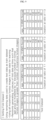
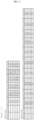
FIG. 10 is a diagram for describing a process of modeling an incidence prediction model.FIG. 11 is a diagram illustrating a score according to a data type indicating a ratio of 1:1. -
FIG. 12 is a diagram illustrating a score according to a data type indicating a ratio of 1:2.FIG. 13 is a diagram illustrating a score according to a data type indicating a ratio of 1:3.FIG. 14 is a diagram illustrating a ROC curve for each data type.FIG. 15 is a diagram illustrating a ROC curve for each data type according to a plurality of fold datasets. - Referring to
FIG. 10 , a data type may be a ratio of a fold dataset to fundus images labeled as normal patients. For example, the data type may include a data ratio of 1:1, a data ratio of 1:2, and a data ratio of 1:3. - Trasnform may include resizing a fundus image, adjusting a verticalflip of the fundus image, adjusting a color jitter of the fundus image, and adjusting a channel of lambda of the fundus image. In the meantime, a horizontalflip of the fundus image may not reflect a difference between the left and right eyes.
- When an incidence prediction model is finetuned, the performance of the incidence prediction model may vary based on the range of a frozen layer. For example, the number of freezing layers for freezing layers may be 5.
- A verification operation of a model may be performed. In this case, 20% of patients randomly split may follow the age distribution of the training dataset.
- In some embodiments, the
processor 220 may learn a plurality of incidence prediction models as many as 'n' data types, each of which indicates the data ratio of 1:n, the number of freezing layers, and the number of the fold dataset. Besides, theprocessor 220 may select an incidence prediction model, which has the highest AUROC, from among the plural incidence prediction models. For example, the incidence prediction models may be learned as many as the number (e.g., 150) corresponding to the product of 3 data types, 5 freezing layers, and 10 fold datasets. In this case, the incidence prediction model with the maximum AUROC may be selected from among 150 incidence prediction models. - In the data type of a data ratio of 1:1, a score for each freezing layer of the incidence prediction model learned depending on fold datasets may be calculated as shown in
FIG. 11 . In this case, best scores may indicate the highest AUROC calculated for each freezing layer. Average scores may indicate that average AUROC is calculated for each freezing layer. - In the data type of a data ratio of 1:2, best scores and average scores of the incidence prediction model learned for each freezing layer may be calculated as shown in
FIG. 12 . - In the data type of a data ratio of 1:3, best scores and average scores of the incidence prediction model learned for each freezing layer may be calculated as shown in
FIG. 13 . - The best score with the highest AUROC in each of the data ratio of 1:1, the data ratio of 1:2, and the data ratio of 1:3 shown in
FIGS. 11 to 13 may be calculated as shown inFIG. 14 . - The average score with the highest AUROC in each of the data ratio of 1:1, the data ratio of 1:2, and the data ratio of 1:3 shown in
FIGS. 11 to 13 may be calculated as shown inFIG. 15 . -
FIG. 16 is a diagram showing fundus images for each stage.FIG. 17 is a diagram for describing a plurality of stages. - Referring to
FIGS. 2 and16 , a fundus image may include nerves, ocular substances, and blood vessels. The Fundus image may vary for each age and for each stage. The fundus image may be preprocessed as shown inFIG. 16 . The fundus image analyzed by Grad-CAM may be as shown inFIG. 16 . The Grad-CAM may be a method used to interpret a model based on CNN. The image of blood vessels in the fundus image may be as shown inFIG. 16 . - The
processor 220 may determine the moyamoya disease based on at least one of the thickness of a blood vessel and the number of blood vessels included in the new fundus image of the moyamoya disease patient, and a predetermined reference value. In some embodiments, in normal patients, theprocessor 220 may determine a moyamoya disease based on the entire fundus image. - Referring to
FIG. 17 , a change in cerebral blood vessels may vary depending on stages. This change may be expected to be visible through a fundus. Accordingly, the binary classification shown inFIG. 17 may be added. The plurality of stages may include astage 0 indicating normality, astage 1 indicating stenosis of suprasellar internal carotid artery, astage 2 indicating that stenosis of an internal carotid artery increases, and a moyamoya blood vessel is prominent, astage 3 indicating that the moyamoya blood vessel in a brain is developed, astage 4 indicating that occlusion of a circle of Willis and posterior cerebral artery begin, an extracranial collateral begins to appear, and the moyamoya blood vessel begins to disappear, astage 5 indicating that a phenomenon according to thestage 4 further progresses, and astage 6 indicating that the moyamoya blood vessel of a main cerebral artery completely disappears. - When the plurality of stages include
stages 0 to 6, patients with moyamoya diseases according tostages 1 to 6 may be compared with normal patients. Alternatively, patients with moyamoya diseases according tostages 2 to 6 may be compared with the normal patients. Alternatively, patients with moyamoya diseases according tostages 3 to 6 may be compared with the normal patients. Alternatively, patients with moyamoya diseases according tostages 4 to 6 may be compared with the normal patients. Alternatively, patients with moyamoya diseases according tostages 5 to 6 may be compared with the normal patients. -
FIG. 18 is a diagram for describing a process of modeling an incidence prediction model according to a data type indicating a ratio of 1:3. - Referring to
FIG. 18 , similarly to those described above with reference toFIG. 10 , after the data type is set, trasnform is performed, the number of freezing layers is determined, and validation is performed, an incidence prediction model may be modeled. - Unlike shown in
FIG. 10 , inFIG. 18 , a data ratio of 1:3 may be used, and the incidence prediction model may be modeled based on a freezinglayer 4. Accordingly, the incidence prediction model having the highest test AUROC may be selected from among the number of incidence prediction models corresponding to the product of 5 tasks and 10 fold datasets. -
FIG. 19 is a diagram showing a best score for each target stage range.FIG. 20 is a diagram showing an average score for each target stage range.FIG. 21 is a diagram showing a best score for each target stage.FIG. 22 is a diagram showing an average score for each target stage. - Referring to
FIGS. 18 and19 , a best score for normality and each target stage range (e.g., stages 0 to 6, stages 1 to 6, stages 2 to 6, stages 3 to 6, stages 4 to 6, or stages 5 to 6) may be improved. In this case, a task for normality and stages 4 to 6 may have the highest AUROC. - Referring to
FIGS. 18 and20 , an average score for normality and each target stage range (e.g., stages 0 to 6, stages 1 to 6, stages 2 to 6, stages 3 to 6, stages 4 to 6, or stages 5 to 6) may be improved. In this case, a task for normality and stages 4 to 6 may have the highest AUROC. - Referring to
FIGS. 18 and21 , the best score for normality and a target stage (e.g., any one ofstages 1 to 6) may be improved. In this case, a task for normality andstage 4 may have the highest AUROC. - Referring to
FIGS. 18 and22 , the average score for normality and a target stage (e.g., any one ofstages 1 to 6) may be improved. In this case, a task for normality andstage 4 may have the highest AUROC. - Meanwhile, the disclosed embodiments may be implemented in a form of a recording medium storing instructions executable by a computer. The instructions may be stored in a form of program codes, and, when executed by a processor, generate a program module to perform operations of the disclosed embodiments. The recording medium may be implemented as a computer-readable recording medium.
- The computer-readable recording medium may include all kinds of recording media in which instructions capable of being decoded by a computer are stored. For example, there may be read only memory (ROM), random access memory (RAM), magnetic tape, magnetic disk, flash memory, optical data storage device, and the like.
- Disclosed embodiments are described above with reference to the accompanying drawings. One ordinary skilled in the art to which the present disclosure belongs will understand that the present disclosure may be practiced in forms other than the disclosed embodiments without altering the technical ideas or essential features of the present disclosure. The disclosed embodiments are examples and should not be construed as limited thereto.
- According to the above-mentioned problem solving means of the present disclosure, the incidence of a moyamoya disease may be quickly and accurately predicted by using machine learning that learns retinal images.
- Effects of the present disclosure are not limited to the effects mentioned above, and other effects not mentioned will be clearly understood by those skilled in the art from the following description.
- While the present disclosure has been described with reference to embodiments, it will be apparent to those skilled in the art that various changes and modifications may be made without departing from the spirit and scope of the present disclosure. Therefore, it should be understood that the above embodiments are not limiting, but illustrative.
Claims (15)
- A server comprising:a memory configured to store a deep learning-based incidence prediction model; anda processor configured to predict an incidence probability of a moyamoya disease of a patient from a fundus image by using the incidence prediction model,wherein the processor is configured to:label input fundus images as a moyamoya disease patient with the moyamoya disease and a normal patient without the moyamoya disease;learn the incidence prediction model by recognizing fundus images labeled as the moyamoya disease patient and fundus images labeled as the normal patient into the incidence prediction model; andwhen a new fundus image is input, predict the incidence probability of the moyamoya disease by inputting the new fundus image into the learned incidence prediction model.
- The server of claim 1, wherein the processor is configured to:remove metadata and text data;use only a fundus image including an optic disk for learning;perform gray scaling on an image; andpreprocess the input fundus images by enhancing a contrast of an image.
- The server of claim 2, wherein the processor is configured to:split fundus images labeled in an order of stages, which have fewer patients for each age group, from among a plurality of stages classified depending on severity into a train dataset;split 80% of fundus images labeled as the moyamoya disease patient into a training dataset of the train dataset, and split 20% of fundus images into a test dataset of the train dataset; andsplit the fundus images labeled as the normal patient into the train dataset at a ratio of 1:1 between the number of the fundus images labeled as the normal patient and the number of the fundus images labeled as the moyamoya disease patient, which are split for each age group.
- The server of claim 3, wherein the processor is configured to:split the fundus images labeled as the normal patient into a fold dataset at a ratio of 1:n between the fundus images labeled as the normal patient and the number of the fundus images labeled as the moyamoya disease patient, which are split for each age group, wherein the 'n' is an integer greater than or equal to 1; andgenerate the fold dataset including a plurality of fold datasets.
- The server of claim 4, wherein the processor is configured to:learn a plurality of incidence prediction models as many as 'n' data types, each of which indicates the ratio of 1:n, the number of freezing layers for freezing layers when the incidence prediction model is finetuned, and the number of the fold dataset; andselect an incidence prediction model having the highest area under receiver operating characteristic (AUROC) from among the plural incidence prediction models.
- The server of claim 5, wherein the fundus image includes a nerve, an ocular substance, and a blood vessel, and
wherein the processor is configured to:
determine the moyamoya disease based on a predetermined reference value, and at least one of a thickness of the blood vessel included in the new fundus image of the moyamoya disease patient, and the number of the blood vessel. - The server of claim 6, wherein the processor is configured to:
determine the moyamoya disease based on the entire new fundus image of the normal patient. - The server of claim 7, wherein the plurality of stages include:a stage 0 indicating normality;a stage 1 indicating stenosis of suprasellar internal carotid artery;a stage 2 indicating that stenosis of an internal carotid artery increases, and a moyamoya blood vessel is prominent;a stage 3 indicating that the moyamoya blood vessel in a brain is developed;a stage 4 indicating that occlusion of a circle of Willis and posterior cerebral artery begin, an extracranial collateral begins to appear, and the moyamoya blood vessel begins to disappear;a stage 5 indicating that a phenomenon according to the stage 4 further progresses: anda stage 6 indicating that the moyamoya blood vessel of a main cerebral artery completely disappears.
- A method of predicting an incidence probability of a moyamoya disease in a patient from a fundus image by using a deep learning-based incidence prediction model, the method comprising:labeling input fundus images as a moyamoya disease patient with the moyamoya disease and a normal patient without the moyamoya disease;learning the incidence prediction model by recognizing fundus images labeled as the moyamoya disease patient and fundus images labeled as the normal patient into the incidence prediction model; andpredicting the incidence probability of the moyamoya disease by inputting a new fundus image thus input into the learned incidence prediction model.
- The method of claim 9, further comprising:removing metadata and text data;using only a fundus image including an optic disk for learning;performing gray scaling on an image; andpreprocessing the input fundus images by enhancing a contrast of an image.
- The method of claim 10, wherein the learning of the incidence prediction model includes:
splitting fundus images labeled in an order of stages, which have fewer patients for each age group, from among a plurality of stages classified depending on severity into a train dataset. - The method of claim 11, wherein the learning of the incidence prediction model includes:splitting 80% of fundus images labeled as the moyamoya disease patient into a training dataset of the train dataset, and split 20% of fundus images into a test dataset of the train dataset; andsplitting the fundus images labeled as the normal patient into the train dataset at a ratio of 1:1 between the number of the fundus images labeled as the normal patient and the number of the fundus images labeled as the moyamoya disease patient, which are split for each age group.
- The method of claim 12, wherein the learning of the incidence prediction model includes:splitting the fundus images labeled as the normal patient into a fold dataset at a ratio of 1n between the fundus images labeled as the normal patient and the number of the fundus images labeled as the moyamoya disease patient, which are split for each age group, wherein the 'n' is an integer greater than or equal to 1; andgenerating the fold dataset including a plurality of fold datasets.
- The method of claim 13, wherein the learning of the incidence prediction model includes:learning a plurality of incidence prediction models as many as 'n' data types, each of which indicates the ratio of 1:n, the number of freezing layers for freezing layers when the incidence prediction model is finetuned, and the number of the fold dataset; andselecting an incidence prediction model having the highest AUROC from among the plural incidence prediction models.
- The method of claim 14, wherein the fundus image includes a nerve, an ocular substance, and a blood vessel, and
wherein the predicting of the incidence probability of the moyamoya disease includes:
determining the moyamoya disease based on blood vessels included in a new fundus image of the moyamoya disease patient.
Applications Claiming Priority (2)
| Application Number | Priority Date | Filing Date | Title |
|---|---|---|---|
| KR20230000520 | 2023-01-03 | ||
| KR1020230040570A KR20240109560A (en) | 2023-01-03 | 2023-03-28 | Server, method, and computer program for predicting incidence of moyamoya disease through learning of fundus image |
Publications (2)
| Publication Number | Publication Date |
|---|---|
| EP4398259A1 true EP4398259A1 (en) | 2024-07-10 |
| EP4398259B1 EP4398259B1 (en) | 2025-09-03 |
Family
ID=89430090
Family Applications (1)
| Application Number | Title | Priority Date | Filing Date |
|---|---|---|---|
| EP24150080.0A Active EP4398259B1 (en) | 2023-01-03 | 2024-01-02 | Server and method for predicting incidence of moyamoya disease through learning of fundus image |
Country Status (2)
| Country | Link |
|---|---|
| US (1) | US20240221945A1 (en) |
| EP (1) | EP4398259B1 (en) |
Families Citing this family (1)
| Publication number | Priority date | Publication date | Assignee | Title |
|---|---|---|---|---|
| CN119941646B (en) * | 2024-12-27 | 2025-09-23 | 清华大学 | Fundus image analysis method, fundus image analysis apparatus, fundus image analysis computer device, fundus image analysis program, and fundus image analysis program |
Family Cites Families (14)
| Publication number | Priority date | Publication date | Assignee | Title |
|---|---|---|---|---|
| US7517864B2 (en) * | 2001-05-18 | 2009-04-14 | Sirna Therapeutics, Inc. | RNA interference mediated inhibition of vascular endothelial growth factor and vascular endothelial growth factor receptor gene expression using short interfering nucleic acid (siNA) |
| CN102596019B (en) * | 2009-09-04 | 2014-12-10 | 约翰斯·霍普金斯大学 | Multimode Laser Speckle Imaging |
| GB201518365D0 (en) * | 2015-10-16 | 2015-12-02 | Exonate Ltd | Compounds |
| CA3077749A1 (en) * | 2017-09-27 | 2019-04-04 | Exonate Limited | Srpk1 inhibitors |
| WO2019074545A1 (en) * | 2017-10-13 | 2019-04-18 | iHealthScreen Inc. | Image based screening system for prediction of individual at risk of late age-related macular degeneration (amd) |
| US12254985B2 (en) * | 2017-12-20 | 2025-03-18 | Medi Whale Inc. | Diagnosis assistance method and cardiovascular disease diagnosis assistance method |
| SG11202005927VA (en) * | 2017-12-20 | 2020-07-29 | Medi Whale Inc | Method and apparatus for assisting in diagnosis of cardiovascular disease |
| CN112868068B (en) * | 2018-10-17 | 2024-05-10 | 谷歌有限责任公司 | Processing fundus camera images using machine learning models trained with other modes |
| US20220138998A1 (en) * | 2019-02-04 | 2022-05-05 | Vasoptic Medical Inc. | System and method for augmented reality visualization of biomedical imaging data |
| CN110263755B (en) * | 2019-06-28 | 2021-04-27 | 上海鹰瞳医疗科技有限公司 | Eye ground image recognition model training method, eye ground image recognition method and eye ground image recognition device |
| WO2021087140A1 (en) * | 2019-10-29 | 2021-05-06 | Google Llc | Multi-variable heatmaps for computer-aided diagnostic models |
| US20210374955A1 (en) * | 2020-06-02 | 2021-12-02 | Zasti Inc. | Retinal color fundus image analysis for detection of age-related macular degeneration |
| TWI790572B (en) * | 2021-03-19 | 2023-01-21 | 宏碁智醫股份有限公司 | Detecting method and detecting apparatus related to image |
| US12493958B2 (en) * | 2022-03-02 | 2025-12-09 | Aeye Health, Inc. | Method and system for predicting manifestation or progression of a retinal malady and method for training machine learning (ML) models for the same |
-
2023
- 2023-12-29 US US18/399,951 patent/US20240221945A1/en active Pending
-
2024
- 2024-01-02 EP EP24150080.0A patent/EP4398259B1/en active Active
Non-Patent Citations (3)
| Title |
|---|
| AKIYAMA YUKINORI ET AL: "Deep Learning-Based Approach for the Diagnosis of Moyamoya Disease", JOURNAL OF STROKE AND CEREBROVASCULAR DISEASES, DEMOS PUBLICATIONS, NEW YORK, NY, US, vol. 29, no. 12, 25 September 2020 (2020-09-25), XP086350680, ISSN: 1052-3057, [retrieved on 20200925], DOI: 10.1016/J.JSTROKECEREBROVASDIS.2020.105322 * |
| JAESEONG HONG: "Screening of Moyamoya Disease From Retinal Photographs: Development and Validation of Deep Learning Algorithms", STROKE, vol. 55, no. 3, 1 March 2024 (2024-03-01), US, pages 715 - 724, XP093160324, ISSN: 0039-2499, DOI: 10.1161/STROKEAHA.123.044026 * |
| LO CHUNG-MING ET AL: "Assessing Ischemic Stroke with Convolutional Image Features in Carotid Color Doppler", ULTRASOUND IN MEDICINE AND BIOLOGY, NEW YORK, NY, US, vol. 47, no. 8, 15 May 2021 (2021-05-15), pages 2266 - 2276, XP086677566, ISSN: 0301-5629, [retrieved on 20210515], DOI: 10.1016/J.ULTRASMEDBIO.2021.03.038 * |
Also Published As
| Publication number | Publication date |
|---|---|
| US20240221945A1 (en) | 2024-07-04 |
| EP4398259B1 (en) | 2025-09-03 |
Similar Documents
| Publication | Publication Date | Title |
|---|---|---|
| US20190110753A1 (en) | Deep learning-based diagnosis and referral of ophthalmic diseases and disorders | |
| Joshi et al. | VisionDeep-AI: Deep learning-based retinal blood vessels segmentation and multi-class classification framework for eye diagnosis | |
| Firdous et al. | A survey on diabetes risk prediction using machine learning approaches | |
| Dhanalakshmi et al. | Cloud-based glaucoma diagnosis in medical imaging using machine learning | |
| US20170316180A1 (en) | Behavior prediction apparatus, behavior prediction apparatus controlling method, and behavior prediction apparatus controlling program | |
| Karthika et al. | Improved ResNet_101 assisted attentional global transformer network for automated detection and classification of diabetic retinopathy disease | |
| Hung et al. | A deep learning approach to identify blepharoptosis by convolutional neural networks | |
| US20210134461A1 (en) | Methods and systems for prioritizing comprehensive prognoses and generating an associated treatment instruction set | |
| KR102685550B1 (en) | Method for dynamic optimization of cognitive and language rehabilitation training session based on artificial intelligence and apparatus thereof | |
| Alwakid et al. | RETRACTED: Enhancing diabetic retinopathy classification using deep learning | |
| KR20200129602A (en) | Method And System For Deducting Facial Asymmetry Information | |
| WO2022261513A1 (en) | Methods and systems of detecting and predicting chronic kidney disease and type 2 diabetes using deep learning models | |
| EP4398259B1 (en) | Server and method for predicting incidence of moyamoya disease through learning of fundus image | |
| Wu et al. | Integrating artificial intelligence in strabismus management: current research landscape and future directions | |
| Choi et al. | Automated detection of crystalline retinopathy via fundus photography using multistage generative adversarial networks | |
| CN117038088B (en) | Method, device, equipment and medium for determining onset of diabetic retinopathy | |
| Chen et al. | Optic disc and cup segmentation for glaucoma detection using attention u-net incorporating residual mechanism | |
| Qian et al. | Enhanced diagnosis of thyroid-associated eye diseases based on deep learning: A novel triplet loss design strategy | |
| Bajaj et al. | Machine learning models for enhanced stroke detection and prediction | |
| Agarwal et al. | Artificial intelligence for iris-based diagnosis in healthcare | |
| KR102227969B1 (en) | Method And System For Providing Strabismus Information Based On Symmetry Of Face Image | |
| US20250285740A1 (en) | Method and device for recognizing surgical stage based on visual multiple modality | |
| KR20240109560A (en) | Server, method, and computer program for predicting incidence of moyamoya disease through learning of fundus image | |
| Heo et al. | Development of a robust eye exam diagnosis platform with a deep learning model | |
| Okur et al. | A comparative analysis of deep convolutional networks for automated diagnosis of retinal detachment in dogs |
Legal Events
| Date | Code | Title | Description |
|---|---|---|---|
| PUAI | Public reference made under article 153(3) epc to a published international application that has entered the european phase |
Free format text: ORIGINAL CODE: 0009012 |
|
| STAA | Information on the status of an ep patent application or granted ep patent |
Free format text: STATUS: REQUEST FOR EXAMINATION WAS MADE |
|
| 17P | Request for examination filed |
Effective date: 20240102 |
|
| AK | Designated contracting states |
Kind code of ref document: A1 Designated state(s): AL AT BE BG CH CY CZ DE DK EE ES FI FR GB GR HR HU IE IS IT LI LT LU LV MC ME MK MT NL NO PL PT RO RS SE SI SK SM TR |
|
| GRAP | Despatch of communication of intention to grant a patent |
Free format text: ORIGINAL CODE: EPIDOSNIGR1 |
|
| STAA | Information on the status of an ep patent application or granted ep patent |
Free format text: STATUS: GRANT OF PATENT IS INTENDED |
|
| INTG | Intention to grant announced |
Effective date: 20250331 |
|
| GRAS | Grant fee paid |
Free format text: ORIGINAL CODE: EPIDOSNIGR3 |
|
| GRAA | (expected) grant |
Free format text: ORIGINAL CODE: 0009210 |
|
| STAA | Information on the status of an ep patent application or granted ep patent |
Free format text: STATUS: THE PATENT HAS BEEN GRANTED |
|
| AK | Designated contracting states |
Kind code of ref document: B1 Designated state(s): AL AT BE BG CH CY CZ DE DK EE ES FI FR GB GR HR HU IE IS IT LI LT LU LV MC ME MK MT NL NO PL PT RO RS SE SI SK SM TR |
|
| REG | Reference to a national code |
Ref country code: CH Ref legal event code: EP |
|
| REG | Reference to a national code |
Ref country code: DE Ref legal event code: R096 Ref document number: 602024000542 Country of ref document: DE |
|
| REG | Reference to a national code |
Ref country code: IE Ref legal event code: FG4D |
|
| REG | Reference to a national code |
Ref country code: NL Ref legal event code: MP Effective date: 20250903 |
|
| PG25 | Lapsed in a contracting state [announced via postgrant information from national office to epo] |
Ref country code: NO Free format text: LAPSE BECAUSE OF FAILURE TO SUBMIT A TRANSLATION OF THE DESCRIPTION OR TO PAY THE FEE WITHIN THE PRESCRIBED TIME-LIMIT Effective date: 20251203 |
|
| REG | Reference to a national code |
Ref country code: LT Ref legal event code: MG9D |
|
| PG25 | Lapsed in a contracting state [announced via postgrant information from national office to epo] |
Ref country code: FI Free format text: LAPSE BECAUSE OF FAILURE TO SUBMIT A TRANSLATION OF THE DESCRIPTION OR TO PAY THE FEE WITHIN THE PRESCRIBED TIME-LIMIT Effective date: 20250903 |
|
| PG25 | Lapsed in a contracting state [announced via postgrant information from national office to epo] |
Ref country code: HR Free format text: LAPSE BECAUSE OF FAILURE TO SUBMIT A TRANSLATION OF THE DESCRIPTION OR TO PAY THE FEE WITHIN THE PRESCRIBED TIME-LIMIT Effective date: 20250903 |
|
| PG25 | Lapsed in a contracting state [announced via postgrant information from national office to epo] |
Ref country code: GR Free format text: LAPSE BECAUSE OF FAILURE TO SUBMIT A TRANSLATION OF THE DESCRIPTION OR TO PAY THE FEE WITHIN THE PRESCRIBED TIME-LIMIT Effective date: 20251204 |
|
| PG25 | Lapsed in a contracting state [announced via postgrant information from national office to epo] |
Ref country code: SE Free format text: LAPSE BECAUSE OF FAILURE TO SUBMIT A TRANSLATION OF THE DESCRIPTION OR TO PAY THE FEE WITHIN THE PRESCRIBED TIME-LIMIT Effective date: 20250903 |
|
| PG25 | Lapsed in a contracting state [announced via postgrant information from national office to epo] |
Ref country code: LV Free format text: LAPSE BECAUSE OF FAILURE TO SUBMIT A TRANSLATION OF THE DESCRIPTION OR TO PAY THE FEE WITHIN THE PRESCRIBED TIME-LIMIT Effective date: 20250903 |
|
| PG25 | Lapsed in a contracting state [announced via postgrant information from national office to epo] |
Ref country code: BG Free format text: LAPSE BECAUSE OF FAILURE TO SUBMIT A TRANSLATION OF THE DESCRIPTION OR TO PAY THE FEE WITHIN THE PRESCRIBED TIME-LIMIT Effective date: 20250903 Ref country code: PL Free format text: LAPSE BECAUSE OF FAILURE TO SUBMIT A TRANSLATION OF THE DESCRIPTION OR TO PAY THE FEE WITHIN THE PRESCRIBED TIME-LIMIT Effective date: 20250903 |
|
| PG25 | Lapsed in a contracting state [announced via postgrant information from national office to epo] |
Ref country code: RS Free format text: LAPSE BECAUSE OF FAILURE TO SUBMIT A TRANSLATION OF THE DESCRIPTION OR TO PAY THE FEE WITHIN THE PRESCRIBED TIME-LIMIT Effective date: 20251203 |
|
| PG25 | Lapsed in a contracting state [announced via postgrant information from national office to epo] |
Ref country code: ES Free format text: LAPSE BECAUSE OF FAILURE TO SUBMIT A TRANSLATION OF THE DESCRIPTION OR TO PAY THE FEE WITHIN THE PRESCRIBED TIME-LIMIT Effective date: 20250903 |
|
| REG | Reference to a national code |
Ref country code: AT Ref legal event code: MK05 Ref document number: 1834176 Country of ref document: AT Kind code of ref document: T Effective date: 20250903 |
|
| PG25 | Lapsed in a contracting state [announced via postgrant information from national office to epo] |
Ref country code: NL Free format text: LAPSE BECAUSE OF FAILURE TO SUBMIT A TRANSLATION OF THE DESCRIPTION OR TO PAY THE FEE WITHIN THE PRESCRIBED TIME-LIMIT Effective date: 20250903 |
|
| PG25 | Lapsed in a contracting state [announced via postgrant information from national office to epo] |
Ref country code: SM Free format text: LAPSE BECAUSE OF FAILURE TO SUBMIT A TRANSLATION OF THE DESCRIPTION OR TO PAY THE FEE WITHIN THE PRESCRIBED TIME-LIMIT Effective date: 20250903 |
|
| PGFP | Annual fee paid to national office [announced via postgrant information from national office to epo] |
Ref country code: DE Payment date: 20260115 Year of fee payment: 3 |
|
| PG25 | Lapsed in a contracting state [announced via postgrant information from national office to epo] |
Ref country code: AT Free format text: LAPSE BECAUSE OF FAILURE TO SUBMIT A TRANSLATION OF THE DESCRIPTION OR TO PAY THE FEE WITHIN THE PRESCRIBED TIME-LIMIT Effective date: 20250903 |
|
| PG25 | Lapsed in a contracting state [announced via postgrant information from national office to epo] |
Ref country code: IT Free format text: LAPSE BECAUSE OF FAILURE TO SUBMIT A TRANSLATION OF THE DESCRIPTION OR TO PAY THE FEE WITHIN THE PRESCRIBED TIME-LIMIT Effective date: 20250903 |
|
| PG25 | Lapsed in a contracting state [announced via postgrant information from national office to epo] |
Ref country code: IS Free format text: LAPSE BECAUSE OF FAILURE TO SUBMIT A TRANSLATION OF THE DESCRIPTION OR TO PAY THE FEE WITHIN THE PRESCRIBED TIME-LIMIT Effective date: 20260103 |
|
| PGFP | Annual fee paid to national office [announced via postgrant information from national office to epo] |
Ref country code: FR Payment date: 20260121 Year of fee payment: 3 |
|
| PG25 | Lapsed in a contracting state [announced via postgrant information from national office to epo] |
Ref country code: PT Free format text: LAPSE BECAUSE OF FAILURE TO SUBMIT A TRANSLATION OF THE DESCRIPTION OR TO PAY THE FEE WITHIN THE PRESCRIBED TIME-LIMIT Effective date: 20260105 Ref country code: CZ Free format text: LAPSE BECAUSE OF FAILURE TO SUBMIT A TRANSLATION OF THE DESCRIPTION OR TO PAY THE FEE WITHIN THE PRESCRIBED TIME-LIMIT Effective date: 20250903 |
|
| PG25 | Lapsed in a contracting state [announced via postgrant information from national office to epo] |
Ref country code: EE Free format text: LAPSE BECAUSE OF FAILURE TO SUBMIT A TRANSLATION OF THE DESCRIPTION OR TO PAY THE FEE WITHIN THE PRESCRIBED TIME-LIMIT Effective date: 20250903 Ref country code: SK Free format text: LAPSE BECAUSE OF FAILURE TO SUBMIT A TRANSLATION OF THE DESCRIPTION OR TO PAY THE FEE WITHIN THE PRESCRIBED TIME-LIMIT Effective date: 20250903 |
|
| REG | Reference to a national code |
Ref country code: DE Ref legal event code: R097 Ref document number: 602024000542 Country of ref document: DE |
|
| PG25 | Lapsed in a contracting state [announced via postgrant information from national office to epo] |
Ref country code: LT Free format text: LAPSE BECAUSE OF FAILURE TO SUBMIT A TRANSLATION OF THE DESCRIPTION OR TO PAY THE FEE WITHIN THE PRESCRIBED TIME-LIMIT Effective date: 20250903 |
|
| PG25 | Lapsed in a contracting state [announced via postgrant information from national office to epo] |
Ref country code: DK Free format text: LAPSE BECAUSE OF FAILURE TO SUBMIT A TRANSLATION OF THE DESCRIPTION OR TO PAY THE FEE WITHIN THE PRESCRIBED TIME-LIMIT Effective date: 20250903 |
|
| PLBE | No opposition filed within time limit |
Free format text: ORIGINAL CODE: 0009261 |
|
| STAA | Information on the status of an ep patent application or granted ep patent |
Free format text: STATUS: NO OPPOSITION FILED WITHIN TIME LIMIT |
|
| REG | Reference to a national code |
Ref country code: CH Ref legal event code: L10 Free format text: ST27 STATUS EVENT CODE: U-0-0-L10-L00 (AS PROVIDED BY THE NATIONAL OFFICE) Effective date: 20260715 |
|
| 26N | No opposition filed |
Effective date: 20260604 |