EP4397746A1 - Cell image analysis system, cell image analysis device, and cell image analysis method - Google Patents
Cell image analysis system, cell image analysis device, and cell image analysis method Download PDFInfo
- Publication number
- EP4397746A1 EP4397746A1 EP22864314.4A EP22864314A EP4397746A1 EP 4397746 A1 EP4397746 A1 EP 4397746A1 EP 22864314 A EP22864314 A EP 22864314A EP 4397746 A1 EP4397746 A1 EP 4397746A1
- Authority
- EP
- European Patent Office
- Prior art keywords
- cell
- pseudopodium
- pseudopodia
- area
- image
- Prior art date
- Legal status (The legal status is an assumption and is not a legal conclusion. Google has not performed a legal analysis and makes no representation as to the accuracy of the status listed.)
- Granted
Links
Images





Classifications
-
- C—CHEMISTRY; METALLURGY
- C12—BIOCHEMISTRY; BEER; SPIRITS; WINE; VINEGAR; MICROBIOLOGY; ENZYMOLOGY; MUTATION OR GENETIC ENGINEERING
- C12M—APPARATUS FOR ENZYMOLOGY OR MICROBIOLOGY; APPARATUS FOR CULTURING MICROORGANISMS FOR PRODUCING BIOMASS, FOR GROWING CELLS OR FOR OBTAINING FERMENTATION OR METABOLIC PRODUCTS, i.e. BIOREACTORS OR FERMENTERS
- C12M1/00—Apparatus for enzymology or microbiology
- C12M1/34—Measuring or testing with condition measuring or sensing means, e.g. colony counters
-
- G—PHYSICS
- G06—COMPUTING OR CALCULATING; COUNTING
- G06T—IMAGE DATA PROCESSING OR GENERATION, IN GENERAL
- G06T7/00—Image analysis
- G06T7/0002—Inspection of images, e.g. flaw detection
- G06T7/0012—Biomedical image inspection
- G06T7/0014—Biomedical image inspection using an image reference approach
-
- C—CHEMISTRY; METALLURGY
- C12—BIOCHEMISTRY; BEER; SPIRITS; WINE; VINEGAR; MICROBIOLOGY; ENZYMOLOGY; MUTATION OR GENETIC ENGINEERING
- C12Q—MEASURING OR TESTING PROCESSES INVOLVING ENZYMES, NUCLEIC ACIDS OR MICROORGANISMS; COMPOSITIONS OR TEST PAPERS THEREFOR; PROCESSES OF PREPARING SUCH COMPOSITIONS; CONDITION-RESPONSIVE CONTROL IN MICROBIOLOGICAL OR ENZYMOLOGICAL PROCESSES
- C12Q1/00—Measuring or testing processes involving enzymes, nucleic acids or microorganisms; Compositions therefor; Processes of preparing such compositions
- C12Q1/02—Measuring or testing processes involving enzymes, nucleic acids or microorganisms; Compositions therefor; Processes of preparing such compositions involving viable microorganisms
- C12Q1/04—Determining presence or kind of microorganism; Use of selective media for testing antibiotics or bacteriocides; Compositions containing a chemical indicator therefor
-
- G—PHYSICS
- G01—MEASURING; TESTING
- G01N—INVESTIGATING OR ANALYSING MATERIALS BY DETERMINING THEIR CHEMICAL OR PHYSICAL PROPERTIES
- G01N33/00—Investigating or analysing materials by specific methods not covered by groups G01N1/00 - G01N31/00
- G01N33/48—Biological material, e.g. blood, urine; Haemocytometers
-
- G—PHYSICS
- G06—COMPUTING OR CALCULATING; COUNTING
- G06T—IMAGE DATA PROCESSING OR GENERATION, IN GENERAL
- G06T11/00—Two-dimensional [2D] image generation
-
- G—PHYSICS
- G06—COMPUTING OR CALCULATING; COUNTING
- G06T—IMAGE DATA PROCESSING OR GENERATION, IN GENERAL
- G06T7/00—Image analysis
- G06T7/60—Analysis of geometric attributes
- G06T7/62—Analysis of geometric attributes of area, perimeter, diameter or volume
-
- G—PHYSICS
- G06—COMPUTING OR CALCULATING; COUNTING
- G06T—IMAGE DATA PROCESSING OR GENERATION, IN GENERAL
- G06T2207/00—Indexing scheme for image analysis or image enhancement
- G06T2207/10—Image acquisition modality
- G06T2207/10056—Microscopic image
-
- G—PHYSICS
- G06—COMPUTING OR CALCULATING; COUNTING
- G06T—IMAGE DATA PROCESSING OR GENERATION, IN GENERAL
- G06T2207/00—Indexing scheme for image analysis or image enhancement
- G06T2207/20—Special algorithmic details
- G06T2207/20081—Training; Learning
-
- G—PHYSICS
- G06—COMPUTING OR CALCULATING; COUNTING
- G06T—IMAGE DATA PROCESSING OR GENERATION, IN GENERAL
- G06T2207/00—Indexing scheme for image analysis or image enhancement
- G06T2207/20—Special algorithmic details
- G06T2207/20084—Artificial neural networks [ANN]
-
- G—PHYSICS
- G06—COMPUTING OR CALCULATING; COUNTING
- G06T—IMAGE DATA PROCESSING OR GENERATION, IN GENERAL
- G06T2207/00—Indexing scheme for image analysis or image enhancement
- G06T2207/30—Subject of image; Context of image processing
- G06T2207/30004—Biomedical image processing
- G06T2207/30024—Cell structures in vitro; Tissue sections in vitro
-
- G—PHYSICS
- G06—COMPUTING OR CALCULATING; COUNTING
- G06T—IMAGE DATA PROCESSING OR GENERATION, IN GENERAL
- G06T2210/00—Indexing scheme for image generation or computer graphics
- G06T2210/41—Medical
-
- G—PHYSICS
- G06—COMPUTING OR CALCULATING; COUNTING
- G06T—IMAGE DATA PROCESSING OR GENERATION, IN GENERAL
- G06T7/00—Image analysis
- G06T7/0002—Inspection of images, e.g. flaw detection
- G06T7/0012—Biomedical image inspection
Definitions
- Patent Document 1 Japanese Patent Publication No. JP 6694240
- a cell image analysis method includes a step of acquiring a cell image including a cell(s); a step of acquiring a cell area(s) from the cell image; a step of acquiring an area(s) of a pseudopodium/pseudopodia in the cell area(s); and a step of acquiring aging index information that indicates a degree(s) of aging of the cell(s) based a length(s) of the pseudopodium/pseudopodia.
- the cell image analysis apparatus 100 includes a processor 10, such as CPU (Central Processing Unit), FPGA (Field-Programmable Gate Array), and ASIC (Aplication Specific Integrated Circuit).
- the cell image analysis apparatus 100 is configured to perform arithmetic processing by using the processor 10 by executing a predetermined program 21.
- the cell image analysis apparatus 100 apply analysis processing and image processing to the cell image 30 in response to a request from the computer 110. As a result of the analysis processing, the cell image analysis apparatus 100 acquires the aging index information 24. In addition, as a result of image processing, the cell image analysis apparatus 100 generates the superimposed cell image 80. The cell image analysis apparatus 100 transmits the aging index information 24 acquired and the superimposed cell image 80 generated to the computer 110. The computer 110 displays the aging index information 24 and the superimposed cell image 80 on the display 111 when receiving the information.
- the aged state of the cell 90 refers to a state in which its cell cycle stably stops.
- the term the cell is becoming closer to an aged state refers to that the cell is becoming closer to the state in which its cell cycle stably stop.
- the cell image analysis apparatus 100 acquires the index information 24 that indicates degrees of aging of the cells 90 based pseudopodia 90b of the cells 90 included in the cell image 30.
- the cell image analysis apparatus 100 can generate the superimposed cell image 80, which allows operators to realize the pseudopodia 90b at a glance.
- the following description describes the cell image analysis apparatus 100 in detail.
- the degree of aging of the cell 90 refers to how much the cell 90 is close to the state in which its cell cycle stably stop. In other words, the degree of aging of the cell 90 is a degree of progression of aging of the cell.
- the processor 10 of the cell image analysis apparatus 100 includes a cell image acquirer 11, a cell area acquirer 12, a pseudopodium area acquirer 13, an aging index information acquirer 14, and a superimposed-cell-image generator 15 as functional blocks.
- the processor 10 is configured to execute the program 21 stored in the storage 20 to serve as a cell image acquirer 11, the cell area acquirer 12, the pseudopodium area acquirer 13, the aging index information acquirer 14, and the superimposed-cell-image generator 15.
- the cell image acquirer 11 has a function of acquiring the cell image 30 including the cells 90.
- the cell image acquirer 11 is configured to read a cell image 30 stored in the storage 20 (see FIG. 1 ) and to acquire the cell image 30 to which the binarization is applied.
- the cell image acquirer 11 may acquire a cell image 30 sent from the imaging apparatus 120 or the computer 110 through the network 130 (see FIG. 1 ).
- the cell image acquirer 11 is configured to output the cell image 30 acquired to the cell area acquirer 12, the pseudopodium area acquirer 13, and the superimposed-cell-image generator 15.
- the cell area acquirer 12 acquires the cell areas 91 from the cell image 30.
- the cell area acquirer 12 is configured to acquire the cell areas 91 (see FIG. 5 ) based on a learned model 23 (see FIG. 1 ) having learned to acquire cell areas 91 by extracting areas of cytoskeletons from teacher images including cells 90, and the cell image 30.
- the acquisition of the cell area 91 refers to that the cell area acquirer 12 specifies pixels (coordinates of pixels) that correspond to each cell area 91 in the cell image 30.
- the pseudopodium area acquirer 13 acquires areas 92 (see FIG. 6 ) of the pseudopodia 90b (see FIG. 2 ) that are an elongated areas of the cell areas 91 in the cell image 30.
- the pseudopodium area acquirer 13 acquires the areas 92 of the pseudopodia 90b based on the cell image 30, and a pseudopodium removal image 32 in which the pseudopodia 90b are removed from the cell image 30.
- the pseudopodium area acquirer 13 acquires a pseudopodium area image 36 (see FIG. 8 ) as the areas 92 of the pseudopodia 90b.
- the pseudopodium area acquirer 13 outputs the areas 92 of the pseudopodia 90b acquired (pseudopodium area image 36) to the aging index information acquirer 14.
- the aging index information 24 (distribution 24a of lengths of the pseudopodia 90b, and index value 24b) acquired by the aging index information acquirer 14 is output to the storage 20.
- the aging index information 24 (distribution 24a of lengths of the pseudopodia 90b, and index value 24b) is stored in the storage 20.
- the aging index information acquirer 14 outputs the aging index information 24 (distribution 24a of the lengths of the pseudopodia 90b, and index value 24b) to the superimposed-cell-image generator 15.
- the superimposed-cell-image generator 15 generates the superimposed cell image 80 from the cell image 30.
- the cell areas 91 (see FIG. 5 ) and the areas 92 (see FIG. 6 ) of the pseudopodia 90b (see FIG. 2 ) are superimposed on the cell image 30, and the indices 70 (see FIG. 11 ) indicating lengths of the pseudopodia 90b are superimposed on the areas 92 of the pseudopodium 90b.
- the superimposed cell image 80 generated by the superimposed-cell-image generator 15 is output to the storage 20. As a result, the superimposed cell image 80 is stored in the storage 20.
- the aging index information 24 and the superimposed cell image 80 are transmitted to the computer 110 in response to request, and is displayed on the display 111.
- the step (1) of acquiring the cell image 30 is executed by the cell image acquirer 11.
- the step (2) of acquiring the cell areas 91 is executed by the cell area acquirer 12.
- the step (3) of acquiring the areas 92 of the pseudopodia 90b is executed by the pseudopodium area acquirer 13.
- Step (4) of acquiring the aging index information 24 is executed by the aging index information acquirer 14.
- the cell image analysis method according to this embodiment further includes processes of excluding pseudopodia 90b separated from main bodies 90a of cells 90 by using the pseudopodium area acquirer 13, processes of excluding pseudopodia 90b that are in contact with edges of the cell image 30 by using the pseudopodium area acquirer 13, and processes of generating the superimposed cell image 80 by using the superimposed-cell-image generator 15.
- a processing flow executed by the cell image analysis apparatus 100 is now described in detail with reference to FIGS. 4 to 12 .
- step S1 the cell image acquirer 11 (see FIG. 3 ) acquires a cell image 30 from the storage 20, the imaging apparatus 120 or the computer 110.
- the processing in step S1 is the aforementioned processes in the step (1).
- step S2 the cell area acquirer 12 (see FIG. 3 ) executes processes of acquiring the cell areas 91 (see FIG. 5 ) from the cell image 30 acquired in step S1 (see the aforementioned step (2)).
- the processes of acquiring the cell areas 91 are described in detail with reference to FIG. 5 .
- step S2c the cell area acquirer 12 stores the cell area 91 in the storage 20. Specifically, the cell area acquirer 12 stores the cell area 91 by storing the acquired cell area image 31 in the storage 20.
- step S3a the pseudopodium area acquirer 13 acquires the cell area image 31.
- the pseudopodium area acquirer 13 acquires the cell area image 31 from the storage 20.
- the pseudopodium area acquirer 13 acquires the pseudopodium area image 33 in which only the areas 92 of the pseudopodia 90b is included by subtracting from pixel values of pixels in the cell area image 31, pixel value of their corresponding pixel in the pseudopodium removal image 32.
- the removal process of small cell areas is a process of removing the cell areas 91 that have an area not larger than a predetermined size.
- the pseudopodium area acquirer 13 removes the areas 92 of the pseudopodia 90b that have an area not larger than 10 px (pixel) x 10 px (100 px 2 ), for example.
- noises included in the candidate areas 92 of the pseudopodia 90b extracted by the binarization process can be removed, it is possible to prevent that the noises are extracted as the areas 92 of the pseudopodia 90b.
- step S3f the pseudopodium area acquirer 13 stores the areas 92 of the pseudopodia 90b after the noise removal in the storage 20. Specifically, the pseudopodium area acquirer 13 stores the pseudopodium area image 33a acquired after noise reduction.
- the pseudopodium area acquirer 13 excludes, in the areas 92 (see FIG. 2 ) of the pseudopodia 90b (see FIG. 2 ), the areas 92 of the pseudopodia 90b separated from the main bodies 90a (see FIG. 2 ) of the cells 90 (see FIG. 2 ) from subjects to be acquired for the aging index information 24. processes of excluding the areas 92 of the pseudopodia 90b separated from the main bodies 90a of the cells 90 by using the pseudopodium area acquirer 13 is described in detail with reference to FIG. 7 .
- Step S4 of excluding the areas 92 of the pseudopodia 90b separated from the main bodies 90a of the cells 90 includes step S4a of acquiring the cell area image 31, step S4b of acquiring the pseudopodium area image 33a whose noises are removed, step S4c of acquiring the pseudopodia 90b separated from the main bodies 90a of the cells 90, step S4d of excluding the pseudopodia 90b separated from the main bodies 90a of the cells 90, and step S4e of storing the pseudopodium area image 35 in which the pseudopodia 90b separated from the main bodies 90a of the cells 90 are removed.
- step S4a the pseudopodium area acquirer 13 acquires the cell areas 91. Specifically, the pseudopodium area acquirer 13 acquires the cell area image 31 from the storage 20.
- step S4b the pseudopodium area acquirer 13 acquires the pseudopodium area image 33a from the storage 20.
- the pseudopodium area image 33a includes only the areas 92 of the pseudopodia 90b after noise removal. Accordingly, the pseudopodium area acquirer 13 can specify the areas 92 of the pseudopodia 90b after noise removal based on the pseudopodium area image 33a. Any one of the process in step S4a and the process in step S4b can be executed before another process.
- the pseudopodium area acquirer 13 excludes the areas 92 of the pseudopodia 90b separated from the main bodies 90a of the cells 90 from subjects to be acquired for the aging index information 24.
- the pseudopodium area acquirer 13 acquires the pseudopodium area image 35 after exclusion processing is by removing the areas 92 of the pseudopodia 90b separated from the main bodies 90a of the cells 90 from the pseudopodium area image 33a.
- the pseudopodium area acquirer 13 excludes, in the areas 92 of the pseudopodia 90b, the pseudopodia 90b that are in contact with the edges of the cell image 30 from subjects to be acquired for the aging index information 24.
- the processes of excluding the pseudopodia 90b that are in contact with the edges of the cell image 30 are described in detail with reference to FIG. 8 .
- the step S5 of excluding the pseudopodia 90b that are in contact with the edges of the cell image 30 includes step S5a of acquiring the pseudopodium area image 35 in which the pseudopodia 90b separated from the main bodies 90a of the cells 90 are removed, step S5b of acquiring the pseudopodia 90b that are in contact with the edges of the cell image 30, step S5c of excluding the pseudopodia 90b that are in contact with the edges of the cell image 30, and step S5d of storing the pseudopodium area image 36 in which the pseudopodia 90b that are in contact with the edges of the cell image 30 are excluded.
- step S5a the pseudopodium area acquirer 13 acquires the pseudopodium area image 35. Specifically, the pseudopodium area acquirer 13 reads the pseudopodium area image 35 stored in the storage 20.
- the pseudopodium area acquirer 13 acquires the pseudopodia 90b that are in contact with the edges of the cell image 30.
- the pseudopodium area acquirer 13 acquires the pseudopodia 90b that are in contact with the edges of the pseudopodium area image 35 in which the pseudopodia 90b that are separated from the main bodies 90a of the cells 90 are excluded.
- the pseudopodium area acquirer 13 acquires pixels that have a pixel value of not 0 (zero) in outermost peripheral pixels of the pseudopodium area image 35.
- the pseudopodium area acquirer 13 specifies the areas 92 of the pseudopodia 90b that includes the outermost peripheral pixels that have a pixel value of not 0 (zero) as the pseudopodia 90b that are in contact with the edges of the cell image 30.
- the pseudopodium area acquirer 13 can have a known configuration for acquiring the pseudopodium 90b that are in contact with the edges of the cell image 30 other than the aforementioned configuration.
- the pseudopodium area acquirer 13 acquires, in the areas 92 of the pseudopodia 90b included in the pseudopodium area image 35, the areas 92 of the pseudopodia 90b that are encircled by oval lines 41 as the pseudopodia 90b that are in contact with the edges of the pseudopodium area image 35.
- the pseudopodium area acquirer 13 excludes the pseudopodia 90b that are in contact with the edges of the cell image 30. Specifically, the pseudopodium area acquirer 13 excludes the pseudopodia 90b that are in contact with the pseudopodium area image 35 acquired in step S5b from the pseudopodium area image 35 so as to exclude the pseudopodia 90b that are in contact with the edges of the cell image 30. Specifically, the pseudopodium area acquirer 13 excludes the pseudopodia 90b that are in contact with the edges of the cell image 30 by replacing pixel values of the areas 92 of the pseudopodia 90b that are in contact with the edges of the cell image 30 with 0 (zero). In this embodiment, the pseudopodium area acquirer 13 acquires the pseudopodium area image 36 in which excluding the pseudopodia 90b that are in contact with the edges of the cell image 30 are excluded.
- the pseudopodium area acquirer 13 stores the areas 92 of the pseudopodia 90b that do not include the pseudopodia 90b that are in contact with the edges of the cell image 30 and have been excluded. Specifically, the pseudopodium area acquirer 13 stores the areas 92 of the pseudopodia 90b that do not include the pseudopodia 90b that are in contact with the edges of the cell image 30 and have been excluded by storing the pseudopodium area image 36 in the storage 20.
- the aging index information acquirer 14 acquires the aging index information 24 that indicates degrees of aging of the cells 90 based on the lengths of the pseudopodia 90b (the aforementioned step (4)).
- the aging index information acquirer 14 acquires the aging index information 24 based on the pseudopodia 90b whose length is not smaller than a predetermined length. The processes of acquiring the aging index information 24 by using the aging index information acquirer 14 are described in detail with reference to FIG. 9 .
- Step S6 of acquiring the aging index information 24 includes step S6a of acquiring the pseudopodium area image 36 in which the pseudopodia 90b that are in contact with edges of the cell image 30 are excluded, step S6b of acquiring minimum bounding rectangles 71 of the pseudopodia 90b included in the pseudopodium area image 36, step S6c of acquiring longitudinal lengths of the areas of the pseudopodia 90b, step S6d of acquiring the aging index information 24, and step S6e of storing the aging index information 24.
- step S6a the aging index information acquirer 14 acquires the areas 92 of the pseudopodia 90b that do not include the pseudopodia 90b that are in contact with the edges of the cell image 30 and have been excluded. Specifically, the aging index information acquirer 14 specifies the areas 92 of the pseudopodia 90b that do not include the pseudopodia 90b that are in contact with the edges of the cell image 30 and have been excluded based on the pseudopodium area image 36 read from the storage 20.
- the pseudopodium area acquirer 13 acquires the minimum bounding rectangles 71 of the pseudopodia 90b included in the pseudopodium area image 35 as shown in an image 37.
- the minimum bounding rectangle 71 refers to a rectangle that has the smallest area among rectangles that circumscribe the pseudopodium 90b.
- the pseudopodium area acquirer 13 acquires the minimum bounding rectangle 71 by using calculation processing.
- the technique for acquiring the minimum bounding rectangle 71 by using the pseudopodium area acquirer 13 is not limited to calculation processing.
- the image 37 is an image for illustrating a configuration of acquiring the minimum bounding rectangles 71, and is not an image to actually be generated.
- the aging index information acquirer 14 acquires longitudinal lengths of the areas 92 of the pseudopodia 90b so as to acquire the lengths of the pseudopodia 90b.
- the aging index information acquirer 14 is configured to acquire longitudinal lengths L of the minimum bounding rectangles 71 of the areas 92 of the pseudopodia 90b so as to acquire the lengths of the areas 92 of the pseudopodia 90b.
- the aging index information acquirer 14 acquires longitudinal lengths L of the minimum bounding rectangles 71 for all the areas 92 of the pseudopodia 90b so that the area of each pseudopodium has its length defined by the longitudinal length of its corresponding minimum bounding rectangle.
- the aging index information acquirer 14 calculates the length L of a long side of the minimum bounding rectangle 71 not in px (pixels) but in ⁇ m by using information on a magnification of the cell image 30.
- the area 92 of the pseudopodium 90b acquired by the pseudopodium area acquirer 13 can include not only the area 92 of the actual pseudopodium 90b but also an area other than the pseudopodium 90b, such as a thin part of the cell 90 that becomes thinner due to cell overlapping in some cases.
- the aging index information 24 is acquired in such as case in which the area of the pseudopodium 90b includes an area other than the pseudopodium, accuracy of the aging index information 24 is reduced.
- the aging index information acquirer 14 acquires the aging index information 24 based on the pseudopodia 90b whose length is not smaller than a predetermined length.
- the predetermined length is 40 pm, for example.
- the Number of Long Pseudopodia is the number of the pseudopodia 90b whose length is not smaller than 100 um.
- the Number of Pseudopodia Having Length Not Smaller Than Predetermined Length is the number of pseudopodia 90b whose length is not smaller than the predetermined length among the pseudopodia 90b included in the pseudopodium area image 36. That is, the Number of Pseudopodia Having Length Not Smaller Than Predetermined Length can be obtained by adding the number of pseudopodia 90b whose length is not smaller than 40 um and smaller than 100 ⁇ m, to the number of pseudopodia 90b whose length is not smaller than 100 um.
- step S6e the aging index information acquirer 14 stores the distribution 24a of length of the pseudopodia 90b, and the index value 24b in the storage 20. Also, the aging index information acquirer 14 stores the image 37 on which the minimum bounding rectangles 71 are superimposed in the storage 20.
- the green indices 70 are indicated by solid lines, the blue indices 70 are indicated by dashed lines, the red indices 70 are indicated by thick lines as shown in legends 41.
- the indices 70 are superimposed not on all the pseudopodia 90b for ease of illustration, the indices 70 are actually superimposed on the pseudopodia 90b whose length is not smaller than 40 um in accordance with their lengths.
- a degree of aging of cells 90 can be acquired based on the cell image 30 without extracting miRNA from the supernatant. Consequently, it is possible to easily and speedily analyze a degree of aging of the cells 90 by acquiring the cell image 30.
- miRNA miRNA
- the pseudopodium area acquirer 13 is configured to acquire the pseudopodium removal image 32 by reducing and expanding the cell image 30. Accordingly, narrow and long areas in the image can be easily removed by using reduction processing that can remove narrow parts in the image and expansion processing that returns a size of the image reduced by the reduction processing to its original size. Consequently, it is possible to easily remove the pseudopodia 90b, which are elongated areas, from the cell image 30.
- the pseudopodium area acquirer 13 is configured to exclude, in the areas 92 of pseudopodia 90b acquired, the pseudopodia 90b that are in contact with edges of the cell image 30 from a subject to be acquired for the aging index information 24. Accordingly, it possible to exclude, from the subject to be acquired for the aging index information 24, the pseudopodium 90b whose length cannot be accurately acquired due to the contact with the edges of the cell image 30. Consequently, it is possible to prevent reduction of accuracy of the aging index information 24 caused by the pseudopodia 90b whose length cannot be accurately acquired.
- the cell area acquirer 12 is configured to acquire the cell areas 91 based on a learned model 23 having learned to acquire cell areas 91 by extracting areas of cytoskeletons from teacher images including cells 90, and the cell image 30. Accordingly, because the cell areas 91 can be acquired by the learned model 23, it possible to prevent a burden on operators from becoming larger as compared with a configuration in which operators visually acquires the cell areas 91.
- a superimposed-cell-image generator 15 configured to superimpose the cell area 91 and the areas 92 of the pseudopodia 90b on the cell image 30, and to superimpose indices 70 indicating the lengths of pseudopodia 90b on the areas 92 of pseudopodia 90b so as to generate a superimposed cell image 80. Accordingly, the cell areas 91 and the area 92 in the pseudopodium 90b can be easily grasped at a glance in the cell image 30 by seeing the superimposed cell image 80. Also, because the indices 70 indicating the lengths of the pseudopodia 90b are superimposed on the superimposed cell image 80, the lengths of the pseudopodia 90b can be easily grasped at a glance.
- the superimposed-cell-image generator 15 is configured to superimpose the indices 70 in different indication forms depending on the lengths of the areas 92 of the pseudopodia 90b. Accordingly, difference between the lengths of the pseudopodia 90b can be easily grasped by seeing indication forms of the indices 70.
- the present invention is not limited to this.
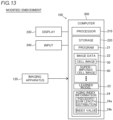
- the cell image analysis apparatus can be constructed of an independent computer, for example, as shown in FIG. 13 .
- the cell image analysis apparatus 100 shown in FIG. 13 includes a computer 300 including a processor 210 and a storage 220.
- a display 230 and an input 240 are connected to the computer 300.
- the computer 300 is connected to and can communicate with an imaging apparatus 120.
- the processor 210 of the computer 300 includes the cell image acquirer 11, the cell area acquirer 12, the pseudopodium area acquirer 13, the aging index information acquirer 14, and superimposed-cell-image generator 15 shown in the aforementioned embodiment (see FIG. 3 ) as functional blocks.
- the present invention is not limited to this.
- the aging index information acquirer 14 can be configured to acquire the aging index information 24 based all the pseudopodia 90b acquired by the pseudopodium area acquirer 13.
- the cells 90 overlap each other, an area that is not actually the pseudopodium 90b is sometimes acquired as the areas 92 of the pseudopodia 90b that are acquired by the pseudopodium area acquirer 13.
- an area other than the pseudopodium 90b is sometimes acquired as the areas 92 of the pseudopodia 90b that are acquired by the pseudopodium area acquirer 13. Accordingly, in a case in which the aging index information acquirer 14 is configured to acquire the aging index information 24 based all the pseudopodia 90b, accuracy of the aging index information 24 can be sometimes reduced. For this reason, it is preferable that the aging index information acquirer 14 is configured to acquire the aging index information 24 based on the pseudopodia 90b whose length is not smaller than a predetermined length.
- the present invention is not limited to this.
- the technique for acquiring the area 92 of the pseudopodium 90b by using the pseudopodium area acquirer 13 is not limited as long as it can acquire the area 92 of the pseudopodium 90b.
- the present invention is not limited to this.
- the technique for acquiring the area 92 of the pseudopodium 90b by using the pseudopodium area acquirer 13 is not limited as long as it can acquire the area 92 of the pseudopodium 90b.
- the present invention is not limited to this.
- the pseudopodium area acquirer 13 does not necessarily exclude the pseudopodium 90b separated from the main bodies 90a of the cells 90.
- the pseudopodia 90b that are used to acquire the aging index information 24 will include the pseudopodium 90b of the dead cell 90 and/or a thin part of the cell 90 that becomes thinner due to overlapping of cells. For this reason, accuracy of the aging index information 24 generated will be reduced.
- the pseudopodium area acquirer 13 is configured to exclude the pseudopodia 90b separated from the main bodies 90a of the cells 90.
- the present invention is not limited to this.
- the pseudopodium area acquirer 13 does not necessarily exclude the pseudopodia 90b that are in contact with the edges of the cell image 30.
- the pseudopodium 90b are in contact with the edge of the cell image 30, it is difficult to accurately determine a shape of the minimum bounding rectangle 71. Accordingly, it becomes difficult to accurately acquire the length of the pseudopodium 90b.
- the present invention is not limited to this.
- the cell image analysis system 200 does not necessarily include the superimposed-cell-image generator 15.
- the cell image analysis system 200 does not include the superimposed-cell-image generator 15
- the superimposed cell image 80 cannot be acquired.
- the cell image analysis system 200 includes the superimposed-cell-image generator 15.
- a cell image analysis system includes a cell image acquirer configured to acquire a cell image including a cell(s); a cell area acquirer configured to acquire a cell area(s) from the cell image; a pseudopodium area acquirer configured to acquire an area(s) of a pseudopodium/pseudopodia that is/are an elongated area(s) in the cell area(s) of the cell image; and an aging index information acquirer configured to acquire aging index information that indicates a degree(s) of aging of the cell(s) based a length(s) of the pseudopodium/pseudopodia.
- a display configured to display the aging index information is further provided.
- the aging index information acquirer is configured to acquire the aging index information based pseudopodia whose length is not smaller than a predetermined length.
- the aging index information acquirer is configured to acquire a longitudinal length(s) of the area(s) of the pseudopodium/pseudopodia so as to acquire the length(s) of the pseudopodium/pseudopodia.
- the pseudopodium area acquirer is configured to acquire a pseudopodium removal image by removing the pseudopodium/pseudopodia from the cell area, and to acquire the area(s) of the pseudopodium/pseudopodia based on difference between the cell image and the pseudopodium removal image.
- the pseudopodium area acquirer is configured to exclude, in the area(s) of the pseudopodium/pseudopodia acquired, the pseudopodium/pseudopodia that is/are in contact with an edge(s) of the cell image from a subject to be acquired for the aging index information.
Landscapes
- Engineering & Computer Science (AREA)
- Health & Medical Sciences (AREA)
- Chemical & Material Sciences (AREA)
- Life Sciences & Earth Sciences (AREA)
- Physics & Mathematics (AREA)
- Organic Chemistry (AREA)
- General Physics & Mathematics (AREA)
- General Health & Medical Sciences (AREA)
- Bioinformatics & Cheminformatics (AREA)
- Zoology (AREA)
- Wood Science & Technology (AREA)
- Theoretical Computer Science (AREA)
- Analytical Chemistry (AREA)
- Biochemistry (AREA)
- Biotechnology (AREA)
- Computer Vision & Pattern Recognition (AREA)
- Medicinal Chemistry (AREA)
- Molecular Biology (AREA)
- Genetics & Genomics (AREA)
- Biomedical Technology (AREA)
- General Engineering & Computer Science (AREA)
- Microbiology (AREA)
- Immunology (AREA)
- Proteomics, Peptides & Aminoacids (AREA)
- Nuclear Medicine, Radiotherapy & Molecular Imaging (AREA)
- Radiology & Medical Imaging (AREA)
- Geometry (AREA)
- Quality & Reliability (AREA)
- Medical Informatics (AREA)
- Urology & Nephrology (AREA)
- Toxicology (AREA)
- Hematology (AREA)
- Pathology (AREA)
- Biophysics (AREA)
- Food Science & Technology (AREA)
- Sustainable Development (AREA)
- Image Processing (AREA)
- Apparatus Associated With Microorganisms And Enzymes (AREA)
- Measuring Or Testing Involving Enzymes Or Micro-Organisms (AREA)
- Image Analysis (AREA)
Abstract
Description
- The present invention relates to a cell image analysis system, a cell image analysis apparatus, and a cell image analysis method, in particular, to a cell image analysis system, a cell image analysis apparatus, and a cell image analysis method for analyzing a degree of cell aging.
- Techniques for analyzing a degree of cell aging are disclosed in the art. Such a technique is disclosed in Japanese Patent Publication No.
JP 6694240 - Japanese Patent Publication No.
JP 6694240 JP 6694240 - Patent Document 1: Japanese Patent Publication No.
JP 6694240 - However, in a configuration disclosed in Japanese Patent Publication No.
JP 6694240 - The present invention is intended to solve the above problem, and one object of the present invention is to provide a cell image analysis system, a cell image analysis apparatus, and a cell image analysis method capable of easily and speedily analyzing a degree of cell aging.
- In order to attain the aforementioned object, a cell image analysis system according to a first aspect of the present invention includes a cell image acquirer configured to acquire a cell image including a cell(s); a cell area acquirer configured to acquire a cell area(s) from the cell image; a pseudopodium area acquirer configured to acquire an area(s) of a pseudopodium/pseudopodia that is/are an elongated area(s) in the cell area(s) of the cell image; and an aging index information acquirer configured to acquire aging index information that indicates a degree(s) of aging of the cell(s) based a length(s) of the pseudopodium/pseudopodia.
- A cell image analysis apparatus according to a second aspect of the present invention includes a cell image acquirer configured to acquire a cell image including a cell(s); a cell area acquirer configured to acquire a cell area(s)from the cell image; a pseudopodium area acquirer configured to acquire an area(s) of a pseudopodium/pseudopodia that is/are an elongated area(s) in the cell area(s) of the cell image; and an aging index information acquirer configured to acquire aging index information that indicates a degree(s) of aging of the cell(s) based a length(s) of the pseudopodium/pseudopodia.
- A cell image analysis method according to a third aspect of the present invention includes a step of acquiring a cell image including a cell(s); a step of acquiring a cell area(s) from the cell image; a step of acquiring an area(s) of a pseudopodium/pseudopodia in the cell area(s); and a step of acquiring aging index information that indicates a degree(s) of aging of the cell(s) based a length(s) of the pseudopodium/pseudopodia.
- In the cell image analysis apparatus according to the first aspect, the cell image analysis apparatus according to the second aspect, and the cell image analysis method according to the third aspect, dissimilar to a configuration that analyzes a degree of cell aging based on types and amounts of miRNA (microRNA) contained in supernatant of cultivation solution for cultivating cells, a degree of cell aging can be acquired based on the cell image without extracting miRNA from the supernatant by acquiring aging index information that indicates a degree(s) of aging of the cell(s) based a length(s) of a pseudopodium/pseudopodia included in the cell image. Consequently, it is possible to easily and speedily analyze a degree of cell aging by acquiring the cell image.
-
-
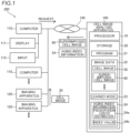
FIG. 1 is a block diagram showing an image processing system including an image processing apparatus according to an embodiment. -
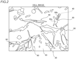
FIG. 2 is a view showing an exemplary cell image. -
FIG. 3 is a functional block diagram functions of processors of the image processing apparatus. -
FIG. 4 is a flowchart illustrating processes of a cell image analyzer according to this embodiment. -
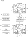
FIG. 5 is a diagram illustrating details of processes of acquiring cell areas. -
FIG. 6 is a diagram illustrating details of processes of acquiring pseudopodium areas. -
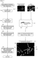
FIG. 7 is a diagram illustrating details of processes of excluding pseudopodia separated from main bodies of cells. -
FIG. 8 is a diagram illustrating details of processes of excluding pseudopodia that are in contact with edges of a cell image. -
FIG. 9 is a diagram illustrating details of processes of acquiring aging index information. -
FIG. 10 is a diagram illustrating distribution of lengths of pseudopodia. -
FIG. 11 is a diagram illustrating details of processes of acquiring a superimposed cell image. -
FIG. 12 is a view showing an exemplary screen showing the aging index information and the superimposed cell image on a display. -
FIG. 13 is a block diagram showing a cell image analysis apparatus according to a modified embodiment. Modes for Carrying Out the Invention - Embodiments embodying the present invention will be described with reference to the drawings.
- The following description describes a configuration of a cell
image analysis system 200 including a cellimage analysis apparatus 100, and a cell image analysis method according to an embodiment with reference toFIGS. 1 to 12 . - The cell
image analysis system 200 shown inFIG. 1 is a single system that allows users who cultivate cells, etc. integrally to capturecell images 30, to apply analysis processing to thecell images 30, and to peruse images to which the analysis processing is applied. - The cell
image analysis system 200 includes a cellimage analysis apparatus 100, acomputer 110, and animaging apparatus 120. - The cell
image analysis system 200 shown inFIG. 1 is illustratively constructed of a client-server model. Thecomputer 110 serves as a client terminal in the cellimage analysis system 200. The cellimage analysis apparatus 100 serves as a server in the cellimage analysis system 200. The cellimage analysis apparatus 100, thecomputer 110, and theimaging apparatus 120 are connected through anetwork 130, and can communicate with each other through the network. The cellimage analysis apparatus 100 performs various types of information processing in response to requests (processing requests) from thecomputer 110 operated by users. The cellimage analysis apparatus 100 is configured to apply analysis processing to thecell images 30 in response to request and to transmit images to which the analysis processing is applied to thecomputer 110. A graphical user interface (GUI) is displayed on adisplay 111 of thecomputer 110, and is configured to accept instructions provided to the cellimage analysis apparatus 100 and to display analysis results analyzed by the cellimage analysis apparatus 100 and images after the analysis. - The
network 130 connects the cellimage analysis apparatus 100, thecomputer 110, and theimaging apparatus 120 to each other so that they can communicate with each other. Thenetwork 130 can be a local area network (LAN) configured in the facility, for example. - The
network 130 can be the Internet, for example. In a case in which thenetwork 130 is the Internet, the cellimage analysis system 200 can be a system configured in a cloud computing form. - The
computer 110 is a so-called personal computer, equipped with a processor and storage. Thedisplay 111 and aninput 112 are connected to thecomputer 110. For example, thedisplay 111 is a liquid crystal display. Thedisplay 111 may be an electro-luminescence display, a projector or a head-mounted display. For example, theinput 112 is an input device including a computer mouse and a key-board. Theinput 112 may be a touch panel. The cellimage analysis system 200 can includes one ormore computers 110. In this embodiment, thedisplay 111 is configured to display agingindex information 24, which will be described later. - The
Imaging apparatus 120 is configured to generate thecell images 30 including cells 90 (seeFIG. 2 ). Theimaging apparatus 120 can send the generatedcell images 30 to thecomputer 110 and/or the cellimage analysis apparatus 100 through thenetwork 130. Theimaging apparatus 120 is configured to capture microscopic images of thecells 90. Theimaging apparatus 120 can use imaging methods such as bright-field observation, dark-field observation, phase contrast observation, and differential interference observation, etc. for imaging. One or more of theimaging apparatuses 120 are used in accordance with imaging methods. The cellimage analysis system 200 can include one ormore imaging apparatuses 120. - The cell
image analysis apparatus 100 includes aprocessor 10, such as CPU (Central Processing Unit), FPGA (Field-Programmable Gate Array), and ASIC (Aplication Specific Integrated Circuit). The cellimage analysis apparatus 100 is configured to perform arithmetic processing by using theprocessor 10 by executing apredetermined program 21. - The cell
image analysis apparatus 100 includes astorage 20. Thestorage 20 includes a non-volatile storage device. For example, the non-volatile storage device is a hard disk drives, solid state drives, etc. Thestorage 20 is configured to storevarious programs 21 to be executed by theprocessor 10. Thestorage 20 is configured to storeimage data 22. Theimage data 22 includes thecell images 30 captured by theImaging apparatus 120, and various images generated by applying image processing to the cell images 30 (superimposed cell images 80). Thestorage 20 stores a learnedmodel 23 that has learned to acquire cell areas 91 (seeFIG. 5 ) from thecell images 30. Also, thestorage 20 stores the agingindex information 24 acquired by analyzing thecell images 30. The agingindex information 24 indicates degrees of aging of thecells 90. Specifically, the agingindex information 24 includes adistribution 24a of lengths of thepseudopodia 90b (seeFIG. 2 ), which will be described later, and anindex value 24b. Among functions of analysis and image processing functions that can be performed by the cellimage analysis apparatus 100, a configuration that acquires the agingindex information 24 and processes of generating asuperimposed cell image 80 fromcell images 30 are described. - The cell
image analysis apparatus 100 apply analysis processing and image processing to thecell image 30 in response to a request from thecomputer 110. As a result of the analysis processing, the cellimage analysis apparatus 100 acquires the agingindex information 24. In addition, as a result of image processing, the cellimage analysis apparatus 100 generates the superimposedcell image 80. The cellimage analysis apparatus 100 transmits the agingindex information 24 acquired and thesuperimposed cell image 80 generated to thecomputer 110. Thecomputer 110 displays the agingindex information 24 and thesuperimposed cell image 80 on thedisplay 111 when receiving the information. - As shown in
FIG. 2 , thecell image 30 is a microscopic image of cultivatedcells 90 cultivated by using cell cultivation tools, for example. For example, thecells 90 included in thecell image 30 are adherent cells (non-colonialized cells), such as mesenchymal stem cells, fibroblasts, vascular endothelial cells, squamous cell carcinoma cells and endometrial cells. - The
cell image 30 includes images of cells 90 (cell images), and abackground 93. Thecell 90 in thecell image 30 shown inFIG. 2 includes amain body 90a of thecell 90, and a subcellular structure. The exemplary subcellular structure shown inFIG. 2 is apseudopodium 90b that is a protrusion of cytoplasm from themain body 90a of thecell 90, more specifically a filipodia that protrudes in a thread shape (filament-shape) from themain body 90a of thecell 90. As the exemplary subcellular structure shown inFIG. 2 , one ofpseudopodia 90b is indicated by arectangular box 40. Therectangular box 40 is merely shown for ease of illustrating thepseudopodium 90b. - It is known that a length of the
pseudopodium 90b increases as thecell 90 is becoming closer to an aged state. The aged state of thecell 90 refers to a state in which its cell cycle stably stops. The term the cell is becoming closer to an aged state refers to that the cell is becoming closer to the state in which its cell cycle stably stop. The cellimage analysis apparatus 100 according to this embodiment acquires theindex information 24 that indicates degrees of aging of thecells 90 basedpseudopodia 90b of thecells 90 included in thecell image 30. The cellimage analysis apparatus 100 according to this embodiment can generate thesuperimposed cell image 80, which allows operators to realize thepseudopodia 90b at a glance. The following description describes the cellimage analysis apparatus 100 in detail. The degree of aging of thecell 90 refers to how much thecell 90 is close to the state in which its cell cycle stably stop. In other words, the degree of aging of thecell 90 is a degree of progression of aging of the cell. -
FIG. 3 is a block diagram showing a configuration part that acquires the aging index information 24 (distribution 24a of lengths ofpseudopodia 90b andindex value 24b), a configuration part that generates the superimposedcell image 80, and other configuration parts in the cellimage analysis apparatus 100. - The
processor 10 of the cellimage analysis apparatus 100 includes acell image acquirer 11, acell area acquirer 12, apseudopodium area acquirer 13, an agingindex information acquirer 14, and a superimposed-cell-image generator 15 as functional blocks. In other words, theprocessor 10 is configured to execute theprogram 21 stored in thestorage 20 to serve as acell image acquirer 11, thecell area acquirer 12, thepseudopodium area acquirer 13, the agingindex information acquirer 14, and the superimposed-cell-image generator 15. - The
cell image acquirer 11 has a function of acquiring thecell image 30 including thecells 90. Thecell image acquirer 11 is configured to read acell image 30 stored in the storage 20 (seeFIG. 1 ) and to acquire thecell image 30 to which the binarization is applied. Thecell image acquirer 11 may acquire acell image 30 sent from theimaging apparatus 120 or thecomputer 110 through the network 130 (seeFIG. 1 ). In addition, thecell image acquirer 11 is configured to output thecell image 30 acquired to thecell area acquirer 12, thepseudopodium area acquirer 13, and the superimposed-cell-image generator 15. - The
cell area acquirer 12 acquires thecell areas 91 from thecell image 30. Specifically, thecell area acquirer 12 is configured to acquire the cell areas 91 (seeFIG. 5 ) based on a learned model 23 (seeFIG. 1 ) having learned to acquirecell areas 91 by extracting areas of cytoskeletons from teacherimages including cells 90, and thecell image 30. The acquisition of thecell area 91 refers to that thecell area acquirer 12 specifies pixels (coordinates of pixels) that correspond to eachcell area 91 in thecell image 30. - The learned
model 23 is produced by training the learned model by using images that includecells 90 as input data and images that include labeled areas of cytoskeletons as output data. The images that include labeled areas of cytoskeletons are images of cytoskeletons ofcells 90 stained by actin, or images of cytoskeletons ofcells 90 in thecell image 30 to which labels are attached by operators, for example. - The
pseudopodium area acquirer 13 acquires areas 92 (seeFIG. 6 ) of thepseudopodia 90b (seeFIG. 2 ) that are an elongated areas of thecell areas 91 in thecell image 30. In this embodiment, thepseudopodium area acquirer 13 acquires theareas 92 of thepseudopodia 90b based on thecell image 30, and apseudopodium removal image 32 in which thepseudopodia 90b are removed from thecell image 30. In this embodiment, thepseudopodium area acquirer 13 acquires a pseudopodium area image 36 (seeFIG. 8 ) as theareas 92 of thepseudopodia 90b. thepseudopodium area acquirer 13 outputs theareas 92 of thepseudopodia 90b acquired (pseudopodium area image 36) to the agingindex information acquirer 14. - The aging
index information acquirer 14 acquires the aging index information 24 (seeFIG. 1 ) that indicates degrees of aging ofcells 90 based on the lengths of thepseudopodia 90b. Specifically, the agingindex information acquirer 14 is configured to acquire, as the agingindex information 24, at least one ofdistribution 24a of lengths of thepseudopodia 90b, and anindex value 24b that indicates a ratio of thepseudopodia 90b whose length is not smaller than a predetermined length to thepseudopodia 90b acquired. In this embodiment, the agingindex information acquirer 14 acquires both thedistribution 24a of the lengths of thepseudopodia 90b, and theindex value 24b. The aging index information 24 (distribution 24a of lengths of thepseudopodia 90b, andindex value 24b) acquired by the agingindex information acquirer 14 is output to thestorage 20. As a result, the aging index information 24 (distribution 24a of lengths of thepseudopodia 90b, andindex value 24b) is stored in thestorage 20. In this embodiment, the agingindex information acquirer 14 outputs the aging index information 24 (distribution 24a of the lengths of thepseudopodia 90b, andindex value 24b) to the superimposed-cell-image generator 15. - The superimposed-cell-
image generator 15 generates the superimposedcell image 80 from thecell image 30. In thesuperimposed cell image 80, the cell areas 91 (seeFIG. 5 ) and the areas 92 (seeFIG. 6 ) of thepseudopodia 90b (seeFIG. 2 ) are superimposed on thecell image 30, and the indices 70 (seeFIG. 11 ) indicating lengths of thepseudopodia 90b are superimposed on theareas 92 of thepseudopodium 90b. Thesuperimposed cell image 80 generated by the superimposed-cell-image generator 15 is output to thestorage 20. As a result, thesuperimposed cell image 80 is stored in thestorage 20. - The aging
index information 24 and thesuperimposed cell image 80 are transmitted to thecomputer 110 in response to request, and is displayed on thedisplay 111. - The cell image analysis method according to this embodiment is now described. The cell image analysis method according to this embodiment is a cell image analysis method for analyzing degrees of aging of
cells 90 in thecell image 30. The cell image analysis method can be executed by the cell image analysis apparatus 100 (processor 10). - The cell image analysis method of this embodiment includes at least the following steps.
- (1) Step of acquiring a
cell image 30 includingcells 90. - (2) Step of acquiring
cell areas 91 from thecell image 30. - (3) Step of acquiring
areas 92 ofpseudopodia 90b in thecell areas 91. - (4) Step of acquiring aging
index information 24 that indicates degrees of aging of thecells 90 based lengths ofpseudopodia 90b. - The step (1) of acquiring the
cell image 30 is executed by thecell image acquirer 11. The step (2) of acquiring thecell areas 91 is executed by thecell area acquirer 12. The step (3) of acquiring theareas 92 of thepseudopodia 90b is executed by thepseudopodium area acquirer 13. Step (4) of acquiring the agingindex information 24 is executed by the agingindex information acquirer 14. The cell image analysis method according to this embodiment further includes processes of excludingpseudopodia 90b separated frommain bodies 90a ofcells 90 by using thepseudopodium area acquirer 13, processes of excludingpseudopodia 90b that are in contact with edges of thecell image 30 by using thepseudopodium area acquirer 13, and processes of generating thesuperimposed cell image 80 by using the superimposed-cell-image generator 15. - A processing flow executed by the cell
image analysis apparatus 100 is now described in detail with reference toFIGS. 4 to 12 . - In step S1, the cell image acquirer 11 (see
FIG. 3 ) acquires acell image 30 from thestorage 20, theimaging apparatus 120 or thecomputer 110. The processing in step S1 is the aforementioned processes in the step (1). - In step S2, the cell area acquirer 12 (see
FIG. 3 ) executes processes of acquiring the cell areas 91 (seeFIG. 5 ) from thecell image 30 acquired in step S1 (see the aforementioned step (2)). The processes of acquiring thecell areas 91 are described in detail with reference toFIG. 5 . - In step S2a, the
cell area acquirer 12 inputs thecell image 30 into the learnedmodel 23. The learnedmodel 23, receives thecell image 30 input, outputs probability values of pixels of thecell image 30 each of which indicates that each pixel belongs to thecell area 91. The learnedmodel 23 outputs one probability value per pixel. - In step S2b, the
cell area acquirer 12 acquires thecell areas 91. Specifically, thecell area acquirer 12 acquires thecell area image 31 based on the probability values, which are output from the learnedmodel 23 and indicate that the pixels belong to thecell areas 91. In this embodiment, thecell area acquirer 12 acquires distribution of probability values indicating that the pixels belong to thecell areas 91 as thecell area image 31. Pixels other than thecell areas 91 in thecell area image 31 are defined as abackground 93. - In step S2c, the
cell area acquirer 12 stores thecell area 91 in thestorage 20. Specifically, thecell area acquirer 12 stores thecell area 91 by storing the acquiredcell area image 31 in thestorage 20. - The
cell area image 31 is an image that shows thecell areas 91 and thebackground 93 discriminatively from each other.. Specifically, thecell areas 91 are colored in thecell area image 31. In this embodiment, thecell areas 91 are colored in blue in thecell area image 31, for example. Thecell areas 91 are shown illustratively discriminatively from thebackground 93 by hatching thecell areas 91 for ease of illustration inFIG. 5 . - In step S3 of
FIG. 4 , thepseudopodium area acquirer 13 executes processes of acquiring theareas 92 of thepseudopodium 90b from thecell areas 91 acquired by the cell area acquirer 12 (the aforementioned step (3)). The processes of acquiring theareas 92 of thepseudopodia 90b are described in detail with reference toFIG. 6 . - Step S3 of acquiring the
areas 92 ofpseudopodia 90b includes step S3a of acquiring thecell area image 31, step S3b of reducing thecell area image 31, step S3c of expanding a reducedcell area image 31a, step S3d of acquiring theareas 92 ofpseudopodia 90b based on thecell area image 31 and thepseudopodium removal image 32, step S3e of removing noises from thepseudopodium area image 33, and step S3f of storing thepseudopodium area image 33 subjected to the noise removal. - In step S3a, the
pseudopodium area acquirer 13 acquires thecell area image 31. In this embodiment, thepseudopodium area acquirer 13 acquires thecell area image 31 from thestorage 20. - In step S3b, the
pseudopodium area acquirer 13 applies reduction processes to thecell area image 31. The reduction refers to processing in which a center of kernel whose size is set to a predetermined kernel size is specified to a target pixel, and a pixel value of the target pixel is replaced with the smallest pixel value in pixel values of pixels that are included in the kernel. - In this embodiment, the
pseudopodium area acquirer 13 acquires the reducedcell area image 31a by executing a predetermined number of the reduction processes. - In the reduced
cell area image 31a, edges of thecell 90 will become equal to a pixel value of thebackground 93 by replacing pixel values of the reduced cell area image with the pixel value of thebackground 93 by in the reduction processing. As a result, thepseudopodia 90b will disappear after the reduction processes. While thepseudopodia 90b disappear, areas other than thepseudopodia 90b (main bodies 90a of cells 90) are also reduced. For this reason, thecell areas 91 in the reducedcell area image 31a become smaller than thecell areas 91 in thecell area image 31 before the reduction processing. The kernel size is set in accordance with a size of thepseudopodium 90b to be removed from thecell area image 31. In this embodiment, the kernel size is, for example, 3 x 3 px (pixels). The number of reduction processes is also set in accordance with the size of thepseudopodium 90b to be removed from thecell area image 31. That is, the kernel size and the number of reduction processes are set in accordance with sizes of thecells 90 included in thecell image 30. In this embodiment, thepseudopodium area acquirer 13 executes four reduction processes, for example. The sizes of thecells 90 included in thecell image 30 varies depending on a magnification when thecell image 30 is captured. For this reason, the kernel size and the number of reduction processes are set for eachcell image 30 in accordance with the sizes of thecells 90 included in thecell image 30 and the magnification when thecell image 30 is captured. - In step S3c, the
pseudopodium area acquirer 13 acquires thepseudopodium removal image 32 by expanding the reducedcell area image 31a. Thepseudopodium removal image 32 includes thecell areas 91 from which theareas 92 of thepseudopodia 90b are removed. Accordingly, thepseudopodium area acquirer 13 can specify pixels of thecell areas 91 that correspond to thecell areas 91 from which theareas 92 of thepseudopodia 90b are removed in thecell area image 31. That is, in this embodiment, thepseudopodium area acquirer 13 acquires thepseudopodium removal image 32 by reducing and expanding the cell image 30 (cell area image 31). Thepseudopodium removal image 32 shows the removal of thepseudopodium 90b from thecell area 91. - The expansion processing is processing opposite to the reduction processing. In other words, the expansion processing refers to processing in which a pixel value of the target pixel is replaced with the largest pixel value in pixel values of pixels that are included in the kernel. The
cell area 91 is increased every when the expansion process is executed. In this embodiment, thepseudopodium area acquirer 13 executes the expansion processes the number of which is equal to the number of the reduction processes executed. In this embodiment, thepseudopodium area acquirer 13 is executes four expansion processes, for example. As a result, thecell areas 91 included in thepseudopodium removal image 32 return to the original sizes of thecell areas 91 included in thecell area image 31. Although shapes of themain bodies 90a of thecells 90 return to their original shapes by the expansion processing, thepseudopodia 90b that have disappeared by the reduction processing do not return. As a result, thepseudopodium removal image 32 in which theareas 92 of thepseudopodia 90b are removed can be acquired by executing the reduction processing and the expansion processing. - In step S3d, the
pseudopodium area acquirer 13 acquires theareas 92 of thepseudopodia 90b. In this embodiment, thepseudopodium area acquirer 13 is configured to acquire theareas 92 of thepseudopodia 90b based on difference between thecell image 30 and thepseudopodium removal image 32. Specifically, thepseudopodium area acquirer 13 acquires thepseudopodium area image 33 including theareas 92 of thepseudopodia 90b by subtracting thepseudopodium removal image 32 from the cell area 91 (cell area image 31) acquired from thecell image 30. Specifically, in thepseudopodium area acquirer 13 acquires thepseudopodium area image 33 in which only theareas 92 of thepseudopodia 90b is included by subtracting from pixel values of pixels in thecell area image 31, pixel value of their corresponding pixel in thepseudopodium removal image 32. - In step S3e, the
pseudopodium area acquirer 13 removes noises from thepseudopodium area image 33. The noise removal processing includes a binarization process, a removal process of small cell areas, and a closing process. - Because areas that have a pixel value not smaller than a threshold value can be extracted as the
areas 92 of thepseudopodia 90b by executing the binarization process, it is possible to prevent that some of theareas 92 of thepseudopodia 90b cannot be extracted. - The removal process of small cell areas is a process of removing the
cell areas 91 that have an area not larger than a predetermined size. Thepseudopodium area acquirer 13 removes theareas 92 of thepseudopodia 90b that have an area not larger than 10 px (pixel) x 10 px (100 px2), for example. As a result, because noises included in thecandidate areas 92 of thepseudopodia 90b extracted by the binarization process can be removed, it is possible to prevent that the noises are extracted as theareas 92 of thepseudopodia 90b. - The closing processing is a process of expanding white areas in the image and then reducing the white areas. The closing processing can connect disconnected lines separated by a short distance and can fill holes (black areas) that locally exist in the cell image without changing sizes of the cell images (white areas).
- In step S3f, the
pseudopodium area acquirer 13 stores theareas 92 of thepseudopodia 90b after the noise removal in thestorage 20. Specifically, thepseudopodium area acquirer 13 stores thepseudopodium area image 33a acquired after noise reduction. - Although images are shown in different sizes in
FIGS. 5 to 9 ,11 , and12 for ease of illustration, the actual image size is not changed in this embodiment. - In step S4 of
FIG. 4 , the pseudopodium area acquirer 13 (seeFIG. 3 ) excludes, in the areas 92 (seeFIG. 2 ) of thepseudopodia 90b (seeFIG. 2 ), theareas 92 of thepseudopodia 90b separated from themain bodies 90a (seeFIG. 2 ) of the cells 90 (seeFIG. 2 ) from subjects to be acquired for the agingindex information 24. processes of excluding theareas 92 of thepseudopodia 90b separated from themain bodies 90a of thecells 90 by using thepseudopodium area acquirer 13 is described in detail with reference toFIG. 7 . - Step S4 of excluding the
areas 92 of thepseudopodia 90b separated from themain bodies 90a of thecells 90 includes step S4a of acquiring thecell area image 31, step S4b of acquiring thepseudopodium area image 33a whose noises are removed, step S4c of acquiring thepseudopodia 90b separated from themain bodies 90a of thecells 90, step S4d of excluding thepseudopodia 90b separated from themain bodies 90a of thecells 90, and step S4e of storing thepseudopodium area image 35 in which thepseudopodia 90b separated from themain bodies 90a of thecells 90 are removed. - In step S4a, the
pseudopodium area acquirer 13 acquires thecell areas 91. Specifically, thepseudopodium area acquirer 13 acquires thecell area image 31 from thestorage 20. - In step S4b, the
pseudopodium area acquirer 13 acquires thepseudopodium area image 33a from thestorage 20. Thepseudopodium area image 33a includes only theareas 92 of thepseudopodia 90b after noise removal. Accordingly, thepseudopodium area acquirer 13 can specify theareas 92 of thepseudopodia 90b after noise removal based on thepseudopodium area image 33a. Any one of the process in step S4a and the process in step S4b can be executed before another process. - In step S4c, the
pseudopodium area acquirer 13 acquires thepseudopodia 90b separated from themain bodies 90a (seeFIG. 2 ) of thecells 90. Specifically, thepseudopodium area acquirer 13 acquires theareas 92 of thepseudopodia 90b that have a size whose ratio of thearea 92 of thepseudopodium 90b to thecell area 91 is not smaller than a predetermined ratio as thepseudopodia 90b separated from themain bodies 90a of thecells 90. In this embodiment, thepseudopodium area acquirer 13 acquires anarea 91a of thecell area 91 and anarea 92a and thearea 92 of thepseudopodium 90b for eachcell area 91 as shown in animage 34. Thepseudopodium area acquirer 13 acquires a ratio of thearea 91a of thecell area 91 to thearea 92a of thearea 92 of thepseudopodium 90b. Specifically, thepseudopodium area acquirer 13 specifies thepseudopodia 90b that have a ratio of thearea 91a of thecell area 91 to thearea 92a and thearea 92 of thepseudopodium 90b as thepseudopodia 90b separated from themain bodies 90a of thecells 90. Theimage 34 is an image for illustrating a process of specifying thepseudopodia 90b that are separated from themain bodies 90a of thecells 90 by thepseudopodium area acquirer 13, and is not an image to be actually generated. - In step S4d, the
pseudopodium area acquirer 13 excludes theareas 92 of thepseudopodia 90b separated from themain bodies 90a of thecells 90 from subjects to be acquired for the agingindex information 24. In this embodiment, thepseudopodium area acquirer 13 acquires thepseudopodium area image 35 after exclusion processing is by removing theareas 92 of thepseudopodia 90b separated from themain bodies 90a of thecells 90 from thepseudopodium area image 33a. Specifically, thepseudopodium area acquirer 13 removes theareas 92 of thepseudopodia 90b separated from themain bodies 90a of thecells 90 from thepseudopodium area image 33a by replacing pixel values of theareas 92 of thepseudopodia 90b separated from themain bodies 90a of thecells 90 with 0 (zero) . - In step S4e, the
pseudopodium area acquirer 13 stores theareas 92 of thepseudopodia 90b after exclusion processing ofpseudopodia 90b separated from themain bodies 90a of thecells 90 in thestorage 20. Specifically, thepseudopodium area acquirer 13 stores thepseudopodium area image 35 in thestorage 20. - In step S5 of
FIG. 4 , thepseudopodium area acquirer 13 excludes, in theareas 92 of thepseudopodia 90b, thepseudopodia 90b that are in contact with the edges of thecell image 30 from subjects to be acquired for the agingindex information 24. The processes of excluding thepseudopodia 90b that are in contact with the edges of thecell image 30 are described in detail with reference toFIG. 8 . - The step S5 of excluding the
pseudopodia 90b that are in contact with the edges of thecell image 30 includes step S5a of acquiring thepseudopodium area image 35 in which thepseudopodia 90b separated from themain bodies 90a of thecells 90 are removed, step S5b of acquiring thepseudopodia 90b that are in contact with the edges of thecell image 30, step S5c of excluding thepseudopodia 90b that are in contact with the edges of thecell image 30, and step S5d of storing thepseudopodium area image 36 in which thepseudopodia 90b that are in contact with the edges of thecell image 30 are excluded. - In step S5a, the
pseudopodium area acquirer 13 acquires thepseudopodium area image 35. Specifically, thepseudopodium area acquirer 13 reads thepseudopodium area image 35 stored in thestorage 20. - In step S5b, the
pseudopodium area acquirer 13 acquires thepseudopodia 90b that are in contact with the edges of thecell image 30. In this embodiment, thepseudopodium area acquirer 13 acquires thepseudopodia 90b that are in contact with the edges of thepseudopodium area image 35 in which thepseudopodia 90b that are separated from themain bodies 90a of thecells 90 are excluded. For example, thepseudopodium area acquirer 13 acquires pixels that have a pixel value of not 0 (zero) in outermost peripheral pixels of thepseudopodium area image 35. Thepseudopodium area acquirer 13 specifies theareas 92 of thepseudopodia 90b that includes the outermost peripheral pixels that have a pixel value of not 0 (zero) as thepseudopodia 90b that are in contact with the edges of thecell image 30. Alternatively, thepseudopodium area acquirer 13 can have a known configuration for acquiring thepseudopodium 90b that are in contact with the edges of thecell image 30 other than the aforementioned configuration. - In this embodiment, the
pseudopodium area acquirer 13 acquires, in theareas 92 of thepseudopodia 90b included in thepseudopodium area image 35, theareas 92 of thepseudopodia 90b that are encircled byoval lines 41 as thepseudopodia 90b that are in contact with the edges of thepseudopodium area image 35. - In step S5c, the
pseudopodium area acquirer 13 excludes thepseudopodia 90b that are in contact with the edges of thecell image 30. Specifically, thepseudopodium area acquirer 13 excludes thepseudopodia 90b that are in contact with thepseudopodium area image 35 acquired in step S5b from thepseudopodium area image 35 so as to exclude thepseudopodia 90b that are in contact with the edges of thecell image 30. Specifically, thepseudopodium area acquirer 13 excludes thepseudopodia 90b that are in contact with the edges of thecell image 30 by replacing pixel values of theareas 92 of thepseudopodia 90b that are in contact with the edges of thecell image 30 with 0 (zero). In this embodiment, thepseudopodium area acquirer 13 acquires thepseudopodium area image 36 in which excluding thepseudopodia 90b that are in contact with the edges of thecell image 30 are excluded. - In step S5d, the
pseudopodium area acquirer 13 stores theareas 92 of thepseudopodia 90b that do not include thepseudopodia 90b that are in contact with the edges of thecell image 30 and have been excluded. Specifically, thepseudopodium area acquirer 13 stores theareas 92 of thepseudopodia 90b that do not include thepseudopodia 90b that are in contact with the edges of thecell image 30 and have been excluded by storing thepseudopodium area image 36 in thestorage 20. - In step S6 of
FIG. 4 , the aging index information acquirer 14 (seeFIG. 3 ) acquires the agingindex information 24 that indicates degrees of aging of thecells 90 based on the lengths of thepseudopodia 90b (the aforementioned step (4)). In this embodiment, the agingindex information acquirer 14 acquires the agingindex information 24 based on thepseudopodia 90b whose length is not smaller than a predetermined length. The processes of acquiring the agingindex information 24 by using the agingindex information acquirer 14 are described in detail with reference toFIG. 9 . - Step S6 of acquiring the aging
index information 24 includes step S6a of acquiring thepseudopodium area image 36 in which thepseudopodia 90b that are in contact with edges of thecell image 30 are excluded, step S6b of acquiringminimum bounding rectangles 71 of thepseudopodia 90b included in thepseudopodium area image 36, step S6c of acquiring longitudinal lengths of the areas of thepseudopodia 90b, step S6d of acquiring the agingindex information 24, and step S6e of storing the agingindex information 24. - In step S6a, the aging
index information acquirer 14 acquires theareas 92 of thepseudopodia 90b that do not include thepseudopodia 90b that are in contact with the edges of thecell image 30 and have been excluded. Specifically, the agingindex information acquirer 14 specifies theareas 92 of thepseudopodia 90b that do not include thepseudopodia 90b that are in contact with the edges of thecell image 30 and have been excluded based on thepseudopodium area image 36 read from thestorage 20. - In step S6b, the
pseudopodium area acquirer 13 acquires theminimum bounding rectangles 71 of thepseudopodia 90b included in thepseudopodium area image 35 as shown in animage 37. Theminimum bounding rectangle 71 refers to a rectangle that has the smallest area among rectangles that circumscribe thepseudopodium 90b. In this embodiment, thepseudopodium area acquirer 13 acquires theminimum bounding rectangle 71 by using calculation processing. The technique for acquiring theminimum bounding rectangle 71 by using thepseudopodium area acquirer 13 is not limited to calculation processing. Theimage 37 is an image for illustrating a configuration of acquiring theminimum bounding rectangles 71, and is not an image to actually be generated. - In step S6c, the aging
index information acquirer 14 acquires longitudinal lengths of theareas 92 of thepseudopodia 90b so as to acquire the lengths of thepseudopodia 90b. In this embodiment, the agingindex information acquirer 14 is configured to acquire longitudinal lengths L of theminimum bounding rectangles 71 of theareas 92 of thepseudopodia 90b so as to acquire the lengths of theareas 92 of thepseudopodia 90b. In this embodiment, the agingindex information acquirer 14 acquires longitudinal lengths L of theminimum bounding rectangles 71 for all theareas 92 of thepseudopodia 90b so that the area of each pseudopodium has its length defined by the longitudinal length of its corresponding minimum bounding rectangle. In this embodiment, the agingindex information acquirer 14 calculates the length L of a long side of theminimum bounding rectangle 71 not in px (pixels) but in µ m by using information on a magnification of thecell image 30. - In step S6d, the aging
index information acquirer 14 acquires the agingindex information 24. In this embodiment, the agingindex information acquirer 14 is configured to acquire, as the agingindex information 24, at least one ofdistribution 24a (seeFIG. 1 ) of lengths of thepseudopodia 90b, and anindex value 24b that indicates a ratio of thepseudopodia 90b whose length is not smaller than a predetermined length to thepseudopodia 90b acquired. - Here, the
area 92 of thepseudopodium 90b acquired by thepseudopodium area acquirer 13 can include not only thearea 92 of theactual pseudopodium 90b but also an area other than thepseudopodium 90b, such as a thin part of thecell 90 that becomes thinner due to cell overlapping in some cases. If the agingindex information 24 is acquired in such as case in which the area of thepseudopodium 90b includes an area other than the pseudopodium, accuracy of the agingindex information 24 is reduced. To address this, in this embodiment, the agingindex information acquirer 14 acquires the agingindex information 24 based on thepseudopodia 90b whose length is not smaller than a predetermined length. The predetermined length is 40 pm, for example. - In this embodiment, the aging
index information acquirer 14 acquires theindex value 24b based on the following Equation.Index Value = Number of Long Pseudopodia/(Number of Pseudopodia Having Length Not Smaller Than Predetermined Length) X 100 - Here, the Number of Long Pseudopodia is the number of the
pseudopodia 90b whose length is not smaller than 100 um. The Number of Pseudopodia Having Length Not Smaller Than Predetermined Length is the number ofpseudopodia 90b whose length is not smaller than the predetermined length among thepseudopodia 90b included in thepseudopodium area image 36. That is, the Number of Pseudopodia Having Length Not Smaller Than Predetermined Length can be obtained by adding the number ofpseudopodia 90b whose length is not smaller than 40 um and smaller than 100 µm, to the number ofpseudopodia 90b whose length is not smaller than 100 um. Alternatively, thelong pseudopodium 90b can be defined by a length other than 100 um. The length that defines thelong pseudopodium 90b is experimentally specified by performing an experiment that determines a criterion depending on a type of thecells 90 and environments in which thecells 90 are cultivated. In this embodiment, thepseudopodium 90b whose length is not smaller than 100 um is illustratively defined as thecell 90 that is becoming closer to an aged state. - In this embodiment, the aging
index information acquirer 14 acquires a ratio of thepseudopodia 90b whose length is not smaller than 100 um to thepseudopodia 90b whose length is not smaller than 40 um in thepseudopodia 90b that are included in thepseudopodium area image 36 as theindex value 24b. - In step S6e, the aging
index information acquirer 14 stores thedistribution 24a of length of thepseudopodia 90b, and theindex value 24b in thestorage 20. Also, the agingindex information acquirer 14 stores theimage 37 on which theminimum bounding rectangles 71 are superimposed in thestorage 20. -
FIG. 10 is a view showing anexemplary distribution 24a of lengths of thepseudopodia 90b (seeFIG. 2 ). Thedistribution 24a of lengths of thepseudopodia 90b is a histogram (frequency distribution) showing a length of thepseudopodium 90b on a horizontal axis, and a ratio (existence ratio of each length of thepseudopodium 90b) on a vertical axis. The ratio of thepseudopodia 90b whose length is not smaller than 100 um increases as thecells 90 are (seeFIG. 2 ) becoming closer to an aged state. That is, degrees of aging of thecells 90 can be visually grasped by seeing thedistribution 24a of lengths of thepseudopodia 90b. - In step S7 of
FIG. 4 , the superimposed-cell-image generator 15 generates the superimposedcell image 80 based on thecell image 30 and the lengths of thepseudopodia 90b. The processes of generatingsuperimposed cell image 80 are described in detail with reference toFIG. 11 . - In this embodiment, step of generating the
superimposed cell image 80 includes step S7a of acquiring thecell image 30, step S7b of acquiring thecell areas 91, step S7c of acquiring lengths of thepseudopodia 90b, step S7d to forming theminimum bounding rectangles 71 in different indication forms, step S7e of generating thesuperimposed cell image 80, step S7f of storing thesuperimposed cell image 80. - In step S7a, the superimposed-cell-
image generator 15 acquires thecell image 30. Specifically, the superimposed-cell-image generator 15 acquires thecell image 30 from thestorage 20. - In step S7b, the superimposed-cell-
image generator 15 acquires thecell areas 91. Specifically, the superimposed-cell-image generator 15 acquires thecell areas 91 by acquiring thecell area image 31 from thestorage 20. - In step S7c, the superimposed-cell-
image generator 15 acquires lengths of thepseudopodia 90b. Specifically, the superimposed-cell-image generator 15 acquires the superimposedimage 37 on which theminimum bounding rectangles 71 are superimposed from thestorage 20. The superimposed-cell-image generator 15 acquires the lengths of thepseudopodia 90b by acquiring lengths of long sides of theminimum bounding rectangles 71 in theimage 37. - In step S7d, the superimposed-cell-
image generator 15 forms theminimum bounding rectangles 71 in different indication forms. In this embodiment, the superimposed-cell-image generator 15 forms theminimum bounding rectangles 71 in different indication forms depending on the lengths of theareas 92 of thepseudopodia 90b. the superimposed-cell-image generator 15 forms thepseudopodia 90b whose length is not smaller than a predetermined length, and thepseudopodia 90b whose length is smaller than the predetermined length in different indication forms. - In step S7e, the superimposed-cell-
image generator 15 generates the superimposedcell image 80 by superimposing thecell area 91, theareas 92 of thepseudopodia 90b, and theminimum bounding rectangles 71, which are formed in different indication forms, on thecell image 30. That is, in this embodiment, the superimposed-cell-image generator 15 is configured to superimpose theminimum bounding rectangles 71, which are formed in different indication forms, as theindices 70. Specifically, theindices 70 that are superimposed on thepseudopodium 90b whose length is not smaller than 40 um, theindices 70 that are superimposed on thepseudopodium 90b whose length is not smaller than 60 um, and theindices 70 that are superimposed on thepseudopodium 90b whose length is not smaller than 100 um are indicated in different indication forms in thesuperimposed cell image 80. - The superimposed-cell-
image generator 15 indicates theindices 70 in different indication forms by assigning different colors to theindices 70 depending on the lengths of thepseudopodia 90b. For example, the superimposed-cell-image generator 15 superimposes thegreen indices 70 on thepseudopodia 90b whose length is not smaller than 40 um. Also, the superimposed-cell-image generator 15 superimposes theblue indices 70 on thepseudopodia 90b whose length is not smaller than 60 um. For example, the superimposed-cell-image generator 15 superimposes thered indices 70 on thepseudopodia 90b whose length is not smaller than 100 um. Accordingly, operators can grasp the lengths of thepseudopodia 90b by seeing theminimum bounding rectangles 71. In exemplary indications shown inFIG. 11 , thegreen indices 70 are indicated by solid lines, theblue indices 70 are indicated by dashed lines, thered indices 70 are indicated by thick lines as shown inlegends 41. In the exemplary indications shown inFIG. 11 , although theindices 70 are superimposed not on all thepseudopodia 90b for ease of illustration, theindices 70 are actually superimposed on thepseudopodia 90b whose length is not smaller than 40 um in accordance with their lengths. - In addition, the superimposed-cell-
image generator 15 superimposes thecell areas 91 and theareas 92 of thepseudopodia 90b in indication forms different from each other on thecell image 30. The superimposed-cell-image generator 15 displays thecell areas 91 in blue, and theareas 92 of thepseudopodia 90b in green, for example. Accordingly, operators can distinguish between thecell areas 91, and theareas 92 of thepseudopodia 90b in thesuperimposed cell image 80. In the exemplary indications shown inFIG. 11 , the different colors of thecell areas 91, and theareas 92 of thepseudopodia 90b are indicated by different hatching patterns as shown inlegends 42. - In step S7f, the superimposed-cell-
image generator 15 stores thesuperimposed cell image 80 in thestorage 20. - In step S8 in
FIG. 4 , theprocessor 10 transmits the agingindex information 24 and thesuperimposed cell image 80 to thecomputer 110 through thenetwork 130. - The
display 111 displays at least one of thedistribution 24a of the lengths of thepseudopodia 90b, and theindex value 24b. In this embodiment, as shown inFIG. 12 , thedisplay 111 displays both thedistribution 24a of lengths of thepseudopodia 90b, and theindex value 24b on adisplay screen 111a. In this embodiment, thedisplay 111 displays thesuperimposed cell image 80 in addition to thedistribution 24a of the lengths of thepseudopodia 90b, and theindex value 24b. - Finally, the cell image analysis method executed by the cell
image analysis apparatus 100 according to this embodiment is completed. - In this embodiment, the following advantages are obtained.
- In this embodiment, as described above, the cell
image analysis system 200 includes acell image acquirer 11 configured to acquire acell image 30 includingcells 90; acell area acquirer 12 configured to acquirecell areas 91 from thecell image 30; apseudopodium area acquirer 13 configured to acquireareas 92 ofpseudopodia 90b that are elongated areas in thecell areas 91 of thecell image 30; and an agingindex information acquirer 14 configured to acquire agingindex information 24 that indicates a degree of aging of thecells 90 based lengths ofpseudopodia 90b. - Accordingly, dissimilar to a configuration that analyzes a degree of aging of
cells 90 based on types and amounts of miRNA (microRNA) contained in supernatant of cultivation solution for cultivatingcells 90, because the agingindex information 24 that indicates a degree of aging of thecells 90 based lengths of thepseudopodia 90b included in thecell image 30 can be acquired, a degree of aging ofcells 90 can be acquired based on thecell image 30 without extracting miRNA from the supernatant. Consequently, it is possible to easily and speedily analyze a degree of aging of thecells 90 by acquiring thecell image 30. - In this embodiment, as described above, the cell
image analysis apparatus 100 includes acell image acquirer 11 configured to acquire acell image 30 includingcells 90; acell area acquirer 12 configured to acquirecell areas 91 from thecell image 30; apseudopodium area acquirer 13 configured to acquireareas 92 ofpseudopodia 90b that are elongated areas in thecell areas 91 of thecell image 30; and an agingindex information acquirer 14 configured to acquire agingindex information 24 that indicates a degree of aging of thecells 90 based lengths ofpseudopodia 90b. - Accordingly, similar to the aforementioned cell
image analysis system 200, it is possible to provide a cellimage analysis apparatus 100 capable of easily and speedily analyzing a degree of aging of thecells 90. - In this embodiment, as described above, a cell image analysis method includes a step of acquiring a
cell image 30 includingcells 90; a step of acquiringcell areas 91 from thecell image 30; a step of acquiringareas 92 ofpseudopodia 90b in thecell areas 91; and a step of acquiring agingindex information 24 that indicates a degree of aging of thecells 90 based lengths ofpseudopodia 90b. - Accordingly, similar to the aforementioned cell
image analysis system 200, it is possible to provide a cell image analysis method capable of easily and speedily analyzing a degree of aging of thecells 90. - In addition, following additional advantages can be obtained by the aforementioned embodiment added with configurations discussed below.
- That is, in this embodiment, as described above, a
display 111 configured to display the agingindex information 24 is further provided. Accordingly, because the agingindex information 24 is displayed on thedisplay 111, it is possible to show the agingindex information 24 to operators. Consequently, operators can easily grasp a degree of aging of thecells 90 included in thecell image 30 by visually recognizing the agingindex information 24. - Also, in this embodiment, as described above, the aging
index information acquirer 14 is configured to acquire the agingindex information 24 based on thepseudopodia 90b whose length is not smaller than a predetermined length. Accordingly, a thin part of thecell 90 that becomes thinner due to overlapping of cells, a part of thecell 90 separated from themain body 90a of thecell 90, thedead cell 90, etc. can be excluded from thepseudopodia 90b that are used to acquire the agingindex information 24 by acquiring the agingindex information 24 based on thepseudopodia 90b whose length is not smaller than the predetermined length. Consequently, it is possible to prevent reduction of accuracy of the agingindex information 24. - In this embodiment, as described above, the aging
index information acquirer 14 is configured to acquire at least one ofdistribution 24a of lengths of thepseudopodia 90b, and anindex value 24b that indicates a ratio of thepseudopodia 90b whose length is not smaller than a predetermined length to thepseudopodia 90b acquired; and thedisplay 111 is configured to display at least one of thedistribution 24a of the lengths of thepseudopodia 90b, and theindex value 24b. Accordingly, in a case in which thedistribution 24a of the lengths of thepseudopodia 90b is displayed on thedisplay 111, degrees of aging of thecells 90 can be visually grasped based on thedistribution 24a of lengths of thepseudopodia 90b. Also, in a case in which theindex value 24b is displayed on thedisplay 111, operators can quantitatively grasp a degree of aging based on a numerical value. - In this embodiment, as described above, the aging
index information acquirer 14 is configured to acquire longitudinal lengths of theareas 92 of thepseudopodia 90b so as to acquire the lengths of thepseudopodia 90b. Accordingly, even if it is difficult to acquire the length of thepseudopodium 90b by using image processing, such as in a case of a undulatingpseudopodium 90b, the longitudinal length of thearea 92 of thepseudopodium 90b can be acquired as the length of thepseudopodium 90b. - In this embodiment, as described above, the aging
index information acquirer 14 is configured to acquire longitudinal lengths L of theminimum bounding rectangles 71 of theareas 92 of thepseudopodia 90b so as to acquire the lengths of theareas 92 of thepseudopodia 90b. Accordingly, the longitudinal length of thearea 92 of thepseudopodium 90b can be easily acquired by acquiring theminimum bounding rectangle 71. - In this embodiment, as described above, the
pseudopodium area acquirer 13 is configured to acquire apseudopodium removal image 32 by removing thepseudopodia 90b from thecell area 91 and to acquire theareas 92 of thepseudopodia 90b based on difference between thecell image 30 and thepseudopodium removal image 32. Accordingly, theareas 92 of thepseudopodia 90b can be easily acquired by acquiring thecell image 30 and thepseudopodium removal image 32. - In this embodiment, as described above, the
pseudopodium area acquirer 13 is configured to acquire thepseudopodium removal image 32 by reducing and expanding thecell image 30. Accordingly, narrow and long areas in the image can be easily removed by using reduction processing that can remove narrow parts in the image and expansion processing that returns a size of the image reduced by the reduction processing to its original size. Consequently, it is possible to easily remove thepseudopodia 90b, which are elongated areas, from thecell image 30. - In this embodiment, as described above, the
pseudopodium area acquirer 13 is configured to exclude, in theareas 92 of thepseudopodia 90b, theareas 92 of thepseudopodia 90b separated from themain bodies 90a of thecells 90 from subjects to be acquired for the agingindex information 24. Accordingly, the agingindex information 24 can be acquired based on thepseudopodia 90b that are connected to themain bodies 90a of thecells 90. As a result, because the agingindex information 24 can be acquired based on thepseudopodia 90b of the livingcells 90, it is possible to prevent reduction of accuracy of the agingindex information 24. - In this embodiment, as described above, the
pseudopodium area acquirer 13 is configured to exclude, in theareas 92 ofpseudopodia 90b acquired, theareas 92 ofpseudopodia 90b whose occupancy ratio to thecell area 91 is not smaller than a predetermined ratio from the subject to be acquired for the agingindex information 24. Here, thepseudopodium 90b that is separated from themain body 90a of thecell 90 has a large occupancy ratio of thepseudopodium 90b to thecell area 91. For this reason, thepseudopodia 90b separated from themain bodies 90a of thecells 90 can be easily excluded from subjects to be acquired for the agingindex information 24 by excluding theareas 92 ofpseudopodia 90b whose occupancy ratio to thecell area 91 is not smaller than a predetermined ratio from the subject to be acquired for the agingindex information 24 as described above, - In this embodiment, as described above, the
pseudopodium area acquirer 13 is configured to exclude, in theareas 92 ofpseudopodia 90b acquired, thepseudopodia 90b that are in contact with edges of thecell image 30 from a subject to be acquired for the agingindex information 24. Accordingly, it possible to exclude, from the subject to be acquired for the agingindex information 24, thepseudopodium 90b whose length cannot be accurately acquired due to the contact with the edges of thecell image 30. Consequently, it is possible to prevent reduction of accuracy of the agingindex information 24 caused by thepseudopodia 90b whose length cannot be accurately acquired. - In this embodiment, as described above, the
cell area acquirer 12 is configured to acquire thecell areas 91 based on a learnedmodel 23 having learned to acquirecell areas 91 by extracting areas of cytoskeletons from teacherimages including cells 90, and thecell image 30. Accordingly, because thecell areas 91 can be acquired by the learnedmodel 23, it possible to prevent a burden on operators from becoming larger as compared with a configuration in which operators visually acquires thecell areas 91. - In this embodiment, as described above, a superimposed-cell-
image generator 15 configured to superimpose thecell area 91 and theareas 92 of thepseudopodia 90b on thecell image 30, and to superimposeindices 70 indicating the lengths ofpseudopodia 90b on theareas 92 ofpseudopodia 90b so as to generate asuperimposed cell image 80. Accordingly, thecell areas 91 and thearea 92 in thepseudopodium 90b can be easily grasped at a glance in thecell image 30 by seeing thesuperimposed cell image 80. Also, because theindices 70 indicating the lengths of thepseudopodia 90b are superimposed on thesuperimposed cell image 80, the lengths of thepseudopodia 90b can be easily grasped at a glance. - In this embodiment, as described above, the superimposed-cell-
image generator 15 is configured to superimpose theindices 70 in different indication forms depending on the lengths of theareas 92 of thepseudopodia 90b. Accordingly, difference between the lengths of thepseudopodia 90b can be easily grasped by seeing indication forms of theindices 70. - In this embodiment, as described above, the superimposed-cell-
image generator 15 is configured to superimpose theindices 70 corresponding to thepseudopodia 90b whose length is not smaller than a predetermined length and the indices corresponding to thepseudopodia 90b whose length is smaller than the predetermined length in different indication forms. Consequently, it is possible to easily grasp thepseudopodium 90b that are subjects to be acquired for the agingindex information 24. - Note that the embodiment disclosed this time must be considered as illustrative in all points and not restrictive. The scope of the present invention is not shown by the above description of the embodiments but by the scope of claims for patent, and all modifications (modified embodiments) within the meaning and scope equivalent to the scope of claims for patent are further included.
- While the example in which the cell
image analysis apparatus 100 serves as a server in the cellimage analysis system 200 constructed of a client-server model has been shown in the aforementioned embodiment, the present invention is not limited to this. In the present invention, alternatively the cell image analysis apparatus can be constructed of an independent computer, for example, as shown inFIG. 13 . The cellimage analysis apparatus 100 shown inFIG. 13 includes acomputer 300 including aprocessor 210 and astorage 220. Adisplay 230 and aninput 240 are connected to thecomputer 300. Thecomputer 300 is connected to and can communicate with animaging apparatus 120. Theprocessor 210 of thecomputer 300 includes thecell image acquirer 11, thecell area acquirer 12, thepseudopodium area acquirer 13, the agingindex information acquirer 14, and superimposed-cell-image generator 15 shown in the aforementioned embodiment (seeFIG. 3 ) as functional blocks. - While the example in which the single processor 10 (210) is configured to execute all image processing functions (functions of the
cell image acquirer 11, thecell area acquirer 12, thepseudopodium area acquirer 13, the agingindex information acquirer 14, and the superimposed-cell-image generator 15) has been shown in the aforementioned embodiment and in the modified embodiment shown inFIG. 13 , the present invention is not limited to this. Alternatively, a plurality of processors can share the processes applied to thecell image 30. Alternatively, each process can be executed by one of the processors. The plurality of processors can be included in independent computers. In other words, the cellimage analysis apparatus 100 can be constructed of a plurality of computers for executing image processing. - While the example in which the cell
image analysis system 200 includes thedisplay 111 has been shown in the aforementioned embodiment, the present invention is not limited to this. For example, the cellimage analysis system 200 does not necessarily include thedisplay 111. In a case in which the cellimage analysis system 200 does not necessarily include thedisplay 111, theprocessor 10 can be configured to output the agingindex information 24 and thesuperimposed cell image 80 to an external display. - While the example in which the aging
index information acquirer 14 is configured to acquire the agingindex information 24 based on thepseudopodia 90b whose length is not smaller than a predetermined length has been shown in the aforementioned embodiment, the present invention is not limited to this. For example, alternatively, the agingindex information acquirer 14 can be configured to acquire the agingindex information 24 based all thepseudopodia 90b acquired by thepseudopodium area acquirer 13. However, if thecells 90 overlap each other, an area that is not actually thepseudopodium 90b is sometimes acquired as theareas 92 of thepseudopodia 90b that are acquired by thepseudopodium area acquirer 13. In other words, an area other than thepseudopodium 90b is sometimes acquired as theareas 92 of thepseudopodia 90b that are acquired by thepseudopodium area acquirer 13. Accordingly, in a case in which the agingindex information acquirer 14 is configured to acquire the agingindex information 24 based all thepseudopodia 90b, accuracy of the agingindex information 24 can be sometimes reduced. For this reason, it is preferable that the agingindex information acquirer 14 is configured to acquire the agingindex information 24 based on thepseudopodia 90b whose length is not smaller than a predetermined length. - While the example in which the aging
index information acquirer 14 is configured to acquire lengths of theminimum bounding rectangles 71 as lengths of thepseudopodia 90b has been shown in the aforementioned embodiment, the present invention is not limited to this. For example, alternatively, the agingindex information acquirer 14 can be configured to acquire actual lengths of thepseudopodia 90b. However, in a case in which the agingindex information acquirer 14 is configured to acquire the actual lengths of thepseudopodia 90b, processes of acquiring the lengths of thepseudopodia 90b will be complicated. For this reason, it is preferable that the agingindex information acquirer 14 is configured to acquire lengths of theminimum bounding rectangles 71 as lengths of thepseudopodia 90b. - While the example in which the
pseudopodium area acquirer 13 is configured to acquire theareas 92 of thepseudopodia 90b based on difference between thecell image 30 and thepseudopodium removal image 32 has been shown in the aforementioned embodiment, the present invention is not limited to this. The technique for acquiring thearea 92 of thepseudopodium 90b by using thepseudopodium area acquirer 13 is not limited as long as it can acquire thearea 92 of thepseudopodium 90b. - While the example in whichthe
pseudopodium area acquirer 13 is configured to acquire theareas 92 of thepseudopodia 90b by reducing and expanding thecell image 30 has been shown in the aforementioned embodiment, the present invention is not limited to this. The technique for acquiring thearea 92 of thepseudopodium 90b by using thepseudopodium area acquirer 13 is not limited as long as it can acquire thearea 92 of thepseudopodium 90b. - While the example in which the
pseudopodium area acquirer 13 is configured to execute four reduction processes and then execute four expansion processes has been shown in the aforementioned embodiment, the present invention is not limited to this. The numbers of reduction processes and expansion processes executed by thepseudopodium area acquirer 13 can be set in accordance with a width of thepseudopodium 90b to be removed for thepseudopodium removal image 32. - While the example in which the
pseudopodium area acquirer 13 is configured to exclude thepseudopodia 90b separated from themain bodies 90a of thecells 90 has been shown in the aforementioned embodiment, the present invention is not limited to this. For example, thepseudopodium area acquirer 13 does not necessarily exclude thepseudopodium 90b separated from themain bodies 90a of thecells 90. However, in a case in which thepseudopodium area acquirer 13 does not exclude thepseudopodium 90b separated from themain bodies 90a of thecells 90, thepseudopodia 90b that are used to acquire the agingindex information 24 will include thepseudopodium 90b of thedead cell 90 and/or a thin part of thecell 90 that becomes thinner due to overlapping of cells. For this reason, accuracy of the agingindex information 24 generated will be reduced. For this reason, it is preferable that thepseudopodium area acquirer 13 is configured to exclude thepseudopodia 90b separated from themain bodies 90a of thecells 90. - While the example in which the
pseudopodium area acquirer 13 is configured to exclude thepseudopodia 90b that are in contact with the edges of thecell image 30 has been shown in the aforementioned embodiment, the present invention is not limited to this. For example, thepseudopodium area acquirer 13 does not necessarily exclude thepseudopodia 90b that are in contact with the edges of thecell image 30. However, if thepseudopodium 90b are in contact with the edge of thecell image 30, it is difficult to accurately determine a shape of theminimum bounding rectangle 71. Accordingly, it becomes difficult to accurately acquire the length of thepseudopodium 90b. In a case in which thepseudopodium area acquirer 13 does not exclude thepseudopodia 90b that are in contact with the edges of thecell image 30, thepseudopodia 90b that are used to acquire the agingindex information 24 will include thepseudopodium 90b whose length is inaccurate. For this reason, accuracy of the agingindex information 24 generated will be reduced. From this viewpoint, it is preferable that thepseudopodium area acquirer 13 is configured to exclude thepseudopodia 90b that are in contact with the edges of thecell image 30. - While the example in which the cell image analysis system 200 (cell image analysis apparatus 100) includes the superimposed-cell-
image generator 15 has been shown in the aforementioned embodiment, the present invention is not limited to this. For example, the cell image analysis system 200 (cell image analysis apparatus 100) does not necessarily include the superimposed-cell-image generator 15. However, in a case in which the cell image analysis system 200 (cell image analysis apparatus 100) does not include the superimposed-cell-image generator 15, thesuperimposed cell image 80 cannot be acquired. For this reason, it is preferable that the cell image analysis system 200 (cell image analysis apparatus 100) includes the superimposed-cell-image generator 15. - While the example in which the superimposed-cell-
image generator 15 is configured to indicate theindices 70 in different indication forms depending on the lengths of thepseudopodia 90b has been shown in the aforementioned embodiment, the present invention is not limited to this. For example, the superimposed-cell-image generator 15 does not necessarily indicate theindices 70 in different indication forms depending on the lengths of thepseudopodia 90b. However, in a case in which the superimposed-cell-image generator 15 does not indicate theindices 70 in different indication forms depending on the lengths of thepseudopodia 90b, operators cannot easily grasp difference between the lengths of thepseudopodia 90b at a glance. For this reason, it is preferable that the superimposed-cell-image generator 15 indicates theindices 70 in different indication forms depending on the lengths of thepseudopodia 90b. - While the example in which the
display 111 is configured to display thedistribution 24a of the lengths of thepseudopodia 90b, theindex value 24b, and thesuperimposed cell image 80 has been shown in the aforementioned embodiment, the present invention is not limited to this. For example, thedisplay 111 does not necessarily display thedistribution 24a of the lengths of thepseudopodia 90b, theindex value 24b, and thesuperimposed cell image 80 if the display displays at least one of thedistribution 24a of the lengths of thepseudopodia 90b and theindex value 24b. - The aforementioned exemplary embodiment will be understood as concrete examples of the following modes by those skilled in the art.
- A cell image analysis system includes a cell image acquirer configured to acquire a cell image including a cell(s); a cell area acquirer configured to acquire a cell area(s) from the cell image; a pseudopodium area acquirer configured to acquire an area(s) of a pseudopodium/pseudopodia that is/are an elongated area(s) in the cell area(s) of the cell image; and an aging index information acquirer configured to acquire aging index information that indicates a degree(s) of aging of the cell(s) based a length(s) of the pseudopodium/pseudopodia.
- In the cell image analysis system according to
mode item 1, a display configured to display the aging index information is further provided. - In the cell image analysis system according to
mode item 2, the aging index information acquirer is configured to acquire the aging index information based pseudopodia whose length is not smaller than a predetermined length. - In the cell image analysis system according to mode item 3, the aging index information acquirer is configured to acquire at least one of distribution of lengths of the pseudopodia, and an index value that indicates a ratio of the pseudopodia whose length is not smaller than a predetermined length to the pseudopodia acquired; and the display is configured to display at least one of the distribution of lengths of the pseudopodia, and the index value.
- In the cell image analysis system according to any of
mode items 1 to 4, the aging index information acquirer is configured to acquire a longitudinal length(s) of the area(s) of the pseudopodium/pseudopodia so as to acquire the length(s) of the pseudopodium/pseudopodia. - In the cell image analysis system according to
mode item 5, the aging index information acquirer is configured to acquire a longitudinal length(s) of a minimum bounding rectangle(s) of the area(s) of the pseudopodium/pseudopodia so as to acquire the length(s) of the area(s) of the pseudopodium/pseudopodia. - In the cell image analysis system according to any of
mode items 1 to 6, the pseudopodium area acquirer is configured to acquire a pseudopodium removal image by removing the pseudopodium/pseudopodia from the cell area, and to acquire the area(s) of the pseudopodium/pseudopodia based on difference between the cell image and the pseudopodium removal image. - In the cell image analysis system according to
mode item 7, the pseudopodium area acquirer is configured to apply reduction processing and expansion processing to the cell image so as to acquire the pseudopodium removal image. - In the cell image analysis system according to any of
mode items 1 to 8, the pseudopodium area acquirer is configured to exclude, in the area(s) of the pseudopodium/pseudopodia acquired, the area(s) of the pseudopodium/pseudopodia that is/are separated from a main body of the cell from a subject to be acquired for the aging index information. - In the cell image analysis system according to mode item 9, the pseudopodium area acquirer is configured to exclude, in the area(s) of the pseudopodium/pseudopodia acquired, the area(s) of the pseudopodium/pseudopodia whose occupancy ratio to the cell area is not smaller than a predetermined ratio from the subject to be acquired for the aging index information.
- In the cell image analysis system according to any of
mode items 1 to 10, the pseudopodium area acquirer is configured to exclude, in the area(s) of the pseudopodium/pseudopodia acquired, the pseudopodium/pseudopodia that is/are in contact with an edge(s) of the cell image from a subject to be acquired for the aging index information. - In the cell image analysis system according to any of
mode items 1 to 11, the cell area acquirer is configured to acquire the cell area based on a learned model having learned to acquire cell areas by extracting areas of cytoskeletons from teacher images including cells, and the cell image. - In the cell image analysis system according to any of
mode items 1 to 12, a superimposed-cell-image generator configured to superimpose the cell area and the area(s) of the pseudopodium/pseudopodia on the cell image, and to superimpose an index/indices indicating the length(s) of the pseudopodium/pseudopodia on the area(s) of the pseudopodium/pseudopodia so as to generate a superimposed cell image is further provided. - In the cell image analysis system according to
mode item 13, the superimposed-cell-image generator is configured to superimpose the indices in different indication forms depending on the lengths of the areas of the pseudopodia. - In the cell image analysis system according to
mode item 14, the superimposed-cell-image generator is configured to superimpose the index/indices corresponding to the pseudopodium/pseudopodia whose length is not smaller than a predetermined length and the index/indices corresponding to the pseudopodium/pseudopodia whose length is smaller than the predetermined length in different indication forms. - A cell image analysis apparatus includes a cell image acquirer configured to acquire a cell image including a cell(s); a cell area acquirer configured to acquire a cell area(s) from the cell image; a pseudopodium area acquirer configured to acquire an area(s) of a pseudopodium/pseudopodia that is/are an elongated area(s) in the cell area(s) of the cell image; and; and an aging index information acquirer configured to acquire aging index information that indicates a degree(s) of aging of the cell(s) based a length(s) of the pseudopodium/pseudopodia.
- A cell image analysis method includes a step of acquiring a cell image including a cell(s); a step of acquiring a cell area(s) from the cell image; a step of acquiring an area(s) of a pseudopodium/pseudopodia in the cell area(s); and a step of acquiring aging index information that indicates a degree(s) of aging of the cell(s) based a length(s) of the pseudopodium/pseudopodia.
-
- 11; cell image acquirer
- 12; cell area acquirer
- 13; pseudopodium area acquirer
- 14; aging index information acquirer
- 15; superimposed-cell-image generator
- 23; learned model
- 24; aging index information
- 24a; distribution of lengths of pseudopodia
- 24b; index value
- 30; cell image
- 70; index
- 71; minimum bounding rectangle
- 80; superimposed cell image
- 90; cell
- 90a; main body of cell
- 90b; pseudopodium
- 91; cell area
- 92; area of pseudopodium
- 100; cell image analysis apparatus
- 200; cell image analysis system
- L; longitudinal length of minimum bounding rectangle
Claims (17)
- A cell image analysis system comprising:a cell image acquirer configured to acquire a cell image including a cell(s);a cell area acquirer configured to acquire a cell area(s) from the cell image;a pseudopodium area acquirer configured to acquire an area(s) of a pseudopodium/pseudopodia that is/are an elongated area(s) in the cell area(s) of the cell image; andan aging index information acquirer configured to acquire aging index information that indicates a degree(s) of aging of the cell(s) based a length(s) of the pseudopodium/pseudopodia.
- The cell image analysis system according to claim 1 further comprising a display configured to display the aging index information.
- The cell image analysis system according to claim 2, wherein the aging index information acquirer is configured to acquire the aging index information based pseudopodia whose length is not smaller than a predetermined length.
- The cell image analysis system according to claim 3, whereinthe aging index information acquirer is configured to acquire at least one of distribution of lengths of the pseudopodia, and an index value that indicates a ratio of the pseudopodia whose length is not smaller than a predetermined length to the pseudopodia acquired; andthe display is configured to display at least one of the distribution of lengths of the pseudopodia, and the index value.
- The cell image analysis system according to claim 1, wherein the aging index information acquirer is configured to acquire a longitudinal length(s) of the area(s) of the pseudopodium/pseudopodia so as to acquire the length(s) of the pseudopodium/pseudopodia.
- The cell image analysis system according to claim 5, wherein the aging index information acquirer is configured to acquire a longitudinal length(s) of a minimum bounding rectangle(s) of the area(s) of the pseudopodium/pseudopodia so as to acquire the length(s) of the area(s) of the pseudopodium/pseudopodia.
- The cell image analysis system according to claim 1, wherein the pseudopodium area acquirer is configured to acquire a pseudopodium removal image by removing the pseudopodium/pseudopodia from the cell area, and to acquire the area(s) of the pseudopodium/pseudopodia based on difference between the cell image and the pseudopodium removal image.
- The cell image analysis system according to claim 7, wherein the pseudopodium area acquirer is configured to apply reduction processing and expansion processing to the cell image so as to acquire the pseudopodium removal image.
- The cell image analysis system according to claim 1, wherein the pseudopodium area acquirer is configured to exclude, in the area(s) of the pseudopodium/pseudopodia acquired, the area(s) of the pseudopodium/pseudopodia that is/are separated from a main body of the cell from a subject to be acquired for the aging index information.
- The cell image analysis system according to claim 9, wherein the pseudopodium area acquirer is configured to exclude, in the area(s) of the pseudopodium/pseudopodia acquired, the area(s) of the pseudopodium/pseudopodia whose occupancy ratio to the cell area is not smaller than a predetermined ratio from the subject to be acquired for the aging index information.
- The cell image analysis system according to claim 1, wherein the pseudopodium area acquirer is configured to exclude, in the area(s) of the pseudopodium/pseudopodia acquired, the pseudopodium/pseudopodia that is/are in contact with an edge(s) of the cell image from a subject to be acquired for the aging index information.
- The cell image analysis system according to claim 1, wherein the cell area acquirer is configured to acquire the cell area based on a learned model having learned to acquire cell areas by extracting areas of cytoskeletons from teacher images including cells, and the cell image.
- The cell image analysis system according to claim 1 further comprising a superimposed-cell-image generator configured to superimpose the cell area and the area(s) of the pseudopodium/pseudopodia on the cell image, and to superimpose an index/indices indicating the length(s) of the pseudopodium/pseudopodia on the area(s) of the pseudopodium/pseudopodia so as to generate a superimposed cell image.
- The cell image analysis system according to claim 13, wherein the superimposed-cell-image generator is configured to superimpose the indices in different indication forms depending on the lengths of the areas of the pseudopodia.
- The cell image analysis system according to claim 14, wherein the superimposed-cell-image generator is configured to superimpose the index/indices corresponding to the pseudopodium/pseudopodia whose length is not smaller than a predetermined length and the index/indices corresponding to the pseudopodium/pseudopodia whose length is smaller than the predetermined length in different indication forms.
- A cell image analysis apparatus comprising:a cell image acquirer configured to acquire a cell image including a cell(s);a cell area acquirer configured to acquire a cell area(s) from the cell image;a pseudopodium area acquirer configured to acquire an area(s) of a pseudopodium/pseudopodia that is/are an elongated area(s) in the cell area(s) of the cell image; andan aging index information acquirer configured to acquire aging index information that indicates a degree(s) of aging of the cell(s) based a length(s) of the pseudopodium/pseudopodia.
- A cell image analysis method comprising:a step of acquiring a cell image including a cell(s);a step of acquiring a cell area(s) from the cell image;a step of acquiring an area(s) of a pseudopodium/pseudopodia in the cell area(s); anda step of acquiring aging index information that indicates a degree(s) of aging of the cell(s) based a length(s) of the pseudopodium/pseudopodia.
Applications Claiming Priority (2)
| Application Number | Priority Date | Filing Date | Title |
|---|---|---|---|
| JP2021140369A JP7769966B2 (en) | 2021-08-30 | 2021-08-30 | Cell image analysis system, cell image analysis device, and cell image analysis method |
| PCT/JP2022/031535 WO2023032730A1 (en) | 2021-08-30 | 2022-08-22 | Cell image analysis system, cell image analysis device, and cell image analysis method |
Publications (3)
| Publication Number | Publication Date |
|---|---|
| EP4397746A1 true EP4397746A1 (en) | 2024-07-10 |
| EP4397746A4 EP4397746A4 (en) | 2025-08-06 |
| EP4397746B1 EP4397746B1 (en) | 2026-03-25 |
Family
ID=85412534
Family Applications (1)
| Application Number | Title | Priority Date | Filing Date |
|---|---|---|---|
| EP22864314.4A Active EP4397746B1 (en) | 2021-08-30 | 2022-08-22 | Cell image analysis system, cell image analysis device, and cell image analysis method |
Country Status (5)
| Country | Link |
|---|---|
| US (1) | US20240428412A1 (en) |
| EP (1) | EP4397746B1 (en) |
| JP (1) | JP7769966B2 (en) |
| CN (1) | CN117897474A (en) |
| WO (1) | WO2023032730A1 (en) |
Family Cites Families (7)
| Publication number | Priority date | Publication date | Assignee | Title |
|---|---|---|---|---|
| JP6694240B2 (en) | 2015-01-30 | 2020-05-13 | ロート製薬株式会社 | Method for evaluating cell quality, and cell quality judgment kit |
| JP6746945B2 (en) * | 2015-06-30 | 2020-08-26 | ソニー株式会社 | Information processing apparatus, information processing system, and information processing method |
| WO2018235251A1 (en) * | 2017-06-23 | 2018-12-27 | 株式会社ニコン | Analysis apparatus, analysis program and analysis method |
| WO2019180848A1 (en) * | 2018-03-20 | 2019-09-26 | 株式会社島津製作所 | Cell image analysis device, cell image analysis system, learning data generation method, learning model generation method, learning data generation program, and learning data production method |
| JP6996633B2 (en) * | 2018-08-06 | 2022-01-17 | 株式会社島津製作所 | Teacher label image correction method, trained model creation method and image analysis device |
| WO2020188813A1 (en) * | 2019-03-20 | 2020-09-24 | 株式会社島津製作所 | Cell analysis device |
| CN114127307B (en) * | 2019-07-18 | 2025-04-04 | 株式会社岛津制作所 | Cell image analysis method and cell image analysis device |
-
2021
- 2021-08-30 JP JP2021140369A patent/JP7769966B2/en active Active
-
2022
- 2022-08-22 EP EP22864314.4A patent/EP4397746B1/en active Active
- 2022-08-22 CN CN202280059054.8A patent/CN117897474A/en active Pending
- 2022-08-22 WO PCT/JP2022/031535 patent/WO2023032730A1/en not_active Ceased
- 2022-08-22 US US18/687,570 patent/US20240428412A1/en active Pending
Also Published As
| Publication number | Publication date |
|---|---|
| CN117897474A (en) | 2024-04-16 |
| JP7769966B2 (en) | 2025-11-14 |
| JP2023034230A (en) | 2023-03-13 |
| EP4397746B1 (en) | 2026-03-25 |
| US20240428412A1 (en) | 2024-12-26 |
| WO2023032730A1 (en) | 2023-03-09 |
| EP4397746A4 (en) | 2025-08-06 |
Similar Documents
| Publication | Publication Date | Title |
|---|---|---|
| CN109791693B (en) | Digital pathology systems and related workflows for providing visual whole-slice image analysis | |
| CN112703531B (en) | Generate annotation data for tissue images | |
| EP3535685B1 (en) | Systems and methods for encoding image features of high-resolution digital images of biological specimens | |
| CN111886630B (en) | 3D cell and tissue image analysis for cellular and subcellular morphology modeling and classification | |
| EP3770853B1 (en) | Image processing method, computer program, and recording medium | |
| JP2013201909A (en) | Apparatus, method, and program for cell image determination | |
| CN118212634B (en) | Intelligent management system and method for mesenchymal stem cells | |
| EP3514755B1 (en) | Image diagnostic assistance device, image diagnostic assistance method, and sample analysis system | |
| Lee et al. | Image analysis using machine learning for automated detection of hemoglobin H inclusions in blood smears-a method for morphologic detection of rare cells | |
| CA3195891A1 (en) | Training end-to-end weakly supervised networks at the specimen (supra-image) level | |
| Xiong et al. | Robust inference of gravitational wave source parameters in the presence of noise transients using normalizing flows | |
| Chen et al. | KaryoXpert: An accurate chromosome segmentation and classification framework for karyotyping analysis without training with manually labeled metaphase-image mask annotations | |
| JP2004054347A (en) | Image processing method, image processing program, and image processing apparatus | |
| EP3588435B1 (en) | Image processing method, computer program and recording medium | |
| EP4397746A1 (en) | Cell image analysis system, cell image analysis device, and cell image analysis method | |
| Rashmi et al. | Multi-channel Chan-Vese model for unsupervised segmentation of nuclei from breast histopathological images | |
| CN119228789A (en) | Large model-based image analysis and diagnosis method and system for pathological images | |
| Villareal et al. | Patch-based convolutional neural networks for TCGA-BRCA breast cancer classification | |
| Chaudhari et al. | New hybrid approach for identification of spermatozoa in human semen sample using microscope image processing techniques | |
| EP4592943A1 (en) | Image processing method and image processing program | |
| US11808760B1 (en) | System and methods for in vitro structural toxicity testing | |
| Pretorius et al. | Visual Parameter Optimization for Biomedical Image Analysis: A Case Study. | |
| Lin et al. | PathoEye: a novel deep learning framework for histopathological image analysis of skin tissue | |
| EP4485347A1 (en) | Model selection method and image processing method | |
| WO2021070372A1 (en) | Cell image analysis method and cell analysis device |
Legal Events
| Date | Code | Title | Description |
|---|---|---|---|
| STAA | Information on the status of an ep patent application or granted ep patent |
Free format text: STATUS: THE INTERNATIONAL PUBLICATION HAS BEEN MADE |
|
| PUAI | Public reference made under article 153(3) epc to a published international application that has entered the european phase |
Free format text: ORIGINAL CODE: 0009012 |
|
| STAA | Information on the status of an ep patent application or granted ep patent |
Free format text: STATUS: REQUEST FOR EXAMINATION WAS MADE |
|
| 17P | Request for examination filed |
Effective date: 20240321 |
|
| AK | Designated contracting states |
Kind code of ref document: A1 Designated state(s): AL AT BE BG CH CY CZ DE DK EE ES FI FR GB GR HR HU IE IS IT LI LT LU LV MC MK MT NL NO PL PT RO RS SE SI SK SM TR |
|
| DAV | Request for validation of the european patent (deleted) | ||
| DAX | Request for extension of the european patent (deleted) | ||
| REG | Reference to a national code |
Ref legal event code: R079 Ipc: G06T0007000000 Ref country code: DE Ref document number: 602022033168 Country of ref document: DE Free format text: PREVIOUS MAIN CLASS: C12M0001340000 |
|
| A4 | Supplementary search report drawn up and despatched |
Effective date: 20250707 |
|
| RIC1 | Information provided on ipc code assigned before grant |
Ipc: G06T 7/00 20170101AFI20250701BHEP Ipc: C12M 1/34 20060101ALI20250701BHEP Ipc: C12Q 1/04 20060101ALI20250701BHEP |
|
| GRAP | Despatch of communication of intention to grant a patent |
Free format text: ORIGINAL CODE: EPIDOSNIGR1 |
|
| STAA | Information on the status of an ep patent application or granted ep patent |
Free format text: STATUS: GRANT OF PATENT IS INTENDED |
|
| INTG | Intention to grant announced |
Effective date: 20251114 |
|
| GRAS | Grant fee paid |
Free format text: ORIGINAL CODE: EPIDOSNIGR3 |
|
| GRAA | (expected) grant |
Free format text: ORIGINAL CODE: 0009210 |
|
| STAA | Information on the status of an ep patent application or granted ep patent |
Free format text: STATUS: THE PATENT HAS BEEN GRANTED |
|
| AK | Designated contracting states |
Kind code of ref document: B1 Designated state(s): AL AT BE BG CH CY CZ DE DK EE ES FI FR GB GR HR HU IE IS IT LI LT LU LV MC MK MT NL NO PL PT RO RS SE SI SK SM TR |
|
| REG | Reference to a national code |
Ref country code: CH Ref legal event code: F10 Free format text: ST27 STATUS EVENT CODE: U-0-0-F10-F00 (AS PROVIDED BY THE NATIONAL OFFICE) Effective date: 20260325 Ref country code: GB Ref legal event code: FG4D |
|
| REG | Reference to a national code |
Ref country code: DE Ref legal event code: R096 Ref document number: 602022033168 Country of ref document: DE |
|
| REG | Reference to a national code |
Ref country code: IE Ref legal event code: FG4D |