EP4394044A1 - Method for detecting bacteria of genus fusobacterium in order to diagnose endometriosis - Google Patents
Method for detecting bacteria of genus fusobacterium in order to diagnose endometriosis Download PDFInfo
- Publication number
- EP4394044A1 EP4394044A1 EP22869861.9A EP22869861A EP4394044A1 EP 4394044 A1 EP4394044 A1 EP 4394044A1 EP 22869861 A EP22869861 A EP 22869861A EP 4394044 A1 EP4394044 A1 EP 4394044A1
- Authority
- EP
- European Patent Office
- Prior art keywords
- endometriosis
- fusobacterium
- bacterium
- subject
- antibacterial agent
- Prior art date
- Legal status (The legal status is an assumption and is not a legal conclusion. Google has not performed a legal analysis and makes no representation as to the accuracy of the status listed.)
- Withdrawn
Links
- 241000605909 Fusobacterium Species 0.000 title claims abstract description 70
- 201000009273 Endometriosis Diseases 0.000 title claims abstract description 67
- 238000000034 method Methods 0.000 title claims abstract description 28
- 241000894006 Bacteria Species 0.000 title abstract description 17
- 238000001514 detection method Methods 0.000 claims abstract description 12
- 239000003242 anti bacterial agent Substances 0.000 claims description 34
- 210000004369 blood Anatomy 0.000 claims description 17
- 239000008280 blood Substances 0.000 claims description 17
- 230000002175 menstrual effect Effects 0.000 claims description 13
- 241000605986 Fusobacterium nucleatum Species 0.000 claims description 12
- 210000004696 endometrium Anatomy 0.000 claims description 12
- 239000003814 drug Substances 0.000 claims description 8
- 229940124597 therapeutic agent Drugs 0.000 claims description 6
- 210000005168 endometrial cell Anatomy 0.000 claims description 5
- 239000012530 fluid Substances 0.000 claims description 5
- 210000002381 plasma Anatomy 0.000 claims description 4
- 210000002966 serum Anatomy 0.000 claims description 4
- 210000002700 urine Anatomy 0.000 claims description 4
- 238000003745 diagnosis Methods 0.000 abstract description 4
- 241000699670 Mus sp. Species 0.000 description 45
- 239000000523 sample Substances 0.000 description 45
- 230000003902 lesion Effects 0.000 description 35
- 210000001519 tissue Anatomy 0.000 description 35
- 230000002357 endometrial effect Effects 0.000 description 29
- 238000003199 nucleic acid amplification method Methods 0.000 description 17
- 230000003321 amplification Effects 0.000 description 15
- JIDDFPFGMDDOLO-UHFFFAOYSA-N 5-fluoro-1-(1-oxothiolan-2-yl)pyrimidine-2,4-dione Chemical compound O=C1NC(=O)C(F)=CN1C1S(=O)CCC1 JIDDFPFGMDDOLO-UHFFFAOYSA-N 0.000 description 10
- 238000012360 testing method Methods 0.000 description 10
- 108020004414 DNA Proteins 0.000 description 9
- VOXZDWNPVJITMN-ZBRFXRBCSA-N 17β-estradiol Chemical compound OC1=CC=C2[C@H]3CC[C@](C)([C@H](CC4)O)[C@@H]4[C@@H]3CCC2=C1 VOXZDWNPVJITMN-ZBRFXRBCSA-N 0.000 description 8
- 210000001672 ovary Anatomy 0.000 description 8
- 108020004707 nucleic acids Proteins 0.000 description 7
- 150000007523 nucleic acids Chemical class 0.000 description 7
- 102000039446 nucleic acids Human genes 0.000 description 7
- 229960005091 chloramphenicol Drugs 0.000 description 6
- WIIZWVCIJKGZOK-RKDXNWHRSA-N chloramphenicol Chemical compound ClC(Cl)C(=O)N[C@H](CO)[C@H](O)C1=CC=C([N+]([O-])=O)C=C1 WIIZWVCIJKGZOK-RKDXNWHRSA-N 0.000 description 6
- LOKCTEFSRHRXRJ-UHFFFAOYSA-I dipotassium trisodium dihydrogen phosphate hydrogen phosphate dichloride Chemical compound P(=O)(O)(O)[O-].[K+].P(=O)(O)([O-])[O-].[Na+].[Na+].[Cl-].[K+].[Cl-].[Na+] LOKCTEFSRHRXRJ-UHFFFAOYSA-I 0.000 description 6
- 238000011081 inoculation Methods 0.000 description 6
- 229960000282 metronidazole Drugs 0.000 description 6
- VAOCPAMSLUNLGC-UHFFFAOYSA-N metronidazole Chemical compound CC1=NC=C([N+]([O-])=O)N1CCO VAOCPAMSLUNLGC-UHFFFAOYSA-N 0.000 description 6
- 239000002953 phosphate buffered saline Substances 0.000 description 6
- 108020004465 16S ribosomal RNA Proteins 0.000 description 5
- 208000015181 infectious disease Diseases 0.000 description 5
- 230000002611 ovarian Effects 0.000 description 5
- 239000000243 solution Substances 0.000 description 5
- FAPWRFPIFSIZLT-UHFFFAOYSA-M Sodium chloride Chemical compound [Na+].[Cl-] FAPWRFPIFSIZLT-UHFFFAOYSA-M 0.000 description 4
- 229960005309 estradiol Drugs 0.000 description 4
- 238000002073 fluorescence micrograph Methods 0.000 description 4
- 201000010099 disease Diseases 0.000 description 3
- 208000037265 diseases, disorders, signs and symptoms Diseases 0.000 description 3
- 230000000694 effects Effects 0.000 description 3
- 238000007912 intraperitoneal administration Methods 0.000 description 3
- 238000009806 oophorectomy Methods 0.000 description 3
- 238000007920 subcutaneous administration Methods 0.000 description 3
- QKNYBSVHEMOAJP-UHFFFAOYSA-N 2-amino-2-(hydroxymethyl)propane-1,3-diol;hydron;chloride Chemical compound Cl.OCC(N)(CO)CO QKNYBSVHEMOAJP-UHFFFAOYSA-N 0.000 description 2
- FWBHETKCLVMNFS-UHFFFAOYSA-N 4',6-Diamino-2-phenylindol Chemical compound C1=CC(C(=N)N)=CC=C1C1=CC2=CC=C(C(N)=N)C=C2N1 FWBHETKCLVMNFS-UHFFFAOYSA-N 0.000 description 2
- 241001324870 Lactobacillus iners Species 0.000 description 2
- 230000002068 genetic effect Effects 0.000 description 2
- 238000004519 manufacturing process Methods 0.000 description 2
- 244000005700 microbiome Species 0.000 description 2
- 238000003752 polymerase chain reaction Methods 0.000 description 2
- 230000035755 proliferation Effects 0.000 description 2
- 238000003753 real-time PCR Methods 0.000 description 2
- 239000011780 sodium chloride Substances 0.000 description 2
- 210000004291 uterus Anatomy 0.000 description 2
- 238000011725 BALB/c mouse Methods 0.000 description 1
- 101100120137 Caenorhabditis elegans fip-3 gene Proteins 0.000 description 1
- 241001112695 Clostridiales Species 0.000 description 1
- 238000007400 DNA extraction Methods 0.000 description 1
- 238000007397 LAMP assay Methods 0.000 description 1
- JLVVSXFLKOJNIY-UHFFFAOYSA-N Magnesium ion Chemical compound [Mg+2] JLVVSXFLKOJNIY-UHFFFAOYSA-N 0.000 description 1
- 208000000450 Pelvic Pain Diseases 0.000 description 1
- DBMJMQXJHONAFJ-UHFFFAOYSA-M Sodium laurylsulphate Chemical compound [Na+].CCCCCCCCCCCCOS([O-])(=O)=O DBMJMQXJHONAFJ-UHFFFAOYSA-M 0.000 description 1
- 235000010724 Wisteria floribunda Nutrition 0.000 description 1
- 238000004458 analytical method Methods 0.000 description 1
- 230000001580 bacterial effect Effects 0.000 description 1
- 239000007853 buffer solution Substances 0.000 description 1
- 210000004027 cell Anatomy 0.000 description 1
- 230000001684 chronic effect Effects 0.000 description 1
- 230000001332 colony forming effect Effects 0.000 description 1
- 238000011161 development Methods 0.000 description 1
- 238000002059 diagnostic imaging Methods 0.000 description 1
- 238000001962 electrophoresis Methods 0.000 description 1
- 238000005516 engineering process Methods 0.000 description 1
- 239000007850 fluorescent dye Substances 0.000 description 1
- 238000001215 fluorescent labelling Methods 0.000 description 1
- 239000011521 glass Substances 0.000 description 1
- 238000001794 hormone therapy Methods 0.000 description 1
- 238000009396 hybridization Methods 0.000 description 1
- 238000012744 immunostaining Methods 0.000 description 1
- 238000007901 in situ hybridization Methods 0.000 description 1
- 230000006698 induction Effects 0.000 description 1
- 208000000509 infertility Diseases 0.000 description 1
- 230000036512 infertility Effects 0.000 description 1
- 231100000535 infertility Toxicity 0.000 description 1
- 239000000138 intercalating agent Substances 0.000 description 1
- 230000009545 invasion Effects 0.000 description 1
- 238000002372 labelling Methods 0.000 description 1
- 239000007788 liquid Substances 0.000 description 1
- 229910001425 magnesium ion Inorganic materials 0.000 description 1
- 239000000463 material Substances 0.000 description 1
- 230000000813 microbial effect Effects 0.000 description 1
- 238000012544 monitoring process Methods 0.000 description 1
- 239000011022 opal Substances 0.000 description 1
- 230000008520 organization Effects 0.000 description 1
- 210000004303 peritoneum Anatomy 0.000 description 1
- 239000002504 physiological saline solution Substances 0.000 description 1
- 230000002980 postoperative effect Effects 0.000 description 1
- 238000011160 research Methods 0.000 description 1
- 238000012827 research and development Methods 0.000 description 1
- 230000001954 sterilising effect Effects 0.000 description 1
- 239000000126 substance Substances 0.000 description 1
- 238000001356 surgical procedure Methods 0.000 description 1
- 238000002636 symptomatic treatment Methods 0.000 description 1
- 238000002560 therapeutic procedure Methods 0.000 description 1
- 210000001215 vagina Anatomy 0.000 description 1
- 239000011534 wash buffer Substances 0.000 description 1
Images




Classifications
-
- C—CHEMISTRY; METALLURGY
- C12—BIOCHEMISTRY; BEER; SPIRITS; WINE; VINEGAR; MICROBIOLOGY; ENZYMOLOGY; MUTATION OR GENETIC ENGINEERING
- C12Q—MEASURING OR TESTING PROCESSES INVOLVING ENZYMES, NUCLEIC ACIDS OR MICROORGANISMS; COMPOSITIONS OR TEST PAPERS THEREFOR; PROCESSES OF PREPARING SUCH COMPOSITIONS; CONDITION-RESPONSIVE CONTROL IN MICROBIOLOGICAL OR ENZYMOLOGICAL PROCESSES
- C12Q1/00—Measuring or testing processes involving enzymes, nucleic acids or microorganisms; Compositions therefor; Processes of preparing such compositions
- C12Q1/02—Measuring or testing processes involving enzymes, nucleic acids or microorganisms; Compositions therefor; Processes of preparing such compositions involving viable microorganisms
- C12Q1/04—Determining presence or kind of microorganism; Use of selective media for testing antibiotics or bacteriocides; Compositions containing a chemical indicator therefor
-
- A—HUMAN NECESSITIES
- A61—MEDICAL OR VETERINARY SCIENCE; HYGIENE
- A61K—PREPARATIONS FOR MEDICAL, DENTAL OR TOILETRY PURPOSES
- A61K31/00—Medicinal preparations containing organic active ingredients
- A61K31/33—Heterocyclic compounds
- A61K31/395—Heterocyclic compounds having nitrogen as a ring hetero atom, e.g. guanethidine or rifamycins
- A61K31/41—Heterocyclic compounds having nitrogen as a ring hetero atom, e.g. guanethidine or rifamycins having five-membered rings with two or more ring hetero atoms, at least one of which being nitrogen, e.g. tetrazole
- A61K31/4164—1,3-Diazoles
-
- A—HUMAN NECESSITIES
- A61—MEDICAL OR VETERINARY SCIENCE; HYGIENE
- A61K—PREPARATIONS FOR MEDICAL, DENTAL OR TOILETRY PURPOSES
- A61K31/00—Medicinal preparations containing organic active ingredients
- A61K31/16—Amides, e.g. hydroxamic acids
- A61K31/165—Amides, e.g. hydroxamic acids having aromatic rings, e.g. colchicine, atenolol, progabide
-
- A—HUMAN NECESSITIES
- A61—MEDICAL OR VETERINARY SCIENCE; HYGIENE
- A61P—SPECIFIC THERAPEUTIC ACTIVITY OF CHEMICAL COMPOUNDS OR MEDICINAL PREPARATIONS
- A61P15/00—Drugs for genital or sexual disorders; Contraceptives
-
- A—HUMAN NECESSITIES
- A61—MEDICAL OR VETERINARY SCIENCE; HYGIENE
- A61P—SPECIFIC THERAPEUTIC ACTIVITY OF CHEMICAL COMPOUNDS OR MEDICINAL PREPARATIONS
- A61P31/00—Antiinfectives, i.e. antibiotics, antiseptics, chemotherapeutics
- A61P31/04—Antibacterial agents
-
- C—CHEMISTRY; METALLURGY
- C12—BIOCHEMISTRY; BEER; SPIRITS; WINE; VINEGAR; MICROBIOLOGY; ENZYMOLOGY; MUTATION OR GENETIC ENGINEERING
- C12Q—MEASURING OR TESTING PROCESSES INVOLVING ENZYMES, NUCLEIC ACIDS OR MICROORGANISMS; COMPOSITIONS OR TEST PAPERS THEREFOR; PROCESSES OF PREPARING SUCH COMPOSITIONS; CONDITION-RESPONSIVE CONTROL IN MICROBIOLOGICAL OR ENZYMOLOGICAL PROCESSES
- C12Q1/00—Measuring or testing processes involving enzymes, nucleic acids or microorganisms; Compositions therefor; Processes of preparing such compositions
- C12Q1/68—Measuring or testing processes involving enzymes, nucleic acids or microorganisms; Compositions therefor; Processes of preparing such compositions involving nucleic acids
- C12Q1/6844—Nucleic acid amplification reactions
- C12Q1/6851—Quantitative amplification
-
- C—CHEMISTRY; METALLURGY
- C12—BIOCHEMISTRY; BEER; SPIRITS; WINE; VINEGAR; MICROBIOLOGY; ENZYMOLOGY; MUTATION OR GENETIC ENGINEERING
- C12Q—MEASURING OR TESTING PROCESSES INVOLVING ENZYMES, NUCLEIC ACIDS OR MICROORGANISMS; COMPOSITIONS OR TEST PAPERS THEREFOR; PROCESSES OF PREPARING SUCH COMPOSITIONS; CONDITION-RESPONSIVE CONTROL IN MICROBIOLOGICAL OR ENZYMOLOGICAL PROCESSES
- C12Q1/00—Measuring or testing processes involving enzymes, nucleic acids or microorganisms; Compositions therefor; Processes of preparing such compositions
- C12Q1/68—Measuring or testing processes involving enzymes, nucleic acids or microorganisms; Compositions therefor; Processes of preparing such compositions involving nucleic acids
- C12Q1/6876—Nucleic acid products used in the analysis of nucleic acids, e.g. primers or probes
- C12Q1/6888—Nucleic acid products used in the analysis of nucleic acids, e.g. primers or probes for detection or identification of organisms
- C12Q1/689—Nucleic acid products used in the analysis of nucleic acids, e.g. primers or probes for detection or identification of organisms for bacteria
-
- G—PHYSICS
- G01—MEASURING; TESTING
- G01N—INVESTIGATING OR ANALYSING MATERIALS BY DETERMINING THEIR CHEMICAL OR PHYSICAL PROPERTIES
- G01N33/00—Investigating or analysing materials by specific methods not covered by groups G01N1/00 - G01N31/00
- G01N33/48—Biological material, e.g. blood, urine; Haemocytometers
- G01N33/50—Chemical analysis of biological material, e.g. blood, urine; Testing involving biospecific ligand binding methods; Immunological testing
- G01N33/53—Immunoassay; Biospecific binding assay; Materials therefor
- G01N33/569—Immunoassay; Biospecific binding assay; Materials therefor for microorganisms, e.g. protozoa, bacteria, viruses
- G01N33/56911—Bacteria
Definitions
- Non-Patent Literature 1 and 2 suggest a relationship between gram-negative bacteria and endometriosis, but a specific causative bacterium have not been identified.
- An object of the present invention is to enable diagnosis of endometriosis by detecting a causative bacterium of endometriosis.
- the present invention has the following aspects.
- endometriosis can be treated by sterilizing the causative bacterium of endometriosis or suppressing proliferation thereof. Such treatment can also be expected to be effective in preventing recurrence of endometriosis.
- nucleatum -target probe FUS664 were used. FISH probe sequences were obtained from probeBase (http://probebase.csb.univie.ac.at/) (accession number: pB-159, pB-782, and pB-1346). The slides were washed with a wash buffer solution (0.9 M NaCl, 20 mM Tris-HCl, pH 7.2) at 58°C for 20 minutes. The tissue slices were counterstained with 4',6-diamidino-2-phenylindole (DAPI) and mounted on glass slides.
- DAPI 4',6-diamidino-2-phenylindole
- Opal Multi-plex IHC Detection Kit manufactured by PerkinElmer, Inc. was used to perform co-immunostaining with an anti-TAGLN antibody and the FISH probes according to the manufacturer's protocol. Fluorescence images were acquired using an inverted microscope Leica DMI6000B (manufactured by Leica Microsystems).
- a quantitative polymerase chain reaction (qPCR) was performed using primers specific Fusobacterium bacteria, and the proportions of 16S rRNA of Fusobacterium within the total 16S rRNA were calculated by a ⁇ CT method. The results are shown in FIG. 1 . 16S rRNA of Fusobacterium was not detected in the normal human endometrial tissues, but was detected in the endometrial tissues from the patients with endometriosis (*P ⁇ 0.05).
- FISH was performed on the lesions using EUB338 and FUSO as probes. Fluorescence images are shown in FIG. 4 (with a scale bar being 100 ⁇ m). Fusobacterium bacteria were detected in the endometriotic lesions of the recipient mice inoculated with the endometrial tissues infected with F. nucleatum.
Landscapes
- Health & Medical Sciences (AREA)
- Chemical & Material Sciences (AREA)
- Life Sciences & Earth Sciences (AREA)
- Organic Chemistry (AREA)
- Engineering & Computer Science (AREA)
- General Health & Medical Sciences (AREA)
- Proteomics, Peptides & Aminoacids (AREA)
- Analytical Chemistry (AREA)
- Zoology (AREA)
- Wood Science & Technology (AREA)
- Medicinal Chemistry (AREA)
- Immunology (AREA)
- Veterinary Medicine (AREA)
- Pharmacology & Pharmacy (AREA)
- Animal Behavior & Ethology (AREA)
- Public Health (AREA)
- Molecular Biology (AREA)
- Bioinformatics & Cheminformatics (AREA)
- Chemical Kinetics & Catalysis (AREA)
- Biochemistry (AREA)
- Microbiology (AREA)
- Biotechnology (AREA)
- Physics & Mathematics (AREA)
- Genetics & Genomics (AREA)
- General Engineering & Computer Science (AREA)
- Biophysics (AREA)
- General Chemical & Material Sciences (AREA)
- Nuclear Medicine, Radiotherapy & Molecular Imaging (AREA)
- Epidemiology (AREA)
- Communicable Diseases (AREA)
- Oncology (AREA)
- Hematology (AREA)
- Urology & Nephrology (AREA)
- Biomedical Technology (AREA)
- Virology (AREA)
- Cell Biology (AREA)
- Tropical Medicine & Parasitology (AREA)
- Food Science & Technology (AREA)
- General Physics & Mathematics (AREA)
- Pathology (AREA)
Abstract
Description
- The present invention relates to a method for detecting a Fusobacterium bacterium for diagnosing endometriosis, a therapeutic agent for endometriosis, and a method for treating endometriosis.
- Endometriosis is a disease in which endometrial tissue grows in parts of the body other than the uterus (such as ovaries and peritoneum). Endometriosis is a disease that causes chronic pelvic pain, infertility, canceration, and the like, and significantly impairs a woman's quality of life (QOL) throughout the life, but the details of its cause are unknown and there is no fundamental cure for the disease. Pseudomenopause therapy by hormone therapy is used as symptomatic treatment, but it has side effects and cannot be applied to women who wish to become pregnant. A treatment which involves removing a lesion by surgery is also performed, but there may be a high postoperative recurrence rate.
- Regarding the cause of endometriosis, Non-Patent Literature 1 and 2 suggest a relationship between gram-negative bacteria and endometriosis, but a specific causative bacterium have not been identified.
-
- Non-Patent Literature 1: Human Reproduction, Vol. 17, No. 7, pp. 1704-1708, 2002
- Non-Patent Literature 2: Journal of Japan Society of Endometriosis, 33, pp. 62-67, 2012
- An object of the present invention is to enable diagnosis of endometriosis by detecting a causative bacterium of endometriosis.
- As a result of extensive studies, the present inventors have found that the infection of the endometrium with a Fusobacterium bacterium is involved in the onset of endometriosis, thus leading to the realization of the present invention.
- The present invention has the following aspects.
- [1] A method for detecting a Fusobacterium bacterium for diagnosing endometriosis including: detecting a Fusobacterium bacterium in a sample collected from a subject, wherein the detection of the presence of the Fusobacterium bacterium in the sample indicates that the subject is likely suffering from or likely to suffer from endometriosis. The Fusobacterium bacterium may be Fusobacterium nucleatum. The sample may be blood, plasma, serum, urine, endometrial cells, vaginal swab fluid, or menstrual blood.
- [2] A therapeutic agent for endometriosis containing an antibacterial agent against a Fusobacterium bacterium as an active component.
- [3] A method for treating endometriosis, including: a step of administering an antibacterial agent to a subject whose endometrium is infected with a Fusobacterium bacterium. A step of detecting the Fusobacterium bacterium in a sample collected from the subject to confirm whether or not the endometrium of the subject is infected with the Fusobacterium bacterium may be performed before the step of administering an antibacterial agent. The antibacterial agent may be administered vaginally.
The present invention further has the following aspects. - [4] A method for collecting data for diagnosing endometriosis, including: detecting a Fusobacterium bacterium in a sample collected from a subject.
- [5] A use of an antibacterial agent for the manufacture of a medicament for the treatment of endometriosis caused by infection with a Fusobacterium bacterium.
- According to the present invention, a simple diagnosis of endometriosis becomes possible by detecting the causative bacterium of endometriosis. Since bacteria can be detected even in trace amounts, it can also be expected to detect early endometriosis that cannot be captured by diagnostic imaging. In addition, according to the present invention, endometriosis can be treated by sterilizing the causative bacterium of endometriosis or suppressing proliferation thereof. Such treatment can also be expected to be effective in preventing recurrence of endometriosis.
-
-
FIG. 1 shows proportions of 16S rRNA of Fusobacterium within the total 16S rRNA in the normal human endometrial tissues and the endometrial tissues of the patients with endometriosis. -
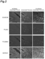
FIG. 2 shows FISH images of the normal human endometrial tissue and the endometrial tissue and ovarian tissue from a patient with endometriosis in which EUB338, FUSO, and FUS664 are used as probes. -
FIG. 3(A) shows the endometriotic lesions in mice inoculated intraperitoneally with endometria infected with the bacteria.FIG. 3(B) shows the number of the lesions, andFIG. 3(C) shows the total weight of the lesions. -
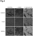
FIG. 4 shows FISH images of the endometriotic lesions in mice inoculated intraperitoneally with endometria infected with the bacteria in which EUB338 and FUSO are used as probes. -
FIG. 5(A) shows the endometriotic lesions in mice inoculated intraperitoneally with the endometrial tissues of the donor mice treated with the antibacterial agent.FIG. 5(B) shows the number of the lesions, andFIG. 5(C) shows the total weight of the lesions. -
FIG. 6 shows FISH images of the endometriotic lesions in mice inoculated intraperitoneally with the endometrial tissues of the donor mice treated with the antibacterial agent in which EUB338 and FUSO are used as probes. -
FIG. 7(A) shows the endometriotic lesions in the endometriosis mice treated with the antibacterial agent.FIG. 7(B) shows the number of the lesions, andFIG. 7(C) shows the total weight of the lesions. - A method for detecting a Fusobacterium bacterium for diagnosing endometriosis according to one aspect of the present invention includes a step of detecting a Fusobacterium bacterium in a sample collected from a subject. If the presence of the Fusobacterium bacterium is detected in the sample collected from a subject, it is likely that the endometrium of the subject is infected with the Fusobacterium bacterium. Therefore, the detection of the presence of the Fusobacterium bacterium in the sample indicates that the subject is likely suffering from or likely to suffer from endometriosis. This result can be used by a medical doctor as an indicator for diagnosing endometriosis.
- The Fusobacterium bacterium is not particularly limited, and may be, for example, Fusobacterium nucleatum.
- The subject is not particularly limited as long as it has a uterus. The subject may be, for example, a human subject, and may be a human subject who may suffer from endometriosis or who is suspected of suffering from endometriosis.
- The sample is not particularly limited as long as it can detect the Fusobacterium bacterium that have infected the endometrium, and may be, for example, blood, plasma, serum, urine, endometrial cells, vaginal swab fluid, menstrual blood, cervical cells, or ovarian tissue. From the viewpoint of collecting the sample without or with minimal invasion, the sample is preferably blood, plasma, serum, urine, endometrial cells, vaginal swab fluid, or menstrual blood. From the viewpoint of specifically detecting endometrial infection, the sample is more preferably endometrial cells, vaginal swab fluid, or menstrual blood.
- The method for detecting a Fusobacterium bacterium in a sample is not particularly limited, and examples thereof include conventionally known bacterial detection methods such as genetic testing by nucleic acid amplification methods such as a PCR (polymerase chain reaction) method and a LAMP (loop-mediated isothermal amplification) method in which primers specific to a Fusobacterium bacterium are used.
- For example, the step of detecting a Fusobacterium bacterium in a sample collected from a subject may include a step of performing a nucleic acid amplification reaction on DNA derived from the sample using primers specific to the Fusobacterium bacterium (nucleic acid amplification step), and may further include a step of detecting an amplification product of the nucleic acid amplification reaction. The detection of an amplification product may be performed after the nucleic acid amplification step or may be performed in real time during the nucleic acid amplification step. If the presence of an amplification product is detected, it can be said that there is a Fusobacterium bacterium in the sample.
- DNA derived from a sample may be DNA extracted and/or isolated from a sample by known techniques. Primers specific to a Fusobacterium bacterium mean one or more kinds of primers capable of amplifying a DNA region specific to the Fusobacterium bacterium.
- The method for detecting an amplification product is not particularly limited, and known technology may be used. An amplification product may be detected, for example, by using a labeling probe such as a fluorescence labeling probe or a fluorescent intercalator, or by performing electrophoresis on a reaction solution. Alternatively, in a case where magnesium ions are contained in a reaction solution of the nucleic acid amplification reaction, the amplification product may be detected by measuring the turbidity of the reaction solution.
- According to the method according to the above-described aspect of the present invention, it is possible to obtain data that assists diagnosis of endometriosis. Accordingly, it can be said that one aspect of the present invention is a method for collecting data for diagnosing endometriosis, the method including a step of detecting a Fusobacterium bacterium in a sample collected from a subject. The details of the step of detecting a Fusobacterium bacterium in a sample collected from a subject are as described above. Data of detection results of the Fusobacterium bacterium in the sample can be used by a medical doctor for diagnosing endometriosis.
- The method for collecting data for diagnosing endometriosis may further include a step of determining the likelihood that the subject is suffering from or may suffer from endometriosis based on the detection results of the Fusobacterium bacterium (the presence or absence of the Fusobacterium bacterium). In this step, when the presence of the Fusobacterium bacterium is detected in the sample, it is determined that the subject is likely suffering from or likely to suffer from endometriosis. The determination data obtained in this manner can be used by a medical doctor for diagnosing endometriosis.
- A therapeutic agent for endometriosis according to one aspect of the present invention contains an antibacterial agent against a Fusobacterium bacterium as an active component. The therapeutic agent for endometriosis may specifically be a therapeutic agent for use in the treatment of endometriosis caused by infection with a Fusobacterium bacterium. In addition, another aspect of the present invention is a use of an antibacterial agent for the manufacture of a medicament for the treatment of endometriosis caused by infection with a Fusobacterium bacterium.
- The details of the Fusobacterium bacterium are as described above. The antibacterial agent is not particularly limited as long as it is an antibacterial agent that can sterilize the Fusobacterium bacterium or suppress its proliferation, and examples thereof include metronidazole antibacterial agents and chloramphenicol antibacterial agents.
- A method for treating endometriosis according to one aspect of the present invention includes a step of administering an antibacterial agent to a subject whose endometrium is infected with a Fusobacterium bacterium.
- As described above, whether or not the endometrium of a subject is infected with a Fusobacterium bacterium can be confirmed based on whether or not the Fusobacterium bacterium is detected in a sample collected from the subject. Namely, in one embodiment, the method for treating endometriosis may include a step of detecting a Fusobacterium bacterium in a sample collected from a subject and a step of administering an antibacterial agent to the subject when the presence of the Fusobacterium bacterium is detected in the sample.
- The details of the antibacterial agent are as described above. The method for administering the antibacterial agent may be, for example, oral administration or vaginal administration. From the viewpoint of reducing the effect of the antibacterial agent on other parts of the body, the administration method is preferably vaginal administration. The dosage of the antibacterial agent is not particularly limited as long as it is a therapeutically effective amount, and may be determined depending on the type of the antibacterial agent.
- Endometrial and ovarian tissues used in test examples below were collected from patients at Nagoya University Hospital and its affiliated hospitals. All mice used in the test examples below are female BALB/c mice (Japan SLC, Inc.). Fusobacterium nucleatum and Lactobacillus iners (indigenous bacterium in the vagina) with which the mice were infected were obtained from the Center for Conservation of Microbial Genetic Resource, Organization for Research and Community Development, Gifu University. F. nucleatum ssp. nucleatum (strain ATCC25586) used in Test Example 5 was obtained from Riken, a National Research and Development Agency. 17β-estradiol administered to mice was manufactured by Fuji Pharma Co., Ltd. Metronidazole (MZ) administered to mice was manufactured by FUJIFILM Wako Pure Chemical Corporation, and chloramphenicol (CP) was manufactured by Sigma Corporation.
- In the following test examples, analysis by fluorescence in situ hybridization (FISH) was performed as follows. First, slides of formalin-fixed paraffin-embedded tissue slices were deparaffinized and treated with 0.2 M HCl for 20 minutes, and then hybridized with 10 ng/µL FISH probes in a hybridization buffer solution (0.9 M NaCl, 20 mM Tris-HCl, pH 7.2, 0.1% sodium dodecyl sulfate) at 56°C overnight. As the FISH probes, 5'-FAM-labeled eubacterial universal probe EUB338, 5'-Cy3-labeled Fusobacterium bacteria-specific probe FUSO, and 5'-Cy5-labeled F. nucleatum-target probe FUS664 were used. FISH probe sequences were obtained from probeBase (http://probebase.csb.univie.ac.at/) (accession number: pB-159, pB-782, and pB-1346). The slides were washed with a wash buffer solution (0.9 M NaCl, 20 mM Tris-HCl, pH 7.2) at 58°C for 20 minutes. The tissue slices were counterstained with 4',6-diamidino-2-phenylindole (DAPI) and mounted on glass slides. Opal Multi-plex IHC Detection Kit (manufactured by PerkinElmer, Inc.) was used to perform co-immunostaining with an anti-TAGLN antibody and the FISH probes according to the manufacturer's protocol. Fluorescence images were acquired using an inverted microscope Leica DMI6000B (manufactured by Leica Microsystems).
- QIAamp DNA Microbiome Kit (manufactured by QIAGEN) was used to extract genomic DNAs from normal human endometrial tissues (n=9) and endometrial tissues (n=9) from patients with endometriosis. A quantitative polymerase chain reaction (qPCR) was performed using primers specific Fusobacterium bacteria, and the proportions of 16S rRNA of Fusobacterium within the total 16S rRNA were calculated by a ΔCT method. The results are shown in
FIG. 1 . 16S rRNA of Fusobacterium was not detected in the normal human endometrial tissues, but was detected in the endometrial tissues from the patients with endometriosis (*P<0.05). - Next, the normal human endometrial tissues, and the endometrial tissues and ovarian tissues from the patients with endometriosis were subjected to FISH using EUB338 specific to eubacteria, FUSO specific to Fusobacterium bacteria, and FUS664 specific to F. nucleatum as probes. Fluorescence images are shown in
FIG. 2 (with a scale bar being 100 µm). Fluorescence of EUB338, FUSO, and FUS664 was observed in the endometrial tissues and ovarian tissues from the patients with endometriosis, indicating that these tissues were infected with F. nucleatum. - Next, the positive rates of the Fusobacterium bacteria in the endometrial tissues from the patients without endometriosis and the patients with endometriosis were investigated by FISH. The fluorescence of FUSO was observed in 3 of 42 patients who were not suffering from endometriosis (positive rate: 7%) and in 27 of 42 patients with endometriosis (positive rate: 64%).
- The ovaries of 6-week-old donor mice were removed, and 107 CFU (colony-forming units)/10 µL PBS/day of F nucleatum or L. iners were intravaginally applied 4 times a week to infect the donor mice with the bacteria. Phosphate-buffered saline (PBS) was applied to a control group. Two weeks after the removal of the ovaries, endometrial tissues collected from the donor mice were inoculated intraperitoneally into recipient mice to induce endometriosis. Subcutaneous administration of 17β-estradiol at 100 µg/kg/week was started after the ovariectomy for the donor mice and after the endometrial tissue inoculation for the recipient mice. Four weeks after the intraperitoneal inoculation, peritoneal endometriotic lesions of the recipient mice were excised, and the number and total weight of the lesions were measured.
-
FIG. 3(A) shows the excised lesions.FIG. 3(B) shows the number of the lesions, andFIG. 3(C) shows the total weight of the lesions (*P<0.05, n=3 for each group). As shown in these drawings, the number and total weight of the lesions were significantly high in the recipient mice inoculated with the endometrial tissues infected with F. nucleatum. - FISH was performed on the lesions using EUB338 and FUSO as probes. Fluorescence images are shown in
FIG. 4 (with a scale bar being 100 µm). Fusobacterium bacteria were detected in the endometriotic lesions of the recipient mice inoculated with the endometrial tissues infected with F. nucleatum. - The ovaries of 6-week-old donor mice were removed, and 107 CFU /10 µL PBS/day of F nucleatum were intravaginally applied 4 times a week to infect the donor mice with F nucleatum. One week after the removal of the ovaries, an antibacterial agent containing 17 µg/day metronidazole (MZ) or 7 µg/day chloramphenicol (CP) was administered vaginally daily for 5 days. PBS was administered to the control group. Three weeks after the removal of the ovaries, endometrial tissues collected from the donor mice were inoculated intraperitoneally into recipient mice to induce endometriosis. Subcutaneous administration of 17β-estradiol at 100 µg/kg/week was started after the ovariectomy for the donor mice and after the endometrial tissue inoculation for the recipient mice. Three weeks after the intraperitoneal inoculation, peritoneal endometriotic lesions of the recipient mice were excised, and the number and total weight of the lesions were measured.
-
FIG. 5(A) shows the excised lesions.FIG. 5(B) shows the number of the lesions, andFIG. 5(C) shows the total weight of the lesions (**P<0.01, n=3 for each group). As shown in these drawings, the number and total weight of the lesions were significantly reduced in the recipient mice inoculated with the endometrial tissues from the donor mice treated with the antibacterial agent. - FISH was performed on the lesions using EUB338 and FUSO as probes. Fluorescence images are shown in
FIG. 6 (with a scale bar being 100 µm). Fusobacterium bacteria were detected in the control group inoculated with the endometrial tissues of the donor mice to which no antibacterial agent was administered, whereas no Fusobacterium bacteria were detected in the recipient mice inoculated with the endometrial tissues from the donor mice treated with the antibacterial agent. - The ovaries of 6-week-old donor mice were removed, and 107 CFU/10 µL PBS/day of F nucleatum were intravaginally applied 4 times a week to infect the donor mice with F nucleatum. Two weeks after the removal of the ovaries, the endometrial tissues collected from the donor mice were inoculated intraperitoneally into recipient mice to induce endometriosis. Subcutaneous administration of 17β-estradiol at 100 µg/kg/week was started after the ovariectomy for the donor mice and after the endometrial tissue inoculation for the recipient mice. Three weeks after the intraperitoneal inoculation, an antibacterial agent containing 17 µg/day metronidazole or 7 µg/day chloramphenicol was administered to the recipient mice orally daily for 5 days. 21 days after the end of administration, peritoneal endometriotic lesions of the recipient mice were excised, and the number and total weight of the lesions were measured.
-
FIG. 7(A) shows the excised lesions (with a scale bar being 1 cm).FIG. 7(B) shows the number of the lesions, andFIG. 7(C) shows the total weight of the lesions (*P<0.05, n.s.: no significant difference, n=3 for each group). As shown in these drawings, for the endometriosis mice treated with the antibacterial agent, the total weight of the lesions was significantly reduced, although there was no significant change in the number of the lesions. - When FISH was performed on the lesions using EUB338 and FUSO as probes, the amount of Fusobacterium bacteria was dramatically reduced in the endometriosis mice treated with the antibacterial agent (not shown in the drawings).
- 10 µL of a bacterium liquid of F nucleatum ssp. nucleatum (strain ATCC25586) was added to 490 µL of menstrual blood negative for Fusobacterium bacteria, and DNA was extracted from 500 µL of the obtained menstrual blood sample using QIAamp DNA Microbiome Kit. A LAMP reaction was performed on the obtained DNA using a F. nucleatum ssp. nucleatum-specific primer set shown in Table 1. The reaction was performed at 65°C for 120 minutes. Detection of an amplification product was performed by monitoring the fluorescence intensity of SYTO (trademark) 63 Red Fluorescent Nucleic Acid Stain (manufactured by Thermo Fisher Scientific Inc.). The time required for a moving average value of the fluorescence intensity to reach a threshold value a certain number of times was measured, and the first time the threshold value was exceeded was defined as a Tt value. As a control, 10 µL of physiological saline was added to 490 µL of menstrual blood negative for Fusobacterium bacteria, and using the obtained menstrual blood sample, DNA extraction and LAMP reaction were performed in the same manner as described above. The Tt values are shown in Table 2.
[Table 1] Primer SEQ ID NO. Sequence (5' to 3') F3 1 TAGGAAATACTTGTAATATGAATGGT B3 2 GTATATCCGATATCAGTTATAGGC FIP 3 
BIP 4 
LF 5 
LB 6 GTCAACAGTTTTATTACAAACATTTGGAATGCC [Table 2] Amount of bacterium of F. nucleatum (CFU) 2×10⁸ 2×10⁴ No bacterium Tt value (minute) 11.7 22.8 Not detected - No amplification product of the LAMP reaction was detected in the menstrual blood sample containing no F. nucleatum ssp. nucleatum (control), whereas an amplification product was detected in the menstrual blood sample containing F nucleatum ssp. nucleatum. Therefore, it was shown that when a Fusobacterium bacterium derived from an endometrium is present in menstrual blood, the bacterium can be detected by a nucleic acid amplification method.
Claims (7)
- A method for detecting a Fusobacterium bacterium for diagnosing endometriosis, comprising:detecting a Fusobacterium bacterium in a sample collected from a subject,wherein a detection of a presence of a Fusobacterium bacterium in the sample indicates that the subject is likely suffering from or likely to suffer from endometriosis.
- The method according to claim 1, wherein the Fusobacterium bacterium is Fusobacterium nucleatum.
- The method according to claim 1 or 2, wherein the sample is blood, plasma, serum, urine, endometrial cells, vaginal swab fluid, or menstrual blood.
- A therapeutic agent for endometriosis comprising:
an antibacterial agent against a Fusobacterium bacterium as an active component. - A method for treating endometriosis, comprising:
a step of administering an antibacterial agent to a subject whose endometrium is infected with a Fusobacterium bacterium. - The method according to claim 5, further comprising:
a step of detecting a Fusobacterium bacterium in a sample collected from the subject to confirm whether or not the endometrium of the subject is infected with the Fusobacterium bacterium before the step of administering an antibacterial agent. - The method according to claim 5 or 6, wherein the antibacterial agent is administered vaginally.
Applications Claiming Priority (2)
| Application Number | Priority Date | Filing Date | Title |
|---|---|---|---|
| JP2021152197 | 2021-09-17 | ||
| PCT/JP2022/033474 WO2023042714A1 (en) | 2021-09-17 | 2022-09-06 | Method for detecting bacteria of genus fusobacterium in order to diagnose endometriosis |
Publications (1)
| Publication Number | Publication Date |
|---|---|
| EP4394044A1 true EP4394044A1 (en) | 2024-07-03 |
Family
ID=85602827
Family Applications (1)
| Application Number | Title | Priority Date | Filing Date |
|---|---|---|---|
| EP22869861.9A Withdrawn EP4394044A1 (en) | 2021-09-17 | 2022-09-06 | Method for detecting bacteria of genus fusobacterium in order to diagnose endometriosis |
Country Status (5)
| Country | Link |
|---|---|
| US (1) | US20240382458A1 (en) |
| EP (1) | EP4394044A1 (en) |
| JP (1) | JPWO2023042714A1 (en) |
| CN (1) | CN117897497A (en) |
| WO (1) | WO2023042714A1 (en) |
Families Citing this family (1)
| Publication number | Priority date | Publication date | Assignee | Title |
|---|---|---|---|---|
| EP4553165A1 (en) * | 2022-09-06 | 2025-05-14 | Eiken Kagaku Kabushiki Kaisha | Method for detecting fusobacterium nucleatum and method for detecting fusobacterium nucleatum nucleatum |
Family Cites Families (3)
| Publication number | Priority date | Publication date | Assignee | Title |
|---|---|---|---|---|
| EP1344527B1 (en) * | 2000-11-15 | 2009-09-09 | Masao Sugamata | Macrolide antibiotics for endometriosis or uterine myoma |
| US20200237786A1 (en) * | 2017-08-01 | 2020-07-30 | Soft Cell Biological Research, Llc | Diagnosis and treatment of endometriosis by screening for l-form bacteria |
| EP3676405A2 (en) * | 2017-08-28 | 2020-07-08 | Psomagen, Inc. | Method and system for characterization for female reproductive system-related conditions associated with microorganisms |
-
2022
- 2022-09-06 WO PCT/JP2022/033474 patent/WO2023042714A1/en not_active Ceased
- 2022-09-06 EP EP22869861.9A patent/EP4394044A1/en not_active Withdrawn
- 2022-09-06 CN CN202280058001.4A patent/CN117897497A/en active Pending
- 2022-09-06 JP JP2023548422A patent/JPWO2023042714A1/ja active Pending
- 2022-09-06 US US18/692,046 patent/US20240382458A1/en active Pending
Also Published As
| Publication number | Publication date |
|---|---|
| JPWO2023042714A1 (en) | 2023-03-23 |
| CN117897497A (en) | 2024-04-16 |
| US20240382458A1 (en) | 2024-11-21 |
| WO2023042714A1 (en) | 2023-03-23 |
Similar Documents
| Publication | Publication Date | Title |
|---|---|---|
| Liu et al. | Endometrial microbiota in infertile women with and without chronic endometritis as diagnosed using a quantitative and reference range-based method | |
| Miles et al. | Investigation of the microbiota of the reproductive tract in women undergoing a total hysterectomy and bilateral salpingo-oopherectomy | |
| Fang et al. | Barcoded sequencing reveals diverse intrauterine microbiomes in patients suffering with endometrial polyps | |
| Akutsu et al. | Detection of bacterial 16S ribosomal RNA genes for forensic identification of vaginal fluid | |
| Cho et al. | Comparison of urine, self-collected vaginal swab, and cervical swab samples for detecting human papillomavirus (HPV) with Roche Cobas HPV, Anyplex II HPV, and RealTime HR-S HPV assay | |
| US11884982B2 (en) | Tumor marker, methylation detection reagent, kit and use thereof | |
| WO2018062743A1 (en) | Composition containing intravaginal microorganisms | |
| US20240382458A1 (en) | Method for detecting bacteria of genus fusobacterium in order to diagnose endometriosis | |
| KR20230160762A (en) | Method of screening agents for treating severe alcoholic hepatitis | |
| WO2024159852A1 (en) | Composition and kit for detecting bacterial vaginitis pathogens, and use thereof | |
| Pourhajibagher et al. | Diagnostic accuracy of multiplex real-time PCR approaches compared with cultivation-based detection methods: Monitoring the endopathogenic microbiota pre and post photo-activated disinfection | |
| US20210189461A1 (en) | Tumor marker, methylation detection reagent, kit and use thereof | |
| CN113913522B (en) | Reagent and kit for detecting endometrial cancer | |
| Sánchez et al. | Evaluation of the viasure Neisseria gonorrhoeae ciprofloxacin resistant assay for the simultaneous identification and direct detection of ciprofloxacin susceptibility | |
| Kerkar et al. | Human papillomavirus infection in asymptomatic population | |
| Chowdhury et al. | Infestation of endometrium by mycobacterium tuberculosis bacilli-cause of reproductive failure | |
| Asl et al. | Prevalence of Helicobacter pylori vacA, cagA, cagE1, cagE2, dupA and oipA genotypes in patients with gastrointestinal diseases | |
| CN107312865A (en) | Purposes of the LOC100130111 in osteosarcoma diagnostic products, medicine is prepared | |
| WO2017092483A1 (en) | Kit for diagnosing tuberculosis through detecting free nucleic acid and use thereof | |
| KR101522398B1 (en) | Composition for diagnosing human papillomavirus infection and cervical cancer | |
| AlFaki | Detection of vacA, cagA and iceA genes of H. pylori in dyspeptic patients and their association with clinical data and histopathological abnormalities | |
| Abdallah et al. | Prevalence, Resistance Profile and Virulence Genes of Streptococcus agalactiae Colonizing Near-term Pregnant Women Attending Ain Shams University Hospital. | |
| Maldonado-Barrueco et al. | Utility of sexually transmitted infection screening in diagnosing clinical gynaecological conditions using endometrial specimens | |
| Azizmohammadi et al. | Antimicrobial susceptibility pattern of Neisseria gonorrhoeae isolated from fertile and infertile women | |
| CN106244727A (en) | Test method for combined effect of HPV-16 and host genetic factors |
Legal Events
| Date | Code | Title | Description |
|---|---|---|---|
| STAA | Information on the status of an ep patent application or granted ep patent |
Free format text: STATUS: THE INTERNATIONAL PUBLICATION HAS BEEN MADE |
|
| PUAI | Public reference made under article 153(3) epc to a published international application that has entered the european phase |
Free format text: ORIGINAL CODE: 0009012 |
|
| STAA | Information on the status of an ep patent application or granted ep patent |
Free format text: STATUS: REQUEST FOR EXAMINATION WAS MADE |
|
| 17P | Request for examination filed |
Effective date: 20240326 |
|
| AK | Designated contracting states |
Kind code of ref document: A1 Designated state(s): AL AT BE BG CH CY CZ DE DK EE ES FI FR GB GR HR HU IE IS IT LI LT LU LV MC MK MT NL NO PL PT RO RS SE SI SK SM TR |
|
| DAV | Request for validation of the european patent (deleted) | ||
| DAX | Request for extension of the european patent (deleted) | ||
| STAA | Information on the status of an ep patent application or granted ep patent |
Free format text: STATUS: THE APPLICATION HAS BEEN WITHDRAWN |
|
| 18W | Application withdrawn |
Effective date: 20250326 |