EP4392177B1 - Detection of sars-cov-2 - Google Patents
Detection of sars-cov-2 Download PDFInfo
- Publication number
- EP4392177B1 EP4392177B1 EP22783494.2A EP22783494A EP4392177B1 EP 4392177 B1 EP4392177 B1 EP 4392177B1 EP 22783494 A EP22783494 A EP 22783494A EP 4392177 B1 EP4392177 B1 EP 4392177B1
- Authority
- EP
- European Patent Office
- Prior art keywords
- sars
- cov
- molecularly imprinted
- nanomip
- nanomips
- Prior art date
- Legal status (The legal status is an assumption and is not a legal conclusion. Google has not performed a legal analysis and makes no representation as to the accuracy of the status listed.)
- Active
Links
Images







Classifications
-
- G—PHYSICS
- G01—MEASURING; TESTING
- G01N—INVESTIGATING OR ANALYSING MATERIALS BY DETERMINING THEIR CHEMICAL OR PHYSICAL PROPERTIES
- G01N33/00—Investigating or analysing materials by specific methods not covered by groups G01N1/00 - G01N31/00
- G01N33/48—Biological material, e.g. blood, urine; Haemocytometers
- G01N33/50—Chemical analysis of biological material, e.g. blood, urine; Testing involving biospecific ligand binding methods; Immunological testing
- G01N33/53—Immunoassay; Biospecific binding assay; Materials therefor
- G01N33/569—Immunoassay; Biospecific binding assay; Materials therefor for microorganisms, e.g. protozoa, bacteria, viruses
- G01N33/56983—Viruses
-
- B—PERFORMING OPERATIONS; TRANSPORTING
- B01—PHYSICAL OR CHEMICAL PROCESSES OR APPARATUS IN GENERAL
- B01J—CHEMICAL OR PHYSICAL PROCESSES, e.g. CATALYSIS OR COLLOID CHEMISTRY; THEIR RELEVANT APPARATUS
- B01J20/00—Solid sorbent compositions or filter aid compositions; Sorbents for chromatography; Processes for preparing, regenerating or reactivating thereof
- B01J20/22—Solid sorbent compositions or filter aid compositions; Sorbents for chromatography; Processes for preparing, regenerating or reactivating thereof comprising organic material
- B01J20/26—Synthetic macromolecular compounds
- B01J20/268—Polymers created by use of a template, e.g. molecularly imprinted polymers
-
- B—PERFORMING OPERATIONS; TRANSPORTING
- B01—PHYSICAL OR CHEMICAL PROCESSES OR APPARATUS IN GENERAL
- B01J—CHEMICAL OR PHYSICAL PROCESSES, e.g. CATALYSIS OR COLLOID CHEMISTRY; THEIR RELEVANT APPARATUS
- B01J20/00—Solid sorbent compositions or filter aid compositions; Sorbents for chromatography; Processes for preparing, regenerating or reactivating thereof
- B01J20/28—Solid sorbent compositions or filter aid compositions; Sorbents for chromatography; Processes for preparing, regenerating or reactivating thereof characterised by their form or physical properties
- B01J20/28002—Solid sorbent compositions or filter aid compositions; Sorbents for chromatography; Processes for preparing, regenerating or reactivating thereof characterised by their form or physical properties characterised by their physical properties
- B01J20/28004—Sorbent size or size distribution, e.g. particle size
- B01J20/28007—Sorbent size or size distribution, e.g. particle size with size in the range 1-100 nanometers, e.g. nanosized particles, nanofibers, nanotubes, nanowires or the like
-
- B—PERFORMING OPERATIONS; TRANSPORTING
- B01—PHYSICAL OR CHEMICAL PROCESSES OR APPARATUS IN GENERAL
- B01J—CHEMICAL OR PHYSICAL PROCESSES, e.g. CATALYSIS OR COLLOID CHEMISTRY; THEIR RELEVANT APPARATUS
- B01J20/00—Solid sorbent compositions or filter aid compositions; Sorbents for chromatography; Processes for preparing, regenerating or reactivating thereof
- B01J20/30—Processes for preparing, regenerating, or reactivating
- B01J20/3085—Chemical treatments not covered by groups B01J20/3007 - B01J20/3078
-
- G—PHYSICS
- G01—MEASURING; TESTING
- G01N—INVESTIGATING OR ANALYSING MATERIALS BY DETERMINING THEIR CHEMICAL OR PHYSICAL PROPERTIES
- G01N33/00—Investigating or analysing materials by specific methods not covered by groups G01N1/00 - G01N31/00
- G01N33/48—Biological material, e.g. blood, urine; Haemocytometers
- G01N33/50—Chemical analysis of biological material, e.g. blood, urine; Testing involving biospecific ligand binding methods; Immunological testing
- G01N33/58—Chemical analysis of biological material, e.g. blood, urine; Testing involving biospecific ligand binding methods; Immunological testing involving labelled substances
- G01N33/582—Chemical analysis of biological material, e.g. blood, urine; Testing involving biospecific ligand binding methods; Immunological testing involving labelled substances with fluorescent label
-
- B—PERFORMING OPERATIONS; TRANSPORTING
- B01—PHYSICAL OR CHEMICAL PROCESSES OR APPARATUS IN GENERAL
- B01J—CHEMICAL OR PHYSICAL PROCESSES, e.g. CATALYSIS OR COLLOID CHEMISTRY; THEIR RELEVANT APPARATUS
- B01J20/00—Solid sorbent compositions or filter aid compositions; Sorbents for chromatography; Processes for preparing, regenerating or reactivating thereof
- B01J20/28—Solid sorbent compositions or filter aid compositions; Sorbents for chromatography; Processes for preparing, regenerating or reactivating thereof characterised by their form or physical properties
- B01J20/28002—Solid sorbent compositions or filter aid compositions; Sorbents for chromatography; Processes for preparing, regenerating or reactivating thereof characterised by their form or physical properties characterised by their physical properties
- B01J20/28004—Sorbent size or size distribution, e.g. particle size
-
- G—PHYSICS
- G01—MEASURING; TESTING
- G01N—INVESTIGATING OR ANALYSING MATERIALS BY DETERMINING THEIR CHEMICAL OR PHYSICAL PROPERTIES
- G01N2333/00—Assays involving biological materials from specific organisms or of a specific nature
- G01N2333/005—Assays involving biological materials from specific organisms or of a specific nature from viruses
- G01N2333/08—RNA viruses
- G01N2333/165—Coronaviridae, e.g. avian infectious bronchitis virus
-
- G—PHYSICS
- G01—MEASURING; TESTING
- G01N—INVESTIGATING OR ANALYSING MATERIALS BY DETERMINING THEIR CHEMICAL OR PHYSICAL PROPERTIES
- G01N2600/00—Assays involving molecular imprinted polymers/polymers created around a molecular template
Definitions
- the present invention relates to compounds for detection of SARS-CoV-2, to methods of producing the same and to conjugates comprising the same, and more particularly to use of the aforementioned in the detection of SARS-CoV-2.
- SARS-CoV-2 severe acute respiratory syndrome coronavirus 2
- RT-PCR testing Whilst RT-PCR testing is widespread, there are problems. Conducting RT-PCR tests can be labour intensive: they require highly skilled personnel and false negative results can occur e.g. due to sampling error or improper sample handling. Indeed false positive results may also occur e.g. due to sample cross-contamination. The tests are also expensive and there is a long turn-around time (1-2 days) with respect to results.
- Rapid antigen tests are also used for population screening and offer portable and rapid (15-30 min) analysis that can be performed at the convenience of the individual. However, they lack sensitivity and utilize antibodies as receptors, which can only function in narrow temperature and pH ranges, which limits their widespread use.
- MIPs Molecularly imprinted polymers
- WO 2021/195626 A1 discloses the preparation of MIPs to SARS-CoV-2 wherein the whole virus is used as a template.
- MIPs Methods of producing MIPs are also known - see e.g. US 10 189 934B2 , Piletsky et al in “Molecularly Imprinted Polymers for Cell Recognition " and Malik et al in "Molecular Imprinted Polymer for human viral pathogen detection ".
- Antibody tests in contrast to RT-PCR and rapid antigen tests, are carried out in an attempt to establish whether a person may have already had the virus rather than, in the case of RT-PCR and rapid antigen tests, whether the person currently has the virus. Antibody tests are often carried out on blood samples from patients and work by detecting the presence of binding between synthetic antigens and antibodies (if present) in a patient's blood sample.
- one aim of the present invention is to provide alternative means for detecting SARS-CoV-2.
- a further aim is that such means will provide an alternative to the polymerase chain reaction-based assay and rapid antigen tests currently used.
- An aim of specific embodiments of the invention includes providing improved detection means.
- the invention provides a molecularly imprinted polymer comprising at least one recognition site that is complementary to a template molecule consisting of an amino acid sequence corresponding to a subsequence of the receptor binding domain of SARS-CoV-2 spike protein, wherein the amino acid sequence is no more than 50 amino acids in length and comprises the sequence NSNNLDSKVGG, and wherein the polymer comprises the monomers:
- the invention also provides a method of preparing a molecularly imprinted polymer comprising at least one recognition site which binds SARS-CoV-2 comprising the steps of:
- conjugate comprising the molecularly imprinted polymer of the invention and a fluorophore.
- Both the molecularly imprinted polymers of the invention and the conjugates of the invention can be used in the detection of SARS-CoV-2.
- the invention provides a molecularly imprinted polymer comprising at least one recognition site that is complementary to a template molecule consisting of an amino acid sequence corresponding to a subsequence of the receptor binding domain of SARS-CoV-2 spike protein, wherein the amino acid sequence is no more than 50 amino acids in length and comprises the sequence NSNNLDSKVGG, and wherein the polymer comprises the monomers:
- monomers are polymerised in an appropriate solvent in the presence of the compound to be imprinted.
- the compound to be imprinted is known as the template.
- the monomers interact with the template through electrostatic, hydrophobic or other interactions leading to the formation of binding sites in the polymer, known as recognition sites, that are complementary to the template molecule.
- recognition sites binding sites in the polymer
- the polymer matrix retains the recognition sites that are complementary to the template.
- Complementary in this sense means that the interacting groups in the polymer are placed such that they are in a suitable position to interact with the interacting groups in the template molecule and other molecules that contain the template molecule as a substructure, i.e. in this invention SARS-CoV-2.
- SARS-CoV-2 makes use of a densely glycosylated spike protein to gain entry into host cells.
- the spike protein is a homotrimeric glycoprotein and each monomer contains two subunits, S1 and S2, which mediate membrane attachment and membrane fusion respectively.
- S1 and S2 which mediate membrane attachment and membrane fusion respectively.
- the receptor binding domain (RBD) of the S1 subunit has been shown to be involved in host cell receptor engagement.
- sequence of the RBD of the SARS-CoV-2 spike protein is known, and theoretically any sequence therein could be used as a template, it is advantageous for some downstream applications to select, as a template, sequences within the target which are non-conserved. This ensures that the molecularly imprinted polymer binds the desired target with high specificity.
- Supplementary Figure 5 of Wrapp et al 2020 1 presents a sequence alignment of the spike protein from SARS-CoV-2 and two other coronavirus strains, SARS-CoV and RaTG13. The RBD is highlighted. Also highlighted are sequences which are conserved between the different strains and sequences which are non-conserved.
- the 'at least one recognition site' in the molecularly imprinted polymer is complementary to a template molecule consisting of an amino acid sequence corresponding to a subsequence of the receptor binding domain of SARS-CoV-2 spike protein, wherein the amino acid sequence is no more than 50 amino acids in length and comprises the sequence NSNNLDSKVGG.
- the size of the template is in the range up to 50 amino acids.
- the template will be less than 50 amino acids, more preferably the template will be less than 30 amino acids; most preferred is that the template is less than 20 amino acids, for example 11, 12, 13, 14, 15, 16, 17, 18 or 19 amino acids.
- the size of the molecularly imprinted polymer of the present invention will be to a large extent dictated by the intended downstream use of the molecular imprinted polymer. That said, suitably the molecular imprinted polymer will be less than 500nm, more suitably less than 250nm and even more suitably less than 100nm, and may be less than 90nm, or even less than 70nm.
- compositions comprising the molecularly imprinted polymer of the invention.
- molecular imprinted polymers in such compositions will have an average size that is less than 500nm, more suitably less than 250nm and even more suitably less than 100nm, and may be less than 90nm, or even less than 70nm.
- Suitable methods for determining the size of molecularly imprinted polymers include dynamic light scattering e.g. using a Zetasizer Ultra (Malvern Panalytical), disc centrifugation, nanoparticle tracking analysis e.g. using a NanoSight NS300 (Malvern Panalytical), tunable resistive pulse sensing, atomic force microscopy and electron microscopy. Nanoparticle tracking analysis, tunable resistive pulse sensing, atomic force microscopy and electron microscopy are particularly suitable for determining the size of single molecularly imprinted polymers. In the examples provided herein, the size of the molecularly imprinted polymers was determined using a NanoSight NS300 instrument.
- Size in the context of the molecularly imprinted polymer refers to the diameter of the molecularly imprinted polymer.
- Monomers which can be used in the preparation of molecularly imprinted polymers include vinyl monomers, allyl monomers, acetylenes, acrylates, methacrylates, acrylamides, methacrylamides, chloroacrylates, itaconates, trifluoromethylacrylates, derivatives of amino acids (e.g. esters or amides), nucleosides, nucleotides, and carbohydrates.
- Cross-linking monomers which help to stabilise molecularly imprinted polymers, may also be included.
- cross-linking monomers suitable for preparing molecularly imprinted polymers include, but are not limited to, ethylene glycol dimethacrylate, trimethylolpropane trimethacrylate, divinylbenzene, methylene bisacrylamide, ethylene bisacrylamide, N,N'-bisacryloylpiperazine and N,N'-Methylenebis(acrylamide) (BIS).
- ethylene glycol dimethacrylate trimethylolpropane trimethacrylate
- divinylbenzene divinylbenzene
- methylene bisacrylamide ethylene bisacrylamide
- N,N'-bisacryloylpiperazine N,N'-Methylenebis(acrylamide)
- the molecularly imprinted polymer will comprise (i) N-Fluoresceinyl acrylamide, (ii) Acrylamide, (iii) Tert-Butyl acrylate (TBAc), (iv) 3-O-Acryloyl-1,2:5,6-bis-O-isopropylidene-D-glucofuranose, (v) 2,2,2-Trifluoroethyl methacrylate (CF3), (vi) N-(3-Aminopropyl) methacrylamide hydrochloride (APMA), (vii) Squareamide monomer 7 (SQ7) and (viii) N,N'-Methylenebis(acrylamide) (BIS).
- the molecularly imprinted polymer is a polymer and is said to comprise an identified monomer when it has been made by polymerising a mixture of monomers comprising the identified monomer.
- the molecularly imprinted polymers of the present invention are robust and function in wide ranges of temperature and pH which leads to improved shelf-life and storage conditions and means that they can function in more challenging environments. They also have excellent biocompatibility and have fast binding kinetics.
- Also provided by the present invention is a method of preparing a molecularly imprinted polymer comprising at least one recognition site which binds SARS-CoV-2 comprising the steps of:
- An advantage of the method is that the MIPs possess a more homogenous distribution of binding site affinities, and provides means by which template elution is enhanced and downstream template leaching is prevented.
- Suitable carrier substances include polymer resins, polysaccharides, glass or metal surfaces.
- the carrier substance may be in the form of beads, fibres, membranes or capillaries.
- the carrier substance is glass. Further preferred is that the carrier substance is in the form of glass beads.
- the polymerisable composition must contain the monomers required for the polymerisation. Suitable monomers are discussed elsewhere herein.
- Step (c) involves the application of controlled polymerisation.
- Various techniques for producing molecularly imprinted polymers by controlled polymerisation are known to those of skill in the art.
- controlled polymerisation examples include radical polymerisation such as controlled living radical polymerisation (LRP), living anionic polymerisation, living cationic polymerisation, and controlled polycondensation.
- LRP controlled living radical polymerisation
- living anionic polymerisation living anionic polymerisation
- living cationic polymerisation living cationic polymerisation
- controlled polycondensation controlled polycondensation
- Polymerisation can be initiated by e.g. heating, by applying current (electropolymerisation), by the addition of redox catalyst(s), persulfate or peroxides, by irradiation, including gamma radiation or by microwave radiation or by irradiation with UV or visible light and normally takes minutes to hours.
- redox catalyst(s) including gamma radiation or by microwave radiation or by irradiation with UV or visible light and normally takes minutes to hours.
- irradiation including gamma radiation or by microwave radiation or by irradiation with UV or visible light and normally takes minutes to hours.
- polymerisation was initiated using persulfate.
- the polymerisation reaction is terminated at a stage when the size of the synthesized molecular imprinted polymer is relatively small. Preferences with respect to size are discussed elsewhere herein.
- the separation of high affinity molecularly imprinted polymers from the immobilised template can be achieved by heating, which disrupts complex formation, by changing solution pH, changing ionic strength, or through the additional of urea, guanidine or a substance which interacts with the template more strongly than the molecularly imprinted polymer.
- weakly bound material is removed by washing.
- the molecularly imprinted polymers may be purified further e.g. by chromatography, filtration and/or electrophoresis.
- the method by which the template molecule is immobilised on the carrier substance is not critical. However, for some methods, the presence of a cysteine residue at one of the termini of the template molecule is advantageous. As such, for embodiments of the method where the template molecule does not naturally comprise a terminal cysteine residue, it is preferred that the template molecule is modified at the N terminus to comprise an additional cysteine residue.
- the additional cysteine residue is attached directly to the termini of the template molecule whilst in other embodiments a spacer is included between the terminal cysteine and the template molecule.
- the spacer comprises a glycine residue.
- the template molecule comprises the amino acid sequence NSNNLDSKVGG and is modified at the N terminus to comprise a cysteine (C) residue and a glycine (G) residue such that the molecule used for imprinting is CGNSNNLDSKVGG.
- the template molecule is directly attached to the carrier substance whilst in other embodiments one or more linkers are included between the carrier substance and the template molecule.
- Suitable linkers for use in the invention are well known to those of skill in the art.
- the carrier substance used was glass beads and these were silanised using (3-aminopropyl)trimethoxysilane (APTMS) prior to immobilisation of the template molecule.
- the APTMS acts as a linker between the surface of the glass bead and the template molecule.
- the APTMS was modified using n-succinimidyl iodoacetate (SIA) prior to template immobilisation.
- SIA n-succinimidyl iodoacetate
- Linkers and spacers function to prevent adverse effects brought about by steric hindrance.
- An advantage of the method of the present invention is that it is fast and scalable and uses only a short template.
- conjugate comprising the molecularly imprinted polymer of the invention and a fluorophore.
- Fluorophores are molecules with the ability to absorb light at a particular wavelength, the absorbance or excitation wavelength, and then emit light at a higher wavelength, the emission wavelength.
- fluorophores can be conjugated via amino, thiol or carbohydrate groups.
- the fluorophore was conjugated to the molecularly imprinted polymer via amine groups provided by the amino monomer in the molecularly imprinted polymer.
- any fluorophore can be used in conjugates of the present invention, it is advantageous for some downstream applications, particularly diagnostic applications, to select a fluorophore that is both bright and stable.
- CPNs Conjugated Polymer Nanoparticles
- TM are highly fluorescent nanoparticles which comprise of a semiconductor light emitting polymer (LEP) core encapsulated within a biocompatible surfactant.
- LEP semiconductor light emitting polymer
- CPNs are particularly suited for use in diagnostic applications due to their intense fluorescence, which means they have a high level of sensitivity, and their high degree of photostability.
- CPNs are around 70-80nm in size and those currently available from Stream Bio exhibit fluorescence with emission wavelengths from 420nm to 1130nm.
- molecularly imprinted polymers of the invention were conjugated to CPN510 and CPN610, both available from Stream Bio.
- the fluorophore is a CPN.
- compositions comprising the conjugate of the invention.
- the molecularly imprinted polymers and conjugates of the present invention may be used in a variety of applications.
- the molecularly imprinted polymers and conjugates of the present invention be used in the detection of SARS-CoV-2.
- the molecularly imprinted polymers and conjugates and compositions may be used in assays for example diagnostic assays which may be used in a medical or point of care setting. Examples of such assays include lateral flow assays (LFA) and enzyme-linked immunosorbent assay (ELISA)-type assays which may be used for high throughput screening.
- LFA lateral flow assays
- ELISA enzyme-linked immunosorbent assay
- the molecularly imprinted polymers and conjugates of the present invention may also be used in sensors.
- nanoMIP-SPR sensors were prepared by covalently immobilising nanoMIPs on 4% mercaptoundecanoic acid (MUDA) chips (prepared using Biacore SIA Kit Au surfaces, functionalised with a self-assembled monolayer of 4% MUDA in ethanol).
- MUDA mercaptoundecanoic acid
- NanoMIP-functionalized Screen-Printed Electrodes (SPEs) were also prepared for use in thermal detection methods.
- the molecularly imprinted polymers and conjugates of the present invention may also be used in the detection of SARS-CoV-2 in the research setting.
- the molecularly imprinted polymers and conjugates of the present invention may also be used as pharmaceuticals.
- the molecularly imprinted polymers of the present invention have high affinity for SARS-CoV-2.
- High affinity in the context of the present specification means that the affinity with which the molecularly imprinted polymers bind SARS-CoV-2 exceeds the affinity with which 'blank' molecularly imprinted polymers bind SARS-CoV-2 by at least three, preferably at least five, more preferably at least 10 times.
- Blank in this context means molecularly imprinted polymers formed in the absence of template.
- Binding affinity is the strength of the binding interaction between a single molecule to its binding partner e.g. in the present case a molecularly imprinted polymer to its target SARS-CoV-2. Binding affinity is typically measured and reported by the equilibrium dissociation constant (K D ). The smaller the K D value, the greater the binding affinity between the two molecules.
- the molecularly imprinted polymer and conjugates of the present invention are used in the detection of SARS-CoV-2, it is preferred that the molecularly imprinted polymer binds to SARS-CoV-2 with a K D of less than 500nM, preferably less than 250nM and even more preferably less than 200nM.
- Suitable methods for measuring K D include surface plasmon resonance (SPR) which may be conducted using e.g. the Biacore series of instruments (Cytiva) or the MP-SPR Navi instruments (BioNavis).
- SPR surface plasmon resonance
- Examples, or parts thereof, which relate to molecularly imprinted polymers comprising at least one recognition site that is complementary to a template molecule consisting of an amino acid sequence corresponding to a subsequence of the receptor binding domain of SARS-CoV-2 wherein the amino acid sequence does not comprise the sequence NSNNLDSKVGG are not part of the claimed invention and are present for comparative purposes only.
- Supplementary Figure 5 of Wrapp et al 2020 1 presents a sequence alignment of the spike protein from SARS-CoV-2 and two other coronavirus strains, SARS-CoV and RaTG13.
- the RBD is highlighted.
- Three peptide sequences (peptide 1: NSNNLDSKVGG, peptide 2: STEIYQAGSTPC and peptide 3: CYFPLQSYGFQP) from the RBD of SARS-CoV-2 were selected by the present inventors as potential candidates for imprinting based on the lack of conservation with the respective sequences from SARS-CoV and RaTG13.
- Peptides 1, 2 and 3 were immobilised onto silanised glass beads prior to nanoMIP synthesis.
- Peptide immobilisation was carried out as follows: glass beads (approximately 100 ⁇ M in diameter, sourced from Microbeads AG) were activated by boiling in 4M NaOH for 10 minutes. The glass beads were then washed with firstly deionised water and then acetone and then dried at 80°C for 2 hours. The glass beads were then incubated in toluene with 2 % v/v (3-aminopropyl)trimethoxysilane (APTMS) for 3 hours, washed with acetone and placed in PBS, pH 7.4 with 0.2mg/ml n-succinimidyl iodoacetate (SIA) for two hours before being washed with acetonitrile.
- ATMS 3-aminopropyl)trimethoxysilane
- the templates (Peptides 1, 2 or 3) were then immobilised on the surface of the glass beads by incubation in a solution of PBS, pH 7.4 with 0.4 mg/ml tris(2-carboxyethyl) phosphine hydrochloride (TCEP) and 0.1mg/ml of peptide for a minimum of 4 hours. Excess template was removed by washing with water and methanol.
- TCEP tris(2-carboxyethyl) phosphine hydrochloride
- the peptide coated glass beads were used for the synthesis of imprinted nanoMIPs as follows: monomer solutions were prepared, sonicated for 10 minutes and purged with nitrogen for 5 minutes.
- monomer solutions were prepared, sonicated for 10 minutes and purged with nitrogen for 5 minutes.
- One example of a monomer solution that was prepared includes 3mg N-Fluoresceinyl acrylamide, 24.2mg Acrylamide, 31.7 ⁇ L Tert-Butyl acrylate (TBAc), 36mg 3-O-Acryloyl-1,2:5,6-bis-O-isopropylidene-D-glucofuranose, 13.8 ⁇ L 2,2,2-Trifluoroethyl methacrylate (CF3), 3.9mg N-(3-Aminopropyl) methacrylamide hydrochloride (APMA), 1.3mg Squareamide monomer 7 (SQ7) and 9mg N,N'-Methylenebis(acrylamide) (BIS)).
- High affinity nanoMIPs were then detached from the glass beads by performing 9 washing steps, each with 10ml of 60°C ethanol. The residue from each of these washes, containing the nanoMIPs, was collected. The nanoMIPs were then concentrated down to approximately 10ml and solvent exchanged to distilled water. Imprinted nanoMIPs had an average diameter of 59nm, as calculated by NanoSight NS300.
- NanoMIP-SPR sensors were prepared by covalently immobilising nanoMIPs (from Example 1) on 4% mercaptoundecanoic acid (MUDA) chips (prepared using Biacore SIA Kit Au surfaces, functionalised with a self-assembled monolayer of 4% MUDA in ethanol).
- MUDA mercaptoundecanoic acid
- NanoMIPs (5nM) were then injected (8-10 minutes at a flow rate of 8 ⁇ L/minute) into all four flow cells on the chip. Finally, unreacted NHS esters were hydrolysed by injecting carbonate buffer (pH 9.2) (30 minutes at a flow rate of between 5 and 30 ⁇ L/minute).
- each of peptides 1, 2 and 3 were conjugated to Bovine Serum Albumin (BSA).
- BSA Bovine Serum Albumin
- SARS-CoV-2 S protein-SPR sensors were prepared by covalently immobilising recombinant SARS-CoV-2 S protein (recombinant full-length SARS-CoV-2 spike glycoprotein sourced from The Native Antigen Company) on 4% mercaptoundecanoic acid (MUDA) chips (prepared using Biacore SIA Kit Au surfaces, functionalised with a self-assembled monolayer of 4% MUDA in ethanol).
- MUDA mercaptoundecanoic acid
- nanoMIPs imprinted against peptide 1 were then injected (5 minutes association phase, 3 minutes dissociation phase, 28 ⁇ l/minute flow rate).
- the software calculated a K D of 5nM ( Figure 1a ) and 15nM ( Figure 1b ).
- NanoMIPs imprinted against peptides 1 and 3 were coupled to Conjugated Polymer Nanoparticles (CPNs), highly fluorescent nanoparticles containing semiconductor Light Emitting Polymer cores encapsulated within a water friendly capping agent, at Stream Bio.
- CPNs Conjugated Polymer Nanoparticles
- the CPNs used were CPN510 and CPN610 which have a diameter of 70-80nm as measured by NanoSight 3000.
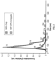
- Figure 2 shows the successful conjugation of a nanoMIP imprinted against peptide 1 to CPN510. Note that the original unconjugated nanoMIPs were predominantly 59nm; however, when conjugated to CPN510, a significant shift in particle size is seen - see the peak at 119nm.
- Example 5 Dot-blot analysis of binding between NanoMIPs and SARS-CoV-2 S protein.
- the membranes were incubated with (1) NanoMIP-CPN510 (imprinted against peptide 1), (2) NanoMIP-CPN510 (imprinted against peptide 3) or (3) CPN510 only (control) dissolved in 5% BSA in PBS-T for 30-60 minutes at room temperature. The membranes were then washed three times, for 5 minutes each time, with PBS-T. Fluorescence was then observed under UV. As shown in Figure 3 , the detection limit using NanoMIP-CPN510 (imprinted against peptide 1) was 0.3ng, much lower than that achieved using CPN510 only (control) indicating that nanoMIPs imprinted against at least peptide 1 specifically interact with SARS-CoV-2 S protein.
- Example 6 Dot-blot analysis of binding between NanoMIPs and SARS-CoV-2 virus.
- SARS-CoV-2 (2 ⁇ 10 4 pfu) was blotted onto two nitrocellulose membranes. Also blotted on to each of the nitrocellulose membranes was 75ng of recombinant SARS-CoV-2 S protein (sourced from The Native Antigen Company) as a positive control and culture media as a negative control. The membranes were then left to dry. The membranes were then blocked by soaking in 5% BSA in PBS-T (0.05% Tween-20 in PBS) for 30-60 minutes at room temperature.
- the membranes were incubated with NanoMIP-CPN510 (imprinted against peptide 1) or (2) NanoMIP-CPN510 (imprinted against troponin) (negative control) dissolved in 5% BSA in PBS-T for 30-60 minutes at room temperature. The membranes were then washed three times, for 5 minutes each time, with PBS-T. Fluorescence was then observed under UV. As shown in Figure 4 , no fluorescence was observed in the area of the nitrocellulose membrane blotted with culture media or when the membrane was incubated with NanoMIP-CPN510 (imprinted against troponin) (negative control).
- Example 7 Dot-blot analysis comparing binding between (1) NanoMIPs and four different human coronaviruses (SARS-CoV-2, HCoV-OC43, HCoV-229E and HCoV-HKU1) and (2) antibodies and four different human coronaviruses (SARS-CoV-2, HcoV-OC43, HCoV-229E and HCoV-HKU1).
- SARS-CoV-2 Spike Glycoprotein (The Native Antigen Company), Human Coronavirus OC43 Spike Glycoprotein (S1) (The Native Antigen Company), Human Coronavirus 229E Spike Glycoprotein (S1) (The Native Antigen Company) and Human Coronavirus HKU1 Spike Glycoprotein (S1) (The Native Antigen Company) (0.6 mg/mL) were blotted onto nitrocellulose membranes in triplicate (as shown in the schematic in Figure 5 ) before allowing the membranes to dry. The membranes were then blocked by soaking in 2 mL BSA (1% in PBS) for 1 hour.
- BSA 1% in PBS
- the membranes were incubated with (1) NanoMIP - CF770 (imprinted against peptide 1) and (2) three different monoclonal antibodies specific to SARS-CoV-2 Spike (RBD), Mab RBD1106-CF770, Mab RBD107-CF770 and Mab RBD5305-CF770 (all prepared in-house and diluted in 0.01% BSA) for 30 minutes.
- the membranes were then washed with 2mL PBST for 5 minutes. Fluorescence was then observed under UV.
- the nanoMIPs selectively bound the SARS-CoV-2 Spike Glycoprotein (S1) and did not bind to any of Human Coronavirus OC43 Spike Glycoprotein (S1), Human Coronavirus 229E Spike Glycoprotein (S1) or Human Coronavirus HKU1 Spike Glycoprotein (S1). Furthermore, the nanoMIPs were shown to have comparable selectivity and binding to each of the antibodies tested.
- Example 8 Analysis of binding between NanoMIPs and SARS-CoV-2 S protein RBD using thermal detection methods.
- a solution of 4-ABA (2 mM) and sodium nitrite (2 mM) in aqueous HCl (0.5 M) was prepared and gently mixed on an orbital shaker for 10 minutes.
- Screen-Printed Electrodes (SPEs) were submerged into the solution and cyclic voltammetry was performed from +0.2 V to -0.6 V at 100 mV s -1 using an Ag/AgCl reference electrode (Alvatek Ltd., Romsey, UK).
- the obtained electrode, denoted SPE/4-ABA was thoroughly rinsed with deionised water to remove any unbound 4-ABA and dried with nitrogen.
- EDC 1-ethyl-3-(3-dimethylaminopropyl)-carbodiimide
- NHS N-hydroxysuccinimide
- NanoMIP imprinted against peptide 1 'P1C6' or NanoMIP imprinted against peptide 3 'P3C6' was deposited onto the working electrodes following a gentle vortex. After 3 hours, the SPE/4-ABA/EDC+NHS/nanoMIP electrodes were rinsed in deionised water and dried with a gentle stream of nitrogen before being stored in PBS at 4°C until use.
- 3D-printed flow cells were used to facilitate measurements using the nanoMIP-functionalised SPEs described above.
- a SPE was mounted into the cell and a thermocouple measured the temperature in the liquid (T 2 ).
- T 2 the temperature in the liquid
- a freshly prepared nanoMIP-functionalised SPE was used.
- the flow cell was connected to a heat-transfer device as described by van Grinsven et al. The device was steered with LabView software that actively controls the temperature of the heat sink (copper block, T 1 ), which was set to 37.00 ⁇ 0.02 °C to mimic in-vivo conditions.
- a proportional-integral-derivative (PID) controller attached to a power resistor (22 ⁇ ) regulated the feedback on the signal as described in Geerets et al.
- the flow cell was filled with PBS and left for 30 minutes to ensure stabilisation of the baseline temperature signal before a first injection of PBS was added to act as the blank measurement.
- Solutions (3 mL) of increasing concentrations of each target biomarker (SARS-CoV-2 S protein RBD or SARS-CoV-2 ORF8 (as a negative control)) (0-10 pg mL -1 ) were prepared in PBS prior to experiments and stored at 4°C until required.
- Each biomarker injection was performed at 250 ⁇ L min -1 for 12 minutes using a LSP02-1B dual channel syringe (Longer Precision Pump Co., Hebei, China) pump. Following each biomarker injection, the system was allowed to stabilise for 30 minutes prior to the next injection.
- the thermal resistance (R th ) was determined throughout the experiments by dividing the temperature gradient (T 1 -T 2 ) over the power required to keep the heat sink at 37.00°C.
- the R th and standard deviation (SD) were calculated using the average of 600 data points from the baseline signal of each concentration and the initial PBS injection, respectively ( Figures 6 , 7a , and 8a ). This data was used to construct dose-response curves, from which the limit of detection (LoD) was calculated using the three-sigma method in the linear range of the sensor ( Figures 7b and 8b ).
- the error bars in the graphs relate to the SD values.
- Figure 6 shows that the thermal response to SARS-CoV-2 ORF8 is very limited whereas Figures 7 and 8 show that a good response was observed with SARS-CoV-2 S protein RBD for nanoMIPs imprinted against both peptides 1 and 3.
- Atomic force microscopy was utilized to examine how increasing temperature impacted the morphology of adsorbed nanoMIPs by imaging the same nanoMIPs at room temperature, 37°C, and 50°C.
- AFM measurements were performed on a JPK Nanowizard 4 XP Bioscience (Bruker, Nano GmbH, Berlin, Germany). Measurements in air were carried out in tapping mode using PPP-NCL-W probes (Nanosensors, Neuchatel, Switzerland) with a cantilever length of ⁇ 225 ⁇ m and spring constant of ⁇ 48 N m -1 . Measurements in liquid were performed in Quantitative Imaging (QI) mode using MLCT-E probes (Bruker, Ca, USA) with a cantilever length of ⁇ 140 ⁇ m and spring constant of ⁇ 0.1 N m -1 . Au-coated Si chips were used as substrates (Si-Mat, Kaufering, Germany).
- the chips Prior to drop-casting, the chips were cleaned by immersion for 5 min in a 5:1:1 mixture of milli-Q water, ammonia, and hydrogen peroxide heated at 75 °C. The chips were then rinsed in milli-Q water and dried with nitrogen. NanoMIP solutions were diluted in milli-Q water to ⁇ 2.54 ⁇ g mL -1 , drop-cast (20 ⁇ L) onto the Au-coated surfaces, and allowed to dry in ambient conditions for a minimum of 4 h in a Petri dish. A high temperature heating stage (HTHS, JPK BioAFM - resolution of 0.1 °C) was used as a temperature controller to facilitate imaging at 37°C and 50°C.
- HTHS high temperature heating stage
- Figure 9a and 9b which show typical AFM images and a corresponding cross-sectional profile plot respectively show that the nanoMIP morphology was unaffected by increasing temperature.
- NanoMIP volume was calculated using Gwyddion. Tracking numerous droplets revealed minimal changes in mean nanoMIP volume ( ⁇ 6% decrease) from room temperature to 50 °C.
- nanoMIP morphology remains consistent across relatively large temperature ranges.
- nanoMIP-SPR sensors were prepared by covalently immobilising nanoMIPs (from Example 1), either as directly prepared or after autoclave at 121°C for approximately 15 minutes, on 4% mercaptoundecanoic acid (MUDA) chips (prepared using Biacore SIA Kit Au surfaces, functionalised with a self-assembled monolayer of 4% MUDA in ethanol) in line with the process directly below.
- MUDA mercaptoundecanoic acid
- NanoMIPs (5nM) were then injected (8-10 minutes at a flow rate of 8 ⁇ L/minute) into all four flow cells on the chip. Finally, unreacted NHS esters were hydrolysed by injecting carbonate buffer (pH 9.2) (30 minutes at a flow rate of between 5 and 30 ⁇ L/minute).
- nanoMIP volume was calculated using Gwyddion which revealed negligible changes to the mean nanoMIP volume from pH 5.5 to 8.5 ( ⁇ 3% decrease), highlighting that adsorbed nanoMIP morphology was consistent across a broad pH range (see Figure 9c ).
- Example 10 Comparison of binding between NanoMIPs and SARS-CoV-2 antigens and SARS-CoV-2 antibodies and SARS-CoV-2 antigens using thermal detection methods.
- NanoMIP-functionalized Screen-Printed Electrodes (using nanoMIPs imprinted against peptide 1) were made in line with the procedure in Example 8.
- SARS-CoV-2 antibody-functionalized SPEs were also made in line with the procedure in Example 8.
- SARS-CoV-2 antigens either recombinant full-length SARS-CoV-2 spike glycoprotein (alpha variant) sourced from The Native Antigen Company, recombinant SARS-CoV-2 spike receptor binding domain (RBD) (delta variant) sourced from Abbexa, Cambridge, UK or SARS-CoV-2 RBD from the Medical Research Council Protein Phosphorylation and Ubiquitylation Unit (Dundee, UK)
- RBD recombinant SARS-CoV-2 spike receptor binding domain
- thermocouples measured the heat sink (T 1 ) and liquid reservoir (T 2 ) temperatures every second, and the thermal resistance (R th ) was obtained by dividing the temperature gradient (T 1 -T 2 ) over the power required to maintain the heat sink at 37.00 ⁇ 0.02 °C. As the target attached to the nanoMIPs, heat transfer at the solid-liquid interface was reduced (larger temperature gradient), which led to a measurable increase in the R th .
- the thermal measurement device was controlled using LabView software and a proportional-integral-derivative (PID) controller attached to a power resistor (22 ⁇ ) regulated the feedback on the signal.
- PID proportional-integral-derivative
- the liquid reservoir was filled with PBS and left for 30 min to ensure stabilization of the baseline R th signal.
- five spiked PBS solutions (3 mL) with increasing concentrations of the SARS-CoV-2 antigen (1 fg mL -1 - 10 pg mL -1 ) were injected into the flow cell at a rate of 250 ⁇ L min -1 for 12 min using an automated syringe pump (LSP02-1B, Longer Precision Pump Co., Hebei, China).
- LSP02-1B Longer Precision Pump Co., Hebei, China
- the system was allowed to stabilize for 30 min prior to each subsequent injection.
- the experiments produced raw thermal data plots which displayed a step-wise increase in R th where each stabilized plateau represents the injection of an increasingly concentrated spiked solution (see Figure 10a ).
- Dose-response curves ( Figures 10b-d ) were composed from the thermal plots by taking the mean and standard deviation (SD) of the stabilized plateau from each concentration injection. Limit of detection (LoD) values were calculated from the dose-response curves using the three-sigma method (3 ⁇ reference SD) in the linear range.
- SARS-CoV-2 antibody-functionalized SPEs were used to facilitate a direct comparison between the sensing performance of the nanoMIPs and those of antibody receptors.
- the ability of the nanoMIP receptors to detect virus mutations was also examined by measuring their specificity against the SARS-CoV-2 spike protein (delta variant). Results show ( Figure 10b ) that the nanoMIP sensor was effective in detecting the delta variant as the resulting LoD (6.1 ⁇ 2.9 fg mL -1 ) was very similar to the value obtained for the alpha variant. This demonstrates that the nanoMIP sensor can detect equally low amounts of both the alpha and delta variant spike protein, which is highly promising for potential commercial applications where there is an ongoing concern regarding the reduction in test efficacy due to the presence of new variants.
- the versatility of nanoMIP receptors was also examined by measuring their thermal response against the SARS-CoV-2 RBD (see Figure 10c ). Antibody receptors were also tested for the purpose of direct comparison.
- the LoD value for the SARS-CoV-2 RBD was ⁇ 20 times smaller for the nanoMIP sensor (3.9 ⁇ 1.0 fg mL -1 ) compared to the antibody sensor (85.5 ⁇ 15.0 fg mL -1 ). Consequently, this demonstrates that the nanoMIPs possessed greater versatility than antibodies as they had a comparable LoD for the full-length spike protein but a significantly lower LoD for the RBD.
- the selectivity of the nanoMIP sensor was also comprehensively examined using three negative controls which are common interferents in clinical samples: open reading frame 8 (ORF8), interleukin-6 (IL-6), and human serum albumin (HSA).
- ORF8 open reading frame 8
- IL-6 interleukin-6
- HSA human serum albumin
- NIPs were prepared using the same synthesis protocol as nanoMIPs except they were not exposed to a target epitope during polymerization, and therefore had no specific cavities to facilitate target binding.
- the thermal response of a nanoMIP-functionalized SPE to the spike protein was ⁇ 6 times larger compared to the NIP-functionalized SPE (0.04 °C W -1 ) showing that specific binding occurred between the spike protein and nanoMIP cavities.
- the LoD values of the nanoMIP sensor, a commercial rapid antigen test, and numerous recently developed antigen tests from the literature 4-15 are presented in Figure 10e .
- Table 1 below provides further details of the experimental setup.
- the LoD value for the nanoMIP sensor is ⁇ 6000 times lower than the commercial rapid antigen test (20 pg mL -1 ). This considerably lower LoD highlights that nanoMIP receptors could be a valuable tool in producing rapid antigen tests with adequate sensitives for effective population screening.
- the LoD of the nanoMIP sensor is among the lowest value of those from recently developed antigen tests in the literature. This demonstrates that thermal detection using nanoMIPs can compete with the best performing antigen tests from recent literature, whilst possessing the added benefit of being able to withstand extremes of temperature and pH.
- Example 11 Use of NanoMIPs to detect SARS-CoV-2 in clinical samples
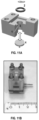
- FIG. 11a and 11b A prototype 3D-printed resin addition cell ( Figure 11a and 11b ) was developed for clinical analysis with disposable components.
- the thermal measurement device was controlled using LabView software and a proportional-integral-derivative (PID) controller attached to a power resistor (22 ⁇ ) regulated the feedback on the signal.
- PID proportional-integral-derivative
- UTM and VPM Viral preservation medium
- UTM and VPM were used as reference liquids for the negative and positive samples, respectively.
- 100 ⁇ L of the reference liquid (UTM/VPM) was pipetted into the reservoir and the R th signal was allowed to stabilize for 10 min.
- the reference liquid was pipetted out and 100 ⁇ L of the sample was added.
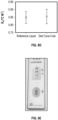
- a pipette resulted in less disturbance to the system (e.g., flow, addition of air bubbles) compared to using a syringe pump. This is highly advantageous for clinical analysis since the sample volume was similar to that collected by a throat and nasal swab (100 ⁇ L), measurement time was reduced to ⁇ 15 min, and device operation was straightforward.
- the nanoMIP sensor possesses excellent sensitivity and specificity for the detection of SARS-CoV-2 in clinical samples. Furthermore, the measurement time of the nanoMIP sensor ( ⁇ 15 mins) is comparable to commercial rapid antigen tests, which is crucially important for potential population screening applications.
- Example 12 Analysis of binding between nanoMIPs and SARS-CoV-2 variants Alpha, Beta and Gamma.
- Peptide 1 was immobilised onto silanised glass beads prior to nanoMIP synthesis in line with the procedure in Example 1.
- the peptide coated glass beads were used for the synthesis of imprinted nanoMIPs as follows: a monomer solution (see Example 1) was degassed under vacuum and sonicated for 5 min, and then purged with N 2 for 20 min before being added to 60 g of peptide 1-coated glass beads. Polymerization was initiated by adding an ammonium persulfate aqueous solution (800 ⁇ L, 60 mg/mL) and N,N,N',N'-tetramethylethylenediamine (24 ⁇ L), both from Sigma Aldrich. The headspace was flushed with N 2 and the bottle sealed with a screw cap. Polymerization was carried-out at room temperature for 1 h.
- the content of the polymerization vessel was poured into a solid-phase extraction (SPE) cartridge (60 mL) equipped with a frit (20 ⁇ m porosity).
- SPE solid-phase extraction
- a total of 9 washes with 20 mL of distilled water at 20 °C were carried out to remove low affinity nanoMIPs, polymer and unreacted monomer.
- the SPE cartridge containing the solid-phase was placed in a water bath at 70 °C for 15 min. An aliquot of 20 mL of distilled water pre-warmed at 65 °C was poured into the SPE to collect the high-affinity nanoMIPs.
- Imprinted nanoMIPs had an average diameter of 69.3nm, as calculated by NanoSight NS300 ( Figure 12 ).
- nanoMIPs imprinted against peptide 1 were integrated in a silver nanoparticle (AgNP) based localized surface plasmon resonance (LSPR) sensor.
- AgNP silver nanoparticle
- LSPR localized surface plasmon resonance
- a nanoMIP stock solution (5 ⁇ g/ml in DI water) was dispensed on the Ag-LSPR chips. Afterwards, the Ag chips were placed inside a humidified chamber for 3h to ensure that the nanoMIPs were immobilized on the surface of the Ag nanoparticles. Since the nanoMIPs bear amine groups, electrostatic interactions between the negative LSPR Ag surface and the positively charged nanoMIPs (-Ag with NH3 +) resulted in binding of the nanoMIPs to the surface. After this the Ag substrates were thoroughly rinsed with DI water to remove any loosely-bound polymers from the electrode surface. The chips were then stored at 4°C.
- the LSPR performance of the nanoMIP-LSPR sensor was then evaluated by detecting spike proteins of Alpha, Beta and Gamma Variants of the SARS-CoV-2 virus (all purchased from antibodies-online: Alpha, SARS-CoV-2 Spike protein lineage B.1.1.7, product number: ABIN6963738; Beta, SARS-CoV-2 Spike protein lineage B.1.351, product number: ABIN6963739; Gamma, SARS-CoV-2 Spike protein lineage P.1, product number: ABIN6964442) in PBS.
- the LSPR performance of the nanoMIP-LSPR sensor against spike proteins of human coronavirus strains HCoVOC43, HKU1 and HCoV-229E was tested as was the LSPR performance of a NIP-based LSPR sensor.
- Figure 13 the sensor demonstrated successful detection of all SARS-CoV-2 variants.
- Figure 13a shows a typical LSPR sensor response upon binding of various concentrations of the alpha variant together with the corresponding absorbances.
- Figure 13b shows the change in LSPR wavelength of the Ag-MIPs complex upon varying the concentration of Alpha, Beta and Gamma from 10 aM to 100 nM.
- the limit of detection (LOD) using wavelength data was found at 466.37 nm, 467.13 nm and 467.71 nm which corresponds to 9.71 fM, 7.32 fM and 8.81 pM respectively for Alpha, Beta and Gamma.
- the LOD was computed using empirical formula involving the use of limit of blank (LOB) and standard deviations of the measurements, where blank refers to the effect of PBS on the MIPs (without any proteins) - see below.
- the wavelength change was calculated using equation 1 ad 2 and then converted to the concentration of spike proteins of Alpha, Beta and Gamma variants.
- Limit of blank LOB mean blank + 1.645 SD blank
- Limit of detection LOD LOB + 1.645 SD lowest concentration
- FIG. 14a shows a typical LSPR sensor response to blood serum spiked with various concentrations of the Alpha spike protein.
- Figure 14b shows the change in LSPR wavelength of the Ag-MIPs complex upon varying the concentration of Alpha, Beta and Gamma spike proteins from 10 aM to 100 nM.
- the LOD computed for Alpha and Beta using wavelength data was found to be at 457.54 nm and 460.49 nm. These LOD values correspond to concentrations of 14 fM for Alpha and 94 fM for Beta. The calculated LOD was 130 fM (at 457.37 nm) for wavelength changes observed for Gamma binding. The method for determining LOD values is shown above. Note that for LOB calculation, necessary for computing LOD, measurements in serum without protein were considered as blank samples in the present experiments.
- the LSPR signal was acquired using an in-house setup which consists of components purchased from Ocean Optics: spectrometer FLAME-T-XR1-ES, reflection probe QR400-7-SR-BX, UV-Vis patch connectors, DH-2000 Deuterium-Tungsten Halogen lamp (DH 2000-S-DUV-TTL), RTL-T stage, and Ocean View software.
- spectrometer FLAME-T-XR1-ES Fluorometer FLAME-T-XR1-ES
- reflection probe QR400-7-SR-BX UV-Vis patch connectors
- DH-2000 Deuterium-Tungsten Halogen lamp DH 2000-S-DUV-TTL
- RTL-T stage Red View software
- Ocean View software Prior to the acquisition of the LSPR spectrum, dark and reference signals for background noise cancellation were measured using a glass slide as a reference. This glass slide was the same substrate on which Ag were deposited. This reference substrate was generated by complete removal of Ag nanoparticles from one of the substrates by sonic
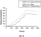
- Example 13 Surface Plasmon Resonance (SPR) Analysis of NanoMIPs to SARS-CoV-2 spike proteins from two different SARS-CoV-2 variant strains.
- NanoMIP-SPR sensors were prepared by covalently immobilising nanoMIPs (from Example 1) to CM5 chips from Cytiva in line with manufacturers guidelines.
Landscapes
- Chemical & Material Sciences (AREA)
- Health & Medical Sciences (AREA)
- Life Sciences & Earth Sciences (AREA)
- Engineering & Computer Science (AREA)
- Immunology (AREA)
- Analytical Chemistry (AREA)
- Urology & Nephrology (AREA)
- Biomedical Technology (AREA)
- Hematology (AREA)
- Molecular Biology (AREA)
- Chemical Kinetics & Catalysis (AREA)
- Organic Chemistry (AREA)
- Virology (AREA)
- Pathology (AREA)
- Medicinal Chemistry (AREA)
- Cell Biology (AREA)
- Biochemistry (AREA)
- General Health & Medical Sciences (AREA)
- General Physics & Mathematics (AREA)
- Microbiology (AREA)
- Physics & Mathematics (AREA)
- Biotechnology (AREA)
- Food Science & Technology (AREA)
- Tropical Medicine & Parasitology (AREA)
- Materials Engineering (AREA)
- Nanotechnology (AREA)
- Crystallography & Structural Chemistry (AREA)
- Peptides Or Proteins (AREA)
- Investigating, Analyzing Materials By Fluorescence Or Luminescence (AREA)
Description
- The present invention relates to compounds for detection of SARS-CoV-2, to methods of producing the same and to conjugates comprising the same, and more particularly to use of the aforementioned in the detection of SARS-CoV-2.
- The outbreak in 2019 of a novel coronavirus, severe acute respiratory syndrome coronavirus 2 (SARS-CoV-2), has given rise to a pandemic which has in turn led to both a public health and economic crisis on a worldwide scale.
- Although the international community quickly recognised that both diagnostic and antibody testing are key to preventing further transmission of the virus and to understanding the prevalence of the virus respectively, testing kits which are reliable, inexpensive to produce and easy to use remain highly sought after.
- Regarding diagnostic testing, at present tests based on reverse transcription polymerase chain reaction (RT-PCR) are the predominant means by which healthcare providers are assessing citizens. The RT-PCR tests work by detecting viral ribonucleic acid (RNA) in a respiratory sample often obtained via a nasopharyngeal swab.
- Whilst RT-PCR testing is widespread, there are problems. Conducting RT-PCR tests can be labour intensive: they require highly skilled personnel and false negative results can occur e.g. due to sampling error or improper sample handling. Indeed false positive results may also occur e.g. due to sample cross-contamination. The tests are also expensive and there is a long turn-around time (1-2 days) with respect to results.
- Rapid antigen tests are also used for population screening and offer portable and rapid (15-30 min) analysis that can be performed at the convenience of the individual. However, they lack sensitivity and utilize antibodies as receptors, which can only function in narrow temperature and pH ranges, which limits their widespread use.
- Molecularly imprinted polymers (MIPs) have been proposed as alternative receptors for use in the detection of SARS-CoV-2.
- Cennamo et al in "Proof of Concept for a Quick and Highly Sensitive On-Site Detection of SARS-CoV-2 by Plasmonic Optical Fibers and Molecularly Imprinted Polymers" details the production of plastic optical fiber (POF) based surface plasmon resonance (SPR) sensors coupled with MIPs. The MIPs were made to the S1 subunit of the SARS-CoV-2 spike protein.
- Parisi et al in ""Monoclonal-type" plastic antibodies for SARS-CoV-2 based on Molecularly Imprinted Polymers" describes the preparation of MIPs using the SARS-CoV-2 receptor binding domain (RBD) as a template.
- Cubuk et al in "Computational analysis of functional monomers used in molecular imprinting for promising COVID-19 detection" describes computational studies aimed at identifying a template and suitable monomers to produce a MIP against SARS-CoV-2.
-
WO 2021/195626 A1 discloses the preparation of MIPs to SARS-CoV-2 wherein the whole virus is used as a template. - Methods of producing MIPs are also known - see e.g.
US 10 189 934B2 - Using either the whole virus, the whole S1 subunit or the whole RBD of SARS-CoV-2 for the production of MIPs leads to significant issues regarding scalability.
- Antibody tests, in contrast to RT-PCR and rapid antigen tests, are carried out in an attempt to establish whether a person may have already had the virus rather than, in the case of RT-PCR and rapid antigen tests, whether the person currently has the virus. Antibody tests are often carried out on blood samples from patients and work by detecting the presence of binding between synthetic antigens and antibodies (if present) in a patient's blood sample.
- Accordingly, one aim of the present invention is to provide alternative means for detecting SARS-CoV-2. A further aim is that such means will provide an alternative to the polymerase chain reaction-based assay and rapid antigen tests currently used. An aim of specific embodiments of the invention includes providing improved detection means.
- Accordingly, the invention provides a molecularly imprinted polymer comprising at least one recognition site that is complementary to a template molecule consisting of an amino acid sequence corresponding to a subsequence of the receptor binding domain of SARS-CoV-2 spike protein, wherein the amino acid sequence is no more than 50 amino acids in length and comprises the sequence NSNNLDSKVGG, and wherein the polymer comprises the monomers:
- (i) N-Fluoresceinyl acrylamide,
- (ii) Acrylamide,
- (iii) Tert-Butyl acrylate (TBAc),
- (iv) 3-O-Acryloyl-1,2:5,6-bis-O-isopropylidene-D-glucofuranose,
- (v) 2,2,2-Trifluoroethyl methacrylate (CF3),
- (vi) N-(3-Aminopropyl) methacrylamide hydrochloride (APMA),
- (vii) Squareamide monomer 7 (SQ7) and
- (viii) N,N'-Methylenebis(acrylamide) (BIS).
- The invention also provides a method of preparing a molecularly imprinted polymer comprising at least one recognition site which binds SARS-CoV-2 comprising the steps of:
- (a) providing a carrier substance having a template molecule consisting of an amino acid sequence corresponding to a subsequence of the receptor binding domain of SARS-CoV-2 spike protein immobilised on it so as to be exposed at a surface;
- (b) providing a polymerisable composition in contact with the surface;
- (c) effecting controlled polymerisation of the polymerisable composition in contact with the surface to produce a molecularly imprinted polymer; and
- (d) separating the molecularly imprinted polymer from the surface and the immobilised template molecule,
- wherein the amino acid sequence is no more than 50 amino acids in length and comprises the sequence NSNNLDSKVGG, and
- wherein the polymerisable composition comprises the monomers:
- (i) N-Fluoresceinyl acrylamide,
- (ii) Acrylamide,
- (iii) Tert-Butyl acrylate (TBAc),
- (iv) 3-O-Acryloyl-1,2:5,6-bis-O-isopropylidene-D-glucofuranose,
- (v) 2,2,2-Trifluoroethyl methacrylate (CF3),
- (vi) N-(3-Aminopropyl) methacrylamide hydrochloride (APMA),
- (vii) Squareamide monomer 7 (SQ7) and
- (viii) N,N'-Methylenebis(acrylamide) (BIS).
- Further provided is a conjugate comprising the molecularly imprinted polymer of the invention and a fluorophore.
- Both the molecularly imprinted polymers of the invention and the conjugates of the invention can be used in the detection of SARS-CoV-2.
- The invention provides a molecularly imprinted polymer comprising at least one recognition site that is complementary to a template molecule consisting of an amino acid sequence corresponding to a subsequence of the receptor binding domain of SARS-CoV-2 spike protein, wherein the amino acid sequence is no more than 50 amino acids in length and comprises the sequence NSNNLDSKVGG, and wherein the polymer comprises the monomers:
- (i) N-Fluoresceinyl acrylamide,
- (ii) Acrylamide,
- (iii) Tert-Butyl acrylate (TBAc),
- (iv) 3-O-Acryloyl-1,2:5,6-bis-O-isopropylidene-D-glucofuranose,
- (v) 2,2,2-Trifluoroethyl methacrylate (CF3),
- (vi) N-(3-Aminopropyl) methacrylamide hydrochloride (APMA),
- (vii) Squareamide monomer 7 (SQ7) and
- (viii) N,N'-Methylenebis(acrylamide) (BIS).
- In molecular imprinting, functional, and sometimes cross-linking, monomers are polymerised in an appropriate solvent in the presence of the compound to be imprinted. The compound to be imprinted is known as the template. During the polymerisation step, the monomers interact with the template through electrostatic, hydrophobic or other interactions leading to the formation of binding sites in the polymer, known as recognition sites, that are complementary to the template molecule. After removal of the template, the polymer matrix retains the recognition sites that are complementary to the template. Complementary in this sense means that the interacting groups in the polymer are placed such that they are in a suitable position to interact with the interacting groups in the template molecule and other molecules that contain the template molecule as a substructure, i.e. in this invention SARS-CoV-2.
- The sequence of SARS-CoV-2 is known. SARS-CoV-2 makes use of a densely glycosylated spike protein to gain entry into host cells. The spike protein is a homotrimeric glycoprotein and each monomer contains two subunits, S1 and S2, which mediate membrane attachment and membrane fusion respectively. The receptor binding domain (RBD) of the S1 subunit has been shown to be involved in host cell receptor engagement.
- Although the sequence of the RBD of the SARS-CoV-2 spike protein is known, and theoretically any sequence therein could be used as a template, it is advantageous for some downstream applications to select, as a template, sequences within the target which are non-conserved. This ensures that the molecularly imprinted polymer binds the desired target with high specificity.
- Supplementary
Figure 5 of Wrapp et al 20201 presents a sequence alignment of the spike protein from SARS-CoV-2 and two other coronavirus strains, SARS-CoV and RaTG13. The RBD is highlighted. Also highlighted are sequences which are conserved between the different strains and sequences which are non-conserved. - In the present invention, the 'at least one recognition site' in the molecularly imprinted polymer is complementary to a template molecule consisting of an amino acid sequence corresponding to a subsequence of the receptor binding domain of SARS-CoV-2 spike protein, wherein the amino acid sequence is no more than 50 amino acids in length and comprises the sequence NSNNLDSKVGG.
- In the present invention, the size of the template is in the range up to 50 amino acids. Preferably, the template will be less than 50 amino acids, more preferably the template will be less than 30 amino acids; most preferred is that the template is less than 20 amino acids, for example 11, 12, 13, 14, 15, 16, 17, 18 or 19 amino acids.
- The use of short peptide sequences (as opposed to e.g. the whole spike protein or RBD) drastically reduces reagent costs and creates high affinity binding sites for a specific region of the target protein, therefore enhancing the monoclonality of the molecularly imprinted polymer.
- The size of the molecularly imprinted polymer of the present invention will be to a large extent dictated by the intended downstream use of the molecular imprinted polymer. That said, suitably the molecular imprinted polymer will be less than 500nm, more suitably less than 250nm and even more suitably less than 100nm, and may be less than 90nm, or even less than 70nm.
- Also provided by the disclosure, and not claimed, is a composition comprising the molecularly imprinted polymer of the invention. Suitably, molecular imprinted polymers in such compositions will have an average size that is less than 500nm, more suitably less than 250nm and even more suitably less than 100nm, and may be less than 90nm, or even less than 70nm.
- Suitable methods for determining the size of molecularly imprinted polymers include dynamic light scattering e.g. using a Zetasizer Ultra (Malvern Panalytical), disc centrifugation, nanoparticle tracking analysis e.g. using a NanoSight NS300 (Malvern Panalytical), tunable resistive pulse sensing, atomic force microscopy and electron microscopy. Nanoparticle tracking analysis, tunable resistive pulse sensing, atomic force microscopy and electron microscopy are particularly suitable for determining the size of single molecularly imprinted polymers. In the examples provided herein, the size of the molecularly imprinted polymers was determined using a NanoSight NS300 instrument.
- Size in the context of the molecularly imprinted polymer refers to the diameter of the molecularly imprinted polymer.
- Monomers which can be used in the preparation of molecularly imprinted polymers include vinyl monomers, allyl monomers, acetylenes, acrylates, methacrylates, acrylamides, methacrylamides, chloroacrylates, itaconates, trifluoromethylacrylates, derivatives of amino acids (e.g. esters or amides), nucleosides, nucleotides, and carbohydrates. Cross-linking monomers, which help to stabilise molecularly imprinted polymers, may also be included. Typical examples of cross-linking monomers suitable for preparing molecularly imprinted polymers include, but are not limited to, ethylene glycol dimethacrylate, trimethylolpropane trimethacrylate, divinylbenzene, methylene bisacrylamide, ethylene bisacrylamide, N,N'-bisacryloylpiperazine and N,N'-Methylenebis(acrylamide) (BIS). Those skilled in the art could select monomers and cross-linking monomers suitable for a particular template. Alternatively a variety of combinatorial and computational methods could be used to assist in this selection.
- In the invention, the molecularly imprinted polymer will comprise (i) N-Fluoresceinyl acrylamide, (ii) Acrylamide, (iii) Tert-Butyl acrylate (TBAc), (iv) 3-O-Acryloyl-1,2:5,6-bis-O-isopropylidene-D-glucofuranose, (v) 2,2,2-Trifluoroethyl methacrylate (CF3), (vi) N-(3-Aminopropyl) methacrylamide hydrochloride (APMA), (vii) Squareamide monomer 7 (SQ7) and (viii) N,N'-Methylenebis(acrylamide) (BIS). In this context it is understood that the molecularly imprinted polymer is a polymer and is said to comprise an identified monomer when it has been made by polymerising a mixture of monomers comprising the identified monomer.
- Advantageously, the molecularly imprinted polymers of the present invention are robust and function in wide ranges of temperature and pH which leads to improved shelf-life and storage conditions and means that they can function in more challenging environments. They also have excellent biocompatibility and have fast binding kinetics.
- Also provided by the present invention is a method of preparing a molecularly imprinted polymer comprising at least one recognition site which binds SARS-CoV-2 comprising the steps of:
- (a) providing a carrier substance having a template molecule consisting of an amino acid sequence corresponding to a subsequence of the receptor binding domain of SARS-CoV-2 spike protein immobilised on it so as to be exposed at a surface;
- (b) providing a polymerisable composition in contact with the surface;
- (c) effecting controlled polymerisation of the polymerisable composition in contact with the surface to produce a molecularly imprinted polymer, and
- (d) separating the molecularly imprinted polymer from the surface and the immobilised template molecule,
- wherein the amino acid sequence is no more than 50 amino acids in length and comprises the sequence NSNNLDSKVGG, and
- wherein the polymerisable composition comprises the monomers:
- (i) N-Fluoresceinyl acrylamide,
- (ii) Acrylamide,
- (iii) Tert-Butyl acrylate (TBAc),
- (iv) 3-O-Acryloyl-1,2:5,6-bis-O-isopropylidene-D-glucofuranose,
- (v) 2,2,2-Trifluoroethyl methacrylate (CF3),
- (vi) N-(3-Aminopropyl) methacrylamide hydrochloride (APMA),
- (vii) Squareamide monomer 7 (SQ7) and
- (viii) N,N'-Methylenebis(acrylamide) (BIS).
- An advantage of the method is that the MIPs possess a more homogenous distribution of binding site affinities, and provides means by which template elution is enhanced and downstream template leaching is prevented.
- Suitable carrier substances include polymer resins, polysaccharides, glass or metal surfaces.
- The carrier substance may be in the form of beads, fibres, membranes or capillaries. In preferred embodiments of the invention, the carrier substance is glass. Further preferred is that the carrier substance is in the form of glass beads.
- The polymerisable composition must contain the monomers required for the polymerisation. Suitable monomers are discussed elsewhere herein.
- Step (c) involves the application of controlled polymerisation. Various techniques for producing molecularly imprinted polymers by controlled polymerisation are known to those of skill in the art.
- Examples of controlled polymerisation include radical polymerisation such as controlled living radical polymerisation (LRP), living anionic polymerisation, living cationic polymerisation, and controlled polycondensation.
- Polymerisation can be initiated by e.g. heating, by applying current (electropolymerisation), by the addition of redox catalyst(s), persulfate or peroxides, by irradiation, including gamma radiation or by microwave radiation or by irradiation with UV or visible light and normally takes minutes to hours. In the examples described herein, polymerisation was initiated using persulfate.
- In preferred embodiments of the invention, the polymerisation reaction is terminated at a stage when the size of the synthesized molecular imprinted polymer is relatively small. Preferences with respect to size are discussed elsewhere herein.
- Once the molecularly imprinted polymers are synthesized they must be separated and collected. The separation of high affinity molecularly imprinted polymers from the immobilised template can be achieved by heating, which disrupts complex formation, by changing solution pH, changing ionic strength, or through the additional of urea, guanidine or a substance which interacts with the template more strongly than the molecularly imprinted polymer.
- In preferred embodiments of the method, prior to the separation step, weakly bound material is removed by washing.
- Once collected, the molecularly imprinted polymers may be purified further e.g. by chromatography, filtration and/or electrophoresis.
- The method by which the template molecule is immobilised on the carrier substance is not critical. However, for some methods, the presence of a cysteine residue at one of the termini of the template molecule is advantageous. As such, for embodiments of the method where the template molecule does not naturally comprise a terminal cysteine residue, it is preferred that the template molecule is modified at the N terminus to comprise an additional cysteine residue.
- In some embodiments of the invention, the additional cysteine residue is attached directly to the termini of the template molecule whilst in other embodiments a spacer is included between the terminal cysteine and the template molecule. In preferred embodiments the spacer comprises a glycine residue.
- Thus in a particular preferred embodiment of the present invention, the template molecule comprises the amino acid sequence NSNNLDSKVGG and is modified at the N terminus to comprise a cysteine (C) residue and a glycine (G) residue such that the molecule used for imprinting is CGNSNNLDSKVGG.
- In some embodiments of the invention, the template molecule is directly attached to the carrier substance whilst in other embodiments one or more linkers are included between the carrier substance and the template molecule. Suitable linkers for use in the invention are well known to those of skill in the art. In the examples provided herein, the carrier substance used was glass beads and these were silanised using (3-aminopropyl)trimethoxysilane (APTMS) prior to immobilisation of the template molecule. The APTMS acts as a linker between the surface of the glass bead and the template molecule. In the examples provided herein, the APTMS was modified using n-succinimidyl iodoacetate (SIA) prior to template immobilisation.
- Linkers and spacers function to prevent adverse effects brought about by steric hindrance.
- An advantage of the method of the present invention is that it is fast and scalable and uses only a short template.
- Further provided by the invention is a conjugate comprising the molecularly imprinted polymer of the invention and a fluorophore.
- Fluorophores are molecules with the ability to absorb light at a particular wavelength, the absorbance or excitation wavelength, and then emit light at a higher wavelength, the emission wavelength.
- Conjugation of fluorophores to molecules for visualization purposes is common in the art. As such, methods for such conjugation are also well known. By way of example, fluorophores can be conjugated via amino, thiol or carbohydrate groups. In the examples provided herein, the fluorophore was conjugated to the molecularly imprinted polymer via amine groups provided by the amino monomer in the molecularly imprinted polymer.
- Although any fluorophore can be used in conjugates of the present invention, it is advantageous for some downstream applications, particularly diagnostic applications, to select a fluorophore that is both bright and stable.
- Conjugated Polymer Nanoparticles (CPNs)™ are highly fluorescent nanoparticles which comprise of a semiconductor light emitting polymer (LEP) core encapsulated within a biocompatible surfactant. CPNs are particularly suited for use in diagnostic applications due to their intense fluorescence, which means they have a high level of sensitivity, and their high degree of photostability. CPNs are around 70-80nm in size and those currently available from Stream Bio exhibit fluorescence with emission wavelengths from 420nm to 1130nm. In the examples provided herein, molecularly imprinted polymers of the invention were conjugated to CPN510 and CPN610, both available from Stream Bio.
- In preferred embodiments of the present invention, the fluorophore is a CPN.
- Also provided by the present disclosure, and not claimed, are compositions comprising the conjugate of the invention.
- The molecularly imprinted polymers and conjugates of the present invention may be used in a variety of applications.
- It is specifically envisaged that the molecularly imprinted polymers and conjugates of the present invention be used in the detection of SARS-CoV-2. The molecularly imprinted polymers and conjugates and compositions may be used in assays for example diagnostic assays which may be used in a medical or point of care setting. Examples of such assays include lateral flow assays (LFA) and enzyme-linked immunosorbent assay (ELISA)-type assays which may be used for high throughput screening. The molecularly imprinted polymers and conjugates of the present invention may also be used in sensors.
- In the examples provided herein, nanoMIP-SPR sensors were prepared by covalently immobilising nanoMIPs on 4% mercaptoundecanoic acid (MUDA) chips (prepared using Biacore SIA Kit Au surfaces, functionalised with a self-assembled monolayer of 4% MUDA in ethanol). NanoMIP-functionalized Screen-Printed Electrodes (SPEs) were also prepared for use in thermal detection methods.
- The molecularly imprinted polymers and conjugates of the present invention may also be used in the detection of SARS-CoV-2 in the research setting.
- The molecularly imprinted polymers and conjugates of the present invention may also be used as pharmaceuticals.
- It is desirable that the molecularly imprinted polymers of the present invention have high affinity for SARS-CoV-2. High affinity in the context of the present specification means that the affinity with which the molecularly imprinted polymers bind SARS-CoV-2 exceeds the affinity with which 'blank' molecularly imprinted polymers bind SARS-CoV-2 by at least three, preferably at least five, more preferably at least 10 times. Blank in this context means molecularly imprinted polymers formed in the absence of template.
- Binding affinity is the strength of the binding interaction between a single molecule to its binding partner e.g. in the present case a molecularly imprinted polymer to its target SARS-CoV-2. Binding affinity is typically measured and reported by the equilibrium dissociation constant (KD). The smaller the KD value, the greater the binding affinity between the two molecules.
- When the molecularly imprinted polymers and conjugates of the present invention are used in the detection of SARS-CoV-2, it is preferred that the molecularly imprinted polymer binds to SARS-CoV-2 with a KD of less than 500nM, preferably less than 250nM and even more preferably less than 200nM.
- Suitable methods for measuring KD include surface plasmon resonance (SPR) which may be conducted using e.g. the Biacore series of instruments (Cytiva) or the MP-SPR Navi instruments (BioNavis).
- The invention is now further described in specific examples with reference to the accompanying drawings in which:
-
Figure 1 shows two surface plasmon resonance sensorgrams showing the binding kinetics for nanoMIPs imprinted againstpeptide 1 and SARS-CoV-2 S recombinant protein; -
Figure 2 is a trace obtained using a NanoSight NS300 instrument showing the successful conjugation of a nanoMIP imprinted againstpeptide 1 to CPN510; -
Figure 3 is a dot blot showing specificity of binding between nanoMIPs imprinted againstpeptides -
Figure 4 is a dot blot showing specificity of binding between nanoMIPs imprinted againstpeptide 1 and SARS-CoV-2; -
Figure 5 is a dot blot showing specificity of binding between nanoMIPs imprinted againstpeptide 1 and SARS-CoV-2 Spike Glycoprotein (S1); -
Figure 6 is a raw Heat-Transfer Method data plot of Rth versus time for the addition of SARS-CoV-2 ORF8 (1fg/mL - 10pg/mL) in PBS to a Screen-Printed Electrode (SPE) functionalised with NanoMIP against peptide 1 (Figure 6a ) and peptide 3 (Figure 6b ); -
Figure 7 is a raw Heat-Transfer Method data plot of Rth versus time for the addition of SARS-CoV-2 S protein RBD (1fg/mL - 10pg/mL) in PBS to a Screen-Printed Electrode (SPE) functionalised with NanoMIP against peptide 1 (Figure 7a ) and a dose response curve whereby % change in the Rth values were plotted against the concentration of SARS-CoV-2 S protein RBD injected into the system (Figure 7b ); -
Figure 8 is a raw Heat-Transfer Method data plot of Rth versus time for the addition of SARS-CoV-2 S protein RBD (1fg/mL - 10pg/mL) in PBS to a Screen-Printed Electrode (SPE) functionalised with NanoMIP against peptide 3 (Figure 8a ) and a dose response curve whereby % change in the Rth values were plotted against the concentration of SARS-CoV-2 S protein RBD injected into the system (Figure 8b ); -
Figure 9 reports the ability of nanoMIPs imprinted againstpeptide 1 to withstand extremes of temperature and pH.Figure 9a shows typical atomic force microscopy (AFM) height images of an isolated nanoMIP on an Au surface in air (represented by the green box in the larger scale image) at room temperature, 37 °C, and 50 °C.Figure 9b shows a corresponding cross-sectional profile plot of the nanoMIP at each temperature.Figure 9c shows a box chart comparing nanoMIP volume (n = 120) from AFM images at various pH levels.Figure 9d compares the thermal response of a nanoMIP-based sensor to a clinical reference liquid (universal transport medium) and Diet Coca-Cola (pH = 3.5).Figure 9e shows a commercial rapid antigen test giving a false positive result when Diet Coca-Cola was used as the test liquid; -
Figure 10 reports the sensitivity of nanoMIPs imprinted againstpeptide 1 to various SARS-CoV-2 antigens and compares the sensitivity of the nanoMIPs to a SARS-CoV-2 antibody.Figure 10a shows typical Heat Transfer Method data for a nanoMIP-functionalized SPE upon exposure to PBS containing 1 fg mL-1 - 10 pg mL-1 of the SARS-CoV-2 RBD.Figures 10b-10d show typical dose-response curves (error bars represent the SD) showing the thermal response of (i) nanoMIP and antibody sensors to the spike protein (nanoMIPs were tested against the SARS-CoV-2 spike protein from both the alpha and delta variants) (Figure 10b ) (ii) nanoMIP and antibody sensors to the SARS-CoV-2 RBD (Figure 10c ), and iii) nanoMIP sensors to SARS-CoV-2 antigens and the negative controls ORF8, IL-6, and HSA. The response of NIP-based sensors to the spike protein is also shown (Figure 10d). Figure 10e compares the LoD values for nanoMIPs of the present invention with a commercial rapid antigen test and numerous recently developed antigen tests from the literature; -
Figure 11 shows binding between nanoMIPs imprinted againstpeptide 1 and SARS-CoV-2 from clinical samples.Figure 11a is a schematic illustration of a 3D-printed prototype addition cell: 1) thermocouple inlet, 2) functionalized SPE, 3) open-bottomed reservoir to facilitate the manual addition of liquid, and 4) removable lid to reduce experimental noise.Figure 11b is a photograph of the addition cell shown inFigure 11a .Figure 11c shows a typical dose-response curve obtained when using the prototype addition cell for the thermal detection of the SARS-CoV-2 spike protein (1 fg mL-1 - 10 pg mL-1) in phosphate-buffered saline (PBS).Figure 11d shows the thermal response of the nanoMIP sensor to clinical samples from COVID-positive and negative patients (n = 7); -
Figure 12 shows the size of nanoMIPs measured using NanoSight particle analyzer; -
Figure 13 shows nanoMIP - LSPR sensor mediated detection of SARS-CoV-2 spike proteins from SARS-CoV-2 Alpha, Beta and Gamma variants in PBS.Figure 13a shows an LSPR spectrum in wavelength vs absorbance plot for concentrations ranging from 10 aM to 100 nM of the Alpha variant of SARS CoV-2.Figure 13b shows the change in LSPR wavelength of the Ag-MIPs complex upon varying the concentration of SARS-CoV-2 Alpha, Beta and Gamma variants from 10 aM to 100 nM; -
Figure 14 shows nanoMIP - LSPR mediated detection of SARS-CoV-2 spike proteins from SARS-CoV-2 Alpha, Beta and Gamma variants in serum.Figure 14a shows an LSPR spectrum in wavelength vs absorbance plot for concentrations ranging from 100 fM to 100 nM of the Alpha variant of SARS-CoV-2.Figure 14b shows the change in LSPR wavelength of the Ag-MIPs complex upon varying the concentration of SARS-CoV-2 Alpha, Beta and Gamma variants from 100 fM to 100 nM; and -
Figure 15 shows two superimposed surface plasmon resonance sensorgrams showing the binding kinetics for nanoMIPs imprinted againstpeptide 1 and either the original SARS-CoV-2 S (spike) recombinant protein and or that from the Omicron variant. - Examples, or parts thereof, which relate to molecularly imprinted polymers comprising at least one recognition site that is complementary to a template molecule consisting of an amino acid sequence corresponding to a subsequence of the receptor binding domain of SARS-CoV-2 wherein the amino acid sequence does not comprise the sequence NSNNLDSKVGG are not part of the claimed invention and are present for comparative purposes only.
- Supplementary
Figure 5 of Wrapp et al 20201 presents a sequence alignment of the spike protein from SARS-CoV-2 and two other coronavirus strains, SARS-CoV and RaTG13. The RBD is highlighted. Three peptide sequences (peptide 1: NSNNLDSKVGG, peptide 2: STEIYQAGSTPC and peptide 3: CYFPLQSYGFQP) from the RBD of SARS-CoV-2 were selected by the present inventors as potential candidates for imprinting based on the lack of conservation with the respective sequences from SARS-CoV and RaTG13. -
Peptides - On the basis that a terminal cysteine was required for peptide immobilisation, when
peptides peptide 1. A further glycine residue was also added between the cysteine and the N terminal asparagine, functioning as a spacer. Thus, the peptides sourced from Ontores for use in imprinting were: - Peptide 1: CGNSNNLDSKVGG
- Peptide 2: STEIYQAGSTPC
- Peptide 3: CYFPLQSYGFQP.
- Peptide immobilisation was carried out as follows: glass beads (approximately 100µM in diameter, sourced from Microbeads AG) were activated by boiling in 4M NaOH for 10 minutes. The glass beads were then washed with firstly deionised water and then acetone and then dried at 80°C for 2 hours. The glass beads were then incubated in toluene with 2 % v/v (3-aminopropyl)trimethoxysilane (APTMS) for 3 hours, washed with acetone and placed in PBS, pH 7.4 with 0.2mg/ml n-succinimidyl iodoacetate (SIA) for two hours before being washed with acetonitrile. The templates (
Peptides - The peptide coated glass beads were used for the synthesis of imprinted nanoMIPs as follows: monomer solutions were prepared, sonicated for 10 minutes and purged with nitrogen for 5 minutes. One example of a monomer solution that was prepared includes 3mg N-Fluoresceinyl acrylamide, 24.2mg Acrylamide, 31.7µL Tert-Butyl acrylate (TBAc), 36mg 3-O-Acryloyl-1,2:5,6-bis-O-isopropylidene-D-glucofuranose, 13.8
µL - A
Biacore 3000 instrument was employed in this study. - NanoMIP-SPR sensors were prepared by covalently immobilising nanoMIPs (from Example 1) on 4% mercaptoundecanoic acid (MUDA) chips (prepared using Biacore SIA Kit Au surfaces, functionalised with a self-assembled monolayer of 4% MUDA in ethanol).
- In more detail, water was utilized to prime the instrument at a constant flow rate of 5µL/minute. Water was utilized as the running buffer throughout the chip preparation at a constant flow rate of 30µl/minute. Water was run until a stable baseline was achieved. A mixture of N-hydroxysuccinimide (NHS) and 1-ethyl-3-(3-dimethylaminopropyl)-carbodiimide (EDC) (2 mg EDC + 3 mg NHS in 400 uL of water) was injected (8 minutes at a flow rate of 8µL/minute) across the sensor surface to activate the surface (the mixture converts the carboxylic terminal groups on the chip surface into N-Hydroxy-succinimide esters). NanoMIPs (5nM) were then injected (8-10 minutes at a flow rate of 8µL/minute) into all four flow cells on the chip. Finally, unreacted NHS esters were hydrolysed by injecting carbonate buffer (pH 9.2) (30 minutes at a flow rate of between 5 and 30µL/minute).
- Prior to testing in SPR, each of
peptides - Once a stable baseline was obtained having switched running buffer from water to PBS, 7 dilutions of BSA or BSA-peptide conjugates (2.33mg/ml, 0.777mg/ml, 259ug/ml, 86ug/ml, 28.8ug/ml, 9.6ug/ml, 3.2ug/ml in PBS) were injected (5 minutes association phase, 4 minutes dissociation phase, 28µl/minute flow rate) as follows: BSA alone was injected into
flow cell 1, BSA-peptide 1 intoflow cell 2 and BSA-peptide 3 intoflow cell 3. - Good selectivity was observed for nanoMIPs imprinted against
peptide 3, for which the software calculated a KD of 160 nM (data not shown). - A
Biacore 3000 instrument was employed in this study. - SARS-CoV-2 S protein-SPR sensors were prepared by covalently immobilising recombinant SARS-CoV-2 S protein (recombinant full-length SARS-CoV-2 spike glycoprotein sourced from The Native Antigen Company) on 4% mercaptoundecanoic acid (MUDA) chips (prepared using Biacore SIA Kit Au surfaces, functionalised with a self-assembled monolayer of 4% MUDA in ethanol).
- In more detail, water was utilized to prime the instrument at a constant flow rate of 5 µl/minute. Water was also utilized as the running buffer throughout the chip preparation at a constant flow rate of 30µl/minute. Water was run until a stable baseline was achieved. A mixture of N-hydroxysuccinimide (NHS) and 1-ethyl-3-(3-dimethylaminopropyl)-carbodiimide (EDC) (2 mg EDC + 3 mg NHS in 400 uL of water) was injected (8 minutes at a flow rate of 8µl/minute) across the sensor surface to activate the surface (the mixture converts the carboxylic terminal groups on the chip surface into N-Hydroxy-succinimide esters). Control (1% BSA (w/v) and 0.5% Pluronic F127 (w/v)) and two different concentrations of recombinant SARS-CoV-2 S protein (60nM and 600nM) were then injected (6
minutes 15 seconds at a flow rate of 8µl/minute) intoflow cells - Once a stable baseline was obtained having switched running buffer from water to PBS, 30nM (and 1:1 serial dilutions thereof) nanoMIPs imprinted against
peptide 1 were then injected (5 minutes association phase, 3 minutes dissociation phase, 28µl/minute flow rate). - The software calculated a KD of 5nM (
Figure 1a ) and 15nM (Figure 1b ). - NanoMIPs imprinted against
peptides NanoSight 3000. -
Figure 2 shows the successful conjugation of a nanoMIP imprinted againstpeptide 1 to CPN510. Note that the original unconjugated nanoMIPs were predominantly 59nm; however, when conjugated to CPN510, a significant shift in particle size is seen - see the peak at 119nm. - Different concentrations (290ng, 145ng, 73ng, 36ng, 18ng, 9ng, 5ng, 2ng, 1ng, 0.6ng, 0.3ng and 0.14ng) of recombinant SARS-CoV-2 S protein (sourced from The Native Antigen Company) were blotted onto nitrocellulose membranes before allowing the membranes to dry. The membranes were then blocked by soaking in 5% BSA in PBS-T (0.05% Tween-20 in PBS) for 30-60 minutes at room temperature. Post blocking, the membranes were incubated with (1) NanoMIP-CPN510 (imprinted against peptide 1), (2) NanoMIP-CPN510 (imprinted against peptide 3) or (3) CPN510 only (control) dissolved in 5% BSA in PBS-T for 30-60 minutes at room temperature. The membranes were then washed three times, for 5 minutes each time, with PBS-T. Fluorescence was then observed under UV. As shown in
Figure 3 , the detection limit using NanoMIP-CPN510 (imprinted against peptide 1) was 0.3ng, much lower than that achieved using CPN510 only (control) indicating that nanoMIPs imprinted against at leastpeptide 1 specifically interact with SARS-CoV-2 S protein. - SARS-CoV-2 (2 × 10⁴ pfu) was blotted onto two nitrocellulose membranes. Also blotted on to each of the nitrocellulose membranes was 75ng of recombinant SARS-CoV-2 S protein (sourced from The Native Antigen Company) as a positive control and culture media as a negative control. The membranes were then left to dry. The membranes were then blocked by soaking in 5% BSA in PBS-T (0.05% Tween-20 in PBS) for 30-60 minutes at room temperature. Post blocking, the membranes were incubated with NanoMIP-CPN510 (imprinted against peptide 1) or (2) NanoMIP-CPN510 (imprinted against troponin) (negative control) dissolved in 5% BSA in PBS-T for 30-60 minutes at room temperature. The membranes were then washed three times, for 5 minutes each time, with PBS-T. Fluorescence was then observed under UV. As shown in
Figure 4 , no fluorescence was observed in the area of the nitrocellulose membrane blotted with culture media or when the membrane was incubated with NanoMIP-CPN510 (imprinted against troponin) (negative control). In contrast, fluorescence was observed when the nitrocellulose membrane was incubated with NanoMIP-CPN510 (imprinted against peptide 1) both with respect to SARS-CoV-2 and also the SARS-CoV-2 S protein positive control. This indicates that nanoMIPs imprinted against at leastpeptide 1 specifically interact with SARS-CoV-2. - 0.5 µL of SARS-CoV-2 Spike Glycoprotein (S1) (The Native Antigen Company), Human Coronavirus OC43 Spike Glycoprotein (S1) (The Native Antigen Company),
Human Coronavirus 229E Spike Glycoprotein (S1) (The Native Antigen Company) and Human Coronavirus HKU1 Spike Glycoprotein (S1) (The Native Antigen Company) (0.6 mg/mL) were blotted onto nitrocellulose membranes in triplicate (as shown in the schematic inFigure 5 ) before allowing the membranes to dry. The membranes were then blocked by soaking in 2 mL BSA (1% in PBS) for 1 hour. Post blocking, the membranes were incubated with (1) NanoMIP - CF770 (imprinted against peptide 1) and (2) three different monoclonal antibodies specific to SARS-CoV-2 Spike (RBD), Mab RBD1106-CF770, Mab RBD107-CF770 and Mab RBD5305-CF770 (all prepared in-house and diluted in 0.01% BSA) for 30 minutes. The membranes were then washed with 2mL PBST for 5 minutes. Fluorescence was then observed under UV. As shown inFigure 5 , the nanoMIPs selectively bound the SARS-CoV-2 Spike Glycoprotein (S1) and did not bind to any of Human Coronavirus OC43 Spike Glycoprotein (S1),Human Coronavirus 229E Spike Glycoprotein (S1) or Human Coronavirus HKU1 Spike Glycoprotein (S1). Furthermore, the nanoMIPs were shown to have comparable selectivity and binding to each of the antibodies tested. - A solution of 4-ABA (2 mM) and sodium nitrite (2 mM) in aqueous HCl (0.5 M) was prepared and gently mixed on an orbital shaker for 10 minutes. Screen-Printed Electrodes (SPEs) were submerged into the solution and cyclic voltammetry was performed from +0.2 V to -0.6 V at 100 mV s-1 using an Ag/AgCl reference electrode (Alvatek Ltd., Romsey, UK). The obtained electrode, denoted SPE/4-ABA, was thoroughly rinsed with deionised water to remove any unbound 4-ABA and dried with nitrogen. The carboxyl group was then activated through incubation with a solution of 1-ethyl-3-(3-dimethylaminopropyl)-carbodiimide (EDC) (100 mM) and N-hydroxysuccinimide (NHS) (20 mM) in PBS buffer (pH = 5). Dropcasting was utilised to
deposit 8 µL of the EDC/NHS solution onto the working electrode of the SPEs. After 1 hour, the electrodes were rinsed with deionised water and dried to obtain the SPE/4-ABA/EDC+NHS electrodes. Following this, 8 µL of a given nanoMIP solution (NanoMIP imprinted against peptide 1 'P1C6' or NanoMIP imprinted against peptide 3 'P3C6') was deposited onto the working electrodes following a gentle vortex. After 3 hours, the SPE/4-ABA/EDC+NHS/nanoMIP electrodes were rinsed in deionised water and dried with a gentle stream of nitrogen before being stored in PBS at 4°C until use. - 3D-printed flow cells were used to facilitate measurements using the nanoMIP-functionalised SPEs described above. A SPE was mounted into the cell and a thermocouple measured the temperature in the liquid (T2). For each measurement, a freshly prepared nanoMIP-functionalised SPE was used. The flow cell was connected to a heat-transfer device as described by van Grinsven et al. The device was steered with LabView software that actively controls the temperature of the heat sink (copper block, T1), which was set to 37.00 ± 0.02 °C to mimic in-vivo conditions. A proportional-integral-derivative (PID) controller attached to a power resistor (22 Ω) regulated the feedback on the signal as described in Geerets et al. The PID parameters were fixed for all experiments at optimised values of P=1, I=10, and D=0.2.
- In all measurements, the flow cell was filled with PBS and left for 30 minutes to ensure stabilisation of the baseline temperature signal before a first injection of PBS was added to act as the blank measurement. Solutions (3 mL) of increasing concentrations of each target biomarker (SARS-CoV-2 S protein RBD or SARS-CoV-2 ORF8 (as a negative control)) (0-10 pg mL-1) were prepared in PBS prior to experiments and stored at 4°C until required. Each biomarker injection was performed at 250 µL min-1 for 12 minutes using a LSP02-1B dual channel syringe (Longer Precision Pump Co., Hebei, China) pump. Following each biomarker injection, the system was allowed to stabilise for 30 minutes prior to the next injection. The thermal resistance (Rth) was determined throughout the experiments by dividing the temperature gradient (T1-T2) over the power required to keep the heat sink at 37.00°C. The Rth and standard deviation (SD) were calculated using the average of 600 data points from the baseline signal of each concentration and the initial PBS injection, respectively (
Figures 6 ,7a , and8a ). This data was used to construct dose-response curves, from which the limit of detection (LoD) was calculated using the three-sigma method in the linear range of the sensor (Figures 7b and8b ). The error bars in the graphs relate to the SD values.Figure 6 shows that the thermal response to SARS-CoV-2 ORF8 is very limited whereasFigures 7 and8 show that a good response was observed with SARS-CoV-2 S protein RBD for nanoMIPs imprinted against bothpeptides - The capability of MIPs imprinted against
peptide 1 to withstand extremes of temperature and pH was investigated. - Atomic force microscopy (AFM) was utilized to examine how increasing temperature impacted the morphology of adsorbed nanoMIPs by imaging the same nanoMIPs at room temperature, 37°C, and 50°C.
- AFM measurements were performed on a
JPK Nanowizard 4 XP Bioscience (Bruker, Nano GmbH, Berlin, Germany). Measurements in air were carried out in tapping mode using PPP-NCL-W probes (Nanosensors, Neuchatel, Switzerland) with a cantilever length of ~225 µm and spring constant of ~48 N m-1. Measurements in liquid were performed in Quantitative Imaging (QI) mode using MLCT-E probes (Bruker, Ca, USA) with a cantilever length of ~140 µm and spring constant of ~0.1 N m-1. Au-coated Si chips were used as substrates (Si-Mat, Kaufering, Germany). Prior to drop-casting, the chips were cleaned by immersion for 5 min in a 5:1:1 mixture of milli-Q water, ammonia, and hydrogen peroxide heated at 75 °C. The chips were then rinsed in milli-Q water and dried with nitrogen. NanoMIP solutions were diluted in milli-Q water to ~2.54 µg mL-1, drop-cast (20 µL) onto the Au-coated surfaces, and allowed to dry in ambient conditions for a minimum of 4 h in a Petri dish. A high temperature heating stage (HTHS, JPK BioAFM - resolution of 0.1 °C) was used as a temperature controller to facilitate imaging at 37°C and 50°C. -
Figure 9a and 9b which show typical AFM images and a corresponding cross-sectional profile plot respectively show that the nanoMIP morphology was unaffected by increasing temperature. - NanoMIP volume was calculated using Gwyddion. Tracking numerous droplets revealed minimal changes in mean nanoMIP volume (~6% decrease) from room temperature to 50 °C.
- Thus, nanoMIP morphology remains consistent across relatively large temperature ranges.
- SPR binding analysis, using a
Biacore 3000 instrument, was also performed against the SARS-CoV-2 spike protein using nanoMIPs that had experienced autoclaving for 15 minutes with a maximum temperature of 121°C. - In more detail, two nanoMIP-SPR sensors were prepared by covalently immobilising nanoMIPs (from Example 1), either as directly prepared or after autoclave at 121°C for approximately 15 minutes, on 4% mercaptoundecanoic acid (MUDA) chips (prepared using Biacore SIA Kit Au surfaces, functionalised with a self-assembled monolayer of 4% MUDA in ethanol) in line with the process directly below.
- Water was utilized to prime the instrument at a constant flow rate of 5µL/minute. Water was utilized as the running buffer throughout the chip preparation at a constant flow rate of 30µl/minute. Water was run until a stable baseline was achieved. A mixture of N-hydroxysuccinimide (NHS) and 1-ethyl-3-(3-dimethylaminopropyl)-carbodiimide (EDC) (2 mg EDC + 3 mg NHS in 400 uL of water) was injected (8 minutes at a flow rate of 8µL/minute) across the sensor surface to activate the surface (the mixture converts the carboxylic terminal groups on the chip surface into N-Hydroxy-succinimide esters). NanoMIPs (5nM) were then injected (8-10 minutes at a flow rate of 8µL/minute) into all four flow cells on the chip. Finally, unreacted NHS esters were hydrolysed by injecting carbonate buffer (pH 9.2) (30 minutes at a flow rate of between 5 and 30µL/minute).
- Once a stable baseline was obtained having switched running buffer from water to PBS, 5 dilutions (30, 15, 7.5, 3.75 and 1.88mM in PBS) of SARS-CoV-2 S protein (recombinant full-length SARS-CoV-2 spike glycoprotein sourced from The Native Antigen Company) were injected (circa 5 minutes association phase, at least 2 minutes dissociation phase, 28µl/minute flow rate) with suitable controls in control channels. The results from the two chips were then superimposed, and the software calculated very similar KD values for the nanoMIPs before and after autoclaving (7 and 3 nM, respectively), demonstrating that there is no impact on binding affinity after exposure to high temperatures. This is highly advantageous for nanoMIP shelf-life as autoclaving (sterilization) facilitates their long-term storage in water without bacterial degradation. In contrast, antibodies experience significant deterioration in affinity at temperatures above 37 °C.
- AFM was also used to assess the ability of nanoMIPs to withstand extremes of pH by measuring the volume of adsorbed nanoMIPs (n = 120) in liquid at various pH levels. More specifically, QI measurements were performed at room temperature (23 ± 1 °C) in liquid at different pH levels (5.5-8.5) within the operating conditions of the AFM. Initial measurements were carried out in pure milli-Q water (pH 5.5) and the pH was subsequently increased with the addition of ammonia.
- As above, nanoMIP volume was calculated using Gwyddion which revealed negligible changes to the mean nanoMIP volume from pH 5.5 to 8.5 (~3% decrease), highlighting that adsorbed nanoMIP morphology was consistent across a broad pH range (see
Figure 9c ). - Additionally, the thermal response of the developed nanoMIP tests (see Example 10) to a clinical reference liquid (universal transport medium, UTM) and Diet Coca-Cola (pH = 3.5) were compared. The results demonstrated that there was no statistically significant difference between the thermal response of the two liquids (see
Figure 9d ). In contrast, commercial antibody-based rapid antigen tests are highly impacted by changes to pH and false positive results are produced when acidic soft drinks (Diet Coca-Cola) are used as the test liquid (seeFigure 9e ). - NanoMIP-functionalized Screen-Printed Electrodes (SPEs) (using nanoMIPs imprinted against peptide 1) were made in line with the procedure in Example 8.
- SARS-CoV-2 antibody-functionalized SPEs were also made in line with the procedure in Example 8.
- SARS-CoV-2 antigens (either recombinant full-length SARS-CoV-2 spike glycoprotein (alpha variant) sourced from The Native Antigen Company, recombinant SARS-CoV-2 spike receptor binding domain (RBD) (delta variant) sourced from Abbexa, Cambridge, UK or SARS-CoV-2 RBD from the Medical Research Council Protein Phosphorylation and Ubiquitylation Unit (Dundee, UK)) were thermally detected by mounting nanoMIP-functionalized SPEs into 3D-printed resin flow cells to create an interface between the heat sink and liquid reservoir. Two thermocouples measured the heat sink (T1) and liquid reservoir (T2) temperatures every second, and the thermal resistance (Rth) was obtained by dividing the temperature gradient (T1-T2) over the power required to maintain the heat sink at 37.00 ± 0.02 °C. As the target attached to the nanoMIPs, heat transfer at the solid-liquid interface was reduced (larger temperature gradient), which led to a measurable increase in the Rth.
- For all experiments, the thermal measurement device was controlled using LabView software and a proportional-integral-derivative (PID) controller attached to a power resistor (22 Ω) regulated the feedback on the signal. The PID parameters were optimized to reduce noise and were set at P = 1, I = 13, and D = 0.2.
- The liquid reservoir was filled with PBS and left for 30 min to ensure stabilization of the baseline R th signal. Subsequently, five spiked PBS solutions (3 mL) with increasing concentrations of the SARS-CoV-2 antigen (1 fg mL-1 - 10 pg mL-1) were injected into the flow cell at a rate of 250 µL min-1 for 12 min using an automated syringe pump (LSP02-1B, Longer Precision Pump Co., Hebei, China). The system was allowed to stabilize for 30 min prior to each subsequent injection. The experiments produced raw thermal data plots which displayed a step-wise increase in R th where each stabilized plateau represents the injection of an increasingly concentrated spiked solution (see
Figure 10a ). Dose-response curves (Figures 10b-d ) were composed from the thermal plots by taking the mean and standard deviation (SD) of the stabilized plateau from each concentration injection. Limit of detection (LoD) values were calculated from the dose-response curves using the three-sigma method (3 × reference SD) in the linear range. - SARS-CoV-2 antibody-functionalized SPEs were used to facilitate a direct comparison between the sensing performance of the nanoMIPs and those of antibody receptors. The thermal detection results (see
Figure 10b ) revealed that the response to the SARS-CoV-2 spike protein (alpha variant) was very similar for the nanoMIP (LoD = 9.9 ± 2.5 fg mL-1) and antibody (LoD = 8.9 ± 4.1 fg mL-1) based sensors highlighting that the specificity of the nanoMIPs against the spike protein is comparable with antibodies used in commercial tests. - The ability of the nanoMIP receptors to detect virus mutations was also examined by measuring their specificity against the SARS-CoV-2 spike protein (delta variant). Results show (
Figure 10b ) that the nanoMIP sensor was effective in detecting the delta variant as the resulting LoD (6.1 ± 2.9 fg mL-1) was very similar to the value obtained for the alpha variant. This demonstrates that the nanoMIP sensor can detect equally low amounts of both the alpha and delta variant spike protein, which is highly promising for potential commercial applications where there is an ongoing concern regarding the reduction in test efficacy due to the presence of new variants. - The versatility of nanoMIP receptors was also examined by measuring their thermal response against the SARS-CoV-2 RBD (see
Figure 10c ). Antibody receptors were also tested for the purpose of direct comparison. The LoD value for the SARS-CoV-2 RBD was ~20 times smaller for the nanoMIP sensor (3.9 ± 1.0 fg mL-1) compared to the antibody sensor (85.5 ± 15.0 fg mL-1). Consequently, this demonstrates that the nanoMIPs possessed greater versatility than antibodies as they had a comparable LoD for the full-length spike protein but a significantly lower LoD for the RBD. - The selectivity of the nanoMIP sensor was also comprehensively examined using three negative controls which are common interferents in clinical samples: open reading frame 8 (ORF8), interleukin-6 (IL-6), and human serum albumin (HSA). A high degree of binding occurred between the SARS-CoV-2 antigens and nanoMIPs, which led to large ΔRth values at the highest concentration (10 pg mL-1) for the spike protein (0.23 °C W-1) and RBD (0.35 °C W-1). In contrast, minimal binding occurred with the negative controls, which led to significantly lower ΔRth values for ORF8 (0.00 °C W-1), IL-6 (0.06 °C W-1), and HSA (0.05 °C W-1). The results demonstrated that the thermal response of the nanoMIP sensor was considerably greater for SARS-CoV-2 antigens compared to the negative controls, which highlighted the excellent selectivity of the nanoMIP sensor. An additional control experiment was also performed using non-imprinted polymers (NIPs) immobilized to SPEs (NIP functionalised SPEs made in line with the procedure in Example 8). NIPs were prepared using the same synthesis protocol as nanoMIPs except they were not exposed to a target epitope during polymerization, and therefore had no specific cavities to facilitate target binding. The thermal response of a nanoMIP-functionalized SPE to the spike protein was ~6 times larger compared to the NIP-functionalized SPE (0.04 °C W-1) showing that specific binding occurred between the spike protein and nanoMIP cavities.
- The LoD values of the nanoMIP sensor, a commercial rapid antigen test, and numerous recently developed antigen tests from the literature4-15 are presented in
Figure 10e . Table 1 below provides further details of the experimental setup. The LoD value for the nanoMIP sensor is ~6000 times lower than the commercial rapid antigen test (20 pg mL-1). This considerably lower LoD highlights that nanoMIP receptors could be a valuable tool in producing rapid antigen tests with adequate sensitives for effective population screening. Additionally, the LoD of the nanoMIP sensor is among the lowest value of those from recently developed antigen tests in the literature. This demonstrates that thermal detection using nanoMIPs can compete with the best performing antigen tests from recent literature, whilst possessing the added benefit of being able to withstand extremes of temperature and pH.

- A prototype 3D-printed resin addition cell (
Figure 11a and 11b ) was developed for clinical analysis with disposable components. As in Example 10, the thermal measurement device was controlled using LabView software and a proportional-integral-derivative (PID) controller attached to a power resistor (22 Ω) regulated the feedback on the signal. The PID parameters were optimized to reduce noise and were set at P = 1, I = 14, and D = 0. - To validate the addition cell design, thermal detection experiments were performed using the SARS-CoV-2 spike protein in PBS. The addition cell sensor showed a good thermal response to the spike protein (
Figure 11c ) as the LoD value (7.0 ± 4.0 fg mL-1) was very similar to the results obtained when using the flow cell design (9.9 ± 2.5 fg mL-1 (Example 10)). - After initial validation, clinical measurements were performed using COVID-positive (positive after <20 PCR cycles) and negative patient samples (n = 7).
- UTM and VPM (Viral preservation medium) were used as reference liquids for the negative and positive samples, respectively. During the measurements, 100 µL of the reference liquid (UTM/VPM) was pipetted into the reservoir and the R th signal was allowed to stabilize for 10 min. Following this, the reference liquid was pipetted out and 100 µL of the sample was added. Note that use of a pipette resulted in less disturbance to the system (e.g., flow, addition of air bubbles) compared to using a syringe pump. This is highly advantageous for clinical analysis since the sample volume was similar to that collected by a throat and nasal swab (100 µL), measurement time was reduced to ~15 min, and device operation was straightforward.
- The thermal detection results (
Figure 11d ) show that specific binding to the nanoMIP cavities occurred during measurements of the positive samples, which led to a large mean ΔRth value (0.27 °C W-1). In contrast, only non-specific binding occurred to the nanoMIPs when measuring the negative samples, which resulted in a mean ΔRth of 0.00 °C W-1. The overall range of ΔRth values was much larger for the positive samples (0.41 °C W-1) compared to the negative samples (0.10 °C W-1), which is due to variations in the viral loads of different COVID-positive patients. Importantly, there was no overlap in any positive and negative measurements as the largest ΔRth for a negative sample was ~4 times lower than the smallest ΔRth for a positive sample. This highlights that the nanoMIP sensor possesses excellent sensitivity and specificity for the detection of SARS-CoV-2 in clinical samples. Furthermore, the measurement time of the nanoMIP sensor (~15 mins) is comparable to commercial rapid antigen tests, which is crucially important for potential population screening applications. -
Peptide 1 was immobilised onto silanised glass beads prior to nanoMIP synthesis in line with the procedure in Example 1. - The peptide coated glass beads were used for the synthesis of imprinted nanoMIPs as follows: a monomer solution (see Example 1) was degassed under vacuum and sonicated for 5 min, and then purged with N2 for 20 min before being added to 60 g of peptide 1-coated glass beads. Polymerization was initiated by adding an ammonium persulfate aqueous solution (800 µL, 60 mg/mL) and N,N,N',N'-tetramethylethylenediamine (24 µL), both from Sigma Aldrich. The headspace was flushed with N2 and the bottle sealed with a screw cap. Polymerization was carried-out at room temperature for 1 h. Subsequently, the content of the polymerization vessel was poured into a solid-phase extraction (SPE) cartridge (60 mL) equipped with a frit (20 µm porosity). A total of 9 washes with 20 mL of distilled water at 20 °C were carried out to remove low affinity nanoMIPs, polymer and unreacted monomer. Afterwards, the SPE cartridge containing the solid-phase was placed in a water bath at 70 °C for 15 min. An aliquot of 20 mL of distilled water pre-warmed at 65 °C was poured into the SPE to collect the high-affinity nanoMIPs. This action was repeated 5 times, until about 100 mL of a solution of high-affinity nanoMIPs in water were collected. To ensure complete removal of any potential unreacted monomer from the bulk of the nanoparticles, the collected solution was concentrated down and dialysed with a SnakeSkin membrane (10 kDa molecular weight cut-off).
- Imprinted nanoMIPs had an average diameter of 69.3nm, as calculated by NanoSight NS300 (
Figure 12 ). - Once produced the nanoMIPs imprinted against
peptide 1 were integrated in a silver nanoparticle (AgNP) based localized surface plasmon resonance (LSPR) sensor. - In more detail, 50 µl of a nanoMIP stock solution (5 µg/ml in DI water) was dispensed on the Ag-LSPR chips. Afterwards, the Ag chips were placed inside a humidified chamber for 3h to ensure that the nanoMIPs were immobilized on the surface of the Ag nanoparticles. Since the nanoMIPs bear amine groups, electrostatic interactions between the negative LSPR Ag surface and the positively charged nanoMIPs (-Ag with NH3 +) resulted in binding of the nanoMIPs to the surface. After this the Ag substrates were thoroughly rinsed with DI water to remove any loosely-bound polymers from the electrode surface. The chips were then stored at 4°C.
- The LSPR performance of the nanoMIP-LSPR sensor was then evaluated by detecting spike proteins of Alpha, Beta and Gamma Variants of the SARS-CoV-2 virus (all purchased from antibodies-online: Alpha, SARS-CoV-2 Spike protein lineage B.1.1.7, product number: ABIN6963738; Beta, SARS-CoV-2 Spike protein lineage B.1.351, product number: ABIN6963739; Gamma, SARS-CoV-2 Spike protein lineage P.1, product number: ABIN6964442) in PBS. For controls, the LSPR performance of the nanoMIP-LSPR sensor against spike proteins of human coronavirus strains HCoVOC43, HKU1 and HCoV-229E was tested as was the LSPR performance of a NIP-based LSPR sensor.
- As shown in
Figure 13 , the sensor demonstrated successful detection of all SARS-CoV-2 variants.Figure 13a shows a typical LSPR sensor response upon binding of various concentrations of the alpha variant together with the corresponding absorbances.Figure 13b shows the change in LSPR wavelength of the Ag-MIPs complex upon varying the concentration of Alpha, Beta and Gamma from 10 aM to 100 nM. - The limit of detection (LOD) using wavelength data was found at 466.37 nm, 467.13 nm and 467.71 nm which corresponds to 9.71 fM, 7.32 fM and 8.81 pM respectively for Alpha, Beta and Gamma. The LOD was computed using empirical formula involving the use of limit of blank (LOB) and standard deviations of the measurements, where blank refers to the effect of PBS on the MIPs (without any proteins) - see below.
- To obtain the LOD of the LSPR sensor, the wavelength change was calculated using
equation 1ad 2 and then converted to the concentration of spike proteins of Alpha, Beta and Gamma variants.

- Following the successful detection of virus samples in PBS, similar experiments were conducted using the clinically relevant fluid human serum. In particular, serum was spiked with 100 fM, 10 pM, 1 nM and 100 nM concentrations of Alpha, Beta and Gamma spike protein and wavelength and absorbance shifts were measured. As for the above experiments using PBS, control experiments were also conducted using spike proteins of human coronavirus strains HCoVOC43, HKU1 and HCoV-229E and the LSPR performance of a NIP-based LSPR sensor was also tested.
Figure 14a shows a typical LSPR sensor response to blood serum spiked with various concentrations of the Alpha spike protein.Figure 14b shows the change in LSPR wavelength of the Ag-MIPs complex upon varying the concentration of Alpha, Beta and Gamma spike proteins from 10 aM to 100 nM. - The LOD computed for Alpha and Beta using wavelength data was found to be at 457.54 nm and 460.49 nm. These LOD values correspond to concentrations of 14 fM for Alpha and 94 fM for Beta. The calculated LOD was 130 fM (at 457.37 nm) for wavelength changes observed for Gamma binding. The method for determining LOD values is shown above. Note that for LOB calculation, necessary for computing LOD, measurements in serum without protein were considered as blank samples in the present experiments.
- For each of the above experiments in PBS and serum, all spike proteins, including those of the human coronavirus used as controls, were prepared or diluted with PBS/serum at different concentrations (10 aM to 100 nM). The Ag-MIP functionalised substrate was exposed to different concentrations of the respective protein. This was done by drop-casting 50 µl of the sample onto the Ag-MIPs. After exposing the surface of the sensor to a given concentration, a 20 min incubation period was included to allow the protein to interact with the MIPs. Thereafter, the surface of the sensor was washed with PBS (for both PBS and serum samples) and the LSPR signal was measured. To acquire the signal, a 30 s period was included during which the ocean view software (see below) acquired multiple spectrum and displayed an average of 10 spectrums with a box car width of 5. The LSPR spectra were later saved and wavelength/absorbance shifts were recorded, analyzed and plotted using Graphpad Prism software.
- The LSPR signal was acquired using an in-house setup which consists of components purchased from Ocean Optics: spectrometer FLAME-T-XR1-ES, reflection probe QR400-7-SR-BX, UV-Vis patch connectors, DH-2000 Deuterium-Tungsten Halogen lamp (DH 2000-S-DUV-TTL), RTL-T stage, and Ocean View software. Prior to the acquisition of the LSPR spectrum, dark and reference signals for background noise cancellation were measured using a glass slide as a reference. This glass slide was the same substrate on which Ag were deposited. This reference substrate was generated by complete removal of Ag nanoparticles from one of the substrates by sonicating the substrate in acetone for 1 hr and then using isopropanol wipes to clean the surfaces. All generated data were analysed and plotted using the in-built functionality of the
GraphPad Prism 9 software. - A Biacore T200 instrument was employed in this study.
- NanoMIP-SPR sensors were prepared by covalently immobilising nanoMIPs (from Example 1) to CM5 chips from Cytiva in line with manufacturers guidelines.
- In more detail, water was utilized to prime the instrument at a constant flow rate of 5µL/minute. Water was utilized as the running buffer throughout the chip preparation at a constant flow rate of 5µl/minute. Water was run until a stable baseline was achieved. A mixture of N-hydroxysuccinimide (NHS) and 1-ethyl-3-(3-dimethylaminopropyl)-carbodiimide (EDC) (2 mg EDC + 3 mg NHS in 400 uL of water) was injected (8 minutes at a flow rate of 5µL/minute) across the sensor surface to activate the surface (the mixture converts the carboxylic terminal groups on the chip surface into N-Hydroxy-succinimide esters). NanoMIPs (5nM) were then injected (7 minutes at a flow rate of 5µL/minute) into multiple flow cells on the chip. Finally, unreacted NHS esters deactivated by injecting ethanolamine in water.
- Once a stable baseline was obtained having switched running buffer from water to PBS, 5 dilutions (1.23, 3.7, 11.11, 33.33 and 100 nM in PBS) of recombinant SARS-CoV-2 S protein (recombinant full-length SARS-CoV-2 spike glycoprotein sourced from The Native Antigen Company, either designated as
Wuhan Hu 1 original strain or Omicron strain) were injected (309 seconds at 5µl/minute) sequentially with minimal dissociation time only due to moving from one solution to the next. The results from the two spike protein variants were then superimposed such that the relative magnitude of binding response could be compared. As shown inFigure 15 , the nanoMIPs continued to function despite the mutations in the spike protein in the Omicron strain, with only a small reduction in performance. Deriving a KD value was not deemed necessary. -
- 1. Wrapp et al., Science 367, 1260-1263 (2020)
- 2. van Grinsven, B.; Eersels, K.; Peeters, M.; Losada-Pérez, P.; Vandenryt, T.; Cleij, T. J.; Wagner, P. The Heat-Transfer Method: A Versatile Low-Cost, Label-Free, Fast, and User-Friendly Readout Platform for Biosensor Applications. ACS Appl. Mater. .
- 3. Geerets, B.; Peeters, M.; van Grinsven, B.; Bers, K.; de Ceuninck, W.; Wagner, P. Optimizing the Thermal Read-out Technique for MIP-Based Biomimetic Sensors: Towards Nanomolar Detection Limits. .
- 4. Diagnostics Wantai SARS-CoV-2. WANTAI SARS-CoV-2 Ag Rapid Test (Colloidal Gold); 2020.
- 5. Grant, B. D.; Anderson, C. E.; Williford, J. R.; Alonzo, L. F.; Glukhova, V. A.; Boyle, D. S.; Weigl, B. H.; Nichols, K. P. SARS-CoV-2 Coronavirus Nucleocapsid Antigen Detecting Half-Strip Lateral Flow Assay toward the Development of Point of Care Tests Using Commercially Available Reagents. Anal. Chem. 2020, 92, 11305-11309.
- 6. Zhang, C.; Zhou, L.; Du, K.; Zhang, Y.; Wang, J.; Chen, L.; Lyu, Y.; Li, J.; Liu, H.; Huo, J.; Li, F.; Wang, J.; Sang, P.; Lin, S.; Xiao, Y.; Zhang, K.; He, K. Foundation and Clinical Evaluation of a New Method for Detecting SARS-CoV-2 Antigen by Fluorescent Microsphere Immunochromatography. Front. Cell. Infect. Microbiol. 2020, 10, 10.3389/fcimb.2020.553837.
- 7. Liu, D.; Ju, C.; Han, C.; Shi, R.; Chen, X.; Duan, D.; Yan, J.; Yan, X. Nanozyme Chemiluminescence Paper Test for Rapid and Sensitive Detection of SARS-CoV-2 Antigen. Biosens. Bioelectron. 2021, 173, 10.1016/j.bios.2020.112817.
- 8. Rahmati, Z.; Roushani, M.; Hosseini, H.; Choobin, H. Electrochemical Immunosensor with Cu2O Nanocube Coating for Detection of SARS-CoV-2 Spike Protein. Microchim. Acta 2021, 188, 10.1007/s00604-021-04762-04769.
- 9. Raziq, A.; Kidakova, A.; Boroznjak, R.; Reut, J.; Öpik, A.; Syritski, V. Development of a Portable MIP-Based Electrochemical Sensor for Detection of SARS-CoV-2 Antigen. Biosens. Bioelectron. 2021, 178, 10.1016/j.bios.2021.113029.
- 10.Seo, G.; Lee, G.; Kim, M. J.; Baek, S. H.; Choi, M.; Ku, K. B.; Lee, C. S.; Jun, S.; Park, D.; Kim, H. G.; Kim, S. J.; Lee, J. O.; Kim, B. T.; Park, E. C.; Kim, S. II. Rapid Detection of COVID-19 Causative Virus (SARS-CoV-2) in Human Nasopharyngeal Swab Specimens Using Field-Effect Transistor-Based Biosensor. .
- 11. Moitra, P.; Alafeef, M.; Alafeef, M.; Alafeef, M.; Dighe, K.; Frieman, M. B.; Pan, D.; Pan, D.; Pan, D. Selective Naked-Eye Detection of SARS-CoV-2 Mediated by .
- 12. Mavrikou, S.; Moschopoulou, G.; Tsekouras, V.; Kintzios, S. Development of a Portable, Ultra-Rapid and Ultra-Sensitive Cell-Based Biosensor for the Direct Detection of the SARS-CoV-2 S1 Spike Protein Antigen. .
- 13.Zhang, M.; Li, X.; Pan, J.; Zhang, Y.; Zhang, L.; Wang, C. Ultrasensitive Detection of SARS-CoV-2 Spike Protein in Untreated Saliva Using SERS-Based Biosensor. Biosens. Bioelectron. 2021, 190, 10.1016/j.bios.2021.113421.
- 14.Yakoh, A.; Pimpitak, U.; Rengpipat, S.; Hirankarn, N.; Chailapakul, O.; Chaiyo, S. PaperBased Electrochemical Biosensor for Diagnosing COVID-19: Detection of SARS-CoV-2 Antibodies and Antigen. Biosens. Bioelectron. 2021, 176, 10.1016/j.bios.2020.112912.
- 15. Fabiani, L.; Saroglia, M.; Galatà, G.; De Santis, R.; Fillo, S.; Luca, V.; Faggioni, G.; D'Amore, N.; Regalbuto, E.; Salvatori, P.; Terova, G.; Moscone, D.; Lista, F.; Arduini, F. Magnetic Beads Combined with Carbon Black-Based Screen-Printed Electrodes for COVID-19: A Reliable and Miniaturized Electrochemical Immunosensor for SARS-CoV2 Detection in Saliva. Biosens. Bioelectron. 2021, 171, 10.1016/j.bios.2020.112686.
-
SED ID NO: Amino Acid Sequence 1 NSNNLDSKVGG 2 NYNYLYRLFRKS 3 YRLFRKSNLKPF 4 STEIYQAGSTPC 5 CNGVEGFNCYF 6 GSTPCNGVEGF 7 CYFPLQSYGFQP 8 GFQPTNGVGYQ 9 LQSYGFQPTNG
Claims (9)
- A molecularly imprinted polymer comprising at least one recognition site that is complementary to a template molecule consisting of an amino acid sequence corresponding to a subsequence of the receptor-binding domain of SARS-CoV-2 spike protein,wherein the amino acid sequence is no more than 50 amino acids in length and comprises the sequence NSNNLDSKVGG, andwherein the polymer comprises the monomers:(i) N-Fluoresceinyl acrylamide,(ii) Acrylamide,(iii) Tert-Butyl acrylate (TBAc),(iv) 3-O-Acryloyl-1,2:5,6-bis-O-isopropylidene-D-glucofuranose,(v) 2,2,2-Trifluoroethyl methacrylate (CF3),(vi) N-(3-Aminopropyl) methacrylamide hydrochloride (APMA),(vii) Squareamide monomer 7 (SQ7) and(viii) N,N'-Methylenebis(acrylamide) (BIS).
- The molecularly imprinted polymer of claim 1, wherein the size of the polymer is less than 500nm.
- The molecularly imprinted polymer of claim 1, wherein the size of the polymer is less than 250nm.
- The molecularly imprinted polymer of claim 1, wherein the size of the polymer is less than 100nm.
- A method of preparing a molecularly imprinted polymer comprising at least one recognition site which binds SARS-CoV-2 comprising the steps of:(a) providing a carrier substance having a template molecule consisting of an amino acid sequence corresponding to a subsequence of the receptor binding domain of SARS-CoV-2 spike protein immobilised on it so as to be exposed at a surface;(b) providing a polymerisable composition in contact with the surface;(c) effecting controlled polymerisation of the polymerisable composition in contact with the surface to produce a molecularly imprinted polymer; and(d) separating the molecularly imprinted polymer from the surface and the immobilised template molecule,wherein the amino acid sequence is no more than 50 amino acids in length and comprises the sequence NSNNLDSKVGG, andwherein the polymerisable composition comprises the monomers:(i) N-Fluoresceinyl acrylamide,(ii) Acrylamide,(iii) Tert-Butyl acrylate (TBAc),(iv) 3-O-Acryloyl-1,2:5,6-bis-O-isopropylidene-D-glucofuranose,(v) 2,2,2-Trifluoroethyl methacrylate (CF3),(vi) N-(3-Aminopropyl) methacrylamide hydrochloride (APMA),(vii) Squareamide monomer 7 (SQ7) and(viii) N,N'-Methylenebis(acrylamide) (BIS).
- The method of claim 5, wherein the template molecule is modified at the N terminus to comprise an additional cysteine residue.
- The method of claim 6, wherein the template molecule is modified so as to comprise a glycine residue between the cysteine residue and the last residue of the template.
- A conjugate comprising:(i) the molecularly imprinted polymer of any of claims 1-4 and(ii) a fluorophore.
- Use of (1) the molecularly imprinted polymer of any of claims 1-4, or (2) the conjugate of claim 8 in the detection of SARS-CoV-2.
Applications Claiming Priority (2)
| Application Number | Priority Date | Filing Date | Title |
|---|---|---|---|
| EP21197636 | 2021-09-20 | ||
| PCT/EP2022/076119 WO2023041808A1 (en) | 2021-09-20 | 2022-09-20 | Detection of sars-cov-2 |
Publications (3)
| Publication Number | Publication Date |
|---|---|
| EP4392177A1 EP4392177A1 (en) | 2024-07-03 |
| EP4392177B1 true EP4392177B1 (en) | 2025-04-23 |
| EP4392177C0 EP4392177C0 (en) | 2025-04-23 |
Family
ID=78077978
Family Applications (1)
| Application Number | Title | Priority Date | Filing Date |
|---|---|---|---|
| EP22783494.2A Active EP4392177B1 (en) | 2021-09-20 | 2022-09-20 | Detection of sars-cov-2 |
Country Status (6)
| Country | Link |
|---|---|
| US (1) | US20240426822A1 (en) |
| EP (1) | EP4392177B1 (en) |
| JP (1) | JP2024536796A (en) |
| KR (1) | KR20240058189A (en) |
| AU (1) | AU2022346926B2 (en) |
| WO (1) | WO2023041808A1 (en) |
Family Cites Families (2)
| Publication number | Priority date | Publication date | Assignee | Title |
|---|---|---|---|---|
| GB0921025D0 (en) | 2009-12-01 | 2010-01-13 | Univ Cranfield | Preparation of soluble and colloidal molecularly imprinted polymers |
| US20230103369A1 (en) | 2020-03-27 | 2023-04-06 | 6Th Wave Innovations Corp | The use of molecularly imprinted polymers for the rapid detection of emerging viral outbreaks |
-
2022
- 2022-09-20 EP EP22783494.2A patent/EP4392177B1/en active Active
- 2022-09-20 JP JP2024517411A patent/JP2024536796A/en active Pending
- 2022-09-20 WO PCT/EP2022/076119 patent/WO2023041808A1/en not_active Ceased
- 2022-09-20 KR KR1020247012946A patent/KR20240058189A/en active Pending
- 2022-09-20 US US18/693,366 patent/US20240426822A1/en active Pending
- 2022-09-20 AU AU2022346926A patent/AU2022346926B2/en active Active
Also Published As
| Publication number | Publication date |
|---|---|
| WO2023041808A1 (en) | 2023-03-23 |
| AU2022346926B2 (en) | 2024-10-10 |
| EP4392177A1 (en) | 2024-07-03 |
| AU2022346926A1 (en) | 2024-03-21 |
| KR20240058189A (en) | 2024-05-03 |
| US20240426822A1 (en) | 2024-12-26 |
| JP2024536796A (en) | 2024-10-08 |
| EP4392177C0 (en) | 2025-04-23 |
Similar Documents
| Publication | Publication Date | Title |
|---|---|---|
| Iravani | Nano-and biosensors for the detection of SARS-CoV-2: challenges and opportunities | |
| Scheller et al. | Molecularly imprinted polymer-based electrochemical sensors for biopolymers | |
| Fu et al. | Recent advances in biosensors for nucleic acid and exosome detection | |
| Cennamo et al. | SARS-CoV-2 spike protein detection through a plasmonic D-shaped plastic optical fiber aptasensor | |
| Bhalla et al. | Nanoplasmonic biosensor for rapid detection of multiple viral variants in human serum | |
| Omar et al. | Recent development of SPR spectroscopy as potential method for diagnosis of dengue virus E-protein | |
| US20100173421A1 (en) | Sensor | |
| CN104316697A (en) | Preparation method of antigen-immobilized immuno-fluorescence slide and immuno-fluoroscence slide prepared thereby | |
| EP2895256B1 (en) | Ion flux based potentiometric sensor and its use | |
| Sehit et al. | Computationally designed epitope-mediated imprinted polymers versus conventional epitope imprints for the detection of human adenovirus in water and human serum samples | |
| WO2017046179A1 (en) | End-cap suitable for optical fiber devices and nanoplasmonic sensors | |
| Lim et al. | Quartz crystal microbalance-based biosensing of hepatitis B antigen using a molecularly imprinted polydopamine film | |
| Çakır et al. | Sensitive and selective detection of amitrole based on molecularly imprinted nanosensor | |
| Singla et al. | Early detection of SARS-CoV-2 with functionalized gold and molecularly imprinted polymeric nanoparticles: a mini review | |
| Xu et al. | Development of ultrasensitive new sensor based on functionalized triangular gold nanosheets layers for SARS-CoV-2 detection | |
| US20240426822A1 (en) | Detection of SARS-CoV-2 | |
| CN101639444B (en) | Method for nano particle reinforced fluorescence polarization analysis | |
| Dong et al. | A novel polymerization of ultrathin sensitive imprinted film on surface plasmon resonance sensor | |
| Yarman et al. | Present state of MIP-based sensors for SARS-CoV-2 | |
| JP5093633B2 (en) | MULTI-SUBSTANCE-RESPONSIVE GEL, PROCESS FOR PRODUCING THE SAME AND USE | |
| CN103698278A (en) | Method for measuring intermolecular interaction | |
| Occhicone et al. | Comparative Silane Surface Functionalization Strategies for Enhanced Bloch Surface Wave Biosensing of Anti-SARS-CoV-2 Antibodies | |
| Sharma et al. | A 3D-printed cassette coupled with a paper-based aptasensor for advanced diagnosis of chikungunya virus towards commercialization | |
| Quintero-Rodriguez et al. | Brief review on Surface Plasmon Resonance biosensors development for Biomedical applications | |
| Naik et al. | Advances on Gold Nanoparticle-based Biosensors for Detection of SARS-CoV-2 |
Legal Events
| Date | Code | Title | Description |
|---|---|---|---|
| STAA | Information on the status of an ep patent application or granted ep patent |
Free format text: STATUS: UNKNOWN |
|
| STAA | Information on the status of an ep patent application or granted ep patent |
Free format text: STATUS: THE INTERNATIONAL PUBLICATION HAS BEEN MADE |
|
| PUAI | Public reference made under article 153(3) epc to a published international application that has entered the european phase |
Free format text: ORIGINAL CODE: 0009012 |
|
| STAA | Information on the status of an ep patent application or granted ep patent |
Free format text: STATUS: REQUEST FOR EXAMINATION WAS MADE |
|
| STAA | Information on the status of an ep patent application or granted ep patent |
Free format text: STATUS: EXAMINATION IS IN PROGRESS |
|
| 17P | Request for examination filed |
Effective date: 20240328 |
|
| AK | Designated contracting states |
Kind code of ref document: A1 Designated state(s): AL AT BE BG CH CY CZ DE DK EE ES FI FR GB GR HR HU IE IS IT LI LT LU LV MC MK MT NL NO PL PT RO RS SE SI SK SM TR |
|
| 17Q | First examination report despatched |
Effective date: 20240617 |
|
| 17Q | First examination report despatched |
Effective date: 20240626 |
|
| RAP3 | Party data changed (applicant data changed or rights of an application transferred) |
Owner name: TOZARO LIMITED |
|
| GRAP | Despatch of communication of intention to grant a patent |
Free format text: ORIGINAL CODE: EPIDOSNIGR1 |
|
| STAA | Information on the status of an ep patent application or granted ep patent |
Free format text: STATUS: GRANT OF PATENT IS INTENDED |
|
| DAV | Request for validation of the european patent (deleted) | ||
| DAX | Request for extension of the european patent (deleted) | ||
| INTG | Intention to grant announced |
Effective date: 20241213 |
|
| GRAS | Grant fee paid |
Free format text: ORIGINAL CODE: EPIDOSNIGR3 |
|
| GRAA | (expected) grant |
Free format text: ORIGINAL CODE: 0009210 |
|
| STAA | Information on the status of an ep patent application or granted ep patent |
Free format text: STATUS: THE PATENT HAS BEEN GRANTED |
|
| AK | Designated contracting states |
Kind code of ref document: B1 Designated state(s): AL AT BE BG CH CY CZ DE DK EE ES FI FR GB GR HR HU IE IS IT LI LT LU LV MC MK MT NL NO PL PT RO RS SE SI SK SM TR |
|
| REG | Reference to a national code |
Ref country code: GB Ref legal event code: FG4D |
|
| REG | Reference to a national code |
Ref country code: CH Ref legal event code: EP |
|
| REG | Reference to a national code |
Ref country code: DE Ref legal event code: R096 Ref document number: 602022013680 Country of ref document: DE |
|
| REG | Reference to a national code |
Ref country code: IE Ref legal event code: FG4D |
|
| U01 | Request for unitary effect filed |
Effective date: 20250512 |
|
| U07 | Unitary effect registered |
Designated state(s): AT BE BG DE DK EE FI FR IT LT LU LV MT NL PT RO SE SI Effective date: 20250516 |
|
| PG25 | Lapsed in a contracting state [announced via postgrant information from national office to epo] |
Ref country code: ES Free format text: LAPSE BECAUSE OF FAILURE TO SUBMIT A TRANSLATION OF THE DESCRIPTION OR TO PAY THE FEE WITHIN THE PRESCRIBED TIME-LIMIT Effective date: 20250423 |
|
| PG25 | Lapsed in a contracting state [announced via postgrant information from national office to epo] |
Ref country code: NO Free format text: LAPSE BECAUSE OF FAILURE TO SUBMIT A TRANSLATION OF THE DESCRIPTION OR TO PAY THE FEE WITHIN THE PRESCRIBED TIME-LIMIT Effective date: 20250723 Ref country code: GR Free format text: LAPSE BECAUSE OF FAILURE TO SUBMIT A TRANSLATION OF THE DESCRIPTION OR TO PAY THE FEE WITHIN THE PRESCRIBED TIME-LIMIT Effective date: 20250724 |
|
| PG25 | Lapsed in a contracting state [announced via postgrant information from national office to epo] |
Ref country code: PL Free format text: LAPSE BECAUSE OF FAILURE TO SUBMIT A TRANSLATION OF THE DESCRIPTION OR TO PAY THE FEE WITHIN THE PRESCRIBED TIME-LIMIT Effective date: 20250423 |
|
| PG25 | Lapsed in a contracting state [announced via postgrant information from national office to epo] |
Ref country code: HR Free format text: LAPSE BECAUSE OF FAILURE TO SUBMIT A TRANSLATION OF THE DESCRIPTION OR TO PAY THE FEE WITHIN THE PRESCRIBED TIME-LIMIT Effective date: 20250423 |
|
| PG25 | Lapsed in a contracting state [announced via postgrant information from national office to epo] |
Ref country code: RS Free format text: LAPSE BECAUSE OF FAILURE TO SUBMIT A TRANSLATION OF THE DESCRIPTION OR TO PAY THE FEE WITHIN THE PRESCRIBED TIME-LIMIT Effective date: 20250723 |
|
| PG25 | Lapsed in a contracting state [announced via postgrant information from national office to epo] |
Ref country code: IS Free format text: LAPSE BECAUSE OF FAILURE TO SUBMIT A TRANSLATION OF THE DESCRIPTION OR TO PAY THE FEE WITHIN THE PRESCRIBED TIME-LIMIT Effective date: 20250823 |
|
| U20 | Renewal fee for the european patent with unitary effect paid |
Year of fee payment: 4 Effective date: 20250926 |
|
| PG25 | Lapsed in a contracting state [announced via postgrant information from national office to epo] |
Ref country code: SM Free format text: LAPSE BECAUSE OF FAILURE TO SUBMIT A TRANSLATION OF THE DESCRIPTION OR TO PAY THE FEE WITHIN THE PRESCRIBED TIME-LIMIT Effective date: 20250423 |
|
| PG25 | Lapsed in a contracting state [announced via postgrant information from national office to epo] |
Ref country code: CZ Free format text: LAPSE BECAUSE OF FAILURE TO SUBMIT A TRANSLATION OF THE DESCRIPTION OR TO PAY THE FEE WITHIN THE PRESCRIBED TIME-LIMIT Effective date: 20250423 |
|
| PG25 | Lapsed in a contracting state [announced via postgrant information from national office to epo] |
Ref country code: SK Free format text: LAPSE BECAUSE OF FAILURE TO SUBMIT A TRANSLATION OF THE DESCRIPTION OR TO PAY THE FEE WITHIN THE PRESCRIBED TIME-LIMIT Effective date: 20250423 |
|
| PLBE | No opposition filed within time limit |
Free format text: ORIGINAL CODE: 0009261 |
|
| STAA | Information on the status of an ep patent application or granted ep patent |
Free format text: STATUS: NO OPPOSITION FILED WITHIN TIME LIMIT |
|
| REG | Reference to a national code |
Ref country code: CH Ref legal event code: L10 Free format text: ST27 STATUS EVENT CODE: U-0-0-L10-L00 (AS PROVIDED BY THE NATIONAL OFFICE) Effective date: 20260304 |
|
| 26N | No opposition filed |
Effective date: 20260126 |
|
| REG | Reference to a national code |
Ref country code: CH Ref legal event code: H13 Free format text: ST27 STATUS EVENT CODE: U-0-0-H10-H13 (AS PROVIDED BY THE NATIONAL OFFICE) Effective date: 20260504 |