EP4386370A1 - Analysis method for bacterium identification and antibiotic sensitivity testing in biological sample - Google Patents
Analysis method for bacterium identification and antibiotic sensitivity testing in biological sample Download PDFInfo
- Publication number
- EP4386370A1 EP4386370A1 EP22855119.8A EP22855119A EP4386370A1 EP 4386370 A1 EP4386370 A1 EP 4386370A1 EP 22855119 A EP22855119 A EP 22855119A EP 4386370 A1 EP4386370 A1 EP 4386370A1
- Authority
- EP
- European Patent Office
- Prior art keywords
- bacterium
- biological sample
- slide
- bacteria
- identifying
- Prior art date
- Legal status (The legal status is an assumption and is not a legal conclusion. Google has not performed a legal analysis and makes no representation as to the accuracy of the status listed.)
- Pending
Links
Images



Classifications
-
- G—PHYSICS
- G01—MEASURING; TESTING
- G01N—INVESTIGATING OR ANALYSING MATERIALS BY DETERMINING THEIR CHEMICAL OR PHYSICAL PROPERTIES
- G01N27/00—Investigating or analysing materials by the use of electric, electrochemical, or magnetic means
- G01N27/62—Investigating or analysing materials by the use of electric, electrochemical, or magnetic means by investigating the ionisation of gases, e.g. aerosols; by investigating electric discharges, e.g. emission of cathode
-
- C—CHEMISTRY; METALLURGY
- C12—BIOCHEMISTRY; BEER; SPIRITS; WINE; VINEGAR; MICROBIOLOGY; ENZYMOLOGY; MUTATION OR GENETIC ENGINEERING
- C12Q—MEASURING OR TESTING PROCESSES INVOLVING ENZYMES, NUCLEIC ACIDS OR MICROORGANISMS; COMPOSITIONS OR TEST PAPERS THEREFOR; PROCESSES OF PREPARING SUCH COMPOSITIONS; CONDITION-RESPONSIVE CONTROL IN MICROBIOLOGICAL OR ENZYMOLOGICAL PROCESSES
- C12Q1/00—Measuring or testing processes involving enzymes, nucleic acids or microorganisms; Compositions therefor; Processes of preparing such compositions
- C12Q1/02—Measuring or testing processes involving enzymes, nucleic acids or microorganisms; Compositions therefor; Processes of preparing such compositions involving viable microorganisms
- C12Q1/04—Determining presence or kind of microorganism; Use of selective media for testing antibiotics or bacteriocides; Compositions containing a chemical indicator therefor
-
- G—PHYSICS
- G01—MEASURING; TESTING
- G01N—INVESTIGATING OR ANALYSING MATERIALS BY DETERMINING THEIR CHEMICAL OR PHYSICAL PROPERTIES
- G01N1/00—Sampling; Preparing specimens for investigation
- G01N1/28—Preparing specimens for investigation including physical details of (bio-)chemical methods covered elsewhere, e.g. G01N33/50, C12Q
-
- G—PHYSICS
- G01—MEASURING; TESTING
- G01N—INVESTIGATING OR ANALYSING MATERIALS BY DETERMINING THEIR CHEMICAL OR PHYSICAL PROPERTIES
- G01N1/00—Sampling; Preparing specimens for investigation
- G01N1/28—Preparing specimens for investigation including physical details of (bio-)chemical methods covered elsewhere, e.g. G01N33/50, C12Q
- G01N1/30—Staining; Impregnating ; Fixation; Dehydration; Multistep processes for preparing samples of tissue, cell or nucleic acid material and the like for analysis
-
- G—PHYSICS
- G01—MEASURING; TESTING
- G01N—INVESTIGATING OR ANALYSING MATERIALS BY DETERMINING THEIR CHEMICAL OR PHYSICAL PROPERTIES
- G01N1/00—Sampling; Preparing specimens for investigation
- G01N1/28—Preparing specimens for investigation including physical details of (bio-)chemical methods covered elsewhere, e.g. G01N33/50, C12Q
- G01N1/34—Purifying; Cleaning
-
- G—PHYSICS
- G01—MEASURING; TESTING
- G01N—INVESTIGATING OR ANALYSING MATERIALS BY DETERMINING THEIR CHEMICAL OR PHYSICAL PROPERTIES
- G01N33/00—Investigating or analysing materials by specific methods not covered by groups G01N1/00 - G01N31/00
- G01N33/48—Biological material, e.g. blood, urine; Haemocytometers
- G01N33/50—Chemical analysis of biological material, e.g. blood, urine; Testing involving biospecific ligand binding methods; Immunological testing
- G01N33/53—Immunoassay; Biospecific binding assay; Materials therefor
- G01N33/569—Immunoassay; Biospecific binding assay; Materials therefor for microorganisms, e.g. protozoa, bacteria, viruses
- G01N33/56911—Bacteria
-
- G—PHYSICS
- G01—MEASURING; TESTING
- G01N—INVESTIGATING OR ANALYSING MATERIALS BY DETERMINING THEIR CHEMICAL OR PHYSICAL PROPERTIES
- G01N33/00—Investigating or analysing materials by specific methods not covered by groups G01N1/00 - G01N31/00
- G01N33/48—Biological material, e.g. blood, urine; Haemocytometers
- G01N33/50—Chemical analysis of biological material, e.g. blood, urine; Testing involving biospecific ligand binding methods; Immunological testing
- G01N33/68—Chemical analysis of biological material, e.g. blood, urine; Testing involving biospecific ligand binding methods; Immunological testing involving proteins, peptides or amino acids
- G01N33/6803—General methods of protein analysis not limited to specific proteins or families of proteins
- G01N33/6848—Methods of protein analysis involving mass spectrometry
Definitions
- the present disclosure relates to the biological field. Specifically, the present disclosure relates to analytical methods for bacterium identification and antibiotic susceptibility testing in biological samples.
- Mass spectrometry is a non-targeting analytical method that enables simultaneous analysis of multiple metabolites based on the differences in mass-to-charge ratios of various analytes. Mass spectrometry does not require labels and does not require prior knowledge of analyte information, and thus it can quickly identify various unknown components and is widely used in the analysis of unknown biological samples.
- individual bacterium is extremely small and has low metabolite content, which brings huge challenges to the analysis of individual bacterium by mass spectrometry.
- mass spectrometry analysis is still based on culture in terms of bacterial detection, wherein a single bacterium is incubated into a colony and then mass spectrometry analysis is performed. The sensitivity of the mass spectrometry analytical method cannot yet reach the sensitivity of a single bacterium. Therefore, mass spectrometry analysis of a single bacterium requires the development of a highly sensitive mass spectrometry analytical method.
- the purpose of the present disclosure includes providing a method for rapid and accurate identification of bacteria in a biological sample directly, without incubation.
- the present disclosure provides a mass spectrometry analytical method for a single living bacterium, wherein, metabolite of a single living bacterium in a biological sample is extracted by in-situ extraction and then subjected to mass spectrometry detection to achieve identification of a single living bacterium in a biological sample and antibiotic susceptibility testing; the process of biological sample treatment, single living bacterium sampling and mass spectrometry detection takes less than 1 hour, which meets the demands for rapid and accurate bacterial analysis in actual biological samples.
- the present disclosure provides the following: The present disclosure provides a method for identifying a bacterium in a biological sample, comprising the steps of staining and fixing the bacterium in the biological sample, and extracting metabolite of the fixed single living bacterium and then performing mass spectrometry detection.
- the metabolite(s) of the fixed single living bacterium is extracted via a capillary tube.
- the capillary tube is pre-pulled before use.
- the fixation is performed by electrostatic interaction or covalent binding
- the slide for fixing includes, but is not limited to, a polylysine slide, a polyethylenimine slide, a gelatin slide, an N-(2-aminoethyl)-3-aminopropyl trimethoxysilane slide, and a 3-aminopropyltriethoxysilane slide.
- a step of precipitating a large particle interfering substance from the biological sample by low-speed centrifugation is performed.
- the method for identifying a bacterium in a biological sample includes the following steps:
- the biological sample includes, but is not limited to, human whole blood, rabbit whole blood, urine samples, saliva, cerebrospinal fluid, alveolar lavage fluid, abscesses, cell solution infected by bacteria, and tissue suspensions infected by bacteria.
- the biological sample is selected from human whole blood, rabbit whole blood, urine samples, saliva, cerebrospinal fluid, alveolar lavage fluid, abscesses, cell solution infected by bacteria, and tissue suspensions infected by bacteria.
- the bacterium includes, but is not limited to, Escherichia coli, Staphylococcus aureus, Pseudomonas aeruginosa, and Acinetobacter baumannii.
- the bacterium is selected from Escherichia coli, Staphylococcus aureus, Pseudomonas aeruginosa, and Acinetobacter baumannii.
- the rotation speed of low-speed centrifugation is 2000rpm-4000rpm, and the centrifugation time is 5min-10min.
- the rotation speed of high-speed centrifugation is 10000rpm-14000rpm, and the centrifugation time is 5min-10min.
- the staining agent includes, but is not limited to, Acridine Orange, Fluorescein Diacetate, Syto 9, Syto 64, hoechst 33258, and DAPI.
- the staining agent is selected from Acridine Orange, Fluorescein Diacetate, Syto 9, Syto 64, hoechst 33258, and DAPI.
- the staining is performed for 10 min-30 min with a concentration of 1 ⁇ M-10 ⁇ M for the staining agent, and the staining is protected from light.
- the interaction time between the solution containing the bacterium and the slide is 10-30 minutes.
- the opening size of the capillary tip is less than 2 ⁇ m, preferably 500nm-1 ⁇ m;
- the extraction solution is one or more of, but not limited to, water, methanol, ethanol, acetonitrile, acetone, chloroform, formic acid, acetic acid, and trifluoroacetic acid.
- the extraction solution is one or more selected from the group consisting of water, methanol, ethanol, acetonitrile, acetone, chloroform, formic acid, acetic acid and trifluoroacetic acid.
- the method further includes a step of establishing a bacterium comparison database.
- the establishment of a bacterium comparison database includes the following steps:
- the present disclosure provides a method for testing the antibiotic susceptibility of a bacterium in a biological sample, comprising the above method, and optionally also includes the step of incubating the bacterium in the biological sample with an antibiotic.
- the method for testing the antibiotic susceptibility of a bacterium in a biological sample includes the following steps:
- T-SNE t-distributed stochastic neighbor embedding
- T-SNE is a machine learning algorithm for dimensionality reduction, which was proposed by Laurens van der Maaten and Geoffrey Hinton in 2008.
- T-SNE is a nonlinear dimensionality reduction algorithm that is very suitable for reducing high-dimensional data to 2 or 3 dimensions for visualization.
- t-SNE analysis can be performed on a lot of high-dimensional bacterial mass spectrometry data; bacterial mass spectrometry data can be processed in batches, and significance indicators can be automatically exported, and thus differences in bacterial mass spectrometry data can be distinguished and effective typing can be performed.
- Example 1 Detection and identification of a single living bacterium in a whole blood, comprising the following steps:
- the resuspended solution containing the bacterium was dropped onto a polylysine slide and leave for 30 minutes to allow the bacterium to be adsorbed and fixed on the polylysine slide, and then the polylysine slide was rinsed with deionized water and dried at room temperature; the location of bacteria was observed under a fluorescence microscope as shown in Figure 3 .
- Capillary preparation A capillary with a tip opening size of 1 ⁇ m was drawn by a P-2000 laser-based micropipette puller; (3) In-situ extraction of metabolites in a single living bacterium: The capillary drawn in step (2) was inserted into a deionized water (containing 1% formic acid) solution, and then taken out and fixed on a micro-operating platform, wherein the capillary was positioned under a microscope to contact the bacterium, and the solution in the capillary was pushed out via a syringe pump to extract the metabolites of bacteria for 1 second and then the solution was sucked back.
- a deionized water containing 1% formic acid
- Table 1 showed the set of metabolites detected in three bacteria: Escherichia coli, Acinetobacter baumannii, and Staphylococcus aureus.
- T-distributed stochastic neighbor embedding (t-SNE) analysis was performed on the three bacteria based on the measured metabolites.
- Figure 6 is the t-distributed stochastic neighbor embedding (t-SNE) analysis for the high-dimensional data of the metabolite fingerprints of three bacteria: Escherichia coli, Acinetobacter baumannii, and Staphylococcus aureus.
- the measured metabolites of single bacterium can be used to classify different bacteria and establish a database for the identification of unknown bacteria.
- Table 1 List of metabolites detected in Escherichia coli, Acinetobacter baumannii, and Staphylococcus aureus. Metabolites molecular formula Ion type theoretical m/z measured m/z Error (ppm) Methylalanine C 4 H 9 NO 2 M+H 104.0706 104.0706 0 Creatinine C 4 H 7 N 3 O M+H 114.0660 114.0662 1 Proline C 5 H 9 NO 2 M+H 116.0703 116.0706 3 Valine C 5 H 11 NO 2 M+H 118.0859 118.0863 3 Agmatine C 5 H 14 N 4 M+H 131.1287 131.1291 3 Creatine C 4 H 9 N 3 O 2 M+H 132.0763 132.0768 4 Leucine C 6 H 13 NO 2 M+H 132.1012 132.1019 5 Ornithine C 5 H 12 N 2 O 2 M+H 133.0966 133.09
- Figure 7 was the neural network test diagram for the accuracy of identification of three types of bacteria, wherein the bacterial metabolite database established in step (5) had an accuracy of 90% for the identification of bacteria. (7) Determination of species for bacteria The bacterial metabolite fingerprints obtained in step (4) were input into the bacterial database established in step (5), and the bacterium were classified into different types based on the differences in detected metabolites. The identification of bacterial species detected in step (4) was shown in Figure 8 . The detected spots fell into the range of Acinetobacter baumannii in the database, indicating that the bacterium detected in step (4) were Acinetobacter baumannii.
- Example 2 The detection and identification of a single living bacterium in cells infected by bacteria, comprising the following steps:
- Example 3 The detection and identification of a single living bacterium in tissues infected by bacteria, comprising the following steps:
- Example 4 The detection and identification of a single living bacterium in urine, comprising the following steps:
- Example 5 Antibiotic susceptibility test of a single living bacterium in the whole blood, comprising the following steps:
Landscapes
- Life Sciences & Earth Sciences (AREA)
- Health & Medical Sciences (AREA)
- Engineering & Computer Science (AREA)
- Chemical & Material Sciences (AREA)
- Molecular Biology (AREA)
- Physics & Mathematics (AREA)
- Immunology (AREA)
- Analytical Chemistry (AREA)
- General Health & Medical Sciences (AREA)
- Biochemistry (AREA)
- Biomedical Technology (AREA)
- General Physics & Mathematics (AREA)
- Pathology (AREA)
- Hematology (AREA)
- Urology & Nephrology (AREA)
- Bioinformatics & Cheminformatics (AREA)
- Proteomics, Peptides & Aminoacids (AREA)
- Microbiology (AREA)
- Biotechnology (AREA)
- Bioinformatics & Computational Biology (AREA)
- Organic Chemistry (AREA)
- Biophysics (AREA)
- Medicinal Chemistry (AREA)
- Cell Biology (AREA)
- Food Science & Technology (AREA)
- Wood Science & Technology (AREA)
- Zoology (AREA)
- Spectroscopy & Molecular Physics (AREA)
- Chemical Kinetics & Catalysis (AREA)
- Electrochemistry (AREA)
- Toxicology (AREA)
- General Engineering & Computer Science (AREA)
- Genetics & Genomics (AREA)
- Tropical Medicine & Parasitology (AREA)
- Virology (AREA)
- Other Investigation Or Analysis Of Materials By Electrical Means (AREA)
- Measuring Or Testing Involving Enzymes Or Micro-Organisms (AREA)
Abstract
Description
- The present disclosure relates to the biological field. Specifically, the present disclosure relates to analytical methods for bacterium identification and antibiotic susceptibility testing in biological samples.
- The detection or identification of bacteria is required in many different fields, and rapid and accurate identification of bacteria is crucial in fields such as medicine and biology. Some biological samples have low bacterial contents in the early stage of bacterial infection. For example, in the early stage of blood infection, there are only 1-1000 bacteria per milliliter of blood. With the overuse and abuse of antibiotics, more and more bacteria are mutating, which puts forward more stringent requirements for rapid and accurate bacterial detection.
- Culture method is currently the most commonly used method for detecting bacteria in biological samples. The density of bacteria increases through long-term culture, so that bacteria can be identified and subsequently analyzed. However, the culture method takes a long time, cannot obtain test results in time, has a large hysteresis, and cannot be used to detect some viable nonculturable bacteria. Therefore, in order to solve these problems, researchers have developed many culture-free methods to achieve rapid detection of bacteria in biological samples. For example, the fluorescence-based method can detect individual bacterium in a biological sample with high sensitivity, but it requires information about known bacteria to target them, so the fluorescence-based method cannot accurately determine unknown bacteria.
- Mass spectrometry is a non-targeting analytical method that enables simultaneous analysis of multiple metabolites based on the differences in mass-to-charge ratios of various analytes. Mass spectrometry does not require labels and does not require prior knowledge of analyte information, and thus it can quickly identify various unknown components and is widely used in the analysis of unknown biological samples. However, individual bacterium is extremely small and has low metabolite content, which brings huge challenges to the analysis of individual bacterium by mass spectrometry. At present, mass spectrometry analysis is still based on culture in terms of bacterial detection, wherein a single bacterium is incubated into a colony and then mass spectrometry analysis is performed. The sensitivity of the mass spectrometry analytical method cannot yet reach the sensitivity of a single bacterium. Therefore, mass spectrometry analysis of a single bacterium requires the development of a highly sensitive mass spectrometry analytical method.
- The purpose of the present disclosure includes providing a method for rapid and accurate identification of bacteria in a biological sample directly, without incubation. In view of the shortcomings of existing detection methods, the present disclosure provides a mass spectrometry analytical method for a single living bacterium, wherein, metabolite of a single living bacterium in a biological sample is extracted by in-situ extraction and then subjected to mass spectrometry detection to achieve identification of a single living bacterium in a biological sample and antibiotic susceptibility testing; the process of biological sample treatment, single living bacterium sampling and mass spectrometry detection takes less than 1 hour, which meets the demands for rapid and accurate bacterial analysis in actual biological samples.
- The present disclosure provides the following:
The present disclosure provides a method for identifying a bacterium in a biological sample, comprising the steps of staining and fixing the bacterium in the biological sample, and extracting metabolite of the fixed single living bacterium and then performing mass spectrometry detection. - In some embodiments, the metabolite(s) of the fixed single living bacterium is extracted via a capillary tube.
- In some embodiments, the capillary tube is pre-pulled before use.
- In some embodiments, the fixation is performed by electrostatic interaction or covalent binding, and the slide for fixing includes, but is not limited to, a polylysine slide, a polyethylenimine slide, a gelatin slide, an N-(2-aminoethyl)-3-aminopropyl trimethoxysilane slide, and a 3-aminopropyltriethoxysilane slide.
- In some embodiments, the slide for fixing is selected from a polylysine slide, a polyethylenimine slide, a gelatin slide, an N-(2-aminoethyl)-3-aminopropyl trimethoxysilane slide, and a 3-aminopropyltriethoxysilane slide.
- In some embodiments, before the staining of the bacterium, a step of precipitating a large particle interfering substance from the biological sample by low-speed centrifugation is performed.
- In some embodiments, the method for identifying a bacterium in a biological sample includes the following steps:
- Step (1): precipitating a large particle interfering substance from the biological sample via a low-speed centrifugation, retaining the supernatant containing the bacterium to obtain the bacterium of the biological sample;
- Step (2): staining the bacterium in the supernatant obtained in Step (1) to locate the bacterium, and performing a high-speed centrifugation to wash away the staining agent;
- Step (3): droping the solution containing the stained bacterium obtained in Step (2) onto a slide, capturing and fixing the bacterium by electrostatic adsorption or covalent binding;
- Step (4): injecting an extraction solution into a capillary, and performing under a microscope an in-situ extraction of metabolites from the fixed single living bacterium obtained in Step (3);
- Step (5): inserting an electrode into the capillary of Step (4), applying a voltage for electrospray, and analyzing the extracted bacterial metabolite by mass spectrometry.
- In some embodiments, the biological sample includes, but is not limited to, human whole blood, rabbit whole blood, urine samples, saliva, cerebrospinal fluid, alveolar lavage fluid, abscesses, cell solution infected by bacteria, and tissue suspensions infected by bacteria.
- In some embodiments, the biological sample is selected from human whole blood, rabbit whole blood, urine samples, saliva, cerebrospinal fluid, alveolar lavage fluid, abscesses, cell solution infected by bacteria, and tissue suspensions infected by bacteria.
- In some embodiments, the bacterium includes, but is not limited to, Escherichia coli, Staphylococcus aureus, Pseudomonas aeruginosa, and Acinetobacter baumannii.
- In some embodiments, the bacterium is selected from Escherichia coli, Staphylococcus aureus, Pseudomonas aeruginosa, and Acinetobacter baumannii.
- In some embodiments, the rotation speed of low-speed centrifugation is 2000rpm-4000rpm, and the centrifugation time is 5min-10min.
- In some embodiments, the rotation speed of high-speed centrifugation is 10000rpm-14000rpm, and the centrifugation time is 5min-10min.
- In some embodiments, the staining agent includes, but is not limited to, Acridine Orange, Fluorescein Diacetate, Syto 9, Syto 64, hoechst 33258, and DAPI.
- In some embodiments, the staining agent is selected from Acridine Orange, Fluorescein Diacetate, Syto 9, Syto 64, hoechst 33258, and DAPI.
- In some embodiments, the staining is performed for 10 min-30 min with a concentration of 1 µM-10 µM for the staining agent, and the staining is protected from light.
- In some embodiments, the interaction time between the solution containing the bacterium and the slide is 10-30 minutes.
- In some embodiments, the opening size of the capillary tip is less than 2µm, preferably 500nm-1µm;
- In some embodiments, the extraction solution is one or more of, but not limited to, water, methanol, ethanol, acetonitrile, acetone, chloroform, formic acid, acetic acid, and trifluoroacetic acid.
- In some embodiments, the extraction solution is one or more selected from the group consisting of water, methanol, ethanol, acetonitrile, acetone, chloroform, formic acid, acetic acid and trifluoroacetic acid.
- In some embodiments, the method further includes a step of establishing a bacterium comparison database.
- In some embodiments, the establishment of a bacterium comparison database includes the following steps:
- (a) performing a mass spectrometry analysis of single living bacterium metabolites for one or more reference species;
- (b) conducting a dimensionality reduction and visual analysis on the reference living bacterium of one or more species based on the metabolite analyzed in step (a), and classifying different bacteria based on the metabolite of the single living bacteriun obtained in step (a) to establish a comparison database for the bacterial identification.
- On the other hand, the present disclosure provides a method for testing the antibiotic susceptibility of a bacterium in a biological sample, comprising the above method, and optionally also includes the step of incubating the bacterium in the biological sample with an antibiotic.
- In some embodiments, the method for testing the antibiotic susceptibility of a bacterium in a biological sample includes the following steps:
- Step (1): precipitating a large particle interfering substance from the biological sample through low-speed centrifugation, and the supernatant containing the bacterium is retained to obtain the bacterium in the biological sample;
- Step (2): adding the supernatant containing the bacterium into solutions containing different concentrations of the antibiotic and incubating them;
- Step (3): centrifuging the incubated solutions containing the bacterium, discarding the supernatants, and resuspending the precipitation in deionized water;
- Step (4): staining the bacterium obtained in Step (3) to locate the bacterium, and centrifuging at high speed after staining to wash away the staining agent;
- Step (5): droping the solution containing the stained bacterium obtained in Step (4) onto a slide, capturing and fixing the bacterium sample by electrostatic adsorption or covalent binding;
- Step (6): injecting an extraction solution into a capillary, and performing under a microscope an in-situ extraction of metabolites from the fixed single living bacterium obtained in Step (5);
- Step (7): inserting an electrode into the capillary of Step (6), applying a voltage for electrospray, and analyzing the extracted bacterial metabolite by mass spectrometry.
- T-SNE (t-distributed stochastic neighbor embedding) is a machine learning algorithm for dimensionality reduction, which was proposed by Laurens van der Maaten and Geoffrey Hinton in 2008. In addition, T-SNE is a nonlinear dimensionality reduction algorithm that is very suitable for reducing high-dimensional data to 2 or 3 dimensions for visualization. Through the programmed MATLAB running script, t-SNE analysis can be performed on a lot of high-dimensional bacterial mass spectrometry data; bacterial mass spectrometry data can be processed in batches, and significance indicators can be automatically exported, and thus differences in bacterial mass spectrometry data can be distinguished and effective typing can be performed.
-
- (1) The method of the present disclosure can be performed quickly without incubation. It can analyze a single living bacterium in a biological sample infected by bacteria, and the process of biological sample treatment, single living bacteria sampling and mass spectrometry detection takes less than 1 hour;
- (2) The method of the present disclosure has a high sensitivity and can perform mass spectrometry analysis of metabolites of a single living bacterium;
- (3) The method of the present disclosure is a non-targeting detection, which can accurately analyze unknown bacteria, identify different bacteria based on differences of metabolites, and analyze the metabolic response of bacteria after antibiotic stimulation.
-
-
Figure 1 shows a flow chart of mass spectrometry analysis of a single living bacterium; -
Figure 2 shows a flow chart of sorting and capturing and fixing bacteria in a whole blood according to an embodiment of the present disclosure; -
Figure 3 shows a bright field image and a fluorescence image after capturing and fixing bacteria according to an embodiment of the present disclosure; -
Figure 4 shows an in-situ extraction of a single living bacterium according to an embodiment of the present disclosure; -
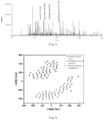
Figure 5 shows the spectrum of metabolites of single bacterium according to an embodiment of the present disclosure; -
Figure 6 shows a t-SNE diagram of Escherichia coli, Acinetobacter baumannii, and Staphylococcus aureus according to an embodiment of the present disclosure; -
Figure 7 is a neural network test diagram showing the identification accuracy of Escherichia coli, Acinetobacter baumannii, and Staphylococcus aureus according to an embodiment of the present disclosure; -
Figure 8 shows a t-SNE diagram of bacterial species identification according to one embodiment of the present disclosure; -
Figure 9 shows a t-SNE diagram of bacterial species identification according to one embodiment of the present disclosure; -
Figure 10 shows a t-SNE diagram of bacterial species identification according to one embodiment of the present disclosure; -
Figure 11 shows a t-SNE diagram of bacterial species identification according to one embodiment of the present disclosure; -
Figure 12 shows a response graph of changes in metabolites of single Escherichia coli after stimulation with different antibiotics according to an embodiment of the present disclosure; -
Figure 13 shows a response graph of changes in metabolites of single Escherichia coli after stimulation with different concentrations of kanamycin according to an embodiment of the present disclosure. - In order to make the purposes, technical solutions, and advantages of the present disclosure more clear, the present disclosure will be further described in details below with reference to specific Examples and Figures.
-
(1) Capture and localization of bacteria in the whole blood:
1 mL of whole blood infected by bacteria was taken and placed in a centrifuge tube, centrifuged at 3000 rpm for 8 min; the blood cell pellet was discarded; 500 µL of supernatant containing the bacterium was centrifuged at 10000 rpm for 8 min, and the supernatant was discarded; the pellet was resuspended in 100 µL of deionized water; 10 µL of 100 µM Acridine Orange as a staining agent was added to perform staining for 15 minutes, the stained solution was centrifuged at 10,000 rpm for 8 minutes; the staining agent was washed away, and the pellet was resuspended in 100 µL of deionized water. The resuspended solution containing the bacterium was dropped onto a polylysine slide and leave for 30 minutes to allow the bacterium to be adsorbed and fixed on the polylysine slide, and then the polylysine slide was rinsed with deionized water and dried at room temperature; the location of bacteria was observed under a fluorescence microscope as shown inFigure 3 .
(2) Capillary preparation:
A capillary with a tip opening size of 1 µm was drawn by a P-2000 laser-based micropipette puller;
(3) In-situ extraction of metabolites in a single living bacterium:
The capillary drawn in step (2) was inserted into a deionized water (containing 1% formic acid) solution, and then taken out and fixed on a micro-operating platform, wherein the capillary was positioned under a microscope to contact the bacterium, and the solution in the capillary was pushed out via a syringe pump to extract the metabolites of bacteria for 1 second and then the solution was sucked back. The in-situ extraction of a single living bacterium was shown inFigure 4 ;
(4) Mass spectrometry analysis:
The capillary after extraction was placed in the front of the mass spectrometer inlet; a voltage was applied; and the mass spectrum was collected as shown inFigure 5 ;
(5) Establishment of a bacterium comparison database:
1 µL of solution containing the bacterium with a concentration of 105 cells/mL was dropped onto the surface of the polylysine slide, and then leave for for 30 minutes to allow the bacterium to be adsorbed on the surface of the polylysine slide; the slide was washed, and dried at room temperature; metabolites of a single living bacterium were extracted via deionized water (containing 1% formic acid) and analyzed by mass spectrometry. Table 1 showed the set of metabolites detected in three bacteria: Escherichia coli, Acinetobacter baumannii, and Staphylococcus aureus. T-distributed stochastic neighbor embedding (t-SNE) analysis was performed on the three bacteria based on the measured metabolites.Figure 6 is the t-distributed stochastic neighbor embedding (t-SNE) analysis for the high-dimensional data of the metabolite fingerprints of three bacteria: Escherichia coli, Acinetobacter baumannii, and Staphylococcus aureus. The measured metabolites of single bacterium can be used to classify different bacteria and establish a database for the identification of unknown bacteria.Table 1: List of metabolites detected in Escherichia coli, Acinetobacter baumannii, and Staphylococcus aureus. Metabolites molecular formula Ion type theoretical m/z measured m/z Error (ppm) Methylalanine C4H9NO2 M+H 104.0706 104.0706 0 Creatinine C4H7N3O M+H 114.0660 114.0662 1 Proline C5H9NO2 M+H 116.0703 116.0706 3 Valine C5H11NO2 M+H 118.0859 118.0863 3 Agmatine C5H14N4 M+H 131.1287 131.1291 3 Creatine C4H9N3O2 M+H 132.0763 132.0768 4 Leucine C6H13NO2 M+H 132.1012 132.1019 5 Ornithine C5H12N2O2 M+H 133.0966 133.0972 4 Urocanic acid C6H6N2O2 M+H 139.0496 139.0502 4 Spermidine C7H19N3 M+H 146.1646 146.1652 4 Histidine C6H9N3O2 M+H 156.0761 156.0768 4 Arginine C6H14N4O2 M+H 175.1182 175.1190 4 Argininic acid C6H13N3O3 M+H 176.1022 176.1030 4 Acetylspermidine C9H21N3O M+H 188.1747 188.1757 6 Alanylthreonine C7H14N2O4 M+H 191.1035 191.1026 4
(6) Test of bacterial species identification accuracy:
In order to verify the accuracy of the bacterial metabolite database established in step (5) for identifying a bacterial species, the metabolites detected in Escherichia coli, Acinetobacter baumannii and Staphylococcus aureus were used as a test set to conduct a neural network test.Figure 7 was the neural network test diagram for the accuracy of identification of three types of bacteria, wherein the bacterial metabolite database established in step (5) had an accuracy of 90% for the identification of bacteria.
(7) Determination of species for bacteria
The bacterial metabolite fingerprints obtained in step (4) were input into the bacterial database established in step (5), and the bacterium were classified into different types based on the differences in detected metabolites. The identification of bacterial species detected in step (4) was shown inFigure 8 . The detected spots fell into the range of Acinetobacter baumannii in the database, indicating that the bacterium detected in step (4) were Acinetobacter baumannii. -
- (1) Isolation of bacteria in cells:
100 µL of 10% saponin was added into 1 mL of RAW264.7 cell suspension infected with bacteria; the cells were lysed at 37°C for 5 min, centrifuged at 3000 rpm for 8 min; the pellet was discarded, and the supernatant containing the bacterium was retained. - (2) Capture and localization of bacteria:
10 µL of 100 µM Acridine Orange as a staining agent was added into the solution containing the bacterium obtained in step (1) to perform staining for 15 min. The stained solution was centrifuged at 10,000 rpm for 8 min. The staining agent was washed away, and the pellet was resuspended in 100 µL of deionized water. The resuspended solution containing the bacterium was dropped onto a polylysine slide and leave for 30 minutes to allow the bacterium to be adsorbed and fixed on the polylysine slide, and then the polylysine slide was rinsed with deionized water and dried at room temperature; the location of bacteria was observed under a fluorescence microscope. - (3) Capillary preparation:
A capillary with a tip diameter opening size of 1 µm was drawn by a P-2000 laser-based micropipette puller; - (4) In-situ extraction of metabolites of a single living bacterium:
The capillary drawn in step (3) was inserted into a deionized water (containing 1% formic acid) solution, and then taken out and fixed on a micro-operating platform, wherein the capillary was positioned under a microscope to contact the bacterium, and the solution in the capillary was pushed out via a syringe pump to extract the metabolites of a single living bacterium for 1 second and then the solution was sucked back. - (5) Mass spectrometry analysis:
The capillary after extraction was placed at the front of the mass spectrometer inlet; a voltage was applied; and the mass spectrum of the extract was collected. - (6) Establishment of a bacterium comparison database:
1 µL of solution containing the bacterium with a concentration of 105 cells/mL was dropped onto the surface of the polylysine slide, and then leave for 30 minutes to allow the bacterium to be adsorbed on the surface of the polylysine slide; the slide was washed, and dried at room temperature; metabolites of a single living bacterium were extracted via deionized water (containing 1% formic acid) and analyzed by mass spectrometry. Table 1 showed the set of metabolites detected in three bacteria: Escherichia coli, Acinetobacter baumannii, and Staphylococcus aureus. T-distributed stochastic neighbor embedding (t-SNE) analysis was performed on the three bacteria based on the measured metabolites.Figure 6 is the t-distributed stochastic neighbor embedding (t-SNE) analysis for the high-dimensional data of the metabolite fingerprints of three bacteria: Escherichia coli, Acinetobacter baumannii, and Staphylococcus aureus. The measured metabolites of single bacterium can be used to classify different bacteria and establish a database for the identification of unknown bacteria. - (7) Test of bacterial species identification accuracy:
In order to verify the accuracy of the bacterial metabolite database established in step (6) for identifying a bacterial species, the metabolites detected in Escherichia coli, Acinetobacter baumannii and Staphylococcus aureus were used as a test set to conduct a neural network test.Figure 7 was the neural network test diagram for the accuracy of identification of three types of bacteria, wherein the bacterial metabolite database established in step (6) had an accuracy of 90% for the identification of bacteria. - (8) Determination of species for bacteria
The bacterial metabolite fingerprints obtained in step (5) were input into the bacterial database established in step (6), and the bacterium were classified into different types based on the differences in detected metabolites. The identification of bacterial species detected in step (5) was shown inFigure 9 . The detected spots fell into the range of Escherichia coli in the database, indicating that the bacterium detected in step (5) was Escherichia coli. -
- (1) Isolation of bacteria in infected tissues:
The infected tissue was wiped by a sterile cotton swab, which was then inserted into 1 mL of deionized water to elute the bacterium, and the eluate containing the bacterium was retained. - (2) Capture and localization of bacteria:
10 µL of 1mM Acridine Orange as a staining agent was added into the solution containing the bacterium obtained in step (1) to perform staining for 15 min. The stained solution was centrifuged at 10,000 rpm for 8 min. The staining agent was washed away, and the pellet was resuspended in 100 µL of deionized water. The resuspended solution containing the bacterium was dropped onto a polylysine slide and leave for 30 minutes to allow the bacterium to be adsorbed and fixed on the polylysine slide, and then the polylysine slide was rinsed with deionized water and dried at room temperature; the location of bacteria was observed under a fluorescence microscope. - (3) Capillary preparation:
A capillary with a tip diameter opening size of 1 µm was drawn by a P-2000 laser-based micropipette puller; - (4) In-situ extraction of metabolites of a single living bacterium:
The capillary drawn in step (3) was inserted into a deionized water (containing 1% formic acid) solution, and then taken out and fixed on a micro-operating platform, wherein the capillary was positioned under a microscope to contact the bacterium, and the solution in the capillary was pushed out via a syringe pump to extract the metabolites of a single living bacterium for 1 second and then the solution was sucked back. - (5) Mass spectrometry analysis:
The capillary after extraction was placed at the front of the mass spectrometer inlet; a voltage was applied; and the mass spectrum was collected. - (6) Establishment of a bacterium comparison database:
1 µL of solution containing the bacterium with a concentration of 105 cells/mL was dropped onto the surface of the polylysine slide, and then leave for 30 minutes to allow the bacterium to be adsorbed on the surface of the polylysine slide; the slide was washed, and dried at room temperature; metabolites of a single living bacterium were extracted via deionized water (containing 1% formic acid) and analyzed by mass spectrometry. Table 1 showed the set of metabolites detected in three bacteria: Escherichia coli, Acinetobacter baumannii, and Staphylococcus aureus. T-distributed stochastic neighbor embedding (t-SNE) analysis was performed on the three bacteria based on the measured metabolites.Figure 6 is the t-distributed stochastic neighbor embedding (t-SNE) analysis for the high-dimensional data of the metabolite fingerprints of three bacteria: Escherichia coli, Acinetobacter baumannii, and Staphylococcus aureus. The measured metabolites of a single bacterium can be used to classify different bacteria and establish a database for the identification of unknown bacteria. - (7) Test of bacterial species identification accuracy:
In order to verify the accuracy of the bacterial metabolite database established in step (6) for identifying a bacterial species, the metabolites detected in Escherichia coli, Acinetobacter baumannii and Staphylococcus aureus were used as a test set to conduct a neural network test.Figure 7 was the neural network test diagram for the accuracy of identification of three types of bacteria, wherein the bacterial metabolite database established in step (6) had an accuracy of 90% for the identification of bacteria. - (8) Determination of species for bacteria
The bacterial metabolite fingerprints obtained in step (5) were input into the bacterial database established in step (6), and the bacterium were classified into different types based on the differences in detected metabolites. The identification of bacterial species detected in step (5) was shown inFigure 10 . The detected spots fell into the range of Staphylococcus aureus in the database, indicating that the bacterium detected in step (5) was Staphylococcus aureus. -
- (1) Capture and localization of bacteria in urine:
1 mL of urine from patients with urinary tract infections was taken and placed in a centrifuge tube, centrifuged at 3000 rpm for 8 min; the large particle pellet was discarded; 800 µL of supernatant containing the bacterium was centrifuged at 10000 rpm for 8 min, and the supernatant was discarded; the pellet was resuspended in 100 µL of deionized water; 10 µL of 100 µM Acridine Orange as a staining agent was added to perform staining for 15 minutes, the stained solution was centrifuged at 10,000 rpm for 8 minutes; the staining agent was washed away, and the pellet was resuspended in 100 µL of deionized water. The resuspended solution containing the bacterium was dropped onto a polylysine slide and leave for 30 minutes to allow the bacterium to be adsorbed and fixed on the polylysine slide, and then the polylysine slide was rinsed with deionized water and dried at room temperature; the location of bacteria was observed under a fluorescence microscope. - (2) Capillary preparation:
A capillary with a tip diameter opening size of 1 µm was drawn by a P-2000 laser-based micropipette puller; - (3) In-situ extraction of metabolites of a single living bacterium:
The capillary drawn in step (2) was inserted into a deionized water (containing 1% formic acid) solution, and then taken out and fixed on a micro-operating platform, wherein the capillary was positioned under a microscope to contact the bacterium, and the solution in the capillary was pushed out via a syringe pump to extract the metabolites of bacteria for 1 second and then the solution was sucked back; - (4) Mass spectrometry analysis:
The capillary after extraction was placed at the front of the mass spectrometer inlet; a voltage was applied; and the mass spectrum was collected; - (5) Establishment of a bacterium comparison database:
1 µL of solution containing the bacterium with a concentration of 105 cells/mL was dropped onto the surface of the polylysine slide, and then leave for 30 minutes to allow the bacterium to be adsorbed on the surface of the polylysine slide; the slide was washed, and dried at room temperature; metabolites of a single living bacterium were extracted via deionized water (containing 1% formic acid) and analyzed by mass spectrometry. Table 1 showed the set of metabolites detected in three bacteria: Escherichia coli, Acinetobacter baumannii, and Staphylococcus aureus. T-distributed stochastic neighbor embedding (t-SNE) analysis was performed on the three bacteria based on the measured metabolites.Figure 6 is the t-distributed stochastic neighbor embedding (t-SNE) analysis for the high-dimensional data of the metabolite fingerprints of three bacteria: Escherichia coli, Acinetobacter baumannii, and Staphylococcus aureus. The measured metabolites of single bacterium can be used to classify different bacteria and establish a database for the identification of unknown bacteria. - (6) Test of bacterial species identification accuracy:
In order to verify the accuracy of the bacterial metabolite database established in step (5) for identifying a bacterial species, the metabolites detected in Escherichia coli, Acinetobacter baumannii and Staphylococcus aureus were used as a test set to conduct a neural network test.Figure 7 was the neural network test diagram for the accuracy of identification of three types of bacteria, wherein the bacterial metabolite database established in step (5) had an accuracy of 90% for the identification of bacteria. - (7) Determination of species for bacteria
The bacterial metabolite fingerprints obtained in step (4) were input into the bacterial database established in step (5), and the bacterial species was determined based on the differences in detected metabolites. The identification of bacterial species detected in step (4) was shown inFigure 11 . The detected spots fell into the range of Acinetobacter baumannii in the database, indicating that the bacterium detected in step (4) was Acinetobacter baumannii. -
- (1) Incubation of bacteria with different antibiotics:
1 mL of whole blood was taken and placed in a centrifuge tube, centrifuged at 3000 rpm for 8 min; the blood cell pellet was discarded; 10 µL of 8 mg/mL kanamycin and 10 µL of 2 mg/mL norfloxacin were respectively added into 500 µL of the supernatant containing the bacterium, and incubated with shaking at 37°C. After 30 min of incubation, the supernatant was centrifuged at 10,000 rpm for 8 min; the supernatant was discarded, and the pellet was resuspended in 100 µL of deionized water; the solution containing the bacterium not having been incubated with the antibiotic was used as a reference group, and the same operations as above were performed. - (2) Incubation of bacteria with kanamycin at different concentrations;
1 mL of whole blood was taken and placed in a centrifuge tube, centrifuged at 3000 rpm for 8 min; the blood cell pellet was discarded; 10 µL of 0.8 mg/mL, 8 mg/mL, and 80 mg/mL kanamycin were respectively added into 500 µL of the supernatant containing the bacterium, and incubated with shaking at 37°C. After 30 min of incubation, the supernatant was centrifuged at 10,000 rpm for 8 min; the supernatant was discarded, and the pellet was resuspended in 100 µL of deionized water; the solution containing the bacterium not having been incubated with the antibiotic was used as a reference group, and the same operations as above were performed. - (3) Capture and localization of bacteria:
10 µL of 100µM Acridine Orange as a staining agent was added into the solution containing the bacterium obtained in step (1) and step (2) to perform staining for 15 min. The stained solution was centrifuged at 10,000 rpm for 8 min. The staining agent was washed away, and the pellet was resuspended in 100 µL of deionized water. The resuspended solution containing the bacterium was dropped onto a polylysine slide and leave for 30 minutes to allow the bacterium to be adsorbed and fixed on the polylysine slide, and then the polylysine slide was rinsed with deionized water and dried at room temperature; the location of bacteria was observed under a fluorescence microscope. - (4) Capillary preparation:
A capillary with a tip diameter opening size of 1 µm was drawn by a P-2000 laser-based micropipette puller; - (5) In-situ extraction of metabolites of a single living bacterium:
The capillary drawn in step (3) was inserted into a deionized water (containing 1% formic acid) solution, and then taken out and fixed on a micro-operating platform, wherein the capillary was positioned under a microscope to contact the bacterium, and the solution in the capillary was pushed out via a syringe pump to extract the metabolites of the bacterium for 1 second and then the solution was sucked back. - (6) Mass spectrometry analysis:
The capillary after extraction was placed at the front of the mass spectrometer inlet; a voltage was applied; and the mass spectrum was collected; - (7) Determination of antibiotic susceptibility:
The signal intensity changes of bacterial metabolites after incubation with different antibiotics were compared. When the bacterium were stimulated by a sensitive antibiotic, the signal intensities of metabolites detected in a single bacterium, such as creatinine, proline, creatine, leucine, spermidine, histidine, arginine, acetyl spermidine, and alanyl-threonine, would all change significantly. Taking the metabolite m/z 191 alanyl-threonine in a single bacterium as an example,Figure 12 showed the response chart of the change in the metabolite alanyl-threonine in a single bacterium after stimulation of E. coli with different antibiotics. After incubation with the sensitive antibiotics kanamycin and norfloxacin, the alanyl-threonine signal was significantly reduced, while incubation without antibiotics did not lead to significant changes in the alanylthreonine signal.Figure 13 showed the response chart of the change of alanyl-threonine in a single E. coli after stimulation with different concentrations of kanamycin. When the concentration of kanamycin reached a minimal inhibitory concentration (MIC), the alanylthreonine signal of a single E. coli decreased significantly, showing that the response of metabolite in single bacterium could be monitored by mass spectrometry of a living single bacterium, and the antibiotic susceptibility of bacterium can be determined based on changes in the detected metabolite signal intensity. - The specific Examples described above further describe the purposes, technical solutions and beneficial effects of the present disclosure in details, but they should not be construed as limiting the scope of the disclosure. It should be noted that any modifications, equivalent replacements and improvements made by those skilled in the art within the spirit and scope of the disclosure should be included within the protection scope of the present disclosure.
Claims (10)
- A method for identifying a bacterium in a biological sample, comprising: staining and fixing the bacterium in the biological sample, extracting metabolites of the fixed single living bacterium, and then performing mass spectrometry detection; preferably, injecting an extraction solution into a capillary, positioning the capillary under a microscope to contact the single living bacterium, and extracting the metabolite from the single living bacterium.
- The method for identifying a bacterium in a biological sample of claim 1, wherein the fixing is performed by electrostatic interaction or covalent binding, and a slide for the fixing is selected from a polylysine slide, a polyethylenimine slide, a gelatin slide, a N-(2-aminoethyl)-3-aminopropyl trimethoxysilane slide, and a 3-aminopropyltriethoxysilane slide; optionally, the interaction time between the bacterium and the slide is 10-30 minutes.
- The method for identifying a bacterium in a biological sample of claim 1 or claim 2, wherein, before staining the bacterium, performing a step of precipitating a large particle interfering substance from the biological sample via a low-speed centrifugation.
- The method for identifying a bacterium in a biological sample of any one of claims 1-3, comprising:Step (1): precipitating a large particle interfering substance from the biological sample via a low-speed centrifugation (preferably the speed of the low-speed centrifugation being 2000rpm-4000rpm, and the centrifugation time being 5min-10min), retaining a supernatant containing the bacterium to obtain the bacterium of the biological sample;Step (2): staining the bacterium in the supernatant obtained in Step (1) to locate the bacterium, and performing a high-speed centrifugation (preferably the speed of the high-speed centrifugation being 10000rpm-14000rpm, and the centrifugation time being 5min-10min) to wash away the staining agent;Step (3): droping the solution containing the stained bacterium obtained in Step (2) onto a slide, capturing and fixing the bacterium by electrostatic adsorption or covalent binding;Step (4): injecting an extraction solution into a capillary (for example, with an opening size of less than 2 µm, preferably 500 nm-1 µm), and performing an in-situ extraction of metabolites from the fixed single living bacterium obtained in Step (3) under a microscope;Step (5): inserting an electrode into the capillary of Step (4), applying a voltage for electrospray, and analyzing the extracted bacterial metabolite by mass spectrometry.
- The method for identifying a bacterium in a biological sample of any one of claims 1-4, wherein the biological sample is selected from a human whole blood, a rabbit whole blood, a urine sample, saliva, cerebrospinal fluid, alveolar lavage fluid, an abscess, a cell solution infected by the bacterium, and a tissue suspension infected by the bacterium; optionally, the bacterium is selected from Escherichia coli, Staphylococcus aureus, Pseudomonas aeruginosa, and Acinetobacter baumannii.
- The method for identifying a bacterium in a biological sample of any one of claims 1-5, wherein the staining agent is selected from Acridine Orange, Fluorescein Diacetate, Syto 9, Syto 64, hoechst 33258 and DAPI; optionally, the staining is performed for 10 min-30 min at a staining agent concentration of 1 µM-10 µM, and the staining is protected from light.
- The method for identifying a bacterium in a biological sample of claim 4, wherein the extraction solution is one or more selected from the group consisting of water, methanol, ethanol, acetonitrile, acetone, chloroform, formic acid, acetic acid and trifluoroacetic acid.
- The method for identifying a bacterium in a biological sample of any one of claims 1-7, further comprising establishing a bacterium comparison database.
- The method for identifying a bacterium in a biological sample of claim 8, wherein establishing the bacterium comparison database comprises the following steps:(a) performing a mass spectrometry analysis of single living bacterium metabolites for one or more reference species;(b) conducting a dimensionality reduction and visual analysis on the reference living bacterium of one or more species based on the metabolite analyzed in step (a), and classifying different bacteria based on the metabolite of the single living bacteriun obtained in step (a) to establish a comparison database for the bacterial identification.
- A method for testing antibiotic susceptibility of a bacterium in a biological sample, comprising the method of any one of claims 1-9, and optionally further comprising a step of incubating the bacterium in the biological sample with the antibiotic.
Applications Claiming Priority (2)
| Application Number | Priority Date | Filing Date | Title |
|---|---|---|---|
| CN202110923179.8A CN113640365B (en) | 2021-08-12 | 2021-08-12 | Analysis method for identifying bacteria and testing antibiotic sensitivity in biological sample |
| PCT/CN2022/103304 WO2023016135A1 (en) | 2021-08-12 | 2022-07-01 | Analysis method for bacterium identification and antibiotic sensitivity testing in biological sample |
Publications (2)
| Publication Number | Publication Date |
|---|---|
| EP4386370A1 true EP4386370A1 (en) | 2024-06-19 |
| EP4386370A4 EP4386370A4 (en) | 2025-07-09 |
Family
ID=78421042
Family Applications (1)
| Application Number | Title | Priority Date | Filing Date |
|---|---|---|---|
| EP22855119.8A Pending EP4386370A4 (en) | 2021-08-12 | 2022-07-01 | ANALYTICAL METHOD FOR IDENTIFYING BACTERIA AND TESTING ANTIBIOTIC SENSITIVITY IN A BIOLOGICAL SAMPLE |
Country Status (4)
| Country | Link |
|---|---|
| US (1) | US20240352502A1 (en) |
| EP (1) | EP4386370A4 (en) |
| CN (1) | CN113640365B (en) |
| WO (1) | WO2023016135A1 (en) |
Families Citing this family (6)
| Publication number | Priority date | Publication date | Assignee | Title |
|---|---|---|---|---|
| CN113640365B (en) * | 2021-08-12 | 2022-09-30 | 中国科学技术大学 | Analysis method for identifying bacteria and testing antibiotic sensitivity in biological sample |
| CN115356450B (en) * | 2022-09-02 | 2025-02-07 | 中国科学技术大学 | Antimicrobial susceptibility analysis method based on direct detection of living bacteria in water |
| CN116990292B (en) * | 2022-09-07 | 2026-02-06 | 中国药科大学 | Single-cell level traditional Chinese medicine microscopic chemical identification method |
| CN117187342B (en) * | 2022-09-27 | 2024-07-26 | 合肥瀚微生物科技有限公司 | A mass spectrometry-based method for detecting metabolites of individual bacteria in the intestine |
| CN117554544A (en) * | 2023-12-01 | 2024-02-13 | 中国科学技术大学 | An integrated mass spectrometry detection method for bacterial species and antibiotic susceptibility |
| CN119643524A (en) * | 2024-12-09 | 2025-03-18 | 成都天台山制药股份有限公司 | Sampling and testing process for the production of sodium fusidate for injection |
Family Cites Families (14)
| Publication number | Priority date | Publication date | Assignee | Title |
|---|---|---|---|---|
| CN101122601B (en) * | 2007-09-24 | 2011-11-30 | 孙艳萍 | Method for separating and authenticating erythroblast of blood |
| WO2011064000A1 (en) * | 2009-11-27 | 2011-06-03 | Creative Antibiotics Sweden Ab | Method for monitoring, identifying or diagnosing a bacterial infection |
| CN103245716A (en) * | 2013-05-23 | 2013-08-14 | 中国科学院化学研究所 | Quick high-sensitivity microbiological identification method based on micromolecular metabolic substance spectral analysis |
| CN103725747A (en) * | 2014-01-26 | 2014-04-16 | 佟雪梅 | Identification method and equipment for sensibility of bacteria on antibiotic |
| CN105969334B (en) * | 2016-05-06 | 2018-07-20 | 东南大学 | Fluorescent dyeing reagent, Preparation method and use for detection bacterium and fungi life or death state |
| JP7752942B2 (en) * | 2017-09-08 | 2025-10-14 | パデュー リサーチ ファウンデイション | Method for determining antibiotic susceptibility using stimulated Raman metabolic imaging |
| CN108593755B (en) * | 2018-04-28 | 2025-05-09 | 清华大学 | A method and device for sampling and in-situ detection of extracellular substances |
| EP3802850B1 (en) * | 2018-06-01 | 2025-11-19 | BT Bidco, Inc. | Devices and systems for gastrointestinal microbiome detection and manipulation |
| CN112080545A (en) * | 2019-06-14 | 2020-12-15 | 中国科学院青岛生物能源与过程研究所 | Single-cell Raman clinical drug resistance kit and detection method |
| CN110763752B (en) * | 2019-11-27 | 2022-01-11 | 清华大学 | Single cell extraction electrospray mass spectrometry system and method |
| CN112098642A (en) * | 2020-02-13 | 2020-12-18 | 普众发现医药科技(上海)有限公司 | Living cell screening method based on high-throughput antibody chip |
| CN111397985B (en) * | 2020-04-14 | 2021-01-08 | 清华大学 | Single cell mass spectrometry |
| CN112444582B (en) * | 2021-02-01 | 2021-06-04 | 宁波大学 | Mass spectrometry method based on droplet extraction |
| CN113640365B (en) * | 2021-08-12 | 2022-09-30 | 中国科学技术大学 | Analysis method for identifying bacteria and testing antibiotic sensitivity in biological sample |
-
2021
- 2021-08-12 CN CN202110923179.8A patent/CN113640365B/en active Active
-
2022
- 2022-07-01 WO PCT/CN2022/103304 patent/WO2023016135A1/en not_active Ceased
- 2022-07-01 US US18/682,241 patent/US20240352502A1/en active Pending
- 2022-07-01 EP EP22855119.8A patent/EP4386370A4/en active Pending
Also Published As
| Publication number | Publication date |
|---|---|
| WO2023016135A1 (en) | 2023-02-16 |
| EP4386370A4 (en) | 2025-07-09 |
| CN113640365B (en) | 2022-09-30 |
| US20240352502A1 (en) | 2024-10-24 |
| CN113640365A (en) | 2021-11-12 |
Similar Documents
| Publication | Publication Date | Title |
|---|---|---|
| US20240352502A1 (en) | Analytical methods for bacterial identification and antibiotic susceptibility testing in biological samples | |
| Khan et al. | Power of scanning electron microscopy and energy dispersive X-ray analysis in rapid microbial detection and identification at the single cell level | |
| CN111982789B (en) | High-throughput detection method of metal ions and metal nanoparticles based on single-cell enrichment and single-cell mass spectrometry | |
| US7214299B2 (en) | Method for separation, identification and evaluation of microbes and cells | |
| DE102013022016B4 (en) | Microbial identification by mass spectrometry and infrared spectrometry | |
| CN107024370B (en) | A kit for pretreatment of microbial samples in a time-of-flight mass spectrometry system | |
| AU2018235992B2 (en) | Device, method, and system for identifying organisms and determining their sensitivity to toxic substances using the changes in the concentrations of metabolites present in growth medium | |
| EP2959293A1 (en) | Method to identify bacterial species by means of gas chromatography/mass spectrometry in biological samples | |
| US6803208B2 (en) | Automated epifluorescence microscopy for detection of bacterial contamination in platelets | |
| CN103245716A (en) | Quick high-sensitivity microbiological identification method based on micromolecular metabolic substance spectral analysis | |
| CN114858777A (en) | Method for label-free detection of bacteria based on surface enhanced Raman spectroscopy and application thereof | |
| CN107219110A (en) | Suitable for the MALDI TOF microculture liquid processing methods detected and rapid identification method | |
| CN112051321A (en) | A Rapid Antibiotic Susceptibility Testing Method Combining Deuterium Water Culture and Matrix-Assisted Laser Desorption Ionization Time-of-Flight Mass Spectrometry | |
| CN117929350A (en) | Quick detection of mixed bacterial drug resistance based on hyperspectral imaging system | |
| CN113156027A (en) | Derivatization method of carboxyl metabolites and efficient non-targeted metabonomics analysis method | |
| CN115308295A (en) | Double-chemistry plasma fingerprint model construction method and infection diagnosis device of fingerprint model constructed by using same | |
| CN113588771B (en) | Application of novel mixed matrix in MALDI-MS bacteria identification | |
| CN120060430A (en) | Method for directly preparing bacterial liquid for drug sensitivity experiment by blood culture positive specimen | |
| CN113237982A (en) | Derivatization method of hydroxyl metabolites and efficient non-targeted metabonomics analysis method | |
| CN115356450B (en) | Antimicrobial susceptibility analysis method based on direct detection of living bacteria in water | |
| Lin et al. | A new naphthimidazole derivative for saccharide labeling with enhanced sensitivity in mass spectrometry detection | |
| CN102749233A (en) | Pretreatment method for directly detecting infection urine pathogen by MALDI-TOF MS | |
| CN108303404B (en) | Method for rapidly distinguishing microorganisms by using conjugated polymer | |
| CN111220594A (en) | A method for screening Streptomyces strains with pesticide activity by ultra-high resolution mass spectrometry | |
| CN118258744A (en) | Method for detecting extracellular vesicles |
Legal Events
| Date | Code | Title | Description |
|---|---|---|---|
| STAA | Information on the status of an ep patent application or granted ep patent |
Free format text: STATUS: THE INTERNATIONAL PUBLICATION HAS BEEN MADE |
|
| PUAI | Public reference made under article 153(3) epc to a published international application that has entered the european phase |
Free format text: ORIGINAL CODE: 0009012 |
|
| STAA | Information on the status of an ep patent application or granted ep patent |
Free format text: STATUS: REQUEST FOR EXAMINATION WAS MADE |
|
| 17P | Request for examination filed |
Effective date: 20240306 |
|
| AK | Designated contracting states |
Kind code of ref document: A1 Designated state(s): AL AT BE BG CH CY CZ DE DK EE ES FI FR GB GR HR HU IE IS IT LI LT LU LV MC MK MT NL NO PL PT RO RS SE SI SK SM TR |
|
| DAV | Request for validation of the european patent (deleted) | ||
| DAX | Request for extension of the european patent (deleted) | ||
| A4 | Supplementary search report drawn up and despatched |
Effective date: 20250606 |
|
| RIC1 | Information provided on ipc code assigned before grant |
Ipc: G01N 33/68 20060101ALI20250602BHEP Ipc: G01N 33/569 20060101ALI20250602BHEP Ipc: C12Q 1/04 20060101ALI20250602BHEP Ipc: G01N 1/30 20060101ALI20250602BHEP Ipc: G01N 1/28 20060101ALI20250602BHEP Ipc: G01N 27/62 20210101AFI20250602BHEP |