EP4382609A1 - Fusion protein between antigen-binding molecule and streptavidin variant - Google Patents
Fusion protein between antigen-binding molecule and streptavidin variant Download PDFInfo
- Publication number
- EP4382609A1 EP4382609A1 EP22849576.8A EP22849576A EP4382609A1 EP 4382609 A1 EP4382609 A1 EP 4382609A1 EP 22849576 A EP22849576 A EP 22849576A EP 4382609 A1 EP4382609 A1 EP 4382609A1
- Authority
- EP
- European Patent Office
- Prior art keywords
- fusion protein
- amino acid
- mutation
- substituted
- antigen
- Prior art date
- Legal status (The legal status is an assumption and is not a legal conclusion. Google has not performed a legal analysis and makes no representation as to the accuracy of the status listed.)
- Withdrawn
Links
Images


Classifications
-
- A—HUMAN NECESSITIES
- A61—MEDICAL OR VETERINARY SCIENCE; HYGIENE
- A61P—SPECIFIC THERAPEUTIC ACTIVITY OF CHEMICAL COMPOUNDS OR MEDICINAL PREPARATIONS
- A61P35/00—Antineoplastic agents
-
- A—HUMAN NECESSITIES
- A61—MEDICAL OR VETERINARY SCIENCE; HYGIENE
- A61K—PREPARATIONS FOR MEDICAL, DENTAL OR TOILETRY PURPOSES
- A61K41/00—Medicinal preparations obtained by treating materials with wave energy or particle radiation ; Therapies using these preparations
- A61K41/0057—Photodynamic therapy with a photosensitizer, i.e. agent able to produce reactive oxygen species upon exposure to light or radiation, e.g. UV or visible light; photocleavage of nucleic acids with an agent
- A61K41/0071—PDT with porphyrins having exactly 20 ring atoms, i.e. based on the non-expanded tetrapyrrolic ring system, e.g. bacteriochlorin, chlorin-e6, or phthalocyanines
-
- A—HUMAN NECESSITIES
- A61—MEDICAL OR VETERINARY SCIENCE; HYGIENE
- A61K—PREPARATIONS FOR MEDICAL, DENTAL OR TOILETRY PURPOSES
- A61K47/00—Medicinal preparations characterised by the non-active ingredients used, e.g. carriers or inert additives; Targeting or modifying agents chemically bound to the active ingredient
- A61K47/50—Medicinal preparations characterised by the non-active ingredients used, e.g. carriers or inert additives; Targeting or modifying agents chemically bound to the active ingredient the non-active ingredient being chemically bound to the active ingredient, e.g. polymer-drug conjugates
- A61K47/51—Medicinal preparations characterised by the non-active ingredients used, e.g. carriers or inert additives; Targeting or modifying agents chemically bound to the active ingredient the non-active ingredient being chemically bound to the active ingredient, e.g. polymer-drug conjugates the non-active ingredient being a modifying agent
- A61K47/54—Medicinal preparations characterised by the non-active ingredients used, e.g. carriers or inert additives; Targeting or modifying agents chemically bound to the active ingredient the non-active ingredient being chemically bound to the active ingredient, e.g. polymer-drug conjugates the non-active ingredient being a modifying agent the modifying agent being an organic compound
- A61K47/55—Medicinal preparations characterised by the non-active ingredients used, e.g. carriers or inert additives; Targeting or modifying agents chemically bound to the active ingredient the non-active ingredient being chemically bound to the active ingredient, e.g. polymer-drug conjugates the non-active ingredient being a modifying agent the modifying agent being an organic compound the modifying agent being also a pharmacologically or therapeutically active agent, i.e. the entire conjugate being a codrug
- A61K47/551—Medicinal preparations characterised by the non-active ingredients used, e.g. carriers or inert additives; Targeting or modifying agents chemically bound to the active ingredient the non-active ingredient being chemically bound to the active ingredient, e.g. polymer-drug conjugates the non-active ingredient being a modifying agent the modifying agent being an organic compound the modifying agent being also a pharmacologically or therapeutically active agent, i.e. the entire conjugate being a codrug one of the codrug's components being a vitamin, e.g. niacinamide, vitamin B3, cobalamin, vitamin B12, folate, vitamin A or retinoic acid
-
- A—HUMAN NECESSITIES
- A61—MEDICAL OR VETERINARY SCIENCE; HYGIENE
- A61K—PREPARATIONS FOR MEDICAL, DENTAL OR TOILETRY PURPOSES
- A61K47/00—Medicinal preparations characterised by the non-active ingredients used, e.g. carriers or inert additives; Targeting or modifying agents chemically bound to the active ingredient
- A61K47/50—Medicinal preparations characterised by the non-active ingredients used, e.g. carriers or inert additives; Targeting or modifying agents chemically bound to the active ingredient the non-active ingredient being chemically bound to the active ingredient, e.g. polymer-drug conjugates
- A61K47/51—Medicinal preparations characterised by the non-active ingredients used, e.g. carriers or inert additives; Targeting or modifying agents chemically bound to the active ingredient the non-active ingredient being chemically bound to the active ingredient, e.g. polymer-drug conjugates the non-active ingredient being a modifying agent
- A61K47/62—Medicinal preparations characterised by the non-active ingredients used, e.g. carriers or inert additives; Targeting or modifying agents chemically bound to the active ingredient the non-active ingredient being chemically bound to the active ingredient, e.g. polymer-drug conjugates the non-active ingredient being a modifying agent the modifying agent being a protein, peptide or polyamino acid
- A61K47/66—Medicinal preparations characterised by the non-active ingredients used, e.g. carriers or inert additives; Targeting or modifying agents chemically bound to the active ingredient the non-active ingredient being chemically bound to the active ingredient, e.g. polymer-drug conjugates the non-active ingredient being a modifying agent the modifying agent being a protein, peptide or polyamino acid the modifying agent being a pre-targeting system involving a peptide or protein for targeting specific cells
- A61K47/665—Medicinal preparations characterised by the non-active ingredients used, e.g. carriers or inert additives; Targeting or modifying agents chemically bound to the active ingredient the non-active ingredient being chemically bound to the active ingredient, e.g. polymer-drug conjugates the non-active ingredient being a modifying agent the modifying agent being a protein, peptide or polyamino acid the modifying agent being a pre-targeting system involving a peptide or protein for targeting specific cells the pre-targeting system, clearing therapy or rescue therapy involving biotin-(strept) avidin systems
-
- A—HUMAN NECESSITIES
- A61—MEDICAL OR VETERINARY SCIENCE; HYGIENE
- A61K—PREPARATIONS FOR MEDICAL, DENTAL OR TOILETRY PURPOSES
- A61K49/00—Preparations for testing in vivo
- A61K49/001—Preparation for luminescence or biological staining
- A61K49/0013—Luminescence
- A61K49/0017—Fluorescence in vivo
- A61K49/0019—Fluorescence in vivo characterised by the fluorescent group, e.g. oligomeric, polymeric or dendritic molecules
- A61K49/0021—Fluorescence in vivo characterised by the fluorescent group, e.g. oligomeric, polymeric or dendritic molecules the fluorescent group being a small organic molecule
- A61K49/0036—Porphyrins
-
- A—HUMAN NECESSITIES
- A61—MEDICAL OR VETERINARY SCIENCE; HYGIENE
- A61K—PREPARATIONS FOR MEDICAL, DENTAL OR TOILETRY PURPOSES
- A61K49/00—Preparations for testing in vivo
- A61K49/001—Preparation for luminescence or biological staining
- A61K49/0013—Luminescence
- A61K49/0017—Fluorescence in vivo
- A61K49/005—Fluorescence in vivo characterised by the carrier molecule carrying the fluorescent agent
- A61K49/0056—Peptides, proteins, polyamino acids
-
- C—CHEMISTRY; METALLURGY
- C07—ORGANIC CHEMISTRY
- C07K—PEPTIDES
- C07K14/00—Peptides having more than 20 amino acids; Gastrins; Somatostatins; Melanotropins; Derivatives thereof
- C07K14/195—Peptides having more than 20 amino acids; Gastrins; Somatostatins; Melanotropins; Derivatives thereof from bacteria
-
- C—CHEMISTRY; METALLURGY
- C07—ORGANIC CHEMISTRY
- C07K—PEPTIDES
- C07K14/00—Peptides having more than 20 amino acids; Gastrins; Somatostatins; Melanotropins; Derivatives thereof
- C07K14/195—Peptides having more than 20 amino acids; Gastrins; Somatostatins; Melanotropins; Derivatives thereof from bacteria
- C07K14/36—Peptides having more than 20 amino acids; Gastrins; Somatostatins; Melanotropins; Derivatives thereof from bacteria from Actinomyces; from Streptomyces (G)
-
- C—CHEMISTRY; METALLURGY
- C07—ORGANIC CHEMISTRY
- C07K—PEPTIDES
- C07K16/00—Immunoglobulins [IG], e.g. monoclonal or polyclonal antibodies
- C07K16/18—Immunoglobulins [IG], e.g. monoclonal or polyclonal antibodies against material from animals or humans
- C07K16/32—Immunoglobulins [IG], e.g. monoclonal or polyclonal antibodies against material from animals or humans against translation products of oncogenes
-
- C—CHEMISTRY; METALLURGY
- C07—ORGANIC CHEMISTRY
- C07K—PEPTIDES
- C07K2317/00—Immunoglobulins specific features
- C07K2317/70—Immunoglobulins specific features characterized by effect upon binding to a cell or to an antigen
- C07K2317/73—Inducing cell death, e.g. apoptosis, necrosis or inhibition of cell proliferation
-
- C—CHEMISTRY; METALLURGY
- C07—ORGANIC CHEMISTRY
- C07K—PEPTIDES
- C07K2317/00—Immunoglobulins specific features
- C07K2317/90—Immunoglobulins specific features characterized by (pharmaco)kinetic aspects or by stability of the immunoglobulin
- C07K2317/92—Affinity (KD), association rate (Ka), dissociation rate (Kd) or EC50 value
-
- C—CHEMISTRY; METALLURGY
- C07—ORGANIC CHEMISTRY
- C07K—PEPTIDES
- C07K2317/00—Immunoglobulins specific features
- C07K2317/90—Immunoglobulins specific features characterized by (pharmaco)kinetic aspects or by stability of the immunoglobulin
- C07K2317/94—Stability, e.g. half-life, pH, temperature or enzyme-resistance
-
- C—CHEMISTRY; METALLURGY
- C07—ORGANIC CHEMISTRY
- C07K—PEPTIDES
- C07K2318/00—Antibody mimetics or scaffolds
- C07K2318/20—Antigen-binding scaffold molecules wherein the scaffold is not an immunoglobulin variable region or antibody mimetics
-
- C—CHEMISTRY; METALLURGY
- C07—ORGANIC CHEMISTRY
- C07K—PEPTIDES
- C07K2319/00—Fusion polypeptide
- C07K2319/20—Fusion polypeptide containing a tag with affinity for a non-protein ligand
Definitions
- the present invention relates to a fusion protein of an antigen-binding molecule and a mutant streptavidin and the use thereof.
- the interaction between avidin/streptavidin and biotin has been widely applied in the field of biochemistry, molecular biology, or medicine.
- a drug delivery method and a pretargeting method have been devised, in which high binding ability between avidin/streptavidin and biotin is combined with an antibody molecule.
- Patent Document 1 describes a streptavidin mutant whose immunogenicity is reduced by introducing mutations into amino acid sequence. Patent Document 1 also describes a therapeutic or diagnostic kit which comprises the above mutant streptavidin and a diagnostic or therapeutic substance labeled with biotin or a derivative thereof.
- Patent Document 1 International Publication WO2010/95455
- the present inventor has selected a molecule having a molecular weight smaller than that of an antibody, as a molecule that recognizes cancer cells, and then, the present inventor has prepared a fusion protein of the aforementioned molecule and a mutant streptavidin.
- the present inventor has found that the proliferation of cancer cells can be suppressed by photoimmunotherapy using the above-described fusion protein and a conjugate of a biotin and a phthalocyanine dye, thereby completing the present invention.
- the proliferation of cancer cells can be suppressed.
- the fusion protein of the present invention is a fusion protein, wherein an antigen-binding molecule having a molecular weight of 20,000 or less is bound, via a linker sequence, to the N-terminal side and/or C-terminal side of mutant streptavidin which comprises an amino acid sequence having the following mutations (1) to (6) with respect to the amino acid sequence of a core streptavidin as shown in SEQ ID NO: 17 provided that the C-terminal amino acid sequence consisting of Pro-Ser-Ala-Ala-Ser may be partially or entirely deleted, and has a decreased immunogenicity as compared with that of a wild-type streptavidin:
- the antigen-binding molecules When the antigen-binding molecule having a molecular weight of 20,000 or less is bound to both the N-terminus and C-terminus of the mutant streptavidin via a linker sequence, the antigen-binding molecules may be the same or different.
- the fusion protein has the antigen-binding molecule having a molecular weight of 20,000 or less, the linker sequence, and the amino acid sequence of the mutant streptavidin in this order from the N-terminal side to the C-terminal side.
- the mutant streptavidin in the present invention is a mutant streptavidin whose immunogenicity is reduced by introducing mutations into amino acid sequence, as described in International Publication WO 2010/95455 .
- the mutant streptavidin in the present invention is a mutant streptavidin which comprises an amino acid sequence having the following mutations (1) to (6) with respect to the amino acid sequence of a core streptavidin as shown in SEQ ID NO: 17 provided that the C-terminal amino acid sequence consisting of Pro-Ser-Ala-Ala-Ser may be partially or entirely deleted,:
- the expression "... having decreased immunogenicity as compared with that of a wild-type streptavidin” is used in the present invention to mean that, when a mutant streptavidin is administered to a mammal such as a human, the immunogenicity of the mutant streptavidin is reduced.
- a decrease of the immunogenicity can be confirmed by the following method, for example. That is to say, the reactivity of the mutant streptavidin of the present invention with anti-streptavidin antiserum, which has been obtained by immunizing a crab-eating monkey with a wild-type streptavidin, is analyzed.
- the immunogenicity of the mutant streptavidin is decreased as compared with that of the wild-type streptavidin.
- the immunogenicity of the mutant streptavidin of the present invention is decreased by preferably 80% or less, more preferably 60% or less, further preferably 20% or less, still further preferably 15% or less, still further preferably 10% or less, and particularly preferably 5% or less, as compared with the immunogenicity of the wild-type streptavidin.
- the fusion protein of the present invention is a fusion protein of an antigen-binding molecule having a molecular weight of 20,000 or less and a mutant streptavidin.
- the fusion protein of the present invention forms a tetramer as a result of the affinity between the two amino acid sequences of the mutsnt streptavidin.
- the molecular weight of the tetramer formed by the fusion protein of the present invention is approximately 96 kDa.
- the fusion protein as used in the present invention When the fusion protein as used in the present invention is administered to a living body to treat cancer, it is important to simultaneously achieve the following points: favorable tumor uptake; good clearance rate; and favorable tumor penetration. It is conceived that the above-described molecular weight (approximately 96 kDa) of the present invention is a molecular weight, with which the above-described three parameters can be simultaneously achieved.
- the molecular weight of the antigen-binding molecule is 20,000 or less.
- the molecular weight of the antigen-binding molecule is generally 4,000 or more and 20,000 or less, preferably 4,000 or more and 10,000 or less, and more preferably 4,000 or more and 8,000 or less.
- the antigen-binding molecule of the present invention is a molecule having a concept different from an antibody.
- the molecular weight of an IgG antibody is generally approximately 150,000, whereas the antigen-binding molecule of the present invention is a molecule having a molecular weight much smaller than that of the IgG antibody.
- the antigen-binding molecule of the present invention may also be a molecule produced by imitating an antibody.
- proteins each having a molecular weight of approximately 6,000 are produced by modifying a portion of the IgG-binding domain of Protein A (VDNKFNKEQQNAFYEILHLPNLNEEQRNAFIQSLKDDPSQSANLLAEAKKLNDAQAPK) (SEQ ID NO: 18), and the thus produced proteins are then screened, so that a desired antigen-binding molecule can be selected, as appropriate.
- VDNKFNKEQQNAFYEILHLPNLNEEQRNAFIQSLKDDPSQSANLLAEAKKLNDAQAPK VDNKFNKEQQNAFYEILHLPNLNEEQRNAFIQSLKDDPSQSANLLAEAKKLNDAQAPK
- the antigen in the antigen-binding molecule is not particularly limited.
- the antigen is preferably an antigen that is expressed in cancer cells.
- Examples of the antigen that is specifically expressed in cancer may include the following antigens:
- Epiregulin EREG
- ROBO 1, 2, 3 and 4, 1-40- ⁇ -amyloid 4-1BB, 5AC, 5T4, ACVR2B, adenocarcinoma antigen, ⁇ -fetoprotein, angiopoetin 2, anthrax toxin, AOC3 (VAP-1), B-lymphoma cells, B7-H3, BAFF, ⁇ amyloid, C242 antigen, C5, CA-125, carbonic anhydrase 9 (CA-IX), cardiac myosin, CCL11 (eotaxin-1), CCR4, CCR5, CD11, CD18, CD125, CD140a, CD147 (basigin), CD147 (basigin), CD15, CD152, CD154 (CD40L), CD154, CD19, CD2, CD20, CD200, CD22, CD221, CD23 (IgE receptor), CD25 (IL-2 receptor ⁇ chain), CD28, CD3, CD30 (TNFRSF8), CD33, CD37, CD38 (cyclic A
- coli Shiga toxin type 1 E. coli Shiga toxin type 2, EGFL7, EGFR, endotoxin, EpCAM, episialin, ERBB3, Escherichia coli, F protein of respiratory syncytial virus, FAP, fibrin II ⁇ chain, fibronectin extra domain-B, folate receptor 1, Frizzled receptor, GD2, GD3 ganglioside, GMCSF receptor ⁇ chain, GPNMB, hepatitis B surface antigen, hepatitis B virus, HER1, HER2/neu, HER3, HGF, HIV-1, HLA-DR ⁇ , HNGF, Hsp90, human ⁇ amyloid, human scatter factor receptor kinase, human TNF, ICAM-1 (CD54), IFN- ⁇ , IFN- ⁇ , IgE, IgEFc region, IGF-1 receptor, IGF-I, IgG4, IGHE, IL-1 ⁇ , IL-12, IL-13, IL-17,
- HER2 is particularly preferable.
- antigen-binding molecule may be a protein having the amino acid sequence as set forth in SEQ ID NO: 2, which binds to HER2.
- the linker sequence is not particularly limited, as long as the effects of the present invention can be achieved.
- the number of amino acids in the linker sequence is preferably 5 to 25 amino acids, more preferably 10 to 25 amino acids, and further preferably 15 to 20 amino acids.
- a specific example of the linker sequence may be a sequence consisting of glycine residues and serine residues.
- a linker sequence for example, an amino acid sequence represented by [(Gly) m -Ser] n (wherein m represents an integer of 1 to 10, and n represents an integer of 1 to 5) can be used.
- a specific example of the linker sequence may be Gly-Gly-Gly-Gly-Gly-Ser-Gly-Gly-Gly-Gly-Gly-Gly-Ser-Gly-Gly-Gly-Gly-Gly-Gly-Ser, but is not particularly limited thereto.
- a specific example of the fusion protein of the present invention may be a fusion protein having the amino acid sequence as set forth in SEQ ID NO: 3 or SEQ ID NO:9.
- nucleic acid for example, DNA
- a specific example of the nucleic acid of the present invention may be a nucleic acid that encodes the fusion protein having the amino acid sequence as set forth in SEQ ID NO: 3 or SEQ ID NO:9.
- One example of the nucleic acid of the present invention may be a nucleic acid having the nucleotide sequence as set forth in SEQ ID NO: 4 or SEQ ID NO: 10.
- a nucleic acid (for example, DNA) encoding the fusion protein of the present invention can be used by being incorporated into a vector.
- a nucleic acid encoding the fusion protein of the present invention is incorporated into an expression vector, and a host is then transformed with this expression vector, so that the fusion protein of the present invention can be expressed in the host.
- a method for producing the fusion protein of the present invention comprising a step of allowing a nucleic acid encoding the fusion protein of the present invention to express in a host.
- the fusion protein can be expressed in a bacterial inclusion body and recovered therefrom.
- the vector When Escherichia coli is used as a host, the vector preferably has a replication origin (ori) and also has a gene for selecting the transformed host (e.g. a drug-resistance gene that is resistant to drugs, such as ampicillin, tetracycline, kanamycin or chloramphenicol, etc.). Moreover, an expression vector preferably has a promoter capable of efficiently expressing the mutant streptavidin of the present invention in a host, such as a lacZ promoter or a T7 promoter.
- a promoter capable of efficiently expressing the mutant streptavidin of the present invention in a host such as a lacZ promoter or a T7 promoter.
- Such a vector examples include an M13 vector, a pUC vector, pBR322, pBluescript, pCR-Script, pGEX-SX-1 (Pharmacia), "QIAexpress system” (QIAGEN), pEGFP, and pET (in this case, BL21 that expresses T7 RNA polymerase is preferably used as a host).
- a vector can be introduced into a host cell by applying a calcium chloride method or an electroporation method, for example.
- a sequence that encodes a tag for improving solubility such as glutathione S-transferase, thioredoxin or a maltose-binding protein, may be added.
- a sequence that encodes a tag designed for facilitating purification such as a polyhistidine tag, a Myc epitope, a hemagglutinin (HA) epitope, a T7 epitope, an Xpress tag, a FLAG tag or other known tag sequences, may also be added.
- examples of the expression vector include: mammal-derived expression vectors (for example, pcDNA3 (manufactured by Invitrogen), pEGF-BOS (Nucleic Acids. Res. 1990, 18(17), p.
- mammal-derived expression vectors for example, pcDNA3 (manufactured by Invitrogen), pEGF-BOS (Nucleic Acids. Res. 1990, 18(17), p.
- insect cell-derived expression vectors for example, "Bac-to-BAC baculovirus expression system” (manufactured by Gibco-BRL) and pBacPAK8); plant-derived expression vectors (for example, pMH1 and pMH2); animal virus-derived expression vectors (for example, pHSV, pMV and pAdexLcw); retrovirus-derived expression vectors (for example, pZIPneo); yeast-derived expression vectors (for example, "Pichia Expression Kit” (manufactured by Invitrogen), pNV11 and SP-Q01); and Bacillus subtilis- derived expression vectors (for example, pPL608 and pKTH50).
- Bacillus subtilis- derived expression vectors for example, pPL608 and pKTH50.
- the expression vector When the expression of the present fusion protein in an animal cell such as a CHO cell, a COS cell or an NIH3T3 cell is intended, it is essential for the expression vector to have a promoter necessary for the expression of the fusion protein in such an animal cell, such as an SV40 promoter ( Mulligan et al., Nature (1979) 277, 108 ), an MNILV-LTR promoter, an EF1 ⁇ promoter ( Mizushima et al., Nucleic Acids Res. (1990) 18, 5322 ) or a CMV promoter.
- SV40 promoter Mulligan et al., Nature (1979) 277, 108
- MNILV-LTR promoter an MNILV-LTR promoter
- an EF1 ⁇ promoter Mizushima et al., Nucleic Acids Res. (1990) 18, 5322
- CMV promoter a promoter necessary for the expression of the fusion protein in such an animal cell
- the expression vector has a gene for selecting the transformation of a cell (for example, a drug-resistance gene capable of determining transformation with the use of drugs (neomycin, G418, etc.)).
- a vector having such properties include pMAM, pDR2, pBK-RSV, pBK-CMV, pOPRSV and pOP13.
- the type of a host cell, into which the vector is introduced is not particularly limited. Either prokaryotes or eukaryotes may be used. It is possible to use Escherichia coli or various types of animal cells, for example.
- an animal cell for example, an animal cell, a plant cell or a fungal cell can be used as a host.
- an animal cell that can be used herein include: mammalian cells such as a CHO cell, a COS cell, a 3T3 cell, a HeLa cell or a Vero cell; and insect cells such as Sf9, Sf21 or Tn5.
- mammalian cells such as a CHO cell, a COS cell, a 3T3 cell, a HeLa cell or a Vero cell
- insect cells such as Sf9, Sf21 or Tn5.
- a CHO cell is particularly preferable.
- a vector can be introduced into a host cell by a calcium phosphate method, a DEAE-dextran method, a method using cationic ribosome DOTAP (manufactured by Boehringer Mannheim), an electroporation method, a lipofection method or the like.
- a cell from Nicotiana tabacum is known as a protein-producing system, for example. These cells may be subj ected to callus culture.
- yeast cells including genus Saccharomyces such as Saccharomyces cerevisiae ; and filamentous fungi including genus Aspergillus such as Aspergillus niger.
- Examples of a prokaryotic cell that can be used herein include Escherichia coli ( E. coli ), such as JM109, DH5 ⁇ or HB101. Moreover, Bacillus subtilis has been known.
- Escherichia coli E. coli
- JM109 JM109
- DH5 ⁇ DH5 ⁇
- HB101 HB101
- Bacillus subtilis has been known.
- the culture can be carried out in accordance with a known culture method.
- a culture solution of animal cells that can be used herein include DMEM, MEM, RPMI1640, and IMDM.
- FCS fetal calf serum
- FCS fetal calf serum
- the pH applied during the culture is preferably approximately pH 6 to 8.
- the culture is generally carried out at a temperature of approximately 30°C to 40°C for approximately 15 to 200 hours. As necessary, medium replacement, ventilation and stirring are carried out.
- growth factors may also be added to promote the growth of cells.
- the fusion protein of the present invention is useful as a cancer therapeutic agent or a cancer diagnostic agent.
- kits for treating or diagnosing cancer comprising (1) the fusion protein of the present invention, and (2) a conjugate of bioyin and a diagnostic substance or a therapeutic substance.
- the fusion protein of the present invention is administered to a patient, so that a mutant streptavidin can be accumulated specifically into cancer cells. Subsequently, by administering a conjugate of biotin and a diagnostic substance or a therapeutic substance to the patient, it becomes possible to accumulate the diagnostic substance or the therapeutic substance precisely into the cancer cells.
- a complex is prepared by binding the "fusion protein of the present invention” with the “conjugate of biotin and a diagnostic substance or a therapeutic substance,” and the thus prepared complex can be administered to the patient.
- a conjugate of the biotin and the diagnostic substance or the therapeutic substance can be prepared.
- the diagnostic substance or the therapeutic substance may include a fluorochrome, a chemiluminescent agent, a radioisotope, a sensitizer consisting of a metal compound or the like, a neutron-capturing agent consisting of a metal compound or the like, a phthalocyanine dye, a low-molecular-weight compound, micro- or nano-bubbles, and a protein.
- a phthalocyanine dye can be used.
- a phthalocyanine dye specifically, a compound represented by the following formula (1) or a salt thereof can be used.
- X represents a substituent having a hydrophilic group at the terminus thereof.
- the hydrophilic group is preferably a sulfonic acid group, a phosphoric acid group, an ammonium group, or the like.
- X is preferably a substituent having a sulfonic acid group at the terminus thereof.
- X may be a group represented by the following formula:
- Me represents a methyl group.
- L 3 , L 4 , L 5 , and L 6 each independently represent a divalent linking group.
- L 3 is preferably the following: wherein m represents an integer of 1 to 5.
- L 4 is preferably -[(CH 2 ) p -O)] q - (wherein p and q each independently represent an integer of 1 to 5).
- L 5 is preferably -CONH-, -NHCO-, -COO-, or -OCO-, and is more preferably -CONH-.
- L 6 is preferably -(CH 2 ) r , -(CH 2 ) r O-, or -(CH 2 ) r Si(R 1 )(R 2 )-O- (wherein r represents an integer of 1 to 5, and R 1 and R 2 each independently represent an alkyl group containing 1 to 4 carbon atoms). R 1 and R 2 are particularly preferably methyl groups.
- Y represents a group capable of binding to an antigen-binding molecule.
- Y is preferably an active ester of a carboxyl group, and is more preferably an succinimidyl ester of a carboxyl group.
- R 3 represents an alkyl group containing 1 to 6 carbon atoms, an alkoxy group containing 1 to 6 carbon atoms, a halogen atom, -SR 11 , -SOR 12 , an aryl group containing 6 to 10 carbon atoms, - N(R 13 )(R 14 ), or -NO 2 , or two adjacent R 3 s may together form an aryl group containing 6 to 10 carbon atoms.
- R 11 , R 12 , R 13 , and R 14 each independently represent an alkyl group containing 1 to 6 carbon atoms or an alkoxy group containing 1 to 6 carbon atoms.
- the aryl group may be optionally substituted with an alkyl group containing 1 to 6 carbon atoms or an alkoxy group containing 1 to 6 carbon atoms.
- the alkyl group and the alkoxy group may be optionally substituted with a halogen atom.
- the plurality of R 3 s may be identical to or different from one another.
- s represents an integer of 0 to 4. s is preferably 0.
- the halogen atom may be any of fluorine, chlorine, bromine and iodine, and the halogen atom is preferably fluorine, chlorine, or bromine.
- the compound of the phthalocyanine dye can be synthesized in accordance with the methods described in Production Examples 1 to 5 in the after-mentioned Examples.
- Photoimmunotherapy is a therapeutic method of using a photosensitizer and an irradiation light to destroy specific cells in a body.
- a photosensitizer When a photosensitizer is exposed to a light with a specific wavelength, it generates cytotoxic reactive oxygen species capable of inducing apoptosis, necrosis, and/or autophagy to around cells.
- Japanese Patent No. Japanese Patent No.
- 6127045 discloses a method of killing cells, comprising: a step of allowing cells comprising a cell surface protein to come into contact with a therapeutically effective amount of one or more antibodies-IR700 molecules, wherein the antibodies specifically bind to the cell surface protein; a step of irradiating the cells with a light at a wavelength of 660 to 740 nm and at a dose of at least 1 Jcm -2 ; and a step of allowing the cells to come into contact with one or more therapeutic agents at approximately 0 to 8 hours after the irradiation, thereby killing the cells.
- 2017-524659 A discloses a method of inducing cytotoxicity to a subject affected with a disease or a pathology, comprising: (a) administering to a subject, a therapeutically effective drug comprising a phthalocyanine dye such as IRDye (registered trademark) 700DX conjugated with a probe specifically binding to the cell of the subject; and (b) irradiating the cell with an appropriate excitation light in an amount effective for inducing cell death.
- a therapeutically effective drug comprising a phthalocyanine dye such as IRDye (registered trademark) 700DX conjugated with a probe specifically binding to the cell of the subject.
- the fusion protein of the present invention and the conjugate of a biotin-modified dimer and a phthalocyanine dye are administered to a subject, and the cells are then irradiated with an excitation light in an amount effective for suppression of cell proliferation or induction of cell death, so that the cell proliferation can be suppressed or the cell death can be induced, and thereby the subject can be treated.
- the fusion protein of the present invention and the conjugate of biotin and a phthalocyanine dye are administered to a subject, and the cells are then irradiated with an excitation light in an amount effective for suppression of cell proliferation or induction of cell death, so that the cell proliferation can be suppressed or the cell death can be induced, and thereby the subject can be treated.
- the subj ect used herein includes humans and non-human animals.
- Examples of the subject may include humans and experimental animals such as mice.
- the subject is preferably affected with a disease regarding which suppression of cell proliferation or induction of cell death is desired.
- the subject is affected with a cancer or a solid tumor.
- cancer may include carcinoma, lymphoma, blastoma, sarcoma, and leukemia or malignant lymphoma.
- specific examples of the cancer may include squamous cell carcinoma (e.g., epithelial squamous cell carcinoma), lung cancer including small cell lung cancer, non-small cell lung cancer ("NSCLC”), pulmonary adenocarcinoma and pulmonary squamous cell carcinoma, peritoneal cancer, hepatocarcinoma, corpus ventriculi or stomach cancer, including digestive cancer, pancreatic cancer, glioblastoma, cervical cancer, ovarian cancer, liver cancer, bladder cancer, hepatocellular cancer, breast cancer, colon cancer, rectal cancer, colorectal cancer, endometrial membrane cancer or endometrial carcinoma, salivary gland carcinoma, kidney or renal region cancer, prostate cancer, vulvar cancer, thyroid cancer, hepatocellular carcinoma, anal carcinoma, penile carcinoma, and head and neck cancer.
- squamous cell carcinoma e
- the solid tumor means a benign or malignant, abnormal cell mass that generally does not contain a capsule.
- the solid tumor may include glioma, astrocytoma, medulloblastoma, craniopharyngioma, ependymoma, pineal gland tumor, hemangioblastoma, acoustic neuroma, oligodendrocyte, meningioma, melanoma, neuroblastoma, and retinoblastoma.
- Examples of the administration method to the subject may include, but are not limited to, a local route, an injection (a subcutaneous injection, an intramuscular injection, an intradermal injection, an intraperitoneal injection, an intratumoral injection, an intravenous injection, etc.), an oral route, an ocular route, a sublingual route, a rectal route, a percutaneous route, an intranasal route, a vaginal route, and an inhalation route.
- an injection a subcutaneous injection, an intramuscular injection, an intradermal injection, an intraperitoneal injection, an intratumoral injection, an intravenous injection, etc.
- an oral route a ocular route, a sublingual route, a rectal route, a percutaneous route, an intranasal route, a vaginal route, and an inhalation route.
- the conjugate of biotin and a phthalocyanine dye and the fusion protein of the present invention are each administered in a therapeutically effective amount.
- the therapeutically effective amount per 60 kg is at least 0.5 mg (mg/60 kg), at least 5 mg/60 kg, at least 10 mg/60 kg, at least 20 mg/60 kg, at least 30 mg/60 kg, or at least 50 mg/60 kg.
- the applied dose is 1 mg/60 kg, 2 mg/60 kg, 5 mg/60 kg, 20 mg/60 kg, or 50 mg/60 kg, and it is, for example, 0.5 to 50 mg/60 kg.
- the therapeutically effective amount is at least 100 ⁇ g/kg, at least 500 ⁇ g/kg or at least 500 ⁇ g/kg, and it is, for example, at least 10 ⁇ g/kg.
- the dose is 100 ⁇ g/kg, 250 ⁇ g/kg, approximately 500 ⁇ g/kg, 750 ⁇ g/kg, or 1000 ⁇ g/kg, and it is, for example, 10 ⁇ g/kg to 1000 ⁇ g/kg.
- the therapeutically effective amount is 10 ⁇ g/ml, 20 ⁇ g/ml, 30 ⁇ g/ml, 40 ⁇ g/ml, 50 ⁇ g/ml, 60 ⁇ g/ml, 70 ⁇ g/ml, 80 ⁇ g/ml, 90 ⁇ g/ml, 100 ⁇ g/ml or the like, or it is 20 ⁇ g/ml to 100 ⁇ g/ml, or it is at least 500 ⁇ g/ml, or at least 1 ⁇ g/ml.
- the above-described dose can be administered once or divided doses over several administrations (2, 3, or 4 times, etc.), or as a single preparation.
- the conjugate of biotinr and a phthalocyanine dye and the fusion protein of the present invention can be each administered alone, or can also be administered in the presence of a pharmaceutically acceptable carrier, or can also be administered in the presence of other therapeutic agents (other anticancer agents, etc.).
- the conjugate of biotin and a phthalocyanine dye and the fusion protein of the present invention can bind to target cells or target tissues, such as circulating tumor cells or solid tumor cells. Thereafter, the target cells or tissues are irradiated with a light, so that the above-described conjugate or complex can absorb the light and can damage or destroy the target cells or tissues.
- the wavelength of the irradiation light is preferably 660 to 740 nm, and the irradiation light has a wavelength of, for example, 660 nm, 670 nm, 680 nm, 690 nm, 700 nm, 710 nm, 720 nm, 730 nm, or 740 nm.
- Light irradiation may be carried out using a device equipped with a near infrared (NIR) light emitting diode.
- NIR near infrared
- the light irradiation amount is at least 1 J/cm 2 , for example, at least 4 J/cm 2 , at least 10 J/cm 2 , at least 15 J/cm 2 , at least 20 J/cm 2 , at least 50 J/cm 2 , or at least 100 J/cm 2 . It is, for example, 1 to 500 J/cm 2 . Light irradiation may be carried out several times (e.g., 2, 3, 4, 5, 6, 7, 8, 9, or 10 times).
- NHS-SiPc (1.8 mg, 1.0 ⁇ mol), disodium hydrogen phosphate buffer (pH 8.4, 200 ⁇ L) and dimethyl sulfoxide (200 ⁇ L) was added to biotin-PEG 3 -NH2 (0.5 mg, 1.2 ⁇ mol), and the mixture was was stirred at room temperature for 12 hours while shielding from light by aluminum foil.
- LISA314 molecule (SEQ ID NO: 1) was fused with a molecules recognizing the HER2 antigen ( Löfblom, J. FEBS Lett. 584(12): 2670-80 (2010 )) (SEQ ID NO: 2), and a protein having binding ability to both biotin molecule and Her2/ErbB2 (hereafter this protein is referred to as MFL2-G5Sx3-His) was synthesized in E . coli (SEQ ID NO: 3).
- the gene sequence of MFL2-G5Sx3-His (SEQ ID NO: 4) was synthesized as an artificial gene by Eurofins Genomics.
- an expression vector was constructed according to a common method. Specifically, using a primer set (Rv: catggtatatctccttctttaaagttaaac(SEQ ID NO: 5) and Fw: cgcagcttaattaacctaggctgctgccac(SEQ ID NO: 6)), the vector was linearized by a PCR reaction.
- the sequence prepared by the artificial gene synthesis was amplified by a PCR reaction using a primer set(Fw: aggagatataccatgGTGGACAACAAATTCAACAAAGAG(SEQ ID NO:7), Rv: gttaattaagctgcgTTAATGATGGTGGTGATGATGCGATG(SEQ ID NO:8)). Bands were cut out of both of the PCR reaction products according to agarose gel electrophoresis, and were then purified. Using In-Fusion HD Cloning Kit (TaKaRa Bio), the purified vector and the insert were subjected to a ligation reaction and cloning in accordance with the dosage and administration described in the instruction manual.
- a deletion mutant of MFL2-G5 Sx3-His expression vector was preparesd, which expresses MFL2-G5Sx3-del5 having the amino acid sequence shown in SEQ ID NO: 9, which lacks the C-terminal amino acid sequence PSAASHHHHHH of the amino acid sequence shown in SEQ ID NO: 3.
- a primer set Fw;AAGTCAAATAACGCAGCTTAATTAACCTAG (SEQ ID NO: 11), Rw;TGCGTTATTTGACTTTGGTAAAGGTGTCATG(SEQ ID NO: 12)
- a PCR reaction was carried out using the expression vector as a template.
- reaction solution was subjected to a DpnI treatment at 37°C for 1 hour, and E. coli was then transformed with the obtained reaction solution, followed by cloning. After completion of the cloning, a sequence analysis was carried out to confirm that the gene sequence as set forth in SEQ ID NO: 10 was incorporated.
- the vector was named "pET45-MFL2-G5Sx3-del5.”
- Plasmid vectors (pET45-MFL2-G5Sx3-His, pET45-MFL2-G5Sx3-del5) which werere confirmed that the gene sequences shown in SEQ ID Nos 4 and 10 had been incorporated therein, was introduced into competent cells BL21(DE3) (ECOS Competent E . coli BL21(DE3), Nippon Gene Co., Ltd.), so that the cells were transformed.
- a culture medium prepared by culturing the cells in 100 mL of 2 ⁇ YT medium overnight was inoculated into 1 L of a culture solution, and the obtained mixture was then cultured at 37°C.
- IPTG was added to the culture to a final concentration of 0.5 mM, and the thus obtained mixture was then cultured at 37°C for 4 hours. Thereafter, the resulting cells were recovered by centrifugation (7500 ⁇ g, 20 min at 4°C).
- IB insoluble fraction
- B-PER Thermo Scientific
- the IB recovered by centrifugation was resuspended in ultrapure water (Milli Q). Thereafter, the resuspension was dispensed in an amount of 1 mL each into 1.5-mL tubes, and was then cryopreserved at -80°C.
- a denature buffer (0.1 M Tris-HCl, pH8.5, 6 M guanidium HCl, 10 mM EDTA) was added to IB, and the IB was then dissolved in the buffer by pipetting. Thereafter, while stirring the obtained solution using a rotator, the solution was incubated at 4°C overnight, and was then centrifuged (15,000 ⁇ g , 20 min at 4°C) to recover a supernatant. The recovered supernatant was adjusted with a denature buffer, so the protein concentration (OD 280 nm) in the recovered supernatant became 30 to 50 mg/mL.
- refolding buffer (0.05 M sodium phosphate, 0.4 M arginine hydrochloride, pH6.8) was added dropwise in such an amount that the denatured protein solution having an adjusted concentration was 50-fold diluted. Thereafter, the obtained solution was incubated at 4°C for 24 to 48 hours, and was centrifuged (15,000 ⁇ g , 20 min at 4°C), and the supernatant was recoverd. The recovered supernatant was subjected to SDS-PAGE electrophoresis to confirm tetramer formation by CBB staining. As a result, as shown in FIG.
- MFL2-G5Sx3-del5 existed as a monomer compared to MFL2-G5Sx3-His.
- MFL2-G5Sx3-His as a result of enrichment, it was confirmed that MFL2-G5 Sx3-His shown in FIG. 1B was obtained.
- MFL2-G5Sx3-His was analyzed using SPR (Biacore T200, Cytiva) in terms of the binding activity to HER2 (ErbB2).
- the Her2 antigen Recombinant Human ErbB2/Her2 Fc Chimera Protein, R & D SYSTEMS
- CM5 Sensor Chip
- the binding activity was measured using the FL dilution series (2-fold dilution series from 1E-08 M to 6.25E-10 M). The results are shown in Fig. 2 . From the results, it was confirmed that stable binding is shown.
- MFL2-G5Sx3-His was analyzed using SPR (Biacore T200, Cytiva), in terms of the binding activity to Psyche.
- MFL2-G5Sx3-His was immobilized on a Sensor Chip CM5 (Cytiva) according to the amine coupling method.
- the binding activity was then analyzed according to a single cycle kinetics method using dilution series of biotin-SiPc (Compound Y produced in Production Example X) (twice dilution series from 1E-08 M to 6.25E-10 M: 5 seriies) as analytes.
- the results are shown in Fig. 3 . From the results, it was confirmed that stable binding is shown.
- Her2-positive cells were seeded on a 96-well plate to result in a cell count of 1 ⁇ 10 4 cells/well and a culture solution amount of 50 ⁇ L/well, and were then cultured overnight.
- Pirified MFL2-G5Sx3-His and biotin-SiPc (which was produced in Production Example 1) were mixed with each other to a molar ratio of 1 : 4 (hereinafter referred to as a "MFL2-G5Sx3-Hi complex"), and 8 series of 10-fold dilution series (including zero concentration) were prepared from 10 ⁇ g/mL.
- the complex was added to the cells, and 24 hours later, the cells were irradiated with an LED emitting a 690-nm light at 100 J/cm 2 from the bottom surface of the culture plate. Thereafter, the cells were cultured for 24 hours. After removing the MFL2-G5Sx3-His complex-containing medium, the cells were washed once with PBS, new medium was added, and the number of viable cells was compared using Cell Counting Kit-8 (DOJINDO LABORATORIES). A reagent was added in accordance with the dosage and administration described in the instruction manual, and the resulting cells were then incubated at 37°C in a CO 2 incubator for 1.5 hours.
- protein expression vectors were constructed, which has a different length of an amino acid linker that links a molecule recognizing a HER2 antigen in MFL2-G5Sx3-del5 to Cupid-del5, and has 5 ammino acids PSAAS which was added to the C-teminal.
- a peptide formed by combining 4 glycine residues and 1 serine residue was set to be a basic unit, and expression vectors each having a linker with a length of 1 unit, 2 units or 3 units and having PSAAS at the C-terminal were produced.
- the production method thereof is as follows.
- Gene sequences (SEQ ID NOs: 14-16) encoding amino acid sequences having the each linker length shown in SEQ ID NOs: 11 to 13 and C-terminal were created by artificial gene synthesis. Then, using a primer set (Rv: catggtatatctccttcttaaagttaaac(SEQ ID NO:5), Fw: cgcagcttaattaacctaggctgctgccac(SEQ ID NO:6)), pET45b vector was linearized by a PCR reaction.
- the sequence prepared by the artificial gene synthesis was amplified by a PCR reaction using a primer set(Fw: aggagatataccatgGTGGACAACAAATTCAACAAAGAG(SEQ ID NO:7), Rv: gttaattaagctgcgTTAATGATGGTGGTGATGATGCGATG(SEQ ID NO:8)). Bands were cut out of both of the PCR reaction products according to agarose gel electrophoresis, and were then purified. Using In-Fusion HD Cloning Kit (TaKaRa Bio), the vector and the insert were subjected to a ligation reaction and cloning in accordance with the dosage and administration described in the instruction manual.
- pET45-MFL2-G4Sx1-PSAAS , pET45-MFL2-G4Sx2-PSAAS , pET45-MFL2-G4Sx3-PSAAS were introduced into competent cells BL21(DE3) (ECOS Competent E. coli BL21(DE3), Nippon Gene Co., Ltd.), so that the cells were transformed.
- a culture medium prepared by culturing the cells in 100 mL of 2 ⁇ YT medium overnight was inoculated into 1 L of a culture solution, and the obtained mixture was then cultured at 37°C.
- IPTG was added to the culture to a final concentration of 0.5 mM, and the thus obtained mixture was then cultured at 37°C for 4 hours. Thereafter, the resulting cells were recovered by centrifugation (7500 ⁇ g , 20 min at 4°C).
- IB insoluble fraction
- B-PER Thermo Scientific
- the IB recovered by centrifugation was resuspended in ultrapure water (Milli Q). Thereafter, the resuspension was dispensed in an amount of 1 mL each into 1.5-mL tubes, and was then cryopreserved at -80°C.
- a denature buffer (0.1 M Tris-HCl, pH 8.5, 6 M guanidine hydrochloride, and 10 mM EDTA) was added to IB, and the IB was then dissolved in the buffer by pipetting. Thereafter, while stirring the obtained solution using a rotator, the solution was incubated at 4°C overnight, and was then centrifuged (15,000 ⁇ g, 20 min at 4°C) to recover a supernatant. The recovered supernatant was adjusted with a denature buffer, so that the protein concentration (OD 280 nm) in the supernatant became 30 to 50 mg/mL.
- a denature buffer 0.1 M Tris-HCl, pH 8.5, 6 M guanidine hydrochloride, and 10 mM EDTA
- the refolding buffers were prepared by adding 0.4 M arginine hydrochloride to Mcilvaine buffer and adjusting the pH value to pH4.0, pH4.5, pH5.0, pH6.5, pH7.5, pH8.0 or the like by NaOH. 25 ⁇ L of the denatured solution supernatant was added to 1 mL of refolding buffer having each pH, and rfolding was performed by dilution. The samples were incubated at 4°C, and the formation of tetramers was confirmed by SDS-PAGE electrophoresis.
Landscapes
- Health & Medical Sciences (AREA)
- Chemical & Material Sciences (AREA)
- Life Sciences & Earth Sciences (AREA)
- General Health & Medical Sciences (AREA)
- Medicinal Chemistry (AREA)
- Organic Chemistry (AREA)
- Animal Behavior & Ethology (AREA)
- Public Health (AREA)
- Veterinary Medicine (AREA)
- Engineering & Computer Science (AREA)
- Molecular Biology (AREA)
- Epidemiology (AREA)
- Proteomics, Peptides & Aminoacids (AREA)
- Biochemistry (AREA)
- Bioinformatics & Cheminformatics (AREA)
- Pharmacology & Pharmacy (AREA)
- Biophysics (AREA)
- Genetics & Genomics (AREA)
- Biomedical Technology (AREA)
- Gastroenterology & Hepatology (AREA)
- Cell Biology (AREA)
- Oncology (AREA)
- Immunology (AREA)
- Chemical Kinetics & Catalysis (AREA)
- General Chemical & Material Sciences (AREA)
- Nuclear Medicine, Radiotherapy & Molecular Imaging (AREA)
- Peptides Or Proteins (AREA)
Abstract
(1) a mutation in which the tyrosine residue at position 10 is substituted with serine or threonine;
(2) a mutation in which the tyrosine residue at position 71 is substituted with alanine or serine;
(3) a mutation in which the arginine residue at position 72 is substituted with lysine;
(4) a mutation in which the glutamic acid residue at position 89 is substituted with aspartic acid;
(5) a mutation in which the arginine residue at position 91 is substituted with lysine; and
(6) a mutation in which the glutamic acid residue at position 104 is substituted with glutamine or asparagine.
Description
- The present invention relates to a fusion protein of an antigen-binding molecule and a mutant streptavidin and the use thereof.
- Avidin and biotin, or streptavidin and biotin have an extremely high affinity between them (Kd = 10-15 to 10-14 M). This is one of the extremely strong interactions between two biomolecules. At present, the interaction between avidin/streptavidin and biotin has been widely applied in the field of biochemistry, molecular biology, or medicine. A drug delivery method and a pretargeting method have been devised, in which high binding ability between avidin/streptavidin and biotin is combined with an antibody molecule.
-
Patent Document 1 describes a streptavidin mutant whose immunogenicity is reduced by introducing mutations into amino acid sequence.Patent Document 1 also describes a therapeutic or diagnostic kit which comprises the above mutant streptavidin and a diagnostic or therapeutic substance labeled with biotin or a derivative thereof. - Patent Document 1: International Publication
WO2010/95455 - It is an object of the present invention to provide a fusion protein of a molecule that recognizes cancer cells or the like and a mutant streptavidin, wherein the fusion protein is used to treat or diagnose cancer. It is another obj ect of the present invention to provide a means for treating cancer or a means for diagnosing cancer, in which the above-described fusion protein is used.
- As a result of intensive studies directed towards achieving the above-described objects, the present inventor has selected a molecule having a molecular weight smaller than that of an antibody, as a molecule that recognizes cancer cells, and then, the present inventor has prepared a fusion protein of the aforementioned molecule and a mutant streptavidin. The present inventor has found that the proliferation of cancer cells can be suppressed by photoimmunotherapy using the above-described fusion protein and a conjugate of a biotin and a phthalocyanine dye, thereby completing the present invention.
- Specifically, according to the present invention, the following inventions are provided.
- <1> A fusion protein, wherein an antigen-binding molecule having a molecular weight of 20,000 or less is bound, via a linker sequence, to the N-terminal side and/or C-terminal side of mutant streptavidin which comprises an amino acid sequence having the following mutations (1) to (6) with respect to the amino acid sequence of a core streptavidin as shown in SEQ ID NO: 17 provided that the C-terminal amino acid sequence consisting of Pro-Ser-Ala-Ala-Ser may be partially or entirely deleted, and has a decreased immunogenicity as compared with that of a wild-type streptavidin:
- (1) a mutation in which the tyrosine residue at
position 10 is substituted with serine or threonine; - (2) a mutation in which the tyrosine residue at position 71 is substituted with alanine or serine;
- (3) a mutation in which the arginine residue at position 72 is substituted with lysine;
- (4) a mutation in which the glutamic acid residue at position 89 is substituted with aspartic acid;
- (5) a mutation in which the arginine residue at position 91 is substituted with lysine; and
- (6) a mutation in which the glutamic acid residue at position 104 is substituted with glutamine or asparagine.
- (1) a mutation in which the tyrosine residue at
- <2> The fusion protein according to <1>, which has the antigen-binding molecule having a molecular weight of 20,000 or less, the linker sequence, and the amino acid sequence of the mutant streptavidin in this order from the N-terminal side to the C-terminal side.
- <3> The fusion protein according to <1> or <2>, wherein the antigen-binding molecule is a molecule that binds to an antigen expressing in a cancer cell.
- <4> The fusion protein according to any one of <1> to <3>, wherein the antigen-binding molecule is a molecule that binds to Her2.
- <5> The fusion protein according to any one of <1> to <4>, wherein the antigen-binding molecule has the amino acid sequence as set forth in SEQ ID NO: 2.
- <6> The fusion protein according to any one of <1> to <5>, wherein the linker sequence consists of a glycine residue(s) and a serine residue(s) and the number of the amino acid residues is 5 to 25.
- <7> The fusion protein according to any one of <1> to <6>, wherein the linker sequence is an amino acid sequence represented by [(Gly)m-Ser]n wherein m represents an integer of 1 to 10, and n represents an integer of 1 to 5.
- <8> The fusion protein according to any one of <1> to <7>, which has the amino acid sequence as set forth in SEQ ID NO: 3 or SEQ ID NO: 9.
- <9> A nucleic acid encoding the fusion protein having the amino acid sequence as set forth in SEQ ID NO: 3 or SEQ ID NO: 9.
- <10> A cancer therapeutic agent or a cancer diagnostic agent, comprising the fusion protein according to any one of <1> to <8>.
- <11> A kit for treating or diagnosing cancer, comprising (1) the fusion protein according to any one of <1> to <8>, and (2) a conjugate of biotin and a diagnostic substance or a therapeutic substance.
- <12> The kit according to <11>, wherein the diagnostic substance or the therapeutic substance is a phthalocyanine dye.
- <13> A method for producing the fusion protein according to any one of <1> to <8>, comprising a step of allowing a nucleic acid encoding the fusion protein according to any one of <1> to <8> to express in a host.
- <14> The method according to <13>, wherein the fusion protein is allowed to express in a bacterial inclusion body and is then recovered.
- By using the fusion protein of the present invention consisting of an antigen-binding molecule and a mutant streptavidin, for example, the proliferation of cancer cells can be suppressed.
-
- [
Fig. 1] Fig. 1 shows the results of refolding studies for MFL2-G5Sx3-His and MFL2-G5Sx3-del5. - [
Fig. 2] Fig. 2 shows the results of Biacore assay (MFL2-His vs. HER2). - [
Fig. 3] Fig. 3 shows the results of the Biacore assay (MFL2-His vs. Biotin-SiPc). - [
Fig. 4] Fig. 4 shows the results of the MFL2-G5Sx3-His-SiPc cytotoxicity assay. - [
Fig. 5] Fig. 5 shows the results of expression/purification and performance evaluation of MFL2-G5Sx1-PSAAS, MFL2-G5Sx2-PSAAS, and MFL2-G5Sx3-PSAAS. - Hereinafter, the present invention will be described in more detail.
- The fusion protein of the present invention is a fusion protein, wherein an antigen-binding molecule having a molecular weight of 20,000 or less is bound, via a linker sequence, to the N-terminal side and/or C-terminal side of mutant streptavidin which comprises an amino acid sequence having the following mutations (1) to (6) with respect to the amino acid sequence of a core streptavidin as shown in SEQ ID NO: 17 provided that the C-terminal amino acid sequence consisting of Pro-Ser-Ala-Ala-Ser may be partially or entirely deleted, and has a decreased immunogenicity as compared with that of a wild-type streptavidin:
- (1) a mutation in which the tyrosine residue at
position 10 is substituted with serine or threonine; - (2) a mutation in which the tyrosine residue at position 71 is substituted with alanine or serine;
- (3) a mutation in which the arginine residue at position 72 is substituted with lysine;
- (4) a mutation in which the glutamic acid residue at position 89 is substituted with aspartic acid;
- (5) a mutation in which the arginine residue at position 91 is substituted with lysine; and
- (6) a mutation in which the glutamic acid residue at position 104 is substituted with glutamine or asparagine.
- When the antigen-binding molecule having a molecular weight of 20,000 or less is bound to both the N-terminus and C-terminus of the mutant streptavidin via a linker sequence, the antigen-binding molecules may be the same or different.
- Preferably, the fusion protein has the antigen-binding molecule having a molecular weight of 20,000 or less, the linker sequence, and the amino acid sequence of the mutant streptavidin in this order from the N-terminal side to the C-terminal side.
- The mutant streptavidin in the present invention is a mutant streptavidin whose immunogenicity is reduced by introducing mutations into amino acid sequence, as described in International Publication
WO 2010/95455 - (1) a mutation in which the tyrosine residue at
position 10 is substituted with serine or threonine; - (2) a mutation in which the tyrosine residue at position 71 is substituted with alanine or serine;
- (3) a mutation in which the arginine residue at position 72 is substituted with lysine;
- (4) a mutation in which the glutamic acid residue at position 89 is substituted with aspartic acid;
- (5) a mutation in which the arginine residue at position 91 is substituted with lysine; and
- (6) a mutation in which the glutamic acid residue at position 104 is substituted with glutamine or asparagine.
- The expression "... having decreased immunogenicity as compared with that of a wild-type streptavidin" is used in the present invention to mean that, when a mutant streptavidin is administered to a mammal such as a human, the immunogenicity of the mutant streptavidin is reduced. A decrease of the immunogenicity can be confirmed by the following method, for example. That is to say, the reactivity of the mutant streptavidin of the present invention with anti-streptavidin antiserum, which has been obtained by immunizing a crab-eating monkey with a wild-type streptavidin, is analyzed. If the reactivity of the mutant streptavidin with the aforementioned anti-streptavidin antiserum is decreased as compared with that of the wild-type streptavidin, it can be determined that the immunogenicity of the mutant streptavidin is decreased as compared with that of the wild-type streptavidin. When a decrease of the immunogenicity is determined by the above-described method, the immunogenicity of the mutant streptavidin of the present invention is decreased by preferably 80% or less, more preferably 60% or less, further preferably 20% or less, still further preferably 15% or less, still further preferably 10% or less, and particularly preferably 5% or less, as compared with the immunogenicity of the wild-type streptavidin.
- As described above, the fusion protein of the present invention is a fusion protein of an antigen-binding molecule having a molecular weight of 20,000 or less and a mutant streptavidin. The fusion protein of the present invention forms a tetramer as a result of the affinity between the two amino acid sequences of the mutsnt streptavidin. When the protein having the amino acid sequence described in Example 2 is, for example, used as an antigen-binding molecule, the molecular weight of the tetramer formed by the fusion protein of the present invention is approximately 96 kDa. When the fusion protein as used in the present invention is administered to a living body to treat cancer, it is important to simultaneously achieve the following points: favorable tumor uptake; good clearance rate; and favorable tumor penetration. It is conceived that the above-described molecular weight (approximately 96 kDa) of the present invention is a molecular weight, with which the above-described three parameters can be simultaneously achieved.
- It may be adequate if the molecular weight of the antigen-binding molecule is 20,000 or less. The molecular weight of the antigen-binding molecule is generally 4,000 or more and 20,000 or less, preferably 4,000 or more and 10,000 or less, and more preferably 4,000 or more and 8,000 or less.
- The antigen-binding molecule of the present invention is a molecule having a concept different from an antibody. The molecular weight of an IgG antibody is generally approximately 150,000, whereas the antigen-binding molecule of the present invention is a molecule having a molecular weight much smaller than that of the IgG antibody. The antigen-binding molecule of the present invention may also be a molecule produced by imitating an antibody. For example, proteins each having a molecular weight of approximately 6,000 are produced by modifying a portion of the IgG-binding domain of Protein A (VDNKFNKEQQNAFYEILHLPNLNEEQRNAFIQSLKDDPSQSANLLAEAKKLNDAQAPK) (SEQ ID NO: 18), and the thus produced proteins are then screened, so that a desired antigen-binding molecule can be selected, as appropriate.
- The antigen in the antigen-binding molecule is not particularly limited. The antigen is preferably an antigen that is expressed in cancer cells. Examples of the antigen that is specifically expressed in cancer may include the following antigens:
- Epiregulin (EREG), ROBO 1, 2, 3 and 4, 1-40-β-amyloid, 4-1BB, 5AC, 5T4, ACVR2B, adenocarcinoma antigen, α-fetoprotein, angiopoetin 2, anthrax toxin, AOC3 (VAP-1), B-lymphoma cells, B7-H3, BAFF, β amyloid, C242 antigen, C5, CA-125, carbonic anhydrase 9 (CA-IX), cardiac myosin, CCL11 (eotaxin-1), CCR4, CCR5, CD11, CD18, CD125, CD140a, CD147 (basigin), CD147 (basigin), CD15, CD152, CD154 (CD40L), CD154, CD19, CD2, CD20, CD200, CD22, CD221, CD23 (IgE receptor), CD25 (IL-2 receptor α chain), CD28, CD3, CD30 (TNFRSF8), CD33, CD37, CD38 (cyclic ADP ribose hydrolase), CD4, CD40, CD41 (integrin α-IIb), CD44 v6, CD5, CD51, CD52, CD56, CD6, CD70, CD74, CD79B, CD80, CEA, CFD, ch4D5, CLDN18.2, Clostridium difficile, clumping factor A, CSF2, CTLA-4, cytomegalovirus, cytomegalovirus glycoprotein B, DLL4, DR5, E. coli Shiga toxin type 1, E. coli Shiga toxin type 2, EGFL7, EGFR, endotoxin, EpCAM, episialin, ERBB3, Escherichia coli, F protein of respiratory syncytial virus, FAP, fibrin II β chain, fibronectin extra domain-B, folate receptor 1, Frizzled receptor, GD2, GD3 ganglioside, GMCSF receptor α chain, GPNMB, hepatitis B surface antigen, hepatitis B virus, HER1, HER2/neu, HER3, HGF, HIV-1, HLA-DRβ, HNGF, Hsp90, human β amyloid, human scatter factor receptor kinase, human TNF, ICAM-1 (CD54), IFN-α, IFN-γ, IgE, IgEFc region, IGF-1 receptor, IGF-I, IgG4, IGHE, IL-1β, IL-12, IL-13, IL-17, IL-17A, IL-22, IL-23, IL-4, IL-5, IL-6, IL-6 receptor, IL-9, ILGF2, influenza A hemagglutinin, insulin-like growth factor I receptor, integrin a4, integrin α4β7, integrin α5β1, integrin α7β7, integrin αIIbβ3, integrin αvβ3, integrin γ induced protein, interferon receptor, interferon α/β receptor, ITGA2, ITGB2 (CD 18), KIR2D, L-selectin (CD62L), Lewis-Y antigen, LFA-1 (CD11a), lipoteichoic acid, LOXL2, LTA, MCP-1, mesothelin, MS4A1, MUC1, mucin CanAg, myostatin, N-glycolylneuraminic acid, NARP-1, NCA-90 (granulocyte antigen), NGF, NOGO-A, NRP1, Oryctolagus cuniculus, OX-40, oxLDL, PCSK9, PD-1, PDCD1, PDGF-R α, phosphatidylserine, prostate cancer cells, Pseudomonas aeruginosa, Rabies virus glycoprotein, RANKL, respiratory syncytial virus, RHD, Rh (Rhesus) factor, RON, RTN4, sclerostin, SDC1, selectin P, SLAMF7, SOST, sphingosine-1-phosphate, TAG-72, TEM1, tenascin C, TFPI, TGFβ1, TGFβ2, TGF-β, TNF-a, TRAIL-R1, TRAII,-R2, tumor antigen CTAA16.88, MUC1 tumor-specific glycosylation, TWEAK receptor, TYRP1 (glycoprotein 75), VEGF-A, VEGFR-1, VEGFR2, vimentin, and VWF.
- Among the above-described antigens, HER2 is particularly preferable.
- An example of the antigen-binding molecule may be a protein having the amino acid sequence as set forth in SEQ ID NO: 2, which binds to HER2.
- The linker sequence is not particularly limited, as long as the effects of the present invention can be achieved. The number of amino acids in the linker sequence is preferably 5 to 25 amino acids, more preferably 10 to 25 amino acids, and further preferably 15 to 20 amino acids.
- A specific example of the linker sequence may be a sequence consisting of glycine residues and serine residues. As such a linker sequence, for example, an amino acid sequence represented by [(Gly)m-Ser]n (wherein m represents an integer of 1 to 10, and n represents an integer of 1 to 5) can be used. A specific example of the linker sequence may be Gly-Gly-Gly-Gly-Gly-Ser-Gly-Gly-Gly-Gly-Gly-Ser-Gly-Gly-Gly-Gly-Gly-Ser, but is not particularly limited thereto.
- A specific example of the fusion protein of the present invention may be a fusion protein having the amino acid sequence as set forth in SEQ ID NO: 3 or SEQ ID NO:9.
- According to the present invention, a nucleic acid (for example, DNA) that encodes the above-described fusion protein of the present invention is further provided. A specific example of the nucleic acid of the present invention may be a nucleic acid that encodes the fusion protein having the amino acid sequence as set forth in SEQ ID NO: 3 or SEQ ID NO:9. One example of the nucleic acid of the present invention may be a nucleic acid having the nucleotide sequence as set forth in SEQ ID NO: 4 or SEQ ID NO: 10.
- A nucleic acid (for example, DNA) encoding the fusion protein of the present invention can be used by being incorporated into a vector. In order to produce the fusion protein of the present invention, a nucleic acid encoding the fusion protein of the present invention is incorporated into an expression vector, and a host is then transformed with this expression vector, so that the fusion protein of the present invention can be expressed in the host. According to the present invention, there is a method for producing the fusion protein of the present invention, comprising a step of allowing a nucleic acid encoding the fusion protein of the present invention to express in a host. Preferably, the fusion protein can be expressed in a bacterial inclusion body and recovered therefrom.
- When Escherichia coli is used as a host, the vector preferably has a replication origin (ori) and also has a gene for selecting the transformed host (e.g. a drug-resistance gene that is resistant to drugs, such as ampicillin, tetracycline, kanamycin or chloramphenicol, etc.). Moreover, an expression vector preferably has a promoter capable of efficiently expressing the mutant streptavidin of the present invention in a host, such as a lacZ promoter or a T7 promoter. Examples of such a vector include an M13 vector, a pUC vector, pBR322, pBluescript, pCR-Script, pGEX-SX-1 (Pharmacia), "QIAexpress system" (QIAGEN), pEGFP, and pET (in this case, BL21 that expresses T7 RNA polymerase is preferably used as a host).
- A vector can be introduced into a host cell by applying a calcium chloride method or an electroporation method, for example. Further, a sequence that encodes a tag for improving solubility, such as glutathione S-transferase, thioredoxin or a maltose-binding protein, may be added. Still further, a sequence that encodes a tag designed for facilitating purification, such as a polyhistidine tag, a Myc epitope, a hemagglutinin (HA) epitope, a T7 epitope, an Xpress tag, a FLAG tag or other known tag sequences, may also be added.
- Other than Escherichia coli, examples of the expression vector include: mammal-derived expression vectors (for example, pcDNA3 (manufactured by Invitrogen), pEGF-BOS (Nucleic Acids. Res. 1990, 18(17), p. 5322), pEF and pCDM8); insect cell-derived expression vectors (for example, "Bac-to-BAC baculovirus expression system" (manufactured by Gibco-BRL) and pBacPAK8); plant-derived expression vectors (for example, pMH1 and pMH2); animal virus-derived expression vectors (for example, pHSV, pMV and pAdexLcw); retrovirus-derived expression vectors (for example, pZIPneo); yeast-derived expression vectors (for example, "Pichia Expression Kit" (manufactured by Invitrogen), pNV11 and SP-Q01); and Bacillus subtilis-derived expression vectors (for example, pPL608 and pKTH50).
- When the expression of the present fusion protein in an animal cell such as a CHO cell, a COS cell or an NIH3T3 cell is intended, it is essential for the expression vector to have a promoter necessary for the expression of the fusion protein in such an animal cell, such as an SV40 promoter (Mulligan et al., Nature (1979) 277, 108), an MNILV-LTR promoter, an EF1α promoter (Mizushima et al., Nucleic Acids Res. (1990) 18, 5322) or a CMV promoter. It is more preferable if the expression vector has a gene for selecting the transformation of a cell (for example, a drug-resistance gene capable of determining transformation with the use of drugs (neomycin, G418, etc.)). Examples of a vector having such properties include pMAM, pDR2, pBK-RSV, pBK-CMV, pOPRSV and pOP13.
- The type of a host cell, into which the vector is introduced, is not particularly limited. Either prokaryotes or eukaryotes may be used. It is possible to use Escherichia coli or various types of animal cells, for example.
- In the case of using a eukaryotic cell, for example, an animal cell, a plant cell or a fungal cell can be used as a host. Examples of an animal cell that can be used herein include: mammalian cells such as a CHO cell, a COS cell, a 3T3 cell, a HeLa cell or a Vero cell; and insect cells such as Sf9, Sf21 or Tn5. When the expression of a large amount of the fusion protein in an animal cell is intended, a CHO cell is particularly preferable. A vector can be introduced into a host cell by a calcium phosphate method, a DEAE-dextran method, a method using cationic ribosome DOTAP (manufactured by Boehringer Mannheim), an electroporation method, a lipofection method or the like.
- As a plant cell, a cell from Nicotiana tabacum is known as a protein-producing system, for example. These cells may be subj ected to callus culture. Examples of a known fungal cell include: yeast cells including genus Saccharomyces such as Saccharomyces cerevisiae; and filamentous fungi including genus Aspergillus such as Aspergillus niger.
- Examples of a prokaryotic cell that can be used herein include Escherichia coli (E. coli), such as JM109, DH5α or HB101. Moreover, Bacillus subtilis has been known.
- These cells are transformed with the nucleic acid of the present invention, and the transformed cells are then cultured in vitro, so as to obtain the fusion protein of the present invention. The culture can be carried out in accordance with a known culture method. Examples of a culture solution of animal cells that can be used herein include DMEM, MEM, RPMI1640, and IMDM. During the culture, a serum infusion such as fetal calf serum (FCS) may be used in combination, or serum free culture may also be carried out. The pH applied during the culture is preferably approximately pH 6 to 8. The culture is generally carried out at a temperature of approximately 30°C to 40°C for approximately 15 to 200 hours. As necessary, medium replacement, ventilation and stirring are carried out. Furthermore, growth factors may also be added to promote the growth of cells.
- < Cancer therapeutic agent or cancer diagnostic agent >
- The fusion protein of the present invention is useful as a cancer therapeutic agent or a cancer diagnostic agent.
- According to the present invention, provided is a kit for treating or diagnosing cancer, comprising (1) the fusion protein of the present invention, and (2) a conjugate of bioyin and a diagnostic substance or a therapeutic substance.
- When a molecule that is bound to an antigen existing in a cancer cell is used as such an antigen-binding molecule, the fusion protein of the present invention is administered to a patient, so that a mutant streptavidin can be accumulated specifically into cancer cells. Subsequently, by administering a conjugate of biotin and a diagnostic substance or a therapeutic substance to the patient, it becomes possible to accumulate the diagnostic substance or the therapeutic substance precisely into the cancer cells.
- Otherwise, a complex is prepared by binding the "fusion protein of the present invention" with the "conjugate of biotin and a diagnostic substance or a therapeutic substance," and the thus prepared complex can be administered to the patient.
- By binding a diagnostic substance or a therapeutic substance to biotin, a conjugate of the biotin and the diagnostic substance or the therapeutic substance can be prepared. Examples of the diagnostic substance or the therapeutic substance may include a fluorochrome, a chemiluminescent agent, a radioisotope, a sensitizer consisting of a metal compound or the like, a neutron-capturing agent consisting of a metal compound or the like, a phthalocyanine dye, a low-molecular-weight compound, micro- or nano-bubbles, and a protein. Preferably, a phthalocyanine dye can be used.
- As a phthalocyanine dye, specifically, a compound represented by the following formula (1) or a salt thereof can be used.

- X represents a substituent having a hydrophilic group at the terminus thereof. The hydrophilic group is preferably a sulfonic acid group, a phosphoric acid group, an ammonium group, or the like.
- X is preferably a substituent having a sulfonic acid group at the terminus thereof.
- An example of X may be a group represented by the following formula:

- In the present description, Me represents a methyl group.
- L3, L4, L5, and L6 each independently represent a divalent linking group.
- L3 is preferably the following:

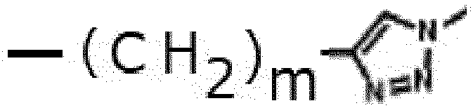
- L4 is preferably -[(CH2)p-O)]q- (wherein p and q each independently represent an integer of 1 to 5).
- L5 is preferably -CONH-, -NHCO-, -COO-, or -OCO-, and is more preferably -CONH-.
- L6 is preferably -(CH2)r, -(CH2)rO-, or -(CH2)rSi(R1)(R2)-O- (wherein r represents an integer of 1 to 5, and R1 and R2 each independently represent an alkyl group containing 1 to 4 carbon atoms). R1 and R2 are particularly preferably methyl groups.
- Y represents a group capable of binding to an antigen-binding molecule. Y is preferably an active ester of a carboxyl group, and is more preferably an succinimidyl ester of a carboxyl group.
- R3 represents an alkyl group containing 1 to 6 carbon atoms, an alkoxy group containing 1 to 6 carbon atoms, a halogen atom, -SR11, -SOR12, an aryl group containing 6 to 10 carbon atoms, - N(R13)(R14), or -NO2, or two adjacent R3s may together form an aryl group containing 6 to 10 carbon atoms. R11, R12, R13, and R14 each independently represent an alkyl group containing 1 to 6 carbon atoms or an alkoxy group containing 1 to 6 carbon atoms.
- Herein, the aryl group may be optionally substituted with an alkyl group containing 1 to 6 carbon atoms or an alkoxy group containing 1 to 6 carbon atoms.
- Herein, the alkyl group and the alkoxy group may be optionally substituted with a halogen atom.
- When a plurality of R3s are present, the plurality of R3s may be identical to or different from one another.
- The halogen atom may be any of fluorine, chlorine, bromine and iodine, and the halogen atom is preferably fluorine, chlorine, or bromine.
- The compound of the phthalocyanine dye can be synthesized in accordance with the methods described in Production Examples 1 to 5 in the after-mentioned Examples.
- Photoimmunotherapy is a therapeutic method of using a photosensitizer and an irradiation light to destroy specific cells in a body. When a photosensitizer is exposed to a light with a specific wavelength, it generates cytotoxic reactive oxygen species capable of inducing apoptosis, necrosis, and/or autophagy to around cells. For example,
Japanese Patent No. 6127045 JP Patent Publication (Kohyo) No. 2017-524659 A - The fusion protein of the present invention and the conjugate of a biotin-modified dimer and a phthalocyanine dye are administered to a subject, and the cells are then irradiated with an excitation light in an amount effective for suppression of cell proliferation or induction of cell death, so that the cell proliferation can be suppressed or the cell death can be induced, and thereby the subject can be treated.
- Preferably, the fusion protein of the present invention and the conjugate of biotin and a phthalocyanine dye are administered to a subject, and the cells are then irradiated with an excitation light in an amount effective for suppression of cell proliferation or induction of cell death, so that the cell proliferation can be suppressed or the cell death can be induced, and thereby the subject can be treated.
- The subj ect used herein includes humans and non-human animals. Examples of the subject may include humans and experimental animals such as mice. The subject is preferably affected with a disease regarding which suppression of cell proliferation or induction of cell death is desired. For example, the subject is affected with a cancer or a solid tumor.
- Examples of the "cancer" may include carcinoma, lymphoma, blastoma, sarcoma, and leukemia or malignant lymphoma. Specific examples of the cancer may include squamous cell carcinoma (e.g., epithelial squamous cell carcinoma), lung cancer including small cell lung cancer, non-small cell lung cancer ("NSCLC"), pulmonary adenocarcinoma and pulmonary squamous cell carcinoma, peritoneal cancer, hepatocarcinoma, corpus ventriculi or stomach cancer, including digestive cancer, pancreatic cancer, glioblastoma, cervical cancer, ovarian cancer, liver cancer, bladder cancer, hepatocellular cancer, breast cancer, colon cancer, rectal cancer, colorectal cancer, endometrial membrane cancer or endometrial carcinoma, salivary gland carcinoma, kidney or renal region cancer, prostate cancer, vulvar cancer, thyroid cancer, hepatocellular carcinoma, anal carcinoma, penile carcinoma, and head and neck cancer.
- The solid tumor means a benign or malignant, abnormal cell mass that generally does not contain a capsule. Examples of the solid tumor may include glioma, astrocytoma, medulloblastoma, craniopharyngioma, ependymoma, pineal gland tumor, hemangioblastoma, acoustic neuroma, oligodendrocyte, meningioma, melanoma, neuroblastoma, and retinoblastoma.
- Examples of the administration method to the subject may include, but are not limited to, a local route, an injection (a subcutaneous injection, an intramuscular injection, an intradermal injection, an intraperitoneal injection, an intratumoral injection, an intravenous injection, etc.), an oral route, an ocular route, a sublingual route, a rectal route, a percutaneous route, an intranasal route, a vaginal route, and an inhalation route.
- It is preferable that the conjugate of biotin and a phthalocyanine dye and the fusion protein of the present invention are each administered in a therapeutically effective amount. Regarding each of the above-described conjugate and fusion protein, the therapeutically effective amount per 60 kg is at least 0.5 mg (mg/60 kg), at least 5 mg/60 kg, at least 10 mg/60 kg, at least 20 mg/60 kg, at least 30 mg/60 kg, or at least 50 mg/60 kg. For example, when it is intravenously administered, the applied dose is 1 mg/60 kg, 2 mg/60 kg, 5 mg/60 kg, 20 mg/60 kg, or 50 mg/60 kg, and it is, for example, 0.5 to 50 mg/60 kg. In another example, the therapeutically effective amount is at least 100 µg/kg, at least 500 µg/kg or at least 500 µg/kg, and it is, for example, at least 10 µg/kg. For example, when it is intratumorally or intraperitoneally administered, the dose is 100 µg/kg, 250 µg/kg, approximately 500 µg/kg, 750 µg/kg, or 1000 µg/kg, and it is, for example, 10 µg/kg to 1000 µg/kg. In one example, when it is administered in the form of a solution for local administration, the therapeutically effective amount is 10 µg/ml, 20 µg/ml, 30 µg/ml, 40 µg/ml, 50 µg/ml, 60 µg/ml, 70 µg/ml, 80 µg/ml, 90 µg/ml, 100 µg/ml or the like, or it is 20 µg/ml to 100 µg/ml, or it is at least 500 µg/ml, or at least 1 µg/ml.
- The above-described dose can be administered once or divided doses over several administrations (2, 3, or 4 times, etc.), or as a single preparation.
- The conjugate of biotinr and a phthalocyanine dye and the fusion protein of the present invention can be each administered alone, or can also be administered in the presence of a pharmaceutically acceptable carrier, or can also be administered in the presence of other therapeutic agents (other anticancer agents, etc.).
- The conjugate of biotin and a phthalocyanine dye and the fusion protein of the present invention can bind to target cells or target tissues, such as circulating tumor cells or solid tumor cells. Thereafter, the target cells or tissues are irradiated with a light, so that the above-described conjugate or complex can absorb the light and can damage or destroy the target cells or tissues.
- In the photoimmunotherapy, the wavelength of the irradiation light is preferably 660 to 740 nm, and the irradiation light has a wavelength of, for example, 660 nm, 670 nm, 680 nm, 690 nm, 700 nm, 710 nm, 720 nm, 730 nm, or 740 nm. Light irradiation may be carried out using a device equipped with a near infrared (NIR) light emitting diode.
- The light irradiation amount is at least 1 J/cm2, for example, at least 4 J/cm2, at least 10 J/cm2, at least 15 J/cm2, at least 20 J/cm2, at least 50 J/cm2, or at least 100 J/cm2. It is, for example, 1 to 500 J/cm2. Light irradiation may be carried out several times (e.g., 2, 3, 4, 5, 6, 7, 8, 9, or 10 times).
- The present invention will be more specifically described in the following examples. However, these examples are not intended to limit the scope of the present invention.
-


- NHS-SiPc (1.8 mg, 1.0 µmol), disodium hydrogen phosphate buffer (pH 8.4, 200 µL) and dimethyl sulfoxide (200 µL) was added to biotin-PEG3-NH2 (0.5 mg, 1.2 µmol), and the mixture was was stirred at room temperature for 12 hours while shielding from light by aluminum foil. The reaction solution was diluted to 1 mL with water and was purified by reverse phase HPLC (gradient: 30% for 5 min; 30-80% for 30 min acetonitrile in 50 mM triethylammonium acetate aqueous solution (pH 7.0), retention time = 23.8 min, YMC -Triart C18, flow rate = 3.5 mL/min) to obtain a target compound Biotin-SiPc (0.7 mg, 0.34 µmol, 34%, deep blue color).
- LRMS (ESI): m/z 941.45 [M-2H]2-
- LISA314 molecule (SEQ ID NO: 1) was fused with a molecules recognizing the HER2 antigen (Löfblom, J. FEBS Lett. 584(12): 2670-80 (2010)) (SEQ ID NO: 2), and a protein having binding ability to both biotin molecule and Her2/ErbB2 (hereafter this protein is referred to as MFL2-G5Sx3-His) was synthesized in E. coli (SEQ ID NO: 3). The gene sequence of MFL2-G5Sx3-His (SEQ ID NO: 4) was synthesized as an artificial gene by Eurofins Genomics. Using pET45b(+) as an expression vector, an expression vector was constructed according to a common method. Specifically, using a primer set (Rv: catggtatatctccttcttaaagttaaac(SEQ ID NO: 5) and Fw: cgcagcttaattaacctaggctgctgccac(SEQ ID NO: 6)), the vector was linearized by a PCR reaction. The sequence prepared by the artificial gene synthesis was amplified by a PCR reaction using a primer set(Fw: aggagatataccatgGTGGACAACAAATTCAACAAAGAG(SEQ ID NO:7), Rv: gttaattaagctgcgTTAATGATGGTGGTGATGATGCGATG(SEQ ID NO:8)). Bands were cut out of both of the PCR reaction products according to agarose gel electrophoresis, and were then purified. Using In-Fusion HD Cloning Kit (TaKaRa Bio), the purified vector and the insert were subjected to a ligation reaction and cloning in accordance with the dosage and administration described in the instruction manual.
- In addition, a deletion mutant of MFL2-G5 Sx3-His expression vector was preparesd, which expresses MFL2-G5Sx3-del5 having the amino acid sequence shown in SEQ ID NO: 9, which lacks the C-terminal amino acid sequence PSAASHHHHHH of the amino acid sequence shown in SEQ ID NO: 3. Specifically, using a primer set (Fw;AAGTCAAATAACGCAGCTTAATTAACCTAG (SEQ ID NO: 11), Rw;TGCGTTATTTGACTTTGGTAAAGGTGTCATG(SEQ ID NO: 12)), a PCR reaction was carried out using the expression vector as a template. Thereafter, the reaction solution was subjected to a DpnI treatment at 37°C for 1 hour, and E. coli was then transformed with the obtained reaction solution, followed by cloning. After completion of the cloning, a sequence analysis was carried out to confirm that the gene sequence as set forth in SEQ ID NO: 10 was incorporated. The vector was named "pET45-MFL2-G5Sx3-del5."
- Plasmid vectors (pET45-MFL2-G5Sx3-His, pET45-MFL2-G5Sx3-del5) which werre confirmed that the gene sequences shown in
SEQ ID Nos 4 and 10 had been incorporated therein, was introduced into competent cells BL21(DE3) (ECOS Competent E. coli BL21(DE3), Nippon Gene Co., Ltd.), so that the cells were transformed. A culture medium prepared by culturing the cells in 100 mL of 2 × YT medium overnight was inoculated into 1 L of a culture solution, and the obtained mixture was then cultured at 37°C. When the OD value at 600 nm became 0.5 to 0.8, IPTG was added to the culture to a final concentration of 0.5 mM, and the thus obtained mixture was then cultured at 37°C for 4 hours. Thereafter, the resulting cells were recovered by centrifugation (7500 × g, 20 min at 4°C). - The method of recovering IB from the cells is described below. After Benzonase (Merck) had been added to B-PER (Thermo Scientific) and had been suspended therein, the obtained mixture was incubated at room temperature for 10 minutes, and an insoluble fraction (inclusion body; hereinafter referred to as "IB") was separated from a soluble fraction by centrifugation. Thereafter, the IB was recovered. Subsequently, the recovered IB was resuspended in a 10-fold diluted B-PER buffer (without addition of Benzonase), and was centrifuged. The supernatant was discarded, and the washing of IB was repeated three times. After completion of the washing three times, the IB recovered by centrifugation was resuspended in ultrapure water (Milli Q). Thereafter, the resuspension was dispensed in an amount of 1 mL each into 1.5-mL tubes, and was then cryopreserved at -80°C.
- Denaturation and refolding of IB are described below. A denature buffer (0.1 M Tris-HCl, pH8.5, 6 M guanidium HCl, 10 mM EDTA) was added to IB, and the IB was then dissolved in the buffer by pipetting. Thereafter, while stirring the obtained solution using a rotator, the solution was incubated at 4°C overnight, and was then centrifuged (15,000 × g, 20 min at 4°C) to recover a supernatant. The recovered supernatant was adjusted with a denature buffer, so the protein concentration (OD 280 nm) in the recovered supernatant became 30 to 50 mg/mL. Thereafter, while stirring, refolding buffer (0.05 M sodium phosphate, 0.4 M arginine hydrochloride, pH6.8) was added dropwise in such an amount that the denatured protein solution having an adjusted concentration was 50-fold diluted. Thereafter, the obtained solution was incubated at 4°C for 24 to 48 hours, and was centrifuged (15,000 × g, 20 min at 4°C), and the supernatant was recoverd. The recovered supernatant was subjected to SDS-PAGE electrophoresis to confirm tetramer formation by CBB staining. As a result, as shown in
FIG. 1A , it was confirmed that most ofMFL2-G5Sx3-del5 existed as a monomer compared to MFL2-G5Sx3-His. Regarding MFL2-G5Sx3-His, as a result of enrichment, it was confirmed that MFL2-G5 Sx3-His shown inFIG. 1B was obtained. - MFL2-G5Sx3-His was analyzed using SPR (Biacore T200, Cytiva) in terms of the binding activity to HER2 (ErbB2). The Her2 antigen (Recombinant Human ErbB2/Her2 Fc Chimera Protein, R & D SYSTEMS) was immobilized on a Sensor Chip CM5 (Cytiva) according to an amine coupling method (Amine Coupling Kit, Cytiva). Subsequently, the binding activity was measured using the FL dilution series (2-fold dilution series from 1E-08 M to 6.25E-10 M). The results are shown in
Fig. 2 . From the results, it was confirmed that stable binding is shown. The analysis results were ka = 8.509E+6, kd = 0.006598, and KD = 7.753E-10. - MFL2-G5Sx3-Hiswas analyzed using SPR (Biacore T200, Cytiva), in terms of the binding activity to Psyche. MFL2-G5Sx3-His was immobilized on a Sensor Chip CM5 (Cytiva) according to the amine coupling method. The binding activity was then analyzed according to a single cycle kinetics method using dilution series of biotin-SiPc (Compound Y produced in Production Example X) (twice dilution series from 1E-08 M to 6.25E-10 M: 5 seriies) as analytes. The results are shown in

Fig. 3 . From the results, it was confirmed that stable binding is shown. The analysis results were ka = 1.160E+6, kd = 0.002770, and KD = 2.387E-9. - Her2-positive cells (KPL-4) were seeded on a 96-well plate to result in a cell count of 1 × 10⁴ cells/well and a culture solution amount of 50 µL/well, and were then cultured overnight. Pirified MFL2-G5Sx3-His and biotin-SiPc (which was produced in Production Example 1) were mixed with each other to a molar ratio of 1 : 4 (hereinafter referred to as a "MFL2-G5Sx3-Hi complex"), and 8 series of 10-fold dilution series (including zero concentration) were prepared from 10 µg/mL. After the dilution series had been prepared, the complex was added to the cells, and 24 hours later, the cells were irradiated with an LED emitting a 690-nm light at 100 J/cm2 from the bottom surface of the culture plate. Thereafter, the cells were cultured for 24 hours. After removing the MFL2-G5Sx3-His complex-containing medium, the cells were washed once with PBS, new medium was added, and the number of viable cells was compared using Cell Counting Kit-8 (DOJINDO LABORATORIES). A reagent was added in accordance with the dosage and administration described in the instruction manual, and the resulting cells were then incubated at 37°C in a CO2 incubator for 1.5 hours. After that, the absorbance at 450 nm was measured, and the mean value was then calculated, followed by background correction. Thereafter, the control was set at 100%, and the cell proliferation percentage to the control under individual conditions was calculated. The results are shown in
Fig. 4 . It was confirmed that the complex of the FL and the photosensitizer biotin-SiPc compound has photoirradiation-dependent and concentration-dependent cytotoxicity. - Subsequently, protein expression vectors were constructed, which has a different length of an amino acid linker that links a molecule recognizing a HER2 antigen in MFL2-G5Sx3-del5 to Cupid-del5, and has 5 ammino acids PSAAS which was added to the C-teminal. Specifically, as shown in
Fig.5A , a peptide formed by combining 4 glycine residues and 1 serine residue was set to be a basic unit, and expression vectors each having a linker with a length of 1 unit, 2 units or 3 units and having PSAAS at the C-terminal were produced. The production method thereof is as follows. - Gene sequences (SEQ ID NOs: 14-16) encoding amino acid sequences having the each linker length shown in SEQ ID NOs: 11 to 13 and C-terminal were created by artificial gene synthesis. Then, using a primer set (Rv: catggtatatctccttcttaaagttaaac(SEQ ID NO:5), Fw: cgcagcttaattaacctaggctgctgccac(SEQ ID NO:6)), pET45b vector was linearized by a PCR reaction. The sequence prepared by the artificial gene synthesis was amplified by a PCR reaction using a primer set(Fw: aggagatataccatgGTGGACAACAAATTCAACAAAGAG(SEQ ID NO:7), Rv: gttaattaagctgcgTTAATGATGGTGGTGATGATGCGATG(SEQ ID NO:8)). Bands were cut out of both of the PCR reaction products according to agarose gel electrophoresis, and were then purified. Using In-Fusion HD Cloning Kit (TaKaRa Bio), the vector and the insert were subjected to a ligation reaction and cloning in accordance with the dosage and administration described in the instruction manual. Sequence analysis was carried out, and the expression vectors which were confirmed to have incorporated the gene sequences shown in SEQ ID NOs: 14-16 were named pET45-MFL2-G4Sx1-PSAAS, pET45-MFL2-G4Sx2-PSAAS, and pET45-MFL2-G4Sx3-PSAAS, respectively.
- pET45-MFL2-G4Sx1-PSAAS , pET45-MFL2-G4Sx2-PSAAS , pET45-MFL2-G4Sx3-PSAAS were introduced into competent cells BL21(DE3) (ECOS Competent E. coli BL21(DE3), Nippon Gene Co., Ltd.), so that the cells were transformed. A culture medium prepared by culturing the cells in 100 mL of 2 × YT medium overnight was inoculated into 1 L of a culture solution, and the obtained mixture was then cultured at 37°C. When the OD value at 600 nm became 0.5 to 0.8, IPTG was added to the culture to a final concentration of 0.5 mM, and the thus obtained mixture was then cultured at 37°C for 4 hours. Thereafter, the resulting cells were recovered by centrifugation (7500 × g, 20 min at 4°C).
- The method of recovering IB from the cells is described below. After Benzonase (Merck) had been added to B-PER (Thermo Scientific) and had been suspended therein, the obtained mixture was incubated at room temperature for 10 minutes, and an insoluble fraction (inclusion body; hereinafter referred to as "IB") was separated from a soluble fraction by centrifugation. Thereafter, the IB was recovered. Subsequently, the recovered IB was resuspended in a 10-fold diluted B-PER buffer (without addition of Benzonase), and was centrifuged. The supernatant was discarded, and the washing of IB was repeated three times. After completion of the washing three times, the IB recovered by centrifugation was resuspended in ultrapure water (Milli Q). Thereafter, the resuspension was dispensed in an amount of 1 mL each into 1.5-mL tubes, and was then cryopreserved at -80°C.
- Denaturation of IB is described below. A denature buffer (0.1 M Tris-HCl, pH 8.5, 6 M guanidine hydrochloride, and 10 mM EDTA) was added to IB, and the IB was then dissolved in the buffer by pipetting. Thereafter, while stirring the obtained solution using a rotator, the solution was incubated at 4°C overnight, and was then centrifuged (15,000 × g, 20 min at 4°C) to recover a supernatant. The recovered supernatant was adjusted with a denature buffer, so that the protein concentration (OD 280 nm) in the supernatant became 30 to 50 mg/mL.
- Refolding is described below. The refolding buffers were prepared by adding 0.4 M arginine hydrochloride to Mcilvaine buffer and adjusting the pH value to pH4.0, pH4.5, pH5.0, pH6.5, pH7.5, pH8.0 or the like by NaOH. 25 µL of the denatured solution supernatant was added to 1 mL of refolding buffer having each pH, and rfolding was performed by dilution. The samples were incubated at 4°C, and the formation of tetramers was confirmed by SDS-PAGE electrophoresis.
- For MFL2-G4Sx2-PSAAS and MFL2-G4Sx3-PSAAS, the results of confirming tetramer formation at each pH at the time of hour and 24 hours after refolding are shown in
FIG. 5B . From this result, it was confirmed that tetramer formation from monomers was promoted at pH5.0-pH6.5. However, after 72 hours, it was confirmed that the tetramer band had disappeared at all pH values. - Similarly, as shown in
Figure 5C , for the refolded product at pH 6.0 for MFL2-G4Sx1-PSAAS, MFL2-G4Sx2-PSAAS, and MFL2-G4Sx3-PSAAS, stable tetramer formation could not be confirmed after 72 hours. - SEQ ID NO:1 Amino acid sequence of LISA314


- SEQ ID NO:2 Molecule which recognizes HER2 antigen
VDNKFNKEMRNAYWEIALLPNLNNQQKRAFIRSIYDDPSQSANLLAEAKKLNDAQAPK - SEQ ID NO: 3 Amino acid sequence of MFL2-G5 Sx3 -Hi s

- SEQ ID NO:4 Gene sequence of MFL2-G4Sx3-His

- SEQ ID NO:9 Amino acid sequence of MFL2-G5Sx3-del5


- SEQ ID NO:10 Gene sequence ofMFL2-G5Sx3-del5
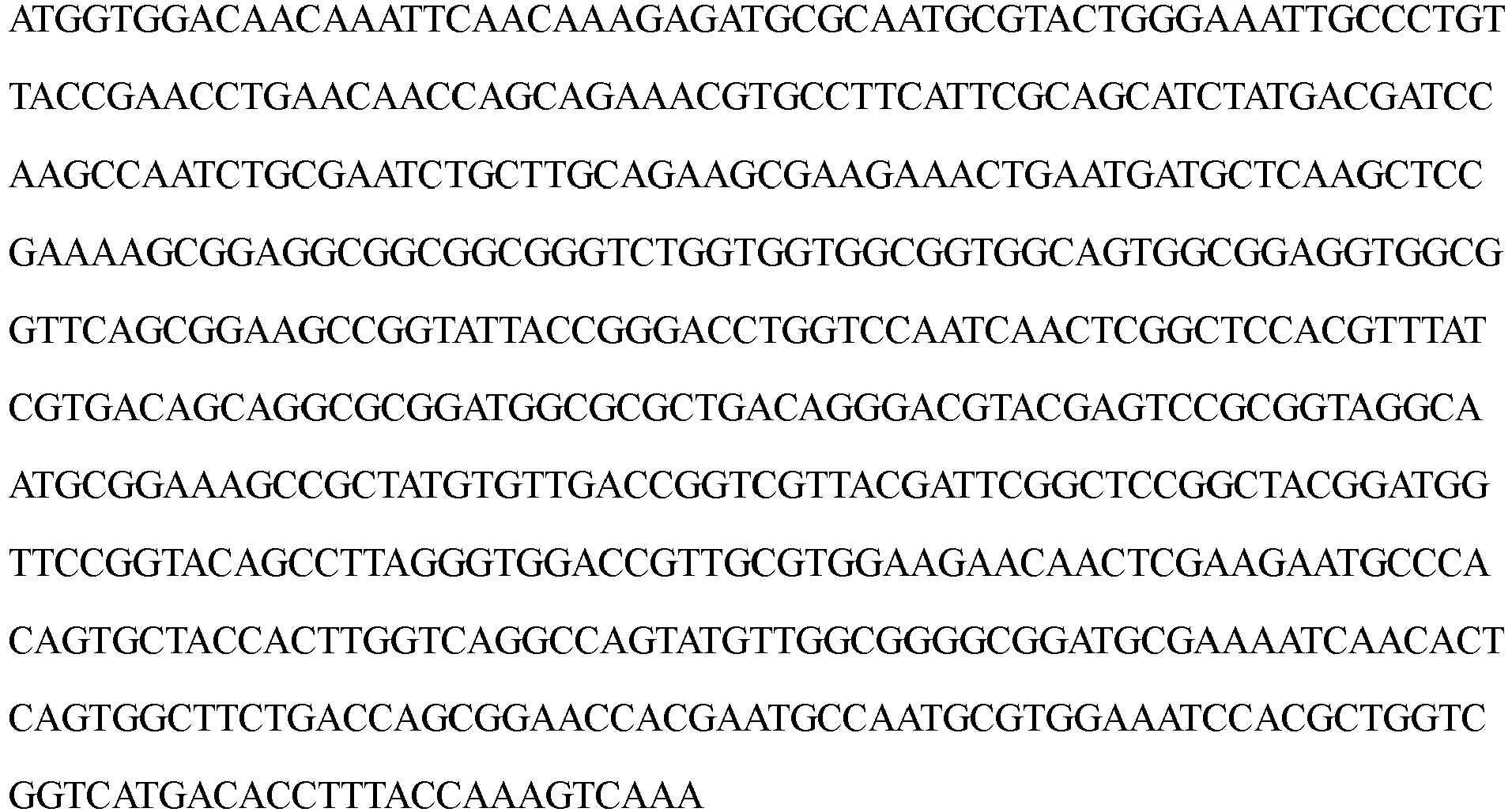
- SEQ ID NO: 11 Amino acid sequence of MFL2-G4Sx1-PSAA

- SEQ ID NO: 12 Amino acid sequence of MFL2-G4Sx2-PSAA

- SEQ ID NO: 13 Amino acid sequence of MFL2-G4Sx3-PSAAS


- SEQ ID NO: 14 Gene sequence of FL2-G4Sx1-PSAAS

- SEQ ID NO: 15 Gene sequence of MFL2-G4Sx2-PSAAS
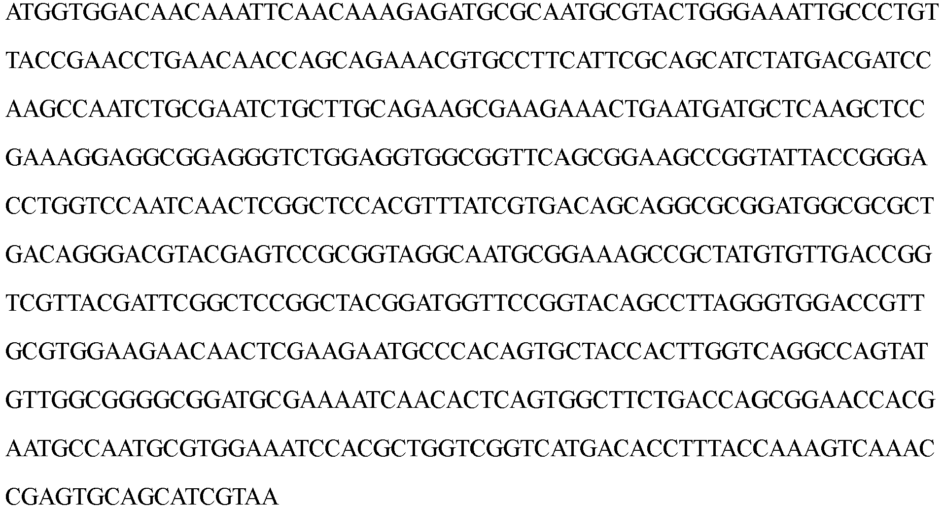
- SEQ ID NO:16 Gene sequence of MFL2-G4Sx3-PSAAS
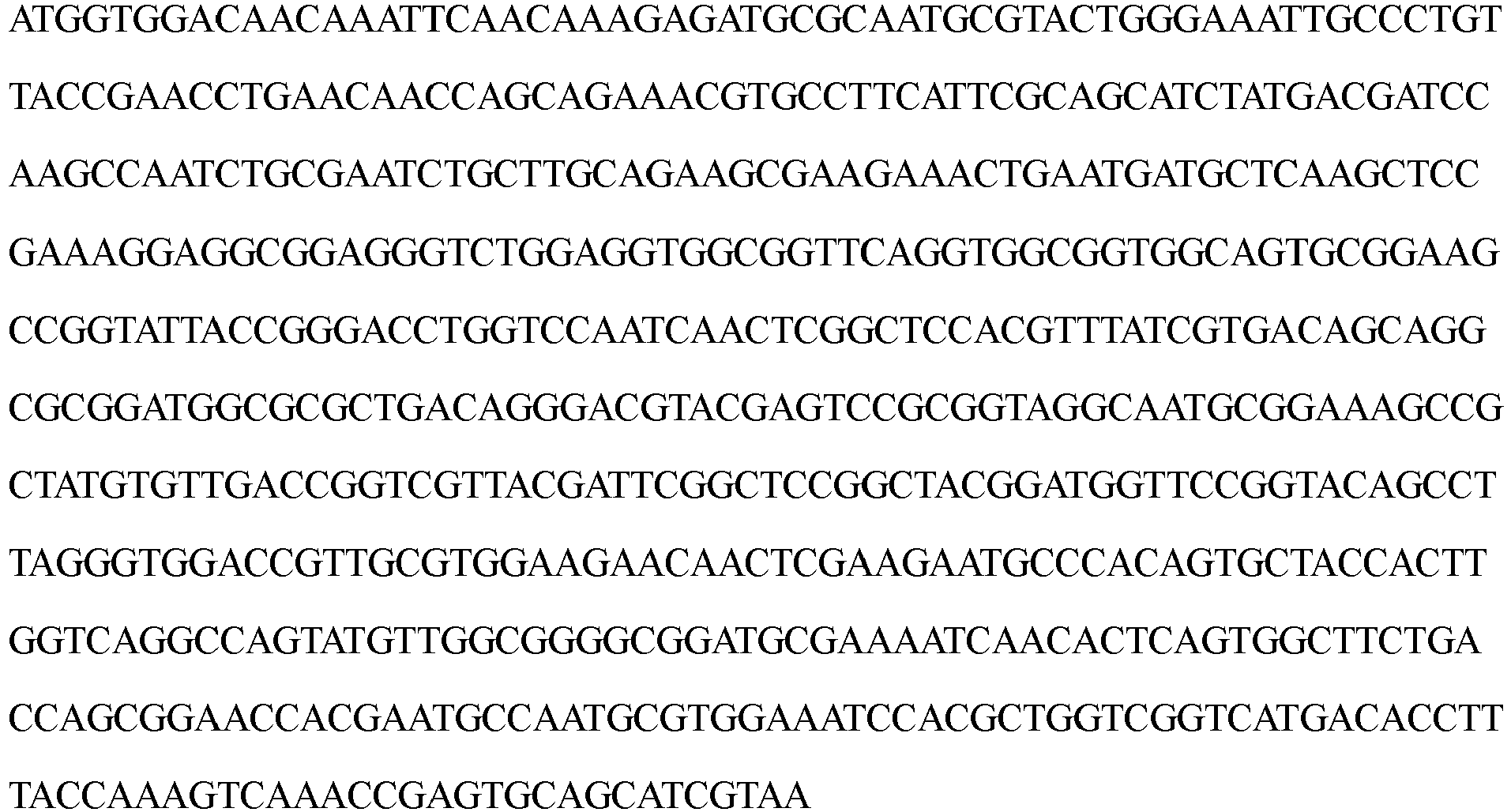
- SEQ ID NO: 17

Claims (14)
- A fusion protein, wherein an antigen-binding molecule having a molecular weight of 20,000 or less is bound, via a linker sequence, to the N-terminal side and/or C-terminal side of mutant streptavidin which comprises an amino acid sequence having the following mutations (1) to (6) with respect to the amino acid sequence of a core streptavidin as shown in SEQ ID NO: 17 provided that the C-terminal amino acid sequence consisting of Pro-Ser-Ala-Ala-Ser may be partially or entirely deleted, and has a decreased immunogenicity as compared with that of a wild-type streptavidin:(1) a mutation in which the tyrosine residue at position 10 is substituted with serine or threonine;(2) a mutation in which the tyrosine residue at position 71 is substituted with alanine or serine;(3) a mutation in which the arginine residue at position 72 is substituted with lysine;(4) a mutation in which the glutamic acid residue at position 89 is substituted with aspartic acid;(5) a mutation in which the arginine residue at position 91 is substituted with lysine; and(6) a mutation in which the glutamic acid residue at position 104 is substituted with glutamine or asparagine.
- The fusion protein according to claim 1, which has the antigen-binding molecule having a molecular weight of 20,000 or less, the linker sequence, and the amino acid sequence of the mutant streptavidin in this order from the N-terminal side to the C-terminal side.
- The fusion protein according to claim 1 or 2, wherein the antigen-binding molecule is a molecule that binds to an antigen expressing in a cancer cell.
- The fusion protein according to any one of claims 1 to 3, wherein the antigen-binding molecule is a molecule that binds to Her2.
- The fusion protein according to any one of claims 1 to 4, wherein the antigen-binding molecule has the amino acid sequence as set forth in SEQ ID NO: 2.
- The fusion protein according to any one of claims 1 to 5, wherein the linker sequence consists of a glycine residue(s) and a serine residue(s) and the number of the amino acid residues is 5 to 25.
- The fusion protein according to any one of claims 1 to 6, wherein the linker sequence is an amino acid sequence represented by [(Gly)m-Ser]n wherein m represents an integer of 1 to 10, and n represents an integer of 1 to 5.
- The fusion protein according to any one of claims 1 to 7, which has the amino acid sequence as set forth in SEQ ID NO: 3 or SEQ ID NO: 9.
- A nucleic acid encoding the fusion protein having the amino acid sequence as set forth in SEQ ID NO: 3 or SEQ ID NO: 9.
- A cancer therapeutic agent or a cancer diagnostic agent, comprising the fusion protein according to any one of claims 1 to 8.
- A kit for treating or diagnosing cancer, comprising (1) the fusion protein according to any one of claims 1 to 8, and (2) a conjugate of biotin and a diagnostic substance or a therapeutic substance.
- The kit according to claim 11, wherein the diagnostic substance or the therapeutic substance is a phthalocyanine dye.
- A method for producing the fusion protein according to any one of claims 1 to 8, comprising a step of allowing a nucleic acid encoding the fusion protein according to any one of claims 1 to 8 to express in a host.
- The method according to claim 13, wherein the fusion protein is allowed to express in a bacterial inclusion body and is then recovered.
Applications Claiming Priority (2)
| Application Number | Priority Date | Filing Date | Title |
|---|---|---|---|
| JP2021124126 | 2021-07-29 | ||
| PCT/JP2022/029073 WO2023008515A1 (en) | 2021-07-29 | 2022-07-28 | Fusion protein between antigen-binding molecule and streptavidin variant |
Publications (1)
| Publication Number | Publication Date |
|---|---|
| EP4382609A1 true EP4382609A1 (en) | 2024-06-12 |
Family
ID=85086132
Family Applications (1)
| Application Number | Title | Priority Date | Filing Date |
|---|---|---|---|
| EP22849576.8A Withdrawn EP4382609A1 (en) | 2021-07-29 | 2022-07-28 | Fusion protein between antigen-binding molecule and streptavidin variant |
Country Status (4)
| Country | Link |
|---|---|
| EP (1) | EP4382609A1 (en) |
| JP (1) | JPWO2023008515A1 (en) |
| CN (1) | CN117677701A (en) |
| WO (1) | WO2023008515A1 (en) |
Family Cites Families (5)
| Publication number | Priority date | Publication date | Assignee | Title |
|---|---|---|---|---|
| US8546537B2 (en) | 2009-02-20 | 2013-10-01 | The University Of Tokyo | Streptavidin having low immunogenicity and use thereof |
| WO2013009475A1 (en) | 2011-07-11 | 2013-01-17 | The United States Of America, As Represented By The Secretary, Department Of Health & Human Services | Photosensitizing antibody-phuorophore conjugates |
| EP3148583B1 (en) | 2014-06-02 | 2020-04-29 | Li-Cor, Inc. | Phthalocyanine probes and uses thereof |
| EP3834845A4 (en) * | 2018-08-08 | 2022-03-16 | The University of Tokyo | CONJUGATE OF A BIOTIN VARIANT DIMERE WITH A PHTHALOCYANINE PIGMENT |
| WO2020138427A1 (en) * | 2018-12-28 | 2020-07-02 | 国立大学法人 東京大学 | Fusion protein of streptavidin variant and antibody recognizing cancer cells |
-
2022
- 2022-07-28 CN CN202280049796.2A patent/CN117677701A/en active Pending
- 2022-07-28 EP EP22849576.8A patent/EP4382609A1/en not_active Withdrawn
- 2022-07-28 JP JP2023538614A patent/JPWO2023008515A1/ja active Pending
- 2022-07-28 WO PCT/JP2022/029073 patent/WO2023008515A1/en not_active Ceased
Also Published As
| Publication number | Publication date |
|---|---|
| WO2023008515A1 (en) | 2023-02-02 |
| JPWO2023008515A1 (en) | 2023-02-02 |
| CN117677701A (en) | 2024-03-08 |
Similar Documents
| Publication | Publication Date | Title |
|---|---|---|
| JP6723913B2 (en) | Biotin variants, streptavidin variants and their use | |
| ES3058443T3 (en) | Specific modification of antibody with IgG-binding peptide | |
| EP2994487B1 (en) | Trimeric antigen binding molecules | |
| EP3834845A1 (en) | Conjugate of biotin variant dimer with phthalocyanine pigment | |
| CN108866635B (en) | Multispecific protein medicine and library thereof, and preparation method and application thereof | |
| JP2024504777A (en) | MULTABODY constructs, compositions and methods | |
| JP2024507693A (en) | bispecific antibody | |
| KR20230083316A (en) | Polypeptides targeting SARS-COV-2, and related compositions and methods | |
| CN113226329A (en) | Fusion protein of cancer cell-recognizing antibody and streptavidin mutant | |
| JP2023061976A (en) | VEGFR-2 antibody | |
| JP2021052669A (en) | Fusion protein of protein recognition substance and streptavidin mutant | |
| CN109983024A (en) | scavenger | |
| JP7625188B2 (en) | Conjugates of biotin-modified dimers with phthalocyanine dyes | |
| US20240182544A1 (en) | Polypeptides targeting dr4 and/or dr5 and related compositions and methods | |
| US12115223B2 (en) | Conjugate of biotin-modified dimer and phthalocyanine dye | |
| EP4321538A1 (en) | Fusion protein between antigen-binding molecule and streptavidin variant | |
| CN115397835A (en) | Duocarmycin derivative and application thereof | |
| WO2023008515A1 (en) | Fusion protein between antigen-binding molecule and streptavidin variant | |
| WO2012023579A1 (en) | Process for production of streptavidin | |
| US20220348681A1 (en) | Fused protein of an antibody which recognizes cancer-cells and a streptavidin mutant | |
| US20240374747A1 (en) | Fusion protein of vhh antibody and streptavidin mutant | |
| US11952402B2 (en) | Fusion protein containing trail and IgG binding domain and the uses thereof | |
| ES2957266T3 (en) | Polypeptide improved in terms of protein purity and affinity for the antigen, conjugated thereof with an antibody or antigen-binding fragment and method for its preparation | |
| CN115605487A (en) | Biotin variant dimers and uses thereof | |
| CN117377698A (en) | Fusion proteins of antigen-binding molecules and streptavidin mutants |
Legal Events
| Date | Code | Title | Description |
|---|---|---|---|
| STAA | Information on the status of an ep patent application or granted ep patent |
Free format text: STATUS: THE INTERNATIONAL PUBLICATION HAS BEEN MADE |
|
| PUAI | Public reference made under article 153(3) epc to a published international application that has entered the european phase |
Free format text: ORIGINAL CODE: 0009012 |
|
| STAA | Information on the status of an ep patent application or granted ep patent |
Free format text: STATUS: REQUEST FOR EXAMINATION WAS MADE |
|
| 17P | Request for examination filed |
Effective date: 20240227 |
|
| AK | Designated contracting states |
Kind code of ref document: A1 Designated state(s): AL AT BE BG CH CY CZ DE DK EE ES FI FR GB GR HR HU IE IS IT LI LT LU LV MC MK MT NL NO PL PT RO RS SE SI SK SM TR |
|
| STAA | Information on the status of an ep patent application or granted ep patent |
Free format text: STATUS: THE APPLICATION HAS BEEN WITHDRAWN |
|
| 18W | Application withdrawn |
Effective date: 20240807 |