EP4382122A1 - Peptide complex ipf-h2a for modulation immune function of the human body - Google Patents
Peptide complex ipf-h2a for modulation immune function of the human body Download PDFInfo
- Publication number
- EP4382122A1 EP4382122A1 EP22020605.6A EP22020605A EP4382122A1 EP 4382122 A1 EP4382122 A1 EP 4382122A1 EP 22020605 A EP22020605 A EP 22020605A EP 4382122 A1 EP4382122 A1 EP 4382122A1
- Authority
- EP
- European Patent Office
- Prior art keywords
- ipf
- cells
- cell
- viral
- complex
- Prior art date
- Legal status (The legal status is an assumption and is not a legal conclusion. Google has not performed a legal analysis and makes no representation as to the accuracy of the status listed.)
- Pending
Links
- 108090000765 processed proteins & peptides Proteins 0.000 title claims abstract description 54
- 230000036737 immune function Effects 0.000 title claims abstract description 5
- 238000009169 immunotherapy Methods 0.000 claims abstract description 7
- 108010033040 Histones Proteins 0.000 claims description 12
- 229960005486 vaccine Drugs 0.000 claims description 11
- 102000057297 Pepsin A Human genes 0.000 claims description 10
- 108090000284 Pepsin A Proteins 0.000 claims description 10
- 229940111202 pepsin Drugs 0.000 claims description 10
- 125000003275 alpha amino acid group Chemical group 0.000 claims description 6
- 230000002427 irreversible effect Effects 0.000 claims description 4
- 230000003612 virological effect Effects 0.000 abstract description 56
- 206010028980 Neoplasm Diseases 0.000 abstract description 17
- 208000037265 diseases, disorders, signs and symptoms Diseases 0.000 abstract description 12
- 238000013459 approach Methods 0.000 abstract description 10
- 201000011510 cancer Diseases 0.000 abstract description 10
- 201000010099 disease Diseases 0.000 abstract description 10
- 208000026278 immune system disease Diseases 0.000 abstract description 2
- 210000004027 cell Anatomy 0.000 description 131
- 239000000126 substance Substances 0.000 description 46
- 238000012360 testing method Methods 0.000 description 43
- 102100036011 T-cell surface glycoprotein CD4 Human genes 0.000 description 40
- 230000000840 anti-viral effect Effects 0.000 description 38
- 241000700605 Viruses Species 0.000 description 36
- 230000000120 cytopathologic effect Effects 0.000 description 34
- 241000700588 Human alphaherpesvirus 1 Species 0.000 description 28
- 230000027455 binding Effects 0.000 description 27
- 238000010790 dilution Methods 0.000 description 27
- 239000012895 dilution Substances 0.000 description 27
- 208000015181 infectious disease Diseases 0.000 description 27
- 230000004083 survival effect Effects 0.000 description 26
- 230000000694 effects Effects 0.000 description 25
- 241000701074 Human alphaherpesvirus 2 Species 0.000 description 23
- 239000000427 antigen Substances 0.000 description 23
- 108091007433 antigens Proteins 0.000 description 23
- 102000036639 antigens Human genes 0.000 description 23
- 230000002458 infectious effect Effects 0.000 description 23
- 238000011282 treatment Methods 0.000 description 22
- 238000002474 experimental method Methods 0.000 description 21
- 239000002609 medium Substances 0.000 description 21
- 241000711467 Human coronavirus 229E Species 0.000 description 19
- 230000002401 inhibitory effect Effects 0.000 description 19
- 239000013553 cell monolayer Substances 0.000 description 18
- 230000007423 decrease Effects 0.000 description 18
- 238000000034 method Methods 0.000 description 18
- 238000002360 preparation method Methods 0.000 description 17
- 238000004113 cell culture Methods 0.000 description 15
- PGZUMBJQJWIWGJ-ONAKXNSWSA-N oseltamivir phosphate Chemical compound OP(O)(O)=O.CCOC(=O)C1=C[C@@H](OC(CC)CC)[C@H](NC(C)=O)[C@@H](N)C1 PGZUMBJQJWIWGJ-ONAKXNSWSA-N 0.000 description 15
- 229940061367 tamiflu Drugs 0.000 description 15
- 238000004458 analytical method Methods 0.000 description 14
- 230000005764 inhibitory process Effects 0.000 description 14
- 108090000623 proteins and genes Proteins 0.000 description 14
- 231100000673 dose–response relationship Toxicity 0.000 description 13
- 108700018351 Major Histocompatibility Complex Proteins 0.000 description 12
- 239000003814 drug Substances 0.000 description 12
- 102000005962 receptors Human genes 0.000 description 12
- 108020003175 receptors Proteins 0.000 description 12
- 230000010076 replication Effects 0.000 description 12
- 230000020382 suppression by virus of host antigen processing and presentation of peptide antigen via MHC class I Effects 0.000 description 12
- JGSARLDLIJGVTE-MBNYWOFBSA-N Penicillin G Chemical compound N([C@H]1[C@H]2SC([C@@H](N2C1=O)C(O)=O)(C)C)C(=O)CC1=CC=CC=C1 JGSARLDLIJGVTE-MBNYWOFBSA-N 0.000 description 11
- 239000012091 fetal bovine serum Substances 0.000 description 11
- 239000002356 single layer Substances 0.000 description 11
- 239000006144 Dulbecco’s modified Eagle's medium Substances 0.000 description 10
- 210000001744 T-lymphocyte Anatomy 0.000 description 10
- 101800001690 Transmembrane protein gp41 Proteins 0.000 description 10
- 230000001413 cellular effect Effects 0.000 description 10
- 235000015097 nutrients Nutrition 0.000 description 10
- 102000004169 proteins and genes Human genes 0.000 description 10
- 241000711573 Coronaviridae Species 0.000 description 9
- 230000003602 anti-herpes Effects 0.000 description 9
- 210000003719 b-lymphocyte Anatomy 0.000 description 9
- 238000011161 development Methods 0.000 description 9
- 230000018109 developmental process Effects 0.000 description 9
- 235000016709 nutrition Nutrition 0.000 description 9
- 102000004196 processed proteins & peptides Human genes 0.000 description 9
- 239000000243 solution Substances 0.000 description 9
- 241000713772 Human immunodeficiency virus 1 Species 0.000 description 8
- 206010061535 Ovarian neoplasm Diseases 0.000 description 8
- 239000002671 adjuvant Substances 0.000 description 8
- 239000012634 fragment Substances 0.000 description 8
- 230000007246 mechanism Effects 0.000 description 8
- 201000001441 melanoma Diseases 0.000 description 8
- 239000013641 positive control Substances 0.000 description 8
- 239000000523 sample Substances 0.000 description 8
- QTENRWWVYAAPBI-YCRXJPFRSA-N streptomycin sulfate Chemical compound OS(O)(=O)=O.OS(O)(=O)=O.OS(O)(=O)=O.CN[C@H]1[C@H](O)[C@@H](O)[C@H](CO)O[C@H]1O[C@@H]1[C@](C=O)(O)[C@H](C)O[C@H]1O[C@@H]1[C@@H](N=C(N)N)[C@H](O)[C@@H](N=C(N)N)[C@H](O)[C@H]1O.CN[C@H]1[C@H](O)[C@@H](O)[C@H](CO)O[C@H]1O[C@@H]1[C@](C=O)(O)[C@H](C)O[C@H]1O[C@@H]1[C@@H](N=C(N)N)[C@H](O)[C@@H](N=C(N)N)[C@H](O)[C@H]1O QTENRWWVYAAPBI-YCRXJPFRSA-N 0.000 description 8
- 210000001519 tissue Anatomy 0.000 description 8
- 206010033128 Ovarian cancer Diseases 0.000 description 7
- 230000008859 change Effects 0.000 description 7
- 238000011109 contamination Methods 0.000 description 7
- LOKCTEFSRHRXRJ-UHFFFAOYSA-I dipotassium trisodium dihydrogen phosphate hydrogen phosphate dichloride Chemical compound P(=O)(O)(O)[O-].[K+].P(=O)(O)([O-])[O-].[Na+].[Na+].[Cl-].[K+].[Cl-].[Na+] LOKCTEFSRHRXRJ-UHFFFAOYSA-I 0.000 description 7
- 210000004698 lymphocyte Anatomy 0.000 description 7
- 239000002953 phosphate buffered saline Substances 0.000 description 7
- 102000006947 Histones Human genes 0.000 description 6
- 235000014676 Phragmites communis Nutrition 0.000 description 6
- 208000036142 Viral infection Diseases 0.000 description 6
- 230000009471 action Effects 0.000 description 6
- 239000012984 antibiotic solution Substances 0.000 description 6
- 229940079593 drug Drugs 0.000 description 6
- 238000011081 inoculation Methods 0.000 description 6
- 210000002966 serum Anatomy 0.000 description 6
- 238000002198 surface plasmon resonance spectroscopy Methods 0.000 description 6
- 238000010257 thawing Methods 0.000 description 6
- 210000003501 vero cell Anatomy 0.000 description 6
- 230000009385 viral infection Effects 0.000 description 6
- 241000725303 Human immunodeficiency virus Species 0.000 description 5
- 231100000002 MTT assay Toxicity 0.000 description 5
- 238000000134 MTT assay Methods 0.000 description 5
- 108091008874 T cell receptors Proteins 0.000 description 5
- 102000016266 T-Cell Antigen Receptors Human genes 0.000 description 5
- 230000015572 biosynthetic process Effects 0.000 description 5
- 210000004970 cd4 cell Anatomy 0.000 description 5
- 150000001875 compounds Chemical class 0.000 description 5
- 230000007812 deficiency Effects 0.000 description 5
- 210000000987 immune system Anatomy 0.000 description 5
- 238000000338 in vitro Methods 0.000 description 5
- 230000001965 increasing effect Effects 0.000 description 5
- 206010022000 influenza Diseases 0.000 description 5
- 238000002347 injection Methods 0.000 description 5
- 239000007924 injection Substances 0.000 description 5
- 238000012423 maintenance Methods 0.000 description 5
- 239000000203 mixture Substances 0.000 description 5
- 230000003287 optical effect Effects 0.000 description 5
- 230000004044 response Effects 0.000 description 5
- 238000001179 sorption measurement Methods 0.000 description 5
- KZDCMKVLEYCGQX-UDPGNSCCSA-N 2-(diethylamino)ethyl 4-aminobenzoate;(2s,5r,6r)-3,3-dimethyl-7-oxo-6-[(2-phenylacetyl)amino]-4-thia-1-azabicyclo[3.2.0]heptane-2-carboxylic acid;hydrate Chemical compound O.CCN(CC)CCOC(=O)C1=CC=C(N)C=C1.N([C@H]1[C@H]2SC([C@@H](N2C1=O)C(O)=O)(C)C)C(=O)CC1=CC=CC=C1 KZDCMKVLEYCGQX-UDPGNSCCSA-N 0.000 description 4
- 102100035875 C-C chemokine receptor type 5 Human genes 0.000 description 4
- 101710149870 C-C chemokine receptor type 5 Proteins 0.000 description 4
- 208000031886 HIV Infections Diseases 0.000 description 4
- 239000004107 Penicillin G sodium Substances 0.000 description 4
- 239000003443 antiviral agent Substances 0.000 description 4
- 238000004630 atomic force microscopy Methods 0.000 description 4
- 231100000135 cytotoxicity Toxicity 0.000 description 4
- 230000003013 cytotoxicity Effects 0.000 description 4
- 230000003247 decreasing effect Effects 0.000 description 4
- 238000009472 formulation Methods 0.000 description 4
- 238000007710 freezing Methods 0.000 description 4
- 230000008014 freezing Effects 0.000 description 4
- 239000003446 ligand Substances 0.000 description 4
- 239000012528 membrane Substances 0.000 description 4
- 239000013642 negative control Substances 0.000 description 4
- VSZGPKBBMSAYNT-RRFJBIMHSA-N oseltamivir Chemical compound CCOC(=O)C1=C[C@@H](OC(CC)CC)[C@H](NC(C)=O)[C@@H](N)C1 VSZGPKBBMSAYNT-RRFJBIMHSA-N 0.000 description 4
- 235000019369 penicillin G sodium Nutrition 0.000 description 4
- 229940056360 penicillin g Drugs 0.000 description 4
- 230000002062 proliferating effect Effects 0.000 description 4
- 230000035755 proliferation Effects 0.000 description 4
- DAEPDZWVDSPTHF-UHFFFAOYSA-M sodium pyruvate Chemical compound [Na+].CC(=O)C([O-])=O DAEPDZWVDSPTHF-UHFFFAOYSA-M 0.000 description 4
- 241000894007 species Species 0.000 description 4
- 208000024891 symptom Diseases 0.000 description 4
- 208000030507 AIDS Diseases 0.000 description 3
- IAZDPXIOMUYVGZ-UHFFFAOYSA-N Dimethylsulphoxide Chemical compound CS(C)=O IAZDPXIOMUYVGZ-UHFFFAOYSA-N 0.000 description 3
- 108010088652 Histocompatibility Antigens Class I Proteins 0.000 description 3
- 102000008949 Histocompatibility Antigens Class I Human genes 0.000 description 3
- 108010002350 Interleukin-2 Proteins 0.000 description 3
- 229930182555 Penicillin Natural products 0.000 description 3
- HEMHJVSKTPXQMS-UHFFFAOYSA-M Sodium hydroxide Chemical compound [OH-].[Na+] HEMHJVSKTPXQMS-UHFFFAOYSA-M 0.000 description 3
- 210000000447 Th1 cell Anatomy 0.000 description 3
- MKUXAQIIEYXACX-UHFFFAOYSA-N aciclovir Chemical compound N1C(N)=NC(=O)C2=C1N(COCCO)C=N2 MKUXAQIIEYXACX-UHFFFAOYSA-N 0.000 description 3
- 230000003213 activating effect Effects 0.000 description 3
- 239000003242 anti bacterial agent Substances 0.000 description 3
- 229940088710 antibiotic agent Drugs 0.000 description 3
- 238000003556 assay Methods 0.000 description 3
- 238000004364 calculation method Methods 0.000 description 3
- 239000000969 carrier Substances 0.000 description 3
- 230000003833 cell viability Effects 0.000 description 3
- 230000007969 cellular immunity Effects 0.000 description 3
- 230000002354 daily effect Effects 0.000 description 3
- 230000001900 immune effect Effects 0.000 description 3
- 230000002163 immunogen Effects 0.000 description 3
- 238000001727 in vivo Methods 0.000 description 3
- 230000003993 interaction Effects 0.000 description 3
- 230000010534 mechanism of action Effects 0.000 description 3
- 238000002156 mixing Methods 0.000 description 3
- 229960002194 oseltamivir phosphate Drugs 0.000 description 3
- 229940049954 penicillin Drugs 0.000 description 3
- 125000002924 primary amino group Chemical group [H]N([H])* 0.000 description 3
- 230000008569 process Effects 0.000 description 3
- 239000012146 running buffer Substances 0.000 description 3
- 239000000725 suspension Substances 0.000 description 3
- 229940021747 therapeutic vaccine Drugs 0.000 description 3
- 231100000331 toxic Toxicity 0.000 description 3
- 230000002588 toxic effect Effects 0.000 description 3
- 108091003079 Bovine Serum Albumin Proteins 0.000 description 2
- 102000009410 Chemokine receptor Human genes 0.000 description 2
- 108050000299 Chemokine receptor Proteins 0.000 description 2
- 241000282552 Chlorocebus aethiops Species 0.000 description 2
- 208000035473 Communicable disease Diseases 0.000 description 2
- 108090000695 Cytokines Proteins 0.000 description 2
- 102000004127 Cytokines Human genes 0.000 description 2
- 238000001061 Dunnett's test Methods 0.000 description 2
- 108090000174 Interleukin-10 Proteins 0.000 description 2
- 102000003814 Interleukin-10 Human genes 0.000 description 2
- 102000043131 MHC class II family Human genes 0.000 description 2
- 108091054438 MHC class II family Proteins 0.000 description 2
- 102100028389 Melanoma antigen recognized by T-cells 1 Human genes 0.000 description 2
- 231100000645 Reed–Muench method Toxicity 0.000 description 2
- 201000003176 Severe Acute Respiratory Syndrome Diseases 0.000 description 2
- FAPWRFPIFSIZLT-UHFFFAOYSA-M Sodium chloride Chemical compound [Na+].[Cl-] FAPWRFPIFSIZLT-UHFFFAOYSA-M 0.000 description 2
- 238000000692 Student's t-test Methods 0.000 description 2
- 230000001464 adherent effect Effects 0.000 description 2
- 230000000240 adjuvant effect Effects 0.000 description 2
- 238000000540 analysis of variance Methods 0.000 description 2
- 239000012491 analyte Substances 0.000 description 2
- 230000000844 anti-bacterial effect Effects 0.000 description 2
- 230000002924 anti-infective effect Effects 0.000 description 2
- 230000001028 anti-proliverative effect Effects 0.000 description 2
- 210000000612 antigen-presenting cell Anatomy 0.000 description 2
- 230000004071 biological effect Effects 0.000 description 2
- 230000000903 blocking effect Effects 0.000 description 2
- 210000004369 blood Anatomy 0.000 description 2
- 239000008280 blood Substances 0.000 description 2
- 210000000170 cell membrane Anatomy 0.000 description 2
- 239000003153 chemical reaction reagent Substances 0.000 description 2
- 230000000973 chemotherapeutic effect Effects 0.000 description 2
- 230000009260 cross reactivity Effects 0.000 description 2
- 208000035475 disorder Diseases 0.000 description 2
- 238000004090 dissolution Methods 0.000 description 2
- 238000009826 distribution Methods 0.000 description 2
- 238000005516 engineering process Methods 0.000 description 2
- 108700020302 erbB-2 Genes Proteins 0.000 description 2
- 239000000284 extract Substances 0.000 description 2
- 230000006870 function Effects 0.000 description 2
- 230000004927 fusion Effects 0.000 description 2
- 230000014509 gene expression Effects 0.000 description 2
- PCHJSUWPFVWCPO-UHFFFAOYSA-N gold Chemical compound [Au] PCHJSUWPFVWCPO-UHFFFAOYSA-N 0.000 description 2
- 239000010931 gold Substances 0.000 description 2
- 229910052737 gold Inorganic materials 0.000 description 2
- 210000002443 helper t lymphocyte Anatomy 0.000 description 2
- 230000005934 immune activation Effects 0.000 description 2
- 230000008073 immune recognition Effects 0.000 description 2
- 230000028993 immune response Effects 0.000 description 2
- 230000036039 immunity Effects 0.000 description 2
- 230000003116 impacting effect Effects 0.000 description 2
- 230000001939 inductive effect Effects 0.000 description 2
- 108091008042 inhibitory receptors Proteins 0.000 description 2
- 206010023332 keratitis Diseases 0.000 description 2
- 210000003734 kidney Anatomy 0.000 description 2
- 230000021633 leukocyte mediated immunity Effects 0.000 description 2
- 239000012669 liquid formulation Substances 0.000 description 2
- 210000004072 lung Anatomy 0.000 description 2
- 210000003712 lysosome Anatomy 0.000 description 2
- 230000001868 lysosomic effect Effects 0.000 description 2
- 210000002540 macrophage Anatomy 0.000 description 2
- 239000011159 matrix material Substances 0.000 description 2
- 238000005259 measurement Methods 0.000 description 2
- 230000004660 morphological change Effects 0.000 description 2
- 230000003472 neutralizing effect Effects 0.000 description 2
- 238000010899 nucleation Methods 0.000 description 2
- 210000000496 pancreas Anatomy 0.000 description 2
- 230000035515 penetration Effects 0.000 description 2
- 239000002831 pharmacologic agent Substances 0.000 description 2
- 239000011148 porous material Substances 0.000 description 2
- 230000003389 potentiating effect Effects 0.000 description 2
- 238000012545 processing Methods 0.000 description 2
- 230000002829 reductive effect Effects 0.000 description 2
- 230000000241 respiratory effect Effects 0.000 description 2
- 238000012216 screening Methods 0.000 description 2
- 229940054269 sodium pyruvate Drugs 0.000 description 2
- 230000002269 spontaneous effect Effects 0.000 description 2
- 239000011550 stock solution Substances 0.000 description 2
- 230000001360 synchronised effect Effects 0.000 description 2
- 238000012353 t test Methods 0.000 description 2
- 230000001225 therapeutic effect Effects 0.000 description 2
- 210000001541 thymus gland Anatomy 0.000 description 2
- 231100000419 toxicity Toxicity 0.000 description 2
- 230000001988 toxicity Effects 0.000 description 2
- 230000001052 transient effect Effects 0.000 description 2
- 241001529453 unidentified herpesvirus Species 0.000 description 2
- 230000029812 viral genome replication Effects 0.000 description 2
- XNCSCQSQSGDGES-UHFFFAOYSA-N 2-[2-[bis(carboxymethyl)amino]propyl-(carboxymethyl)amino]acetic acid Chemical compound OC(=O)CN(CC(O)=O)C(C)CN(CC(O)=O)CC(O)=O XNCSCQSQSGDGES-UHFFFAOYSA-N 0.000 description 1
- PMUNIMVZCACZBB-UHFFFAOYSA-N 2-hydroxyethylazanium;chloride Chemical compound Cl.NCCO PMUNIMVZCACZBB-UHFFFAOYSA-N 0.000 description 1
- 206010001167 Adenocarcinoma of colon Diseases 0.000 description 1
- 102100034540 Adenomatous polyposis coli protein Human genes 0.000 description 1
- 208000031504 Asymptomatic Infections Diseases 0.000 description 1
- 208000023275 Autoimmune disease Diseases 0.000 description 1
- 208000003950 B-cell lymphoma Diseases 0.000 description 1
- 102100024222 B-lymphocyte antigen CD19 Human genes 0.000 description 1
- 101150087157 BAGE gene Proteins 0.000 description 1
- 208000011691 Burkitt lymphomas Diseases 0.000 description 1
- 102100031650 C-X-C chemokine receptor type 4 Human genes 0.000 description 1
- 108010041397 CD4 Antigens Proteins 0.000 description 1
- 208000025721 COVID-19 Diseases 0.000 description 1
- 241000282832 Camelidae Species 0.000 description 1
- 241000282693 Cercopithecidae Species 0.000 description 1
- 206010008342 Cervix carcinoma Diseases 0.000 description 1
- 241000288673 Chiroptera Species 0.000 description 1
- 108010077544 Chromatin Proteins 0.000 description 1
- 206010009944 Colon cancer Diseases 0.000 description 1
- 208000001333 Colorectal Neoplasms Diseases 0.000 description 1
- 208000032170 Congenital Abnormalities Diseases 0.000 description 1
- 241000494545 Cordyline virus 2 Species 0.000 description 1
- 102000003903 Cyclin-dependent kinases Human genes 0.000 description 1
- 108090000266 Cyclin-dependent kinases Proteins 0.000 description 1
- 238000000018 DNA microarray Methods 0.000 description 1
- 102000004190 Enzymes Human genes 0.000 description 1
- 108090000790 Enzymes Proteins 0.000 description 1
- 206010053172 Fatal outcomes Diseases 0.000 description 1
- 241000282326 Felis catus Species 0.000 description 1
- 241000287828 Gallus gallus Species 0.000 description 1
- 206010061978 Genital lesion Diseases 0.000 description 1
- 108090000288 Glycoproteins Proteins 0.000 description 1
- 102000003886 Glycoproteins Human genes 0.000 description 1
- 229940033330 HIV vaccine Drugs 0.000 description 1
- 208000009889 Herpes Simplex Diseases 0.000 description 1
- 206010019973 Herpes virus infection Diseases 0.000 description 1
- 101710103773 Histone H2B Proteins 0.000 description 1
- 102100021639 Histone H2B type 1-K Human genes 0.000 description 1
- 241000282412 Homo Species 0.000 description 1
- 101000924577 Homo sapiens Adenomatous polyposis coli protein Proteins 0.000 description 1
- 101000980825 Homo sapiens B-lymphocyte antigen CD19 Proteins 0.000 description 1
- 101000922348 Homo sapiens C-X-C chemokine receptor type 4 Proteins 0.000 description 1
- 101100005713 Homo sapiens CD4 gene Proteins 0.000 description 1
- 101000578784 Homo sapiens Melanoma antigen recognized by T-cells 1 Proteins 0.000 description 1
- 244000309467 Human Coronavirus Species 0.000 description 1
- 241001109669 Human coronavirus HKU1 Species 0.000 description 1
- 241000482741 Human coronavirus NL63 Species 0.000 description 1
- 241001428935 Human coronavirus OC43 Species 0.000 description 1
- 241001502974 Human gammaherpesvirus 8 Species 0.000 description 1
- 101900330621 Human immunodeficiency virus type 1 group M subtype B Transmembrane protein gp41 Proteins 0.000 description 1
- 206010020751 Hypersensitivity Diseases 0.000 description 1
- XQFRJNBWHJMXHO-RRKCRQDMSA-N IDUR Chemical compound C1[C@H](O)[C@@H](CO)O[C@H]1N1C(=O)NC(=O)C(I)=C1 XQFRJNBWHJMXHO-RRKCRQDMSA-N 0.000 description 1
- 206010061598 Immunodeficiency Diseases 0.000 description 1
- 206010062016 Immunosuppression Diseases 0.000 description 1
- 102000014150 Interferons Human genes 0.000 description 1
- 108010050904 Interferons Proteins 0.000 description 1
- 208000008771 Lymphadenopathy Diseases 0.000 description 1
- 206010025280 Lymphocytosis Diseases 0.000 description 1
- 206010025323 Lymphomas Diseases 0.000 description 1
- 239000004472 Lysine Substances 0.000 description 1
- KDXKERNSBIXSRK-UHFFFAOYSA-N Lysine Natural products NCCCCC(N)C(O)=O KDXKERNSBIXSRK-UHFFFAOYSA-N 0.000 description 1
- 108010010995 MART-1 Antigen Proteins 0.000 description 1
- 108050008953 Melanoma-associated antigen Proteins 0.000 description 1
- 108010090054 Membrane Glycoproteins Proteins 0.000 description 1
- 102000012750 Membrane Glycoproteins Human genes 0.000 description 1
- 201000009906 Meningitis Diseases 0.000 description 1
- 206010027476 Metastases Diseases 0.000 description 1
- 241001465754 Metazoa Species 0.000 description 1
- 208000025370 Middle East respiratory syndrome Diseases 0.000 description 1
- 241000699670 Mus sp. Species 0.000 description 1
- 101000641826 Mycobacterium phage L5 Gene 75 protein Proteins 0.000 description 1
- 229940123424 Neuraminidase inhibitor Drugs 0.000 description 1
- 108700020796 Oncogene Proteins 0.000 description 1
- 102000043276 Oncogene Human genes 0.000 description 1
- 206010068319 Oropharyngeal pain Diseases 0.000 description 1
- 206010061902 Pancreatic neoplasm Diseases 0.000 description 1
- 208000030852 Parasitic disease Diseases 0.000 description 1
- 241000237988 Patellidae Species 0.000 description 1
- 208000037581 Persistent Infection Diseases 0.000 description 1
- 201000007100 Pharyngitis Diseases 0.000 description 1
- 206010035664 Pneumonia Diseases 0.000 description 1
- 229920001213 Polysorbate 20 Polymers 0.000 description 1
- 101710093543 Probable non-specific lipid-transfer protein Proteins 0.000 description 1
- 206010037660 Pyrexia Diseases 0.000 description 1
- 102100030086 Receptor tyrosine-protein kinase erbB-2 Human genes 0.000 description 1
- 206010057190 Respiratory tract infections Diseases 0.000 description 1
- 206010038910 Retinitis Diseases 0.000 description 1
- 241000315672 SARS coronavirus Species 0.000 description 1
- 241000700584 Simplexvirus Species 0.000 description 1
- 201000001322 T cell deficiency Diseases 0.000 description 1
- 210000004241 Th2 cell Anatomy 0.000 description 1
- 108010040002 Tumor Suppressor Proteins Proteins 0.000 description 1
- 102000001742 Tumor Suppressor Proteins Human genes 0.000 description 1
- 102100039094 Tyrosinase Human genes 0.000 description 1
- 108060008724 Tyrosinase Proteins 0.000 description 1
- 208000006105 Uterine Cervical Neoplasms Diseases 0.000 description 1
- 108010003533 Viral Envelope Proteins Proteins 0.000 description 1
- 108010067390 Viral Proteins Proteins 0.000 description 1
- 229960004150 aciclovir Drugs 0.000 description 1
- XAGFODPZIPBFFR-UHFFFAOYSA-N aluminium Chemical compound [Al] XAGFODPZIPBFFR-UHFFFAOYSA-N 0.000 description 1
- 229910052782 aluminium Inorganic materials 0.000 description 1
- 150000001412 amines Chemical class 0.000 description 1
- 239000002260 anti-inflammatory agent Substances 0.000 description 1
- 229940121363 anti-inflammatory agent Drugs 0.000 description 1
- 230000000798 anti-retroviral effect Effects 0.000 description 1
- 230000002155 anti-virotic effect Effects 0.000 description 1
- 230000005875 antibody response Effects 0.000 description 1
- 230000030741 antigen processing and presentation Effects 0.000 description 1
- 230000000890 antigenic effect Effects 0.000 description 1
- 239000002246 antineoplastic agent Substances 0.000 description 1
- 230000006907 apoptotic process Effects 0.000 description 1
- 125000000637 arginyl group Chemical group N[C@@H](CCCNC(N)=N)C(=O)* 0.000 description 1
- 235000015278 beef Nutrition 0.000 description 1
- 229940098773 bovine serum albumin Drugs 0.000 description 1
- 210000000481 breast Anatomy 0.000 description 1
- 201000008274 breast adenocarcinoma Diseases 0.000 description 1
- 206010006451 bronchitis Diseases 0.000 description 1
- 210000000234 capsid Anatomy 0.000 description 1
- 239000002775 capsule Substances 0.000 description 1
- 230000021164 cell adhesion Effects 0.000 description 1
- 230000003915 cell function Effects 0.000 description 1
- 230000010261 cell growth Effects 0.000 description 1
- 230000015861 cell surface binding Effects 0.000 description 1
- 230000019522 cellular metabolic process Effects 0.000 description 1
- 201000010881 cervical cancer Diseases 0.000 description 1
- 238000006243 chemical reaction Methods 0.000 description 1
- 238000002512 chemotherapy Methods 0.000 description 1
- 235000013330 chicken meat Nutrition 0.000 description 1
- 210000003483 chromatin Anatomy 0.000 description 1
- 239000007979 citrate buffer Substances 0.000 description 1
- 210000001072 colon Anatomy 0.000 description 1
- 201000010897 colon adenocarcinoma Diseases 0.000 description 1
- 238000004590 computer program Methods 0.000 description 1
- 238000012790 confirmation Methods 0.000 description 1
- 230000001268 conjugating effect Effects 0.000 description 1
- 238000007796 conventional method Methods 0.000 description 1
- 230000008878 coupling Effects 0.000 description 1
- 238000010168 coupling process Methods 0.000 description 1
- 238000005859 coupling reaction Methods 0.000 description 1
- 210000004748 cultured cell Anatomy 0.000 description 1
- 208000035250 cutaneous malignant susceptibility to 1 melanoma Diseases 0.000 description 1
- 210000000805 cytoplasm Anatomy 0.000 description 1
- 230000001086 cytosolic effect Effects 0.000 description 1
- 231100000433 cytotoxic Toxicity 0.000 description 1
- 210000001151 cytotoxic T lymphocyte Anatomy 0.000 description 1
- 230000001472 cytotoxic effect Effects 0.000 description 1
- 230000009849 deactivation Effects 0.000 description 1
- 210000004443 dendritic cell Anatomy 0.000 description 1
- 230000001419 dependent effect Effects 0.000 description 1
- 238000001514 detection method Methods 0.000 description 1
- 238000003113 dilution method Methods 0.000 description 1
- 230000006806 disease prevention Effects 0.000 description 1
- 239000006185 dispersion Substances 0.000 description 1
- 238000010494 dissociation reaction Methods 0.000 description 1
- 230000005593 dissociations Effects 0.000 description 1
- 241001493065 dsRNA viruses Species 0.000 description 1
- 230000004064 dysfunction Effects 0.000 description 1
- 230000008030 elimination Effects 0.000 description 1
- 238000003379 elimination reaction Methods 0.000 description 1
- 206010014599 encephalitis Diseases 0.000 description 1
- 229940088598 enzyme Drugs 0.000 description 1
- 230000004049 epigenetic modification Effects 0.000 description 1
- 229940073579 ethanolamine hydrochloride Drugs 0.000 description 1
- 238000011156 evaluation Methods 0.000 description 1
- 230000017188 evasion or tolerance of host immune response Effects 0.000 description 1
- 230000003203 everyday effect Effects 0.000 description 1
- 230000001605 fetal effect Effects 0.000 description 1
- 210000000540 fraction c Anatomy 0.000 description 1
- 238000001502 gel electrophoresis Methods 0.000 description 1
- 238000010353 genetic engineering Methods 0.000 description 1
- 239000011521 glass Substances 0.000 description 1
- 239000001963 growth medium Substances 0.000 description 1
- 201000003911 head and neck carcinoma Diseases 0.000 description 1
- 206010073071 hepatocellular carcinoma Diseases 0.000 description 1
- 230000002962 histologic effect Effects 0.000 description 1
- 230000003284 homeostatic effect Effects 0.000 description 1
- 230000028996 humoral immune response Effects 0.000 description 1
- 230000002519 immonomodulatory effect Effects 0.000 description 1
- 239000002955 immunomodulating agent Substances 0.000 description 1
- 229940121354 immunomodulator Drugs 0.000 description 1
- 230000002584 immunomodulator Effects 0.000 description 1
- 230000001506 immunosuppresive effect Effects 0.000 description 1
- 230000001024 immunotherapeutic effect Effects 0.000 description 1
- 238000011534 incubation Methods 0.000 description 1
- 230000015788 innate immune response Effects 0.000 description 1
- 239000002054 inoculum Substances 0.000 description 1
- 229940079322 interferon Drugs 0.000 description 1
- 230000003834 intracellular effect Effects 0.000 description 1
- 238000001990 intravenous administration Methods 0.000 description 1
- 238000011835 investigation Methods 0.000 description 1
- 238000002955 isolation Methods 0.000 description 1
- 201000010666 keratoconjunctivitis Diseases 0.000 description 1
- 230000002147 killing effect Effects 0.000 description 1
- 238000011005 laboratory method Methods 0.000 description 1
- 239000010410 layer Substances 0.000 description 1
- 231100000518 lethal Toxicity 0.000 description 1
- 230000001665 lethal effect Effects 0.000 description 1
- 150000002632 lipids Chemical class 0.000 description 1
- 239000002502 liposome Substances 0.000 description 1
- 238000011068 loading method Methods 0.000 description 1
- 208000015486 malignant pancreatic neoplasm Diseases 0.000 description 1
- 238000004519 manufacturing process Methods 0.000 description 1
- 239000000463 material Substances 0.000 description 1
- 230000001404 mediated effect Effects 0.000 description 1
- 210000000274 microglia Anatomy 0.000 description 1
- 239000004005 microsphere Substances 0.000 description 1
- 238000012986 modification Methods 0.000 description 1
- 230000004048 modification Effects 0.000 description 1
- 238000012544 monitoring process Methods 0.000 description 1
- HZAXFHJVJLSVMW-UHFFFAOYSA-N monoethanolamine hydrochloride Natural products NCCO HZAXFHJVJLSVMW-UHFFFAOYSA-N 0.000 description 1
- 238000004264 monolayer culture Methods 0.000 description 1
- 210000000822 natural killer cell Anatomy 0.000 description 1
- 231100000252 nontoxic Toxicity 0.000 description 1
- 230000003000 nontoxic effect Effects 0.000 description 1
- 231100000956 nontoxicity Toxicity 0.000 description 1
- 229940127073 nucleoside analogue Drugs 0.000 description 1
- 238000001543 one-way ANOVA Methods 0.000 description 1
- 229960003752 oseltamivir Drugs 0.000 description 1
- 229960005030 other vaccine in atc Drugs 0.000 description 1
- 230000002018 overexpression Effects 0.000 description 1
- 108700025694 p53 Genes Proteins 0.000 description 1
- 201000002094 pancreatic adenocarcinoma Diseases 0.000 description 1
- 208000008443 pancreatic carcinoma Diseases 0.000 description 1
- 230000036961 partial effect Effects 0.000 description 1
- 244000052769 pathogen Species 0.000 description 1
- 230000001575 pathological effect Effects 0.000 description 1
- 230000037361 pathway Effects 0.000 description 1
- 239000013610 patient sample Substances 0.000 description 1
- 235000010486 polyoxyethylene sorbitan monolaurate Nutrition 0.000 description 1
- 239000000256 polyoxyethylene sorbitan monolaurate Substances 0.000 description 1
- 239000000843 powder Substances 0.000 description 1
- 230000002265 prevention Effects 0.000 description 1
- 210000004986 primary T-cell Anatomy 0.000 description 1
- 230000002250 progressing effect Effects 0.000 description 1
- 230000000069 prophylactic effect Effects 0.000 description 1
- 238000011321 prophylaxis Methods 0.000 description 1
- 230000001681 protective effect Effects 0.000 description 1
- 238000003908 quality control method Methods 0.000 description 1
- 230000008929 regeneration Effects 0.000 description 1
- 238000011069 regeneration method Methods 0.000 description 1
- 238000011160 research Methods 0.000 description 1
- 230000028327 secretion Effects 0.000 description 1
- 238000012163 sequencing technique Methods 0.000 description 1
- 230000000405 serological effect Effects 0.000 description 1
- 239000002911 sialidase inhibitor Substances 0.000 description 1
- 230000011664 signaling Effects 0.000 description 1
- 239000011780 sodium chloride Substances 0.000 description 1
- 230000009870 specific binding Effects 0.000 description 1
- 238000007920 subcutaneous administration Methods 0.000 description 1
- 239000000758 substrate Substances 0.000 description 1
- 230000001629 suppression Effects 0.000 description 1
- 238000013268 sustained release Methods 0.000 description 1
- 239000012730 sustained-release form Substances 0.000 description 1
- 208000011580 syndromic disease Diseases 0.000 description 1
- 238000002560 therapeutic procedure Methods 0.000 description 1
- 238000013518 transcription Methods 0.000 description 1
- 230000035897 transcription Effects 0.000 description 1
- 230000032258 transport Effects 0.000 description 1
- 238000003211 trypan blue cell staining Methods 0.000 description 1
- 210000004881 tumor cell Anatomy 0.000 description 1
- 241000712461 unidentified influenza virus Species 0.000 description 1
- 239000012646 vaccine adjuvant Substances 0.000 description 1
- 229940124931 vaccine adjuvant Drugs 0.000 description 1
- 230000035899 viability Effects 0.000 description 1
- 230000007502 viral entry Effects 0.000 description 1
- 239000013603 viral vector Substances 0.000 description 1
- 210000002845 virion Anatomy 0.000 description 1
- 238000012800 visualization Methods 0.000 description 1
- XLYOFNOQVPJJNP-UHFFFAOYSA-N water Substances O XLYOFNOQVPJJNP-UHFFFAOYSA-N 0.000 description 1
- 239000012224 working solution Substances 0.000 description 1
- 238000002424 x-ray crystallography Methods 0.000 description 1
- 229960002555 zidovudine Drugs 0.000 description 1
Images



Classifications
-
- A—HUMAN NECESSITIES
- A61—MEDICAL OR VETERINARY SCIENCE; HYGIENE
- A61P—SPECIFIC THERAPEUTIC ACTIVITY OF CHEMICAL COMPOUNDS OR MEDICINAL PREPARATIONS
- A61P31/00—Antiinfectives, i.e. antibiotics, antiseptics, chemotherapeutics
- A61P31/12—Antivirals
-
- A—HUMAN NECESSITIES
- A61—MEDICAL OR VETERINARY SCIENCE; HYGIENE
- A61K—PREPARATIONS FOR MEDICAL, DENTAL OR TOILETRY PURPOSES
- A61K38/00—Medicinal preparations containing peptides
- A61K38/04—Peptides having up to 20 amino acids in a fully defined sequence; Derivatives thereof
- A61K38/10—Peptides having 12 to 20 amino acids
-
- A—HUMAN NECESSITIES
- A61—MEDICAL OR VETERINARY SCIENCE; HYGIENE
- A61K—PREPARATIONS FOR MEDICAL, DENTAL OR TOILETRY PURPOSES
- A61K38/00—Medicinal preparations containing peptides
- A61K38/16—Peptides having more than 20 amino acids; Gastrins; Somatostatins; Melanotropins; Derivatives thereof
- A61K38/43—Enzymes; Proenzymes; Derivatives thereof
- A61K38/46—Hydrolases (3)
- A61K38/48—Hydrolases (3) acting on peptide bonds (3.4)
- A61K38/488—Aspartic endopeptidases (3.4.23), e.g. pepsin, chymosin, renin, cathepsin E
-
- A—HUMAN NECESSITIES
- A61—MEDICAL OR VETERINARY SCIENCE; HYGIENE
- A61K—PREPARATIONS FOR MEDICAL, DENTAL OR TOILETRY PURPOSES
- A61K39/00—Medicinal preparations containing antigens or antibodies
- A61K39/39—Medicinal preparations containing antigens or antibodies characterised by the immunostimulating additives, e.g. chemical adjuvants
-
- C—CHEMISTRY; METALLURGY
- C07—ORGANIC CHEMISTRY
- C07K—PEPTIDES
- C07K14/00—Peptides having more than 20 amino acids; Gastrins; Somatostatins; Melanotropins; Derivatives thereof
- C07K14/435—Peptides having more than 20 amino acids; Gastrins; Somatostatins; Melanotropins; Derivatives thereof from animals; from humans
- C07K14/46—Peptides having more than 20 amino acids; Gastrins; Somatostatins; Melanotropins; Derivatives thereof from animals; from humans from vertebrates
- C07K14/47—Peptides having more than 20 amino acids; Gastrins; Somatostatins; Melanotropins; Derivatives thereof from animals; from humans from vertebrates from mammals
-
- C—CHEMISTRY; METALLURGY
- C07—ORGANIC CHEMISTRY
- C07K—PEPTIDES
- C07K7/00—Peptides having 5 to 20 amino acids in a fully defined sequence; Derivatives thereof
- C07K7/04—Linear peptides containing only normal peptide links
- C07K7/08—Linear peptides containing only normal peptide links having 12 to 20 amino acids
-
- C—CHEMISTRY; METALLURGY
- C12—BIOCHEMISTRY; BEER; SPIRITS; WINE; VINEGAR; MICROBIOLOGY; ENZYMOLOGY; MUTATION OR GENETIC ENGINEERING
- C12N—MICROORGANISMS OR ENZYMES; COMPOSITIONS THEREOF; PROPAGATING, PRESERVING, OR MAINTAINING MICROORGANISMS; MUTATION OR GENETIC ENGINEERING; CULTURE MEDIA
- C12N9/00—Enzymes; Proenzymes; Compositions thereof; Processes for preparing, activating, inhibiting, separating or purifying enzymes
- C12N9/14—Hydrolases (3)
- C12N9/48—Hydrolases (3) acting on peptide bonds (3.4)
- C12N9/50—Proteinases, e.g. Endopeptidases (3.4.21-3.4.25)
- C12N9/64—Proteinases, e.g. Endopeptidases (3.4.21-3.4.25) derived from animal tissue
- C12N9/6402—Proteinases, e.g. Endopeptidases (3.4.21-3.4.25) derived from animal tissue from non-mammals
- C12N9/6405—Proteinases, e.g. Endopeptidases (3.4.21-3.4.25) derived from animal tissue from non-mammals not being snakes
- C12N9/6413—Aspartic endopeptidases (3.4.23)
-
- A—HUMAN NECESSITIES
- A61—MEDICAL OR VETERINARY SCIENCE; HYGIENE
- A61K—PREPARATIONS FOR MEDICAL, DENTAL OR TOILETRY PURPOSES
- A61K39/00—Medicinal preparations containing antigens or antibodies
- A61K2039/58—Medicinal preparations containing antigens or antibodies raising an immune response against a target which is not the antigen used for immunisation
-
- C—CHEMISTRY; METALLURGY
- C07—ORGANIC CHEMISTRY
- C07K—PEPTIDES
- C07K2319/00—Fusion polypeptide
-
- C—CHEMISTRY; METALLURGY
- C12—BIOCHEMISTRY; BEER; SPIRITS; WINE; VINEGAR; MICROBIOLOGY; ENZYMOLOGY; MUTATION OR GENETIC ENGINEERING
- C12N—MICROORGANISMS OR ENZYMES; COMPOSITIONS THEREOF; PROPAGATING, PRESERVING, OR MAINTAINING MICROORGANISMS; MUTATION OR GENETIC ENGINEERING; CULTURE MEDIA
- C12N2710/00—MICROORGANISMS OR ENZYMES; COMPOSITIONS THEREOF; PROPAGATING, PRESERVING, OR MAINTAINING MICROORGANISMS; MUTATION OR GENETIC ENGINEERING; CULTURE MEDIA dsDNA viruses
- C12N2710/00011—Details
- C12N2710/16011—Herpesviridae
- C12N2710/16611—Simplexvirus, e.g. human herpesvirus 1, 2
- C12N2710/16634—Use of virus or viral component as vaccine, e.g. live-attenuated or inactivated virus, VLP, viral protein
-
- C—CHEMISTRY; METALLURGY
- C12—BIOCHEMISTRY; BEER; SPIRITS; WINE; VINEGAR; MICROBIOLOGY; ENZYMOLOGY; MUTATION OR GENETIC ENGINEERING
- C12N—MICROORGANISMS OR ENZYMES; COMPOSITIONS THEREOF; PROPAGATING, PRESERVING, OR MAINTAINING MICROORGANISMS; MUTATION OR GENETIC ENGINEERING; CULTURE MEDIA
- C12N2770/00—MICROORGANISMS OR ENZYMES; COMPOSITIONS THEREOF; PROPAGATING, PRESERVING, OR MAINTAINING MICROORGANISMS; MUTATION OR GENETIC ENGINEERING; CULTURE MEDIA ssRNA viruses positive-sense
- C12N2770/00011—Details
- C12N2770/20011—Coronaviridae
- C12N2770/20034—Use of virus or viral component as vaccine, e.g. live-attenuated or inactivated virus, VLP, viral protein
-
- C—CHEMISTRY; METALLURGY
- C12—BIOCHEMISTRY; BEER; SPIRITS; WINE; VINEGAR; MICROBIOLOGY; ENZYMOLOGY; MUTATION OR GENETIC ENGINEERING
- C12Y—ENZYMES
- C12Y304/00—Hydrolases acting on peptide bonds, i.e. peptidases (3.4)
- C12Y304/23—Aspartic endopeptidases (3.4.23)
- C12Y304/23001—Pepsin A (3.4.23.1)
Definitions
- the present invention relates to a peptide complex IPF- H2A applicable for modulation of the immune function of the human body and for antigen-specific immunotherapy.
- MHC Major Histocompatibility Complex
- APC Professional antigen presenting cells
- B cells express MHC class II. Once antigen has been bound by the Ag receptor on the B cell, the Ag and receptor are engulfed into endosomal compartment. This compartment fuses with another compartment- lysosome. The B cells are efficient and breaking down Ag into smaller parts and loading the parts of MHC II in the lysosome. The MHC then trafficked to the cell surface where B cell "shows" the Ag to a CD4 T cell.
- CD4 cells are two categories- Th1 and Th2.
- T cell and CD4 cell encounter in the groove (antigen binding location in the MHC molecule) T cell receptor to recognize antigenic peptide by class I MCH on the surface of APC.
- Flexibility in both T-cell receptor's (TCR's and peptides plays key role in antigen recognition and discrimination. Flexibility of peptide binding groove impacting region contacted by TCR as well as other activating and inhibitory receptors of the immune system. Altered MHC flexibility will influence receptor engagement associated with receptor recognition. The "altered self" mechanism of immune recognition has implication for specificity cross-reactivity and antigenicity in cellular immunity ( W . F. Hawse- 6.8.2013 ). Class I MHC proteins reflecting intrinsic class II MHC dynamics that could be modulated by peptides.
- SCNP Single cell network profiling
- a reported structure comprising an HIV-1 gp120 core complexed with a two-domain fragment of human CD4 and an antigen-binding fragment of a neutralizing antibody that blocks chemokine-receptor binding, is said to reveal a CD4-gp120 interface, a conserved binding site for the chemokine receptor, evidence for a conformational change on CD4 binding, the nature of a CD4-induced antibody epitope, and specific mechanisms for viral immune evasion, "which should guide efforts to intervene” ( Nature 393 (6686):632-1, 1998 ).
- TF Thymus Factor
- This protein exhibits a stronger adjuvant effect in various HIV vaccine platforms in mice.
- IPF also provides a protective adjuvant effect with a candidate HIV and HCV vaccine. While the majority of the studies performed focus on the potential of the IPF as a vaccine adjuvant, it is foreseeable that the compound could also be used as a potential immunotherapeutic to treat cancer, infectious disease, and autoimmune diseases.
- histones are conservative proteins and significant components in the chromatin structure of the cells. They are highly basic proteins rich in lysine and arginine residues and play an important role in various cellular functions such as immunity. There are five families of histones which are designated H1/H5 (linker histones), H2, H3, and H4 (core histones). Histones may be chemically modified through the action of enzymes to regulate gene transcription. The epigenetic modifications of histones contribute to their homeostatic functions of the cell, one of which is apoptosis. The research results of Williams, W. M. at al.
- the invention provide a peptide complex IPF-H2A for modulation of the immune function of the human body and for antigen- specific immunotherapy.
- the peptide complex according to the invention, provides a new approach for treating immune disorders, viral diseases, cancer and immune related implications and also for antigen- specific immunotherapy.
- the peptide complex IPF-H2A is a result of spontaneous binding (e.g. covalently bonded) of the isolated from lyophilized porcine Irreversible Pepsin Fraction (IPF) and the unfractionated whole Histone type 2-A (H2A).
- IPF structure having amino sequence GDEPLENYLDTEYF SEQ ID: NO. 2
- H2A structure having amino sequence NVPVSVEGYQITLDSIT SEQ ID: NO. 3
- IPF-H2A peptide complex representing a linear peptide with amino acid sequence GDEPLENYLDTEYFNVPVSVEGYQITLDSIT (SEQ ID: No. 1).
- IPF Pepsin Fraction
- H2A Histone type 2-A
- the peptide with amino acid sequence GDEPLENYLDTEYFNVPVSVEGYQITLDSIT provides a novel mechanism to reduce viral burden and stimulate innate immune responses to the virus for patients with significant antiretroviral resistance; it appears to modulate helper T1 cells' expression of elaborate cytokines INFy, IL-2, which selectively promote cell- mediated immune response and subsequently stimulate cytotoxic lymphocytes. These lymphocytes have a prominent role in the host's immunologic response to viral infections. Proteins encoded by these pathogens enter the endogenous pathway for antigen presentation and are expressed on the surface of infected cell as a complex with class I MHC proteins.
- TCR-s T- cell receptor
- Complex molecule of IPF-H2A and CD4 receptors reactivate immune system after activating Th1 Th2 cellular immune answer.
- Flexibility in both TCR's and peptides plays key role in antigen recognition and discrimination.
- Altered MHC flexibility will influence receptor engagement associated withreceptor recognition.
- "Altered self" mechanism of immune recognition has implication for specificity cross-reactivity, and antigenicity in cellular immunity ( W . F. Hawse - 6.8.2013 ).
- Peptides can modulate the flexibility of the MHC complex. Tuning of MHC dynamics by peptides reflects a fundamental mechanism for modulating immune system. SCNP ( Single Cell Network Profiling ) plus CD4 complex form complex molecule reactivating Th1 cellular immune answer. Th1 cells secrete IL, IFN alpha, which are responsible for mediatingdilate type hypersensitivity reaction. Th2 cells secrete IL 2, IL4, IL5, IL6 and IL10, which are involved in, inducing humoral immune response. IPF-H2A peptide complex can modulate the flexibility of the region which dynamics reflects a fundamental mechanism for modulating immune response.
- the blocking of cellular receptors by chemical molecules or specific antibodies is possible.
- the viralaggression uses one cellular receptor- CD4 and two co-receptors CCR5 and CXCR4.
- the first step in cell entry occurs when a glycoprotein on the viral surface (gp120) binds to surface receptors on the target cell.
- the next step is a new binding between envelop protein (for example HIV-1) and cell co-receptor CCR5.
- gp120 a trans-membrane viralcomplex
- IPF-H2A the protein complex
- gp41 non-glycosylated fragment 579-601
- Biacore Binding responses RV- resonance units are illustrated for the protein immobilized surface in response to gp41, gp120, gp160 CD4.
- Another preferred embodiment of the present invention is the immunomodulatory use of IPF-H2A complex and its use in the preparation of antigen- specific therapeutic vaccine, suitable for the treatment of several disorders including congenital defects involving primary T cell deficiency,viral infections, HIV, immunosuppression following cancer treatment, alone or in combination with antiviral disorders.
- IPF-H2A peptide complex is preferably delivered with adjuvant, or is chemically conjugated to an immunogenic carrier that would provoke an antibody response in the patient.
- immunogenic carriers are keyhole limpet and bovine serum albumin.
- IPF-H2A peptide complex is used to treat HIV infections, especially AIDS and ARC.
- IPF-H2A is prepared for administration by mixing it at the desired degree of purity with adjuvant or physiologically acceptable carriers. Carriers, which are nontoxic to recipients at the dosages and concentrations are employed.
- Carriers which are nontoxic to recipients at the dosages and concentrations are employed.
- IPF-H2A complex is desirably administered with an adjuvant, in situations where the initial inoculation is delivered with IPF-H2A complex, boosters with IPF-H2A may not require adjuvant.
- Injections injections (intramuscularly or subcutaneous) will be the primary route for therapeutic administration of the compositions of this invention. However, intravenous delivery, delivery through catheter, or other surgical tubing may also be used.
- Alternative routes include tablets and the like, liquid formulations, and inhalation of lyophilized or aerosolized receptors. Liquid formulations may be utilized after reconstitution from powder formulations.
- IPF-H2A peptide complex could be administered also via microspheres, liposome is another micro particulate delivery systems or sustained release formulations placed in certain tissues including blood.
- the dosage of IPF-H2A peptide complex administered will depend upon the properties of the formulation employed. Its binding activity and in vivo plasma half-life, the concentration of IPF-H2A complex in the formulation, the administration route, the site and rate of dosage, the clinical tolerance of the patient involved, the pathological condition afflicting the patient and the like, are well within the skills of the physician. Different dosages are utilized during a series of sequential inoculations: the practitioner may administer an initial inoculation and then boost with smaller doses of IPF-H2A complex.
- IPF-H2A peptide complex can be administered in several ways and to different classes of recipients. It can also be used to treat individuals who may or may not be at risk of exposure to HIV-1, and additionally, the IPF-H2A complex is desirably administered to seropositive individuals and to individuals who have been previously exposed to HIV-1. IPF-H2A peptide complex can be administered in combination with other antigens in a single inoculation "cocktail”. IPF-H2A peptide complex can also be administered as in a series of inoculations administered over time. Such a series may include inoculation with the same or different preparations of HIV antigens or other vaccines.
- the adequacy of the treatment parameters chosen is determined by taking aliquots of serum from the patient and assaying for Antibody and/or T cell titers during the treatment program.
- T cell titer may be monitored by conventional methods.
- T lymphocytes can be detected by E-rosette formation as described in Bach, J.F., (Contemporary Topics in Immuno Biology, Vol. 2: Thymus Dependency, p. 189, Plenum Press, New York, 1973 ; Hoffman, T. & Kunkel, H. G., and Kaplan, M.
- T cell rosette formation may be assayed after the third but before the tenth week of IPF-H2A peptide complex treatment. An over sixty-five percent rosette formation indicates a good cell mediated immune response in the patient.
- the clinical condition of the patient can be monitored for the desired effect, e.g., anti-infective effect. If inadequate anti-infective effect is achieved, then the patient can be boosted with further IPF-H2A peptide complex treatment and the treatment parameters can be modified, e.g., to potentate the immune response, such as by increasing the amount of IPF-H2A peptide complex and/or adjuvant, complexion IPF-H2A complex with a carrier or conjugating it to an immunogenic protein or varying the route of administration.
- IPF-H2A peptide complex may optionally be administered along with other pharmacological agents used to treat HIV-1 infections such as AIDS and ARC. Examples of these pharmacological agents are AZT, antibiotics, and immuno-modulators such as interferon, anti-inflammatory agents, and anti-tumor agents.
- the IPF-H2A peptide complex of the invention is prepared by spontaneous binding (e.g. covalently bonded) of IPF, isolated from active pepsin according to procedure described by Zhabilov, H. in patent US 7,479,538 B2 and the ready marketed unfractionated whole Histone type 2-A, in a ratio 10: 1. After preparation of the IPF-H2A complex, 5.6% aluminum adjuvant or IL2 is added.
- the resulting IPF-H2A peptide structure represents a 42.4 kDa combined linear peptide with amino acid sequence GDEPLENYLDTEYFNVPVSVEGYQITLDSIT (SEQ ID: No. 1).
- IPF-H2A activity was determined by Surface Plasmon Resonance (SPR) technology in Karolinska University Hospital, Sweden.
- a receptor assay has been developed and validated to characterize the quality of different batches of IPF-H2A peptide complex preparations. Since biological activity always depends on a first binding step, the primary criterion for assessing biological activity is the ability of a compound to bind specifically to a ligand.
- the most sensitive biosensor instrument for detection compound-ligand interaction is the BIACORE. It directly captures any proteins including molecules from cytosolic or tissue extracts on sensor surface and measure binding kinetics with considerable ease and precision. It is widely used as the most sensitive method for measuring the active concentration of biomolecules and for their quality control.
- SPR is an optical technique that measures the refractive index change occurring at the sensor-fluidinterface layer.
- a plastic fluidics system plate is pressed into contact with a gold-coated glass chip (CM5), the surface of which is coated with an un-cross-linked carboxymethylated dextran polymermatrix. This allowed forming micro reaction chambers or flowing cells.
- the gold partial mirror is the optical port as well. Experiments are performed by a computer-program-driven robotics systemto facilitate consistency.
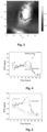
- FIG. 3 shows an image of uninfected and untreated cells (cell control) by atomic force microscopy (AFM). The seeding density of the cells was controlled so that several thousand cells determined the changes in plasmon resonance.
- HSV-1 cells As a viral model in antiviral experiment were used HSV-1 cells. As a positive control in antiviral experiments, the widely used antiviral drug was used Acyclovir- (2-amino-1,9-dihydro-9-((2-hydroxyethoxy)methyl)-6H-purin-6-one), ACV) ( Sigma-Aldrich, Germany ). Measurements were made on every 2 hours until the 32-nd hour of viral infection development, the last hours coinciding with the initial cytopathic cellular changes. As a rule, the increase/ decrease of the PPR signal is a consequence of the increase/ decrease of the effective index of refraction of the cell monolayer as a result of the morphological changes in the cells caused in the process of viral replication.
- Fig. 4 The stages of the replication cycle- adsorption, penetration of the virus into the cell, changes in cellular metabolism, as well as the assemblyand release of the daughter offspring, cause such changes and accordingly generate a PPR signal.
- IPF-H2A acts at the time of virus penetration into the cell. There is no characteristic increase in the signal in the range of 8-16 hours associated with the presence of viral capsids in the cytoplasm, which indicates the absence of virus. The difference in PPR signals becomes clear after the 14-th hour. This completely coincides with the inhibition of the process of viral replication, which corresponds to the mechanism of action of the drug. After the 20th hour, there is no increase in the SPR signal, as there are no newly created virions.
- Example 4 Immunological examination of coded blood samples of patients with HIV-1
- the average (min- max) values of the examined populations are summarized on Table 1.
- the absolute number of CD 4 T lymphocytes is (average, min-max) 528 (116-1261)cells/ ⁇ l and the percentage is 22 (4-44)%.
- CD4AC In practice in 17 patients (55% of the examined) is observed increase of CD4AC. In 9 of them (29% of the examined) it is accompanied by increase of the relative share of CD4and the CD4/CD8 index that corresponds to decrease of the immune activation. In four patients (13% of the examined) is reported decrease of CD4. Only in one patient the decrease of CD4 AC is accompanied also by decrease of the percentage of CD4 cells and the CD4/CD8 index. In the rest 10 patients (29% of the examined) there is no significant change in the absolute number of CD4 in comparison with baseline examination. In three of them is observed increase of CD4% and CD4/CD8 index.
- Coronaviruses are family of a single chain RNA viruses. In January 2020, the family comprises 40 species combined in two subfamilies: Orthocoronavirinae and Letovirinae. Most of them infect animals like bats, chickens, camels, and cats. Sometimes viruses who infect one species may mutate in such a way that they become able to infect another species. That's so called "cross-transmission of species" or "overflow”. To date seven coronaviruses can cause diseases in humans. They belong to Orthocoronavirinae subfamily.
- Coronaviruses cause diseases that vary from mild cold (HCoV-NL63, HCoV-229E, HCoV-OC43 and HCoV-HKU1) to more severe diseases likeMiddle East respiratory syndrome (MERS- CoV), severe acute respiratory syndrome (SARS-CoV) and the new Covid-19 (SARS- CoV-2).
- Coronaviruses HCoV-229E, -NL63, -OC43-HKU1 constantly circulate in human population and cause respiratory infections with basic symptoms fever and sore throat, swollen lymph nodes, bronchitis, rarely pneumonia in winter and early spring in adults and children. They rarely are with fatal outcome.
- There are no vaccines or antiviral medicines that have been approved for prophylaxis or treatment of corona viruses.
- the study provides dissolving of IPF-H2A peptide complex to initial concentration of 5 mg/ml (starting solution), from which ex tempore are prepared series solutions with nutritional medium DMEM (Dulbecco's Modified Eagle Medium, Sigma- Aldrich, Germany), containing 2% fetal beef serum (FBS, Gibco) and 1% antibiotic solution (100 U/ml penicillin and 100 ⁇ g/ml streptomycin sulphate (Gibco, USA)) (operational solutions), that are filtrated throughsterile antibacterial filters with pore diameters 0.45 ⁇ m (Sartorius Stadium Biotech, Australia).
- starting solution initial concentration of 5 mg/ml (starting solution)
- DMEM Dulbecco's Modified Eagle Medium, Sigma- Aldrich, Germany
- FBS fetal beef serum
- antibiotic solution 100 U/ml penicillin and 100 ⁇ g/ml streptomycin sulphate (Gibco, USA)
- a model system monolayer of human embryonic lung (Lep) cell line.
- the cells are cultivated in DMEM with added thermally inactivated 10% FBS, 1% Sodium pyruvate (Sigma-Aldrich, Germany) and antibiotics (penicillin- 100 U/ml), streptomycin sulphate (100 ⁇ g/ml) (growth medium) and the cell cultures are incubated at 37°C in 5% CO2 and in the presence of the required humidity.
- the reference strain of human coronavirus HCoV-229E collection of "Robert Koch” Institute, Germany.
- the viral suspension is cultivated in maintenance nutritional medium DMEM, containing 2% FBS, 1% antibiotic solution (100 U/ml penicillin G sodium and 100 ⁇ g/ml streptomycin sulphate).
- the infectious viral titer is accounted in two methods: 1. Reed and Muench method of final dissolutions (1938) and is expressed as TCID 50 (50% Tissue Culture Infectious Dose) (reciprocal viraldissolution in which 50% of the cell culture is infected) and 2. colorimetrical MTT test. After triple freezing and thawing the obtained virus-containing material is centrifuged (2500 rpm/min, 15 min at 4°C), aliquoted and frozen at -80°C.
- oseltamivir phosphate As a positive control in antiviral experiments is used oseltamivir phosphate (trade name Tamiflu).
- Each capsule contains 75 mg oseltamivir phosphate, corresponding to 75 mg oseltamivir, that initially is dissolved in PBS (stock solution with initial concentration 20 mg/ml) which serves for preparation of series operational solutions.
- Cell freezing and thawing has been performed according to the generally accepted methods as described by Pegg D., 2007.
- the cells are frozen in concentration at least 1 ⁇ 10 6 cells/ml.
- the ampoules are quickly put in a vessel with warmed to 37-40°C water and after thawing they are immediately transferred in a nutritional medium containing 10% FBS.
- the nutritional medium is substituted by new immediately after the cell adhesion to the padding (not later than 20 hours after seeding).
- the cell monolayer is contaminated with the prepared viral dilutions in volume 0.1 ml. With each viral dilution are contaminated 4 wells. The virus adsorbs for 1 hour in a thermostat at temperature 33°C, thereafter to each well are added 0.1 ml maintenance nutritional medium. For cell control serve four wells in which are placed uncontaminated cells. The processed in this way plate is incubated in a thermostat at temperature33°C and the required humidity. In occurrence of cytopathic effect (CPE) (occurrence of rounded cells irregularly disseminated in the cell monolayer tending to formation of foci) is waiting till development of 2 -3 + (50 - 75% of the cell monolayer) or till the process stops.
- CPE cytopathic effect
- the viral titer is expressed as TCID 50 (50% Tissue Culture Infectious Dose).
- T logarithm of the dilution at which the % of contamination is > 50 % + reciprocity factor ⁇ logarithm of the dilution factor .
- Proportionality factor % of contamination is > 50 % ⁇ 50 % / % of contamination is > 50 % ⁇ % of contamination is ⁇ 50 % .
- the antiviral action of the tested substance is determined by Mosmann MTT test for determination the of cell survival, modified by Shigeta S. for quick screening for anti-influenza effect of compounds under the influence of the viral cytopathic effect the cells die.
- the determination of cell viability through MTT test of contaminated with virus and treated with the substance cells is indicative for the antiviral effect of the substance itself.
- the volume of the inoculated in a well viral suspension is 0.1 ml, in operational dose 100 TCID 50 .
- the virus adsorbs for 1 h in a thermostat at temperature 33°C, thereafter toeach well is added 0.1 ml maintenance nutritional medium.
- During the viral adsorption are prepared dilutions of the tested substance. After the end of the viral adsorption the plate is processed as follows:
- the cell cultures are incubated in a thermostat at 33°C 5% CO 2 and are observed every day under light microscope for development of CPE.
- the effect of the action of IPFH2-A is accounted after the contamination ( p.i. ).
- the infectious titer is calculated according to Reed and Muench method.
- the calculations are performed through the programs Graph Pad Prizm software version 8.00 for Windows, LaJolla California USA and Origin 6.1 TM .
- Inhibitory concentration 50% is the concentration of the test substance that inhibits with 50% the virusinduced cytopathic effect as is directly determined from the "dose-response" curve.
- the first important stage of the antiviral experiments includes determination of the cytotoxicity of the tested substance on Lep line cells, cultivated in laboratory conditions.
- MTT analysis of the cell survival and proliferation was performed in a wide range of concentrations of the tested substance. The limits within which varied the applied concentrations have been selected on the grounds of preliminary experiments that included larger concentration intervals in order more precise determination of CD 50 and MNC values.
- the test substance is applied in concentration range 0.0001- 1 mg/ml, while the anti-nfluenza medicine Tamiflu ® - 0.001- 7.5 mg/ml for 96 hours.
- the untreated Lep cells whose survival was accepted as 100%, were used as a negative control. From the applied MTT test are obtained dose-response curves of the influence of the applied IPF-H2A complex on the cell monolayer.
- the analysis of the cell morphology surveillance is synchronized with the MTT analysis of the cell surveillance.
- the tested IPFH2-A complex displays no toxicity on cells of Lep line.
- IPF-H2A complex has been examined for antiviral effect on HCoV-229E in cell cultures.
- concentrations within the limits from 0.001 mg/ml to 1 mg/ml (MNC and close to it).
- MNC 0.001 mg/ml to 1 mg/ml
- Teamiflu oseltamivir phosphate
- IC 50 inhibitory concentration leading to 50% inhibitionof the viral cytopathic effect
- the antiviral effect of the tested substance IPF-H2A peptide complex has been assessed through CPE inhibitiontest.
- CPE inhibitiontest After cell contamination with HCoV-229E the CPE occurs after approximately 96 hours and is expressed with occurrence of rounded cells irregularly disseminated in the cell monolayer tending to form foci.
- the cell cultures are daily observed for CPE development under light microscope.
- the effect of the action of the tested substances is accounted 96 hours (the time of the replication cycle of coronaviruses) after Lep cell contamination with HCoV-229E (p.i.).
- the results of the performed experiments are assessed in comparison with cells uncontaminated with virus and without addition of substance (cell control) as well as with cells contaminated with human coronavirus 229E strain without addition of substance (virus control).
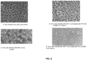
- the contaminated withHCoV-229E cells are with a typical rounded shape, clustered like "islands”, isolated each of other and separated from the padding on which they are cultivated.
- concentrations close to the minimum effective were applied.
- IPF-H2A complex The inactivated pepsin fragment (IPF-H2A complex) effectively inhibits the infectious viral yield in concentration range 0.001-1 mg/ml. Applied in MNC (0.1 mg/ml) it reduces the viral yield with ⁇ log 10 1.3, asits MIC 50 is 0.01 mg/ml (Table 3). Despite that, the anti- influenza medicine Tamiflu effectively inhibits HCoV-229E replication in larger concentration range 0.00001-1 mg/ml (Table 4).
- Inhibitory concentration 50% is the concentration of the tested substance that inhibits with 50% the virus-induced cytopathic effect as it is directly determined from the "dose-response" curve.
- the principle chemotherapeutic indicators IC 50 of the tested substances are accounted based on the experimental results.
- the results according to IC 50 allow a hierarchical scheme to be drawn as follows: For HCoV-229: Tamiflu ® > IPF-H2A peptide complex
- herpes viruses A common feature of herpes viruses is that after having infected the host they cause a latent, chronic infection. Under the influence of stress factors, the latent herpes virus gets reactivated, causing diseases with an extremely diverse clinical symptoms- from asymptomatic infections and self-limiting, transient labial and genital lesions (HSV-1 and HSV-2), through retinitis, keratitis, keratoconjunctivitis (HSV-1, HSV-2, CMV), to severe generalized infections, encephalitis, meningitis, including malignant neoplasms (HSV-1/2, EBV, HHV-8).
- HSV-1 and HSV-2 transient labial and genital lesions
- HSV-1, HSV-2 keratitis
- keratoconjunctivitis HSV-1, HSV-2, CMV
- HSV-1, HSV-2 keratoconjunctivitis
- IPF-H2A was dissolved to an initial concentration of 50 mg/ml (background solution).
- background solution a series of dilutions are done ex tempore with a nutrient medium DMEM ( Dulbecco's Modified Eagle Medium, Sigma-Aldrich, Germany ), which includes 2% fetal bovine serum (FBS, Gibco) and 1% antibiotic solution (100 U/ml penicillin and 100 ⁇ g/ml streptomycin sulphate (Gibco, USA )) (working solutions), which solutions are then filtered through sterile antibacterial filters with a pore diameter of 0.45 ⁇ m.
- DMEM Dulbecco's Modified Eagle Medium, Sigma-Aldrich, Germany
- antibiotic solution 100 U/ml penicillin and 100 ⁇ g/ml streptomycin sulphate (Gibco, USA )
- Cells were cultured on DMEM nutrient medium with termoinactivated 10% FBS, 1% sodium pyruvate ( Sigma- Aldrich, Germany ) and antibiotics (penicillin (100 U/ml), streptomycin sulphate (100 ⁇ g/ml) (growingmedium). The cell cultures were incubated at 37 °C at 5% CO2 and the required humidity. The blending of the Vero cells was done at 1:3 - 1:5 at a density of about 2 ⁇ 10 5 cells/ml, then resuspended several times and transferred onto cell culture mattresses (25 cm 2 ).
- the monolayer was washed three times with phosphate-buffered saline (PBS) with pH 7.4 for 1- 2 minutes, after which the solution was removed. All experiments were performed during the exponential phase of cell growth.
- PBS phosphate-buffered saline
- Viral strains were cultured in DMEM nutrient medium containing 2% FBS,1% antibiotic solution (100 U/ml penicillin G sodium and 100 ⁇ g/ml streptomycin sulphate).
- Infectious virus titer was account for, also by Reed and Muench's method of final dilutions, and expressed as TCID 50 (50% Tissue Culture Infectious Dose) and also quantitatively through the colorimetric MTT method. After triple freezing and thawing, the resulting virus- containing was centrifuged (2500 rpm/min, 15 min at 4 °C), aliquoted and frozen at -80°C.
- Acyclovir 2-amino-1 ,9-dihydro-9-[(2-hydroxyethoxy)methyl]-6H-purin-6-one (Sigma- Aldrich, Germany), which was initially dissolved in PBS (stock solution with a background concentration of 1 mg/mL), and it was used for a series of working dilutions.
- the cells After blending the culture, conducting a Trypan Blue Dye Exclusion Test, freezing and thawing, the cells are transferred to a nutrient medium containing 10% FBS and proceeded to determination of the cell viability. The latter is conducted by a Microscopic observation of morphological changes in the monolayer of treated cells and Colorimetric MTT analysis according to Mosmann, 1983, with minor modifications.
- ten-fold logarithmic dilutions of the relevant virus strain were prepared in DMEM nutrient medium containing 2% FBS, 1% antibiotic solution (100 U/mL penicillin G sodium and 100 ⁇ g/mL streptomycin sulphate).
- the nutrient medium was removed from the wells.
- the cell monolayer was infected with the prepared viral dilutions of 0.1 mL. With each viral dilution, 4 wells were infected.
- the virus was adsorbed for 1 h ina thermostat at a temperature of 37°C, then to each well was added 0.1 mL of nutrient medium. Four wells containing uninfected cells were used for cell control. Thus treated plate was incubated in a thermostat at a temperature of 37°C and at the required humidity. In the event of a cytopathic effect (CPE) (appearance of round cells, unevenly disseminated in the cell monolayer, with a tendency to formfoci), it was waited until the development of 2-3 + (50 - 75% of the cell monolayer) or until it stopped progressing.
- CPE cytopathic effect
- the viral infection titer is expressed as TCID 50 (50% Tissue Culture Infectious Dose).
- T log of dilution , where % infected is over 50 % + proportionality factor ⁇ log of dilution factor .
- Proportionality factor % infected over 50 % ⁇ 50 % % infected over 50 % ⁇ % infected below 50 % .
- the antiviral activity of the test substance was determined by the Mosmann MTT cell survival test, modified by Shigeta S. for rapid screening of compounds for anti-influenza activity. Under the influence of the cytopathic action of the virus, the cells die. Determination of cell viability by MTT - a test of virus-infected and substance-treated cells, is indicative of the antiviral action of the substance itself.
- the 50% inhibitory concentration is the concentration of the test substance that inhibits the virus-induced cytopathic effect by 50%, as determined directly from the dose-response curve.
- SI CD 50 / IC 50
- the MTT analysis of cell survival and proliferation was performed in a wide range of concentrations of the test substance.
- the limits within which the applied concentrations varied were selected on the basis of preliminary experiments involving larger concentration intervals in order to accurately determine the values of CD 50 and MNC.
- the test substance was administered in the concentration range 0.0001- 20 mg/mL and the anti-herpes preparation ACV-0.0005- 0.1 mg/mL for 72 h.
- Dose-dependent curves on the viability of the cell monolayer of the applied IPFH2-A were obtained from the MTT Assay used.
- the analysis during the observation of the cell morphology was synchronized with the MTT analysis of cell survival.
- test substances Based on their cytotoxicity, the test substances are arranged in the following hierarchical order and:
- the tested IPF-H2A does not exhibit toxicity on the cells of the Vero line after 72 h of treatment, as applied in a concentration of 0.0001 mg/mL to 5 mg/mL.
- the decrease in the number of adherent living cells, as well as the rounding and in the size decrease of the cells follow the trend established during the MTT analysis.
- Visualization of changes in the morphology of the cell monolayer after treatment is an accessible and reliable approach in the initial study of the cytotoxicity of various substances.
- changes in the morphology of the cells exposed to the tested IPFH2-Aand the antiviral drug ACV were observed under an inverter light microscope.
- the non-treated controlcells and the cells treated with IPF-H2A in the concentration range 0.0001-5 mg/mL retain their morphology and structure, while the treated cells with the anti-herpes preparation ACV at the highest concentrations (1- 20 mg/mL) acquired a round shape, got reduced in volume and disintegrated from the monolayer.
- IPFH2-A and anti-herpes preparation ACV on survival/proliferative activity ofcells of the Vero line, reported by various methods after 72 h of treatment is summarized on Table 5 below.
- Table 5 IPF-H2A and ACV Method MNC (mg/mL) CD 50 (mg/mL)
- IPF-H2A Microscopic observation of changes in the morphology of the cell monolayer 0.1 ⁇ 0.1 15 ⁇ 0.05 MTT Assay 0.1 ⁇ 0.12 15 ⁇ 0.22
- ACV Microscopic observation of changes in the morphology of the cell monolayer 0.05 ⁇ 0.6 0.5 ⁇ 0.02 MTT Assay 0.05 ⁇ 0.07 0.5 ⁇ 0.1
- the IPF-H2A peptide complex was tested for its antiviral effect on HSV-1/HSV-2 in cell cultures.
- concentrations were administered ranging from 0.001 mg/ml to 1 mg/ml (MNC and close to it).
- the anti-herpes preparation ACV was used for positive control in the antiviral experiments, applied in the concentrationrange of 0.05 - 0.5 mg/mL.
- Dose-dependent curves were derived to determine the inhibitory concentration leading to 50% inhibition of the viral cytopathic effect (IC 50 ).
- the antiviral activity of the IPF test substance was assessed by a CPE inhibition test.
- CPE After infection of the cells with HSV-1/HSV-2, CPE develops in about 72 hours and resultsin the appearance of round cells, unevenly disseminated in the cell monolayer, with a tendency to form foci and the formation of syncytia.
- Cell cultures were observed daily under a light microscope as to theCPE development.
- the effect of the test substances was observed for 72 h (the time for one replicate cycle of coronaviruses) after infection with HSV-1/HSV-2 in Vero cells (p.i.).
- Concentrations around and close to the minimum effective ones were used to determine the anti-HSV-1/HSV-2 activity of the IPF-H2A test substance.
- Extracellular virus samples were titrated in a 24-hour Vero monolayer in 96-well plates, by taking into account the infectious viral titer in log10, according to the final dilution method of Reed & Muenh. Differences in infectious viral titers between experimental and control samples are expressed as ⁇ log. Based on the reported results, dose-response graphs were plotted and minimum inhibitory concentrations 50 (MIC 50 ) were determined. Values of ⁇ log10 ⁇ 1 are considered to be reliable results for the performed tests.
- Table 6 Viral strain Concentration (mg/mL) Viral yield (log 10 TCID 50 ) ⁇ log1 0 HSV-1 (Victoria strain) 0 5.833 ⁇ 0.000 0 0.0 5.567 ⁇ 0.288 0.8 001 0.0 4.520 ⁇ 0.166 0.8 01 0.0 4.500 ⁇ 0.002 1.0 1 0.1 4.353 ⁇ 0.068 1.0 1 4.000 ⁇ 0.442 1.3
- HSV-2 (Bja strain) 0 6.000 ⁇ 0,166 0 0.0 5.800 ⁇ 0.022 0 001 0.0 5.567 ⁇ 0.288 0 01 0.0 4.500 ⁇ 0.015 0.8 1 0.1 4.600 ⁇ 0.288 0.8 1 4.542 ⁇ 0.500 1.0
- the IPF-H2A complex effectively inhibits the yield of infectious viral progeny in the concentration range of 0.0001- 1 mg/mL.
- MNC 0.1 mg/mL
- its MIC50 is 0.0001 mg/ml.
- the anti-influenza drug ACV effectively inhibited the replication of HSV-1/HSV-2 in a larger concentration range of 0.00001- 1 mg/mL.
- the individual effect of the anti-herpes preparation ACV on the reporting of the infectious titer (log10 TCID 50 / 0.2 ml) is presented in Table 7 below.
- the 50% inhibitory concentration (IC 50 ) is the concentration of the test substance that inhibits the virus-induced cytopathic effect by 50%, as derived directly from the dose-response curve. Based on the experimental results, the main chemotherapeutic parameter IC 50 of the tested substances has been reported.
- the IC 50 values are an indicator of the antiviral efficacy of a substance, and S! provides information on its specificity and selectivity.
- Viral strain Concentration (mg/mL) Viral yield (log 10 TCID 50 ) ⁇ log 10 HSV-1 (Victoria strain) 0 5.300 ⁇ 0.242 - 0.0005 4.500 ⁇ 0.763 0.8 0.005 4.530 ⁇ 0.166 0.8 0.05 4.300 ⁇ 0.763 1.0 0.1 4.000 ⁇ 0.288 1.6 1 2.7 ⁇ 0.500 2.6 HSV-2 (Bja strain) 0 0 0 0.0005 5.567 ⁇ 0.000 - 0.005 5.300 ⁇ 0.288 0.8 0.05 4.500 ⁇ 0.125 1.0 0.1 4.300 ⁇ 0.237 1.0 1 4.005 ⁇ 0.166 1.3
- Table 8 Antiviral Activity HSV-1 (Victoria strain) HSV-2 (Bja strain) % cellular protection at MNC IC 50 (mg/mL) ⁇ SEM SI % cellular protection at MNC IC 50 (mg/mL) ⁇ SEM SI IPFH2-A 90.5 0.0001 ⁇ 0.08 1000 30.5 0.001 ⁇ 0.03 100 ACV 100 0.0005 ⁇ 0.02 800 90.7 0.005 ⁇ 0.12 80
- IPFH2-A effectively inhibits the replication of the Victoria strain (HSV-1) at a concentration of 0.0001 mg/mL (with 90.5% cellular protection).
- HSV-1 strain Victoria with 100% complete protection
- HSV-2 strain Bja has proven to be 10 times less sensitive.
- Example 7 Antigen-specific therapeutic vaccine based on the IPF-H2A peptide complex
- Tumor associated antigens have now been identified for several cancers including adenocarcinoma of the breast, colon, and pancreas, as well as for malignant melanoma. These antigens can be classified by tissue distribution and function, shown on Table 9. Many of these antigens have been found to be shared among other patients' tumors of the same histologic site. These shared antigens are potential candidates for vaccine development. Several of these antigens are binding in vitro or mixed with defined IPF-H2A or pulsed directly onto autologousdendritic cells (Professional APCs) that are activated in vivo in each patient.
- IPF-H2A defined immunologousdendritic cells
- Future immunotherapy of cancer approaches will employ newly identified tumor-associated antigens bind with IPF-H2A that are delivered in patients with different type of cancer.
- the conduction of well controlled clinical trials will be necessary to identify the most effective approaches for the treatment of advanced cancer. More than likely, it will be necessary to combine vaccine approaches with other cancer treatment as IL-10 cytokines, Th1 MHC class II antigens, and co-stimulatory molecules macrophages modalities to effectively treat larger metastases.
Landscapes
- Health & Medical Sciences (AREA)
- Life Sciences & Earth Sciences (AREA)
- Chemical & Material Sciences (AREA)
- General Health & Medical Sciences (AREA)
- Organic Chemistry (AREA)
- Medicinal Chemistry (AREA)
- Engineering & Computer Science (AREA)
- Proteomics, Peptides & Aminoacids (AREA)
- Bioinformatics & Cheminformatics (AREA)
- Veterinary Medicine (AREA)
- Public Health (AREA)
- Pharmacology & Pharmacy (AREA)
- Animal Behavior & Ethology (AREA)
- Genetics & Genomics (AREA)
- Gastroenterology & Hepatology (AREA)
- Epidemiology (AREA)
- Molecular Biology (AREA)
- Biochemistry (AREA)
- Zoology (AREA)
- Immunology (AREA)
- Biomedical Technology (AREA)
- General Chemical & Material Sciences (AREA)
- Wood Science & Technology (AREA)
- Microbiology (AREA)
- Chemical Kinetics & Catalysis (AREA)
- Biophysics (AREA)
- Virology (AREA)
- Communicable Diseases (AREA)
- Oncology (AREA)
- Mycology (AREA)
- Nuclear Medicine, Radiotherapy & Molecular Imaging (AREA)
- Biotechnology (AREA)
- Toxicology (AREA)
- General Engineering & Computer Science (AREA)
- Medicines That Contain Protein Lipid Enzymes And Other Medicines (AREA)
Abstract
The invention provide a peptide complex IPF-H2A for modulation of the immune function of the human body and for antigen- specific immunotherapy. The peptide complex IPF-H2A, having SEQ ID: NO. 1, provides a new approach for treating immune disorders, viral diseases, cancer and immune related implications and also for antigen- specific immunotherapy
Description
- The present invention relates to a peptide complex IPF- H2A applicable for modulation of the immune function of the human body and for antigen- specific immunotherapy.
- Major Histocompatibility Complex (MHC) is a key component of T cell immunity. There are two types ofMHC- class I and class II. Professional antigen presenting cells (APC) include B cells, macrophages and microglia. B cells express MHC class II. Once antigen has been bound by the Ag receptor on the B cell, the Ag and receptor are engulfed into endosomal compartment. This compartment fuses with another compartment- lysosome. The B cells are efficient and breaking down Ag into smaller parts and loading the parts of MHC II in the lysosome. The MHC then trafficked to the cell surface where B cell "shows" the Ag to a CD4 T cell. CD4 cells are two categories- Th1 and Th2. B cell and CD4 cell encounter in the groove (antigen binding location in the MHC molecule) T cell receptor to recognize antigenic peptide by class I MCH on the surface of APC. Flexibility in both T-cell receptor's (TCR's and peptides plays key role in antigen recognition and discrimination. Flexibility of peptide binding groove impacting region contacted by TCR as well as other activating and inhibitory receptors of the immune system. Altered MHC flexibility will influence receptor engagement associated with receptor recognition. The "altered self" mechanism of immune recognition has implication for specificity cross-reactivity and antigenicity in cellular immunity (W. F. Hawse- 6.8.2013). Class I MHC proteins reflecting intrinsic class II MHC dynamics that could be modulated by peptides.
- Peptides can modulate the flexibility of the MHC complex. Tuning of MHC dynamics by peptides reflects a fundamental mechanism for modulating the immune system. Single cell network profiling (SCNP) is a technology that quantifies functional immune signaling capacity and connectivity at a systems biology level.
- The blocking of cellular receptors by chemical molecules or specific antibodies is possible. It has been recognized that denying entry into CD4+ cells to the HIV-1 virus could at least slow the progress of the infection and alleviate, if not cure, the disease and/or its symptoms. The complex mechanism by which the virus crosses the cell membrane has been widely investigated. Broadly, the entry of human immunodeficiency virus into, for example, CD4+ Th1 cells (T-
helper type 1 cells), is dependent upon a sequential interaction of the gp120/gp41 subunits of the viral envelope glycoprotein gp160 with the CD4+Th1 cell surface glycoprotein and the cell surface receptor CCR5. On binding of gp120 with its cell surface binding sites, a conformational change in the latent gp41 subunit through an intermediate state to an active state is initiated, inducing fusion of the viral and cellular membranes and transport of the virus into the cell (Nature 387:426-30, 1997). - Accordingly, numerous binding experiments have been conducted in an effort to find antiviral ligands that will effectively compete with the HIV-1 for CD4+ gp and/or CCR5 binding sites, or that will preferentially block gp120 and/or gp41 binding domains. In one example, a reported structure (X-ray crystallography) comprising an HIV-1 gp120 core complexed with a two-domain fragment of human CD4 and an antigen-binding fragment of a neutralizing antibody that blocks chemokine-receptor binding, is said to reveal a CD4-gp120 interface, a conserved binding site for the chemokine receptor, evidence for a conformational change on CD4 binding, the nature of a CD4-induced antibody epitope, and specific mechanisms for viral immune evasion, "which should guide efforts to intervene" (Nature 393 (6686):632-1, 1998). Also, it has been shown that inhibition of the change in structure of gp41 from its intermediate to active state with peptides used as competitors for critical cell receptors may reduce viral load, and that while gp120 masks epitopes on the gp41 subunit in its latent state, gp41 may be vulnerable to neutralizing antibodies in its transient or intermediate state (Molecular Membrane Biology 16: 3-9, 1999). In another study, disclosed in patent
US 2004/0018639 , a protein designated "Thymus Factor" ("TF") is stated to have the ability to bind to a fragment of HIV-1 gp41 in gel electrophoresis, and that this binding property can be used to assay TF activity or in the treatment of HIV. - In addition, it is known by patent
EP1830867 (A2 ) that the irreversibly inactivated pepsin fraction (IPF) having amino sequence GDEPLENYLDTEYF (-Gly-Asp-Glu-Pro-Leu-Glu-Asn-Tyr-Leu-Asp-Thr-Glu-Tyr-Phe-), effectively blocks gp41 and gp120 domains essential for viral entry into CD4+ cells and inhibit viral infection, in vivo and in vitro. According to the invention, this structure (IPF) is used to diagnose viral infection, particularly HIV-1 infection and as a prophylactic or therapeutic to prevent or to treat HIV infections. The inventor tested this compound for its ability to stimulate human natural killing T (NKT) cell lines, secretion of key cytokines such as IFN- IL2 and IL-12, and to activate autologous dendritic cells, as well as binding to CD1d and the invariant T-cell receptor. This protein exhibits a stronger adjuvant effect in various HIV vaccine platforms in mice. IPF also provides a protective adjuvant effect with a candidate HIV and HCV vaccine. While the majority of the studies performed focus on the potential of the IPF as a vaccine adjuvant, it is foreseeable that the compound could also be used as a potential immunotherapeutic to treat cancer, infectious disease, and autoimmune diseases. - The histones are conservative proteins and significant components in the chromatin structure of the cells. They are highly basic proteins rich in lysine and arginine residues and play an important role in various cellular functions such as immunity. There are five families of histones which are designated H1/H5 (linker histones), H2, H3, and H4 (core histones). Histones may be chemically modified through the action of enzymes to regulate gene transcription. The epigenetic modifications of histones contribute to their homeostatic functions of the cell, one of which is apoptosis. The research results of Williams, W. M. at al. (Clin Exp Immunol 1996, 104: 18-24) on the correlation between expression of antibodies of histone H2B and clinical activity in HIV- infected individuals, particularly those with symptomatic disease, revealed a significantly elevated titres of anti-H2B antibodies. Because the titres of H2B antibodies were found to parallel the numbers of circulated CD4 cells, the authors presume that they are intimately involved in the sequence of events that leads to the dysfunction and elimination of the helper/ inducer T lymphocyte population.
-
-
Fig. 1 - Presents the binding of IPF-H2A complex to ovarian cancer human sera molecules -
Fig. 2 - Shows adjusted sensogram, presenting the specific signal in the positive 1:25 serum sample -
Fig. 3 - Shows image of uninfected and untreated cells (cell control) by atomic force microscopy (AFM) -
Fig. 4 - Shows the changes of the PPR signal -
Fig. 5 - Shows the differences in the kinetics of the antiviral activity of ACV and IPF-H2A -
Fig. 6 - Shows the effect of IPF-H2A complex on the survival/ proliferative activity of cells of Lep line accounted through MTT analysis after 96 hours of treatment -
Fig. 7 - Shows the protection (%) of Lep cells, contaminated with HCoV-229E and treated with the IPF-H2A complex and Tamiflu -
Fig. 8 - Shows the statistically significant survival differences between untreated control and treated with IPF-H2A complex cell cultures infected with HSV-1/ HSV-2 -
Fig. 9 - Presents the inhibitory effects of IPF-H2A and of the anti-herpes preparation ACV - The invention provide a peptide complex IPF-H2A for modulation of the immune function of the human body and for antigen- specific immunotherapy. The peptide complex, according to the invention, provides a new approach for treating immune disorders, viral diseases, cancer and immune related implications and also for antigen- specific immunotherapy.
- According to one preferred embodiment of the present invention, the peptide complex IPF-H2A is a result of spontaneous binding (e.g. covalently bonded) of the isolated from lyophilized porcine Irreversible Pepsin Fraction (IPF) and the unfractionated whole Histone type 2-A (H2A). Thus, the binding of IPF structure having amino sequence GDEPLENYLDTEYF (SEQ ID: NO. 2) and the H2A structure having amino sequence NVPVSVEGYQITLDSIT (SEQ ID: NO. 3) leads to the structure, that of IPF-H2A peptide complex, representing a linear peptide with amino acid sequence GDEPLENYLDTEYFNVPVSVEGYQITLDSIT (SEQ ID: No. 1).
- The Irreversible Pepsin Fraction (IPF) and the unfractionated whole Histone type 2-A (H2A) could be derived also from any other source, containing these sequences. For isolating and sequencing the components of IPF-H2A complex could be used common laboratory methods and reagents.
- The peptide with amino acid sequence GDEPLENYLDTEYFNVPVSVEGYQITLDSIT (SEQ ID: No. 1) provides a novel mechanism to reduce viral burden and stimulate innate immune responses to the virus for patients with significant antiretroviral resistance; it appears to modulate helper T1 cells' expression of elaborate cytokines INFy, IL-2, which selectively promote cell- mediated immune response and subsequently stimulate cytotoxic lymphocytes. These lymphocytes have a prominent role in the host's immunologic response to viral infections. Proteins encoded by these pathogens enter the endogenous pathway for antigen presentation and are expressed on the surface of infected cell as a complex with class I MHC proteins. Peptide modulation of class I MHC in both TCR-s (T- cell receptor) plays a significant role in antigen recognition. Complex molecule of IPF-H2A and CD4 receptors reactivate immune system after activating Th1 Th2 cellular immune answer. Flexibility in both TCR's and peptides plays key role in antigen recognition and discrimination. Flexibility of peptide binding groove impacting region contacted by TCR as well as other activating and inhibitory receptors of the immune system. Altered MHC flexibility will influence receptor engagement associated withreceptor recognition. "Altered self" mechanism of immune recognition has implication for specificity cross-reactivity, and antigenicity in cellular immunity (W. F. Hawse - 6.8.2013).
- Peptides can modulate the flexibility of the MHC complex. Tuning of MHC dynamics by peptides reflects a fundamental mechanism for modulating immune system. SCNP (Single Cell Network Profiling) plus CD4 complex form complex molecule reactivating Th1 cellular immune answer. Th1 cells secrete IL, IFN alpha, which are responsible for mediatingdilate type hypersensitivity reaction. Th2 cells secrete
IL 2, IL4, IL5, IL6 and IL10, which are involved in, inducing humoral immune response. IPF-H2A peptide complex can modulate the flexibility of the region which dynamics reflects a fundamental mechanism for modulating immune response. - The blocking of cellular receptors by chemical molecules or specific antibodies is possible. The viralaggression uses one cellular receptor- CD4 and two co-receptors CCR5 and CXCR4. The first step in cell entry occurs when a glycoprotein on the viral surface (gp120) binds to surface receptors on the target cell. The next step is a new binding between envelop protein (for example HIV-1) and cell co-receptor CCR5. Once gp120 links to both receptors, gp41 (a trans-membrane viralcomplex) undergoes a conformational change and brings the viral membrane into proximity with the cell membrane. Fusion of two lipid delayers then occurs, thus allowing intracellular entry of viral contents.
- It has been discovered in accordance with the present invention that the protein complex, named IPF-H2A, demonstrate in vitro equal binding with HIV-1 envelope protein gp41 (non-glycosylated fragment 579-601). The results are demonstrated on sensograms by Biacore. Binding responses RV- resonance units are illustrated for the protein immobilized surface in response to gp41, gp120, gp160 CD4.
- Another preferred embodiment of the present invention is the immunomodulatory use of IPF-H2A complex and its use in the preparation of antigen- specific therapeutic vaccine, suitable for the treatment of several disorders including congenital defects involving primary T cell deficiency,viral infections, HIV, immunosuppression following cancer treatment, alone or in combination with antiviral disorders.
- IPF-H2A peptide complex is preferably delivered with adjuvant, or is chemically conjugated to an immunogenic carrier that would provoke an antibody response in the patient. Examples of immunogenic carriers are keyhole limpet and bovine serum albumin.
- In one aspect of the invention, IPF-H2A peptide complex is used to treat HIV infections, especially AIDS and ARC. For example, IPF-H2A is prepared for administration by mixing it at the desired degree of purity with adjuvant or physiologically acceptable carriers. Carriers, which are nontoxic to recipients at the dosages and concentrations are employed. Although IPF-H2A complex is desirably administered with an adjuvant, in situations where the initial inoculation is delivered with IPF-H2A complex, boosters with IPF-H2A may not require adjuvant. Injections (intramuscularly or subcutaneous) will be the primary route for therapeutic administration of the compositions of this invention. However, intravenous delivery, delivery through catheter, or other surgical tubing may also be used. Alternative routes include tablets and the like, liquid formulations, and inhalation of lyophilized or aerosolized receptors. Liquid formulations may be utilized after reconstitution from powder formulations.
- IPF-H2A peptide complex could be administered also via microspheres, liposome is another micro particulate delivery systems or sustained release formulations placed in certain tissues including blood. The dosage of IPF-H2A peptide complex administered will depend upon the properties of the formulation employed. Its binding activity and in vivo plasma half-life, the concentration of IPF-H2A complex in the formulation, the administration route, the site and rate of dosage, the clinical tolerance of the patient involved, the pathological condition afflicting the patient and the like, are well within the skills of the physician. Different dosages are utilized during a series of sequential inoculations: the practitioner may administer an initial inoculation and then boost with smaller doses of IPF-H2A complex.
- IPF-H2A peptide complex can be administered in several ways and to different classes of recipients. It can also be used to treat individuals who may or may not be at risk of exposure to HIV-1, and additionally, the IPF-H2A complex is desirably administered to seropositive individuals and to individuals who have been previously exposed to HIV-1. IPF-H2A peptide complex can be administered in combination with other antigens in a single inoculation "cocktail". IPF-H2A peptide complex can also be administered as in a series of inoculations administered over time. Such a series may include inoculation with the same or different preparations of HIV antigens or other vaccines. The adequacy of the treatment parameters chosen, e.g., dose, schedule, adjuvant choice and the like, is determined by taking aliquots of serum from the patient and assaying for Antibody and/or T cell titers during the treatment program. T cell titer may be monitored by conventional methods. For example, T lymphocytes can be detected by E-rosette formation as described in Bach, J.F., (Contemporary Topics in Immuno Biology, Vol. 2: Thymus Dependency, p. 189, Plenum Press, New York, 1973 ; Hoffman, T. & Kunkel, H. G., and Kaplan, M. E., et aL, both papers are in In Vitro Metlzo & in Cell Mediated and Tumor Immunity, B. R. Bloom & J. R. David Eds. Academic Press, New York (1976 )). For example, the amount of T cell rosette formation may be assayed after the third but before the tenth week of IPF-H2A peptide complex treatment. An over sixty-five percent rosette formation indicates a good cell mediated immune response in the patient.
- In addition, the clinical condition of the patient can be monitored for the desired effect, e.g., anti-infective effect. If inadequate anti-infective effect is achieved, then the patient can be boosted with further IPF-H2A peptide complex treatment and the treatment parameters can be modified, e.g., to potentate the immune response, such as by increasing the amount of IPF-H2A peptide complex and/or adjuvant, complexion IPF-H2A complex with a carrier or conjugating it to an immunogenic protein or varying the route of administration. IPF-H2A peptide complex may optionally be administered along with other pharmacological agents used to treat HIV-1 infections such as AIDS and ARC. Examples of these pharmacological agents are AZT, antibiotics, and immuno-modulators such as interferon, anti-inflammatory agents, and anti-tumor agents.
- The examples presented below illustrate the invention without limitation its scope of protection.
- According to one preferred embodiment, the IPF-H2A peptide complex of the invention is prepared by spontaneous binding (e.g. covalently bonded) of IPF, isolated from active pepsin according to procedure described by
Zhabilov, H. in patent US 7,479,538 B2 and the ready marketed unfractionated whole Histone type 2-A, in a ratio 10: 1. After preparation of the IPF-H2A complex, 5.6% aluminum adjuvant or IL2 is added. - The resulting IPF-H2A peptide structure represents a 42.4 kDa combined linear peptide with amino acid sequence GDEPLENYLDTEYFNVPVSVEGYQITLDSIT (SEQ ID: No. 1).
- The assessment of IPF-H2A activity was determined by Surface Plasmon Resonance (SPR) technology in Karolinska University Hospital, Stockholm, Sweden.
- A receptor assay has been developed and validated to characterize the quality of different batches of IPF-H2A peptide complex preparations. Since biological activity always depends on a first binding step, the primary criterion for assessing biological activity is the ability of a compound to bind specifically to a ligand. The most sensitive biosensor instrument for detection compound-ligand interaction is the BIACORE. It directly captures any proteins including molecules from cytosolic or tissue extracts on sensor surface and measure binding kinetics with considerable ease and precision. It is widely used as the most sensitive method for measuring the active concentration of biomolecules and for their quality control.
- SPR is an optical technique that measures the refractive index change occurring at the sensor-fluidinterface layer. A plastic fluidics system plate is pressed into contact with a gold-coated glass chip (CM5), the surface of which is coated with an un-cross-linked carboxymethylated dextran polymermatrix. This allowed forming micro reaction chambers or flowing cells. The gold partial mirror is the optical port as well. Experiments are performed by a computer-program-driven robotics systemto facilitate consistency. The injection of analyte across a ligand immobilized in the matrix produces a real-time change in refractive index signifying an increase in associated molecular mass.ln our work IPF-H2A preparation is the analyte, which binds to immobilized ovarian cancer human sera molecules. The data trace is a sensogram plotting response units (RU) against time (
Fig. 1 ). - In the given assay it was measured the direct binding of an active component of IPF-H2A complex sample (currently unidentified biomolecule/s) to an immobilized human ovarian cancer sera molecule, which are binding for IPF-H2A complex. Immobilization of ovarian cancer human sera on the CM5 sensor surface was performed by standard amine coupling. The Biacore system was equilibrated in running buffer (PBS, 0.05
% Tween 20, 1 m MEDTA, pH 8.4) at a flow rate of 5 ml/min. The carboxymethylated dextran matrix was activated by injection of 35 ml of a solution containing NHS (0.05 M)/ EDC (0.2 M) (50/50). Thirty-five microlitersof ovarian cancer human sera at 500 mg/ml in citrate buffer (pH 4; 0.01 M) was injected. The deactivation of the remaining NHS-ester groups was performed by injection of 35 ml of ethanolamine hydrochloride (pH 8.5; 1 M). A regeneration of the sensor surface was done by the injection of 5 ml of 1M NaCl and 0.1 M NaOH. The experiments were performed at a constant flow rate of 5 ml/min of running buffer. All the reagents were prepared by dilution in running buffer.Figure 2 shows kinetics of the specific binding and subsequent dissociation of IPF-H2A complex active component with immobilized ovarian cancer human sera. Maximal sample response (at the arrow) will be used in calculations and comparisons. The sensograms are performed on the Biacore system for one positive Ovarian cancer sample and one negative control where both samples have been diluted 1:25 and 1:100 for the serum: FC1 & FC2 are the negative sample diluted 1:25 and 1:100, respectively. FC3 & FC4 are the positive patient sample diluted the same. As could be seen on the sensogram ofFig. 2 , a specific signal in the positive 1:25 serum sample was found that is significant and distinct from the negative control; app 100RU. The 1:10 and the 1:100 peptide seems to be the best dilution of the serum. - The inhibitory mechanism of action of an IPF-H2A peptide complex was confirmed on the HSV-1 replication cycle by establishing the kinetics of biomolecular host cell-virus interaction as well as IPF-H2A-treated infected cells under single-step replicationcycle. The study was performed by the Surface Plasmon Resonance (PPR) method activated on a gold-plated diffraction grating. As a model system in the tests was used a Vero monolayer cell line (from monkey kidney), where the cells were seeded on a biochip representing a gold-plated diffraction grating. The groove of the diffraction grating provides a tight cell/ surface contact, which is very important for correct measurements.
FIG. 3 shows an image of uninfected and untreated cells (cell control) by atomic force microscopy (AFM). The seeding density of the cells was controlled so that several thousand cells determined the changes in plasmon resonance. - As a viral model in antiviral experiment were used HSV-1 cells. As a positive control in antiviral experiments, the widely used antiviral drug was used Acyclovir- (2-amino-1,9-dihydro-9-((2-hydroxyethoxy)methyl)-6H-purin-6-one), ACV) (Sigma-Aldrich, Germany). Measurements were made on every 2 hours until the 32-nd hour of viral infection development, the last hours coinciding with the initial cytopathic cellular changes. As a rule, the increase/ decrease of the PPR signal is a consequence of the increase/ decrease of the effective index of refraction of the cell monolayer as a result of the morphological changes in the cells caused in the process of viral replication. The stages of the replication cycle- adsorption, penetration of the virus into the cell, changes in cellular metabolism, as well as the assemblyand release of the daughter offspring, cause such changes and accordingly generate a PPR signal. The changes of the PPR signal are shown in
Fig. 4 where the inhibitory effect of ACV-treated cells under conditions of a single-cycle test for HSV-1 development: viral control (HCV-infected cells at MOI = 1); Infected and treated with ACV cells: at ACVconcentration = 0.001 mg/mL (MNC) - at ACV concentration = 0.01 mg / mL and in theFig. 5 , showing the differences in the kinetics of the antiviral activity of ACV and IPF-H2A. - Obviously, IPF-H2A acts at the time of virus penetration into the cell. There is no characteristic increase in the signal in the range of 8-16 hours associated with the presence of viral capsids in the cytoplasm, which indicates the absence of virus. The difference in PPR signals becomes clear after the 14-th hour. This completely coincides with the inhibition of the process of viral replication, which corresponds to the mechanism of action of the drug. After the 20th hour, there is no increase in the SPR signal, as there are no newly created virions.
- For the period 02.11.2015-14.05.2016 in the Specialized Hospital of Active Treatment of Infectious and Parasitic Diseases "Prof. I. Kirov", Sofia, were performed totally 115 immunological examinations and the indices are reported as follows: absolute number (AN) of the general lymphocytes, percentage, and absolute number of T (CD3+) lymphocytes, B (CD19+) lymphocytes and NK (natural killers) (CD4+CD3+) cells, percentage, and absolute number of T-lymphocyte subpopulations CD4 T (CD4+CD3+), CD8 T (CD8+CD3+) as well as their ratio (CD4/CD8).
- Complete follow-up course (three visits) has been done in 31 patients. Nine have given up after the first examination; 5 (five) patients have two examinations. There are also 3 (three) examinations done out of schedule. The results of the first, second and third examinations of patients with complete follow-up course are summarized. The results of all performed examinations are presented in Table 1 below.
- The average (min- max) values of the examined populations are summarized on Table 1. The absolute number of CD 4 T lymphocytes is (average, min-max) 528 (116-1261)cells/µl and the percentage is 22 (4-44)%. The distribution of patients according to the degree of immune deficiency at the baseline examination is as follows: 9% (n = 3) with severe immune deficiency (CD4 AC < 200); 32% (n = 10) with initial to moderate immune deficiency (CD4 AC 200 - 400) and 59% (n=18) without manifested immune deficiency (CD4 AC > 400). Respectively in the greater part of the patients (81%, n = 25) the CD8 Tcells are increased and the CD4/CD8 index is decreased below 0.9 (93.5%, n = 29).
- From the rest indices: lymphocyte number (average, min- max) 2.379 (0.604- 4.835) with deviation from normal in 22.5% (7 persons); 2 persons with decreased lymphocyte number (< 900) and 5 persons with lymphocytosis (> 3100); B cells (average, min-max) 0.221 (0.044-0.425) with deviation from normal in 71%, respectively: absolute decrease in52% (n = 16); relative decrease in 19% (n = 6); NK cells (average, min- max) 0.341 (0.07- 0.846) with deviation from the absolute number in 10% (n = 3), respectively- below the lower reference limit in two and above the upper reference limit in one patient; the relative share is decreased in one patient and is above the upper limit- in two.
- The results are summarized in Table 1. It is observed increase of the absolute number and the relative share of: B cells (Wilcoxon's p < 0.05) and CD4 T subpopulation (Wilcoxon'sp < 0.01). At the same time, the share of CD8 T lymphocytes decreases (p < 0.01) which is reflected by the statistically significant increase of the CD4/CD8 index (p < 0.01). At the end of the 8th week in the category without manifested immune deficiency (CD4 AC > 400) are 71% of the examined patients (n = 22).
Table 1 ABSOLUTE NUMBER PERCENT Ly CD3 T CD4 T CD8 T B Ly NK CD3 T CD4 T CD8 T B Ly NK CD4/CD8 index FIRST EXAMINATION AVERAGE 2. 379 1.802 0.473 1.238 0.221 0.341 74.3 20.9 49.9 10,6 15.0 0.47 MAX 4.835 4.276 1.261 2.818 0.425 0.846 88.9 42.7 74.7 20.0 44.0 1.23 MIN 0.604 0.477 0.116 0.209 0.044 0.070 42.0 3.9 23.9 2.5 3.8 0.06 P5 0.824 0.665 0.120 0.381 0.048 0.076 44.1 4.6 25.2 3.0 5.4 0.07 P95 4.810 4.081 1.103 2.810 0.422 0.774 88.6 38.7 722.1 20.0 38.1 1.07 SECOND EXAMINATION AVERAGE 2.443 1.839 0.530 1.210 0.254 0.338 74.29 22.35 48.16 10.86 14.22 0.51 MAX 4.075 3.273 0.966 2.708 0.486 0.642 87.76 38.01 71.93 20.08 32.33 1.01 MIN 1.094 0.631 0.122 0.350 0.069 0.093 44.42 5.49 24.63 2.79 4.77 0.08 P5 1.251 0.728 0.148 0.450 0.086 0.101 49.33 5.86 25.60 3.39 6.11 0.09 P95 4.053 3.165 0.915 2.378 0.479 0.637 87.48 37.45 71.40 19.33 29.00 1.01 THIRD EXAMINATION AVERAGE 2.560 1.923 0.581 1.217 0.263 0.352 74.0 23.3 46.2 10.8 14.3 0.56 MAX 4.189 3.638 1.232 2.926 0.505 0.876 89.1 44.3 71.7 20.4 45.8 1.34 MIN 1.300 0.761 0.160 0.425 0.073 0.064 39.8 5.3 22.3 3.3 3.8 0.09 P5 1.433 1.034 0.208 0.521 0.112 0.111 54.8 8.9 29.1 4.2 5.5 0.13 P95 4.045 3.329 1.093 2.059 0.458 0.716 87.2 36.6 67.6 18.5 31.1 0.97 Reference values AVERAGE 1,800 1,200 0,700 0,400 0,200 0,300 72 44 24 18 12 1,90 - In practice in 17 patients (55% of the examined) is observed increase of CD4AC. In 9 of them (29% of the examined) it is accompanied by increase of the relative share of CD4and the CD4/CD8 index that corresponds to decrease of the immune activation. In four patients (13% of the examined) is reported decrease of CD4. Only in one patient the decrease of CD4 AC is accompanied also by decrease of the percentage of CD4 cells and the CD4/CD8 index. In the rest 10 patients (29% of the examined) there is no significant change in the absolute number of CD4 in comparison with baseline examination. In three of them is observed increase of CD4% and CD4/CD8 index.
- The results are summarized in Table 1. In comparison with the baseline examination are observed: increase of the absolute number and the relative share of B cells (respectively p< 0.05 and P< 0.01) and of CD4 T subpopulation (p< 0.001). At the same time, the share of CD8 T lymphocytes decreases (p< 0.001) which is reflected by the statistically significant increase of the CD4/CD8 index (p< 0.001).
- In 21 of the examined patients (68%) is observed increase of the absolute number of CD4 in comparison with the baseline examination. In 16 of them the increase of the absolute number of CD4 is accompanied by increase of the CD4/CD8 index and/or CD4% that corresponds to decrease of the immune activation. In the rest 10 patients (32%) on the third examination are not found significant changes in the absolute number of CD4. In comparison with the baseline values on the third examination, there are no patients with decrease of the percentage of CD4 cells or of the CD4/CD8 index.
- Currently in medical practice are applied 50 antiviral preparations as most of them are used for treatment and prevention of diseases caused by some viruses including respiratory. Despite of significant successes of chemotherapy, cannot be considered that at the present stage of treatment of viral diseases have been achieved any results. Low selectivity, significant toxicity, occurrence of resistant mutants, high mortality rate and occurrence of new zooantroponotic viruses are the basic reason for searching new antiviral means.
- Coronaviruses (Coronaviridae) are family of a single chain RNA viruses. In January 2020, the family comprises 40 species combined in two subfamilies: Orthocoronavirinae and Letovirinae. Most of them infect animals like bats, chickens, camels, and cats. Sometimes viruses who infect one species may mutate in such a way that they become able to infect another species. That's so called "cross-transmission of species" or "overflow". To date seven coronaviruses can cause diseases in humans. They belong to Orthocoronavirinae subfamily. Coronaviruses (CoV) cause diseases that vary from mild cold (HCoV-NL63, HCoV-229E, HCoV-OC43 and HCoV-HKU1) to more severe diseases likeMiddle East respiratory syndrome (MERS- CoV), severe acute respiratory syndrome (SARS-CoV) and the new Covid-19 (SARS- CoV-2). Coronaviruses HCoV-229E, -NL63, -OC43-HKU1 constantly circulate in human population and cause respiratory infections with basic symptoms fever and sore throat, swollen lymph nodes, bronchitis, rarely pneumonia in winter and early spring in adults and children. They rarely are with fatal outcome. There are no vaccines or antiviral medicines that have been approved for prophylaxis or treatment of corona viruses.
- The study analyzed the antiproliferative and potential antiviral properties of the IPF-H2A complex, according to the present invention, against human coronavirus (HCoV-229E strain) in cell cultures.
- The study provides dissolving of IPF-H2A peptide complex to initial concentration of 5 mg/ml (starting solution), from which ex tempore are prepared series solutions with nutritional medium DMEM (Dulbecco's Modified Eagle Medium, Sigma- Aldrich, Germany), containing 2% fetal beef serum (FBS, Gibco) and 1% antibiotic solution (100 U/ml penicillin and 100 µg/ml streptomycin sulphate (Gibco, USA)) (operational solutions), that are filtrated throughsterile antibacterial filters with pore diameters 0.45 µm (Sartorius Stadium Biotech, Australia).
- As a model system is used monolayer of human embryonic lung (Lep) cell line. The cells are cultivated in DMEM with added thermally inactivated 10% FBS, 1% Sodium pyruvate (Sigma-Aldrich, Germany) and antibiotics (penicillin- 100 U/ml), streptomycin sulphate (100 µg/ml) (growth medium) and the cell cultures are incubated at 37°C in 5% CO2 and in the presence of the required humidity.
- In the antiviral experiments has been used the reference strain of human coronavirus HCoV-229E, collection of "Robert Koch" Institute, Germany. The viral suspension is cultivated in maintenance nutritional medium DMEM, containing 2% FBS, 1% antibiotic solution (100 U/ml penicillin G sodium and 100 µg/ml streptomycin sulphate). The infectious viral titer is accounted in two methods: 1. Reed and Muench method of final dissolutions (1938) and is expressed as TCID 50 (50% Tissue Culture Infectious Dose) (reciprocal viraldissolution in which 50% of the cell culture is infected) and 2. colorimetrical MTT test. After triple freezing and thawing the obtained virus-containing material is centrifuged (2500 rpm/min, 15 min at 4°C), aliquoted and frozen at -80°C.
- As a positive control in antiviral experiments is used oseltamivir phosphate (trade name Tamiflu). Each capsule contains 75 mg oseltamivir phosphate, corresponding to 75 mg oseltamivir, that initially is dissolved in PBS (stock solution with
initial concentration 20 mg/ml) which serves for preparation of series operational solutions. - Cell freezing and thawing has been performed according to the generally accepted methods as described by Pegg D., 2007. The cells are frozen in concentration at least 1×10⁶ cells/ml. During the cell thawing the ampoules are quickly put in a vessel with warmed to 37-40°C water and after thawing they are immediately transferred in a nutritional medium containing 10% FBS. Aiming prevention of the toxic effect of DMSO, the nutritional medium is substituted by new immediately after the cell adhesion to the padding (not later than 20 hours after seeding).
- Immediately before work are prepared tenfold logarithmic dilutions of the relevant viral strain in maintenance nutritional medium DMEM, containing 2% FBS, 1% antibiotic solution (100 U/ml penicillin G sodium and100 µg/ml streptomycin sulphate). When the cell monolayer reaches between 70- 80% confluence (
concentration 5 × 10³ cells/well, commonly after 24 hours) the nutritional medium in the wells is removed. - The cell monolayer is contaminated with the prepared viral dilutions in volume 0.1 ml. With each viral dilution are contaminated 4 wells. The virus adsorbs for 1 hour in a thermostat at temperature 33°C, thereafter to each well are added 0.1 ml maintenance nutritional medium. For cell control serve four wells in which are placed uncontaminated cells. The processed in this way plate is incubated in a thermostat at temperature33°C and the required humidity. In occurrence of cytopathic effect (CPE) (occurrence of rounded cells irregularly disseminated in the cell monolayer tending to formation of foci) is waiting till development of 2 -3 + (50 - 75% of the cell monolayer) or till the process stops. The viral titer is expressed as TCID50 (50% Tissue Culture Infectious Dose). The presence or the absence of viral cytopathic effect is assessed according to the following formula:


- The antiviral action of the tested substance is determined by Mosmann MTT test for determination the of cell survival, modified by Shigeta S. for quick screening for anti-influenza effect of compounds under the influence of the viral cytopathic effect the cells die. The determination of cell viability through MTT test of contaminated with virus and treated with the substance cells is indicative for the antiviral effect of the substance itself.
- Twenty-four- hour monolayer Lep cultures, seeded in 96-well plates (with
density 5 × 10³ cells/well) are inoculated with tenfold logarithmic HCoV-229E dilutions, starting from 10-1 to 10-8 included, as with each dilution are contaminated 4 wells. The volume of the inoculated in a well viral suspension is 0.1 ml, inoperational dose 100 TCID50. The virus adsorbs for 1 h in a thermostat at temperature 33°C, thereafter toeach well is added 0.1 ml maintenance nutritional medium. During the viral adsorption are prepared dilutions of the tested substance. After the end of the viral adsorption the plate is processed as follows: - Cell control (uncontaminated with virus and not treated cells) - to the designed for cell control wells is dripped 0.1 ml maintenance nutritional medium.
- Influenced cells - (cells contaminated with virus and treated with various dilutions of the tested substance) -in wells are dripped 0.1 ml of the preliminary prepared dilutions of the tested substance.
- The cell cultures are incubated in a thermostat at 33°
C 5% CO 2 and are observed every day under light microscope for development of CPE. The effect of the action of IPFH2-A is accounted after the contamination (p.i.). The infectious titer is calculated according to Reed and Muench method. After accounting the results under microscope, the medium is ravaged, the plates are once washed with 0.2 ml/well PBS (pH = 7.2), thereafterthe cell survival is determined through MTT test as described above. The IPFH2-A activity is expressed as % protection and is calculated according to the formula: [CDS - ODVC] % CPE = × 100 where: - ODS - Optic density of cells contaminated with virus and treated with various dilutions of the tested substance;
- ODVC - Optic density of contaminated with virus cells (virus control)
- The calculations are performed through the programs Graph Pad Prizm software version 8.00 for Windows, LaJolla California USA and Origin 6.1 ™ .
-
Inhibitory concentration 50% (IC50) is the concentration of the test substance that inhibits with 50% the virusinduced cytopathic effect as is directly determined from the "dose-response" curve. - The selectivity index (SI) is calculated based on the CD50/ IC50 ratio. SI = CD50/IC50.
- The obtained results are presented through mean values ± standard error of the mean (SEM) from at least two separate experiments, every one performed in three to five reps. Statistically significant differences in survival/proliferation between the untreated controls and the treated with tests in different concentrations wereassessed through ANOVA unifactorial dispersion analysis and subsequent Dunnett test. Statistically significant differences between the untreated controls and the treated samples have been assessed through t-test as the values at p< 0.05 are defined as statistically significant.
- The first important stage of the antiviral experiments includes determination of the cytotoxicity of the tested substance on Lep line cells, cultivated in laboratory conditions.
- MTT analysis of the cell survival and proliferation was performed in a wide range of concentrations of the tested substance. The limits within which varied the applied concentrations have been selected on the grounds of preliminary experiments that included larger concentration intervals in order more precise determination of CD50 and MNC values. The test substance is applied in concentration range 0.0001- 1 mg/ml, while the anti-nfluenza medicine Tamiflu® - 0.001- 7.5 mg/ml for 96 hours. The untreated Lep cells whose survival was accepted as 100%, were used as a negative control. From the applied MTT test are obtained dose-response curves of the influence of the applied IPF-H2A complex on the cell monolayer. The analysis of the cell morphology surveillance is synchronized with the MTT analysis of the cell surveillance.
- The obtained results show that the general survival of the treated monolayer of the Lep line is not drasticallydecreased under the influence of the tested inactivated pepsin fragment (IPFH2-A complex), unlike under the effect of the antiviral medicine Tamiflu. Like in Tamiflu the observed effect increases in direct proportion with the increase of its concentration. In the lowest tested incubational concentrations (0.0001-0.1 mg/ml) is observed mild survival inhibition (85.34- 99.52 %), while in the subsequent increasing concentrations (from 1 mg/ml to 2.5 mg/ml) the cell survival rate is not decreased and does not reach rates near to 50%. The differences between the survival in the untreated control and treated sample are statisticallysignificant in the applied doses of 0.0001 mg/ml (p < 0.001) and of 1 mg/ml (p < 0.05) (
Fig. 6 ). Based on their cytotoxicity the tested substances are ranked in the following hierarchical sequence: Pursuant MNC: Tamiflu > IPF-H2A complex. - Comparing the experimental data for cell survival it becomes clear that the widely applied anti-influenza medicine Tamiflu is twentyfold more toxic than IPF-H2A peptide complex (
Figure 6 and Table 2). - In conclusion, after the 96th hour from the treatment, the tested IPFH2-A complex displays no toxicity on cells of Lep line.
- The decrease of the number adherent viable cells as well their rounding and reduce of size follow the trend found by the MTT analysis and is shown on Table 2 below.
Table 2 IPF-H2A and Tamiflu® Method MNC mg/ml CD50 IPF-H2A complex Microscopic surveillance of the changes in the morphology of cell monolayer 0.1 ± 0.1 No data MTT test 0.1 ± 0.12 No data Tamiflu® Microscopic surveillance of the changes in the morphology of cell monolayer 0.001 ± 0.6 2.5 ±0.02 MMT test 0.005 ± 0.15 2.5 ±0.06 - The IPF-H2A complex has been examined for antiviral effect on HCoV-229E in cell cultures. For testing the effect of the examined substance IPF-H2A complex on the HCoV-229E replication have been applied concentrations within the limits from 0.001 mg/ml to 1 mg/ml (MNC and close to it). For positive control in the antiviral experiments has been used oseltamivir phosphate (Tamiflu), applied in concentration range 2.5 mg/ml. For defining the inhibitory concentration leading to 50% inhibitionof the viral cytopathic effect (IC50) have been drawn dose-response curves.
- Initially the antiviral effect of the tested substance IPF-H2A peptide complex has been assessed through CPE inhibitiontest. After cell contamination with HCoV-229E the CPE occurs after approximately 96 hours and is expressed with occurrence of rounded cells irregularly disseminated in the cell monolayer tending to form foci. The cell cultures are daily observed for CPE development under light microscope. The effect of the action of the tested substances is accounted 96 hours (the time of the replication cycle of coronaviruses) after Lep cell contamination with HCoV-229E (p.i.). The results of the performed experiments are assessed in comparison with cells uncontaminated with virus and without addition of substance (cell control) as well as with cells contaminated with human coronavirus 229E strain without addition of substance (virus control).
- As a positive control has been used a medicine with proven antiviral effect (in our case Tamiflu - applied in the clinical practice neuraminidase inhibitor of the influenza virus).
- The study showed clearly that unlike the uncontaminated Lep cells, the contaminated withHCoV-229E cells are with a typical rounded shape, clustered like "islands", isolated each of other and separated from the padding on which they are cultivated.
- The best inhibition of CPE of HCoV-229E, is observed after treatment of the contaminated Lep cells with IPF-H2A complex. Less expressed CPE inhibition but closer to the caused by IPF-H2A complex was found in the antiviral medicine Tamiflu®. The titer of the infectious viral yield is significantly reduced when the contaminated Lep cells have been treated with IPF-H2A complex, according to the present invention.
- For determination of the anti-HCOV-229E action of the tested IPF-H2A peptide complex, concentrations close to the minimum effective (0.0001 - 1 mg/ml) were applied.
- The samples for accounting extracellular virus have been titrated in 24 hours Lep cell's monolayer in 96-well plates with accounting of the infectious viral titer in log10, according to Reed & Muench method of final dissolutions.The differences between in the infectious viral titers of the experimental and control samples have been expressed as Δlog. Based on the results are drawn "dose-response" curves and are defined minimum inhibitory concentrations 50 (MIC50). For reliable result in the performed tests are accepted Δlog10 values ≥ 1.
- Based on the obtained data "dose - anti-HCoV-229E" it was found that applied alone every one of the tested substances inhibits HCoV-229E replication in a dose- response way.
- The inactivated pepsin fragment (IPF-H2A complex) effectively inhibits the infectious viral yield in concentration range 0.001-1 mg/ml. Applied in MNC (0.1 mg/ml) it reduces the viral yield with Δlog10 1.3, asits MIC50 is 0.01 mg/ml (Table 3). Despite that, the anti- influenza medicine Tamiflu effectively inhibits HCoV-229E replication in larger concentration range 0.00001-1 mg/ml (Table 4).
Table 3 Viral strain Concentration (mg/ml) Viral yield (log10 CID50)* Δlog10 HCoV- 229E 0 5.833 ± 0.000 - 0.001 4.498 ± 0.763 1.3 0.01 4.296 ± 0.500 1.3 0.1 4.000 ± 0.363 1.3 1.0 4.251 ± 0.288 1.3 *Data are presented with mean values ± SEM Table 4 Viral strain Concentration (mg/ml) Viral yield (log10 CID50)* Δlog10 HCoV- 229E 0 5.300 ± 0.000 - 0.0001 4.562 ± 0.288 0.8 0.001 4.000 ± 0.500 1.3 0.01 4.124 ± 0.083 1.3 0.1 4.000 ± 0.763 1.3 1.0 3.515 ± 0.333 1.8 *Data are presented with mean values ± SEM -
Inhibitory concentration 50% (IC50) is the concentration of the tested substance that inhibits with 50% the virus-induced cytopathic effect as it is directly determined from the "dose-response" curve. The principle chemotherapeutic indicators IC50 of the tested substances are accounted based on the experimental results. The results according to IC50 allow a hierarchical scheme to be drawn as follows: For HCoV-229: Tamiflu® > IPF-H2A peptide complex - The protection (%) of Lep cells, contaminated with HCoV-229E and treated with the IPF -H2A peptide complex and Tamiflu, applied in MNC is illustrated on
Fig.7 . - It is known that for 90-95% of the adult population there is serological evidence of at least one HSV infection. A common feature of herpes viruses is that after having infected the host they cause a latent, chronic infection. Under the influence of stress factors, the latent herpes virus gets reactivated, causing diseases with an extremely diverse clinical symptoms- from asymptomatic infections and self-limiting, transient labial and genital lesions (HSV-1 and HSV-2), through retinitis, keratitis, keratoconjunctivitis (HSV-1, HSV-2, CMV), to severe generalized infections, encephalitis, meningitis, including malignant neoplasms (HSV-1/2, EBV, HHV-8). The importance of herpes infections as a concomitant disease and as a factor in the aggravated clinical symptoms in patients with AIDS has increased in recent years. In many cases, the lethal outcome for these patients is the result of a herpes relapse.
- Despite the availability of highly effective therapy with drugs from the group of the nucleoside analogues in clinical practice, the need for new and effective drugs is becoming increasingly urgent. The main reason for this is the rapidly developing resistance of viruses to this class of drugs, with immunocompromised patients being particularly severely affected.
- For analysis of the anti-proliferative and potentially antiviral properties of the IPF-H2A complex, according to the present invention, in cell culture, IPF-H2A was dissolved to an initial concentration of 50 mg/ml (background solution). Using the solution, a series of dilutions are done ex tempore with a nutrient medium DMEM (Dulbecco's Modified Eagle Medium, Sigma-Aldrich, Germany), which includes 2% fetal bovine serum (FBS, Gibco) and 1% antibiotic solution (100 U/ml penicillin and 100 µg/ml streptomycin sulphate (Gibco, USA)) (working solutions), which solutions are then filtered through sterile antibacterial filters with a pore diameter of 0.45 µm. As a model system in the tests was used a kidney monolayer cell line of African Green Monkey (Cercopithecus aethiops).
- Cells were cultured on DMEM nutrient medium with termoinactivated 10% FBS, 1% sodium pyruvate (Sigma- Aldrich, Germany) and antibiotics (penicillin (100 U/ml), streptomycin sulphate (100 µg/ml) (growingmedium). The cell cultures were incubated at 37 °C at 5% CO2 and the required humidity. The blending of the Vero cells was done at 1:3 - 1:5 at a density of about 2 × 10⁵ cells/ml, then resuspended several times and transferred onto cell culture mattresses (25 cm2). Before inoculating the cells with virus and/or IPF-H2A dilutions, the monolayer was washed three times with phosphate-buffered saline (PBS) with pH 7.4 for 1- 2 minutes, after which the solution was removed. All experiments were performed during the exponential phase of cell growth.
- In the antiviral experiments were used two ACV-sensitive strains: Herpes Simplex virus type 1 (HSV-1) and type 2 (HSV-2), and Victoria strain (HSV -1) and strain Bja (HSV -2).
- Viral strains were cultured in DMEM nutrient medium containing 2% FBS,1% antibiotic solution (100 U/ml penicillin G sodium and 100 µg/ml streptomycin sulphate).
- Infectious virus titer was account for, also by Reed and Muench's method of final dilutions, and expressed as TCID50 (50% Tissue Culture Infectious Dose) and also quantitatively through the colorimetric MTT method. After triple freezing and thawing, the resulting virus- containing was centrifuged (2500 rpm/min, 15 min at 4 °C), aliquoted and frozen at -80°C. As a positive control in antiviral experiments was used Acyclovir 2-amino-1 ,9-dihydro-9-[(2-hydroxyethoxy)methyl]-6H-purin-6-one (ACV) (Sigma- Aldrich, Germany), which was initially dissolved in PBS (stock solution with a background concentration of 1 mg/mL), and it was used for a series of working dilutions.
- After blending the culture, conducting a Trypan Blue Dye Exclusion Test, freezing and thawing, the cells are transferred to a nutrient medium containing 10% FBS and proceeded to determination of the cell viability. The latter is conducted by a Microscopic observation of morphological changes in the monolayer of treated cells and Colorimetric MTT analysis according to Mosmann, 1983, with minor modifications.
- Immediately prior to work, ten-fold logarithmic dilutions of the relevant virus strain were prepared in DMEM nutrient medium containing 2% FBS, 1% antibiotic solution (100 U/mL penicillin G sodium and 100 µg/mL streptomycin sulphate). When the cell monolayer reached between 70- 80% confluence (
concentration 5 × 10³ cells/well, usually after 24 h), the nutrient medium was removed from the wells. The cell monolayer was infected with the prepared viral dilutions of 0.1 mL. With each viral dilution, 4 wells were infected. The virus was adsorbed for 1 h ina thermostat at a temperature of 37°C, then to each well was added 0.1 mL of nutrient medium. Four wells containing uninfected cells were used for cell control. Thus treated plate was incubated in a thermostat at a temperature of 37°C and at the required humidity. In the event of a cytopathic effect (CPE) (appearance of round cells, unevenly disseminated in the cell monolayer, with a tendency to formfoci), it was waited until the development of 2-3 + (50 - 75% of the cell monolayer) or until it stopped progressing. The viral infection titer is expressed as TCID50 (50% Tissue Culture Infectious Dose). The presence or absence of a viral cytopathic effect is determined by the following formula:

- The antiviral activity of the test substance was determined by the Mosmann MTT cell survival test, modified by Shigeta S. for rapid screening of compounds for anti-influenza activity. Under the influence of the cytopathic action of the virus, the cells die. Determination of cell viability by MTT - a test of virus-infected and substance-treated cells, is indicative of the antiviral action of the substance itself.
- Twenty-four hour Vero monolayer cultures seeded in 96-well plates (5 × 10³ cells/well) were inoculated with ten-fold logarithmic dilutions of different HSV-1/HSV-2 strains, ranging from 10-1 to 10-8 inclusive, with 4 wells infected with each dilution. The volume of inoculum virus suspension/well was 0.1 mL at an operating dose of 100 TCID50. The virus was adsorbed for 1 h in a thermostat at 37 °C, then to each well was added 0.1 mL of growth-supporting nutrient medium. Dilutions of the test substance were done during viral adsorption. After the viral adsorption time elapsed, the plate was treated as follows:
- Cell control (virus-free and untreated cells) - growth-supporting nutrient medium was added to the wells designated for cell control.
- Viral control (infected with virus and untreated cells) - 0.1 mL of growth-supporting nutrient medium was added to the wells designated for virus control.
- Affected cells - (infected with virus and treated with different dilutions of the test substance cells) - 0.1 mL of the previously prepared dilutions of the test substance were dropped into the wells.
- Cell cultures were incubated in a thermostat at 37 ºC and 5% CO2 and were observed daily under a light microscope for the development of CPE. The effects of the IPF-H2A impact was recorded afterinfection (p.i.). The infectious titer was calculated by the method of Reed and Muench. After reading the results under a microscope, the medium was decanted, the plates were washed once with 0.2 mL/well of PBS (pH= 7.2), after which the cell survival was determined by the MTT Assay as described above. IPF-H2A activity is expressed as % protection and determined by the formula:

- Where:
- ODB - Optical density of infected cells treated with the test substance
- ODκB - Optical density of virus-infected cells (viral control)
- ODκκ - Optical density of uninfected and untreated cells (cell control)

- Calculations were performed using the program GraphPad Prizm software version 8.00 for Windows. The 50% inhibitory concentration (IC50) is the concentration of the test substance that inhibits the virus-induced cytopathic effect by 50%, as determined directly from the dose-response curve.
- Based on the ratio of CD50 and IC50, the selective index (SI) was calculated.

- The data obtained are presented with means ± standard error of the mean (SEM) of at least two separate experiments, each performed in three to five replicates. Statistically significant differences in survival/proliferation between untreated controls and samples treated with different concentrations were determined by one-way analysis of variance ANOVA and subsequent Dunnett's test. Statistically significant differences between untreated controls and treated samples were determined by t-test, with values at p < 0.05 being determined as statistically significant.
- The MTT analysis of cell survival and proliferation was performed in a wide range of concentrations of the test substance. The limits within which the applied concentrations varied were selected on the basis of preliminary experiments involving larger concentration intervals in order to accurately determine the values of CD50 and MNC. The test substance was administered in the concentration range 0.0001- 20 mg/mL and the anti-herpes preparation ACV-0.0005- 0.1 mg/mL for 72 h. Untreated Vero cells, whose survival was assumed to be 100%, were used as a negative control. Dose-dependent curves on the viability of the cell monolayer of the applied IPFH2-A were obtained from the MTT Assay used. The analysis during the observation of the cell morphology was synchronized with the MTT analysis of cell survival.
- In general, the results show that the survival of the treated Vero monolayer cells did not decrease dramatically under the influence of the inactivated pepsin fragment with Histone Type 2A (IPFH2-A) tested, in contrast to the action of the antiviral drug ACV. As with ACV, the observed effect increases in direct proportion to the increase in its concentration. At the lowest studied incubation concentrations (0.0001- 0.1 mg/mL) relatively weak inhibition of survival was observed (85.34-99.52 %), while at the subsequent increasing concentrations (from 5 mg/mL to 20 mg/mL) the value of cell survival decreases and reaches close to 50%. Survival differences between untreated control and treated sample at doses of 0.0001 mg/mL (p<0.001) and 1 mg/mL (p<0.05) were statistically significant (
Figure 8 ). - Based on their cytotoxicity, the test substances are arranged in the following hierarchical order and:
- According to MNC:ACV > IPF-H2A
- According to CD50 :ACV> IPF-H2A
- When comparing the experimental data on cell survival, it became clear that the widely used anti-herpes preparation ACV is about 30 times more toxic than IPF-H2A.
- In conclusion, the tested IPF-H2A does not exhibit toxicity on the cells of the Vero line after 72 h of treatment, as applied in a concentration of 0.0001 mg/mL to 5 mg/mL. The decrease in the number of adherent living cells, as well as the rounding and in the size decrease of the cells follow the trend established during the MTT analysis. Visualization of changes in the morphology of the cell monolayer after treatment is an accessible and reliable approach in the initial study of the cytotoxicity of various substances. In this regard, in parallel with the MTT analysis, changes in the morphology of the cells exposed to the tested IPFH2-Aand the antiviral drug ACV were observed under an inverter light microscope. The non-treated controlcells and the cells treated with IPF-H2A in the concentration range 0.0001-5 mg/mL retain their morphology and structure, while the treated cells with the anti-herpes preparation ACV at the highest concentrations (1- 20 mg/mL) acquired a round shape, got reduced in volume and disintegrated from the monolayer.
- Effect of IPFH2-A and anti-herpes preparation ACV on survival/proliferative activity ofcells of the Vero line, reported by various methods after 72 h of treatment is summarized on Table 5 below.
Table 5 IPF-H2A and ACV Method MNC (mg/mL) CD50 (mg/mL) IPF-H2A Microscopic observation of changes in the morphology of the cell monolayer 0.1 ± 0.1 15 ± 0.05 MTT Assay 0.1 ± 0.12 15 ± 0.22 ACV Microscopic observation of changes in the morphology of the cell monolayer 0.05 ± 0.6 0.5 ± 0.02 MTT Assay 0.05 ± 0.07 0.5 ± 0.1 - The IPF-H2A peptide complex was tested for its antiviral effect on HSV-1/HSV-2 in cell cultures.
- To test the effects of the IPF-H2A test substance on the HSV-1/HSV-2 replication, concentrations were administered ranging from 0.001 mg/ml to 1 mg/ml (MNC and close to it). The anti-herpes preparation ACV was used for positive control in the antiviral experiments, applied in the concentrationrange of 0.05 - 0.5 mg/mL. Dose-dependent curves were derived to determine the inhibitory concentration leading to 50% inhibition of the viral cytopathic effect (IC50).
- Initially, the antiviral activity of the IPF test substance was assessed by a CPE inhibition test. After infection of the cells with HSV-1/HSV-2, CPE develops in about 72 hours and resultsin the appearance of round cells, unevenly disseminated in the cell monolayer, with a tendency to form foci and the formation of syncytia. Cell cultures were observed daily under a light microscope as to theCPE development. The effect of the test substances was observed for 72 h (the time for one replicate cycle of coronaviruses) after infection with HSV-1/HSV-2 in Vero cells (p.i.). The results of the experiments were evaluated on the basis of comparison with virus-free and no-added cells (cell control),and with cells infected with HSV-1/HSV-2 without substance addition (viral control). As a positive control, a preparation with proven anti-herpes action ACV was used.
- It is clear from the studies that in contrast to uninfected Vero cells, the infected cells with HSV-1/HSV-2 have a characteristic rounded shape, clustered in the form of "islands", isolated from one another and separated from the substrate on which are cultivated.
- From the microscopic monitoring of the morphology of the monolayers it can be concluded that the substances tested at the MNC showed varying degrees of inhibition of CPE in HSV-1/ HSV- 2 infected cells after 72 hours of treatment.
- The best CPE suppression of HSV-1/HSV-2 was observed after IPF-H2A treatment of infected Vero cells. A less pronounced CPE inhibitory effect but close to that of IPF-H2A was manifested by the antiviral drug ACV. The titer of infectious viral yield decreases significantly when infected Vero cells are treated with IPF-H2A.
- The Inhibitory effects of IPF-H2A and of the anti-herpes preparation ACV, evaluated by CPE inhibition test on HSV-1/HSV-2 infected cells of the Vero line after 72 hours of treatment is shown on
Figure 9 . The test substance and ACV were administered at MNC (mg/mL). - Concentrations around and close to the minimum effective ones (0.0001-1 mg/mL) were used to determine the anti-HSV-1/HSV-2 activity of the IPF-H2A test substance. Extracellular virus samples were titrated in a 24-hour Vero monolayer in 96-well plates, by taking into account the infectious viral titer in log10, according to the final dilution method of Reed & Muenh. Differences in infectious viral titers between experimental and control samples are expressed as Δlog. Based on the reported results, dose-response graphs were plotted and minimum inhibitory concentrations 50 (MIC50) were determined. Values of Δlog10 ≥ 1 are considered to be reliable results for the performed tests. Data related to the individual effect of IPF-H2A on the reporting of the infectious titer are summarized in Table 6 below.
Table 6 Viral strain Concentration (mg/mL) Viral yield (log10 TCID50) Δlog1 0HSV-1 (Victoria strain) 0 5.833 ± 0.000 0 0.0 5.567 ± 0.288 0.8 001 0.0 4.520 ± 0.166 0.8 01 0.0 4.500 ± 0.002 1.0 1 0.1 4.353 ± 0.068 1.0 1 4.000 ± 0.442 1.3 HSV-2 (Bja strain) 0 6.000 ± 0,166 0 0.0 5.800 ± 0.022 0 001 0.0 5.567 ± 0.288 0 01 0.0 4.500 ± 0.015 0.8 1 0.1 4.600 ± 0.288 0.8 1 4.542 ± 0.500 1.0 - Based on the data obtained for "dose- anti-HSV-1/HSV-2" it has been established that when IPF-H2A is administered separately, it inhibits the replication of HSV-1/HSV-2 in a dose-dependent manner.
- The IPF-H2A complex effectively inhibits the yield of infectious viral progeny in the concentration range of 0.0001- 1 mg/mL. When applied at MNC (0.1 mg/mL) it reduces the viral yield by Δlog10 1.3 and its MIC50 is 0.0001 mg/ml. However, the anti-influenza drug ACV effectively inhibited the replication of HSV-1/HSV-2 in a larger concentration range of 0.00001- 1 mg/mL. The individual effect of the anti-herpes preparation ACV on the reporting of the infectious titer (log10 TCID50/ 0.2 ml) is presented in Table 7 below.
- The 50% inhibitory concentration (IC50) is the concentration of the test substance that inhibits the virus-induced cytopathic effect by 50%, as derived directly from the dose-response curve. Based on the experimental results, the main chemotherapeutic parameter IC50 of the tested substances has been reported. The IC50 values are an indicator of the antiviral efficacy of a substance, and S! provides information on its specificity and selectivity.
Table 7 Viral strain Concentration (mg/mL) Viral yield (log10 TCID50) Δlog10 HSV-1 (Victoria strain) 0 5.300 ± 0.242 - 0.0005 4.500 ± 0.763 0.8 0.005 4.530 ± 0.166 0.8 0.05 4.300 ± 0.763 1.0 0.1 4.000 ± 0.288 1.6 1 2.7 ± 0.500 2.6 HSV-2 (Bja strain) 0 0 0 0.0005 5.567 ± 0.000 - 0.005 5.300 ± 0.288 0.8 0.05 4.500 ± 0.125 1.0 0.1 4.300 ± 0.237 1.0 1 4.005 ± 0.166 1.3 - The total IC50 and SI of the tested GP extract and active fraction C according to the MTT Assay are presented in Table 8 below.
Table 8 Antiviral Activity HSV-1 (Victoria strain) HSV-2 (Bja strain) % cellular protection at MNC IC50 (mg/mL) ± SEM SI % cellular protection at MNC IC50 (mg/mL) ± SEM SI IPFH2-A 90.5 0.0001 ± 0.08 1000 30.5 0.001 ± 0.03 100 ACV 100 0.0005 ± 0.02 800 90.7 0.005 ± 0.12 80 - The analysis of the antiviral results has shown that the inhibitory effect of IPFH2-A is selective, strain-specific and dose-dependent. IPFH2-A effectively inhibits the replication of the Victoria strain (HSV-1) at a concentration of 0.0001 mg/mL (with 90.5% cellular protection). In this study the ACV, which was used as a positive control in the concentration range of 0.0005- 0.01 mg/mL has been found to inhibit the replication of the HSV-1 strain Victoria (with 100% complete protection), while the HSV-2 strain Bja has proven to be 10 times less sensitive.
- Even as we test these early vaccine approaches in patients, new and more potent vaccine approaches are being developed in preclinical models. These approaches employ tumor-associated antigens either in the form of proteins or peptides, mixed with defined adjuvants administered intradermally or subcutaneously or as genes expressed in viral vectors administered systemically. Antigen-based therapeutic vaccines get rid of the need for the genetic manipulation of tumor cells. This simplifies the vaccine production process and should result in more generalizable vaccines allow for greater control over the amount of antigen formulated in the vaccine, which should translate into improved efficacy. In fact, some of these strategies have been demonstrated in preclinical models to be magnitudes more potent than a whole cell of vaccine approach. Recent advances in molecular biology have allowed the direct isolation of tumor-associated antigens that are the targets of cytotoxic T cells. Tumor associated antigens have now been identified for several cancers including adenocarcinoma of the breast, colon, and pancreas, as well as for malignant melanoma. These antigens can be classified by tissue distribution and function, shown on Table 9. Many of these antigens have been found to be shared among other patients' tumors of the same histologic site. These shared antigens are potential candidates for vaccine development. Several of these antigens are binding in vitro or mixed with defined IPF-H2A or pulsed directly onto autologousdendritic cells (Professional APCs) that are activated in vivo in each patient. Future immunotherapy of cancer approaches will employ newly identified tumor-associated antigens bind with IPF-H2A that are delivered in patients with different type of cancer. The conduction of well controlled clinical trials will be necessary to identify the most effective approaches for the treatment of advanced cancer. More than likely, it will be necessary to combine vaccine approaches with other cancer treatment as IL-10 cytokines, Th1 MHC class II antigens, and co-stimulatory molecules macrophages modalities to effectively treat larger metastases.
Table 9 Antigen Category Human Tumor Antigens Mutated gene products for melanoma Cyclin-dependent kinase Cell carcinoma of head and neck Mutated intron B33-B Reactivated silent gene B-catenin Melanoma MAGE gene family Melanoma BAGE gene family Tissue-specific antigens (different antigens) Tyrosinase (melanoma) Melan-A/MART-1 Melanoma Gp100 (melanoma) Idio-typic epitopes Gp75 (melanoma) Viral gene products Ig (B cell lymphoma) Oncogene products (CML) TCR (T lymphoma) Products (pancreas and colorectal cancer EBV (Burkitt's Lymphoma) Overexpression (breast, lung, ovarian cancers) HPV (cervical cancer) Mutated tumor suppressor HBV (hepatoma) Products (most cancer) HER-2/neu Gene products Mutated p53 gene 
Claims (3)
- Peptide complex IPF-H2A for modulation immune function of the human body with amino acid sequence SEQ ID: NO. 1, consisting of irreversible pepsin fraction (IPF) and histone type 2A (H2A).
- Peptide complex IPF-H2A according to claim 1, wherein the irreversible pepsin fraction has amino acid sequence SEQ ID: NO. 2 and the histone type 2A has amino acid sequence SEQ ID NO. 3.
- Peptide complex IPF-H2A according to claims 1 and 2 for use as an antigen- specific vaccine for immunotherapy.
Priority Applications (1)
| Application Number | Priority Date | Filing Date | Title |
|---|---|---|---|
| EP22020605.6A EP4382122A1 (en) | 2022-12-08 | 2022-12-08 | Peptide complex ipf-h2a for modulation immune function of the human body |
Applications Claiming Priority (1)
| Application Number | Priority Date | Filing Date | Title |
|---|---|---|---|
| EP22020605.6A EP4382122A1 (en) | 2022-12-08 | 2022-12-08 | Peptide complex ipf-h2a for modulation immune function of the human body |
Publications (1)
| Publication Number | Publication Date |
|---|---|
| EP4382122A1 true EP4382122A1 (en) | 2024-06-12 |
Family
ID=84487791
Family Applications (1)
| Application Number | Title | Priority Date | Filing Date |
|---|---|---|---|
| EP22020605.6A Pending EP4382122A1 (en) | 2022-12-08 | 2022-12-08 | Peptide complex ipf-h2a for modulation immune function of the human body |
Country Status (1)
| Country | Link |
|---|---|
| EP (1) | EP4382122A1 (en) |
Citations (3)
| Publication number | Priority date | Publication date | Assignee | Title |
|---|---|---|---|---|
| US20040018639A1 (en) | 1995-05-01 | 2004-01-29 | Zhabilov Harry P. | Compositions and methods for detecting and treating acquired immunodeficiency syndrome |
| US20060104992A1 (en) * | 2004-11-12 | 2006-05-18 | Zhabilov Harry H | Irreversibly-inactivated pepsinogen fragment and pharmaceutical compositions comprising this fragment for detecting, preventing, and treating HIV |
| US20090285776A1 (en) * | 2004-11-12 | 2009-11-19 | Zhabilov Harry H | Irreversibly-inactivated pepsinogen fragments for modulating immune function |
-
2022
- 2022-12-08 EP EP22020605.6A patent/EP4382122A1/en active Pending
Patent Citations (5)
| Publication number | Priority date | Publication date | Assignee | Title |
|---|---|---|---|---|
| US20040018639A1 (en) | 1995-05-01 | 2004-01-29 | Zhabilov Harry P. | Compositions and methods for detecting and treating acquired immunodeficiency syndrome |
| US20060104992A1 (en) * | 2004-11-12 | 2006-05-18 | Zhabilov Harry H | Irreversibly-inactivated pepsinogen fragment and pharmaceutical compositions comprising this fragment for detecting, preventing, and treating HIV |
| US7479538B2 (en) | 2004-11-12 | 2009-01-20 | The Zhabilov Trust | Irreversibly-inactivated pepsinogen fragment and pharmaceutical compositions comprising the same for detecting, preventing, and treating HIV |
| US20090285776A1 (en) * | 2004-11-12 | 2009-11-19 | Zhabilov Harry H | Irreversibly-inactivated pepsinogen fragments for modulating immune function |
| EP1830867A2 (en) | 2004-12-15 | 2007-09-12 | Harry H. Zhabilov | Irreversibly-inactivated pepsinogen fragment and pharmaceutical compositions comprising this fragment for detecting, preventing, and treating hiv |
Non-Patent Citations (9)
| Title |
|---|
| AGAZIO AMANDA ET AL: "Histone H2A-Reactive B Cells Are Functionally Anergic in Healthy Mice With Potential to Provide Humoral Protection Against HIV-1", FRONTIERS IN IMMUNOLOGY, vol. 11, 1 January 2020 (2020-01-01), pages 1565, XP093044947, DOI: 10.3389/fimmu.2020.01565 * |
| BACH, J.F.: "Contemporary Topics in Immuno Biology", vol. 2, 1973, PLENUM PRESS, article "Thymus Dependency", pages: 189 |
| CHEW GUO-LIANG ET AL: "Short H2A histone variants are expressed in cancer", NATURE COMMUNICATIONS, vol. 12, no. 1, 20 January 2021 (2021-01-20), XP093044973, DOI: 10.1038/s41467-020-20707-x * |
| GUSTAVO DOMÍNGUEZ-BERNAL ET AL: "Mitigating an undesirable immune response of inherent susceptibility to cutaneous leishmaniosis in a mouse model: the role of the pathoantigenic HISA70 DNA vaccine", VETERINARY RESEARCH, BIOMED CENTRAL LTD, LONDON, UK, vol. 43, no. 1, 9 August 2012 (2012-08-09), pages 59, XP021119817, ISSN: 1297-9716, DOI: 10.1186/1297-9716-43-59 * |
| HOFFMAN, T.KUNKEL, H. G.KAPLAN, M. E. ET AL.: "In Vitro Metlzo & in Cell Mediated and Tumor Immunity", 1976, ACADEMIC PRESS |
| MOLECULAR MEMBRANE BIOLOGY, vol. 16, 1999, pages 3 - 9 |
| NATURE, vol. 387, 1997, pages 426 - 30 |
| NATURE, vol. 393, no. 6686, 1998, pages 632 - 1 |
| WILLIAMS, W. M., CLIN EXP IMMUNOL, vol. 104, 1996, pages 18 - 24 |
Similar Documents
| Publication | Publication Date | Title |
|---|---|---|
| CN105079781B (en) | Composition for treating chylous diarrhea and method | |
| US9119801B2 (en) | Cancer vaccine composition | |
| JP2023515387A (en) | T cell epitope clusters and related compositions useful for prevention, diagnosis and treatment of COVID-19 | |
| EP2914278B1 (en) | Xbp1, cd138, and cs1 peptides, pharmaceutical compositions that include the peptides, and methods of using such peptides and compositions | |
| AU2024205755B2 (en) | Treatment of covid-19 and methods therefor | |
| WO2021233885A1 (en) | Mimotope peptides of the spike protein from the sars-cov-2 virus | |
| WO2016143816A1 (en) | Peptide derived from gpc3, pharmaceutical composition for treatment or prevention of cancer using same, immunity inducer, and method for producing antigen-presenting cells | |
| WO2019018796A1 (en) | Herpes simplex virus vaccine epitopes specifically recognized by tissue resident memory t cells | |
| EP4382122A1 (en) | Peptide complex ipf-h2a for modulation immune function of the human body | |
| Ishizawa et al. | Short‐term cultured autologous peripheral blood mononuclear cells as a potential immunogen to activate Tax‐specific CTL response in adult T‐cell leukemia patients | |
| Bovolenta et al. | Human T-cell leukemia virus type 2 induces survival and proliferation of CD34+ TF-1 cells through activation of STAT1 and STAT5 by secretion of interferon-γ and granulocyte macrophage–colony-stimulating factor | |
| JP2010029217A (en) | Hiv-specific ctl inducing peptide and medicament for preventing or treating aids comprising the peptide | |
| US20070042949A1 (en) | Pharmaceutical compositions comprising an epitope of platelet gpiiia protein | |
| CA2540736A1 (en) | Polypeptides of leishmania major and polynucleotides encoding same and vaccinal, therapeutical and diagnostic applications thereof | |
| EP0594638A1 (en) | Peptides for use in induction of t cell activation against hiv-1 | |
| EP4118210A1 (en) | Treatment of covid-19 and methods therefor | |
| JP2022522875A (en) | New vaccine strategy to prevent or treat rheumatoid arthritis | |
| EP0826003B1 (en) | Compositions and methods for detecting and treating acquired immunodeficiency syndrome | |
| US20250177514A1 (en) | Coronavirus antigen variants | |
| LU102995B1 (en) | Immunization against coronavirus | |
| JP3816524B2 (en) | Compositions and methods for detecting and treating acquired immune deficiency syndrome | |
| WO2025117490A1 (en) | Arenavirus t cell epitopes, megapools and uses thereof | |
| Michaelidou | A human organ-specific neoantigen defined as a mycoplasma 40 KD protein (P40) | |
| JP2006232697A (en) | Thrombopoietin-derived peptide | |
| CN120712104A (en) | Peptide vaccine against glioma and its use |
Legal Events
| Date | Code | Title | Description |
|---|---|---|---|
| PUAI | Public reference made under article 153(3) epc to a published international application that has entered the european phase |
Free format text: ORIGINAL CODE: 0009012 |
|
| STAA | Information on the status of an ep patent application or granted ep patent |
Free format text: STATUS: REQUEST FOR EXAMINATION WAS MADE |
|
| 17P | Request for examination filed |
Effective date: 20221209 |
|
| AK | Designated contracting states |
Kind code of ref document: A1 Designated state(s): AL AT BE BG CH CY CZ DE DK EE ES FI FR GB GR HR HU IE IS IT LI LT LU LV MC ME MK MT NL NO PL PT RO RS SE SI SK SM TR |