EP4364803A2 - Viral vectors comprising rdh12 coding regions and methods of treating retinal dystrophies - Google Patents
Viral vectors comprising rdh12 coding regions and methods of treating retinal dystrophies Download PDFInfo
- Publication number
- EP4364803A2 EP4364803A2 EP24163209.0A EP24163209A EP4364803A2 EP 4364803 A2 EP4364803 A2 EP 4364803A2 EP 24163209 A EP24163209 A EP 24163209A EP 4364803 A2 EP4364803 A2 EP 4364803A2
- Authority
- EP
- European Patent Office
- Prior art keywords
- rdh12
- human
- adeno
- viral vector
- seq
- Prior art date
- Legal status (The legal status is an assumption and is not a legal conclusion. Google has not performed a legal analysis and makes no representation as to the accuracy of the status listed.)
- Pending
Links
Images




Classifications
-
- A—HUMAN NECESSITIES
- A61—MEDICAL OR VETERINARY SCIENCE; HYGIENE
- A61K—PREPARATIONS FOR MEDICAL, DENTAL OR TOILETRY PURPOSES
- A61K48/00—Medicinal preparations containing genetic material which is inserted into cells of the living body to treat genetic diseases; Gene therapy
- A61K48/005—Medicinal preparations containing genetic material which is inserted into cells of the living body to treat genetic diseases; Gene therapy characterised by an aspect of the 'active' part of the composition delivered, i.e. the nucleic acid delivered
- A61K48/0058—Nucleic acids adapted for tissue specific expression, e.g. having tissue specific promoters as part of a contruct
-
- A—HUMAN NECESSITIES
- A61—MEDICAL OR VETERINARY SCIENCE; HYGIENE
- A61K—PREPARATIONS FOR MEDICAL, DENTAL OR TOILETRY PURPOSES
- A61K38/00—Medicinal preparations containing peptides
- A61K38/16—Peptides having more than 20 amino acids; Gastrins; Somatostatins; Melanotropins; Derivatives thereof
- A61K38/43—Enzymes; Proenzymes; Derivatives thereof
- A61K38/44—Oxidoreductases (1)
-
- A—HUMAN NECESSITIES
- A61—MEDICAL OR VETERINARY SCIENCE; HYGIENE
- A61K—PREPARATIONS FOR MEDICAL, DENTAL OR TOILETRY PURPOSES
- A61K38/00—Medicinal preparations containing peptides
- A61K38/16—Peptides having more than 20 amino acids; Gastrins; Somatostatins; Melanotropins; Derivatives thereof
- A61K38/43—Enzymes; Proenzymes; Derivatives thereof
- A61K38/44—Oxidoreductases (1)
- A61K38/443—Oxidoreductases (1) acting on CH-OH groups as donors, e.g. glucose oxidase, lactate dehydrogenase (1.1)
-
- A—HUMAN NECESSITIES
- A61—MEDICAL OR VETERINARY SCIENCE; HYGIENE
- A61K—PREPARATIONS FOR MEDICAL, DENTAL OR TOILETRY PURPOSES
- A61K48/00—Medicinal preparations containing genetic material which is inserted into cells of the living body to treat genetic diseases; Gene therapy
- A61K48/005—Medicinal preparations containing genetic material which is inserted into cells of the living body to treat genetic diseases; Gene therapy characterised by an aspect of the 'active' part of the composition delivered, i.e. the nucleic acid delivered
-
- A—HUMAN NECESSITIES
- A61—MEDICAL OR VETERINARY SCIENCE; HYGIENE
- A61P—SPECIFIC THERAPEUTIC ACTIVITY OF CHEMICAL COMPOUNDS OR MEDICINAL PREPARATIONS
- A61P27/00—Drugs for disorders of the senses
- A61P27/02—Ophthalmic agents
-
- C—CHEMISTRY; METALLURGY
- C12—BIOCHEMISTRY; BEER; SPIRITS; WINE; VINEGAR; MICROBIOLOGY; ENZYMOLOGY; MUTATION OR GENETIC ENGINEERING
- C12N—MICROORGANISMS OR ENZYMES; COMPOSITIONS THEREOF; PROPAGATING, PRESERVING, OR MAINTAINING MICROORGANISMS; MUTATION OR GENETIC ENGINEERING; CULTURE MEDIA
- C12N15/00—Mutation or genetic engineering; DNA or RNA concerning genetic engineering, vectors, e.g. plasmids, or their isolation, preparation or purification; Use of hosts therefor
- C12N15/09—Recombinant DNA-technology
- C12N15/63—Introduction of foreign genetic material using vectors; Vectors; Use of hosts therefor; Regulation of expression
- C12N15/79—Vectors or expression systems specially adapted for eukaryotic hosts
- C12N15/85—Vectors or expression systems specially adapted for eukaryotic hosts for animal cells
- C12N15/86—Viral vectors
-
- C—CHEMISTRY; METALLURGY
- C12—BIOCHEMISTRY; BEER; SPIRITS; WINE; VINEGAR; MICROBIOLOGY; ENZYMOLOGY; MUTATION OR GENETIC ENGINEERING
- C12N—MICROORGANISMS OR ENZYMES; COMPOSITIONS THEREOF; PROPAGATING, PRESERVING, OR MAINTAINING MICROORGANISMS; MUTATION OR GENETIC ENGINEERING; CULTURE MEDIA
- C12N7/00—Viruses; Bacteriophages; Compositions thereof; Preparation or purification thereof
-
- C—CHEMISTRY; METALLURGY
- C12—BIOCHEMISTRY; BEER; SPIRITS; WINE; VINEGAR; MICROBIOLOGY; ENZYMOLOGY; MUTATION OR GENETIC ENGINEERING
- C12Y—ENZYMES
- C12Y101/00—Oxidoreductases acting on the CH-OH group of donors (1.1)
- C12Y101/01—Oxidoreductases acting on the CH-OH group of donors (1.1) with NAD+ or NADP+ as acceptor (1.1.1)
- C12Y101/01105—Retinol dehydrogenase (1.1.1.105)
-
- A—HUMAN NECESSITIES
- A01—AGRICULTURE; FORESTRY; ANIMAL HUSBANDRY; HUNTING; TRAPPING; FISHING
- A01K—ANIMAL HUSBANDRY; AVICULTURE; APICULTURE; PISCICULTURE; FISHING; REARING OR BREEDING ANIMALS, NOT OTHERWISE PROVIDED FOR; NEW BREEDS OF ANIMALS
- A01K2217/00—Genetically modified animals
- A01K2217/07—Animals genetically altered by homologous recombination
- A01K2217/075—Animals genetically altered by homologous recombination inducing loss of function, i.e. knock out
-
- A—HUMAN NECESSITIES
- A01—AGRICULTURE; FORESTRY; ANIMAL HUSBANDRY; HUNTING; TRAPPING; FISHING
- A01K—ANIMAL HUSBANDRY; AVICULTURE; APICULTURE; PISCICULTURE; FISHING; REARING OR BREEDING ANIMALS, NOT OTHERWISE PROVIDED FOR; NEW BREEDS OF ANIMALS
- A01K2227/00—Animals characterised by species
- A01K2227/10—Mammal
- A01K2227/105—Murine
-
- A—HUMAN NECESSITIES
- A01—AGRICULTURE; FORESTRY; ANIMAL HUSBANDRY; HUNTING; TRAPPING; FISHING
- A01K—ANIMAL HUSBANDRY; AVICULTURE; APICULTURE; PISCICULTURE; FISHING; REARING OR BREEDING ANIMALS, NOT OTHERWISE PROVIDED FOR; NEW BREEDS OF ANIMALS
- A01K2267/00—Animals characterised by purpose
- A01K2267/03—Animal model, e.g. for test or diseases
- A01K2267/0306—Animal model for genetic diseases
-
- C—CHEMISTRY; METALLURGY
- C12—BIOCHEMISTRY; BEER; SPIRITS; WINE; VINEGAR; MICROBIOLOGY; ENZYMOLOGY; MUTATION OR GENETIC ENGINEERING
- C12N—MICROORGANISMS OR ENZYMES; COMPOSITIONS THEREOF; PROPAGATING, PRESERVING, OR MAINTAINING MICROORGANISMS; MUTATION OR GENETIC ENGINEERING; CULTURE MEDIA
- C12N2710/00—MICROORGANISMS OR ENZYMES; COMPOSITIONS THEREOF; PROPAGATING, PRESERVING, OR MAINTAINING MICROORGANISMS; MUTATION OR GENETIC ENGINEERING; CULTURE MEDIA dsDNA viruses
- C12N2710/00011—Details
- C12N2710/10011—Adenoviridae
- C12N2710/10041—Use of virus, viral particle or viral elements as a vector
- C12N2710/10043—Use of virus, viral particle or viral elements as a vector viral genome or elements thereof as genetic vector
-
- C—CHEMISTRY; METALLURGY
- C12—BIOCHEMISTRY; BEER; SPIRITS; WINE; VINEGAR; MICROBIOLOGY; ENZYMOLOGY; MUTATION OR GENETIC ENGINEERING
- C12N—MICROORGANISMS OR ENZYMES; COMPOSITIONS THEREOF; PROPAGATING, PRESERVING, OR MAINTAINING MICROORGANISMS; MUTATION OR GENETIC ENGINEERING; CULTURE MEDIA
- C12N2750/00—MICROORGANISMS OR ENZYMES; COMPOSITIONS THEREOF; PROPAGATING, PRESERVING, OR MAINTAINING MICROORGANISMS; MUTATION OR GENETIC ENGINEERING; CULTURE MEDIA ssDNA viruses
- C12N2750/00011—Details
- C12N2750/14011—Parvoviridae
- C12N2750/14111—Dependovirus, e.g. adenoassociated viruses
- C12N2750/14141—Use of virus, viral particle or viral elements as a vector
- C12N2750/14143—Use of virus, viral particle or viral elements as a vector viral genome or elements thereof as genetic vector
Definitions
- the disclosure relates to medical treatment methods, such as methods for treating a human subject with an ophthalmological condition, e.g ., Leber congenital amaurosis, due to at least one loss-of-function mutation in the gene encoding the Retinol Dehydrogenase 12 protein (RDH12), the method comprising administering to the subject an effective amount of a nucleic acid comprising an adeno-associated viral vector comprising a human RDH12 complementary DNA (cDNA).
- an ophthalmological condition e.g ., Leber congenital amaurosis
- RDH12 Retinol Dehydrogenase 12 protein
- LCA/EOSRD is associated with autosomal dominant and autosomal recessive modes of inheritance, involving the retinal pigment epithelium and the rod and cone photoreceptors as primary targets (Weleber et al. 2013). Approximately 10% of LCA/EOSRD is caused by mutations in the gene encoding RDH12 (Kumaran et al. 2017).
- RDH12 Given the role of RDH12 in the visual cycle that provides chromophore to the photoreceptor cells (Haeseleer et al. 2002; Chen et al. 2012), and which constitutes a critical therapeutic target, RDH12 is one of the most important LCA genes.
- the invention provides an adeno-associated virus (AAV) vector comprising a coding region for the gene product of the RDH12 gene, a gene encoding a retinol dehydrogenase enzyme.
- AAV vector comprising RDH12 is useful in treating retinal dystrophy disorders such as Leber Congenital Amaurosis (LCA) by providing a recombinant construct in which an RDH12 coding region is placed under the control of a regulable, or controllable, promoter, such as a heterologous promoter, to provide complementing retinol dehydrogenase to subjects lacking wild-type levels of RDH12 activity, such as would result from mutations in RDH12.
- a regulable, or controllable, promoter such as a heterologous promoter
- Subjects who can be treated by the present methods can include those who have loss of visual function (e.g., impaired response on electroretinogram (ERG) testing), but who retain some photoreceptor cells as determined by optical coherence tomography (OCT).
- ERG impaired response on electroretinogram
- OCT optical coherence tomography
- the disclosure provides a method of treating a human subject who has an ophthalmological condition, such as Leber Congenital Amaurosis, or LCA, or another clinically defined ophthalmological condition, due to one or more loss-of-function mutations in the gene encoding the Retinol Dehydrogenase12 (RDH12) protein.
- an ophthalmological condition such as Leber Congenital Amaurosis, or LCA, or another clinically defined ophthalmological condition, due to one or more loss-of-function mutations in the gene encoding the Retinol Dehydrogenase12 (RDH12) protein.
- one aspect of the disclosure provides a method of treating a human subject who has an ophthalmological condition due to one or more loss-of-function mutations in the gene encoding the Retinol Dehydrogenase12 (RDH12) protein, the method comprising administering to at least one eye of the subject an adeno-associated viral vector comprising a nucleic acid, wherein the nucleic acid comprises human RDH12 DNA, e.g., human RDH12 cDNA, and wherein the RDH12 DNA (e.g ., human RDH12 DNA) encodes a protein that is at least 70%, 80%, 90%, 95%, or 99% identical to the full length of SEQ ID NO:2.
- RDH12 DNA e.g ., human RDH12 DNA
- the ophthalmological condition is Leber Congenital Amaurosis (LCA). In some embodiments noted above, the ophthalmological condition is Leber Congenital Amaurosis (LCA).
- the RDH12 DNA e.g ., RDH12 cDNA
- the RDH12 DNA is under the expression control of a human rhodopsin kinase 1 (hGRK1) promoter, such as wherein the hGRK1 promoter comprises or consists essentially of SEQ ID NO:3.
- the adeno-associated viral vector is AAV-2, serotype-5 (AAV2/5) or AAV-5.
- the RDH12 DNA e.g ., RDH12 cDNA
- the nucleic acid comprises a sequence that is at least 60% or 70% identical to SEQ ID NO:1.
- the nucleic acid is administered at a titer of about 2 ⁇ 10 10 viral genomes per milliliter (vg/mL) to about 2 ⁇ 10 12 vg/mL, e.g., a titer of about 2 ⁇ 10 10 viral genomes per milliliter (vg/mL) to about 2 ⁇ 10 12 vg/mL, such as about 2 ⁇ 10 11 vg/mL or about 2 ⁇ 10 12 vg/mL.
- the nucleic acid is administered into the subretinal space, e.g ., wherein a micro injection cannula is inserted into the subretinal space.
- a nucleic acid encoding a human RDH12 DNA, e.g., a human RDH12 cDNA
- the RDH12 DNA e.g., human RDH12 DNA such as cDNA
- the RDH12 DNA encodes a protein that is at least 70%, 80%, 90%, 95%, or 99% identical to the full length of SEQ ID NO:2, wherein the RDH12 DNA is under the control of a human rhodopsin kinase 1 (hGRK1) promoter.
- the hGRK1 promoter comprises or consists essentially of SEQ ID NO:3.
- the human RDH12 DNA e.g., human RDH12 cDNA
- the human RDH12 DNA e.g., human RDH12 cDNA
- nucleic acid as disclosed herein for use in treating a human subject who has an ophthalmological condition due to one or more loss-of-function mutations in the gene encoding the Retinol Dehydrogenase 12 (RDH12) protein.
- the ophthalmological condition is Leber Congenital Amaurosis (LCA).
- viral vector comprising a nucleic acid encoding RDH12 as disclosed herein.
- the viral vector is an adeno-associated viral vector.
- the adeno-associated viral vector is AAV-2, serotype-5 (AAV2/5) or AAV-5.
- Another aspect of the disclosure is a viral vector as disclosed herein for use in treating a human subject who has an ophthalmological condition due to one or more loss-of-function mutations in the gene encoding the Retinol Dehydrogenase 12 (RDH12) protein.
- the ophthalmological condition is Leber Congenital Amaurosis (LCA).
- Another aspect of the disclosure is directed to an isolated host cell comprising a viral vector as disclosed herein, or a nucleic acid as disclosed herein.
- the isolated host cell expresses a human RDH12 protein.
- Inherited retinal degeneration is a rare cause of profound vision loss that is a focus of current efforts to develop targeted gene-therapy.
- Viral vector-mediated somatic gene therapy has shown great promise in treating animal models of human retinal degenerative disease.
- AAV adeno-associated virus
- RPE retinal pigment epithelium
- photoreceptors have been the primary targets for transgene expression.
- phase I clinical trials involving gene therapy for patients with Leber Congenital Amaurosis (LCA) targeting the RPE (Bainbridge et al. 2008; Cideciyan et al. 2008; Maguire et al. 2008) and more recently choroideremia (Maclaren et al. 2014) have already met with some success.
- LCA Leber Congenital Amaurosis
- Maclaren et al. 2014 have already met with some success.
- RDH12 retinol dehydrogenase 12
- LCA Leber congenital amaurosis
- EOSRD early-onset severe retinal dystrophy
- Visual pigment inactivation involves release of all-trans retinal, its reduction to all-trans retinol and return to the retinal pigment epithelium (RPE) for regeneration of the 11- cis retinal chromophore ( Figure 1A ).
- RPE retinal pigment epithelium
- Figure 1A When these recycling reactions are inefficient or disrupted, e.g ., by aging or inherited disease, retinaldehydes and retinaldehyde-condensation products accumulate in the photoreceptors and retinal pigment epithelium, resulting in profound damage to the outer retina (Ben-Shabat et al. 2001; Thompson et al. 2003; Sparrow, 2010; Chen et al. 2012).
- RDH12 is a member of the family of short-chain dehydrogenases/reductases that uses NADPH to reduce a broad range of substrates, including cis- and trans -retinaldehydes (Haeseleer et al. 2002), C9 aldehydes generated as a result of lipid photo-oxidation (Belyaeva et al. 2005; Lee et al. 2008; Marchette et al. 2010), and steroid substrates (Keller et al. 2007).
- RDH12 localizes to the inner segments of rod and cone photoreceptor cells (Haeseleer et al. 2002; Maeda et al. 2006) where it protects against light-induced damage caused, at least in part, by reactive retinaldehydes (Maeda et al. 2006).
- the full-length human RDH12 protein sequence is provided in SEQ ID NO:2.
- Rhodopsin kinase promoter hGRK1p
- a replacement gene construct in which a human RDH12 cDNA as described herein is placed under the control of a human rhodopsin kinase (hGRK1) promoter.
- the hGRK1 promoter is approximately 200 base pairs (bp) in length containing a short promoter derived from the rhodopsin kinase (RK) hGRK1 gene, which has been shown to drive cell-specific expression in rods and cones (Khani et al. 2007; Sun et al. 2010; Young et al. 2003).
- An exemplary hGRK1 promoter sequence contains nucleotides -112 to +87 of SEQ ID NO:3 (Khani et al. 2007).
- the abbreviated human RDH12 cDNA as described above, is packaged into a delivery vector, e.g., an AAV5 or AAV2/5 vector.
- Replacement genes can be administered in any effective carrier, e.g., any formulation or composition capable of effectively delivering the component gene to cells in vivo.
- Approaches include insertion of the gene into non-pathogenic, non-replicating viral vectors, including recombinant retroviruses, adenovirus, adeno-associated virus, lentivirus, and herpes simplex virus-1, or recombinant bacterial or eukaryotic plasmids.
- Viral vectors transfect cells directly; plasmid DNA can be delivered naked or with the help of, for example, cationic liposomes (lipofectamine) or derivatized (e.g., antibody conjugated), poly lysine conjugates, gramacidin S, artificial viral envelopes or other such intracellular carriers, as well as direct injection of the gene construct or Ca 3 (PO 4 ) 2 precipitation carried out in vivo.
- lipofectamine lipofectamine
- derivatized e.g., antibody conjugated
- poly lysine conjugates e.g., gramacidin S
- artificial viral envelopes or other such intracellular carriers e.g., viral envelopes or other such intracellular carriers
- a preferred approach for in vivo introduction of nucleic acid into a cell is by use of a viral vector containing nucleic acid, e.g., a cDNA.
- Infection of cells with a viral vector has the advantage that a large proportion of the targeted cells can receive the nucleic acid.
- molecules encoded within the viral vector, e.g., by a cDNA contained in the viral vector are expressed efficiently in cells that have taken up viral vector nucleic acid.
- Retrovirus vectors and adenovirus derived vectors can be used as a recombinant gene delivery system for the transfer of exogenous genes in vivo, particularly into humans, in a number of cell types. However, they do not transduce the photoreceptor cells with sufficient efficiency to make them useful for this application.
- Adeno-associated virus is a naturally occurring defective virus that requires another virus, such as an adenovirus or a herpes virus, as a helper virus for efficient replication and a productive life cycle (Muzyczka et al. 1992). It is also one of the few viruses that may integrate its DNA into non-dividing cells, and exhibits a high frequency of stable integration (see, for example, Flotte et al. 1992; Samulski et al. 1989; and McLaughlin et al. 1988). Vectors containing as little as 300 base pairs of AAV can be packaged and can integrate. Space for exogenous DNA is limited to about 4.5 kb.
- An AAV vector such as that described in Tratschin et al., 1985 can be used to introduce DNA into cells.
- a variety of nucleic acids have been introduced into different cell types using AAV vectors (see, for example, Hermonat et al. 1984; Tratschin et al. 1984a; Tratschin et al. 1984b; Wondisford et al. 1988; and Flotte et al. 1993).
- the viral delivery vector is a recombinant AAV2/5 virus.
- the final product Prior to administration, the final product can undergo a series of ultrapurification steps to meet clinical grade criteria.
- Subjects who are candidates for the present methods of treatment include those who have a diagnosis of LCA caused by mutations in the gene encoding RDH12.
- Subjects suffering from other ophthalmological clinically-defined conditions caused by mutations in the gene encoding RDH12 e.g ., early-onset retinitis pigmentosa, can also be treated using the methods described herein.
- a diagnosis of LCA or another ophthalmological condition caused by mutations in the gene encoding RDH12 can be made using methods known in the art.
- the methods described herein can include identifying a subject, e.g., a child, adolescent, or young adult subject, who has LCA or another ophthalmological condition caused by one or more mutations in the gene encoding RDH12, or who is suspected of having LCA or another ophthalmological condition caused by one or more mutations in the gene encoding RDH12 ( e.g ., based on the presence of symptoms of the condition and no other obvious cause), and obtaining a sample comprising genomic DNA from the subject, detecting the presence of mutations in RDH12 using known molecular biological methods, and selecting a patient who has mutations in both RDH12 alleles that cause LCA or another condition.
- Symptoms of the condition include macular atrophy, foveal thinning and disruption of laminar architecture, resulting in early central vision loss and progression to LP vision. Visual fields are constricted at the earliest age measured, and ERG responses become unrecordable by early adulthood.
- Detecting mutations in RDH12 can include sequencing all or part of the RDH12 gene in a subject, and comparing the sequence to a reference sequence (e.g ., GenBank Accession No. NG_008321.1) to detect a mutation.
- Frameshift mutations, truncation mutations, mutations that alter a conserved amino acid, mutations that affect transcript splicing, or mutations that affect a regulatory (e.g ., promoter) region are considered to be mutations that can cause LCA or another ophthalmological condition as described herein; an alteration in function can be confirmed by expressing the mutant in vitro ( e.g ., in cultured cells), and assaying, e.g., enzymatic function.
- Exemplary mutations in the homozygous state include: Glu127X, Gln189X, Tyr226Cys, Ala269GlyfsX1, and Leu274Pro (all position references refer to the RDH12 protein sequence of SEQ ID NO:2).
- Exemplary mutations in the compound heterozygous state include: Thr49Met/Arg62X; Arg65X/Ala269GlyfsX1; His151D/Thr155Ile; His151D/Arg269GlyfsX1 (Janecke et al. 2004; Schuster et al. 2007). (Positions refer to the protein sequence of SEQ ID NO:2.)
- Patients with LCA or another ophthalmological condition due to at least one RDH12 mutation that can be treated using a method described herein preferably retain some photoreceptors and visual function, e.g ., as measured by standard visual function or field tests and/or Optical Coherence Tomography (OCT, e.g., Spectral Domain-OCT (SD-OCT)).
- OCT Optical Coherence Tomography
- the methods described herein can include identifying subjects who have been diagnosed with LCA or another ophthalmological condition due to at least one RDH12 mutation, who have at least one confirmed mutation in RDH12 that causes their condition, and testing their visual ability and detecting the presence of residual central photoreceptors.
- Rdh12 - / - mice The generation and analysis of Rdh12 - / - mice have been described previously (Kurth et al. 2007).
- mice of the following genotypes were used for the studies disclosed herein: Rdh12 - / - mice on C57BL/6J background homozygous for the Rpe65-Met450 (M/M) variant (Kurth, 2007), and albino Rdh12 - / - mice on BALB/c background homozygous for the Rpe65-Leu450 (L/L) variant (Chrispell, 2009), that were obtained by breeding. Mice were reared in a 12-hour (light)/12-hour (dark) cycle and were euthanized by CO 2 inhalation followed by bilateral pneumothorax.
- M/M Rpe65-Met450
- L/L Rpe65-Leu450
- Human RDH12 cDNA were amplified from human retinal cDNA by PCR using primers designed to encompass the entire RDH12 coding region, cloned, and sequenced to verify fidelity, as described previously (Janecke et al. 2004).
- AAV vectors RDH12 cDNAs were inserted into the multiple cloning site of the parental pAAV- hGRK1-hrGFP vector. The resulting pAAV- hGRK1-Rdh12 vector was packaged into AAV.
- AAV2/5 and AAV2/8 pseudotyped vectors were generated by bipartite transfection: (1) AAV vector plasmid encoding the gene of interest, (2) AAV helper plasmid encoding AAV Rep proteins from serotype 2 and Cap proteins from either serotype 5 or serotype 8, and adenovirus helper functions into 293T cells.
- the transfection and purification were performed using a protocol as published (Nishiguchi et al 2015). Two days after transfection, cells were lysed by repeated freeze and thaw cycles. After initial clearing of cell debris, the nucleic acid component of the virus producer cells was removed by Benzonase treatment.
- the recombinant AAV vector particles were purified by affinity chromatography using a AVB matrix, washed in 1 ⁇ PBS and concentrated to a volume of 100-150 ml using Vivaspin 4 (10 kDa) concentrators. Vectors were titered by qPCR amplification.
- mice at approximately 4 weeks of age were placed under general anesthesia with an intraperitoneal injection of ketamine (90 mg/kg)/xylazine (9 mg/kg).
- a 0.5% proparacaine solution was applied to the cornea as a topical anesthetic.
- Pupils were dilated with topical application of tropicamide (0.5%).
- a small incision was made through the cornea adjacent to the limbus using a 30-gauge needle.
- a 34-gauge blunt needle fitted to a Hamilton syringe was inserted through the incision behind the lens and pushed through the retina. All injections were made subretinally in a location within the nasal quadrant of the retina.
- RDH12-encoding vector was administered separately to one eye of each mouse receiving treatment, and the contralateral eyes were uninjected. Fundus examination following the injection found more than 30% of the retina detached in most cases, confirming successful subretinal delivery.
- CSP rabbit anti-Rdh12 polyclonal antibody
- 2C9 mouse anti-RDH12 monoclonal antibody
- 2D4 mouse anti-RHO monoclonal antibody
- Proteins in retina homogenates were separated by SDS-PAGE, transferred onto nitrocellulose membranes that were then blocked, incubated with primary antibody overnight, washed, incubated with alkaline phosphatase-conjugated secondary antibody, and developed using 5-bromo-4-chloro-3'-indolylphosphate p-toluidine and nitro-blue tetrazolium chloride.
- mice were euthanized, eyes scored for orientation, then enucleated.
- lens and anterior segments were removed, eyes briefly fixed with 4% paraformaldehyde, washed with PBS, transitioned to sucrose/OCT, flash-frozen, and sectioned at a thickness of 10 ⁇ m.
- whole eyes were flash-frozen in dry-ice-cooled isopentane for 30 seconds, and then transferred to dry- ice-cooled methanol containing 3% glacial acetic acid. Eyes were incubated at 80°C for 48 hours, then overnight at -20°C, embedded in paraffin, and sectioned at a thickness of 6 ⁇ m.
- Paraffin sections were de-paraffinized and antigens retrieved by incubating in 1mM EDTA, 0.05% Tween 20, pH8.0, at 90°C for 30 minutes prior to immune labeling as follows. Briefly, retinal cross sections were washed with PBS and permeabilized with PBS-T (0.3% Triton X-100); blocked with 1% bovine serum albumin, 10% normal goat serum, and 0.3% Triton X-100; and incubated with primary antibodies overnight at 4°C, washed, then incubated with fluorophore-conjugated secondary antibodies for 1 hour at room temperature. Sections were cover-slipped using ProLong Gold gel mount containing 4',6-diamidino-2-phenylindole (DAPI; Invitrogen), and imaged using a Leica DM6000 fluorescence microscope.
- DAPI ProLong Gold gel mount containing 4',6-diamidino-2-phenylindole
- ERGs were performed as described previously (Thompson, 2012) using the Espion e2 recording system (Diagnosys, Lowell, MA). Briefly, mice were dark-adapted overnight and anesthetized with an intra-peritoneal injection of Ketamine (93 mg/kg) and Xylazine (8 mg/kg). Pupils were dilated with topical tropicamide (0.5%). Body temperature was maintained at 37°C with a heating pad. Corneal ERGs were recorded from both eyes using gold wire loops with 0.5% tetracaine topical anesthesia and a drop of 2% methylcellulose for corneal hydration. A gold wire loop placed in the mouth was used as reference, and a ground electrode was on the tail.
- the ERG protocol consisted of recording dark-adapted (scotopic) responses to brief white flashes (-2.31 log cd.s.m -2 for rod isolated B-waves; 1.09 log cd.s.m -2 for rod-cone combined A- and B-waves).
- Light-adapted (photopic) ERGs were recorded after 10 minutes of adaptation to a white 32 cd.m -2 rod-suppressing background in response to 1.09 log cd.s.m -2 intensity flashes (for cone isolated B-waves).
- Responses were amplified at 1,000 gain at 1.25 to 1000 Hz, and digitized at a rate of 2000 Hz.
- a notch filter was used to remove 60 Hz line noise.
- Responses were computer-averaged and recorded at 3- to 60-second intervals depending upon the stimulus intensity. For statistical analysis, paired t-tests were used to determine if ERG amplitudes in treated eyes were significantly different from untreated eyes.
- Albino Rdh12 - / - mice were injected in one eye with AAV2 / 5-hGRK1p.hRDH12 (1.3 ⁇ 10 9 vg), or with an equal volume of PBS, and contralateral eyes were uninjected.
- ERG analysis was performed and scotopic responses were quantified as described above.
- mice were dark-adapted overnight, their pupils were dilated with tropicamide (0.5%), and then were placed in a light-box in individual clear trays. The mice were exposed to 5,000 lux for 2 hours, and then were returned to vivarium housing (12-hour dark/12-hour light ( ⁇ 20 lux)) for 7 days, after which ERG analysis was repeated.
- OCT optical coherence tomography
- mice All-trans retinal and 11- cis retinal in mouse eyes were extracted using a modification of a previously described method (Bligh and Dyer, 1959).
- Six-week, post-injection mice were dark-adapted overnight, then under dim-red light, euthanized via CO 2 overdose, and eyes enucleated and frozen in liquid N 2 .
- each eye was homogenized in 1 mL chloroform: methanol: hydroxylamine (2 M) (3:6:1) and incubated at room temperature for 2 minutes. Next, 200 ⁇ L chloroform and 240 ⁇ L water were added, and each sample was vortexed and centrifuged at 14,000 rpm for 5 minutes.
- Quantitative analysis was done by comparison of peak areas at 347 and 351 nm for syn- and anti-11- cis retinal oxime, respectively, and at 357 and 361 nm for syn- and anti-all- trans retinal oxime, respectively (Kurth et al. 2007).
- Mouse retina homogenates were assayed for retinal reductase activity at 6 weeks post-injection. Light-adapted mice were euthanized, and each retina was homogenized individually in 125 ⁇ L of 0.25 M sucrose, 25 mM Tris-acetate, pH 7, 1 mM dithiothreitol. The homogenates were centrifuged at 1000 ⁇ g for 5 minutes to remove unbroken cells, and then the supernates were sonicated with a microtip probe (30 times for 1 second each) on ice. Protein concentrations were determined by a modification of the Lowry procedure (Peterson et al. 1977), and levels of RDH12 were evaluated by western blot.
- RDH12-replacement therapy A variety of vectors for RDH12-replacement therapy were developed and tested.
- the optimal RDH12 vector construct is shown in Figure 2A . It comprises the human RDH12 cDNA under control of a human Rhodopsin Kinase (GRK1) promoter fragment.
- the construct is packaged in an AAV2/5 serotype.
- the AAV serotype 5 capsid has been shown to mediate photoreceptor transduction, but with slower kinetics and less robust expression compared to the AAV8 capsid (Yang et al. 2002; Lotery et al. 2003; Allocca et al. 2007; Lebherz et al. 2008).
- Human RDH12 protein expression in mouse retinas 6 weeks following sub-retinal injection of AAV2 / 5-hGRK1p.hRDH12 was evaluated using antibodies specific for the mouse Rdh12 or the human RDH12 proteins.
- Vector-delivered levels of human RDH12 appear to be roughly comparable to the amount of mouse Rdh12 ( Figure 2B ).
- Indirect immunofluorescence imaging on retinal sections was used to evaluate native mouse Rdh12 and recombinant human RDH12, using species-specific antibodies. Localization of endogenous and recombinant RDH12 appears to be identical, indicating that the protein is being processed normally ( Figure 2C ).
- retinas of Rdh12 - / - mice The capacity of retinas of Rdh12 - / - mice to reduce exogenous retinaldehydes is significantly reduced compared to wild-type mice (Chrispell et al. 2009).
- Toxicity of the vector was assessed through the direct effect of human RDH12 activity on retinoid metabolism, and for indirect effects on retinal structure and retinal function, as described in the following passage.
- ERG responses of retinal activity were measured in C57BL/6J and Rdh12 - / - mice that received 1.3 ⁇ 10 9 vg of AAV2 / 5-hGRK1p.hRDH12 and were maintained in vivarium housing (12-hour dark/12-hour light ( ⁇ 20 lux)).
- Scotopic rod-isolated -2.3 log cd.s.m --2 stimulus
- combined rod-cone (1.09 log cd.s.m -2 stimulus
- photopic cone-mediated responses, 1.09 log cd.s.m -2 stimulus
- ERGs from a representative animal in each treatment group measured at 6 weeks post-injection showed no significant effect of human RDH12 expression on retinal function ( Figure 6 ).
- OCT optical coherence tomography
- a capsid derived from AAV serotype 8 has been shown to mediate efficient and robust transduction of photoreceptor cells (Allocca et al. 2007; Natkunarajah et al. 2008; Vandenberghe et al. 2011; Vandenberghe et al. 2013).
- Initial studies were performed with a AAV2/8 serotype carrying the vector construct described above.
- Rdh12 - / - mice treated by subretinal injection of AAV2 / 8-hGRK1p.hRDH12 at doses of 10 8 -10 9 viral genomes (vg) resulted in robust expression of recombinant human RDH12 protein.
Landscapes
- Health & Medical Sciences (AREA)
- Life Sciences & Earth Sciences (AREA)
- Chemical & Material Sciences (AREA)
- Engineering & Computer Science (AREA)
- Genetics & Genomics (AREA)
- Bioinformatics & Cheminformatics (AREA)
- Organic Chemistry (AREA)
- General Health & Medical Sciences (AREA)
- Wood Science & Technology (AREA)
- Medicinal Chemistry (AREA)
- Zoology (AREA)
- Pharmacology & Pharmacy (AREA)
- Animal Behavior & Ethology (AREA)
- Public Health (AREA)
- Veterinary Medicine (AREA)
- General Engineering & Computer Science (AREA)
- Biotechnology (AREA)
- Biochemistry (AREA)
- Biomedical Technology (AREA)
- Epidemiology (AREA)
- Immunology (AREA)
- Molecular Biology (AREA)
- Virology (AREA)
- Microbiology (AREA)
- Proteomics, Peptides & Aminoacids (AREA)
- Gastroenterology & Hepatology (AREA)
- Ophthalmology & Optometry (AREA)
- Chemical Kinetics & Catalysis (AREA)
- General Chemical & Material Sciences (AREA)
- Nuclear Medicine, Radiotherapy & Molecular Imaging (AREA)
- Physics & Mathematics (AREA)
- Biophysics (AREA)
- Plant Pathology (AREA)
- Medicines Containing Material From Animals Or Micro-Organisms (AREA)
- Medicines That Contain Protein Lipid Enzymes And Other Medicines (AREA)
- Micro-Organisms Or Cultivation Processes Thereof (AREA)
Abstract
Description
- This application claims the priority benefit of provisional
U.S. Patent Application No. 62/586,624, filed November 15, 2017 - This application contains, as a separate part of the disclosure, a Sequence Listing in computer-readable form (filename: 51914A_Seqlisting.txt; created November 9, 2018; 5,230 bytes - ASCII text file), which is incorporated herein by reference in its entirety.
- The disclosure relates to medical treatment methods, such as methods for treating a human subject with an ophthalmological condition, e.g., Leber congenital amaurosis, due to at least one loss-of-function mutation in the gene encoding the Retinol Dehydrogenase 12 protein (RDH12), the method comprising administering to the subject an effective amount of a nucleic acid comprising an adeno-associated viral vector comprising a human RDH12 complementary DNA (cDNA).
- Inherited retinal disease (IRD) is a leading cause of legal blindness in children. Leber congenital amaurosis (LCA) and early-onset severe retinal dystrophy (EOSRD) result in severe visual impairment beginning at birth to a few years of age, and together account for 5% or more of all IRD (Koenekoop et al. 2004). LCA/EOSRD is associated with autosomal dominant and autosomal recessive modes of inheritance, involving the retinal pigment epithelium and the rod and cone photoreceptors as primary targets (Weleber et al. 2013). Approximately 10% of LCA/EOSRD is caused by mutations in the gene encoding RDH12 (Kumaran et al. 2017). Given the role of RDH12 in the visual cycle that provides chromophore to the photoreceptor cells (Haeseleer et al. 2002; Chen et al. 2012), and which constitutes a critical therapeutic target, RDH12 is one of the most important LCA genes.
- Despite the prevalence of ophthalmological conditions such as inherited retinal disease in humans and non-human mammals, and knowledge of genes encoding retinol dehydrogenases as well as the effect some mutations in those genes have, there is no known treatment for LCA caused by one or more mutations in RDH12. Accordingly, a need continues to exist in the art for materials and methods useful in treating retinal dystrophies such as LCA as well as materials and methods useful in correcting genetic anomalies that can lead to such dystrophies.
- The invention provides an adeno-associated virus (AAV) vector comprising a coding region for the gene product of the RDH12 gene, a gene encoding a retinol dehydrogenase enzyme. The AAV vector comprising RDH12 is useful in treating retinal dystrophy disorders such as Leber Congenital Amaurosis (LCA) by providing a recombinant construct in which an RDH12 coding region is placed under the control of a regulable, or controllable, promoter, such as a heterologous promoter, to provide complementing retinol dehydrogenase to subjects lacking wild-type levels of RDH12 activity, such as would result from mutations in RDH12. Despite the advantages of relatively small genome size, relatively low tendency to integrate into host DNA, and relatively low immunogenic profile, a surprising finding has been that some AAV serotype combinations, or pseudotypes, yield expression levels for encoded RDH12 product that is not therapeutically effective and/or that exhibits undesirable toxicity in the context of administration to subjects to treat retinal dystrophies. Coincident with that finding is the finding that certain pseudotypes, such as the AAV2/5 pseudotype, demonstrate unexpected and surprisingly effective expression levels and toxicity profiles compatible with therapeutic use to treat retinal dystrophies. Subjects who can be treated by the present methods can include those who have loss of visual function (e.g., impaired response on electroretinogram (ERG) testing), but who retain some photoreceptor cells as determined by optical coherence tomography (OCT). Thus, in one aspect, the disclosure provides a method of treating a human subject who has an ophthalmological condition, such as Leber Congenital Amaurosis, or LCA, or another clinically defined ophthalmological condition, due to one or more loss-of-function mutations in the gene encoding the Retinol Dehydrogenase12 (RDH12) protein. More particularly, one aspect of the disclosure provides a method of treating a human subject who has an ophthalmological condition due to one or more loss-of-function mutations in the gene encoding the Retinol Dehydrogenase12 (RDH12) protein, the method comprising administering to at least one eye of the subject an adeno-associated viral vector comprising a nucleic acid, wherein the nucleic acid comprises human RDH12 DNA, e.g., human RDH12 cDNA, and wherein the RDH12 DNA (e.g., human RDH12 DNA) encodes a protein that is at least 70%, 80%, 90%, 95%, or 99% identical to the full length of SEQ ID NO:2. In some embodiments, the ophthalmological condition is Leber Congenital Amaurosis (LCA). In some embodiments noted above, the ophthalmological condition is Leber Congenital Amaurosis (LCA). In some embodiments, the RDH12 DNA, e.g., RDH12 cDNA, is under the expression control of a human rhodopsin kinase 1 (hGRK1) promoter, such as wherein the hGRK1 promoter comprises or consists essentially of SEQ ID NO:3. In some embodiments, the adeno-associated viral vector is AAV-2, serotype-5 (AAV2/5) or AAV-5. In some embodiments, the RDH12 DNA, e.g., RDH12 cDNA, comprises a sequence that is at least 60% or 70% identical to SEQ ID NO:1. In some embodiments, the nucleic acid is administered at a titer of about 2 × 10¹⁰ viral genomes per milliliter (vg/mL) to about 2 × 10¹² vg/mL, e.g., a titer of about 2 × 10¹⁰ viral genomes per milliliter (vg/mL) to about 2 × 10¹² vg/mL, such as about 2 × 10¹¹ vg/mL or about 2 × 10¹² vg/mL. In some embodiments, the nucleic acid is administered into the subretinal space, e.g., wherein a micro injection cannula is inserted into the subretinal space.
- Another aspect of the disclosure is drawn to a nucleic acid encoding a human RDH12 DNA, e.g., a human RDH12 cDNA, wherein the RDH12 DNA (e.g., human RDH12 DNA such as cDNA) encodes a protein that is at least 70%, 80%, 90%, 95%, or 99% identical to the full length of SEQ ID NO:2, wherein the RDH12 DNA is under the control of a human rhodopsin kinase 1 (hGRK1) promoter. In some embodiments, the hGRK1 promoter comprises or consists essentially of SEQ ID NO:3. In some embodiments, the human RDH12 DNA, e.g., human RDH12 cDNA, encodes a protein comprising SEQ ID NO:2. In some embodiments, the human RDH12 DNA, e.g., human RDH12 cDNA, is at least 60% or 70% identical to the full length of SEQ ID NO: 1.
- Yet another aspect of the disclosure is a nucleic acid as disclosed herein for use in treating a human subject who has an ophthalmological condition due to one or more loss-of-function mutations in the gene encoding the Retinol Dehydrogenase 12 (RDH12) protein. In some embodiments, the ophthalmological condition is Leber Congenital Amaurosis (LCA).
- Still another aspect of the disclosure is a viral vector comprising a nucleic acid encoding RDH12 as disclosed herein. In some embodiments, the viral vector is an adeno-associated viral vector. In some embodiments, the adeno-associated viral vector is AAV-2, serotype-5 (AAV2/5) or AAV-5.
- Another aspect of the disclosure is a viral vector as disclosed herein for use in treating a human subject who has an ophthalmological condition due to one or more loss-of-function mutations in the gene encoding the Retinol Dehydrogenase 12 (RDH12) protein. In some embodiments, the ophthalmological condition is Leber Congenital Amaurosis (LCA).
- Another aspect of the disclosure is directed to an isolated host cell comprising a viral vector as disclosed herein, or a nucleic acid as disclosed herein. In some embodiments, the isolated host cell expresses a human RDH12 protein.
- Unless otherwise defined, all technical and scientific terms used herein have the same meaning as commonly understood by one of ordinary skill in the art to which this invention belongs. Methods and materials are described herein for use in the present invention; other, suitable methods and materials known in the art can also be used. The materials, methods, and examples are illustrative only and not intended to be limiting. All publications, patent applications, patents, sequences, database entries, and other references mentioned herein are incorporated by reference in their entirety. In case of conflict, the present specification, including definitions, will control.
- Other features and advantages of the invention will be apparent from the following detailed description and figures, and from the claims.
-
-
Figure 1 . RDH activity in the visual cycle and photoreceptor cells. (A) The visual cycle converts vitamin A to 11-cis retinal, the chromophore of the visual pigments, and recycles all-trans retinal released after bleaching. (B) Retinoid flow shown for a RPE-photoreceptor cell pair. RDH8 in the outer segment can reduce all-trans retinal. RDH12 in the inner segment can reduce all-trans retinal, 11 -cis retinal, and other toxic short chain aldehydes. Abbreviations: 11cRAL, 11 -cis retinal; 11cROL, 11 -cis retinol; AtRAL, all-trans retinal; AtROL, all-trans retinol; RCHO, short-chain aldehyde; RCHOH, short-chain alcohol; Rh, rhodopsin; MRh, metarhodopsin. -
Figure 2 . Expression and localization of recombinant RDH12 in AAV2/5-hGRK1p.hRDH12 injected mice. (A) Schematic of the AAV2/5-hGRK1p.hRDH12 gene-therapy construct in which a human RDH12 cDNA is cloned downstream of a human rhodopsin kinase promoter, between inverted terminal repeat sequences derived from the AAV2 genome. (B, C) Expression of human RDH12 protein in mouse retinas at 6 weeks following sub-retinal injection of AAV2/5-hGRK1p.hRDH12 (1.3 × 10⁹ vg) or PBS, evaluated using antibodies specific for mouse Rdh12 or human RDH12. (B) Western analysis of retinal lysates from C57BL/6J mice, PBS injected Rdh12 -/- mice, and AAV2/5-hGRK1p.hRDH12-injected Rdh12 -/- mice. (C) Immunohistochemical analysis shows localization of native mouse Rdh12 (dark gray) to the IS, ONL, and OPL of the retina in C57BL/6J mice but not in Rdh12 -/- mice, whereas recombinant human RDH12 (light gray) resulting from injection of AAV2/5-hGRK1p.hRDH12 shows similar localization both C57BL/6J and Rdh12 -/- mice. Phase contrast images (left). Abbreviations: ITR, inverted terminal repeat; hGRK1, human rhodopsin kinase promoter; SD/SA,Simian virus 40 splice donor/splice acceptor site; hRDH12, human RDH12 cDNA; polyA,Simian virus 40 polyadenylation signal; RPE, retinal pigment epithelium; OS, outer segments; IS, inner segments; ONL, outer nuclear layer; OPL, outer plexiform layer; INL, inner nuclear layer; IPL, inner plexiform layer; GCL, ganglion cell layer. -
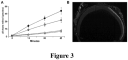
Figure 3 . AAV2/5-hGRK1p.hRDH12 gene-replacement therapy restores RDH12 function in Rdh12-deficent mice. (A) HPLC analysis of retinal reductase activity in retinas from C57BL/6J and Rdh12 -/- mice injected with AAV2/5-hGRK1p.hRDH12 (1.3 × 10⁹ vg) or PBS, or non-injected. At 6 weeks post-injection, all-trans retinol formation was quantitated in assays with all-trans retinal as a substrate. Each data point represents the mean ± standard error for a minimum of 5 independent experiments where retinas from 3 to 5 mice were pooled and assayed in triplicate. ● C57BL/6J ; Δ Rdh12 -/ - ;Rdh12 -/- PBS-injected; Rdh12 -/- AAV-injected. (B) Immunohistochemistry of recombinant RDH12 expression in an Rdh12 -/- mouse whole-retina section evaluated 16 weeks post-injection of AAV2/5-hGRK1p.hRDH12. Human RDH12 (light gray) localizes to the IS, ONL, and OPL of the injected region of the retina (right-hand side of the image).
-
Figure 4 . AAV2/5-hGRK1p.hRDH12 gene-replacement therapy reduces light-damage in albino Rdh12-deficent mice. ERG analysis was performed, one week before and one week after exposure to 5,000 lux for 2 hours, on mice that were injected in one eye with AAV2/5-hGRK1p.hRDH12 and were uninjected in the contralateral eye. Scotopic (rod-isolated and combined rod-cone) responses were quantified for groups of 10-13 mice, and the percentage of the initial ERG response remaining after light damage was calculated. Averaged outcomes with standard errors are shown, as well as the significance of the differences between injected and uninjected eyes calculated using two-tailed paired t-test analysis. -
Figure 5 . AAV2/5-hGRK1p.hRDH12 does not significantly affect steady-state levels of 11-cis retinal in the retina. Mice received AAV2/5-hGRK1p.hRDH12 (1.3 × 10⁹ vg) or PBS via sub-retinal injection, or were non-injected. Following overnight dark adaptation, retinoids were extracted under dim-red light, and quantified by HPLC analysis. (A) Representative chromatograms from each treatment condition; peaks for syn-11-cis retinal oxime, anti-11-cis retinal oxime, and syn-all-trans retinal oxime are indicated. Total retinal levels of (B) 11 -cis retinal and (C) all-trans retinal for each treatment condition ± standard error (■ C57BL/6J non-injected;Rdh12 -/- non-injected; Rdh12 -/- AAV injected;
 Rdh12 -/- PBS injected).
-
Figure 6 . Retinal function is not adversely affected by AAV2/5-hGRK1p.hRDH12. Scotopic (rod-isolated and combined rod-cone) and photopic (cone-mediated) electroretinogram (ERG) responses recorded at 6 weeks post treatment from C57BL/6J, non-injected; Rdh2 -/ -, non-injected; and Rdh12 -/ - AAV2/5-hGRK1p.hRDH12 injected mice (up to 2 × 10⁹ vg). ERGs from a representative animal in each treatment group measured at 6 weeks post-injection are shown. -
Figure 7 . Visual pigment localization is not perturbed by AAV2/5-hGRK1p.hRDH12. Immunohistochemical localization of rhodopsin and cone opsin in non-injected and injected (1.3 × 10⁹ vg) Rdh12 -/- mice evaluated at 16 weeks post treatment. Human RDH12 protein expression in IS and ONL (light gray). Rhodopsin and red/green opsin (dark gray) in AAV2/5-hGRK1p.hRDH12 injected eyes. Abbreviations are as described forFigure 2 . -
Figure 8 . Retinal structure is not damaged by long-term expression of AAV2/5-hGRK1p.hRDH12. Optical coherence tomography (OCT) analysis of eyes of C57BL/6J and Rdh12 -/- mice evaluated at 54 weeks post injection of AAV2/5-hGRK1p.hRDH12 (up to 2 × 10⁹ vg). -
Figure 9 . Infiltrating CD68+ macrophages and RDH12 expression in AAV2/8-hGRK1p.hRDH12 injected retinas. Rdh12 -/- and C57BL/6J mice evaluated by immunohistochemical analysis 8 weeks following subretinal injection of AAV2/8-hGRK1p.hRDH12 (2 × 10⁹ vg). Human RDH12 expression in IS, ONL, and OPL (light gray) and CD68 labeling of macrophages (white). Abbreviations are as described forFigure 2 . - Inherited retinal degeneration is a rare cause of profound vision loss that is a focus of current efforts to develop targeted gene-therapy. Viral vector-mediated somatic gene therapy has shown great promise in treating animal models of human retinal degenerative disease. To date, there have been a number of successful studies using adeno-associated virus (AAV)-mediated gene delivery to rescue photoreceptor degeneration in small animal models (Ali et al. 2000; Pang et al. 2012; Pawlyk et al. 2010; Pawlyk et al. 2005; Tan et al. 2009) and large animal models (Acland et al. 2001; Alexander et al. 2007; Beltran et al. 2012; Komaromy et al. 2010; Lheriteau et al. 2009). In these cases, the retinal pigment epithelium (RPE) or photoreceptors have been the primary targets for transgene expression. In addition, phase I clinical trials involving gene therapy for patients with Leber Congenital Amaurosis (LCA) targeting the RPE (Bainbridge et al. 2008; Cideciyan et al. 2008; Maguire et al. 2008) and more recently choroideremia (Maclaren et al. 2014), have already met with some success. There are currently no clinical trials using AAV-mediated gene replacement therapy for the treatment of patients with inherited retinal degeneration caused by mutations in RDH12.
- With an interest in developing AAV-mediated gene therapy for the treatment of individuals with mutations in RDH12, we generated adeno-associated virus vectors carrying human RDH12 cDNA in which expression is under the control of a rhodopsin-kinase promoter. In the disclosed study of vectors with capsids derived from AAV serotype 5 or AAV serotype 8, we have shown that subretinal delivery of AAV2/5-hGRK1p.RDH12 in Rdh12-deficient (Rdh12 -/- ) mice results in expression of recombinant human RDH12 that is stable, correctly localized, reconstitutes retinal reductase activity, reduces light-damage susceptibility, and does not cause the retinal toxicity seen with AAV2/8-hGRK1p.hRDH12. This AAV2/5-hGRK1p.RDH12 construct provides a product for RDH12 gene-replacement therapy. RDH12
- Mutations in the gene encoding retinol dehydrogenase 12 (RDH12) cause severe early-onset retinal degeneration most often diagnosed as Leber congenital amaurosis (LCA) or early-onset severe retinal dystrophy (EOSRD). A member of the family of short-chain dehydrogenases/reductases, RDH12 is essential for reducing retinaldehydes that are generated by the activity of the vitamin A visual cycle that is integral to the light response of the photoreceptor cells. When visual pigments containing the 11-cis retinal chromophore absorb a photon of light, 11-cis retinal is isomerized to all-trans retinal, thus initiating a signal transduction cascade that regulates synaptic signaling. Visual pigment inactivation involves release of all-trans retinal, its reduction to all-trans retinol and return to the retinal pigment epithelium (RPE) for regeneration of the 11-cis retinal chromophore (
Figure 1A ). When these recycling reactions are inefficient or disrupted, e.g., by aging or inherited disease, retinaldehydes and retinaldehyde-condensation products accumulate in the photoreceptors and retinal pigment epithelium, resulting in profound damage to the outer retina (Ben-Shabat et al. 2001; Thompson et al. 2003; Sparrow, 2010; Chen et al. 2012). - To protect against toxicity, a number of retinoid binding proteins and enzymes are expressed in the retina. RDH12 is a member of the family of short-chain dehydrogenases/reductases that uses NADPH to reduce a broad range of substrates, including cis- and trans-retinaldehydes (Haeseleer et al. 2002), C9 aldehydes generated as a result of lipid photo-oxidation (Belyaeva et al. 2005; Lee et al. 2008; Marchette et al. 2010), and steroid substrates (Keller et al. 2007). Individuals with loss-of-function mutations in the RDH12 gene exhibit a severe retinal degeneration phenotype often diagnosed as Leber congenital amaurosis (LCA) (Janecke et al. 2004; Thompson et al. 2005; Perrault et al. 2004; den Hollander et al. 2008; Mackay et al. 2011), for which there is currently no treatments or cures. RDH12 localizes to the inner segments of rod and cone photoreceptor cells (Haeseleer et al. 2002; Maeda et al. 2006) where it protects against light-induced damage caused, at least in part, by reactive retinaldehydes (Maeda et al. 2006). All-trans retinal generated after light exposure has been shown to leak from photoreceptor outer segments to inner segments, and its effective clearance from photoreceptor cells requires the activity of both RDH8 present in the outer segments, and RDH12 present in the inner segments (Chen et al. 2012) (
Figure 1B ). As such, RDH12 also has the potential to play an important role in reducing 11-cis retinal present in excess of that required for opsin biosynthesis, which can enter the inner segment from the subretinal space (Chen et al. 2012). - To investigate the potential of gene replacement therapy for LCA/EOSRD due to RDH12 mutations, we generated adeno-associated virus vectors carrying a human RDH12 cDNA, in which expression is under the control of a rhodopsin-kinase promoter that directs photoreceptor-cell specific expression (Khani et al. 2007; Sun et al. 2010; Young et al. 2003). The DNA construct was packaged with a capsid derived from AAV serotype 8 that mediates efficient and robust transduction of photoreceptor cells (Allocca et al. 2007; Natkunarajah et al. 2008; Vandenberghe et al. 2011; Vandenberghe et al. 2013), or with a AAV serotype 5 capsid that mediates photoreceptor transduction, but with slower kinetics and less robust expression compared to the AAV serotype 8 capsid (Yang et al. 2002; Lotery et al. 2003; Allocca et al. 2007; Lebherz et al. 2008).
- In comparative studies performed in a mouse model of Rdh12-deficiency (Rdh12 -/- ) (Kurth et al. 2007), our work has shown that the two vectors have significantly different safety profiles, with AAV2/5-hGRK1p.hRDH12 demonstrating unexpected and surprisingly effective expression levels and toxicity profiles compatible with therapeutic use to treat retinal dystrophies.
- Sequence of an exemplary human RDH12 cDNA, consisting of nucleotides -10 to +980 relative to the translation initiation site, encoding all seven translated RDH12 exons (GenBank # NM_152443) is provided in SEQ ID NO:1.
- The full-length human RDH12 protein sequence is provided in SEQ ID NO:2.
- In some embodiments of the methods described herein, a replacement gene construct is used in which a human RDH12 cDNA as described herein is placed under the control of a human rhodopsin kinase (hGRK1) promoter. In some embodiments, the hGRK1 promoter is approximately 200 base pairs (bp) in length containing a short promoter derived from the rhodopsin kinase (RK) hGRK1 gene, which has been shown to drive cell-specific expression in rods and cones (Khani et al. 2007; Sun et al. 2010; Young et al. 2003). An exemplary hGRK1 promoter sequence contains nucleotides -112 to +87 of SEQ ID NO:3 (Khani et al. 2007).
- The abbreviated human RDH12 cDNA, as described above, is packaged into a delivery vector, e.g., an AAV5 or AAV2/5 vector.
- Replacement genes (cDNA) can be administered in any effective carrier, e.g., any formulation or composition capable of effectively delivering the component gene to cells in vivo. Approaches include insertion of the gene into non-pathogenic, non-replicating viral vectors, including recombinant retroviruses, adenovirus, adeno-associated virus, lentivirus, and herpes simplex virus-1, or recombinant bacterial or eukaryotic plasmids. Viral vectors transfect cells directly; plasmid DNA can be delivered naked or with the help of, for example, cationic liposomes (lipofectamine) or derivatized (e.g., antibody conjugated), poly lysine conjugates, gramacidin S, artificial viral envelopes or other such intracellular carriers, as well as direct injection of the gene construct or Ca3(PO4)2 precipitation carried out in vivo.
- A preferred approach for in vivo introduction of nucleic acid into a cell is by use of a viral vector containing nucleic acid, e.g., a cDNA. Infection of cells with a viral vector has the advantage that a large proportion of the targeted cells can receive the nucleic acid. Additionally, molecules encoded within the viral vector, e.g., by a cDNA contained in the viral vector, are expressed efficiently in cells that have taken up viral vector nucleic acid. Retrovirus vectors and adenovirus derived vectors can be used as a recombinant gene delivery system for the transfer of exogenous genes in vivo, particularly into humans, in a number of cell types. However, they do not transduce the photoreceptor cells with sufficient efficiency to make them useful for this application.
- Yet another viral vector system useful for delivery of nucleic acids is the adeno-associated virus (AAV). Adeno-associated virus is a naturally occurring defective virus that requires another virus, such as an adenovirus or a herpes virus, as a helper virus for efficient replication and a productive life cycle (Muzyczka et al. 1992). It is also one of the few viruses that may integrate its DNA into non-dividing cells, and exhibits a high frequency of stable integration (see, for example, Flotte et al. 1992; Samulski et al. 1989; and McLaughlin et al. 1988). Vectors containing as little as 300 base pairs of AAV can be packaged and can integrate. Space for exogenous DNA is limited to about 4.5 kb. An AAV vector such as that described in Tratschin et al., 1985 can be used to introduce DNA into cells. A variety of nucleic acids have been introduced into different cell types using AAV vectors (see, for example, Hermonat et al. 1984; Tratschin et al. 1984a; Tratschin et al. 1984b; Wondisford et al. 1988; and Flotte et al. 1993).
- In preferred embodiments, the viral delivery vector is a recombinant AAV2/5 virus. Prior to administration, the final product can undergo a series of ultrapurification steps to meet clinical grade criteria.
- Subjects who are candidates for the present methods of treatment include those who have a diagnosis of LCA caused by mutations in the gene encoding RDH12. Subjects suffering from other ophthalmological clinically-defined conditions caused by mutations in the gene encoding RDH12, e.g., early-onset retinitis pigmentosa, can also be treated using the methods described herein. A diagnosis of LCA or another ophthalmological condition caused by mutations in the gene encoding RDH12 can be made using methods known in the art.
- The methods described herein can include identifying a subject, e.g., a child, adolescent, or young adult subject, who has LCA or another ophthalmological condition caused by one or more mutations in the gene encoding RDH12, or who is suspected of having LCA or another ophthalmological condition caused by one or more mutations in the gene encoding RDH12 (e.g., based on the presence of symptoms of the condition and no other obvious cause), and obtaining a sample comprising genomic DNA from the subject, detecting the presence of mutations in RDH12 using known molecular biological methods, and selecting a patient who has mutations in both RDH12 alleles that cause LCA or another condition. Symptoms of the condition include macular atrophy, foveal thinning and disruption of laminar architecture, resulting in early central vision loss and progression to LP vision. Visual fields are constricted at the earliest age measured, and ERG responses become unrecordable by early adulthood. Detecting mutations in RDH12 can include sequencing all or part of the RDH12 gene in a subject, and comparing the sequence to a reference sequence (e.g., GenBank Accession No. NG_008321.1) to detect a mutation. Frameshift mutations, truncation mutations, mutations that alter a conserved amino acid, mutations that affect transcript splicing, or mutations that affect a regulatory (e.g., promoter) region are considered to be mutations that can cause LCA or another ophthalmological condition as described herein; an alteration in function can be confirmed by expressing the mutant in vitro (e.g., in cultured cells), and assaying, e.g., enzymatic function. Exemplary mutations in the homozygous state include: Glu127X, Gln189X, Tyr226Cys, Ala269GlyfsX1, and Leu274Pro (all position references refer to the RDH12 protein sequence of SEQ ID NO:2). Exemplary mutations in the compound heterozygous state include: Thr49Met/Arg62X; Arg65X/Ala269GlyfsX1; His151D/Thr155Ile; His151D/Arg269GlyfsX1 (Janecke et al. 2004; Schuster et al. 2007). (Positions refer to the protein sequence of SEQ ID NO:2.)
- Patients with LCA or another ophthalmological condition due to at least one RDH12 mutation that can be treated using a method described herein preferably retain some photoreceptors and visual function, e.g., as measured by standard visual function or field tests and/or Optical Coherence Tomography (OCT, e.g., Spectral Domain-OCT (SD-OCT)). The methods described herein can include identifying subjects who have been diagnosed with LCA or another ophthalmological condition due to at least one RDH12 mutation, who have at least one confirmed mutation in RDH12 that causes their condition, and testing their visual ability and detecting the presence of residual central photoreceptors.
- The disclosed and claimed subject matter is further described in the following examples, which do not limit the scope of the invention described in the claims.
- The following materials and methods were used in the experiments disclosed in the Examples set forth below.
- The generation and analysis of Rdh12 -/- mice have been described previously (Kurth et al. 2007). The Rdh12 -/- mice used in this study were bred from sibling mating among nullizygous males and females maintained in our institutional animal facility. WT mice used in the study were C57BL/6 from The Jackson Laboratory (Wilmington, MA).
- Transgenic mice of the following genotypes were used for the studies disclosed herein: Rdh12 -/- mice on C57BL/6J background homozygous for the Rpe65-Met450 (M/M) variant (Kurth, 2007), and albino Rdh12 -/- mice on BALB/c background homozygous for the Rpe65-Leu450 (L/L) variant (Chrispell, 2009), that were obtained by breeding. Mice were reared in a 12-hour (light)/12-hour (dark) cycle and were euthanized by CO2 inhalation followed by bilateral pneumothorax.
- Human RDH12 cDNA were amplified from human retinal cDNA by PCR using primers designed to encompass the entire RDH12 coding region, cloned, and sequenced to verify fidelity, as described previously (Janecke et al. 2004). To construct the AAV vectors, RDH12 cDNAs were inserted into the multiple cloning site of the parental pAAV- hGRK1-hrGFP vector. The resulting pAAV- hGRK1-Rdh12 vector was packaged into AAV. AAV2/5 and AAV2/8 pseudotyped vectors were generated by bipartite transfection: (1) AAV vector plasmid encoding the gene of interest, (2) AAV helper plasmid encoding AAV Rep proteins from
serotype 2 and Cap proteins from either serotype 5 or serotype 8, and adenovirus helper functions into 293T cells. The transfection and purification were performed using a protocol as published (Nishiguchi et al 2015). Two days after transfection, cells were lysed by repeated freeze and thaw cycles. After initial clearing of cell debris, the nucleic acid component of the virus producer cells was removed by Benzonase treatment. The recombinant AAV vector particles were purified by affinity chromatography using a AVB matrix, washed in 1× PBS and concentrated to a volume of 100-150 ml using Vivaspin 4 (10 kDa) concentrators. Vectors were titered by qPCR amplification. - Cohorts of mice at approximately 4 weeks of age were placed under general anesthesia with an intraperitoneal injection of ketamine (90 mg/kg)/xylazine (9 mg/kg). A 0.5% proparacaine solution was applied to the cornea as a topical anesthetic. Pupils were dilated with topical application of tropicamide (0.5%). Under an ophthalmic surgical microscope, a small incision was made through the cornea adjacent to the limbus using a 30-gauge needle. A 34-gauge blunt needle fitted to a Hamilton syringe was inserted through the incision behind the lens and pushed through the retina. All injections were made subretinally in a location within the nasal quadrant of the retina. Each eye received up to 2 × 10⁹ vg AAV2/5-(hGRK1)-hRDH12 in up to a 2 µL volume. RDH12-encoding vector was administered separately to one eye of each mouse receiving treatment, and the contralateral eyes were uninjected. Fundus examination following the injection found more than 30% of the retina detached in most cases, confirming successful subretinal delivery.
- Primary antibodies used were: a rabbit anti-Rdh12 polyclonal antibody (CSP) specific for the mouse protein (against 252SPFFKSTSQGAQ263, SEQ ID NO:4), and a mouse anti-RDH12 monoclonal antibody (2C9) specific for the human protein (against C-284DCKRTWVSPRARNNKT299; SEQ ID NO:5) (Kurth et al. 2007); a mouse anti-RHO monoclonal antibody (1D4) (MacKenzie et al. 1984); a rabbit anti-RHO polyclonal antibody generated against the denatured protein; and a rabbit anti-red/green cone opsin polyclonal antibody (Millipore cat# AB5405).
- Proteins in retina homogenates were separated by SDS-PAGE, transferred onto nitrocellulose membranes that were then blocked, incubated with primary antibody overnight, washed, incubated with alkaline phosphatase-conjugated secondary antibody, and developed using 5-bromo-4-chloro-3'-indolylphosphate p-toluidine and nitro-blue tetrazolium chloride.
- Mice were euthanized, eyes scored for orientation, then enucleated. For cryosections, lens and anterior segments were removed, eyes briefly fixed with 4% paraformaldehyde, washed with PBS, transitioned to sucrose/OCT, flash-frozen, and sectioned at a thickness of 10 µm. For freeze-substitution preparation, whole eyes were flash-frozen in dry-ice-cooled isopentane for 30 seconds, and then transferred to dry- ice-cooled methanol containing 3% glacial acetic acid. Eyes were incubated at 80°C for 48 hours, then overnight at -20°C, embedded in paraffin, and sectioned at a thickness of 6 µm. Paraffin sections were de-paraffinized and antigens retrieved by incubating in 1mM EDTA, 0.05
% Tween 20, pH8.0, at 90°C for 30 minutes prior to immune labeling as follows. Briefly, retinal cross sections were washed with PBS and permeabilized with PBS-T (0.3% Triton X-100); blocked with 1% bovine serum albumin, 10% normal goat serum, and 0.3% Triton X-100; and incubated with primary antibodies overnight at 4°C, washed, then incubated with fluorophore-conjugated secondary antibodies for 1 hour at room temperature. Sections were cover-slipped using ProLong Gold gel mount containing 4',6-diamidino-2-phenylindole (DAPI; Invitrogen), and imaged using a Leica DM6000 fluorescence microscope. - ERGs were performed as described previously (Thompson, 2012) using the Espion e2 recording system (Diagnosys, Lowell, MA). Briefly, mice were dark-adapted overnight and anesthetized with an intra-peritoneal injection of Ketamine (93 mg/kg) and Xylazine (8 mg/kg). Pupils were dilated with topical tropicamide (0.5%). Body temperature was maintained at 37°C with a heating pad. Corneal ERGs were recorded from both eyes using gold wire loops with 0.5% tetracaine topical anesthesia and a drop of 2% methylcellulose for corneal hydration. A gold wire loop placed in the mouth was used as reference, and a ground electrode was on the tail. The ERG protocol consisted of recording dark-adapted (scotopic) responses to brief white flashes (-2.31 log cd.s.m-2 for rod isolated B-waves; 1.09 log cd.s.m-2 for rod-cone combined A- and B-waves). Light-adapted (photopic) ERGs were recorded after 10 minutes of adaptation to a white 32 cd.m-2 rod-suppressing background in response to 1.09 log cd.s.m-2 intensity flashes (for cone isolated B-waves). Responses were amplified at 1,000 gain at 1.25 to 1000 Hz, and digitized at a rate of 2000 Hz. A notch filter was used to remove 60 Hz line noise. Responses were computer-averaged and recorded at 3- to 60-second intervals depending upon the stimulus intensity. For statistical analysis, paired t-tests were used to determine if ERG amplitudes in treated eyes were significantly different from untreated eyes.
- Albino Rdh12 -/- mice were injected in one eye with AAV2/5-hGRK1p.hRDH12 (1.3 × 10⁹ vg), or with an equal volume of PBS, and contralateral eyes were uninjected. At 6 weeks post-injection, ERG analysis was performed and scotopic responses were quantified as described above. One week later, mice were dark-adapted overnight, their pupils were dilated with tropicamide (0.5%), and then were placed in a light-box in individual clear trays. The mice were exposed to 5,000 lux for 2 hours, and then were returned to vivarium housing (12-hour dark/12-hour light (<20 lux)) for 7 days, after which ERG analysis was repeated. The percent of the original ERG response remaining after light damage was calculated for each eye, and the averages plotted with standard errors shown. Two-tailed paired t-tests were used to determine if the ERG amplitudes in treated eyes were significantly different from untreated eyes.
- Mice were anesthetized and pupils dilated with 0.5% tropicamide. A spectral domain optical coherence tomography (OCT) system (Bioptigen Envisu R2200 SD-OCT system (Durham, NC, USA)), with a volume analysis size of 1.4 × 1.4 mm, was centered on the optic nerve head. Systane (Alcon) lubricating drops were used throughout the imaging process.
- All-trans retinal and 11-cis retinal in mouse eyes were extracted using a modification of a previously described method (Bligh and Dyer, 1959). Six-week, post-injection mice were dark-adapted overnight, then under dim-red light, euthanized via CO2 overdose, and eyes enucleated and frozen in liquid N2. Under dim red light and on ice, each eye was homogenized in 1 mL chloroform: methanol: hydroxylamine (2 M) (3:6:1) and incubated at room temperature for 2 minutes. Next, 200 µL chloroform and 240 µL water were added, and each sample was vortexed and centrifuged at 14,000 rpm for 5 minutes. The lower phase was collected, the solvent was evaporated under nitrogen, and the sample was dissolved in hexane. Retinoids in the extracts were identified and quantified by high-performance liquid chromatography (HPLC) analysis, using a Waters Alliance separation module and photodiode array detector with a Supelcosil LC-31 column (25 cm by 4.6 mm by 3 µm) developed with 5% 1,4-dioxane in hexane. Peak identification was done by comparison to retention times of standard compounds and evaluation of wavelength maxima. Quantitative analysis was done by comparison of peak areas at 347 and 351 nm for syn- and anti-11-cis retinal oxime, respectively, and at 357 and 361 nm for syn- and anti-all-trans retinal oxime, respectively (Kurth et al. 2007).
- Mouse retina homogenates were assayed for retinal reductase activity at 6 weeks post-injection. Light-adapted mice were euthanized, and each retina was homogenized individually in 125 µL of 0.25 M sucrose, 25 mM Tris-acetate, pH 7, 1 mM dithiothreitol. The homogenates were centrifuged at 1000 × g for 5 minutes to remove unbroken cells, and then the supernates were sonicated with a microtip probe (30 times for 1 second each) on ice. Protein concentrations were determined by a modification of the Lowry procedure (Peterson et al. 1977), and levels of RDH12 were evaluated by western blot. Like samples were pooled and 20 µg of each pooled lysate was assayed (in triplicate) in buffer containing 200 µM all-trans retinal and 200 µM NADPH in HEPES buffer (pH 8); reactions were incubated for 0-45 minutes in a 37°C water bath (a reaction temperature that minimized thermal isomerization of the retinoid substrates, as well as enzyme inactivation). All-trans retinol formation was quantitated using normal-phase HPLC analysis with comparison to known standards (Chrispell et al. 2009).
- A variety of vectors for RDH12-replacement therapy were developed and tested. The optimal RDH12 vector construct is shown in
Figure 2A . It comprises the human RDH12 cDNA under control of a human Rhodopsin Kinase (GRK1) promoter fragment. The construct is packaged in an AAV2/5 serotype. The AAV serotype 5 capsid has been shown to mediate photoreceptor transduction, but with slower kinetics and less robust expression compared to the AAV8 capsid (Yang et al. 2002; Lotery et al. 2003; Allocca et al. 2007; Lebherz et al. 2008). Human RDH12 protein expression in mouse retinas 6 weeks following sub-retinal injection of AAV2/5-hGRK1p.hRDH12 (1.3 × 10⁹ vg) was evaluated using antibodies specific for the mouse Rdh12 or the human RDH12 proteins. Vector-delivered levels of human RDH12 appear to be roughly comparable to the amount of mouse Rdh12 (Figure 2B ). Indirect immunofluorescence imaging on retinal sections was used to evaluate native mouse Rdh12 and recombinant human RDH12, using species-specific antibodies. Localization of endogenous and recombinant RDH12 appears to be identical, indicating that the protein is being processed normally (Figure 2C ). - The capacity of retinas of Rdh12 -/- mice to reduce exogenous retinaldehydes is significantly reduced compared to wild-type mice (Chrispell et al. 2009). Six weeks after subretinal injection of 1.3 × 10⁹ vg AAV2/5-hGRK1p.hRDH12 into Rdh12-/- mice, retinal homogenates were assayed for in vitro retinal reductase activity. All-trans retinol formation was quantitated using normal-phase HPLC analysis. Each data point represents the mean ± standard error for a minimum of 5 independent experiments where retinas from 3 to 5 mice were pooled and assayed in triplicate. Initial rates of all-trans retinol formation in retinas from uninjected Rdh12 -/- and C57BL/6J mice were 0.013 pmol minute-1 µg protein-1, and 0.071 pmol minute-1 µg protein-1, respectively (
Figure 3A ). The residual activity present in Rdh12-/- mice reflects the presence of other RDH isoforms that are capable of reducing all-trans retinal (Rattner et al. 2000; Haeseleer et al. 2002). In Rdh12 -/- mice injected with AAV2/5-hGRK1p.hRDH12, the rate of all-trans retinol formation of 0.046 pmol minute-1 µg protein-1 was significantly greater than for PBS injected Rdh12 -/- mice (0.016 pmol minute-1 µg protein-1). The recovery of retinal reductase activity (about 50%) is consistent with the partial transduction of the retina (about 30%) and indicated that the vector restored normal levels of activity in transduced photoreceptors. The transgene was expressed for over 12 months (longest time point examined). Immunofluorescence labelling showed the extent of retinal transduction and recombinant RDH12 expression in an Rdh12 -/- mouse evaluated at 16 weeks post-injection covering approximately one-third of the retinal surface (Figure 3B ). - The effect of AAV2/5-hGRK1p.hRDH12 on susceptibility to light-induced damage was evaluated in albino Rdh12 -/- mice that were injected in one eye with 1.3 × 10⁹ vg or an equal volume of PBS, and received no treatment in the contralateral eye. ERG analysis of scotopic retinal activity (rod-isolated and combined rod-cone) was performed 1 week before, and 1 week after, subjecting the mice to light levels that cause significant retinal damage in albino animals (5,000 lux for 2 hours). The percent retinal activity remaining in vector-treated eyes was significantly greater than that remaining in untreated eyes (p ≤ 0.0168) (
Figure 4 ). In contrast, in control animals injected with PBS, the percent of scotopic retinal activity remaining in uninjected eyes was not significantly greater than in untreated eyes (p ≥ 0.2255). These findings are consistent with AAV215-hGRK1p.HRDH12-mediated protection against increased susceptibility to light-induced damage associated with RDH12 deficiency. - Toxicity of the vector was assessed through the direct effect of human RDH12 activity on retinoid metabolism, and for indirect effects on retinal structure and retinal function, as described in the following passage.
- The broad substrate specificity of RDH12 that enables the reduction of both 11-cis retinal and all-trans retinal created the potential for overexpression or mislocalization of recombinant protein to negatively impact visual cycle function. HPLC analysis of retinoid content was used to evaluate visual-cycle activity in eyes from Rdh12 -/- mice injected with AAV2/5-hGRK1p.hRDH12, or with PBS, or from uninjected C57BL/6J controls. Analysis of retinoids at 6 weeks post-subretinal-injection of AAV2/5-hGRK1p.hRDH12 (1.3 × 10⁹ vg) into Rdh12 -/- mice showed that hRDH12 expression did not significantly affect the steady state 11-cis retinal levels in the retina (
Figure 5A ). A representative chromatogram shows various controls with elution times for various retinoids indicated. Total retinal levels of 11-cis retinal and all-trans retinal represent average values ± standard error for a minimum of 5 independent experiments (Figures 5B,C ). ERG responses of retinal activity were measured in C57BL/6J and Rdh12 -/- mice that received 1.3 × 10⁹ vg of AAV2/5-hGRK1p.hRDH12 and were maintained in vivarium housing (12-hour dark/12-hour light (<20 lux)). Scotopic (rod-isolated -2.3 log cd.s.m--2 stimulus), combined rod-cone (1.09 log cd.s.m-2 stimulus), and photopic (cone-mediated responses, 1.09 log cd.s.m -2 stimulus) ERGs from a representative animal in each treatment group measured at 6 weeks post-injection showed no significant effect of human RDH12 expression on retinal function (Figure 6 ). - Mislocalization of rhodopsin and cone opsin is a well-characterized indicator of decreased photoreceptor cell viability (Adamian et al. 2006; Turney et al. 2007; Brunner et al. 2010; Lopes et al. 2010). Immunohistochemical localization of rhodopsin and red/green opsin was evaluated in Rdh12 -/- mice that were non-injected or AAV215-hGRK1p.HRDH12-injected (1.3 × 10⁹ vg). In mice exhibiting robust transgene expression, no decrease in opsin expression levels or evidence of rod and cone opsin mislocalization were observed at 16 weeks post-treatment; retinal structure remained intact at 54 weeks post-injection (
Figure 7 ). - Optical coherence tomography (OCT) analysis of C57BL/6J and Rdhl2 -/- mice evaluated at 54 weeks post-injection of AAV2/5-hGRK1p.hRDH12 (2 × 10⁹ vg) showed no gross alterations in retinal lamination across a wide area including both sides of the optic nerve. Retinal structure was stable for at least one year post-injection when compared to uninjected contralateral eyes (
Figure 8 ). - A capsid derived from AAV serotype 8 has been shown to mediate efficient and robust transduction of photoreceptor cells (Allocca et al. 2007; Natkunarajah et al. 2008; Vandenberghe et al. 2011; Vandenberghe et al. 2013). Initial studies were performed with a AAV2/8 serotype carrying the vector construct described above. Rdh12 -/- mice treated by subretinal injection of AAV2/8-hGRK1p.hRDH12 at doses of 108-109 viral genomes (vg) resulted in robust expression of recombinant human RDH12 protein. However, the injected eyes developed significant retinal damage starting as early as 3 weeks post-injection, and in almost all cases by 6 weeks post-injection. Macrophage infiltration was observed in the majority of cases by the presence of CD68+ cells (
Figure 9 ). Retinal damage was not mitigated by systemic administration of cyclosporin (Borel et al. 1976) to Rdh12 -/- mice continuously from the time of weaning to the time of sacrifice, indicating that immune responses against the vector or the transgene were not the cause of damage. This view is consistent with outcomes obtained in wild-type C57BL/6J mice injected with constructs encoding either human RDH12 (AAV2/8-hGRK1p.hRDH12) or mouse Rdh12 (AAV2/8-hGRK1p.mRdh12), in which significant retinal thinning and macrophage infiltration also occurred by 6 weeks post-injection. -
- ADAMIAN M, PAWLYK BS, HONG DH, BERSON EL. (2006). Rod and cone opsin mislocalization in an autopsy eye from a carrier of X-linked retinitis pigmentosa with a Gly436Asp mutation in the RPGR gene. Am J Ophthalmol 142, 515-8.
- ACLAND GM, AGUIRRE GD, RAY J, ZHANG Q, et al. (2001). Gene therapy restores vision in a canine model of childhood blindness. Nat Genet 28, 92-5.
- ALEXANDER JJ, UMINO Y, EVERHART D, CHANG B, et al. (2007). Restoration of cone vision in a mouse model of achromatopsia. Nat Med 13, 685-7.
- ALI RR, SARRA GM, STEPHENS C, ALWIS MD, et al. (2000). Restoration of photoreceptor ultrastructure and function in retinal degeneration slow mice by gene therapy. .
- ALLOCCA M, MUSSOLINO C, GARCIA-HOYOS M, SANGES D, et al. (2007). Novel adeno-associated virus serotypes efficiently transduce murine photoreceptors. J Virol 81, 11372-80.
- BAINB RIDGE JW, SMITH AJ, BARKER SS, ROBBIE S, et al. (2008). Effect of gene therapy on visual function in Leber's congenital amaurosis. N Engl J Med 358, 2231-9.
- BELTRAN WA, CIDECIYAN AV, LEWIN AS, IWABE S, et al. (2012). Gene therapy rescues photoreceptor blindness in dogs and paves the way for treating human X-linked retinitis pigmentosa. Proc Natl Acad Sci USA 109, 2132-7.
- BEN-SHABAT S, PARISH CA, HASHIMOTO M, LIU J, et al. (2001). Fluorescent pigments of the retinal pigment epithelium and age-related macular degeneration. Bioorg .
- BELYAEVA OV, KORKINA OV, STETSENKO AV, KIM T, et al. (2005). Biochemical properties of purified human retinol dehydrogenase 12 (RDH12): catalytic efficiency toward retinoids and C9 aldehydes and effects of cellular retinol-binding protein type I (CRBPI) and cellular retinaldehyde-binding protein (CRALBP) on the oxidation and reduction of retinoids. Biochemistry 44, 7035-47.
- BLIGH EG, DYER WJ. (1959). A rapid method of total lipid extraction and purification. Can J Biochem Physiol 37, 911-7.
- BOREL JF, FEURER C, GUBLER HU, STÄHELIN H. (1976). Biological effects of cyclosporin A: a new antilymphocytic agent. Agents Actions 6, 468-75.
- BRUNNER S, SKOSYRSKI S, KIRSCHNER-SCHWABE R, KNOBELOCH KP, et al. (2010). Cone versus rod disease in a mutant Rpgr mouse caused by different genetic backgrounds. Invest Ophthalmol Vis Sci, 51, 1106-15.
- CARLSON NB, KURTZ D, HEATH DA, HINES C. in: Clinical Procedures for Ocular Examination. 3rd Ed. Appleton & Lange, Norwalk, CT; 1990:63-77.
- CHEN C, THOMPSON DA, KOUTALOS Y. (2012). Reduction of all-trans-retinal in vertebrate rod photoreceptors requires the combined action of RDH8 and RDH12. J Biol Chem, 287, 24662-70.
- CHEN Y, OKANO K, MAEDA T, CHAUHAN V, et al. (2012). Mechanism of all-trans-retinal toxicity with implications for stargardt disease and age-related macular degeneration. J Biol Chem 287, 5059-69.
- CHRISPELL JD, FEATHERS KL, KANE MA, KIM, CY, et al. (2009). Rdh12 activity and effects on retinoid processing in the murine retina. J Biol Chem 284, 21468-77.
- CIDECIYAN AV, ALEMAN TS, BOYE SL, SCHWARTZ SB, et al. (2008). Human gene therapy for RPE65 isomerase deficiency activates the retinoid cycle of vision but with slow rod kinetics. Proc Natl Acad Sci USA 105, 151 12-7.
- DEN HOLLANDER AI, ROEPMAN R, KOENEKOOP RK, CREMERS FP. (2008). Leber congenital amaurosis: genes, proteins and disease mechanisms. Prog Retin Eye Res 27, 391-419.
- FLOTTE TR, SOLOW R, OWENS RA, AFIONE S, et al. (1992). Gene expression from adeno-associated virus vectors in airway epithelial cells. Am J Respir Cell Mol Biol 7, 349-56.
- FLOTTE TR, AFIONE SA, SOLOW R, DRUMM ML, et al. (1993). Expression of the cystic fibrosis transmembrane conductance regulator from a novel adeno-associated virus promoter. J Biol Chem 268, 3781-90.
- HAESELEER F, JANG GF, IMANISHI Y, DRIESSEN CA, et al. (2002). Dual-substrate specificity short chain retinol dehydrogenases from the vertebrate retina. J Biol Chem 277, 45537-46.
- HERMONAT PL, MUZYCZKA N. (1984). Use of adeno-associated virus as a mammalian DNA cloning vector: transduction of neomycin resistance into mammalian tissue culture cells. Proc Natl Acad Sci U S A 81, 6466-70.
- JANECKE AR, THOMPSON DA, UTERMANN G, BECKER C, et al. (2004). Mutations in RDH12 encoding a photoreceptor cell retinol dehydrogenase cause childhood-onset severe retinal dystrophy. .
- JOHNSON DD. Deafness and Vision Disorders: Anatomy and Physiology, Assessment Procedures, Ocular Anomalies, and Educational Implications, Charles C. Thomas Publisher; 1999.
- KELLER B, ADAMSKI J. (2007). RDH12, a retinol dehydrogenase causing Leber's congenital amaurosis, is also involved in steroid metabolism. J Steroid Biochem Mol Biol 104, 190-4.
- KHANI SC, PAWLYK BS, BULGAKOV OV, KASPEREK E, et al. (2007). AAV-mediated expression targeting of rod and cone photoreceptors with a human rhodopsin kinase promoter. Invest Ophthalmol Vis Sci 48, 3954-61.
- KOENEKOOP RK. (2004). An overview of Leber congenital amaurosis: a model to understand human retinal development. Surv Ophthalmol 49, 379-98.
- KOMAROMY AM, ALEXANDER JJ, ROWLAN JS, GARCIA MM, et al. (2010). Gene therapy rescues cone function in congenital achromatopsia. Hum Mol Genet 19, 2581-93.
- KUMARAN N, MOORE AT, WELEBER RG, MICHAELIDES M. (2017). Leber congenital amaurosis/early-onset severe retinal dystrophy: clinical features, molecular genetics and therapeutic interventions. Br J Ophthalmol 101, 1147-1154.
- KURTH I, THOMPSON DA, RÜTHER K, FEATHERS KL, et al. (2007). Targeted disruption of the murine retinal dehydrogenase gene Rdh12 does not limit visual cycle function. Mol Cell Biol 27, 1370-9.
- LEBHERZ C, MAGUIRE A, TANG W, BENNETT J, et al. (2008). Novel AAV serotypes for improved ocular gene transfer. .
- LEE SA, BELYAEVA OV, KEDISHVILI NY. (2008). Effect of lipid peroxidation products on the activity of human retinol dehydrogenase 12 (RDH12) and retinoid metabolism. Biochim Biophys Acta 1782, 421-5.
- LHERITEAU E, LIBEAU L, STIEGER K, DESCHAMPS JY, et al. (2009). The RPGRIP 1 - deficient dog, a promising canine model for gene therapy. Mol Vis 15, 349-61.
- LOPES VS, JIMENO D, KHANOBDEE K, SONG X, et al. (2010). Dysfunction of heterotrimeric kinesin-2 in rod photoreceptor cells and the role of opsin mislocalization in rapid cell death. Mol Biol Cell 21, 4076-88.
- LOTERY AJ, YANG GS, MULLINS RF, RUSSELL SR, et al. (2003). Adeno-associated virus type 5: transduction efficiency and cell-type specificity in the primate retina. Hum Gene Ther 14, 1663-71.
- MACKAY DS, DEV BORMAN A, MORADI P, HENDERSON RH, et al. (2011). RDH12 retinopathy: novel mutations and phenotypic description. Mol Vis 17, 2706-16.
- MAEDA A, MAEDA T, IMANISHI Y, SUN W, et al. Retinol dehydrogenase (RDH12) protects photoreceptors from light-induced degeneration in mice. J Biol Chem 281, 37697-704.
- MAGUIRE AM, SIMONELLI F, PIERCE EA, PUGH EN, JR., et al. (2008). Safety and efficacy of gene transfer for Leber's congenital amaurosis. N Engl J Med 358, 2240-8.
- MARCHETTE LD, THOMPSON DA, KRAVTSOVA M, NGANSOP TN, et al. (2010). Retinol dehydrogenase 12 detoxifies 4-hydroxynonenal in photoreceptor cells. Free Radic Biol Med 48, 16-25.
- MCLAUGHLIN SK, COLLIS P, HERMONAT PL, MUZYCZKA N. (1988). Adeno-associated virus general transduction vectors: analysis of proviral structures. J Virol 62, 1963-73.
- MACKENZIE D, ARENDT A, HARGRAVE P, MCDOWELL JH, et al. (1984). Localization of binding sites for carboxyl terminal specific anti-rhodopsin monoclonal antibodies using synthetic peptides. Biochemistry 23, 6544-9.
- MACLAREN RE, GROPPE M, BARNARD AR, COTTRIALL CL, et al. (2014). Retinal gene therapy in patients with choroideremia: initial findings from a phase 1/2 clinical trial. Lancet 383, 1129-37.
- MUZYCZKA N. (1992). Use of adeno-associated virus as a general transduction vector for mammalian cells. Curr Top Microbiol Immunol 158, 97-129.
- NATKUNARAJAH M, TRITTIBACH P, MCINTOSH J, DURAN Y, et al. (2008). Assessment of ocular transduction using single-stranded and self-complementary recombinant adeno-associated .
- NISHIGUCHI KM, CARVALHO LS, RIZZI M, POWELL K, et al. (2015). Gene therapy restores vision in rd1 mice after removal of a confounding mutation in Gpr179. Nat Commun 6, 6006.PANG JJ, LEI L, DAI X, SHI W, et al. (2012). AAV-mediated gene therapy in mouse models of recessive retinal degeneration. Curr Mol Med 12, 316-30.
- PAWLYK BS, SMITH AJ, BUCH PK, ADAMIAN M, et al. (2005). Gene replacement therapy rescues photoreceptor degeneration in a murine model of Leber congenital amaurosis lacking RPGRIP. Invest Ophthalmol Vis Sci 46, 3039-45.
- PAWLYK BS, BULGAKOV OV, LIU X, XU X, et al. (2010). Replacement gene therapy with a human RPGRIPl sequence slows photoreceptor degeneration in a murine model of Leber congenital amaurosis. Hum Gene Ther 21, 993-1004.
- PERRAULT I, HANEIN S, GERBER S, BARBET F, et al. (2004). Retinal dehydrogenase 12 (RDH12) mutations in leber congenital amaurosis. Am J Hum Genet 75, 639-46.
- PETERSON GL. (1977). A simplification of the protein assay method of Lowry et al. which is more generally applicable. Anal Biochem 83, 346-56.
- RATTNER A, SMALLWOOD PM, NATHANS J. (2000). Identification and characterization of all-trans-retinol dehydrogenase from photoreceptor outer segments, the visual cycle enzyme that reduces all-trans-retinal to all-trans-retinol. J Biol Chem 275, 11034-43.
- SAMULSKI RJ, CHANG LS, SHENK T. (1989). Helper-free stocks of recombinant adeno-associated viruses: normal integration does not require viral gene expression. J Virol 63, 3822-8.
- SCHUSTER A, JANECKE AR, WILKE R, SCHMID E, et al. (2007). The phenotype of early-onset retinal degeneration in persons with RDH12 mutations. Invest Ophthalmol Vis Sci 48, 1824-31.
- SPARROW JR. (2010). Bisretinoids of RPE lipofuscin: trigger for complement activation in age-related macular degeneration. Adv Exp Med Biol 703, 63-74.
- SUN X, PAWLYK B, XU X, LIU X, et al. (2010). Gene therapy with a promoter targeting both rods and cones rescues retinal degeneration caused by AIPL1 mutations. Gene Ther 17, 117-31.
- TAN MH, SMITH AJ, PAWLYK B, XU X, et al. (2009). Gene therapy for retinitis pigmentosa and Leber congenital amaurosis caused by defects in AIPL1 : effective rescue of mouse models of partial and complete Aipll deficiency using AAV2/2 and AAV2/8 vectors. Hum Mol Genet.
- THOMPSON DA, GAL A. (2003). Vitamin A metabolism in the retinal pigment epithelium: genes, mutations, and diseases. Prog Retin Eye Res 22, 683-703.
- THOMPSON DA, JANECKE AR, LANGE J, FEATHERS KL, et al. (2005). Retinal degeneration associated with RDH12 mutations results from decreased 11-cis retinal synthesis due to disruption of the visual cycle. Hum Mol Genet 14, 3865-75.
- THOMPSON DA, KHAN NW, OTHMAN MI, CHANG B, et al. (2012). Rd9 is a naturally occurring mouse model of a common form of retinitis pigmentosa caused by mutations in RPGR-ORF15. PLoS One 7, e35865.
- TRATSCHIN JD, WEST MH, SANDBANK T, CARTER BJ. (1984a). A human parvovirus, adeno-associated virus, as a eucaryotic vector: transient expression and encapsidation of the procaryotic gene for chloramphenicol acetyltransferase. Mol Cell Biol 4, 2072-81.
- TRATSCHIN JD, MILLER IL, CARTER BJ. (1984b). Genetic analysis of adeno-associated virus: properties of deletion mutants constructed in vitro and evidence for an adeno-associated virus replication function. J Virol 51, 611-9.
- TRATSCHIN JD, MILLER IL, SMITH MG, CARTER BJ. (1985). Adeno-associated virus vector for high-frequency integration, expression, and rescue of genes in mammalian cells. Mol Cell Biol 5, 3251-60.
- TURNEY C, CHONG NH, ALEXANDER RA, HOGG CR, et al. (2007). Pathological and electrophysiological features of a canine cone-rod dystrophy in the miniature longhaired dachshund. Invest Ophthalmol Vis Sci 48, 4240-9.
- VANDENBERGHE LH, BELL P, MAGUIRE AM, CEARLEY CN, et al. (2011). Dosage thresholds for AAV2 and AAV8 photoreceptor gene therapy in monkey. Sci Transl Med 3, 88ra54.
- VANDENBERGHE LH, BELL P, MAGUIRE AM, XIAO R, et al. (2013). AAV9 targets cone photoreceptors in the nonhuman primate retina. PLoS One 8, e53463.
- XIAO X, LI J, SAMULSKI RJ. (1998). Production of high-titer recombinant adeno-associated virus vectors in the absence of helper adenovirus. J Virol 72, 2224-32.
- YANG GS, SCHMIDT M, YAN Z, LINDBLOOM JD, et al. (2002). Virus-mediated transduction of murine retina with adeno-associated virus: effects of viral capsid and genome size. J Virol 76, 7651-60.
- YOUNG JE, VOGT T, GROSS KW, KHANI SC. (2003). A short, highly active photoreceptor-specific enhancer/promoter region upstream of the human rhodopsin kinase gene. Invest Ophthalmol Vis Sci 44, 4076-85.
- WELEBER RG, FRANCIS PJ, TRZUPEK KM, BEATTIE C. (2004). Leber Congenital Amaurosis. In: PAGON RA, ADAM MP, ARDINGER HH, WALLACE SE, et al. GeneReviews® [Internet]. Seattle (WA): University of Washington, Seattle; 1993-2017. [updated 2013 May 2].
- WONDISFORD FE, USALA SJ, DECHERNEY GS, CASTREN M, et al. (1988). Cloning of the human thyrotropin beta-subunit gene and transient expression of biologically active human thyrotropin after gene transfection. .
- Each of the references cited herein is hereby incorporated by reference in its entirety or in relevant part, as would be apparent from the context of the citation.
- It is to be understood that while the claimed subject matter has been described in conjunction with the detailed description thereof, the foregoing description is intended to illustrate and not limit the scope of that claimed subject matter, which is defined by the scope of the appended claims. Other aspects, advantages, and modifications are within the scope of the following claims.
The invention further includes the subject matter of the claims ofPCT/US2018/061319 from which this application is derived, the content of which is reproduced below as numbered paragraphs. - 1. A method of treating a human subject who has an ophthalmological condition due to one or more loss-of-function mutations in the gene encoding the Retinol Dehydrogenase 12 (RDH12) protein, the method comprising administering to at least one eye of the subject an adeno-associated viral vector comprising a nucleic acid, wherein the nucleic acid comprises human RDH12 DNA and wherein the human RDH12 DNA encodes a protein that is at least 70%, 80%, 90%, 95%, or 99% identical to the full length of SEQ ID NO:2.
- 2. The method of paragraph 1 wherein the ophthalmological condition is Leber Congenital Amaurosis (LCA).
- 3. The method of paragraph 1 wherein the RDH12 DNA is under the expression control of a human rhodopsin kinase 1 (hGRK1) promoter.
- 4. The method of paragraph 3 wherein the hGRKl promoter comprises SEQ ID NO:3.
- 5. The method of paragraph 1 wherein the adeno-associated viral vector is AAV-2, serotype-5 (AAV2/5) or AAV-5.
- 6. The method of paragraph 1 wherein the RDH12 DNA comprises a sequence that is at least 60% or 70% identical to SEQ ID NO:1.
- 7. The method of paragraph 1 comprising administering the nucleic acid at a titer of about 2 × 10¹⁰ viral genomes per milliliter (vg/mL) to a titer of about 2 × 10¹² vg/mL.
- 8. The method of paragraph 1 wherein the nucleic acid is administered into the subretinal space.
- 9. The method of paragraph 8, wherein a micro injection cannula is inserted into the subretinal space.
- 10. A nucleic acid encoding a human RDH12 DNA, wherein the human RDH12 DNA encodes a protein that is at least 70%, 80%, 90%, 95%, or 99% identical to the full length of SEQ ID NO:2, wherein the RDH12 DNA is under the control of a human rhodopsin kinase 1 (hGRK1) promoter.
- 11. The nucleic acid of
paragraph 10, wherein the hGRK1 promoter comprises SEQ ID NO:3. - 12. The nucleic acid of
paragraph 10, wherein the human RDH12 DNA encodes a protein comprising SEQ ID NO:2. - 13. The nucleic acid of
paragraph 10, wherein the human RDH12 DNA is at least 60% or 70% identical to the full length of SEQ ID NO: 1. - 14. The nucleic acid of paragraphs 10-13, for use in treating a human subject who has an ophthalmological condition due to one or more loss-of-function mutations in the gene encoding the Retinol Dehydrogenase 12 (RDH12) protein.
- 15. The nucleic acid of paragraph 14 wherein the ophthalmological condition is Leber Congenital Amaurosis (LCA).
- 16. A viral vector comprising the nucleic acid of any of paragraphs 10-15.
- 17. The viral vector of paragraph 16, which is an adeno-associated viral vector.
- 18. The viral vector of paragraph 17, wherein the adeno-associated viral vector is AAV-2, serotype-5 (AAV2/5) or AAV-5.
- 19. The viral vector of any one of paragraphs 16 to 18, for use in treating a human subject who has an ophthalmological condition due to one or more loss-of-function mutations in the gene encoding the Retinol Dehydrogenase 12 (RDH12) protein.
- 20. The viral vector of paragraph 19 wherein the ophthalmological condition is Leber Congenital Amaurosis (LCA).
- 21. An isolated host cell comprising the viral vector of any one of paragraphs 16 to 20, or the nucleic acid of any one of
paragraphs 10 to 15. - 22. The isolated host cell of paragraph 21, wherein the cell expresses a human RDH12 protein.
Claims (10)
- An adeno-associated viral vector comprising a nucleic acid, wherein the nucleic acid comprises human RDH12 DNA and wherein the human RDH12 DNA encodes a protein that is at least 70%, 80%, 90%, 95%, or 99% identical to the full length of SEQ ID NO:2 for use in a method of treating a human subject who has an ophthalmological condition due to one or more loss-of-function mutations in the gene encoding the Retinol Dehydrogenase 12 (RDH12) protein, the method comprising administering the adeno-associated viral vector to at least one eye of the subject, wherein the adeno-associated viral vector is AAV-2, serotype-5 (AAV2/5), wherein the RDH12 DNA is under the expression control of a human rhodopsin kinase 1 (hGRK1) promoter, and wherein the ophthalmological condition is early-onset severe retinal dystrophy (EOSRD).
- The adeno-associated viral vector for use according to claim 1 wherein the hGRKl promoter comprises SEQ ID NO:3, orwherein the hGRKl promoter consists of SEQ ID NO:3, orwherein the RDH12 DNA comprises a sequence that is at least 60% or 70% identical to SEQ ID NO: 1, orcomprising administering the nucleic acid at a titer of about 2 × 10¹⁰ viral genomes per milliliter (vg/mL) to a titer of about 2 × 10¹² vg/mL, orwherein the nucleic acid is administered into the subretinal space.
- The adeno-associated viral vector for use according to claim 2, wherein the nucleic acid is administered into the subretinal space with a micro injection cannula that is inserted into the subretinal space.
- The adeno-associated viral vector for use according to claim 1 wherein the human RDH12 DNA encodes a protein comprising SEQ ID NO:2 and, wherein the hGRKl promoter consists of SEQ ID NO:3,
- An adeno-associated viral vector comprising a nucleic acid encoding a human RDH12 DNA, wherein the human RDH12 DNA encodes a protein that is at least 70%, 80%, 90%, 95%, or 99% identical to the full length of SEQ ID NO:2, wherein the RDH12 DNA is under the control of a human rhodopsin kinase 1 (hGRK1) promoter, and wherein the adeno-associated viral vector is AAV-2, serotype-5 (AAV2/5).
- The adeno-associated viral vector of claim 5, wherein the hGRKl promoter comprises SEQ ID NO:3, or wherein the human RDH12 DNA encodes a protein comprising SEQ ID NO:2.
- The adeno-associated viral vector of claim 5 or claim 6, wherein the hGRKl promoter consists of SEQ ID NO:3.
- The adeno-associated viral vector of claim 5, wherein the human RDH12 DNA is at least 60% or 70% identical to the full length of SEQ ID NO: 1.
- An isolated host cell comprising the adeno-associated viral vector of any one of claims 5 to 8.
- The isolated host cell of claim 9, wherein the cell expresses a human RDH12 protein.
Applications Claiming Priority (3)
| Application Number | Priority Date | Filing Date | Title |
|---|---|---|---|
| US201762586624P | 2017-11-15 | 2017-11-15 | |
| PCT/US2018/061319 WO2019099696A1 (en) | 2017-11-15 | 2018-11-15 | Viral vectors comprising rdh12 coding regions and methods of treating retinal dystrophies |
| EP18878835.0A EP3710590B1 (en) | 2017-11-15 | 2018-11-15 | Viral vectors comprising rdh12 coding regions and methods of treating retinal dystrophies |
Related Parent Applications (2)
| Application Number | Title | Priority Date | Filing Date |
|---|---|---|---|
| EP18878835.0A Division EP3710590B1 (en) | 2017-11-15 | 2018-11-15 | Viral vectors comprising rdh12 coding regions and methods of treating retinal dystrophies |
| EP18878835.0A Division-Into EP3710590B1 (en) | 2017-11-15 | 2018-11-15 | Viral vectors comprising rdh12 coding regions and methods of treating retinal dystrophies |
Publications (2)
| Publication Number | Publication Date |
|---|---|
| EP4364803A2 true EP4364803A2 (en) | 2024-05-08 |
| EP4364803A3 EP4364803A3 (en) | 2024-06-05 |
Family
ID=66431192
Family Applications (2)
| Application Number | Title | Priority Date | Filing Date |
|---|---|---|---|
| EP24163209.0A Pending EP4364803A3 (en) | 2017-11-15 | 2018-11-15 | Viral vectors comprising rdh12 coding regions and methods of treating retinal dystrophies |
| EP18878835.0A Active EP3710590B1 (en) | 2017-11-15 | 2018-11-15 | Viral vectors comprising rdh12 coding regions and methods of treating retinal dystrophies |
Family Applications After (1)
| Application Number | Title | Priority Date | Filing Date |
|---|---|---|---|
| EP18878835.0A Active EP3710590B1 (en) | 2017-11-15 | 2018-11-15 | Viral vectors comprising rdh12 coding regions and methods of treating retinal dystrophies |
Country Status (27)
| Country | Link |
|---|---|
| US (2) | US20190142909A1 (en) |
| EP (2) | EP4364803A3 (en) |
| JP (2) | JP2021502978A (en) |
| KR (1) | KR102704296B1 (en) |
| CN (2) | CN117752822A (en) |
| AU (1) | AU2018367606B2 (en) |
| BR (1) | BR112020009828A2 (en) |
| DK (1) | DK3710590T3 (en) |
| EA (1) | EA202091194A1 (en) |
| ES (1) | ES2983990T3 (en) |
| FI (1) | FI3710590T3 (en) |
| HR (1) | HRP20241073T1 (en) |
| HU (1) | HUE068393T2 (en) |
| IL (2) | IL274609B2 (en) |
| LT (1) | LT3710590T (en) |
| MA (1) | MA50914A (en) |
| MX (2) | MX2020005008A (en) |
| MY (1) | MY204271A (en) |
| PH (1) | PH12020551319A1 (en) |
| PL (1) | PL3710590T3 (en) |
| PT (1) | PT3710590T (en) |
| RS (1) | RS65881B1 (en) |
| SG (1) | SG11202004527YA (en) |
| SI (1) | SI3710590T1 (en) |
| SM (1) | SMT202400321T1 (en) |
| WO (1) | WO2019099696A1 (en) |
| ZA (1) | ZA202003416B (en) |
Families Citing this family (6)
| Publication number | Priority date | Publication date | Assignee | Title |
|---|---|---|---|---|
| KR102799528B1 (en) | 2016-07-08 | 2025-04-23 | 더 트러스티스 오브 더 유니버시티 오브 펜실베니아 | Methods and compositions for treatment of disorders and diseases involving rdh12 |
| WO2022232249A1 (en) * | 2021-04-27 | 2022-11-03 | The Trustees Of Columbia University In The City Of New York | Methods and compositions to regulate cholesterol efflux to prevent, treat, or cure macular degeneration |
| CN115725659A (en) * | 2022-04-01 | 2023-03-03 | 首都医科大学附属北京同仁医院 | Method for preparing congenital amaurosis model of non-human primate by using retina in-situ gene editing |
| CN118453915A (en) * | 2024-04-29 | 2024-08-09 | 中国医学科学院北京协和医院 | Gene therapy medicine for RDH12 mutation |
| CN120290554B (en) * | 2024-09-10 | 2026-04-14 | 广州市未来基因递送研究院 | Photoreceptor cell specific bidirectional promoter and application thereof |
| CN119432856B (en) * | 2024-12-30 | 2025-08-12 | 云舟生物科技(广州)股份有限公司 | Nucleic acid molecule, virus particle, preparation method and application thereof |
Family Cites Families (14)
| Publication number | Priority date | Publication date | Assignee | Title |
|---|---|---|---|---|
| US20120172419A1 (en) * | 2009-09-15 | 2012-07-05 | Medical College Of Wisconsin Research Foundation Inc. | Reagents and methods for modulating cone photoreceptor activity |
| KR20200047642A (en) * | 2010-04-23 | 2020-05-07 | 유니버시티 오브 플로리다 리서치 파운데이션, 인크. | Raav-guanylate cyclase compositions and methods for treating leber's congenital amaurosis-1 (lca1) |
| WO2012167109A2 (en) * | 2011-06-03 | 2012-12-06 | Massachusetts Eye & Ear Infirmary | Rpgrip1 gene therapy for leber congenital amaurosis |
| EP2872183B1 (en) * | 2012-07-11 | 2018-09-26 | The Trustees Of The University Of Pennsylvania | Aav-mediated gene therapy for rpgr x-linked retinal degeneration |
| WO2015075154A2 (en) | 2013-11-20 | 2015-05-28 | Fondazione Telethon | Artificial dna-binding proteins and uses thereof |
| EP3800191B1 (en) | 2014-03-17 | 2025-08-20 | Adverum Biotechnologies, Inc. | Compositions and methods for enhanced gene expression in cone cells |
| EP3137497B2 (en) * | 2014-05-02 | 2025-03-05 | Genzyme Corporation | Aav vectors for retinal and cns gene therapy |
| GB201412011D0 (en) * | 2014-07-04 | 2014-08-20 | Ucl Business Plc | Treatments |
| WO2016014353A1 (en) * | 2014-07-24 | 2016-01-28 | Massachusetts Eye & Ear Infirmary | Rpgr gene therapy for retinitis pigmentosa |
| US10392622B2 (en) | 2014-08-01 | 2019-08-27 | The Trustees Of The University Of Pennsylvania | Compositions and methods for self-regulated inducible gene expression |
| AU2016228751B2 (en) * | 2015-03-11 | 2022-01-13 | The United States Of America, As Represented By The Secretary, Department Of Health And Human Services | Rp2 vectors for treating x-linked retinitis pigmentosa |
| US20190275168A1 (en) * | 2015-04-30 | 2019-09-12 | The Trustees Of Columbia University In The City Of New York | Gene therapy for autosomal dominant diseases |
| KR102799528B1 (en) | 2016-07-08 | 2025-04-23 | 더 트러스티스 오브 더 유니버시티 오브 펜실베니아 | Methods and compositions for treatment of disorders and diseases involving rdh12 |
| TR2021022284A2 (en) | 2021-12-31 | 2022-01-21 | T C Ueskuedar Ueniversitesi | Crispr-pe system for retinol dehydrogenase 12 (rdh12) gene mutations for use in the treatment of retinitis pigmentosa (rp) disease. |
-
2018
- 2018-11-15 MY MYPI2020002341A patent/MY204271A/en unknown
- 2018-11-15 MA MA050914A patent/MA50914A/en unknown
- 2018-11-15 EP EP24163209.0A patent/EP4364803A3/en active Pending
- 2018-11-15 BR BR112020009828-0A patent/BR112020009828A2/en unknown
- 2018-11-15 KR KR1020207016694A patent/KR102704296B1/en active Active
- 2018-11-15 HU HUE18878835A patent/HUE068393T2/en unknown
- 2018-11-15 PL PL18878835.0T patent/PL3710590T3/en unknown
- 2018-11-15 IL IL274609A patent/IL274609B2/en unknown
- 2018-11-15 AU AU2018367606A patent/AU2018367606B2/en active Active
- 2018-11-15 HR HRP20241073TT patent/HRP20241073T1/en unknown
- 2018-11-15 ES ES18878835T patent/ES2983990T3/en active Active
- 2018-11-15 EP EP18878835.0A patent/EP3710590B1/en active Active
- 2018-11-15 FI FIEP18878835.0T patent/FI3710590T3/en active
- 2018-11-15 IL IL308645A patent/IL308645B2/en unknown
- 2018-11-15 SM SM20240321T patent/SMT202400321T1/en unknown
- 2018-11-15 LT LTEPPCT/US2018/061319T patent/LT3710590T/en unknown
- 2018-11-15 JP JP2020526473A patent/JP2021502978A/en active Pending
- 2018-11-15 WO PCT/US2018/061319 patent/WO2019099696A1/en not_active Ceased
- 2018-11-15 RS RS20240857A patent/RS65881B1/en unknown
- 2018-11-15 PT PT188788350T patent/PT3710590T/en unknown
- 2018-11-15 EA EA202091194A patent/EA202091194A1/en unknown
- 2018-11-15 MX MX2020005008A patent/MX2020005008A/en unknown
- 2018-11-15 SG SG11202004527YA patent/SG11202004527YA/en unknown
- 2018-11-15 DK DK18878835.0T patent/DK3710590T3/en active
- 2018-11-15 SI SI201831135T patent/SI3710590T1/en unknown
- 2018-11-15 CN CN202311261068.0A patent/CN117752822A/en active Pending
- 2018-11-15 CN CN201880086649.6A patent/CN111886340A/en active Pending
- 2018-11-15 US US16/192,382 patent/US20190142909A1/en not_active Abandoned
-
2020
- 2020-05-15 PH PH12020551319A patent/PH12020551319A1/en unknown
- 2020-06-08 ZA ZA2020/03416A patent/ZA202003416B/en unknown
- 2020-07-13 MX MX2026001243A patent/MX2026001243A/en unknown
-
2022
- 2022-08-09 US US17/818,623 patent/US12589136B2/en active Active
-
2023
- 2023-07-26 JP JP2023121924A patent/JP7615238B2/en active Active
Non-Patent Citations (70)
| Title |
|---|
| ACLAND GMAGUIRRE GDRAY JZHANG Q ET AL.: "Gene therapy restores vision in a canine model of childhood blindness.", NAT GENET, vol. 28, 2001, pages 92 - 5, XP002305878, DOI: 10.1038/88327 |
| ADAMIAN MPAWLYK BSHONG DHBERSON EL.: "Rod and cone opsin mislocalization in an autopsy eye from a carrier of X-linked retinitis pigmentosa with a Gly436Asp mutation in the RPGR gene.", AM J OPHTHALMOL, vol. 142, 2006, pages 515 - 8, XP025045903, DOI: 10.1016/j.ajo.2006.03.061 |
| ALEXANDER JJUMINO YEVERHART DCHANG B ET AL.: "Restoration of cone vision in a mouse model of achromatopsia.", NAT MED, vol. 13, 2007, pages 685 - 7, XP055560915, DOI: 10.1038/nm1596 |
| ALI RRSARRA GMSTEPHENS CALWIS MD ET AL.: "Restoration of photoreceptor ultrastructure and function in retinal degeneration slow mice by gene therapy.", NAT GENET, vol. 25, 2000, pages 306 - 10, XP002954668, DOI: 10.1038/77068 |
| ALLOCCA MMUSSOLINO CGARCIA-HOYOS MSANGES D ET AL.: "Novel adeno-associated virus serotypes efficiently transduce murine photoreceptors.", J VIROL, vol. 81, 2007, pages 11372 - 80, XP002528463, DOI: 10.1128/JVI.01327-07 |
| BAINB RIDGE JWSMITH AJBARKER SSROBBIE S ET AL.: "Effect of gene therapy on visual function in Leber's congenital amaurosis", N ENGL J MED, vol. 358, 2008, pages 2231 - 9, XP002718542, DOI: 10.1056/NEJMoa0802268 |
| BELTRAN WACIDECIYAN AVLEWIN ASIWABE S ET AL.: "Gene therapy rescues photoreceptor blindness in dogs and paves the way for treating human X-linked retinitis pigmentosa.", PROC NATL ACAD SCI USA, vol. 109, 2012, pages 2132 - 7, XP055155762, DOI: 10.1073/pnas.1118847109 |
| BELYAEVA OVKORKINA OVSTETSENKO AVKIM T ET AL.: "Biochemical properties of purified human retinol dehydrogenase 12 (RDH12): catalytic efficiency toward retinoids and C9 aldehydes and effects of cellular retinol-binding protein type I (CRBPI) and cellular retinaldehyde-binding protein (CRALBP) on the oxidation and reduction of retinoids.", BIOCHEMISTRY, vol. 44, 2005, pages 7035 - 47 |
| BEN-SHABAT SPARISH CAHASHIMOTO MLIU J ET AL.: "Fluorescent pigments of the retinal pigment epithelium and age-related macular degeneration.", BIOORG MED CHEM LETT, vol. 11, 2001, pages 1533 - 40, XP002388756, DOI: 10.1016/S0960-894X(01)00314-6 |
| BLIGH EGDYER WJ.: "A rapid method of total lipid extraction and purification.", CAN J BIOCHEM PHYSIOL, vol. 37, 1959, pages 911 - 7, XP000998224 |
| BOREL JFFEURER CGUBLER HUSTAHELIN H: "Biological effects of cyclosporin A: a new antilymphocytic agent.", AGENTS ACTIONS, vol. 6, 1976, pages 468 - 75 |
| BRUNNER S, SKOSYRSKI S, KIRSCHNER-SCHWABE R, KNOBELOCH KP: "Cone versus rod disease in a mutant Rpgr mouse caused by different genetic backgrounds", INVEST OPHTHALMOL VIS SCI, vol. 51, 2010, pages 1106 - 15 |
| CARLSON NBKURTZ DHEATH DAHINES C.: "Clinical Procedures for Ocular Examination.", APPLETON & LANGE, NORWALK, CT;, 1990, pages 63 - 77 |
| CHEN CTHOMPSON DAKOUTALOS Y.: "Reduction of all-trans-retinal in vertebrate rod photoreceptors requires the combined action of RDH8 and RDH12.", J BIOL CHEM, vol. 287, 2012, pages 24662 - 70 |
| CHEN YOKANO KMAEDA TCHAUHAN V ET AL.: "Mechanism of all-trans-retinal toxicity with implications for stargardt disease and age-related macular degeneration", J BIOL CHEM, vol. 287, 2012, pages 5059 - 69, XP008160966, DOI: 10.1074/jbc.M111.315432 |
| CHRISPELL JDFEATHERS KLKANE MAKIM, CY ET AL.: "Rdh12 activity and effects on retinoid processing in the murine retina.", J BIOL CHEM, vol. 284, 2009, pages 21468 - 77 |
| CIDECIY AN AVALEMAN TSBOYE SLSCHWARTZ SB ET AL.: "Human gene therapy for RPE65 isomerase deficiency activates the retinoid cycle of vision but with slow rod kinetics.", PROC NATL ACAD SCI USA, vol. 105, no. 151, 2008, pages 12 - 7 |
| DEN HOLLANDER AIROEPMAN RKOENEKOOP RKCREMERS FP.: "Leber congenital amaurosis: genes, proteins and disease mechanisms.", PROG RETIN EYE RES, vol. 27, 2008, pages 391 - 419, XP023611414, DOI: 10.1016/j.preteyeres.2008.05.003 |
| FLOTTE TRAFIONE SASOLOW RDRUMM ML ET AL.: "Expression of the cystic fibrosis transmembrane conductance regulator from a novel adeno-associated virus promoter", J BIOL CHEM, vol. 268, 1993, pages 3781 - 90 |
| FLOTTE TRSOLOW ROWENS RAAFIONE S ET AL.: "Gene expression from adeno-associated virus vectors in airway epithelial cells.", AM J RESPIR CELL MOL BIOL, vol. 7, 1992, pages 349 - 56, XP000609213 |
| HAESELEER FJANG GFIMANISHI YDRIESSEN CA ET AL.: "Dual-substrate specificity short chain retinol dehydrogenases from the vertebrate retina.", J BIOL CHEM, vol. 277, 2002, pages 45537 - 46, XP002256875, DOI: 10.1074/jbc.M208882200 |
| HERMONAT PLMUZYCZKA N.: "Use of adeno-associated virus as a mammalian DNA cloning vector: transduction of neomycin resistance into mammalian tissue culture cells.", PROC NATL ACAD SCI U S A, vol. 81, 1984, pages 6466 - 70, XP002219339, DOI: 10.1073/pnas.81.20.6466 |
| JANECKE ARTHOMPSON DAUTERMANN GBECKER C ET AL.: "Mutations in RDH12 encoding a photoreceptor cell retinol dehydrogenase cause childhood-onset severe retinal dystrophy.", NAT GENET, vol. 36, 2004, pages 850 - 854 |
| JOHNSON DD: "Deafness and Vision Disorders: Anatomy and Physiology, Assessment Procedures, Ocular Anomalies, and Educational Implications", CHARLES C. THOMAS PUBLISHER, 1999 |
| KELLER BADAMSKI J: "RDH12, a retinol dehydrogenase causing Leber's congenital amaurosis, is also involved in steroid metabolism.", J STEROID BIOCHEM MOL BIOL, vol. 104, 2007, pages 190 - 4, XP022113866, DOI: 10.1016/j.jsbmb.2007.03.015 |
| KHANI SCPAWLYK BSBULGAKOV OVKASPEREK E ET AL.: "AAV-mediated expression targeting of rod and cone photoreceptors with a human rhodopsin kinase promoter", INVEST OPHTHALMOL VIS SCI, vol. 48, 2007, pages 3954 - 61, XP055312990, DOI: 10.1167/iovs.07-0257 |
| KOENEKOOP RK.: "An overview of Leber congenital amaurosis: a model to understand human retinal development.", SURV OPHTHALMOL, vol. 49, 2004, pages 379 - 98 |
| KOMAROMY AMALEXANDER JJROWLAN JSGARCIA MM ET AL.: "Gene therapy rescues cone function in congenital achromatopsia.", HUM MOL GENET, vol. 19, 2010, pages 2581 - 93, XP055560917, DOI: 10.1093/hmg/ddq136 |
| KUMARAN NMOORE ATWELEBER RGMICHAELIDES M: "Leber congenital amaurosis/early-onset severe retinal dystrophy: clinical features, molecular genetics and therapeutic interventions.", BR J OPHTHALMOL, vol. 101, 2017, pages 1147 - 1154 |
| KURTH ITHOMPSON DARUTHER KFEATHERS KL ET AL.: "Targeted disruption of the murine retinal dehydrogenase gene Rdh12 does not limit visual cycle function.", MOL CELL BIOL, vol. 27, 2007, pages 1370 - 9 |
| LEBHERZ CMAGUIRE ATANG WBENNETT J ET AL.: "Novel AAV serotypes for improved ocular gene transfer", J GENE MED, vol. 10, 2008, pages 375 - 82 |
| LEE SABELYAEVA OVKEDISHVILI NY.: "Effect of lipid peroxidation products on the activity of human retinol dehydrogenase 12 (RDH12) and retinoid metabolism.", BIOCHIM BIOPHYS ACTA, vol. 1782, 2008, pages 421 - 5, XP022669585, DOI: 10.1016/j.bbadis.2008.03.004 |
| LHERITEAU ELIBEAU LSTIEGER KDESCHAMPS JY ET AL.: "The RPGRIP 1 - deficient dog, a promising canine model for gene therapy.", MOL VIS, vol. 15, 2009, pages 349 - 61 |
| LOPES VSJIMENO DKHANOBDEE KSONG X ET AL.: "Dysfunction of heterotrimeric kinesin-2 in rod photoreceptor cells and the role of opsin mislocalization in rapid cell death.", MOL BIOL CELL, vol. 21, 2010, pages 4076 - 88 |
| LOTERY AJYANG GSMULLINS RFRUSSELL SR ET AL.: "Adeno-associated virus type 5: transduction efficiency and cell-type specificity in the primate retina", HUM GENE THER, vol. 14, 2003, pages 1663 - 71 |
| MACKAY DSDEV BORMAN AMORADI PHENDERSON RH ET AL.: "RDH12 retinopathy: novel mutations and phenotypic description.", MOL VIS, vol. 17, 2011, pages 2706 - 16, XP055451472 |
| MACKENZIE DARENDT AHARGRAVE PMCDOWELL JH ET AL.: "Localization of binding sites for carboxyl terminal specific anti-rhodopsin monoclonal antibodies using synthetic peptides.", BIOCHEMISTRY, vol. 23, 1984, pages 6544 - 9 |
| MACLAREN REGROPPE MBARNARD ARCOTTRIALL CL ET AL.: "Retinal gene therapy in patients with choroideremia: initial findings from a phase 1/2 clinical trial", LANCET, vol. 383, 2014, pages 1129 - 37, XP055588455, DOI: 10.1016/S0140-6736(13)62117-0 |
| MAEDA AMAEDA TIMANISHI YSUN W ET AL.: "Retinol dehydrogenase (RDH12) protects photoreceptors from light-induced degeneration in mice.", J BIOL CHEM, vol. 281, pages 37697 - 704 |
| MAGUIRE AMSIMONELLI FPIERCE EAPUGH EN, JR. ET AL.: "Safety and efficacy of gene transfer for Leber's congenital amaurosis.", N ENGL J MED, vol. 358, 2008, pages 2240 - 8, XP055027817, DOI: 10.1056/NEJMoa0802315 |
| MARCHETTE LDTHOMPSON DAKRAVTSOVA MNGANSOP TN ET AL.: "Retinol dehydrogenase 12 detoxifies 4-hydroxynonenal in photoreceptor cells.", FREE RADIC BIOL MED, vol. 48, 2010, pages 16 - 25, XP026813323 |
| MCLAUGHLIN SKCOLLIS PHERMONAT PLMUZYCZKA N.: "Adeno-associated virus general transduction vectors: analysis of proviral structures.", J VIROL, vol. 62, 1988, pages 1963 - 73, XP002106651 |
| MUZYCZKA N.: "Use of adeno-associated virus as a general transduction vector for mammalian cells.", CURR TOP MICROBIOL IMMUNOL, vol. 158, 1992, pages 97 - 129 |
| NATKUNARAJAH MTRITTIBACH PMCINTOSH JDURAN Y ET AL.: "Assessment of ocular transduction using single-stranded and self-complementary recombinant adeno-associated virus serotype 2/8", GENE THER, vol. 15, 2008, pages 463 - 7, XP002552807, DOI: 10.1038/sj.gt.3303074 |
| NISHIGUCHI KMCARVALHO LSRIZZI MPOWELL K ET AL.: "Gene therapy restores vision in rd1 mice after removal of a confounding mutation in Gpr179.", NAT COMMUN, vol. 6, 2015, pages 6006 |
| PANG JJLEI LDAI XSHI W ET AL.: "AAV-mediated gene therapy in mouse models of recessive retinal degeneration", CURR MOL MED, vol. 12, 2012, pages 316 - 30, XP055335865, DOI: 10.2174/156652412799218877 |
| PAWLYK BSBULGAKOV OVLIU XXU X ET AL.: "Replacement gene therapy with a human RPGRIP1 sequence slows photoreceptor degeneration in a murine model of Leber congenital amaurosis.", HUM GENE THER, vol. 21, 2010, pages 993 - 1004 |
| PAWLYK BSSMITH AJBUCH PKADAMIAN M ET AL.: "Gene replacement therapy rescues photoreceptor degeneration in a murine model of Leber congenital amaurosis lacking RPGRIP", INVEST OPHTHALMOL VIS SCI, vol. 46, 2005, pages 3039 - 45 |
| PERRAULT IHANEIN SGERBER SBARBET F ET AL.: "Retinal dehydrogenase 12 (RDH12) mutations in leber congenital amaurosis.", AM J HUM GENET, vol. 75, 2004, pages 639 - 46, XP055777841, DOI: 10.1086/424889 |
| PETERSON GL. ET AL.: "A simplification of the protein assay method of Lowry et al. which is more generally applicable.", ANAL BIOCHEM, vol. 83, 1977, pages 346 - 56, XP024817038, DOI: 10.1016/0003-2697(77)90043-4 |
| RATTNER ASMALLWOOD PMNATHANS J.: "Identification and characterization of all-trans-retinol dehydrogenase from photoreceptor outer segments, the visual cycle enzyme that reduces all-trans-retinal to all-trans-retinol.", J BIOL CHEM, vol. 275, 2000, pages 11034 - 43, XP002226992, DOI: 10.1074/jbc.275.15.11034 |
| SAMULSKI RJCHANG LSSHENK T.: "Helper-free stocks of recombinant adeno-associated viruses: normal integration does not require viral gene expression", J VIROL, vol. 63, 1989, pages 3822 - 8, XP000283071 |
| SCHUSTER AJANECKE ARWILKE RSCHMID E ET AL.: "The phenotype of early-onset retinal degeneration in persons with RDH12 mutations.", INVEST OPHTHALMOL VIS SCI, vol. 48, 2007, pages 1824 - 31 |
| SPARROW JR.: "Bisretinoids of RPE lipofuscin: trigger for complement activation in age-related macular degeneration.", ADV EXP MED BIOL, vol. 703, 2010, pages 63 - 74 |
| SUN XPAWLYK BXU XLIU X ET AL.: "Gene therapy with a promoter targeting both rods and cones rescues retinal degeneration caused by AIPL1 mutations", GENE THER, vol. 17, 2010, pages 117 - 31, XP055042703, DOI: 10.1038/gt.2009.104 |
| TAN MHSMITH AJPAWLYK BXU X ET AL.: "Gene therapy for retinitis pigmentosa and Leber congenital amaurosis caused by defects in AIPL1 : effective rescue of mouse models of partial and complete Aipll deficiency using AAV2/2 and AAV2/8 vectors.", HUM MOL GENET, 2009 |
| THOMPSON DAGAL A: "Vitamin A metabolism in the retinal pigment epithelium: genes, mutations, and diseases", PROG RETIN EYE RES, vol. 22, 2003, pages 683 - 703 |
| THOMPSON DAJANECKE ARLANGE JFEATHERS KL ET AL.: "Retinal degeneration associated with RDH12 mutations results from decreased 11-cis retinal synthesis due to disruption of the visual cycle", HUM MOL GENET, vol. 14, 2005, pages 3865 - 75, XP093089269, DOI: 10.1093/hmg/ddi411 |
| THOMPSON DAKHAN NWOTHMAN MICHANG B ET AL.: "Rd9 is a naturally occurring mouse model of a common form of retinitis pigmentosa caused by mutations in RPGR-ORF15.", PLOS ONE, vol. 7, 2012, pages e35865 |
| TRATSCHIN JDMILLER ILCARTER BJ.: "Genetic analysis of adeno-associated virus: properties of deletion mutants constructed in vitro and evidence for an adeno-associated virus replication function", J VIROL, vol. 51, 1984, pages 611 - 9, XP055213696 |
| TRATSCHIN JDMILLER ILSMITH MGCARTER BJ: "Adeno-associated virus vector for high-frequency integration, expression, and rescue of genes in mammalian cells", MOL CELL BIOL, vol. 5, 1985, pages 3251 - 60 |
| TRATSCHIN JDWEST MHSANDBANK TCARTER BJ: "A human parvovirus, adeno-associated virus, as a eucaryotic vector: transient expression and encapsidation of the procaryotic gene for chloramphenicol acetyltransferase", MOL CELL BIOL, vol. 4, 1984, pages 2072 - 81, XP055787724, DOI: 10.1128/MCB.4.10.2072 |
| TURNEY CCHONG NHALEXANDER RAHOGG CR ET AL.: "Pathological and electrophysiological features of a canine cone-rod dystrophy in the miniature longhaired dachshund.", INVEST OPHTHALMOL VIS SCI, vol. 48, 2007, pages 4240 - 9 |
| VANDENBERGHE LHBELL PMAGUIRE AMCEARLEY CN ET AL.: "Dosage thresholds for AAV2 and AAV8 photoreceptor gene therapy in monkey", SCI TRANSL MED, vol. 3, 2011, pages 8854 |
| VANDENBERGHE LHBELL PMAGUIRE AMXIAO R ET AL.: "AAV9 targets cone photoreceptors in the nonhuman primate retina.", PLOS ONE, vol. 8, 2013, pages e53463, XP093066266, DOI: 10.1371/journal.pone.0053463 |
| WELEBER RGFRANCIS PJTRZUPEK KMBEATTIE C. ET AL., LEBER CONGENITAL AMAUROSIS. IN: PAGON RA, ADAM MP, ARDINGER HH, WALLACE SE, ET AL. GENEREVIEWS® [INTERNET, 2004 |
| WONDISFORD FEUSALA SJDECHERNEY GSCASTREN M ET AL.: "Cloning of the human thyrotropin beta-subunit gene and transient expression of biologically active human thyrotropin after gene transfection.", MOL ENDOCRINOL, vol. 2, 1988, pages 32 - 9 |
| XIAO XLI JSAMULSKI RJ: "Production of high-titer recombinant adeno-associated virus vectors in the absence of helper adenovirus.", J VIROL, vol. 72, 1998, pages 2224 - 32 |
| YANG GSSCHMIDT MYAN ZLINDBLOOM JD ET AL.: "Virus-mediated transduction of murine retina with adeno-associated virus: effects of viral capsid and genome size.", J VIROL, vol. 76, 2002, pages 7651 - 60, XP002295353, DOI: 10.1128/JVI.76.15.7651-7660.2002 |
| YOUNG JEVOGT TGROSS KWKHANI SC: "A short, highly active photoreceptor-specific enhancer/promoter region upstream of the human rhodopsin kinase gene", INVEST OPHTHALMOL VIS SCI, vol. 44, 2003, pages 4076 - 85 |
Also Published As
Similar Documents
| Publication | Publication Date | Title |
|---|---|---|
| US12589136B2 (en) | Viral vectors comprising RDH12 coding regions and methods of treating retinal dystrophies | |
| Ong et al. | Adeno-Associated Viral Gene Therapy for Inherited Retinal Disease: Ong et al. | |
| JP6827320B2 (en) | Recombinant AAV-Crumbs homologous compositions and methods for treating LCA-8 and progressive RP | |
| EP2797613B1 (en) | Vectors encoding rod-derived cone viability factor | |
| JP2011512145A (en) | Method for treating eye diseases by gene therapy | |
| EP4194556A1 (en) | Fusion protein and use thereof | |
| US20240218394A1 (en) | Gene therapy for ocular disorders | |
| US20260049332A1 (en) | Abca4 trans-splicing molecules | |
| Stieger et al. | Adeno-associated virus mediated gene therapy for retinal degenerative diseases | |
| CA3082586C (en) | Viral vectors comprising rdh12 coding regions and methods of treating retinal dystrophies | |
| US20220088222A1 (en) | Compositions and methods for the treatment of degenerative ocular diseases | |
| HK40106588A (en) | Viral vectors comprising rdh12 coding regions and methods of treating retinal dystrophies | |
| HK40037202B (en) | Viral vectors comprising rdh12 coding regions and methods of treating retinal dystrophies | |
| HK40037202A (en) | Viral vectors comprising rdh12 coding regions and methods of treating retinal dystrophies | |
| EA049649B1 (en) | VIRAL VECTORS CONTAINING RDH12 ENCODING REGIONS AND METHODS OF TREATMENT OF RETINAL DYSTROPHIES | |
| EA044520B1 (en) | VIRAL VECTORS CONTAINING RDH12-CODING REGIONS AND METHODS FOR TREATING RETINAL DYSTROPHIES | |
| WO2023213817A1 (en) | Gene therapy for gyrate atrophy of the choroid and retina | |
| CN115379834A (en) | Gene therapy for NMNAT 1-related retinal degeneration | |
| HK1202049B (en) | Vectors encoding rod-derived cone viability factor |
Legal Events
| Date | Code | Title | Description |
|---|---|---|---|
| PUAI | Public reference made under article 153(3) epc to a published international application that has entered the european phase |
Free format text: ORIGINAL CODE: 0009012 |
|
| STAA | Information on the status of an ep patent application or granted ep patent |
Free format text: STATUS: THE APPLICATION HAS BEEN PUBLISHED |
|
| REG | Reference to a national code |
Ref country code: DE Ref legal event code: R079 Free format text: PREVIOUS MAIN CLASS: A61P0027020000 Ipc: A61K0038440000 |
|
| PUAL | Search report despatched |
Free format text: ORIGINAL CODE: 0009013 |
|
| AC | Divisional application: reference to earlier application |
Ref document number: 3710590 Country of ref document: EP Kind code of ref document: P |
|
| AK | Designated contracting states |
Kind code of ref document: A2 Designated state(s): AL AT BE BG CH CY CZ DE DK EE ES FI FR GB GR HR HU IE IS IT LI LT LU LV MC MK MT NL NO PL PT RO RS SE SI SK SM TR |
|
| AK | Designated contracting states |
Kind code of ref document: A3 Designated state(s): AL AT BE BG CH CY CZ DE DK EE ES FI FR GB GR HR HU IE IS IT LI LT LU LV MC MK MT NL NO PL PT RO RS SE SI SK SM TR |
|
| RIC1 | Information provided on ipc code assigned before grant |
Ipc: A61P 27/02 20060101ALI20240429BHEP Ipc: C12N 15/861 20060101ALI20240429BHEP Ipc: A61K 48/00 20060101ALI20240429BHEP Ipc: C12N 15/63 20060101ALI20240429BHEP Ipc: A61K 38/44 20060101AFI20240429BHEP |
|
| REG | Reference to a national code |
Ref country code: HK Ref legal event code: DE Ref document number: 40106588 Country of ref document: HK |
|
| STAA | Information on the status of an ep patent application or granted ep patent |
Free format text: STATUS: REQUEST FOR EXAMINATION WAS MADE |
|
| STAA | Information on the status of an ep patent application or granted ep patent |
Free format text: STATUS: EXAMINATION IS IN PROGRESS |
|
| 17P | Request for examination filed |
Effective date: 20241115 |
|
| RBV | Designated contracting states (corrected) |
Designated state(s): AL AT BE BG CH CY CZ DE DK EE ES FI FR GB GR HR HU IE IS IT LI LT LU LV MC MK MT NL NO PL PT RO RS SE SI SK SM TR |
|
| 17Q | First examination report despatched |
Effective date: 20241209 |