EP4364666A1 - Image processing device, correct-answer data generation device, similar image retrieval device, image processing method, and program - Google Patents
Image processing device, correct-answer data generation device, similar image retrieval device, image processing method, and program Download PDFInfo
- Publication number
- EP4364666A1 EP4364666A1 EP22832981.9A EP22832981A EP4364666A1 EP 4364666 A1 EP4364666 A1 EP 4364666A1 EP 22832981 A EP22832981 A EP 22832981A EP 4364666 A1 EP4364666 A1 EP 4364666A1
- Authority
- EP
- European Patent Office
- Prior art keywords
- image
- similarity
- reliability
- converter
- processing device
- Prior art date
- Legal status (The legal status is an assumption and is not a legal conclusion. Google has not performed a legal analysis and makes no representation as to the accuracy of the status listed.)
- Pending
Links
Images






Classifications
-
- G—PHYSICS
- G06—COMPUTING OR CALCULATING; COUNTING
- G06T—IMAGE DATA PROCESSING OR GENERATION, IN GENERAL
- G06T7/00—Image analysis
- G06T7/30—Determination of transform parameters for the alignment of images, i.e. image registration
- G06T7/33—Determination of transform parameters for the alignment of images, i.e. image registration using feature-based methods
-
- A—HUMAN NECESSITIES
- A61—MEDICAL OR VETERINARY SCIENCE; HYGIENE
- A61B—DIAGNOSIS; SURGERY; IDENTIFICATION
- A61B6/00—Apparatus or devices for radiation diagnosis; Apparatus or devices for radiation diagnosis combined with radiation therapy equipment
- A61B6/02—Arrangements for diagnosis sequentially in different planes; Stereoscopic radiation diagnosis
- A61B6/03—Computed tomography [CT]
- A61B6/032—Transmission computed tomography [CT]
-
- A—HUMAN NECESSITIES
- A61—MEDICAL OR VETERINARY SCIENCE; HYGIENE
- A61B—DIAGNOSIS; SURGERY; IDENTIFICATION
- A61B6/00—Apparatus or devices for radiation diagnosis; Apparatus or devices for radiation diagnosis combined with radiation therapy equipment
- A61B6/52—Devices using data or image processing specially adapted for radiation diagnosis
- A61B6/5205—Devices using data or image processing specially adapted for radiation diagnosis involving processing of raw data to produce diagnostic data
-
- A—HUMAN NECESSITIES
- A61—MEDICAL OR VETERINARY SCIENCE; HYGIENE
- A61B—DIAGNOSIS; SURGERY; IDENTIFICATION
- A61B6/00—Apparatus or devices for radiation diagnosis; Apparatus or devices for radiation diagnosis combined with radiation therapy equipment
- A61B6/52—Devices using data or image processing specially adapted for radiation diagnosis
- A61B6/5211—Devices using data or image processing specially adapted for radiation diagnosis involving processing of medical diagnostic data
- A61B6/5217—Devices using data or image processing specially adapted for radiation diagnosis involving processing of medical diagnostic data extracting a diagnostic or physiological parameter from medical diagnostic data
-
- A—HUMAN NECESSITIES
- A61—MEDICAL OR VETERINARY SCIENCE; HYGIENE
- A61B—DIAGNOSIS; SURGERY; IDENTIFICATION
- A61B6/00—Apparatus or devices for radiation diagnosis; Apparatus or devices for radiation diagnosis combined with radiation therapy equipment
- A61B6/52—Devices using data or image processing specially adapted for radiation diagnosis
- A61B6/5211—Devices using data or image processing specially adapted for radiation diagnosis involving processing of medical diagnostic data
- A61B6/5229—Devices using data or image processing specially adapted for radiation diagnosis involving processing of medical diagnostic data combining image data of a patient, e.g. combining a functional image with an anatomical image
- A61B6/5235—Devices using data or image processing specially adapted for radiation diagnosis involving processing of medical diagnostic data combining image data of a patient, e.g. combining a functional image with an anatomical image combining images from the same or different ionising radiation imaging techniques, e.g. PET and CT
-
- G—PHYSICS
- G06—COMPUTING OR CALCULATING; COUNTING
- G06T—IMAGE DATA PROCESSING OR GENERATION, IN GENERAL
- G06T7/00—Image analysis
-
- G—PHYSICS
- G06—COMPUTING OR CALCULATING; COUNTING
- G06V—IMAGE OR VIDEO RECOGNITION OR UNDERSTANDING
- G06V10/00—Arrangements for image or video recognition or understanding
- G06V10/20—Image preprocessing
- G06V10/25—Determination of region of interest [ROI] or a volume of interest [VOI]
-
- G—PHYSICS
- G06—COMPUTING OR CALCULATING; COUNTING
- G06V—IMAGE OR VIDEO RECOGNITION OR UNDERSTANDING
- G06V10/00—Arrangements for image or video recognition or understanding
- G06V10/40—Extraction of image or video features
- G06V10/44—Local feature extraction by analysis of parts of the pattern, e.g. by detecting edges, contours, loops, corners, strokes or intersections; Connectivity analysis, e.g. of connected components
-
- G—PHYSICS
- G06—COMPUTING OR CALCULATING; COUNTING
- G06V—IMAGE OR VIDEO RECOGNITION OR UNDERSTANDING
- G06V10/00—Arrangements for image or video recognition or understanding
- G06V10/40—Extraction of image or video features
- G06V10/54—Extraction of image or video features relating to texture
-
- G—PHYSICS
- G06—COMPUTING OR CALCULATING; COUNTING
- G06V—IMAGE OR VIDEO RECOGNITION OR UNDERSTANDING
- G06V10/00—Arrangements for image or video recognition or understanding
- G06V10/70—Arrangements for image or video recognition or understanding using pattern recognition or machine learning
- G06V10/74—Image or video pattern matching; Proximity measures in feature spaces
- G06V10/761—Proximity, similarity or dissimilarity measures
-
- G—PHYSICS
- G16—INFORMATION AND COMMUNICATION TECHNOLOGY [ICT] SPECIALLY ADAPTED FOR SPECIFIC APPLICATION FIELDS
- G16H—HEALTHCARE INFORMATICS, i.e. INFORMATION AND COMMUNICATION TECHNOLOGY [ICT] SPECIALLY ADAPTED FOR THE HANDLING OR PROCESSING OF MEDICAL OR HEALTHCARE DATA
- G16H50/00—ICT specially adapted for medical diagnosis, medical simulation or medical data mining; ICT specially adapted for detecting, monitoring or modelling epidemics or pandemics
- G16H50/20—ICT specially adapted for medical diagnosis, medical simulation or medical data mining; ICT specially adapted for detecting, monitoring or modelling epidemics or pandemics for computer-aided diagnosis, e.g. based on medical expert systems
-
- G—PHYSICS
- G06—COMPUTING OR CALCULATING; COUNTING
- G06V—IMAGE OR VIDEO RECOGNITION OR UNDERSTANDING
- G06V2201/00—Indexing scheme relating to image or video recognition or understanding
- G06V2201/03—Recognition of patterns in medical or anatomical images
-
- G—PHYSICS
- G06—COMPUTING OR CALCULATING; COUNTING
- G06V—IMAGE OR VIDEO RECOGNITION OR UNDERSTANDING
- G06V2201/00—Indexing scheme relating to image or video recognition or understanding
- G06V2201/03—Recognition of patterns in medical or anatomical images
- G06V2201/031—Recognition of patterns in medical or anatomical images of internal organs
Definitions
- the present invention is a technique related to an image processing device, a correct answer data generation device, a similar image search device, an image processing method, and a program.
- the present invention has been made in view of the above circumstances, and an object of the present invention is to provide an image processing device, a correct answer data generation device, a similar image search device, an image processing method, and a program that can easily calculate an interpretable similarity with high accuracy, without requiring correct answer data, even in a case in which it is not easy to design a similarity of an input image in an image space.
- an image processing device comprising a processor.
- the processor is configured to execute: an image acquisition process of acquiring a first image in a first image space and a second image in a second image space different from the first image space; a first conversion process of converting the first image into an image in a third image space that is characterized by a region of interest, using a first converter; a second conversion process of converting the second image into an image in the third image space using a second converter; and a first similarity calculation process of calculating a first similarity between the first image and the second image in the third image space.
- the first image and the second image are converted on the third image space characterized by the region of interest, and the similarity between the converted first image and second image is calculated. Therefore, in this aspect, even in a case in which it is not easy to design the similarity of the input image in the image space, it is possible to easily calculate an interpretable similarity with high accuracy, without requiring correct answer data.
- the first image is converted into images in a plurality of the third image spaces in the first conversion process.
- the second image is converted into images in the plurality of third image spaces to correspond to the first conversion process.
- the first similarity calculation process the first similarity in each of the plurality of third image spaces is calculated, and a plurality of the calculated first similarities are integrated.
- the first image is converted by at least two or more first converters into images in the third image spaces which correspond to each of the first converters.
- the second image is converted by at least two or more second converters into images in the third image spaces, which correspond to each of the second converters, to correspond to the first conversion process.
- the first similarity is calculated for each of the plurality of third image spaces.
- the region of interest is an anatomical region of interest
- the third image space is a feature image space characterized by the anatomical region of interest.
- the processor is configured to execute a region-of-interest receiving process of receiving an input of the region of interest, the first converter converts the first image on the third image space characterized by the region of interest received in the region-of-interest receiving process, and the second converter converts the second image on the third image space characterized by the region of interest received in the region-of-interest receiving process.
- the first converter outputs a first reliability indicating a reliability of the image after the conversion.
- the first similarity that has a weight or is to be selected is calculated on the basis of the first reliability.
- the first converter outputs the first reliability for each pixel of the image after the conversion.
- the first converter outputs a first reliability indicating a reliability of the image after the conversion
- the second converter outputs a second reliability indicating a reliability of the image after the conversion.
- the first similarity having a weight is calculated on the basis of the first reliability and the second reliability.
- the first converter outputs the first reliability for each pixel of the image after the conversion
- the second converter outputs the second reliability for each pixel of the image after the conversion.
- the first converter converts the first image into a first shape feature and a first texture feature
- the second converter converts the second image into a second shape feature and a second texture feature.
- the first similarity is calculated on the basis of a similarity between the first shape feature and the second shape feature and a similarity between the first texture feature and the second texture feature.
- the first converter outputs a (1-1)-th reliability indicating a reliability of the first shape feature and outputs a (1-2)-th reliability indicating a reliability of the first texture feature.
- a (1-1)-th similarity between the first shape feature and the second shape feature and a (1-2)-th similarity between the first texture feature and the second texture feature are calculated on the basis of the (1-1)-th reliability and the (1-2)-th reliability, and the first similarity is calculated.
- the (1-1)-th similarity and the (1-2)-th similarity are weighted on the basis of the (1-1)-th reliability and the (1-2)-th reliability, and the first similarity is calculated.
- the (1-1)-th similarity or the (1-2)-th similarity is selected on the basis of the (1-1)-th reliability and the (1-2)-th reliability, and the first similarity is calculated.
- the first converter outputs a (1-1)-th reliability indicating a reliability of the first shape feature and outputs a (1-2)-th reliability indicating a reliability of the first texture feature
- the second converter outputs a (2-1)-th reliability indicating a reliability of the second shape feature and outputs a (2-2)-th reliability indicating a reliability of the second texture feature.
- a (1-1)-th similarity between the first shape feature and the second shape feature and a (1-2)-th similarity between the first texture feature and the second texture feature are calculated on the basis of the (1-1)-th reliability, the (1-2)-th reliability, the (2-1)-th reliability, and the (2-2)-th reliability, and the first similarity is calculated.
- the (1-1)-th similarity is weighted on the basis of the (1-1)-th reliability and the (2-1)-th reliability
- the (1-2)-th similarity is weighted on the basis of the (1-2)-th reliability and the (2-2)-th reliability
- the first similarity is calculated.
- the (1-1)-th similarity or the (1-2)-th similarity is selected on the basis of the (1-1)-th reliability, the (1-2)-th reliability, the (2-1)-th reliability, and the (2-2)-th reliability, and the first similarity is calculated.
- the first converter converts the first image into a first anatomical region and a first disease region
- the second converter converts the second image into a second anatomical region and a second disease region.
- the first similarity is calculated on the basis of a similarity between the first anatomical region and the second anatomical region and a similarity between the first disease region and the second disease region.
- the first converter outputs a (1-1)-th reliability indicating a reliability of the first anatomical region and outputs a (1-2)-th reliability indicating a reliability of the first disease region.
- a (1-1)-th similarity between the first anatomical region and the second anatomical region and a (1-2)-th similarity between the first disease region and the second disease region are calculated on the basis of the (1-1)-th reliability and the (1-2)-th reliability, and the first similarity is calculated.
- the (1-1)-th similarity and the (1-2)-th similarity are weighted on the basis of the (1-1)-th reliability and the (1-2)-th reliability, and the first similarity is calculated.
- the (1-1)-th similarity or the (1-2)-th similarity is selected on the basis of the (1-1)-th reliability and the (1-2)-th reliability, and the first similarity is calculated.
- the first converter outputs a (1-1)-th reliability indicating a reliability of the first anatomical region and outputs a (1-2)-th reliability indicating a reliability of the first disease region
- the second converter outputs a (2-1)-th reliability indicating a reliability of the second anatomical region and outputs a (2-2)-th reliability indicating a reliability of the second disease region.
- a (1-1)-th similarity between the first anatomical region and the second anatomical region and a (1-2)-th similarity between the first disease region and the second disease region are calculated on the basis of the (1-1)-th reliability, the (1-2)-th reliability, the (2-1)-th reliability, and the (2-2)-th reliability, and the first similarity is calculated.
- the (1-1)-th similarity is weighted on the basis of the (1-1)-th reliability and the (2-1)-th reliability
- the (1-2)-th similarity is weighted on the basis of the (1-2)-th reliability and the (2-2)-th reliability
- the first similarity is calculated.
- the (1-1)-th similarity or the (1-2)-th similarity is selected on the basis of the (1-1)-th reliability, the (1-2)-th reliability, the (2-1)-th reliability, and the (2-2)-th reliability, and the first similarity is calculated.
- the processor is configured to execute a second similarity calculation process of calculating a second similarity between the first image and the second image.
- the first similarity weighted on the basis of the second similarity is calculated.
- the second similarity is an amount of mutual information between a simple X-ray image and a pseudo X-ray image generated from a three-dimensional X-ray CT image.
- the second similarity is a gradient correlation between a simple X-ray image and a pseudo X-ray image generated from a three-dimensional X-ray CT image.
- a registration parameter between the first image and the second image is input to the second converter, and the processor is configured to execute an optimization process of updating the registration parameter on the basis of the first similarity.
- the first image is a simple X-ray image
- the second image is a three-dimensional X-ray CT image.
- the second converter converts the three-dimensional X-ray CT image into an image obtained by correcting an attenuation value of the second image in a projection plane.
- the first converter converts the simple X-ray image on a two-dimensional feature image space with a model constructed using machine learning
- the second converter converts the three-dimensional X-ray CT image into the image obtained by correcting the attenuation value in the projection plane by masking a label of the region of interest on the three-dimensional X-ray CT image and projecting the masked three-dimensional X-ray CT image onto the two-dimensional feature image space.
- a correct answer data generation device that generates correct answer data for learning interpretation of a simple X-ray image on the basis of the first similarity obtained by the above-described image processing device.
- a similar image search device that searches for a similar image on the basis of the first similarity obtained by the above-described image processing device.
- an image processing method using an image processing device including a processor.
- the image processing method comprises causing the processor to execute: an image acquisition step of acquiring a first image in a first image space and a second image in a second image space different from the first image space; a first conversion step of converting the first image into an image in a third image space that is characterized by a region of interest, using a first converter; a second conversion step of converting the second image into an image in the third image space using a second converter; and a first similarity calculation step of calculating a first similarity between the first image and the second image in the third image space.
- a program causing an image processing device including a processor to execute an image processing method.
- the program causes the processor to execute: an image acquisition step of acquiring a first image in a first image space and a second image in a second image space different from the first image space; a first conversion step of converting the first image into an image in a third image space that is characterized by a region of interest, using a first converter; a second conversion step of converting the second image into an image in the third image space using a second converter; and a first similarity calculation step of calculating a first similarity between the first image and the second image in the third image space.
- the first image and the second image are converted on the third image space characterized by the region of interest, and the similarity between the converted first image and second image is calculated. Therefore, even in a case in which it is not easy to design the similarity of an input image in an image space, it is possible to easily calculate an interpretable similarity with high accuracy, without requiring correct answer data.
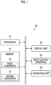
- Fig. 1 is a block diagram illustrating an embodiment of a hardware configuration of an image processing device according to a first embodiment of the present invention.
- An image processing device 10 illustrated in Fig. 1 can be configured by a personal computer, a workstation, or the like and comprises a processor 12, a memory 14, a display unit 20, an input/output interface 22, an operation unit 24, and the like.
- the processor 12 is composed of a central processing unit (CPU) and the like and controls an overall operation of each unit of the image processing device 10.
- the processor 12 executes, for example, a first converter 16 (first converter program) and a second converter 18 (second converter program) stored in the memory 14. Further, the processor 12 functions as an image acquisition unit 30, a first conversion unit 32, a second conversion unit 34, a first similarity calculation unit 36, and an optimization unit 38 illustrated in Fig. 2 .
- the processor 12 may be configured to include a graphics processing unit (GPU).
- GPU graphics processing unit
- the memory 14 includes a flash memory, a read-only memory (ROM), a random access memory (RAM), a hard disk apparatus, and the like.
- the flash memory, the ROM, and the hard disk apparatus are non-volatile memories that store, for example, an operation system, the first converter 16, the second converter 18, and various programs causing the processor 12 to execute processes in each step of an image processing method according to the embodiment of the present invention.
- the hard disk apparatus can function as a large-capacity memory that stores simple X-ray images, three-dimensional X-ray computed tomography (CT) images, and anatomical labels corresponding to the three-dimensional X-ray CT images, which will be described below.
- CT computed tomography
- the RAM functions as a work area for processing by the processor 12 and temporarily stores the programs and the like stored in the non-volatile memory. Further, a portion (RAM) of the memory 14 may be provided in the processor 12.
- the programs corresponding to the first converter 16 and the second converter 18 are stored in the memory 14.
- the processor 12 executes each of the program corresponding to the first converter 16 and the program corresponding to the second converter 18.
- the first converter 16 receives a simple X-ray image 102 (first image) in a two-dimensional image space (first image space) as an input and converts the input simple X-ray image into an image in a two-dimensional feature image space (third image space) characterized by a region of interest. Specifically, the first converter 16 converts the simple X-ray image 102 ( Fig. 3 ) into an image (separated image) in a feature image space characterized by an anatomical region of interest (for example, a bone, a lung, a heart, or an artery).
- the first converter 16 is a converter that is configured (constructed) by a conversion model (separation model) which is a trained model subjected to machine learning in advance. For example, the first converter 16 has been subjected to machine learning in advance to decompose the input simple X-ray image 102 into anatomical structures and to output the separated images.
- the second converter 18 receives a three-dimensional X-ray CT image 106 (second image) in a three-dimensional image space (second image space) and an anatomical label 108 as an input and converts the three-dimensional X-ray CT image 106 into an image in the two-dimensional feature image space (third image space) characterized by the region of interest. Specifically, the second converter 18 masks a label corresponding to the region of interest on the three-dimensional X-ray CT image 106 on the basis of the input anatomical label 108 and projects the masked three-dimensional X-ray CT image 106 onto the two-dimensional feature image space.
- the second converter 18 converts the three-dimensional X-ray CT image 106 into the image (separated image) in the two-dimensional feature image space.
- the separated image is an image having an attenuation value (attenuation) corrected in a projection plane by the second converter 18.
- the anatomical label 108 has three-dimensional label information of the anatomical structure of the three-dimensional X-ray CT image 106. Further, in this example, the anatomical label 108 of the three-dimensional X-ray CT image 106 is input.
- Structural label information of the three-dimensional X-ray CT image 106 may be input.
- the first converter 16 and the second converter 18 perform conversion into an image (separated image) in the feature image space characterized by the region of interest.
- the region of interest is received from the operation unit 24 and is input to the processor 12 (region-of-interest receiving process).
- the reception of the region of interest is not limited to the reception from the operation unit 24, and a preset region of interest may be used.
- the display unit 20 is configured by a display such as a liquid crystal monitor. Various types of necessary information are displayed on the display unit 20. For example, the display unit 20 displays the similarity or final registration parameters calculated by the image processing device 10. In addition, the display unit 20 can be used as a portion of a user interface in a case in which various instructions of the user are received.
- the input/output interface 22 includes, for example, a connection unit that can be connected to an external apparatus and a communication unit that can be connected to a network.
- a universal serial bus (USB) or a high-definition multimedia interface (HDMI) (HDMI is a registered trademark) can be applied as the connection unit that can be connected to the external apparatus.
- the processor 12 can acquire the simple X-ray image, the three-dimensional X-ray CT image 106, the anatomical label 108, and various programs stored in the memory 14 through the input/output interface 22.
- an external display device connected to the input/output interface 22 can be used instead of the display unit 20.
- the operation unit 24 includes a keyboard, a pointing device, such as a mouse, and a keyboard and functions as a user interface that receives various designations.
- Fig. 2 is a functional block diagram illustrating the image processing device 10 according to the first embodiment of the present invention.
- a similarity first similarity
- the simple X-ray image 102 is used as an example of the first image
- the three-dimensional X-ray CT image 106 is used as an example of the second image.
- the present invention is not limited thereto.
- a two-dimensional image is used as the first image
- a three-dimensional image is used as the second image.
- the image processing device 10 is configured by the memory 14 and the processor 12 having the hardware configuration illustrated in Fig. 1 .
- the processor 12 functions as the image acquisition unit 30, the first conversion unit 32, the second conversion unit 34, the first similarity calculation unit 36, and the optimization unit 38.
- the image acquisition unit 30 performs an image acquisition process to acquire the simple X-ray image 102, the three-dimensional X-ray CT image 106, and the anatomical label 108 corresponding to the three-dimensional X-ray CT image 106 stored in the memory 14.
- the simple X-ray image 102, the three-dimensional X-ray CT image 106, and the anatomical label 108 are data for the same patient A.
- the simple X-ray image 102 is an X-ray image obtained by irradiating the patient A with X-rays in one direction.
- the three-dimensional X-ray CT image 106 is an image generated by irradiating a body of the patient A with X-rays while rotating around the body of the patient A, collecting three-dimensional information of the inside of the body, and performing computer processing.
- the anatomical label 108 is label information indicating an anatomical structure, such as an organ or a bone of the patient A, obtained on the basis of the three-dimensional X-ray CT image 106.
- the image acquisition unit 30 acquires the simple X-ray image 102 as a two-dimensional input and acquires the three-dimensional X-ray CT image 106 and the anatomical label 108 as a three-dimensional input.
- the first conversion unit 32 performs a first conversion process using the first converter 16.
- the first conversion unit 32 converts the simple X-ray image 102 into a separated image in the feature image space (third image space) characterized by the region of interest, using the first converter 16.
- anatomical regions of interest are adopted as the regions of interest, and four regions of a bone, a lung, a heart, and an artery in the chest of the human body are adopted.
- Fig. 3 is a diagram illustrating the process of the first conversion unit 32.
- the first conversion unit 32 has the first converter 16, and the simple X-ray image 102 obtained by imaging the chest of the human body is input to the first converter 16. Then, the first converter 16 converts the simple X-ray image 102 into a separated image in the feature image space characterized by the region of interest. Specifically, the first converter 16 converts the simple X-ray image 102 into a feature image space of the bone, a feature image space of the lung, a feature image space of the heart, a feature image space of an aorta as the feature image spaces characterized by the regions of interest. Fig.
- the simple X-ray image 102 is converted into images in a plurality of feature image spaces.
- the present invention is not limited thereto.
- the simple X-ray image 102 may be converted into an image in a single feature image space.
- the second conversion unit 34 ( Fig. 2 ) performs a second conversion process using the second converter 18.
- the second conversion unit 34 converts the three-dimensional X-ray CT image 106 into a separated image in the feature image space (third image space) characterized by the region of interest, using the second converter 18.
- the anatomical region of interest is adopted as the region of interest, and four regions of the bone, the lung, the heart, and the artery in the chest of the human body are adopted.
- Fig. 4 is a diagram illustrating the process of the second conversion unit 34.
- the second conversion unit 34 has the second converter 18, and the three-dimensional X-ray CT image 106 obtained by imaging the chest of the human body and the anatomical label 108 are input to the second converter 18. Further, a geometry 40 and a registration parameter ⁇ (indicated by reference numeral 42) are input to the second converter 18.
- the geometry 40 is information related to an X-ray examination apparatus including imaging conditions for capturing the three-dimensional X-ray CT image 106.
- the geometry 40 includes, for example, a source-to-object distance (SOD), a source-to-detector distance (SDD), and general X-ray imaging conditions.
- the registration parameter ⁇ is a deformation parameter for the three-dimensional X-ray CT image 106 as described below. Further, ⁇ (initial) is set as an initial value of the registration parameter ⁇ , and the registration parameter ⁇ is updated by the optimization unit 38 which will be described below. A registration parameter ⁇ F that maximizes the similarity is calculated.
- Fig. 5 is a diagram illustrating the registration parameter ⁇ .
- Fig. 5 illustrates a virtual X-ray source O and a virtual detector Q.
- the virtual detector Q detects a projected two-dimensional simple X-ray image I.
- the simple X-ray image I can be changed by changing the registration parameter ⁇ .
- the registration parameter ⁇ is a parameter for changing the direction and inclination of the three-dimensional X-ray CT image 106 and is also a parameter for deforming the organ in the three-dimensional X-ray CT image 106 in the second converter 18.
- the registration parameter ⁇ is a parameter for changing the direction and inclination of the three-dimensional X-ray CT image 106 and is also a parameter for deforming the organ in the three-dimensional X-ray CT image 106 in the second converter 18.
- the description has been made on the premise of rigid deformation.
- the three-dimensional X-ray CT image H may be deformed and registered using a well-known non-rigid deformation method.
- the three-dimensional X-ray CT image 106, the anatomical label 108, the geometry 40, and the registration parameter ⁇ are input to the second converter 18. Then, the second converter 18 converts the three-dimensional X-ray CT image 106 into a separated image 110A projected to the feature image space of the bone, a separated image 110B projected to the feature image space of the lung, a separated image 110C projected to the feature image space of the heart, and a separated image 110D projected to the feature image space of the aorta.
- the first similarity calculation unit 36 ( Fig. 2 ) performs a first similarity calculation process to calculate the similarities between the separated images in each of the feature image spaces characterized by the regions of interest converted by the first conversion process and the second conversion process.
- the first similarity calculation unit 36 can calculate the similarities between the separated images using various methods. For example, the first similarity calculation unit 36 calculates the similarities using a Euclidean distance. For example, in a case in which the separated image 104A and the separated image 110A are composed of n ⁇ m pixels, the first similarity calculation unit 36 can calculate the distance between the separated image 104A and the separated image 110A as an n ⁇ m-dimensional vector using the Euclidean distance to obtain the similarity.
- the first similarity calculation unit 36 can calculate the similarity between the separated image 104B and the separated image 110B, the similarity between the separated image 104C and the separated image 110C, and the similarity between the separated image 104D and the separated image 110D. Then, the first similarity calculation unit 36 integrates the similarities calculated in each feature image space.
- Fig. 6 is a diagram illustrating the calculation of the similarity in the first similarity calculation unit 36.
- the first similarity calculation unit 36 calculates the similarities in each feature image space and integrates the calculated similarities to calculate the similarity (total) between the simple X-ray image 102 and the three-dimensional X-ray CT image 106. Specifically, the first similarity calculation unit 36 integrates the similarity (bone) calculated between the separated images (the separated image 104A and the separated image 110A) in the feature image space of the bone, the similarity (lung) calculated between the separated images (the separated image 104B and the separated image 110B) in the feature image space of the lung, the similarity (heart) calculated between the separated images (the separated image 104C and the separated image 110C) in the feature image space of the heart, and the similarity (artery) calculated from the separated images (the separated image 104D and the separated image 110D) of the feature image space of the artery to calculate the similarity (total).
- the first similarity calculation unit 36 integrates the similarities to calculate the similarity (total), using various methods. For example, the first similarity calculation unit 36 adds, multiplies, or averages the similarities to calculate the similarity (total) which is one scalar.
- the first similarity calculation unit 36 adds, multiplies, or averages the similarities to calculate the similarity (total) which is one scalar.
- the optimization unit 38 ( Fig. 2 ) performs an optimization process. Specifically, the optimization unit 38 optimizes the registration parameter ⁇ on the basis of the similarity (total) calculated by the first similarity calculation unit 36. For example, the optimization unit 38 updates the registration parameter ⁇ using an optimization algorithm (covariance matrix adaptation evolution strategy (CMA-ES)) and calculates a registration parameter ⁇ F that maximizes the similarity. As described above, the optimization unit 38 can optimize the registration parameter ⁇ to acquire the registration parameter ⁇ F that maximizes the similarity.
- CMA-ES covariance matrix adaptation evolution strategy
- Fig. 7 is a flowchart illustrating the image processing method using the image processing device 10.
- the processor 12 of the image processing device 10 executes a dedicated program to perform each step of the image processing method.
- the image acquisition unit 30 acquires, for example, the simple X-ray image 102 stored in the memory 14 (Step S10: image acquisition step). In addition, the image acquisition unit 30 acquires, for example, the three-dimensional X-ray CT image 106 stored in the memory 14 (Step S11: image acquisition step). Further, the image acquisition unit 30 acquires, for example, the anatomical label 108 corresponding to the three-dimensional X-ray CT image 106 stored in the memory 14 (Step S12). Then, the first conversion unit 32 performs the first conversion process (Step S13: first conversion step). Specifically, the first conversion unit 32 converts the simple X-ray image 102 into the separated images 104A to 104D.
- the second conversion unit 34 performs the second conversion process (Step S14: second conversion step). Specifically, the second conversion unit 34 converts the three-dimensional X-ray CT image 106 into the separated images 110A to 110D. Then, the first similarity calculation unit 36 calculates the similarity between the simple X-ray image 102 and the three-dimensional X-ray CT image 106 (Step S15: first similarity calculation step). Then, the optimization unit 38 determines whether or not the calculated similarity has a maximum value (Step S16). In a case in which the calculated similarity does not have the maximum value, the optimization unit 38 updates the registration parameter ⁇ (Step S18).
- Step S14 the second conversion process
- the conversion into the separated images 110A to 110D is performed on the basis of the updated registration parameter ⁇ .
- the registration parameter ⁇ F at which the maximum similarity has been calculated is output (Step S17).
- the simple X-ray image 102 and the three-dimensional X-ray CT image 106 are converted into the feature image spaces characterized by the regions of interest, and the similarities between the separated images 104A to 104D and the separated images 110A to 110D after the conversion are calculated. Therefore, even in a case in which it is not easy to design the similarity between the input images in the image space, it is possible to easily calculate an interpretable similarity with high accuracy without requiring correct answer data.
- Fig. 8 is a diagram illustrating a first conversion unit 32 and a second conversion unit 34 according to Modification Example 1.
- the portions already described in Fig. 6 are denoted by the same reference numerals, and the description thereof will not be repeated.
- the first conversion unit 32 comprises a first converter-1, a first converter-2, a first converter-3, and a first converter-4.
- the first conversion unit 32 converts the simple X-ray image 102 into the separated image 104A, the separated image 104B, the separated image 104C, and the separated image 104D using the first converter-1, the first converter-2, the first converter-3, and the first converter-4, respectively.
- the second conversion unit 34 comprises a second converter-1, a second converter-2, a second converter-3, and a second converter-4.
- the second conversion unit 34 converts the three-dimensional X-ray CT image 106 into the separated image 110A, the separated image 110B, the separated image 110C, and the separated image 110D using the second converter-1, the second converter-2, the second converter-3, and the second converter-4, respectively.
- the first conversion unit 32 and the second conversion unit 34 according to this example comprise a plurality of converters and perform conversion into the separated images in the feature image spaces corresponding to each converter.
- the first similarity calculation unit 36 calculates similarities (a similarity (bone), a similarity (lung), a similarity (heart), and a similarity (artery)) for the images in each feature image space and integrates the similarities in each feature image space.
- the first conversion unit 32 and the second conversion unit 34 comprise a plurality of converters corresponding to the feature image spaces to be converted. As described above, since a plurality of converters corresponding to the feature image spaces are provided, it is possible to perform conversion more suitable for the feature image spaces.
- the first converter 16 outputs a first reliability indicating the reliability of the separated image after conversion
- the second converter outputs a second reliability indicating the reliability of the separated image after conversion.
- the first reliability and the second reliability may be output for each separated image or may be output for each pixel of the separated image.
- the reliability is a value indicating the certainty of the separated image after conversion.
- the first converter is configured by a trained model (conversion model (separation model)) as described above
- the second converter is configured by a trained model (conversion model (projection model)) as described above. Therefore, in a case in which data that is different or heterogeneous from the training data subjected to machine learning is input to the first converter, the reliability of the separated image converted by the first converter 16 is reduced. On the other hand, in a case in which the same data as the training data subjected to machine learning is input to the first converter 16, the reliability of the separated image converted by the first converter 16 is increased.
- the reliability of the separated image to be converted by the second converter is reduced.
- the reliability of the separated image converted by the second converter 18 is increased.
- Fig. 9 is a diagram illustrating the reliability.
- the portions already described in Figs. 6 and 8 are denoted by the same reference numerals, and the description thereof will not be repeated.
- the first converter-1 outputs a reliability A for the separated image 104A
- the first converter-2 outputs a reliability B for the separated image 104B
- the first converter-3 outputs a reliability C for the separated image 104C
- the first converter-4 outputs a reliability D for the separated image 104D.
- the second converter-1 outputs a reliability E for the separated image 110A
- the second converter-2 outputs a reliability F for the separated image 110B
- the second converter-3 outputs a reliability G for the separated image 110C
- the second converter-4 outputs a reliability H for the separated image 110D.
- the first similarity calculation unit 36 performs weighting on the basis of the reliability A and the reliability E in a case in which the similarity (bone) between the separated image 104A and the separated image 110A is calculated. Further, the first similarity calculation unit 36 performs weighting on the basis of the reliability B and the reliability F in a case in which the similarity (lung) between the separated image 104B and the separated image 110B is calculated. Furthermore, the first similarity calculation unit 36 performs weighting on the basis of the reliability C and the reliability G in a case in which the similarity (heart) between the separated image 104C and the separated image 110C is calculated.
- the first similarity calculation unit 36 performs weighting on the basis of the reliability D and the reliability H in a case in which the similarity (artery) between the separated image 104D and the separated image 110D is calculated. Then, the first similarity calculation unit 36 integrates the similarity (bone), the similarity (lung), the similarity (heart), and the similarity (artery) to calculate the similarity (total). The first similarity calculation unit 36 calculates the similarity (total) weighted on the basis of the reliabilities A to H.
- the similarity (bone), the similarity (lung), the similarity (heart), and the similarity (artery) are weighted on the basis of the reliabilities A to H, respectively, and the similarity (total) obtained by integrating the similarity (bone), the similarity (lung), the similarity (heart), and the similarity (artery) is also weighted on the basis of the reliabilities A to H.
- the similarity (total) is weighted on the basis of the reliabilities, it is possible to output the registration parameter ⁇ F with higher accuracy.
- the aspect has been described in which all of the first converter-1 to the first converter-4 and the second converter-1 to the second converter-4 output the reliability.
- the reliability may be output only from the first converters (the first converter-1 to the first converter-4), and the similarity (total) having a weight may be calculated on the basis of the reliability.
- the reliability may be output only from the second converters (second converter-1 to second converter-4), and the similarity (total) having a weight may be calculated on the basis of the reliability.
- selection may be performed on the basis of the reliabilities A to H to calculate each similarity. Specifically, for example, the separated image having a relatively low reliability may be excluded, and the similarity may be calculated.
- a second similarity calculation unit 120 is further provided to calculate a pre-conversion similarity (second similarity) which is a similarity between the simple X-ray image 102 and the three-dimensional X-ray CT image 106 before conversion by the first conversion unit 32 and the second conversion unit 34.
- second similarity a pre-conversion similarity
- Fig. 10 is a diagram illustrating the calculation of the similarity in the second similarity calculation unit 120 according to this example.
- the portions already described in Fig. 6 are denoted by the same reference numerals, and the description thereof will not be repeated.
- the second similarity calculation unit 120 performs a second similarity calculation process by the execution of the program stored in the memory by the processor 12.
- the simple X-ray image 102 and the three-dimensional X-ray CT image 106 are input to the second similarity calculation unit 120.
- the second similarity calculation unit 120 calculates the pre-conversion similarity on the basis of the simple X-ray image 102 and the three-dimensional X-ray CT image 106.
- the second similarity calculation unit 120 calculates the pre-conversion similarity before the simple X-ray image 102 and the three-dimensional X-ray CT image 106 are converted by the first conversion unit 32 and the second conversion unit 34, respectively.
- the second similarity calculation unit 120 can calculate the pre-conversion similarity between the simple X-ray image 102 and the three-dimensional X-ray CT image 106 using a known method. For example, the second similarity calculation unit 120 superimposes bone portions in the simple X-ray image 102 and the three-dimensional X-ray CT image 106 to calculate the pre-conversion similarity. In addition, for example, the second similarity calculation unit 120 calculates, as the pre-conversion similarity, an amount of mutual information between the simple X-ray image 102 and a pseudo X-ray image generated from the three-dimensional X-ray CT image 106.
- the second similarity calculation unit 120 calculates, as the pre-conversion similarity, a gradient correlation between the simple X-ray image 102 and the pseudo X-ray image generated from the three-dimensional X-ray CT image 106.
- the pseudo X-ray image is generated by the second similarity calculation unit 120.
- the second similarity calculation unit 120 generates a pseudo X-ray image that imitates the two-dimensional simple X-ray image from the three-dimensional X-ray CT image 106 using a known method.
- the first similarity calculation unit 36 calculates a similarity obtained by weighting the calculated similarity (total) on the basis of the pre-conversion similarity. Then, the optimization unit 38 optimizes the registration parameter ⁇ on the basis of the weighted similarity.
- the similarity weighted on the basis of the pre-conversion similarity is output, which makes it possible to efficiently optimize the registration parameter ⁇ .
- the similarity and the registration parameter ⁇ F can be used for various purposes.
- the registration parameter ⁇ F calculated by the image processing device 10 can be used for a correct answer data generation device that generates correct answer data for performing machine learning on a detector which interprets the simple X-ray image.
- Fig. 11 is a diagram illustrating generation of a learning data set including correct answer data.
- Fig. 11 illustrates a simple X-ray image 122 and a three-dimensional X-ray CT image 124 of a patient B.
- the patient B has a lesion portion S in the lung.
- the maximum similarity between the simple X-ray image 122 and the three-dimensional X-ray CT image 124 is calculated by the image processing device 10, and the registration parameter ⁇ F is calculated. Therefore, correct answer data 126 having the lesion portion S can be generated by inputting the three-dimensional X-ray CT image 124 and the registration parameter ⁇ F to the correct answer data generation device comprising a converter (projection model).
- the correct answer data 126 is data corresponding to the two-dimensional simple X-ray image 122.
- the position of the lesion portion S is accurately projected onto the correct answer data 126. Therefore, machine learning can be performed by performing learning such that an error between the correct answer data 126 and an interpretation result output from the detector, which receives the input of the simple X-ray image 122 and interprets the simple X-ray image 122, is reduced.
- the use of the registration parameter ⁇ F makes it possible to efficiently generate correct answer data for performing machine learning on the interpretation of the simple X-ray image.
- Another example is a similar image search device that searches for a similar image on the basis of the similarity calculated by the image processing device 10. Specifically, for example, in a case in which a three-dimensional X-ray CT image similar to a simple X-ray image is searched for, the similar image is searched for on the basis of the similarity calculated by the image processing device 10. For example, a three-dimensional X-ray CT image having the maximum similarity with the input simple X-ray image can be extracted as the similar image.
- the first converter 16 converts the simple X-ray image 102 into the separated images and the second converter 18 converts the three-dimensional X-ray CT image 106 into the separated images.
- the first converter 16 converts the simple X-ray image 102 into a shape feature and a texture feature
- the second converter 18 converts the three-dimensional X-ray CT image 106 into a shape feature and a texture feature.
- the existing similarities are broadly divided into measures based on the shape feature or the texture feature.
- a technique related to registration using the similarity based on the shape feature is described in, for example, Toth, Daniel, et al. "3D/2D model-to-image registration by imitation learning for cardiac procedures.”, UCARS, 2018 . and Ruijters, Daniel, Bart M. ter Haar Romeny, and Paul Suetens. "Vesselness-based 2D-3D registration of the coronary arteries.” International journal of computer assisted radiology and surgery 4.4 (2009): 391-397 .
- a technique related to registration using the similarity based on the texture feature is disclosed in, for example, Van der Bom, I. M. J., et al.
- the above-mentioned trade-off between the shape feature and the texture feature can be automatically adjusted to calculate the similarity with high accuracy and high robustness.
- the similarity is not automatically designed by metric learning, it is possible to provide the similarity that can be interpreted by humans.
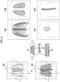
- Fig. 12 is a diagram illustrating a case in which the first converter 16 converts the simple X-ray image 102 into a first shape feature 202 and a first texture feature 204.
- the simple X-ray image 102 obtained by imaging the chest of the human body in a state in which a surgical tool T is inserted is input to the first converter 16. Then, the first converter 16 converts the simple X-ray image 102 into the first shape feature 202 and the first texture feature 204. In the case illustrated in Fig. 12 , the first converter 16 converts the simple X-ray image 102 into the first shape feature 202 of a lung shape indicated by thickness information of the lungs. Further, in the case illustrated in Fig. 12 , the first converter 16 converts the simple X-ray image 102 into the first texture feature 204 of a lung texture indicated by blood vessels in the lungs.
- the first converter 16 outputs a first shape feature reliability 202A ((1-1)-th reliability) and a first texture feature reliability 204A ((1-2)-th reliability).
- a black portion indicates a portion having a high reliability
- a white portion indicates a portion having a low reliability.
- the first shape feature reliability 202A has a high reliability as a whole since the shape feature is indicated by information related to the thickness of the lungs.
- the first texture feature reliability 204A since the lung texture indicated by the blood vessels in the lungs is shown, a portion in which the blood vessels are not capable of being detected well has a low reliability.
- the first texture feature reliability 204A since fine blood vessels are not capable of being detected in the vicinity of the pleura Q1, the vicinity of the pleura Q1 has a low reliability.
- the blood vessels overlapping the surgical tool T are not capable of being detected, a portion Q2 overlapping the surgical tool T has a low reliability.
- the reliability may be calculated for each pixel or may be calculated for each predetermined region.
- the conversion by the first converter 16 has been described in Fig. 12 .
- the three-dimensional X-ray CT image 106 is input to the second converter 18, and the second converter 18 outputs a second shape feature 206 ( Fig. 13 ) and a second texture feature 208 (see Fig. 13 ).
- the second converter 18 outputs a second shape feature reliability 206A ((2-1)-th reliability) and a second texture feature reliability 208A ((2-2)-th reliability).
- first shape feature reliability 202A (second shape feature reliability 206A) and the first texture feature reliability 204A (second texture feature reliability 208A) are output has been described with reference to Fig. 12 .
- these reliabilities may not be output.
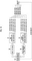
- Fig. 13 is a diagram illustrating a first example of the second embodiment.
- Fig. 13 is a diagram in which the image acquisition unit 30 of Fig. 2 is omitted.
- the portions that have already been described are denoted by the same reference numerals, and the description thereof will not be repeated.
- the first converter 16 receives the simple X-ray image 102 as an input and converts the simple X-ray image 102 into the first shape feature 202 and the first texture feature 204.
- the first shape feature 202 is an image in the third image space characterized by the region of interest and has information related to the shape feature of the region of interest in the simple X-ray image 102.
- the first texture feature 204 is an image in the third image space characterized by the region of interest and has information related to the texture feature in the simple X-ray image 102.
- the first converter 16 converts the simple X-ray image 102 into the first shape feature 202 which is an image in the feature image space (third image space) characterized by the shape feature of the anatomical region of interest (lung) (see Fig. 12 ).
- the first converter 16 converts the simple X-ray image 102 into the first texture feature 204 which is an image in the feature image space (third image space) characterized by the texture feature of the anatomical region of interest (lung) (see Fig. 12 ).
- the second converter 18 converts the three-dimensional X-ray CT image 106 into the second shape feature 206 and the second texture feature 208.
- the second shape feature 206 is an image in the third image space characterized by the region of interest and has information related to the shape feature of the region of interest in the three-dimensional X-ray CT image 106.
- the second texture feature 208 is an image in the third image space characterized by the region of interest and has information related to the texture feature of the region of interest in the three-dimensional X-ray CT image 106.
- the first similarity calculation unit 36 performs the first similarity calculation process to calculate the similarity between the shape features and the similarity between the texture features converted by the first conversion process and the second conversion process. Specifically, the first similarity calculation unit 36 calculates a shape similarity 210 ((1-1)-th similarity) which is a similarity between the first shape feature 202 and the second shape feature 206. Further, the first similarity calculation unit 36 calculates a texture similarity 212 ((1-2)-th similarity) which is a similarity between the first texture feature 204 and the second texture feature 208. Then, the first similarity calculation unit 36 calculates a final similarity 214 (first similarity) from the shape similarity 210 and the texture similarity 212. In addition, since the calculation of the similarity has already been described in the first embodiment, the description thereof will not be repeated here.
- the final similarity 214 is input to the optimization unit 38 as described in the first embodiment.
- the optimization unit 38 optimizes the registration parameter ⁇ on the basis of the final similarity calculated by the first similarity calculation unit 36.
- the optimization unit 38 updates the registration parameter ⁇ to calculate the registration parameter ⁇ F that maximizes the final similarity, using an optimization algorithm. This optimization of the registration parameter ⁇ by the optimization unit 38 makes it possible to automatically adjust the trade-off between the shape feature and the texture feature and to obtain a similarity with high accuracy and high robustness.
- the first converter 16 converts the simple X-ray image 102 into the first shape feature 202 and the first texture feature 204
- the second converter 18 converts the three-dimensional X-ray CT image 106 into the second shape feature 206 and the second texture feature 208.
- the first similarity calculation unit 36 calculates the shape similarity 210 from the first shape feature 202 and the second shape feature 206 and calculates the texture similarity 212 from the first texture feature 204 and the second texture feature 208.
- the first similarity calculation unit 36 calculates the final similarity.
- the optimization unit 38 updates the registration parameter ⁇ such that the final similarity is maximized.
- the first converter 16 further outputs the reliability of the first shape feature 202 and the reliability of the first texture feature 204.
- Fig. 14 is a diagram illustrating the second example of the second embodiment.
- the portions already described in Fig. 13 are denoted by the same reference numerals, and the description thereof will not be repeated.
- the first converter 16 converts the simple X-ray image 102 into the first shape feature 202 and calculates the first shape feature reliability 202A.
- the first shape feature reliability 202A is information indicating the reliability of the first shape feature 202.
- the first converter 16 converts the simple X-ray image 102 into the first texture feature 204 and calculates the first texture feature reliability 204A.
- the first texture feature reliability 204A is information indicating the calculated reliability of the first texture feature 204.
- the first similarity calculation unit 36 weights the first shape feature 202 on the basis of the first shape feature reliability 202A and calculates the shape similarity 210. Further, in a case in which the texture similarity 212 is calculated, the first similarity calculation unit 36 weights the first texture feature 204 on the basis of the first texture feature reliability 204A and calculates the texture similarity 212. As described above, the first similarity calculation unit 36 performs weighting on the basis of the first shape feature reliability 202A and the first texture feature reliability 204A and calculates the shape similarity 210 and the texture similarity 212.
- the first similarity calculation unit 36 may select the shape similarity 210 and the texture similarity 212 on the basis of the first shape feature reliability 202A and the first texture feature reliability 204A.
- the first converter 16 further outputs the reliability of the first shape feature 202 and the reliability of the first texture feature 204
- the second converter 18 further outputs the reliability of the second shape feature 206 and the reliability of the second texture feature 208.
- Fig. 15 is a diagram illustrating the third example of the second embodiment.
- the portions already described in Figs. 13 and 14 are denoted by the same reference numerals, and the description thereof will not be repeated.
- the first converter 16 converts the simple X-ray image 102 into the first shape feature 202 and calculates the first shape feature reliability 202A. Further, the first converter 16 converts the simple X-ray image 102 into the first texture feature 204 and calculates the first texture feature reliability 204A.
- the second converter 18 converts the three-dimensional X-ray CT image 106 into the second shape feature 206 and calculates the second shape feature reliability 206A. Further, the second converter 18 converts the three-dimensional X-ray CT image 106 into the second texture feature 208 and calculates the second texture feature reliability 208A.
- the first similarity calculation unit 36 weights the first shape feature 202 on the basis of the first shape feature reliability 202A, weights the second shape feature 206 on the basis of the second shape feature reliability 206A, and calculates the shape similarity 210. Further, in a case in which the texture similarity 212 is calculated, the first similarity calculation unit 36 weights the first texture feature 204 on the basis of the first texture feature reliability 204A, weights the second texture feature 208 on the basis of the second texture feature reliability 208A, and calculates the texture similarity 212.
- the first similarity calculation unit 36 performs weighting on the basis of the first shape feature reliability 202A, the first texture feature reliability 204A, the second shape feature reliability 206A, and the second texture feature reliability 208A and calculates the shape similarity 210 and the texture similarity 212. Therefore, in this example, even in a case in which the reliability of the conversion by the first converter 16 and the reliability of the conversion by the second converter 18 are significantly different from each other, it is possible to accurately calculate the final similarity 214.
- the first converter 16 further outputs the reliability of the first shape feature 202 and the reliability of the first texture feature 204
- the second converter 18 further outputs the reliability of the second shape feature 206 and the reliability of the second texture feature 208.
- Fig. 16 is a diagram illustrating the fourth example of the second embodiment.
- the portions already described in Figs. 13 to 15 are denoted by the same reference numerals, and the description thereof will not be repeated.
- the first converter 16 converts the simple X-ray image 102 into the first shape feature 202 and the first texture feature 204. Further, the first converter 16 outputs the first shape feature reliability 202A and the first texture feature reliability 204A.
- the second converter 18 converts the three-dimensional X-ray CT image 106 into the second shape feature 206 and the second texture feature 208. Further, the second converter 18 outputs the second shape feature reliability 206A and the second texture feature reliability 208A.
- the first similarity calculation unit 36 selects, as the final similarity, the shape similarity 210 or the texture similarity 212 on the basis of the first shape feature reliability 202A, the first texture feature reliability 204A, the second shape feature reliability 206A, and the second texture feature reliability 208A. Specifically, the first similarity calculation unit 36 compares the first shape feature reliability 202A and the second shape feature reliability 206A with the first texture feature reliability 204A and the second texture feature reliability 208A, selects the shape similarity 210 in a case in which the first shape feature reliability 202A and the second shape feature reliability 206A are higher, and selects the texture similarity 212 in a case in which the first texture feature reliability 204A and the second texture feature reliability 208A are higher.
- the first similarity calculation unit 36 selects the shape similarity 210 or the texture similarity 212 as the final similarity on the basis of the first shape feature reliability 202A, the first texture feature reliability 204A, the second shape feature reliability 206A, and the second texture feature reliability 208A. Therefore, it is possible to calculate the final similarity on the basis of the shape similarity 210 or the texture feature similarity 212 having a higher reliability.
- a first shape feature 220 according to this example is a shape of bronchi.
- Fig. 17 is a diagram illustrating a case in which the first converter 16 converts the simple X-ray image 102 into the first shape feature 220 and the first texture feature 204.
- the portions already described in Fig. 12 are denoted by the same reference numerals, and the description thereof will not be repeated.
- the second converter 18 converts the three-dimensional X-ray CT image 106 into the second shape feature 206 and the second texture feature 208 to correspond to the first converter 16.
- the simple X-ray image 102 obtained by imaging the chest of the human body is input to the first converter 16. Then, the first converter 16 converts the simple X-ray image 102 into the first shape feature 220 and the first texture feature 204. In the case illustrated in Fig. 17 , the first converter 16 converts the simple X-ray image 102 into the first shape feature 220 having information related to the shape of the bronchi. Further, in the case illustrated in Fig. 17 , the first converter 16 converts the simple X-ray image 102 into the first texture feature 204 of the lung texture indicated by the blood vessels in the lungs.
- the first converter 16 outputs a first shape feature reliability 220A and the first texture feature reliability 204A.
- a black portion indicates a portion with a high reliability
- a white portion indicates a portion with a low reliability.
- a white dotted line indicates a shape.
- the first shape feature reliability 220A a portion in which the shape of a part other than a peripheral part Q3 of the bronchus can be sufficiently detected has a high reliability.
- the peripheral part Q3 since blood vessels are not capable of being detected well in the peripheral part Q3 of the bronchus, the peripheral part Q3 has a low reliability. Therefore, in the simple X-ray image 102, a portion in which the shape of the bronchi is visible has a high reliability, and registration is directly performed.
- the surgical tool T has restrictions on passing through the bronchus. Therefore, it is considered that uncertainty is not increased even in a case in which the bronchus and the surgical tool T overlap each other.
- the peripheral part of the bronchus is not capable of being detected well from the simple X-ray image 102. Therefore, registration is indirectly performed on a peripheral region of the bronchus by the running of the blood vessels.
- registration is performed on the basis of the similarity between the shape feature of the shape of the bronchi and the lung texture. Therefore, it is possible to obtain a similarity with high accuracy and high robustness.
- the registration is performed by calculating the similarity between the shape feature and the texture feature.
- the registration is performed by calculating a similarity between an anatomical region and a disease region.
- Fig. 18 is a diagram illustrating a case in which the first converter 16 converts the simple X-ray image 102 into a first anatomical region 230 and a first disease region 240.
- the first converter 16 converts the simple X-ray image 102 into a first anatomical region 230 and a first disease region 240.
- the second converter 18 converts the three-dimensional X-ray CT image 106 into a second anatomical region and a second disease region to correspond to the first converter 16.
- the first converter 16 receives the simple X-ray image 102 obtained by imaging the chest of the human body as an input and converts the simple X-ray image 102 into the first anatomical region 230 and the first disease region 240.
- the first converter 16 converts the simple X-ray image 102 into the first anatomical region 230 related to the blood vessels in the lungs.
- the first converter 16 converts the simple X-ray image 102 into the first disease region 240 related to lung nodules.
- the second converter 18 converts the three-dimensional X-ray CT image 106 into the second anatomical region and the second disease region.
- the first converter 16 outputs a first anatomical region reliability 230A ((1-1)-th reliability) indicating the reliability of the first anatomical region 230 and a first disease region reliability 240A ((1-2)-th reliability) indicating the reliability of the first disease region 240.
- the second converter 18 outputs a second anatomical region reliability ((2-1)-th reliability) indicating the reliability of the second anatomical region and a second disease region reliability ((2-2)-th reliability) indicating the reliability of the second disease region.
- a black portion indicates a portion having a high reliability
- a white portion indicates a portion having a low reliability.
- a white dotted line indicates a shape.
- the first anatomical region reliability 230A is indicated by the anatomical region of the blood vessels running in the lungs, a portion in which the blood vessels are not capable of being detected well has a low reliability. Specifically, in the first anatomical region reliability 230A, since fine blood vessels are not capable of being detected in the vicinity of the pleura Q4, the vicinity of the pleura Q4 has a low reliability. In addition, in the first anatomical region reliability 230A, since the blood vessel overlapping the surgical tool T is not capable of being detected, a portion Q5 overlapping the surgical tool T has a low reliability.
- a large nodule (see reference numeral Q8) has a high reliability, and a minute nodule or a nodule overlapping a bone or a hilum of the lung has a low reliability (see reference numerals Q6 and Q7).
- the registration is indirectly performed for the small nodule having a low reliability or the nodule overlapping the bone or the hilum of the lung due to the running of the blood vessels (with reference to the first anatomical region and the second anatomical region) and is directly performed for the nodule having a large reliability (with reference to the first disease region and the second disease region).
- the first similarity calculation unit 36 performs the first similarity calculation process to calculate the similarity between the anatomical regions converted by the first conversion process and the similarity between the disease regions converted by the second conversion process. Specifically, the first similarity calculation unit 36 calculates an anatomical region similarity ((1-1)-th similarity) which is a similarity between the first anatomical region 230 and the second anatomical region. In addition, the first similarity calculation unit 36 calculates a disease region similarity ((1-2)-th similarity) which is a similarity between the first disease region 240 and the second disease region. The first similarity calculation unit 36 calculates the final similarity (first similarity) from the anatomical region similarity and the disease region similarity.
- anatomical region similarity ((1-1)-th similarity) which is a similarity between the first anatomical region 230 and the second anatomical region.
- a disease region similarity ((1-2)-th similarity) which is a similarity between the first disease region 240 and the second disease region
- the first similarity calculation unit 36 may calculate the anatomical region similarity and the disease region similarity on the basis of the first anatomical region reliability 230A and the first disease region reliability 240A and calculate the final similarity.
- the first similarity calculation unit 36 may weight the anatomical region similarity and the disease region similarity on the basis of the first anatomical region reliability 230A and the first disease region reliability 240A and calculate the final similarity.
- the first similarity calculation unit 36 may select the anatomical region similarity or the disease region similarity on the basis of the anatomical region reliability 230A and the first disease region reliability 240A and calculate the final similarity.
- the first similarity calculation unit 36 may calculate the anatomical region similarity and the disease region similarity on the basis of the first anatomical region reliability 230A, the first disease region reliability 240A, the second anatomical region reliability, and the second disease region reliability and calculate the final similarity.
- the first similarity calculation unit 36 may weight the anatomical region similarity on the basis of the first anatomical region reliability 230A and the second anatomical region reliability and may weight the disease region similarity on the basis of the first disease region reliability 240A and the second disease region reliability to calculate the final similarity.
- the first similarity calculation unit 36 may select the anatomical region similarity or the disease region similarity on the basis of the first anatomical region reliability 230A, the first disease region reliability 240A, the second anatomical region reliability, and the second disease region reliability and calculate the final similarity.
- the registration is performed on the basis of the similarity between the anatomical region and the disease region. Therefore, it is possible to obtain a similarity with high accuracy and high robustness.
- a hardware structure of a processing unit that executes various processes is the following various processors.
- the various processors include, for example, a central processing unit (CPU) which is a general-purpose processor executing software (program) to function as various processing units, a programmable logic device (PLD), such as a field programmable gate array (FPGA), which is a processor whose circuit configuration can be changed after manufacture, and a dedicated electric circuit, such as an application specific integrated circuit (ASIC), which is a processor having a dedicated circuit configuration designed to perform a specific process.
- CPU central processing unit
- PLD programmable logic device
- FPGA field programmable gate array
- ASIC application specific integrated circuit
- One processing unit may be configured by one of these various processors or by two or more processors of the same type or different types (for example, a plurality of FPGAs or a combination of a CPU and an FPGA). Moreover, a plurality of processing units may be configured by one processor.
- a first example of the configuration in which a plurality of processing units are configured by one processor is an aspect in which one processor is configured by a combination of one or more CPUs and software and functions as a plurality of processing units.
- a representative example of this aspect is a client computer or a server computer.
- a second example of the configuration is an aspect in which a processor that implements the functions of the entire system including a plurality of processing units using one integrated circuit (IC) chip is used.
- IC integrated circuit
- a representative example of this aspect is a system-on-chip (SoC).
- SoC system-on-chip
- an electric circuit obtained by combining circuit elements, such as semiconductor elements, can be used as the hardware structure of the various processors.
- Each of the above-described configurations and functions can be implemented by any hardware, software, or a combination thereof as appropriate.
- the present invention can be also applied to a program that causes a computer to execute the above-described processing steps (processing procedures), a computer-readable recording medium (non-transitory recording medium) having the program recorded thereon, or a computer in which the program can be installed.
Landscapes
- Engineering & Computer Science (AREA)
- Health & Medical Sciences (AREA)
- Physics & Mathematics (AREA)
- Medical Informatics (AREA)
- Theoretical Computer Science (AREA)
- Life Sciences & Earth Sciences (AREA)
- Computer Vision & Pattern Recognition (AREA)
- General Physics & Mathematics (AREA)
- Multimedia (AREA)
- Public Health (AREA)
- General Health & Medical Sciences (AREA)
- Biomedical Technology (AREA)
- Pathology (AREA)
- Databases & Information Systems (AREA)
- Optics & Photonics (AREA)
- Molecular Biology (AREA)
- Veterinary Medicine (AREA)
- Animal Behavior & Ethology (AREA)
- Surgery (AREA)
- Heart & Thoracic Surgery (AREA)
- Radiology & Medical Imaging (AREA)
- Biophysics (AREA)
- High Energy & Nuclear Physics (AREA)
- Nuclear Medicine, Radiotherapy & Molecular Imaging (AREA)
- Artificial Intelligence (AREA)
- Computing Systems (AREA)
- Primary Health Care (AREA)
- Data Mining & Analysis (AREA)
- Epidemiology (AREA)
- Evolutionary Computation (AREA)
- Software Systems (AREA)
- Physiology (AREA)
- Pulmonology (AREA)
- Image Analysis (AREA)
Abstract
Description
- The present invention is a technique related to an image processing device, a correct answer data generation device, a similar image search device, an image processing method, and a program.
- In the related art, registration between simple X-ray images has been often performed, using contrast-enhanced blood vessels, bones, implants, and the like as main targets, on the premise of clear features (for example, edges) in the images. However, in a case in which non-rigid registration of organs in a chest is performed using the simple X-ray images of the chest, it is not easy to calculate a similarity (cost function) specializing in soft tissues since bones are superimposed in the simple X-ray image in addition to the soft tissues.
- In recent years, various proposals have been made for designing a similarity (designing a trained model for calculating the similarity). In metric learning which is one of techniques related to the design of the similarity, the similarity can be automatically designed by deep learning. For example, Simonovsky, Martin, et al. "A deep metric for multimodal registration.", MICCAI, 2016. discloses metric learning in multimodal image registration. In addition, for example, Toth, Daniel, et al. "3D/2D model-to-image registration by imitation learning for cardiac procedures.", UCARS, 2018. discloses a three-dimensional model and metric learning in registration between two-dimensional images. Further, there is a method using monomodal conversion as another technique related to the design of the similarity. For example, Zhou, Bo, et al. "Anatomy-guided multimodal registration by learning segmentation without ground truth: Application to intra-procedural CBCT/MR liver segmentation and registration.", Medical image analysis, 2021. discloses a technique related to conversion of an input image pair into a three-dimensional Euclidean space in multimodal image registration. Furthermore, for example, Arar, Moab, et al. "Unsupervised multi-modal image registration via geometry preserving image-to-image translation.", CVPR, 2020. discloses a technique related to monomodal conversion in multimodal image registration.
- However, the metric learning techniques disclosed in Simonovsky, Martin, et al. "A deep metric for multimodal registration.", MICCAI, 2016. and Toth, Daniel, et al. "3D/2D model-to-image registration by imitation learning for cardiac procedures.", UCARS, 2018. have a problem that correct answer data after registration is required or interpretability is low. Further, the methods using monomodal conversion disclosed in Zhou, Bo, et al. "Anatomy-guided multimodal registration by learning segmentation without ground truth: Application to intra-procedural CBCT/MR liver segmentation and registration.", Medical image analysis, 2021. and Arar, Moab, et al. "Unsupervised multi-modal image registration via geometry preserving image-to-image translation.", CVPR, 2020. require the precondition that it is easy to design the similarity in the image space of either of the input images.
- The present invention has been made in view of the above circumstances, and an object of the present invention is to provide an image processing device, a correct answer data generation device, a similar image search device, an image processing method, and a program that can easily calculate an interpretable similarity with high accuracy, without requiring correct answer data, even in a case in which it is not easy to design a similarity of an input image in an image space.
- In order to achieve the object, according to an aspect of the present invention, there is provided an image processing device comprising a processor. The processor is configured to execute: an image acquisition process of acquiring a first image in a first image space and a second image in a second image space different from the first image space; a first conversion process of converting the first image into an image in a third image space that is characterized by a region of interest, using a first converter; a second conversion process of converting the second image into an image in the third image space using a second converter; and a first similarity calculation process of calculating a first similarity between the first image and the second image in the third image space.
- According to this aspect, the first image and the second image are converted on the third image space characterized by the region of interest, and the similarity between the converted first image and second image is calculated. Therefore, in this aspect, even in a case in which it is not easy to design the similarity of the input image in the image space, it is possible to easily calculate an interpretable similarity with high accuracy, without requiring correct answer data.
- Preferably, the first image is converted into images in a plurality of the third image spaces in the first conversion process. Preferably, in the second conversion process, the second image is converted into images in the plurality of third image spaces to correspond to the first conversion process. Preferably, in the first similarity calculation process, the first similarity in each of the plurality of third image spaces is calculated, and a plurality of the calculated first similarities are integrated.
- Preferably, in the first conversion process, the first image is converted by at least two or more first converters into images in the third image spaces which correspond to each of the first converters. Preferably, in the second conversion process, the second image is converted by at least two or more second converters into images in the third image spaces, which correspond to each of the second converters, to correspond to the first conversion process.
- Preferably, in the first similarity calculation process, the first similarity is calculated for each of the plurality of third image spaces.
- Preferably, the region of interest is an anatomical region of interest, and the third image space is a feature image space characterized by the anatomical region of interest.
- Preferably, the processor is configured to execute a region-of-interest receiving process of receiving an input of the region of interest, the first converter converts the first image on the third image space characterized by the region of interest received in the region-of-interest receiving process, and the second converter converts the second image on the third image space characterized by the region of interest received in the region-of-interest receiving process.
- Preferably, the first converter outputs a first reliability indicating a reliability of the image after the conversion. Preferably, in the first similarity calculation process, the first similarity that has a weight or is to be selected is calculated on the basis of the first reliability.
- Preferably, the first converter outputs the first reliability for each pixel of the image after the conversion.
- Preferably, the first converter outputs a first reliability indicating a reliability of the image after the conversion, and the second converter outputs a second reliability indicating a reliability of the image after the conversion. Preferably, in the first similarity calculation process, the first similarity having a weight is calculated on the basis of the first reliability and the second reliability.
- Preferably, the first converter outputs the first reliability for each pixel of the image after the conversion, and the second converter outputs the second reliability for each pixel of the image after the conversion.
- Preferably, the first converter converts the first image into a first shape feature and a first texture feature, and the second converter converts the second image into a second shape feature and a second texture feature. Preferably, in the first similarity calculation process, the first similarity is calculated on the basis of a similarity between the first shape feature and the second shape feature and a similarity between the first texture feature and the second texture feature.
- Preferably, the first converter outputs a (1-1)-th reliability indicating a reliability of the first shape feature and outputs a (1-2)-th reliability indicating a reliability of the first texture feature. Preferably, in the first similarity calculation process, a (1-1)-th similarity between the first shape feature and the second shape feature and a (1-2)-th similarity between the first texture feature and the second texture feature are calculated on the basis of the (1-1)-th reliability and the (1-2)-th reliability, and the first similarity is calculated.
- Preferably, in the first similarity calculation process, the (1-1)-th similarity and the (1-2)-th similarity are weighted on the basis of the (1-1)-th reliability and the (1-2)-th reliability, and the first similarity is calculated.
- Preferably, in the first similarity calculation process, the (1-1)-th similarity or the (1-2)-th similarity is selected on the basis of the (1-1)-th reliability and the (1-2)-th reliability, and the first similarity is calculated.
- Preferably, the first converter outputs a (1-1)-th reliability indicating a reliability of the first shape feature and outputs a (1-2)-th reliability indicating a reliability of the first texture feature, and the second converter outputs a (2-1)-th reliability indicating a reliability of the second shape feature and outputs a (2-2)-th reliability indicating a reliability of the second texture feature. Preferably, in the first similarity calculation process, a (1-1)-th similarity between the first shape feature and the second shape feature and a (1-2)-th similarity between the first texture feature and the second texture feature are calculated on the basis of the (1-1)-th reliability, the (1-2)-th reliability, the (2-1)-th reliability, and the (2-2)-th reliability, and the first similarity is calculated.
- Preferably, in the first similarity calculation process, the (1-1)-th similarity is weighted on the basis of the (1-1)-th reliability and the (2-1)-th reliability, the (1-2)-th similarity is weighted on the basis of the (1-2)-th reliability and the (2-2)-th reliability, and the first similarity is calculated.
- Preferably, in the first similarity calculation process, the (1-1)-th similarity or the (1-2)-th similarity is selected on the basis of the (1-1)-th reliability, the (1-2)-th reliability, the (2-1)-th reliability, and the (2-2)-th reliability, and the first similarity is calculated.
- Preferably, the first converter converts the first image into a first anatomical region and a first disease region, and the second converter converts the second image into a second anatomical region and a second disease region. Preferably, in the first similarity calculation process, the first similarity is calculated on the basis of a similarity between the first anatomical region and the second anatomical region and a similarity between the first disease region and the second disease region.
- Preferably, the first converter outputs a (1-1)-th reliability indicating a reliability of the first anatomical region and outputs a (1-2)-th reliability indicating a reliability of the first disease region. Preferably, in the first similarity calculation process, a (1-1)-th similarity between the first anatomical region and the second anatomical region and a (1-2)-th similarity between the first disease region and the second disease region are calculated on the basis of the (1-1)-th reliability and the (1-2)-th reliability, and the first similarity is calculated.
- Preferably, in the first similarity calculation process, the (1-1)-th similarity and the (1-2)-th similarity are weighted on the basis of the (1-1)-th reliability and the (1-2)-th reliability, and the first similarity is calculated.
- Preferably, in the first similarity calculation process, the (1-1)-th similarity or the (1-2)-th similarity is selected on the basis of the (1-1)-th reliability and the (1-2)-th reliability, and the first similarity is calculated.
- Preferably, the first converter outputs a (1-1)-th reliability indicating a reliability of the first anatomical region and outputs a (1-2)-th reliability indicating a reliability of the first disease region, and the second converter outputs a (2-1)-th reliability indicating a reliability of the second anatomical region and outputs a (2-2)-th reliability indicating a reliability of the second disease region. Preferably, in the first similarity calculation process, a (1-1)-th similarity between the first anatomical region and the second anatomical region and a (1-2)-th similarity between the first disease region and the second disease region are calculated on the basis of the (1-1)-th reliability, the (1-2)-th reliability, the (2-1)-th reliability, and the (2-2)-th reliability, and the first similarity is calculated.
- Preferably, in the first similarity calculation process, the (1-1)-th similarity is weighted on the basis of the (1-1)-th reliability and the (2-1)-th reliability, the (1-2)-th similarity is weighted on the basis of the (1-2)-th reliability and the (2-2)-th reliability, and the first similarity is calculated.
- Preferably, in the first similarity calculation process, the (1-1)-th similarity or the (1-2)-th similarity is selected on the basis of the (1-1)-th reliability, the (1-2)-th reliability, the (2-1)-th reliability, and the (2-2)-th reliability, and the first similarity is calculated.
- Preferably, the processor is configured to execute a second similarity calculation process of calculating a second similarity between the first image and the second image. Preferably, in the first similarity calculation process, the first similarity weighted on the basis of the second similarity is calculated.
- Preferably, the second similarity is an amount of mutual information between a simple X-ray image and a pseudo X-ray image generated from a three-dimensional X-ray CT image.
- Preferably, the second similarity is a gradient correlation between a simple X-ray image and a pseudo X-ray image generated from a three-dimensional X-ray CT image.
- Preferably, a registration parameter between the first image and the second image is input to the second converter, and the processor is configured to execute an optimization process of updating the registration parameter on the basis of the first similarity.
- Preferably, the first image is a simple X-ray image, and the second image is a three-dimensional X-ray CT image.
- Preferably, the second converter converts the three-dimensional X-ray CT image into an image obtained by correcting an attenuation value of the second image in a projection plane.
- Preferably, the first converter converts the simple X-ray image on a two-dimensional feature image space with a model constructed using machine learning, and the second converter converts the three-dimensional X-ray CT image into the image obtained by correcting the attenuation value in the projection plane by masking a label of the region of interest on the three-dimensional X-ray CT image and projecting the masked three-dimensional X-ray CT image onto the two-dimensional feature image space.
- According to another aspect of the present invention, there is provided a correct answer data generation device that generates correct answer data for learning interpretation of a simple X-ray image on the basis of the first similarity obtained by the above-described image processing device.
- According to still another aspect of the present invention, there is provided a similar image search device that searches for a similar image on the basis of the first similarity obtained by the above-described image processing device.
- According to yet another aspect of the present invention, there is provided an image processing method using an image processing device including a processor. The image processing method comprises causing the processor to execute: an image acquisition step of acquiring a first image in a first image space and a second image in a second image space different from the first image space; a first conversion step of converting the first image into an image in a third image space that is characterized by a region of interest, using a first converter; a second conversion step of converting the second image into an image in the third image space using a second converter; and a first similarity calculation step of calculating a first similarity between the first image and the second image in the third image space.
- According to still yet another aspect of the present invention, there is provided a program causing an image processing device including a processor to execute an image processing method. The program causes the processor to execute: an image acquisition step of acquiring a first image in a first image space and a second image in a second image space different from the first image space; a first conversion step of converting the first image into an image in a third image space that is characterized by a region of interest, using a first converter; a second conversion step of converting the second image into an image in the third image space using a second converter; and a first similarity calculation step of calculating a first similarity between the first image and the second image in the third image space.
- According to the present invention, the first image and the second image are converted on the third image space characterized by the region of interest, and the similarity between the converted first image and second image is calculated. Therefore, even in a case in which it is not easy to design the similarity of an input image in an image space, it is possible to easily calculate an interpretable similarity with high accuracy, without requiring correct answer data.
-
-
Fig. 1 is a block diagram illustrating an embodiment of a hardware configuration of an image processing device. -
Fig. 2 is a functional block diagram illustrating the image processing device. -
Fig. 3 is a diagram illustrating a process of a first conversion unit. -
Fig. 4 is a diagram illustrating a process of a second conversion unit. -
Fig. 5 is a diagram illustrating a registration parameter θ. -
Fig. 6 is a diagram illustrating calculation of a similarity in a first similarity calculation unit. -
Fig. 7 is a flowchart illustrating an image processing method using the image processing device. -
Fig. 8 is a diagram illustrating a first conversion unit and a second conversion unit according to Modification Example 1. -
Fig. 9 is a diagram illustrating a reliability. -
Fig. 10 is a diagram illustrating the calculation of the similarity in the first similarity calculation unit. -
Fig. 11 is a diagram illustrating generation of a learning data set including correct answer data. -
Fig. 12 is a diagram illustrating conversion in a first converter. -
Fig. 13 is a diagram illustrating a first example. -
Fig. 14 is a diagram illustrating a second example. -
Fig. 15 is a diagram illustrating a third example. -
Fig. 16 is a diagram illustrating a fourth example. -
Fig. 17 is a diagram illustrating the conversion in the first converter. -
Fig. 18 is a diagram illustrating the conversion in the first converter. - Hereinafter, preferred embodiments of an image processing device, a correct answer data generation device, a similar image search device, an image processing method, and a program according to embodiments of the present invention will be described with reference to the accompanying drawings.
- First, a first embodiment of the present invention will be described.
-
Fig. 1 is a block diagram illustrating an embodiment of a hardware configuration of an image processing device according to a first embodiment of the present invention. - An
image processing device 10 illustrated inFig. 1 can be configured by a personal computer, a workstation, or the like and comprises aprocessor 12, amemory 14, adisplay unit 20, an input/output interface 22, anoperation unit 24, and the like. - The
processor 12 is composed of a central processing unit (CPU) and the like and controls an overall operation of each unit of theimage processing device 10. Theprocessor 12 executes, for example, a first converter 16 (first converter program) and a second converter 18 (second converter program) stored in thememory 14. Further, theprocessor 12 functions as animage acquisition unit 30, afirst conversion unit 32, asecond conversion unit 34, a firstsimilarity calculation unit 36, and anoptimization unit 38 illustrated inFig. 2 . In addition, theprocessor 12 may be configured to include a graphics processing unit (GPU). - The
memory 14 includes a flash memory, a read-only memory (ROM), a random access memory (RAM), a hard disk apparatus, and the like. The flash memory, the ROM, and the hard disk apparatus are non-volatile memories that store, for example, an operation system, thefirst converter 16, thesecond converter 18, and various programs causing theprocessor 12 to execute processes in each step of an image processing method according to the embodiment of the present invention. In addition, the hard disk apparatus can function as a large-capacity memory that stores simple X-ray images, three-dimensional X-ray computed tomography (CT) images, and anatomical labels corresponding to the three-dimensional X-ray CT images, which will be described below. - The RAM functions as a work area for processing by the
processor 12 and temporarily stores the programs and the like stored in the non-volatile memory. Further, a portion (RAM) of thememory 14 may be provided in theprocessor 12. - The programs corresponding to the
first converter 16 and thesecond converter 18 are stored in thememory 14. Theprocessor 12 executes each of the program corresponding to thefirst converter 16 and the program corresponding to thesecond converter 18. - The
first converter 16 receives a simple X-ray image 102 (first image) in a two-dimensional image space (first image space) as an input and converts the input simple X-ray image into an image in a two-dimensional feature image space (third image space) characterized by a region of interest. Specifically, thefirst converter 16 converts the simple X-ray image 102 (Fig. 3 ) into an image (separated image) in a feature image space characterized by an anatomical region of interest (for example, a bone, a lung, a heart, or an artery). Thefirst converter 16 is a converter that is configured (constructed) by a conversion model (separation model) which is a trained model subjected to machine learning in advance. For example, thefirst converter 16 has been subjected to machine learning in advance to decompose the inputsimple X-ray image 102 into anatomical structures and to output the separated images. - The
second converter 18 receives a three-dimensional X-ray CT image 106 (second image) in a three-dimensional image space (second image space) and ananatomical label 108 as an input and converts the three-dimensionalX-ray CT image 106 into an image in the two-dimensional feature image space (third image space) characterized by the region of interest. Specifically, thesecond converter 18 masks a label corresponding to the region of interest on the three-dimensionalX-ray CT image 106 on the basis of the inputanatomical label 108 and projects the masked three-dimensionalX-ray CT image 106 onto the two-dimensional feature image space. In this way, thesecond converter 18 converts the three-dimensionalX-ray CT image 106 into the image (separated image) in the two-dimensional feature image space. Here, the separated image is an image having an attenuation value (attenuation) corrected in a projection plane by thesecond converter 18. In addition, theanatomical label 108 has three-dimensional label information of the anatomical structure of the three-dimensionalX-ray CT image 106. Further, in this example, theanatomical label 108 of the three-dimensionalX-ray CT image 106 is input. However, the present invention is not limited thereto. Structural label information of the three-dimensionalX-ray CT image 106 may be input. - Furthermore, the
first converter 16 and thesecond converter 18 perform conversion into an image (separated image) in the feature image space characterized by the region of interest. The region of interest is received from theoperation unit 24 and is input to the processor 12 (region-of-interest receiving process). Moreover, the reception of the region of interest is not limited to the reception from theoperation unit 24, and a preset region of interest may be used. - The
display unit 20 is configured by a display such as a liquid crystal monitor. Various types of necessary information are displayed on thedisplay unit 20. For example, thedisplay unit 20 displays the similarity or final registration parameters calculated by theimage processing device 10. In addition, thedisplay unit 20 can be used as a portion of a user interface in a case in which various instructions of the user are received. - The input/
output interface 22 includes, for example, a connection unit that can be connected to an external apparatus and a communication unit that can be connected to a network. For example, a universal serial bus (USB) or a high-definition multimedia interface (HDMI) (HDMI is a registered trademark) can be applied as the connection unit that can be connected to the external apparatus. Theprocessor 12 can acquire the simple X-ray image, the three-dimensionalX-ray CT image 106, theanatomical label 108, and various programs stored in thememory 14 through the input/output interface 22. In addition, an external display device connected to the input/output interface 22 can be used instead of thedisplay unit 20. - The
operation unit 24 includes a keyboard, a pointing device, such as a mouse, and a keyboard and functions as a user interface that receives various designations. -
Fig. 2 is a functional block diagram illustrating theimage processing device 10 according to the first embodiment of the present invention. Further, in the following description, an example will be described in which a similarity (first similarity) between thesimple X-ray image 102 and the three-dimensionalX-ray CT image 106 of the chest of the same patient (patient A) is calculated and the registration parameters of thesimple X-ray image 102 and the three-dimensionalX-ray CT image 106 based on the similarity are output. In addition, in this example, an example has been described in which thesimple X-ray image 102 is used as an example of the first image and the three-dimensionalX-ray CT image 106 is used as an example of the second image. However, the present invention is not limited thereto. Preferably, a two-dimensional image is used as the first image, and a three-dimensional image is used as the second image. - The
image processing device 10 is configured by thememory 14 and theprocessor 12 having the hardware configuration illustrated inFig. 1 . As described above, theprocessor 12 functions as theimage acquisition unit 30, thefirst conversion unit 32, thesecond conversion unit 34, the firstsimilarity calculation unit 36, and theoptimization unit 38. - The
image acquisition unit 30 performs an image acquisition process to acquire thesimple X-ray image 102, the three-dimensionalX-ray CT image 106, and theanatomical label 108 corresponding to the three-dimensionalX-ray CT image 106 stored in thememory 14. Thesimple X-ray image 102, the three-dimensionalX-ray CT image 106, and theanatomical label 108 are data for the same patient A. Thesimple X-ray image 102 is an X-ray image obtained by irradiating the patient A with X-rays in one direction. The three-dimensionalX-ray CT image 106 is an image generated by irradiating a body of the patient A with X-rays while rotating around the body of the patient A, collecting three-dimensional information of the inside of the body, and performing computer processing. In addition, theanatomical label 108 is label information indicating an anatomical structure, such as an organ or a bone of the patient A, obtained on the basis of the three-dimensionalX-ray CT image 106. Theimage acquisition unit 30 acquires thesimple X-ray image 102 as a two-dimensional input and acquires the three-dimensionalX-ray CT image 106 and theanatomical label 108 as a three-dimensional input. - The
first conversion unit 32 performs a first conversion process using thefirst converter 16. Thefirst conversion unit 32 converts thesimple X-ray image 102 into a separated image in the feature image space (third image space) characterized by the region of interest, using thefirst converter 16. In this example, anatomical regions of interest are adopted as the regions of interest, and four regions of a bone, a lung, a heart, and an artery in the chest of the human body are adopted. -
Fig. 3 is a diagram illustrating the process of thefirst conversion unit 32. - The
first conversion unit 32 has thefirst converter 16, and thesimple X-ray image 102 obtained by imaging the chest of the human body is input to thefirst converter 16. Then, thefirst converter 16 converts thesimple X-ray image 102 into a separated image in the feature image space characterized by the region of interest. Specifically, thefirst converter 16 converts thesimple X-ray image 102 into a feature image space of the bone, a feature image space of the lung, a feature image space of the heart, a feature image space of an aorta as the feature image spaces characterized by the regions of interest.Fig. 3 illustrates a separatedimage 104A converted into the feature image space of the bone, aseparated image 104B converted into the feature image space of the lung, aseparated image 104C converted into the feature image space of the heart, and aseparated image 104D converted into the feature image space of the aorta. Further, in this example, thesimple X-ray image 102 is converted into images in a plurality of feature image spaces. However, the present invention is not limited thereto. For example, thesimple X-ray image 102 may be converted into an image in a single feature image space. - The second conversion unit 34 (
Fig. 2 ) performs a second conversion process using thesecond converter 18. Thesecond conversion unit 34 converts the three-dimensionalX-ray CT image 106 into a separated image in the feature image space (third image space) characterized by the region of interest, using thesecond converter 18. Here, in this example, as described above, the anatomical region of interest is adopted as the region of interest, and four regions of the bone, the lung, the heart, and the artery in the chest of the human body are adopted. -
Fig. 4 is a diagram illustrating the process of thesecond conversion unit 34. - The
second conversion unit 34 has thesecond converter 18, and the three-dimensionalX-ray CT image 106 obtained by imaging the chest of the human body and theanatomical label 108 are input to thesecond converter 18. Further, ageometry 40 and a registration parameter θ (indicated by reference numeral 42) are input to thesecond converter 18. Here, thegeometry 40 is information related to an X-ray examination apparatus including imaging conditions for capturing the three-dimensionalX-ray CT image 106. Thegeometry 40 includes, for example, a source-to-object distance (SOD), a source-to-detector distance (SDD), and general X-ray imaging conditions. In addition, the registration parameter θ is a deformation parameter for the three-dimensionalX-ray CT image 106 as described below. Further, θ (initial) is set as an initial value of the registration parameter θ, and the registration parameter θ is updated by theoptimization unit 38 which will be described below. A registration parameter θF that maximizes the similarity is calculated. -
Fig. 5 is a diagram illustrating the registration parameter θ. -
Fig. 5 illustrates a virtual X-ray source O and a virtual detector Q. Assuming that X-rays are emitted from the virtual X-ray source O, in a case in which a virtual three-dimensional human body corresponding to a three-dimensional X-ray CT image H is disposed at the SOD, the virtual detector Q detects a projected two-dimensional simple X-ray image I. The simple X-ray image I can be changed by changing the registration parameter θ. In addition, the registration parameter θ is a parameter for changing the direction and inclination of the three-dimensionalX-ray CT image 106 and is also a parameter for deforming the organ in the three-dimensionalX-ray CT image 106 in thesecond converter 18. Further, in this example, the description has been made on the premise of rigid deformation. However, the three-dimensional X-ray CT image H may be deformed and registered using a well-known non-rigid deformation method. - Returning to
Fig. 4 , as described above, the three-dimensionalX-ray CT image 106, theanatomical label 108, thegeometry 40, and the registration parameter θ (reference numeral 42) are input to thesecond converter 18. Then, thesecond converter 18 converts the three-dimensionalX-ray CT image 106 into aseparated image 110A projected to the feature image space of the bone, aseparated image 110B projected to the feature image space of the lung, aseparated image 110C projected to the feature image space of the heart, and aseparated image 110D projected to the feature image space of the aorta. - The first similarity calculation unit 36 (
Fig. 2 ) performs a first similarity calculation process to calculate the similarities between the separated images in each of the feature image spaces characterized by the regions of interest converted by the first conversion process and the second conversion process. The firstsimilarity calculation unit 36 can calculate the similarities between the separated images using various methods. For example, the firstsimilarity calculation unit 36 calculates the similarities using a Euclidean distance. For example, in a case in which the separatedimage 104A and the separatedimage 110A are composed of n × m pixels, the firstsimilarity calculation unit 36 can calculate the distance between theseparated image 104A and the separatedimage 110A as an n × m-dimensional vector using the Euclidean distance to obtain the similarity. Similarly, the firstsimilarity calculation unit 36 can calculate the similarity between theseparated image 104B and the separatedimage 110B, the similarity between theseparated image 104C and the separatedimage 110C, and the similarity between theseparated image 104D and the separatedimage 110D. Then, the firstsimilarity calculation unit 36 integrates the similarities calculated in each feature image space. -
Fig. 6 is a diagram illustrating the calculation of the similarity in the firstsimilarity calculation unit 36. - The first
similarity calculation unit 36 calculates the similarities in each feature image space and integrates the calculated similarities to calculate the similarity (total) between thesimple X-ray image 102 and the three-dimensionalX-ray CT image 106. Specifically, the firstsimilarity calculation unit 36 integrates the similarity (bone) calculated between the separated images (theseparated image 104A and the separatedimage 110A) in the feature image space of the bone, the similarity (lung) calculated between the separated images (theseparated image 104B and the separatedimage 110B) in the feature image space of the lung, the similarity (heart) calculated between the separated images (theseparated image 104C and the separatedimage 110C) in the feature image space of the heart, and the similarity (artery) calculated from the separated images (the separatedimage 104D and the separatedimage 110D) of the feature image space of the artery to calculate the similarity (total). Here, the firstsimilarity calculation unit 36 integrates the similarities to calculate the similarity (total), using various methods. For example, the firstsimilarity calculation unit 36 adds, multiplies, or averages the similarities to calculate the similarity (total) which is one scalar. As described above, in this embodiment, even in a case in which it is not easy to design the similarity between the input images in the image space, it is possible to calculate the similarity in the feature image space characterized by the region of interest with high accuracy. In addition, in this embodiment, since the similarity is calculated in each feature image space, it is possible to understand the feature image space having a high or low similarity and to calculate an interpretable similarity. Further, in this embodiment, in a case in which the similarity is calculated, it is possible to calculate the similarity with high accuracy without requiring correct answer data. - The optimization unit 38 (
Fig. 2 ) performs an optimization process. Specifically, theoptimization unit 38 optimizes the registration parameter θ on the basis of the similarity (total) calculated by the firstsimilarity calculation unit 36. For example, theoptimization unit 38 updates the registration parameter θ using an optimization algorithm (covariance matrix adaptation evolution strategy (CMA-ES)) and calculates a registration parameter θF that maximizes the similarity. As described above, theoptimization unit 38 can optimize the registration parameter θ to acquire the registration parameter θF that maximizes the similarity. - Next, an image processing method using the
image processing device 10 will be described. -
Fig. 7 is a flowchart illustrating the image processing method using theimage processing device 10. In addition, theprocessor 12 of theimage processing device 10 executes a dedicated program to perform each step of the image processing method. - First, the
image acquisition unit 30 acquires, for example, thesimple X-ray image 102 stored in the memory 14 (Step S10: image acquisition step). In addition, theimage acquisition unit 30 acquires, for example, the three-dimensionalX-ray CT image 106 stored in the memory 14 (Step S11: image acquisition step). Further, theimage acquisition unit 30 acquires, for example, theanatomical label 108 corresponding to the three-dimensionalX-ray CT image 106 stored in the memory 14 (Step S12). Then, thefirst conversion unit 32 performs the first conversion process (Step S13: first conversion step). Specifically, thefirst conversion unit 32 converts thesimple X-ray image 102 into the separatedimages 104A to 104D. Then, thesecond conversion unit 34 performs the second conversion process (Step S14: second conversion step). Specifically, thesecond conversion unit 34 converts the three-dimensionalX-ray CT image 106 into the separatedimages 110A to 110D. Then, the firstsimilarity calculation unit 36 calculates the similarity between thesimple X-ray image 102 and the three-dimensional X-ray CT image 106 (Step S15: first similarity calculation step). Then, theoptimization unit 38 determines whether or not the calculated similarity has a maximum value (Step S16). In a case in which the calculated similarity does not have the maximum value, theoptimization unit 38 updates the registration parameter θ (Step S18). Then, the second conversion process (Step S14) is performed again on the basis of the updated registration parameter θ. In the second conversion process, the conversion into the separatedimages 110A to 110D is performed on the basis of the updated registration parameter θ. On the other hand, in a case in which the calculated similarity has the maximum value, the registration parameter θF at which the maximum similarity has been calculated is output (Step S17). - As described above, according to this embodiment, the
simple X-ray image 102 and the three-dimensionalX-ray CT image 106 are converted into the feature image spaces characterized by the regions of interest, and the similarities between the separatedimages 104A to 104D and the separatedimages 110A to 110D after the conversion are calculated. Therefore, even in a case in which it is not easy to design the similarity between the input images in the image space, it is possible to easily calculate an interpretable similarity with high accuracy without requiring correct answer data. - Next, Modification Example 1 of this embodiment will be described. In this example, in the first conversion process and the second conversion process, conversion into separated images is performed using a plurality of converters.
-
Fig. 8 is a diagram illustrating afirst conversion unit 32 and asecond conversion unit 34 according to Modification Example 1. In addition, the portions already described inFig. 6 are denoted by the same reference numerals, and the description thereof will not be repeated. - The
first conversion unit 32 comprises a first converter-1, a first converter-2, a first converter-3, and a first converter-4. Thefirst conversion unit 32 converts thesimple X-ray image 102 into the separatedimage 104A, the separatedimage 104B, the separatedimage 104C, and the separatedimage 104D using the first converter-1, the first converter-2, the first converter-3, and the first converter-4, respectively. - The
second conversion unit 34 comprises a second converter-1, a second converter-2, a second converter-3, and a second converter-4. Thesecond conversion unit 34 converts the three-dimensionalX-ray CT image 106 into the separatedimage 110A, the separatedimage 110B, the separatedimage 110C, and the separatedimage 110D using the second converter-1, the second converter-2, the second converter-3, and the second converter-4, respectively. As described above, thefirst conversion unit 32 and thesecond conversion unit 34 according to this example comprise a plurality of converters and perform conversion into the separated images in the feature image spaces corresponding to each converter. - Then, the first
similarity calculation unit 36 according to this example calculates similarities (a similarity (bone), a similarity (lung), a similarity (heart), and a similarity (artery)) for the images in each feature image space and integrates the similarities in each feature image space. - As described above, in this example, the
first conversion unit 32 and thesecond conversion unit 34 comprise a plurality of converters corresponding to the feature image spaces to be converted. As described above, since a plurality of converters corresponding to the feature image spaces are provided, it is possible to perform conversion more suitable for the feature image spaces. - Next, Modification Example 2 of this embodiment will be described. In this example, the
first converter 16 outputs a first reliability indicating the reliability of the separated image after conversion, and the second converter outputs a second reliability indicating the reliability of the separated image after conversion. The first reliability and the second reliability may be output for each separated image or may be output for each pixel of the separated image. - Here, the reliability is a value indicating the certainty of the separated image after conversion. The first converter is configured by a trained model (conversion model (separation model)) as described above, and the second converter is configured by a trained model (conversion model (projection model)) as described above. Therefore, in a case in which data that is different or heterogeneous from the training data subjected to machine learning is input to the first converter, the reliability of the separated image converted by the
first converter 16 is reduced. On the other hand, in a case in which the same data as the training data subjected to machine learning is input to thefirst converter 16, the reliability of the separated image converted by thefirst converter 16 is increased. Similarly, in a case in which data that is different or heterogeneous from the training data subjected to machine learning is input to thesecond converter 18, the reliability of the separated image to be converted by the second converter is reduced. On the other hand, in a case in which the same data as the training data subjected to machine learning is input to the second converter, the reliability of the separated image converted by thesecond converter 18 is increased. -
Fig. 9 is a diagram illustrating the reliability. In addition, the portions already described inFigs. 6 and8 are denoted by the same reference numerals, and the description thereof will not be repeated. - The first converter-1 outputs a reliability A for the separated
image 104A, the first converter-2 outputs a reliability B for the separatedimage 104B, the first converter-3 outputs a reliability C for the separatedimage 104C, and the first converter-4 outputs a reliability D for the separatedimage 104D. In addition, the second converter-1 outputs a reliability E for the separatedimage 110A, the second converter-2 outputs a reliability F for the separatedimage 110B, the second converter-3 outputs a reliability G for the separatedimage 110C, and the second converter-4 outputs a reliability H for the separatedimage 110D. - Then, the first
similarity calculation unit 36 performs weighting on the basis of the reliability A and the reliability E in a case in which the similarity (bone) between theseparated image 104A and the separatedimage 110A is calculated. Further, the firstsimilarity calculation unit 36 performs weighting on the basis of the reliability B and the reliability F in a case in which the similarity (lung) between theseparated image 104B and the separatedimage 110B is calculated. Furthermore, the firstsimilarity calculation unit 36 performs weighting on the basis of the reliability C and the reliability G in a case in which the similarity (heart) between theseparated image 104C and the separatedimage 110C is calculated. Moreover, the firstsimilarity calculation unit 36 performs weighting on the basis of the reliability D and the reliability H in a case in which the similarity (artery) between theseparated image 104D and the separatedimage 110D is calculated. Then, the firstsimilarity calculation unit 36 integrates the similarity (bone), the similarity (lung), the similarity (heart), and the similarity (artery) to calculate the similarity (total). The firstsimilarity calculation unit 36 calculates the similarity (total) weighted on the basis of the reliabilities A to H. Specifically, the similarity (bone), the similarity (lung), the similarity (heart), and the similarity (artery) are weighted on the basis of the reliabilities A to H, respectively, and the similarity (total) obtained by integrating the similarity (bone), the similarity (lung), the similarity (heart), and the similarity (artery) is also weighted on the basis of the reliabilities A to H. As described above, since the similarity (total) is weighted on the basis of the reliabilities, it is possible to output the registration parameter θF with higher accuracy. Further, in the above-mentioned example, the aspect has been described in which all of the first converter-1 to the first converter-4 and the second converter-1 to the second converter-4 output the reliability. However, the present invention is not limited thereto. For example, the reliability may be output only from the first converters (the first converter-1 to the first converter-4), and the similarity (total) having a weight may be calculated on the basis of the reliability. Further, the reliability may be output only from the second converters (second converter-1 to second converter-4), and the similarity (total) having a weight may be calculated on the basis of the reliability. In addition, selection may be performed on the basis of the reliabilities A to H to calculate each similarity. Specifically, for example, the separated image having a relatively low reliability may be excluded, and the similarity may be calculated. - Next, Modification Example 3 of this embodiment will be described. In this example, a second
similarity calculation unit 120 is further provided to calculate a pre-conversion similarity (second similarity) which is a similarity between thesimple X-ray image 102 and the three-dimensionalX-ray CT image 106 before conversion by thefirst conversion unit 32 and thesecond conversion unit 34. -
Fig. 10 is a diagram illustrating the calculation of the similarity in the secondsimilarity calculation unit 120 according to this example. In addition, the portions already described inFig. 6 are denoted by the same reference numerals, and the description thereof will not be repeated. - The second
similarity calculation unit 120 performs a second similarity calculation process by the execution of the program stored in the memory by theprocessor 12. Thesimple X-ray image 102 and the three-dimensionalX-ray CT image 106 are input to the secondsimilarity calculation unit 120. The secondsimilarity calculation unit 120 calculates the pre-conversion similarity on the basis of thesimple X-ray image 102 and the three-dimensionalX-ray CT image 106. Here, the secondsimilarity calculation unit 120 calculates the pre-conversion similarity before thesimple X-ray image 102 and the three-dimensionalX-ray CT image 106 are converted by thefirst conversion unit 32 and thesecond conversion unit 34, respectively. The secondsimilarity calculation unit 120 can calculate the pre-conversion similarity between thesimple X-ray image 102 and the three-dimensionalX-ray CT image 106 using a known method. For example, the secondsimilarity calculation unit 120 superimposes bone portions in thesimple X-ray image 102 and the three-dimensionalX-ray CT image 106 to calculate the pre-conversion similarity. In addition, for example, the secondsimilarity calculation unit 120 calculates, as the pre-conversion similarity, an amount of mutual information between thesimple X-ray image 102 and a pseudo X-ray image generated from the three-dimensionalX-ray CT image 106. Further, for example, the secondsimilarity calculation unit 120 calculates, as the pre-conversion similarity, a gradient correlation between thesimple X-ray image 102 and the pseudo X-ray image generated from the three-dimensionalX-ray CT image 106. Here, the pseudo X-ray image is generated by the secondsimilarity calculation unit 120. The secondsimilarity calculation unit 120 generates a pseudo X-ray image that imitates the two-dimensional simple X-ray image from the three-dimensionalX-ray CT image 106 using a known method. - The first
similarity calculation unit 36 calculates a similarity obtained by weighting the calculated similarity (total) on the basis of the pre-conversion similarity. Then, theoptimization unit 38 optimizes the registration parameter θ on the basis of the weighted similarity. - As described above, the similarity weighted on the basis of the pre-conversion similarity is output, which makes it possible to efficiently optimize the registration parameter θ.
- Next, an application example using the similarity and the registration parameter θF calculated in the above-described embodiment will be described.
- As described above, according to this embodiment, it is possible to acquire the similarity and the registration parameter θF with high accuracy. The similarity and the registration parameter θF can be used for various purposes.
- For example, the registration parameter θF calculated by the
image processing device 10 can be used for a correct answer data generation device that generates correct answer data for performing machine learning on a detector which interprets the simple X-ray image. -
Fig. 11 is a diagram illustrating generation of a learning data set including correct answer data. -
Fig. 11 illustrates asimple X-ray image 122 and a three-dimensionalX-ray CT image 124 of a patient B. The patient B has a lesion portion S in the lung. Here, the maximum similarity between thesimple X-ray image 122 and the three-dimensionalX-ray CT image 124 is calculated by theimage processing device 10, and the registration parameter θF is calculated. Therefore,correct answer data 126 having the lesion portion S can be generated by inputting the three-dimensionalX-ray CT image 124 and the registration parameter θF to the correct answer data generation device comprising a converter (projection model). Thecorrect answer data 126 is data corresponding to the two-dimensionalsimple X-ray image 122. The position of the lesion portion S is accurately projected onto thecorrect answer data 126. Therefore, machine learning can be performed by performing learning such that an error between thecorrect answer data 126 and an interpretation result output from the detector, which receives the input of thesimple X-ray image 122 and interprets thesimple X-ray image 122, is reduced. As described above, the use of the registration parameter θF makes it possible to efficiently generate correct answer data for performing machine learning on the interpretation of the simple X-ray image. - Another example is a similar image search device that searches for a similar image on the basis of the similarity calculated by the
image processing device 10. Specifically, for example, in a case in which a three-dimensional X-ray CT image similar to a simple X-ray image is searched for, the similar image is searched for on the basis of the similarity calculated by theimage processing device 10. For example, a three-dimensional X-ray CT image having the maximum similarity with the input simple X-ray image can be extracted as the similar image. - Next, a second embodiment will be described. In the first embodiment, an example has been described in which the
first converter 16 converts thesimple X-ray image 102 into the separated images and thesecond converter 18 converts the three-dimensionalX-ray CT image 106 into the separated images. In the second embodiment, thefirst converter 16 converts thesimple X-ray image 102 into a shape feature and a texture feature, and thesecond converter 18 converts the three-dimensionalX-ray CT image 106 into a shape feature and a texture feature. - The existing similarities are broadly divided into measures based on the shape feature or the texture feature. A technique related to registration using the similarity based on the shape feature is described in, for example, Toth, Daniel, et al. "3D/2D model-to-image registration by imitation learning for cardiac procedures.", UCARS, 2018. and Ruijters, Daniel, Bart M. ter Haar Romeny, and Paul Suetens. "Vesselness-based 2D-3D registration of the coronary arteries." International journal of computer assisted radiology and surgery 4.4 (2009): 391-397. In addition, a technique related to registration using the similarity based on the texture feature is disclosed in, for example, Van der Bom, I. M. J., et al. "Evaluation of optimization methods for intensity-based 2D-3D registration in x-ray guided interventions.", Medical Imaging, 2011. and Hiasa, Yuta, et al. "Recovery of 3D rib motion from dynamic chest radiography and CT data using local contrast normalization and articular motion model." Medical Image Analysis, 2019.
- In the registration using the shape features, since global anatomical registration is performed, accuracy is low, but robustness against disturbance is relatively high. On the other hand, in the registration using the texture feature, since local registration is performed, accuracy is high, but robustness is relatively low. As described above, there is a trade-off relationship between the registration using the shape feature and the registration using the texture feature.
- Therefore, in this embodiment, the above-mentioned trade-off between the shape feature and the texture feature can be automatically adjusted to calculate the similarity with high accuracy and high robustness. In addition, in this embodiment, as in the first embodiment, since the similarity is not automatically designed by metric learning, it is possible to provide the similarity that can be interpreted by humans.
- First, the shape feature and the texture feature will be described.
-
Fig. 12 is a diagram illustrating a case in which thefirst converter 16 converts thesimple X-ray image 102 into afirst shape feature 202 and afirst texture feature 204. - The
simple X-ray image 102 obtained by imaging the chest of the human body in a state in which a surgical tool T is inserted is input to thefirst converter 16. Then, thefirst converter 16 converts thesimple X-ray image 102 into thefirst shape feature 202 and thefirst texture feature 204. In the case illustrated inFig. 12 , thefirst converter 16 converts thesimple X-ray image 102 into thefirst shape feature 202 of a lung shape indicated by thickness information of the lungs. Further, in the case illustrated inFig. 12 , thefirst converter 16 converts thesimple X-ray image 102 into thefirst texture feature 204 of a lung texture indicated by blood vessels in the lungs. - Furthermore, in the case illustrated in
Fig. 12 , thefirst converter 16 outputs a firstshape feature reliability 202A ((1-1)-th reliability) and a firsttexture feature reliability 204A ((1-2)-th reliability). In addition, inFig. 12 , a black portion indicates a portion having a high reliability, and a white portion indicates a portion having a low reliability. The firstshape feature reliability 202A has a high reliability as a whole since the shape feature is indicated by information related to the thickness of the lungs. On the other hand, in the firsttexture feature reliability 204A, since the lung texture indicated by the blood vessels in the lungs is shown, a portion in which the blood vessels are not capable of being detected well has a low reliability. Specifically, in the firsttexture feature reliability 204A, since fine blood vessels are not capable of being detected in the vicinity of the pleura Q1, the vicinity of the pleura Q1 has a low reliability. In addition, since the blood vessels overlapping the surgical tool T are not capable of being detected, a portion Q2 overlapping the surgical tool T has a low reliability. Further, the reliability may be calculated for each pixel or may be calculated for each predetermined region. - In addition, the conversion by the
first converter 16 has been described inFig. 12 . However, similarly, the three-dimensionalX-ray CT image 106 is input to thesecond converter 18, and thesecond converter 18 outputs a second shape feature 206 (Fig. 13 ) and a second texture feature 208 (seeFig. 13 ). Further, similarly to thefirst converter 16, thesecond converter 18 outputs a secondshape feature reliability 206A ((2-1)-th reliability) and a secondtexture feature reliability 208A ((2-2)-th reliability). - Furthermore, a case in which the first
shape feature reliability 202A (secondshape feature reliability 206A) and the firsttexture feature reliability 204A (secondtexture feature reliability 208A) are output has been described with reference toFig. 12 . However, these reliabilities may not be output. -
Fig. 13 is a diagram illustrating a first example of the second embodiment. In addition,Fig. 13 is a diagram in which theimage acquisition unit 30 ofFig. 2 is omitted. InFig. 13 , the portions that have already been described are denoted by the same reference numerals, and the description thereof will not be repeated. - The
first converter 16 receives thesimple X-ray image 102 as an input and converts thesimple X-ray image 102 into thefirst shape feature 202 and thefirst texture feature 204. Here, thefirst shape feature 202 is an image in the third image space characterized by the region of interest and has information related to the shape feature of the region of interest in thesimple X-ray image 102. In addition, thefirst texture feature 204 is an image in the third image space characterized by the region of interest and has information related to the texture feature in thesimple X-ray image 102. - Specifically, the
first converter 16 converts thesimple X-ray image 102 into thefirst shape feature 202 which is an image in the feature image space (third image space) characterized by the shape feature of the anatomical region of interest (lung) (seeFig. 12 ). - In addition, the
first converter 16 converts thesimple X-ray image 102 into thefirst texture feature 204 which is an image in the feature image space (third image space) characterized by the texture feature of the anatomical region of interest (lung) (seeFig. 12 ). - The
second converter 18 converts the three-dimensionalX-ray CT image 106 into thesecond shape feature 206 and thesecond texture feature 208. Here, thesecond shape feature 206 is an image in the third image space characterized by the region of interest and has information related to the shape feature of the region of interest in the three-dimensionalX-ray CT image 106. Thesecond texture feature 208 is an image in the third image space characterized by the region of interest and has information related to the texture feature of the region of interest in the three-dimensionalX-ray CT image 106. - The first
similarity calculation unit 36 performs the first similarity calculation process to calculate the similarity between the shape features and the similarity between the texture features converted by the first conversion process and the second conversion process. Specifically, the firstsimilarity calculation unit 36 calculates a shape similarity 210 ((1-1)-th similarity) which is a similarity between thefirst shape feature 202 and thesecond shape feature 206. Further, the firstsimilarity calculation unit 36 calculates a texture similarity 212 ((1-2)-th similarity) which is a similarity between thefirst texture feature 204 and thesecond texture feature 208. Then, the firstsimilarity calculation unit 36 calculates a final similarity 214 (first similarity) from theshape similarity 210 and thetexture similarity 212. In addition, since the calculation of the similarity has already been described in the first embodiment, the description thereof will not be repeated here. - In a case in which the final similarity is calculated by the first
similarity calculation unit 36, thefinal similarity 214 is input to theoptimization unit 38 as described in the first embodiment. Theoptimization unit 38 optimizes the registration parameter θ on the basis of the final similarity calculated by the firstsimilarity calculation unit 36. Theoptimization unit 38 updates the registration parameter θ to calculate the registration parameter θF that maximizes the final similarity, using an optimization algorithm. This optimization of the registration parameter θ by theoptimization unit 38 makes it possible to automatically adjust the trade-off between the shape feature and the texture feature and to obtain a similarity with high accuracy and high robustness. - As described above, in this example, the
first converter 16 converts thesimple X-ray image 102 into thefirst shape feature 202 and thefirst texture feature 204, and thesecond converter 18 converts the three-dimensionalX-ray CT image 106 into thesecond shape feature 206 and thesecond texture feature 208. Then, the firstsimilarity calculation unit 36 calculates theshape similarity 210 from thefirst shape feature 202 and thesecond shape feature 206 and calculates thetexture similarity 212 from thefirst texture feature 204 and thesecond texture feature 208. Then, the firstsimilarity calculation unit 36 calculates the final similarity. Theoptimization unit 38 updates the registration parameter θ such that the final similarity is maximized. Therefore, it is possible to automatically adjust the trade-off between the shape feature and the texture feature and to obtain a similarity with high accuracy and high robustness. In addition, in this embodiment, as in the first embodiment, since the similarity is not automatically designed by metric learning, it is possible to provide the similarity that can be interpreted by humans. - Next, a second example of the second embodiment will be described. In this example, the
first converter 16 further outputs the reliability of thefirst shape feature 202 and the reliability of thefirst texture feature 204. -
Fig. 14 is a diagram illustrating the second example of the second embodiment. In addition, the portions already described inFig. 13 are denoted by the same reference numerals, and the description thereof will not be repeated. - In this example, the
first converter 16 converts thesimple X-ray image 102 into thefirst shape feature 202 and calculates the firstshape feature reliability 202A. Here, the firstshape feature reliability 202A is information indicating the reliability of thefirst shape feature 202. Further, thefirst converter 16 converts thesimple X-ray image 102 into thefirst texture feature 204 and calculates the firsttexture feature reliability 204A. Here, the firsttexture feature reliability 204A is information indicating the calculated reliability of thefirst texture feature 204. - In a case in which the
shape similarity 210 is calculated, the firstsimilarity calculation unit 36 weights thefirst shape feature 202 on the basis of the firstshape feature reliability 202A and calculates theshape similarity 210. Further, in a case in which thetexture similarity 212 is calculated, the firstsimilarity calculation unit 36 weights thefirst texture feature 204 on the basis of the firsttexture feature reliability 204A and calculates thetexture similarity 212. As described above, the firstsimilarity calculation unit 36 performs weighting on the basis of the firstshape feature reliability 202A and the firsttexture feature reliability 204A and calculates theshape similarity 210 and thetexture similarity 212. Therefore, in this example, even in a case in which the reliability of the conversion of thesimple X-ray image 102 into thefirst shape feature 202 and thefirst texture feature 204 by thefirst converter 16 is low, it is possible to accurately calculate thefinal similarity 214. In addition, the firstsimilarity calculation unit 36 may select theshape similarity 210 and thetexture similarity 212 on the basis of the firstshape feature reliability 202A and the firsttexture feature reliability 204A. - Next, a third example of the second embodiment will be described. In this example, the
first converter 16 further outputs the reliability of thefirst shape feature 202 and the reliability of thefirst texture feature 204, and thesecond converter 18 further outputs the reliability of thesecond shape feature 206 and the reliability of thesecond texture feature 208. -
Fig. 15 is a diagram illustrating the third example of the second embodiment. In addition, the portions already described inFigs. 13 and14 are denoted by the same reference numerals, and the description thereof will not be repeated. - In this example, the
first converter 16 converts thesimple X-ray image 102 into thefirst shape feature 202 and calculates the firstshape feature reliability 202A. Further, thefirst converter 16 converts thesimple X-ray image 102 into thefirst texture feature 204 and calculates the firsttexture feature reliability 204A. - The
second converter 18 converts the three-dimensionalX-ray CT image 106 into thesecond shape feature 206 and calculates the secondshape feature reliability 206A. Further, thesecond converter 18 converts the three-dimensionalX-ray CT image 106 into thesecond texture feature 208 and calculates the secondtexture feature reliability 208A. - In a case in which the
shape similarity 210 is calculated, the firstsimilarity calculation unit 36 weights thefirst shape feature 202 on the basis of the firstshape feature reliability 202A, weights thesecond shape feature 206 on the basis of the secondshape feature reliability 206A, and calculates theshape similarity 210. Further, in a case in which thetexture similarity 212 is calculated, the firstsimilarity calculation unit 36 weights thefirst texture feature 204 on the basis of the firsttexture feature reliability 204A, weights thesecond texture feature 208 on the basis of the secondtexture feature reliability 208A, and calculates thetexture similarity 212. - As described above, the first
similarity calculation unit 36 performs weighting on the basis of the firstshape feature reliability 202A, the firsttexture feature reliability 204A, the secondshape feature reliability 206A, and the secondtexture feature reliability 208A and calculates theshape similarity 210 and thetexture similarity 212. Therefore, in this example, even in a case in which the reliability of the conversion by thefirst converter 16 and the reliability of the conversion by thesecond converter 18 are significantly different from each other, it is possible to accurately calculate thefinal similarity 214. - Next, a fourth example of the second embodiment will be described. In this example, the
first converter 16 further outputs the reliability of thefirst shape feature 202 and the reliability of thefirst texture feature 204, and thesecond converter 18 further outputs the reliability of thesecond shape feature 206 and the reliability of thesecond texture feature 208. -
Fig. 16 is a diagram illustrating the fourth example of the second embodiment. In addition, the portions already described inFigs. 13 to 15 are denoted by the same reference numerals, and the description thereof will not be repeated. - In the example, the
first converter 16 converts thesimple X-ray image 102 into thefirst shape feature 202 and thefirst texture feature 204. Further, thefirst converter 16 outputs the firstshape feature reliability 202A and the firsttexture feature reliability 204A. Thesecond converter 18 converts the three-dimensionalX-ray CT image 106 into thesecond shape feature 206 and thesecond texture feature 208. Further, thesecond converter 18 outputs the secondshape feature reliability 206A and the secondtexture feature reliability 208A. - The first
similarity calculation unit 36 selects, as the final similarity, theshape similarity 210 or thetexture similarity 212 on the basis of the firstshape feature reliability 202A, the firsttexture feature reliability 204A, the secondshape feature reliability 206A, and the secondtexture feature reliability 208A. Specifically, the firstsimilarity calculation unit 36 compares the firstshape feature reliability 202A and the secondshape feature reliability 206A with the firsttexture feature reliability 204A and the secondtexture feature reliability 208A, selects theshape similarity 210 in a case in which the firstshape feature reliability 202A and the secondshape feature reliability 206A are higher, and selects thetexture similarity 212 in a case in which the firsttexture feature reliability 204A and the secondtexture feature reliability 208A are higher. - As described above, the first
similarity calculation unit 36 selects theshape similarity 210 or thetexture similarity 212 as the final similarity on the basis of the firstshape feature reliability 202A, the firsttexture feature reliability 204A, the secondshape feature reliability 206A, and the secondtexture feature reliability 208A. Therefore, it is possible to calculate the final similarity on the basis of theshape similarity 210 or thetexture feature similarity 212 having a higher reliability. - Next, Modification Example 1 of the second embodiment will be described. In the above-described example, the case in which the
first shape feature 202 is a lung shape has been described. However, afirst shape feature 220 according to this example is a shape of bronchi. -
Fig. 17 is a diagram illustrating a case in which thefirst converter 16 converts thesimple X-ray image 102 into thefirst shape feature 220 and thefirst texture feature 204. In addition, the portions already described inFig. 12 are denoted by the same reference numerals, and the description thereof will not be repeated. Further, inFig. 17 , only thefirst converter 16 will be described. However, similarly, thesecond converter 18 converts the three-dimensionalX-ray CT image 106 into thesecond shape feature 206 and thesecond texture feature 208 to correspond to thefirst converter 16. - The
simple X-ray image 102 obtained by imaging the chest of the human body is input to thefirst converter 16. Then, thefirst converter 16 converts thesimple X-ray image 102 into thefirst shape feature 220 and thefirst texture feature 204. In the case illustrated inFig. 17 , thefirst converter 16 converts thesimple X-ray image 102 into thefirst shape feature 220 having information related to the shape of the bronchi. Further, in the case illustrated inFig. 17 , thefirst converter 16 converts thesimple X-ray image 102 into thefirst texture feature 204 of the lung texture indicated by the blood vessels in the lungs. - Furthermore, in the case illustrated in
Fig. 17 , thefirst converter 16 outputs a firstshape feature reliability 220A and the firsttexture feature reliability 204A. Moreover, in the case illustrated inFig. 17 , a black portion indicates a portion with a high reliability, and a white portion indicates a portion with a low reliability. In addition, a white dotted line indicates a shape. - In the first
shape feature reliability 220A, a portion in which the shape of a part other than a peripheral part Q3 of the bronchus can be sufficiently detected has a high reliability. On the other hand, in the firstshape feature reliability 220A, since blood vessels are not capable of being detected well in the peripheral part Q3 of the bronchus, the peripheral part Q3 has a low reliability. Therefore, in thesimple X-ray image 102, a portion in which the shape of the bronchi is visible has a high reliability, and registration is directly performed. In addition, the surgical tool T has restrictions on passing through the bronchus. Therefore, it is considered that uncertainty is not increased even in a case in which the bronchus and the surgical tool T overlap each other. On the other hand, the peripheral part of the bronchus is not capable of being detected well from thesimple X-ray image 102. Therefore, registration is indirectly performed on a peripheral region of the bronchus by the running of the blood vessels. - As described above, in this example, registration is performed on the basis of the similarity between the shape feature of the shape of the bronchi and the lung texture. Therefore, it is possible to obtain a similarity with high accuracy and high robustness.
- Next, Modification Example 2 of the second embodiment will be described. In the above-described example, the registration is performed by calculating the similarity between the shape feature and the texture feature. In this example, the registration is performed by calculating a similarity between an anatomical region and a disease region.
-
Fig. 18 is a diagram illustrating a case in which thefirst converter 16 converts thesimple X-ray image 102 into a firstanatomical region 230 and afirst disease region 240. In addition, inFig. 18 , only thefirst converter 16 will be described. However, similarly, thesecond converter 18 converts the three-dimensionalX-ray CT image 106 into a second anatomical region and a second disease region to correspond to thefirst converter 16. - The
first converter 16 receives thesimple X-ray image 102 obtained by imaging the chest of the human body as an input and converts thesimple X-ray image 102 into the firstanatomical region 230 and thefirst disease region 240. In the case illustrated inFig. 18 , thefirst converter 16 converts thesimple X-ray image 102 into the firstanatomical region 230 related to the blood vessels in the lungs. Further, in the case illustrated inFig. 18 , thefirst converter 16 converts thesimple X-ray image 102 into thefirst disease region 240 related to lung nodules. Moreover, thesecond converter 18 converts the three-dimensionalX-ray CT image 106 into the second anatomical region and the second disease region. - In addition, in the case illustrated in
Fig. 18 , thefirst converter 16 outputs a firstanatomical region reliability 230A ((1-1)-th reliability) indicating the reliability of the firstanatomical region 230 and a firstdisease region reliability 240A ((1-2)-th reliability) indicating the reliability of thefirst disease region 240. Further, thesecond converter 18 outputs a second anatomical region reliability ((2-1)-th reliability) indicating the reliability of the second anatomical region and a second disease region reliability ((2-2)-th reliability) indicating the reliability of the second disease region. Furthermore, in the case illustrated inFig. 18 , a black portion indicates a portion having a high reliability, and a white portion indicates a portion having a low reliability. In addition, a white dotted line indicates a shape. - Since the first
anatomical region reliability 230A is indicated by the anatomical region of the blood vessels running in the lungs, a portion in which the blood vessels are not capable of being detected well has a low reliability. Specifically, in the firstanatomical region reliability 230A, since fine blood vessels are not capable of being detected in the vicinity of the pleura Q4, the vicinity of the pleura Q4 has a low reliability. In addition, in the firstanatomical region reliability 230A, since the blood vessel overlapping the surgical tool T is not capable of being detected, a portion Q5 overlapping the surgical tool T has a low reliability. In the firstdisease region reliability 240A, a large nodule (see reference numeral Q8) has a high reliability, and a minute nodule or a nodule overlapping a bone or a hilum of the lung has a low reliability (see reference numerals Q6 and Q7). In this case, the registration is indirectly performed for the small nodule having a low reliability or the nodule overlapping the bone or the hilum of the lung due to the running of the blood vessels (with reference to the first anatomical region and the second anatomical region) and is directly performed for the nodule having a large reliability (with reference to the first disease region and the second disease region). - The first
similarity calculation unit 36 performs the first similarity calculation process to calculate the similarity between the anatomical regions converted by the first conversion process and the similarity between the disease regions converted by the second conversion process. Specifically, the firstsimilarity calculation unit 36 calculates an anatomical region similarity ((1-1)-th similarity) which is a similarity between the firstanatomical region 230 and the second anatomical region. In addition, the firstsimilarity calculation unit 36 calculates a disease region similarity ((1-2)-th similarity) which is a similarity between thefirst disease region 240 and the second disease region. The firstsimilarity calculation unit 36 calculates the final similarity (first similarity) from the anatomical region similarity and the disease region similarity. In addition, since the calculation of the similarity has already been described in the first embodiment, the description thereof will not be repeated here. Further, the firstsimilarity calculation unit 36 may calculate the anatomical region similarity and the disease region similarity on the basis of the firstanatomical region reliability 230A and the firstdisease region reliability 240A and calculate the final similarity. For example, the firstsimilarity calculation unit 36 may weight the anatomical region similarity and the disease region similarity on the basis of the firstanatomical region reliability 230A and the firstdisease region reliability 240A and calculate the final similarity. Furthermore, for example, the firstsimilarity calculation unit 36 may select the anatomical region similarity or the disease region similarity on the basis of theanatomical region reliability 230A and the firstdisease region reliability 240A and calculate the final similarity. The firstsimilarity calculation unit 36 may calculate the anatomical region similarity and the disease region similarity on the basis of the firstanatomical region reliability 230A, the firstdisease region reliability 240A, the second anatomical region reliability, and the second disease region reliability and calculate the final similarity. For example, the firstsimilarity calculation unit 36 may weight the anatomical region similarity on the basis of the firstanatomical region reliability 230A and the second anatomical region reliability and may weight the disease region similarity on the basis of the firstdisease region reliability 240A and the second disease region reliability to calculate the final similarity. In addition, for example, the firstsimilarity calculation unit 36 may select the anatomical region similarity or the disease region similarity on the basis of the firstanatomical region reliability 230A, the firstdisease region reliability 240A, the second anatomical region reliability, and the second disease region reliability and calculate the final similarity. - As described above, in this example, the registration is performed on the basis of the similarity between the anatomical region and the disease region. Therefore, it is possible to obtain a similarity with high accuracy and high robustness.
- In the above-described embodiments, a hardware structure of a processing unit that executes various processes is the following various processors. The various processors include, for example, a central processing unit (CPU) which is a general-purpose processor executing software (program) to function as various processing units, a programmable logic device (PLD), such as a field programmable gate array (FPGA), which is a processor whose circuit configuration can be changed after manufacture, and a dedicated electric circuit, such as an application specific integrated circuit (ASIC), which is a processor having a dedicated circuit configuration designed to perform a specific process.
- One processing unit may be configured by one of these various processors or by two or more processors of the same type or different types (for example, a plurality of FPGAs or a combination of a CPU and an FPGA). Moreover, a plurality of processing units may be configured by one processor. A first example of the configuration in which a plurality of processing units are configured by one processor is an aspect in which one processor is configured by a combination of one or more CPUs and software and functions as a plurality of processing units. A representative example of this aspect is a client computer or a server computer. A second example of the configuration is an aspect in which a processor that implements the functions of the entire system including a plurality of processing units using one integrated circuit (IC) chip is used. A representative example of this aspect is a system-on-chip (SoC). As described above, various processing units are configured by using one or more of the various processors as a hardware structure.
- In addition, specifically, an electric circuit (circuitry) obtained by combining circuit elements, such as semiconductor elements, can be used as the hardware structure of the various processors.
- Each of the above-described configurations and functions can be implemented by any hardware, software, or a combination thereof as appropriate. For example, the present invention can be also applied to a program that causes a computer to execute the above-described processing steps (processing procedures), a computer-readable recording medium (non-transitory recording medium) having the program recorded thereon, or a computer in which the program can be installed.
- The examples of the present invention have been described above. However, it is needless to say that the present invention is not limited to the above-described embodiments and various modifications can be made without departing from the gist of the present invention.
-
- 10: image processing device
- 12: processor
- 14: memory
- 16: first converter
- 18: second converter
- 20: display unit
- 22: input/output interface
- 24: operation unit
- 30: image acquisition unit
- 32: first conversion unit
- 34: second conversion unit
- 36: first similarity calculation unit
- 38: optimization unit
- 40: geometry
- 102: simple X-ray image
- 106: three-dimensional X-ray CT image
- 108: anatomical label
Claims (35)
- An image processing device comprising:a processor,wherein the processor is configured to execute:an image acquisition process of acquiring a first image in a first image space and a second image in a second image space different from the first image space;a first conversion process of converting the first image into an image in a third image space that is characterized by a region of interest, using a first converter;a second conversion process of converting the second image into an image in the third image space, using a second converter; anda first similarity calculation process of calculating a first similarity between the first image and the second image in the third image space.
- The image processing device according to claim 1,wherein, in the first conversion process, the first image is converted into images in a plurality of the third image spaces,in the second conversion process, the second image is converted into images in the plurality of third image spaces to correspond to the first conversion process, andin the first similarity calculation process, the first similarity in each of the plurality of third image spaces is calculated, and a plurality of the calculated first similarities are integrated.
- The image processing device according to claim 2,wherein, in the first conversion process, the first image is converted by at least two or more first converters into images in the third image spaces which correspond to each of the first converters, andin the second conversion process, the second image is converted by at least two or more second converters into images in the third image spaces, which correspond to each of the second converters, to correspond to the first conversion process.
- The image processing device according to claim 2 or 3,
wherein, in the first similarity calculation process, the first similarity is calculated for each of the plurality of third image spaces. - The image processing device according to any one of claims 1 to 4,wherein the region of interest is an anatomical region of interest, andthe third image space is a feature image space that is characterized by the anatomical region of interest.
- The image processing device according to claim 1,
wherein the processor is configured to:execute a region-of-interest receiving process of receiving an input of the region of interest,the first converter converts the first image on the third image space characterized by the region of interest received in the region-of-interest receiving process, andthe second converter converts the second image on the third image space characterized by the region of interest received in the region-of-interest receiving process. - The image processing device according to any one of claims 1 to 6,wherein the first converter outputs a first reliability indicating a reliability of the image after the conversion, andin the first similarity calculation process, the first similarity that has a weight or is to be selected is calculated on the basis of the first reliability.
- The image processing device according to claim 7,
wherein the first converter outputs the first reliability for each pixel of the image after the conversion. - The image processing device according to any one of claims 1 to 6,wherein the first converter outputs a first reliability indicating a reliability of the image after the conversion,the second converter outputs a second reliability indicating a reliability of the image after the conversion, andin the first similarity calculation process, the first similarity having a weight is calculated on the basis of the first reliability and the second reliability.
- The image processing device according to claim 9,wherein the first converter outputs the first reliability for each pixel of the image after the conversion, andthe second converter outputs the second reliability for each pixel of the image after the conversion.
- The image processing device according to claim 1,wherein the first converter converts the first image into a first shape feature and a first texture feature,the second converter converts the second image into a second shape feature and a second texture feature, andin the first similarity calculation process, the first similarity is calculated on the basis of a similarity between the first shape feature and the second shape feature and a similarity between the first texture feature and the second texture feature.
- The image processing device according to claim 11,wherein the first converter outputs a (1-1)-th reliability indicating a reliability of the first shape feature and outputs a (1-2)-th reliability indicating a reliability of the first texture feature, andin the first similarity calculation process, a (1-1)-th similarity between the first shape feature and the second shape feature and a (1-2)-th similarity between the first texture feature and the second texture feature are calculated on the basis of the (1-1)-th reliability and the (1-2)-th reliability, and the first similarity is calculated.
- The image processing device according to claim 12,
wherein, in the first similarity calculation process, the (1-1)-th similarity and the (1-2)-th similarity are weighted on the basis of the (1-1)-th reliability and the (1-2)-th reliability, and the first similarity is calculated. - The image processing device according to claim 12,
wherein, in the first similarity calculation process, the (1-1)-th similarity or the (1-2)-th similarity is selected on the basis of the (1-1)-th reliability and the (1-2)-th reliability, and the first similarity is calculated. - The image processing device according to claim 11,wherein the first converter outputs a (1-1)-th reliability indicating a reliability of the first shape feature and outputs a (1-2)-th reliability indicating a reliability of the first texture feature,the second converter outputs a (2-1)-th reliability indicating a reliability of the second shape feature and outputs a (2-2)-th reliability indicating a reliability of the second texture feature, andin the first similarity calculation process, a (1-1)-th similarity between the first shape feature and the second shape feature and a (1-2)-th similarity between the first texture feature and the second texture feature are calculated on the basis of the (1-1)-th reliability, the (1-2)-th reliability, the (2-1)-th reliability, and the (2-2)-th reliability, and the first similarity is calculated.
- The image processing device according to claim 15,
wherein, in the first similarity calculation process, the (1-1)-th similarity is weighted on the basis of the (1-1)-th reliability and the (2-1)-th reliability, the (1-2)-th similarity is weighted on the basis of the (1-2)-th reliability and the (2-2)-th reliability, and the first similarity is calculated. - The image processing device according to claim 15,
wherein, in the first similarity calculation process, the (1-1)-th similarity or the (1-2)-th similarity is selected on the basis of the (1-1)-th reliability, the (1-2)-th reliability, the (2-1)-th reliability, and the (2-2)-th reliability, and the first similarity is calculated. - The image processing device according to claim 1,wherein the first converter converts the first image into a first anatomical region and a first disease region,the second converter converts the second image into a second anatomical region and a second disease region, andin the first similarity calculation process, the first similarity is calculated on the basis of a similarity between the first anatomical region and the second anatomical region and a similarity between the first disease region and the second disease region.
- The image processing device according to claim 18,wherein the first converter outputs a (1-1)-th reliability indicating a reliability of the first anatomical region and outputs a (1-2)-th reliability indicating a reliability of the first disease region, andin the first similarity calculation process, a (1-1)-th similarity between the first anatomical region and the second anatomical region and a (1-2)-th similarity between the first disease region and the second disease region are calculated on the basis of the (1-1)-th reliability and the (1-2)-th reliability, and the first similarity is calculated.
- The image processing device according to claim 19,
wherein, in the first similarity calculation process, the (1-1)-th similarity and the (1-2)-th similarity are weighted on the basis of the (1-1)-th reliability and the (1-2)-th reliability, and the first similarity is calculated. - The image processing device according to claim 19,
wherein, in the first similarity calculation process, the (1-1)-th similarity or the (1-2)-th similarity is selected on the basis of the (1-1)-th reliability and the (1-2)-th reliability, and the first similarity is calculated. - The image processing device according to claim 18,wherein the first converter outputs a (1-1)-th reliability indicating a reliability of the first anatomical region and outputs a (1-2)-th reliability indicating a reliability of the first disease region,the second converter outputs a (2-1)-th reliability indicating a reliability of the second anatomical region and outputs a (2-2)-th reliability indicating a reliability of the second disease region, andin the first similarity calculation process, a (1-1)-th similarity between the first anatomical region and the second anatomical region and a (1-2)-th similarity between the first disease region and the second disease region are calculated on the basis of the (1-1)-th reliability, the (1-2)-th reliability, the (2-1)-th reliability, and the (2-2)-th reliability, and the first similarity is calculated.
- The image processing device according to claim 22,
wherein, in the first similarity calculation process, the (1-1)-th similarity is weighted on the basis of the (1-1)-th reliability and the (2-1)-th reliability, the (1-2)-th similarity is weighted on the basis of the (1-2)-th reliability and the (2-2)-th reliability, and the first similarity is calculated. - The image processing device according to claim 22,
wherein, in the first similarity calculation process, the (1-1)-th similarity or the (1-2)-th similarity is selected on the basis of the (1-1)-th reliability, the (1-2)-th reliability, the (2-1)-th reliability, and the (2-2)-th reliability, and the first similarity is calculated. - The image processing device according to any one of claims 1 to 24,
wherein the processor is configured to:execute a second similarity calculation process of calculating a second similarity between the first image and the second image, andin the first similarity calculation process, the first similarity weighted on the basis of the second similarity is calculated. - The image processing device according to claim 25,
wherein the second similarity is an amount of mutual information between a simple X-ray image and a pseudo X-ray image generated from a three-dimensional X-ray CT image. - The image processing device according to claim 25,
wherein the second similarity is a gradient correlation between a simple X-ray image and a pseudo X-ray image generated from a three-dimensional X-ray CT image. - The image processing device according to any one of claims 1 to 27,wherein a registration parameter between the first image and the second image is input to the second converter, andthe processor is configured to:
execute an optimization process of updating the registration parameter on the basis of the first similarity. - The image processing device according to any one of claims 1 to 28,wherein the first image is a simple X-ray image, andthe second image is a three-dimensional X-ray CT image.
- The image processing device according to claim 29,
wherein the second converter converts the three-dimensional X-ray CT image into an image obtained by correcting an attenuation value of the second image in a projection plane. - The image processing device according to claim 30,wherein the first converter converts the simple X-ray image on a two-dimensional feature image space with a model constructed using machine learning, andthe second converter converts the three-dimensional X-ray CT image into the image obtained by correcting the attenuation value in the projection plane by masking a label of the region of interest on the three-dimensional X-ray CT image and projecting the masked three-dimensional X-ray CT image onto the two-dimensional feature image space.
- A correct answer data generation device that generates correct answer data for learning interpretation of a simple X-ray image on the basis of the first similarity obtained by the image processing device according to any one of claims 1 to 31.
- A similar image search device that searches for a similar image on the basis of the first similarity obtained by the image processing device according to any one of claims 1 to 31.
- An image processing method using an image processing device including a processor, the image processing method comprising:
causing the processor to execute:an image acquisition step of acquiring a first image in a first image space and a second image in a second image space different from the first image space;a first conversion step of converting the first image into an image in a third image space that is characterized by a region of interest, using a first converter;a second conversion step of converting the second image into an image in the third image space using a second converter; anda first similarity calculation step of calculating a first similarity between the first image and the second image in the third image space. - A program causing an image processing device including a processor to execute an image processing method, the program causing the processor to execute:an image acquisition step of acquiring a first image in a first image space and a second image in a second image space different from the first image space;a first conversion step of converting the first image into an image in a third image space that is characterized by a region of interest, using a first converter;a second conversion step of converting the second image into an image in the third image space using a second converter; anda first similarity calculation step of calculating a first similarity between the first image and the second image in the third image space.
Applications Claiming Priority (3)
| Application Number | Priority Date | Filing Date | Title |
|---|---|---|---|
| JP2021107710 | 2021-06-29 | ||
| JP2022084433 | 2022-05-24 | ||
| PCT/JP2022/024947 WO2023276834A1 (en) | 2021-06-29 | 2022-06-22 | Image processing device, correct-answer data generation device, similar image retrieval device, image processing method, and program |
Publications (2)
| Publication Number | Publication Date |
|---|---|
| EP4364666A1 true EP4364666A1 (en) | 2024-05-08 |
| EP4364666A4 EP4364666A4 (en) | 2024-10-16 |
Family
ID=84691283
Family Applications (1)
| Application Number | Title | Priority Date | Filing Date |
|---|---|---|---|
| EP22832981.9A Pending EP4364666A4 (en) | 2021-06-29 | 2022-06-22 | IMAGE PROCESSING DEVICE, CORRECT RESPONSE DATA GENERATING DEVICE, SIMILAR IMAGE RECOVERY DEVICE, IMAGE PROCESSING METHOD, AND PROGRAM |
Country Status (4)
| Country | Link |
|---|---|
| US (1) | US20240127578A1 (en) |
| EP (1) | EP4364666A4 (en) |
| JP (1) | JPWO2023276834A1 (en) |
| WO (1) | WO2023276834A1 (en) |
Families Citing this family (14)
| Publication number | Priority date | Publication date | Assignee | Title |
|---|---|---|---|---|
| GB2536650A (en) | 2015-03-24 | 2016-09-28 | Augmedics Ltd | Method and system for combining video-based and optic-based augmented reality in a near eye display |
| US12458411B2 (en) | 2017-12-07 | 2025-11-04 | Augmedics Ltd. | Spinous process clamp |
| US12521201B2 (en) | 2017-12-07 | 2026-01-13 | Augmedics Ltd. | Spinous process clamp |
| EP3787543A4 (en) | 2018-05-02 | 2022-01-19 | Augmedics Ltd. | Registration of a fiducial marker for an augmented reality system |
| US11766296B2 (en) | 2018-11-26 | 2023-09-26 | Augmedics Ltd. | Tracking system for image-guided surgery |
| US12178666B2 (en) | 2019-07-29 | 2024-12-31 | Augmedics Ltd. | Fiducial marker |
| US11382712B2 (en) | 2019-12-22 | 2022-07-12 | Augmedics Ltd. | Mirroring in image guided surgery |
| US11389252B2 (en) | 2020-06-15 | 2022-07-19 | Augmedics Ltd. | Rotating marker for image guided surgery |
| US12502163B2 (en) | 2020-09-09 | 2025-12-23 | Augmedics Ltd. | Universal tool adapter for image-guided surgery |
| US12239385B2 (en) | 2020-09-09 | 2025-03-04 | Augmedics Ltd. | Universal tool adapter |
| US12150821B2 (en) | 2021-07-29 | 2024-11-26 | Augmedics Ltd. | Rotating marker and adapter for image-guided surgery |
| US12475662B2 (en) | 2021-08-18 | 2025-11-18 | Augmedics Ltd. | Stereoscopic display and digital loupe for augmented-reality near-eye display |
| WO2023203521A1 (en) | 2022-04-21 | 2023-10-26 | Augmedics Ltd. | Systems and methods for medical image visualization |
| JP2025531829A (en) | 2022-09-13 | 2025-09-25 | オーグメディックス リミテッド | Augmented reality eyewear for image-guided medical interventions |
Family Cites Families (4)
| Publication number | Priority date | Publication date | Assignee | Title |
|---|---|---|---|---|
| US10733744B2 (en) * | 2017-05-11 | 2020-08-04 | Kla-Tencor Corp. | Learning based approach for aligning images acquired with different modalities |
| EP3517988A1 (en) * | 2018-01-29 | 2019-07-31 | Koninklijke Philips N.V. | Mr imaging using a stack-of-stars acquisition with intrinsic motion correction |
| CN111768394A (en) * | 2020-07-01 | 2020-10-13 | 上海商汤智能科技有限公司 | Image processing method and device, electronic device and storage medium |
| CN112150419A (en) * | 2020-09-10 | 2020-12-29 | 东软医疗系统股份有限公司 | Image processing method and device and electronic equipment |
-
2022
- 2022-06-22 WO PCT/JP2022/024947 patent/WO2023276834A1/en not_active Ceased
- 2022-06-22 EP EP22832981.9A patent/EP4364666A4/en active Pending
- 2022-06-22 JP JP2023531871A patent/JPWO2023276834A1/ja active Pending
-
2023
- 2023-12-25 US US18/395,714 patent/US20240127578A1/en active Pending
Also Published As
| Publication number | Publication date |
|---|---|
| WO2023276834A1 (en) | 2023-01-05 |
| JPWO2023276834A1 (en) | 2023-01-05 |
| EP4364666A4 (en) | 2024-10-16 |
| US20240127578A1 (en) | 2024-04-18 |
Similar Documents
| Publication | Publication Date | Title |
|---|---|---|
| EP4364666A1 (en) | Image processing device, correct-answer data generation device, similar image retrieval device, image processing method, and program | |
| US8620050B2 (en) | System and method for 2-D/3-D registration between 3-D volume and 2-D angiography | |
| US9155470B2 (en) | Method and system for model based fusion on pre-operative computed tomography and intra-operative fluoroscopy using transesophageal echocardiography | |
| US20250166224A1 (en) | Registration of deformable structures | |
| US12423838B2 (en) | Medical image registration method and apparatus | |
| US9292917B2 (en) | Method and system for model-based fusion of computed tomography and non-contrasted C-arm computed tomography | |
| US9687204B2 (en) | Method and system for registration of ultrasound and physiological models to X-ray fluoroscopic images | |
| JP4879901B2 (en) | Image processing method, image processing program, and image processing apparatus | |
| US8675944B2 (en) | Method of registering image data | |
| US8934693B2 (en) | Method and system for intervention planning for transcatheter aortic valve implantation from 3D computed tomography data | |
| US20190021677A1 (en) | Methods and systems for classification and assessment using machine learning | |
| EP3909020A1 (en) | Methods and systems for dynamic coronary roadmapping | |
| US9002078B2 (en) | Method and system for shape-constrained aortic valve landmark detection | |
| US8064673B2 (en) | Combined segmentation and registration framework for parametric shapes | |
| JP6301277B2 (en) | Diagnostic auxiliary image generation apparatus, diagnostic auxiliary image generation method, and diagnostic auxiliary image generation program | |
| US9135696B2 (en) | Implant pose determination in medical imaging | |
| US9198603B2 (en) | Device, method and program for searching for the shortest path in a tubular structure | |
| US20120071755A1 (en) | Method and System for Automatic Native and Bypass Coronary Ostia Detection in Cardiac Computed Tomography Volumes | |
| EP4364665A1 (en) | Disease label creation device, method, and program, learning device, and disease detection model | |
| Ma et al. | Real-time registration of 3D echo to x-ray fluoroscopy based on cascading classifiers and image registration | |
| JP2007135858A (en) | Image processing device | |
| US9286688B2 (en) | Automatic segmentation of articulated structures | |
| JP5364009B2 (en) | Image generating apparatus, image generating method, and program thereof | |
| Mao et al. | Direct 3D ultrasound fusion for transesophageal echocardiography | |
| US20250315944A1 (en) | Method and system for utilizing volumetric image data to support coronary interventions |
Legal Events
| Date | Code | Title | Description |
|---|---|---|---|
| STAA | Information on the status of an ep patent application or granted ep patent |
Free format text: STATUS: THE INTERNATIONAL PUBLICATION HAS BEEN MADE |
|
| PUAI | Public reference made under article 153(3) epc to a published international application that has entered the european phase |
Free format text: ORIGINAL CODE: 0009012 |
|
| STAA | Information on the status of an ep patent application or granted ep patent |
Free format text: STATUS: REQUEST FOR EXAMINATION WAS MADE |
|
| 17P | Request for examination filed |
Effective date: 20231221 |
|
| AK | Designated contracting states |
Kind code of ref document: A1 Designated state(s): AL AT BE BG CH CY CZ DE DK EE ES FI FR GB GR HR HU IE IS IT LI LT LU LV MC MK MT NL NO PL PT RO RS SE SI SK SM TR |
|
| DAV | Request for validation of the european patent (deleted) | ||
| DAX | Request for extension of the european patent (deleted) | ||
| A4 | Supplementary search report drawn up and despatched |
Effective date: 20240913 |
|
| RIC1 | Information provided on ipc code assigned before grant |
Ipc: G16H 50/20 20180101ALI20240909BHEP Ipc: G06V 10/74 20220101ALI20240909BHEP Ipc: G06V 10/54 20220101ALI20240909BHEP Ipc: G06V 10/44 20220101ALI20240909BHEP Ipc: G06V 10/25 20220101ALI20240909BHEP Ipc: G06T 7/33 20170101ALI20240909BHEP Ipc: G06T 7/00 20170101ALI20240909BHEP Ipc: A61B 6/03 20060101ALI20240909BHEP Ipc: A61B 6/00 20240101AFI20240909BHEP |