EP4345169B1 - Gene specific tissue information and sequencing - Google Patents
Gene specific tissue information and sequencing Download PDFInfo
- Publication number
- EP4345169B1 EP4345169B1 EP23199266.0A EP23199266A EP4345169B1 EP 4345169 B1 EP4345169 B1 EP 4345169B1 EP 23199266 A EP23199266 A EP 23199266A EP 4345169 B1 EP4345169 B1 EP 4345169B1
- Authority
- EP
- European Patent Office
- Prior art keywords
- locator
- region
- umi
- dna
- sequence
- Prior art date
- Legal status (The legal status is an assumption and is not a legal conclusion. Google has not performed a legal analysis and makes no representation as to the accuracy of the status listed.)
- Active
Links
Images

Classifications
-
- C—CHEMISTRY; METALLURGY
- C12—BIOCHEMISTRY; BEER; SPIRITS; WINE; VINEGAR; MICROBIOLOGY; ENZYMOLOGY; MUTATION OR GENETIC ENGINEERING
- C12Q—MEASURING OR TESTING PROCESSES INVOLVING ENZYMES, NUCLEIC ACIDS OR MICROORGANISMS; COMPOSITIONS OR TEST PAPERS THEREFOR; PROCESSES OF PREPARING SUCH COMPOSITIONS; CONDITION-RESPONSIVE CONTROL IN MICROBIOLOGICAL OR ENZYMOLOGICAL PROCESSES
- C12Q1/00—Measuring or testing processes involving enzymes, nucleic acids or microorganisms; Compositions therefor; Processes of preparing such compositions
- C12Q1/68—Measuring or testing processes involving enzymes, nucleic acids or microorganisms; Compositions therefor; Processes of preparing such compositions involving nucleic acids
- C12Q1/6813—Hybridisation assays
- C12Q1/6841—In situ hybridisation
-
- C—CHEMISTRY; METALLURGY
- C12—BIOCHEMISTRY; BEER; SPIRITS; WINE; VINEGAR; MICROBIOLOGY; ENZYMOLOGY; MUTATION OR GENETIC ENGINEERING
- C12Q—MEASURING OR TESTING PROCESSES INVOLVING ENZYMES, NUCLEIC ACIDS OR MICROORGANISMS; COMPOSITIONS OR TEST PAPERS THEREFOR; PROCESSES OF PREPARING SUCH COMPOSITIONS; CONDITION-RESPONSIVE CONTROL IN MICROBIOLOGICAL OR ENZYMOLOGICAL PROCESSES
- C12Q1/00—Measuring or testing processes involving enzymes, nucleic acids or microorganisms; Compositions therefor; Processes of preparing such compositions
- C12Q1/68—Measuring or testing processes involving enzymes, nucleic acids or microorganisms; Compositions therefor; Processes of preparing such compositions involving nucleic acids
- C12Q1/6869—Methods for sequencing
- C12Q1/6874—Methods for sequencing involving nucleic acid arrays, e.g. sequencing by hybridisation
-
- C—CHEMISTRY; METALLURGY
- C12—BIOCHEMISTRY; BEER; SPIRITS; WINE; VINEGAR; MICROBIOLOGY; ENZYMOLOGY; MUTATION OR GENETIC ENGINEERING
- C12Q—MEASURING OR TESTING PROCESSES INVOLVING ENZYMES, NUCLEIC ACIDS OR MICROORGANISMS; COMPOSITIONS OR TEST PAPERS THEREFOR; PROCESSES OF PREPARING SUCH COMPOSITIONS; CONDITION-RESPONSIVE CONTROL IN MICROBIOLOGICAL OR ENZYMOLOGICAL PROCESSES
- C12Q1/00—Measuring or testing processes involving enzymes, nucleic acids or microorganisms; Compositions therefor; Processes of preparing such compositions
- C12Q1/68—Measuring or testing processes involving enzymes, nucleic acids or microorganisms; Compositions therefor; Processes of preparing such compositions involving nucleic acids
- C12Q1/6813—Hybridisation assays
- C12Q1/6816—Hybridisation assays characterised by the detection means
- C12Q1/682—Signal amplification
Definitions
- This invention describes the method for recognizing and amplifying a specific region on a messenger RNA (mRNA) using a probe containing non pre-defined and randomly generated Unique Molecular Identifier (UMI) juxtaposed to a portion targeting a region of interest which contains a mutation or sequence variant on the mRNA captured by reverse transcription, and a gap padlock locator probe copying the sequence information of the linear probe UMI.
- the ligated padlock probe is used as a circular template to generate locator DNA nanoballs using Rolling Circle Amplified (RCA).
- RCA Rolling Circle Amplified
- Document EP4039822 relates to spatial transcription determination based on gap-filling barcoded padlock probes having a spatial unique molecular identifier (SUMI) and which directly bind to the target, this document does not describe reverse transcription of linear nucleic acid probes.
- Document EP3936623 relates to gene expression mapping comprising the use of padlock probes comprising SUMI and which bind directly to the target, this document makes use of multiple detection probes and makes not mention of reverse transcribing linear probes.
- Object of the invention is a method to obtain the spatial location and sequence information of a target sequence of at least one m-RNA strand on a tissue sample comprising the steps
- the locator molecule is hybridized first with its binding region to the m-RNA strand and then the linear probe is then hybridized to the locator molecule.
- steps a) to d) are performed in the sequence a), c), d), and d).
- the locator molecule is hybridized first to the linear probe, which is then hybridized with its binding region to the m-RNA strand.
- steps a) to d) are performed in the sequence a), d), b), and c).
- the present invention is directed to a method of performing a spatial localization of a gene of interest in a given tissue using a linear DNA primer containing a sequence complementary to a region upstream of a region of interest on an mRNA or a poly T sequence complementary to mRNA poly A tails and also contains a non-binding portion which includes a random sequence (UMI) located between two regions that are used for the hybridization of a padlock locator probe molecule (region A & B).
- UMI random sequence located between two regions that are used for the hybridization of a padlock locator probe molecule
- the padlock locator probe molecule can be either pre-attached to the non-binding region of linear DNA primer( regions A & B) or it can be hybridized to the linear DNA primer after the linear primer attaches to the sequence upstream of a desired region on an mRNA directly on a tissue ( Fig 1 ).
- the hybridization of the locator probe to the linear DNA primer creates an open circle padlock-like structure with a gap corresponding to the random sequence on the linear primer.
- the gap is filled by a non strand displacement enzyme and the two extremities are ligated forming a closed circular molecule that can then be amplified by rolling circle amplification (RCA) using an oligonucleotide primer directly on a tissue.
- RCA rolling circle amplification
- the RCA generates DNA nanoballs (rolonies) at the same location where the mRNA was copied into a cDNA on the tissue section.
- the random and not pre-defined sequence can then be decoded by sequencing the RCA product on tissue where it is used a series of coordinates for each of the rolonies generated.
- the cDNA generated with the linear primer is then physically retrieved and extracted from the tissue.
- the retrieved cDNA is then PCR amplified using a PCR primer which anneals to the 5' and 3' end of the cDNA and carries DNA adapters.
- the PCR amplified product can then be circularized using a splint bridge oligonucleotide primer which brings the two ends together.
- the circle is then amplified by Rolling Circle Amplification (RCA).
- RCA Rolling Circle Amplification
- NGS sequencing the random sequence from the linear primer linked to the cDNA is identified and then can be assigned to the exact tissue location via the coordinates obtained by the sequencing of the same random sequence on the tissue.
- Step 1 Hybridization of the linear probe containing a random sequence and two anchors (A & B) to mRNA on tissue via the specific sequence portion located at the 3'end and copying the downstream mRNA region of interest into a cDNA molecule by a reverse transcription using the linear probe as a primer.
- Step 2 Hybridization of the circle locator padlock molecule to the linear probe via regions A & B, leaving a gap corresponding to the UMI region. Copying the UMI region by filling the gap using a non-strand displacement enzyme and ligation of the two extremities of the circle locator padlock using a DNA ligase, generating a circular molecule.
- the circular molecule is used as a template to generate DNA nanoballs (rolonies) using RCA with a circle specific RCA primer (Step 3).
- the UMI portion of the rolonies are sequenced directly on tissue, providing the location coordinates of the target regions (Step 4).
- the image acquisition performed during the in situ sequencing can be performed using a wide field microscope (single focal plan) or using a confocal microscope (multiple focal plans).
- Step 5 Extraction of the cDNA containing the linear primer and UMI sequences from tissue (Step 5) and 2nd strand cDNA Synthesis using a branched primer adapter sequence (P2); (Step 6). PCR amplification of the cDNA with a branched adapter sequence complementary to the linear probe A region (P1) followed by P1/P2 adaptor primers (Step7) . Circularization of the PCR amplified product and RCA amplification of the circle which contains UMI and the region of interest (Step 8). NGS Sequencing of the region of interest and the UMI and determining the location and the sequences of the region of interest by matching the UMI sequence off the tissue and on the tissue with the corresponding coordinates (x.y and/or z); Step 9).
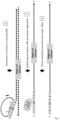
- Figure 1 shows the first three steps of the process where a linear probe containing a random sequence flanked by two anchors (A & B) and a specific sequence complementary to the target region of interest mRNA gene which needs to be sequenced.
- the specific portion of the linear probe is hybridized to the target first followed by Reverse Transcription.
- the region of interest will contain a sequence suspected of nucleotide change of variant difference.
- the circle locator is hybridized to the linear probe via regions A & B, leaving a gap corresponding to the UMI region.
- the UMI region is copied by filling the gap and the circle locator is ligated, generating a circular molecule (Step2).
- the circular molecule is used as a template to generate DNA nanoballs (rolonies).
- the UMI portion of the rolonies are sequenced directly on tissue, providing the location coordinates of the target region (x.y and/or z).
- Figure 1 shows further the hybridization of the linear probe containing a random sequence and two anchors (A & B) to mRNA on a fixed tissue on a slide to a region upstream from the region of interest in a particular mRNA (dotted line) via the specific sequence portion located at the 3'end.
- the reverse transcription of the region of interest onto a cDNA (Step 1) is initiated by the linear probe, that is used as a primer.
- the 3'end of the linear probe consists of sequence about 30 bp long which can hybridize to a specific region in an mRNA target of interest on tissue.
- Hybridization of the circle locator padlock molecule to the linear probe via regions A & B, leaving a gap corresponding to the UMI region is shown in step 2.
- the UMI region is copied to the circle locator padlock by filling the gap using a non strand-displacement enzyme for example Phusion DNA polymerase and ligation of the two extremities of the circle locator padlock using a DNA ligase, generating a circular molecule (step2).
- the size of the circle can be between 100 to 400bp long with the gap portion to be about 10-40bp .
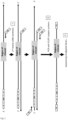
- FIG. 2 shows the circular molecule used as a template to generate DNA nanoballs.
- the circular molecule is amplified by (Rolling Circle Amplification) RCA (Step 3) and the UMI portion is sequenced in situ (Step 4).
- the cDNA strand containing a copied UMI is then extracted for sequencing of the region of interest (Step 5).
- Figure 2 shows further the process of how the circle locator is used to generated DNA nanoballs (rolonies) on the tissue via isothermal rolling circle amplification (RCA) using an enzyme with highly processivity and strand displacement activity like Phi29 DNA polymerase and RCA oligonucleotide binding to circle locator as a primer (step 3).
- the resulting location of the RCA product on the tissue is determined by sequencing the randomly generated UMI on tissue and obtaining coordinates (x, y and/or z) of all the rolonies (step 4).
- the circle locator probe is hybridized first to the linear probe anchor before the latest binds to a region upstream from the region of interest in a particular mRNA (dotted line).
- the extended cDNA is extracted from the tissue section slide in step 5.
- Figure 3 shows how the extracted cDNA strand from the tissue can be PCR amplified and later circularized by adding adaptor regions (P1 and P2) to the PCR primers.
- the 2 nd strand cDNA synthesis is performed in Step 6 and PCR amplified to generate double stranded DNA molecule with P1/P2 adaptor ends (Step 7) that can be analyzed by NGS sequencing (Step 8).
- Figure 3 shows further the 2 nd strand of cDNA synthesis performed (step 6) and subsequent PCR reaction (step 7) performed using a pair of branched primers P1 and P2.
- the binding regions are located upstream of the circle locator region for Primer P1 and is located downstream of the "Region of Interest" for Primer P2 generating a adapted DNA library containing the region of interest and the UMI portion. Sequencing of the DNA library allows for determining determine if either a mutation/nucleotide variant exist is the target region of interest and by sequencing the UMI, to identify the original location/origin of each region of interest on the tissue by correlating them with the circle locator UMI sequences coordinates generated sequencing the rolonies on a tissue section (step 8).
- Sequencing of the DNA library allows to detect the presence of a mutation/nucleotide variant in the targeted region of interest, and by sequencing the UMI, to identify the original location/origin of each region of interest on the tissue by correlating them with the circle locator UMI sequences coordinates generated sequencing the rolonies on a tissue section (step 8).
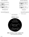
- Figure 4 shows an example of the sequence analysis performed on libraries generated from extracted and PCR amplified cDNA (MS4A1 gene) and DNA nanoballs.
- the sequencing was performed ex-situ using a primer oligonucleotide located upstream of the region of interest and the unique molecular identifier (UMI) portions.
- UMI unique molecular identifier
- the UMI portion along with the locator padlock is sequenced, and the reads originating from the locator rolonies were identified by mapping the locator sequence reads to the known padlock reference sequence.
- the transcripts were sequenced also where the UMI portion as well as the coding sequence from the cDNA transcript containing the linear primer and UMI sequences that were copied to the locator padlock.
- the reads originating from the cDNA transcripts were identified by mapping the sequence reads to the known reference sequence of the coding region from the gene of interest (in this example MS4A1 gene).
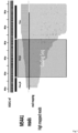
- Figure 5 shows the actual reads obtained from the libraries generated from extracted and PCR amplified cDNA the transcript portion and that actually mapped to the (MS4A1 gene).
- the sequencing was performed ex-situ using a primer oligonucleotide located upstream of the region of interest and the unique molecular identifier (UMI) portions.
- UMI unique molecular identifier
Landscapes
- Chemical & Material Sciences (AREA)
- Life Sciences & Earth Sciences (AREA)
- Organic Chemistry (AREA)
- Proteomics, Peptides & Aminoacids (AREA)
- Zoology (AREA)
- Wood Science & Technology (AREA)
- Health & Medical Sciences (AREA)
- Engineering & Computer Science (AREA)
- Microbiology (AREA)
- Immunology (AREA)
- Physics & Mathematics (AREA)
- Molecular Biology (AREA)
- Biotechnology (AREA)
- Biophysics (AREA)
- Analytical Chemistry (AREA)
- Biochemistry (AREA)
- Bioinformatics & Cheminformatics (AREA)
- General Engineering & Computer Science (AREA)
- General Health & Medical Sciences (AREA)
- Genetics & Genomics (AREA)
- Measuring Or Testing Involving Enzymes Or Micro-Organisms (AREA)
Description
- It is challenging to detect all expressed genes on a sub cellular level on tissue and simultaneously obtain the sequence and/or spatial information of these genes.
- This invention describes the method for recognizing and amplifying a specific region on a messenger RNA (mRNA) using a probe containing non pre-defined and randomly generated Unique Molecular Identifier (UMI) juxtaposed to a portion targeting a region of interest which contains a mutation or sequence variant on the mRNA captured by reverse transcription, and a gap padlock locator probe copying the sequence information of the linear probe UMI. The ligated padlock probe is used as a circular template to generate locator DNA nanoballs using Rolling Circle Amplified (RCA). Following the sequencing of the locator DNA nanoballs, the cDNA with UMI information is retrieved, extracted, amplified and sequenced by NGS.
- Accordingly, it was an object of the invention to provide a method to simultaneously obtain both the spatial location and sequence information of a target sequence with a higher resolution than the known technologies. Document
EP4039822 relates to spatial transcription determination based on gap-filling barcoded padlock probes having a spatial unique molecular identifier (SUMI) and which directly bind to the target, this document does not describe reverse transcription of linear nucleic acid probes. DocumentEP3936623 relates to gene expression mapping comprising the use of padlock probes comprising SUMI and which bind directly to the target, this document makes use of multiple detection probes and makes not mention of reverse transcribing linear probes. - Here we describe a method to
- (1) Spatially localize the mRNA expressed on a tissue by the use of a hybrid circular linear DNA probe with UMI.
- (2) The nucleotide information of an adj acent region downstream from the region where the linear portion of a probe with UMI is copied with reverse transcriptase. Concurrently a padlock locator probe whose ends are complementary to target sequences flanking the UMI portion on the linear portion of the probes are hybridized creating a small gap corresponding to the length of the UMI that can be copied using Phusion DNA polymerase generating a circular template .
- (3) The circular template is used to generate DNA nanoballs for which the UMI portion are sequenced on the tissue, and used as reporters for the linear probes hybridization and mRNA coordinate generation.
- (4) The UMI containing cDNA strand generate by reverse transcription is then extracted from the tissue.
- (5) The extracted UMI containing cDNA strand is then amplified by PCR using primers with known branched adaptor ends.
- (6) The PCR product is circularized and RCA amplified
- (7) The RCA product can be sequenced by NGS.
- (8) The sequenced obtained are matched to the actual tissue location using the UMI's
- Object of the invention is a method to obtain the spatial location and sequence information of a target sequence of at least one m-RNA strand on a tissue sample comprising the steps
- a. providing a linear probe, containing a) a binding region capable of binding to the at least one m-RNA strand and b) an anchor sequence comprising a UMI region located between a first and a second locator regions and c) a primer region;
- b. hybridizing the linear probe with its binding region to the m-RNA strand;
- c. complementing the linear probe using the m-RNA strand as template thereby obtaining a reversed transcribed c-DNA strand
- d. hybridizing a locator molecule with its 3' and 5' ends to the first and second locator regions thereby creating a gap corresponding to the length of the UMI of the linear probe
- e. Filling the gap in the locator molecule with nucleotides complementary to the UMI using a non-strand displacement enzyme thereby creating a circular template comprising a copy of the UMI region from the linear probe.
- f. multiplying the circular template molecule by RCA on the tissue sample, starting from a primer region thereby creating a rolony
- g. Sequencing at least the UMI portion of the rolonies thereby obtaining the spatial location of the m-RNA on the tissue
- h. removing the reversed transcribed c-DNA strand from the tissue and dehybridizing the m-RNA strand thereby obtaining a single stranded c-DNA oligomer
- i. providing the single stranded cDNA oligomer with a first and a second adaptor primer at the 3' and 5' ends obtaining a primed single stranded oligomer; amplification of the primed single stranded oligomer by PCR
- j. Sequencing the amplified primed single stranded oligomer and linking the spatial information of the rolonies with the sequence information of the amplified primed single stranded oligomer via the UMI sequence
- In a first embodiment, the locator molecule is hybridized first with its binding region to the m-RNA strand and then the linear probe is then hybridized to the locator molecule. In other words, steps a) to d) are performed in the sequence a), c), d), and d).
- In a second embodiment, the locator molecule is hybridized first to the linear probe, which is then hybridized with its binding region to the m-RNA strand. In other words, steps a) to d) are performed in the sequence a), d), b), and c).
- The present invention is directed to a method of performing a spatial localization of a gene of interest in a given tissue using a linear DNA primer containing a sequence complementary to a region upstream of a region of interest on an mRNA or a poly T sequence complementary to mRNA poly A tails and also contains a non-binding portion which includes a random sequence (UMI) located between two regions that are used for the hybridization of a padlock locator probe molecule (region A & B). The sequence information of a the particular region of interest on the mRNA is first captured by reverse transcription of the mRNA into a cDNA molecule using the linear DNA primer.
- The padlock locator probe molecule can be either pre-attached to the non-binding region of linear DNA primer( regions A & B) or it can be hybridized to the linear DNA primer after the linear primer attaches to the sequence upstream of a desired region on an mRNA directly on a tissue (
Fig 1 ). - The hybridization of the locator probe to the linear DNA primer creates an open circle padlock-like structure with a gap corresponding to the random sequence on the linear primer. The gap is filled by a non strand displacement enzyme and the two extremities are ligated forming a closed circular molecule that can then be amplified by rolling circle amplification (RCA) using an oligonucleotide primer directly on a tissue. The RCA generates DNA nanoballs (rolonies) at the same location where the mRNA was copied into a cDNA on the tissue section. The random and not pre-defined sequence can then be decoded by sequencing the RCA product on tissue where it is used a series of coordinates for each of the rolonies generated.
- The cDNA generated with the linear primer is then physically retrieved and extracted from the tissue.
- The retrieved cDNA is then PCR amplified using a PCR primer which anneals to the 5' and 3' end of the cDNA and carries DNA adapters.
- The PCR amplified product can then be circularized using a splint bridge oligonucleotide primer which brings the two ends together.
- The circle is then amplified by Rolling Circle Amplification (RCA). The RCA product is then sequenced.
- By NGS sequencing the random sequence from the linear primer linked to the cDNA is identified and then can be assigned to the exact tissue location via the coordinates obtained by the sequencing of the same random sequence on the tissue.
- By NGS sequencing the targeted region of interest on genomic DNA or mRNA, mutation or nucleotide variant can be analyzed.
-
-
Figure 1 shows the first three steps of the method of the invention -
Figure 2 shows the circular molecule used as a template to generate DNA nanoballs. -
Figure 3 shows how the extracted cDNA strand from the tissue can be PCR amplified and later circularized by adding adaptor regions (P1 and P2) to the PCR primers. -
Figure 3 and5 show experimental data - The method of the invention is described in detail in
Figs 1-3 , and the term "steps" refers to the respective steps in the figures. - Spatial localization of mRNA molecule by RCA followed by ex-situ sequencing of the region of interest from a copied RNA molecule involves RCA and in situ sequencing of UMIs performed directly on tissue and consists of an eight step process. (Step 1) Hybridization of the linear probe containing a random sequence and two anchors (A & B) to mRNA on tissue via the specific sequence portion located at the 3'end and copying the downstream mRNA region of interest into a cDNA molecule by a reverse transcription using the linear probe as a primer. (Step 2) Hybridization of the circle locator padlock molecule to the linear probe via regions A & B, leaving a gap corresponding to the UMI region. Copying the UMI region by filling the gap using a non-strand displacement enzyme and ligation of the two extremities of the circle locator padlock using a DNA ligase, generating a circular molecule.
- The circular molecule is used as a template to generate DNA nanoballs (rolonies) using RCA with a circle specific RCA primer (Step 3). The UMI portion of the rolonies are sequenced directly on tissue, providing the location coordinates of the target regions (Step 4). The image acquisition performed during the in situ sequencing can be performed using a wide field microscope (single focal plan) or using a confocal microscope (multiple focal plans).
- Extraction of the cDNA containing the linear primer and UMI sequences from tissue (Step 5) and 2nd strand cDNA Synthesis using a branched primer adapter sequence (P2); (Step 6). PCR amplification of the cDNA with a branched adapter sequence complementary to the linear probe A region (P1) followed by P1/P2 adaptor primers (Step7) . Circularization of the PCR amplified product and RCA amplification of the circle which contains UMI and the region of interest (Step 8). NGS Sequencing of the region of interest and the UMI and determining the location and the sequences of the region of interest by matching the UMI sequence off the tissue and on the tissue with the corresponding coordinates (x.y and/or z); Step 9).
-
Figure 1 shows the first three steps of the process where a linear probe containing a random sequence flanked by two anchors (A & B) and a specific sequence complementary to the target region of interest mRNA gene which needs to be sequenced. The specific portion of the linear probe is hybridized to the target first followed by Reverse Transcription. The region of interest will contain a sequence suspected of nucleotide change of variant difference. Once the linear probe has been elongated creating a cDNA copy and is attached stably to the target, then the circle locator is hybridized to the linear probe via regions A & B, leaving a gap corresponding to the UMI region. The UMI region is copied by filling the gap and the circle locator is ligated, generating a circular molecule (Step2). The circular molecule is used as a template to generate DNA nanoballs (rolonies). The UMI portion of the rolonies are sequenced directly on tissue, providing the location coordinates of the target region (x.y and/or z). -
Figure 1 shows further the hybridization of the linear probe containing a random sequence and two anchors (A & B) to mRNA on a fixed tissue on a slide to a region upstream from the region of interest in a particular mRNA (dotted line) via the specific sequence portion located at the 3'end. The reverse transcription of the region of interest onto a cDNA (Step 1) is initiated by the linear probe, that is used as a primer. The 3'end of the linear probe consists of sequence about 30 bp long which can hybridize to a specific region in an mRNA target of interest on tissue. Hybridization of the circle locator padlock molecule to the linear probe via regions A & B, leaving a gap corresponding to the UMI region is shown in step 2. The UMI region is copied to the circle locator padlock by filling the gap using a non strand-displacement enzyme for example Phusion DNA polymerase and ligation of the two extremities of the circle locator padlock using a DNA ligase, generating a circular molecule (step2). The size of the circle can be between 100 to 400bp long with the gap portion to be about 10-40bp . -
Figure 2 shows the circular molecule used as a template to generate DNA nanoballs. The circular molecule is amplified by (Rolling Circle Amplification) RCA (Step 3) and the UMI portion is sequenced in situ (Step 4). The cDNA strand containing a copied UMI is then extracted for sequencing of the region of interest (Step 5). -
Figure 2 shows further the process of how the circle locator is used to generated DNA nanoballs (rolonies) on the tissue via isothermal rolling circle amplification (RCA) using an enzyme with highly processivity and strand displacement activity like Phi29 DNA polymerase and RCA oligonucleotide binding to circle locator as a primer (step 3). The resulting location of the RCA product on the tissue is determined by sequencing the randomly generated UMI on tissue and obtaining coordinates (x, y and/or z) of all the rolonies (step 4). In one embodiment of the invention, the circle locator probe, is hybridized first to the linear probe anchor before the latest binds to a region upstream from the region of interest in a particular mRNA (dotted line). The extended cDNA is extracted from the tissue section slide instep 5. -
Figure 3 shows how the extracted cDNA strand from the tissue can be PCR amplified and later circularized by adding adaptor regions (P1 and P2) to the PCR primers. The 2nd strand cDNA synthesis is performed inStep 6 and PCR amplified to generate double stranded DNA molecule with P1/P2 adaptor ends (Step 7) that can be analyzed by NGS sequencing (Step 8). -
Figure 3 shows further the 2nd strand of cDNA synthesis performed (step 6) and subsequent PCR reaction (step 7) performed using a pair of branched primers P1 and P2. The binding regions are located upstream of the circle locator region for Primer P1 and is located downstream of the "Region of Interest" for Primer P2 generating a adapted DNA library containing the region of interest and the UMI portion. Sequencing of the DNA library allows for determining determine if either a mutation/nucleotide variant exist is the target region of interest and by sequencing the UMI, to identify the original location/origin of each region of interest on the tissue by correlating them with the circle locator UMI sequences coordinates generated sequencing the rolonies on a tissue section (step 8). - Sequencing of the DNA library allows to detect the presence of a mutation/nucleotide variant in the targeted region of interest, and by sequencing the UMI, to identify the original location/origin of each region of interest on the tissue by correlating them with the circle locator UMI sequences coordinates generated sequencing the rolonies on a tissue section (step 8).
-
Figure 4 shows an example of the sequence analysis performed on libraries generated from extracted and PCR amplified cDNA (MS4A1 gene) and DNA nanoballs. The sequencing was performed ex-situ using a primer oligonucleotide located upstream of the region of interest and the unique molecular identifier (UMI) portions. The UMI portion along with the locator padlock is sequenced, and the reads originating from the locator rolonies were identified by mapping the locator sequence reads to the known padlock reference sequence. Additionally, the transcripts were sequenced also where the UMI portion as well as the coding sequence from the cDNA transcript containing the linear primer and UMI sequences that were copied to the locator padlock. The reads originating from the cDNA transcripts were identified by mapping the sequence reads to the known reference sequence of the coding region from the gene of interest (in this example MS4A1 gene). -
Figure 5 shows the actual reads obtained from the libraries generated from extracted and PCR amplified cDNA the transcript portion and that actually mapped to the (MS4A1 gene). The sequencing was performed ex-situ using a primer oligonucleotide located upstream of the region of interest and the unique molecular identifier (UMI) portions. The coding sequence portion of MS4A1 that was generated by reverse transcription of the linear primer is highlighted (blocked region)
Claims (9)
- A method to obtain the spatial location and sequence information of a target sequence of at least one m-RNA strand on a tissue sample comprising the stepsa. providing a linear probe, containing a) a binding region capable of binding to the at least one m-RNA strand and b) an anchor sequence comprising a UMI region located between a first and a second locator regions and c) a primer region;b. hybridizing the linear probe with its binding region to the m-RNA strand;c. complementing the linear probe using the m-RNA strand as template thereby obtaining a reversed transcribed c-DNA strandd. hybridizing a locator molecule with its 3' and 5' ends to the first and second locator regions thereby creating a gap corresponding to the length of the UMI of the linear probee. Filling the gap in the locator molecule with nucleotides complementary to the UMI using a non-strand displacement enzyme thereby creating a circular template comprising a copy of the UMI region from the linear probe.f. multiplying the circular template molecule by RCA on the tissue sample, starting from a primer region thereby creating a rolonyg. Sequencing at least the UMI portion of the rolonies thereby obtaining the spatial location of the m-RNA on the tissueh. removing the reversed transcribed c-DNA strand from the tissue and dehybridizing the m-RNA strand thereby obtaining a single stranded c-DNA oligomeri. providing the single stranded cDNA oligomer with a first and a second adaptor primer at the 3' and 5' ends obtaining a primed single stranded oligomer; amplification of the primed single stranded oligomer by PCRj. Sequencing the amplified primed single stranded oligomer and linking the spatial information of the rolonies with the sequence information of the amplified primed single stranded oligomer via the UMI sequence
- Method according to claim 1 characterized in that steps a) to d) are performed in the sequence a), b), c) and d).
- Method according to claim 1 characterized in that steps a) to d) are performed in the sequence a), d), b), and c).
- Method according to any of the claims 1 to 3 characterized in that filling the gap of the locator molecule in step e) is performed by Phusion DNA Polymerase or non-strand displacement polymerase using a primer hybridized to a region of the circular locator.
- Method according to any of the claims 1 to 4 characterized in that the 3' and 5' ends of the gap-filled DNA molecule are ligated with each other wherein the UMI and the first region of the linear locator acts as a bridge splint.
- Method according to claim 5 characterized in that the ligation reaction in step e) is performed by a DNA ligase
- Method according to claim 6 characterized in that the locator probe is provided by first hybridizing the linear locator to the at least one m-RNA strand, complementing the RNA anchor region of the locator probe into a reversed transcribed c-DNA strand and then hybridizing the circular locator to the linear locator.
- Method according to any of the claims 1 to 7 characterized in that the single stranded oligomer is physically sheared to smaller fragment before adding a first and a second adaptor primer at the 3' and 5' ends
- Method according to any of the claims 1 to 8 characterized in that the single stranded adapted DNA oligomer is circularized and multiplied by RCA into second rolonies before sequencing
Applications Claiming Priority (1)
| Application Number | Priority Date | Filing Date | Title |
|---|---|---|---|
| EP22198689 | 2022-09-29 |
Publications (2)
| Publication Number | Publication Date |
|---|---|
| EP4345169A1 EP4345169A1 (en) | 2024-04-03 |
| EP4345169B1 true EP4345169B1 (en) | 2024-07-31 |
Family
ID=83508384
Family Applications (1)
| Application Number | Title | Priority Date | Filing Date |
|---|---|---|---|
| EP23199266.0A Active EP4345169B1 (en) | 2022-09-29 | 2023-09-25 | Gene specific tissue information and sequencing |
Country Status (5)
| Country | Link |
|---|---|
| US (1) | US20240110232A1 (en) |
| EP (1) | EP4345169B1 (en) |
| JP (1) | JP2024050504A (en) |
| CN (1) | CN117778537A (en) |
| ES (1) | ES2989026T3 (en) |
Family Cites Families (6)
| Publication number | Priority date | Publication date | Assignee | Title |
|---|---|---|---|---|
| US12146188B2 (en) * | 2020-06-29 | 2024-11-19 | Miltenyi Biotec B.V. & Co. KG | Method combining single cell gene expression mapping and targeted RNA or c-DNA sequencing using padlock oligonucleotides comprising a barcode region |
| EP4121561A4 (en) * | 2020-08-06 | 2024-04-17 | Singular Genomics Systems, Inc. | METHODS FOR IN SITU TRANSCRIPTOMICS AND PROTEOMICS |
| US12509717B2 (en) * | 2020-12-11 | 2025-12-30 | 10X Genomics, Inc. | Methods and compositions for multimodal in situ analysis |
| EP4284945B1 (en) * | 2021-01-26 | 2024-10-23 | 10X Genomics, Inc. | Nucleic acid analog probes for in situ analysis |
| CN114854838A (en) * | 2021-02-04 | 2022-08-05 | 美天施生物科技有限两合公司 | Method for combining in-vitro sequencing and in-situ sequencing of target RNA or cDNA |
| US20230287478A1 (en) * | 2022-03-11 | 2023-09-14 | 10X Genomics, Inc. | Concatemeric detectable probes and related methods |
-
2023
- 2023-09-25 EP EP23199266.0A patent/EP4345169B1/en active Active
- 2023-09-25 ES ES23199266T patent/ES2989026T3/en active Active
- 2023-09-27 US US18/373,441 patent/US20240110232A1/en active Pending
- 2023-09-27 CN CN202311271013.8A patent/CN117778537A/en active Pending
- 2023-09-28 JP JP2023167622A patent/JP2024050504A/en active Pending
Also Published As
| Publication number | Publication date |
|---|---|
| US20240110232A1 (en) | 2024-04-04 |
| JP2024050504A (en) | 2024-04-10 |
| CN117778537A (en) | 2024-03-29 |
| EP4345169A1 (en) | 2024-04-03 |
| ES2989026T3 (en) | 2024-11-25 |
Similar Documents
| Publication | Publication Date | Title |
|---|---|---|
| US8999677B1 (en) | Method for differentiation of polynucleotide strands | |
| Little et al. | Array CGH using whole genome amplification of fresh-frozen and formalin-fixed, paraffin-embedded tumor DNA | |
| CN110036117B (en) | Method to increase the throughput of single-molecule sequencing by multiplexing short DNA fragments | |
| CN108431233B (en) | Efficient construction of DNA libraries | |
| CN117778545A (en) | Accurate and large-scale parallel quantification of nucleic acids | |
| EP3356552B1 (en) | High molecular weight dna sample tracking tags for next generation sequencing | |
| KR102932555B1 (en) | Methods for accurate parallel quantification of nucleic acids in dilute or non-purified samples | |
| EP3652345A2 (en) | Sequencing method for genomic rearrangement detection | |
| CN103221552A (en) | Methods for Amplifying Nucleic Acids | |
| JP2022082574A (en) | Preparation of nucleic acid libraries from rna and dna | |
| EP2785865A1 (en) | Method and kit for characterizing rna in a composition | |
| EP4345169A1 (en) | Gene specific tissue information and sequencing | |
| US20230407383A1 (en) | Gene specific spatial rolling circle amplification and ngs sequencing | |
| KR20210104108A (en) | Nucleic Acid Amplification and Identification Methods | |
| US20220325317A1 (en) | Methods for generating a population of polynucleotide molecules | |
| EP4455307A1 (en) | Ex-situ sequencing of rca product generated in-situ | |
| US20230348961A1 (en) | Ex-situ sequencing of rca product generated in-situ | |
| WO2026059922A1 (en) | Molecular deduplication analysis methods | |
| WO2026024817A1 (en) | Methods and compositions for anchored multiplex ngs workflows | |
| WO2025087931A1 (en) | Light-bloc: light-induced barcode ligation of cassette | |
| Robinson et al. | Illumina Technology | |
| HK40017021A (en) | Accurate and massively parallel quantification of nucleic acid |
Legal Events
| Date | Code | Title | Description |
|---|---|---|---|
| PUAI | Public reference made under article 153(3) epc to a published international application that has entered the european phase |
Free format text: ORIGINAL CODE: 0009012 |
|
| STAA | Information on the status of an ep patent application or granted ep patent |
Free format text: STATUS: THE APPLICATION HAS BEEN PUBLISHED |
|
| AK | Designated contracting states |
Kind code of ref document: A1 Designated state(s): AL AT BE BG CH CY CZ DE DK EE ES FI FR GB GR HR HU IE IS IT LI LT LU LV MC ME MK MT NL NO PL PT RO RS SE SI SK SM TR |
|
| STAA | Information on the status of an ep patent application or granted ep patent |
Free format text: STATUS: REQUEST FOR EXAMINATION WAS MADE |
|
| 17P | Request for examination filed |
Effective date: 20240404 |
|
| GRAP | Despatch of communication of intention to grant a patent |
Free format text: ORIGINAL CODE: EPIDOSNIGR1 |
|
| STAA | Information on the status of an ep patent application or granted ep patent |
Free format text: STATUS: GRANT OF PATENT IS INTENDED |
|
| GRAS | Grant fee paid |
Free format text: ORIGINAL CODE: EPIDOSNIGR3 |
|
| RIC1 | Information provided on ipc code assigned before grant |
Ipc: C12Q 1/6841 20180101AFI20240510BHEP |
|
| INTG | Intention to grant announced |
Effective date: 20240529 |
|
| GRAA | (expected) grant |
Free format text: ORIGINAL CODE: 0009210 |
|
| STAA | Information on the status of an ep patent application or granted ep patent |
Free format text: STATUS: THE PATENT HAS BEEN GRANTED |
|
| AK | Designated contracting states |
Kind code of ref document: B1 Designated state(s): AL AT BE BG CH CY CZ DE DK EE ES FI FR GB GR HR HU IE IS IT LI LT LU LV MC ME MK MT NL NO PL PT RO RS SE SI SK SM TR |
|
| RBV | Designated contracting states (corrected) |
Designated state(s): AL AT BE BG CH CY CZ DE DK EE ES FI FR GB GR HR HU IE IS IT LI LT LU LV MC ME MK MT NL NO PL PT RO RS SE SI SK SM TR |
|
| REG | Reference to a national code |
Ref country code: CH Ref legal event code: EP Ref country code: GB Ref legal event code: FG4D |
|
| REG | Reference to a national code |
Ref country code: DE Ref legal event code: R096 Ref document number: 602023000312 Country of ref document: DE |
|
| REG | Reference to a national code |
Ref country code: IE Ref legal event code: FG4D |
|
| REG | Reference to a national code |
Ref country code: LT Ref legal event code: MG9D Ref country code: ES Ref legal event code: FG2A Ref document number: 2989026 Country of ref document: ES Kind code of ref document: T3 Effective date: 20241125 |
|
| REG | Reference to a national code |
Ref country code: NL Ref legal event code: MP Effective date: 20240731 |
|
| PG25 | Lapsed in a contracting state [announced via postgrant information from national office to epo] |
Ref country code: PT Free format text: LAPSE BECAUSE OF FAILURE TO SUBMIT A TRANSLATION OF THE DESCRIPTION OR TO PAY THE FEE WITHIN THE PRESCRIBED TIME-LIMIT Effective date: 20241202 |
|
| REG | Reference to a national code |
Ref country code: AT Ref legal event code: MK05 Ref document number: 1708509 Country of ref document: AT Kind code of ref document: T Effective date: 20240731 |
|
| PG25 | Lapsed in a contracting state [announced via postgrant information from national office to epo] |
Ref country code: PT Free format text: LAPSE BECAUSE OF FAILURE TO SUBMIT A TRANSLATION OF THE DESCRIPTION OR TO PAY THE FEE WITHIN THE PRESCRIBED TIME-LIMIT Effective date: 20241202 |
|
| PG25 | Lapsed in a contracting state [announced via postgrant information from national office to epo] |
Ref country code: NO Free format text: LAPSE BECAUSE OF FAILURE TO SUBMIT A TRANSLATION OF THE DESCRIPTION OR TO PAY THE FEE WITHIN THE PRESCRIBED TIME-LIMIT Effective date: 20241031 |
|
| PG25 | Lapsed in a contracting state [announced via postgrant information from national office to epo] |
Ref country code: NL Free format text: LAPSE BECAUSE OF FAILURE TO SUBMIT A TRANSLATION OF THE DESCRIPTION OR TO PAY THE FEE WITHIN THE PRESCRIBED TIME-LIMIT Effective date: 20240731 Ref country code: FI Free format text: LAPSE BECAUSE OF FAILURE TO SUBMIT A TRANSLATION OF THE DESCRIPTION OR TO PAY THE FEE WITHIN THE PRESCRIBED TIME-LIMIT Effective date: 20240731 Ref country code: PL Free format text: LAPSE BECAUSE OF FAILURE TO SUBMIT A TRANSLATION OF THE DESCRIPTION OR TO PAY THE FEE WITHIN THE PRESCRIBED TIME-LIMIT Effective date: 20240731 Ref country code: GR Free format text: LAPSE BECAUSE OF FAILURE TO SUBMIT A TRANSLATION OF THE DESCRIPTION OR TO PAY THE FEE WITHIN THE PRESCRIBED TIME-LIMIT Effective date: 20241101 |
|
| PG25 | Lapsed in a contracting state [announced via postgrant information from national office to epo] |
Ref country code: BG Free format text: LAPSE BECAUSE OF FAILURE TO SUBMIT A TRANSLATION OF THE DESCRIPTION OR TO PAY THE FEE WITHIN THE PRESCRIBED TIME-LIMIT Effective date: 20240731 |
|
| PG25 | Lapsed in a contracting state [announced via postgrant information from national office to epo] |
Ref country code: LV Free format text: LAPSE BECAUSE OF FAILURE TO SUBMIT A TRANSLATION OF THE DESCRIPTION OR TO PAY THE FEE WITHIN THE PRESCRIBED TIME-LIMIT Effective date: 20240731 |
|
| PG25 | Lapsed in a contracting state [announced via postgrant information from national office to epo] |
Ref country code: IS Free format text: LAPSE BECAUSE OF FAILURE TO SUBMIT A TRANSLATION OF THE DESCRIPTION OR TO PAY THE FEE WITHIN THE PRESCRIBED TIME-LIMIT Effective date: 20241130 Ref country code: AT Free format text: LAPSE BECAUSE OF FAILURE TO SUBMIT A TRANSLATION OF THE DESCRIPTION OR TO PAY THE FEE WITHIN THE PRESCRIBED TIME-LIMIT Effective date: 20240731 |
|
| PG25 | Lapsed in a contracting state [announced via postgrant information from national office to epo] |
Ref country code: HR Free format text: LAPSE BECAUSE OF FAILURE TO SUBMIT A TRANSLATION OF THE DESCRIPTION OR TO PAY THE FEE WITHIN THE PRESCRIBED TIME-LIMIT Effective date: 20240731 |
|
| PG25 | Lapsed in a contracting state [announced via postgrant information from national office to epo] |
Ref country code: RS Free format text: LAPSE BECAUSE OF FAILURE TO SUBMIT A TRANSLATION OF THE DESCRIPTION OR TO PAY THE FEE WITHIN THE PRESCRIBED TIME-LIMIT Effective date: 20241031 |
|
| PG25 | Lapsed in a contracting state [announced via postgrant information from national office to epo] |
Ref country code: RS Free format text: LAPSE BECAUSE OF FAILURE TO SUBMIT A TRANSLATION OF THE DESCRIPTION OR TO PAY THE FEE WITHIN THE PRESCRIBED TIME-LIMIT Effective date: 20241031 Ref country code: PL Free format text: LAPSE BECAUSE OF FAILURE TO SUBMIT A TRANSLATION OF THE DESCRIPTION OR TO PAY THE FEE WITHIN THE PRESCRIBED TIME-LIMIT Effective date: 20240731 Ref country code: NO Free format text: LAPSE BECAUSE OF FAILURE TO SUBMIT A TRANSLATION OF THE DESCRIPTION OR TO PAY THE FEE WITHIN THE PRESCRIBED TIME-LIMIT Effective date: 20241031 Ref country code: NL Free format text: LAPSE BECAUSE OF FAILURE TO SUBMIT A TRANSLATION OF THE DESCRIPTION OR TO PAY THE FEE WITHIN THE PRESCRIBED TIME-LIMIT Effective date: 20240731 Ref country code: LV Free format text: LAPSE BECAUSE OF FAILURE TO SUBMIT A TRANSLATION OF THE DESCRIPTION OR TO PAY THE FEE WITHIN THE PRESCRIBED TIME-LIMIT Effective date: 20240731 Ref country code: IS Free format text: LAPSE BECAUSE OF FAILURE TO SUBMIT A TRANSLATION OF THE DESCRIPTION OR TO PAY THE FEE WITHIN THE PRESCRIBED TIME-LIMIT Effective date: 20241130 Ref country code: HR Free format text: LAPSE BECAUSE OF FAILURE TO SUBMIT A TRANSLATION OF THE DESCRIPTION OR TO PAY THE FEE WITHIN THE PRESCRIBED TIME-LIMIT Effective date: 20240731 Ref country code: GR Free format text: LAPSE BECAUSE OF FAILURE TO SUBMIT A TRANSLATION OF THE DESCRIPTION OR TO PAY THE FEE WITHIN THE PRESCRIBED TIME-LIMIT Effective date: 20241101 Ref country code: FI Free format text: LAPSE BECAUSE OF FAILURE TO SUBMIT A TRANSLATION OF THE DESCRIPTION OR TO PAY THE FEE WITHIN THE PRESCRIBED TIME-LIMIT Effective date: 20240731 Ref country code: BG Free format text: LAPSE BECAUSE OF FAILURE TO SUBMIT A TRANSLATION OF THE DESCRIPTION OR TO PAY THE FEE WITHIN THE PRESCRIBED TIME-LIMIT Effective date: 20240731 Ref country code: AT Free format text: LAPSE BECAUSE OF FAILURE TO SUBMIT A TRANSLATION OF THE DESCRIPTION OR TO PAY THE FEE WITHIN THE PRESCRIBED TIME-LIMIT Effective date: 20240731 |
|
| PG25 | Lapsed in a contracting state [announced via postgrant information from national office to epo] |
Ref country code: RO Free format text: LAPSE BECAUSE OF FAILURE TO SUBMIT A TRANSLATION OF THE DESCRIPTION OR TO PAY THE FEE WITHIN THE PRESCRIBED TIME-LIMIT Effective date: 20240731 Ref country code: SM Free format text: LAPSE BECAUSE OF FAILURE TO SUBMIT A TRANSLATION OF THE DESCRIPTION OR TO PAY THE FEE WITHIN THE PRESCRIBED TIME-LIMIT Effective date: 20240731 Ref country code: DK Free format text: LAPSE BECAUSE OF FAILURE TO SUBMIT A TRANSLATION OF THE DESCRIPTION OR TO PAY THE FEE WITHIN THE PRESCRIBED TIME-LIMIT Effective date: 20240731 |
|
| PG25 | Lapsed in a contracting state [announced via postgrant information from national office to epo] |
Ref country code: MC Free format text: LAPSE BECAUSE OF FAILURE TO SUBMIT A TRANSLATION OF THE DESCRIPTION OR TO PAY THE FEE WITHIN THE PRESCRIBED TIME-LIMIT Effective date: 20240731 Ref country code: EE Free format text: LAPSE BECAUSE OF FAILURE TO SUBMIT A TRANSLATION OF THE DESCRIPTION OR TO PAY THE FEE WITHIN THE PRESCRIBED TIME-LIMIT Effective date: 20240731 |
|
| PG25 | Lapsed in a contracting state [announced via postgrant information from national office to epo] |
Ref country code: CZ Free format text: LAPSE BECAUSE OF FAILURE TO SUBMIT A TRANSLATION OF THE DESCRIPTION OR TO PAY THE FEE WITHIN THE PRESCRIBED TIME-LIMIT Effective date: 20240731 |
|
| PG25 | Lapsed in a contracting state [announced via postgrant information from national office to epo] |
Ref country code: SK Free format text: LAPSE BECAUSE OF FAILURE TO SUBMIT A TRANSLATION OF THE DESCRIPTION OR TO PAY THE FEE WITHIN THE PRESCRIBED TIME-LIMIT Effective date: 20240731 |
|
| REG | Reference to a national code |
Ref country code: DE Ref legal event code: R097 Ref document number: 602023000312 Country of ref document: DE |
|
| PLBE | No opposition filed within time limit |
Free format text: ORIGINAL CODE: 0009261 |
|
| STAA | Information on the status of an ep patent application or granted ep patent |
Free format text: STATUS: NO OPPOSITION FILED WITHIN TIME LIMIT |
|
| 26N | No opposition filed |
Effective date: 20250501 |
|
| PG25 | Lapsed in a contracting state [announced via postgrant information from national office to epo] |
Ref country code: SE Free format text: LAPSE BECAUSE OF FAILURE TO SUBMIT A TRANSLATION OF THE DESCRIPTION OR TO PAY THE FEE WITHIN THE PRESCRIBED TIME-LIMIT Effective date: 20240731 |
|
| PGFP | Annual fee paid to national office [announced via postgrant information from national office to epo] |
Ref country code: DE Payment date: 20250919 Year of fee payment: 3 |
|
| PGFP | Annual fee paid to national office [announced via postgrant information from national office to epo] |
Ref country code: FR Payment date: 20250922 Year of fee payment: 3 |
|
| PGFP | Annual fee paid to national office [announced via postgrant information from national office to epo] |
Ref country code: IT Payment date: 20250930 Year of fee payment: 3 |
|
| PGFP | Annual fee paid to national office [announced via postgrant information from national office to epo] |
Ref country code: ES Payment date: 20251030 Year of fee payment: 3 |