EP4343688A1 - Scoring and ranking angiograms - Google Patents
Scoring and ranking angiograms Download PDFInfo
- Publication number
- EP4343688A1 EP4343688A1 EP22197745.7A EP22197745A EP4343688A1 EP 4343688 A1 EP4343688 A1 EP 4343688A1 EP 22197745 A EP22197745 A EP 22197745A EP 4343688 A1 EP4343688 A1 EP 4343688A1
- Authority
- EP
- European Patent Office
- Prior art keywords
- angiogram
- score
- frame
- frames
- computer
- Prior art date
- Legal status (The legal status is an assumption and is not a legal conclusion. Google has not performed a legal analysis and makes no representation as to the accuracy of the status listed.)
- Pending
Links
Images


Classifications
-
- G—PHYSICS
- G06—COMPUTING OR CALCULATING; COUNTING
- G06T—IMAGE DATA PROCESSING OR GENERATION, IN GENERAL
- G06T7/00—Image analysis
- G06T7/0002—Inspection of images, e.g. flaw detection
- G06T7/0012—Biomedical image inspection
-
- A—HUMAN NECESSITIES
- A61—MEDICAL OR VETERINARY SCIENCE; HYGIENE
- A61B—DIAGNOSIS; SURGERY; IDENTIFICATION
- A61B6/00—Apparatus or devices for radiation diagnosis; Apparatus or devices for radiation diagnosis combined with radiation therapy equipment
- A61B6/44—Constructional features of apparatus for radiation diagnosis
- A61B6/4429—Constructional features of apparatus for radiation diagnosis related to the mounting of source units and detector units
- A61B6/4435—Constructional features of apparatus for radiation diagnosis related to the mounting of source units and detector units the source unit and the detector unit being coupled by a rigid structure
- A61B6/4441—Constructional features of apparatus for radiation diagnosis related to the mounting of source units and detector units the source unit and the detector unit being coupled by a rigid structure the rigid structure being a C-arm or U-arm
-
- A—HUMAN NECESSITIES
- A61—MEDICAL OR VETERINARY SCIENCE; HYGIENE
- A61B—DIAGNOSIS; SURGERY; IDENTIFICATION
- A61B6/00—Apparatus or devices for radiation diagnosis; Apparatus or devices for radiation diagnosis combined with radiation therapy equipment
- A61B6/46—Arrangements for interfacing with the operator or the patient
- A61B6/461—Displaying means of special interest
- A61B6/463—Displaying means of special interest characterised by displaying multiple images or images and diagnostic data on one display
-
- A—HUMAN NECESSITIES
- A61—MEDICAL OR VETERINARY SCIENCE; HYGIENE
- A61B—DIAGNOSIS; SURGERY; IDENTIFICATION
- A61B6/00—Apparatus or devices for radiation diagnosis; Apparatus or devices for radiation diagnosis combined with radiation therapy equipment
- A61B6/48—Diagnostic techniques
- A61B6/481—Diagnostic techniques involving the use of contrast agents
-
- A—HUMAN NECESSITIES
- A61—MEDICAL OR VETERINARY SCIENCE; HYGIENE
- A61B—DIAGNOSIS; SURGERY; IDENTIFICATION
- A61B6/00—Apparatus or devices for radiation diagnosis; Apparatus or devices for radiation diagnosis combined with radiation therapy equipment
- A61B6/50—Apparatus or devices for radiation diagnosis; Apparatus or devices for radiation diagnosis combined with radiation therapy equipment specially adapted for specific body parts; specially adapted for specific clinical applications
- A61B6/504—Apparatus or devices for radiation diagnosis; Apparatus or devices for radiation diagnosis combined with radiation therapy equipment specially adapted for specific body parts; specially adapted for specific clinical applications for diagnosis of blood vessels, e.g. by angiography
-
- G—PHYSICS
- G16—INFORMATION AND COMMUNICATION TECHNOLOGY [ICT] SPECIALLY ADAPTED FOR SPECIFIC APPLICATION FIELDS
- G16H—HEALTHCARE INFORMATICS, i.e. INFORMATION AND COMMUNICATION TECHNOLOGY [ICT] SPECIALLY ADAPTED FOR THE HANDLING OR PROCESSING OF MEDICAL OR HEALTHCARE DATA
- G16H50/00—ICT specially adapted for medical diagnosis, medical simulation or medical data mining; ICT specially adapted for detecting, monitoring or modelling epidemics or pandemics
- G16H50/20—ICT specially adapted for medical diagnosis, medical simulation or medical data mining; ICT specially adapted for detecting, monitoring or modelling epidemics or pandemics for computer-aided diagnosis, e.g. based on medical expert systems
-
- G—PHYSICS
- G06—COMPUTING OR CALCULATING; COUNTING
- G06T—IMAGE DATA PROCESSING OR GENERATION, IN GENERAL
- G06T2207/00—Indexing scheme for image analysis or image enhancement
- G06T2207/10—Image acquisition modality
- G06T2207/10072—Tomographic images
- G06T2207/10081—Computed x-ray tomography [CT]
-
- G—PHYSICS
- G06—COMPUTING OR CALCULATING; COUNTING
- G06T—IMAGE DATA PROCESSING OR GENERATION, IN GENERAL
- G06T2207/00—Indexing scheme for image analysis or image enhancement
- G06T2207/10—Image acquisition modality
- G06T2207/10116—X-ray image
- G06T2207/10121—Fluoroscopy
-
- G—PHYSICS
- G06—COMPUTING OR CALCULATING; COUNTING
- G06T—IMAGE DATA PROCESSING OR GENERATION, IN GENERAL
- G06T2207/00—Indexing scheme for image analysis or image enhancement
- G06T2207/20—Special algorithmic details
- G06T2207/20081—Training; Learning
-
- G—PHYSICS
- G06—COMPUTING OR CALCULATING; COUNTING
- G06T—IMAGE DATA PROCESSING OR GENERATION, IN GENERAL
- G06T2207/00—Indexing scheme for image analysis or image enhancement
- G06T2207/20—Special algorithmic details
- G06T2207/20084—Artificial neural networks [ANN]
-
- G—PHYSICS
- G06—COMPUTING OR CALCULATING; COUNTING
- G06T—IMAGE DATA PROCESSING OR GENERATION, IN GENERAL
- G06T2207/00—Indexing scheme for image analysis or image enhancement
- G06T2207/30—Subject of image; Context of image processing
- G06T2207/30004—Biomedical image processing
- G06T2207/30048—Heart; Cardiac
-
- G—PHYSICS
- G06—COMPUTING OR CALCULATING; COUNTING
- G06T—IMAGE DATA PROCESSING OR GENERATION, IN GENERAL
- G06T2207/00—Indexing scheme for image analysis or image enhancement
- G06T2207/30—Subject of image; Context of image processing
- G06T2207/30004—Biomedical image processing
- G06T2207/30101—Blood vessel; Artery; Vein; Vascular
- G06T2207/30104—Vascular flow; Blood flow; Perfusion
Definitions
- Various examples of the disclosure generally relate to angiography. Various examples specifically relate to processing angiograms acquired during an angiography exam to quantify a diagnostic value of the respective angiograms.
- Angiography is a medical imaging technique used to visualize the inside, or lumen, of blood vessels and organs of the body, with a particular interest in the arteries, veins, and heart chambers.
- Modern angiography is performed by injecting a radio-opaque contrast agent into the blood vessel and imaging using X-ray based techniques such as fluoroscopy.
- the acquired film or video (i.e., a sequence of images) of the blood vessels is called an angiograph, or more commonly an angiogram.
- each angiogram comprises multiple frames. Different ones of the multiple frames correspond to different distribution phases of the contrast agent throughout the body or organ.
- angiography may be used to image the heart and nearby blood vessels (called coronary angiography), to image the blood vessels in and around the brain (called cerebral angiography), to image the blood vessels supplying the lung (called pulmonary angiography), to image the blood vessels supplying the kidneys (called renal angiography), or to image the eyes (called fluorescein angiography).
- An angiography study comprises acquisitions of several angiograms.
- a coronary angiography exam comprises acquisitions of several angiograms of both coronary arteries acquired from multiple angulations.
- the angiography exam can take between 30 minutes and 2 hours.
- a radio-opaque contrast agent is injected into the blood vessels.
- Exposure to the contrast agent may cause kidney diseases or allergic reactions.
- being exposed to X-rays may carry a risk of radiation-induced injury and/or of causing cancer.
- the contrast agent dose and/or the radiation dose is kept to a minimum to reduce the risks outlined above.
- the image quality is reduced as the contrast agent dose and/or the radiation dose reduces. Accordingly, during an angiography exam, the contrast agent dose and/or the radiation dose needs to be controlled in such a way that the image quality is adequate to make a diagnosis whilst keeping the radiation dose as low as reasonably achievable. It is difficult to control the contrast agent dose and/or of the radiation dose during an angiography exam.
- a computer-implemented method for processing an angiogram comprises obtaining one or more frames of an angiogram, which is acquired during an angiography exam of an anatomical region of interest.
- the method further comprises based on at least one pre-defined criterion, determining, among the one or more frames, whether the angiogram at least comprises one frame with a diagnostic value.
- the method also comprises if the angiogram comprises at least one frame with the diagnostic value, determining, based on the angiogram, a score using a trained machine-learning, ML, algorithm, wherein the score quantifies the diagnostic value of the angiogram.
- a computer-implemented method for processing multiple angiograms comprises obtaining multiple angiograms associated with an anatomical region of interest of a patient. The method further comprises determining, for each of the multiple angiograms, a score using the method for processing an angiogram mentioned above. The method also comprises sorting the multiple angiograms by score descending.
- a computing device comprising a processor and a memory. Upon loading and executing program code from the memory, the processor is configured to perform a method for processing an angiogram.
- the method comprises obtaining one or more frames of the angiogram, which is acquired during an angiography exam of an anatomical region of interest.
- the method further comprises based on at least one pre-defined criterion, determining, among the one or more frames, whether the angiogram at least comprises one frame with a diagnostic value.
- the method also comprises if the angiogram comprises at least one frame with the diagnostic value, determining, based on the angiogram, a score using a trained machine-learning, ML, algorithm, wherein the score quantifies the diagnostic value of the angiogram.
- a computing device comprising a processor and a memory is provided.
- the processor Upon loading and executing program code from the memory, the processor is configured to perform a method for processing multiple angiograms.
- the method comprises obtaining multiple angiograms associated with an anatomical region of interest of a patient.
- the method further comprises determining, for each of the multiple angiograms, a score using the method for processing an angiogram mentioned above.
- the method also comprises sorting the multiple angiograms by score descending.
- An angiography device comprising a computing device.
- the computing device comprises a processor and a memory.
- the processor Upon loading and executing program code from the memory, the processor is configured to perform a method for processing an angiogram.
- the method comprises obtaining one or more frames of the angiogram, which is acquired during an angiography exam of an anatomical region of interest.
- the method further comprises based on at least one pre-defined criterion, determining, among the one or more frames, whether the angiogram at least comprises one frame with a diagnostic value.
- the method also comprises if the angiogram comprises at least one frame with the diagnostic value, determining, based on the angiogram, a score using a trained machine-learning, ML, algorithm, wherein the score quantifies the diagnostic value of the angiogram.
- An angiography device comprising a computing device.
- the computing device comprises a processor and a memory.
- the processor Upon loading and executing program code from the memory, the processor is configured to perform a method for processing multiple angiograms.
- the method comprises obtaining multiple angiograms associated with an anatomical region of interest of a patient.
- the method further comprises determining, for each of the multiple angiograms, a score using the method for processing an angiogram mentioned above.
- the method also comprises sorting the multiple angiograms by score descending.
- a computer program product or a computer program or a computer-readable storage medium including program code is provided.
- the program code can be executed by at least one processor. Executing the program code causes the at least one processor to perform a method for processing an angiogram.
- the method comprises obtaining one or more frames of the angiogram, which is acquired during an angiography exam of an anatomical region of interest.
- the method further comprises based on at least one pre-defined criterion, determining, among the one or more frames, whether the angiogram at least comprises one frame with a diagnostic value.
- the method also comprises if the angiogram comprises at least one frame with the diagnostic value, determining, based on the angiogram, a score using a trained machine-learning, ML, algorithm, wherein the score quantifies the diagnostic value of the angiogram.
- a computer program product or a computer program or a computer-readable storage medium including program code is provided.
- the program code can be executed by at least one processor. Executing the program code causes the at least one processor to perform a method for processing multiple angiograms.
- the method comprises obtaining multiple angiograms associated with an anatomical region of interest of a patient.
- the method further comprises determining, for each of the multiple angiograms, a score using the method for processing an angiogram mentioned above.
- the method also comprises sorting the multiple angiograms by score descending.
- circuits and other electrical devices generally provide for a plurality of circuits or other electrical devices. All references to the circuits and other electrical devices and the functionality provided by each are not intended to be limited to encompassing only what is illustrated and described herein. While particular labels may be assigned to the various circuits or other electrical devices disclosed, such labels are not intended to limit the scope of operation for the circuits and the other electrical devices. Such circuits and other electrical devices may be combined with each other and/or separated in any manner based on the particular type of electrical implementation that is desired.
- any circuit or other electrical device disclosed herein may include any number of microcontrollers, a graphics processor unit (GPU), integrated circuits, memory devices (e.g., FLASH, random access memory (RAM), read only memory (ROM), electrically programmable read only memory (EPROM), electrically erasable programmable read only memory (EEPROM), or other suitable variants thereof), and software which co-act with one another to perform operation(s) disclosed herein.
- any one or more of the electrical devices may be configured to execute a program code that is embodied in a non-transitory computer readable medium programmed to perform any number of the functions as disclosed.
- Angiography may be employed to obtain angiograms of an anatomical region of interest, e.g., a region of the heart, the brain, the kidney, or the lung.
- techniques for processing one or more frames of an angiogram are disclosed.
- the processing may take place either during or after an angiography exam of an anatomical region of interest, i.e., either real-time processing or post-processing is possible.
- the one or more frames of the angiogram are acquired during the angiography exam.
- the one or more frames are processed to determine, based on at least one pre-defined criterion, whether the angiogram at least comprises one frame with a diagnostic value among the one or more frames. If the angiogram comprises at least one frame with the diagnostic value, based on the angiogram, a score quantifying the diagnostic value of the angiogram is determined using a trained machine-learning (ML) algorithm.
- ML machine-learning
- the diagnostic value can generally describe whether certain features are visible or accurately displayed in the respective frame.
- the diagnostic value can generally pertain to whether the frame is correctly aligned with a distribution phase of the contrast agent.
- the diagnostic value can generally correspond to whether a certain frame includes significant artefacts or disturbances.
- one or more appropriate countermeasures may be taken. For example, the acquisition of the (current) angiogram may be stopped and one or more imaging parameters associated with the angiography exam may be adjusted to acquire one or more frames of a further angiogram.
- the further angiogram may be processed using the same technique as that for processing the (current) angiogram.
- the contrast agent dose and/or the radiation dose can be reduced by stopping the acquisition of the respective angiogram which does not comprise any frame with a diagnostic value. Further, the (current) angiogram which may not comprise any frame with a diagnostic value may be discarded, and thereby such an angiogram may not be processed using any one of post-processing algorithms, e.g., vessel layer enhancement algorithms, vessel segmentation algorithms, or stenosis detection algorithms. As such, computational costs incurred by post-processing the angiogram can be reduced.
- post-processing algorithms e.g., vessel layer enhancement algorithms, vessel segmentation algorithms, or stenosis detection algorithms.
- the angiogram that includes at least one frame with a diagnostic value is further processed using a trained ML algorithm to determine a score quantifying the diagnostic value of the angiogram.
- the score can again be used to determine whether one or more countermeasures are to be taken or whether further acquisition is possible. For instance, if the score is below a pre-defined score threshold, the acquisition of the (current) angiogram may be stopped and one or more imaging parameters associated with the angiography exam may be adjusted to acquire one or more frames of a further angiogram.
- the further angiogram may be processed using the same technique as that for processing the (current) angiogram.
- the contrast agent dose and/or the radiation dose can be reduced by stopping the acquisition of the respective angiogram which may not be qualified according to the comparison between the determined score and the pre-defined score threshold. Further, the respective angiogram which may not be qualified may be discarded, and thereby such an angiogram may not be processed using any one of post-processing algorithms. As such, computational costs incurred by post-processing the angiogram can be reduced.
- the score equals to or is above the pre-defined score threshold, the angiography exam may be stopped. I.e., no further angiograms may be needed to be acquired once at least one angiogram is determined to achieve a qualified score.
- the number of angiograms acquired in the angiography exam can be precisely tailored, and thereby the contrast agent dose and/or the radiation dose can be reduced and the computational costs incurred by post-processing can also be reduced.
- the techniques disclosed herein may utilize a "two-step" processing approach for qualifying and quantifying a diagnostic value of an angiogram to facilitate a reduction of at least one of a contrast agent dose, a radiation dose, and a computational cost incurred by post-processing.
- the one or more frames of the angiogram may be obtained directly from an X-ray scanner, e.g., a C-arm machine, or from a database for storing the one or more frames of the angiogram acquired by the X-ray scanner, e.g., a picture archiving and communication system (PACS) .
- X-ray scanner e.g., a C-arm machine
- a database for storing the one or more frames of the angiogram acquired by the X-ray scanner e.g., a picture archiving and communication system (PACS) .
- PACS picture archiving and communication system
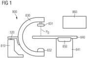
- FIG. 1 schematically illustrates aspects with respect to a C-arm machine 800.
- the C-arm machine 800 may be used for both angiography and fluoroscopy.
- the C-arm machine 800 is in neutral position P 0 . It is possible to manipulate the C-arm machine 800 to be in a rotated position deviating from the neutral position P 0 .
- the angle between the neutral position P0 and a specific rotated position is referred to as the angiography angle or the fluoroscopy angle.
- the C-arm machine 800 comprises a rotatable C arm 830 on which X-ray emission means 831 and X-ray detection means 832 may be mounted.
- the C arm 830 and thereby X-ray emission means 831 and X-ray detection means 832 are positioned to center around patient surface 840.
- X-ray emission means 831 may emit X-rays which may penetrate through a patient positioned on patient surface 840.
- X-ray detection means 832 detects the X -rays emitted from X-ray emission means 831.
- X-ray emission means 831 and X-ray detection means 832 may also collectively be referred to as x-ray imaging means.
- the C arm 830 may be coupled to a C arm rotation unit 820.
- the C arm rotation unit 820 may be any motorized means configured to rotate the C arm 830 according to an angiography angel or a fluoroscopy angle.
- the C arm rotation unit 820 may be attached to and controlled by a C arm control until 810.
- the C arm control unit 810 may be any kind of circuitry capable of controlling C arm 830.
- the C arm control unit 810 may include a computing device.
- the C-arm machine 800 may further include a control panel 850 mounted onto a side surface of patient surface support 841.
- the control panel 850 may be used to control C arm 830 in order to guide medical specialists to pathological vessels.
- Fig. 1 does not show any connections between control panel 850 and C arm 830 to simplify the depiction of the exemplary C-arm machine 800.
- the connection may be wireless.
- the connection may be wired and may e.g., be integrated into the ceiling of the room where the C-arm machine 800 is located.
- the C-arm machine 800 may also include a display 860.
- the display 860 may be used to display information to the medical specialist, such as real-time fluoroscopy image with an overlaid vessel roadmap image including a path to one or more pathological vessels. Further, the display 860 may be used to display vessel segmentation data included in overlaid vessel roadmap images, including labels for various vessel segments. In some examples, display 860 may be a touch screen, which may be used to toggle the display of the vessel segmentation data on and off.
- the C-arm machine 800 may be connectable to a database (not shown in FIG. 1 ), such as a PACS located within a local network of a hospital, for storing angiograms.
- a database such as a PACS located within a local network of a hospital, for storing angiograms.
- various pre-defined criteria may be used to determine, among one or more frames of an angiogram, whether the angiogram at least comprises one frame with a diagnostic value.
- the pre-defined criteria may comprise at least one of the following: Criterion 1: whether at least one stenosis in a segment of a blood vessel within the anatomical region of interest can be determined based on a respective frame of the angiogram; Criterion 2: whether a specific segment of a blood vessel within the anatomical region of interest can be determined based on a respective frame of the angiogram; Criterion 3: whether the specific segment of the blood vessel comprises a contrast above a contrast threshold in at least one frame of the one or more frames of the angiogram; Criterion 4: whether at least one segment of a blood vessel can be determined based on a respective frame of the angiogram; Criterion 5: whether blood vessels are blocked by wires or other artefacts.
- stenosis refers to any condition in which a blood vessel -- such as an artery -- or other tubular organ becomes abnormally narrow.
- the severity of the stenosis for a vessel segment can be assessed visually by a medical imagistic expert. More objectively, the percentage of area stenosis can be used to quantify the severity of the stenosis.
- a name (e.g., any one in the column "Abbreviation" in Table 1) of a specific segment of a blood vessel within the anatomical region of interest can be determined based on a diagnostic purpose of the angiography exam.
- a diagnostic purpose may comprise that the left or right branch of the coronary vessel tree shall be analyzed, or that a specific vessel section shall be analyzed.
- the diagnostic purpose may be determined based on user input, or on a patient's electronic medical record included in a health information system.
- the specific segment of a blood vessel within the anatomical region of interest may be one segment of the coronary arteries of the heart.
- FIG. 2 schematically illustrates an exemplary 3D frontal view 200 of a heart and of coronary arteries.
- the segments of the coronary arteries in FIG. 2 are based on and numbered according to the vessel segmentation as proposed by the American Heart Association (AHA) and as amended by the Society of Cardiovascular Computed Tomography (SCCT).
- AHA American Heart Association
- SCCT Society of Cardiovascular Computed Tomography
- FIG. 2 also indicates the aortic valve 220.
- any method for solving the ranking problem can be used to determine a score quantifying the diagnostic value of the angiogram.
- FIG. 3 is a flowchart of a method 3000 according to various examples.
- the method 3000 according to FIG. 3 may be executed by the C arm control unit 810 of the C-arm machine 800 according to the example of FIG. 1 , e.g., upon loading program code from a memory.
- the method 3000 may be executed by the C arm control unit 810 together with the control panel 850, for example, the C arm control unit 810 and the control panel 850 may be together configured to acquire angiograms during an angiography exam of an anatomical region of interest and the C arm control unit 810 may be further configured to process one or more frames of the acquired angiograms.
- the method 3000 may be executed by the C arm control unit 810 itself. It would also be possible that the method 3000 is at least partially executed by a separate compute unit, e.g., at a server backend.
- FIG. 3 illustrates aspects with respect to processing one or more frames of an angiogram either during or after an angiography exam of an anatomical region of interest (e.g., the heart as shown in FIG. 2 ), i.e., either real-time processing or post-processing.
- the one or more frames of the angiogram are acquired during the angiography exam.
- the one or more frames are processed to determine, based on at least one pre-defined criterion, whether the angiogram at least comprises one frame with a diagnostic value among the one or more frames. If the angiogram comprises at least one frame with the diagnostic value, based on the angiogram, a score quantifying the diagnostic value of the angiogram is determined using a trained machine-learning (ML) algorithm. Details of the method 3000 are described below.
- ML machine-learning
- Block 3100 obtaining one or more frames of an angiogram, which is acquired during an angiography exam of an anatomical region of interest.
- the one or more frames of the angiogram may be obtained directly from an X-ray scanner, e.g., the C-arm machine 800 of FIG. 1 , or from a database for storing the one or more frames of the angiogram acquired by the X-ray scanner, e.g., a PACS. It is possible to select several frames of the angiogram, e.g., 5 or 10 continuous frames starting from the first frame. Frames obtained/acquired in the beginning may be usually of bad quality because the contrast agent may not arrive at a target segment of a blood vessel. Thus, it may be better to select several frames starting from the fifth or eighth frame.
- Block 3200 based on at least one pre-defined criterion, determining, among the one or more frames, whether the angiogram at least comprises one frame with a diagnostic value.
- the at least one criterion may comprise a single criterion, e.g., any one of criteria 1-5.
- the at least one criterion may comprise multiple criteria, e.g., any two, three, four of criteria 1-5, or all criteria 1-5.
- the angiogram may be determined to comprise at least one frame with the diagnostic value, if all of the multiple criteria can be fulfilled. The more criteria the angiogram can fulfill the higher the score may be determined at block 3300.
- each of the at least one pre-defined criterion may be assessed using a respective trained ML algorithm, e.g., a deep neural network.
- each of the at least one pre-defined criterion may be assessed using a respective trained ML algorithm.
- a stenosis detection/classification ML algorithm may be used to assess Criterion 1
- a blood vessel detection/classification ML algorithm may be used to assess Criterion 2.
- Block 3300 if the angiogram comprises at least one frame with the diagnostic value, determining, based on the angiogram, a score using a trained ML algorithm, wherein the score quantifies the diagnostic value of the angiogram.
- the angiogram comprises at least one frame with the diagnostic value if each of the at least one criterion is fulfilled. Then, the angiogram is further processed using a trained ML algorithm to determine a score quantifying the diagnostic value of the angiogram. Any approaches for solving the ranking problem available before the filing date of this disclosure can be used to determine the score.
- the method 3000 may further comprise, at block 3600, determining whether the score is below a pre-defined score threshold.
- the method 3000 may optionally further comprise the following two blocks.
- the method 3000 may comprise discarding the angiogram.
- Such post-processing workflows or algorithms may comprise AI (Artificial Intelligence) algorithms to provide analysis for image guidance and/or reporting, algorithms for vessel highlighting, labeling, stenosis localization, and lumen segmentation. It may be important to quickly filter out angiograms either with low diagnostic values or without diagnostic values, which may be generally not selected by clinicians during manual analysis, such as angiograms with low quality, overlap/foreshortening, before sending the respective angiogram to those algorithms.
- the method 3000 can automatically determine angiograms with a diagnostic value and then determine a score for the angiograms with a diagnostic value.
- the one or more frames of the further angiogram obtained at block 3500 may be further processed based on the method 3000, e.g., starting from block 3200. I.e., the method 3000 may be iteratively applied to frames of different angiograms until at least one angiogram is obtained which comprises at least one frame with the diagnostic value and achieves a score equaling to or above the pre-defined score threshold.
- the method 3000 may optionally further comprise, at block 3700, stopping the angiography exam, if the score equals to or is above the pre-defined score threshold.
- the at least one pre-defined criterion may comprise whether at least one stenosis in a segment of a blood vessel within the anatomical region of interest can be determined based on a respective frame of the angiogram, i.e., Criterion 1. If the at least one stenosis can be determined based on at least one frame of the one or more frames of the angiogram, the angiogram may be determined to comprise at least one frame with the diagnostic value.
- a further trained ML algorithm may be used to determine whether the at least one stenosis can be determined.
- the further trained ML algorithm may be a deep neural network, e.g., a multi-label classification network for detecting/determining if any (salient) stenosis is visible in a certain branch/segment of the coronary arteries, e.g., 201-218 of FIG. 2 .
- the further trained ML algorithm may comprise neural networks disclosed in a non-patent literature - Zhou, Bolei, et al. "Learning deep features for discriminative localization.” Proceedings of the IEEE conference on computer vision and pattern recognition. 2016 .
- the at least one pre-defined criterion may comprise whether a specific segment of a blood vessel (e.g., any one of 201-218 of FIG. 2 ) within the anatomical region of interest can be determined based on a respective frame of the angiogram, i.e., Criterion 2. If the specific segment of the blood vessel within the anatomical region of interest can be determined based on at least one frame of the one or more frames of the angiogram, the angiogram is determined to comprise at least one frame with the diagnostic value.
- a specific segment of a blood vessel e.g., any one of 201-218 of FIG. 2
- Criterion 2 i.e., Criterion 2.
- the at least one pre-defined criterion may further comprise whether the specific segment of the blood vessel comprises a contrast above a contrast threshold in at least one frame of the one or more frames of the angiogram, i.e., Criterion 3. If the angiogram comprises at least one frame of the one or more frames in which the contrast of the specific segment of the blood vessel is above the contrast threshold, the angiogram may be determined to comprise at least one frame with the diagnostic value.
- the score may comprise a probability that at least one abnormality, e.g., a stenosis, can be diagnosed based on the angiogram.
- said determining of the score may be further based on at least one query associated with a diagnosis purpose.
- the at least one query may comprise labels of a specific segment of the blood vessel, e.g., any one or more of segments 201-218 of FIG. 2 .
- the at least one query may comprise a specific branch of the blood vessel, e.g., LAD (i.e., a combination of pLAD 206, mLAD 207, and dLAD 208), or RCA (i.e., a combination of pRCA 201, mRCA 202, and dRCA 203).
- the method 3000 may further comprise training both the ML algorithm and the further ML algorithm.
- various training methods of ML algorithm may be applied to train either the ML algorithm or the further ML algorithm, such as supervised learning, unsupervised learning, semi-supervised learning, self-supervised learning, reinforcement learning and etc.
- a large dataset can be filtered to generate candidates and the candidates can be manually ranked/scored by experts based on a respective diagnostic value. Therefore, experts' experience/preferences can be directly included in the ranking/scoring method 3000.
- additional ranking signals for training can be derived from the algorithms that perform further post-processing. For example, some algorithms may generate measures of confidence for their primary outputs (e.g. stenosis detection) as secondary outputs, which may be correlating with the "quality" of angiograms from the algorithm's perspective (i.e., how well can the algorithm process this specific angiogram; which may depend on how algorithms themselves were trained/developed). Such confidence measures can be used to favor sequences/angiograms in the ranking/scoring training that the algorithms are able to process with good accuracy.
- the method 3000 may utilize a "two-step" processing approach for qualifying and quantifying a diagnostic value of an angiogram to facilitate a reduction of at least one of a contrast agent dose, a radiation dose, and a computational cost incurred by post-processing the angiogram.
- the method 3000 for processing one or more frames of an angiogram during the acquisition of the angiogram or after the acquisition of the angiogram. Details with respect to several use cases of the method 3000 will be described below.
- the method 3000 can be executed during the acquisition of an angiogram to optimize at least one of exam time, delivery of radiation dose, and administration of contrast agent.
- Example 1 may comprise starting acquisition of an angiogram; selecting a subsequence of the acquired frames of the angiogram; applying the method 3000 to the subsequence. If the subsequence meets the pre-defined criteria, stop delivery of radiation or switch from angiography settings to fluoroscopy settings.
- Example 2 may comprise starting acquisition of an angiogram; selecting a subsequence of the acquired frames of the angiogram; applying the method 3000 to the subsequence. If the subsequence achieves a score below a pre-defined score threshold, stop delivery of radiation or switch from angiography settings to fluoroscopy settings.
- the method 3000 can be used to advise the operator on the need to acquire additional angiograms, e.g., a further angiogram.
- Example 3 may comprise acquiring one angiogram; applying the method 3000 to the angiogram either inline or after the acquisition is completed. If the angiogram achieves a score below a pre-defined score threshold, advise the operator to acquire a further angiogram.
- the method 3000 can perform more advanced filtering of angiograms based on at least one pre-defined criterion and select angiograms with a high diagnostic value for further processing, e.g., real-time processing or post-processing. Therefore, the amount of time for the overall workflow of acquiring and processing angiograms can be reduced.
- FIG. 4 is a flowchart of a further method 4000 according to various examples.
- the method 4000 according to FIG. 4 may be executed by the C arm control unit 810 of the C-arm machine 800 according to the example of FIG. 1 , e.g., upon loading program code from a memory.
- the method 4000 may be executed by the C arm control unit 810 together with the control panel 850, for example, the C arm control unit 810 and the control panel 850 may be together configured to acquire angiograms during an angiography exam of an anatomical region of interest and the C arm control unit 810 may be further configured to process one or more frames of the acquired angiograms.
- the method 4000 may be executed by the C arm control unit 810 itself. It would also be possible that the method 4000 is at least partially executed by a separate compute unit, e.g., at a server backend.
- FIG. 4 illustrates aspects with respect to processing multiple angiograms associated with an anatomical region of interest of a patient. For each of the multiple angiograms, a respective score quantifying the diagnostic value of the respective angiogram is determined using the method 3000 of FIG. 3 . Then, the multiple angiograms are sorted by score descending or ascending. Details of the method 4000 are described below.
- At block 4100 obtaining multiple angiograms associated with an anatomical region of interest of a patient.
- the multiple angiograms may be obtained directly from an X-ray scanner, e.g., the C-arm machine 800 of FIG. 1 , or from a database for storing angiograms acquired by the X-ray scanner, e.g., a PACS.
- an X-ray scanner e.g., the C-arm machine 800 of FIG. 1
- a database for storing angiograms acquired by the X-ray scanner e.g., a PACS.
- At block 4200 determining, for each of the multiple angiograms, a score using the method 3000 of FIG. 3 .
- the method 4000 may further comprise displaying and/or processing the multiple angiograms in an order associated with the sorting.
- the results of the sorting can be used to define an order of the then fully-processed angiograms shown to the user. For example, the angiogram with the highest score may be displayed to a user, or the two or three angiograms with the highest score may be selected for post-processing.
- FIG. 5 is a block diagram of a computing device 9000 according to various examples.
- the computing device 9000 may comprise a processor 9020, a memory 9030, and an input/output interface 9010.
- the processor 9020 is configured to load program code from the memory 9030 and execute the program code. Upon executing the program code, the processor 9020 performs the method 3000 for processing one or more frames of an angiogram and/or the method 4000 for processing multiple angiograms.
- the computing device 9000 may be embedded in or connected with an angiography device such as the C-arm machine 800.
- the angiography device comprising the computing device 9000 may be configured to perform the method 3000 and/or the method 4000.
- the C-arm machine 800 may further comprise the computing device 9000 configured to perform either the method 3000 or the method 4000.
- the computing device 9000 may be the C arm control unit 810 and/or the control panel 850. I.e., the computing device 9000 may be embedded in or connected with the C-arm machine 800, and thereby the C-arm machine 800 may be also configured to perform the method 3000 and/or the method 4000.
- the techniques disclosed herein may utilize a "two-step" processing approach for qualifying and quantifying a diagnostic value of an angiogram to facilitate a reduction of at least one of a contrast agent dose, a radiation dose, and a computational cost incurred by post-processing.
- angiography For illustration, the disclosure is explained in detail based on coronary angiography.
- the techniques disclosed herein can be also applied to other angiography techniques, such as cerebral angiography, pulmonary angiography, renal angiography, and fluorescein angiography.
- the techniques disclosed herein can be also applied to angiography that may be done using scans instead of X-rays, such as CT (computed tomography) angiography or MRI (Magnetic resonance imaging) angiography.
- CT computed tomography
- MRI Magnetic resonance imaging
Landscapes
- Health & Medical Sciences (AREA)
- Engineering & Computer Science (AREA)
- Life Sciences & Earth Sciences (AREA)
- Medical Informatics (AREA)
- General Health & Medical Sciences (AREA)
- Public Health (AREA)
- Biomedical Technology (AREA)
- Physics & Mathematics (AREA)
- Radiology & Medical Imaging (AREA)
- Nuclear Medicine, Radiotherapy & Molecular Imaging (AREA)
- Pathology (AREA)
- Veterinary Medicine (AREA)
- Animal Behavior & Ethology (AREA)
- Surgery (AREA)
- Molecular Biology (AREA)
- Heart & Thoracic Surgery (AREA)
- Biophysics (AREA)
- High Energy & Nuclear Physics (AREA)
- Optics & Photonics (AREA)
- Quality & Reliability (AREA)
- Computer Vision & Pattern Recognition (AREA)
- Oral & Maxillofacial Surgery (AREA)
- Dentistry (AREA)
- Vascular Medicine (AREA)
- Theoretical Computer Science (AREA)
- General Physics & Mathematics (AREA)
- Data Mining & Analysis (AREA)
- Databases & Information Systems (AREA)
- Epidemiology (AREA)
- Primary Health Care (AREA)
- Human Computer Interaction (AREA)
- Apparatus For Radiation Diagnosis (AREA)
Abstract
Description
- Various examples of the disclosure generally relate to angiography. Various examples specifically relate to processing angiograms acquired during an angiography exam to quantify a diagnostic value of the respective angiograms.
- Angiography (or arteriography) is a medical imaging technique used to visualize the inside, or lumen, of blood vessels and organs of the body, with a particular interest in the arteries, veins, and heart chambers. Modern angiography is performed by injecting a radio-opaque contrast agent into the blood vessel and imaging using X-ray based techniques such as fluoroscopy. The acquired film or video (i.e., a sequence of images) of the blood vessels is called an angiograph, or more commonly an angiogram. I.e., each angiogram comprises multiple frames. Different ones of the multiple frames correspond to different distribution phases of the contrast agent throughout the body or organ.
- In clinical practice, angiography may be used to image the heart and nearby blood vessels (called coronary angiography), to image the blood vessels in and around the brain (called cerebral angiography), to image the blood vessels supplying the lung (called pulmonary angiography), to image the blood vessels supplying the kidneys (called renal angiography), or to image the eyes (called fluorescein angiography).
- An angiography study (or exam) comprises acquisitions of several angiograms. For example, a coronary angiography exam comprises acquisitions of several angiograms of both coronary arteries acquired from multiple angulations. The angiography exam can take between 30 minutes and 2 hours.
- During an angiography exam, a radio-opaque contrast agent is injected into the blood vessels.
- Exposure to the contrast agent may cause kidney diseases or allergic reactions. In addition, being exposed to X-rays may carry a risk of radiation-induced injury and/or of causing cancer.
- Further, to reduce the risks incurred due to injection of contrast agent and/or X-ray radiation, the contrast agent dose and/or the radiation dose is kept to a minimum to reduce the risks outlined above. On the other hand, the image quality is reduced as the contrast agent dose and/or the radiation dose reduces. Accordingly, during an angiography exam, the contrast agent dose and/or the radiation dose needs to be controlled in such a way that the image quality is adequate to make a diagnosis whilst keeping the radiation dose as low as reasonably achievable. It is difficult to control the contrast agent dose and/or of the radiation dose during an angiography exam.
- Therefore, a need exists for advanced techniques for angiography exams. Specifically, a need exists for reducing the contrast agent dose and/or the radiation dose. A need exists for angiography providing high quality images.
- This need is met by the features of the independent claims. The features of the dependent claims define embodiments.
- A computer-implemented method for processing an angiogram is provided. The method comprises obtaining one or more frames of an angiogram, which is acquired during an angiography exam of an anatomical region of interest. The method further comprises based on at least one pre-defined criterion, determining, among the one or more frames, whether the angiogram at least comprises one frame with a diagnostic value. The method also comprises if the angiogram comprises at least one frame with the diagnostic value, determining, based on the angiogram, a score using a trained machine-learning, ML, algorithm, wherein the score quantifies the diagnostic value of the angiogram.
- A computer-implemented method for processing multiple angiograms is provided. The method comprises obtaining multiple angiograms associated with an anatomical region of interest of a patient. The method further comprises determining, for each of the multiple angiograms, a score using the method for processing an angiogram mentioned above. The method also comprises sorting the multiple angiograms by score descending.
- A computing device comprising a processor and a memory is provided. Upon loading and executing program code from the memory, the processor is configured to perform a method for processing an angiogram. The method comprises obtaining one or more frames of the angiogram, which is acquired during an angiography exam of an anatomical region of interest. The method further comprises based on at least one pre-defined criterion, determining, among the one or more frames, whether the angiogram at least comprises one frame with a diagnostic value. The method also comprises if the angiogram comprises at least one frame with the diagnostic value, determining, based on the angiogram, a score using a trained machine-learning, ML, algorithm, wherein the score quantifies the diagnostic value of the angiogram.
- A computing device comprising a processor and a memory is provided. Upon loading and executing program code from the memory, the processor is configured to perform a method for processing multiple angiograms. The method comprises obtaining multiple angiograms associated with an anatomical region of interest of a patient. The method further comprises determining, for each of the multiple angiograms, a score using the method for processing an angiogram mentioned above. The method also comprises sorting the multiple angiograms by score descending.
- An angiography device comprising a computing device is provided. The computing device comprises a processor and a memory. Upon loading and executing program code from the memory, the processor is configured to perform a method for processing an angiogram. The method comprises obtaining one or more frames of the angiogram, which is acquired during an angiography exam of an anatomical region of interest. The method further comprises based on at least one pre-defined criterion, determining, among the one or more frames, whether the angiogram at least comprises one frame with a diagnostic value. The method also comprises if the angiogram comprises at least one frame with the diagnostic value, determining, based on the angiogram, a score using a trained machine-learning, ML, algorithm, wherein the score quantifies the diagnostic value of the angiogram.
- An angiography device comprising a computing device is provided. The computing device comprises a processor and a memory. Upon loading and executing program code from the memory, the processor is configured to perform a method for processing multiple angiograms. The method comprises obtaining multiple angiograms associated with an anatomical region of interest of a patient. The method further comprises determining, for each of the multiple angiograms, a score using the method for processing an angiogram mentioned above. The method also comprises sorting the multiple angiograms by score descending.
- A computer program product or a computer program or a computer-readable storage medium including program code is provided. The program code can be executed by at least one processor. Executing the program code causes the at least one processor to perform a method for processing an angiogram. The method comprises obtaining one or more frames of the angiogram, which is acquired during an angiography exam of an anatomical region of interest. The method further comprises based on at least one pre-defined criterion, determining, among the one or more frames, whether the angiogram at least comprises one frame with a diagnostic value. The method also comprises if the angiogram comprises at least one frame with the diagnostic value, determining, based on the angiogram, a score using a trained machine-learning, ML, algorithm, wherein the score quantifies the diagnostic value of the angiogram.
- A computer program product or a computer program or a computer-readable storage medium including program code is provided. The program code can be executed by at least one processor. Executing the program code causes the at least one processor to perform a method for processing multiple angiograms. The method comprises obtaining multiple angiograms associated with an anatomical region of interest of a patient. The method further comprises determining, for each of the multiple angiograms, a score using the method for processing an angiogram mentioned above. The method also comprises sorting the multiple angiograms by score descending.
- It is to be understood that the features mentioned above and those yet to be explained below may be used not only in the respective combinations indicated, but also in other combinations or in isolation without departing from the scope of the invention.
-
-
FIG. 1 schematically illustrates aspects with respect to a C-arm machine. -
FIG. 2 schematically illustrates an exemplary 3D frontal view of a heart and of coronary arteries. -
FIG. 3 is a flowchart of a method according to various examples. -
FIG. 4 is a flowchart of a further method according to various examples. -
FIG. 5 is a block diagram of a computing device according to various examples. - Some examples of the present disclosure generally provide for a plurality of circuits or other electrical devices. All references to the circuits and other electrical devices and the functionality provided by each are not intended to be limited to encompassing only what is illustrated and described herein. While particular labels may be assigned to the various circuits or other electrical devices disclosed, such labels are not intended to limit the scope of operation for the circuits and the other electrical devices. Such circuits and other electrical devices may be combined with each other and/or separated in any manner based on the particular type of electrical implementation that is desired. It is recognized that any circuit or other electrical device disclosed herein may include any number of microcontrollers, a graphics processor unit (GPU), integrated circuits, memory devices (e.g., FLASH, random access memory (RAM), read only memory (ROM), electrically programmable read only memory (EPROM), electrically erasable programmable read only memory (EEPROM), or other suitable variants thereof), and software which co-act with one another to perform operation(s) disclosed herein. In addition, any one or more of the electrical devices may be configured to execute a program code that is embodied in a non-transitory computer readable medium programmed to perform any number of the functions as disclosed.
- In the following, embodiments of the invention will be described in detail with reference to the accompanying drawings. It is to be understood that the following description of embodiments is not to be taken in a limiting sense. The scope of the invention is not intended to be limited by the embodiments described hereinafter or by the drawings, which are taken to be illustrative only.
- The drawings are to be regarded as being schematic representations and elements illustrated in the drawings are not necessarily shown to scale. Rather, the various elements are represented such that their function and general purpose become apparent to a person skilled in the art. Any connection or coupling between functional blocks, devices, components, or other physical or functional units shown in the drawings or described herein may also be implemented by an indirect connection or coupling. A coupling between components may also be established over a wireless connection. Functional blocks may be implemented in hardware, firmware, software, or a combination thereof.
- Hereinafter, techniques of angiography are described. Angiography may be employed to obtain angiograms of an anatomical region of interest, e.g., a region of the heart, the brain, the kidney, or the lung.
- According to this disclosure, techniques for processing one or more frames of an angiogram are disclosed. The processing may take place either during or after an angiography exam of an anatomical region of interest, i.e., either real-time processing or post-processing is possible.
- The one or more frames of the angiogram are acquired during the angiography exam. The one or more frames are processed to determine, based on at least one pre-defined criterion, whether the angiogram at least comprises one frame with a diagnostic value among the one or more frames. If the angiogram comprises at least one frame with the diagnostic value, based on the angiogram, a score quantifying the diagnostic value of the angiogram is determined using a trained machine-learning (ML) algorithm.
- The diagnostic value can generally describe whether certain features are visible or accurately displayed in the respective frame. The diagnostic value can generally pertain to whether the frame is correctly aligned with a distribution phase of the contrast agent. The diagnostic value can generally correspond to whether a certain frame includes significant artefacts or disturbances.
- During the acquisition of an angiogram of an angiography exam, if it is determined that the angiogram does not comprise any frame with a diagnostic value, one or more appropriate countermeasures may be taken. For example, the acquisition of the (current) angiogram may be stopped and one or more imaging parameters associated with the angiography exam may be adjusted to acquire one or more frames of a further angiogram. The further angiogram may be processed using the same technique as that for processing the (current) angiogram.
- Using such techniques, the contrast agent dose and/or the radiation dose can be reduced by stopping the acquisition of the respective angiogram which does not comprise any frame with a diagnostic value. Further, the (current) angiogram which may not comprise any frame with a diagnostic value may be discarded, and thereby such an angiogram may not be processed using any one of post-processing algorithms, e.g., vessel layer enhancement algorithms, vessel segmentation algorithms, or stenosis detection algorithms. As such, computational costs incurred by post-processing the angiogram can be reduced.
- In addition, the angiogram that includes at least one frame with a diagnostic value is further processed using a trained ML algorithm to determine a score quantifying the diagnostic value of the angiogram. The score can again be used to determine whether one or more countermeasures are to be taken or whether further acquisition is possible. For instance, if the score is below a pre-defined score threshold, the acquisition of the (current) angiogram may be stopped and one or more imaging parameters associated with the angiography exam may be adjusted to acquire one or more frames of a further angiogram. The further angiogram may be processed using the same technique as that for processing the (current) angiogram. Thus, the contrast agent dose and/or the radiation dose can be reduced by stopping the acquisition of the respective angiogram which may not be qualified according to the comparison between the determined score and the pre-defined score threshold. Further, the respective angiogram which may not be qualified may be discarded, and thereby such an angiogram may not be processed using any one of post-processing algorithms. As such, computational costs incurred by post-processing the angiogram can be reduced. On the other hand, if the score equals to or is above the pre-defined score threshold, the angiography exam may be stopped. I.e., no further angiograms may be needed to be acquired once at least one angiogram is determined to achieve a qualified score. As such, the number of angiograms acquired in the angiography exam can be precisely tailored, and thereby the contrast agent dose and/or the radiation dose can be reduced and the computational costs incurred by post-processing can also be reduced.
- In summary, the techniques disclosed herein may utilize a "two-step" processing approach for qualifying and quantifying a diagnostic value of an angiogram to facilitate a reduction of at least one of a contrast agent dose, a radiation dose, and a computational cost incurred by post-processing.
- According to various examples, the one or more frames of the angiogram may be obtained directly from an X-ray scanner, e.g., a C-arm machine, or from a database for storing the one or more frames of the angiogram acquired by the X-ray scanner, e.g., a picture archiving and communication system (PACS) .
-
FIG. 1 schematically illustrates aspects with respect to a C-arm machine 800. The C-arm machine 800 may be used for both angiography and fluoroscopy. InFIG. 1 , the C-arm machine 800 is in neutral position P0. It is possible to manipulate the C-arm machine 800 to be in a rotated position deviating from the neutral position P0. The angle between the neutral position P0 and a specific rotated position is referred to as the angiography angle or the fluoroscopy angle. The C-arm machine 800 comprises arotatable C arm 830 on which X-ray emission means 831 and X-ray detection means 832 may be mounted. TheC arm 830 and thereby X-ray emission means 831 and X-ray detection means 832 are positioned to center aroundpatient surface 840. X-ray emission means 831 may emit X-rays which may penetrate through a patient positioned onpatient surface 840. X-ray detection means 832 detects the X -rays emitted from X-ray emission means 831. When a patient onpatient surface 840 is injected with a radio-opaque contrast agent into the patient's vessels, some of the X-rays emitted by X-ray emission means 831 are absorbed by the radio-opaque contrast agent, leading X-ray detection means 832 to detect a sequence of images of the vessels filled with the radio-opaque contrast agent, i.e. an angiogram. X-ray emission means 831 and X-ray detection means 832 may also collectively be referred to as x-ray imaging means. - The
C arm 830 may be coupled to a C arm rotation unit 820. The C arm rotation unit 820 may be any motorized means configured to rotate theC arm 830 according to an angiography angel or a fluoroscopy angle. The C arm rotation unit 820 may be attached to and controlled by a C arm control until 810. The Carm control unit 810 may be any kind of circuitry capable of controllingC arm 830. For example, the Carm control unit 810 may include a computing device. - The C-
arm machine 800 may further include acontrol panel 850 mounted onto a side surface ofpatient surface support 841. Thecontrol panel 850 may be used to controlC arm 830 in order to guide medical specialists to pathological vessels.Fig. 1 does not show any connections betweencontrol panel 850 andC arm 830 to simplify the depiction of the exemplary C-arm machine 800. In some examples, the connection may be wireless. In some further examples, the connection may be wired and may e.g., be integrated into the ceiling of the room where the C-arm machine 800 is located. - The C-
arm machine 800 may also include adisplay 860. Thedisplay 860 may be used to display information to the medical specialist, such as real-time fluoroscopy image with an overlaid vessel roadmap image including a path to one or more pathological vessels. Further, thedisplay 860 may be used to display vessel segmentation data included in overlaid vessel roadmap images, including labels for various vessel segments. In some examples,display 860 may be a touch screen, which may be used to toggle the display of the vessel segmentation data on and off. - The C-
arm machine 800 may be connectable to a database (not shown inFIG. 1 ), such as a PACS located within a local network of a hospital, for storing angiograms. - According to this disclosure, various pre-defined criteria may be used to determine, among one or more frames of an angiogram, whether the angiogram at least comprises one frame with a diagnostic value. For example, the pre-defined criteria may comprise at least one of the following:
Criterion 1: whether at least one stenosis in a segment of a blood vessel within the anatomical region of interest can be determined based on a respective frame of the angiogram; Criterion 2: whether a specific segment of a blood vessel within the anatomical region of interest can be determined based on a respective frame of the angiogram; Criterion 3: whether the specific segment of the blood vessel comprises a contrast above a contrast threshold in at least one frame of the one or more frames of the angiogram; Criterion 4: whether at least one segment of a blood vessel can be determined based on a respective frame of the angiogram; Criterion 5: whether blood vessels are blocked by wires or other artefacts. - In general, stenosis refers to any condition in which a blood vessel -- such as an artery -- or other tubular organ becomes abnormally narrow. The severity of the stenosis for a vessel segment can be assessed visually by a medical imagistic expert. More objectively, the percentage of area stenosis can be used to quantify the severity of the stenosis.
- According to various examples, a name (e.g., any one in the column "Abbreviation" in Table 1) of a specific segment of a blood vessel within the anatomical region of interest can be determined based on a diagnostic purpose of the angiography exam. For example, such a diagnostic purpose may comprise that the left or right branch of the coronary vessel tree shall be analyzed, or that a specific vessel section shall be analyzed. The diagnostic purpose may be determined based on user input, or on a patient's electronic medical record included in a health information system.
- For example, the specific segment of a blood vessel within the anatomical region of interest may be one segment of the coronary arteries of the heart.
FIG. 2 schematically illustrates an exemplary 3Dfrontal view 200 of a heart and of coronary arteries. The segments of the coronary arteries inFIG. 2 are based on and numbered according to the vessel segmentation as proposed by the American Heart Association (AHA) and as amended by the Society of Cardiovascular Computed Tomography (SCCT). Table 1 provides a short description of the respective vessel segments as well as an abbreviation for each vessel segment.Table 1: Segments of the Coronary Arteries in FIG. 1. Ref. Sign Vessel Segment Abbreviation Description 201 proximal right coronary artery pRCA Ostium of the RCA to one-half the distance to the acute margin of heart 202 Mid RCA mRCA End of pRCA to the acute margin of heart 203 Distal RCA dRCA End of mRCA to origin of the PDA (posterior descending artery) 204 PDA-R R-PDA PDA from RCA 205 Left main LM Ostium of LM to bifurcation of LAD (left anterior descending artery) and LCx (left circumflex artery) 206 Proximal LAD pLAD End of LM to the first large septal or D1 (first diagonal), whichever is most proximal 207 Mid LAD mLAD End of proximal LAD to one-half the distance to the apex 208 Distal LAD dLAD End of mid LAD to end of LAD 209 D1 D1 First diagonal branch D1 210 D2 D2 Second diagonal branch D2 211 Proximal LCx pCx End of LM to the origin of the OM1 (first obtuse marginal) 212 OM1 OM1 First OM1 traversing the lateral wall of the left ventricle 213 Mid and distal LCx LCx Traveling in the atrioventricular groove, distal to the OM1 branch to the end of the vessel or origin of the L-PDA 214 OM2 OM2 Second marginal OM2 215 PDA-L L-PDA PDA from LCx 216 PLB-R R-PLB PLB from RCA 217 Ramus intermedius RI Vessel originating from the left main between the LAD and LCx in case of a trifurcation 218 PLB-L L-PLB PLB from LCx - In addition to the vessel segments listed in Table 1,
FIG. 2 also indicates theaortic valve 220. - According to various examples, any method for solving the ranking problem can be used to determine a score quantifying the diagnostic value of the angiogram. For example, pointwise approach, pairwise approach, listwise approach, or any other approaches disclosed in the non-patent literature - Li, Hang. "A short introduction to learning to rank." IEICE TRANSACTIONS on Information and Systems 94.10 (2011): 1854-1862.
-
FIG. 3 is a flowchart of amethod 3000 according to various examples. For example, themethod 3000 according toFIG. 3 may be executed by the Carm control unit 810 of the C-arm machine 800 according to the example ofFIG. 1 , e.g., upon loading program code from a memory. Themethod 3000 may be executed by the Carm control unit 810 together with thecontrol panel 850, for example, the Carm control unit 810 and thecontrol panel 850 may be together configured to acquire angiograms during an angiography exam of an anatomical region of interest and the Carm control unit 810 may be further configured to process one or more frames of the acquired angiograms. Further, themethod 3000 may be executed by the Carm control unit 810 itself. It would also be possible that themethod 3000 is at least partially executed by a separate compute unit, e.g., at a server backend. -
FIG. 3 illustrates aspects with respect to processing one or more frames of an angiogram either during or after an angiography exam of an anatomical region of interest (e.g., the heart as shown inFIG. 2 ), i.e., either real-time processing or post-processing. The one or more frames of the angiogram are acquired during the angiography exam. The one or more frames are processed to determine, based on at least one pre-defined criterion, whether the angiogram at least comprises one frame with a diagnostic value among the one or more frames. If the angiogram comprises at least one frame with the diagnostic value, based on the angiogram, a score quantifying the diagnostic value of the angiogram is determined using a trained machine-learning (ML) algorithm. Details of themethod 3000 are described below. - Optional steps/features are indicated by dashed lines or dashed blocks.
- Block 3100: obtaining one or more frames of an angiogram, which is acquired during an angiography exam of an anatomical region of interest.
- For example, the one or more frames of the angiogram may be obtained directly from an X-ray scanner, e.g., the C-
arm machine 800 ofFIG. 1 , or from a database for storing the one or more frames of the angiogram acquired by the X-ray scanner, e.g., a PACS. It is possible to select several frames of the angiogram, e.g., 5 or 10 continuous frames starting from the first frame. Frames obtained/acquired in the beginning may be usually of bad quality because the contrast agent may not arrive at a target segment of a blood vessel. Thus, it may be better to select several frames starting from the fifth or eighth frame. - Block 3200: based on at least one pre-defined criterion, determining, among the one or more frames, whether the angiogram at least comprises one frame with a diagnostic value.
- According to this disclosure, the at least one criterion may comprise a single criterion, e.g., any one of criteria 1-5. Alternatively, the at least one criterion may comprise multiple criteria, e.g., any two, three, four of criteria 1-5, or all criteria 1-5. The angiogram may be determined to comprise at least one frame with the diagnostic value, if all of the multiple criteria can be fulfilled. The more criteria the angiogram can fulfill the higher the score may be determined at
block 3300. - According to various examples, the requirements of each of the at least one pre-defined criterion may be assessed using a respective trained ML algorithm, e.g., a deep neural network. I.e., each of the at least one pre-defined criterion may be assessed using a respective trained ML algorithm. For example, a stenosis detection/classification ML algorithm may be used to assess Criterion 1, and a blood vessel detection/classification ML algorithm may be used to assess Criterion 2.
- Block 3300: if the angiogram comprises at least one frame with the diagnostic value, determining, based on the angiogram, a score using a trained ML algorithm, wherein the score quantifies the diagnostic value of the angiogram.
- It may be determined that the angiogram comprises at least one frame with the diagnostic value if each of the at least one criterion is fulfilled. Then, the angiogram is further processed using a trained ML algorithm to determine a score quantifying the diagnostic value of the angiogram. Any approaches for solving the ranking problem available before the filing date of this disclosure can be used to determine the score.
- Optionally, the
method 3000 may further comprise, atblock 3600, determining whether the score is below a pre-defined score threshold. - If it is determined, at
block 3200, that the angiogram comprises no frame with the diagnostic value, or atblock 3600, that the score is below a pre-defined score threshold, themethod 3000 may optionally further comprise the following two blocks. - Block 3400: stopping acquisition of the angiogram and adjusting one or more imaging parameters associated with the angiography exam.
- For example, the one or more imaging parameters may comprise parameters associated with the C arm 830 (including the left/right anterior oblique (LAO/RAO) angles, the caudal/cranial (CAUD/CRAN) angles, a source-to-image distance (SID), a source-to-object distance (SOD)), parameters associated with the X-ray emission means 831 such as an X-ray tube (including tube potential (kV), tube intensity (mA), exposure time (s) as well as focus to detector distance (cm)), parameters associated with administration of contrast agent (including contrast concentration or volume, and a delivery rate).
- Block 3500: acquiring one or more frames of a further angiogram based on the adjusted one or more imaging parameters.
- For example, the one or more frames of the further angiogram may be acquired using a new set of imaging parameters and thereby the quality of the one or more frames may be improved.
- Optionally or additionally, if it is determined, at
block 3200, that the angiogram comprises no frame with the diagnostic value, or atblock 3600, that the score is below a pre-defined score threshold, themethod 3000 may comprise discarding the angiogram. - By discarding the angiograms which cannot fulfill the at least one criterion, various post-processing workflows or algorithms may not be applied to the discarded angiograms, and thereby computational costs incurred by post-processing the angiogram can be reduced.
- Such post-processing workflows or algorithms may comprise AI (Artificial Intelligence) algorithms to provide analysis for image guidance and/or reporting, algorithms for vessel highlighting, labeling, stenosis localization, and lumen segmentation. It may be important to quickly filter out angiograms either with low diagnostic values or without diagnostic values, which may be generally not selected by clinicians during manual analysis, such as angiograms with low quality, overlap/foreshortening, before sending the respective angiogram to those algorithms. The
method 3000 can automatically determine angiograms with a diagnostic value and then determine a score for the angiograms with a diagnostic value. - According to various examples, the one or more frames of the further angiogram obtained at block 3500 may be further processed based on the
method 3000, e.g., starting fromblock 3200. I.e., themethod 3000 may be iteratively applied to frames of different angiograms until at least one angiogram is obtained which comprises at least one frame with the diagnostic value and achieves a score equaling to or above the pre-defined score threshold. - As such, the
method 3000 may optionally further comprise, atblock 3700, stopping the angiography exam, if the score equals to or is above the pre-defined score threshold. According to various examples, the at least one pre-defined criterion may comprise whether at least one stenosis in a segment of a blood vessel within the anatomical region of interest can be determined based on a respective frame of the angiogram, i.e., Criterion 1. If the at least one stenosis can be determined based on at least one frame of the one or more frames of the angiogram, the angiogram may be determined to comprise at least one frame with the diagnostic value. - For example, a further trained ML algorithm may be used to determine whether the at least one stenosis can be determined. The further trained ML algorithm may be a deep neural network, e.g., a multi-label classification network for detecting/determining if any (salient) stenosis is visible in a certain branch/segment of the coronary arteries, e.g., 201-218 of
FIG. 2 . The further trained ML algorithm may comprise neural networks disclosed in a non-patent literature - Zhou, Bolei, et al. "Learning deep features for discriminative localization." Proceedings of the IEEE conference on computer vision and pattern recognition. 2016. - According to various examples, the at least one pre-defined criterion may comprise whether a specific segment of a blood vessel (e.g., any one of 201-218 of
FIG. 2 ) within the anatomical region of interest can be determined based on a respective frame of the angiogram, i.e., Criterion 2. If the specific segment of the blood vessel within the anatomical region of interest can be determined based on at least one frame of the one or more frames of the angiogram, the angiogram is determined to comprise at least one frame with the diagnostic value. - According to various examples, the at least one pre-defined criterion may further comprise whether the specific segment of the blood vessel comprises a contrast above a contrast threshold in at least one frame of the one or more frames of the angiogram, i.e., Criterion 3. If the angiogram comprises at least one frame of the one or more frames in which the contrast of the specific segment of the blood vessel is above the contrast threshold, the angiogram may be determined to comprise at least one frame with the diagnostic value.
- According to this disclosure, the score may comprise a probability that at least one abnormality, e.g., a stenosis, can be diagnosed based on the angiogram.
- Optionally or additionally, said determining of the score may be further based on at least one query associated with a diagnosis purpose. For example, the at least one query may comprise labels of a specific segment of the blood vessel, e.g., any one or more of segments 201-218 of
FIG. 2 . The at least one query may comprise a specific branch of the blood vessel, e.g., LAD (i.e., a combination ofpLAD 206,mLAD 207, and dLAD 208), or RCA (i.e., a combination ofpRCA 201,mRCA 202, and dRCA 203). - Optionally or additionally, the
method 3000 may further comprise training both the ML algorithm and the further ML algorithm. According to the disclosure, various training methods of ML algorithm may be applied to train either the ML algorithm or the further ML algorithm, such as supervised learning, unsupervised learning, semi-supervised learning, self-supervised learning, reinforcement learning and etc. - For example, to generate a dataset for training, a large dataset can be filtered to generate candidates and the candidates can be manually ranked/scored by experts based on a respective diagnostic value. Therefore, experts' experience/preferences can be directly included in the ranking/
scoring method 3000. In addition to the expert's experience/preference, additional ranking signals for training can be derived from the algorithms that perform further post-processing. For example, some algorithms may generate measures of confidence for their primary outputs (e.g. stenosis detection) as secondary outputs, which may be correlating with the "quality" of angiograms from the algorithm's perspective (i.e., how well can the algorithm process this specific angiogram; which may depend on how algorithms themselves were trained/developed). Such confidence measures can be used to favor sequences/angiograms in the ranking/scoring training that the algorithms are able to process with good accuracy. - The
method 3000 may utilize a "two-step" processing approach for qualifying and quantifying a diagnostic value of an angiogram to facilitate a reduction of at least one of a contrast agent dose, a radiation dose, and a computational cost incurred by post-processing the angiogram. - According to this disclosure, it is possible to utilize the
method 3000 for processing one or more frames of an angiogram during the acquisition of the angiogram or after the acquisition of the angiogram. Details with respect to several use cases of themethod 3000 will be described below. - Inline application of the method 3000:
Themethod 3000 can be executed during the acquisition of an angiogram to optimize at least one of exam time, delivery of radiation dose, and administration of contrast agent. - Example 1 may comprise starting acquisition of an angiogram; selecting a subsequence of the acquired frames of the angiogram; applying the
method 3000 to the subsequence. If the subsequence meets the pre-defined criteria, stop delivery of radiation or switch from angiography settings to fluoroscopy settings. - Example 2 may comprise starting acquisition of an angiogram; selecting a subsequence of the acquired frames of the angiogram; applying the
method 3000 to the subsequence. If the subsequence achieves a score below a pre-defined score threshold, stop delivery of radiation or switch from angiography settings to fluoroscopy settings. - Further, the
method 3000 can be used to advise the operator on the need to acquire additional angiograms, e.g., a further angiogram. - Example 3 may comprise acquiring one angiogram; applying the
method 3000 to the angiogram either inline or after the acquisition is completed. If the angiogram achieves a score below a pre-defined score threshold, advise the operator to acquire a further angiogram. - The
method 3000 can perform more advanced filtering of angiograms based on at least one pre-defined criterion and select angiograms with a high diagnostic value for further processing, e.g., real-time processing or post-processing. Therefore, the amount of time for the overall workflow of acquiring and processing angiograms can be reduced. -
FIG. 4 is a flowchart of afurther method 4000 according to various examples. For example, themethod 4000 according toFIG. 4 may be executed by the Carm control unit 810 of the C-arm machine 800 according to the example ofFIG. 1 , e.g., upon loading program code from a memory. Themethod 4000 may be executed by the Carm control unit 810 together with thecontrol panel 850, for example, the Carm control unit 810 and thecontrol panel 850 may be together configured to acquire angiograms during an angiography exam of an anatomical region of interest and the Carm control unit 810 may be further configured to process one or more frames of the acquired angiograms. Further, themethod 4000 may be executed by the Carm control unit 810 itself. It would also be possible that themethod 4000 is at least partially executed by a separate compute unit, e.g., at a server backend. -
FIG. 4 illustrates aspects with respect to processing multiple angiograms associated with an anatomical region of interest of a patient. For each of the multiple angiograms, a respective score quantifying the diagnostic value of the respective angiogram is determined using themethod 3000 ofFIG. 3 . Then, the multiple angiograms are sorted by score descending or ascending. Details of themethod 4000 are described below. - At block 4100: obtaining multiple angiograms associated with an anatomical region of interest of a patient.
- For example, the multiple angiograms may be obtained directly from an X-ray scanner, e.g., the C-
arm machine 800 ofFIG. 1 , or from a database for storing angiograms acquired by the X-ray scanner, e.g., a PACS. - At block 4200: determining, for each of the multiple angiograms, a score using the
method 3000 ofFIG. 3 . - At block 4300: sorting the multiple angiograms by score descending or ascending.
- Optionally or additionally, the
method 4000 may further comprise displaying and/or processing the multiple angiograms in an order associated with the sorting. - According to various examples, after scoring and sorting (or ranking) each of the multiple angiograms, the results of the sorting can be used to define an order of the then fully-processed angiograms shown to the user. For example, the angiogram with the highest score may be displayed to a user, or the two or three angiograms with the highest score may be selected for post-processing.
-
FIG. 5 is a block diagram of acomputing device 9000 according to various examples. Thecomputing device 9000 may comprise aprocessor 9020, amemory 9030, and an input/output interface 9010. Theprocessor 9020 is configured to load program code from thememory 9030 and execute the program code. Upon executing the program code, theprocessor 9020 performs themethod 3000 for processing one or more frames of an angiogram and/or themethod 4000 for processing multiple angiograms. - The
computing device 9000 may be embedded in or connected with an angiography device such as the C-arm machine 800. I.e., the angiography device comprising thecomputing device 9000 may be configured to perform themethod 3000 and/or themethod 4000. - Referring to
FIG. 1 again, the C-arm machine 800 may further comprise thecomputing device 9000 configured to perform either themethod 3000 or themethod 4000. Thecomputing device 9000 may be the Carm control unit 810 and/or thecontrol panel 850. I.e., thecomputing device 9000 may be embedded in or connected with the C-arm machine 800, and thereby the C-arm machine 800 may be also configured to perform themethod 3000 and/or themethod 4000. - Summarizing, techniques have been described that facilitate processing angiograms. Specifically, the techniques disclosed herein may utilize a "two-step" processing approach for qualifying and quantifying a diagnostic value of an angiogram to facilitate a reduction of at least one of a contrast agent dose, a radiation dose, and a computational cost incurred by post-processing.
- Although the disclosure has been shown and described with respect to certain preferred embodiments, equivalents and modifications will occur to others skilled in the art upon the reading and understanding of the specification. The present disclosure includes all such equivalents and modifications and is limited only by the scope of the appended claims.
- For illustration, the disclosure is explained in detail based on coronary angiography. The techniques disclosed herein can be also applied to other angiography techniques, such as cerebral angiography, pulmonary angiography, renal angiography, and fluorescein angiography.
- Further, the techniques disclosed herein can be also applied to angiography that may be done using scans instead of X-rays, such as CT (computed tomography) angiography or MRI (Magnetic resonance imaging) angiography.
Claims (15)
- A computer-implemented method (3000), comprising:- obtaining (3100) one or more frames of an angiogram, which is acquired during an angiography exam of an anatomical region of interest;- based on at least one pre-defined criterion, determining (3200), among the one or more frames, whether the angiogram at least comprises one frame with a diagnostic value;- if the angiogram comprises at least one frame with the diagnostic value, determining (3300), based on the angiogram, a score using a trained machine-learning, ML, algorithm, wherein the score quantifies the diagnostic value of the angiogram.
- The computer-implemented method (3000) of claim 1, further comprising:
if the angiogram comprises no frame with the diagnostic value or if the score is below a pre-defined score threshold,- stopping (3400) acquisition of the angiogram and adjusting one or more imaging parameters associated with the angiography exam;- acquiring (3500) one or more frames of a further angiogram based on the adjusted one or more imaging parameters. - The computer-implemented method (3000) of claim 2, further comprising:- discarding the angiogram.
- The computer-implemented method (3000) of claim 1, further comprising:
if the score equals to or is above a pre-defined score threshold,- stopping (3700) the angiography exam. - The computer-implemented method (3000) of any one of the preceding claims,wherein the at least one pre-defined criterion comprises whether at least one stenosis in a segment of a blood vessel within the anatomical region of interest can be determined based on a respective frame of the angiogram;if the at least one stenosis can be determined based on at least one frame of the one or more frames of the angiogram, the angiogram is determined to comprise at least one frame with the diagnostic value.
- The computer-implemented method (3000) of claim 5,
wherein a further trained ML algorithm is used to determine whether the at least one stenosis can be determined. - The computer-implemented method (3000) of any one of the preceding claims,wherein the at least one pre-defined criterion comprises whether a specific segment of a blood vessel within the anatomical region of interest can be determined based on a respective frame of the angiogram;if the specific segment of the blood vessel within the anatomical region of interest can be determined based on at least one frame of the one or more frames of the angiogram, the angiogram is determined to comprise at least one frame with the diagnostic value.
- The computer-implemented method (3000) of claim 7,wherein the at least one pre-defined criterion further comprises whether the specific segment of the blood vessel comprises a contrast above a contrast threshold in at least one frame of the one or more frames of the angiogram;if the angiogram comprises at least one frame of the one or more frames in which the contrast of the specific segment of the blood vessel is above the contrast threshold, the angiogram is determined to comprise at least one frame with the diagnostic value.
- The computer-implemented method (3000) of any one of the preceding claims,
wherein the score comprises a probability that at least one abnormality can be diagnosed based on the angiogram. - The computer-implemented method (3000) of any one of the preceding claims,
wherein said determining of the score is further based on at least one query associated with a diagnosis purpose. - A computer-implemented method (4000), comprising:- obtaining (4100) multiple angiograms associated with an anatomical region of interest of a patient;- determining (4200), for each of the multiple angiograms, a score using the method of any one of the preceding claims;- sorting (4300) the multiple angiograms by score descending.
- The computer-implemented method (4000) of claim 11, further comprising:- displaying and/or processing the multiple angiograms in an order associated with the sorting.
- A computing device (9000), the device comprising a processor (9020) and a memory (9030), wherein upon loading and executing program code from the memory (9030), the processor (9020) is configured to perform the method of any one of the preceding claims.
- An angiography device (800) comprising the computing device (9000) of claim 13.
- A computer program product or a computer program or a computer-readable storage medium includes program code being executed by at least one processor (9020), wherein upon executing the program code, the at least one processor (9020) is configured to perform the method according to any one of claims 1-12.
Priority Applications (2)
| Application Number | Priority Date | Filing Date | Title |
|---|---|---|---|
| EP22197745.7A EP4343688A1 (en) | 2022-09-26 | 2022-09-26 | Scoring and ranking angiograms |
| US18/454,102 US20240099683A1 (en) | 2022-09-26 | 2023-08-23 | Scoring and Ranking Angiograms |
Applications Claiming Priority (1)
| Application Number | Priority Date | Filing Date | Title |
|---|---|---|---|
| EP22197745.7A EP4343688A1 (en) | 2022-09-26 | 2022-09-26 | Scoring and ranking angiograms |
Publications (1)
| Publication Number | Publication Date |
|---|---|
| EP4343688A1 true EP4343688A1 (en) | 2024-03-27 |
Family
ID=83457241
Family Applications (1)
| Application Number | Title | Priority Date | Filing Date |
|---|---|---|---|
| EP22197745.7A Pending EP4343688A1 (en) | 2022-09-26 | 2022-09-26 | Scoring and ranking angiograms |
Country Status (2)
| Country | Link |
|---|---|
| US (1) | US20240099683A1 (en) |
| EP (1) | EP4343688A1 (en) |
Families Citing this family (7)
| Publication number | Priority date | Publication date | Assignee | Title |
|---|---|---|---|---|
| US10210956B2 (en) | 2012-10-24 | 2019-02-19 | Cathworks Ltd. | Diagnostically useful results in real time |
| EP4300419A3 (en) | 2016-05-16 | 2024-04-03 | Cathworks Ltd. | System for vascular assessment |
| US12315076B1 (en) | 2021-09-22 | 2025-05-27 | Cathworks Ltd. | Four-dimensional motion analysis of a patient's coronary arteries and myocardial wall |
| EP4645221A3 (en) | 2022-02-10 | 2025-12-31 | Cathworks Ltd. | SYSTEM AND METHOD FOR MACHINE LEARNING-BASED SENSOR ANALYSIS AND VASCULAR TREE SEGMENTATION |
| KR20260048313A (en) | 2023-08-09 | 2026-04-09 | 캐스웍스 엘티디. | Coronary artery analysis after PCI |
| KR20260052078A (en) | 2023-08-09 | 2026-04-17 | 캐스웍스 엘티디. | Enhanced user interface and crosstalk analysis for vascular index measurement |
| US12567489B2 (en) | 2024-06-12 | 2026-03-03 | Cathworks Ltd. | Systems and methods for displaying distal fractional flow reserve values in vascular analysis |
Citations (1)
| Publication number | Priority date | Publication date | Assignee | Title |
|---|---|---|---|---|
| US20220092776A1 (en) * | 2020-09-19 | 2022-03-24 | The Cleveland Clinic Foundation | Automated quality assessment of ultra-widefield angiography images |
Family Cites Families (6)
| Publication number | Priority date | Publication date | Assignee | Title |
|---|---|---|---|---|
| US10667776B2 (en) * | 2016-08-11 | 2020-06-02 | Siemens Healthcare Gmbh | Classifying views of an angiographic medical imaging system |
| US10152571B1 (en) * | 2017-05-25 | 2018-12-11 | Enlitic, Inc. | Chest x-ray differential diagnosis system |
| US11227384B2 (en) * | 2019-02-11 | 2022-01-18 | International Business Machines Corporation | Methods and systems for determining a diagnostically unacceptable medical image |
| CN111626974B (en) * | 2019-02-28 | 2024-03-22 | 苏州润迈德医疗科技有限公司 | Quality scoring method and device for coronary angiography image sequence |
| CN110298310A (en) * | 2019-06-28 | 2019-10-01 | 深圳市商汤科技有限公司 | Image processing method and device, electronic equipment and storage medium |
| US20240273723A1 (en) * | 2021-06-08 | 2024-08-15 | The Regents Of The University Of California | Method and System for Automated Analysis of Coronary Angiograms |
-
2022
- 2022-09-26 EP EP22197745.7A patent/EP4343688A1/en active Pending
-
2023
- 2023-08-23 US US18/454,102 patent/US20240099683A1/en active Pending
Patent Citations (1)
| Publication number | Priority date | Publication date | Assignee | Title |
|---|---|---|---|---|
| US20220092776A1 (en) * | 2020-09-19 | 2022-03-24 | The Cleveland Clinic Foundation | Automated quality assessment of ultra-widefield angiography images |
Non-Patent Citations (4)
| Title |
|---|
| "A short introduction to learning to rank", IEICE TRANSACTIONS ON INFORMATION AND SYSTEMS, vol. 94.10, 2011, pages 1854 - 1862 |
| BIN LU ET AL: "Comparison of Diagnostic Accuracy and Radiation Dose Between Prospective Triggering and Retrospective Gated Coronary Angiography by Dual-Source Computed Tomography", AMERICAN JOURNAL OF CARDIOLOGY, CAHNERS PUBLISHING CO., NEWTON, MA, US, vol. 107, no. 9, 31 December 2010 (2010-12-31), pages 1278 - 1284, XP028192124, ISSN: 0002-9149, [retrieved on 20110212], DOI: 10.1016/J.AMJCARD.2010.12.038 * |
| CHENGYANG ZHOU ET AL: "Automated Deep Learning Analysis of Angiography Video Sequences for Coronary Artery Disease", ARXIV.ORG, CORNELL UNIVERSITY LIBRARY, 201 OLIN LIBRARY CORNELL UNIVERSITY ITHACA, NY 14853, 29 January 2021 (2021-01-29), XP081870607 * |
| ZHOU, BOLEI ET AL.: "Learning deep features for discriminative localization", PROCEEDINGS OF THE IEEE CONFERENCE ON COMPUTER VISION AND PATTERN RECOGNITION, 2016 |
Also Published As
| Publication number | Publication date |
|---|---|
| US20240099683A1 (en) | 2024-03-28 |
Similar Documents
| Publication | Publication Date | Title |
|---|---|---|
| US20240099683A1 (en) | Scoring and Ranking Angiograms | |
| Bernard et al. | Deep learning reconstruction versus iterative reconstruction for cardiac CT angiography in a stroke imaging protocol: reduced radiation dose and improved image quality | |
| Tanaka | Dynamic chest radiography: flat-panel detector (FPD) based functional X-ray imaging | |
| US9760830B2 (en) | Control method and control system | |
| CN103429158B (en) | Medical imaging device for providing an image representation that supports positioning of an interventional device | |
| JP4374234B2 (en) | Method and apparatus for medical invasive treatment planning | |
| US9280837B2 (en) | Angiographic image acquisition system and method with automatic shutter adaptation for yielding a reduced field of view covering a segmented target structure or lesion for decreasing X-radiation dose in minimally invasive X-ray-guided interventions | |
| Tatsugami et al. | Coronary artery stent evaluation with model-based iterative reconstruction at coronary CT angiography | |
| US20170209113A1 (en) | Methods and systems for adaptive scan control | |
| CN114469153B (en) | Angiography device and equipment based on CT (computed tomography) image and computer readable medium | |
| US10238355B2 (en) | Tomography apparatus and method of reconstructing tomography image thereof | |
| US8929633B2 (en) | Diagnostic X-ray system and method | |
| US11484221B2 (en) | Dynamic analysis apparatus, dynamic analysis system, expected rate calculation method, and recording medium | |
| WO2015105944A1 (en) | Systems and methods for identifying medical image acquisition parameters | |
| US9836861B2 (en) | Tomography apparatus and method of reconstructing tomography image | |
| Entezari et al. | Analysis of the thoracic aorta using a semi-automated post processing tool | |
| EP4530975A1 (en) | Determination of hemodynamic indices | |
| US11523792B2 (en) | Methods and systems for an adaptive multi-zone perfusion scan | |
| Spears et al. | Comparison of the effect of iterative reconstruction versus filtered back projection on cardiac CT postprocessing | |
| US12350081B2 (en) | Method and system for subject position instructions during an imaging examination | |
| KR101669793B1 (en) | Information providing method and system for clinical diagnosis | |
| US10453184B2 (en) | Image processing apparatus and X-ray diagnosis apparatus | |
| DE102012217724B4 (en) | Device for determining a damage characteristic value of a kidney | |
| JP2018538073A (en) | Goal-oriented CT perfusion protocol | |
| US12471859B2 (en) | X-ray diagnostic apparatus that changes a display mode of a target based on a detection result and computer program product |
Legal Events
| Date | Code | Title | Description |
|---|---|---|---|
| PUAI | Public reference made under article 153(3) epc to a published international application that has entered the european phase |
Free format text: ORIGINAL CODE: 0009012 |
|
| STAA | Information on the status of an ep patent application or granted ep patent |
Free format text: STATUS: REQUEST FOR EXAMINATION WAS MADE |
|
| 17P | Request for examination filed |
Effective date: 20220926 |
|
| AK | Designated contracting states |
Kind code of ref document: A1 Designated state(s): AL AT BE BG CH CY CZ DE DK EE ES FI FR GB GR HR HU IE IS IT LI LT LU LV MC MK MT NL NO PL PT RO RS SE SI SK SM TR |
|
| RBV | Designated contracting states (corrected) |
Designated state(s): AL AT BE BG CH CY CZ DE DK EE ES FI FR GB GR HR HU IE IS IT LI LT LU LV MC MK MT NL NO PL PT RO RS SE SI SK SM TR |
|
| STAA | Information on the status of an ep patent application or granted ep patent |
Free format text: STATUS: EXAMINATION IS IN PROGRESS |
|
| 17Q | First examination report despatched |
Effective date: 20250926 |
|
| GRAP | Despatch of communication of intention to grant a patent |
Free format text: ORIGINAL CODE: EPIDOSNIGR1 |
|
| STAA | Information on the status of an ep patent application or granted ep patent |
Free format text: STATUS: GRANT OF PATENT IS INTENDED |
|
| INTG | Intention to grant announced |
Effective date: 20260327 |