EP4339588A1 - Quality control substance for use in the analysis of leukocytes - Google Patents
Quality control substance for use in the analysis of leukocytes Download PDFInfo
- Publication number
- EP4339588A1 EP4339588A1 EP23197655.6A EP23197655A EP4339588A1 EP 4339588 A1 EP4339588 A1 EP 4339588A1 EP 23197655 A EP23197655 A EP 23197655A EP 4339588 A1 EP4339588 A1 EP 4339588A1
- Authority
- EP
- European Patent Office
- Prior art keywords
- particles
- leukocytes
- sample
- quality control
- analyzer
- Prior art date
- Legal status (The legal status is an assumption and is not a legal conclusion. Google has not performed a legal analysis and makes no representation as to the accuracy of the status listed.)
- Pending
Links
Images






Classifications
-
- G—PHYSICS
- G01—MEASURING; TESTING
- G01N—INVESTIGATING OR ANALYSING MATERIALS BY DETERMINING THEIR CHEMICAL OR PHYSICAL PROPERTIES
- G01N15/00—Investigating characteristics of particles; Investigating permeability, pore-volume or surface-area of porous materials
- G01N15/10—Investigating individual particles
- G01N15/1012—Calibrating particle analysers; References therefor
-
- G—PHYSICS
- G01—MEASURING; TESTING
- G01N—INVESTIGATING OR ANALYSING MATERIALS BY DETERMINING THEIR CHEMICAL OR PHYSICAL PROPERTIES
- G01N33/00—Investigating or analysing materials by specific methods not covered by groups G01N1/00 - G01N31/00
- G01N33/48—Biological material, e.g. blood, urine; Haemocytometers
- G01N33/50—Chemical analysis of biological material, e.g. blood, urine; Testing involving biospecific ligand binding methods; Immunological testing
- G01N33/96—Chemical analysis of biological material, e.g. blood, urine; Testing involving biospecific ligand binding methods; Immunological testing involving blood or serum control standard
-
- G—PHYSICS
- G01—MEASURING; TESTING
- G01N—INVESTIGATING OR ANALYSING MATERIALS BY DETERMINING THEIR CHEMICAL OR PHYSICAL PROPERTIES
- G01N15/00—Investigating characteristics of particles; Investigating permeability, pore-volume or surface-area of porous materials
- G01N15/02—Investigating particle size or size distribution
- G01N15/0205—Investigating particle size or size distribution by optical means
- G01N15/0227—Investigating particle size or size distribution by optical means using imaging; using holography
-
- G—PHYSICS
- G01—MEASURING; TESTING
- G01N—INVESTIGATING OR ANALYSING MATERIALS BY DETERMINING THEIR CHEMICAL OR PHYSICAL PROPERTIES
- G01N15/00—Investigating characteristics of particles; Investigating permeability, pore-volume or surface-area of porous materials
- G01N15/04—Investigating sedimentation of particle suspensions
-
- G—PHYSICS
- G01—MEASURING; TESTING
- G01N—INVESTIGATING OR ANALYSING MATERIALS BY DETERMINING THEIR CHEMICAL OR PHYSICAL PROPERTIES
- G01N15/00—Investigating characteristics of particles; Investigating permeability, pore-volume or surface-area of porous materials
- G01N15/04—Investigating sedimentation of particle suspensions
- G01N15/05—Investigating sedimentation of particle suspensions in blood
-
- G—PHYSICS
- G01—MEASURING; TESTING
- G01N—INVESTIGATING OR ANALYSING MATERIALS BY DETERMINING THEIR CHEMICAL OR PHYSICAL PROPERTIES
- G01N15/00—Investigating characteristics of particles; Investigating permeability, pore-volume or surface-area of porous materials
- G01N15/06—Investigating concentration of particle suspensions
- G01N15/0606—Investigating concentration of particle suspensions by collecting particles on a support
- G01N15/0612—Optical scan of the deposits
-
- G—PHYSICS
- G01—MEASURING; TESTING
- G01N—INVESTIGATING OR ANALYSING MATERIALS BY DETERMINING THEIR CHEMICAL OR PHYSICAL PROPERTIES
- G01N15/00—Investigating characteristics of particles; Investigating permeability, pore-volume or surface-area of porous materials
- G01N15/10—Investigating individual particles
- G01N15/14—Optical investigation techniques, e.g. flow cytometry
- G01N15/1456—Optical investigation techniques, e.g. flow cytometry without spatial resolution of the texture or inner structure of the particle, e.g. processing of pulse signals
-
- G—PHYSICS
- G01—MEASURING; TESTING
- G01N—INVESTIGATING OR ANALYSING MATERIALS BY DETERMINING THEIR CHEMICAL OR PHYSICAL PROPERTIES
- G01N33/00—Investigating or analysing materials by specific methods not covered by groups G01N1/00 - G01N31/00
- G01N33/48—Biological material, e.g. blood, urine; Haemocytometers
- G01N33/483—Physical analysis of biological material
- G01N33/487—Physical analysis of biological material of liquid biological material
- G01N33/493—Physical analysis of biological material of liquid biological material urine
-
- G—PHYSICS
- G01—MEASURING; TESTING
- G01N—INVESTIGATING OR ANALYSING MATERIALS BY DETERMINING THEIR CHEMICAL OR PHYSICAL PROPERTIES
- G01N15/00—Investigating characteristics of particles; Investigating permeability, pore-volume or surface-area of porous materials
- G01N15/01—Investigating characteristics of particles; Investigating permeability, pore-volume or surface-area of porous materials specially adapted for biological cells, e.g. blood cells
- G01N2015/016—White blood cells
-
- G—PHYSICS
- G01—MEASURING; TESTING
- G01N—INVESTIGATING OR ANALYSING MATERIALS BY DETERMINING THEIR CHEMICAL OR PHYSICAL PROPERTIES
- G01N15/00—Investigating characteristics of particles; Investigating permeability, pore-volume or surface-area of porous materials
- G01N15/04—Investigating sedimentation of particle suspensions
- G01N15/042—Investigating sedimentation of particle suspensions by centrifuging and investigating centrifugates
- G01N2015/045—Investigating sedimentation of particle suspensions by centrifuging and investigating centrifugates by optical analysis
-
- G—PHYSICS
- G01—MEASURING; TESTING
- G01N—INVESTIGATING OR ANALYSING MATERIALS BY DETERMINING THEIR CHEMICAL OR PHYSICAL PROPERTIES
- G01N15/00—Investigating characteristics of particles; Investigating permeability, pore-volume or surface-area of porous materials
- G01N15/10—Investigating individual particles
- G01N2015/1006—Investigating individual particles for cytology
-
- G—PHYSICS
- G01—MEASURING; TESTING
- G01N—INVESTIGATING OR ANALYSING MATERIALS BY DETERMINING THEIR CHEMICAL OR PHYSICAL PROPERTIES
- G01N15/00—Investigating characteristics of particles; Investigating permeability, pore-volume or surface-area of porous materials
- G01N15/10—Investigating individual particles
- G01N15/1012—Calibrating particle analysers; References therefor
- G01N2015/1014—Constitution of reference particles
-
- G—PHYSICS
- G01—MEASURING; TESTING
- G01N—INVESTIGATING OR ANALYSING MATERIALS BY DETERMINING THEIR CHEMICAL OR PHYSICAL PROPERTIES
- G01N15/00—Investigating characteristics of particles; Investigating permeability, pore-volume or surface-area of porous materials
- G01N15/10—Investigating individual particles
- G01N15/14—Optical investigation techniques, e.g. flow cytometry
- G01N2015/1486—Counting the particles
-
- G—PHYSICS
- G01—MEASURING; TESTING
- G01N—INVESTIGATING OR ANALYSING MATERIALS BY DETERMINING THEIR CHEMICAL OR PHYSICAL PROPERTIES
- G01N15/00—Investigating characteristics of particles; Investigating permeability, pore-volume or surface-area of porous materials
- G01N15/10—Investigating individual particles
- G01N15/14—Optical investigation techniques, e.g. flow cytometry
- G01N2015/1493—Particle size
-
- G—PHYSICS
- G01—MEASURING; TESTING
- G01N—INVESTIGATING OR ANALYSING MATERIALS BY DETERMINING THEIR CHEMICAL OR PHYSICAL PROPERTIES
- G01N15/00—Investigating characteristics of particles; Investigating permeability, pore-volume or surface-area of porous materials
- G01N15/10—Investigating individual particles
- G01N15/14—Optical investigation techniques, e.g. flow cytometry
- G01N2015/1497—Particle shape
-
- G—PHYSICS
- G01—MEASURING; TESTING
- G01N—INVESTIGATING OR ANALYSING MATERIALS BY DETERMINING THEIR CHEMICAL OR PHYSICAL PROPERTIES
- G01N2496/00—Reference solutions for assays of biological material
- G01N2496/10—Reference solutions for assays of biological material containing particles to mimic blood cells
Definitions
- the present disclosure relates to a quality control substance which is used for quality control in the analysis of sediments in a sample. More particularly, the present disclosure relates to a quality control substance to be used in the analysis of leukocyte items.
- a disease of a patient is diagnosed by observing sediments contained in a sample, such as urine, collected from the patient with a microscope, identifying leukocytes among the thus observed sediments and counting the number thereof. For example, if the leukocyte count is higher than a reference value, in urine sediment examination in which the types of the sediments contained in urine are identified, and the concentration of each sediment is measured, a urinary tract infection or the like is suspected.
- the examination of sediments using a microscope is performed by a method such as microscopy in which a laboratory technician directly observes a sample and counts the number of sediments, a method in which an automatic analyzer mounted on a microscope is used to identify the types of sediments, and to count the number of the sediments, or the like.
- a method such as microscopy in which a laboratory technician directly observes a sample and counts the number of sediments, a method in which an automatic analyzer mounted on a microscope is used to identify the types of sediments, and to count the number of the sediments, or the like.
- quality control is necessary in order to accurately detect and identify leukocytes based on the forms of the sediments.
- Non-patent Document 1 discloses a control urine for urine sediment analysis, which includes erythrocytes and leukocytes separated from human-derived blood and immobilized with glutaraldehyde, as a quality control substance intended for use in quality control.
- Patent Document 1 discloses a quality control substance which includes: a cancer cell as a substance that indicates the form of a non-squamous epithelial cell in urine; an algae cell as a substance that indicates the form of an abnormal cast in urine; a yeast cell as a substance that indicates the form of a yeast cell in urine; and egg white as a substance that indicates the form of mucus in urine.
- a cancer cell as a substance that indicates the form of a non-squamous epithelial cell in urine
- an algae cell as a substance that indicates the form of an abnormal cast in urine
- a yeast cell as a substance that indicates the form of a yeast cell in urine
- egg white as a substance that indicates the form of mucus in urine.
- an object of the technique disclosed in Patent Document 1 is to perform quality control, in the case of observing a urine sample with a microscope, by using other substances whose forms are similar to urine sediments in the urine sample, and comparing the forms of components in the quality control substance with the forms of components in the urine sample.
- Non-patent Document 1 Nobuko Imai et al., "Preparation and utility of control urine for urinary sediment examination", Hygiene Examination, Vol. 39, Issue 1, 1990, p. 55 to 61 .
- an object of the present disclosure is to provide a quality control substance for use in the analysis of leukocytes (namely, a quality control substance for controlling the measurement accuracy for measuring a concentration of leukocytes), which can be prepared safely with easily available raw materials, which has excellent preservation stability, and which can be used in the analysis of sediments in a sample.
- the present inventors have found out that the form of a specific type of artificial particle is similar to the form of leukocytes in a sample when observed with a microscope, and that the artificial particles can be suitably used as a quality control substance for use in the analysis of leukocytes, in the analysis of sediments in the sample, thereby completing the present disclosure.
- the present disclosure provides the following:
- the artificial particles included in the quality control substance according to the present disclosure have high preservation stability, it is possible to perform quality control in a preferred manner, in the case of analyzing leukocytes which can be present in a sample using the quality control substance according to the present disclosure. Further, since the quality control substance for use in the analysis of leukocytes according to the present disclosure can be prepared safely without ethical problems, it is possible to expect improvements in operability at production sites and convenience at clinical sites, and the like. In addition, the use of a commercially available product of artificial particles enables to obtain a large amount of a quality control substance having the same quality. Artificial particles are preferred from the viewpoints that they can be obtained and used without any ethical problem due to being industrial products, that they have excellent preservation stability, and that they can be used safely due to not being infectious.
- One embodiment of the present disclosure is a quality control substance which is for use in a sediment analysis with a microscope, and which includes artificial particles whose form as observed with the microscope is similar to the form of leukocytes as observed therewith.
- the microscope to be used is preferably a light microscope.
- the quality control substance according to the present embodiment is preferably a quality control substance for use in a urine sediment analyzer, particularly, a urine sediment analyzer based on a microscope, and is a quality control substance for controlling the measurement accuracy for measuring a concentration of leukocytes in a sample.
- the quality control substance according to the present embodiment is also preferably a quality control substance which is used by a laboratory technician, for observing leukocytes in a sample with a microscope to count the number of leukocytes in the sample, and is also a quality control substance for controlling the measurement accuracy for measuring the concentration of the leukocytes in the sample.
- the definition of the quality control substance includes a quality control substance used for the quality control of a urine sediment analyzer based on a microscope, in the analysis of leukocyte items.
- the quality control substance includes artificial particles whose form as observed with a microscope is similar to the form of leukocytes as observed therewith.
- the quality control substance includes such artificial particles that an image similar to the image obtained by observing an individual leukocyte with a microscope can be obtained by observing an individual artificial particle with the microscope.
- the definition of the quality control substance includes a quality control substance which is used for controlling a measurement accuracy in the case of counting the number of particles indicating the form of leukocytes, in a sample, and measuring the leukocyte concentration in the sample, based on the counted value.
- the definition of the expression "counting the number of particles indicating the form of leukocytes, in a sample” includes: a situation in which a laboratory technician observes and counts the number of such particles in the sample with a microscope; and a situation in which an analyzer including a microscope and a camera is used to capture particles in the sample by the camera via the microscope, to acquire an image in which the particles in the sample are captured, and the number of particles indicating the form of leukocytes, among the particles captured in the acquired image, is automatically counted by the analyzer.
- the concentration of the particles in a sample may be measured based on the counted value of the particles contained in a predetermined amount of the sample.
- the artificial particles included in the quality control substance preferably include artificial particles whose form as observed with a microscope is similar to the form of leukocytes as observed therewith.
- Examples of the form of the artificial particles whose form is similar to the form of leukocytes when observed with a microscope include a spherical form, an approximate spherical form, a golf ball-like form, a raspberry-like form, and a polyhedral form. Of these, a spherical form is preferred.
- the artificial particles included in the quality control substance can have an average particle size of from 2 ⁇ m to 20 ⁇ m, and preferably from 5 ⁇ m to 10 ⁇ m. In cases where the artificial particles do not have a spherical form, the particle size of each artificial particle is defined by the diameter of its circumscribed sphere.
- Examples of the artificial particles included in the quality control substance include artificially-produced inorganic particles and organic particles. Of these, organic particles are preferred.
- Examples of the material of the organic particles include natural polymers, semisynthetic polymers and synthetic polymers. Of these, natural polymers are preferred.
- Examples of the natural polymers include cellulose, starch, proteins and natural rubber. Of these, cellulose is preferred.
- Viscopearl ® manufactured by Rengo Co., Ltd.
- CELLULOBEADS ® manufactured by Daito Kasei Kogyo Co., Ltd.
- Viscopearl ® D-5 having an average particle size of 5 ⁇ m, CELLULOBEADS ® D-5 having an average particle size of 5 ⁇ m, Viscopearl ® D-10 having an average particle size of 10 ⁇ m and CELLULOBEADS ® D-10 having an average particle size of 10 ⁇ m are more preferred. It is noted here that Viscopearl ® D-5 is another name for CELLULOBEADS ® D-5, and Viscopearl ® D-10 is another name for CELLULOBEADS D-10.
- examples of the material of the inorganic particles include metals; oxides, nitrides, fluorides, sulfides and borides of metals; complex compounds thereof; and hydroxyapatite.
- Examples of the semisynthetic polymers include cellulose derivatives (such as, for example, cellulose acetate, cellulose xanthate, carboxymethyl cellulose and hydroxyethyl cellulose), and starch derivatives (such as, for example, oxidized starch and dextrin).
- Examples of the synthetic polymers include silicone-based polymers (such as, for example, polymethylsilsesquioxane and (dimethicone/vinyl dimethicone) crosspolymers), acrylic polymers (such as, for example, polymethyl methacrylate), and styrene-based polymers (such as, for example, polystyrene and styrene-acrylonitrile copolymers).
- the color of the artificial particles included in the quality control substance as observed with the microscope is preferably similar to the color of leukocytes as observed therewith.
- Examples of the structure of the artificial particles included in the quality control substance include a non-porous structure and a porous structure. Of these, a non-porous structure is preferred.
- the quality control substance according to the present embodiment may include an aqueous solvent as long as the effects of the present disclosure are not impaired.
- the aqueous solvent include water and phosphate buffered saline (PBS).
- the quality control substance according to the present embodiment may further include an additional component as long as the effects of the present disclosure are not impaired.
- the additional component include a surfactant, an ionic substance and a water-soluble polymer.
- the surfactant may be, for example, polyoxyethylene (20) sorbitan monolaurate (such as one commercially available under the name of Tween ® 20).
- leukocytes examples include polymorphonuclear granulocytes, monocytes and lymphocytes.
- Another embodiment of the present disclosure is a method for controlling a measurement accuracy of an analyzer that measures a concentration of particles whose form is similar to the form of leukocytes in a sample, by counting the number of the particles in the sample, the method including the steps of:
- concentration of particles refers to the number of particles contained in a unit volume, which is also referred to as "number concentration”.
- the analyzer is, for example, an analyzer that includes a microscope, and a camera for imaging particles in a sample.
- the analyzer acquires an image in which the particles in the sample are captured, by imaging the particles in the sample with the camera via the microscope; counts the number of particles (similar particles) which are captured in the acquired image and whose degree of similarity with the form of leukocyte particles stored in the analyzer in advance is equal to or higher than a predetermined value, as leukocytes; and measure the concentration of the similar particles in the sample as the leukocyte concentration, based on the counted value.
- the analyzer that measures the leukocyte concentration in a sample may be, for example, a urine sediment analyzer based on a microscope, and the urine sediment analyzer based on a microscope may be, for example, AUTION EYE ® AI-4510 (manufactured by Arkray, Inc.).
- the sample is not particularly limited as long as the sample possibly contains leukocytes.
- Examples of the sample include urine and blood. Of these, urine is preferred.
- the artificial particles whose form as observed with a microscope is similar to the form of leukocytes as observed therewith, included in the quality control substance are counted as the similar particles (leukocytes), and the concentration of the artificial particles included in the quality control substance is measured as the concentration of the similar particles (concentration of leukocytes).
- the measurement accuracy of the analyzer may be determined by comparing the thus measured value of the concentration of the artificial particles with a reference value, and based, for example, on the difference between the measured value of the concentration of the artificial particles and the reference value. In this case, the analyzer can be determined to have a good measurement accuracy, if the difference is less than a predetermined value.
- the analyzer can be determined to have a poor measurement accuracy, if the difference is equal to or higher than the predetermined value.
- the measurement accuracy of the analyzer may be determined based on the proportion of the measured value of the concentration of the artificial particles with respect to the reference value. In this case, the analyzer can be determined to have a good measurement accuracy, if the proportion is less than a predetermined value.
- the analyzer can be determined to have a poor measurement accuracy, if the proportion is equal to or higher than the predetermined value.
- the predetermined value can be set as appropriate depending on the accuracy required for the analyzer.
- the reference value can be, for example: the value obtained by analyzing the same quality control substance with a plurality of analyzers to obtain a plurality of measured values of the concentration of the similar particles, and averaging the thus obtained measured values; the measured value of the concentration of the similar particles obtained by measuring the quality control substance by microscopy; the measured value of the concentration of the similar particles (measured value of the concentration of leukocytes) obtained by measuring the quality control substance with a quality-controlled analyzer; or the like.
- the reference value can also be the mean value of the concentrations of the similar particles (concentrations of leukocytes) which have been measured by analyzing the quality control substance multiple times, in advance, with the above-described analyzer.
- Another embodiment of the present disclosure is a method for detecting the presence of leukocytes in a sample derived from a subject, the method including the steps of:
- the "subject” refers to an individual who undergoes the examination of sediments in a sample derived from the individual.
- the subject may be an individual with a disease, an individual suspected to have a disease, or a healthy individual.
- the subject is preferably a human being.
- the sample is not particularly limited as long as the sample possibly contains leukocytes.
- Examples of the sample include urine and blood. Of these, urine is preferred.
- Whether or not the form of leukocytes in the sample matches the form of the artificial particles in the quality control substance can be automatically determined by the urine sediment analyzer to be used.
- the analyzer that identifies the types of sediments extracts the feature values (such as color, form and particle size) of the artificial particles in the quality control substance from the images of the artificial particles, and stores the feature values thereof as the feature values of leukocytes in the urine sediment analyzer, in advance.
- the above-described analyzer extracts the feature values of the respective sediments in the sample from the images of the sediments; performs a pattern matching with the feature values of leukocytes, which had been stored in the urine sediment analyzer in advance, to determine whether or not any of the forms of the sediments matches the form of leukocytes (namely, whether or not the degree of similarity between the forms of both is equal to or higher than a certain value); and indicates the presence of leukocytes in the sample, if it is determined that the forms of both match with each other.
- the urine sediment analyzer may count the number of leukocytes detected.
- the definition of the urine sediment analyzer includes a urine sediment analyzer based on a microscope.
- the urine sediment analyzer based on a microscope may be, for example, AUTION EYE ® AI-4510.
- FIG. 1 The images (photographs) of the particles of Viscopearl ® D-10 contained in Sample 1 are shown in FIG. 1
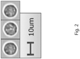
- FIG. 2 the images (photographs) of the immobilized leukocytes contained in the reference sample are shown in FIG. 2 .
- both the particles of Viscopearl ® D-10 and the immobilized leukocytes have a spherical form, both have a particle size of about 10 ⁇ m, and the condition of the particles of Viscopearl ® D-10 was extremely similar to that of the cytoplasm of the immobilized leukocytes.
- the particles included in Viscopearl ® D-10 were automatically identified as leukocytes by the urine sediment analyzer.
- Example 2 The same procedure as in Example 1 was carried out except that Viscopearl ® D-5 (manufactured by Rengo Co., Ltd.) (final concentration: 5.0 ⁇ 10 -4 mg/mL), which is spherical cellulose particles having an average particle size of 5 ⁇ m, was used instead of Viscopearl ® D-10 used in Example 1, to prepare Sample 2.
- Viscopearl ® D-5 manufactured by Rengo Co., Ltd.
- Viscopearl ® D-5 particles included in Viscopearl ® D-5 contained in Sample 2 were imaged, and the evaluation of Viscopearl ® D-5 was carried out, in the same manner as in Example 1.
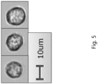
- the images (photographs) of the particles of Viscopearl ® D-5 contained in Sample 2 are shown in FIG. 5 .
- the form, the particle size and the condition of the particles of Viscopearl ® D-5 were extremely similar to those of the immobilized leukocytes, also in the case of Viscopearl ® D-5 having an average particle size different from that of Viscopearl ® D-10. Further, the particles included in Viscopearl ® D-5 were automatically identified as leukocytes by the urine sediment analyzer.
- Example 3 The same procedure as in Example 1 was carried out except that KCX-5000 (manufactured by Dainichiseika Color & Chemicals Mfg. Co., Ltd.) (final concentration: 5.0 ⁇ 10 -4 mg/mL), which is cellulose particles having an average particle size of 11 ⁇ m, was used instead of Viscopearl ® D-10 used in Example 1, to prepare Sample 3.
- KCX-5000 manufactured by Dainichiseika Color & Chemicals Mfg. Co., Ltd.
- Viscopearl ® D-10 used in Example 1, to prepare Sample 3.
- the particles of KCX-5000 contained in Sample 3 were imaged, and the evaluation of KCX-5000 was carried out, in the same manner as in Example 1.
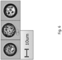
- the images (photographs) of the particles of KCX-5000 contained in Sample 3 are shown in FIG. 6 .
- patterns were observed within the spheres of the particles of KCX-5000, but the condition of these particles was different from that of the immobilized leukocytes; therefore, it was determined that the particles of KCX-5000 are not similar to the immobilized leukocytes.
- Example 4 The same procedure as in Example 1 was carried out except that MX-1000 (manufactured by Soken Kagaku Co., Ltd.) (final concentration: 8.0 ⁇ 10 -1 mg/mL), which is cross-linked acrylic monodispersed particles having an average particle size of 10 ⁇ m, was used instead of Viscopearl ® D-10 used in Example 1, to prepare Sample 4.
- MX-1000 manufactured by Soken Kagaku Co., Ltd.
- the particles of MX-1000 contained in Sample 4 were imaged, and the evaluation of MX-1000 was carried out, in the same manner as in Example 1.
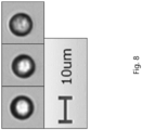
- the images (photographs) of the particles of MX-1000 contained in Sample 4 are shown in FIG. 8 .
- the particles of MX-1000 have a spherical form, but the condition of the cytoplasm of the immobilized leukocytes could not be confirmed, because the images tend to have a white blurred core or an emphasized contour; therefore, it was determined that the particles are not similar to the immobilized leukocytes.
- Example 2 The same procedure as in Example 1 was carried out except that Techpolymer ® BIO EF-C (manufactured by Sekisui Kasei Co., Ltd.) (final concentration: 5.0 ⁇ 10 -4 mg/mL), which is cross-linked polymer microparticles having an average particle size of 8.15 ⁇ m, was used instead of Viscopearl ® D-10 used in Example 1, to prepare Sample 5.
- Techpolymer ® BIO EF-C manufactured by Sekisui Kasei Co., Ltd.
- Viscopearl ® D-10 used in Example 1, to prepare Sample 5.
- the particles of Techpolymer ® BIO EF-C contained in Sample 5 were imaged, and the evaluation of Techpolymer ® BIO EF-C was carried out, in the same manner as in Example 1.
- the images (photographs) of the particles of Techpolymer ® BIO EF-C contained in Sample 5 are shown in FIG. 9 .
- the particles of Techpolymer ® BIO EF-C have a spherical form but have a non-uniform particle size, and the condition of the cytoplasm of the immobilized leukocytes could not be confirmed, as well; therefore, it was determined that the particles are not similar to the immobilized leukocytes.
- Example 6 The same procedure as in Example 1 was carried out except that NIKKALYCO ® AS-100SG (manufactured by Nikka Limited) (final concentration: 5.0 ⁇ 10 -4 mg/mL), which is starch particles having an average particle size of 17 ⁇ m, was used instead of Viscopearl ® D-10 used in Example 1, to prepare Sample 6.
- NIKKALYCO ® AS-100SG manufactured by Nikka Limited
- final concentration: 5.0 ⁇ 10 -4 mg/mL which is starch particles having an average particle size of 17 ⁇ m
- the particles of NIKKALYCO ® AS-100SG contained in Sample 6 were imaged, and the evaluation of NIKKALYCO ® AS-100SG was carried out, in the same manner as in Example 1.
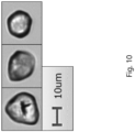
- the images (photographs) of the particles of NIKKALYCO ® AS-100SG contained in Sample 6 are shown in FIG. 10 .
- the particles of NIKKALYCO ® AS-100SG have a distorted spherical form, and the condition of the cytoplasm of the immobilized leukocytes could not be confirmed, as well; therefore, it was determined that the particles are not similar to the immobilized leukocytes.
- Example 7 The same procedure as in Example 1 was carried out except that Techpolymer ® BIO EF-B (manufactured by Sekisui Kasei Co., Ltd.) (final concentration: 5.0 ⁇ 10 -4 mg/mL), which is polymer microparticles having an average particle size of 10 ⁇ m, was used instead of Viscopearl ® D-10 used in Example 1, to prepare Sample 7.
- Techpolymer ® BIO EF-B manufactured by Sekisui Kasei Co., Ltd.
- Viscopearl ® D-10 used in Example 1, to prepare Sample 7.
- the particles of Techpolymer ® BIO EF-B contained in Sample 7 were imaged, and the evaluation of Techpolymer ® BIO EF-B was carried out, in the same manner as in Example 1.
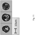
- the images (photographs) of the particles of Techpolymer ® BIO EF-B contained in Sample 7 are shown in FIG. 11 .
- the particles of Techpolymer ® BIO EF-B have a distorted form, and the condition of the cytoplasm of the immobilized leukocytes could not be confirmed, as well, because the particles appear blackish overall; therefore, it was determined that the particles are not similar to the immobilized leukocytes.
- Example 8 The same procedure as in Example 1 was carried out except that NH-RAS06 (manufactured by Nikko Spain Corporation) (final concentration: 1.0 ⁇ 10 -4 mg/mL), which is a raspberry-like, alumina microparticle-encapsulated silicone powder having an average particle size of 6 ⁇ m, was used instead of Viscopearl ® D-10 used in Example 1, to prepare Sample 8.
- NH-RAS06 manufactured by Nikko Spain Corporation
- Viscopearl ® D-10 Viscopearl ® D-10
- the powder particles of H-RAS06 contained in Sample 8 were imaged, and the evaluation of NH-RAS06 was carried out, in the same manner as in Example 1.
- the images (photographs) of the particles of NH-RAS06 contained in Sample 8 are shown in FIG. 12 .
- the particles of NH-RAS06 have a spherical form but have a small particle size, and the condition of the cytoplasm of the immobilized leukocytes could not be confirmed, as well; therefore, it was determined that the particles are not similar to the immobilized leukocytes.
- Example 9 The same procedure as in Example 1 was carried out except that Twinone SS-1000 (manufactured by CQV Co., Ltd.) (final concentration: 1.0 ⁇ 10 -4 mg/mL), which is golf ball-like amphipathic silicone beads having an average particle size of from 2 ⁇ m to 5 ⁇ m, was used instead of Viscopearl ® D-10 used in Example 1, to prepare Sample 9.
- Twinone SS-1000 manufactured by CQV Co., Ltd.
- Final concentration: 1.0 ⁇ 10 -4 mg/mL which is golf ball-like amphipathic silicone beads having an average particle size of from 2 ⁇ m to 5 ⁇ m
- the bead particles of Twinone SS-1000 contained in Sample 9 were imaged, and the evaluation of Twinone SS-1000 was carried out, in the same manner as in Example 1.
- the images (photographs) of the particles of Twinone SS-1000 contained in Sample 9 are shown in FIG. 13 .
- the particles of Twinone SS-1000 have a spherical form but have a small particle size, and the condition of the cytoplasm of the immobilized leukocytes could not be confirmed, as well; therefore, it was determined that the particles are not similar to the immobilized leukocytes.
Landscapes
- Health & Medical Sciences (AREA)
- Chemical & Material Sciences (AREA)
- Life Sciences & Earth Sciences (AREA)
- Immunology (AREA)
- Physics & Mathematics (AREA)
- General Physics & Mathematics (AREA)
- Pathology (AREA)
- Biochemistry (AREA)
- General Health & Medical Sciences (AREA)
- Analytical Chemistry (AREA)
- Engineering & Computer Science (AREA)
- Dispersion Chemistry (AREA)
- Hematology (AREA)
- Biomedical Technology (AREA)
- Urology & Nephrology (AREA)
- Molecular Biology (AREA)
- Food Science & Technology (AREA)
- Medicinal Chemistry (AREA)
- Microbiology (AREA)
- Cell Biology (AREA)
- Biotechnology (AREA)
- Biophysics (AREA)
- Investigating Or Analysing Biological Materials (AREA)
- Investigating Or Analysing Materials By Optical Means (AREA)
Abstract
Description
- The present disclosure relates to a quality control substance which is used for quality control in the analysis of sediments in a sample. More particularly, the present disclosure relates to a quality control substance to be used in the analysis of leukocyte items.
- Examinations have been performed in which a disease of a patient is diagnosed by observing sediments contained in a sample, such as urine, collected from the patient with a microscope, identifying leukocytes among the thus observed sediments and counting the number thereof. For example, if the leukocyte count is higher than a reference value, in urine sediment examination in which the types of the sediments contained in urine are identified, and the concentration of each sediment is measured, a urinary tract infection or the like is suspected. The examination of sediments using a microscope is performed by a method such as microscopy in which a laboratory technician directly observes a sample and counts the number of sediments, a method in which an automatic analyzer mounted on a microscope is used to identify the types of sediments, and to count the number of the sediments, or the like. In any of the cases of counting the number of leukocytes in a sample by such a method, quality control is necessary in order to accurately detect and identify leukocytes based on the forms of the sediments.
- Non-patent Document 1 discloses a control urine for urine sediment analysis, which includes erythrocytes and leukocytes separated from human-derived blood and immobilized with glutaraldehyde, as a quality control substance intended for use in quality control.
- Patent Document 1 discloses a quality control substance which includes: a cancer cell as a substance that indicates the form of a non-squamous epithelial cell in urine; an algae cell as a substance that indicates the form of an abnormal cast in urine; a yeast cell as a substance that indicates the form of a yeast cell in urine; and egg white as a substance that indicates the form of mucus in urine. As a substance imitating the form of leukocytes, hybridoma cells (obtained by hybridization of murine spleen cells with myeloma cells (p653 cells)) have been used. In other words, an object of the technique disclosed in Patent Document 1 is to perform quality control, in the case of observing a urine sample with a microscope, by using other substances whose forms are similar to urine sediments in the urine sample, and comparing the forms of components in the quality control substance with the forms of components in the urine sample.
-
- [Patent Document 1]
JP2020-531852A - [Patent Document 2]
JP2019-66461A - [Non-patent Document 1] Nobuko Imai et al., "Preparation and utility of control urine for urinary sediment examination", Hygiene Examination, Vol. 39, Issue 1, 1990, p. 55 to 61.
- In the case of preparing a quality control substance containing leukocytes, such as one disclosed in Patent Document 1 or Non-patent Document 1, the difficulty to obtain leukocytes due to ethical reasons has been a problem. Further, since components derived from living bodies are unstable and infectious, the handling of such components requires certain skills, which has also been a problem. Regarding leukocytes, in particular, the period of time allowed from the collection thereof until the preparation of a quality control substance is short, and there is a concern for the preservation stability of the resulting quality control substance.
- In view of such circumstances, an object of the present disclosure is to provide a quality control substance for use in the analysis of leukocytes (namely, a quality control substance for controlling the measurement accuracy for measuring a concentration of leukocytes), which can be prepared safely with easily available raw materials, which has excellent preservation stability, and which can be used in the analysis of sediments in a sample.
- The present inventors have found out that the form of a specific type of artificial particle is similar to the form of leukocytes in a sample when observed with a microscope, and that the artificial particles can be suitably used as a quality control substance for use in the analysis of leukocytes, in the analysis of sediments in the sample, thereby completing the present disclosure.
- Specifically, the present disclosure provides the following:
- [1] Use of artificial particles whose form as observed with a microscope is similar to the form of leukocytes as observed therewith, in controlling a measurement accuracy of a urine sediment analyzer.
- [2] The use according to [1], wherein the artificial particles have a spherical form.
- [3] The use according to [1] or [2], wherein the artificial particles have an average particle size of from 5 µm to 10 µm.
- [4] The use according to any one of [1] to [3], wherein the artificial particles are made of cellulose.
- [5] The use according to any one of [1] to [4], wherein the color of the artificial particles as observed with the microscope is similar to the color of the leukocytes as observed therewith.
- [6] The use according to any one of [1] to [5], wherein the artificial particles have a non-porous structure.
- [7] The use according to any one of [1] to [6], wherein the artificial particles are non-porous spherical cellulose particles having an average particle size of from 5 µm to 10 µm.
- [8] The use according to any one of [1] to [7], wherein the artificial particles are artificial particles included in at least one selected from the group consisting of Viscopearl® D-5, CELLULOBEADS® D-5, Viscopearl® D-10 and CELLULOBEADS® D-10.
- [9] A method for controlling a measurement accuracy of an analyzer that measures a concentration of particles whose form is similar to the form of leukocytes in a sample, by counting the number of the particles in the sample, the method comprising the steps of:
- measuring a concentration of particles in a quality control substance comprising artificial particles whose form is similar to the form of leukocytes, using the analyzer, to obtain a measured value; and
- determining the measurement accuracy of the analyzer by comparing the measured value with a reference value.
- [10] The method according to [9], wherein the artificial particles have a spherical form.
- [11] The method according to [9] or [10], wherein the artificial particles have an average particle size of from 5 µm to 10 µm.
- [12] The method according to any one of [9] to [11], wherein the artificial particles are made of cellulose.
- [13] The method according to any one of [9] to [12], wherein the color of the artificial particles as observed with the microscope is similar to the color of the leukocytes as observed therewith.
- [14] The method according to any one of [9] to [13], wherein the artificial particles have a non-porous structure.
- [15] The method according to any one of [9] to [14], wherein the artificial particles are non-porous spherical cellulose particles having an average particle size of from 5 µm to 10 µm.
- [16] The method according to any one of [9] to [15], wherein the artificial particles are artificial particles included in at least one selected from the group consisting of Viscopearl® D-5, CELLULOBEADS® D-5, Viscopearl® D-10 and CELLULOBEADS® D-10. EFFECT
- Since the artificial particles included in the quality control substance according to the present disclosure have high preservation stability, it is possible to perform quality control in a preferred manner, in the case of analyzing leukocytes which can be present in a sample using the quality control substance according to the present disclosure. Further, since the quality control substance for use in the analysis of leukocytes according to the present disclosure can be prepared safely without ethical problems, it is possible to expect improvements in operability at production sites and convenience at clinical sites, and the like. In addition, the use of a commercially available product of artificial particles enables to obtain a large amount of a quality control substance having the same quality. Artificial particles are preferred from the viewpoints that they can be obtained and used without any ethical problem due to being industrial products, that they have excellent preservation stability, and that they can be used safely due to not being infectious.
-
-
FIG. 1 shows images (photographs) illustrating the form of the particles of Viscopearl® D-10 contained in Sample 1, captured by a urine sediment analyzer. -
FIG. 2 shows images (photographs) illustrating the form of immobilized leukocytes contained in a reference sample, captured by the urine sediment analyzer. -
FIG. 3 shows images (photographs) illustrating the form of the particles of Viscopearl® D-10 contained in Sample 1, obtained by observation with a biological microscope. -
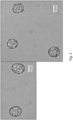
FIG. 4 shows images (photographs) illustrating the form of immobilized leukocytes contained in the reference sample, obtained by observation with the biological microscope. The immobilized leukocytes are indicated with arrows in the figure. -
FIG. 5 shows images (photographs) illustrating the form of the particles of Viscopearl® D-5 contained in Sample 2, captured by the urine sediment analyzer. -
FIG. 6 shows images (photographs) illustrating the form of the particles of KCX-5000 contained in Sample 3, captured by the urine sediment analyzer. -
FIG. 7 shows an image (photograph) illustrating the form of the particles of KCX-5000 contained in Sample 3, obtained by observation with the biological microscope. -
FIG. 8 shows images (photographs) illustrating the form of the particles of MX-1000 contained in Sample 4, captured by the urine sediment analyzer. -
FIG. 9 shows images (photographs) illustrating the form of the particles of Techpolymer® BIO EF-C contained in Sample 5, captured by the urine sediment analyzer. -
FIG. 10 shows images (photographs) illustrating the form of the particles of NIKKALYCO® AS-100SG contained in Sample 6, captured by the urine sediment analyzer. -
FIG. 11 shows images (photographs) illustrating the form of the particles of Techpolymer® BIO EF-B contained in Sample 7, captured by the urine sediment analyzer. -
FIG. 12 shows images (photographs) illustrating the form of the particles of NH-RAS06 contained in Sample 8, captured by the urine sediment analyzer. -
FIG. 13 shows images (photographs) illustrating the form of the particles of Twinone SS-1000 contained in Sample 9, captured by the urine sediment analyzer. - One embodiment of the present disclosure is a quality control substance which is for use in a sediment analysis with a microscope, and which includes artificial particles whose form as observed with the microscope is similar to the form of leukocytes as observed therewith. In the present specification, the microscope to be used is preferably a light microscope.
- The quality control substance according to the present embodiment is preferably a quality control substance for use in a urine sediment analyzer, particularly, a urine sediment analyzer based on a microscope, and is a quality control substance for controlling the measurement accuracy for measuring a concentration of leukocytes in a sample. The quality control substance according to the present embodiment is also preferably a quality control substance which is used by a laboratory technician, for observing leukocytes in a sample with a microscope to count the number of leukocytes in the sample, and is also a quality control substance for controlling the measurement accuracy for measuring the concentration of the leukocytes in the sample.
- The definition of the quality control substance includes a quality control substance used for the quality control of a urine sediment analyzer based on a microscope, in the analysis of leukocyte items. The quality control substance includes artificial particles whose form as observed with a microscope is similar to the form of leukocytes as observed therewith. In other words, the quality control substance includes such artificial particles that an image similar to the image obtained by observing an individual leukocyte with a microscope can be obtained by observing an individual artificial particle with the microscope.
- The definition of the quality control substance includes a quality control substance which is used for controlling a measurement accuracy in the case of counting the number of particles indicating the form of leukocytes, in a sample, and measuring the leukocyte concentration in the sample, based on the counted value. The definition of the expression "counting the number of particles indicating the form of leukocytes, in a sample" includes: a situation in which a laboratory technician observes and counts the number of such particles in the sample with a microscope; and a situation in which an analyzer including a microscope and a camera is used to capture particles in the sample by the camera via the microscope, to acquire an image in which the particles in the sample are captured, and the number of particles indicating the form of leukocytes, among the particles captured in the acquired image, is automatically counted by the analyzer. The concentration of the particles in a sample may be measured based on the counted value of the particles contained in a predetermined amount of the sample. Alternatively, it is possible to obtain a correlation between the number of particles observed within the range of a predetermined visual field of a microscope and the particle concentration in a sample, in advance; count the number of particles observed within the range of the predetermined visual field of the microscope, and measure the particle concentration from the counted value and the correlation.
- The artificial particles included in the quality control substance preferably include artificial particles whose form as observed with a microscope is similar to the form of leukocytes as observed therewith. Examples of the form of the artificial particles whose form is similar to the form of leukocytes when observed with a microscope, include a spherical form, an approximate spherical form, a golf ball-like form, a raspberry-like form, and a polyhedral form. Of these, a spherical form is preferred.
- The artificial particles included in the quality control substance can have an average particle size of from 2 µm to 20 µm, and preferably from 5 µm to 10 µm. In cases where the artificial particles do not have a spherical form, the particle size of each artificial particle is defined by the diameter of its circumscribed sphere.
- Examples of the artificial particles included in the quality control substance include artificially-produced inorganic particles and organic particles. Of these, organic particles are preferred. Examples of the material of the organic particles include natural polymers, semisynthetic polymers and synthetic polymers. Of these, natural polymers are preferred. Examples of the natural polymers include cellulose, starch, proteins and natural rubber. Of these, cellulose is preferred.
- More specifically, it is preferred to use, as the quality control substance, Viscopearl® (manufactured by Rengo Co., Ltd.) or CELLULOBEADS® (manufactured by Daito Kasei Kogyo Co., Ltd.), which is non-porous, spherical cellulose particles derived from wood pulp, and which includes particles whose form is similar to that of leukocytes. Of these, Viscopearl® D-5 having an average particle size of 5 µm, CELLULOBEADS® D-5 having an average particle size of 5 µm, Viscopearl® D-10 having an average particle size of 10 µm and CELLULOBEADS® D-10 having an average particle size of 10 µm are more preferred. It is noted here that Viscopearl® D-5 is another name for CELLULOBEADS® D-5, and Viscopearl® D-10 is another name for CELLULOBEADS D-10.
- In addition, examples of the material of the inorganic particles include metals; oxides, nitrides, fluorides, sulfides and borides of metals; complex compounds thereof; and hydroxyapatite.
- Examples of the semisynthetic polymers include cellulose derivatives (such as, for example, cellulose acetate, cellulose xanthate, carboxymethyl cellulose and hydroxyethyl cellulose), and starch derivatives (such as, for example, oxidized starch and dextrin). Examples of the synthetic polymers include silicone-based polymers (such as, for example, polymethylsilsesquioxane and (dimethicone/vinyl dimethicone) crosspolymers), acrylic polymers (such as, for example, polymethyl methacrylate), and styrene-based polymers (such as, for example, polystyrene and styrene-acrylonitrile copolymers).
- The color of the artificial particles included in the quality control substance as observed with the microscope is preferably similar to the color of leukocytes as observed therewith.
- Examples of the structure of the artificial particles included in the quality control substance include a non-porous structure and a porous structure. Of these, a non-porous structure is preferred.
- The quality control substance according to the present embodiment may include an aqueous solvent as long as the effects of the present disclosure are not impaired. Examples of the aqueous solvent include water and phosphate buffered saline (PBS).
- The quality control substance according to the present embodiment may further include an additional component as long as the effects of the present disclosure are not impaired. Examples of the additional component include a surfactant, an ionic substance and a water-soluble polymer. The surfactant may be, for example, polyoxyethylene (20) sorbitan monolaurate (such as one commercially available under the name of Tween® 20).
- Examples of leukocytes include polymorphonuclear granulocytes, monocytes and lymphocytes.
- Another embodiment of the present disclosure is a method for controlling a measurement accuracy of an analyzer that measures a concentration of particles whose form is similar to the form of leukocytes in a sample, by counting the number of the particles in the sample, the method including the steps of:
- measuring the concentration of the particles in the quality control substance described in the section of < Quality Control Substance >, using the analyzer, to obtain a measured value; and
- determining the measurement accuracy of the analyzer by comparing the measured value with a reference value.
- The "concentration of particles" refers to the number of particles contained in a unit volume, which is also referred to as "number concentration".
- The analyzer is, for example, an analyzer that includes a microscope, and a camera for imaging particles in a sample. The analyzer acquires an image in which the particles in the sample are captured, by imaging the particles in the sample with the camera via the microscope; counts the number of particles (similar particles) which are captured in the acquired image and whose degree of similarity with the form of leukocyte particles stored in the analyzer in advance is equal to or higher than a predetermined value, as leukocytes; and measure the concentration of the similar particles in the sample as the leukocyte concentration, based on the counted value. The analyzer that measures the leukocyte concentration in a sample may be, for example, a urine sediment analyzer based on a microscope, and the urine sediment analyzer based on a microscope may be, for example, AUTION EYE® AI-4510 (manufactured by Arkray, Inc.).
- The sample is not particularly limited as long as the sample possibly contains leukocytes. Examples of the sample include urine and blood. Of these, urine is preferred.
- When the quality control substance is analyzed using the analyzer, the artificial particles whose form as observed with a microscope is similar to the form of leukocytes as observed therewith, included in the quality control substance, are counted as the similar particles (leukocytes), and the concentration of the artificial particles included in the quality control substance is measured as the concentration of the similar particles (concentration of leukocytes). The measurement accuracy of the analyzer may be determined by comparing the thus measured value of the concentration of the artificial particles with a reference value, and based, for example, on the difference between the measured value of the concentration of the artificial particles and the reference value. In this case, the analyzer can be determined to have a good measurement accuracy, if the difference is less than a predetermined value. On the other hand, the analyzer can be determined to have a poor measurement accuracy, if the difference is equal to or higher than the predetermined value. Alternatively, the measurement accuracy of the analyzer may be determined based on the proportion of the measured value of the concentration of the artificial particles with respect to the reference value. In this case, the analyzer can be determined to have a good measurement accuracy, if the proportion is less than a predetermined value. On the other hand, the analyzer can be determined to have a poor measurement accuracy, if the proportion is equal to or higher than the predetermined value. The predetermined value can be set as appropriate depending on the accuracy required for the analyzer.
- The reference value can be, for example: the value obtained by analyzing the same quality control substance with a plurality of analyzers to obtain a plurality of measured values of the concentration of the similar particles, and averaging the thus obtained measured values; the measured value of the concentration of the similar particles obtained by measuring the quality control substance by microscopy; the measured value of the concentration of the similar particles (measured value of the concentration of leukocytes) obtained by measuring the quality control substance with a quality-controlled analyzer; or the like. The reference value can also be the mean value of the concentrations of the similar particles (concentrations of leukocytes) which have been measured by analyzing the quality control substance multiple times, in advance, with the above-described analyzer.
- Another embodiment of the present disclosure is a method for detecting the presence of leukocytes in a sample derived from a subject, the method including the steps of:
- analyzing the quality control substance described in the section of < Quality Control Substance > described above, using a urine sediment analyzer, in order to determine the form of the artificial particles in the quality control substance;
- analyzing the sample derived from the subject and containing sediments, using the urine sediment analyzer;
- comparing the form of the artificial particles in the quality control substance with the forms of the sediments in the sample; and
- determining the presence of leukocytes in the sample, if any of the forms of the sediments in the sample matches the form of the artificial particles in the quality control substance.
- The "subject" refers to an individual who undergoes the examination of sediments in a sample derived from the individual. The subject may be an individual with a disease, an individual suspected to have a disease, or a healthy individual. The subject is preferably a human being.
- The sample is not particularly limited as long as the sample possibly contains leukocytes. Examples of the sample include urine and blood. Of these, urine is preferred.
- Whether or not the form of leukocytes in the sample matches the form of the artificial particles in the quality control substance can be automatically determined by the urine sediment analyzer to be used. For example, in the case of using the urine sediment analyzer disclosed in Patent Document 2, which is based on an unstained image analysis performed in a fluid, the analyzer that identifies the types of sediments extracts the feature values (such as color, form and particle size) of the artificial particles in the quality control substance from the images of the artificial particles, and stores the feature values thereof as the feature values of leukocytes in the urine sediment analyzer, in advance. In the case of automatically identifying the sediments in the sample, the above-described analyzer extracts the feature values of the respective sediments in the sample from the images of the sediments; performs a pattern matching with the feature values of leukocytes, which had been stored in the urine sediment analyzer in advance, to determine whether or not any of the forms of the sediments matches the form of leukocytes (namely, whether or not the degree of similarity between the forms of both is equal to or higher than a certain value); and indicates the presence of leukocytes in the sample, if it is determined that the forms of both match with each other.
- Further, the urine sediment analyzer may count the number of leukocytes detected.
- The definition of the urine sediment analyzer includes a urine sediment analyzer based on a microscope. The urine sediment analyzer based on a microscope may be, for example, AUTION EYE® AI-4510.
- By detecting the presence of leukocytes in a sample derived from a subject, or by the result of counting the number of leukocytes in the sample derived from the subject, it is possible to suspect the presence of a disease such as urinary tract infection, and to determine a treatment plan (for example, the administration of a drug) for such a disease, as necessary.
- Examples are described for the purpose of disclosure, and are not intended to limit the scope of the present disclosure.
- At room temperature, polyoxyethylene (20) sorbitan monolaurate (Tween® 20; manufactured by FUJIFILM Wako Pure Chemical Corporation) having a final concentration of 0.01% was added to PBS (pH: 7.4) (27575-31, manufactured by Nakalai Tesque, Inc. (1x concentration), and the mixture was stirred. To the resulting solution, Viscopearl® D-10 (manufactured by Rengo Co., Ltd.) (final concentration: 5.0 × 10⁻⁴ mg/mL), which is spherical cellulose particles having an average particle size of 10 µm, was added, and then the resulting mixture was sufficiently mixed by inversion, to prepare Sample 1. Further, a pooled urine (reference sample) containing immobilized leukocytes was prepared by the method disclosed in Non-patent Document 1.
- Using a urine sediment analyzer, AUTION EYE® AI-4510 (manufactured by Arkray, Inc.), the particles of Viscopearl® D-10 contained in Sample 1 and particles contained in the reference sample were imaged, to obtain the images of the particles of Viscopearl® D-10 in Sample 1 and of the immobilized leukocytes in the reference sample. By observing the images of the particles of Viscopearl® D-10 and the immobilized leukocytes, it was determined whether or not the form and the particle size of the particles of Viscopearl® D-10 contained in Sample 1 are similar to those of the immobilized leukocytes contained in the reference sample. The images (photographs) of the particles of Viscopearl® D-10 contained in Sample 1 are shown in
FIG. 1 , and the images (photographs) of the immobilized leukocytes contained in the reference sample are shown inFIG. 2 . As can be seen from the images shown inFIG. 1 andFIG. 2 , both the particles of Viscopearl® D-10 and the immobilized leukocytes have a spherical form, both have a particle size of about 10 µm, and the condition of the particles of Viscopearl® D-10 was extremely similar to that of the cytoplasm of the immobilized leukocytes. Further, the particles included in Viscopearl® D-10 were automatically identified as leukocytes by the urine sediment analyzer. - Using a biological microscope BX-2700TL (manufactured by WRAYMER Inc.) in HPF mode (magnification: 400 times), the particles of Viscopearl® D-10 contained in Sample 1 and the immobilized leukocytes contained in the reference sample were observed, to determine whether or not the forms and the particle sizes of both are similar to each other. The images (photographs) of the particles of Viscopearl® D-10 contained in Sample 1 are shown in
FIG. 3 , and the images (photographs) of the immobilized leukocytes contained in the reference sample are shown inFIG. 4 . As can be seen from the images shown inFIG. 3 andFIG. 4 , the form, the particle size and the condition of the particles of Viscopearl® D-10 were extremely similar to those of the immobilized leukocytes, also in the observation with the biological microscope. - The same procedure as in Example 1 was carried out except that Viscopearl® D-5 (manufactured by Rengo Co., Ltd.) (final concentration: 5.0 × 10⁻⁴ mg/mL), which is spherical cellulose particles having an average particle size of 5 µm, was used instead of Viscopearl® D-10 used in Example 1, to prepare Sample 2.
- Using AUTION EYE® AI-4510, particles included in Viscopearl® D-5 contained in Sample 2 were imaged, and the evaluation of Viscopearl® D-5 was carried out, in the same manner as in Example 1. The images (photographs) of the particles of Viscopearl® D-5 contained in Sample 2 are shown in
FIG. 5 . As can be seen from the images shown inFIG. 5 , the form, the particle size and the condition of the particles of Viscopearl® D-5 were extremely similar to those of the immobilized leukocytes, also in the case of Viscopearl® D-5 having an average particle size different from that of Viscopearl® D-10. Further, the particles included in Viscopearl® D-5 were automatically identified as leukocytes by the urine sediment analyzer. - The same procedure as in Example 1 was carried out except that KCX-5000 (manufactured by Dainichiseika Color & Chemicals Mfg. Co., Ltd.) (final concentration: 5.0 × 10⁻⁴ mg/mL), which is cellulose particles having an average particle size of 11 µm, was used instead of Viscopearl® D-10 used in Example 1, to prepare Sample 3.
- Using AUTION EYE® AI-4510, the particles of KCX-5000 contained in Sample 3 were imaged, and the evaluation of KCX-5000 was carried out, in the same manner as in Example 1. The images (photographs) of the particles of KCX-5000 contained in Sample 3 are shown in
FIG. 6 . As can be seen from the images shown inFIG. 6 , patterns were observed within the spheres of the particles of KCX-5000, but the condition of these particles was different from that of the immobilized leukocytes; therefore, it was determined that the particles of KCX-5000 are not similar to the immobilized leukocytes. - Using the BX-2700TL in HPF mode (magnification: 400 times), the particles of KCX-5000 contained in Sample 3 were observed. The image (photograph) of the particles of KCX-5000 contained in Sample 3 is shown in
FIG. 7 . In the image shown in shown inFIG. 7 , the condition of the interior of the spheres of the KCX-5000 particles as observed in the images inFIG. 6 could not be confirmed. - The same procedure as in Example 1 was carried out except that MX-1000 (manufactured by Soken Kagaku Co., Ltd.) (final concentration: 8.0 × 10⁻¹ mg/mL), which is cross-linked acrylic monodispersed particles having an average particle size of 10 µm, was used instead of Viscopearl® D-10 used in Example 1, to prepare Sample 4.
- Using AUTION EYE® AI-4510, the particles of MX-1000 contained in Sample 4 were imaged, and the evaluation of MX-1000 was carried out, in the same manner as in Example 1. The images (photographs) of the particles of MX-1000 contained in Sample 4 are shown in
FIG. 8 . As can be seen from the images shown inFIG. 8 , the particles of MX-1000 have a spherical form, but the condition of the cytoplasm of the immobilized leukocytes could not be confirmed, because the images tend to have a white blurred core or an emphasized contour; therefore, it was determined that the particles are not similar to the immobilized leukocytes. - The same procedure as in Example 1 was carried out except that Techpolymer® BIO EF-C (manufactured by Sekisui Kasei Co., Ltd.) (final concentration: 5.0 × 10⁻⁴ mg/mL), which is cross-linked polymer microparticles having an average particle size of 8.15 µm, was used instead of Viscopearl® D-10 used in Example 1, to prepare Sample 5.
- Using AUTION EYE® AI-4510, the particles of Techpolymer® BIO EF-C contained in Sample 5 were imaged, and the evaluation of Techpolymer® BIO EF-C was carried out, in the same manner as in Example 1. The images (photographs) of the particles of Techpolymer® BIO EF-C contained in Sample 5 are shown in
FIG. 9 . As can be seen from the images shown inFIG. 9 , the particles of Techpolymer® BIO EF-C have a spherical form but have a non-uniform particle size, and the condition of the cytoplasm of the immobilized leukocytes could not be confirmed, as well; therefore, it was determined that the particles are not similar to the immobilized leukocytes. - The same procedure as in Example 1 was carried out except that NIKKALYCO® AS-100SG (manufactured by Nikka Limited) (final concentration: 5.0 × 10⁻⁴ mg/mL), which is starch particles having an average particle size of 17 µm, was used instead of Viscopearl® D-10 used in Example 1, to prepare Sample 6.
- Using AUTION EYE® AI-4510, the particles of NIKKALYCO® AS-100SG contained in Sample 6 were imaged, and the evaluation of NIKKALYCO® AS-100SG was carried out, in the same manner as in Example 1. The images (photographs) of the particles of NIKKALYCO® AS-100SG contained in Sample 6 are shown in
FIG. 10 . As can be seen from the images shown inFIG. 10 , the particles of NIKKALYCO® AS-100SG have a distorted spherical form, and the condition of the cytoplasm of the immobilized leukocytes could not be confirmed, as well; therefore, it was determined that the particles are not similar to the immobilized leukocytes. - The same procedure as in Example 1 was carried out except that Techpolymer® BIO EF-B (manufactured by Sekisui Kasei Co., Ltd.) (final concentration: 5.0 × 10⁻⁴ mg/mL), which is polymer microparticles having an average particle size of 10 µm, was used instead of Viscopearl® D-10 used in Example 1, to prepare Sample 7.
- Using AUTION EYE® AI-4510, the particles of Techpolymer® BIO EF-B contained in Sample 7 were imaged, and the evaluation of Techpolymer® BIO EF-B was carried out, in the same manner as in Example 1. The images (photographs) of the particles of Techpolymer® BIO EF-B contained in Sample 7 are shown in
FIG. 11 . As can be seen from the images shown inFIG. 11 , the particles of Techpolymer® BIO EF-B have a distorted form, and the condition of the cytoplasm of the immobilized leukocytes could not be confirmed, as well, because the particles appear blackish overall; therefore, it was determined that the particles are not similar to the immobilized leukocytes. - The same procedure as in Example 1 was carried out except that NH-RAS06 (manufactured by Nikko Rica Corporation) (final concentration: 1.0 × 10⁻⁴ mg/mL), which is a raspberry-like, alumina microparticle-encapsulated silicone powder having an average particle size of 6 µm, was used instead of Viscopearl® D-10 used in Example 1, to prepare Sample 8.
- Using AUTION EYE® AI-4510, the powder particles of H-RAS06 contained in Sample 8 were imaged, and the evaluation of NH-RAS06 was carried out, in the same manner as in Example 1. The images (photographs) of the particles of NH-RAS06 contained in Sample 8 are shown in
FIG. 12 . As can be seen from the images shown inFIG. 12 , the particles of NH-RAS06 have a spherical form but have a small particle size, and the condition of the cytoplasm of the immobilized leukocytes could not be confirmed, as well; therefore, it was determined that the particles are not similar to the immobilized leukocytes. - The same procedure as in Example 1 was carried out except that Twinone SS-1000 (manufactured by CQV Co., Ltd.) (final concentration: 1.0 × 10⁻⁴ mg/mL), which is golf ball-like amphipathic silicone beads having an average particle size of from 2 µm to 5 µm, was used instead of Viscopearl® D-10 used in Example 1, to prepare Sample 9.
- Using AUTION EYE® AI-4510, the bead particles of Twinone SS-1000 contained in Sample 9 were imaged, and the evaluation of Twinone SS-1000 was carried out, in the same manner as in Example 1. The images (photographs) of the particles of Twinone SS-1000 contained in Sample 9 are shown in
FIG. 13 . As can be seen from the images shown inFIG. 13 , the particles of Twinone SS-1000 have a spherical form but have a small particle size, and the condition of the cytoplasm of the immobilized leukocytes could not be confirmed, as well; therefore, it was determined that the particles are not similar to the immobilized leukocytes.
Claims (9)
- Use of artificial particles whose form as observed with a microscope is similar to the form of leukocytes as observed therewith, in controlling a measurement accuracy of a urine sediment analyzer.
- A method for controlling a measurement accuracy of an analyzer that measures a concentration of particles whose form is similar to the form of leukocytes in a sample, by counting the number of the particles in the sample, the method comprising the steps of:measuring a concentration of particles in a quality control substance comprising artificial particles whose form is similar to the form of leukocytes, using the analyzer, to obtain a measured value; anddetermining the measurement accuracy of the analyzer by comparing the measured value with a reference value.
- The method according to claim 2, wherein the artificial particles have a spherical form.
- The method according to claim 2 or claim 3, wherein the artificial particles have an average particle size of from 5 µm to 10 µm.
- The method according to any one of claims 2 to 4, wherein the artificial particles are made of cellulose.
- The method according to any one of claims 2 to 5, wherein the color of the artificial particles as observed with the microscope is similar to the color of the leukocytes as observed therewith.
- The method according to any one of claims 2 to 6, wherein the artificial particles have a non-porous structure.
- The method according to any one of claims 2 to 7, wherein the artificial particles are non-porous spherical cellulose particles having an average particle size of from 5 µm to 10 µm.
- The method according to any one of claims 2 to 8, wherein the artificial particles are artificial particles included in at least one selected from the group consisting of Viscopearl® D-5, CELLULOBEADS® D-5, Viscopearl® D-10 and CELLULOBEADS® D-10.
Applications Claiming Priority (1)
| Application Number | Priority Date | Filing Date | Title |
|---|---|---|---|
| JP2022148424A JP2024043307A (en) | 2022-09-16 | 2022-09-16 | White blood cell quality control substance |
Publications (1)
| Publication Number | Publication Date |
|---|---|
| EP4339588A1 true EP4339588A1 (en) | 2024-03-20 |
Family
ID=88093160
Family Applications (1)
| Application Number | Title | Priority Date | Filing Date |
|---|---|---|---|
| EP23197655.6A Pending EP4339588A1 (en) | 2022-09-16 | 2023-09-15 | Quality control substance for use in the analysis of leukocytes |
Country Status (4)
| Country | Link |
|---|---|
| US (1) | US20240094230A1 (en) |
| EP (1) | EP4339588A1 (en) |
| JP (1) | JP2024043307A (en) |
| CN (1) | CN117723469A (en) |
Citations (3)
| Publication number | Priority date | Publication date | Assignee | Title |
|---|---|---|---|---|
| JP2019066461A (en) | 2017-10-02 | 2019-04-25 | アークレイ株式会社 | Analysis equipment |
| JP2020531852A (en) | 2017-08-28 | 2020-11-05 | シーメンス・ヘルスケア・ダイアグノスティックス・インコーポレーテッドSiemens Healthcare Diagnostics Inc. | Quality control substances used in urine sediment analyzers using a microscope and how to use them |
| WO2021127424A1 (en) * | 2019-12-19 | 2021-06-24 | Roche Diagnostics Hematology, Inc. | Compositions and methods for calibration and quality control |
Family Cites Families (4)
| Publication number | Priority date | Publication date | Assignee | Title |
|---|---|---|---|---|
| EP0678742B1 (en) * | 1994-04-21 | 2002-07-24 | Hitachi, Ltd. | Monitoring method of stain solution for particle analysis and calibration method of particle analysis |
| JP4953710B2 (en) * | 2005-07-12 | 2012-06-13 | シスメックス株式会社 | Reference material for urine sediment analyzer |
| JP4946047B2 (en) * | 2005-12-28 | 2012-06-06 | 東洋紡績株式会社 | Particle standard reagent for formed component classifier |
| CN113826000A (en) * | 2019-04-26 | 2021-12-21 | 芯易诊有限公司 | Method and apparatus for correction in particle size measurement |
-
2022
- 2022-09-16 JP JP2022148424A patent/JP2024043307A/en active Pending
-
2023
- 2023-09-15 US US18/468,585 patent/US20240094230A1/en active Pending
- 2023-09-15 CN CN202311193632.XA patent/CN117723469A/en active Pending
- 2023-09-15 EP EP23197655.6A patent/EP4339588A1/en active Pending
Patent Citations (3)
| Publication number | Priority date | Publication date | Assignee | Title |
|---|---|---|---|---|
| JP2020531852A (en) | 2017-08-28 | 2020-11-05 | シーメンス・ヘルスケア・ダイアグノスティックス・インコーポレーテッドSiemens Healthcare Diagnostics Inc. | Quality control substances used in urine sediment analyzers using a microscope and how to use them |
| JP2019066461A (en) | 2017-10-02 | 2019-04-25 | アークレイ株式会社 | Analysis equipment |
| WO2021127424A1 (en) * | 2019-12-19 | 2021-06-24 | Roche Diagnostics Hematology, Inc. | Compositions and methods for calibration and quality control |
Non-Patent Citations (2)
| Title |
|---|
| CHIEN TZU-I ET AL: "Urine sediment examination: A comparison of automated urinalysis systems and manual microscopy", CLINICA CHIMICA ACTA, vol. 384, no. 1, 26 May 2007 (2007-05-26), pages 28 - 34, XP029160703, ISSN: 0009-8981, DOI: 10.1016/J.CCA.2007.05.012 * |
| NOBUKO IMAI ET AL.: "Preparation and utility of control urine for urinary sediment examination", HYGIENE EXAMINATION, vol. 39, 1990, pages 55 - 61 |
Also Published As
| Publication number | Publication date |
|---|---|
| CN117723469A (en) | 2024-03-19 |
| US20240094230A1 (en) | 2024-03-21 |
| JP2024043307A (en) | 2024-03-29 |
Similar Documents
| Publication | Publication Date | Title |
|---|---|---|
| AU2017263807B2 (en) | Performing optical measurements on a sample | |
| AU2014244557B2 (en) | Portable blood count monitor | |
| AU2007275926B2 (en) | Enumeration of thrombocytes | |
| AU2010232583B2 (en) | Detection of infectious disease in a human or animal by measuring specific phagocytosis in a thin film sample of their anticoagulated blood | |
| ES2641015T3 (en) | Method and system to analyze a blood sample | |
| Manoni et al. | Mid-stream vs. first-voided urine collection by using automated analyzers for particle examination in healthy subjects: an Italian multicenter study. | |
| JP2003517569A (en) | Method and apparatus for performing chemical, qualitative, quantitative, and semi-quantitative analysis of urine samples | |
| US11079718B2 (en) | Specific malaria detection with digital holographic microscopy | |
| JP4160117B2 (en) | Methods for testing cell samples | |
| US20240094230A1 (en) | Quality control substance for use in the analysis of leukocytes | |
| EP4339589A1 (en) | Quality control substance for use in the analysis of erythrocytes | |
| Santos et al. | Evaluation of phagocytic capacity with a modified flow cytometry technique | |
| CN110530824B (en) | Erythrocyte osmotic fragility determination method and kit thereof | |
| EP4286827B1 (en) | Quality control substance used in urine sediment analyzer | |
| US3834874A (en) | Detection of malarial parasites in blood | |
| US3476514A (en) | Cancer cytoscreening | |
| US20230380412A1 (en) | Preservation liquid for calcium oxalate dihydrate crystals and preservation method | |
| JP2023177266A (en) | Quality control substances used in urine sediment analyzers | |
| CN117147243A (en) | Precision controlled substances used in urine sediment analysis equipment | |
| WO2025075824A1 (en) | Urine sediment interface with probabilistic classification binning and methods of producing and using same | |
| WO2025262239A1 (en) | System & method for collecting particles in exhaled air | |
| CN119955888A (en) | Method for detecting viability and density of melanin-containing cells | |
| RU2210769C2 (en) | Method for detecting sternheimermalbin's cells in patients with pyelonephritis | |
| RU2227280C2 (en) | Method for detecting reticulocytes in incubated blood in poultry | |
| EP1366409B1 (en) | Light scattering determination of treatment potencies |
Legal Events
| Date | Code | Title | Description |
|---|---|---|---|
| PUAI | Public reference made under article 153(3) epc to a published international application that has entered the european phase |
Free format text: ORIGINAL CODE: 0009012 |
|
| STAA | Information on the status of an ep patent application or granted ep patent |
Free format text: STATUS: THE APPLICATION HAS BEEN PUBLISHED |
|
| AK | Designated contracting states |
Kind code of ref document: A1 Designated state(s): AL AT BE BG CH CY CZ DE DK EE ES FI FR GB GR HR HU IE IS IT LI LT LU LV MC ME MK MT NL NO PL PT RO RS SE SI SK SM TR |
|
| STAA | Information on the status of an ep patent application or granted ep patent |
Free format text: STATUS: REQUEST FOR EXAMINATION WAS MADE |
|
| 17P | Request for examination filed |
Effective date: 20240910 |
|
| RBV | Designated contracting states (corrected) |
Designated state(s): AL AT BE BG CH CY CZ DE DK EE ES FI FR GB GR HR HU IE IS IT LI LT LU LV MC ME MK MT NL NO PL PT RO RS SE SI SK SM TR |
|
| STAA | Information on the status of an ep patent application or granted ep patent |
Free format text: STATUS: EXAMINATION IS IN PROGRESS |
|
| 17Q | First examination report despatched |
Effective date: 20250730 |