EP4332881A1 - Method for determining a myocardial extracellular volume fraction, processing system, medical imaging device, computer program and computer-readable storage medium - Google Patents
Method for determining a myocardial extracellular volume fraction, processing system, medical imaging device, computer program and computer-readable storage medium Download PDFInfo
- Publication number
- EP4332881A1 EP4332881A1 EP22192839.3A EP22192839A EP4332881A1 EP 4332881 A1 EP4332881 A1 EP 4332881A1 EP 22192839 A EP22192839 A EP 22192839A EP 4332881 A1 EP4332881 A1 EP 4332881A1
- Authority
- EP
- European Patent Office
- Prior art keywords
- photon
- energy
- data set
- computer
- contrast agent
- Prior art date
- Legal status (The legal status is an assumption and is not a legal conclusion. Google has not performed a legal analysis and makes no representation as to the accuracy of the status listed.)
- Pending
Links
Images


Classifications
-
- G—PHYSICS
- G06—COMPUTING OR CALCULATING; COUNTING
- G06T—IMAGE DATA PROCESSING OR GENERATION, IN GENERAL
- G06T7/00—Image analysis
- G06T7/0002—Inspection of images, e.g. flaw detection
- G06T7/0012—Biomedical image inspection
- G06T7/0014—Biomedical image inspection using an image reference approach
-
- G—PHYSICS
- G06—COMPUTING OR CALCULATING; COUNTING
- G06V—IMAGE OR VIDEO RECOGNITION OR UNDERSTANDING
- G06V10/00—Arrangements for image or video recognition or understanding
- G06V10/70—Arrangements for image or video recognition or understanding using pattern recognition or machine learning
- G06V10/77—Processing image or video features in feature spaces; using data integration or data reduction, e.g. principal component analysis [PCA] or independent component analysis [ICA] or self-organising maps [SOM]; Blind source separation
- G06V10/774—Generating sets of training patterns; Bootstrap methods, e.g. bagging or boosting
-
- A—HUMAN NECESSITIES
- A61—MEDICAL OR VETERINARY SCIENCE; HYGIENE
- A61B—DIAGNOSIS; SURGERY; IDENTIFICATION
- A61B6/00—Apparatus or devices for radiation diagnosis; Apparatus or devices for radiation diagnosis combined with radiation therapy equipment
- A61B6/02—Arrangements for diagnosis sequentially in different planes; Stereoscopic radiation diagnosis
- A61B6/03—Computed tomography [CT]
- A61B6/032—Transmission computed tomography [CT]
-
- A—HUMAN NECESSITIES
- A61—MEDICAL OR VETERINARY SCIENCE; HYGIENE
- A61B—DIAGNOSIS; SURGERY; IDENTIFICATION
- A61B6/00—Apparatus or devices for radiation diagnosis; Apparatus or devices for radiation diagnosis combined with radiation therapy equipment
- A61B6/50—Apparatus or devices for radiation diagnosis; Apparatus or devices for radiation diagnosis combined with radiation therapy equipment specially adapted for specific body parts; specially adapted for specific clinical applications
- A61B6/503—Apparatus or devices for radiation diagnosis; Apparatus or devices for radiation diagnosis combined with radiation therapy equipment specially adapted for specific body parts; specially adapted for specific clinical applications for diagnosis of the heart
-
- A—HUMAN NECESSITIES
- A61—MEDICAL OR VETERINARY SCIENCE; HYGIENE
- A61B—DIAGNOSIS; SURGERY; IDENTIFICATION
- A61B6/00—Apparatus or devices for radiation diagnosis; Apparatus or devices for radiation diagnosis combined with radiation therapy equipment
- A61B6/52—Devices using data or image processing specially adapted for radiation diagnosis
- A61B6/5211—Devices using data or image processing specially adapted for radiation diagnosis involving processing of medical diagnostic data
- A61B6/5217—Devices using data or image processing specially adapted for radiation diagnosis involving processing of medical diagnostic data extracting a diagnostic or physiological parameter from medical diagnostic data
-
- A—HUMAN NECESSITIES
- A61—MEDICAL OR VETERINARY SCIENCE; HYGIENE
- A61B—DIAGNOSIS; SURGERY; IDENTIFICATION
- A61B6/00—Apparatus or devices for radiation diagnosis; Apparatus or devices for radiation diagnosis combined with radiation therapy equipment
- A61B6/52—Devices using data or image processing specially adapted for radiation diagnosis
- A61B6/5258—Devices using data or image processing specially adapted for radiation diagnosis involving detection or reduction of artifacts or noise
-
- G—PHYSICS
- G06—COMPUTING OR CALCULATING; COUNTING
- G06T—IMAGE DATA PROCESSING OR GENERATION, IN GENERAL
- G06T12/00—Tomographic reconstruction from projections
-
- G—PHYSICS
- G06—COMPUTING OR CALCULATING; COUNTING
- G06T—IMAGE DATA PROCESSING OR GENERATION, IN GENERAL
- G06T7/00—Image analysis
- G06T7/0002—Inspection of images, e.g. flaw detection
- G06T7/0012—Biomedical image inspection
-
- G—PHYSICS
- G06—COMPUTING OR CALCULATING; COUNTING
- G06T—IMAGE DATA PROCESSING OR GENERATION, IN GENERAL
- G06T7/00—Image analysis
- G06T7/10—Segmentation; Edge detection
-
- G—PHYSICS
- G06—COMPUTING OR CALCULATING; COUNTING
- G06T—IMAGE DATA PROCESSING OR GENERATION, IN GENERAL
- G06T7/00—Image analysis
- G06T7/10—Segmentation; Edge detection
- G06T7/11—Region-based segmentation
-
- G—PHYSICS
- G16—INFORMATION AND COMMUNICATION TECHNOLOGY [ICT] SPECIALLY ADAPTED FOR SPECIFIC APPLICATION FIELDS
- G16H—HEALTHCARE INFORMATICS, i.e. INFORMATION AND COMMUNICATION TECHNOLOGY [ICT] SPECIALLY ADAPTED FOR THE HANDLING OR PROCESSING OF MEDICAL OR HEALTHCARE DATA
- G16H30/00—ICT specially adapted for the handling or processing of medical images
- G16H30/20—ICT specially adapted for the handling or processing of medical images for handling medical images, e.g. DICOM, HL7 or PACS
-
- A—HUMAN NECESSITIES
- A61—MEDICAL OR VETERINARY SCIENCE; HYGIENE
- A61B—DIAGNOSIS; SURGERY; IDENTIFICATION
- A61B6/00—Apparatus or devices for radiation diagnosis; Apparatus or devices for radiation diagnosis combined with radiation therapy equipment
- A61B6/48—Diagnostic techniques
- A61B6/481—Diagnostic techniques involving the use of contrast agents
-
- A—HUMAN NECESSITIES
- A61—MEDICAL OR VETERINARY SCIENCE; HYGIENE
- A61B—DIAGNOSIS; SURGERY; IDENTIFICATION
- A61B6/00—Apparatus or devices for radiation diagnosis; Apparatus or devices for radiation diagnosis combined with radiation therapy equipment
- A61B6/48—Diagnostic techniques
- A61B6/482—Diagnostic techniques involving multiple energy imaging
-
- G—PHYSICS
- G06—COMPUTING OR CALCULATING; COUNTING
- G06T—IMAGE DATA PROCESSING OR GENERATION, IN GENERAL
- G06T2207/00—Indexing scheme for image analysis or image enhancement
- G06T2207/10—Image acquisition modality
- G06T2207/10072—Tomographic images
- G06T2207/10081—Computed x-ray tomography [CT]
-
- G—PHYSICS
- G06—COMPUTING OR CALCULATING; COUNTING
- G06T—IMAGE DATA PROCESSING OR GENERATION, IN GENERAL
- G06T2207/00—Indexing scheme for image analysis or image enhancement
- G06T2207/30—Subject of image; Context of image processing
- G06T2207/30004—Biomedical image processing
- G06T2207/30024—Cell structures in vitro; Tissue sections in vitro
-
- G—PHYSICS
- G06—COMPUTING OR CALCULATING; COUNTING
- G06T—IMAGE DATA PROCESSING OR GENERATION, IN GENERAL
- G06T2207/00—Indexing scheme for image analysis or image enhancement
- G06T2207/30—Subject of image; Context of image processing
- G06T2207/30004—Biomedical image processing
- G06T2207/30048—Heart; Cardiac
Definitions
- the invention concerns a computer-implemented method for determining at least one myocardial extracellular volume fraction for a patient. Additionally, the invention concerns a processing system, a medical imaging device, a computer program and a computer-readable storage medium.
- Interstitial myocardial fibrosis is an important disease process that has been associated with different kinds of cardio myopathy.
- the myocardial extracellular volume fraction (ECV) increases in the case of diffuse myocardial fibrosis or infiltrates and is therefore considered as a distinguishing feature of myocardial pathologic findings for cardiomyopathies.
- the myocardial extracellular volume fraction can be determined in magnetic resonance imaging by determining the equilibrium distribution of gadolinium contrast agent between the myocardium and the blood. As iodine-based computed tomography contrast agents exhibit similar kinetics as gadolinium, the myocardial extracellular volume fraction can also be determined using computed tomography.
- normal single energy computed tomography scans can be used for this purpose.
- it is however necessary to perform a baseline native scan without contrast agent a normal coronary computed tomography angiography scan using contrast agent and a so-called late enhancing scan that is recorded several minutes after the contrast agent injection and depicts an equilibrium distribution of the contrast agent in which the contrast agent has already entered the intercellular volume of the myocardium.
- the use of three separate scans leads to relatively high x-ray-exposure of the patient and requires a registering of scans acquired at different times, which can lead to artefacts and therefore less exact results.
- a notable improvement can be achieved when dual energy computed tomography is used. Since the x-ray-absorption is determined for different photon-energy bands in a dual energy computed tomography, different materials can be distinguished by analyzing the photon-energy dependent x-ray-absorption. When using such a dual energy acquisition, an, e.g., iodine-based contrast agent can be selectively imaged, therefore providing a contrast agent map. On the other hand, it is also possible to map water and therefore essentially tissue from the same dual energy scan.
- the variable h represents the hematocrit value for the patient that can, e.g., be known from patient data, determined via a blood test or calculated from computed tomography data.
- a myocardial extracellular volume fraction based on dual energy computed tomography either require manual segmentation of the image data or the acquisition of an additional scan used for an automatic segmentation, that is preferably performed at a time relatively shortly after the contrast agent injection.
- a complete computed tomography angiography sequence can be performed or at least the scan comprising contrast agent essentially only in the blood vessel can be used as an additional scan. In this case it is however still necessary to register at least two scans taken at different times and the x-ray-exposure of the patient is increased over the use of a single scan.
- the invention is therefore based on the problem of providing an improved approach for determining at least one myocardial extracellular volume fraction for a patient, that especially avoids the necessity of manual segmentation, even when using only a single computed tomography scan.
- the problem is solved by a computer-implemented method for determining at least one myocardial extracellular volume fraction for a patient, comprising the steps of:
- photon-energy bands that can also be called energy bins
- To combine energy bands it is e.g., possible to add or subtract images reconstructed from different energy bands.
- Two of the possible impressions are the previously discussed contrast agent map and the mapping of tissue or water.
- Such a material contrast is caused by the fact that most contrast agents, especially iodine, have an x-ray-absorption that strongly depends on the photon-energy. Therefore, a contrast agent map can, e.g., be determined from the difference in absorption in different photon-energy bands. Once the absorption due to the contrast agent is known, the remaining absorption can be attributed to water and tissue.
- the differentiation between different materials can obviously be further improved, when differences between the x-ray absorption for more than two photon-energy bands are considered.
- a morphology preserving image data set can be generated that allows for the use of normal segmentation algorithms that are typically used to segment image data sets acquired by coronary computed tomography angiography, and therefore especially image data sets recorded after a relatively short waiting time after injecting the contrast agent and therefore in a condition where essentially all of the contrast agent is still contained in the blood pool and essentially no contrast agent has migrated to the tissue.
- the first photon-energy band or first combination of photon-energy bands is especially chosen in such a way that the contrast-to-noise-ratio for the morphological features to be segmented, especially for the blood pool and/or the myocardium, is notably higher, e.g., at least by a factor of two or three, than in the contrast agent map. As discussed in more detail below, it was found that this can especially be achieved by only or mainly considering photons with a photon-energy at the lower end of the available spectrum.
- the considered myocardium and blood pool can especially be the myocardium of the left ventricle and the blood pool therein.
- a photon-counting detector it is possible to at least approximately determine the photon-energy of each detected photon, since the height of a current spike generated by the detection of the respective photon depends on the photon-energy of that photon. It is therefore, e.g., possible to log each individual photon detection in each pixel of a detector with the associated photon energy and afterwards perform an essentially arbitrary binning of the events depending on the detected energy to provide multiple photon-energy bands.
- the presence of a contrast agent can especially be detected, when the local absorption strongly varies with the photon-energy.
- the contrast agent map can therefore preferably be reconstructed based on a combination of at least two, preferably more, photon-energy bands described in the measurement data set.
- both the morphology preserving image data set and the contrast agent map are based on a respective single photon-energy band or that they are both based on a combination of photon-energy bands.
- the different photon-energy bands can be non-overlapping or at least one pair of the photon-energy bands can overlap.
- the morphology preserving image data set and the contrast agent map are preferably three-dimensional image data sets, especially formed from a three-dimensional array of voxels. Alternatively, they can be two-dimensional image data sets, e.g., formed by an array of pixels.
- the measurement data set can comprise multiple projection images, e.g., a single projection image for each photon-energy band and projection angle, or it can e.g., comprise a respective event list comprising all individual photon detection events for each detector pixel and projection angle.
- the image acquisition is preferably not part of the computer-implemented method and is completed before the start of the method.
- the image acquisition could be included as an additional step and the acquired image data could, e.g., be directly received from a computed tomography scanner.
- all discussed steps of the computer-implemented method are performed fully automatic.
- the selection of the energy band or energy bands used to reconstruct the morphology preserving image data set and/or the segmentation of the blood pool and/or the segmentation of the myocardium can be modified by a user.
- the segmentation of the blood pool and/or the myocardium or at least an initial segmentation that can then be modified by the user is however performed automatically without user interaction.
- the approach for automatic segmentation for a three-dimensional cardiac computed tomography volume discussed in the initially cited paper by Y. Zheng was used.
- further approaches for automatically segmenting contrast enhanced images in computed tomography angiography can typically be directly used to segment the morphology preserving image data set in the invention without any further modification.
- the measurement data set is preferably based on a photon-counting computed tomography scan.
- the weight of lower energy photons in a given energy band is reduced due to the used measurement principle. This effect can be avoided in photon-counting computed tomography data sets, since each individual photon is countered and the photon energy is detected as a separate parameter of the detection.
- the use of photon-counting computed tomography scan as a base for the input data set allows for more robust morphology preservation, especially for a higher contrast-to-noise-ration in the morphology preserving image data set and therefore for a more exact and robust automatic segmentation.
- photon counted computed tomography also allows for an essentially free and, if needed, a dynamic selection of photon-energy bands that are used in reconstructing the morphology preserving image data set on the one hand and the contrast agent map on the other hand.
- the only limitation for the number of used photon-energy bands is a sufficient photon count within the respective band and a sufficient energy resolution of the detector.
- classic multi-energy computed tomography is limited to two or at most three or four photon-energy bands, that are typically fixed
- photon-counting computed tomography allow for the use of a larger number of bands and/or the dynamic adjustment of the position and widths of the different energy bands to provide optimum results. This can be used to further improve the contrast-to-noise-ratio in the morphology preserving image data set and/or to further improve material selectivity for the contrast agent map.
- the myocardium is segmented in the morphology preserving image data set, wherein a respective myocardial extracellular volume fraction is only determined for pixels or voxels within the segmented myocardium.
- an extracellular volume image data set can be generated that comprises the respective myocardial extracellular volume fraction for each respective pixel or voxel within the segmented myocardium and either a fixed value or a value exclusively based on the morphology preserving image data set for each pixel or voxel outside the segmented myocardium. Determining the extracellular volume fraction outside of the myocardium is typically not necessary or useful when, e.g., trying to detect myocardial fibrosis.
- Using a fixed value for pixels or voxels outside the segmented myocardium can be useful to, e.g., generate a three-dimensional data set that has full transparency in those areas.
- Such a data set can be useful for easy viewing, to allow for a robust mapping to a two-dimensional coordinate system, e.g., using a two-dimensional polar map, and/or for generating an overlay that can be superimposed e.g., onto the morphology preserving image data set.
- Such a superposition allows for an easier orientation of e.g., a medical examiner within the image due to the recognizability of morphological features.
- different color palletes can be used for a projection generated from the morphology preserving image data set and a projection generated from the extracellular volume image data set. It is e.g. possible to generate a colored image with a given pallete from the extracellular volume image data set and to superimpose this image with an image generated from the morphology preserving image data set using a gray scale pallete.
- the alternative is a combination of these two sources of data within a single two- or three-dimensional image data set.
- the limitation of the determination of the myocardial extracellular volume fraction to the volume actually covered by the myocardium according to the segmentation or a masking of the generated values to only leave these values can be advantageous for a statistical analysis of the myocardial extracellular volume fraction, since this analysis can then be limited to the relevant volume.
- Simple examples for such a statistical analysis would be the calculation of an average and/or median myocardial extracellular volume faction and/or the determination of measures describing the distribution, e.g. of a standard deviation, etc.
- the reference value can be determined based on an average or median of multiple pixels or voxels, preferably based on an average or median of all pixels or voxels, of the contrast agent map within the segmented blood pool. Instead of using all the pixels or voxels of the segmented blood pool, it is also possible to discard the pixels or voxels in a given number of outer rows of pixels or voxels and therefore reduce the influence of potential segmentation errors.
- the use of an average or median is advantageous, since the distribution of the contrast agent in the blood is expected to be essentially homogeneous in the given region and therefore an averaging or median determination can be used to further reduce errors.
- the measurement data set can comprise energy resolved data based on a computed tomography scan of the patient performed at least 2 minutes or at least 3 minutes and/or at most 6 minutes or at least 5 minutes after the beginning of a contrast agent injection.
- a computed tomography scan of the patient performed at least 2 minutes or at least 3 minutes and/or at most 6 minutes or at least 5 minutes after the beginning of a contrast agent injection.
- the average photon-energy in the first photon-energy band or in the first combination of photon-energy bands can be less than 70 keV or less than 50 keV.
- the contrast-to-noise-ratio in the morphology preserving image data set can be increased, when mainly photons with a relatively low photon-energy are used. It is e.g. possible to use a single first photon-energy band centered at 40 keV.
- the width of the first photon-energy band or the energy spread covered by the first combination of photon-energy bands can, e.g., be less than 30 keV or even less than 10 keV.
- the position and width of the considered photon-energy band can essentially be freely chosen when using photon-counting computed tomography. If necessary, it is also possible to e.g. pool photon detections from multiple adjacent pixels of a detector to increase the overall photon count and therefore e.g. to allow for the use of less-wide photon-energy bands. Since the photon-energy of each individual photon can be detected in photon-counting computed tomography, it is in principle also possible to continuously vary the weighting factor of individual photon within a considered photon-energy band.
- the second photon-energy band or the second combination of photon-energy bands can comprise at least one photon-energy that is higher than the highest photon-energy in the first photon-energy band or the first combination of photon-energy bands.
- the contrast agent map can be based on the second combination of photon-energy bands, wherein the second combination of photon-energy bands comprises at least one photon-energy band or at least two photon-energy bands that does or do not overlap with the first photon-energy band or any photon-energy bands of the first combination of photon-energy bands.
- the second combination of photon-energy bands can additionally comprise the first photon-energy band or at least one photon-energy band of the first combination of photon-energy bands.
- the at least one myocardial extracellular volume fraction can be determined from energy resolved data based on exactly one computed tomography scan of the patient forming the measurement data set and a provided hematocrit value, wherein no other input data besides the measurement data set and the hematocrit value is used.
- the use of only a single computed tomography scan avoids potential artefacts due to a registration of multiple scans.
- the morphology preserving image data set and the contrast agent map in the computer-implemented method according to the present invention are inherently registered to each other, since they are based on the same received measurement data set that especially represents only a single computed tomography scan. The determination is therefore more robust and less error-prone than the initially discussed approaches that require a registration of multiple computed tomography scans.
- a representation of the segmented blood pool, especially of the outline of the segmented blood pool in the morphology preserving image data set, and/or a representation of the segmented myocardium, especially of the outline of the segmented myocardium in the morphology preserving image data set, can displayed for a user, wherein the user can edit the segmentation of the blood pool and/or the myocardium, whereafter the respective edited segmentation can be used to determine the reference value and/or the at least one myocardial extracellular volume fraction. While the automatic segmentation of the blood pool and the myocardium is very robust in the discussed method, the acceptance of the method can be improved when medical personal can review the results of intermediate steps and potentially modify the results if deemed necessary.
- a user can, e.g., interactively vary the first photon-energy band or the first combination of photon-energy bands used to reconstruct the morphology preserving image data set. It would, e.g., be possible to show at least one projection image of an initially generated morphology preserving image data set and, e.g., to provide a fader or some other control that allows a use to slightly shift the used first photon-energy band until an optimum contrast is reached. Alternatively, such an optimization could be performed automatically.
- the invention concerns a processing system, comprising
- the processing system can, e.g., be part of a computed tomography scanner or of a work station used to control this scanner. Alternatively, the processing system could also be a separate work station that is not used for control of the scanner or even be implemented as a cloud-based solution.
- the invention also concerns a medical imaging device comprising an energy resolving computed tomography scanner and a processing system according to the present invention.
- the computed tomography scanner can preferably be a photon-counting computed tomography scanner.
- a non-photon counting multi energy computed tomography scanner can be used.
- the invention additionally concerns a computer program comprising instructions to cause the or a processing device to execute the computer-implemented method according to the present invention.
- the invention concerns a computer-readable storage medium having stored thereon the computer program according to the present invention.
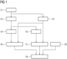
- Fig. 1 shows a flow chart of a computer-implemented method for determining at least one myocardial extracellular volume fraction for a patient. The method will be discussed with additional reference to fig. 2 that shows relevant data structures that are used in the course of the determination.
- the central idea of the method discussed in more detail below is to use different photon-energy bands 9, 14, 15, 16 and/or combinations 13 of photon-energy bands 14, 15, 16 to generate on the one hand a morphology preserving image data set 8 and on the other hand a contrast agent map 12 from the same measurement data set 3 that comprises energy resolved data based on a computed tomography scan of a patient.
- the contrast agent map 12 allows for a determination of the change of x-ray-absorption in each pixel or voxel 19, especially expressed as a change in Hounsfield units, due to the presence of the contrast agent in the area of the patient depicted by the respective voxel 19.
- the additional use of the morphology preserving image data set 8 allows for a robust automatic segmentation of a myocardium, preferably the myocardium 11 of the left ventricle of the heart of the patient, and the blood pool 10 therein. Therefore, the change of x-ray-absorption in the blood pool 10 and the myocardium 11 can be determined and based on these values the myocardial extracellular volume fraction 1 can be calculated.
- a measurement data set 3 is received in step S1.
- the measurement data set 3 comprises energy resolved data based on a computed tomography scan of the patient.
- the measurement data set 3 is acquired using a photon-counting computed tomography scan.
- the measurement data set can be directly provided by an energy resolving computed tomography scanner or the discussed processing can happen at a later point in time and the measurement data set 3 can, e.g., be read from a data base.
- the computed tomography scan to acquire the data set is preferably performed at a time at which an approximate equilibrium between the contrast agent in the blood vessels and the contrast agent in the extracellular volume fraction of the tissue is present. Such an equilibrium is typically reached approximately 3 to 5 minutes after the beginning of a contrast agent injection and therefore the scan should preferably be performed in this time frame.
- the measurement data set 3 comprises a list of a detection events 6 for the respective projection 4 and the pixel 5. Since photon-counting tomography scans allow the acquisition of a photon-energy for each detected photon, each detection event 6 also describes a measure for the photon-energy 7 of the detected photon.
- a measurement data set 3 for which a binning of the photon-energy 7 into respective photon-energy bands is already performed.
- Such a measurement data set 3 could, e.g., provide respective photon counts for each energy band, pixel 5 and projection 4.
- a photon-counting computed tomography scan it would also be possible to use a classic multi-energy-acquisition that only provides detected intensities for each given projection 4, pixel 5 and energy band.
- Step S2 the morphology preserving image data set 8 is reconstructed.
- the reconstruction is based only on detected photons within a given photon-energy band 9. This can, e.g., be achieved by only taken those detection events 6 in the measurement data set 3 into account, for which the given photon-energy 7 lies within the given photon-energy band 9. Any other detection events 6 are disregarded and therefore essentially discarded during the reconstruction of the morphology preserving image data set.
- the count of the remaining detection events 6 for a given pixel 5 and projection 4 can then be directly considered to describe the x-ray-intensity in the given photon-energy band 9 or further processing, e.g. an energy weighting, can be applied.
- the result is a number of projection images that only take into consideration of photon-energies 7 within the given photon-energy band 9, that can then be used to reconstruct the morphology preserving image data set 8 using well known image reconstruction techniques, e.g. a filtered back projection or an iterative reconstruction approach.
- counts or intensities provided in the measurement data set 3 for the relevant photon-energy band 9 can be directly used to reconstruct the morphology preserving image data set 8.
- a contrast agent map 12 is reconstructed based on a combination 13 of multiple photon-energy bands 14, 15, 16.
- photon counts or intensities for each of the energy bands 14, 15, 16, pixel 5 and projection 4 can be determined or given by the measurement data set 3.
- the material contrast for the contrast agent e.g. for iodine
- the combination of the counts and/or intensities in the various photon-energy bands 14, 15, 16 can advantageously be more complex than a weighted sum and e.g.
- a material contrast for a contrast agent especially for iodine, based on intensities or photon-counts for multiple photon-energy bands is well known in the prior art and there is a multitude of different approaches to determine such a material contrast that is used in different existing products by various manufacturers. Therefore, the determination of a value describing the material contrast, especially of a change of Hounsfield units due to the x-ray-absorption by the contrast agent, will not be described in detail.
- the photon-energy band 9 preferably only comprises photons with a relatively low photon-energy 7.
- the photon-energy band 9 can, e.g., be centered at 40 keV or 50 keV and have a width of e.g. 10 keV to 30 keV.
- the photon-energy bands 14, 15, 16 are preferably spaced across the available spectrum, e.g., from 40 keV to 120 keV. It Is, e.g., possible that the photon-energy band 14 is identical to the photon-energy band 9 or partially overlaps with the photon-energy band 9.
- the photon-energy bands 15, 16 preferably comprise detected photons with photon-energies 7 above the photon-energy band 9 and are preferably separated from each other to describe absorptions of the contrast agent in different parts of the spectrum.
- step S4 and S5 an automatic segmentation is applied to the morphology preserving image data set 8.
- step S4 the blood pool 10 is segmented and in step S5 the myocardium 11 is segmented. While these segmentations are shown at separate steps for reasons of clarity, it can be advantageous, to use a common segmentation algorithm to perform both of these segmentations at the same time.
- the example uses the segmentation algorithm given by the initially cited paper by Y. Zheng.
- any segmentation algorithm for segmenting a computed tomography volume of the heart can be used.
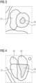
- Examples of the segmentation are schematically shown in figures 3 and 4 , wherein fig. 3 shows a short-axis view and fig. 4 shows a long-axis view of the heart.
- the used segmentation algorithm allows an automatic segmentation of five regions of the heart in the volume depicted by the morphology preserving image data set 8. These regions are shaded in figures 3 and 4 . These regions are the myocardium 11 of the left ventricle, the blood pool 10 of the left ventricle, the right ventricle 21 and the left and right atrium 22, 23.
- Other exemplary structures are shown in figures 3 and 4 but not segmented.
- the segmentation of the right ventricle 21 and the left and right atrium 22, 23 is not used in the discussed example of the method and the segmentation of these areas could therefore be skipped or the segmentation can be discarded.
- the contrast agent map 12 is inherently registered to the morphology preserving image data set 8 and therefore each voxel 19 of the contrast agent map 12 corresponds to the respective voxel 19 of the morphology preserving image data set 8 at the same position and describes the same area in the heart of the patient.
- the segmentation of the blood pool 10 and the myocardium 11 in the morphology preserving image data set 8 does therefore also describe the segmentation of the blood pool 10 and the myocardium 11 in the contrast agent map 12.
- a reference value 17 can be determined that describes the change of x-ray-absorption in the blood pool due to the presence of the contrast agent in step S6. If this reference value 17 describes a change in Hounsfield units it can be labeled ⁇ HU b .
- EVC CT myocardial extracellular volume faction
- step S8 the hematocrit value 20, that is labeled as h in the given formula, is provided, e.g. from a patient data record. Therefore, all terms on the right-hand side of the given formula are known and the myocardial extracellular volume fraction 1 for the given voxel 19 within the segmented myocardium 11 can therefore be calculated in step S9.
- an extracellular volume image data set 18 can be generated that comprises the respective myocardial extracellular volume fraction 1 for each respective voxel 19 within the segmented myocardium 11.
- Voxels outside the segmented myocardium 11 can be set to a fixed value, e.g., to full transparency.
- the extracellular volume image data set 18 can, e.g., be used to generate projections that are super imposed to corresponding projections of the morphology preserving image data set 8, especially using a different color pallete as already discussed in the general description. Since the extracellular volume image data set 18 is automatically registered to the morphology preserving image data set 8, additional registration is not necessary.
- statistical analysis of the determined myocardial extracellular volume fraction 1 is possible. It is, e.g., possible to calculate an average or select a median from these determined fractions, to determine parameters of the distribution or to analyze a local variance of this fraction.
- Fig. 5 shows a medical imaging device 28 that comprises an energy resolving computed-tomography scanner 29, preferably a photon-counting computed tomography scanner, and a processing system 30.
- the processing system 30 comprises a first interface 24, configured for receiving the measurement data set 3, and a second interface 25, configured for providing the myocardial extracellular volume fraction 1, especially in the form of the extracellular volume image data set 18.
- the measurement data set 3 is provided by the energy resolving computed tomography scanner 29 immediately after the scanning of the patient 2.
- the determined myocardial extracellular volume fraction 1, especially in the form of the extracellular volume image data set 18, is stored in a patient data base 27 in the example.
- information concerning the myocardial extracellular volume fraction 1 could be directly output to a user, e.g. using a screen of a processing device 26 implementing the processing system 30.
Landscapes
- Engineering & Computer Science (AREA)
- Health & Medical Sciences (AREA)
- Life Sciences & Earth Sciences (AREA)
- Medical Informatics (AREA)
- Physics & Mathematics (AREA)
- General Health & Medical Sciences (AREA)
- Radiology & Medical Imaging (AREA)
- Nuclear Medicine, Radiotherapy & Molecular Imaging (AREA)
- Theoretical Computer Science (AREA)
- Computer Vision & Pattern Recognition (AREA)
- Public Health (AREA)
- General Physics & Mathematics (AREA)
- Surgery (AREA)
- Pathology (AREA)
- Optics & Photonics (AREA)
- Biomedical Technology (AREA)
- Heart & Thoracic Surgery (AREA)
- Molecular Biology (AREA)
- High Energy & Nuclear Physics (AREA)
- Animal Behavior & Ethology (AREA)
- Biophysics (AREA)
- Veterinary Medicine (AREA)
- Quality & Reliability (AREA)
- Physiology (AREA)
- Pulmonology (AREA)
- Oral & Maxillofacial Surgery (AREA)
- Dentistry (AREA)
- Cardiology (AREA)
- Epidemiology (AREA)
- Primary Health Care (AREA)
- Artificial Intelligence (AREA)
- Computing Systems (AREA)
- Databases & Information Systems (AREA)
- Evolutionary Computation (AREA)
- Software Systems (AREA)
- Multimedia (AREA)
- Apparatus For Radiation Diagnosis (AREA)
Abstract
- receiving a measurement data set (3), wherein the measurement data set (3) comprises energy resolved data based on a computed tomography scan of the patient (2),
- reconstructing a morphology preserving image data set (8) based a first photon-energy band (9) or a first combination of photon-energy bands described by the measurement data set (3),
- segmenting a blood pool (10) within a myocardium (11) in the morphology preserving image data set (8),
- reconstructing a contrast agent map (12) based a second photon-energy band or a second combination (13) of photon-energy bands (14, 15, 16) described in the measurement data set (3),
- determining a reference value (17) based on at least one pixel or voxel (19) of the contrast agent map (12) within the segmented blood pool (10), and
- determining a respective myocardial extracellular volume fraction (1) depending on the reference value (17) and the value given for at least one respective pixel or voxel (19) outside the segmented blood pool (10) by the contrast agent map (12).
Description
- The invention concerns a computer-implemented method for determining at least one myocardial extracellular volume fraction for a patient. Additionally, the invention concerns a processing system, a medical imaging device, a computer program and a computer-readable storage medium.
- Interstitial myocardial fibrosis is an important disease process that has been associated with different kinds of cardio myopathy. The myocardial extracellular volume fraction (ECV) increases in the case of diffuse myocardial fibrosis or infiltrates and is therefore considered as a distinguishing feature of myocardial pathologic findings for cardiomyopathies.
- The myocardial extracellular volume fraction can be determined in magnetic resonance imaging by determining the equilibrium distribution of gadolinium contrast agent between the myocardium and the blood. As iodine-based computed tomography contrast agents exhibit similar kinetics as gadolinium, the myocardial extracellular volume fraction can also be determined using computed tomography.
- In principle, normal single energy computed tomography scans can be used for this purpose. In this case, it is however necessary to perform a baseline native scan without contrast agent, a normal coronary computed tomography angiography scan using contrast agent and a so-called late enhancing scan that is recorded several minutes after the contrast agent injection and depicts an equilibrium distribution of the contrast agent in which the contrast agent has already entered the intercellular volume of the myocardium. The use of three separate scans leads to relatively high x-ray-exposure of the patient and requires a registering of scans acquired at different times, which can lead to artefacts and therefore less exact results.
- A notable improvement can be achieved when dual energy computed tomography is used. Since the x-ray-absorption is determined for different photon-energy bands in a dual energy computed tomography, different materials can be distinguished by analyzing the photon-energy dependent x-ray-absorption. When using such a dual energy acquisition, an, e.g., iodine-based contrast agent can be selectively imaged, therefore providing a contrast agent map. On the other hand, it is also possible to map water and therefore essentially tissue from the same dual energy scan.
- When a segmentation of a myocardium, e.g., of the myocardium of the left ventricle, and of a blood pool, especially of the blood pool within the myocardium, is known, the myocardial extracellular volume fraction can be determined from such a contrast agent map using the following formular:

- Approaches for using dual energy computed tomography for determining the myocardial extracellular volume fraction are well known, e.g., from the publications Lee, H.-J., et al.: Myocardial Extracellular Volume Fraction with Dual-Energy Equilibrium Contrast-enhanced Cardiac CT in Nonischemic Cardiomyopathy: A Prospective Comparison with Cardiac MR Imaging. Radiology 280:1 (2016) 49-57; Abadia, A. F., et al.: Myocardial extracellular volume fraction to differentiate healthy from cardiomyopathic myocardium using dual-source dual-energy CT. J Cardiovasc Comput Tomogr 14(2) (2020) 162-167; Oda, S., et al.: Myocardial Late Iodine Enhancement and Extracellular Volume Quantification with Dual-Layer Spectral Detector Dual-Energy Cardiac CT. Radiology: Cardiothoracic Imaging 2019.
- A possible approach for determining the hematocrit value directly from computed tomography data is given by Treibel, T. A., et al.: Automatic quantification of the myocardial extracellular volume by cardiac computed tomography: Synthetic ECV by CCT. J Cardiovasc Comput Tomogr 11(3) (2017) 221-226.
- In principle it is possible to perform an automatic segmentation of e.g., the blood pool within the left ventricle and the myocardium of the left ventricle. An exemplary approach for such an automatic segmentation is given in Zheng, Y., et al.: Four-chamber heart modeling and automatic segmentation for 3-D cardiac CT volumes using marginal space learning and steerable features. IEEE Trans Med Imaging 27(11) (2008) 1668-81.
- Known segmentation algorithms for the myocardium and the blood pool do however perform poorly when applied to a contrast agent map. Therefore, the discussed approaches for determining a myocardial extracellular volume fraction based on dual energy computed tomography either require manual segmentation of the image data or the acquisition of an additional scan used for an automatic segmentation, that is preferably performed at a time relatively shortly after the contrast agent injection. E.g., a complete computed tomography angiography sequence can be performed or at least the scan comprising contrast agent essentially only in the blood vessel can be used as an additional scan. In this case it is however still necessary to register at least two scans taken at different times and the x-ray-exposure of the patient is increased over the use of a single scan.
- The invention is therefore based on the problem of providing an improved approach for determining at least one myocardial extracellular volume fraction for a patient, that especially avoids the necessity of manual segmentation, even when using only a single computed tomography scan.
- The problem is solved by a computer-implemented method for determining at least one myocardial extracellular volume fraction for a patient, comprising the steps of:
- receiving a measurement data set, wherein the measurement data set comprises energy resolved data based on a computed tomography scan of the patient,
- reconstructing a morphology preserving image data set based a first photon-energy band or a first combination of photon-energy bands described by the measurement data set,
- segmenting a blood pool within a myocardium in the morphology preserving image data set,
- reconstructing a contrast agent map based a second photon-energy band or a second combination of photon-energy bands described in the measurement data set,
- determining a reference value based on at least one pixel or voxel of the contrast agent map within the segmented blood pool, and
- determining a respective myocardial extracellular volume fraction depending on the reference value and the value given for at least one respective pixel or voxel outside the segmented blood pool by the contrast agent map.
- When resolving multiple, preferably at least three, photon-energy bands, that can also be called energy bins, it is possible to generate different image impressions by using different energy bands and/or different combinations of energy bands during reconstruction. To combine energy bands, it is e.g., possible to add or subtract images reconstructed from different energy bands. Two of the possible impressions are the previously discussed contrast agent map and the mapping of tissue or water. Such a material contrast is caused by the fact that most contrast agents, especially iodine, have an x-ray-absorption that strongly depends on the photon-energy. Therefore, a contrast agent map can, e.g., be determined from the difference in absorption in different photon-energy bands. Once the absorption due to the contrast agent is known, the remaining absorption can be attributed to water and tissue.
- The differentiation between different materials can obviously be further improved, when differences between the x-ray absorption for more than two photon-energy bands are considered.
- It was found that by selecting the right photon-energy band or the right combination of photon-energy bands, a morphology preserving image data set can be generated that allows for the use of normal segmentation algorithms that are typically used to segment image data sets acquired by coronary computed tomography angiography, and therefore especially image data sets recorded after a relatively short waiting time after injecting the contrast agent and therefore in a condition where essentially all of the contrast agent is still contained in the blood pool and essentially no contrast agent has migrated to the tissue.
- The first photon-energy band or first combination of photon-energy bands is especially chosen in such a way that the contrast-to-noise-ratio for the morphological features to be segmented, especially for the blood pool and/or the myocardium, is notably higher, e.g., at least by a factor of two or three, than in the contrast agent map. As discussed in more detail below, it was found that this can especially be achieved by only or mainly considering photons with a photon-energy at the lower end of the available spectrum.
- The considered myocardium and blood pool can especially be the myocardium of the left ventricle and the blood pool therein.
- In classic dual- or multi-energy computed tomography acquisitions it is possible to modify an acceleration voltage or use multiple x-ray sources that generate photons with different photon-energies. Alternatively, or additionally, it is possible to use sandwich-detectors with different sensitivity bands to directly measure x-ray intensities in two or more given energy bands. In both cases intensities for the different photon-energy bands are measured by the detector.
- In a photon-counting detector it is possible to at least approximately determine the photon-energy of each detected photon, since the height of a current spike generated by the detection of the respective photon depends on the photon-energy of that photon. It is therefore, e.g., possible to log each individual photon detection in each pixel of a detector with the associated photon energy and afterwards perform an essentially arbitrary binning of the events depending on the detected energy to provide multiple photon-energy bands.
- As previously discussed, the presence of a contrast agent can especially be detected, when the local absorption strongly varies with the photon-energy. The contrast agent map can therefore preferably be reconstructed based on a combination of at least two, preferably more, photon-energy bands described in the measurement data set.
- For the reconstruction of the morphology preserving image data set it can be sufficient to use a single photon-energy band. It is however also possible that both the morphology preserving image data set and the contrast agent map are based on a respective single photon-energy band or that they are both based on a combination of photon-energy bands. In principle it would also be possible to determine the morphology preserving image data set based on a combination of photon-energy bands while the contrast agent map is based on a single photon-energy band. The different photon-energy bands can be non-overlapping or at least one pair of the photon-energy bands can overlap.
- The morphology preserving image data set and the contrast agent map are preferably three-dimensional image data sets, especially formed from a three-dimensional array of voxels. Alternatively, they can be two-dimensional image data sets, e.g., formed by an array of pixels. The measurement data set can comprise multiple projection images, e.g., a single projection image for each photon-energy band and projection angle, or it can e.g., comprise a respective event list comprising all individual photon detection events for each detector pixel and projection angle.
- The image acquisition is preferably not part of the computer-implemented method and is completed before the start of the method. Alternatively, the image acquisition could be included as an additional step and the acquired image data could, e.g., be directly received from a computed tomography scanner.
- Preferably, all discussed steps of the computer-implemented method are performed fully automatic. Optionally it would also be possible, to allow for a user interaction to, e.g., modify the results of certain steps. E.g., the selection of the energy band or energy bands used to reconstruct the morphology preserving image data set and/or the segmentation of the blood pool and/or the segmentation of the myocardium can be modified by a user.
- Preferably, the segmentation of the blood pool and/or the myocardium or at least an initial segmentation that can then be modified by the user is however performed automatically without user interaction. During the development of the invention, mainly the approach for automatic segmentation for a three-dimensional cardiac computed tomography volume discussed in the initially cited paper by Y. Zheng was used. There are however a multitude of further approaches for automatically segmenting contrast enhanced images in computed tomography angiography that can typically be directly used to segment the morphology preserving image data set in the invention without any further modification.
- The measurement data set is preferably based on a photon-counting computed tomography scan. In non-photon counting multi-energy computed tomography scans, the weight of lower energy photons in a given energy band is reduced due to the used measurement principle. This effect can be avoided in photon-counting computed tomography data sets, since each individual photon is countered and the photon energy is detected as a separate parameter of the detection. Since lower energy photons are especially relevant to conserve the morphology of the depicted heart and therefore for a robust segmentation of the myocardium and the blood pool, the use of photon-counting computed tomography scan as a base for the input data set allows for more robust morphology preservation, especially for a higher contrast-to-noise-ration in the morphology preserving image data set and therefore for a more exact and robust automatic segmentation.
- The use of photon counted computed tomography also allows for an essentially free and, if needed, a dynamic selection of photon-energy bands that are used in reconstructing the morphology preserving image data set on the one hand and the contrast agent map on the other hand. The only limitation for the number of used photon-energy bands is a sufficient photon count within the respective band and a sufficient energy resolution of the detector. While classic multi-energy computed tomography is limited to two or at most three or four photon-energy bands, that are typically fixed, photon-counting computed tomography allow for the use of a larger number of bands and/or the dynamic adjustment of the position and widths of the different energy bands to provide optimum results. This can be used to further improve the contrast-to-noise-ratio in the morphology preserving image data set and/or to further improve material selectivity for the contrast agent map.
- Preferably, the myocardium is segmented in the morphology preserving image data set, wherein a respective myocardial extracellular volume fraction is only determined for pixels or voxels within the segmented myocardium. Additionally, or alternatively an extracellular volume image data set can be generated that comprises the respective myocardial extracellular volume fraction for each respective pixel or voxel within the segmented myocardium and either a fixed value or a value exclusively based on the morphology preserving image data set for each pixel or voxel outside the segmented myocardium. Determining the extracellular volume fraction outside of the myocardium is typically not necessary or useful when, e.g., trying to detect myocardial fibrosis. At the same time generating and displaying such a value in the area outside the myocardium can notably lower the available contrast, especially since the initially discussed formula for calculating the extracellular volume fraction tends to lead to a maximum value in the area of the blood pool and therefore outside of the relevant area.
- Using a fixed value for pixels or voxels outside the segmented myocardium can be useful to, e.g., generate a three-dimensional data set that has full transparency in those areas. Such a data set can be useful for easy viewing, to allow for a robust mapping to a two-dimensional coordinate system, e.g., using a two-dimensional polar map, and/or for generating an overlay that can be superimposed e.g., onto the morphology preserving image data set. Such a superposition allows for an easier orientation of e.g., a medical examiner within the image due to the recognizability of morphological features. In such a superposition, different color palletes can be used for a projection generated from the morphology preserving image data set and a projection generated from the extracellular volume image data set. It is e.g. possible to generate a colored image with a given pallete from the extracellular volume image data set and to superimpose this image with an image generated from the morphology preserving image data set using a gray scale pallete.
- As discussed above, the alternative is a combination of these two sources of data within a single two- or three-dimensional image data set.
- Alternatively or additionally, the limitation of the determination of the myocardial extracellular volume fraction to the volume actually covered by the myocardium according to the segmentation or a masking of the generated values to only leave these values can be advantageous for a statistical analysis of the myocardial extracellular volume fraction, since this analysis can then be limited to the relevant volume. Simple examples for such a statistical analysis would be the calculation of an average and/or median myocardial extracellular volume faction and/or the determination of measures describing the distribution, e.g. of a standard deviation, etc.
- The reference value can be determined based on an average or median of multiple pixels or voxels, preferably based on an average or median of all pixels or voxels, of the contrast agent map within the segmented blood pool. Instead of using all the pixels or voxels of the segmented blood pool, it is also possible to discard the pixels or voxels in a given number of outer rows of pixels or voxels and therefore reduce the influence of potential segmentation errors. The use of an average or median is advantageous, since the distribution of the contrast agent in the blood is expected to be essentially homogeneous in the given region and therefore an averaging or median determination can be used to further reduce errors.
- The measurement data set can comprise energy resolved data based on a computed tomography scan of the patient performed at least 2 minutes or at least 3 minutes and/or at most 6 minutes or at least 5 minutes after the beginning of a contrast agent injection. When the waiting time between the contrast agent injection and that image acquisition is too short, an equilibrium between the concentration of the contrast agent in the extracellular volume of the tissue and the blood is not yet reached, since the contrast agent did not yet have time to diffuse from the blood to the tissue. Very long wait times would on the other hand not noticeably improve the equilibrium, but typically lead to an increased noise due to a reduced overall concentration of contrast agent.
- The average photon-energy in the first photon-energy band or in the first combination of photon-energy bands can be less than 70 keV or less than 50 keV. As previously discussed, the contrast-to-noise-ratio in the morphology preserving image data set can be increased, when mainly photons with a relatively low photon-energy are used. It is e.g. possible to use a single first photon-energy band centered at 40 keV. The width of the first photon-energy band or the energy spread covered by the first combination of photon-energy bands can, e.g., be less than 30 keV or even less than 10 keV.
- As previously discussed, the position and width of the considered photon-energy band can essentially be freely chosen when using photon-counting computed tomography. If necessary, it is also possible to e.g. pool photon detections from multiple adjacent pixels of a detector to increase the overall photon count and therefore e.g. to allow for the use of less-wide photon-energy bands. Since the photon-energy of each individual photon can be detected in photon-counting computed tomography, it is in principle also possible to continuously vary the weighting factor of individual photon within a considered photon-energy band.
- The second photon-energy band or the second combination of photon-energy bands can comprise at least one photon-energy that is higher than the highest photon-energy in the first photon-energy band or the first combination of photon-energy bands. The use of a relatively wide spread of photon-energies to determine the contrast agent map is advantageous, since a material contrast used to generate the contrast agent map mainly depends on a detection of a different absorption behavior of the contrast agent at different photon-energies. It is therefore advantageous to use absorptions at multiple photon-energy bands that are preferably spaced apart as far as possible.
- The contrast agent map can be based on the second combination of photon-energy bands, wherein the second combination of photon-energy bands comprises at least one photon-energy band or at least two photon-energy bands that does or do not overlap with the first photon-energy band or any photon-energy bands of the first combination of photon-energy bands. The second combination of photon-energy bands can additionally comprise the first photon-energy band or at least one photon-energy band of the first combination of photon-energy bands. As previously discussed, it is advantageous to take into account at least two, preferably more, photon-energy bands spaced across a wide energy spectrum to determine x-ray-absorption at a multitude of photon energies and therefore clearly and robustly distinguish between different materials, especially between the contrast agent and water or tissue.
- The at least one myocardial extracellular volume fraction can be determined from energy resolved data based on exactly one computed tomography scan of the patient forming the measurement data set and a provided hematocrit value, wherein no other input data besides the measurement data set and the hematocrit value is used. The use of only a single computed tomography scan avoids potential artefacts due to a registration of multiple scans. The morphology preserving image data set and the contrast agent map in the computer-implemented method according to the present invention are inherently registered to each other, since they are based on the same received measurement data set that especially represents only a single computed tomography scan. The determination is therefore more robust and less error-prone than the initially discussed approaches that require a registration of multiple computed tomography scans.
- A representation of the segmented blood pool, especially of the outline of the segmented blood pool in the morphology preserving image data set, and/or a representation of the segmented myocardium, especially of the outline of the segmented myocardium in the morphology preserving image data set, can displayed for a user, wherein the user can edit the segmentation of the blood pool and/or the myocardium, whereafter the respective edited segmentation can be used to determine the reference value and/or the at least one myocardial extracellular volume fraction. While the automatic segmentation of the blood pool and the myocardium is very robust in the discussed method, the acceptance of the method can be improved when medical personal can review the results of intermediate steps and potentially modify the results if deemed necessary.
- Alternatively or additionally, a user can, e.g., interactively vary the first photon-energy band or the first combination of photon-energy bands used to reconstruct the morphology preserving image data set. It would, e.g., be possible to show at least one projection image of an initially generated morphology preserving image data set and, e.g., to provide a fader or some other control that allows a use to slightly shift the used first photon-energy band until an optimum contrast is reached. Alternatively, such an optimization could be performed automatically.
- Besides the discussed computer-implemented method the invention concerns a processing system, comprising
- a first interface, configured for receiving a measurement data set, wherein the measurement data set comprises energy resolved data based on a computed tomography scan of a patient,
- a second interface, configured for providing at least one myocardial extracellular volume fraction for the patient,
- a computation unit, configured for implementing the computer-implemented method according to one of the preceding claims.
- The processing system can, e.g., be part of a computed tomography scanner or of a work station used to control this scanner. Alternatively, the processing system could also be a separate work station that is not used for control of the scanner or even be implemented as a cloud-based solution.
- The invention also concerns a medical imaging device comprising an energy resolving computed tomography scanner and a processing system according to the present invention. As previously discussed, the computed tomography scanner can preferably be a photon-counting computed tomography scanner. Alternatively, a non-photon counting multi energy computed tomography scanner can be used.
- The invention additionally concerns a computer program comprising instructions to cause the or a processing device to execute the computer-implemented method according to the present invention.
- Additionally, the invention concerns a computer-readable storage medium having stored thereon the computer program according to the present invention.
- In particular, the features and advantages described in connection with the computer implemented method according to the invention can also be designed as corresponding subunits of the device according to the invention or of the computer program according to the invention. Conversely, the features and advantages described in connection with the device according to the invention or the computer program according to the invention can also be designed as corresponding process steps of the process according to the invention.
- Other objects and features of the present invention will become apparent from the following detailed description considered in conjunction with the accompanying drawings. The drawings, however, are only principle sketches designed solely for the purpose of illustration and do not limit the invention. The drawings show:
- Fig. 1
- a flow chart of an exemplary embodiment of the computer-implemented method according to the present invention,
- Fig. 2
- relevant data structure used in an exemplary embodiment of the computer-implemented method according to the present invention,
- Fig. 3 and 4
- exemplary segmentations of the myocardium and the blood pool in two different views of the heart, and
- Fig. 5
- an exemplary embodiment of a medical imaging device according to the present invention comprising an exemplary embodiment of a processing system according to the present invention.
-
Fig. 1 shows a flow chart of a computer-implemented method for determining at least one myocardial extracellular volume fraction for a patient. The method will be discussed with additional reference tofig. 2 that shows relevant data structures that are used in the course of the determination. - The central idea of the method discussed in more detail below is to use different photon-
energy bands combinations 13 of photon-energy bands contrast agent map 12 from the same measurement data set 3 that comprises energy resolved data based on a computed tomography scan of a patient. Thecontrast agent map 12 allows for a determination of the change of x-ray-absorption in each pixel orvoxel 19, especially expressed as a change in Hounsfield units, due to the presence of the contrast agent in the area of the patient depicted by therespective voxel 19. The additional use of the morphology preserving image data set 8 allows for a robust automatic segmentation of a myocardium, preferably themyocardium 11 of the left ventricle of the heart of the patient, and theblood pool 10 therein. Therefore, the change of x-ray-absorption in theblood pool 10 and themyocardium 11 can be determined and based on these values the myocardial extracellular volume fraction 1 can be calculated. - To determine the myocardial extracellular volume fraction 1 firstly a
measurement data set 3 is received in step S1. Themeasurement data set 3 comprises energy resolved data based on a computed tomography scan of the patient. In the example, themeasurement data set 3 is acquired using a photon-counting computed tomography scan. The measurement data set can be directly provided by an energy resolving computed tomography scanner or the discussed processing can happen at a later point in time and themeasurement data set 3 can, e.g., be read from a data base. - The computed tomography scan to acquire the data set is preferably performed at a time at which an approximate equilibrium between the contrast agent in the blood vessels and the contrast agent in the extracellular volume fraction of the tissue is present. Such an equilibrium is typically reached approximately 3 to 5 minutes after the beginning of a contrast agent injection and therefore the scan should preferably be performed in this time frame.
- Due to the use of a photon-counting detector, for each projection 4 acquired and each
pixel 5 of the detector themeasurement data set 3 comprises a list of adetection events 6 for the respective projection 4 and thepixel 5. Since photon-counting tomography scans allow the acquisition of a photon-energy for each detected photon, eachdetection event 6 also describes a measure for the photon-energy 7 of the detected photon. - Alternatively, it would be possible to provide a
measurement data set 3 for which a binning of the photon-energy 7 into respective photon-energy bands is already performed. Such ameasurement data set 3 could, e.g., provide respective photon counts for each energy band,pixel 5 and projection 4. Instead of a photon-counting computed tomography scan it would also be possible to use a classic multi-energy-acquisition that only provides detected intensities for each given projection 4,pixel 5 and energy band. - In Step S2 the morphology preserving image data set 8 is reconstructed. In the example the reconstruction is based only on detected photons within a given photon-
energy band 9. This can, e.g., be achieved by only taken thosedetection events 6 in themeasurement data set 3 into account, for which the given photon-energy 7 lies within the given photon-energy band 9. Anyother detection events 6 are disregarded and therefore essentially discarded during the reconstruction of the morphology preserving image data set. - The count of the remaining
detection events 6 for a givenpixel 5 and projection 4 can then be directly considered to describe the x-ray-intensity in the given photon-energy band 9 or further processing, e.g. an energy weighting, can be applied. The result is a number of projection images that only take into consideration of photon-energies 7 within the given photon-energy band 9, that can then be used to reconstruct the morphology preserving image data set 8 using well known image reconstruction techniques, e.g. a filtered back projection or an iterative reconstruction approach. - In alternate embodiments it would also be possible, to take multiple photon-energy bands into account when reconstructing the morphology preserving
image data set 8. It could, e.g., be possible to calculate counts or intensities for eachpixel 5, each projection 4 and each energy band considered and then, e.g., calculate a weighted sum of the counts or intensities during the reconstruction. - If the
individual detection events 6 are not available, e.g., due to a prior binning of the events based on photon-energy 7 or due to a direct detection of intensities within several energy bands, counts or intensities provided in themeasurement data set 3 for the relevant photon-energy band 9 can be directly used to reconstruct the morphology preservingimage data set 8. - In step S3 a
contrast agent map 12 is reconstructed based on acombination 13 of multiple photon-energy bands energy band 9, photon counts or intensities for each of theenergy bands pixel 5 and projection 4 can be determined or given by themeasurement data set 3. Since the material contrast for the contrast agent, e.g. for iodine, is typically based on the fact that contrast agents have an x-ray-absorption that strongly varies with photon-energy, the combination of the counts and/or intensities in the various photon-energy bands energy bands energy bands - Concerning the different photon-
energy bands energy band 9 preferably only comprises photons with a relatively low photon-energy 7. The photon-energy band 9 can, e.g., be centered at 40 keV or 50 keV and have a width of e.g. 10 keV to 30 keV. - The photon-
energy bands energy band 14 is identical to the photon-energy band 9 or partially overlaps with the photon-energy band 9. The photon-energy bands energies 7 above the photon-energy band 9 and are preferably separated from each other to describe absorptions of the contrast agent in different parts of the spectrum. - In step S4 and S5 an automatic segmentation is applied to the morphology preserving
image data set 8. In step S4 theblood pool 10 is segmented and in step S5 themyocardium 11 is segmented. While these segmentations are shown at separate steps for reasons of clarity, it can be advantageous, to use a common segmentation algorithm to perform both of these segmentations at the same time. - The example uses the segmentation algorithm given by the initially cited paper by Y. Zheng. In principle any segmentation algorithm for segmenting a computed tomography volume of the heart can be used. Examples of the segmentation are schematically shown in
figures 3 and 4 , whereinfig. 3 shows a short-axis view andfig. 4 shows a long-axis view of the heart. The used segmentation algorithm allows an automatic segmentation of five regions of the heart in the volume depicted by the morphology preservingimage data set 8. These regions are shaded infigures 3 and 4 . These regions are themyocardium 11 of the left ventricle, theblood pool 10 of the left ventricle, theright ventricle 21 and the left andright atrium figures 3 and 4 but not segmented. - The segmentation of the
right ventricle 21 and the left andright atrium - Since the morphology preserving image data set 8 and the
contrast agent map 12 are determined from the same measurement data set and therefore from a single computed tomography scan of the patient, thecontrast agent map 12 is inherently registered to the morphology preserving image data set 8 and therefore eachvoxel 19 of thecontrast agent map 12 corresponds to therespective voxel 19 of the morphology preserving image data set 8 at the same position and describes the same area in the heart of the patient. The segmentation of theblood pool 10 and themyocardium 11 in the morphology preserving image data set 8 does therefore also describe the segmentation of theblood pool 10 and themyocardium 11 in thecontrast agent map 12. - Using the segmentation of the
blood pool 10, it is therefore possible to select thevoxels 19 of thecontrast agent map 12 that lie within theblood pool 10. Since the image data of the respective voxels shows the change of the x-ray-absorption due to the contrast agent, especially a change in Hounsfield units due to the contrast agent, by averaging the image data from these voxels 19 areference value 17 can be determined that describes the change of x-ray-absorption in the blood pool due to the presence of the contrast agent in step S6. If thisreference value 17 describes a change in Hounsfield units it can be labeled ΔHUb. - In step S7 the segmentation of the
myocardium 11 is used to select thosevoxels 19 in thecontrast agent map 12 that depict themyocardium 11 of the left ventricle. Since the image data of thesevoxels 19 describe the change of x-ray-absorption at the respective position in the tissue of themyocardium 11, it can be labeled ΔHUt. When these changes given in Hounsfield units, it already describes an information that is expected to be proportional to the myocardial extracellular volume faction (EVCCT) for the area of the myocardium tissue depicted in therespective voxel 19 according to the already discussed formula
- In step S8 the
hematocrit value 20, that is labeled as h in the given formula, is provided, e.g. from a patient data record. Therefore, all terms on the right-hand side of the given formula are known and the myocardial extracellular volume fraction 1 for the givenvoxel 19 within thesegmented myocardium 11 can therefore be calculated in step S9. - To allow for an easier representation of the determined, locally resolved myocardial extracellular volume fraction 1, an extracellular volume image data set 18 can be generated that comprises the respective myocardial extracellular volume fraction 1 for each
respective voxel 19 within thesegmented myocardium 11. Voxels outside thesegmented myocardium 11 can be set to a fixed value, e.g., to full transparency. The extracellular volume image data set 18 can, e.g., be used to generate projections that are super imposed to corresponding projections of the morphology preserving image data set 8, especially using a different color pallete as already discussed in the general description. Since the extracellular volume image data set 18 is automatically registered to the morphology preserving image data set 8, additional registration is not necessary. - Additionally or alternatively, statistical analysis of the determined myocardial extracellular volume fraction 1 is possible. It is, e.g., possible to calculate an average or select a median from these determined fractions, to determine parameters of the distribution or to analyze a local variance of this fraction.
- While the previously discussed method uses a fully automated approach for determining the myocardial extracellular volume fractions 1, it can in some used cases be advantageous when a supervising user can modify the results of certain steps. It is, e.g., possible that the segmentation of the
blood pool 10 andmyocardium 11 are output for user, e.g., using the views shown infigures 3 and 4 , and that the user can modify these segmentations. Modified segmentations can then be used in the further steps. -
Fig. 5 shows amedical imaging device 28 that comprises an energy resolving computed-tomography scanner 29, preferably a photon-counting computed tomography scanner, and aprocessing system 30. Theprocessing system 30 comprises afirst interface 24, configured for receiving themeasurement data set 3, and asecond interface 25, configured for providing the myocardial extracellular volume fraction 1, especially in the form of the extracellular volumeimage data set 18. - In the example the
measurement data set 3 is provided by the energy resolving computedtomography scanner 29 immediately after the scanning of thepatient 2. Alternatively, it would also be possible to intermediately store themeasurement data set 3, either in thescanner 29 itself or, e.g., in a patient data base. The determined myocardial extracellular volume fraction 1, especially in the form of the extracellular volume image data set 18, is stored in apatient data base 27 in the example. Alternatively or additionally, information concerning the myocardial extracellular volume fraction 1 could be directly output to a user, e.g. using a screen of aprocessing device 26 implementing theprocessing system 30. - Although the present invention has been described in detail with reference to the preferred embodiment, the present invention is not limited by the disclosed examples from which the skilled person is able to derive other variations without departing from the scope of the invention.
Claims (14)
- Computer-implemented method for determining at least one myocardial extracellular volume fraction (1) for a patient (2), comprising the steps of:- receiving a measurement data set (3), wherein the measurement data set (3) comprises energy resolved data based on a computed tomography scan of the patient (2),- reconstructing a morphology preserving image data set (8) based a first photon-energy band (9) or a first combination of photon-energy bands described by the measurement data set (3),- segmenting a blood pool (10) within a myocardium (11) in the morphology preserving image data set (8),- reconstructing a contrast agent map (12) based a second photon-energy band or a second combination (13) of photon-energy bands (14, 15, 16) described in the measurement data set (3),- determining a reference value (17) based on at least one pixel or voxel (19) of the contrast agent map (12) within the segmented blood pool (10), and- determining a respective myocardial extracellular volume fraction (1) depending on the reference value (17) and the value given for at least one respective pixel or voxel (19) outside the segmented blood pool (10) by the contrast agent map (12).
- Computer-implemented method according to claim 1, characterized in that the measurement data set (3) is based on a photon-counting computed tomography scan.
- Computer-implemented method according to claim 1 or 2, characterized in that the myocardium (11) is segmented in the morphology preserving image data set (8), wherein a respective myocardial extracellular volume fraction (1) is only determined for pixels or voxels (19) within the segmented myocardium (11) and/or wherein an extracellular volume image data set (18) is generated, that comprises the respective myocardial extracellular volume fraction (1) for each respective pixel or voxel (19) within the segmented myocardium and either a fixed value or a value exclusively based on the morphology preserving image data set (8) for each pixel or voxel (19) outside the segmented myocardium (11).
- Computer-implemented method according to one of the preceding claims, characterize in that the reference value (17) is determined based on an average or median of multiple pixels or voxels (19), preferably based on an average or median of all pixels or voxels (19), of the contrast agent map (12) within the segmented blood pool (10) .
- Computer-implemented method according to one of the preceding claims, characterize in that the measurement data (3) set comprises energy resolved data based on a computed tomography scan of the patient (2) performed at least 2 minutes or at least 3 minutes and/or at most 6 minutes or at most 5 minutes after the beginning of a contrast agent injection.
- Computer-implemented method according to one of the preceding claims, characterized in that the average photon energy (7) in the first photon-energy band (9) or in the first combination of photon energy bands is less than 70 keV or less than 50 keV.
- Computer-implemented method according to one of the preceding claims, characterized in that the second photon-energy band or the second combination (13) of photon-energy bands (14, 15, 16) comprises at least one photon energy (7) that is higher than the highest photon energy (7) in the first photon-energy band (9) or the first combination of photon-energy bands.
- Computer-implemented method according to one of the preceding claims, characterized in that the contrast agent map (12) is based on the second combination (13) of photon-energy bands (14, 15, 16), wherein the second combination (13) of photon-energy bands (14, 15, 16) comprises at least one photon-energy band (15, 16) or at least two photon-energy bands (15, 16) that does or do not overlap with the first photon-energy band (9) or any photon-energy band of the first combination of photon-energy bands.
- Computer-implemented method according to one of the preceding claims, characterized in that the at least one myocardial extracellular volume fraction (1) is determined from energy resolved data based on exactly one computed tomography scan of the patient (2) forming the measurement data set (3) and a provided hematocrit value (20), wherein no other input data besides the measurement data set (3) and the hematocrit value (20) is used.
- Computer-implemented method according to one of the preceding claims, characterized in that a representation of the segmented blood pool (10), especially of the outline of the segmented blood pool (10) in the morphology preserving image data set (8), and/or a representation of the segmented myocardium (11), especially of the outline of the segmented myocardium (11) in the morphology preserving image data set (8), is displayed for a user, wherein the user edits the segmentation of the blood pool (10) and/or the myocardium (11), whereafter the respective edited segmentation is used to determine the reference value (17) and/or the at least one myocardial extracellular volume fraction (1).
- Processing system, comprising- a first interface (24), configured for receiving a measurement data set (3), wherein the measurement data set (3) comprises energy resolved data based on a computed tomography scan of a patient (2),- a second interface (25), configured for providing at least one myocardial extracellular volume fraction (1) for the patient (2),- a computation unit (26), configured for implementing the computer-implemented method according to one of the preceding claims.
- Medical imaging device comprising an energy resolving computed tomography scanner (29) and a processing system (30) according to claim 11.
- Computer program comprising instructions to cause the or a processing device to execute the computer-implemented method according to one of the claims 1 to 10.
- Computer-readable storage medium having stored thereon the computer program (12) according to claim 13.
Priority Applications (4)
| Application Number | Priority Date | Filing Date | Title |
|---|---|---|---|
| EP22192839.3A EP4332881A1 (en) | 2022-08-30 | 2022-08-30 | Method for determining a myocardial extracellular volume fraction, processing system, medical imaging device, computer program and computer-readable storage medium |
| JP2023120597A JP7760558B2 (en) | 2022-08-30 | 2023-07-25 | Method for determining myocardial extracellular volume fraction, processing system, medical imaging device, computer program, and computer-readable storage medium |
| CN202311095954.0A CN117643477A (en) | 2022-08-30 | 2023-08-29 | Method, system, device and storage medium for determining myocardial extracellular volume fraction |
| US18/457,736 US12536662B2 (en) | 2022-08-30 | 2023-08-29 | Method for determining a myocardial extracellular volume fraction, processing system, medical imaging device, computer program and computer-readable storage medium |
Applications Claiming Priority (1)
| Application Number | Priority Date | Filing Date | Title |
|---|---|---|---|
| EP22192839.3A EP4332881A1 (en) | 2022-08-30 | 2022-08-30 | Method for determining a myocardial extracellular volume fraction, processing system, medical imaging device, computer program and computer-readable storage medium |
Publications (1)
| Publication Number | Publication Date |
|---|---|
| EP4332881A1 true EP4332881A1 (en) | 2024-03-06 |
Family
ID=83151740
Family Applications (1)
| Application Number | Title | Priority Date | Filing Date |
|---|---|---|---|
| EP22192839.3A Pending EP4332881A1 (en) | 2022-08-30 | 2022-08-30 | Method for determining a myocardial extracellular volume fraction, processing system, medical imaging device, computer program and computer-readable storage medium |
Country Status (4)
| Country | Link |
|---|---|
| US (1) | US12536662B2 (en) |
| EP (1) | EP4332881A1 (en) |
| JP (1) | JP7760558B2 (en) |
| CN (1) | CN117643477A (en) |
Family Cites Families (8)
| Publication number | Priority date | Publication date | Assignee | Title |
|---|---|---|---|---|
| US6836528B2 (en) * | 2002-07-23 | 2004-12-28 | Ge Medical Systems Global Technology Company, Llc | Methods and apparatus for detecting structural, perfusion, and functional abnormalities |
| US7711160B2 (en) | 2003-05-28 | 2010-05-04 | Siemens Medical Solutions Usa, Inc. | Automatic optimal view determination for cardiac acquisitions |
| DE102005002949A1 (en) * | 2005-01-21 | 2006-08-03 | Siemens Ag | Myocard damage viewing procedure uses surface model segmentation matching of computer tomography images for automatic display |
| JP6411117B2 (en) | 2013-07-31 | 2018-10-24 | キヤノンメディカルシステムズ株式会社 | Medical image diagnostic apparatus and ultrasonic diagnostic apparatus |
| DE102014211587B4 (en) | 2014-06-17 | 2023-04-20 | Siemens Healthcare Gmbh | Representation of a hollow organ |
| US9990712B2 (en) * | 2015-04-08 | 2018-06-05 | Algotec Systems Ltd. | Organ detection and segmentation |
| DE102020216306A1 (en) | 2020-12-18 | 2022-06-23 | Siemens Healthcare Gmbh | Computer-implemented method for operating a medical imaging device, imaging device, computer program and electronically readable data carrier |
| EP4156091B1 (en) | 2021-09-24 | 2026-03-18 | Siemens Healthineers AG | Computer-implemented method for evaluating an image data set of an imaged region, evaluation device, imaging device, computer program and electronically readable storage medium |
-
2022
- 2022-08-30 EP EP22192839.3A patent/EP4332881A1/en active Pending
-
2023
- 2023-07-25 JP JP2023120597A patent/JP7760558B2/en active Active
- 2023-08-29 US US18/457,736 patent/US12536662B2/en active Active
- 2023-08-29 CN CN202311095954.0A patent/CN117643477A/en active Pending
Non-Patent Citations (8)
Also Published As
| Publication number | Publication date |
|---|---|
| US20240070866A1 (en) | 2024-02-29 |
| JP7760558B2 (en) | 2025-10-27 |
| CN117643477A (en) | 2024-03-05 |
| JP2024035096A (en) | 2024-03-13 |
| US12536662B2 (en) | 2026-01-27 |
Similar Documents
| Publication | Publication Date | Title |
|---|---|---|
| CN101313332B (en) | Data handling and analysis in computed tomography with multiple energy windows | |
| US9153012B2 (en) | Diagnostic image features close to artifact sources | |
| Zange et al. | Quantification in cardiovascular magnetic resonance: agreement of software from three different vendors on assessment of left ventricular function, 2D flow and parametric mapping | |
| Emoto et al. | Myocardial extracellular volume quantification using cardiac computed tomography: a comparison of the dual-energy iodine method and the standard subtraction method | |
| Jaspers et al. | Improving the reproducibility of MR-derived left ventricular volume and function measurements with a semi-automatic threshold-based segmentation algorithm | |
| EP3600045B1 (en) | Myocardial ct perfusion image synthesis | |
| Fuchs et al. | Automated assessment of heart chamber volumes and function in patients with previous myocardial infarction using multidetector computed tomography | |
| JP2017067765A (en) | Medical image processing device and positron emission tomography device | |
| US10902562B2 (en) | Noise reduction in image data | |
| US8831323B2 (en) | Method and apparatus for measuring activity of a tracer | |
| US9089308B2 (en) | Method for processing measurement data from perfusion computer tomography | |
| Kim et al. | EVCMR: a tool for the quantitative evaluation and visualization of cardiac MRI data | |
| JP2017506938A (en) | Segmentation of motion structure in image data | |
| US20170014069A1 (en) | Spectral imaging based fluid volume map | |
| US8290224B2 (en) | Method and device for imaging cyclically moving objects | |
| JP7760558B2 (en) | Method for determining myocardial extracellular volume fraction, processing system, medical imaging device, computer program, and computer-readable storage medium | |
| EP2882343B1 (en) | Chronic obstructive pulmonary disease (copd) phantom for computed tomography (ct) and methods of using the same | |
| Dorniak et al. | Respiratory motion-registered isotropic whole-heart T2 mapping in patients with acute non-ischemic myocardial injury | |
| Delbeke et al. | Estimation of left ventricular mass and infarct size from nitrogen-13-ammonia PET images based on pathological examination of explanted human hearts | |
| Mota-Cobian et al. | The effect of tissue-segmented attenuation maps on PET quantification with a special focus on large arteries | |
| Slomka | Hybrid quantitative imaging: Will it enter clinical practice? | |
| Feng et al. | Development and validation of an accurate input function from carotid arteries using the uEXPLORER | |
| Tsuneta et al. | The Impact of Model-based Deep-learning Reconstruction Compared with that of Compressed Sensing–Sensitivity Encoding on the Image Quality and Precision of Cine Cardiac MR in Evaluating Left-ventricular Volume and Strain: A Study on Healthy Volunteers | |
| Vembar | Brendan L. Eck, Jacob Levi 2, Hao Wu, Rachid Fahmi 3, Yuemeng Li, Anas Fares 4, Mani Vembar 5, Amar Dhanantwari 5, Hiram G. Bezerra 4, Raymond F. Muzic, Jr., and David L. Wilson Department of Biomedical Engineering, Case Western Reserve University, Cleveland, Ohio | |
| Brennecke et al. | Functional analysis of angiograms by digital image processing techniques |
Legal Events
| Date | Code | Title | Description |
|---|---|---|---|
| PUAI | Public reference made under article 153(3) epc to a published international application that has entered the european phase |
Free format text: ORIGINAL CODE: 0009012 |
|
| STAA | Information on the status of an ep patent application or granted ep patent |
Free format text: STATUS: THE APPLICATION HAS BEEN PUBLISHED |
|
| AK | Designated contracting states |
Kind code of ref document: A1 Designated state(s): AL AT BE BG CH CY CZ DE DK EE ES FI FR GB GR HR HU IE IS IT LI LT LU LV MC MK MT NL NO PL PT RO RS SE SI SK SM TR |
|
| STAA | Information on the status of an ep patent application or granted ep patent |
Free format text: STATUS: REQUEST FOR EXAMINATION WAS MADE |
|
| 17P | Request for examination filed |
Effective date: 20240903 |
|
| RBV | Designated contracting states (corrected) |
Designated state(s): AL AT BE BG CH CY CZ DE DK EE ES FI FR GB GR HR HU IE IS IT LI LT LU LV MC MK MT NL NO PL PT RO RS SE SI SK SM TR |