EP4331595B1 - Use of retinal pigment epithelial cells in replacement of corneal endothelia - Google Patents
Use of retinal pigment epithelial cells in replacement of corneal endothelia Download PDFInfo
- Publication number
- EP4331595B1 EP4331595B1 EP22919275.2A EP22919275A EP4331595B1 EP 4331595 B1 EP4331595 B1 EP 4331595B1 EP 22919275 A EP22919275 A EP 22919275A EP 4331595 B1 EP4331595 B1 EP 4331595B1
- Authority
- EP
- European Patent Office
- Prior art keywords
- retinal pigment
- pigment epithelial
- cells
- epithelial cells
- corneal
- Prior art date
- Legal status (The legal status is an assumption and is not a legal conclusion. Google has not performed a legal analysis and makes no representation as to the accuracy of the status listed.)
- Active
Links
Images

Classifications
-
- A—HUMAN NECESSITIES
- A61—MEDICAL OR VETERINARY SCIENCE; HYGIENE
- A61K—PREPARATIONS FOR MEDICAL, DENTAL OR TOILETRY PURPOSES
- A61K35/00—Medicinal preparations containing materials or reaction products thereof with undetermined constitution
- A61K35/12—Materials from mammals; Compositions comprising non-specified tissues or cells; Compositions comprising non-embryonic stem cells; Genetically modified cells
- A61K35/30—Nerves; Brain; Eyes; Corneal cells; Cerebrospinal fluid; Neuronal stem cells; Neuronal precursor cells; Glial cells; Oligodendrocytes; Schwann cells; Astroglia; Astrocytes; Choroid plexus; Spinal cord tissue
-
- A—HUMAN NECESSITIES
- A61—MEDICAL OR VETERINARY SCIENCE; HYGIENE
- A61K—PREPARATIONS FOR MEDICAL, DENTAL OR TOILETRY PURPOSES
- A61K31/00—Medicinal preparations containing organic active ingredients
- A61K31/33—Heterocyclic compounds
- A61K31/395—Heterocyclic compounds having nitrogen as a ring hetero atom, e.g. guanethidine or rifamycins
- A61K31/435—Heterocyclic compounds having nitrogen as a ring hetero atom, e.g. guanethidine or rifamycins having six-membered rings with one nitrogen as the only ring hetero atom
- A61K31/44—Non condensed pyridines; Hydrogenated derivatives thereof
- A61K31/4409—Non condensed pyridines; Hydrogenated derivatives thereof only substituted in position 4, e.g. isoniazid, iproniazid
-
- A—HUMAN NECESSITIES
- A61—MEDICAL OR VETERINARY SCIENCE; HYGIENE
- A61K—PREPARATIONS FOR MEDICAL, DENTAL OR TOILETRY PURPOSES
- A61K31/00—Medicinal preparations containing organic active ingredients
- A61K31/33—Heterocyclic compounds
- A61K31/395—Heterocyclic compounds having nitrogen as a ring hetero atom, e.g. guanethidine or rifamycins
- A61K31/435—Heterocyclic compounds having nitrogen as a ring hetero atom, e.g. guanethidine or rifamycins having six-membered rings with one nitrogen as the only ring hetero atom
- A61K31/44—Non condensed pyridines; Hydrogenated derivatives thereof
- A61K31/4427—Non condensed pyridines; Hydrogenated derivatives thereof containing further heterocyclic ring systems
- A61K31/4439—Non condensed pyridines; Hydrogenated derivatives thereof containing further heterocyclic ring systems containing a five-membered ring with nitrogen as a ring hetero atom, e.g. omeprazole
-
- A—HUMAN NECESSITIES
- A61—MEDICAL OR VETERINARY SCIENCE; HYGIENE
- A61K—PREPARATIONS FOR MEDICAL, DENTAL OR TOILETRY PURPOSES
- A61K31/00—Medicinal preparations containing organic active ingredients
- A61K31/33—Heterocyclic compounds
- A61K31/395—Heterocyclic compounds having nitrogen as a ring hetero atom, e.g. guanethidine or rifamycins
- A61K31/435—Heterocyclic compounds having nitrogen as a ring hetero atom, e.g. guanethidine or rifamycins having six-membered rings with one nitrogen as the only ring hetero atom
- A61K31/44—Non condensed pyridines; Hydrogenated derivatives thereof
- A61K31/455—Nicotinic acids, e.g. niacin; Derivatives thereof, e.g. esters, amides
-
- A—HUMAN NECESSITIES
- A61—MEDICAL OR VETERINARY SCIENCE; HYGIENE
- A61K—PREPARATIONS FOR MEDICAL, DENTAL OR TOILETRY PURPOSES
- A61K35/00—Medicinal preparations containing materials or reaction products thereof with undetermined constitution
- A61K35/12—Materials from mammals; Compositions comprising non-specified tissues or cells; Compositions comprising non-embryonic stem cells; Genetically modified cells
- A61K35/48—Reproductive organs
- A61K35/54—Ovaries; Ova; Ovules; Embryos; Foetal cells; Germ cells
- A61K35/545—Embryonic stem cells; Pluripotent stem cells; Induced pluripotent stem cells; Uncharacterised stem cells
-
- A—HUMAN NECESSITIES
- A61—MEDICAL OR VETERINARY SCIENCE; HYGIENE
- A61K—PREPARATIONS FOR MEDICAL, DENTAL OR TOILETRY PURPOSES
- A61K45/00—Medicinal preparations containing active ingredients not provided for in groups A61K31/00 - A61K41/00
- A61K45/06—Mixtures of active ingredients without chemical characterisation, e.g. antiphlogistics and cardiaca
-
- A—HUMAN NECESSITIES
- A61—MEDICAL OR VETERINARY SCIENCE; HYGIENE
- A61P—SPECIFIC THERAPEUTIC ACTIVITY OF CHEMICAL COMPOUNDS OR MEDICINAL PREPARATIONS
- A61P27/00—Drugs for disorders of the senses
- A61P27/02—Ophthalmic agents
-
- C—CHEMISTRY; METALLURGY
- C12—BIOCHEMISTRY; BEER; SPIRITS; WINE; VINEGAR; MICROBIOLOGY; ENZYMOLOGY; MUTATION OR GENETIC ENGINEERING
- C12N—MICROORGANISMS OR ENZYMES; COMPOSITIONS THEREOF; PROPAGATING, PRESERVING, OR MAINTAINING MICROORGANISMS; MUTATION OR GENETIC ENGINEERING; CULTURE MEDIA
- C12N5/00—Undifferentiated human, animal or plant cells, e.g. cell lines; Tissues; Cultivation or maintenance thereof; Culture media therefor
- C12N5/06—Animal cells or tissues; Human cells or tissues
- C12N5/0602—Vertebrate cells
- C12N5/0618—Cells of the nervous system
- C12N5/0621—Eye cells, e.g. cornea, iris pigmented cells
-
- C—CHEMISTRY; METALLURGY
- C12—BIOCHEMISTRY; BEER; SPIRITS; WINE; VINEGAR; MICROBIOLOGY; ENZYMOLOGY; MUTATION OR GENETIC ENGINEERING
- C12N—MICROORGANISMS OR ENZYMES; COMPOSITIONS THEREOF; PROPAGATING, PRESERVING, OR MAINTAINING MICROORGANISMS; MUTATION OR GENETIC ENGINEERING; CULTURE MEDIA
- C12N2506/00—Differentiation of animal cells from one lineage to another; Differentiation of pluripotent cells
- C12N2506/02—Differentiation of animal cells from one lineage to another; Differentiation of pluripotent cells from embryonic cells
-
- C—CHEMISTRY; METALLURGY
- C12—BIOCHEMISTRY; BEER; SPIRITS; WINE; VINEGAR; MICROBIOLOGY; ENZYMOLOGY; MUTATION OR GENETIC ENGINEERING
- C12N—MICROORGANISMS OR ENZYMES; COMPOSITIONS THEREOF; PROPAGATING, PRESERVING, OR MAINTAINING MICROORGANISMS; MUTATION OR GENETIC ENGINEERING; CULTURE MEDIA
- C12N2506/00—Differentiation of animal cells from one lineage to another; Differentiation of pluripotent cells
- C12N2506/45—Differentiation of animal cells from one lineage to another; Differentiation of pluripotent cells from artificially induced pluripotent stem cells
-
- C—CHEMISTRY; METALLURGY
- C12—BIOCHEMISTRY; BEER; SPIRITS; WINE; VINEGAR; MICROBIOLOGY; ENZYMOLOGY; MUTATION OR GENETIC ENGINEERING
- C12N—MICROORGANISMS OR ENZYMES; COMPOSITIONS THEREOF; PROPAGATING, PRESERVING, OR MAINTAINING MICROORGANISMS; MUTATION OR GENETIC ENGINEERING; CULTURE MEDIA
- C12N2509/00—Methods for the dissociation of cells, e.g. specific use of enzymes
-
- C—CHEMISTRY; METALLURGY
- C12—BIOCHEMISTRY; BEER; SPIRITS; WINE; VINEGAR; MICROBIOLOGY; ENZYMOLOGY; MUTATION OR GENETIC ENGINEERING
- C12N—MICROORGANISMS OR ENZYMES; COMPOSITIONS THEREOF; PROPAGATING, PRESERVING, OR MAINTAINING MICROORGANISMS; MUTATION OR GENETIC ENGINEERING; CULTURE MEDIA
- C12N2510/00—Genetically modified cells
-
- C—CHEMISTRY; METALLURGY
- C12—BIOCHEMISTRY; BEER; SPIRITS; WINE; VINEGAR; MICROBIOLOGY; ENZYMOLOGY; MUTATION OR GENETIC ENGINEERING
- C12N—MICROORGANISMS OR ENZYMES; COMPOSITIONS THEREOF; PROPAGATING, PRESERVING, OR MAINTAINING MICROORGANISMS; MUTATION OR GENETIC ENGINEERING; CULTURE MEDIA
- C12N2533/00—Supports or coatings for cell culture, characterised by material
- C12N2533/90—Substrates of biological origin, e.g. extracellular matrix, decellularised tissue
Definitions
- the present invention belongs to the field of medicine, and relates to an application of retinal pigment epithelial cells in relieving or treating corneal endothelial functional decompensation.
- the cornea is a transparent tissue located on the anterior of the eyeball, and is divided into five layers, which sequentially comprises epithelial cell layer, bowman membrane, stromal layer, descemet's membrane, and endothelial cell layer from front to back.
- the highly transparent and optical properties of the cornea are one of the prerequisites that normally exert physiological functions, while corneal endothelial cells play an important role in maintaining the normal physiological function of the cornea.
- Corneal endothelial cells are single-layer cells in corneal inner layer, which forms a physical barrier between descemet's membrane and aqueous humor, adjusts the concentration and moisture of ions in the cornea by the ion "pump" function to maintain the semi-dehydration state of the cornea, and ensures the normal thickness and transparency of the cornea. Once the function of corneal endothelial cells is disordered, it often leads to corneal edema, causing partial or even complete corneal blindness.
- the current main corneal endothelial alternative seed cell research comprises cultured adult stem cells such as human corneal endothelial cells and skin progenitor cells, and corneal endothelial cells derived from human embryonic stem cells (hESCs) and human induced pluripotent stem cells (hiPSCs).
- hESCs human embryonic stem cells
- hiPSCs human induced pluripotent stem cells
- retinal pigment epithelial cells are greatly different from corneal endothelial cells in terms of tissue differentiation source, anatomical location, and somatic tissue cell function, they have a homologous regular hexagonal morphology and express tight junction proteins, suggesting that retinal pigment epithelial cells have the possibility of providing barrier function as substitutes for corneal endothelial cells and treating corneal endothelial functional decompensation.
- the object of the present invention is to provide corneal endothelial alternative cells; the further objective is to select hESC/hiPSC-derived retinal pigment epithelial cells as seed cells, providing a seed cell source that could be supplied indefinitely and used clinically safely; more further purpose is to improve the function of transplanted cells, optimize the preparation process of cell suspension, and ensure the normal function of transplanted cells.
- the present invention provides:
- the retinal epithelial cells may be in the form of a retinal pigment epithelial cell suspension, the cell suspension comprising retinal pigment epithelial cells and a DMEM low-sugar culture medium, wherein the ratio of the retinal pigment epithelial cells to the DMEM low-sugar culture medium is 3 ⁇ 10 5 -1.2 ⁇ 10 6 : 200 -300 ⁇ L; preferably, the ratio of the retinal pigment epithelial cells to the DMEM low-sugar culture medium is 5 ⁇ 10 5 -1 ⁇ 10 6 : 200 -300 ⁇ L.
- the retinal pigment epithelial cells are obtained by differentiation of human embryonic stem cells or human-induced pluripotent stem cells.
- the pigment-producing gene Tyrosinase of the human embryonic stem cells or human induced pluripotent stem cells is knocked out.
- the cell suspension further comprises one or more specific inhibitors, and the specific inhibitor comprises Y27632, nicotinamide and/or TGF- ⁇ inhibitor SB431542.
- a method for preparing retinal pigment epithelial cell suspension comprising the following steps:
- the differentiation medium in step 1 comprises 1: 1 proportion of DMEM/F12 and Neuralbasal Medium, 1- 4mM glutamine, 0.1-1.3mM non-essential amino acid, 0.1 -1.3mM ⁇ -mercaptoethanol, and 1% N2 additive;
- the differentiation culture medium in step 1 comprises DMEM/F12 culture medium, 5 -15% serum substitute, 1-4 mM glutamine, 0.1 -1.3 mM non-essential amino acid, and 0.1 -1.3 mM ⁇ -mercaptoethanol.
- the differentiation medium in step 1 comprises differentiation medium 1 and differentiation medium 2, wherein the differentiation medium 1 comprises DMEM/F12 and Neuralbasal Medium (1: 1), 2 mM glutamine, 0.1 mM non-essential amino acids, 0.1 mM ⁇ -mercaptoethanol, and 1% N2 additives; the differentiation medium 2 comprises DMEM/F12 medium, 10% serum substitute, 2 mM glutamine, 0.1 mM non-essential amino acid, and 0.1 mM ⁇ -mercaptoethanol.
- the differentiation medium 1 comprises DMEM/F12 and Neuralbasal Medium (1: 1), 2 mM glutamine, 0.1 mM non-essential amino acids, 0.1 mM ⁇ -mercaptoethanol, and 1% N2 additives
- the differentiation medium 2 comprises DMEM/F12 medium, 10% serum substitute, 2 mM glutamine, 0.1 mM non-essential amino acid, and 0.1 mM ⁇ -mercaptoethanol.
- the method of induction differentiation comprises using cell differentiation medium 1 (DMEM/F12 and Neuralbasal medium (1:1), 2 mM glutamine, 0.1 mM non-essential amino acids, 0.1 mM ⁇ -mercaptoethanol and 1% N2 supplement) mixed with 2% Matrigel to culture for 2 days, then changing to Matrigel-free medium for 5 days; using differentiation medium 2 (DMEM/F12 medium, 10% serum substitute, 2 mM glutamine, 0.1 mM non-essential amino acid, 0.1 mM ⁇ -mercaptoethanol) to culture for 3 weeks; mechanical separation of retinal pigment epithelial cells and cell expansion.
- DMEM/F12 and Neuralbasal medium (1:1) 2 mM glutamine, 0.1 mM non-essential amino acids, 0.1 mM ⁇ -mercaptoethanol and 1% N2 supplement
- differentiation medium 2 DMEM/F12 medium, 10% serum substitute, 2 mM glutamine, 0.1 mM non-essential amino acid, 0.1
- the cell digestive enzyme in step 2 is Accutase, and the processing temperature is 37°C; and 5 -15 ⁇ m Y27632 is added in step 4.
- retinal pigment epithelial cells as substitutes for corneal endothelial cells, refers to use for replacing damaged, diseased or missing corneal endothelial cells.
- retinal pigment epithelial cells provided by the present invention can be administered in any convenient dosage form, and the preferred dosage form comprises injection, cell sheet or kit. Regardless of the dosage form, retinal pigment epithelial cells are administered to the anterior chamber of the individual's eyeballs.
- retinal pigment epithelial cells are provided for use as alternative seed cells of corneal endothelium for the first time; the cell suspension and the preparation method can effectively replace corneal endothelial function to recover corneal transparency and corneal thickness while ensuring cell viability.
- the seed cells for replacing corneal endothelium provided by the present invention can be obtained by inducing differentiation from hESC/hiPSCs, and are infinitely supplied, and the application security thereof has been reported in existing clinical experiments.
- the invention has wide application values and positive social benefits.
- the hESC cell line H1 was donated by Professor Yin Zhengqin's laboratory;
- the hiPSC cell line DY0100 was purchased from the Chinese Academy of Sciences Cell Bank/Stem Cell Bank; Tryosinase-specific knockout hESC H1 cell line: Tryosinase-specific knockout virus were purchased from Shanghai GK Gene Medical Technology Co., Ltd. and the Tryosinase-specific knockout hESC H1 cell line was prepared according to its instruction; New Zealand white rabbits and grey rabbits were purchased from Xilingjiao Breeding Center in Jinan.
- hESC cell line H1 cells were cultured till fusion rate reached approximately 80% using mTeSR1 medium, then digested with 5 mg/ml type IV collagenase for 15 mins and seeded into 1% Matrigel-coated dish; cell differentiation medium 1 (DMEM/F12 and Neuralbasal medium (1:1), 2 mM glutamine, 0.1 mM non-essential amino acids, 0.1 mM ⁇ -mercaptoethanol and 1% N2 supplement) mixed with 2% Matrigel was utilized to culture for 2 days, then changed to Matrigel-free medium for 5 days; using differentiation medium 2 (DMEM/F12 medium, 10% serum substitute, 2 mM glutamine, 0.1 mM non-essential
- hESC-derived retinal pigment epithelial cells were treated at 37°C by Accutase enzyme for 10-20 mins, and the enzyme reaction was terminated by complete medium; cells were gently pipetted into single cells and collected into 15 ml centrifuge tubes, then centrifuged at 1000 rpm for 3 mins; the supernatant was discarded and the precipitate was retained.
- Cells were resuspended using DMEM low-glucose basal medium, the number of cells were counted by cell counter for distribution. Every 5 ⁇ 10 5 ⁇ 1 ⁇ 10 6 cells were dissolved in 200-300 ⁇ l DMEM basal medium in addition of 10 ⁇ M Y27632 for transplantation.
- Ketamine hydrochloride (40 mg/kg) and chlorpromazine hydrochloride (20 mg/kg) was intramuscularly administrated to anesthetize 10 New Zealand white rabbits.
- the right eye was washed after the eyelids were opened by eye speculum.
- a lateral incision of about 2mm was made at 10 o'clock spot at the corneal limbus, and carbacholine was injected into the anterior chamber to shrink the pupil.
- Sodium hyaluronate was injected from the lateral incision to stabilize the anterior chamber.
- Autologous corneal endothelial cells within a diameter of 7 ⁇ 9 mm in the center of the eye were scraped with a 20-gauge silicone needle.
- the scraped cell fragments and the residual sodium hyaluronate in the anterior chamber were washed with normal saline. 1:10 heparin sodium injection was injected to prevent anterior chamber exudation, and 10-0 nylon thread was intermittently sutured to the limbal lateral incision.
- the cell suspension was injected into the anterior chamber of the right eye from the limbus into the anterior chamber using a 1 ml syringe and tobramycin dexamethasone eye ointment was applied to cover the eye.
- the rabbit was held in the side-lying position under anesthesia for 3 hours to keep the right eye downward in order to facilitate rapid attachment of transplanted cells.
- 10 mM Y-27632 was given four times daily postoperatively. After one week change to 1 mM Y-27632 four times daily, while tobramycin dexamethasone eye drops four times daily and cyclosporine eye drops twice daily were given.
- corneal thickness gauge After surgery, the recovery of corneal transparency was observed by slit lamp microscopy, the morphology and density of transplanted corneal endothelial cells were evaluated by living confocal corneal microscopy, and the change of corneal thickness was measured by ultrasound corneal thickness gauge.
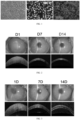
- the differentiation from the hESC cell line H1 into retinal pigment epithelial cells can be induced ( Figure 1 ); Transplantation of hESC-derived retinal pigment epithelial cells restored corneal clarity and corneal thickness within 7 days and remained corneal transparency till 14 days ( Figure 2 ).
- Figure 1 Transplantation of hESC-derived retinal pigment epithelial cells restored corneal clarity and corneal thickness within 7 days and remained corneal transparency till 14 days.
- Figure 2 The results suggest that hESC-derived retinal pigment epithelial cells can replace the function of corneal endothelial cells and quickly restore corneal transparency.
- This example used the hiPSC cell line DY0100 to induce retinal pigment epithelial cells.
- the induction differentiation method was the same as Example 1, using mTeSR1 medium to culture hiPSC cell line DY0100 till cell fusion reach approximately 80%, then using 5mg/ml type IV collagenase to digest for 15 minutes, inoculating into 1% Matrigel-coated dishes, and using cell differentiation medium 1 mixed with 2% Matrigel to culture for 2 days, changing to Matrigel-free medium for 5 days; using differentiation medium 2 to culture for 3 weeks; mechanical separation of retinal pigment epithelial cells and cell expansion.
- Cells were resuspended using DMEM low-glucose basal medium, the number of cells were counted by cell counter for distribution. Every 8 ⁇ 10 5 ⁇ 1 ⁇ 10 6 cells were dissolved in 200-300 ⁇ l DMEM basal medium with addition of 10 ⁇ M Y27632 and SmM nicotinamide for transplantation.
- the differentiation of the hiPSC cell line DY0100 into retinal pigment epithelial cells can be induced; Transplantation of hiPSC-derived retinal pigment epithelial cells restored corneal clarity and corneal thickness within 7 days and remained corneal transparent till 14 days ( Figure 3 ).
- the present example knocked out the chromogenic gene Tyrosinase, and prepared unpigmented hESC/hiPSC-RPE cells, which can also maintain corneal transparency after transplantation.
- the present embodiment used CRISPR-Cas9 technology to specifically knock out the Tryosinase gene to prepare pigment-free retinal pigment epithelial cells.
- the hES cell line H1 was used; In some other embodiments, the hiPS cell line DY0100 was used.
- change to mTeSR1 complete medium for 72-96 hours culture, and the transfection performance was evaluated according to fluorescence intensity. The sets with most fluorescent signals were selected for flow cytometry sorting, culture, and expansion to establish Tryosinase knockout cell lines.
- Example 1 (2) Induction of differentiation, (3) Enzymatic digestion of cells, (4) Preparation of cell suspension, (5) Injection of cell suspension into the anterior chamber and (6) Functional evaluation were the same as Example 1.
- Example 4 isolation and culture of primary retinal pigment epithelial cells derived from New Zealand white rabbits and gray rabbits
- primary unpigmented retinal pigment epithelial cells of New Zealand white rabbit cultured in vitro were used; In some other embodiments, pigmented retinal pigment epithelial cells of gray rabbit cultured in vitro were used.
- New Zealand white rabbits and gray rabbits were used, sacrificed by air embolization method; eyeballs were removed under sterile conditions, soaked in 1000u gentamicin saline at 4°C for 30 minutes, and then replaced in normal saline for 3 hours.
- Cells were resuspended using DMEM low-glucose basal medium, the number of cells was counted by cell counter for distribution. Every 5 ⁇ 10 5 ⁇ 8 ⁇ 10 6 cells were dissolved in 200-300 ⁇ l DMEM basal medium in addition of 10 ⁇ M Y27632 for transplantation.
- hESC-derived retinal pigment epithelial cells were treated at 37°C by 0.25% pancreatin for 3 ⁇ 10 mins, or hESC-derived retinal pigment epithelial cells were treated at 37°C by 5mg/ml IV collagenase for 5-15 mins, and the enzyme reaction was terminated by complete medium; cells were gently pipetted into single cells and collected into 15 ml centrifuge tubes, then centrifuged at 1000 rpm for 3 mins; the supernatant was discarded and the precipitate was retained.
- pancreatic digestion The mortality rate of cells obtained by pancreatic digestion was high, and that by collagenase digestion was not easy to dissociate to obtain single cells, and the digestion time was too long, so pancreatase and collagenase can be used for enzymatic digestion of cells, but Accutase is preferred.
- cell suspension was prepared using DMEM high sugar medium (containing 4.5 g/ml glucose).
- DMEM high sugar medium containing 4.5 g/ml glucose
- the number of cells were counted by cell counter for distribution. Every 5 ⁇ 10 5 ⁇ 1 ⁇ 10 6 cells were dissolved in 200-300 ⁇ l DMEM high sugar medium for transplantation.
- the present example prepares cell suspensions with different cell volumes.
- Resuspend cells using DMEM low-glucose basal medium count the number of cells by cell counter for distribution. Dissolve every 1.5 ⁇ 10 6 ⁇ 1 ⁇ 10 6 , 1 ⁇ 10 6 ⁇ 5 ⁇ 10 5 , 5 ⁇ 10 5 ⁇ 1 ⁇ 10 5 cells in 200-300 ⁇ l DMEM basal medium respectively for transplantation.
Landscapes
- Health & Medical Sciences (AREA)
- Life Sciences & Earth Sciences (AREA)
- Engineering & Computer Science (AREA)
- Biomedical Technology (AREA)
- Chemical & Material Sciences (AREA)
- General Health & Medical Sciences (AREA)
- Cell Biology (AREA)
- Veterinary Medicine (AREA)
- Medicinal Chemistry (AREA)
- Public Health (AREA)
- Animal Behavior & Ethology (AREA)
- Pharmacology & Pharmacy (AREA)
- Zoology (AREA)
- Biotechnology (AREA)
- Developmental Biology & Embryology (AREA)
- Epidemiology (AREA)
- Bioinformatics & Cheminformatics (AREA)
- Ophthalmology & Optometry (AREA)
- Organic Chemistry (AREA)
- Neurosurgery (AREA)
- Wood Science & Technology (AREA)
- Neurology (AREA)
- Genetics & Genomics (AREA)
- Virology (AREA)
- Immunology (AREA)
- General Engineering & Computer Science (AREA)
- Biochemistry (AREA)
- Microbiology (AREA)
- Reproductive Health (AREA)
- General Chemical & Material Sciences (AREA)
- Gynecology & Obstetrics (AREA)
- Nuclear Medicine, Radiotherapy & Molecular Imaging (AREA)
- Chemical Kinetics & Catalysis (AREA)
- Medicines Containing Material From Animals Or Micro-Organisms (AREA)
- Micro-Organisms Or Cultivation Processes Thereof (AREA)
Description
- The present invention belongs to the field of medicine, and relates to an application of retinal pigment epithelial cells in relieving or treating corneal endothelial functional decompensation.
- The cornea is a transparent tissue located on the anterior of the eyeball, and is divided into five layers, which sequentially comprises epithelial cell layer, bowman membrane, stromal layer, descemet's membrane, and endothelial cell layer from front to back. The highly transparent and optical properties of the cornea are one of the prerequisites that normally exert physiological functions, while corneal endothelial cells play an important role in maintaining the normal physiological function of the cornea. Corneal endothelial cells are single-layer cells in corneal inner layer, which forms a physical barrier between descemet's membrane and aqueous humor, adjusts the concentration and moisture of ions in the cornea by the ion "pump" function to maintain the semi-dehydration state of the cornea, and ensures the normal thickness and transparency of the cornea. Once the function of corneal endothelial cells is disordered, it often leads to corneal edema, causing partial or even complete corneal blindness.
- Normal human corneal endothelial cells have extremely limited proliferation capacity in vivo. Endothelial cell damage and loss caused by trauma, inflammation, cataract surgery, etc., can only be filled by the enlargement and migration of surrounding cells. When the density of human corneal endothelial cells drops to its physiological critical value (about 400 -500 cells/mm2), corneal edema occurs and vision loss in severe cases occurs. At present, there are about 4 million patients with corneal blindness in China, including nearly 1 million patients with endothelial blindness. Corneal transplantation is the only clinical therapeutic strategy for corneal endothelial decompensation. Due to the lack of corneal donors in China, only less than 10000 patients regain their sight through corneal transplantation every year, which is far from covering all clinical needs. In order to solve the problem of the corneal donor shortage, the current main corneal endothelial alternative seed cell research comprises cultured adult stem cells such as human corneal endothelial cells and skin progenitor cells, and corneal endothelial cells derived from human embryonic stem cells (hESCs) and human induced pluripotent stem cells (hiPSCs).
- Although the transplantation of primary corneal endothelial cells, adult stem cells, or pluripotent stem cells-derived cells can improve corneal endothelial function, the effect is limited. Up to now, there are no ideal corneal endothelial alternative seed cells to achieve long-term corneal transparency and wide clinical application. The team of Japanese scientist Professor Kinoshita transplanted human corneal endothelial cells by anterior chamber injection to clinically treat 11 patients with corneal endothelial dystrophy, and they restored corneal transparency, but the results of 5-year follow-up showed that the corneal endothelium of some patients reappeared pathological "guttae" structure. Cultured human corneal endothelial cells still rely on high-quality donor cornea, and adult corneal endothelial seed cells cannot be expanded in vitro in large quantities, so the source and number of these cells is limited. Adult stem cell-derived alternative cells, such as skin progenitor cells, have poor purity, difficult industrial preparation and limited therapeutic effect; hESC/hiPSC has unlimited proliferative ability and pluripotency, it has been reported that hESC/hiPSC differentiated into neural crest, corneal endothelial precursor and mature corneal endothelioid cells, some experiments have proved that corneal endothelial precursor cells and mature corneal endothelioid cells can be applied to animal models to restore corneal transparency, however, there is currently no standardized method for directed differentiation of hESC/hiPSC to corneal endothelial cells, the long-term efficacy and safety of hESC/hiPSC-derived corneal endothelial cells in vivo remains to be studied. Therefore, to find the ideal corneal endothelial alternative seed cells is still an urgent problem to be solved in the field of corneal endothelial treatment.
- After a large number of studies, the inventors find that although retinal pigment epithelial cells are greatly different from corneal endothelial cells in terms of tissue differentiation source, anatomical location, and somatic tissue cell function, they have a homologous regular hexagonal morphology and express tight junction proteins, suggesting that retinal pigment epithelial cells have the possibility of providing barrier function as substitutes for corneal endothelial cells and treating corneal endothelial functional decompensation. In order to solve the shortcomings of the prior art, the object of the present invention is to provide corneal endothelial alternative cells; the further objective is to select hESC/hiPSC-derived retinal pigment epithelial cells as seed cells, providing a seed cell source that could be supplied indefinitely and used clinically safely; more further purpose is to improve the function of transplanted cells, optimize the preparation process of cell suspension, and ensure the normal function of transplanted cells.
- The present invention provides:
- 1. Retinal pigment epithelial cells for use in a method of medical treatment as substitutes for corneal endothelial cells.
- 2. Retinal pigment epithelial cells for use in relieving or treating corneal endothelial injury, corneal endothelial lesion, corneal endothelial cell dysfunction, or corneal endothelial functional decompensation.
- 3. Retinal pigment epithelial cells for use in an individual suffering from corneal endothelial function decompensation to relieve or treat corneal thickness abnormality, corneal transparency decline, corneal edema, vision decline or loss, eye dryness, or eye pain.
- The retinal epithelial cells may be in the form of a retinal pigment epithelial cell suspension, the cell suspension comprising retinal pigment epithelial cells and a DMEM low-sugar culture medium, wherein the ratio of the retinal pigment epithelial cells to the DMEM low-sugar culture medium is 3 × 10⁵ -1.2 × 10⁶: 200 -300 µL; preferably, the ratio of the retinal pigment epithelial cells to the DMEM low-sugar culture medium is 5 × 10⁵ -1 × 10⁶: 200 -300 µL.
- In a preferred embodiment of the present invention, the retinal pigment epithelial cells are obtained by differentiation of human embryonic stem cells or human-induced pluripotent stem cells.
- In a preferred embodiment of the present invention, the pigment-producing gene Tyrosinase of the human embryonic stem cells or human induced pluripotent stem cells is knocked out.
- In a preferred embodiment of the present invention, the cell suspension further comprises one or more specific inhibitors, and the specific inhibitor comprises Y27632, nicotinamide and/or TGF-β inhibitor SB431542.
- A method for preparing retinal pigment epithelial cell suspension is disclosed but not explicitly claimed as such, comprising the following steps:
- Step 1. directed differentiation: obtaining hESC/hiPSC-derived retinal pigment epithelial cells by using differentiation medium;
- Step 2. enzymatic dissociation: treating the hESC/hiPSC-derived retinal pigment epithelial cells in step 1 with cell digestive enzyme, and using complete culture medium to terminate the enzyme reaction;
- Step 3. single cell collection: using a pipettor to gently blow cells in step 2 into single cells, collecting the cells into a centrifuge tube, centrifuging and discarding supernatant to retain the cell precipitate;
- Step 4. cell suspension preparation: resuspending the cells in step 3 by using DMEM basal culture medium, and dissolving 3 × 10⁵ -1.2 × 10⁶ cells per 200 -300 µL of DMEM basal culture medium to obtain cell suspension.
- Preferably, the differentiation medium in step 1 comprises 1: 1 proportion of DMEM/F12 and Neuralbasal Medium, 1- 4mM glutamine, 0.1-1.3mM non-essential amino acid, 0.1 -1.3mM β-mercaptoethanol, and 1% N2 additive;
- Preferably, the differentiation culture medium in step 1 comprises DMEM/F12 culture medium, 5 -15% serum substitute, 1-4 mM glutamine, 0.1 -1.3 mM non-essential amino acid, and 0.1 -1.3 mM β-mercaptoethanol.
- More preferably, the differentiation medium in step 1 comprises differentiation medium 1 and differentiation medium 2, wherein the differentiation medium 1 comprises DMEM/F12 and Neuralbasal Medium (1: 1), 2 mM glutamine, 0.1 mM non-essential amino acids, 0.1 mM β-mercaptoethanol, and 1% N2 additives; the differentiation medium 2 comprises DMEM/F12 medium, 10% serum substitute, 2 mM glutamine, 0.1 mM non-essential amino acid, and 0.1 mM β-mercaptoethanol. The method of induction differentiation comprises using cell differentiation medium 1 (DMEM/F12 and Neuralbasal medium (1:1), 2 mM glutamine, 0.1 mM non-essential amino acids, 0.1 mM β-mercaptoethanol and 1% N2 supplement) mixed with 2% Matrigel to culture for 2 days, then changing to Matrigel-free medium for 5 days; using differentiation medium 2 (DMEM/F12 medium, 10% serum substitute, 2 mM glutamine, 0.1 mM non-essential amino acid, 0.1 mM β-mercaptoethanol) to culture for 3 weeks; mechanical separation of retinal pigment epithelial cells and cell expansion.
- More preferably, the cell digestive enzyme in step 2 is Accutase, and the processing temperature is 37°C; and 5 -15 µm Y27632 is added in step 4.
- The use of retinal pigment epithelial cells as substitutes for corneal endothelial cells, refers to use for replacing damaged, diseased or missing corneal endothelial cells.
- The retinal pigment epithelial cells provided by the present invention can be administered in any convenient dosage form, and the preferred dosage form comprises injection, cell sheet or kit. Regardless of the dosage form, retinal pigment epithelial cells are administered to the anterior chamber of the individual's eyeballs.
- The following 1 to 13 items are disclosed but not explicitly claimed as such:
- 1. Applications of Retinal Pigment Epithelial Cells as Corneal Endothelial substitute.
- 2. An application of retinal pigment epithelial cells in the preparation of pharmaceutical composition for relieving or treating corneal endothelial injury, corneal endothelial lesion, corneal endothelial cell dysfunction, and corneal endothelial functional decompensation.
- 3. An application of retinal pigment epithelial cells in the preparation of pharmaceutical composition for relieving or treating corneal thickness abnormality, corneal transparency decline, corneal edema, vision decline or loss, eye dryness, eye pain of an individual suffering from corneal endothelial functional decompensation.
- 4. The application of items 1-3, wherein said retinal pigment epithelial cells are administered to the anterior chamber of an individual's eyeballs, and said pharmaceutical composition comprises retinal pigment epithelial cells and DMEM low-sugar culture medium, wherein the ratio of said retinal pigment epithelial cells and DMEM low-sugar culture medium is 3 × 10⁵ -1.2 × 10⁶: 200 -300 micro liters.
- 5. The application of items 1-4, wherein the ratio of said retinal pigment epithelial cells and DMEM low-sugar culture medium is 5 × 10⁵ -1× 10⁶: 200 -300 micro liters.
- 6. The application of item 4, said pharmaceutical composition further comprises one or more specific inhibitors, and said specific inhibitors comprise Y27632, nicotinamide and/or TGF-β inhibitor SB431542.
- 7. The application of items 1-6, said retinal pigment epithelial cells are obtained by differentiation of human embryonic stem cells or human-induced pluripotent stem cells.
- 8. The application of item 7, the pigment-producing gene Tyrosinase of said human embryonic stem cells or human induced pluripotent stem cells are knocked out.
- 9. The application of items 1-3, the dosage form of said pharmaceutical composition comprises injection, cell sheet or kit.
- 10. A method for preparing retinal pigment epithelial cell suspension, comprising the following steps:
- Step 1. directed differentiation: obtaining hESC/hiPSC-derived retinal pigment epithelial cells by using differentiation medium;
- Step 2. enzymatic dissociation: treating the hESC/hiPSC-derived retinal pigment epithelial cells in step 1 with cell digestive enzyme, and using complete culture medium to terminate the enzyme reaction;
- Step 3. single cell collection: using a pipettor to gently blow cells in step 2 into single cells, collecting the cells into a centrifuge tube, centrifuging and discarding supernatant to retain the cell precipitate;
- Step 4. cell suspension preparation: resuspending the cells in step 3 by using DMEM basal culture medium, and dissolving every 3 × 10⁵ -1.2 × 10⁶ cells in 200 -300 µL of DMEM basal culture medium to obtain cell suspension.
- 11. The method of item 10, said differentiation medium in step 1 comprises differentiation medium 1 and differentiation medium 2, wherein the differentiation medium 1 comprises DMEM/F12, Neuralbasal Medium, glutamine, non-essential amino acids, β-mercaptoethanol, and N2 additives; said differentiation medium 2 comprises DMEM/F12 medium, serum substitute, glutamine, non-essential amino acid, and β-mercaptoethanol; said differentiation culture medium 1 is mixed with the Neuralbasal Medium culture medium in a 1: 1 proportion.
- 12. The method of items 10-11, step 1 comprises using cell differentiation medium 1 (DMEM/F12 and Neuralbasal medium (1:1), 2 mM glutamine, 0.1 mM non-essential amino acids, 0.1 mM β-mercaptoethanol and 1% N2 supplement) mixed with 2% Matrigel to culture for 2 days, then changing to Matrigel-free medium for 5 days; using differentiation medium 2 (DMEM/F12 medium, 10% serum substitute, 2 mM glutamine, 0.1 mM non-essential amino acid, 0.1 mM β-mercaptoethanol) to culture for 3 weeks; mechanical separation of retinal pigment epithelial cells and cell expansion.
- 13. The method of items 10-12, said cell digestive enzyme in step 2 is Accutase, and the processing temperature is 37°C; and said cell suspension in step 4 also comprises 5 -15 µm Y27632.
- The beneficial technical effects of the present invention are as follows:
According to the invention, retinal pigment epithelial cells are provided for use as alternative seed cells of corneal endothelium for the first time; the cell suspension and the preparation method can effectively replace corneal endothelial function to recover corneal transparency and corneal thickness while ensuring cell viability. In addition, the seed cells for replacing corneal endothelium provided by the present invention can be obtained by inducing differentiation from hESC/hiPSCs, and are infinitely supplied, and the application security thereof has been reported in existing clinical experiments. According to the preparation method disclosed herein and the transplantation method using the retinal pigment epithelial cells for use of the invention, highly specialized equipment, reagents or skills are not needed, so that researchers and clinical personnel can operate conveniently, and therefore, the invention has wide application values and positive social benefits. -
-
Figure 1 shows the cell morphology diagram of the hESC-derived retinal pigment epithelial cells and the staining diagram of symbolic gene in Example 1. -
Figure 2 shows the general image and OCT photos of the cornea at 1st day, 3rd day, 7th day and 14th day after retinal pigment epithelial cell transplantation in Example 1, showing corneal transparency and thickness. -
Figure 3 shows the general image and OCT photos of the cornea at 1st day, 3rd day, 7th day and 14th day after retinal pigment epithelial cell transplantation in Example 2, showing corneal transparency and thickness. -
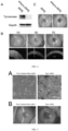
Figure 4 shows the identification of the knockout of the pigment-producing gene Tyrosinase in retinal pigment epithelial cells and corneal endothelial repair function in vivo in Example 3. (A) gene expression of retinal pigment epithelial cell before and after Tyrosinase knockout; iRPE: normal induced retinal pigment epithelial cells, shtyro-iRPE: induced retinal pigment epithelial cells with Tyrosinase knockout; (B) general image and OCT photographs of the cornea at 1st day, 3rd day, and 7th day after shtyro-iRPE transplantation; (C) general image of the corneal 1 month after transplantation of normal cells and knockout cells. -
Figure 5 shows the repair effect of rabbit primary retinal pigment epithelial cells on corneal endothelial functional decompensation in Example 4. (A) photos of isolated and cultured retinal pigment epithelial cells from New Zealand white rabbits and grey rabbits; (B) general image of the cornea from New Zealand white rabbits and grey rabbits at 7th day after retinal pigment epithelial cell transplantation. -
Figure 6 general image of the cornea at 7th day after transplantation in Comparative Example 2. -
Figure 7 general image of the cornea at 7th day after transplantation in Comparative Example 3. - The present invention is further illustrated by the following embodiments explaining the present invention, the following embodiments are only used to illustrate the present invention and should not be regarded as limiting the scope of the present invention. Unless otherwise indicated, the technical and scientific terms used herein are generally understood by those of ordinary skill in the art to which the invention belongs. If the specific conditions are not indicated in the embodiment, the conditions recommended by the manufacturer shall be carried out in accordance with the general conditions or the conditions recommended by the manufacturer. The reagents or instruments used are conventional products that are commercially available if the manufacturer is not identified.
- In the embodiments of the present invention, the hESC cell line H1 was donated by Professor Yin Zhengqin's laboratory; The hiPSC cell line DY0100 was purchased from the Chinese Academy of Sciences Cell Bank/Stem Cell Bank; Tryosinase-specific knockout hESC H1 cell line: Tryosinase-specific knockout virus were purchased from Shanghai GK Gene Medical Technology Co., Ltd. and the Tryosinase-specific knockout hESC H1 cell line was prepared according to its instruction; New Zealand white rabbits and grey rabbits were purchased from Xilingjiao Breeding Center in Jinan.
- Based on the differentiation method priorly disclosed (Rapid Differentiation of Multi-Zone Ocular Cells from Human Induced Pluripotent Stem Cells and Generation of Corneal Epithelial and Endothelial Cells, Stem Cells Dev. 2019 Apr 1; 28(7): 454-463), hESC cell line H1 cells were cultured till fusion rate reached approximately 80% using mTeSR1 medium, then digested with 5 mg/ml type IV collagenase for 15 mins and seeded into 1% Matrigel-coated dish; cell differentiation medium 1 (DMEM/F12 and Neuralbasal medium (1:1), 2 mM glutamine, 0.1 mM non-essential amino acids, 0.1 mM β-mercaptoethanol and 1% N2 supplement) mixed with 2% Matrigel was utilized to culture for 2 days, then changed to Matrigel-free medium for 5 days; using differentiation medium 2 (DMEM/F12 medium, 10% serum substitute, 2 mM glutamine, 0.1 mM non-essential amino acid, 0.1 mM β-mercaptoethanol) to culture for 3 weeks; mechanical separation of retinal pigment epithelial cells and cell expansion.
- hESC-derived retinal pigment epithelial cells were treated at 37°C by Accutase enzyme for 10-20 mins, and the enzyme reaction was terminated by complete medium; cells were gently pipetted into single cells and collected into 15 ml centrifuge tubes, then centrifuged at 1000 rpm for 3 mins; the supernatant was discarded and the precipitate was retained.
- Cells were resuspended using DMEM low-glucose basal medium, the number of cells were counted by cell counter for distribution. Every 5×10⁵∼1×10⁶ cells were dissolved in 200-300 µl DMEM basal medium in addition of 10 µM Y27632 for transplantation.
- Ketamine hydrochloride (40 mg/kg) and chlorpromazine hydrochloride (20 mg/kg) was intramuscularly administrated to anesthetize 10 New Zealand white rabbits. The right eye was washed after the eyelids were opened by eye speculum. A lateral incision of about 2mm was made at 10 o'clock spot at the corneal limbus, and carbacholine was injected into the anterior chamber to shrink the pupil. Sodium hyaluronate was injected from the lateral incision to stabilize the anterior chamber. Autologous corneal endothelial cells within a diameter of 7~9 mm in the center of the eye were scraped with a 20-gauge silicone needle. The scraped cell fragments and the residual sodium hyaluronate in the anterior chamber were washed with normal saline. 1:10 heparin sodium injection was injected to prevent anterior chamber exudation, and 10-0 nylon thread was intermittently sutured to the limbal lateral incision.
- The cell suspension was injected into the anterior chamber of the right eye from the limbus into the anterior chamber using a 1 ml syringe and tobramycin dexamethasone eye ointment was applied to cover the eye. The rabbit was held in the side-lying position under anesthesia for 3 hours to keep the right eye downward in order to facilitate rapid attachment of transplanted cells. 10 mM Y-27632 was given four times daily postoperatively. After one week change to 1 mM Y-27632 four times daily, while tobramycin dexamethasone eye drops four times daily and cyclosporine eye drops twice daily were given.
- After surgery, the recovery of corneal transparency was observed by slit lamp microscopy, the morphology and density of transplanted corneal endothelial cells were evaluated by living confocal corneal microscopy, and the change of corneal thickness was measured by ultrasound corneal thickness gauge.
- Based on the present example, the differentiation from the hESC cell line H1 into retinal pigment epithelial cells can be induced (
Figure 1 ); Transplantation of hESC-derived retinal pigment epithelial cells restored corneal clarity and corneal thickness within 7 days and remained corneal transparency till 14 days (Figure 2 ). The results suggest that hESC-derived retinal pigment epithelial cells can replace the function of corneal endothelial cells and quickly restore corneal transparency. - This example used the hiPSC cell line DY0100 to induce retinal pigment epithelial cells.
- The induction differentiation method was the same as Example 1, using mTeSR1 medium to culture hiPSC cell line DY0100 till cell fusion reach approximately 80%, then using 5mg/ml type IV collagenase to digest for 15 minutes, inoculating into 1% Matrigel-coated dishes, and using cell differentiation medium 1 mixed with 2% Matrigel to culture for 2 days, changing to Matrigel-free medium for 5 days; using differentiation medium 2 to culture for 3 weeks; mechanical separation of retinal pigment epithelial cells and cell expansion.
- Cells were resuspended using DMEM low-glucose basal medium, the number of cells were counted by cell counter for distribution. Every 8×10⁵∼1×10⁶ cells were dissolved in 200-300 µl DMEM basal medium with addition of 10 µM Y27632 and SmM nicotinamide for transplantation.
- (2) Enzymatic digestion of cells, (4) Injection of cell suspension into the anterior chamber and (5) Functional evaluation were the same as Example 1.
- Based on the present example, the differentiation of the hiPSC cell line DY0100 into retinal pigment epithelial cells can be induced; Transplantation of hiPSC-derived retinal pigment epithelial cells restored corneal clarity and corneal thickness within 7 days and remained corneal transparent till 14 days (
Figure 3 ). - In order to reduce pigmentation, the present example knocked out the chromogenic gene Tyrosinase, and prepared unpigmented hESC/hiPSC-RPE cells, which can also maintain corneal transparency after transplantation. The present embodiment used CRISPR-Cas9 technology to specifically knock out the Tryosinase gene to prepare pigment-free retinal pigment epithelial cells.
- In some embodiments, the hES cell line H1 was used; In some other embodiments, the hiPS cell line DY0100 was used.
- After digestion of the hES cell line H1 or hiPS cell line DY0100 which reached about 80% cell fusion, the cells were inoculated according to the ratio of 1:20~30, and transfection reagent mixed with siRNA was added in the next day, transfection was performed when the confluence reached about 50-60% after 16-24 hours culture, and the amount of the added virus = (MOI× number of cells) / virus titers. After 12-20 hours of transfection, change to mTeSR1 complete medium for 72-96 hours culture, and the transfection performance was evaluated according to fluorescence intensity. The sets with most fluorescent signals were selected for flow cytometry sorting, culture, and expansion to establish Tryosinase knockout cell lines.
- (2) Induction of differentiation, (3) Enzymatic digestion of cells, (4) Preparation of cell suspension, (5) Injection of cell suspension into the anterior chamber and (6) Functional evaluation were the same as Example 1.
- Knockout of the chromogenesis-related gene Tyrosinase in hESC/hiPSC-derived retinal pigment epithelial cells was achieved by the method described in the present example (
Figure 4A ); After transplantation, genetically modified retinal pigment epithelial cells can quickly restore corneal transparency and reduce corneal thickness (Figure 4B ); Corneal transparency and corneal thickness were maintained one month after transplantation with no pigmentation (Figure 4C ). - In some embodiments, primary unpigmented retinal pigment epithelial cells of New Zealand white rabbit cultured in vitro were used; In some other embodiments, pigmented retinal pigment epithelial cells of gray rabbit cultured in vitro were used.
- 2-4 weeks old New Zealand white rabbits and gray rabbits were used, sacrificed by air embolization method; eyeballs were removed under sterile conditions, soaked in 1000u gentamicin saline at 4°C for 30 minutes, and then replaced in normal saline for 3 hours. Cut the anterior segment and neuroretinal epithelium under a dissecting microscope, put the posterior eye cup into a 12-well plate Petri dish, added 0.25% pancreatic enzyme to about 3/4 eye cup, put it into a 37 °C incubator for 30 min for digestion, added a stop solution to terminate digestion, gently pipetted to detach RPE cells, then collected, centrifuged and inoculated; DMEM/F12 medium containing 10% fetal bovine serum was used to culture, and the solution was refreshed every two days for approximately 2 weeks, and identification was performed by morphology, PCR, and staining.
- Cells were resuspended using DMEM low-glucose basal medium, the number of cells was counted by cell counter for distribution. Every 5×10⁵~8×10⁶ cells were dissolved in 200-300 µl DMEM basal medium in addition of 10 µM Y27632 for transplantation.
- (2) Enzymatic digestion of cells, (4) Injection of cell suspension into the anterior chamber and (5) Functional evaluation were the same as Example 1.
- The preparation of primary retinal pigment epithelial cells of New Zealand white rabbits and gray rabbits was achieved by the method described in the present example, which exhibited a regular cell morphology (
Figure 5A ); Corneal transparency can be quickly restored 7 days after cell transplantation (Figure 5B ). - hESC-derived retinal pigment epithelial cells were treated at 37°C by 0.25% pancreatin for 3~10 mins, or hESC-derived retinal pigment epithelial cells were treated at 37°C by 5mg/ml IV collagenase for 5-15 mins, and the enzyme reaction was terminated by complete medium; cells were gently pipetted into single cells and collected into 15 ml centrifuge tubes, then centrifuged at 1000 rpm for 3 mins; the supernatant was discarded and the precipitate was retained.
- Trypan blue staining was performed and cell viability, size, etc. were counted by cell counter.
- The mortality rate of cells obtained by pancreatic digestion was high, and that by collagenase digestion was not easy to dissociate to obtain single cells, and the digestion time was too long, so pancreatase and collagenase can be used for enzymatic digestion of cells, but Accutase is preferred.
- In this example, cell suspension was prepared using DMEM high sugar medium (containing 4.5 g/ml glucose).
- (1) Induction of differentiation, (2) Enzymatic digestion of cells, (4) Injection of cell suspension into the anterior chamber and (5) Functional evaluation were the same as Example 1.
- Cells were resuspended using DMEM high sugar medium (containing 4.5 g/ml glucose), the number of cells were counted by cell counter for distribution. Every 5×10⁵∼1×10⁶ cells were dissolved in 200-300 µl DMEM high sugar medium for transplantation.
- After transplantation of cell suspension resuspended in DMEM high-glucose medium as a solvent, the anterior chamber exudation was severe, and cornea edema lasted, and corneal transparency did not restore (
Figure 6 ). - The present example prepares cell suspensions with different cell volumes.
- (1) Induction of differentiation, (2) Enzymatic digestion of cells, (4) Injection of cell suspension into the anterior chamber and (5) Functional evaluation were the same as Example 1.
- (3) Preparation of cell suspensions
- Resuspend cells using DMEM low-glucose basal medium, count the number of cells by cell counter for distribution. Dissolve every 1.5×10⁶∼1×10⁶, 1×10⁶∼5×10⁵, 5×10⁵∼1×10⁵ cells in 200-300 µl DMEM basal medium respectively for transplantation.
- Postoperative evaluation found that severe exudation of the anterior chamber and persistent cornea edema occurred after transplantation with over 1.2×10⁶ cells; while persistent cornea edema occurred and cornea transparency recovery failed after transplantation with less than 3×10⁵ cells (
Figure 7 ).
Claims (9)
- Retinal pigment epithelial cells for use in a method of medical treatment as substitutes for corneal endothelial cells.
- Retinal pigment epithelial cells for use in relieving or treating corneal endothelial injury, corneal endothelial lesion, corneal endothelial cell dysfunction, or corneal endothelial functional decompensation.
- Retinal pigment epithelial cells for use in an individual suffering from corneal endothelial function decompensation to relieve or treat corneal thickness abnormality, corneal transparency decline, corneal edema, vision decline or loss, eye dryness, or eye pain.
- Retinal pigment epithelial cells for use according to any one of claims 1-3, wherein in said use said retinal pigment epithelial cells are administered to the anterior chamber of an individual's eyeballs in the form of a pharmaceutical composition , which comprises retinal pigment epithelial cells and DMEM low-sugar culture medium, wherein the ratio of said retinal pigment epithelial cells and DMEM low-sugar culture medium is 3 × 10⁵ -1.2 × 10⁶: 200 -300 micro liters.
- Retinal pigment epithelial cells for use according to claim 4, wherein the ratio of said retinal pigment epithelial cells and DMEM low-sugar culture medium is 5 × 10⁵ - 1 × 10⁶: 200 -300 micro liters.
- Retinal pigment epithelial cells for use according to claim 4, wherein said pharmaceutical composition also comprises one or more specific inhibitors, and said specific inhibitor comprises Y27632, nicotinamide and/or TGF-β inhibitor SB431542.
- Retinal pigment epithelial cells for use according to any one of claims 1-6 wherein said retinal pigment epithelial cells are obtained by differentiation of human embryonic stem cells or human induced pluripotent stem cells.
- Retinal pigment epithelial cells for use according to claim 7, wherein the pigment-producing gene Tyrosinase of said human embryonic stem cells or human induced pluripotent stem cells is knocked out.
- Retinal pigment epithelial cells for use according to any one of claims 1-3, wherein in said use said retinal pigment epithelial cells are in the form of a cell suspension, cell sheet or kit.
Applications Claiming Priority (1)
| Application Number | Priority Date | Filing Date | Title |
|---|---|---|---|
| PCT/CN2022/105465 WO2024011449A1 (en) | 2022-07-13 | 2022-07-13 | Use of retinal pigment epithelial cells in replacement of corneal endothelia |
Publications (4)
| Publication Number | Publication Date |
|---|---|
| EP4331595A1 EP4331595A1 (en) | 2024-03-06 |
| EP4331595A4 EP4331595A4 (en) | 2024-03-06 |
| EP4331595C0 EP4331595C0 (en) | 2024-08-21 |
| EP4331595B1 true EP4331595B1 (en) | 2024-08-21 |
Family
ID=88114964
Family Applications (1)
| Application Number | Title | Priority Date | Filing Date |
|---|---|---|---|
| EP22919275.2A Active EP4331595B1 (en) | 2022-07-13 | 2022-07-13 | Use of retinal pigment epithelial cells in replacement of corneal endothelia |
Country Status (6)
| Country | Link |
|---|---|
| US (1) | US20240016852A1 (en) |
| EP (1) | EP4331595B1 (en) |
| JP (1) | JP7682398B2 (en) |
| CN (1) | CN116829699B (en) |
| WO (1) | WO2024011449A1 (en) |
| ZA (1) | ZA202405769B (en) |
Families Citing this family (1)
| Publication number | Priority date | Publication date | Assignee | Title |
|---|---|---|---|---|
| CN117243954A (en) * | 2023-10-26 | 2023-12-19 | 山东第一医科大学附属眼科研究所(山东省眼科研究所、山东第一医科大学附属青岛眼科医院) | Nicotinamide-containing intraocular operation perfusate and preparation method and application thereof |
Family Cites Families (8)
| Publication number | Priority date | Publication date | Assignee | Title |
|---|---|---|---|---|
| WO2015175783A1 (en) * | 2014-05-15 | 2015-11-19 | International Stem Cell Corporation | Chemical differentiation of pluripotentstem cells into retinal epithelial cells |
| CN106609256B (en) * | 2015-10-22 | 2020-04-07 | 同济大学 | Method for inducing human embryonic stem cells to differentiate into retinal pigment epithelial cells in vitro |
| CN105838676B (en) * | 2016-05-06 | 2020-06-05 | 中国科学院动物研究所 | A kind of culture solution for retinal pigment epithelial cells, preparation method and application thereof |
| CN108728413A (en) * | 2017-06-13 | 2018-11-02 | 中山大学中山眼科中心 | A kind of preparation of human pluripotent stem cells source Human RPE Cells in Vitro and amplification cultivation method |
| CN109136184B (en) * | 2018-07-16 | 2021-09-03 | 同济大学 | Method for inducing differentiation of human pluripotent stem cells into RPE cells |
| CN110628696B (en) * | 2019-08-28 | 2021-09-07 | 郑州大学 | Small molecule composition for directionally inducing cell differentiation and preparation method of retinal pigment epithelial cells |
| CN113699097B (en) * | 2021-09-08 | 2023-06-09 | 湖南光琇高新生命科技有限公司 | Method for preparing retinal pigment epithelial cells |
| CN116407561A (en) * | 2023-05-11 | 2023-07-11 | 山东第一医科大学附属眼科研究所(山东省眼科研究所、山东第一医科大学附属青岛眼科医院) | Application of tubular epithelial cells in replacing corneal endothelial cells |
-
2022
- 2022-07-13 WO PCT/CN2022/105465 patent/WO2024011449A1/en not_active Ceased
- 2022-07-13 EP EP22919275.2A patent/EP4331595B1/en active Active
- 2022-07-13 JP JP2024550876A patent/JP7682398B2/en active Active
- 2022-07-13 CN CN202280007264.2A patent/CN116829699B/en active Active
-
2023
- 2023-07-14 US US18/352,690 patent/US20240016852A1/en not_active Abandoned
-
2024
- 2024-07-25 ZA ZA2024/05769A patent/ZA202405769B/en unknown
Also Published As
| Publication number | Publication date |
|---|---|
| US20240016852A1 (en) | 2024-01-18 |
| WO2024011449A1 (en) | 2024-01-18 |
| EP4331595A1 (en) | 2024-03-06 |
| CN116829699B (en) | 2025-04-01 |
| CN116829699A (en) | 2023-09-29 |
| EP4331595C0 (en) | 2024-08-21 |
| JP2025505874A (en) | 2025-02-28 |
| ZA202405769B (en) | 2024-09-25 |
| JP7682398B2 (en) | 2025-05-23 |
Similar Documents
| Publication | Publication Date | Title |
|---|---|---|
| Li et al. | A novel method of isolation, preservation, and expansion of human corneal endothelial cells | |
| CN102166375B (en) | Reconstruction method of tissue engineering human corneal epithelium | |
| EP2940126A1 (en) | Human corneal endothelial cell sheet | |
| US12059438B2 (en) | Eyedrop applicable to limbal stem cell deficiency and preparation | |
| CN106916787B (en) | A kind of limbal stem cell culture medium and its cultural method | |
| CN109136184B (en) | Method for inducing differentiation of human pluripotent stem cells into RPE cells | |
| CN103656742B (en) | Preparation method of functionalized retinal pigment epithelial cell graft | |
| CN109749997B (en) | Limbal stem cell serum-free medium and culture method thereof | |
| CN105838676A (en) | Culture solution for retinal pigment epitheliums and preparation method and application thereof | |
| US20240016852A1 (en) | Application of Retinal Pigment Epithelial Cells as Corneal Endothelial Substitute | |
| JP2022502047A (en) | Methods of isolation and culture of human retinal progenitor cells | |
| US20240034997A1 (en) | Myogenin-expressing fibroblast-like cell (meflc) line and construction method and use thereof | |
| RU2409663C1 (en) | Method for producing dedifferentiated cells of adult retinal pigment eye epithelium | |
| CN101597592B (en) | Human corneal endothelial cell culture solution as well as preparation method and application thereof | |
| US20090047738A1 (en) | Feeder cell derived from tissue stem cell | |
| Qu et al. | Reconstruction of corneal epithelium with cryopreserved corneal limbal stem cells in a rabbit model | |
| CN112538454A (en) | Culture method of corneal endothelial cells | |
| CN111110921A (en) | A method for in vitro construction of lamellar cornea after tissue engineering | |
| CN117778321A (en) | Isolation and expansion of primary corneal stromal cells in humans using corneal lenses obtained during SMILE surgery | |
| US20240226177A1 (en) | Retinal pigment epithelial cell transplantation for the treatment of corneal endothelial dysfunction | |
| CN102168064A (en) | Method for constructing human corneal epithelial cell system | |
| CN115068504A (en) | Cornea repairing material and preparation method and application thereof | |
| Gimeno et al. | One-year follow-up of epithelial corneal cell sheet allografts mounted on platelet poor plasma in rabbits | |
| CN1749393A (en) | A kind of construction method of human corneal endothelial cell line | |
| CN121022744B (en) | A method and application for constructing tissue-engineered corneas based on human amniotic epithelial stem cells. |
Legal Events
| Date | Code | Title | Description |
|---|---|---|---|
| STAA | Information on the status of an ep patent application or granted ep patent |
Free format text: STATUS: UNKNOWN |
|
| STAA | Information on the status of an ep patent application or granted ep patent |
Free format text: STATUS: THE INTERNATIONAL PUBLICATION HAS BEEN MADE |
|
| PUAI | Public reference made under article 153(3) epc to a published international application that has entered the european phase |
Free format text: ORIGINAL CODE: 0009012 |
|
| STAA | Information on the status of an ep patent application or granted ep patent |
Free format text: STATUS: REQUEST FOR EXAMINATION WAS MADE |
|
| 17P | Request for examination filed |
Effective date: 20230718 |
|
| A4 | Supplementary search report drawn up and despatched |
Effective date: 20240118 |
|
| AK | Designated contracting states |
Kind code of ref document: A1 Designated state(s): AL AT BE BG CH CY CZ DE DK EE ES FI FR GB GR HR HU IE IS IT LI LT LU LV MC MK MT NL NO PL PT RO RS SE SI SK SM TR |
|
| GRAP | Despatch of communication of intention to grant a patent |
Free format text: ORIGINAL CODE: EPIDOSNIGR1 |
|
| STAA | Information on the status of an ep patent application or granted ep patent |
Free format text: STATUS: GRANT OF PATENT IS INTENDED |
|
| GRAS | Grant fee paid |
Free format text: ORIGINAL CODE: EPIDOSNIGR3 |
|
| RIC1 | Information provided on ipc code assigned before grant |
Ipc: A61K 35/545 20150101ALI20240603BHEP Ipc: A61P 27/02 20060101ALI20240603BHEP Ipc: C12N 5/0735 20100101ALI20240603BHEP Ipc: C12N 5/074 20100101ALI20240603BHEP Ipc: A61K 35/30 20150101AFI20240603BHEP |
|
| INTG | Intention to grant announced |
Effective date: 20240619 |
|
| GRAA | (expected) grant |
Free format text: ORIGINAL CODE: 0009210 |
|
| STAA | Information on the status of an ep patent application or granted ep patent |
Free format text: STATUS: THE PATENT HAS BEEN GRANTED |
|
| DAV | Request for validation of the european patent (deleted) | ||
| DAX | Request for extension of the european patent (deleted) | ||
| AK | Designated contracting states |
Kind code of ref document: B1 Designated state(s): AL AT BE BG CH CY CZ DE DK EE ES FI FR GB GR HR HU IE IS IT LI LT LU LV MC MK MT NL NO PL PT RO RS SE SI SK SM TR |
|
| REG | Reference to a national code |
Ref country code: GB Ref legal event code: FG4D |
|
| REG | Reference to a national code |
Ref country code: CH Ref legal event code: EP |
|
| REG | Reference to a national code |
Ref country code: IE Ref legal event code: FG4D |
|
| REG | Reference to a national code |
Ref country code: DE Ref legal event code: R096 Ref document number: 602022005548 Country of ref document: DE |
|
| U01 | Request for unitary effect filed |
Effective date: 20240830 |
|
| U07 | Unitary effect registered |
Designated state(s): AT BE BG DE DK EE FI FR IT LT LU LV MT NL PT RO SE SI Effective date: 20240911 |
|
| PG25 | Lapsed in a contracting state [announced via postgrant information from national office to epo] |
Ref country code: NO Free format text: LAPSE BECAUSE OF FAILURE TO SUBMIT A TRANSLATION OF THE DESCRIPTION OR TO PAY THE FEE WITHIN THE PRESCRIBED TIME-LIMIT Effective date: 20241121 |
|
| PG25 | Lapsed in a contracting state [announced via postgrant information from national office to epo] |
Ref country code: GR Free format text: LAPSE BECAUSE OF FAILURE TO SUBMIT A TRANSLATION OF THE DESCRIPTION OR TO PAY THE FEE WITHIN THE PRESCRIBED TIME-LIMIT Effective date: 20241122 Ref country code: PL Free format text: LAPSE BECAUSE OF FAILURE TO SUBMIT A TRANSLATION OF THE DESCRIPTION OR TO PAY THE FEE WITHIN THE PRESCRIBED TIME-LIMIT Effective date: 20240821 |
|
| PG25 | Lapsed in a contracting state [announced via postgrant information from national office to epo] |
Ref country code: IS Free format text: LAPSE BECAUSE OF FAILURE TO SUBMIT A TRANSLATION OF THE DESCRIPTION OR TO PAY THE FEE WITHIN THE PRESCRIBED TIME-LIMIT Effective date: 20241221 |
|
| PG25 | Lapsed in a contracting state [announced via postgrant information from national office to epo] |
Ref country code: HR Free format text: LAPSE BECAUSE OF FAILURE TO SUBMIT A TRANSLATION OF THE DESCRIPTION OR TO PAY THE FEE WITHIN THE PRESCRIBED TIME-LIMIT Effective date: 20240821 |
|
| PG25 | Lapsed in a contracting state [announced via postgrant information from national office to epo] |
Ref country code: RS Free format text: LAPSE BECAUSE OF FAILURE TO SUBMIT A TRANSLATION OF THE DESCRIPTION OR TO PAY THE FEE WITHIN THE PRESCRIBED TIME-LIMIT Effective date: 20241121 Ref country code: ES Free format text: LAPSE BECAUSE OF FAILURE TO SUBMIT A TRANSLATION OF THE DESCRIPTION OR TO PAY THE FEE WITHIN THE PRESCRIBED TIME-LIMIT Effective date: 20240821 |
|
| PG25 | Lapsed in a contracting state [announced via postgrant information from national office to epo] |
Ref country code: RS Free format text: LAPSE BECAUSE OF FAILURE TO SUBMIT A TRANSLATION OF THE DESCRIPTION OR TO PAY THE FEE WITHIN THE PRESCRIBED TIME-LIMIT Effective date: 20241121 Ref country code: PL Free format text: LAPSE BECAUSE OF FAILURE TO SUBMIT A TRANSLATION OF THE DESCRIPTION OR TO PAY THE FEE WITHIN THE PRESCRIBED TIME-LIMIT Effective date: 20240821 Ref country code: NO Free format text: LAPSE BECAUSE OF FAILURE TO SUBMIT A TRANSLATION OF THE DESCRIPTION OR TO PAY THE FEE WITHIN THE PRESCRIBED TIME-LIMIT Effective date: 20241121 Ref country code: IS Free format text: LAPSE BECAUSE OF FAILURE TO SUBMIT A TRANSLATION OF THE DESCRIPTION OR TO PAY THE FEE WITHIN THE PRESCRIBED TIME-LIMIT Effective date: 20241221 Ref country code: HR Free format text: LAPSE BECAUSE OF FAILURE TO SUBMIT A TRANSLATION OF THE DESCRIPTION OR TO PAY THE FEE WITHIN THE PRESCRIBED TIME-LIMIT Effective date: 20240821 Ref country code: GR Free format text: LAPSE BECAUSE OF FAILURE TO SUBMIT A TRANSLATION OF THE DESCRIPTION OR TO PAY THE FEE WITHIN THE PRESCRIBED TIME-LIMIT Effective date: 20241122 Ref country code: ES Free format text: LAPSE BECAUSE OF FAILURE TO SUBMIT A TRANSLATION OF THE DESCRIPTION OR TO PAY THE FEE WITHIN THE PRESCRIBED TIME-LIMIT Effective date: 20240821 |
|
| PG25 | Lapsed in a contracting state [announced via postgrant information from national office to epo] |
Ref country code: SM Free format text: LAPSE BECAUSE OF FAILURE TO SUBMIT A TRANSLATION OF THE DESCRIPTION OR TO PAY THE FEE WITHIN THE PRESCRIBED TIME-LIMIT Effective date: 20240821 |
|
| PG25 | Lapsed in a contracting state [announced via postgrant information from national office to epo] |
Ref country code: CZ Free format text: LAPSE BECAUSE OF FAILURE TO SUBMIT A TRANSLATION OF THE DESCRIPTION OR TO PAY THE FEE WITHIN THE PRESCRIBED TIME-LIMIT Effective date: 20240821 |
|
| PG25 | Lapsed in a contracting state [announced via postgrant information from national office to epo] |
Ref country code: SK Free format text: LAPSE BECAUSE OF FAILURE TO SUBMIT A TRANSLATION OF THE DESCRIPTION OR TO PAY THE FEE WITHIN THE PRESCRIBED TIME-LIMIT Effective date: 20240821 |
|
| PLBE | No opposition filed within time limit |
Free format text: ORIGINAL CODE: 0009261 |
|
| STAA | Information on the status of an ep patent application or granted ep patent |
Free format text: STATUS: NO OPPOSITION FILED WITHIN TIME LIMIT |
|
| 26N | No opposition filed |
Effective date: 20250522 |
|
| U20 | Renewal fee for the european patent with unitary effect paid |
Year of fee payment: 4 Effective date: 20250630 |
|
| REG | Reference to a national code |
Ref country code: CH Ref legal event code: H13 Free format text: ST27 STATUS EVENT CODE: U-0-0-H10-H13 (AS PROVIDED BY THE NATIONAL OFFICE) Effective date: 20260224 |
|
| PG25 | Lapsed in a contracting state [announced via postgrant information from national office to epo] |
Ref country code: CH Free format text: LAPSE BECAUSE OF NON-PAYMENT OF DUE FEES Effective date: 20250731 |