EP4321893A1 - Detection of mis-triggering in heart mri - Google Patents
Detection of mis-triggering in heart mri Download PDFInfo
- Publication number
- EP4321893A1 EP4321893A1 EP22190216.6A EP22190216A EP4321893A1 EP 4321893 A1 EP4321893 A1 EP 4321893A1 EP 22190216 A EP22190216 A EP 22190216A EP 4321893 A1 EP4321893 A1 EP 4321893A1
- Authority
- EP
- European Patent Office
- Prior art keywords
- data sets
- cardiac
- time
- acquired
- time difference
- Prior art date
- Legal status (The legal status is an assumption and is not a legal conclusion. Google has not performed a legal analysis and makes no representation as to the accuracy of the status listed.)
- Granted
Links
Images

Classifications
-
- A—HUMAN NECESSITIES
- A61—MEDICAL OR VETERINARY SCIENCE; HYGIENE
- A61B—DIAGNOSIS; SURGERY; IDENTIFICATION
- A61B5/00—Measuring for diagnostic purposes; Identification of persons
- A61B5/05—Detecting, measuring or recording for diagnosis by means of electric currents or magnetic fields; Measuring using microwaves or radio waves
- A61B5/055—Detecting, measuring or recording for diagnosis by means of electric currents or magnetic fields; Measuring using microwaves or radio waves involving electronic [EMR] or nuclear [NMR] magnetic resonance, e.g. magnetic resonance imaging
-
- G—PHYSICS
- G01—MEASURING; TESTING
- G01R—MEASURING ELECTRIC VARIABLES; MEASURING MAGNETIC VARIABLES
- G01R33/00—Arrangements or instruments for measuring magnetic variables
- G01R33/20—Arrangements or instruments for measuring magnetic variables involving magnetic resonance
- G01R33/44—Arrangements or instruments for measuring magnetic variables involving magnetic resonance using nuclear magnetic resonance [NMR]
- G01R33/48—NMR imaging systems
- G01R33/54—Signal processing systems, e.g. using pulse sequences ; Generation or control of pulse sequences; Operator console
- G01R33/56—Image enhancement or correction, e.g. subtraction or averaging techniques, e.g. improvement of signal-to-noise ratio and resolution
- G01R33/567—Image enhancement or correction, e.g. subtraction or averaging techniques, e.g. improvement of signal-to-noise ratio and resolution gated by physiological signals, i.e. synchronization of acquired MR data with periodical motion of an object of interest, e.g. monitoring or triggering system for cardiac or respiratory gating
- G01R33/5673—Gating or triggering based on a physiological signal other than an MR signal, e.g. ECG gating or motion monitoring using optical systems for monitoring the motion of a fiducial marker
-
- A—HUMAN NECESSITIES
- A61—MEDICAL OR VETERINARY SCIENCE; HYGIENE
- A61B—DIAGNOSIS; SURGERY; IDENTIFICATION
- A61B5/00—Measuring for diagnostic purposes; Identification of persons
- A61B5/0033—Features or image-related aspects of imaging apparatus, e.g. for MRI, optical tomography or impedance tomography apparatus; Arrangements of imaging apparatus in a room
- A61B5/004—Features or image-related aspects of imaging apparatus, e.g. for MRI, optical tomography or impedance tomography apparatus; Arrangements of imaging apparatus in a room adapted for image acquisition of a particular organ or body part
- A61B5/0044—Features or image-related aspects of imaging apparatus, e.g. for MRI, optical tomography or impedance tomography apparatus; Arrangements of imaging apparatus in a room adapted for image acquisition of a particular organ or body part for the heart
-
- A—HUMAN NECESSITIES
- A61—MEDICAL OR VETERINARY SCIENCE; HYGIENE
- A61B—DIAGNOSIS; SURGERY; IDENTIFICATION
- A61B5/00—Measuring for diagnostic purposes; Identification of persons
- A61B5/72—Signal processing specially adapted for physiological signals or for diagnostic purposes
- A61B5/7271—Specific aspects of physiological measurement analysis
- A61B5/7285—Specific aspects of physiological measurement analysis for synchronizing or triggering a physiological measurement or image acquisition with a physiological event or waveform, e.g. an ECG signal
- A61B5/7289—Retrospective gating, i.e. associating measured signals or images with a physiological event after the actual measurement or image acquisition, e.g. by simultaneously recording an additional physiological signal during the measurement or image acquisition
-
- A—HUMAN NECESSITIES
- A61—MEDICAL OR VETERINARY SCIENCE; HYGIENE
- A61B—DIAGNOSIS; SURGERY; IDENTIFICATION
- A61B5/00—Measuring for diagnostic purposes; Identification of persons
- A61B5/74—Details of notification to user or communication with user or patient; User input means
- A61B5/742—Details of notification to user or communication with user or patient; User input means using visual displays
- A61B5/7425—Displaying combinations of multiple images regardless of image source, e.g. displaying a reference anatomical image with a live image
-
- G—PHYSICS
- G01—MEASURING; TESTING
- G01R—MEASURING ELECTRIC VARIABLES; MEASURING MAGNETIC VARIABLES
- G01R33/00—Arrangements or instruments for measuring magnetic variables
- G01R33/20—Arrangements or instruments for measuring magnetic variables involving magnetic resonance
- G01R33/44—Arrangements or instruments for measuring magnetic variables involving magnetic resonance using nuclear magnetic resonance [NMR]
- G01R33/48—NMR imaging systems
- G01R33/54—Signal processing systems, e.g. using pulse sequences ; Generation or control of pulse sequences; Operator console
- G01R33/56—Image enhancement or correction, e.g. subtraction or averaging techniques, e.g. improvement of signal-to-noise ratio and resolution
- G01R33/563—Image enhancement or correction, e.g. subtraction or averaging techniques, e.g. improvement of signal-to-noise ratio and resolution of moving material, e.g. flow contrast angiography
- G01R33/56366—Perfusion imaging
Definitions
- the present application relates to a method for processing a plurality of cardiac MR data sets, to the processing unit configured to process the plurality of cardiac MR data sets, to a computer program comprising program code and a carrier comprising the computer program.
- Dynamic contrast-enhanced (DCE-) MRI Magnetic Resonance Imaging captures multiple single-shot images of tissue perfusion after contrast injection, aiming at identifying hypoperfusion in an organ of interest.
- Key factors for clinical application of dynamic MRI are image resolution and morphologic (slice) coverage, which can usually only be increased when using higher acceleration factors.
- Use of high acceleration factors (>3) demands more complex reconstruction frameworks employing regularization through time, making use of data redundancy across the dynamic series.
- regularization methods generally do not perform well in the presence of motion, with the two types of motion commonly encountered being cardiac (contraction of the heart) and respiratory.
- post-processing of reconstructed image data such as absolute quantification of perfusion, also requires the heart to be in the same motion state across the whole dynamic series.
- the task at hand is to identify and exclude frames impacted by mis-triggering, ideally based only on ECG acquisition timestamps, because reconstructed image data, on which the different cardiac phases would be obvious, may not be available before a regularized reconstruction or further post-processing is executed.
- motion information into the reconstruction, i.e. implicitly, e.g. assuming a "low-rank" periodicity, or explicitly, e.g. in the form of motion fields.
- cardiac motion is periodic (e.g. in cine imaging)
- intermittent variations in cardiac contraction states across a dynamic acquisition due to mis-triggering are not periodic, and thus no a-priori assumption on the motion can be made, which challenges approaches assuming periodicity.
- Explicit motion estimation and correction algorithms are usually optimized for tackling respiratory motion, with cardiac motion (deformation) manifesting at a smaller spatial scale.
- Optimizing an algorithm to simultaneously estimate motion at different scales is challenging, because a priori-assumptions on the spatial scale of the motion are made implicitly by controlling hyperparameters such as the smoothness of the resulting motion fields.
- correcting cardiac motion in practice means warping e.g. a systolic into a diastolic frame.
- Different cardiac contraction states represent different physiological states of tissue perfusion and blood volume, so that a series containing a mix of both is ultimately not meaningful in the context of a perfusion analysis.
- Rejection of mis-triggered data before final reconstruction and/or additional post-processing could in some situations be based on already reconstructed images, e.g. preliminary reconstructions or motion fields.
- Classification of cardiac phases with the purpose of rejecting mis-triggered data in an image series is, however, not straightforward, and training a neural network would need large amounts of training data. The latter problem for a neural network-based approach would also apply for the purpose of rejecting mis-triggered data using only the ECG trace.
- a method for processing a plurality of cardiac MR data sets comprises the step of providing the plurality of cardiac MR data sets, which were acquired over a plurality of cardiac cycles wherein each cardiac MR data set has a corresponding timestamp at which the cardiac MR data set was acquired. Furthermore, each of the cardiac MR data sets was acquired based on a trigger event triggering the acquisition of the corresponding cardiac MR data set. Furthermore, a series of time differences between the acquisitions of the cardiac MR data sets is determined based on the timestamps. In the series of time differences a default time difference is determined representing the time difference between two consecutive cardiac MR data sets, which were acquired using correct trigger events present in the trigger events.
- At least one pair of incorrect time differences is determined in the series of time differences wherein each incorrect time difference represents the time difference between two consecutive MR data sets, in which at least one MR data set from the 2 consecutive MR data sets was acquired using an incorrect trigger event present in the trigger events.
- approved MR data sets comprising only cardiac MR data sets from the plurality of cardiac MR data sets are determined which were acquired using the correct trigger event based on the at least one pair of incorrect time differences. The approved MR data sets can then be further processed.
- the approved MR data sets which correspond to the data sets which were acquired based on the correct trigger event, it is possible to determine, in the plurality of cardiac MR data sets, first MR data sets which were acquired between the at least one pair of incorrect time differences. Furthermore, the approved MR data set is then determined while excluding the first MR data sets from the plurality of cardiac MR data sets.
- the first MR data sets correspond to the data sets, which were acquired between missed trigger events. Accordingly the images generated from the first MR data sets might have another cardiac phase compared to the approved MR data sets so that these images and data sets can be excluded. All the MR data sets acquired after entering the wrong trigger cycle and until exiting the wrong trigger cycle are removed.
- the method can furthermore comprise the step of determining, based on the timestamps, for the at least one pair of time differences, a time interval between the acquisition of cardiac MR data sets having incorrect time differences.
- the cardiac MR data sets which were acquired between the at least one pair of incorrect time differences is only confirmed as belonging to the first MR data sets if the time interval is smaller than a threshold value.
- images are only excluded when the image acquisition in the time interval between the pair of time differences is smaller than a defined number, by way of example a number smaller than ⁇ 10 acquisitions, smaller than 7 acquisitions, smaller than 5 acquisitions or smaller than any number between 4 and 7 acquisitions.
- the step of determining the default time difference can comprise the step of determining an average time difference in the series of time differences and the default time difference is then determined based on the average time difference.
- the default time difference corresponding to the time difference occurring between two correctly identified trigger events is assumed to be present in the major cases of the acquired MR data sets. Accordingly, the default time difference will be close to the average time difference.
- a clustering step in which the series of time difference is clustered and the default time difference is determined based on the clustering based on an assumption that the cluster having the highest number of time differences is used for determining the default time difference.
- the clustering helps to identify the default time difference. If the acquisition conditions are very poor and it is hardly possible to identify the correct trigger event, the acquired MR image data will likely not provide fruitful results. Only when the major part of the cardiac MR data sets was acquired based on the correctly identified trigger events, the data provide a useful basis for a further processing.
- the cardiac MR data sets belonging to the identified single time differences are excluded when determining the approved MR data sets.
- the acquisition is started with an incorrect trigger event and after a while, the correct trigger event is used.
- no pair of incorrect time differences can be detected, but the acquisition starts with an incorrect trigger event or finishes with an incorrect trigger event.
- the MR data sets belonging to those single time differences are also excluded for the further processing of the MR data sets.
- the plurality of cardiac MR data sets can contain a number of cardiac MR images, but may also contain a number of cardiac MR raw data sets. Accordingly, it is not absolutely necessary to do an image reconstruction, the above mentioned analysis might only be carried out on the raw data sets as only the timestamp is needed from the different data sets.
- each time difference in the at least one pair of incorrect time differences has a time period larger than a time period of the default time difference. This means that the entering and exiting of the incorrect trigger events leads to a prolonged time distance between the acquisition of two consecutive MR data sets.
- the processing of the approved MR data sets can contain the step of determining a perfusion parameter using the approved MR data sets.
- a processing unit which is configured to process the cardiac MR data sets is provided as indicated above.
- a computer program comprising program code is provided that, when executed by a processing unit cause the processing unit to perform a method as mentioned above or as explained in further detail below.
- a carrier comprising the computer program wherein the carrier is one of an electronic signal, optical signal, radio signal, or computer readable storage medium.
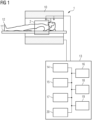
- Figure 1 shows a schematic view of an MR system 1, which compromises a magnet 10 generating a polarization field B0.
- An object under examination 12 lying on a table 11 is moved into the center of the MR system 1 where MR signals after RF excitation can be detected by receiving coils 2 which can comprise different coil sections wherein each coil section is associated with a corresponding detection channel 3.
- receiving coils 2 which can comprise different coil sections wherein each coil section is associated with a corresponding detection channel 3.
- the nuclear spins in the object 12 and especially in the part located in the receiving coil 2, here the heart are exited and location coded and the currents induced by the relaxation can be detected.
- the way how MR images are generated and how the MR signals are detected using a sequence of RF pulses and the sequence of magnetic field gradients are known in the art so that a detailed explanation thereof is omitted.
- the MR system comprises a control module 13 which is used for controlling the MR system.
- the control module 20 comprises a gradient control unit 14 for controlling and switching the magnetic field gradients, an RF control unit 15 for controlling and generating the RF pulses for the imaging sequences.
- An image sequence control unit 16 is provided which controls the sequence of the applied RF pulses and magnetic field gradients and thus controls the gradient control unit 14 and the RF control unit 15.
- computer programs needed for operating the MR system and the imaging sequences necessary for generating the MR images can be stored together with the generated MR images.
- the generated MR images can be displayed on a display 18 wherein an input unit 19 is provided used by a user of the MR system to control the functioning of the MR system.
- a processing unit 20 can coordinate the operation of the different functional units show in Figure 1 and can comprise one or more processors which can carry out instructions stored on the memory 17.
- the memory includes the program code to be executed by the processing unit 20.
- the processing unit can, based on the detected images reconstruct MR images.
- the processing unit 20 can, as discussed below process a set of cardiac MR data in such a way that incorrect trigger events or missed triggers are identified in a reliable and effective way.
- the proposed solution solves the problem of rejecting mis-triggered data in cardiac MR data sets prior to reconstruction of any image data by analyzing logged trigger events and identifying a signature of mis-triggered data, that is specific to cardiac perfusion MR.
- This can be applied for both, (pharmacological) stress and rest perfusion data.
- the method discussed below is applied in dynamic perfusion examinations, however it might be applied to any other cardiac MR image where several MR data sets are acquired over different cardiac cycles.
- Fig. 2 shows in more detail the interplay of correct and incorrect ECG trigger events, which can perturb a dynamic cardiac magnetic resonance perfusion examination.
- the data points and MR data sets to be rejected can be identified by identifying prolonged pairs in the time series of differences between successive acquisition times. For simplicity only one slice position is indicated per RR interval.
- Fig. 2 shows how several data MR data sets 21 to 25 are acquired based on an ECG trigger signal of the heart.
- the R-wave in the ECG signal is shown by reference numerals 30, 32, 34, 36, 38 and 40 and plays the role of the correct trigger event.
- artefactual trigger events such as the events 41, 43, 45, 47 and 49 shown in Fig. 2 .
- a trigger lock time is present, in which no trigger event is detected, and in which no trigger event can trigger the acquisition of the next MR image.
- a trigger lock time is present, in which no trigger event is detected, and in which no trigger event can trigger the acquisition of the next MR image.
- the system misses the correct trigger event 34 and the next data acquisition is triggered based on trigger event 45, which is an incorrect trigger.
- trigger event 45 which is an incorrect trigger.
- the correct triggers 36 and 38 are not detected.
- the system misses the trigger event 49, which is again an artefactual trigger and the next image is acquired based on a correct trigger 40.
- Images 21, 22 and 25 are images, which are acquired in the correct cardiac phase (the systole) as shown by reference numerals 55 and 57 whereas images 23 and 24 are acquired in the wrong cardiac phase, by way of example in the diastole compared to the correct cardiac phase the systole.
- Fig. 2 furthermore shows that the time difference between the acquisition of the images varies in dependence of the fact whether a correct or wrong trigger event is used.
- the time difference 50 is a correctly identified time difference and corresponds to a default time difference.
- the following time difference is a prolonged time difference 51 followed by another time difference 52, which seems to be in the same range as the time difference 50.
- the corresponding images are acquired in the wrong cardiac phase.
- another prolonged time difference 53 is present.
- a missed trigger leads to a specific pattern in the temporal distance between the acquisition of successive dynamic MR data sets. While a trigger lock prevents detection of triggers during image acquisition, wherein only one slice position is shown in Fig. 1 for simplicity, artefactual triggers due to disturbances on the ECG trace may be accepted after a correct R-wave trigger is missed, pushing the trigger period into a shifted cycle until the artefactual trigger itself is missed. Accordingly, dynamic image acquisitions affected by missed trigger events are encapsulated by a pair of prolonged distances in time to the following acquisition, which corresponds to entering and exiting the shifted artefactual triggering cycle.
- time differences 51 and 53 it is possible, to identify MR data sets, which were acquired based on an incorrect or missed trigger event even though the time difference itself between missed triggers, here the time difference 52, seems to be similar to the time difference 50.
- the MR data sets 23 and 24 can be detected using the following procedure: in step number 1 multiple MRI images of one or more slice-positions are acquired as a dynamic series with the acquisition of each frame being triggered by an ECG trigger signal or an alternative cardiac triggering device. Furthermore, for each image data set the corresponding acquisition timestamp is determined and recorded.
- a second step the following processing can be carried out either before reconstruction of the image data, by way of example for temporarily regularized reconstructions or before further post-processing the acquired images, by way of example for an absolute quantification of the flow:
- a first situation (case A) is shown in the upper two graphs, which is called the first case in the following where at the beginning three clusters of time differences were identified cluster 51, 52 and 53.
- cluster 53 corresponds to a multiple of the cluster 50
- the time difference occurring in a cluster 53 is assumed to be a correct time difference, namely a multiple of the correct trigger distance.
- Cluster 52 is a prolonged trigger interval and is not a multiple of the correct trigger interval so it can be determined that the corresponding images were acquired at the wrong cardiac cycle.
- Cluster 54 represents the correct trigger interval whereas cluster 55 represents the wrong trigger interval.
- next step 2.f it is necessary to identify pairs of missed triggers as the pair 60 shown in the left upper part and the pair 61 shown in the upper right part of Fig. 3 .
- these 2 data points could correspond to the prolonged time difference 51 and 53 shown in Fig. 2 . Accordingly, it is necessary to exclude the data acquisitions between these two time differences. Accordingly, the time differences occurring between these two pairs here including the first end excluding the second partner of the pair, the time differences 65 and 60a or 68 and 61a, and the corresponding MR data sets have to be identified. Even though the time differences 65 and 68 seem to be based on the correct trigger interval, they belong to the wrong cardiac phase or cycle so that it is necessary to exclude those data sets from the image processing.
- the identification of the pair of time differences 60 or 61 shown in Fig. 3 helps to identify the time differences 65 and 68 occurring between the corresponding pair.
- a possible exception to the encapsulation between pairs represents the case in which one of the two missed trigger events is outside of the dynamic image acquisition, accordingly at the end or at the start of the image acquisition. As a consequence it is advisable to also exclude image acquisitions before/after a single missed trigger at the beginning/end of the series of acquisitions. Referring to Fig. 3 , this means that a single missed trigger 66 occurs at the end of the acquisition. Based on the assumption that the images after this trigger are based on the wrong cardiac cycle all the MR data sets occurring after this single time difference are excluded.
- the distance between incorrect pairs should not be larger than a certain number of successive wrong triggers, the number being a number between 4 and 10 such as a number between 4 and 6. Accordingly it can be assumed that the data points 67a and 61a shown in Fig. 3 do not correspond to a single pair of incorrect time differences as the distance between those image acquisitions is too large.
- step 2.g Summarising, in step 2.g) for each pair found in step 2.f, accordingly for each pair 60 or 61 shown in Fig. 3 , the data acquisitions in between those missed triggers are excluded from the further processing.
- approved MR data sets are determined which contain all the MR data sets which were acquired based on the correct trigger interval or at least on a multiple of the correct trigger interval.
- the data sets which were acquired between the pair of incorrect time differences can be called first data sets, which are excluded from the further processing.
- a further optional step it is possible to reconstruct and display also the rejected data separately to avoid a loss of any diagnostic information.
- Case B as shown in Fig. 3 shows another situation where only a single missed trigger is detected, which is however a multiple of the correct trigger interval. Accordingly, no significant cluster difference occurs and none of the data has to be rejected. The same is true for the lower right part of Fig. 3 .
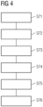
- Fig. 4 summarizes some of the major steps carried out in the above-discussed identification of MR data sets, which were acquired at the wrong cardiac phase of the cycle.
- MR data the cardiac MR data sets are provided, which were acquired over several cardiac cycles (step S71). Those data set comprise data sets, which were acquired based on the correct trigger event and based on an incorrect trigger event.
- step S72 time differences are determined between the different acquisitions based on the timestamps of the acquisitions. Accordingly a series of time differences is generated.
- a default time difference is determined representing the time difference between two consecutive cardiac MR data sets, which were acquired using a correct trigger event (step S73). As discussed above, this determination of the default time difference can be based on autocorrelation or clustering of the time series as it is assumed that the major part of the images were acquired with the correct trigger event.
- step S74 determines pairs of incorrect time differences in step S74, as discussed above in connection with Fig. 3 the pairs of time differences 61 or 60. Based on this pair of incorrect time differences it is possible to determine the approved data sets in step S75, which only contain the cardiac MR data sets, which were acquired based on a correct trigger event.
- step S76 the approved data set can be further processed meaning that an image reconstruction is carried out or the image data of further processed such as the determination of a perfusion parameter.
- the proposed approach identifies cardiac MR perfusion data affected by missed or incorrect triggers by using a specific signature in the acquisition timestamp.
- the above described method increases the sharpness and the fidelity of motion compensated image series after temporal regularization and/or before the post-processing such as the absolute perfusion quantification.
- any a-priori assumptions on the nature of the underlying cardiac motion is avoided.
- there is no need to reconstruct image data before the data to be rejected is identified. This is possible as only the time difference is needed between the acquisitions.
- no user interaction or ground truth training data or a neural network is required. An efficient way is provided to remove MR data sets acquired in a series of MR images, which were acquired in a wrong cardiac phase.
Landscapes
- Health & Medical Sciences (AREA)
- Life Sciences & Earth Sciences (AREA)
- Physics & Mathematics (AREA)
- Engineering & Computer Science (AREA)
- Nuclear Medicine, Radiotherapy & Molecular Imaging (AREA)
- Biophysics (AREA)
- General Health & Medical Sciences (AREA)
- Radiology & Medical Imaging (AREA)
- Animal Behavior & Ethology (AREA)
- Heart & Thoracic Surgery (AREA)
- Medical Informatics (AREA)
- Molecular Biology (AREA)
- Surgery (AREA)
- Pathology (AREA)
- Public Health (AREA)
- Veterinary Medicine (AREA)
- Biomedical Technology (AREA)
- Physiology (AREA)
- High Energy & Nuclear Physics (AREA)
- Cardiology (AREA)
- Signal Processing (AREA)
- Artificial Intelligence (AREA)
- Psychiatry (AREA)
- Computer Vision & Pattern Recognition (AREA)
- Power Engineering (AREA)
- Pulmonology (AREA)
- Condensed Matter Physics & Semiconductors (AREA)
- General Physics & Mathematics (AREA)
- Magnetic Resonance Imaging Apparatus (AREA)
Abstract
Description
- The present application relates to a method for processing a plurality of cardiac MR data sets, to the processing unit configured to process the plurality of cardiac MR data sets, to a computer program comprising program code and a carrier comprising the computer program.
- Dynamic contrast-enhanced (DCE-) MRI (Magnetic Resonance Imaging) captures multiple single-shot images of tissue perfusion after contrast injection, aiming at identifying hypoperfusion in an organ of interest. Key factors for clinical application of dynamic MRI are image resolution and morphologic (slice) coverage, which can usually only be increased when using higher acceleration factors. Use of high acceleration factors (>3) demands more complex reconstruction frameworks employing regularization through time, making use of data redundancy across the dynamic series. However, regularization methods generally do not perform well in the presence of motion, with the two types of motion commonly encountered being cardiac (contraction of the heart) and respiratory. Additionally, post-processing of reconstructed image data, such as absolute quantification of perfusion, also requires the heart to be in the same motion state across the whole dynamic series.
- It is generally assumed that cardiac motion is frozen across the whole dynamic series by acquiring each image for a given slice position at the same distance to the ECG trigger. However, this assumption may break down for individual frames in the case of mis-triggering, which can have different causes and is a common phenomenon especially if the patient is pharmacologically stressed during the exam. Mis-triggering occurs when an incorrect trigger event in the detected trigger events is used for the image acquisition. Frames impacted by mis-triggering may display any cardiac phase different to the rest of the dynamic series, which poses a challenge to motion compensation, both in the context of regularized reconstructions and/or for post-processing such as quantitative perfusion MRI.
- Thus, the task at hand is to identify and exclude frames impacted by mis-triggering, ideally based only on ECG acquisition timestamps, because reconstructed image data, on which the different cardiac phases would be obvious, may not be available before a regularized reconstruction or further post-processing is executed.
- Different methods exist to include motion information into the reconstruction, i.e. implicitly, e.g. assuming a "low-rank" periodicity, or explicitly, e.g. in the form of motion fields. While cardiac motion is periodic (e.g. in cine imaging), intermittent variations in cardiac contraction states across a dynamic acquisition due to mis-triggering are not periodic, and thus no a-priori assumption on the motion can be made, which challenges approaches assuming periodicity.
- Explicit motion estimation and correction algorithms are usually optimized for tackling respiratory motion, with cardiac motion (deformation) manifesting at a smaller spatial scale. Optimizing an algorithm to simultaneously estimate motion at different scales is challenging, because a priori-assumptions on the spatial scale of the motion are made implicitly by controlling hyperparameters such as the smoothness of the resulting motion fields. Even if there were motion estimation or compensation algorithms that can handle respiratory and extreme cardiac motion at the same time, correcting cardiac motion in practice means warping e.g. a systolic into a diastolic frame. Different cardiac contraction states, however, represent different physiological states of tissue perfusion and blood volume, so that a series containing a mix of both is ultimately not meaningful in the context of a perfusion analysis.
- Rejection of mis-triggered data before final reconstruction and/or additional post-processing could in some situations be based on already reconstructed images, e.g. preliminary reconstructions or motion fields. Classification of cardiac phases with the purpose of rejecting mis-triggered data in an image series is, however, not straightforward, and training a neural network would need large amounts of training data. The latter problem for a neural network-based approach would also apply for the purpose of rejecting mis-triggered data using only the ECG trace.
- Accordingly, a need exists to overcome the above identified problems and to provide a way to identify MR images in a series of MR images which were acquired based on a wrong trigger event based solely on the ECG trace.
- This need is met by the features of the independent claims. Further aspects are described in the dependent claims.
- According to a first aspect a method for processing a plurality of cardiac MR data sets is provided. The method comprises the step of providing the plurality of cardiac MR data sets, which were acquired over a plurality of cardiac cycles wherein each cardiac MR data set has a corresponding timestamp at which the cardiac MR data set was acquired. Furthermore, each of the cardiac MR data sets was acquired based on a trigger event triggering the acquisition of the corresponding cardiac MR data set. Furthermore, a series of time differences between the acquisitions of the cardiac MR data sets is determined based on the timestamps. In the series of time differences a default time difference is determined representing the time difference between two consecutive cardiac MR data sets, which were acquired using correct trigger events present in the trigger events. Furthermore, in the time differences at least one pair of incorrect time differences is determined in the series of time differences wherein each incorrect time difference represents the time difference between two consecutive MR data sets, in which at least one MR data set from the 2 consecutive MR data sets was acquired using an incorrect trigger event present in the trigger events. Furthermore, approved MR data sets comprising only cardiac MR data sets from the plurality of cardiac MR data sets are determined which were acquired using the correct trigger event based on the at least one pair of incorrect time differences. The approved MR data sets can then be further processed.
- It was found that the series of time differences especially the pair of incorrect time differences can form a basis to identify MR data sets, which were acquired based on an incorrect trigger event. A missed trigger or an incorrect trigger leads to a specific pattern in the temporal distance between the acquisition of consecutive or successive MR data sets. It was especially found that MR data sets, which are affected by an incorrect trigger event are encapsulated by a pair of incorrect time differences. It can be interpreted as corresponding to the fact that the image acquisition enters the wrong trigger cycle and then exits the wrong trigger cycle.
- For determining the approved MR data sets, which correspond to the data sets which were acquired based on the correct trigger event, it is possible to determine, in the plurality of cardiac MR data sets, first MR data sets which were acquired between the at least one pair of incorrect time differences. Furthermore, the approved MR data set is then determined while excluding the first MR data sets from the plurality of cardiac MR data sets. The first MR data sets correspond to the data sets, which were acquired between missed trigger events. Accordingly the images generated from the first MR data sets might have another cardiac phase compared to the approved MR data sets so that these images and data sets can be excluded. All the MR data sets acquired after entering the wrong trigger cycle and until exiting the wrong trigger cycle are removed.
- The method can furthermore comprise the step of determining, based on the timestamps, for the at least one pair of time differences, a time interval between the acquisition of cardiac MR data sets having incorrect time differences. The cardiac MR data sets which were acquired between the at least one pair of incorrect time differences is only confirmed as belonging to the first MR data sets if the time interval is smaller than a threshold value. When the image acquisition enters a wrong trigger cycle, it can be assumed that within a certain time, the image acquisition returns to the correct trigger event. Accordingly, images are only excluded when the image acquisition in the time interval between the pair of time differences is smaller than a defined number, by way of example a number smaller than <10 acquisitions, smaller than 7 acquisitions, smaller than 5 acquisitions or smaller than any number between 4 and 7 acquisitions.
- The step of determining the default time difference can comprise the step of determining an average time difference in the series of time differences and the default time difference is then determined based on the average time difference. The default time difference corresponding to the time difference occurring between two correctly identified trigger events is assumed to be present in the major cases of the acquired MR data sets. Accordingly, the default time difference will be close to the average time difference.
- Furthermore, it is possible to carry out a clustering step in which the series of time difference is clustered and the default time difference is determined based on the clustering based on an assumption that the cluster having the highest number of time differences is used for determining the default time difference. Here again, it is assumed that most of the acquired MR images are acquired based on the correct trigger event, so that the clustering helps to identify the default time difference. If the acquisition conditions are very poor and it is hardly possible to identify the correct trigger event, the acquired MR image data will likely not provide fruitful results. Only when the major part of the cardiac MR data sets was acquired based on the correctly identified trigger events, the data provide a useful basis for a further processing.
- Aditionally it is possible to determine integer multiples of the default time difference and the cardiac MR data sets having a time difference in the series of time differences corresponding to integer multiples of the default time difference are confirmed as belonging to the approved MR data sets. It is assumed that the trigger events occur in a regular pattern and it might be possible that one or two trigger events were missed for the image acquisition, however the image acquisition is based on the correct trigger events. Accordingly, data sets which were acquired corresponding to an integer multiple of correct trigger events are considered as belonging to the approved MR data sets even though the time difference is larger than the default time difference.
- Furthermore, it is possible to identify single time differences in the series of time differences which are located at a beginning or at an end of an acquisition of the cardiac MR data sets and which differ from the default time difference by more than a threshold. The cardiac MR data sets belonging to the identified single time differences are excluded when determining the approved MR data sets. At the beginning of the acquisition, it is also possible that the acquisition is started with an incorrect trigger event and after a while, the correct trigger event is used. Here no pair of incorrect time differences can be detected, but the acquisition starts with an incorrect trigger event or finishes with an incorrect trigger event. The MR data sets belonging to those single time differences are also excluded for the further processing of the MR data sets.
- The plurality of cardiac MR data sets can contain a number of cardiac MR images, but may also contain a number of cardiac MR raw data sets. Accordingly, it is not absolutely necessary to do an image reconstruction, the above mentioned analysis might only be carried out on the raw data sets as only the timestamp is needed from the different data sets.
- Preferably, each time difference in the at least one pair of incorrect time differences has a time period larger than a time period of the default time difference. This means that the entering and exiting of the incorrect trigger events leads to a prolonged time distance between the acquisition of two consecutive MR data sets. The processing of the approved MR data sets can contain the step of determining a perfusion parameter using the approved MR data sets.
- According to a further aspect a processing unit which is configured to process the cardiac MR data sets is provided as indicated above. Additionally, a computer program comprising program code is provided that, when executed by a processing unit cause the processing unit to perform a method as mentioned above or as explained in further detail below.
- Furthermore, a carrier is provided comprising the computer program wherein the carrier is one of an electronic signal, optical signal, radio signal, or computer readable storage medium.
- The foregoing and additional features and effects of the application will become apparent from the following detailed description, when read in conjunction with the accompanying drawings in which like reference numerals refer to like elements.
-
Figure 1 shows a schematic view of an MR system with which MR images of the heart can be generated and processed in such a way that cardiac MR data sets acquired with an incorrect trigger event are identified in an efficient way. -
Figure 2 shows a schematic view of an interplay of correct and incorrect trigger events perturbing a cardiac imaging sequence. -
Figure 3 shows different situations how time differences between consecutive cardiac acquisitions can be used to identify incorrect trigger events. -
Figure 4 shows a schematic view of schematic view of a flowchart comprising steps carried out for identifying MR data sets which were acquired based on incorrect trigger events. - In the following, embodiments of the invention will be described in detail with reference to the accompanying drawings. It is to be understood the following description of embodiments is not to be taken in a limiting sense. The scope of the invention is not intended to be limited by the embodiments described hereinafter or by the drawings, which are to be illustrative only.
- The drawings are to be regarded as being schematic representations, and elements illustrated in the drawings are not necessarily shown to scale. Rather, the various elements are represented such that their function and general purpose becomes apparent to a person skilled in the art. Any connection or coupling between functional blocks, devices, components of physical or functional units shown in the drawings and described hereinafter may be implemented by an indirect connection or coupling. A coupling between components may be established over a wired or wireless connection. Functional blocks may be implemented in hardware, software, firmware, or a combination thereof.
-
Figure 1 shows a schematic view of an MR system 1, which compromises amagnet 10 generating a polarization field B0. An object underexamination 12 lying on a table 11 is moved into the center of the MR system 1 where MR signals after RF excitation can be detected by receivingcoils 2 which can comprise different coil sections wherein each coil section is associated with a corresponding detection channel 3. By applying RF pulses and magnetic field gradients, the nuclear spins in theobject 12 and especially in the part located in the receivingcoil 2, here the heart are exited and location coded and the currents induced by the relaxation can be detected. The way how MR images are generated and how the MR signals are detected using a sequence of RF pulses and the sequence of magnetic field gradients are known in the art so that a detailed explanation thereof is omitted. - The MR system comprises a
control module 13 which is used for controlling the MR system. Thecontrol module 20 comprises agradient control unit 14 for controlling and switching the magnetic field gradients, anRF control unit 15 for controlling and generating the RF pulses for the imaging sequences. An imagesequence control unit 16 is provided which controls the sequence of the applied RF pulses and magnetic field gradients and thus controls thegradient control unit 14 and theRF control unit 15. In amemory 17, computer programs needed for operating the MR system and the imaging sequences necessary for generating the MR images can be stored together with the generated MR images. The generated MR images can be displayed on adisplay 18 wherein aninput unit 19 is provided used by a user of the MR system to control the functioning of the MR system. Aprocessing unit 20 can coordinate the operation of the different functional units show inFigure 1 and can comprise one or more processors which can carry out instructions stored on thememory 17. The memory includes the program code to be executed by theprocessing unit 20. The processing unit can, based on the detected images reconstruct MR images. - The
processing unit 20 can, as discussed below process a set of cardiac MR data in such a way that incorrect trigger events or missed triggers are identified in a reliable and effective way. - The proposed solution solves the problem of rejecting mis-triggered data in cardiac MR data sets prior to reconstruction of any image data by analyzing logged trigger events and identifying a signature of mis-triggered data, that is specific to cardiac perfusion MR. This can be applied for both, (pharmacological) stress and rest perfusion data. Preferably, the method discussed below is applied in dynamic perfusion examinations, however it might be applied to any other cardiac MR image where several MR data sets are acquired over different cardiac cycles.
-
Fig. 2 shows in more detail the interplay of correct and incorrect ECG trigger events, which can perturb a dynamic cardiac magnetic resonance perfusion examination. The data points and MR data sets to be rejected can be identified by identifying prolonged pairs in the time series of differences between successive acquisition times. For simplicity only one slice position is indicated per RR interval. -
Fig. 2 shows how several data MR data sets 21 to 25 are acquired based on an ECG trigger signal of the heart. The R-wave in the ECG signal is shown byreference numerals events Fig. 2 . During the acquisition of an image, a trigger lock time is present, in which no trigger event is detected, and in which no trigger event can trigger the acquisition of the next MR image. In the example shown inFig. 2 , the system misses thecorrect trigger event 34 and the next data acquisition is triggered based ontrigger event 45, which is an incorrect trigger. In view of the trigger lock time, thecorrect triggers trigger event 49, which is again an artefactual trigger and the next image is acquired based on acorrect trigger 40.Images reference numerals images -
Fig. 2 furthermore shows that the time difference between the acquisition of the images varies in dependence of the fact whether a correct or wrong trigger event is used. As can be deduced fromFig. 2 , thetime difference 50 is a correctly identified time difference and corresponds to a default time difference. The following time difference is aprolonged time difference 51 followed by anothertime difference 52, which seems to be in the same range as thetime difference 50. However, the corresponding images are acquired in the wrong cardiac phase. When the system returns to the correct trigger events anotherprolonged time difference 53 is present. - Accordingly as shown in
Fig. 2 a missed trigger leads to a specific pattern in the temporal distance between the acquisition of successive dynamic MR data sets. While a trigger lock prevents detection of triggers during image acquisition, wherein only one slice position is shown inFig. 1 for simplicity, artefactual triggers due to disturbances on the ECG trace may be accepted after a correct R-wave trigger is missed, pushing the trigger period into a shifted cycle until the artefactual trigger itself is missed. Accordingly, dynamic image acquisitions affected by missed trigger events are encapsulated by a pair of prolonged distances in time to the following acquisition, which corresponds to entering and exiting the shifted artefactual triggering cycle. Accordingly, by detecting a pair of incorrect time differences, here thetime differences time difference 52, seems to be similar to thetime difference 50. - Accordingly, the MR data sets 23 and 24 can be detected using the following procedure:
in step number 1 multiple MRI images of one or more slice-positions are acquired as a dynamic series with the acquisition of each frame being triggered by an ECG trigger signal or an alternative cardiac triggering device. Furthermore, for each image data set the corresponding acquisition timestamp is determined and recorded. - In a second step the following processing can be carried out either before reconstruction of the image data, by way of example for temporarily regularized reconstructions or before further post-processing the acquired images, by way of example for an absolute quantification of the flow:
- in step 2.a) a time series of the differences between the acquisition timestamps is determined.
Fig. 3 shows different cases wherein the graphs inFig. 3 show the number of image acquisitions N and the corresponding time differences in milliseconds. - in step 2.b) an average, correct trigger interval has to be found, here, the correct RR trigger interval. This may be obtained via autocorrelation or by clustering the time series into different groups such as three groups using clustering methods such as Jenks Clustering. It is possible to use a clustering into three groups for the robustness against actual ectopic short heartbeats and full multi-heartbeat pauses
- in step 2.c) multiples of the correct trigger interval are eliminated in the time series, by way of example by taking a two-sided modulus.
Fig. 3 , the upper left side shows the time differences for the correct trigger interval, which is slightly larger than 600 ms where also a time difference of more than 1200 ms is detected. This is double of the correct trigger interval, but as the correct trigger was used the data which were acquired with a time difference of the double correct trigger interval should not be rejected. - In step 2.d) a clustering of the time series created in step 2.c) is carried out wherein a clustering into two groups might be used. In step 2.e) the smaller cluster is identified as being the cluster of the incorrect triggers. This step is based on the assumption that most of the times the correct trigger was identified. If this is not the case, the complete image data set may not be appropriate for further processing. The identification of the smaller cluster could be carried out under the condition that by way of example the difference between the cluster means is larger than a certain time threshold set such as 150 ms and the size of the smaller cluster is such that it contains less than 30% or any other threshold number of the complete data.
- Referring to
Fig. 3 a first situation (case A) is shown in the upper two graphs, which is called the first case in the following where at the beginning three clusters of time differences were identifiedcluster cluster 53 corresponds to a multiple of thecluster 50, the time difference occurring in acluster 53 is assumed to be a correct time difference, namely a multiple of the correct trigger distance.Cluster 52 is a prolonged trigger interval and is not a multiple of the correct trigger interval so it can be determined that the corresponding images were acquired at the wrong cardiac cycle. - The same is true for the upper right part of
Fig. 3 showing acluster 54 and acluster 55.Cluster 54 represents the correct trigger interval whereascluster 55 represents the wrong trigger interval. - In the next step 2.f) it is necessary to identify pairs of missed triggers as the
pair 60 shown in the left upper part and thepair 61 shown in the upper right part ofFig. 3 . Referring toFig. 2 , these 2 data points could correspond to theprolonged time difference Fig. 2 . Accordingly, it is necessary to exclude the data acquisitions between these two time differences. Accordingly, the time differences occurring between these two pairs here including the first end excluding the second partner of the pair, thetime differences time differences time differences Fig. 3 helps to identify thetime differences time differences - A possible exception to the encapsulation between pairs represents the case in which one of the two missed trigger events is outside of the dynamic image acquisition, accordingly at the end or at the start of the image acquisition. As a consequence it is advisable to also exclude image acquisitions before/after a single missed trigger at the beginning/end of the series of acquisitions. Referring to
Fig. 3 , this means that a single missedtrigger 66 occurs at the end of the acquisition. Based on the assumption that the images after this trigger are based on the wrong cardiac cycle all the MR data sets occurring after this single time difference are excluded. - For the determination of the pairs of incorrect time differences, one can furthermore assume that the distance between incorrect pairs should not be larger than a certain number of successive wrong triggers, the number being a number between 4 and 10 such as a number between 4 and 6. Accordingly it can be assumed that the
data points Fig. 3 do not correspond to a single pair of incorrect time differences as the distance between those image acquisitions is too large. - Summarising, in step 2.g) for each pair found in step 2.f, accordingly for each
pair Fig. 3 , the data acquisitions in between those missed triggers are excluded from the further processing. At the end approved MR data sets are determined which contain all the MR data sets which were acquired based on the correct trigger interval or at least on a multiple of the correct trigger interval. The data sets which were acquired between the pair of incorrect time differences can be called first data sets, which are excluded from the further processing. In a further third step it is then possible to reconstruct or process the image data, namely the approved MR data sets after the data acquired in the wrong cardiac phase was excluded as discussed above. In a further optional step, it is possible to reconstruct and display also the rejected data separately to avoid a loss of any diagnostic information. - Case B as shown in
Fig. 3 shows another situation where only a single missed trigger is detected, which is however a multiple of the correct trigger interval. Accordingly, no significant cluster difference occurs and none of the data has to be rejected. The same is true for the lower right part ofFig. 3 . -
Fig. 4 summarizes some of the major steps carried out in the above-discussed identification of MR data sets, which were acquired at the wrong cardiac phase of the cycle. - In a first step MR data the cardiac MR data sets are provided, which were acquired over several cardiac cycles (step S71). Those data set comprise data sets, which were acquired based on the correct trigger event and based on an incorrect trigger event. In step S72 time differences are determined between the different acquisitions based on the timestamps of the acquisitions. Accordingly a series of time differences is generated. In a further step, in the series of time differences a default time difference is determined representing the time difference between two consecutive cardiac MR data sets, which were acquired using a correct trigger event (step S73). As discussed above, this determination of the default time difference can be based on autocorrelation or clustering of the time series as it is assumed that the major part of the images were acquired with the correct trigger event. If it is detected that one cluster is not much larger than the other cluster, it is also possible to discard the complete data set as it might be assumed that the complete image acquisition cannot provide diagnostic relevant information. Based on the default time difference is also possible to determine pairs of incorrect time differences in step S74, as discussed above in connection with
Fig. 3 the pairs oftime differences - Summarizing, the proposed approach identifies cardiac MR perfusion data affected by missed or incorrect triggers by using a specific signature in the acquisition timestamp. The above described method increases the sharpness and the fidelity of motion compensated image series after temporal regularization and/or before the post-processing such as the absolute perfusion quantification. Furthermore, any a-priori assumptions on the nature of the underlying cardiac motion is avoided. Furthermore, it is not necessary to estimate and/or correct the motion between different cardiac contraction states in addition to the respiratory motion. Furthermore, there is no need to reconstruct image data before the data to be rejected is identified. This is possible as only the time difference is needed between the acquisitions. Finally, no user interaction or ground truth training data or a neural network is required. An efficient way is provided to remove MR data sets acquired in a series of MR images, which were acquired in a wrong cardiac phase.
Claims (15)
- A method for processing a plurality of cardiac MR data sets, the method comprising:- providing the plurality of cardiac MR data sets(21-25) which were acquired over a plurality of cardiac cycles, each cardiac MR data set having a corresponding time stamp at which the corresponding cardiac MR data set was acquired, wherein each of the cardiac MR data sets was acquired based on a trigger event triggering an acquisition of the corresponding cardiac MR data set,- determining a series of time differences (50-53) between the acquisitions of the cardiac MR data sets based on the time stamps,- determining, in the series of time differences, a default time difference (50) representing the time difference between 2 consecutive cardiac MR data sets which were acquired using correct trigger events present in the trigger events,- determining, in the series of time differences, at least one pair of incorrect time differences (51,53), each incorrect time difference representing the time difference between 2 consecutive MR data sets in which at least one MR data set from the 2 consecutive MR data sets was acquired using an incorrect trigger event present in the trigger events,- determining approved MR data sets comprising only cardiac MR data sets from the plurality of cardiac MR data sets which were acquired using the correct trigger event, based on the at least one pair of incorrect time differences- processing the approved MR data sets.
- The method of claim 1, wherein determining approved MR data sets comprises- determining, in the plurality of cardiac MR data sets, first MR data sets (23, 24) which were acquired between the at least one pair of incorrect time differences,- determining the approved MR data sets while excluding the first MR data sets from the plurality of cardiac MR data sets.
- The method of claim 2, further comprising the step of determining, based on the time stamps, for the at least one pair of time differences (51,53), a time interval between the acquisition of cardiac MR data sets having incorrect time differences, wherein cardiac MR data sets acquired between the at least one pair of incorrect time differences is only confirmed as belonging to the first MR data sets, if the time interval is smaller than a threshold value.
- The method of claim 3, wherein the threshold corresponds to a number of consecutive cardiac MR data sets from the plurality of cardiac MR data sets, the number being an integer smaller than 10, preferably smaller than 7, more preferably smaller than 5.
- The method of any preceding claim, wherein determining the default time difference comprises determining an average time difference in the series of time differences, wherein the default time difference is determined based on the average time difference.
- The method of any preceding claim further determining integer multiples of the default time difference, wherein cardiac MR data sets having a time difference in the series of time differences corresponding to integer multiples of the default time difference are confirmed as belonging to the approved MR data sets.
- The method of any preceding claim, further comprising the step of clustering the series of time differences, wherein the default time difference is determined based on the clustering based on an assumption that the largest cluster of time differences is used for determining the default time difference.
- The method of any preceding claim, further identifying single time differences in the series of time differences which are located at a beginning or at an end in the cardiac MR data sets and which differ from the default time difference by more than a threshold, wherein the cardiac MR data sets belonging to the identified single time differences are excluded when determining the approved MR data sets.
- The method of any preceding claim wherein the plurality of cardiac MR data sets comprise at least one of a number of cardiac MR images and a number of cardiac MR raw data sets.
- The method of any of claims 2 to 9, wherein each time difference in the at least one pair of incorrect time differences has a time period larger than a time period of the default time difference.
- The method of any preceding claim, wherein processing the approved MR data set comprises reconstructing a dynamic image series using a temporal regularization term.
- The method of any preceding claim, wherein processing the approved MR data set comprises determining a perfusion parameter based on the approved MR data sets.
- A processing unit (20) configured to process a plurality of cardiac MR data sets, the processing unit being configured to:- provide the plurality of cardiac MR data sets(21-25) which were acquired over a plurality of cardiac cycles, each cardiac MR data set having a corresponding time stamp at which the corresponding cardiac MR data set was acquired, wherein each of the cardiac MR data sets was acquired based on a trigger event triggering an acquisition of the corresponding cardiac MR data set,- determine a series of time differences (50-53) between the acquisitions of the cardiac MR data sets based on the time stamps,- determine, in the series of time differences, a default time difference (50) representing the time difference between 2 consecutive cardiac MR data sets which were acquired using correct trigger events present in the trigger events,- determine, in the series of time differences, at least one pair of incorrect time differences (51,53), each incorrect time difference representing the time difference between 2 consecutive MR data sets in which at least one MR data set from the 2 consecutive MR data sets was acquired using an incorrect trigger event present in the trigger events,- determine approved MR data sets comprising only cardiac MR data sets from the plurality of cardiac MR data sets which were acquired using the correct trigger event, based on the at least one pair of incorrect time differences- process the approved MR data sets.
- The processing unit of claim 13, further being configured to carry out a method as mentioned in any of claims 2 to 12.
- A computer program comprising program code that, when executed by a processing unit cause the processing unit to perform a method as mentioned in any of claims 1 to 12.
Priority Applications (2)
| Application Number | Priority Date | Filing Date | Title |
|---|---|---|---|
| EP22190216.6A EP4321893B1 (en) | 2022-08-12 | 2022-08-12 | Detection of mis-triggering in heart mri |
| US18/232,440 US12369812B2 (en) | 2022-08-12 | 2023-08-10 | Detection of mis-triggering in heart MRI |
Applications Claiming Priority (1)
| Application Number | Priority Date | Filing Date | Title |
|---|---|---|---|
| EP22190216.6A EP4321893B1 (en) | 2022-08-12 | 2022-08-12 | Detection of mis-triggering in heart mri |
Publications (3)
| Publication Number | Publication Date |
|---|---|
| EP4321893A1 true EP4321893A1 (en) | 2024-02-14 |
| EP4321893B1 EP4321893B1 (en) | 2025-07-02 |
| EP4321893C0 EP4321893C0 (en) | 2025-07-02 |
Family
ID=82932493
Family Applications (1)
| Application Number | Title | Priority Date | Filing Date |
|---|---|---|---|
| EP22190216.6A Active EP4321893B1 (en) | 2022-08-12 | 2022-08-12 | Detection of mis-triggering in heart mri |
Country Status (2)
| Country | Link |
|---|---|
| US (1) | US12369812B2 (en) |
| EP (1) | EP4321893B1 (en) |
Family Cites Families (2)
| Publication number | Priority date | Publication date | Assignee | Title |
|---|---|---|---|---|
| US5000182A (en) * | 1989-08-11 | 1991-03-19 | Picker International, Inc. | Cardiac synchronization magnetic resonance imaging |
| GB2606207B (en) | 2021-04-29 | 2024-06-05 | Siemens Healthcare Gmbh | Image reconstruction method |
-
2022
- 2022-08-12 EP EP22190216.6A patent/EP4321893B1/en active Active
-
2023
- 2023-08-10 US US18/232,440 patent/US12369812B2/en active Active
Non-Patent Citations (1)
| Title |
|---|
| LORENZO DI SOPRA ET AL: "An automated approach to fully self-gated free-running cardiac and respiratory motion-resolved 5D whole-heart MRI", MAGNETIC RESONANCE IN MEDICINE, vol. 82, no. 6, 2 December 2019 (2019-12-02), US, pages 2118 - 2132, XP055722705, ISSN: 0740-3194, DOI: 10.1002/mrm.27898 * |
Also Published As
| Publication number | Publication date |
|---|---|
| US20240049976A1 (en) | 2024-02-15 |
| EP4321893B1 (en) | 2025-07-02 |
| EP4321893C0 (en) | 2025-07-02 |
| US12369812B2 (en) | 2025-07-29 |
Similar Documents
| Publication | Publication Date | Title |
|---|---|---|
| US11026636B2 (en) | Method for generating a medical data set of a moving body part | |
| CN1692886B (en) | Acquiring contrast-enhanced, T1-weighted, cine magnetic resonance images | |
| US6798199B2 (en) | Method for synchronizing magnetic resonance imaging data to body motion | |
| EP3413075B1 (en) | Mri involving the generation of a physiological motion signal using a pilot tone navigator | |
| CN101380232B (en) | Method of optimizing angiography magnetic resonance image | |
| CN102143707B (en) | Magnetic resonance imaging apparatus | |
| CN101874733B (en) | Method and magnetic resonance tomography apparatus for triggered acquisition of magnetic resonance data | |
| US12004882B2 (en) | Method and device for magnetic resonance imaging data acquisition guided by physiologic feedback | |
| Feinstein et al. | Using ca rdiac p hase too rder r econstruction (CAPTOR): A method to improve diastolic images | |
| US20160377693A1 (en) | Real-time adaptive physiology synchronization and gating for steady state mr sequences | |
| JP3992973B2 (en) | Acquisition of freely breathing MR images with high temporal resolution | |
| CN101455565A (en) | Magnetic resonance imaging apparatus and magnetic resonance imaging method | |
| WO2015164761A1 (en) | Non-cartesian retrospective reconstruction of cardiac motion in patients with severe arrhythmia | |
| CA2918481A1 (en) | Method for determining diastasis timing using an mri septal scout | |
| US6009341A (en) | Three-dimensional magnetic resonance angiography of coronary arteries | |
| US11344262B2 (en) | Automated computation of trigger delay for triggered magnetic resonance imaging sequences | |
| JP2000023945A (en) | Method and apparatus for performing MR inspection | |
| US20230079852A1 (en) | Assignment of mr images to cardiac phases | |
| US11119176B2 (en) | Automatic capture of cardiac motion by pre-scan and automated data evaluation for determination of motionless periods within the RR-interval | |
| EP4321893B1 (en) | Detection of mis-triggering in heart mri | |
| EP3384842B1 (en) | Detection of position and frequency of a periodically moving organ in an mri examination | |
| US7738943B2 (en) | System and method for identifying periodic motion complexes for MR imaging triggering | |
| US20240324971A1 (en) | Synchronisation system with trigger delay | |
| EP4140404A1 (en) | Using cardiac motion for beat-to-beat optimisation of varying and constant fractions of cardiac cycles in segmented k-space mri acquisitions | |
| JP5689595B2 (en) | Magnetic resonance imaging system |
Legal Events
| Date | Code | Title | Description |
|---|---|---|---|
| PUAI | Public reference made under article 153(3) epc to a published international application that has entered the european phase |
Free format text: ORIGINAL CODE: 0009012 |
|
| STAA | Information on the status of an ep patent application or granted ep patent |
Free format text: STATUS: THE APPLICATION HAS BEEN PUBLISHED |
|
| AK | Designated contracting states |
Kind code of ref document: A1 Designated state(s): AL AT BE BG CH CY CZ DE DK EE ES FI FR GB GR HR HU IE IS IT LI LT LU LV MC MK MT NL NO PL PT RO RS SE SI SK SM TR |
|
| STAA | Information on the status of an ep patent application or granted ep patent |
Free format text: STATUS: REQUEST FOR EXAMINATION WAS MADE |
|
| 17P | Request for examination filed |
Effective date: 20240318 |
|
| RBV | Designated contracting states (corrected) |
Designated state(s): AL AT BE BG CH CY CZ DE DK EE ES FI FR GB GR HR HU IE IS IT LI LT LU LV MC MK MT NL NO PL PT RO RS SE SI SK SM TR |
|
| GRAP | Despatch of communication of intention to grant a patent |
Free format text: ORIGINAL CODE: EPIDOSNIGR1 |
|
| STAA | Information on the status of an ep patent application or granted ep patent |
Free format text: STATUS: GRANT OF PATENT IS INTENDED |
|
| INTG | Intention to grant announced |
Effective date: 20250303 |
|
| GRAS | Grant fee paid |
Free format text: ORIGINAL CODE: EPIDOSNIGR3 |
|
| GRAA | (expected) grant |
Free format text: ORIGINAL CODE: 0009210 |
|
| STAA | Information on the status of an ep patent application or granted ep patent |
Free format text: STATUS: THE PATENT HAS BEEN GRANTED |
|
| AK | Designated contracting states |
Kind code of ref document: B1 Designated state(s): AL AT BE BG CH CY CZ DE DK EE ES FI FR GB GR HR HU IE IS IT LI LT LU LV MC MK MT NL NO PL PT RO RS SE SI SK SM TR |
|
| REG | Reference to a national code |
Ref country code: GB Ref legal event code: FG4D |
|
| REG | Reference to a national code |
Ref country code: CH Ref legal event code: EP |
|
| REG | Reference to a national code |
Ref country code: DE Ref legal event code: R096 Ref document number: 602022016751 Country of ref document: DE |
|
| REG | Reference to a national code |
Ref country code: IE Ref legal event code: FG4D |
|
| U01 | Request for unitary effect filed |
Effective date: 20250722 |
|
| U07 | Unitary effect registered |
Designated state(s): AT BE BG DE DK EE FI FR IT LT LU LV MT NL PT RO SE SI Effective date: 20250728 |
|
| U20 | Renewal fee for the european patent with unitary effect paid |
Year of fee payment: 4 Effective date: 20250820 |
|
| PG25 | Lapsed in a contracting state [announced via postgrant information from national office to epo] |
Ref country code: IS Free format text: LAPSE BECAUSE OF FAILURE TO SUBMIT A TRANSLATION OF THE DESCRIPTION OR TO PAY THE FEE WITHIN THE PRESCRIBED TIME-LIMIT Effective date: 20251102 |
|
| PG25 | Lapsed in a contracting state [announced via postgrant information from national office to epo] |
Ref country code: NO Free format text: LAPSE BECAUSE OF FAILURE TO SUBMIT A TRANSLATION OF THE DESCRIPTION OR TO PAY THE FEE WITHIN THE PRESCRIBED TIME-LIMIT Effective date: 20251002 |
|
| PG25 | Lapsed in a contracting state [announced via postgrant information from national office to epo] |
Ref country code: HR Free format text: LAPSE BECAUSE OF FAILURE TO SUBMIT A TRANSLATION OF THE DESCRIPTION OR TO PAY THE FEE WITHIN THE PRESCRIBED TIME-LIMIT Effective date: 20250702 |
|
| PG25 | Lapsed in a contracting state [announced via postgrant information from national office to epo] |
Ref country code: GR Free format text: LAPSE BECAUSE OF FAILURE TO SUBMIT A TRANSLATION OF THE DESCRIPTION OR TO PAY THE FEE WITHIN THE PRESCRIBED TIME-LIMIT Effective date: 20251003 |
|
| PG25 | Lapsed in a contracting state [announced via postgrant information from national office to epo] |
Ref country code: CZ Free format text: LAPSE BECAUSE OF FAILURE TO SUBMIT A TRANSLATION OF THE DESCRIPTION OR TO PAY THE FEE WITHIN THE PRESCRIBED TIME-LIMIT Effective date: 20250702 |
|
| PG25 | Lapsed in a contracting state [announced via postgrant information from national office to epo] |
Ref country code: PL Free format text: LAPSE BECAUSE OF FAILURE TO SUBMIT A TRANSLATION OF THE DESCRIPTION OR TO PAY THE FEE WITHIN THE PRESCRIBED TIME-LIMIT Effective date: 20250702 |
|
| PG25 | Lapsed in a contracting state [announced via postgrant information from national office to epo] |
Ref country code: RS Free format text: LAPSE BECAUSE OF FAILURE TO SUBMIT A TRANSLATION OF THE DESCRIPTION OR TO PAY THE FEE WITHIN THE PRESCRIBED TIME-LIMIT Effective date: 20251002 |
|
| PG25 | Lapsed in a contracting state [announced via postgrant information from national office to epo] |
Ref country code: ES Free format text: LAPSE BECAUSE OF FAILURE TO SUBMIT A TRANSLATION OF THE DESCRIPTION OR TO PAY THE FEE WITHIN THE PRESCRIBED TIME-LIMIT Effective date: 20250702 |
|
| REG | Reference to a national code |
Ref country code: CH Ref legal event code: H13 Free format text: ST27 STATUS EVENT CODE: U-0-0-H10-H13 (AS PROVIDED BY THE NATIONAL OFFICE) Effective date: 20260324 |
|
| PG25 | Lapsed in a contracting state [announced via postgrant information from national office to epo] |
Ref country code: SM Free format text: LAPSE BECAUSE OF FAILURE TO SUBMIT A TRANSLATION OF THE DESCRIPTION OR TO PAY THE FEE WITHIN THE PRESCRIBED TIME-LIMIT Effective date: 20250702 |
|
| PG25 | Lapsed in a contracting state [announced via postgrant information from national office to epo] |
Ref country code: CH Free format text: LAPSE BECAUSE OF NON-PAYMENT OF DUE FEES Effective date: 20250831 |
|
| PG25 | Lapsed in a contracting state [announced via postgrant information from national office to epo] |
Ref country code: SK Free format text: LAPSE BECAUSE OF FAILURE TO SUBMIT A TRANSLATION OF THE DESCRIPTION OR TO PAY THE FEE WITHIN THE PRESCRIBED TIME-LIMIT Effective date: 20250702 |
|
| PLBE | No opposition filed within time limit |
Free format text: ORIGINAL CODE: 0009261 |
|
| STAA | Information on the status of an ep patent application or granted ep patent |
Free format text: STATUS: NO OPPOSITION FILED WITHIN TIME LIMIT |