EP4321871A2 - Alpha-synuclein detection assay and method for diagnosing alpha-synucleinopathies - Google Patents
Alpha-synuclein detection assay and method for diagnosing alpha-synucleinopathies Download PDFInfo
- Publication number
- EP4321871A2 EP4321871A2 EP23203130.2A EP23203130A EP4321871A2 EP 4321871 A2 EP4321871 A2 EP 4321871A2 EP 23203130 A EP23203130 A EP 23203130A EP 4321871 A2 EP4321871 A2 EP 4321871A2
- Authority
- EP
- European Patent Office
- Prior art keywords
- alpha
- synuclein
- beads
- reaction mixture
- reaction
- Prior art date
- Legal status (The legal status is an assumption and is not a legal conclusion. Google has not performed a legal analysis and makes no representation as to the accuracy of the status listed.)
- Pending
Links
Images





Classifications
-
- G—PHYSICS
- G01—MEASURING; TESTING
- G01N—INVESTIGATING OR ANALYSING MATERIALS BY DETERMINING THEIR CHEMICAL OR PHYSICAL PROPERTIES
- G01N33/00—Investigating or analysing materials by specific methods not covered by groups G01N1/00 - G01N31/00
- G01N33/48—Biological material, e.g. blood, urine; Haemocytometers
- G01N33/50—Chemical analysis of biological material, e.g. blood, urine; Testing involving biospecific ligand binding methods; Immunological testing
- G01N33/68—Chemical analysis of biological material, e.g. blood, urine; Testing involving biospecific ligand binding methods; Immunological testing involving proteins, peptides or amino acids
- G01N33/6893—Chemical analysis of biological material, e.g. blood, urine; Testing involving biospecific ligand binding methods; Immunological testing involving proteins, peptides or amino acids related to diseases not provided for elsewhere
- G01N33/6896—Neurological disorders, e.g. Alzheimer's disease
-
- G—PHYSICS
- G01—MEASURING; TESTING
- G01N—INVESTIGATING OR ANALYSING MATERIALS BY DETERMINING THEIR CHEMICAL OR PHYSICAL PROPERTIES
- G01N21/00—Investigating or analysing materials by the use of optical means, i.e. using sub-millimetre waves, infrared, visible or ultraviolet light
- G01N21/62—Systems in which the material investigated is excited whereby it emits light or causes a change in wavelength of the incident light
- G01N21/63—Systems in which the material investigated is excited whereby it emits light or causes a change in wavelength of the incident light optically excited
- G01N21/64—Fluorescence; Phosphorescence
- G01N21/6428—Measuring fluorescence of fluorescent products of reactions or of fluorochrome labelled reactive substances, e.g. measuring quenching effects, using measuring "optrodes"
-
- G—PHYSICS
- G01—MEASURING; TESTING
- G01N—INVESTIGATING OR ANALYSING MATERIALS BY DETERMINING THEIR CHEMICAL OR PHYSICAL PROPERTIES
- G01N33/00—Investigating or analysing materials by specific methods not covered by groups G01N1/00 - G01N31/00
- G01N33/48—Biological material, e.g. blood, urine; Haemocytometers
- G01N33/50—Chemical analysis of biological material, e.g. blood, urine; Testing involving biospecific ligand binding methods; Immunological testing
- G01N33/53—Immunoassay; Biospecific binding assay; Materials therefor
- G01N33/536—Immunoassay; Biospecific binding assay; Materials therefor with immune complex formed in liquid phase
- G01N33/542—Immunoassay; Biospecific binding assay; Materials therefor with immune complex formed in liquid phase with steric inhibition or signal modification, e.g. fluorescent quenching
-
- G—PHYSICS
- G01—MEASURING; TESTING
- G01N—INVESTIGATING OR ANALYSING MATERIALS BY DETERMINING THEIR CHEMICAL OR PHYSICAL PROPERTIES
- G01N33/00—Investigating or analysing materials by specific methods not covered by groups G01N1/00 - G01N31/00
- G01N33/48—Biological material, e.g. blood, urine; Haemocytometers
- G01N33/50—Chemical analysis of biological material, e.g. blood, urine; Testing involving biospecific ligand binding methods; Immunological testing
- G01N33/53—Immunoassay; Biospecific binding assay; Materials therefor
- G01N33/543—Immunoassay; Biospecific binding assay; Materials therefor with an insoluble carrier for immobilising immunochemicals
- G01N33/54313—Immunoassay; Biospecific binding assay; Materials therefor with an insoluble carrier for immobilising immunochemicals the carrier being characterised by its particulate form
-
- G—PHYSICS
- G01—MEASURING; TESTING
- G01N—INVESTIGATING OR ANALYSING MATERIALS BY DETERMINING THEIR CHEMICAL OR PHYSICAL PROPERTIES
- G01N33/00—Investigating or analysing materials by specific methods not covered by groups G01N1/00 - G01N31/00
- G01N33/48—Biological material, e.g. blood, urine; Haemocytometers
- G01N33/50—Chemical analysis of biological material, e.g. blood, urine; Testing involving biospecific ligand binding methods; Immunological testing
- G01N33/58—Chemical analysis of biological material, e.g. blood, urine; Testing involving biospecific ligand binding methods; Immunological testing involving labelled substances
- G01N33/582—Chemical analysis of biological material, e.g. blood, urine; Testing involving biospecific ligand binding methods; Immunological testing involving labelled substances with fluorescent label
-
- G—PHYSICS
- G01—MEASURING; TESTING
- G01N—INVESTIGATING OR ANALYSING MATERIALS BY DETERMINING THEIR CHEMICAL OR PHYSICAL PROPERTIES
- G01N21/00—Investigating or analysing materials by the use of optical means, i.e. using sub-millimetre waves, infrared, visible or ultraviolet light
- G01N21/62—Systems in which the material investigated is excited whereby it emits light or causes a change in wavelength of the incident light
- G01N21/63—Systems in which the material investigated is excited whereby it emits light or causes a change in wavelength of the incident light optically excited
- G01N21/64—Fluorescence; Phosphorescence
- G01N21/6428—Measuring fluorescence of fluorescent products of reactions or of fluorochrome labelled reactive substances, e.g. measuring quenching effects, using measuring "optrodes"
- G01N2021/6439—Measuring fluorescence of fluorescent products of reactions or of fluorochrome labelled reactive substances, e.g. measuring quenching effects, using measuring "optrodes" with indicators, stains, dyes, tags, labels, marks
-
- G—PHYSICS
- G01—MEASURING; TESTING
- G01N—INVESTIGATING OR ANALYSING MATERIALS BY DETERMINING THEIR CHEMICAL OR PHYSICAL PROPERTIES
- G01N2333/00—Assays involving biological materials from specific organisms or of a specific nature
- G01N2333/435—Assays involving biological materials from specific organisms or of a specific nature from animals; from humans
- G01N2333/46—Assays involving biological materials from specific organisms or of a specific nature from animals; from humans from vertebrates
- G01N2333/47—Assays involving proteins of known structure or function as defined in the subgroups
- G01N2333/4701—Details
- G01N2333/4709—Amyloid plaque core protein
-
- G—PHYSICS
- G01—MEASURING; TESTING
- G01N—INVESTIGATING OR ANALYSING MATERIALS BY DETERMINING THEIR CHEMICAL OR PHYSICAL PROPERTIES
- G01N2800/00—Detection or diagnosis of diseases
- G01N2800/28—Neurological disorders
- G01N2800/2814—Dementia; Cognitive disorders
-
- G—PHYSICS
- G01—MEASURING; TESTING
- G01N—INVESTIGATING OR ANALYSING MATERIALS BY DETERMINING THEIR CHEMICAL OR PHYSICAL PROPERTIES
- G01N2800/00—Detection or diagnosis of diseases
- G01N2800/28—Neurological disorders
- G01N2800/2835—Movement disorders, e.g. Parkinson, Huntington, Tourette
-
- G—PHYSICS
- G01—MEASURING; TESTING
- G01N—INVESTIGATING OR ANALYSING MATERIALS BY DETERMINING THEIR CHEMICAL OR PHYSICAL PROPERTIES
- G01N2800/00—Detection or diagnosis of diseases
- G01N2800/70—Mechanisms involved in disease identification
- G01N2800/7047—Fibrils-Filaments-Plaque formation
Definitions
- the invention relates to an assay for the detection of aggregated proteins in a biological sample, more specifically to an assay for the detection of aggregated alpha-synuclein in a biological sample.
- Alpha-synuclein (a-syn) is a well conserved, small acidic protein of 140-amino acids and a molecular weight of 19kDa that is encoded by the SNCA gene located on chromosome 4. 1 It is located in high concentration in the presynaptic nerve terminals within the central nervous system where it plays a role in synaptic vesicle biology. 2
- the synucleinopathies are a set of neurodegenerative disorders associated with the deposition of fibrillary aggregates of a-syn within selective populations of neurones and glia.
- LB Lewy bodies
- PD Parkinson's disease
- DLB Dementia with Lewy bodies
- MSA glial cytoplasmic inclusions in multiple system atrophy
- CSF cerebrospinal fluid
- TSE transmissible spongiform encephalopathies
- 10 Indeed, there is much discussion in current literature as to whether the alpha-synucleinopathies are actually prion-like diseases.
- 11 A recently-described technique called real-time quaking induced conversion (RT-QuIC), which exploits the ability of prion protein to induce self-aggregation, has been used to develop a diagnostic CSF test for sporadic Creutzfeldt-Jakob disease (sCJD), the most common human form of TSE. 12
- At least one aim of the present invention is to provide an improved assay to detect the presence of a-syn and/or the aggregation of a-syn in a biological sample.
- a method of detecting the presence of alpha-synuclein aggregation in a biological sample comprising the steps:
- alpha-synuclein refers to the alpha-synuclein protein and are used interchangeably throughout.
- the alpha-synuclein is human alpha-synuclein having the sequence SEQ ID NO.1:
- fragment with reference to alpha-synuclein is intended to mean either a polynucleotide of at least 10, 20, 30, 50, 100, 120, 130, or 140 contiguous nucleotides of the sequence of alpha-synuclein, or any integer there between; preferred fragments are those which are capable of aggregation under the conditions of the method of the invention.
- the fragment may comprise the amino acid sequence 1-60 or 61-140 of the native alpha-synuclein sequence.
- variant with reference to alpha-synuclein is meant an alpha-synuclein protein with at least one amino acid substitution, insertion, and/or deletion in the peptide or polypeptide sequence or an alteration to a moiety chemically linked to the protein.
- Conservative substitution One or more amino acid substitutions (for example of 1, 2, 5 or 10 residues) for amino acid residues having similar biochemical properties. Typically, conservative substitutions have little to no impact on the activity of a resulting peptide or polypeptide.
- a conservative substitution in alpha-synuclein may be an amino acid substitution that does not substantially affect the ability of the peptide to aggregate in the reaction conditions of the method of the invention.
- a conservative substitution in alpha-synuclein is an amino acid substitution that does not significantly alter the ability of the protein to aggregate under the reaction conditions of the method of the invention. Screening of variants of alpha-synuclein can be used to identify which amino acid residues can tolerate an amino acid substitution.
- the alpha-synuclein may be human alpha-synuclein and one conservative substitution may be included in the peptide, such as a conservative substitution in SEQ ID NO:1. In another example, 10 or fewer conservative substitutions are included in the peptide, such as five or fewer.
- a peptide or protein of the invention may therefore include 1, 2, 3, 4, 5, 6, 7, 8, 9, 10 or more conservative substitutions.
- a polypeptide can be produced to contain one or more conservative substitutions by manipulating the nucleotide sequence that encodes that polypeptide using, for example, standard procedures such as site-directed mutagenesis or PCR. Alternatively, a polypeptide can be produced to contain one or more conservative substitutions by using peptide synthesis methods, for example as known in the art.
- Substitutional variants are those in which at least one residue in the amino acid sequence has been removed and a different residue inserted in its place.
- amino acids which may be substituted for an original amino acid in a protein and which are regarded as conservative substitutions include: Ser for Ala; Lys for Arg; Gln or His for Asn; Glu for Asp; Asn for Gln; Asp for Glu; Pro for Gly; Asn or Gln for His; Leu or Val for Ile; lie or Val for Leu; Arg or Gln for Lys; Leu or lie for Met; Met, Leu or Tyr for Phe; Thr for Ser; Ser for Thr; Tyr for Trp; Trp or Phe for Tyr; and lie or Leu for Val.
- the substitutions are among Ala, Val, Leu and Ile; among Ser and Thr; among Asp and Glu; among Asn and Gln; among Lys and Arg; and/or among Phe and Tyr.
- alpha-synuclein forms a stably folded tetramer that resists aggregation.
- mutations in alpha-synuclein promote aggregation, and that these aggregates of alpha-synuclein are a primary building block of the fibrils that are believed to be involved in alpha-synucleinopathies such as Dementia with Lewy bodies and Parkinson's disease.
- the detection or determination of whether a biological sample comprises aggregates of alpha-synuclein may allow the detection of such pathologies without requiring invasive or expensive techniques, and may allow the detection of such pathologies before they would be possible using standard methods.
- RT-QuIC real-time quaking induced conversion
- the biological sample is a bodily fluid sample.
- the biological sample may be a cell-based tissue sample.
- the biological sample may be taken from a subject for analysis using the method of the invention, to allow a health care practitioner to determine whether the subject may have a disease that is associated with the presence of aggregates of alpha-synuclein.
- the bodily fluid may be selected from the group including cerebrospinal fluid, blood, or blood fractions, nasal fluid or tissue, urine, faeces, and lymph.
- the bodily fluid is selected from the group including cerebrospinal fluid, blood plasma or other fraction, and nasal tissue or fluid.
- the bodily fluid may be cerebrospinal fluid.
- the bodily fluid may be nasal tissue or fluid that has been extracted using a nasal swab, for example.
- the bodily fluid may be a blood sample or blood fraction.
- the method of the invention may allow the detection of a disease associated with aggregation of alpha-synuclein from a biological sample taken from a subject, allowing a non-invasive method of diagnosis or prognosis.
- the biological sample is typically taken from a mammalian subject, preferably from a human subject.
- the biological sample may be taken from piscine, avian, reptilian or amphibian subject.
- the reaction sample typically comprises a solvent.
- the solvent may be an aqueous solvent such that the reaction sample is an aqueous solution.
- the reaction sample may be a buffered reaction sample to substantially maintain the pH of the reaction sample.
- the reaction sample may comprise a biologically acceptable buffer such as tris(hydroxymethyl)aminomethane (TRIS), phosphate buffered saline (PBS), 4-(2-hydroxyethyl)-1-piperazineethanesulfonic acid (HEPES), Piperazine-N,N'-bis(2-ethanesulfonic acid) (PIPES), or Sorensen's phosphate buffer, for example.
- a biologically acceptable buffer such as tris(hydroxymethyl)aminomethane (TRIS), phosphate buffered saline (PBS), 4-(2-hydroxyethyl)-1-piperazineethanesulfonic acid (HEPES), Piperazine-N,N'-bis(2-ethanesulfonic acid) (PIPES), or Sorensen's phosphate buffer, for example.
- the reaction sample is buffered to maintain the pH of the reaction sample from about pH 6 to about pH 8.5.
- the reaction sample is buffered to maintain the pH of the reaction sample from about 7.0 to about 8.5.
- the reaction sample may be buffered to maintain the pH of the sample at pH 7.0, 7.2, 7.4, 7.6, 7.8, 8.0, 8.2 or 8.4.
- the reaction sample is buffered to maintain a pH of less than a pH of 9.0.
- the reaction sample is buffered to maintain a pH of the reaction sample at pH 8.0, 8.2 or 8.4.
- the fluorophore of the reaction sample typically changes fluorescence when bound to aggregates of protein.
- the fluorophore may fluoresce weakly when free in solution and may fluoresce strongly when bound to protein aggregates.
- the fluorophore may not fluoresce when the fluorophore is free in solution and may fluoresce when the fluorophore is bound to protein aggregates.
- the protein aggregates may comprise significant beta-sheet content, and the non-aggregated protein may lack significant beta-sheet content, and the fluorophore may be configured to bind to beta-sheets of protein.
- the emission spectra of the fluorophore may shift when the fluorophore is bound to the aggregated protein.
- the emission spectra of the fluorophore may be red-shifted when the fluorophore binds to the protein aggregates.
- the emission spectra of the fluorophore may be blue-shifted when the fluorophore binds to the protein aggregates.
- the fluorophore is a thioflavin.
- the fluorophore is thioflavin T (ThT).
- the fluorophore may be cyanine T-284, or another fluorophore that increases fluorescence when bound to protein aggregates such as alpha-synuclein aggregates.
- the reaction sample may comprise less than 1mg/mL alpha-synuclein to act as an aggregation substrate.
- the reaction sample may comprise less than 0.5mg/mL alpha-synuclein.
- the reaction sample may comprise less than 0.1mg/mL alpha synuclein.
- the reaction sample may comprise from approximately 0.01mg/mL alpha-synuclein to about 10mg/mL alpha-synuclein to act as an aggregation substrate.
- the reaction sample may comprise from approximately 0.1 mg/mL to about 1mg/mL alpha-synuclein to act as an aggregation substrate.
- the alpha-synuclein of the reaction sample may be recombinant alpha-synuclein, from human or other species.
- the alpha-synuclein of the reaction sample may be a fragment of full length alpha-synuclein.
- the fragment may comprise amino acids 1-60 or 61 to 140 of full length alpha-synuclein.
- the beads of the population of beads of the reaction sample may comprise zirconia, silica, glass, quartz, or a polymer such as polystyrene, polytetrafluoroethylene (PTFE), polymethylmethacrylate (PMMA), or combinations thereof.
- PTFE polytetrafluoroethylene
- PMMA polymethylmethacrylate
- the population of beads have a mean diameter of the beads from approximately 1 mm to approximately 0.001mm.
- the mean diameter of the population of beads may be from approximately 1mm to approximately 0.1mm.
- the mean diameter of the beads may be 1mm, 0.5mm or 0.1mm.
- the population of beads may have a standard distribution of diameters of approximately plus or minus 10% from the mean diameter. Accordingly, the diameter of the beads may be substantially homogeneous.
- the reaction sample may comprise from approximately 1mg to approximately 150mg of beads per 100 ⁇ L of reaction mixture.
- the reaction sample may comprise from approximately 10mg to approximately 100mg of beads per 100 ⁇ L of reaction mixture.
- the reaction mixture may comprise approximately 10, 20, 30, 40 or 50mg of beads per 100 ⁇ L of reaction mixture, or an amount there between.
- the reaction mixture may comprise from about 10 to 50mg of beads per 100 ⁇ L of reaction mixture, from 20 to 50mg of beads per 100 ⁇ L of reaction mixture, or 30 to 50mg of beads per 100 ⁇ L of reaction mixture.
- the presence of aggregates of alpha-synuclein in the biological sample is indicative of a disease associated with the abnormal aggregation of alpha-synucein.
- the disease may be Dementia with Lewy Bodies or Parkinson's disease, or other alpha-synucleinopathies.
- the reaction mixture may be incubated for more than 40 hours, more than 60 hours, more than 80 hours, or more than 120 hours.
- the reaction mixture may be agitated for a period and allowed to stand for a period. Typically, the period within which the reaction mixture is agitated is significantly shorter than the period within which the reaction mixture is allowed to stand without agitation.
- the reaction mixture may be agitated for a period of at least 1 minute, at least 2 minutes, or at least 5 minutes.
- the reaction mixture may be allowed to stand for a period of at least 10 minutes, at least 15 minutes, or at least 20 minutes.
- the reaction mixture may be agitated for a period of one minute, and allowed to stand without agitation for a period of fourteen minutes.
- the intermittent agitation cycles may comprise intermittent shaking cycles.
- the shaking cycles may move the reaction mixture in a circular or orbital motion and therefore, the rate of shaking may be defined in terms of rotations per minute, for example.
- the shaking cycles may move the reaction mixture back and forth along a single axis, and therefore, the rate of shaking may be defined in terms of repetitions per minute.
- the reaction mixture may be subjected to agitation at a fixed rate.
- the rate of agitation is expressed in rotations per minute, but can equally be applied to repetitions per minute for embodiments where the shaking follows a linear motion.
- the reaction mixture may be subjected to agitation at at least 100 rotations per minute.
- the reaction mixture may be subjected to agitation at at least 150 rotations per minute.
- the reaction mixture may be subjected to agitation at at least 200 rotations per minute.
- the reaction mixture may be subjected to agitation from at least 100 rotations per minute to at least 300 rotations per minute.
- the reaction mixture may be subjected to agitation from at least 150 rotations per minute to at least 250 rotations per minute.
- the reaction mixture may be subjected to agitation at 150, 200 or 250 rotations per minute.
- the reaction mixture is subjected to agitation at 200 rotations per minute.
- the required rate of agitation of the reaction mixture may vary depending on the shaking orbital of the shaker.
- the required rate of agitation of the reaction mixture may vary depending on the biological sample used in the method.
- the biological sample is a blood fraction such as platelets in plasma
- the reaction mixture may be agitated at a rate of 400, 600, 800, 900 or 1000 rotations per minute.
- the intermittent agitation cycles may comprise intermittent sonication of the reaction mixture. Sonication of the reaction mixture may agitate the reaction mixture to promote aggregation. Alternative methods of agitating the reaction mixture may also be used.
- the method of the invention may be carried out at a temperature of from 25°C to 45°C.
- the method of the invention may be carried out at a temperature of from 30°C to 40°C.
- the method may be carried out at a temperature of 30°C, 32°C, 34°C, 36°C, 38°C or 40°C, and values between.
- the method of the invention may be carried out at a temperature of about 30°C. In an alternative embodiment, the method of the invention may be carried out at a temperature of about 35°C, or about 37°C.
- the biological sample may be treated prior to the method of the invention to concentrate the alpha-synuclein in the biological sample.
- the biological sample may be treated prior to the method of the invention to extract alpha-synuclein from a complex biological sample.
- the biological sample may be treated with a population of beads.
- the beads may comprise a magnetic material.
- the beads may comprise a paramagnetic material and the beads may allow the separation of material that specifically binds to the surface of the beads using externally applied magnetic fields to extract the beads from a complex mixture.
- the magnetic beads may allow the extraction or concentration of alpha-synuclein in a biological sample prior to the method of the invention.
- the beads may comprise superparamagnetic iron oxide, or another paramagnetic material and the alpha-synuclein may bind to the beads and the application of a magnetic field may allow the beads, and therefore the alpha-synuclein to be separated or concentrated in a biological sample.
- a method of diagnosing alpha-synucleinopathies comprising the steps:
- the alpha-synucleinopathy is typically, Parkinson's or Dementia with Lewy Bodies.
- the invention extends in a third aspect to the provision of a kit of parts comprising an aqueous buffered solution, the aqueous buffered solution comprising a population of beads, a fluorophore and alpha-synuclein or a fragment or variant thereof.
- the kit of parts may be used as the reaction sample for the methods of the first and second aspects.
- Preferred and optional features for the first aspect of the invention are preferred and optional features for the second and third aspects.
- the RT-QuIC reaction buffer was composed of 100mM phosphate buffer (pH 8.2), 10 ⁇ M Thioflavin T (ThT) and 0.1mg/mL human recombinant full-length (1-140aa) alpha-synuclein (Stratech, Cambridge, UK).
- a black 96-well plate with a clear bottom contained 98 ⁇ L, 90 ⁇ L or 85 ⁇ L RB (depending on volume of seed added) and 37 ⁇ 3mg of 0.5mm zirconium/silica beads (Thistle Scientific Ltd, Glasgow, UK).
- Reactions were seeded with 2 ⁇ L of working strength brain homogenate (BH), 5 ⁇ l, 10 ⁇ l or 15 ⁇ l of undiluted CSF to a final reaction volume of 100 ⁇ l.
- the plates were sealed with a plate sealer film (Fisher Scientific Ltd, UK) and incubated in a BMG OPTIMA FluoSTAR plate reader at 30°C for 120h with intermittent shaking cycles: double orbital with 1 minute shake (200rpm), 14 minute rest.
- ThT fluorescence measurements (450nm excitation and 480nm emission) were taken every 15 minutes. Each sample was run in duplicate, allowing 2 negative control samples (reactions seeded with SD and AD BH), 1 positive control (reaction seeded with DLB BH), an unseeded reaction and 44 CSF samples to be tested on one plate.
- RT-QuIC The validation phase of RT-QuIC was carried out on CSF samples (20 PD, 15 controls and 3 at-risk) obtained from the Oxford Discovery cohort, which is one of the largest, clinically best-characterized longitudinal PD cohorts to date. Full clinical details of this cohort have been described previously. 14 In brief, patients with idiopathic PD diagnosed within 3.5 years according to UK PD Society Brain Bank diagnostic criteria 15 were recruited between September 2010 and September 2014 from a 2.9 million population (ethics study 10/H0505/71). Mean disease duration among 20 PD patients was 1.6 ⁇ 1.1 years (range 0.1 - 3.2 years) and Hoehn and Yahr stage 1.9 ⁇ 0.4 (range 1-3, maximum possible score 5).
- the control population were recruited from spouses and friends of patients taking part in the study, as well as the general public.
- the at-risk group comprised patients with REM sleep behaviour confirmed on overnight polysomnography, 16 80% of which have shown to develop Lewy body disorder over time. 17 Demographic information is given in Table 1.
- Table 1 Patient demographic information for the Optima and Discovery patients investigated OPTIMA patients (n) Age at death Mean ⁇ SD (range) F/M Pure LBD (12) 80.8 ⁇ 6.5 (71-92) 4/8 Parkinson's disease (2) 77.5 ⁇ 7.8 (72-83) 0/2 Mixed LBD/AD (17) 80.1 ⁇ 6.4 (69-90) 10/7 AD with incidental LB (13) 79.8 ⁇ 7.8 (67-91) 9/4 Pure AD (30) 77.7 ⁇ 8.6 (61-93) 17/13 Progressive supranuclear palsy (PSP) (2) 69.5 ⁇ 3.5 (67-72) 2/0 Corticobasal degeneration (CBD) (3) 64.0 ⁇ 10.6 (52-72) 1/2 Controls (20) 82.9 ⁇ 6.9 (68-93) 10/10 Discovery patients (n) Parkinson's disease (20) 65.1 ⁇ 9.1 (42-80) 6/14 At-risk RBD patients (3) 67.6 ⁇ 7.7 (59-74) 0/3 Controls (15) 65.8 ⁇ 7.
- Brain tissue was provided the MRC Brain Bank in the NCJDRSU (ethical licence 11/ES/0022). All tissue was frozen at -80°C within 2 hours of being sampled and stored at -80°C prior to analysis. Brain tissues had been stored between 2-18 years prior to use.
- Frontal cortex tissue was taken from patients with Alzheimer's disease (AD); sporadic Creutzfeldt-Jakob disease (sCJD); and Diffuse Lewy body dementia (DLB).
- frontal cortex was obtained from individuals without neurodegenerative disease from the MRC Sudden Death Brain and Tissue Bank (Sudden Death (SD) controls).
- Both frontal cortex and substantia nigra tissue was obtained from patients with mixed AD/DLB; mixed sCJD/DLB and mixed AD/PD. All cases used had been examined histologically and the diagnosis reached using internationally accepted criteria.
- BH brain homogenates
- CSF samples were stored in 0.5mL aliquots in polypropylene tubes at -80°C prior to analysis.
- 99 CSF samples from the OPTIMA cohort and 38 CSF samples from the Oxford Parkinson's Disease Centre (OPDC) Discovery study were received from the Nuffield Department of Clinical Neurosciences, University of Oxford. All CSF samples were transported from Oxford to Edinburgh on dry-ice and stored at -80°C on arrival.
- CSF samples from patients with neuropathologically confirmed sCJD or DLB from the NCJDRSU CSF Bank were analysed. Ethical approval for the use of CSF samples from the NCJDRSU CSF Bank was covered by Multi-centre Research Ethics Committee for Scotland 05/MRE00/67. All CSF were spun and stored at -80°C prior to analysis.
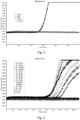
- RT-QuIC The development of RT-QuIC was undertaken using frontal cortex BH from patients with a clinico-pathological diagnosis of DLB, Alzheimer's disease (AD) and sCJD. Patients with no neuropathological evidence of neurological disease who died suddenly and were part of the MRC Sudden Death brain bank were used as controls (SD) ( Fig 2 ).
- SD Sudden Death brain bank
- the RT-QuIC reactions seeded with BH from DLB had a lag-phase of 50 hours, after which an increasing thioflavine T fluorescent signal was seen that became maximal at 70 hours. None of the reactions seeded with BH from patients with other protein misfolding disorders (AD or sCJD) or the SD controls, gave a positive response even after 120 hours.
- a positive RT-QuIC response was classified as a relative fluorescence unit (rfu) value of >2SD above the mean of the negative controls at 120 hours of at least one of the CSF duplicates.
- Exploratory Patient Group (n) Number of Positive RT-QuIC (%) using 5 ⁇ l Number of Positive RT-QuIC (%) using 10 ⁇ l Number of Positive RT-QuIC (%) using 15 ⁇ l AD with incidental LB (13) 2 (15%) 4 (31%) 2 (15%) Healthy Controls (20) 0 (0%) 0 (0%) 0 (0%) 0 (0%) 0 (0%) Mixed DLB/AD (17) 9 (53%) 11 (65%) 11 (65%) Parkinson's disease (2) 2 (100%) 2 (100%) 2 (100%) Progressive Supranuclear 0 (0%) 0 (0%) 0 (0%) 0 (0%) Corticobasal degeneration (3) 0 (0%) 0 (0%) 0 (0%) 0 (0%) 0 (0%) Pure AD (30) 2 (7%) 1 (3%) 0 (0%) 0 (0%) 0 (0%) Pure
- the second phase of the study was to apply these analytical conditions and cut-off criteria to a set of confirmatory in vivo CSF samples from 20 patients with clinically diagnosed PD, 15 control patients and 3 patients with REM sleep behaviour disorder (RBD) recognised to be at high risk of developing future alpha-synucleinopathies, 20 obtained from the large prospective, OPDC Discovery cohort 20 .
- CSF samples were coded, analysed and reported without prior knowledge of the final diagnosis. After the samples were de-coded the results showed that 19 of the 20 PD patients had a positive RT-QuIC response ( Fig 9 ) and none of the 15 controls were found to be positive. This resulted in a RT-QuIC sensitivity and specificity for PD of 95% and 100% respectively (Table 2).
- all three patients at-risk of developing PD had a positive RT-QuIC response.
- These patients had RBD, of whom 80% have been shown to progress to develop a Lewy body disorder.
- RT-QuIC offers a new approach to the detection of abnormal a-syn and one which has the potential to improve the early clinical diagnosis of PD and DLB in addition to other alpha-synucleinopathies such as MSA.
- RT-QuIC reactions with the addition of 37mg of 0.1mm, 0.5mm or 2.3 mm beads were seeded with 5 ⁇ L of 1:200,000 dilution of identical BH from control subjects (SDBH), Lewy body disease patients (LBDBH), Alzheimer's Disease (ADBH) or sporadic Creutzfeldt-Jakob disease (sCJDBH) or unseeded.
- SDBH control subjects
- LDBH Lewy body disease patients
- ADBH Alzheimer's Disease
- sCJDBH sporadic Creutzfeldt-Jakob disease
- RT-QuIC reactions with the addition of 37mg of 0.1mm, 0.5mm or 2.3mm beads were seeded with 15 ⁇ L CSF samples from patients with Lewy Body disease (LB) or from control subjects.
- LB Lewy Body disease
- Fig 16A The a-syn RT-QuIC responses obtained with 0.1mm zirconium/silica beads are shown in Fig 16A
- those with 0.5mm zirconium/silica beads are shown in Fig 16B
- those with 2.3mm zirconium/silica beads are shown in Fig 16C .
- RT-QuIC reactions with the addition of 37mg of 0.5mm steel or glass beads were seeded with 5 ⁇ L of 1:200,000 dilution of identical BH from control subjects (SDBH), Lewy body disease patients (LBDBH) and Alzheimer's Disease (ADBH) and compared to those reactions left unseeded.
- SDBH control subjects
- LDBH Lewy body disease patients
- ADBH Alzheimer's Disease
- 0.5mm steel beads does not support a-syn RT-QuIC reactions seeded with LBDBH.
- use of 0.5mm glass beads does result in positive a-syn RT-QuIC reactions seeded with LBDBH, however the unseeded and ADBH seeded reactions show a gradual increase in fluorescence resulting in a lack of a steady baseline.
- RT-QuIC reactions with the addition of 37mg of 0.5mm steel or glass beads were seeded with 15 ⁇ L CSF samples from patients with Lewy Body disease (LB) or from control subjects.
- the a-syn RT-QuIC reactions seeded with CSF using 0.5m steel beads are shown in Fig 18A and those for glass beads are shown in Fig 18B .
- the use of steel beads inhibits the ability of CSF samples from LB patients to seed the a-syn RT-QuIC, whilst the use of glass beads results in non-specific increase in fluorescence and also does not support the a-syn RT-QuIC reaction.
- PD Parkinson's Disease
- ⁇ -syn alpha-synuclein
- RT-QuIC Real-time quaking induced conversion
- the method of the invention is effective at determining whether a blood sample comprises aggregates of a-syn, and therefore to potentially diagnose whether the subject from whom the blood sample originated has an alpha-synucleinopathy such as Parkinson's Disease or Dementia with Lewy Bodies.
Landscapes
- Health & Medical Sciences (AREA)
- Life Sciences & Earth Sciences (AREA)
- Engineering & Computer Science (AREA)
- Immunology (AREA)
- Chemical & Material Sciences (AREA)
- Biomedical Technology (AREA)
- Hematology (AREA)
- Urology & Nephrology (AREA)
- Molecular Biology (AREA)
- Physics & Mathematics (AREA)
- Analytical Chemistry (AREA)
- Biochemistry (AREA)
- General Health & Medical Sciences (AREA)
- General Physics & Mathematics (AREA)
- Pathology (AREA)
- Microbiology (AREA)
- Food Science & Technology (AREA)
- Biotechnology (AREA)
- Medicinal Chemistry (AREA)
- Cell Biology (AREA)
- Chemical Kinetics & Catalysis (AREA)
- Optics & Photonics (AREA)
- Nuclear Medicine, Radiotherapy & Molecular Imaging (AREA)
- Neurology (AREA)
- Neurosurgery (AREA)
- Proteomics, Peptides & Aminoacids (AREA)
- Investigating Or Analysing Biological Materials (AREA)
- Investigating Or Analysing Materials By The Use Of Chemical Reactions (AREA)
- Investigating, Analyzing Materials By Fluorescence Or Luminescence (AREA)
- Peptides Or Proteins (AREA)
- Measuring Or Testing Involving Enzymes Or Micro-Organisms (AREA)
Abstract
Description
- The invention relates to an assay for the detection of aggregated proteins in a biological sample, more specifically to an assay for the detection of aggregated alpha-synuclein in a biological sample.
- Alpha-synuclein (a-syn) is a well conserved, small acidic protein of 140-amino acids and a molecular weight of 19kDa that is encoded by the SNCA gene located on chromosome 4.1 It is located in high concentration in the presynaptic nerve terminals within the central nervous system where it plays a role in synaptic vesicle biology.2 The synucleinopathies are a set of neurodegenerative disorders associated with the deposition of fibrillary aggregates of a-syn within selective populations of neurones and glia. These deposits can be found within neuronal soma as Lewy bodies (LB) or in dystrophic neurites in diseases such as Parkinson's disease (PD) or Dementia with Lewy bodies (DLB); or in glial cytoplasmic inclusions in multiple system atrophy (MSA).3
- The presence of a-syn has been detected in biological fluids such as cerebrospinal fluid (CSF)4 and serum. 5 The measurement of a-syn concentrations in CSF by ELISAs has been proposed as a biomarker for a-syn related disorders. However, despite many studies showing a reduction in CSF a-syn levels in PD and in DLB4,6, overall results are not consistent.7 Even in those studies where a reduction in CSF a-syn has been demonstrated, the differences are small8 and there is considerable overlap within patient groups and between patient and control groups. In addition the standardisation of CSF a-syn measurement between laboratories has proven difficult.9
- The aggregation properties of a-syn have recently been compared to prion protein, the aggregation of which causes transmissible spongiform encephalopathies (TSEs).10 Indeed, there is much discussion in current literature as to whether the alpha-synucleinopathies are actually prion-like diseases.11 A recently-described technique called real-time quaking induced conversion (RT-QuIC), which exploits the ability of prion protein to induce self-aggregation, has been used to develop a diagnostic CSF test for sporadic Creutzfeldt-Jakob disease (sCJD), the most common human form of TSE.12
- However, given the significant differences between prion proteins and a-syn, these techniques have failed to detect in any reliable way the presence of a-syn or a-syn aggregation in a biological sample. Therefore, there remains a need for a reliable assay to detect the presence of a-syn and a-syn aggregation in a biological sample.
- At least one aim of the present invention is to provide an improved assay to detect the presence of a-syn and/or the aggregation of a-syn in a biological sample.
- According to a first aspect of the invention there is provided a method of detecting the presence of alpha-synuclein aggregation in a biological sample, the method comprising the steps:
- (i) providing a biological sample;
- (ii) providing a reaction sample comprising a population of beads, a fluorophore adapted to bind to protein aggregates and to increase fluorescence when bound to protein aggregates, and alpha-synuclein or a fragment or variant thereof;
- (iii) combining the biological sample and the reaction sample to form a reaction mixture;
- (iv) incubating the reaction mixture with intermittent agitation cycles;
- (v) illuminating the sample with a wavelength of light that excites the fluorophore of the reaction sample; and
- (vi) determining the level of fluorescence of the reaction mixture during incubation, wherein steps (iv) to (vi) are carried out at the same time, and a significant increase in the fluorescence of the reaction mixture during steps (iv) to (vi) is indicative of the presence of aggregates of alpha-synuclein in the reaction mixture, and wherein the presence of aggregates of alpha-synuclein in the reaction mixture is indicative of the presence of aggregates of alpha-synuclein in the biological sample.
- The person skilled in the art will understand that the terms "alpha-synuclein", "a-syn", "α-synuclein", and α-syn" refer to the alpha-synuclein protein and are used interchangeably throughout. For example, in embodiments where the biological sample is obtained from a human subject, the alpha-synuclein is human alpha-synuclein having the sequence SEQ ID NO.1:

- The term "fragment" with reference to alpha-synuclein is intended to mean either a polynucleotide of at least 10, 20, 30, 50, 100, 120, 130, or 140 contiguous nucleotides of the sequence of alpha-synuclein, or any integer there between; preferred fragments are those which are capable of aggregation under the conditions of the method of the invention. For example, the fragment may comprise the amino acid sequence 1-60 or 61-140 of the native alpha-synuclein sequence.
- By "variant" with reference to alpha-synuclein is meant an alpha-synuclein protein with at least one amino acid substitution, insertion, and/or deletion in the peptide or polypeptide sequence or an alteration to a moiety chemically linked to the protein.
- Conservative substitution: One or more amino acid substitutions (for example of 1, 2, 5 or 10 residues) for amino acid residues having similar biochemical properties. Typically, conservative substitutions have little to no impact on the activity of a resulting peptide or polypeptide. For example, a conservative substitution in alpha-synuclein may be an amino acid substitution that does not substantially affect the ability of the peptide to aggregate in the reaction conditions of the method of the invention. In a particular example, a conservative substitution in alpha-synuclein, is an amino acid substitution that does not significantly alter the ability of the protein to aggregate under the reaction conditions of the method of the invention. Screening of variants of alpha-synuclein can be used to identify which amino acid residues can tolerate an amino acid substitution.
- In one example, the alpha-synuclein may be human alpha-synuclein and one conservative substitution may be included in the peptide, such as a conservative substitution in SEQ ID NO:1. In another example, 10 or fewer conservative substitutions are included in the peptide, such as five or fewer. A peptide or protein of the invention may therefore include 1, 2, 3, 4, 5, 6, 7, 8, 9, 10 or more conservative substitutions. A polypeptide can be produced to contain one or more conservative substitutions by manipulating the nucleotide sequence that encodes that polypeptide using, for example, standard procedures such as site-directed mutagenesis or PCR. Alternatively, a polypeptide can be produced to contain one or more conservative substitutions by using peptide synthesis methods, for example as known in the art.
- Substitutional variants are those in which at least one residue in the amino acid sequence has been removed and a different residue inserted in its place. Examples of amino acids which may be substituted for an original amino acid in a protein and which are regarded as conservative substitutions include: Ser for Ala; Lys for Arg; Gln or His for Asn; Glu for Asp; Asn for Gln; Asp for Glu; Pro for Gly; Asn or Gln for His; Leu or Val for Ile; lie or Val for Leu; Arg or Gln for Lys; Leu or lie for Met; Met, Leu or Tyr for Phe; Thr for Ser; Ser for Thr; Tyr for Trp; Trp or Phe for Tyr; and lie or Leu for Val.
- In one embodiment, the substitutions are among Ala, Val, Leu and Ile; among Ser and Thr; among Asp and Glu; among Asn and Gln; among Lys and Arg; and/or among Phe and Tyr.
- Further information about conservative substitutions can be found in, among other locations, Ben-Bassat et al., (J. Bacteriol. 169:751-7, 1987), O'Regan et al., (Gene 77:237-51, 1989), Sahin-Toth et al., (Protein Sci. 3:240-7, 1994), Hochuli et al., (Bio/Technology 6:1321-5, 1988),
WO 00/67796 (Curd et al. - Normally alpha-synuclein forms a stably folded tetramer that resists aggregation. However, it has been suggested that mutations in alpha-synuclein promote aggregation, and that these aggregates of alpha-synuclein are a primary building block of the fibrils that are believed to be involved in alpha-synucleinopathies such as Dementia with Lewy bodies and Parkinson's disease.
- Therefore, the detection or determination of whether a biological sample comprises aggregates of alpha-synuclein may allow the detection of such pathologies without requiring invasive or expensive techniques, and may allow the detection of such pathologies before they would be possible using standard methods.
- Assays such as real-time quaking induced conversion (RT-QuIC) have not to date been able to detect the presence of aggregates of alpha-synuclein. The inventors have surprisingly found that the modified RT-QuIC method of the invention allows the detection of alpha-synuclein at very high sensitivity and specificity, and allows alpha-synuclein aggregate related pathologies to be differentiated from other protein aggregate related pathologies.
- By "significant increase in fluorescence" we refer to an increase in fluorescence of at least the mean relative fluorescence plus two times the standard deviation of the mean relative fluorescence of the negative controls or baseline fluorescence at a suitable time point, typically within 90-120 hours.
- Typically, the biological sample is a bodily fluid sample. However, the biological sample may be a cell-based tissue sample. The biological sample may be taken from a subject for analysis using the method of the invention, to allow a health care practitioner to determine whether the subject may have a disease that is associated with the presence of aggregates of alpha-synuclein.
- The bodily fluid may be selected from the group including cerebrospinal fluid, blood, or blood fractions, nasal fluid or tissue, urine, faeces, and lymph. Preferably, the bodily fluid is selected from the group including cerebrospinal fluid, blood plasma or other fraction, and nasal tissue or fluid. For example, the bodily fluid may be cerebrospinal fluid. In another example, the bodily fluid may be nasal tissue or fluid that has been extracted using a nasal swab, for example. In a further example, the bodily fluid may be a blood sample or blood fraction.
- Accordingly, the method of the invention may allow the detection of a disease associated with aggregation of alpha-synuclein from a biological sample taken from a subject, allowing a non-invasive method of diagnosis or prognosis.
- The biological sample is typically taken from a mammalian subject, preferably from a human subject. However, the biological sample may be taken from piscine, avian, reptilian or amphibian subject.
- The reaction sample typically comprises a solvent. The solvent may be an aqueous solvent such that the reaction sample is an aqueous solution.
- The reaction sample may be a buffered reaction sample to substantially maintain the pH of the reaction sample. For example, the reaction sample may comprise a biologically acceptable buffer such as tris(hydroxymethyl)aminomethane (TRIS), phosphate buffered saline (PBS), 4-(2-hydroxyethyl)-1-piperazineethanesulfonic acid (HEPES), Piperazine-N,N'-bis(2-ethanesulfonic acid) (PIPES), or Sorensen's phosphate buffer, for example.
- Typically, the reaction sample is buffered to maintain the pH of the reaction sample from about pH 6 to about pH 8.5. Preferably, the reaction sample is buffered to maintain the pH of the reaction sample from about 7.0 to about 8.5. For example, the reaction sample may be buffered to maintain the pH of the sample at pH 7.0, 7.2, 7.4, 7.6, 7.8, 8.0, 8.2 or 8.4.
- Preferably, the reaction sample is buffered to maintain a pH of less than a pH of 9.0.
- Preferably, the reaction sample is buffered to maintain a pH of the reaction sample at pH 8.0, 8.2 or 8.4.
- The fluorophore of the reaction sample typically changes fluorescence when bound to aggregates of protein. For example, the fluorophore may fluoresce weakly when free in solution and may fluoresce strongly when bound to protein aggregates. Alternatively, the fluorophore may not fluoresce when the fluorophore is free in solution and may fluoresce when the fluorophore is bound to protein aggregates.
- The protein aggregates may comprise significant beta-sheet content, and the non-aggregated protein may lack significant beta-sheet content, and the fluorophore may be configured to bind to beta-sheets of protein.
- The emission spectra of the fluorophore may shift when the fluorophore is bound to the aggregated protein. For example, the emission spectra of the fluorophore may be red-shifted when the fluorophore binds to the protein aggregates. Alternatively, the emission spectra of the fluorophore may be blue-shifted when the fluorophore binds to the protein aggregates.
- Typically, the fluorophore is a thioflavin. Preferably, the fluorophore is thioflavin T (ThT).
- Alternatively, the fluorophore may be cyanine T-284, or another fluorophore that increases fluorescence when bound to protein aggregates such as alpha-synuclein aggregates.
- In standard RT-QuIC a small quantity of the protein that is to be aggregated is included in the reaction sample to act as a substrate for protein aggregation. Accordingly, the reaction sample may comprise less than 1mg/mL alpha-synuclein to act as an aggregation substrate. The reaction sample may comprise less than 0.5mg/mL alpha-synuclein. The reaction sample may comprise less than 0.1mg/mL alpha synuclein.
- The reaction sample may comprise from approximately 0.01mg/mL alpha-synuclein to about 10mg/mL alpha-synuclein to act as an aggregation substrate. The reaction sample may comprise from approximately 0.1 mg/mL to about 1mg/mL alpha-synuclein to act as an aggregation substrate.
- The alpha-synuclein of the reaction sample may be recombinant alpha-synuclein, from human or other species. The alpha-synuclein of the reaction sample may be a fragment of full length alpha-synuclein. For example, the fragment may comprise amino acids 1-60 or 61 to 140 of full length alpha-synuclein.
- The beads of the population of beads of the reaction sample may comprise zirconia, silica, glass, quartz, or a polymer such as polystyrene, polytetrafluoroethylene (PTFE), polymethylmethacrylate (PMMA), or combinations thereof.
- Typically, the population of beads have a mean diameter of the beads from approximately 1 mm to approximately 0.001mm.
- The mean diameter of the population of beads may be from approximately 1mm to approximately 0.1mm. For example, the mean diameter of the beads may be 1mm, 0.5mm or 0.1mm.
- The population of beads may have a standard distribution of diameters of approximately plus or minus 10% from the mean diameter. Accordingly, the diameter of the beads may be substantially homogeneous.
- The reaction sample may comprise from approximately 1mg to approximately 150mg of beads per 100µL of reaction mixture. The reaction sample may comprise from approximately 10mg to approximately 100mg of beads per 100µL of reaction mixture. The reaction mixture may comprise approximately 10, 20, 30, 40 or 50mg of beads per 100µL of reaction mixture, or an amount there between. For example, the reaction mixture may comprise from about 10 to 50mg of beads per 100µL of reaction mixture, from 20 to 50mg of beads per 100µL of reaction mixture, or 30 to 50mg of beads per 100µL of reaction mixture.
- The presence of aggregates of alpha-synuclein in the biological sample is indicative of a disease associated with the abnormal aggregation of alpha-synucein. For example, the disease may be Dementia with Lewy Bodies or Parkinson's disease, or other alpha-synucleinopathies.
- The reaction mixture may be incubated for more than 40 hours, more than 60 hours, more than 80 hours, or more than 120 hours. The reaction mixture may be agitated for a period and allowed to stand for a period. Typically, the period within which the reaction mixture is agitated is significantly shorter than the period within which the reaction mixture is allowed to stand without agitation. The reaction mixture may be agitated for a period of at least 1 minute, at least 2 minutes, or at least 5 minutes. The reaction mixture may be allowed to stand for a period of at least 10 minutes, at least 15 minutes, or at least 20 minutes. For example, the reaction mixture may be agitated for a period of one minute, and allowed to stand without agitation for a period of fourteen minutes.
- The intermittent agitation cycles may comprise intermittent shaking cycles. The shaking cycles may move the reaction mixture in a circular or orbital motion and therefore, the rate of shaking may be defined in terms of rotations per minute, for example. Alternatively, the shaking cycles may move the reaction mixture back and forth along a single axis, and therefore, the rate of shaking may be defined in terms of repetitions per minute.
- The reaction mixture may be subjected to agitation at a fixed rate. In the following, the rate of agitation is expressed in rotations per minute, but can equally be applied to repetitions per minute for embodiments where the shaking follows a linear motion. The reaction mixture may be subjected to agitation at at least 100 rotations per minute. The reaction mixture may be subjected to agitation at at least 150 rotations per minute. The reaction mixture may be subjected to agitation at at least 200 rotations per minute. The reaction mixture may be subjected to agitation from at least 100 rotations per minute to at least 300 rotations per minute. The reaction mixture may be subjected to agitation from at least 150 rotations per minute to at least 250 rotations per minute. For example, the reaction mixture may be subjected to agitation at 150, 200 or 250 rotations per minute. Preferably, the reaction mixture is subjected to agitation at 200 rotations per minute.
- The required rate of agitation of the reaction mixture may vary depending on the shaking orbital of the shaker. The required rate of agitation of the reaction mixture may vary depending on the biological sample used in the method. For example, in embodiments where the biological sample is a blood fraction such as platelets in plasma, for example, the reaction mixture may be agitated at a rate of 400, 600, 800, 900 or 1000 rotations per minute.
- The intermittent agitation cycles may comprise intermittent sonication of the reaction mixture. Sonication of the reaction mixture may agitate the reaction mixture to promote aggregation. Alternative methods of agitating the reaction mixture may also be used.
- The method of the invention may be carried out at a temperature of from 25°C to 45°C. The method of the invention may be carried out at a temperature of from 30°C to 40°C. For example, the method may be carried out at a temperature of 30°C, 32°C, 34°C, 36°C, 38°C or 40°C, and values between.
- In one embodiment, the method of the invention may be carried out at a temperature of about 30°C. In an alternative embodiment, the method of the invention may be carried out at a temperature of about 35°C, or about 37°C.
- The biological sample may be treated prior to the method of the invention to concentrate the alpha-synuclein in the biological sample. The biological sample may be treated prior to the method of the invention to extract alpha-synuclein from a complex biological sample.
- The biological sample may be treated with a population of beads. The beads may comprise a magnetic material. For example, the beads may comprise a paramagnetic material and the beads may allow the separation of material that specifically binds to the surface of the beads using externally applied magnetic fields to extract the beads from a complex mixture. The magnetic beads may allow the extraction or concentration of alpha-synuclein in a biological sample prior to the method of the invention.
- For example, the beads may comprise superparamagnetic iron oxide, or another paramagnetic material and the alpha-synuclein may bind to the beads and the application of a magnetic field may allow the beads, and therefore the alpha-synuclein to be separated or concentrated in a biological sample.
- In a second aspect of the invention, there is provided a method of diagnosing alpha-synucleinopathies, the method comprising the steps:
- (i) providing a biological sample from a subject;
- (ii) providing a reaction sample comprising a population of beads, a fluorophore adapted to bind to protein aggregates and to increase fluorescence when bound to protein aggregates, and alpha-synuclein or a fragment or variant thereof;
- (iii) combining the biological sample and the reaction sample to form a reaction mixture;
- (iv) incubating the reaction mixture with intermittent agitation cycles;
- (v) illuminating the sample with a wavelength of light that excites the fluorophore of the reaction sample; and
- (vi) determining the level of fluorescence of the reaction mixture during incubation, wherein steps (iv) to (vi) are carried out at the same time, and a significant increase in the fluorescence of the reaction mixture during steps (iv) to (vi) is indicative of the subject having an alpha-synucleinopathy.
- The alpha-synucleinopathy is typically, Parkinson's or Dementia with Lewy Bodies.
- The invention extends in a third aspect to the provision of a kit of parts comprising an aqueous buffered solution, the aqueous buffered solution comprising a population of beads, a fluorophore and alpha-synuclein or a fragment or variant thereof.
- The kit of parts may be used as the reaction sample for the methods of the first and second aspects.
- Preferred and optional features for the first aspect of the invention are preferred and optional features for the second and third aspects.
- Embodiments of the present invention will now be described, by way of non-limiting example, with reference to the accompanying drawings.
-
Figure 1 : A schematic diagram showing the process of standard RT-QuIC; -
Figure 2 : RT-QuIC traces for brain homogenate (BH) samples from patients with Dementia with Lewy Bodies (DLB), Alzheimer's disease (AD) and sporadic Creutzfeldt-Jakob disease (sCJD) with patients with no neuropathological evidence of disease who died suddenly (SD) as controls; -
Figure 3 : RT-QuIC traces for BH samples from patients with mixed pathologies; -
Figure 4 : RT-QuIC traces for cerebrospinal fluid (CSF) samples from confirmed DLB and sCJD patients; -
Figure 5 : RT-QuIC traces for control CSF samples and one BH sample from a patient with DLB; -
Figure 6 : RT-QuIC traces for CSF samples from patients with AD and one BH sample from a patient with DLB; -
Figure 7 : RT-QuIC traces for CSF samples from patients with DLB and one BH sample from a patient with DLB; -
Figure 8 : RT-QuIC traces for CSF samples from patients with DLB and AD and one BH sample from a patient with DLB; -
Figure 9 : RT-QuIC traces for CSF samples from patients with Parkinson's Disease (PD) and CSF samples from control subjects; -
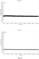
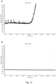
Figure 10 : RT-QuIC traces for SD BH samples A) with beads and B) without beads; -
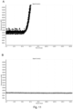
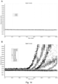
Figure 11 : RT-QuIC traces for DLB BH samples A) with beads and B) without beads; -
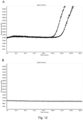
Figure 12 : RT-QuIC traces for Parkinson's Disease cerebrospinal fluid (CSF) samples A) with beads and B) without beads; -
Figure 13 : RT-QuIC traces for Dementia with Lewy Bodies CSF samples A) with beads and B) without beads; -
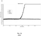
Figure 14 : RT-QuIC traces for Dementia with Lewy Bodies BH samples with A) no beads B) 18.7mg beads per well and C) 37.5mg beads per well; -
Figure 15 : RT-QuIC traces for BH from control subjects (SDBH), Lewy body disease patients (LBDBH), Alzheimer's Disease (ADBH) or sporadic Creutzfeldt-Jakob disease (sCJDBH) or unseeded for (A) 0.1mm zirconium/silica beads, (B) 0.5mm zirconium/silica beads and (C) 2.3mm zirconium/silica beads; -
Figure 16 : RT-QuIC traces for CSF samples from patients with Lewy Body disease (LB) or from control subjects with the addition of 37mg of (A) 0.1mm zirconium/silica beads, (B) 0.5mm zirconium/silica beads and (C) 2.3mm zirconium/silica beads; -
Figure 17 : RT-QuIC traces for BH samples from control subjects (SDBH), Lewy body disease patients (LBDBH) and Alzheimer's Disease (ADBH) and compared to those reactions left unseeded using (A) 0.5mm steel beads or (B) 0.5mm glass beads; -
Figure 18 : RT-QuIC traces for CSF samples from control subjects, and Lewy body disease patients (LBDBH) and compared to those reactions left unseeded using (A) 0.5mm steel beads or (B) 0.5mm glass beads; and -
Figure 19 : RT-QuIC traces for platelet samples isolated form EDTA anti-coagulated blood samples from PD and control subjects. - While the making and using of various embodiments of the present invention are discussed in detail below, it should be appreciated that the present invention provides many applicable inventive concepts that can be embodied in a wide variety of specific contexts. The specific embodiments discussed herein are merely illustrative of specific ways to make and use the invention and do not delimit the scope of the invention.
- To facilitate the understanding of this invention, a number of terms are defined below. Terms defined herein have meanings as commonly understood by a person of ordinary skill in the areas relevant to the present invention. Terms such as "a", "an" and "the" are not intended to refer to only a singular entity, but include the general class of which a specific example may be used for illustration. The terminology herein is used to describe specific embodiments of the invention, but their usage does not delimit the invention, except as outlined in the claims.
- The RT-QuIC reaction buffer (RB) was composed of 100mM phosphate buffer (pH 8.2), 10µM Thioflavin T (ThT) and 0.1mg/mL human recombinant full-length (1-140aa) alpha-synuclein (Stratech, Cambridge, UK). Each well of a black 96-well plate with a clear bottom (Nalgene Nunc International, Fisher Scientific Ltd, UK) contained 98µL, 90µL or 85µL RB (depending on volume of seed added) and 37 ± 3mg of 0.5mm zirconium/silica beads (Thistle Scientific Ltd, Glasgow, UK). Reactions were seeded with 2µL of working strength brain homogenate (BH), 5 µl, 10µl or 15µl of undiluted CSF to a final reaction volume of 100µl. The plates were sealed with a plate sealer film (Fisher Scientific Ltd, UK) and incubated in a BMG OPTIMA FluoSTAR plate reader at 30°C for 120h with intermittent shaking cycles: double orbital with 1 minute shake (200rpm), 14 minute rest. ThT fluorescence measurements (450nm excitation and 480nm emission) were taken every 15 minutes. Each sample was run in duplicate, allowing 2 negative control samples (reactions seeded with SD and AD BH), 1 positive control (reaction seeded with DLB BH), an unseeded reaction and 44 CSF samples to be tested on one plate.
- Initial phase of CSF RT-QuIC development was carried out on 99 CSF samples obtained from the OPTIMA cohort (Oxford Project to Investigate Memory and Ageing) with clinically and neuropathologically confirmed diagnosis of pure DLB (n=12), PD (n=2), progressive supranuclear palsy (PSP) (n=2), corticobasal degeneration (n=3), DLB with AD pathology (n=17), AD with incidental LBs (n=13), pure AD (n=30) and controls (n=20). OPTIMA initiated in 1988, is a prospective, longitudinal clinico-pathological study of dementia and aging including CSF collection at multiple time points during clinical follow-up. All clinical and pathological protocols have been described in detail13 and were approved by the local ethics committee and participants provided informed consent prior to enrolment.
- The validation phase of RT-QuIC was carried out on CSF samples (20 PD, 15 controls and 3 at-risk) obtained from the Oxford Discovery cohort, which is one of the largest, clinically best-characterized longitudinal PD cohorts to date. Full clinical details of this cohort have been described previously.14 In brief, patients with idiopathic PD diagnosed within 3.5 years according to UK PD Society Brain Bank diagnostic criteria15 were recruited between September 2010 and September 2014 from a 2.9 million population (
ethics study 10/H0505/71). Mean disease duration among 20 PD patients was 1.6 ± 1.1 years (range 0.1 - 3.2 years) and Hoehn and Yahr stage 1.9 ± 0.4 (range 1-3, maximum possible score 5). The control population were recruited from spouses and friends of patients taking part in the study, as well as the general public. The at-risk group comprised patients with REM sleep behaviour confirmed on overnight polysomnography,16 80% of which have shown to develop Lewy body disorder over time.17 Demographic information is given in Table 1.Table 1: Patient demographic information for the Optima and Discovery patients investigated OPTIMA patients (n) Age at death Mean ± SD (range) F/M Pure LBD (12) 80.8 ± 6.5 (71-92) 4/8 Parkinson's disease (2) 77.5 ± 7.8 (72-83) 0/2 Mixed LBD/AD (17) 80.1 ± 6.4 (69-90) 10/7 AD with incidental LB (13) 79.8 ± 7.8 (67-91) 9/4 Pure AD (30) 77.7 ± 8.6 (61-93) 17/13 Progressive supranuclear palsy (PSP) (2) 69.5 ± 3.5 (67-72) 2/0 Corticobasal degeneration (CBD) (3) 64.0 ± 10.6 (52-72) 1/2 Controls (20) 82.9 ± 6.9 (68-93) 10/10 Discovery patients (n) Parkinson's disease (20) 65.1 ± 9.1 (42-80) 6/14 At-risk RBD patients (3) 67.6 ± 7.7 (59-74) 0/3 Controls (15) 65.8 ± 7.4 (55-83) 8/7 - Brain tissue was provided the MRC Brain Bank in the NCJDRSU (ethical licence 11/ES/0022). All tissue was frozen at -80°C within 2 hours of being sampled and stored at -80°C prior to analysis. Brain tissues had been stored between 2-18 years prior to use.
- Frontal cortex tissue was taken from patients with Alzheimer's disease (AD); sporadic Creutzfeldt-Jakob disease (sCJD); and Diffuse Lewy body dementia (DLB). In addition frontal cortex was obtained from individuals without neurodegenerative disease from the MRC Sudden Death Brain and Tissue Bank (Sudden Death (SD) controls). Both frontal cortex and substantia nigra tissue was obtained from patients with mixed AD/DLB; mixed sCJD/DLB and mixed AD/PD. All cases used had been examined histologically and the diagnosis reached using internationally accepted criteria.18 Initial 10% w/v brain homogenates (BH) were prepared using phosphate buffered saline (PBS) containing 1mM EDTA, 150mM NaCl, 0.5% Triton X and complete protease inhibitor cocktail from Roche. Subsequent working strength BHs were prepared by diluting the above 1:20,000 with PBS.
- CSF samples were stored in 0.5mL aliquots in polypropylene tubes at -80°C prior to analysis. 99 CSF samples from the OPTIMA cohort and 38 CSF samples from the Oxford Parkinson's Disease Centre (OPDC) Discovery study were received from the Nuffield Department of Clinical Neurosciences, University of Oxford. All CSF samples were transported from Oxford to Edinburgh on dry-ice and stored at -80°C on arrival. In addition, CSF samples from patients with neuropathologically confirmed sCJD or DLB from the NCJDRSU CSF Bank were analysed. Ethical approval for the use of CSF samples from the NCJDRSU CSF Bank was covered by Multi-centre Research Ethics Committee for Scotland 05/MRE00/67. All CSF were spun and stored at -80°C prior to analysis.
- The development of RT-QuIC was undertaken using frontal cortex BH from patients with a clinico-pathological diagnosis of DLB, Alzheimer's disease (AD) and sCJD. Patients with no neuropathological evidence of neurological disease who died suddenly and were part of the MRC Sudden Death brain bank were used as controls (SD) (
Fig 2 ). The RT-QuIC reactions seeded with BH from DLB had a lag-phase of 50 hours, after which an increasing thioflavine T fluorescent signal was seen that became maximal at 70 hours. None of the reactions seeded with BH from patients with other protein misfolding disorders (AD or sCJD) or the SD controls, gave a positive response even after 120 hours. - Many disease pathologies commonly co-exist, especially AD-related and a-syn pathology19. To investigate whether the presence of an alternative protein-misfolding disorder can interfere with the a-syn aggregation induced by either DLB or PD, BHs from the frontal cortex of patients with mixed pathologies were examined (
Fig 3 ). The presence of a second protein-misfolding disorders such as AD or sCJD does not inhibit the RT-QuIC reaction induced by DLB or PD BHs (Figs 2 and 3 ). To investigate whether the RT-QuIC method developed was sensitive enough to detect a-syn in CSF, two CSF samples from patients with neuropathologically confirmed DLB and 1 CSF from a neuropathologically confirmed case of sCJD were analysed (Fig 4 ). Both CSF samples from the DLB patients gave positive responses with a lag-phase between 60-100 hours. - An exploratory set of 99 in vivo CSF samples obtained as part of the OPTIMA study from patients with subsequent neuropathologically confirmed disease were analysed at three different volumes (i.e. 5, 10 and 15µl) to investigate the sensitivity and specificity of the RT-QuIC and to calculate the optimal CSF volume for the analysis (Table 2). Using a volume of 15µl a sensitivity of 92% was obtained for CSF samples from DLB (
Figs 7 and 8 ) and a sensitivity of 65% was obtained for CSF samples from patients with mixed DLB/AD pathology. None of the CSF samples from the control subjects (Fig 5 ) or patients with pure AD (Fig 6 ), CBD or PSP were positive. Using this exploratory set of CSF samples a positive response was defined as a relative fluorescence unit (rfu) value of >2SD above the mean of the negative controls at 120 hours of at least one of the CSF duplicates.Table 2. Positive RT-QuIC reactions seeded with CSF samples from patients with neuropathologically confirmed DLB, mixed DLB/AD, AD with incidental LB, AD, PD and healthy controls (Exploratory Group) and patients with clinically diagnosed PD, at risk- PD, neuropathologically confirmed corticobasal degeneration and supranuclear palsy and PD controls (Confirmatory Group). A positive RT-QuIC response was classified as a relative fluorescence unit (rfu) value of >2SD above the mean of the negative controls at 120 hours of at least one of the CSF duplicates. Exploratory Patient Group (n) Number of Positive RT-QuIC (%) using 5µl Number of Positive RT-QuIC (%) using 10µl Number of Positive RT-QuIC (%) using 15µl AD with incidental LB (13) 2 (15%) 4 (31%) 2 (15%) Healthy Controls (20) 0 (0%) 0 (0%) 0 (0%) Mixed DLB/AD (17) 9 (53%) 11 (65%) 11 (65%) Parkinson's disease (2) 2 (100%) 2 (100%) 2 (100%) Progressive Supranuclear 0 (0%) 0 (0%) 0 (0%) Corticobasal degeneration (3) 0 (0%) 0 (0%) 0 (0%) Pure AD (30) 2 (7%) 1 (3%) 0 (0%) Pure DLB (12) 10 (83%) 11 (92%) 11 (92%) Sensitivity (DLB) 83% 92% 92% Specificity (vs controls) 100% 100% 100% Specificity (vs AD) 93% 97% 100% Specificity (vs Controls + AD) 96% 98% 100% Confirmatory Patient Group (n) Number of Positive RT-QuiC (%) using Parkinson disease (20) / / 19 (95%) At-risk PD patients (3) / / 3 (100%) Parkinson's disease controls / / 0 (0%) Sensitivity (PD) / / 95% Specificity / / 100% - The second phase of the study was to apply these analytical conditions and cut-off criteria to a set of confirmatory in vivo CSF samples from 20 patients with clinically diagnosed PD, 15 control patients and 3 patients with REM sleep behaviour disorder (RBD) recognised to be at high risk of developing future alpha-synucleinopathies,20 obtained from the large prospective, OPDC Discovery cohort20. These CSF samples were coded, analysed and reported without prior knowledge of the final diagnosis. After the samples were de-coded the results showed that 19 of the 20 PD patients had a positive RT-QuIC response (
Fig 9 ) and none of the 15 controls were found to be positive. This resulted in a RT-QuIC sensitivity and specificity for PD of 95% and 100% respectively (Table 2). Interestingly, all three patients at-risk of developing PD had a positive RT-QuIC response. These patients had RBD, of whom 80% have been shown to progress to develop a Lewy body disorder.20 - The early diagnosis of both DLB and PD is hampered by the lack of sensitive and reliable clinical diagnostic tests. Both conditions are underpinned by the neuropathological deposition of an aggregated form of a-syn which is released into the CSF. We have exploited the ability of the aggregated a-syn to induce further aggregation of non-aggregated a-syn in a cyclical manner to develop a technique that can detect abnormal CSF a-syn in DLB and PD with a sensitivity of 92% and 95% respectively with 100% specificity. Uniquely, we found that 3 RBD patients at high future risk of developing a Lewy body disorder gave a positive RT-QuIC response, suggesting that this test could be used as an early diagnostic test for prodromal PD. Future work focusing on test validation in a larger cohort of RBD patients, followed by ongoing longitudinal assessment, will test the assay's utility in risk-stratifying those prodromal individuals most at risk of early PD conversion in whom neuroprotective therapies might be trialled. We also found that CSF samples from CBD and PSP patients do not give positive RT-QuIC responses. These are movement disorders associated with abnormalities in tau protein rather than a-syn which may be mistaken for PD in the early stages. Therefore, RT-QuIC offers a new approach to the detection of abnormal a-syn and one which has the potential to improve the early clinical diagnosis of PD and DLB in addition to other alpha-synucleinopathies such as MSA.
- The role of beads in a modified RT-QuiC assay for the detection of alpha-synuclein With reference to
Figures 10-13 , to show the role being played by the beads used in the above assays, parallel assays were carried out for Sudden Death (SD) Brain Homogenate (BH) samples A) with beads and B) without beads, Dementia with Lewy Bodies BH samples A) with beads and B) without beads, Parkinson's Disease cerebrospinal fluid (CSF) samples A) with beads and B) without beads, and Dementia with Lewy Bodies CSF samples A) with beads and B) without beads. These assays were carried out using the same general method described above. As can be seen fromFigure 10 , no reading was seen for the SD samples. In addition, those samples with Lewy Body Disease and Parkinson's Disease without beads (Figure 11B ,Figure 12B orFigure 13B ) showed no reading. However, readings are clearly seen for those samples with either Lewy Body Disease or Parkinson's Disease with the beads. Therefore, the presence of the beads is vital to the ability to detect the presence of a-syn aggregates using RT-QuiC. - With reference to
Figure 14 , the effect of bead concentration was investigated using Dementia with Lewy Bodies brain homogenate samples. Samples with no beads present in the reaction mixture showed a constant fluorescent signal, indicated a negative RT-QuiC result. In contrast, samples with 18.7mg beads per well and 37.5mg beads per well gave a clear increase in fluorescence and the higher concentration of beads giving a clearer and earlier signal. - Therefore it is clear the presence of the beads in the reaction mixture is crucial for a signal for the detection of alpha-synuclein aggregates to be obtained.
- RT-QuIC reactions with the addition of 37mg of 0.1mm, 0.5mm or 2.3 mm beads were seeded with 5µL of 1:200,000 dilution of identical BH from control subjects (SDBH), Lewy body disease patients (LBDBH), Alzheimer's Disease (ADBH) or sporadic Creutzfeldt-Jakob disease (sCJDBH) or unseeded. The a-syn RT-QuIC responses obtained with 0.1mm zirconium/silica beads are shown in
Fig 15A , those with 0.5mm zirconium/silica beads are shown inFig 15B and those with 2.3mm zirconium/silica beads are shown inFig 15C . - It can be seen from
Figs 15A,B ,C that increasing the size of the zirconium/silica beads results in an increase in the time taken to illicit a positive a-syn RT-QuIC response with LBDBH seeded reactions. However despite having a quicker response time using 0.1mm zirconium/silica beads, the a-syn RT-QuIC gave positive reactions with ADBH seeded reactions. - RT-QuIC reactions with the addition of 37mg of 0.1mm, 0.5mm or 2.3mm beads were seeded with 15µL CSF samples from patients with Lewy Body disease (LB) or from control subjects.
- The a-syn RT-QuIC responses obtained with 0.1mm zirconium/silica beads are shown in
Fig 16A , those with 0.5mm zirconium/silica beads are shown inFig 16B and those with 2.3mm zirconium/silica beads are shown inFig 16C . - The use of 0.1mm and 0.5mm zirconium/silica beads resulted in positive a-syn RT-QuIC reactions seeded with LB CSF samples but not with control CSF samples. In contrast the use of 2.3mm zirconium/silica beads did not support a-syn RT-QuIC reactions seeded with CSF samples from LB patients. Identical CSF samples were used in the experiments illustrated in
Fig 16A , B and C. - The results from the experiments illustrated in
Fig 15 and16 demonstrate that the use of 2.3mm zirconium/silica does not accelerate the a-syn RT-QuIC reaction seeded by either BH or CSF from LB patients. The results a-syn RT-QuIC reactions using either the 0.1mm or the 0.5mm zirconium/silica beads were similar, although a-syn RT-QuIC reactions using the 0.1mm zirconium/silica beads resulted in ADBH inducing a positive reaction. Handling 0.1mm zirconium/silica beads was difficult as the very small size meant they were more prone to static than the larger 0.5mm zirconium/silica. Therefore all further investigations into the composition of the beads were undertaken beads of 0.5mm in diameter. - RT-QuIC reactions with the addition of 37mg of 0.5mm steel or glass beads were seeded with 5µL of 1:200,000 dilution of identical BH from control subjects (SDBH), Lewy body disease patients (LBDBH) and Alzheimer's Disease (ADBH) and compared to those reactions left unseeded. The a-syn RT-QuIC reactions using 0.5m steel beads are shown in
Fig 17A and those for glass beads are shown inFig 17B . - The use of 0.5mm steel beads does not support a-syn RT-QuIC reactions seeded with LBDBH. In contrast the use of 0.5mm glass beads does result in positive a-syn RT-QuIC reactions seeded with LBDBH, however the unseeded and ADBH seeded reactions show a gradual increase in fluorescence resulting in a lack of a steady baseline.
- RT-QuIC reactions with the addition of 37mg of 0.5mm steel or glass beads were seeded with 15µL CSF samples from patients with Lewy Body disease (LB) or from control subjects. The a-syn RT-QuIC reactions seeded with CSF using 0.5m steel beads are shown in
Fig 18A and those for glass beads are shown inFig 18B . The use of steel beads inhibits the ability of CSF samples from LB patients to seed the a-syn RT-QuIC, whilst the use of glass beads results in non-specific increase in fluorescence and also does not support the a-syn RT-QuIC reaction. - The overall conclusion from the above experiments is that the addition of 37mg/well of 0.5mm zirconium/silica beads is the best promoter of a-syn RT-QuIC reactions seeded with either BH or CSF samples from patients with LBD.
- Investigation of the ability of blood components from Parkinson's Disease (PD) patients to seed the alpha-synuclein (α-syn) Real-time quaking induced conversion (RT-QuIC) Platelets were isolated from EDTA anti-coagulated blood samples from two PD patients and a control subject. By increasing the shaking speed from 200rpm to 600rpm and the temperature of the reaction to 42°C it was possible to seed the a-syn RT-QuIC reaction using 15µL of platelets from these 2 patients with PD (
Fig 19 ). In contrast the platelets from the control subject failed to seed the reaction. Therefore, this demonstrated that the method of the invention is effective at determining whether a blood sample comprises aggregates of a-syn, and therefore to potentially diagnose whether the subject from whom the blood sample originated has an alpha-synucleinopathy such as Parkinson's Disease or Dementia with Lewy Bodies. - Further examples and embodiments are provided in the following numbered clauses:
- 1. A method of detecting the presence of alpha-synuclein aggregation in a biological sample, the method comprising the steps: (i) providing a biological sample; (ii) providing a reaction sample comprising a population of beads, a fluorophore adapted to bind to protein aggregates and to increase fluorescence when bound to protein aggregates, and alpha-synuclein or a fragment or variant thereof; (iii) combining the biological sample and the reaction sample to form a reaction mixture; (iv) incubating the reaction mixture with intermittent agitation cycles; (v) illuminating the sample with a wavelength of light that excites the fluorophore of the reaction sample; and (vi) determining the level of fluorescence of the reaction mixture during incubation, wherein steps (iv) to (vi) are carried out at the same time, and a significant increase in the fluorescence of the reaction mixture during steps (iv) to (vi) is indicative of the presence of aggregates of alpha-synuclein in the reaction mixture, and wherein the presence of aggregates of alpha-synuclein in the reaction mixture is indicative of the presence of aggregates of alpha-synuclein in the biological sample.
- 2. The method according to clause 1, wherein the biological sample is a bodily fluid sample.
- 3. The method according to clause 2, wherein the bodily fluid is selected from the group including cerebrospinal fluid, blood, or blood fractions, nasal fluid or tissue, urine, faeces, and lymph.
- 4. The method according to any one preceding clause, wherein the reaction sample is a buffered reaction sample.
- 5. The method according to clause 4, wherein the reaction sample is buffered to maintain the pH of the reaction sample from about pH 6 to about pH 8.5.
- 6. The method according to any one preceding clause, wherein the protein aggregates comprise significant beta-sheet content.
- 7. The method according to any one preceding clause, wherein the fluorophore is a thioflavin or cyanine T-284.
- 8. The method according to any one preceding clause, wherein the reaction sample comprises from approximately 0.01 mg/mL alpha-synuclein to about 10mg/mL alpha-synuclein to act as an aggregation substrate.
- 9. The method according to any one preceding clause, wherein the alpha-synuclein of the reaction sample is a fragment of full length alpha-synuclein.
- 10. The method according to any one preceding clause, wherein the beads of the population of beads of the reaction sample comprise zirconia, silica, glass, quartz, or a polymer such as polystyrene, polytetrafluoroethylene (PTFE), polymethylmethacrylate (PMMA), or combinations thereof.
- 11. The method according to
clause 10, wherein the beads of the population of beads comprise zirconia, silica or glass. - 12. The method according to any one preceding clause, wherein the population of beads have a mean diameter of the beads from approximately 1mm to approximately 0.001mm.
- 13. The method according to clause 12, wherein the mean diameter of the population of beads is from approximately 1mm to approximately 0.1mm.
- 14. The method according to any one preceding clause, wherein the reaction sample comprises from approximately 1mg to approximately 150mg of beads per 100µL of reaction mixture.
- 15. The method according to clause 14, wherein the reaction sample comprises from approximately 1mg to approximately 50mg of beads per 100µL of reaction mixture.
- 16. The method according to any one preceding clause, wherein the presence of aggregates of alpha-synuclein in the biological sample is indicative of a disease associated with the abnormal aggregation of alpha-synucein such as Dementia with Lewy Bodies or Parkinson's Disease, or other alpha-synucleinopathies.
- 17. The method according to any one preceding clause, wherein the reaction mixture is incubated for more than 40 hours, more than 60 hours, more than 80 hours, or more than 120 hours.
- 18. The method according to any one preceding clause, wherein the method is carried out at a temperature of from 25°C to 45°C.
- 19. The method according to any one preceding clause, wherein the biological sample is treated prior step (i) to concentrate the alpha-synuclein in the biological sample.
- 20. The method according to clause 19, wherein the biological sample is treated with a population of beads.
- 21. The method according to
clause 20, wherein the beads comprise a magnetic material. - 22. A method of diagnosing alpha-synucleinopathies, the method comprising the steps: (i) providing a biological sample from a subject; (ii) providing a reaction sample comprising a population of beads, a fluorophore adapted to bind to protein aggregates and to increase fluorescence when bound to protein aggregates, and alpha-synuclein or a fragment or variant thereof; (iii) combining the biological sample and the reaction sample to form a reaction mixture; (iv) incubating the reaction mixture with intermittent agitation cycles; (v) illuminating the sample with a wavelength of light that excites the fluorophore of the reaction sample; and (vi) determining the level of fluorescence of the reaction mixture during incubation, wherein steps (iv) to (vi) are carried out at the same time, and a significant increase in the fluorescence of the reaction mixture during steps (iv) to (vi) is indicative of the subject having an alpha-synucleinopathy.
- 23. The method according to clause 22, the alpha-synucleinopathy is Parkinson's or Dementia with Lewy Bodies.
- 24. A kit of parts comprising an aqueous buffered solution, the aqueous buffered solution comprising a population of beads, a fluorophore and alpha-synuclein or a fragment or variant thereof.
- 25. The kit of parts according to clause 24, wherein the kit of parts is used as the reaction sample for the methods of any one preceding claim.
-
- 1. Spillantini MG, Divane A, and Goedert M. Assignment of human alpha-synuclein (SNCA) and beta-synuclein (SNCB) geners to chromosome 4q21 and 5q35. Genomics. 1995; 27:379-381.
- 2. Goedert M, Spillantini MG, Del Tredici K, et al. 100 years of Lewy pathology. Nat Rev Neurol. 2013; 9:13-24.
- 3. Martf MJ, Tolsa E, and Campdelacreu J. Clinical Overview of the Synucleinopathies. Movement disorders. 2003; 18:S21-S27.
- 4. Mollenhauer M, Cullen V, Kahn I, et al. Direct quantification of CSF alpha-synuclein by ELISA and first cross-sectional study in patients with neurodegeneration. Exp Neurol. 2008; 213:315-325.
- 5. Williams SM, Schult P, and Sierks MR. Oligomeric a-synuclein an B-amyloid variants as potential biomarkers for Parkinson's and Alzheimer's diseases. Eur J Neurosci. 2015.
- 6. Shi M, Bradner J, and Hancock AM. Cerebrospinal fluid biomarkers for Parkinson's disease diagnosis and progression. Annals of Neurology. 2011; 69:570-580.
- 7. Ohrfelt A, Grognet P, Andreasen N, et al. Cerebrospinal fluid alpha-synuclein in neurodegenerative disorders- a marker of synapse loss? Neuroscience Letts. 2009; 450:332-335.
- 8. Hong Z, Shi M, Chung KA, et al. DJ-1 and alpha-synucelin in human cerebrospinal fluid as biomarkers of Parkinson's disease. Brain. 2010; 133:713-726.
- 9. Kruse N, Persson S, Alcolea D, et al. Validation of a quantitative cerebrospinal fluid alpha-synuclein assay in a European-wide interlaboratory study. Neurobiol aging. 2015; 36:2587-2596.
- 10. Bernis ME, Babila JT, Breid S, et al. Prion-lie propagation of human brain-derived alpha-synuclein in transgenic mice expressing human wild-type alpha-synuclein. Acta Neuropathol Commun. 2015; 3.
- 11. Brandel JP, Corbille AG, Derkinderen P, et al. Is Parkinson's disease a prion disease? Rev Neurol (Paris). 2015; 171:812-824.
- 12. L McGuire, A Peden, C Orru, et al. Prion seeding activity in cerebrospinal fluid from patients with sporadic Creutzfeldt-Jakob disease patients using real-time QuIC analysis: a potential new clinical diagnostic test with high sensitivity and specificity. Annals of Neurology 72 (2), 278-285.
- 13. Clarke R, Smith AD, Jobst KA, et al. Folate, Vitamin B12 and serum total homocysteine levels n confirmed Alzheimer's disease. Archives of Neurology. 1998; 55:1449-1455.
- 14. Szewwczyk-Krolikowski K, Tomlinson P, Nithi K, et al. The influence of age and gender on motor and non-motor features of early parkinson's disease: initial findings form the Oxford Parkinson Disease Center (OPDC) discovery cohort. Parkinsonism Relat Disord. 2014; 20:99-105.
- 15. Hughes AJ, Daniel SE, Kilford L, et al. Accuracy of clinical diagnosis of idiopathic Parkinson's disease - a clinicopathological study of 100 cases. J Neurol Neurosurg Psychiatry. 1992; 55:181-184.
- 16. Rolinski M, Zokaei N, Baig F, et al. Visual short-term memory deficits in REM sleep behaviour disordermirror those in parkinson's disease. Brain. 2016; 139:47-53.
- 17. Iranzo A, Tolosa E, Gelpi E, et al. Neurodegnerative disease status and post-mortem pathology in idiopathic rapid-eye-movement sleep behaviour disorder: an observational cohort study. Lancet. 2013; 12:443-453.
- 18. Thomas J Montine, Creighton H Phelps, Thomas G Beach, et al. National Institute on Aging-Alzheimer's Association guidleines for the neuropathological assessment of Alzheimer's disease: a practical approach. Acta Neuropathol. 2012; 123:1-11.
- 19. Kovacs GG, Alafuzoff I, A-SS, Arzberger T, et al. Mixed brain pathologies in dementia:the BrainNet Europe consortium experience. Dement Geriatr Cogn Disord. 2008; 26:343-350.
- 20. Iranzo A, Tolosa E, Gelpi E, et al. Neurodegnerative disease status and post-mortem pathology in idiopathic rapid-eye-movement sleep behaviour disorder: an observational cohort study. Lancet. 2013; 12:443-453.
Claims (12)
- An in vitro method of detecting the presence of alpha-synuclein aggregation in a biological sample, the method comprising the steps:(i) providing a reaction sample comprising(a) a population of beads comprising zirconia and/or silica, or glass, wherein the population of beads have a mean diameter of less than 2.3 mm,(b) thioflavin T; and(c) alpha-synuclein or a variant thereof having from 1 to 10 amino acid substitutions, insertions, or deletions that do not substantially affect the ability of the peptide to aggregate in the reaction conditions of the method;(ii) combining a biological sample and the reaction sample to form a reaction mixture;(iii) incubating the reaction mixture with intermittent agitation cycles;(iv) illuminating the reaction mixture with a wavelength of light that excites the thioflavin T; and(v) determining the level of fluorescence of the reaction mixture during incubation,wherein steps (iii) to (v) are carried out at the same time, and a significant increase in the fluorescence of the reaction mixture during steps (iii) to (v) is indicative of the presence of aggregates of alpha-synuclein in the reaction mixture, and wherein the presence of aggregates of alpha-synuclein in the reaction mixture is indicative of the presence of aggregates of alpha-synuclein in the biological sample.
- The method according to claim 1, wherein the biological sample is a bodily fluid sample selected from the group consisting of cerebrospinal fluid, blood, blood fractions, nasal fluid, nasal tissue, urine, faeces, and lymph.
- The method according to claim 1 or 2, wherein the reaction sample is a buffered reaction sample comprising tris(hydroxymethyl)aminomethane (TRIS), phosphate buffered saline (PBS), 4-(2-hydroxyethyl)-1-piperazineethanesulfonic acid (HEPES), Piperazine-N,N'-bis(2-ethanesulfonic acid) (PIPES), or Sorensen's phosphate buffer.
- The method according to any one preceding claim, wherein the reaction sample is a buffered reaction sample and is buffered to maintain the pH of the reaction sample from pH 6 to pH 8.5.
- The method according to any one preceding claim, wherein the reaction sample comprises from 0.01mg/mL alpha-synuclein or variant thereof to 10mg/mL alpha-synuclein or variant thereof to act as an aggregation substrate.
- The method according to any one preceding claim, wherein the alpha-synuclein or variant thereof is human recombinant full length (amino acids 1-140) alpha-synuclein or a variant thereof.
- The method according to any one preceding claim, wherein the beads of the population of beads of the reaction sample comprise zirconia or silica.
- The method according to any one preceding claim, wherein the reaction mixture is incubated for more than 40 hours, more than 60 hours, more than 80 hours, or more than 120 hours.
- The method according to any one preceding claim, wherein the method is carried out at a temperature of from 25°C to 45°C.
- The method according to any one of claims 1-6 and 8-9, wherein the beads of the population of beads of the reaction sample comprise glass.
- An in vitro method of diagnosing alpha-synucleinopathies, the method comprising the steps:(i) providing a reaction sample comprising:(a) a population of beads comprising zirconia and/or silica, or glass, wherein the population of beads have a mean diameter of less than 2.3 mm;(b) thioflavin T; and(c) alpha-synuclein or a variant thereof having from 1 to 10 amino acid substitutions, insertions, or deletions that do not substantially affect the ability of the peptide to aggregate in the reaction conditions of the method;(ii) combining a biological sample and the reaction sample to form a reaction mixture;(iii) incubating the reaction mixture with intermittent agitation cycles;(iv) illuminating the reaction mixture with a wavelength of light that excites the thioflavin T; and(v) determining the level of fluorescence of the reaction mixture during incubation,wherein steps (iii) to (v) are carried out at the same time, and a significant increase in the fluorescence of the reaction mixture during steps (iii) to (v) is indicative of the subject having an alpha-synucleinopathy.
- The method according to claim 11, wherein the alpha-synucleinopathy is Parkinson's Disease or Dementia with Lewy Bodies.
Priority Applications (1)
| Application Number | Priority Date | Filing Date | Title |
|---|---|---|---|
| EP26164745.7A EP4733767A2 (en) | 2016-07-07 | 2017-07-06 | Alpha-synuclein detection assay and method for diagnosing alpha-synucleinopathies |
Applications Claiming Priority (3)
| Application Number | Priority Date | Filing Date | Title |
|---|---|---|---|
| GBGB1611840.8A GB201611840D0 (en) | 2016-07-07 | 2016-07-07 | Alpha-synuclein detection assay |
| EP17742509.7A EP3482209B1 (en) | 2016-07-07 | 2017-07-06 | Alpha-synuclein detection assay and method for diagnosing alpha-synucleinopathies |
| PCT/GB2017/051988 WO2018007817A1 (en) | 2016-07-07 | 2017-07-06 | Alpha-synuclein detection assay and method for diagnosing alpha-synucleinopathies |
Related Parent Applications (2)
| Application Number | Title | Priority Date | Filing Date |
|---|---|---|---|
| EP17742509.7A Division EP3482209B1 (en) | 2016-07-07 | 2017-07-06 | Alpha-synuclein detection assay and method for diagnosing alpha-synucleinopathies |
| EP17742509.7A Division-Into EP3482209B1 (en) | 2016-07-07 | 2017-07-06 | Alpha-synuclein detection assay and method for diagnosing alpha-synucleinopathies |
Related Child Applications (1)
| Application Number | Title | Priority Date | Filing Date |
|---|---|---|---|
| EP26164745.7A Division EP4733767A2 (en) | 2016-07-07 | 2017-07-06 | Alpha-synuclein detection assay and method for diagnosing alpha-synucleinopathies |
Publications (2)
| Publication Number | Publication Date |
|---|---|
| EP4321871A2 true EP4321871A2 (en) | 2024-02-14 |
| EP4321871A3 EP4321871A3 (en) | 2024-05-08 |
Family
ID=56890945
Family Applications (3)
| Application Number | Title | Priority Date | Filing Date |
|---|---|---|---|
| EP23203130.2A Pending EP4321871A3 (en) | 2016-07-07 | 2017-07-06 | Alpha-synuclein detection assay and method for diagnosing alpha-synucleinopathies |
| EP17742509.7A Active EP3482209B1 (en) | 2016-07-07 | 2017-07-06 | Alpha-synuclein detection assay and method for diagnosing alpha-synucleinopathies |
| EP26164745.7A Pending EP4733767A2 (en) | 2016-07-07 | 2017-07-06 | Alpha-synuclein detection assay and method for diagnosing alpha-synucleinopathies |
Family Applications After (2)
| Application Number | Title | Priority Date | Filing Date |
|---|---|---|---|
| EP17742509.7A Active EP3482209B1 (en) | 2016-07-07 | 2017-07-06 | Alpha-synuclein detection assay and method for diagnosing alpha-synucleinopathies |
| EP26164745.7A Pending EP4733767A2 (en) | 2016-07-07 | 2017-07-06 | Alpha-synuclein detection assay and method for diagnosing alpha-synucleinopathies |
Country Status (11)
| Country | Link |
|---|---|
| US (4) | US11099197B2 (en) |
| EP (3) | EP4321871A3 (en) |
| JP (1) | JP6980750B2 (en) |
| KR (1) | KR102323688B1 (en) |
| CN (2) | CN116930506A (en) |
| AU (2) | AU2017292377B2 (en) |
| ES (1) | ES2964810T3 (en) |
| GB (1) | GB201611840D0 (en) |
| HR (1) | HRP20231592T1 (en) |
| PL (1) | PL3482209T3 (en) |
| WO (2) | WO2018007817A1 (en) |
Families Citing this family (25)
| Publication number | Priority date | Publication date | Assignee | Title |
|---|---|---|---|---|
| US12265089B2 (en) | 2014-09-11 | 2025-04-01 | Board Of Regents Of The University Of Texas System | Discriminating Parkinson's disease from multiple system atrophy using alpha-synuclein PMCA |
| GB201611840D0 (en) * | 2016-07-07 | 2016-08-24 | Univ Court Of The Univ Of Edinburgh The | Alpha-synuclein detection assay |
| GB201720970D0 (en) | 2017-12-15 | 2018-01-31 | Ucb Biopharma Sprl | Antibodies |
| GB201720975D0 (en) | 2017-12-15 | 2018-01-31 | Ucb Biopharma Sprl | Anti-alpha synuclein antibodies |
| GB201803553D0 (en) * | 2018-03-06 | 2018-04-18 | Univ Newcastle | Detection of pathological protein aggregation |
| KR102069435B1 (en) * | 2018-03-27 | 2020-02-24 | 성균관대학교산학협력단 | Method for diagnosing parkinson's disease using nasal mucus, composition therefore, and kit comprising the same |
| US12099070B2 (en) | 2019-02-05 | 2024-09-24 | Osaka University | Method and kit for discriminating between parkinson's disease and multiple system atrophy |
| EP3947718A4 (en) * | 2019-04-02 | 2022-12-21 | Enumera Molecular, Inc. | METHODS, SYSTEMS AND COMPOSITIONS FOR COUNTING NUCLEIC ACID MOLECULES |
| AU2020266589A1 (en) * | 2019-04-30 | 2021-10-28 | Chase Therapeutics Corporation | Alpha-synuclein assays |
| JP7376040B2 (en) * | 2019-09-03 | 2023-11-08 | 国立大学法人京都工芸繊維大学 | How to detect biomarker molecules in neurodegenerative diseases |
| IL291067B1 (en) * | 2019-09-04 | 2026-05-01 | Amprion Inc | Alpha-synuclein detection using beads |
| US20220390443A1 (en) * | 2019-11-11 | 2022-12-08 | Oxford University Innovation Limited | Method of isolating a selected population of exosomes |
| KR102277871B1 (en) * | 2019-12-17 | 2021-07-15 | 원광대학교산학협력단 | The collection method of diagnosis information of Parkinson's disease by assessment of the α-synuclein oligomers in the urine and the kit |
| US20210223268A1 (en) * | 2020-01-21 | 2021-07-22 | Board Of Regents Of The University Of Texas System | Discriminating parkinson's disease from multiple system atrophy using alpha-synuclein pmca |
| KR102466943B1 (en) | 2020-04-02 | 2022-11-14 | 한국과학기술연구원 | Method and System for Monitoring Parkinson's Disease using Bio-Sensor with Gap |
| KR102376482B1 (en) * | 2020-08-21 | 2022-03-17 | 재단법인대구경북과학기술원 | Quantitative analysis method for seeding activity of α-synuclein aggregates |
| MX2023014511A (en) * | 2021-06-07 | 2024-04-16 | Univ Minnesota | Methods and materials for detecting prion diseases. |
| WO2022270901A1 (en) * | 2021-06-25 | 2022-12-29 | 장재원 | Early diagnosis system utilizing standard deviation and autocorrelation of dynamic fluorescent or x-ray |
| CN115032400A (en) * | 2022-03-28 | 2022-09-09 | 首都医科大学附属北京天坛医院 | Use of alpha-synuclein in auxiliary diagnosis of neurodegenerative diseases |
| CN115043927B (en) * | 2022-06-17 | 2024-03-19 | 武汉大学 | Antigenic peptides targeting anti-α-synuclein K80Hcy antibodies and their applications |
| WO2024089232A1 (en) | 2022-10-28 | 2024-05-02 | Stavanger University Hospital | Alpha-synuclein detection assay comprising zinc ions in the reaction mixture and method for diagnosing alpha-synucleinopathies |
| WO2024144287A1 (en) * | 2022-12-30 | 2024-07-04 | 재단법인대구경북과학기술원 | Markers for diagnosing blood-brain barrier damage caused by alpha-synuclein, and use thereof |
| WO2025097367A1 (en) * | 2023-11-09 | 2025-05-15 | 首都医科大学附属北京天坛医院 | USE OF SEED AMPLIFICATION ASSAY OF ORAL MUCOSA-DERIVED α-SYNUCLEIN IN AUXILIARY DIAGNOSIS OF NEURODEGENERATIVE DISEASES |
| EP4682539A1 (en) * | 2024-07-15 | 2026-01-21 | Modag GmbH | Method for detecting misfolded alpha-synuclein protein in biological samples to determine the presence of synucleinopathies in an individual |
| CN121276061B (en) * | 2025-09-22 | 2026-05-01 | 首都医科大学宣武医院 | Detection reagent, detection kit and detection method for amyloid aggregates |
Citations (1)
| Publication number | Priority date | Publication date | Assignee | Title |
|---|---|---|---|---|
| WO2000067796A1 (en) | 1999-05-07 | 2000-11-16 | Genentech, Inc. | Treatment of autoimmune diseases with antagonists which bind to b cell surface markers |
Family Cites Families (9)
| Publication number | Priority date | Publication date | Assignee | Title |
|---|---|---|---|---|
| WO2004073651A2 (en) | 2003-02-18 | 2004-09-02 | The Ohio State University Research Foundation | Identifying inhibitors of intracellular protein fibrillization |
| WO2010000372A2 (en) * | 2008-06-09 | 2010-01-07 | Ludwig-Maximilians-Universität München | New drug for inhibiting aggregation of proteins involved in diseases linked to protein aggregation and/or neurodegenerative diseases |
| US10989718B2 (en) * | 2014-09-11 | 2021-04-27 | Amprion, Inc. | Detection of misfolded alpha synuclein protein |
| US9588129B2 (en) | 2013-03-15 | 2017-03-07 | Amira Medical Technologies Inc. | Methods for analyzing blood to detect diseases associated with abnormal protein aggregation |
| EP3191599B1 (en) * | 2014-09-11 | 2023-08-02 | Board Of Regents Of the University Of Texas System | Detection of misfolded proteins |
| GB201611840D0 (en) * | 2016-07-07 | 2016-08-24 | Univ Court Of The Univ Of Edinburgh The | Alpha-synuclein detection assay |
| CA3074914A1 (en) * | 2017-10-02 | 2019-04-11 | The United States Of America, As Represented By The Secretary, Department Of Health And Human Services | Assay for the detection of alpha-synuclein seeding activity associated with synucleinopathies |
| US11356684B2 (en) | 2019-08-30 | 2022-06-07 | Alibaba Group Holding Limited | Method and system for signaling chroma quantization parameter table |
| IL291067B1 (en) * | 2019-09-04 | 2026-05-01 | Amprion Inc | Alpha-synuclein detection using beads |
-
2016
- 2016-07-07 GB GBGB1611840.8A patent/GB201611840D0/en not_active Ceased
-
2017
- 2017-07-06 AU AU2017292377A patent/AU2017292377B2/en active Active
- 2017-07-06 JP JP2019500334A patent/JP6980750B2/en active Active
- 2017-07-06 EP EP23203130.2A patent/EP4321871A3/en active Pending
- 2017-07-06 KR KR1020197003376A patent/KR102323688B1/en active Active
- 2017-07-06 HR HRP20231592TT patent/HRP20231592T1/en unknown
- 2017-07-06 EP EP17742509.7A patent/EP3482209B1/en active Active
- 2017-07-06 CN CN202310836912.1A patent/CN116930506A/en active Pending
- 2017-07-06 WO PCT/GB2017/051988 patent/WO2018007817A1/en not_active Ceased
- 2017-07-06 EP EP26164745.7A patent/EP4733767A2/en active Pending
- 2017-07-06 CN CN201780041987.3A patent/CN109496272B/en active Active
- 2017-07-06 PL PL17742509.7T patent/PL3482209T3/en unknown
- 2017-07-06 ES ES17742509T patent/ES2964810T3/en active Active
- 2017-07-06 US US16/315,854 patent/US11099197B2/en active Active
-
2021
- 2021-05-17 US US17/314,531 patent/US12078643B2/en active Active
- 2021-08-23 US US17/409,562 patent/US11360103B2/en active Active
-
2023
- 2023-09-12 AU AU2023229490A patent/AU2023229490A1/en active Pending
-
2024
- 2024-07-29 US US18/787,497 patent/US20250027957A1/en active Pending
- 2024-09-04 WO PCT/IB2024/000479 patent/WO2025052171A1/en active Pending
Patent Citations (1)
| Publication number | Priority date | Publication date | Assignee | Title |
|---|---|---|---|---|
| WO2000067796A1 (en) | 1999-05-07 | 2000-11-16 | Genentech, Inc. | Treatment of autoimmune diseases with antagonists which bind to b cell surface markers |
Non-Patent Citations (23)
| Title |
|---|
| BEN-BASSAT ET AL., J. BACTERIOL., vol. 169, 1987, pages 751 - 7 |
| BERNIS MEBABILA JTBREID S ET AL.: "Prion-lie propagation of human brain-derived alpha-synuclein in transgenic mice expressing human wild-type alpha-synuclein.", ACTA NEUROPATHOL COMMUN., vol. 3, 2015 |
| BRANDEL JPCORBILLE AGDERKINDEREN P ET AL.: "Is Parkinson's disease a prion disease?", REV NEUROL, vol. 171, 2015, pages 812 - 824 |
| CLARKE RSMITH ADJOBST KA ET AL.: "Folate, Vitamin B12 and serum total homocysteine levels n confirmed Alzheimer's disease.", ARCHIVES OF NEUROLOGY., vol. 55, 1998, pages 1449 - 1455, XP001024463, DOI: 10.1001/archneur.55.11.1449 |
| GOEDERT MSPILLANTINI MGDEL TREDICI K ET AL.: "100 years of Lewy pathology.", NAT REV NEUROL, vol. 9, 2013, pages 13 - 24 |
| HOCHULI ET AL., BIO/TECHNOLOGY, vol. 6, 1988, pages 1321 - 5 |
| HONG ZSHI MCHUNG KA ET AL.: "DJ-1 and alpha-synucelin in human cerebrospinal fluid as biomarkers of Parkinson's disease.", BRAIN., vol. 133, 2010, pages 713 - 726 |
| HUGHES AJDANIEL SEKILFORD L ET AL.: "Accuracy of clinical diagnosis of idiopathic Parkinson's disease - a clinicopathological study of 100 cases", J NEUROL NEUROSURG PSYCHIATRY, vol. 55, 1992, pages 181 - 184, XP001051610, DOI: 10.1136/jnnp.55.3.181 |
| IRANZO ATOLOSA EGELPI E ET AL.: "Neurodegnerative disease status and post-mortem pathology in idiopathic rapid-eye-movement sleep behaviour disorder: an observational cohort study.", LANCET., vol. 12, 2013, pages 443 - 453 |
| KOVACS GGALAFUZOFF IA-SSARZBERGER T ET AL.: "Mixed brain pathologies in dementia:the BrainNet Europe consortium experience", DEMENT GERIATR COGN DISORD., vol. 26, 2008, pages 343 - 350 |
| KRUSE NPERSSON SALCOLEA D ET AL.: "Validation of a quantitative cerebrospinal fluid alpha-synuclein assay in a European-wide interlaboratory study.", NEUROBIOL AGING., vol. 36, 2015, pages 2587 - 2596, XP029252598, DOI: 10.1016/j.neurobiolaging.2015.05.003 |
| L MCGUIREA PEDENC ORRU ET AL.: "Prion seeding activity in cerebrospinal fluid from patients with sporadic Creutzfeldt-Jakob disease patients using real-time QuIC analysis: a potential new clinical diagnostic test with high sensitivity and specificity.", ANNALS OF NEUROLOGY, vol. 72, no. 2, pages 278 - 285 |
| MARTF MJTOLSA ECAMPDELACREU J, CLINICAL OVERVIEW OF THE SYNUCLEINOPATHIES. MOVEMENT DISORDERS., vol. 18, 2003, pages S21 - S27 |
| MOLLENHAUER MCULLEN VKAHN I ET AL.: "Direct quantification of CSF alpha-synuclein by ELISA and first cross-sectional study in patients with neurodegeneration.", EXP NEUROL., vol. 213, 2008, pages 315 - 325, XP025426427, DOI: 10.1016/j.expneurol.2008.06.004 |
| OHRFELT AGROGNET PANDREASEN N ET AL.: "Cerebrospinal fluid alpha-synuclein in neurodegenerative disorders- a marker of synapse loss?", NEUROSCIENCE LETTS., vol. 450, 2009, pages 332 - 335, XP025873105, DOI: 10.1016/j.neulet.2008.11.015 |
| O'REGAN ET AL., GENE, vol. 77, 1989, pages 237 - 51 |
| ROLINSKI MZOKAEI NBAIG F ET AL.: "Visual short-term memory deficits in REM sleep behaviour disordermirror those in parkinson's disease.", BRAIN., vol. 139, 2016, pages 47 - 53 |
| SAHIN-TOTH ET AL., PROTEIN SCI., vol. 3, 1994, pages 240 - 7 |
| SHI MBRADNER JHANCOCK AM: "Cerebrospinal fluid biomarkers for Parkinson's disease diagnosis and progression.", ANNALS OF NEUROLOGY., vol. 69, 2011, pages 570 - 580, XP071640055, DOI: 10.1002/ana.22311 |
| SPILLANTINI MGDIVANE AGOEDERT M: "Assignment of human alpha-synuclein (SNCA) and beta-synuclein (SNCB) geners to chromosome 4q21 and 5q35", GENOMICS, vol. 27, 1995, pages 379 - 381 |
| SZEWWCZYK-KROLIKOWSKI K, TOMLINSON P, NITHI K: "The influence of age and gender on motor and non-motor features of early parkinson's disease: initial findings form the Oxford Parkinson Disease Center (OPDC) discovery cohort", PARKINSONISM RELAT DISORD, vol. 20, 2014, pages 99 - 105 |
| THOMAS J MONTINECREIGHTON H PHELPSTHOMAS G BEACH ET AL.: "National Institute on Aging-Alzheimer's Association guidleines for the neuropathological assessment of Alzheimer's disease: a practical approach.", ACTA NEUROPATHOL., vol. 123, 2012, pages 1 - 11 |
| WILLIAMS SMSCHULT PSIERKS MR: "Oligomeric a-synuclein an B-amyloid variants as potential biomarkers for Parkinson's and Alzheimer's diseases", EUR J NEUROSCI., 2015 |
Also Published As
| Publication number | Publication date |
|---|---|
| WO2018007817A1 (en) | 2018-01-11 |
| NZ750096A (en) | 2025-09-26 |
| EP3482209C0 (en) | 2023-11-15 |
| CN109496272B (en) | 2023-07-25 |
| GB201611840D0 (en) | 2016-08-24 |
| EP3482209A1 (en) | 2019-05-15 |
| US11099197B2 (en) | 2021-08-24 |
| CN116930506A (en) | 2023-10-24 |
| JP2019527353A (en) | 2019-09-26 |
| AU2023229490A1 (en) | 2025-03-27 |
| US20250027957A1 (en) | 2025-01-23 |
| US20190302128A1 (en) | 2019-10-03 |
| US11360103B2 (en) | 2022-06-14 |
| HRP20231592T1 (en) | 2024-03-15 |
| EP4733767A2 (en) | 2026-04-29 |
| ES2964810T3 (en) | 2024-04-09 |
| AU2017292377B2 (en) | 2023-06-15 |
| US12078643B2 (en) | 2024-09-03 |
| KR102323688B1 (en) | 2021-11-08 |
| KR20190027847A (en) | 2019-03-15 |
| WO2025052171A1 (en) | 2025-03-13 |
| CA3029204A1 (en) | 2018-01-11 |
| JP6980750B2 (en) | 2021-12-15 |
| PL3482209T3 (en) | 2024-04-08 |
| EP4321871A3 (en) | 2024-05-08 |
| US20210263049A1 (en) | 2021-08-26 |
| EP3482209B1 (en) | 2023-11-15 |
| AU2017292377A1 (en) | 2019-02-07 |
| CN109496272A (en) | 2019-03-19 |
| US20210405069A1 (en) | 2021-12-30 |
Similar Documents
| Publication | Publication Date | Title |
|---|---|---|
| US20250027957A1 (en) | Alpha-synuclein detection assay and method for diagnosing alpha-synucleinopathies | |
| Fairfoul et al. | Alpha‐synuclein RT‐Qu IC in the CSF of patients with alpha‐synucleinopathies | |
| Verbeek et al. | Brain-specific proteins in cerebrospinal fluid for the diagnosis of neurodegenerative diseases | |
| US10024871B2 (en) | Antibody array for measuring a panel of amyloids | |
| Oeckl et al. | Neurochemical biomarkers in the diagnosis of frontotemporal lobar degeneration: an update | |
| Kay et al. | Alterations in cerebrospinal fluid apolipoprotein E and amyloid β-protein after traumatic brain injury | |
| JP2025111420A (en) | Alpha-synuclein detection using bead | |
| Manzine et al. | Blood-based biomarkers of Alzheimer’s disease: the long and winding road | |
| Nutu et al. | Aβ1-15/16 as a potential diagnostic marker in neurodegenerative diseases | |
| CA3029204C (en) | Alpha-synuclein detection assay and method for diagnosing alpha-synucleinopathies | |
| US20250060375A1 (en) | Alpha-synuclein detection assay | |
| HK40007562A (en) | Alpha-synuclein detection assay and method for diagnosing alpha-synucleinopathies | |
| HK40007562B (en) | Alpha-synuclein detection assay and method for diagnosing alpha-synucleinopathies | |
| Zetterberg et al. | Biological CSF markers of Alzheimer's disease | |
| Parkkinen et al. | Alpha-synuclein RT-QuIC in the CSF of patients with alpha-synucleinopathies | |
| Tokuda et al. | Alpha-synuclein in cerebrospinal fluid | |
| EP4619416A1 (en) | Methods to detect csf mtbr-tau and uses thereof | |
| Galasko | Biological markers | |
| Buratti | TDP-43 Proteinopathy Specific Biomarker Development | |
| Bergström et al. | Alpha-synuclein as a diagnostic biomarker for Parkinson’s disease | |
| Carrette et al. | Proteomic Applications for Molecular Assessment of Alzheimer's Disease |
Legal Events
| Date | Code | Title | Description |
|---|---|---|---|
| PUAI | Public reference made under article 153(3) epc to a published international application that has entered the european phase |
Free format text: ORIGINAL CODE: 0009012 |
|
| STAA | Information on the status of an ep patent application or granted ep patent |
Free format text: STATUS: THE APPLICATION HAS BEEN PUBLISHED |
|
| AC | Divisional application: reference to earlier application |
Ref document number: 3482209 Country of ref document: EP Kind code of ref document: P |
|
| AK | Designated contracting states |
Kind code of ref document: A2 Designated state(s): AL AT BE BG CH CY CZ DE DK EE ES FI FR GB GR HR HU IE IS IT LI LT LU LV MC MK MT NL NO PL PT RO RS SE SI SK SM TR |
|
| PUAL | Search report despatched |
Free format text: ORIGINAL CODE: 0009013 |
|
| AK | Designated contracting states |
Kind code of ref document: A3 Designated state(s): AL AT BE BG CH CY CZ DE DK EE ES FI FR GB GR HR HU IE IS IT LI LT LU LV MC MK MT NL NO PL PT RO RS SE SI SK SM TR |
|
| RIC1 | Information provided on ipc code assigned before grant |
Ipc: G01N 33/58 20060101ALI20240402BHEP Ipc: G01N 21/64 20060101ALI20240402BHEP Ipc: G01N 33/68 20060101AFI20240402BHEP |
|
| STAA | Information on the status of an ep patent application or granted ep patent |
Free format text: STATUS: REQUEST FOR EXAMINATION WAS MADE |
|
| 17P | Request for examination filed |
Effective date: 20241104 |
|
| RBV | Designated contracting states (corrected) |
Designated state(s): AL AT BE BG CH CY CZ DE DK EE ES FI FR GB GR HR HU IE IS IT LI LT LU LV MC MK MT NL NO PL PT RO RS SE SI SK SM TR |
|
| STAA | Information on the status of an ep patent application or granted ep patent |
Free format text: STATUS: EXAMINATION IS IN PROGRESS |
|
| 17Q | First examination report despatched |
Effective date: 20260216 |