EP4317401A1 - Device containing a macroporous elastomeric material for the enrichment and cultivation of microorganisms in situ - Google Patents
Device containing a macroporous elastomeric material for the enrichment and cultivation of microorganisms in situ Download PDFInfo
- Publication number
- EP4317401A1 EP4317401A1 EP22188233.5A EP22188233A EP4317401A1 EP 4317401 A1 EP4317401 A1 EP 4317401A1 EP 22188233 A EP22188233 A EP 22188233A EP 4317401 A1 EP4317401 A1 EP 4317401A1
- Authority
- EP
- European Patent Office
- Prior art keywords
- macroporous
- silicone foam
- mesif
- pdms
- elastomeric silicone
- Prior art date
- Legal status (The legal status is an assumption and is not a legal conclusion. Google has not performed a legal analysis and makes no representation as to the accuracy of the status listed.)
- Withdrawn
Links
- 244000005700 microbiome Species 0.000 title claims abstract description 53
- 239000013536 elastomeric material Substances 0.000 title claims abstract description 31
- 238000011065 in-situ storage Methods 0.000 title claims abstract description 18
- 238000002955 isolation Methods 0.000 claims abstract description 16
- 229920002323 Silicone foam Polymers 0.000 claims description 74
- 239000013514 silicone foam Substances 0.000 claims description 74
- 239000004205 dimethyl polysiloxane Substances 0.000 claims description 58
- 229920000435 poly(dimethylsiloxane) Polymers 0.000 claims description 58
- 239000000463 material Substances 0.000 claims description 49
- 239000011148 porous material Substances 0.000 claims description 48
- 239000003361 porogen Substances 0.000 claims description 36
- 239000013078 crystal Substances 0.000 claims description 35
- 230000007613 environmental effect Effects 0.000 claims description 25
- 238000005266 casting Methods 0.000 claims description 17
- 229920001296 polysiloxane Polymers 0.000 claims description 17
- -1 Polydimethylsiloxane Polymers 0.000 claims description 14
- 239000007788 liquid Substances 0.000 claims description 13
- 238000004519 manufacturing process Methods 0.000 claims description 12
- 108091034117 Oligonucleotide Proteins 0.000 claims description 9
- JLCPHMBAVCMARE-UHFFFAOYSA-N [3-[[3-[[3-[[3-[[3-[[3-[[3-[[3-[[3-[[3-[[3-[[5-(2-amino-6-oxo-1H-purin-9-yl)-3-[[3-[[3-[[3-[[3-[[3-[[5-(2-amino-6-oxo-1H-purin-9-yl)-3-[[5-(2-amino-6-oxo-1H-purin-9-yl)-3-hydroxyoxolan-2-yl]methoxy-hydroxyphosphoryl]oxyoxolan-2-yl]methoxy-hydroxyphosphoryl]oxy-5-(5-methyl-2,4-dioxopyrimidin-1-yl)oxolan-2-yl]methoxy-hydroxyphosphoryl]oxy-5-(6-aminopurin-9-yl)oxolan-2-yl]methoxy-hydroxyphosphoryl]oxy-5-(6-aminopurin-9-yl)oxolan-2-yl]methoxy-hydroxyphosphoryl]oxy-5-(6-aminopurin-9-yl)oxolan-2-yl]methoxy-hydroxyphosphoryl]oxy-5-(6-aminopurin-9-yl)oxolan-2-yl]methoxy-hydroxyphosphoryl]oxyoxolan-2-yl]methoxy-hydroxyphosphoryl]oxy-5-(5-methyl-2,4-dioxopyrimidin-1-yl)oxolan-2-yl]methoxy-hydroxyphosphoryl]oxy-5-(4-amino-2-oxopyrimidin-1-yl)oxolan-2-yl]methoxy-hydroxyphosphoryl]oxy-5-(5-methyl-2,4-dioxopyrimidin-1-yl)oxolan-2-yl]methoxy-hydroxyphosphoryl]oxy-5-(5-methyl-2,4-dioxopyrimidin-1-yl)oxolan-2-yl]methoxy-hydroxyphosphoryl]oxy-5-(6-aminopurin-9-yl)oxolan-2-yl]methoxy-hydroxyphosphoryl]oxy-5-(6-aminopurin-9-yl)oxolan-2-yl]methoxy-hydroxyphosphoryl]oxy-5-(4-amino-2-oxopyrimidin-1-yl)oxolan-2-yl]methoxy-hydroxyphosphoryl]oxy-5-(4-amino-2-oxopyrimidin-1-yl)oxolan-2-yl]methoxy-hydroxyphosphoryl]oxy-5-(4-amino-2-oxopyrimidin-1-yl)oxolan-2-yl]methoxy-hydroxyphosphoryl]oxy-5-(6-aminopurin-9-yl)oxolan-2-yl]methoxy-hydroxyphosphoryl]oxy-5-(4-amino-2-oxopyrimidin-1-yl)oxolan-2-yl]methyl [5-(6-aminopurin-9-yl)-2-(hydroxymethyl)oxolan-3-yl] hydrogen phosphate Polymers Cc1cn(C2CC(OP(O)(=O)OCC3OC(CC3OP(O)(=O)OCC3OC(CC3O)n3cnc4c3nc(N)[nH]c4=O)n3cnc4c3nc(N)[nH]c4=O)C(COP(O)(=O)OC3CC(OC3COP(O)(=O)OC3CC(OC3COP(O)(=O)OC3CC(OC3COP(O)(=O)OC3CC(OC3COP(O)(=O)OC3CC(OC3COP(O)(=O)OC3CC(OC3COP(O)(=O)OC3CC(OC3COP(O)(=O)OC3CC(OC3COP(O)(=O)OC3CC(OC3COP(O)(=O)OC3CC(OC3COP(O)(=O)OC3CC(OC3COP(O)(=O)OC3CC(OC3COP(O)(=O)OC3CC(OC3COP(O)(=O)OC3CC(OC3COP(O)(=O)OC3CC(OC3COP(O)(=O)OC3CC(OC3COP(O)(=O)OC3CC(OC3CO)n3cnc4c(N)ncnc34)n3ccc(N)nc3=O)n3cnc4c(N)ncnc34)n3ccc(N)nc3=O)n3ccc(N)nc3=O)n3ccc(N)nc3=O)n3cnc4c(N)ncnc34)n3cnc4c(N)ncnc34)n3cc(C)c(=O)[nH]c3=O)n3cc(C)c(=O)[nH]c3=O)n3ccc(N)nc3=O)n3cc(C)c(=O)[nH]c3=O)n3cnc4c3nc(N)[nH]c4=O)n3cnc4c(N)ncnc34)n3cnc4c(N)ncnc34)n3cnc4c(N)ncnc34)n3cnc4c(N)ncnc34)O2)c(=O)[nH]c1=O JLCPHMBAVCMARE-UHFFFAOYSA-N 0.000 claims description 8
- 229920001400 block copolymer Polymers 0.000 claims description 8
- 238000004065 wastewater treatment Methods 0.000 claims description 8
- 238000001723 curing Methods 0.000 claims description 7
- 235000015097 nutrients Nutrition 0.000 claims description 7
- 239000002245 particle Substances 0.000 claims description 7
- 108090000623 proteins and genes Proteins 0.000 claims description 6
- 239000002689 soil Substances 0.000 claims description 6
- 238000001029 thermal curing Methods 0.000 claims description 6
- 238000001994 activation Methods 0.000 claims description 5
- 230000004913 activation Effects 0.000 claims description 5
- 239000007792 gaseous phase Substances 0.000 claims description 5
- 239000001963 growth medium Substances 0.000 claims description 5
- 238000002386 leaching Methods 0.000 claims description 5
- 150000001282 organosilanes Chemical class 0.000 claims description 5
- 102000004169 proteins and genes Human genes 0.000 claims description 5
- 239000008346 aqueous phase Substances 0.000 claims description 4
- 238000005119 centrifugation Methods 0.000 claims description 4
- 238000001035 drying Methods 0.000 claims description 4
- 229910052751 metal Inorganic materials 0.000 claims description 4
- 239000002184 metal Substances 0.000 claims description 4
- 238000002156 mixing Methods 0.000 claims description 4
- 229920000642 polymer Polymers 0.000 claims description 4
- 239000007790 solid phase Substances 0.000 claims description 4
- 239000000725 suspension Substances 0.000 claims description 4
- 239000005667 attractant Substances 0.000 claims description 3
- 230000002209 hydrophobic effect Effects 0.000 claims description 3
- 125000002924 primary amino group Chemical group [H]N([H])* 0.000 claims description 3
- 239000005871 repellent Substances 0.000 claims description 3
- 230000002940 repellent Effects 0.000 claims description 3
- 238000007789 sealing Methods 0.000 claims description 3
- 230000008614 cellular interaction Effects 0.000 claims description 2
- 238000005520 cutting process Methods 0.000 claims description 2
- 229920001477 hydrophilic polymer Polymers 0.000 claims description 2
- 229920001600 hydrophobic polymer Polymers 0.000 claims description 2
- 229910052500 inorganic mineral Inorganic materials 0.000 claims description 2
- 150000002739 metals Chemical class 0.000 claims description 2
- 239000011707 mineral Substances 0.000 claims description 2
- 230000004001 molecular interaction Effects 0.000 claims description 2
- 238000005070 sampling Methods 0.000 abstract description 17
- 230000000813 microbial effect Effects 0.000 description 27
- 239000002609 medium Substances 0.000 description 25
- 235000002639 sodium chloride Nutrition 0.000 description 22
- 108020004414 DNA Proteins 0.000 description 21
- 239000000243 solution Substances 0.000 description 21
- 239000005562 Glyphosate Substances 0.000 description 19
- XDDAORKBJWWYJS-UHFFFAOYSA-N glyphosate Chemical compound OC(=O)CNCP(O)(O)=O XDDAORKBJWWYJS-UHFFFAOYSA-N 0.000 description 19
- 229940097068 glyphosate Drugs 0.000 description 19
- FAPWRFPIFSIZLT-UHFFFAOYSA-M Sodium chloride Chemical compound [Na+].[Cl-] FAPWRFPIFSIZLT-UHFFFAOYSA-M 0.000 description 17
- 238000009792 diffusion process Methods 0.000 description 17
- 239000006137 Luria-Bertani broth Substances 0.000 description 16
- 239000000126 substance Substances 0.000 description 15
- 238000009826 distribution Methods 0.000 description 14
- 239000012154 double-distilled water Substances 0.000 description 14
- 238000012163 sequencing technique Methods 0.000 description 14
- 108091093088 Amplicon Proteins 0.000 description 13
- 238000012986 modification Methods 0.000 description 13
- 150000003839 salts Chemical class 0.000 description 13
- 238000000034 method Methods 0.000 description 12
- 230000004048 modification Effects 0.000 description 12
- 238000004458 analytical method Methods 0.000 description 11
- 210000004027 cell Anatomy 0.000 description 11
- 108020004465 16S ribosomal RNA Proteins 0.000 description 10
- 238000000018 DNA microarray Methods 0.000 description 10
- 239000011780 sodium chloride Substances 0.000 description 10
- XLYOFNOQVPJJNP-UHFFFAOYSA-N water Substances O XLYOFNOQVPJJNP-UHFFFAOYSA-N 0.000 description 10
- WYTZZXDRDKSJID-UHFFFAOYSA-N (3-aminopropyl)triethoxysilane Chemical compound CCO[Si](OCC)(OCC)CCCN WYTZZXDRDKSJID-UHFFFAOYSA-N 0.000 description 9
- 239000000203 mixture Substances 0.000 description 9
- 239000006260 foam Substances 0.000 description 7
- 241000894006 Bacteria Species 0.000 description 6
- 241000192700 Cyanobacteria Species 0.000 description 6
- YXFVVABEGXRONW-UHFFFAOYSA-N Toluene Chemical compound CC1=CC=CC=C1 YXFVVABEGXRONW-UHFFFAOYSA-N 0.000 description 6
- 238000002389 environmental scanning electron microscopy Methods 0.000 description 6
- 238000002474 experimental method Methods 0.000 description 6
- 238000011534 incubation Methods 0.000 description 6
- NJYVEMPWNAYQQN-UHFFFAOYSA-N 5-carboxyfluorescein Chemical compound C12=CC=C(O)C=C2OC2=CC(O)=CC=C2C21OC(=O)C1=CC(C(=O)O)=CC=C21 NJYVEMPWNAYQQN-UHFFFAOYSA-N 0.000 description 5
- 241000192142 Proteobacteria Species 0.000 description 5
- 239000003795 chemical substances by application Substances 0.000 description 5
- 229920003229 poly(methyl methacrylate) Polymers 0.000 description 5
- 229920001223 polyethylene glycol Polymers 0.000 description 5
- 239000004926 polymethyl methacrylate Substances 0.000 description 5
- 238000012360 testing method Methods 0.000 description 5
- 241000251468 Actinopterygii Species 0.000 description 4
- 241000192125 Firmicutes Species 0.000 description 4
- 238000013459 approach Methods 0.000 description 4
- 230000001580 bacterial effect Effects 0.000 description 4
- 230000015556 catabolic process Effects 0.000 description 4
- 238000006731 degradation reaction Methods 0.000 description 4
- 238000009396 hybridization Methods 0.000 description 4
- 239000011159 matrix material Substances 0.000 description 4
- 239000012528 membrane Substances 0.000 description 4
- 239000007787 solid Substances 0.000 description 4
- 241000894007 species Species 0.000 description 4
- 238000005406 washing Methods 0.000 description 4
- YMWUJEATGCHHMB-UHFFFAOYSA-N Dichloromethane Chemical group ClCCl YMWUJEATGCHHMB-UHFFFAOYSA-N 0.000 description 3
- 241000588724 Escherichia coli Species 0.000 description 3
- 238000001327 Förster resonance energy transfer Methods 0.000 description 3
- 241000425347 Phyla <beetle> Species 0.000 description 3
- 239000004698 Polyethylene Substances 0.000 description 3
- PYMYPHUHKUWMLA-WDCZJNDASA-N arabinose Chemical compound OC[C@@H](O)[C@@H](O)[C@H](O)C=O PYMYPHUHKUWMLA-WDCZJNDASA-N 0.000 description 3
- PYMYPHUHKUWMLA-UHFFFAOYSA-N arabinose Natural products OCC(O)C(O)C(O)C=O PYMYPHUHKUWMLA-UHFFFAOYSA-N 0.000 description 3
- SRBFZHDQGSBBOR-UHFFFAOYSA-N beta-D-Pyranose-Lyxose Natural products OC1COC(O)C(O)C1O SRBFZHDQGSBBOR-UHFFFAOYSA-N 0.000 description 3
- 238000007622 bioinformatic analysis Methods 0.000 description 3
- 238000006243 chemical reaction Methods 0.000 description 3
- 239000012141 concentrate Substances 0.000 description 3
- 150000002118 epoxides Chemical group 0.000 description 3
- 239000007789 gas Substances 0.000 description 3
- 239000011521 glass Substances 0.000 description 3
- 239000004009 herbicide Substances 0.000 description 3
- 229930027917 kanamycin Natural products 0.000 description 3
- 229960000318 kanamycin Drugs 0.000 description 3
- SBUJHOSQTJFQJX-NOAMYHISSA-N kanamycin Chemical compound O[C@@H]1[C@@H](O)[C@H](O)[C@@H](CN)O[C@@H]1O[C@H]1[C@H](O)[C@@H](O[C@@H]2[C@@H]([C@@H](N)[C@H](O)[C@@H](CO)O2)O)[C@H](N)C[C@@H]1N SBUJHOSQTJFQJX-NOAMYHISSA-N 0.000 description 3
- 229930182823 kanamycin A Natural products 0.000 description 3
- 229920000573 polyethylene Polymers 0.000 description 3
- 244000144977 poultry Species 0.000 description 3
- 235000013594 poultry meat Nutrition 0.000 description 3
- 108010054624 red fluorescent protein Proteins 0.000 description 3
- 230000007480 spreading Effects 0.000 description 3
- 238000003892 spreading Methods 0.000 description 3
- BPSIOYPQMFLKFR-UHFFFAOYSA-N trimethoxy-[3-(oxiran-2-ylmethoxy)propyl]silane Chemical compound CO[Si](OC)(OC)CCCOCC1CO1 BPSIOYPQMFLKFR-UHFFFAOYSA-N 0.000 description 3
- 239000002351 wastewater Substances 0.000 description 3
- MGRVRXRGTBOSHW-UHFFFAOYSA-N (aminomethyl)phosphonic acid Chemical compound NCP(O)(O)=O MGRVRXRGTBOSHW-UHFFFAOYSA-N 0.000 description 2
- 241001135756 Alphaproteobacteria Species 0.000 description 2
- 239000002028 Biomass Substances 0.000 description 2
- CURLTUGMZLYLDI-UHFFFAOYSA-N Carbon dioxide Chemical compound O=C=O CURLTUGMZLYLDI-UHFFFAOYSA-N 0.000 description 2
- 102000014914 Carrier Proteins Human genes 0.000 description 2
- IAZDPXIOMUYVGZ-UHFFFAOYSA-N Dimethylsulphoxide Chemical compound CS(C)=O IAZDPXIOMUYVGZ-UHFFFAOYSA-N 0.000 description 2
- 241000147019 Enterobacter sp. Species 0.000 description 2
- 108090000790 Enzymes Proteins 0.000 description 2
- 102000004190 Enzymes Human genes 0.000 description 2
- 239000004593 Epoxy Substances 0.000 description 2
- 241000287828 Gallus gallus Species 0.000 description 2
- TWRXJAOTZQYOKJ-UHFFFAOYSA-L Magnesium chloride Chemical compound [Mg+2].[Cl-].[Cl-] TWRXJAOTZQYOKJ-UHFFFAOYSA-L 0.000 description 2
- 208000012868 Overgrowth Diseases 0.000 description 2
- 241001180199 Planctomycetes Species 0.000 description 2
- CZMRCDWAGMRECN-UGDNZRGBSA-N Sucrose Chemical compound O[C@H]1[C@H](O)[C@@H](CO)O[C@@]1(CO)O[C@@H]1[C@H](O)[C@@H](O)[C@H](O)[C@@H](CO)O1 CZMRCDWAGMRECN-UGDNZRGBSA-N 0.000 description 2
- 229930006000 Sucrose Natural products 0.000 description 2
- 150000001413 amino acids Chemical class 0.000 description 2
- 239000003242 anti bacterial agent Substances 0.000 description 2
- 229940088710 antibiotic agent Drugs 0.000 description 2
- 108091008324 binding proteins Proteins 0.000 description 2
- 230000015572 biosynthetic process Effects 0.000 description 2
- 238000012512 characterization method Methods 0.000 description 2
- 235000013330 chicken meat Nutrition 0.000 description 2
- 238000001218 confocal laser scanning microscopy Methods 0.000 description 2
- 238000010586 diagram Methods 0.000 description 2
- 230000000694 effects Effects 0.000 description 2
- 238000005516 engineering process Methods 0.000 description 2
- 125000003700 epoxy group Chemical group 0.000 description 2
- 238000000605 extraction Methods 0.000 description 2
- 238000011049 filling Methods 0.000 description 2
- 239000012530 fluid Substances 0.000 description 2
- 239000000499 gel Substances 0.000 description 2
- 230000002363 herbicidal effect Effects 0.000 description 2
- 238000003384 imaging method Methods 0.000 description 2
- 230000010354 integration Effects 0.000 description 2
- 229920002521 macromolecule Polymers 0.000 description 2
- 238000005259 measurement Methods 0.000 description 2
- 238000001000 micrograph Methods 0.000 description 2
- 230000007935 neutral effect Effects 0.000 description 2
- 150000007524 organic acids Chemical class 0.000 description 2
- BASFCYQUMIYNBI-UHFFFAOYSA-N platinum Chemical compound [Pt] BASFCYQUMIYNBI-UHFFFAOYSA-N 0.000 description 2
- 238000002360 preparation method Methods 0.000 description 2
- 239000000047 product Substances 0.000 description 2
- 230000005855 radiation Effects 0.000 description 2
- 239000012925 reference material Substances 0.000 description 2
- 238000005029 sieve analysis Methods 0.000 description 2
- 230000003068 static effect Effects 0.000 description 2
- 238000003860 storage Methods 0.000 description 2
- 239000000758 substrate Substances 0.000 description 2
- 239000005720 sucrose Substances 0.000 description 2
- 238000006557 surface reaction Methods 0.000 description 2
- 230000009897 systematic effect Effects 0.000 description 2
- 238000009423 ventilation Methods 0.000 description 2
- 229920001817 Agar Polymers 0.000 description 1
- 241000203069 Archaea Species 0.000 description 1
- 241000193830 Bacillus <bacterium> Species 0.000 description 1
- 241000065210 Bacteriovoracaceae Species 0.000 description 1
- UXVMQQNJUSDDNG-UHFFFAOYSA-L Calcium chloride Chemical compound [Cl-].[Cl-].[Ca+2] UXVMQQNJUSDDNG-UHFFFAOYSA-L 0.000 description 1
- OKTJSMMVPCPJKN-UHFFFAOYSA-N Carbon Chemical compound [C] OKTJSMMVPCPJKN-UHFFFAOYSA-N 0.000 description 1
- 230000010655 Cellular or Molecular Interactions Effects 0.000 description 1
- 241000210031 Comamonas odontotermitis Species 0.000 description 1
- 108020004635 Complementary DNA Proteins 0.000 description 1
- 241000238424 Crustacea Species 0.000 description 1
- 241000195493 Cryptophyta Species 0.000 description 1
- 102000053602 DNA Human genes 0.000 description 1
- 102000016928 DNA-directed DNA polymerase Human genes 0.000 description 1
- 108010014303 DNA-directed DNA polymerase Proteins 0.000 description 1
- 241000620209 Escherichia coli DH5[alpha] Species 0.000 description 1
- 241000206602 Eukaryota Species 0.000 description 1
- 229930091371 Fructose Natural products 0.000 description 1
- 239000005715 Fructose Substances 0.000 description 1
- RFSUNEUAIZKAJO-ARQDHWQXSA-N Fructose Chemical compound OC[C@H]1O[C@](O)(CO)[C@@H](O)[C@@H]1O RFSUNEUAIZKAJO-ARQDHWQXSA-N 0.000 description 1
- WQZGKKKJIJFFOK-GASJEMHNSA-N Glucose Natural products OC[C@H]1OC(O)[C@H](O)[C@@H](O)[C@@H]1O WQZGKKKJIJFFOK-GASJEMHNSA-N 0.000 description 1
- 108030006517 Glyphosate oxidoreductases Proteins 0.000 description 1
- 241000772415 Neovison vison Species 0.000 description 1
- 229910019142 PO4 Inorganic materials 0.000 description 1
- 239000004952 Polyamide Substances 0.000 description 1
- 239000002202 Polyethylene glycol Substances 0.000 description 1
- 239000004743 Polypropylene Substances 0.000 description 1
- 241000218935 Pseudomonas azotoformans Species 0.000 description 1
- 241000785684 Sander lucioperca Species 0.000 description 1
- BLRPTPMANUNPDV-UHFFFAOYSA-N Silane Chemical compound [SiH4] BLRPTPMANUNPDV-UHFFFAOYSA-N 0.000 description 1
- QAOWNCQODCNURD-UHFFFAOYSA-L Sulfate Chemical compound [O-]S([O-])(=O)=O QAOWNCQODCNURD-UHFFFAOYSA-L 0.000 description 1
- 240000003243 Thuja occidentalis Species 0.000 description 1
- 238000002835 absorbance Methods 0.000 description 1
- 238000011481 absorbance measurement Methods 0.000 description 1
- 239000006096 absorbing agent Substances 0.000 description 1
- 238000009825 accumulation Methods 0.000 description 1
- 239000002253 acid Substances 0.000 description 1
- 238000005903 acid hydrolysis reaction Methods 0.000 description 1
- 239000003929 acidic solution Substances 0.000 description 1
- 230000003213 activating effect Effects 0.000 description 1
- 239000008272 agar Substances 0.000 description 1
- 230000003321 amplification Effects 0.000 description 1
- 210000004102 animal cell Anatomy 0.000 description 1
- 230000001093 anti-cancer Effects 0.000 description 1
- 230000000843 anti-fungal effect Effects 0.000 description 1
- 229940121375 antifungal agent Drugs 0.000 description 1
- 239000003429 antifungal agent Substances 0.000 description 1
- 239000002246 antineoplastic agent Substances 0.000 description 1
- 238000009360 aquaculture Methods 0.000 description 1
- 244000144974 aquaculture Species 0.000 description 1
- 239000012298 atmosphere Substances 0.000 description 1
- 230000008901 benefit Effects 0.000 description 1
- WQZGKKKJIJFFOK-VFUOTHLCSA-N beta-D-glucose Chemical compound OC[C@H]1O[C@@H](O)[C@H](O)[C@@H](O)[C@@H]1O WQZGKKKJIJFFOK-VFUOTHLCSA-N 0.000 description 1
- 239000012620 biological material Substances 0.000 description 1
- 230000006696 biosynthetic metabolic pathway Effects 0.000 description 1
- 238000011138 biotechnological process Methods 0.000 description 1
- 238000010804 cDNA synthesis Methods 0.000 description 1
- 239000001110 calcium chloride Substances 0.000 description 1
- 229910001628 calcium chloride Inorganic materials 0.000 description 1
- 229940041514 candida albicans extract Drugs 0.000 description 1
- 229910052799 carbon Inorganic materials 0.000 description 1
- 239000001569 carbon dioxide Substances 0.000 description 1
- 229910002092 carbon dioxide Inorganic materials 0.000 description 1
- 238000002680 cardiopulmonary resuscitation Methods 0.000 description 1
- 239000000969 carrier Substances 0.000 description 1
- 230000012292 cell migration Effects 0.000 description 1
- 238000007385 chemical modification Methods 0.000 description 1
- 239000000084 colloidal system Substances 0.000 description 1
- 238000004891 communication Methods 0.000 description 1
- 230000000295 complement effect Effects 0.000 description 1
- 239000002299 complementary DNA Substances 0.000 description 1
- 238000004624 confocal microscopy Methods 0.000 description 1
- 229920001577 copolymer Polymers 0.000 description 1
- 230000008878 coupling Effects 0.000 description 1
- 238000010168 coupling process Methods 0.000 description 1
- 238000005859 coupling reaction Methods 0.000 description 1
- 230000001351 cycling effect Effects 0.000 description 1
- 230000000593 degrading effect Effects 0.000 description 1
- 230000001419 dependent effect Effects 0.000 description 1
- 238000010790 dilution Methods 0.000 description 1
- 239000012895 dilution Substances 0.000 description 1
- 150000002009 diols Chemical class 0.000 description 1
- KPUWHANPEXNPJT-UHFFFAOYSA-N disiloxane Chemical class [SiH3]O[SiH3] KPUWHANPEXNPJT-UHFFFAOYSA-N 0.000 description 1
- 238000010494 dissociation reaction Methods 0.000 description 1
- 230000005593 dissociations Effects 0.000 description 1
- 230000009977 dual effect Effects 0.000 description 1
- 229920001971 elastomer Polymers 0.000 description 1
- 239000000806 elastomer Substances 0.000 description 1
- 238000000635 electron micrograph Methods 0.000 description 1
- 239000003623 enhancer Substances 0.000 description 1
- 238000011156 evaluation Methods 0.000 description 1
- 238000001704 evaporation Methods 0.000 description 1
- 230000008020 evaporation Effects 0.000 description 1
- 230000005284 excitation Effects 0.000 description 1
- 230000001747 exhibiting effect Effects 0.000 description 1
- 238000002073 fluorescence micrograph Methods 0.000 description 1
- 239000006261 foam material Substances 0.000 description 1
- 235000013305 food Nutrition 0.000 description 1
- 125000000524 functional group Chemical group 0.000 description 1
- 238000007306 functionalization reaction Methods 0.000 description 1
- 230000004927 fusion Effects 0.000 description 1
- 238000001502 gel electrophoresis Methods 0.000 description 1
- 239000008103 glucose Substances 0.000 description 1
- 239000003102 growth factor Substances 0.000 description 1
- 230000036541 health Effects 0.000 description 1
- 239000002638 heterogeneous catalyst Substances 0.000 description 1
- 229940125721 immunosuppressive agent Drugs 0.000 description 1
- 239000003018 immunosuppressive agent Substances 0.000 description 1
- 238000000338 in vitro Methods 0.000 description 1
- 230000001939 inductive effect Effects 0.000 description 1
- 238000003780 insertion Methods 0.000 description 1
- 230000037431 insertion Effects 0.000 description 1
- 238000009434 installation Methods 0.000 description 1
- 230000008611 intercellular interaction Effects 0.000 description 1
- 238000011835 investigation Methods 0.000 description 1
- 150000002632 lipids Chemical class 0.000 description 1
- 238000009630 liquid culture Methods 0.000 description 1
- 230000033001 locomotion Effects 0.000 description 1
- 229910001629 magnesium chloride Inorganic materials 0.000 description 1
- 238000012423 maintenance Methods 0.000 description 1
- 239000003550 marker Substances 0.000 description 1
- 230000002503 metabolic effect Effects 0.000 description 1
- 230000037353 metabolic pathway Effects 0.000 description 1
- 239000002207 metabolite Substances 0.000 description 1
- 239000012621 metal-organic framework Substances 0.000 description 1
- 235000010755 mineral Nutrition 0.000 description 1
- 230000004899 motility Effects 0.000 description 1
- 238000007481 next generation sequencing Methods 0.000 description 1
- 238000003199 nucleic acid amplification method Methods 0.000 description 1
- 108020004707 nucleic acids Proteins 0.000 description 1
- 102000039446 nucleic acids Human genes 0.000 description 1
- 150000007523 nucleic acids Chemical class 0.000 description 1
- 239000002773 nucleotide Substances 0.000 description 1
- 125000003729 nucleotide group Chemical group 0.000 description 1
- 230000003287 optical effect Effects 0.000 description 1
- 235000005985 organic acids Nutrition 0.000 description 1
- 125000000962 organic group Chemical group 0.000 description 1
- 230000035699 permeability Effects 0.000 description 1
- NBIIXXVUZAFLBC-UHFFFAOYSA-K phosphate Chemical compound [O-]P([O-])([O-])=O NBIIXXVUZAFLBC-UHFFFAOYSA-K 0.000 description 1
- 239000010452 phosphate Substances 0.000 description 1
- 230000000704 physical effect Effects 0.000 description 1
- 238000000678 plasma activation Methods 0.000 description 1
- 238000009832 plasma treatment Methods 0.000 description 1
- 239000013612 plasmid Substances 0.000 description 1
- 229910052697 platinum Inorganic materials 0.000 description 1
- 229920002647 polyamide Polymers 0.000 description 1
- 239000004417 polycarbonate Substances 0.000 description 1
- 229920000515 polycarbonate Polymers 0.000 description 1
- 229920000728 polyester Polymers 0.000 description 1
- 238000006116 polymerization reaction Methods 0.000 description 1
- 229920001155 polypropylene Polymers 0.000 description 1
- 238000012805 post-processing Methods 0.000 description 1
- 230000008569 process Effects 0.000 description 1
- 238000012545 processing Methods 0.000 description 1
- 230000005180 public health Effects 0.000 description 1
- 239000012264 purified product Substances 0.000 description 1
- 239000002096 quantum dot Substances 0.000 description 1
- 239000011535 reaction buffer Substances 0.000 description 1
- 238000011160 research Methods 0.000 description 1
- 239000012266 salt solution Substances 0.000 description 1
- 229920006395 saturated elastomer Polymers 0.000 description 1
- 238000012216 screening Methods 0.000 description 1
- 229930000044 secondary metabolite Natural products 0.000 description 1
- 239000013049 sediment Substances 0.000 description 1
- 230000035945 sensitivity Effects 0.000 description 1
- 229910000077 silane Inorganic materials 0.000 description 1
- 150000004760 silicates Chemical class 0.000 description 1
- 150000003384 small molecules Chemical class 0.000 description 1
- 238000002791 soaking Methods 0.000 description 1
- 239000002904 solvent Chemical group 0.000 description 1
- 238000000527 sonication Methods 0.000 description 1
- 238000003756 stirring Methods 0.000 description 1
- 239000011550 stock solution Substances 0.000 description 1
- 230000002195 synergetic effect Effects 0.000 description 1
- 239000012137 tryptone Substances 0.000 description 1
- 239000002699 waste material Substances 0.000 description 1
- 238000009736 wetting Methods 0.000 description 1
- 239000012138 yeast extract Substances 0.000 description 1
- 239000010457 zeolite Substances 0.000 description 1
Images
Classifications
-
- C—CHEMISTRY; METALLURGY
- C12—BIOCHEMISTRY; BEER; SPIRITS; WINE; VINEGAR; MICROBIOLOGY; ENZYMOLOGY; MUTATION OR GENETIC ENGINEERING
- C12M—APPARATUS FOR ENZYMOLOGY OR MICROBIOLOGY; APPARATUS FOR CULTURING MICROORGANISMS FOR PRODUCING BIOMASS, FOR GROWING CELLS OR FOR OBTAINING FERMENTATION OR METABOLIC PRODUCTS, i.e. BIOREACTORS OR FERMENTERS
- C12M25/00—Means for supporting, enclosing or fixing the microorganisms, e.g. immunocoatings
- C12M25/14—Scaffolds; Matrices
-
- C—CHEMISTRY; METALLURGY
- C12—BIOCHEMISTRY; BEER; SPIRITS; WINE; VINEGAR; MICROBIOLOGY; ENZYMOLOGY; MUTATION OR GENETIC ENGINEERING
- C12M—APPARATUS FOR ENZYMOLOGY OR MICROBIOLOGY; APPARATUS FOR CULTURING MICROORGANISMS FOR PRODUCING BIOMASS, FOR GROWING CELLS OR FOR OBTAINING FERMENTATION OR METABOLIC PRODUCTS, i.e. BIOREACTORS OR FERMENTERS
- C12M25/00—Means for supporting, enclosing or fixing the microorganisms, e.g. immunocoatings
- C12M25/02—Membranes; Filters
-
- C—CHEMISTRY; METALLURGY
- C12—BIOCHEMISTRY; BEER; SPIRITS; WINE; VINEGAR; MICROBIOLOGY; ENZYMOLOGY; MUTATION OR GENETIC ENGINEERING
- C12N—MICROORGANISMS OR ENZYMES; COMPOSITIONS THEREOF; PROPAGATING, PRESERVING, OR MAINTAINING MICROORGANISMS; MUTATION OR GENETIC ENGINEERING; CULTURE MEDIA
- C12N11/00—Carrier-bound or immobilised enzymes; Carrier-bound or immobilised microbial cells; Preparation thereof
- C12N11/02—Enzymes or microbial cells immobilised on or in an organic carrier
- C12N11/08—Enzymes or microbial cells immobilised on or in an organic carrier the carrier being a synthetic polymer
-
- C—CHEMISTRY; METALLURGY
- C12—BIOCHEMISTRY; BEER; SPIRITS; WINE; VINEGAR; MICROBIOLOGY; ENZYMOLOGY; MUTATION OR GENETIC ENGINEERING
- C12R—INDEXING SCHEME ASSOCIATED WITH SUBCLASSES C12C - C12Q, RELATING TO MICROORGANISMS
- C12R2001/00—Microorganisms ; Processes using microorganisms
- C12R2001/01—Bacteria or Actinomycetales ; using bacteria or Actinomycetales
-
- C—CHEMISTRY; METALLURGY
- C12—BIOCHEMISTRY; BEER; SPIRITS; WINE; VINEGAR; MICROBIOLOGY; ENZYMOLOGY; MUTATION OR GENETIC ENGINEERING
- C12R—INDEXING SCHEME ASSOCIATED WITH SUBCLASSES C12C - C12Q, RELATING TO MICROORGANISMS
- C12R2001/00—Microorganisms ; Processes using microorganisms
- C12R2001/01—Bacteria or Actinomycetales ; using bacteria or Actinomycetales
- C12R2001/185—Escherichia
- C12R2001/19—Escherichia coli
Definitions
- the present invention relates to a device containing a macroporous elastomeric material for the enrichment and cultivation of microorganisms in situ and to the application of said device for sampling, colonization and isolation of even previously uncultivable or unknown microorganisms in a wide variety of habitats.
- the ichip platform increases microbial biomass from, for example, soil samples by 5- to 300-fold, depending on the study, the technology is cumbersome because environmental samples must first be collected, then processed for integration into the chip, and the chips thus processed must be re-incubated in the original habitat. Hence, with the ichip platform it is not feasible to sample, for example, aqueous and/or dynamic habitats, such as moving bed biofilters or wastewater treatment plants.
- PDMS polydimethylsiloxane
- foam- and sponge-like porous silicone has been well studied, and it has already been shown that these materials can be used for applications as oil absorbers, heterogeneous catalysts, actuators and sensors, or even for tissue engineering of animal cells ( D. Zhu et al., J. Mater. Chem. A 2017, 5 (32), 16467-16497 ) .
- the objective technical problem was to provide a device containing a macroporous elastomeric material that is suitable for sampling, enrichment and cultivation of microorganisms in situ in a wide variety of habitats, like aqueous or dry and/or dynamic habitats.
- the invention describes a device (1) containing a macroporous elastomeric material (2) that can be used for the enrichment, colonization and isolation of also previously uncultivable or unknown microorganisms in situ from arbitrary habitats ( figure 1 ) .
- in situ refers to the isolation, enrichment, colonization and cultivation of microorganisms in the original habitat without processing the sample of microorganisms after collection in vitro and reincubation in the original habitat.
- the macroporous elastomeric material (2) according to the invention is any material exhibiting elastic or rubber-like properties and having an adjustable porosity, containing physically interconnected pores of an average pore size ranging from 0.1 - 950 ⁇ m, preferably from 10 - 300 ⁇ m.
- interconnected pores refers to an open porosity, wherein a fluid flow is effectively taking place as distinguished from closed pores as non-connected cavities (https:// en.wikipedia.org/wiki/Porosity).
- the interconnected pores according to the invention form a continuous network of connected channels throughout the macroporous elastomeric material (2).
- the macroporous elastomeric material (2) within the device (1) is additionally characterized by flexibility, moldability, gas permeability and adjustable surface properties.
- a device (1) comprising a macroporous elastomeric material (2) as a macroporous matrix connected via open, interconnected pores to a first donor site (3) and a second donor site (4) ( figure 1 ), wherein the first donor site (3) provides contact to an aqueous, solid or gaseous phase containing microorganisms, and the second donor site (4) provides contact to an aqueous, solid or gaseous phase, optionally containing nutrients, a culture medium, attractants, repellents and/or other molecular as well as cellular interaction partners for the microorganisms to be collected and enriched from the first donor site (3).
- microalgae or cyanobacteria cultured in the second donor site (4) and metabolites secreted by the microalgae or cyanobacteria can serve as cellular or molecular interaction partners, respectively.
- first donor site is to be regarded as an entry point for microorganisms, such as bacteria, archaea or microbial eukaryotes (e.g., microalgae), and the term “second donor site” is to be regarded as a nutrient reservoir or culture medium for the macroporous elastomeric material (2) and the microorganisms to be cultured within the macroporous elastomeric material (2).
- microorganisms such as bacteria, archaea or microbial eukaryotes (e.g., microalgae)
- second donor site is to be regarded as a nutrient reservoir or culture medium for the macroporous elastomeric material (2) and the microorganisms to be cultured within the macroporous elastomeric material (2).
- the first donor site (3) is, e.g., a surface suitable for contact with the environment, such as a cover lid provided with environmental contact holes
- the second donor site (4) is, e.g., a reservoir or a bioreactor.
- the first donor site (3) provides contact to any liquid, solid or gaseous environment containing microbial cells, e.g., airborne habitats, soils or biofilters.
- microbial cells e.g., airborne habitats, soils or biofilters.
- cells included in gaseous, liquid, solid or other habitat environments can enter the macroporous elastomeric material (2) from the first donor site (3) and move by diffusion, motility or propagation inside and towards the direction of the second donor site (4).
- Substances contained in the medium of the second donor site (4) can diffuse within the macroporous elastomeric material (2) towards the first donor site (3) via active or passive diffusion, thereby creating gradients of substances with high and low substance concentrations near the second donor site (4) and the first donor site (3), respectively.
- the substance gradients affect the growth, accumulation and establishment of cultures of microbial cells that migrate from the first donor site (3) to the second donor site (4), leading to the formation of stable microbial communities in the macroporous elastomeric material (2).
- the macroporous elastomeric material (2) contained in the device (1) is a macroporous elastomeric silicone foam (MESIF) (5).
- MESIF macroporous elastomeric silicone foam
- a foam belongs to the group of elastomeric materials and is a lightweight, open-cell product of low density to allow air movement through the cell structure ( D. Weaire et al., "The Physics of Foams", Oxford University Press, 1999, ISBN 0198510977, ISBN 978-0-1985-1097-0 ; I. Cantat et al., "Foams: structure and dynamics", Oxford University Press, ed. S.J. Cox, 2013, ISBN 9780199662890 ).
- a silicone is defined as a polymer made up of siloxane (-R 2 Si-O-SiR 2 -, where R is an organic group) .
- the macroporous elastomeric silicone foam (4) has, according to the above specifications, an adjustable porosity, containing physically interconnected pores of an average pore size ranging from 0.1 - 950 ⁇ m, preferably from 10 - 300 ⁇ m.
- the macroporous elastomeric silicone foam (5) contains Polydimethylsiloxane (PDMS).
- PDMS Polydimethylsiloxane
- the macroporous elastomeric silicone foam (5) contains other materials beside Polydimethylsiloxane (PDMS) selected from other hydrophobic or hydrophilic polymers or blockcopolymers, such as polypropylene, polyethylene, polyamides, polyesters or blockcopolymers thereof, and/or particles made of polymers, metals, oxides or minerals are additionally added.
- PDMS Polydimethylsiloxane
- the surface of the macroporous elastomeric silicone foam (5) is chemically modified with established organosilane chemistry.
- the surface of the macroporous elastomeric silicone foam is modified with primary amino groups, epoxy groups or hydrophilic polyethylenglycol (PEG) moieties ( figure 10 ).
- PEG polyethylenglycol
- proteins such as enzymes, antibodies or other binding proteins, are installed on the surface of the macroporous elastomeric silicone foam (5) to affect the colonization, growth and maintenance of microbial communities.
- the device (1) containing a macroporous elastomeric material (2) is a chip for the enrichment and cultivation of microorganisms in situ, applicable in various exemplary habitats.
- a surface suitable for contact with the environment represents the first donor site (3) and a reservoir enclosed by the chip represents the second donor site (4), filled, e.g., with a culture medium supplemented with attractants or repellents.
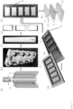
- the chip according to the invention is a chip (12) containing at least one piece of a macroporous elastomeric silicone foam (5) (MESIF-chip) and comprises a PDMS housing structure (9), at least one piece of a macroporous elastomeric silicone foam (5) and a PDMS cover lid (13) into which environmental contact holes (11) are punched ( figure 4 ).
- the PDMS housing structure (9) according to the invention includes a reservoir (10) for fitting at least one piece of the macroporous elastomeric silicone foam (5), into which nutrient medium or substrates can be placed.
- the PDMS cover lid (13) into which environmental contact holes (11) are punched represents the first donor site (3) and the reservoir (10) represents the second donor site (4).
- the device (1) containing a macroporous elastomeric material (2) is a functional interface, like a membrane, in bioreactors, tubular reactors or flat-bed reactor.
- the device (1) containing a macroporous elastomeric material (2) as functional interface is integrated in a bioreactor for cultivating photosynthesis-active microorganisms, such as algae or cyanobacteria.
- the membrane surface itself represents the first donor site (3) and the respective reactor represents the second donor site (4).
- the device (1) is a functional interface of a bioreactor.
- microorganisms can migrate/diffuse from the surrounding marine habitat via the first donor site (3) into the macroporous elastomeric material (2) to form a synergistic community with the aquatic organisms such as microalgae, fish or crustaceans, contained together with an aqueous nutrient medium in the second donor site (4). From the second donor site (4), a gradient of nutrient components from the aquatic organism culture is created by diffusion into the macroporous elastomeric material (2).
- the manufacturing process for a macroporous elastomeric silicone foam (5) is described.
- the production of the macroporous elastomeric silicone foam (5) comprises the following steps ( figure 3A ):
- the porogen crystals (6) in step A) are selected from water dissolvable molecules, such as crystals of sodium chloride, calcium chloride, magnesium chloride or other inorganic salts, or crystals of sucrose, glucose, fructose or amino acids.
- sodium chloride crystals or sucrose crystals are used as porogen crystals (6).
- the grain size of the porogen crystals (6) ranges from 0.1 ⁇ m - 950 ⁇ m, preferably from 10 - 300 ⁇ m.
- the grain size is defined as the diameter of singular sediment grains. This differs from the size of a crystallite, which refers to the size of a single crystal inside a particle or grain.
- the porosity of the macroporous elastomeric silicone foam (5) to be produced is adjusted by using different grain size fractions of porogen crystals (6) ( figure 5B )); example 2).
- the silicone prepolymer solution (7) in step A) is a mixture of silicone oligomers with a curing agent in a ratio of 10:1 (w/w) .
- the curing agent is Sylgard 184 (Down Corning).
- the silicone in the silicone prepolymer solution (7) is Polydimethylsiloxane (PDMS).
- the casting mold (8) in step B) is preferably made of polymethyl methacrylate (PMMA).
- PMMA polymethyl methacrylate
- the casting mold (8) preferable consists of two pieces of rectangular geometry, with edges, grooves and a sealing film, like a PTFE (Polytetrafluorethylen) film or Parafilm ® M, in between, is closed by screws, that is made for centrifugal mixing of the porogen crystals (6) and the liquid silicone ( figures 4A ), 4B)).
- Centrifugation in step C) is preferably done for 20 minutes, at 5 °C and 4600 rpm.
- step D) Thermal curing in step D) is preferable performed at 70 °C for 1 hour.
- step F the resulting molded macroporous elastomeric silicone foam is preferably mechanically compressed several times in a water bath to dissolve the integrated porogen crystals, and thereby a macroporous structure of interconnected pores within the elastomer is created.
- step G Another way of leaching according to the invention is soaking of the resulting molded macroporous elastomeric silicone foam for several hours in a 40°C water bath with or without sonication.
- drying is preferably performed at 90 °C for one hour.
- the surface of the prepared silicone foam is modified by means of established organosilane chemistry (example 3).
- the surface of the macroporous elastomeric silicone foam (5) is modified with primary amino groups or expoxy groups using APTES ((3-Aminopropyl) triethoxysilane) or GPTS (3-Glycidyloxypropyl) trimethoxysilane), respectively ( figure 10 ).
- the silicone prepolymer solution (7) is a mixture of silicone oligomers and a thermal curing agent in a ratio of 10:1 (w/w), where a silicone-PEG block copolymer, like a PDMS-PEG block copolymer, is added in a ratio of 0.25 % (w/w), related to the total mass of the silicone prepolymer solution (7) (example 3).
- the surface of the macroporous elastomeric silicone foam (5) is modified with even large functional macromolecules, such as DNA oligonucleotides ( figure 11 , example 3) or proteins, such as enzymes, antibodies or other binding proteins.
- functional macromolecules such as DNA oligonucleotides ( figure 11 , example 3) or proteins, such as enzymes, antibodies or other binding proteins.
- the manufacturing process of a chip (12) containing at least one piece of a macroporous elastomeric silicone foam (5) comprises the following steps:
- plasma surface activation in step L) is performed with O 2 , N 2 or NH 3 to provide altered surface chemistries that favor or disfavor the enrichment of specific microbial communities.
- Plasma activation is performed for, e.g., 30 seconds at 300 W and 0.2 mbar.
- step N) is performed at 90 °C for 30 minutes.
- the application of the device (1) containing a macroporous elastomeric material (2) is described:
- the device (1) containing a macroporous elastomeric material (2) can be used for the enrichment, colonization and isolation of microorganisms in situ in a wide variety of habitats.
- the device (1) containing a macroporous elastomeric material (2) can be used for the enrichment, colonization and isolation of previously uncultivable or unknown microorganisms in situ in a wide variety of habitats.
- the device (1) containing a macroporous elastomeric material (2) can be used in dry, wet and liquid environments, such as indoor air, soils, moving bed biofilters or wastewater treatment plants.
- a chip (12) containing at least one piece of a macroporous elastomeric silicone foam (5) is used for the enrichment, colonization and isolation of microorganisms in situ ( figures 6-9 ).
- the chip (12) containing at least one piece of a macroporous elastomeric silicone foam (5) is used for the enrichment, colonization and isolation of previously uncultivable or unknown microorganisms in a wide variety of habitats.
- the chip (12) containing at least one piece of a macroporous elastomeric silicone foam (5) can be used in dry, wet and liquid environments, such as indoor air, soils, moving bed biofilters or wastewater treatment plants.
- MESIF-chip macroporous elastomeric silicone foam (5)
- Genomic DNA was extracted from the chips using commercially available extraction kits, and the amount of extracted gDNA was quantified photometrically.
- the resulting KRONA charts illustrate that the microbial community in the Mutag chips (Mutag Biochip TM ) was strongly dominated by Alphaproteobacteria and Planctomycetes, the latter being especially known to form biofilms on various surfaces ( S. Wiegand et al., Nat Microbiol 2020, 5 (1), 126-140 ).
- the MESIF-chip (12) showed a much broader distribution of different bacterial species ( figure 7B )).
- CPR Candidate Phyla Radiation
- CPR genomes seem to lack ubiquitous biosynthetic pathways, e.g. for the production of amino acids, nucleotides, cofactors and membrane lipids, while protein families associated with cell-cell interactions are frequently found in this group ( C. J. Castelle et al., Cell 2018, 172, 1181-1197 ; R. Méheust et al., Nature communications 2019, 10 (1), 4173 ). Also, some CPRs seem to live in symbiotic relationships, which could also be the reason why they are so difficult to culture and still very poorly studied.
- an adapted device comprising a MESIF-chip (12) was filled with LB cultivation medium and connected from the side of the reservoir (10) to a syringe through a needle (example 4C), figure 8A )).
- a sterile syringe-filter was plugged into the other side of the syringe to allow for ventilation.
- the syringe was filled with LB medium to serve as an extended reservoir for the chip, enabling wet conditions and continued viable cultivation.
- Chips with the environmental contact hole (11) facing upwards were placed in an open interior of a poultry farm with a stocking density of 2 chickens per m 2 , and incubated for several days, then collected and stored at -20 °C until further analysis.
- the same LB medium in an open sterile Eppendorf tube was incubated at the same conditions. Genomic DNA was extracted and 16S rRNA amplicon sequencing and bioinformatic analysis were conducted. While the comparison sample showed a complete overgrowth of Proteobacteria ( figure 8 ), representing 86% of the total abundance, the MESIF-chip (12) enabled the isolation of a much more evenly distributed composition of airborne microorganisms, particularly Proteobacteria, Firmicutes and Actinobacteriota. The latter taxonomic group is particularly interesting because of its ability to produce specific secondary metabolites such as anticancer, antifungal and immunosuppressive agents.
- the low-cost, bioinert and widely modifiable chip (12) containing at least one piece of a macroporous elastomeric silicone foam (5) is mechanically robust and resilient, making it applicable in a wide range of very challenging, complex habitats that include dry, wet and liquid environments, such as indoor air, soils, turbulent wet moving bed biofilters or wastewater treatment plants.
- the macroporous silicone foam (5) according to the invention, is, despite its bioinert surface, colonized by microorganisms surprisingly quickly and efficiently in situ, such that after a few days of incubation in diverse environmental habitats, a stable population is formed within the foam that can be identified using next generation sequencing. Even complex microbial communities can be established within the macroporous silicone foam (5).
- the macroporous elastomeric silicone foam (5) according to the invention can be manufactured on a large scale, offers ideal wetting and diffusion properties, can be chemically tailored with respect to its surface area, and can easily be integrated into selection chips available via soft-lithographic fabrication.
- the chip (12) containing at least one piece of a macroporous elastomeric silicone foam (5) can be easily loaded with solid, liquid and gaseous substances that influence microbial colonization, it can also be used for the targeted enrichment of microorganisms and individual inhabitants of a particular environment.
- the present invention allows the systematic study of microbial dark matter and also enables new approaches for sampling, enrichment, cultivation, identification and exploitation of previously uncultivable or unknown microorganisms in situ.
- MESIF macroporous elastomeric silicone foam
- MESIF-chip a chip (12) containing at least one piece of a macroporous elastomeric silicone foam (5)
- a casting mold (8) comprising two parts ( figures 3A ) and 4A )) was designed using Autodesk Inventor software 2020 (Autodesk Inc., USA) and milled with a CNC micromilling machine (Mini Mill GX, Minitech Machinery, USA). The two parts of the mold were closed by 9 screws.
- porogen crystals (6) was used sodium chloride table salt (ChanteSel iodized salt fine from Lidl, Germany), the particles of which were fractionated using a sieve with the following inserts: 25 mesh (707 ⁇ m), 35 mesh (500 ⁇ m), 45 mesh (354 ⁇ m), 60 mesh (250 ⁇ m) and 80 mesh (177 ⁇ m).
- the resultant porogen fractions were weighed individually and the ratio to the mass of the sieved salt was calculated to estimate the mass fractions.
- PDMS oligomers were mixed with the curing agent (Sylgard 184, Down Corning) in a ratio of 10:1, respectively, then degassed in a vacuum chamber for 1 hour.
- 0.9 mL of the PDMS prepolymer solution were poured inside the closed casting mold (8) to fill approximately half of its volume and the remaining free volume was filled with table salt porogen crystals of the desired particle size.
- the filled mold was centrifuged (5804 R, Eppendorf) at 4600 rpm and 4 °C for 20 minutes to disperse and pack the porogen crystals (6) within the liquid PDMS prepolymer. After centrifugation, the PDMS-porogen mixture was cured in the oven (Heraeus, Thermo Scientific Inc.) at 70 °C for one hour, then the mold screws were removed and the solid, casted PDMS-porogen mixture piece was carefully released from the mold as a single piece ( ⁇ 65 ⁇ 10 ⁇ 3 mm 3 ) as shown in figure 4C ).
- the released piece was repeatedly pressed by hand in a 50 °C water bath until the material reached a soft, elastic, and spongelike structure. To remove all salt residues, the material was stirred overnight in a beaker filled with 250 mL ddH 2 O at 40 °C and washed in the next morning with fresh ddH 2 O. The resultant MESIF material was then dried at 90 °C for one hour in the oven, then cut into pieces of ⁇ 18 ⁇ 10 ⁇ 3 mm 3 ( figure 4D )) using a scalpel. These final MESIF pieces fit into the MESIF-chip PDMS housing structure (9) ( figures 4F) and 4G )).
- a casting mold corresponding to the PDMS housing structure (9) was designed using Autodesk Inventor software and milled on a PMMA plate using a CNC micromilling machine.
- a PDMS prepolymer solution was prepared as described above and poured into the chip mold, then cured at 70 °C for one hour in an oven. After curing, the cast was peeled off the mold and was ready to fit the MESIF material pieces inside ( figure 4E )).
- a mold for the PDMS cover lid (13) (76 ⁇ 26 ⁇ 0.75 mm 3 ) was prepared using the same procedure, into which the environmental contact holes (11) ( ⁇ 4 mm) were manually inserted with a press puncher.
- the prepared three components MESIF pieces, PDMS housing structure (9) and PDMS cover lid (13)
- plasma treatment Plasma Treatment
- the MESIF pieces were placed in the corresponding empty space in the PDMS housing structure (9), covered with the PDMS cover lid (13) ( figure 4H )) and the assembled chip was cured at 90 °C for 30 min for permanent bonding.
- the fractions 707 - 500 ⁇ m and 500 - 354 ⁇ m showed the largest mass fraction with together almost 80%. Sufficient amounts of salt crystals were obtained from the >707 ⁇ m, 707 - 500 ⁇ m, 500 - 354 ⁇ m and 354 - 250 ⁇ m salt fractions to prepare materials and perform further analysis of MESIF pore sizes, diffusion and growth behavior with model organisms.
- the MESIF material was cut into approximately 1 mm thick layers with a scalpel, then coated with 1.5 nm platinum and examined using Environmental Scanning Electron Microscopy (ESEM) at 20 KV under vacuum.
- ESEM Environmental Scanning Electron Microscopy
- the software ImageJ was used for further analysis of the acquired images. By setting a gray threshold and manual post-processing, all pores were colored black and separated from each other and evaluated using ImageJ's "Analyze Particle" function.
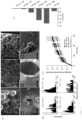
- Figure 5A shows representative ESEM images of MESIF prepared with different porogen grain sizes.
- the images show a stochastic arrangement of pores characterized by different shapes reflecting the morphology of salt crystals.
- many pores show sharp-edged cracks in the pore surface at their contact surfaces ( figure 5A ) IV) through which the pore behind can be seen when focused further ( figure 5A ) V).
- thin, crumpled PDMS pieces are found in some pores ( figure 5A ) VI).
- the ESEM images were also used to determine the pore size distribution of the two-dimensional pore cross sections of the sponges using ImageJ analysis ( C. A. Schneider et al., Nat. Methods 2012, 9 , 671-675 ) ( figure 5B )).
- the histograms show absolute pore sizes smaller than experimentally determined porogen particle sizes, because only a two-dimensional section of the three-dimensional MESIF material was recorded in the ESEM and thus the pores were not imaged in their maximum cross-sectional area depending on the position of the section. Despite this systematic error, it is visible that with larger salt crystals the pore size distribution becomes progressively wider and flatter. In contrast, smaller salt crystals produce narrower and steeper distributions.
- the pore size distribution of the unsieved salt shows a superposition of the distributions of all size fractions.
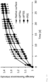
- this method revealed that the fluorophore spreads almost homogeneously throughout the foam.
- the method could be used to quantify differences of the foam on the dissociation of the solute and also of fluorescently labeled model organisms.
- FIG 5C it is shown in figure 5C ) that the pore sizes of the MESIF have little effect on the passive diffusion of the fluorescent solute.
- the trend was evident that smaller pore sizes lead to faster diffusion, presumably due to higher capillary forces in the smaller pores.
- a stock solution of the dye (100 mM in dimethyl sulfoxide) was stored in a -20 °C freezer, and a dilution of 10 ⁇ M in ddH 2 O was freshly prepared upon use. Chips with different MESIF pore sizes were filled with ddH 2 O, which was injected into the chip reservoir with a syringe and perfused through the MESIF until it exited the environmental contact hole (11). At least 1 mL of ddH 2 O was injected into the chip to ensure that the MESIF was completely soaked.
- the filled chips were then placed in a Petri dish with the environmental contact holes (11) facing upwards, then 40 ⁇ L of 10 ⁇ M 5-carboxyfluorescein solution was loaded onto the contact hole. 5 mL ddH 2 O were added to the Petri dish to ensure adequate humidity. The dish lid was closed and sealed with parafilm to prevent the chips from drying. The Petri dish was kept at room temperature in the dark for the entire experiment to avoid photobleaching. To determine the diffusion progress, images of the Petri dish were taken over the experimental time using a fluorescence gel imager (Fusion FX, Vilber) at exposure time of 1 ms. At least three independent chips were used in these experiments. For microbial growth in the MESIF-chip, a lab stock E.
- E. coli-RFP arabinose inducible red fluorescent protein
- MESIF materials were immersed in the activation solution composed of ddH 2 O, concentrate H 2 O 2 (30% v/v) and concentrate HCl (37% v/v) in a volume ratio of 5:1:1 at room temperature.
- the materials were mechanically squeezed by hand for 2 min, followed by stirring in the same solution for 3 min. This procedure was repeated a total of five times.
- the MESIF materials were washed with ddH 2 O until a neutral pH value in the washing water was achieved. Finally, the materials were dried overnight in an oven at 90 °C.
- the surface activated MESIF materials were heated in a solution of toluene containing the appropriate organosilane (2% v/v) at 80 °C for 24 h to obtain the desired surface functionalization. The materials were then washed with toluene and dichloromethane and any solvent residues were removed at 40 °C and under reduced pressure.
- GTPS-functionalized MESIF materials were immersed in a 1:1 mixture of concentrate HCl (37% v/v) and ddH 2 O. The materials were mechanically squeezed by hand for 25 min in the acidic solution in order to open the epoxide to the corresponding diol. Afterwards the MESIF pieces were washed with ddH 2 O until a neutral pH value was reached in the washing solution. The pieces were then dried overnight in an oven at 90 °C.
- PDMS oligomers and a thermal curing agent were mixed at a ratio of 9:1 as a polymerization mixture. Additionally, a PDMS-PEG block copolymer (Gelest Inc.) was added in a ratio of 0.25 % (w/w), to introduce PEG-units into the material ( figure 10F )). The resulting PDMS prepolymer solution was then used to prepare MESIF materials as described in example 1.
- MESIF pieces were incubated in 3 mL of phosphate buffered salt solution (PBS) containing 100 nm of the DNA oligonucleotide AmC12Tr12-Cy5 (5'-Amine-C12-T8-(ATG)4-Cy5-3
- PBS phosphate buffered salt solution
- AmC12Tr12-Cy5 5'-Amine-C12-T8-(ATG)4-Cy5-3
- the MESIF pieces were shaken in the solution for 6 h, at room temperature and at 175 rpm.
- the pieces were squeezed manually with a glass rod every 0.5 h for 1 min.
- the MESIF pieces were washed five times with PBS, each time the MESIF materials were squeezed with a glass rod for 1 min. Removal of unbound DNA was monitored by UV/Vis absorbance measurements of the washing solution.
- the pieces were incubated in 3 mL of PBS containing 100 nM of the complementary oligomer Cy3-cTr12 (5'-Cy3-(CAT)4-3'). Incubation was done under shaking for 6 hours at room temperature at 175 rpm and repeated squeezing every 0.5 h with a glass rod. The pieces were then washed five times with PBS, and the UV/Vis absorbance of the washing solution was measured.
- the so-treated MESIF was then examined to accurately localize DNA within the pore structure, using confocal laser scanning microscopy (CLSM, ZEISS LSM 880, Carl Zeiss Inc.).
- E. coli-RFP red fluorescent protein
- MESIF materials show altered turbidity and opacity due to microbial colonization, which can be detected very easily by UV transillumination.
- UV transillumination Using the example of the very slow growing Bacillus merisflavi, the bacterial growth was monitored in real time with a normal webcam and used to quantify the biomass in the MESIF-chip (12).
- MESIF pieces (10 ⁇ 10 ⁇ 3 mm 3 ) prepared with unsieved porogen crystals and Mutag chips (Mutag Biochip TM ) ( ⁇ 300 mm) were sterilized by autoclaving 121 °C for 20 min.
- Sterile MESIF-chips (12) and Mutag chips (Mutag Biochip TM ) were then incubated freefloating in the biofilter tank for 18 days, then collected, briefly washed with ddH 2 O, and frozen at -20 °C for temporary storage and transport.
- Duplicate MESIF-chips and Mutag chips (Mutag Biochip TM ) were used.
- Genomic DNA was extracted from the chips using commercially available extraction kits according to the manufacturer's protocol, and the amount of extracted gDNA was quantified photometrically. Characterization of the obtained gDNA was done by gel electrophoresis and 16S rRNA amplicon sequencing. The quality of the extracted gDNA was sufficiently high to produce a sequencing library for Illumina sequencing using commercially available kits. Following Illumina 16S rRNA amplicon sequencing, the sequences found were bioinformatically processed, analyzed, and visualized as Krona charts.
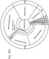
- the resulting KRONA charts in figure 7 illustrate that the microbial community in the Mutag Biochip TM is strongly dominated by Alphaproteobacteria and Planctomycetes ( figure 7A )), the latter being especially known to form biofilms on various surfaces ( S. Wiegand et al., Nat Microbiol 2020, 5 (1), 126-140 ).
- the MESIF-chip (12) showed a much broader distribution of different bacterial species ( figure 7B )).
- species from the so called Candidate Phyla Radiation (CPR) S. Wiegand et al., Front. Microbio. 2021, 12, 635506 ) could be enriched.
- CPR Candidate Phyla Radiation
- the designed reservoir feature of the MESIF-chip (12) was exploited to provide a continuous supply of a LB medium to the porous interior of the chip to enable for a long incubation period in a dry environment.
- the adapted device comprised a MESIF-chip (12) filled with LB medium and was connected from the side of the reservoir (10) to an 1 mL syringe through a needle (both from BBraun GmbH) ( figure 8A )).
- a 0.2 ⁇ m sterile syringe-filter was plugged into the other side of the syringe to allow for ventilation.
- the syringe was filled with LB medium to serve as an extended reservoir for the chip, enabling wet conditions and continued viable cultivation.
- the syringe serves as a sterile aerated fluid reservoir from which the loss of moisture in the MESIF (5) - due to evaporation at the environmental contact - is continuously replenished as the foam acts as a capillary pump.
- Chips with the environmental contact hole (11) facing upwards were placed on top of a 2 m high cabinet in an open interior of a poultry farm with a stocking density of 2 chickens per m 2 , and incubated for nine days, then collected and stored at -20 °C until further analysis.
- 1 mL of the same LB medium in an open 1.5 mL sterile Eppendorf tubes were incubated at the same conditions.
- Triplicate MESIF-chips (12) and control Eppendorf tubes were used.
- Example 4D Sampling of microorganisms from chemical wastewater: To demonstrate that the MESIF-chip is applicable for selective enrichment of microbial communities when the reservoir is filled with a specific chemical, sampling studies were conducted in the highly challenging wet habitat of a wastewater treatment plant (WWTP). For this purpose, the possible selective enrichment of microorganisms was tested by using the herbicide glyphosate, which is a phosphonic acid compound and is used as the biologically active major component of some broad-spectrum or total herbicides on a millionton scale worldwide ( M. E. Richmond, J Environ Stud Sci 2018, 8 (4), 416-434 ).
- the herbicide glyphosate which is a phosphonic acid compound and is used as the biologically active major component of some broad-spectrum or total herbicides on a millionton scale worldwide ( M. E. Richmond, J Environ Stud Sci 2018, 8 (4), 416-434 ).
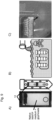
- MESIF-chips (14) were prepared with unsieved porogen salt and autoclaved. To enable sampling, the reservoir of the MESIF-chip (12) was filled with a sterilized native WWTP medium supplemented with 8 g/L glyphosate (Monsanto, Bayer). Chips whose reservoirs were filled with sterilized WWTP medium only were used as controls. Two sterile chips loaded with either glyphosate or the control medium were packed into a 50 mL Falcon tube, in which handmade openings allow inflow of WWTP effluent ( figure 9A )).
- the Falcon tubes loaded in this manner were fixed in a metal rack ( figure 9B )) and then immersed ⁇ 40 cm deep in a static chemical wastewater collection tank of the waste management system of the Düsseldorf Institute of Technology in the open air ( figure 9C )) and left for six days.
- the chips were then collected and gently washed with sterile ddH 2 O followed by storage at 20 °C until further analysis.
- Duplicate MESIF-chips (12) with or without glyphosate were used. 16S rRNA amplicon sequencing and bioinformatic analysis were conducted afterwards.
- the Krona chart shows that the MESIF control chip lacking glyphosate showed mainly different Proteobacteria, as well as some Bacteroidota and a small amount of Cyanobacteria and Bacteriovoracaceae.
- the glyphosate-containing MESIF-chip showed an almost 5-fold larger fraction of Cyanobacteria, suggesting their enrichment due to the presence of glyphosate ( figure 9E )).
- the phyla Desulfobacterota and Firmicutes could be found in the glyphosate MESIF-chip.
- Desulfobacterota is especially interesting, since anaerobic sulfate or sulfur-reducing bacteria in this phylum have been described to utilize organic acids ( D. W. Waite et al., Int. J. Syst. Evol. Microbiol. 2020, 70 (11), 5972-6016 ), which could be a metabolic product of the glyphosate degradation of other bacteria in the sample.
- the first steps in the degradation of glyphosate in bacteria involve the conversion of glyphosate into the weak organic acid aminomethylphosphonic acid through the glyphosate oxidoreductase ( S. Singh et al., Int. J. Environ. Res. Public Health 2020, 17 (20), 7519 ).
- the acid can then be excreted into the environment or cleaved into other molecules.
- the presence of Desulfo-bacterota in the MESIF-chip containing glyphosate could thus indicate that the resulting aminomethylphosphonic acid of the herbicide degradation may be consumed by the Desulfobacterota.
- the data regarding the presence of known glyphosate degrading organisms were analyzed by aligning all amplicon sequences from the chip against the total genomes of three organisms known for their glyphosate degradation: Enterobacter sp. E20 ( G. Cao et al., Proceedings of the International Symposium on Agriculture, Food and Biotechnology 2019 (ISAFB 2019 ), Pseudomonas azotoformans ( V.
- Genomic DNA was extracted from the sampling MESIF-chips (12) described above. Under sterile condition, the MESIF (5) was removed from the chip, then was cut into small pieces using a sterile scalpel and collected in a 2 mL sterile Eppendorf tube.
- the commercial Quick-DNA Miniprep Plus Kit (Zymo Research GmbH, Germany) was used to extract the gDNA from the MESIF according to the manufacturer's protocol. The concentration of extracted gDNA was subsequently quantified photometrically by NanoDrop OneC Microvolume, and fluorometrically by Qubit 3 (both from Thermo Scientific Inc.). From the purified gDNA, the 16S rRNA V3 region was amplified using the degenerate primers 341bf (ACACTCTTTCCCTACACGACGCTCTTCCGATCTCCTACGGGNGGCWGCAG) and 518r (GACTGGAGTTCAGACGTGTGCTCTTCCGATCTWTTACCGCRGCTGCTGG), which include Illumina TruSeq adapter sequences.
- the PCR reactions contained 0.04 U/ ⁇ L High-Fidelity Q5 DNA Polymerase, 1x Q5 Reaction Buffer, 1x High GC Enhancer, 0.25 ⁇ M of each 17 primer, 0.2 mM dNTPs, and 100 ng template DNA in a total reaction volume of 25 ⁇ L.
- Amplification was carried out using a touchdown protocol with the following cycling conditions (95 °C, 2 min; 4 ⁇ [95 °C, 9 s; 56-52 °C, 40 s; 72 °C, 40 s] 25 ⁇ [95 °C, 40 s; 51 °C, 40 s; 72 °C, 40 s]; 72 °C, 5 min; hold at 8 °C).
- Indexed sequencing libraries were constructed from the purified product using the NEBNext Ultra II FS DNA Library Prep Kit (New England BioLabs, Germany), using the protocol for input amounts ⁇ 100 ng and beginning with the size selection step.
- Unique dual indices were added in 6 cycles of an index-PCR.
- DNA library quality was verified using the Agilent High Sensitivity DNA Kit on the Agilent 2100 Bioanalyzer instrument (Agilent Technologies, Germany). Paired-end reads were generated on an Illumina NextSeq 550 (Illumina, USA) with the NextSeq 500/550 High Output Kit v2.5 (300 Cycles) (Illumina, USA).
- the reads were dereplicated (-derep_fulllength) and singletons were removed (--minuniquesize 2). They were then clustered at 97 % identity and the cluster centroid sequences were reported. Chimeras were detected and removed with the --uchime_denovo option, and finally, reads were mapped back onto the centroids via the -usearch_global option and output as OTU tables.
- the centroid sequences were aligned to the SILVA SSU reference database for taxonomic classification ( E. Pruesse et al., Bioinformatics 2012, 28 (14), 1823-1829 ). The results were visualized as a KRONA chart ( B. D. Ondov et al., BMC Bioinf. 2011, 12, 385 ).
Landscapes
- Health & Medical Sciences (AREA)
- Wood Science & Technology (AREA)
- Organic Chemistry (AREA)
- Life Sciences & Earth Sciences (AREA)
- Engineering & Computer Science (AREA)
- Bioinformatics & Cheminformatics (AREA)
- Chemical & Material Sciences (AREA)
- Zoology (AREA)
- Genetics & Genomics (AREA)
- Biochemistry (AREA)
- Microbiology (AREA)
- Biotechnology (AREA)
- General Engineering & Computer Science (AREA)
- General Health & Medical Sciences (AREA)
- Biomedical Technology (AREA)
- Sustainable Development (AREA)
- Immunology (AREA)
- Apparatus Associated With Microorganisms And Enzymes (AREA)
Abstract
The present invention relates to a device (1) containing a macroporous elastomeric material (2) for the enrichment and cultivation of microorganisms in situ and to the application of said device for sampling, colonization and isolation of even previously uncultivable or unknown microorganisms in a wide variety of habitats.
Description
- The present invention relates to a device containing a macroporous elastomeric material for the enrichment and cultivation of microorganisms in situ and to the application of said device for sampling, colonization and isolation of even previously uncultivable or unknown microorganisms in a wide variety of habitats.
- Natural evolution has produced an almost infinite variety of microorganisms that can colonize almost any conceivable habitat in complex communities.
- The vast majority of these microbial consortia are, however, uncharacterized and cannot be cultured in the laboratory. Modern methods of high-throughput nucleic acid sequencing are beginning to allow us to better understand the enormous complexity of this so-called "microbial dark matter" (C. Rinke et al., Nature 2013, 499 (7459), 431-437), however, although a surprisingly large number of branches are already visible, the full extent of the microbial tree of life is still difficult to imagine (L. A. Hug et al., Nat Microbiol 2016, 1 (5), 16048). This lack of knowledge is very unfortunate, as it is becoming increasingly clear that microbial communities not only play an important role in human health and our environment (L. Hall-Stoodley et al., Nat Rev Microbiol 2004, 2 (2), 95-108), but could also serve as invaluable tools for the next generation of biotechnological processes (W. Verstraete, Microb Biotechnol 2015, 8 (1), 36-37). Therefore, there is a growing interest in efficient strategies to isolate novel microorganisms from complex habitats in order to exploit the biological materials and information they contain, for example specific proteins or metabolic pathways, for biotechnological applications (W. H. Lewis et al., Nat Rev Microbiol 2021, 19 (4), 225-240). To improve the accessibility of non-cultivable microorganisms for technical cultivation, the so-called "isolation chip" (ichip) platform had been developed to isolate previously non-cultivable microorganisms from environmental samples by allowing exposure to natural growth factors through in situ culture (B. Berdy et al., Nat Protocols 2017, 12 (10), 2232-2242). To achieve this, cultivation was moved to the microbes' natural habitat by placing cells taken from various environmental samples into miniaturized diffusion chambers of a chip comprising porous polycarbonate membranes, which was then returned to nature for incubation. Although the ichip platform increases microbial biomass from, for example, soil samples by 5- to 300-fold, depending on the study, the technology is cumbersome because environmental samples must first be collected, then processed for integration into the chip, and the chips thus processed must be re-incubated in the original habitat. Hence, with the ichip platform it is not feasible to sample, for example, aqueous and/or dynamic habitats, such as moving bed biofilters or wastewater treatment plants.
- Although a variety of hydrophobic micro-, meso- or macroporous materials suitable for the isolation of microorganisms, like zeolites, active carbon, porous silicates, hybrid metal-organic frameworks and hypercrosslinked porous polymers, exist, several macroporous elastomeric materials combine the features of being, e.g., easy to process, being moldable and gas permeable. Polydimethylsiloxane (PDMS), as an example for a low-cost macroporous elastomeric material, has the further advantages of being biochemically inert and diversely modifiable in terms of its surface properties through well-controlled silane chemistry. The production of foam- and sponge-like porous silicone has been well studied, and it has already been shown that these materials can be used for applications as oil absorbers, heterogeneous catalysts, actuators and sensors, or even for tissue engineering of animal cells (D. Zhu et al., J. Mater. Chem. A 2017, 5 (32), 16467-16497) .
- However, the suitability of macroporous elastomeric materials for the targeted enrichment and cultivation of microorganisms in situ has neither been discussed nor shown before.
- Thus, the objective technical problem was to provide a device containing a macroporous elastomeric material that is suitable for sampling, enrichment and cultivation of microorganisms in situ in a wide variety of habitats, like aqueous or dry and/or dynamic habitats.
- The objective technical problem is solved by the embodiments characterized in the independent claims. Preferred embodiments can be found in the dependent claims.
- The invention describes a device (1) containing a macroporous elastomeric material (2) that can be used for the enrichment, colonization and isolation of also previously uncultivable or unknown microorganisms in situ from arbitrary habitats (
figure 1 ) . - According to the invention, in situ refers to the isolation, enrichment, colonization and cultivation of microorganisms in the original habitat without processing the sample of microorganisms after collection in vitro and reincubation in the original habitat.
- The macroporous elastomeric material (2) according to the invention is any material exhibiting elastic or rubber-like properties and having an adjustable porosity, containing physically interconnected pores of an average pore size ranging from 0.1 - 950 µm, preferably from 10 - 300 µm.
- The term "interconnected pores" refers to an open porosity, wherein a fluid flow is effectively taking place as distinguished from closed pores as non-connected cavities (https:// en.wikipedia.org/wiki/Porosity). Thus, the interconnected pores according to the invention form a continuous network of connected channels throughout the macroporous elastomeric material (2).
- The macroporous elastomeric material (2) within the device (1) is additionally characterized by flexibility, moldability, gas permeability and adjustable surface properties.
- In a first aspect of the invention is provided a device (1) comprising a macroporous elastomeric material (2) as a macroporous matrix connected via open, interconnected pores to a first donor site (3) and a second donor site (4) (
figure 1 ), wherein the first donor site (3) provides contact to an aqueous, solid or gaseous phase containing microorganisms, and the second donor site (4) provides contact to an aqueous, solid or gaseous phase, optionally containing nutrients, a culture medium, attractants, repellents and/or other molecular as well as cellular interaction partners for the microorganisms to be collected and enriched from the first donor site (3). For example, microalgae or cyanobacteria cultured in the second donor site (4) and metabolites secreted by the microalgae or cyanobacteria can serve as cellular or molecular interaction partners, respectively. - According to the invention, the term "first donor site" is to be regarded as an entry point for microorganisms, such as bacteria, archaea or microbial eukaryotes (e.g., microalgae), and the term "second donor site" is to be regarded as a nutrient reservoir or culture medium for the macroporous elastomeric material (2) and the microorganisms to be cultured within the macroporous elastomeric material (2).
- The first donor site (3) is, e.g., a surface suitable for contact with the environment, such as a cover lid provided with environmental contact holes, the second donor site (4) is, e.g., a reservoir or a bioreactor.
- The first donor site (3) provides contact to any liquid, solid or gaseous environment containing microbial cells, e.g., airborne habitats, soils or biofilters. In the device (1) cells included in gaseous, liquid, solid or other habitat environments can enter the macroporous elastomeric material (2) from the first donor site (3) and move by diffusion, motility or propagation inside and towards the direction of the second donor site (4). Substances contained in the medium of the second donor site (4) can diffuse within the macroporous elastomeric material (2) towards the first donor site (3) via active or passive diffusion, thereby creating gradients of substances with high and low substance concentrations near the second donor site (4) and the first donor site (3), respectively.
- The substance gradients affect the growth, accumulation and establishment of cultures of microbial cells that migrate from the first donor site (3) to the second donor site (4), leading to the formation of stable microbial communities in the macroporous elastomeric material (2).
- By modifying the substances added to the second donor site (4) or by creating a specific gas atmosphere in the second donor site (4), like a hydrogen-, methane- or carbon dioxide-enriched gaseous phase, enrichment, colonization and isolation of selected microorganisms, which are attracted by the substances in the second donor site (4), is possible.
- In a preferred embodiment the macroporous elastomeric material (2) contained in the device (1) is a macroporous elastomeric silicone foam (MESIF) (5).
- A foam belongs to the group of elastomeric materials and is a lightweight, open-cell product of low density to allow air movement through the cell structure (D. Weaire et al., "The Physics of Foams", Oxford University Press, 1999, ISBN 0198510977, ISBN 978-0-1985-1097-0; I. Cantat et al., "Foams: structure and dynamics", Oxford University Press, ed. S.J. Cox, 2013, ISBN 9780199662890).
- A silicone is defined as a polymer made up of siloxane (-R2Si-O-SiR2-, where R is an organic group) .
- The macroporous elastomeric silicone foam (4) has, according to the above specifications, an adjustable porosity, containing physically interconnected pores of an average pore size ranging from 0.1 - 950 µm, preferably from 10 - 300 µm.
- In a preferred embodiment the macroporous elastomeric silicone foam (5) contains Polydimethylsiloxane (PDMS).
- In a particular embodiment the macroporous elastomeric silicone foam (5) contains other materials beside Polydimethylsiloxane (PDMS) selected from other hydrophobic or hydrophilic polymers or blockcopolymers, such as polypropylene, polyethylene, polyamides, polyesters or blockcopolymers thereof, and/or particles made of polymers, metals, oxides or minerals are additionally added.
- In a particular embodiment the surface of the macroporous elastomeric silicone foam (5) is chemically modified with established organosilane chemistry.
- In a preferred embodiment the surface of the macroporous elastomeric silicone foam is modified with primary amino groups, epoxy groups or hydrophilic polyethylenglycol (PEG) moieties (
figure 10 ). - Surface modification with functional groups, such as amino-, hydroxyl- or PEG groups significantly affect the passive diffusional transport of small molecules through the macroporous elastomeric silicone foam (5). Such surface modifications can also be used to influence the colonization of macroporous elastomeric silicone foam surfaces by different bacterial communities, as it is well known that the colonization of surfaces can be strongly influenced by their polarity, hydrophobicity, and micro- and nanotopography (R. M. Donlan et al., Emerg Infect Dis 2002, 8 (9), 881-890; H. C. Flemming et al., Nat Rev Microbiol 2016, 14 (9), 563-575).
- In another embodiment even large functional macromolecules, such as DNA oligonucleotides, are installed on the surface of the macroporous elastomeric silicone foam (4) (
figure 11 ). - In another embodiment, following the procedure described in
figure 11 for installing DNA oligonucleotides, proteins, such as enzymes, antibodies or other binding proteins, are installed on the surface of the macroporous elastomeric silicone foam (5) to affect the colonization, growth and maintenance of microbial communities. - In one embodiment the device (1) containing a macroporous elastomeric material (2) is a chip for the enrichment and cultivation of microorganisms in situ, applicable in various exemplary habitats. In this embodiment a surface suitable for contact with the environment represents the first donor site (3) and a reservoir enclosed by the chip represents the second donor site (4), filled, e.g., with a culture medium supplemented with attractants or repellents.
- In a preferred embodiment the chip according to the invention is a chip (12) containing at least one piece of a macroporous elastomeric silicone foam (5) (MESIF-chip) and comprises a PDMS housing structure (9), at least one piece of a macroporous elastomeric silicone foam (5) and a PDMS cover lid (13) into which environmental contact holes (11) are punched (
figure 4 ). The PDMS housing structure (9) according to the invention includes a reservoir (10) for fitting at least one piece of the macroporous elastomeric silicone foam (5), into which nutrient medium or substrates can be placed. In this embodiment the PDMS cover lid (13) into which environmental contact holes (11) are punched represents the first donor site (3) and the reservoir (10) represents the second donor site (4). - In another embodiment the device (1) containing a macroporous elastomeric material (2) is a functional interface, like a membrane, in bioreactors, tubular reactors or flat-bed reactor. In a specific embodiment the device (1) containing a macroporous elastomeric material (2) as functional interface is integrated in a bioreactor for cultivating photosynthesis-active microorganisms, such as algae or cyanobacteria.
- In this embodiment the membrane surface itself represents the first donor site (3) and the respective reactor represents the second donor site (4).
- For sampling microorganisms in situ in an, e.g., marine environment, the device (1) according to the invention is a functional interface of a bioreactor. In this example microorganisms can migrate/diffuse from the surrounding marine habitat via the first donor site (3) into the macroporous elastomeric material (2) to form a synergistic community with the aquatic organisms such as microalgae, fish or crustaceans, contained together with an aqueous nutrient medium in the second donor site (4). From the second donor site (4), a gradient of nutrient components from the aquatic organism culture is created by diffusion into the macroporous elastomeric material (2).
- In another aspect of the invention, the manufacturing process for a macroporous elastomeric silicone foam (5) is described. The production of the macroporous elastomeric silicone foam (5) comprises the following steps (
figure 3A ): - A) Mixing porogen crystals (6) with a grain size of 0.1 µm - 950 µm with a silicone prepolymer solution (7);
- B) Pouring the resulting suspension of step A) into a casting mold (8);
- C) Centrifugation of the filled mold of step B) to disperse and pack the porogen crystals (6) within the liquid silicone prepolymer;
- D) Thermal curing of the suspension of step C);
- E) Releasing the cured macroporous elastomeric silicone foam from the casting mold (8);
- F) Leaching the macroporous elastomeric silicone foam from step E) to dissolve the integrated porogen crystals (6);
- G) Drying the molded macroporous elastomeric silicone foam;
- H) Optionally modifying the surface of the macroporous elastomeric silicone foam (5) after step G) by means of organosilane chemistry.
- The porogen crystals (6) in step A) are selected from water dissolvable molecules, such as crystals of sodium chloride, calcium chloride, magnesium chloride or other inorganic salts, or crystals of sucrose, glucose, fructose or amino acids.
- In a preferred embodiment sodium chloride crystals or sucrose crystals are used as porogen crystals (6).
- The grain size of the porogen crystals (6) ranges from 0.1 µm - 950 µm, preferably from 10 - 300 µm.
- The grain size is defined as the diameter of singular sediment grains. This differs from the size of a crystallite, which refers to the size of a single crystal inside a particle or grain.
- The porosity of the macroporous elastomeric silicone foam (5) to be produced is adjusted by using different grain size fractions of porogen crystals (6) (
figure 5B )); example 2). - In a preferred embodiment the silicone prepolymer solution (7) in step A) is a mixture of silicone oligomers with a curing agent in a ratio of 10:1 (w/w) . In a more preferred embodiment the curing agent is Sylgard 184 (Down Corning).
- In a preferred embodiment the silicone in the silicone prepolymer solution (7) is Polydimethylsiloxane (PDMS).
- The casting mold (8) in step B) is preferably made of polymethyl methacrylate (PMMA). The casting mold (8) preferable consists of two pieces of rectangular geometry, with edges, grooves and a sealing film, like a PTFE (Polytetrafluorethylen) film or Parafilm® M, in between, is closed by screws, that is made for centrifugal mixing of the porogen crystals (6) and the liquid silicone (
figures 4A ), 4B)). - Centrifugation in step C) is preferably done for 20 minutes, at 5 °C and 4600 rpm.
- Thermal curing in step D) is preferable performed at 70 °C for 1 hour.
- In step F), the resulting molded macroporous elastomeric silicone foam is preferably mechanically compressed several times in a water bath to dissolve the integrated porogen crystals, and thereby a macroporous structure of interconnected pores within the elastomer is created.
- Another way of leaching according to the invention is soaking of the resulting molded macroporous elastomeric silicone foam for several hours in a 40°C water bath with or without sonication. In step G), drying is preferably performed at 90 °C for one hour. After step G), in a preferred embodiment the surface of the prepared silicone foam is modified by means of established organosilane chemistry (example 3). In a more preferred embodiment the surface of the macroporous elastomeric silicone foam (5) is modified with primary amino groups or expoxy groups using APTES ((3-Aminopropyl) triethoxysilane) or GPTS (3-Glycidyloxypropyl) trimethoxysilane), respectively (
figure 10 ). For a surface modification with hydrophilic polyethylenglycol (PEG) moieties, in step A) the silicone prepolymer solution (7) is a mixture of silicone oligomers and a thermal curing agent in a ratio of 10:1 (w/w), where a silicone-PEG block copolymer, like a PDMS-PEG block copolymer, is added in a ratio of 0.25 % (w/w), related to the total mass of the silicone prepolymer solution (7) (example 3). - In another preferred embodiment the surface of the macroporous elastomeric silicone foam (5) is modified with even large functional macromolecules, such as DNA oligonucleotides (
figure 11 , example 3) or proteins, such as enzymes, antibodies or other binding proteins. - In a further aspect of the invention, the manufacturing process for a chip (12) containing at least one piece of a macroporous elastomeric silicone foam (5) is described.
- The manufacturing process of a chip (12) containing at least one piece of a macroporous elastomeric silicone foam (5) comprises the following steps:
- I) Fabricating a PDMS housing structure (9) with a reservoir (10) suitable for holding at least one piece of a macroporous elastomeric silicone foam (5), using a suitable casting mold;
- J) Fabricating a PDMS cover lid (13) cast from PDMS, into which environmental contact holes (11) are punched;
- K) Cutting a molded macroporous elastomeric silicone foam (5) into pieces to fit into the PDMS housing structure (9) of step I) and inserting at least one piece of a macroporous elastomeric silicone foam (5) into the reservoir (10);
- L) Plasma surface activation of the PDMS housing structure (9), containing at least one piece of the macroporous elastomeric silicone foam (5) from step K), and of the PDMS cover lid (13).
- M) Covering the housing structure (9) containing at least one piece of the macroporous elastomeric silicone foam (5) with the PDMS cover lid (13);
- N) Curing the PDMS housing structure including the cover lid and sealing the PDMS housing structure including the cover lid by bonding.
- In a preferred embodiment plasma surface activation in step L) is performed with O2, N2 or NH3 to provide altered surface chemistries that favor or disfavor the enrichment of specific microbial communities. Plasma activation is performed for, e.g., 30 seconds at 300 W and 0.2 mbar.
- In a preferred embodiment curing in step N) is performed at 90 °C for 30 minutes.
- In a further aspect of the invention the application of the device (1) containing a macroporous elastomeric material (2) is described:
The device (1) containing a macroporous elastomeric material (2) can be used for the enrichment, colonization and isolation of microorganisms in situ in a wide variety of habitats. In a specific embodiment, the device (1) containing a macroporous elastomeric material (2) can be used for the enrichment, colonization and isolation of previously uncultivable or unknown microorganisms in situ in a wide variety of habitats. The device (1) containing a macroporous elastomeric material (2) can be used in dry, wet and liquid environments, such as indoor air, soils, moving bed biofilters or wastewater treatment plants. - In a preferred embodiment of the invention a chip (12) containing at least one piece of a macroporous elastomeric silicone foam (5) is used for the enrichment, colonization and isolation of microorganisms in situ (
figures 6-9 ). In a specific embodiment, the chip (12) containing at least one piece of a macroporous elastomeric silicone foam (5) is used for the enrichment, colonization and isolation of previously uncultivable or unknown microorganisms in a wide variety of habitats. The chip (12) containing at least one piece of a macroporous elastomeric silicone foam (5) can be used in dry, wet and liquid environments, such as indoor air, soils, moving bed biofilters or wastewater treatment plants. - To test the suitability of the chip (12) containing at least one piece of a macroporous elastomeric silicone foam (5) (MESIF-chip) for enriching complex microbial communities from natural habitats first an aerobic moving bed biofilter of a fish farm was chosen. To evaluate the performance of the MESIF material, commercially available polyethylene biofilter carrier chips (Mutag Biochip™) were used as reference material. These chips are reported by the manufacturer to have a high protected active surface area of more than 5,500 m2 /m3, and are often used as biocarriers for moving bed biofilm reactor studies (N. H. Dan et al., Sci. Total Environ. 2021, 787, 147680; A. A. Mazioti et al., ).
- Sterile MESIF-chips (12) and Mutag chips (Mutag Biochip™) were incubated freefloating in the biofilter tank for several days and then collected (example 4B)).
- Genomic DNA (gDNA) was extracted from the chips using commercially available extraction kits, and the amount of extracted gDNA was quantified photometrically. After sequencing, the resulting KRONA charts (
figures 7A ), 7B)) illustrate that the microbial community in the Mutag chips (Mutag Biochip™) was strongly dominated by Alphaproteobacteria and Planctomycetes, the latter being especially known to form biofilms on various surfaces (S. Wiegand et al., Nat Microbiol 2020, 5 (1), 126-140). In contrast, the MESIF-chip (12) showed a much broader distribution of different bacterial species (figure 7B )). Notably, in the MESIF-chip (12), species from the so called Candidate Phyla Radiation (CPR) (S. Wiegand et al., Front. Microbio. 2021, 12, 635506) could be enriched. Based on cultivation-independent approaches, such as 16S rRNA amplicon sequencing, metagenomics and single-cell genomics (A.-K. Kaster et al., Appl. Microbiol. Biotechnol. 2020, 104 (19), 8209-8220) it is currently assumed that this taxon comprises about 70% of the microbial dark matter (L. A. Hug et al., Nat Microbiol 2016, 1 (5), 16048). Many CPR genomes seem to lack ubiquitous biosynthetic pathways, e.g. for the production of amino acids, nucleotides, cofactors and membrane lipids, while protein families associated with cell-cell interactions are frequently found in this group (C. J. Castelle et al., Cell 2018, 172, 1181-1197; R. Méheust et al., Nature communications 2019, 10 (1), 4173). Also, some CPRs seem to live in symbiotic relationships, which could also be the reason why they are so difficult to culture and still very poorly studied. - To test the real-life applicability of a chip (12) containing at least one piece of a macroporous elastomeric silicone foam (5) for environmental issues, secondly sampling of airborne microbial communities was investigated.
- For this purpose an adapted device comprising a MESIF-chip (12) was filled with LB cultivation medium and connected from the side of the reservoir (10) to a syringe through a needle (example 4C),
figure 8A )). A sterile syringe-filter was plugged into the other side of the syringe to allow for ventilation. The syringe was filled with LB medium to serve as an extended reservoir for the chip, enabling wet conditions and continued viable cultivation. Chips with the environmental contact hole (11) facing upwards were placed in an open interior of a poultry farm with a stocking density of 2 chickens per m2, and incubated for several days, then collected and stored at -20 °C until further analysis. As a control, the same LB medium in an open sterile Eppendorf tube was incubated at the same conditions. Genomic DNA was extracted and 16S rRNA amplicon sequencing and bioinformatic analysis were conducted. While the comparison sample showed a complete overgrowth of Proteobacteria (figure 8 ), representing 86% of the total abundance, the MESIF-chip (12) enabled the isolation of a much more evenly distributed composition of airborne microorganisms, particularly Proteobacteria, Firmicutes and Actinobacteriota. The latter taxonomic group is particularly interesting because of its ability to produce specific secondary metabolites such as anticancer, antifungal and immunosuppressive agents. Thus, two-thirds of all known antibiotics are currently produced by Actinobacteriota, which are therefore of great medical and biotechnological interest (A. van der Meij et al., FEMS Microbiol. Rev. 2017, 41 (3), 392-416; D. De Simeis et al., Antibiotics 2021, 10 (5), 483). Also, the broad distribution of the different microorganisms on the MESIF-chip (12) shows very clearly that the simple and pragmatic reservoir concept presented here is very well suited to sample complex airborne habitats, even when using a non-optimal medium such as LB, which should rather favor the enrichment of well-culturable organisms. Since the MESIF and reservoir can be loaded with a variety of substances, this approach should also be applicable for targeted enrichment of microorganisms, for example, by filling the reservoir with a chemical substance that attracts or repels specific microorganisms. - The low-cost, bioinert and widely modifiable chip (12) containing at least one piece of a macroporous elastomeric silicone foam (5), according to the invention, is mechanically robust and resilient, making it applicable in a wide range of very challenging, complex habitats that include dry, wet and liquid environments, such as indoor air, soils, turbulent wet moving bed biofilters or wastewater treatment plants. The macroporous silicone foam (5), according to the invention, is, despite its bioinert surface, colonized by microorganisms surprisingly quickly and efficiently in situ, such that after a few days of incubation in diverse environmental habitats, a stable population is formed within the foam that can be identified using next generation sequencing. Even complex microbial communities can be established within the macroporous silicone foam (5).
- The macroporous elastomeric silicone foam (5) according to the invention can be manufactured on a large scale, offers ideal wetting and diffusion properties, can be chemically tailored with respect to its surface area, and can easily be integrated into selection chips available via soft-lithographic fabrication.
- Since the chip (12) containing at least one piece of a macroporous elastomeric silicone foam (5) can be easily loaded with solid, liquid and gaseous substances that influence microbial colonization, it can also be used for the targeted enrichment of microorganisms and individual inhabitants of a particular environment.
- The present invention allows the systematic study of microbial dark matter and also enables new approaches for sampling, enrichment, cultivation, identification and exploitation of previously uncultivable or unknown microorganisms in situ.
- The invention is explained in more detail with reference to the following figures, examples of embodiments and descriptions. All features shown and combinations thereof are not limited to these figures and examples of embodiments. Rather, these are to be considered representative of further possible embodiments that can be combined, but which are not explicitly shown.
-
- (1) Device containing a macroporous elastomeric material
- (2) Macroporous elastomeric material/macroporous matrix
- (3) First donor site
- (4) Second donor site
- (5) Macroporous elastomeric silicone foam (MESIF)
- (6) Porogen crystals
- (7) Silicone prepolymer solution
- (8) Casting mold
- (9) PDMS housing structure
- (10) Reservoir
- (11) Environmental contact hole
- (12) Chip containing at least one piece of a macroporous elastomeric silicone foam (5) (MESIF-chip)
- (13) PDMS cover lid
-
Figure 1 :
Figure 1 shows a scheme of a device (1) where a macroporous elastomeric material (2) as macroporous matrix is placed between a first donor site (3) and a second donor site (4). -
Figure 2 :
Figure 2 shows an experimental setup where a macroporous elastomeric silicone foam (MESIF) (5) as macroporous matrix was placed between a first donor site (3) and a second donor site (4) (figure 2A )). For the experiment the macroporous elastomeric silicone foam (MESIF) (5) was integrated into a test tube as well as a cuvette, containing LB medium with or without agar (representing the second donor site (4)) to isolate microbes from air (representing the first donor site (3)) (figures 2B ), 2C)). -
Figure 3 :
Figure 3 shows the preparation of a macroporous elastomeric silicone foam (MESIF) material and the chip concept according to the invention.Figure 3A ) shows the workflow of MESIF material preparation by mixing porogen crystals (6) (sodium chloride) with a PDMS prepolymer solution. The mixture is then molded and cured in a casting mold (8), followed by porogen leaching to yield a macroporous elastomeric silicone foam (MESIF) (5).Figure 3B ) shows the integration of the macroporous elastomeric silicone foam material into a PDMS housing structure (9) bearing a reservoir (10) to take up the foam material and at least one environmental contact hole (hole for contact with the environment) (11).Figure 3C ) shows the use of a MESIF-chip (12) for the targeted enrichment of microbial communities: A culture medium or growth substrate is injected into the reservoir (10), filling the entire MESIF (5). Insertion of the chip (12) containing at least one piece of a macroporous elastomeric silicone foam (5) (MESIF-chip) into the habitat to be sampled allows microbial communities in the isolation sample to enter the environmental contact hole (11) and to spread within the MESIF (5) towards the nutrients in the reservoir. After incubation is complete, the MESIF-chip (12) is recovered from the habitat and DNA is extracted from the MESIF (5) and subsequently sequenced. -
Figure 4 :
Figure 4 shows the workflow of the MESIF-chip (12) assembly. A casting mold (8) comprising two parts (figure 4A )) is closed with screws and filled with porogen crystals (6) (sodium chloride) and a PDMS prepolymer solution (figure 4B )). After thermal curing, the mold is disassembled and the molded MESIF piece (65 x 10 x 3 mm3) (figure 4C )) is cut into pieces of approximately 18 x 10 x 3 mm3 (figure 4D )) to fit into a PDMS housing structure (9) (figure 4F )), which was fabricated using a corresponding casting mold (figure 4E )) and which is bearing a reservoir (10) to take up the MESIF material. The housing structure containing the individual MESIF pieces (figure 4G )) is then covered with a PDMS cover lid (13), which was previously molded and into which the environmental contact holes (11) were manually inserted with a press puncher. Finally, the three components of the chip, housing, MESIF, and lid, were assembled as shown in figure 4H) and sealed by bonding. -
Figure 5 :
Figure 5 shows morphology and diffusion properties of MESIF materials.Figure 5A ): Electron micrographs of the MESIF material. Cross section of a MESIF (5) prepared with porogen crystals (6) of >707 µm (I), 0 - 354 µm (II) size fractions and unsieved table salt (III). Open connection between two pores indicated by a white arrow (IV), with zoom-in through the opening to show the back pore (V). Close up on the surface morphology of the pores, reveals PDMS structures on the pore surface (VI).Figure 5B ): Pore size distribution of MESIF materials prepared with porogen crystals (6) of variable grain size, as determined from ESEM micrographs.Figure 5C ): Influence of different pore sizes on the diffusion behavior of model solute 5-carboxyfluorescein (10 µM), applied as a drop to the environmental contact hole (11) in the MESIF-chip (12). The curves, obtained by automated fluorescence image analysis as detailed infigure 5C ), show the increase in fluorescence over the total area of the chip with time. Note that small pore size leads to slightly faster diffusion. Error bars are standard deviation of three independent chips.Figure 5D ) shows the distribution of grain sizes as determined by sieve analysis. Shown is the percentage distribution of porogen size fractions resulting from screening crude salt crystals through a sieve with interchangeable sieve inserts. The inserts used had sizes of 25 mesh (707 µm), 35 mesh (500 µm), 45 mesh (354 µm), 60 mesh (250 µm), and 80 mesh (177 µm). -
Figure 6 :
Figure 6 shows the growth of E. coli cells expressing the red fluorescent protein (E. coli-RFP) in a MESIF-chip (12).Figure 6A ): Effect of different pore sizes on growth behavior. E. coli-RFP was inoculated into the environmental contact of the MESIF-chip (12) containing at least one piece of a macroporous elastomeric silicone foam (5) and incubated at 37 °C for several days. The time course of growth was determined by the increase in fluorescence intensity over the entire area of the chip. Note that larger pore sizes result in a slightly increased spreading velocity of cells in the MESIF.Figure 6B ): Influence of the surface modification of MESIF prepared with 500 - 707 µm porogen crystals (6) on the growth behavior. Note that the presence of PEG groups appears to promote faster spreading of E. coli-RFP inside the chip. Error bars in (A) and (B) are standard deviation of three independent chips. -
Figure 7 :
Figure 7 shows the phylogenetic distribution of microorganisms in a moving bed biofilter of a fish farm. Shown are KRONA charts of a 16S rRNA amplicon analysis of microorganisms that grew on a commercial biofilm support (Mutag Biochip™) (figure 7A )) or on MESIF (figure 7B )). Both carriers were incubated for 18 days in the biofilter of a commercial aquaculture facility. The MESIF (figure 7B )) shows a significantly higher biodiversity than the Mutag Biochip™. -
Figure 8 :
Figure 8 shows the phylogenetic distribution of airborne microorganisms from a poultry farm environment.Figure 8A ): The reservoir (10) of the MESIF-chip (12) was connected via a needle (liquid-liquid connection) with a syringe containing Luria Broth (LB) medium for continuous supply of sterile cultivation medium, the syringe being plugged with a syringe-filter (sterile vent) and having a minimum tilt angle to the surface to provide a continuous supply of LB medium into the MESIF-chip (12).Figures 8B ), 8C): KRONA charts of the 16S rRNA amplicon sequencing analysis of microorganisms that were allowed to grow for 9 days in open control tubes filled with LB media (figure 8B )) or the MESIF-chip (12) (figure 8C)). Note that the MESIF-chip-based sampling led to a higher biodiversity and was able to enrich the biotechnological relevant group of Actinobacteriota by 5-fold as compared to the liquid medium. -
Figure 9 :
Figure 9 shows the targeted enrichment and sampling of microorganisms from a chemical wastewater treatment plant (WWTP) under glyphosate selection. The MESIF-chip's reservoir (10) was filled with a sterilized native wastewater treatment plant (WWTP) medium supplemented with glyphosate (8 g/L) or pure sterilized WWTP medium as control.Figure 9A ): Two chips were packed into a 50 mL Falcon tube equipped with handmade openings to allow inflow of WWTP effluent. The Falcon tubes were fixed in a metal rack and immersed ~40 cm deep in the static chemical wastewater collection tank in the open air (figures 9B ), 9C)) and left for six days.Figures 9D ), 9E): KRONA charts of the 16S amplicon sequencing analysis of microorganisms that were collected from the control (figure 9D )) or the MESIF-chip (12) (figure 9E )). Note that the glyphosate-based sampling led to an enrichment of Desulfobacteria, Firmicutes as well as a substantial larger fraction of Cyanobacteria. -
Figure 10 :
Figure 10 shows the structural formulae of the surface modification applied to MESIF materials.Figures 10A) and 10D ) show MESIF surfaces modified with 2% (v/v) APTES ((3-Aminopropyl) triethoxysilane) after surface activation with HCl(aq) and H2O2,figures 10B) and 10E ) show MESIF surfaces modified with 2% (v/v) GPTS (3-Glycidyloxypropyl) trimethoxysilane) (I), where the epoxide rings are subsequently opened by acid hydrolysis (II), andfigures 10C) and 10F ) show surfaces modified with PEG (Polyethylene glycol). -
Figure 11 :
Figure 11 shows the installation of DNA oligonucleotides on the surface of the macroporous elastomeric silicone foam (MESIF) (5). Epoxy-activated MESIF, prepared with GPTS as described infigure 10B ), was coupled with DNA oligonucleotides AmC12Tr12-Cy5 (red, λExc = 633 nm, λEm = 666 nm) (figure 11B )), followed by hybridization with the complementary DNA oligomer Cy3-cTr12 (green, λExc = 561 nm, λEm = 568 nm) (figure 11C )). The resulting double helix AmC12Tr12-Cy5 - Cy3-cTr12 can be detected by FRET signals. GPTS-modified MESIF with opened epoxy groups was used as control. -
Figure 12 :
Figure 12 shows diagrams of water contact angle measurements to confirm the successful chemical modification of the MESIF surfaces. A 7 µL deionized H2O droplet was applied either to the smooth cast surface of a MESIF (5) that was in contact with the PMMA mold during casting (figure 12A )), or to a rough, porous surface of freshly scalpel-cut MESIF material (figure 12B )). The angle between the MESIF material surface and the water droplet was recorded with a camera, and the contact angle was quantified with ImageJ. The various MESIF surface modifications were prepared as described in example 3 andfigure 10 . The smooth cast surface of a MESIF that was in contact with the PMMA mold during casting (figure 12A )) and the rough, porous surface of freshly scalpel-cut MESIF material (figure 12B )) were compared with different MESIF-modifications, achieved with 2% (v/v) APTES, 2% (v/v) GPTS and 0.25% (w/w) PEG modified MESIF materials, respectively. -
Figure 13 :
The diagram infigure 13 shows the influence of different surface modifications on the diffusion behavior of 5-carboxyfluorescein in MESIF-chips (12), prepared with porogen crystals (6) of 500 - 707 µm grain size. Compared MESIF materials were either non-modified (native), modified with epoxide ring opened GPTS, APTES, PEG block copolymer, as detailed infigure 10 . The model solute 5-carboxyfluorescein (10 µM) dye was applied to the environmental contact hole (11) in the MESIF-chip (12) and the course of diffusion over time was determined as described infigure 5C ). Error bars are standard deviation of three independent chips. - Fabrication of a macroporous elastomeric silicone foam (MESIF) (5) and a chip (12) containing at least one piece of a macroporous elastomeric silicone foam (5) (MESIF-chip).
- Initially, a casting mold (8) comprising two parts (
figures 3A ) and4A )) was designed using Autodesk Inventor software 2020 (Autodesk Inc., USA) and milled with a CNC micromilling machine (Mini Mill GX, Minitech Machinery, USA). The two parts of the mold were closed by 9 screws. As porogen crystals (6) was used sodium chloride table salt (ChanteSel iodized salt fine from Lidl, Germany), the particles of which were fractionated using a sieve with the following inserts: 25 mesh (707 µm), 35 mesh (500 µm), 45 mesh (354 µm), 60 mesh (250 µm) and 80 mesh (177 µm). The resultant porogen fractions were weighed individually and the ratio to the mass of the sieved salt was calculated to estimate the mass fractions. To prepare a PDMS prepolymer solution, PDMS oligomers were mixed with the curing agent (Sylgard 184, Down Corning) in a ratio of 10:1, respectively, then degassed in a vacuum chamber for 1 hour. 0.9 mL of the PDMS prepolymer solution were poured inside the closed casting mold (8) to fill approximately half of its volume and the remaining free volume was filled with table salt porogen crystals of the desired particle size. The filled mold was centrifuged (5804 R, Eppendorf) at 4600 rpm and 4 °C for 20 minutes to disperse and pack the porogen crystals (6) within the liquid PDMS prepolymer. After centrifugation, the PDMS-porogen mixture was cured in the oven (Heraeus, Thermo Scientific Inc.) at 70 °C for one hour, then the mold screws were removed and the solid, casted PDMS-porogen mixture piece was carefully released from the mold as a single piece (~65 × 10 × 3 mm3) as shown infigure 4C ). To leach the porogen from the cured PDMS-porogen, the released piece was repeatedly pressed by hand in a 50 °C water bath until the material reached a soft, elastic, and spongelike structure. To remove all salt residues, the material was stirred overnight in a beaker filled with 250 mL ddH2O at 40 °C and washed in the next morning with fresh ddH2O. The resultant MESIF material was then dried at 90 °C for one hour in the oven, then cut into pieces of ~18 × 10 × 3 mm3 (figure 4D )) using a scalpel. These final MESIF pieces fit into the MESIF-chip PDMS housing structure (9) (figures 4F) and 4G )). - To produce a MESIF-chip (12), a casting mold corresponding to the PDMS housing structure (9) was designed using Autodesk Inventor software and milled on a PMMA plate using a CNC micromilling machine. To cast the PDMS housing structure (9), a PDMS prepolymer solution was prepared as described above and poured into the chip mold, then cured at 70 °C for one hour in an oven. After curing, the cast was peeled off the mold and was ready to fit the MESIF material pieces inside (
figure 4E )). Likewise, a mold for the PDMS cover lid (13) (76 × 26 × 0.75 mm3) was prepared using the same procedure, into which the environmental contact holes (11) (ø 4 mm) were manually inserted with a press puncher. For assembling the MESIF-chip (12), the prepared three components (MESIF pieces, PDMS housing structure (9) and PDMS cover lid (13)) were individually activated by plasma treatment (Plasma Flecto10, PlasmaTechnology) for 30 seconds at 300 W and 0.2 mbar. Then, the MESIF pieces were placed in the corresponding empty space in the PDMS housing structure (9), covered with the PDMS cover lid (13) (figure 4H )) and the assembled chip was cured at 90 °C for 30 min for permanent bonding. - Physical characterization of the macroporous elastomeric silicone foam (5):
Material and porogen crystals morphology:
To gain insight into the physical properties of the MESIF materials, the grain sizes of the porogen crystals (6) used, commercial table salt (ChanteSel Jodsalz fein from Lidl stores, Germany) was analyzed. For this purpose, the distribution of grain sizes was determined by dividing the salt into the respective grain sizes using a sieve with interchangeable sieve trays. The inserts used had pore sizes of 707 µm, 500 µm, 354 µm, 250 µm and 177 µm. The masses of the respective size fractions of the sieve analysis and the percentage share of the total mass of the sieved salt were determined (figure 5D )). The fractions 707 - 500 µm and 500 - 354 µm showed the largest mass fraction with together almost 80%. Sufficient amounts of salt crystals were obtained from the >707 µm, 707 - 500 µm, 500 - 354 µm and 354 - 250 µm salt fractions to prepare materials and perform further analysis of MESIF pore sizes, diffusion and growth behavior with model organisms. - To visualize the pore structure and morphology within the MESIF material, the MESIF material was cut into approximately 1 mm thick layers with a scalpel, then coated with 1.5 nm platinum and examined using Environmental Scanning Electron Microscopy (ESEM) at 20 KV under vacuum. The software ImageJ was used for further analysis of the acquired images. By setting a gray threshold and manual post-processing, all pores were colored black and separated from each other and evaluated using ImageJ's "Analyze Particle" function.
-
Figure 5A ) shows representative ESEM images of MESIF prepared with different porogen grain sizes. The images show a stochastic arrangement of pores characterized by different shapes reflecting the morphology of salt crystals. At high magnification, many pores show sharp-edged cracks in the pore surface at their contact surfaces (figure 5A ) IV) through which the pore behind can be seen when focused further (figure 5A ) V). Also, thin, crumpled PDMS pieces are found in some pores (figure 5A ) VI). These optical findings suggest that the interconnecting cracks between individual pores are formed in the course of porogen leaching, resulting in interconnectivity between the pores. The ESEM images were also used to determine the pore size distribution of the two-dimensional pore cross sections of the sponges using ImageJ analysis (C. A. Schneider et al., Nat. ) (figure 5B )). The histograms show absolute pore sizes smaller than experimentally determined porogen particle sizes, because only a two-dimensional section of the three-dimensional MESIF material was recorded in the ESEM and thus the pores were not imaged in their maximum cross-sectional area depending on the position of the section. Despite this systematic error, it is visible that with larger salt crystals the pore size distribution becomes progressively wider and flatter. In contrast, smaller salt crystals produce narrower and steeper distributions. The pore size distribution of the unsieved salt shows a superposition of the distributions of all size fractions. - The homogeneous interconnectivity of the pores as well as the diffusion behavior within the chip (12) containing at least one piece of a macroporous elastomeric silicone foam (5) were investigated with the help of the fluorescent model solute 5-carboxyfluorescein (λExc = 492 nm, λEm = 517 nm) (
figure 5C )). - On the one hand, this method revealed that the fluorophore spreads almost homogeneously throughout the foam. On the other hand, the method could be used to quantify differences of the foam on the dissociation of the solute and also of fluorescently labeled model organisms. As an example, it is shown in
figure 5C ) that the pore sizes of the MESIF have little effect on the passive diffusion of the fluorescent solute. However, the trend was evident that smaller pore sizes lead to faster diffusion, presumably due to higher capillary forces in the smaller pores. - A stock solution of the dye (100 mM in dimethyl sulfoxide) was stored in a -20 °C freezer, and a dilution of 10 µM in ddH2O was freshly prepared upon use. Chips with different MESIF pore sizes were filled with ddH2O, which was injected into the chip reservoir with a syringe and perfused through the MESIF until it exited the environmental contact hole (11). At least 1 mL of ddH2O was injected into the chip to ensure that the MESIF was completely soaked. The filled chips were then placed in a Petri dish with the environmental contact holes (11) facing upwards, then 40 µL of 10 µM 5-carboxyfluorescein solution was loaded onto the contact hole. 5 mL ddH2O were added to the Petri dish to ensure adequate humidity. The dish lid was closed and sealed with parafilm to prevent the chips from drying. The Petri dish was kept at room temperature in the dark for the entire experiment to avoid photobleaching. To determine the diffusion progress, images of the Petri dish were taken over the experimental time using a fluorescence gel imager (Fusion FX, Vilber) at exposure time of 1 ms. At least three independent chips were used in these experiments. For microbial growth in the MESIF-chip, a lab stock E. coli DH5α strain (Groesche et al., Small 2019, 15, 1901956) transformed with a plasmid bearing kanamycin selection marker gene and arabinose inducible red fluorescent protein (E. coli-RFP) was used. A pre-culture of 5 mL LB medium (10 g/L tryptone, 5 g/L yeast extract, 10 g/L NaCl, all dissolved in ddH2O and autoclaved), supplemented with 50 µg/mL kanamycin and 10 mM arabinose was inoculated with E. coli-RFP and incubated overnight at 37 °C in 180 rpm shaker. 20 µL of this pre-culture were loaded onto the contact hole of MESIF-chips, previously autoclaved at 121 °C for 20 min and prefilled with LB medium containing 50 µg/mL kanamycin and 10 mM arabinose, using the same method explained above. The inoculated chips were then placed in a sterile Petri dish, and 5 mL ddH2O were added to the Petri dish to ensure humidity. The Petri dish was sealed with parafilm and incubated at 37 °C for the respective incubation times. Growth progress indicated by red fluorescence was documented using the gel imager. At least three independent chips were used.
- To activate the surface of the MESIF materials, the protocol developed by Sui et al. was used (G. Sui et al., Anal. Chem. 2006, 78, 15, 5543-5551). In brief, MESIF materials were immersed in the activation solution composed of ddH2O, concentrate H2O2 (30% v/v) and concentrate HCl (37% v/v) in a volume ratio of 5:1:1 at room temperature. In the activating solution, the materials were mechanically squeezed by hand for 2 min, followed by stirring in the same solution for 3 min. This procedure was repeated a total of five times. Afterwards the MESIF materials were washed with ddH2O until a neutral pH value in the washing water was achieved. Finally, the materials were dried overnight in an oven at 90 °C.
- Three different surface functionalization methods were used to install amino-, epoxy- or polyethylenglycol groups on the MESIF surface using, (3-Aminopropyl) triethoxysilane (APTES, SigmaAldrich) (
figure 10A )), (3-Glycidyloxypropyl) trimethoxysilane (GPTS, SigmaAldrich) (figure 10B )) or a PDMS-PEG block copolymer (figure 10C )). Modification with APTES and GPTS (figures 10D) and 10E )) was achieved by following the method of Yu et al. (Q. Yu et al., Colloids Surf., B, 2010, 76, 468-474). In brief, the surface activated MESIF materials were heated in a solution of toluene containing the appropriate organosilane (2% v/v) at 80 °C for 24 h to obtain the desired surface functionalization. The materials were then washed with toluene and dichloromethane and any solvent residues were removed at 40 °C and under reduced pressure. - To open the epoxy ring of the GPTS (
figure 10E )), GTPS-functionalized MESIF materials were immersed in a 1:1 mixture of concentrate HCl (37% v/v) and ddH2O. The materials were mechanically squeezed by hand for 25 min in the acidic solution in order to open the epoxide to the corresponding diol. Afterwards the MESIF pieces were washed with ddH2O until a neutral pH value was reached in the washing solution. The pieces were then dried overnight in an oven at 90 °C. - PDMS oligomers and a thermal curing agent were mixed at a ratio of 9:1 as a polymerization mixture. Additionally, a PDMS-PEG block copolymer (Gelest Inc.) was added in a ratio of 0.25 % (w/w), to introduce PEG-units into the material (
figure 10F )). The resulting PDMS prepolymer solution was then used to prepare MESIF materials as described in example 1. - To couple DNA oligonucleotides to the surface of GPTS-functionalized MESIF (
figure 11 ), MESIF pieces were incubated in 3 mL of phosphate buffered salt solution (PBS) containing 100 nm of the DNA oligonucleotide AmC12Tr12-Cy5 (5'-Amine-C12-T8-(ATG)4-Cy5-3 The MESIF pieces were shaken in the solution for 6 h, at room temperature and at 175 rpm. To improve mass transport inside the MESIF, the pieces were squeezed manually with a glass rod every 0.5 h for 1 min. Following, the MESIF pieces were washed five times with PBS, each time the MESIF materials were squeezed with a glass rod for 1 min. Removal of unbound DNA was monitored by UV/Vis absorbance measurements of the washing solution. For hybridization of the DNA-functionalized MESIF, the pieces were incubated in 3 mL of PBS containing 100 nM of the complementary oligomer Cy3-cTr12 (5'-Cy3-(CAT)4-3'). Incubation was done under shaking for 6 hours at room temperature at 175 rpm and repeated squeezing every 0.5 h with a glass rod. The pieces were then washed five times with PBS, and the UV/Vis absorbance of the washing solution was measured. The so-treated MESIF was then examined to accurately localize DNA within the pore structure, using confocal laser scanning microscopy (CLSM, ZEISS LSM 880, Carl Zeiss Inc.). - Excitation wavelengths and filters for Cy5 (λExc = 633 nm, λEm = 666 nm), Cy3 (λExc = 561 nm, λEm = 568 nm) and by transmitted light were used. The successful hybridization and thus formation of double-stranded DNA was confirmed via Forster Resonance Energy Transfer (FRET) with Cy5-excitation and measured with Cy3-emission (λExc = 561 nm, λEm = 666 nm). This analysis was applied to DNA-modified MESIF before and after hybridization.
- The growth of model bacteria in MESIF materials was investigated by using genetically modified Escherichia coli cells expressing intracellularly the red fluorescent protein (E. coli-RFP). To obtain quantitative data, growth experiments were performed in MESIF-chips (12) with different pore sizes as well as different surface modifications, analogous to the diffusion experiments described in example 2 and
figure 5 . For this purpose, 20 µL of E. coli-RFP preculture (OD600 ~ 3) in LB medium were pipetted onto the contact hole of the chips, which were previously saturated with LB medium. The inoculated chips were placed in Petri dishes, 5 mL of water were added, and the dishes were sealed with Parafilm and incubated at 37 °C for up to 22 days. The propagation of the fluorescence signal over the entire chip area was documented by regular imaging. After only two days, the growth of fluorescent bacteria was visible to the naked eye as a pink coloration of the material. Confocal microscopy imaging revealed the MESIF microstructure, which showed three-dimensional interconnected pores filled with E. coli-RFP. - Magnified micrographs inside a pore showed cells growing in dense populations and biofilms. Quantitative assessment of the E. coli-RFP growth in the MESIF-chips (12) revealed that larger pore sizes led to a slightly faster cell migration inside the MESIF (
figure 6A )). The influence of MESIF surface modifications on the growth behavior of E. coli-RFP was also investigated (figure 6B )). Triplicate measurements, similar to the investigation of the influence of pore sizes (figure 6A )), suggested that modification with PEG groups led to faster spreading of the E. coli cells within the MESIF. Although the fluorescence-based growth observations are very useful for quantitative assessment under laboratory conditions, this approach is unsuitable for realworld applications in the field because microorganisms rarely exhibit strong autofluorescence signals and often display very slow reproduction times. MESIF materials also show altered turbidity and opacity due to microbial colonization, which can be detected very easily by UV transillumination. Using the example of the very slow growing Bacillus merisflavi, the bacterial growth was monitored in real time with a normal webcam and used to quantify the biomass in the MESIF-chip (12). - To test the suitability of the macroporous elastomeric silicone foam (5) for enriching complex microbial communities from natural habitats, an aerobic moving bed biofilter of a fish farm was chosen where pikeperch are kept at a stocking density of approx. 15 kg/m3. To evaluate the performance of the MESIF material, commercially available polyethylene biofilter carrier chips (Mutag Biochip™) were used as reference material. These chips are reported by the manufacturer to have a high protected active surface area of more than 5,500 m2/m3, and are often used as biocarriers for moving bed biofilm reactor studies (N. H. Dan et al., Sci. Total Environ. 2021, 787, 147680; A. A. Mazioti et al.,
Water 2021, 13, 1810). MESIF pieces (10 × 10 × 3 mm3) prepared with unsieved porogen crystals and Mutag chips (Mutag Biochip™) (ø 300 mm) were sterilized by autoclaving 121 °C for 20 min. Sterile MESIF-chips (12) and Mutag chips (Mutag Biochip™) were then incubated freefloating in the biofilter tank for 18 days, then collected, briefly washed with ddH2O, and frozen at -20 °C for temporary storage and transport. Duplicate MESIF-chips and Mutag chips (Mutag Biochip™) were used. - Genomic DNA (gDNA) was extracted from the chips using commercially available extraction kits according to the manufacturer's protocol, and the amount of extracted gDNA was quantified photometrically. Characterization of the obtained gDNA was done by gel electrophoresis and 16S rRNA amplicon sequencing. The quality of the extracted gDNA was sufficiently high to produce a sequencing library for Illumina sequencing using commercially available kits. Following Illumina 16S rRNA amplicon sequencing, the sequences found were bioinformatically processed, analyzed, and visualized as Krona charts. The resulting KRONA charts in
figure 7 illustrate that the microbial community in the Mutag Biochip™ is strongly dominated by Alphaproteobacteria and Planctomycetes (figure 7A )), the latter being especially known to form biofilms on various surfaces (S. Wiegand et al., Nat Microbiol 2020, 5 (1), 126-140). In contrast, the MESIF-chip (12) showed a much broader distribution of different bacterial species (figure 7B )). Notably, in the MESIF-chip (12), species from the so called Candidate Phyla Radiation (CPR) (S. Wiegand et al., Front. Microbio. 2021, 12, 635506) could be enriched. - To test real-life applicability of MESIF-chips for environmental issues, sampling of airborne microbial communities was investigated. For this purpose, the designed reservoir feature of the MESIF-chip (12) was exploited to provide a continuous supply of a LB medium to the porous interior of the chip to enable for a long incubation period in a dry environment. The adapted device comprised a MESIF-chip (12) filled with LB medium and was connected from the side of the reservoir (10) to an 1 mL syringe through a needle (both from BBraun GmbH) (
figure 8A )). - A 0.2 µm sterile syringe-filter was plugged into the other side of the syringe to allow for ventilation. The syringe was filled with LB medium to serve as an extended reservoir for the chip, enabling wet conditions and continued viable cultivation.
- In this simple setup, the syringe serves as a sterile aerated fluid reservoir from which the loss of moisture in the MESIF (5) - due to evaporation at the environmental contact - is continuously replenished as the foam acts as a capillary pump.
- Chips with the environmental contact hole (11) facing upwards were placed on top of a 2 m high cabinet in an open interior of a poultry farm with a stocking density of 2 chickens per m2, and incubated for nine days, then collected and stored at -20 °C until further analysis. As a control, 1 mL of the same LB medium in an open 1.5 mL sterile Eppendorf tubes were incubated at the same conditions. Triplicate MESIF-chips (12) and control Eppendorf tubes were used.
- Genomic DNA was extracted and 16S rRNA amplicon sequencing and bioinformatic analysis were conducted as described above. While the liquid culture showed a complete overgrowth of Proteobacteria (
figure 8B ) left), representing 86% of the total abundance, the MESIF-chip enabled the isolation of a much more evenly distributed composition of airborne microorganisms, particularly Proteobacteria, Firmicutes and Actinobacteriota (figure 8B ) right). - Example 4D) Sampling of microorganisms from chemical wastewater: To demonstrate that the MESIF-chip is applicable for selective enrichment of microbial communities when the reservoir is filled with a specific chemical, sampling studies were conducted in the highly challenging wet habitat of a wastewater treatment plant (WWTP). For this purpose, the possible selective enrichment of microorganisms was tested by using the herbicide glyphosate, which is a phosphonic acid compound and is used as the biologically active major component of some broad-spectrum or total herbicides on a millionton scale worldwide (M. E. Richmond, J Environ Stud Sci 2018, 8 (4), 416-434).
- MESIF-chips (14) were prepared with unsieved porogen salt and autoclaved. To enable sampling, the reservoir of the MESIF-chip (12) was filled with a sterilized native WWTP medium supplemented with 8 g/L glyphosate (Monsanto, Bayer). Chips whose reservoirs were filled with sterilized WWTP medium only were used as controls. Two sterile chips loaded with either glyphosate or the control medium were packed into a 50 mL Falcon tube, in which handmade openings allow inflow of WWTP effluent (
figure 9A )). The Falcon tubes loaded in this manner were fixed in a metal rack (figure 9B )) and then immersed ~40 cm deep in a static chemical wastewater collection tank of the waste management system of the Karlsruhe Institute of Technology in the open air (figure 9C )) and left for six days. The chips were then collected and gently washed with sterile ddH2O followed by storage at 20 °C until further analysis. Duplicate MESIF-chips (12) with or without glyphosate were used. 16S rRNA amplicon sequencing and bioinformatic analysis were conducted afterwards. - The Krona chart (
figure 9D )) shows that the MESIF control chip lacking glyphosate showed mainly different Proteobacteria, as well as some Bacteroidota and a small amount of Cyanobacteria and Bacteriovoracaceae. In contrast, the glyphosate-containing MESIF-chip showed an almost 5-fold larger fraction of Cyanobacteria, suggesting their enrichment due to the presence of glyphosate (figure 9E )). Furthermore, the phyla Desulfobacterota and Firmicutes could be found in the glyphosate MESIF-chip. The occurrence of Desulfobacterota is especially interesting, since anaerobic sulfate or sulfur-reducing bacteria in this phylum have been described to utilize organic acids (D. W. Waite et al., Int. J. Syst. Evol. Microbiol. 2020, 70 (11), 5972-6016), which could be a metabolic product of the glyphosate degradation of other bacteria in the sample. The first steps in the degradation of glyphosate in bacteria involve the conversion of glyphosate into the weak organic acid aminomethylphosphonic acid through the glyphosate oxidoreductase (S. Singh et al., Int. J. Environ. Res. Public Health 2020, 17 (20), 7519). The acid can then be excreted into the environment or cleaved into other molecules. The presence of Desulfo-bacterota in the MESIF-chip containing glyphosate could thus indicate that the resulting aminomethylphosphonic acid of the herbicide degradation may be consumed by the Desulfobacterota. Furthermore the data regarding the presence of known glyphosate degrading organisms were analyzed by aligning all amplicon sequences from the chip against the total genomes of three organisms known for their glyphosate degradation: Enterobacter sp. E20 (G. Cao et al., Proceedings of the International Symposium on Agriculture, Food and Biotechnology 2019 (ISAFB 2019), Pseudomonas azotoformans (V. Korkmaz et al., Environ. Technol. Innov. 2021, 22, 101535) and Comamonas odontotermitis (S. Firdous et al., Pedosphere 2020, 30 (5), 618-627). Only in case of the glyphosate containing MESIF-chip, more than 100 amplicons could be matched to different sequences within the 16S rRNA genomes of the three mentioned organisms. The same procedure performed for the amplicons from the MESIF-chip lacking glyphosate, amplicons could only be matched to sequences that were found for Enterobacter sp. E20, but not for the other two organisms. These results are a strong indication that the MESIF-chip with its reservoir is a very well suited platform for the targeted enrichment of microorganisms. - Genomic DNA (gDNA) was extracted from the sampling MESIF-chips (12) described above. Under sterile condition, the MESIF (5) was removed from the chip, then was cut into small pieces using a sterile scalpel and collected in a 2 mL sterile Eppendorf tube.
- The commercial Quick-DNA Miniprep Plus Kit (Zymo Research GmbH, Germany) was used to extract the gDNA from the MESIF according to the manufacturer's protocol. The concentration of extracted gDNA was subsequently quantified photometrically by NanoDrop OneC Microvolume, and fluorometrically by Qubit 3 (both from Thermo Scientific Inc.). From the purified gDNA, the 16S rRNA V3 region was amplified using the degenerate primers 341bf (ACACTCTTTCCCTACACGACGCTCTTCCGATCTCCTACGGGNGGCWGCAG) and 518r (GACTGGAGTTCAGACGTGTGCTCTTCCGATCTWTTACCGCRGCTGCTGG), which include Illumina TruSeq adapter sequences. The PCR reactions contained 0.04 U/µL High-Fidelity Q5 DNA Polymerase, 1x Q5 Reaction Buffer, 1x High GC Enhancer, 0.25 µM of each 17 primer, 0.2 mM dNTPs, and 100 ng template DNA in a total reaction volume of 25 µL. Amplification was carried out using a touchdown protocol with the following cycling conditions (95 °C, 2 min; 4 × [95 °C, 9 s; 56-52 °C, 40 s; 72 °C, 40 s] 25× [95 °C, 40 s; 51 °C, 40 s; 72 °C, 40 s]; 72 °C, 5 min; hold at 8 °C). Indexed sequencing libraries were constructed from the purified product using the NEBNext Ultra II FS DNA Library Prep Kit (New England BioLabs, Germany), using the protocol for input amounts ≥100 ng and beginning with the size selection step. Unique dual indices (NEB) were added in 6 cycles of an index-PCR. DNA library quality was verified using the Agilent High Sensitivity DNA Kit on the Agilent 2100 Bioanalyzer instrument (Agilent Technologies, Germany). Paired-end reads were generated on an Illumina NextSeq 550 (Illumina, USA) with the NextSeq 500/550 High Output Kit v2.5 (300 Cycles) (Illumina, USA). Adapter sequences and low-quality reads were removed using Trimmomatic, version 0.39, with the ILLUMINACLIP (2:30:10), SLIDINGWINDOW (4:15), MINLEN (80), and LEADING and TRAILING (both 3) options (A. M. Bolger et al., B., Bioinformatics 2014, 30 (15), 2114-2120). The remaining reads were further trimmed with BBDuk, version 38.73 (ktrim=r, mink=11, minlength=80, entropy=0.25), and Cutadapt, version 1.18 (-a 'AGATCGG$', -a 'CCGATCT$') (M. Martin, EMBnet.journal 2011, 17 (1), 10-12; B. Bushnell, BBMap - https://sourceforge.net/ projects/bbmap/ (accessed June 2022)). The high-quality paired-end reads were merged using FLASH, version 1.2.11 (15 ≤ overlap ≤ 100, maximum 5% mismatches) (T. Magoc et al., Bioinformatics 2011, 27 (21), 2957-2963) . OTU tables were compiled in four steps with the program vsearch, version 2.17.1 (T. Rognes et al., PeerJ. 2016, 4, e2584). First, the reads were dereplicated (-derep_fulllength) and singletons were removed (--minuniquesize 2). They were then clustered at 97 % identity and the cluster centroid sequences were reported. Chimeras were detected and removed with the --uchime_denovo option, and finally, reads were mapped back onto the centroids via the -usearch_global option and output as OTU tables. The centroid sequences were aligned to the SILVA SSU reference database for taxonomic classification (E. Pruesse et al., Bioinformatics 2012, 28 (14), 1823-1829). The results were visualized as a KRONA chart (B. D. Ondov et al., BMC Bioinf. 2011, 12, 385).
Claims (15)
- Device (1) for the enrichment and cultivation of microorganisms in situ comprising a macroporous elastomeric material (2) connected to a first donor site (3) and a second donor site (4), wherein the first donor site (3) provides contact to an aqueous, solid or gaseous phase containing microorganisms, and the second donor site (4) provides contact to an aqueous, solid or gaseous phase.
- Device (1) according to claim 1 wherein the second donor site (4) contains nutrients, a culture medium, attractants, repellents and/or other molecular as well as cellular interaction partners for the microorganisms to be collected and enriched from the first donor site (3).
- Device (1) according to one of claims 1-2, wherein the macroporous elastomeric material (2) has interconnected pores of an average pore size ranging from 0.1 - 950 µm.
- Device (1) according to one of claims 1-3 wherein the macroporous elastomeric material (2) is a macroporous elastomeric silicone foam (5).
- Device (1) according to claim 4 wherein the silicone material in the macroporous elastomeric silicone foam (5) is Polydimethylsiloxane (PDMS).
- Device (1) according to one of claims 4-5, wherein the surface of the macroporous elastomeric silicone foam (5) is chemically modified with primary amino groups, expoxy groups, polyethylenglycol (PEG) moieties, oligonucleotides or proteins.
- Device (1) according to one of claims 4-6, wherein the macroporous elastomeric silicone foam (5) contains other materials beside silicone selected from other hydrophobic or hydrophilic polymers or blockcopolymers, and/or wherein particles made of polymers, metals, oxides or minerals are added.
- Device (1) according to one of claims 1-7, wherein the device is a functional interface in bioreactors, tubular reactors or flat-bed reactors.
- Device (1) according to one of claims 1-7, wherein the device (1) is a chip comprising a surface suitable for contact with the environment as the first donor site (3) and a reservoir as the second donor site (4).
- Chip (12) containing at least one piece of a macroporous elastomeric silicone foam (5), according to claim 9, comprising a PDMS housing structure (9), a reservoir (10) holding at least one piece of a macroporous elastomeric silicone foam (5) and a PDMS cover lid (13), into which environmental contact holes (11) are punched.
- Manufacturing process for a macroporous elastomeric silicone foam (5), comprising the following steps:A) Mixing porogen crystals (6) with a silicone prepolymer solution (7);B) Pouring the resulting suspension of step A) into a casting mold (8);C) Centrifugation of the filled mold of step B) to disperse and pack the porogen crystals (6) within the liquid silicone prepolymer;D) Thermal curing of the suspension of step C);E) Releasing the cured macroporous elastomeric silicone foam from the casting mold (8);F) Leaching the macroporous elastomeric silicone foam from step E) to dissolve the integrated porogen crystals (6);G) Drying the molded macroporous elastomeric silicone foam.H) Optionally modifying the surface of the macroporous elastomeric silicone foam (5) after step G) by means of organosilane chemistry.
- Manufacturing process for a chip (12) containing at least one piece of a macroporous elastomeric silicone foam (5), according to claim 10, comprising the following steps:I) Fabricating a PDMS housing structure (9) with a reservoir (10) suitable for holding at least one piece of a macroporous elastomeric silicone foam (5), using a suitable casting mold;J) Fabricating a PDMS cover lid (13) cast from PDMS, into which environmental contact holes (11) are punched;K) Cutting a molded macroporous elastomeric silicone foam (5) into pieces to fit into the PDMS housing structure (9) of step I) and inserting at least one piece of a macroporous elastomeric silicone foam (5) into the reservoir (10);L) Plasma surface activation of the PDMS housing structure (9), containing at least one piece of the macroporous elastomeric silicone foam (5) from step K), and of the PDMS cover lid (13).M) Covering the housing structure (9) containing at least one piece of the macroporous elastomeric silicone foam (5) with the PDMS cover lid (13);N) Curing the PDMS housing structure including the cover lid and sealing the PDMS housing structure including the cover lid by bonding.
- Use of the device (1) according to one of claims 1-10 for the enrichment, colonization, cultivation and isolation of microorganisms in situ in a wide variety of habitats.
- Use of the device (1) according to one of claims 1-10 for the enrichment, colonization and isolation of previously uncultivable or unknown microorganisms in situ in a wide variety of habitats.
- Use of the device (1) according to one of claims 1-10 in dry, wet and liquid environments, such as indoor air, soils, moving bed biofilters or wastewater treatment plants.
Priority Applications (1)
| Application Number | Priority Date | Filing Date | Title |
|---|---|---|---|
| EP22188233.5A EP4317401A1 (en) | 2022-08-02 | 2022-08-02 | Device containing a macroporous elastomeric material for the enrichment and cultivation of microorganisms in situ |
Applications Claiming Priority (1)
| Application Number | Priority Date | Filing Date | Title |
|---|---|---|---|
| EP22188233.5A EP4317401A1 (en) | 2022-08-02 | 2022-08-02 | Device containing a macroporous elastomeric material for the enrichment and cultivation of microorganisms in situ |
Publications (1)
| Publication Number | Publication Date |
|---|---|
| EP4317401A1 true EP4317401A1 (en) | 2024-02-07 |
Family
ID=82786762
Family Applications (1)
| Application Number | Title | Priority Date | Filing Date |
|---|---|---|---|
| EP22188233.5A Withdrawn EP4317401A1 (en) | 2022-08-02 | 2022-08-02 | Device containing a macroporous elastomeric material for the enrichment and cultivation of microorganisms in situ |
Country Status (1)
| Country | Link |
|---|---|
| EP (1) | EP4317401A1 (en) |
Citations (2)
| Publication number | Priority date | Publication date | Assignee | Title |
|---|---|---|---|---|
| US20100273667A1 (en) * | 2006-02-10 | 2010-10-28 | The Regents Of The University Of Michigan | Cell culture well-plates having inverted colloidal crystal scaffolds |
| WO2021180941A1 (en) * | 2020-03-13 | 2021-09-16 | Syddansk Universitet | Device for capturing microorganisms from the environment |
-
2022
- 2022-08-02 EP EP22188233.5A patent/EP4317401A1/en not_active Withdrawn
Patent Citations (2)
| Publication number | Priority date | Publication date | Assignee | Title |
|---|---|---|---|---|
| US20100273667A1 (en) * | 2006-02-10 | 2010-10-28 | The Regents Of The University Of Michigan | Cell culture well-plates having inverted colloidal crystal scaffolds |
| WO2021180941A1 (en) * | 2020-03-13 | 2021-09-16 | Syddansk Universitet | Device for capturing microorganisms from the environment |
Non-Patent Citations (37)
| Title |
|---|
| A. A. MAZIOTI ET AL., WATER, vol. 13, 2021, pages 1810 |
| A. M. BOLGER ET AL., B., BIOINFORMATICS, vol. 30, no. 15, 2014, pages 2114 - 2120 |
| A. VAN DER MEIJ ET AL., FEMS MICROBIOL. REV., vol. 41, no. 3, 2017, pages 392 - 416 |
| A.-K. KASTER ET AL., APPL. MICROBIOL. BIOTECHNOL., vol. 104, no. 19, 2020, pages 8209 - 8220 |
| B. BERDY ET AL., NAT PROTOCOLS, vol. 12, no. 10, 2017, pages 2232 - 2242 |
| B. D. ONDOV ET AL., BMC BIOINF, vol. 12, 2011, pages 385 |
| C. A. SCHNEIDER ET AL., NAT. METHODS, vol. 9, 2012, pages 671 - 675 |
| C. J. CASTELLE ET AL., CELL, vol. 172, 2018, pages 1181 - 1197 |
| C. RINKE ET AL., NATURE, vol. 499, no. 7459, 2013, pages 431 - 437 |
| D. DE SIMEIS ET AL., ANTIBIOTICS, vol. 10, no. 5, 2021, pages 483 |
| D. W. WAITE ET AL., INT. J. SYST. EVOL. MICROBIOL., vol. 70, no. 11, 2020, pages 5972 - 6016 |
| D. WEAIRE ET AL.: "The Physics of Foams", 1999, OXFORD UNIVERSITY PRESS |
| D. ZHU ET AL., J. MATER. CHEM. A, vol. 5, no. 32, 2017, pages 16467 - 16497 |
| DEYONG ZHU ET AL: "Recent progress in fabrication and application of polydimethylsiloxane sponges", JOURNAL OF MATERIALS CHEMISTRY A, vol. 5, no. 32, 1 January 2017 (2017-01-01), GB, pages 16467 - 16497, XP055576283, ISSN: 2050-7488, DOI: 10.1039/C7TA04577H * |
| E. PRUESSE ET AL., BIOINFORMATICS, vol. 28, no. 14, 2012, pages 1823 - 1829 |
| G. CAO ET AL.: "Proceedings of the International Symposium on Agriculture", FOOD AND BIOTECHNOLOGY, 2019 |
| GROESCHE, SMALL, vol. 15, 2019, pages 1901956 |
| H. C. FLEMMING ET AL., NAT REV MICROBIOL, vol. 14, no. 9, 2016, pages 563 - 575 |
| L. A. HUG ET AL., NAT MICROBIOL, vol. 1, no. 5, 2016, pages 16048 |
| L. HALL-STOODLEY ET AL., NAT REV MICROBIOL, vol. 2, no. 2, 2004, pages 95 - 108 |
| M. E. RICHMOND, J ENVIRON STUD SCI, vol. 8, no. 4, 2018, pages 416 - 434 |
| M. MARTIN, EMBNET.JOURNAL, vol. 17, no. 1, 2011, pages 10 - 12 |
| N. H. DAN ET AL., SCI. TOTAL ENVIRON, vol. 787, 2021, pages 147680 |
| N. H. DAN ET AL., SCI. TOTAL ENVIRON., vol. 787, 2021, pages 147680 |
| PANDEY KRITIKA ET AL: "Smart multi-tasking PDMS Nanocomposite sponges for microbial and oil contamination removal from water", JOURNAL OF POLYMER RESEARCH, SPRINGER NETHERLANDS, DORDRECHT, vol. 27, no. 7, 1 July 2020 (2020-07-01), XP037188206, ISSN: 1022-9760, [retrieved on 20200701], DOI: 10.1007/S10965-020-02109-1 * |
| Q. YU ET AL., COLLOIDS SURF., B, vol. 76, 2010, pages 468 - 474 |
| R. M. DONLAN ET AL., EMERG INFECT DIS, vol. 8, no. 9, 2002, pages 881 - 890 |
| R. MEHEUST ET AL., NATURE COMMUNICATIONS, vol. 10, no. 1, 2019, pages 4173 |
| S. FIRDOUS ET AL., PEDOSPHERE, vol. 30, no. 5, 2020, pages 618 - 627 |
| S. SINGH ET AL., INT. J. ENVIRON. RES. PUBLIC HEALTH, vol. 17, no. 20, 2020, pages 7519 |
| S. WIEGAND ET AL., FRONT. MICROBIO., vol. 12, 2021, pages 635506 |
| S. WIEGAND ET AL., NAT MICROBIOL, vol. 5, no. 1, 2020, pages 126 - 140 |
| T. MAGOC ET AL., BIOINFORMATICS, vol. 27, no. 21, 2011, pages 2957 - 2963 |
| T. ROGNES ET AL., PEERJ, vol. 4, 2016, pages e2584 |
| V. KORKMAZ ET AL., ENVIRON. TECHNOL. INNOV., vol. 22, 2021, pages 101535 |
| W. H. LEWIS ET AL., NAT REV MICROBIOL, vol. 19, no. 4, 2021, pages 225 - 240 |
| W. VERSTRAETE, MICROB BIOTECHNOL, vol. 8, no. 1, 2015, pages 36 - 37 |
Similar Documents
| Publication | Publication Date | Title |
|---|---|---|
| Kapinusova et al. | Reaching unreachables: obstacles and successes of microbial cultivation and their reasons | |
| Milferstedt et al. | The importance of filamentous cyanobacteria in the development of oxygenic photogranules | |
| Wondraczek et al. | Artificial microbial arenas: materials for observing and manipulating microbial consortia | |
| IL167327A (en) | Method for isolating and culturing unculturable microorganisms | |
| Junier et al. | Democratization of fungal highway columns as a tool to investigate bacteria associated with soil fungi | |
| Ingham et al. | MEMS and the microbe | |
| WO2019071163A1 (en) | Hydrogel membrane and methods for selective retrieval of microbial targets | |
| CN110168069A (en) | For screening high resolution system, the kit, device and method of microorganism and the application of other high-flux microorganisms | |
| Zoheir et al. | Macroporous silicone chips for decoding microbial dark matter in environmental microbiomes | |
| Vaiana et al. | Characterizing chemical signaling between engineered “microbial sentinels” in porous microplates | |
| Zhang et al. | Agarose-based microwell array chip for high-throughput screening of functional microorganisms | |
| Liu et al. | Bioluminescent nanopaper for rapid screening of toxic substances | |
| Gao et al. | Microbe observation and cultivation array (MOCA) for cultivating and analyzing environmental microbiota | |
| KR101511999B1 (en) | Method for improving water quality and capsule for improving water quality used in the method | |
| Davidson et al. | Micro-technologies for assessing microbial dynamics in controlled environments | |
| Kallistova et al. | Investigation of formation and development of anammox biofilms by light, epifluorescence, and electron microscopy | |
| EP4317401A1 (en) | Device containing a macroporous elastomeric material for the enrichment and cultivation of microorganisms in situ | |
| Condi Mainardi et al. | Embedding live bacteria in porous hydrogel/ceramic nanocomposites for bioprocessing applications | |
| Yu et al. | Phage predation promotes filamentous bacterium Piscinibacter colonization and improves structural and hydraulic stability of microbial aggregates | |
| Kretschmer et al. | A rotating bioreactor for the production of biofilms at the solid–air interface | |
| Håti et al. | Microarrays for the study of compartmentalized microorganisms in alginate microbeads and (W/O/W) double emulsions | |
| L’Haridon et al. | New approaches for bringing the uncultured into culture | |
| JP2025063058A (en) | Three-dimensional substrates for microbial culture | |
| JP5166399B2 (en) | Devices and methods for isolation and culture of microorganisms | |
| CN109562377A (en) | The high resolution system, kit, device and method using Combined culture base strategy for high-flux microorganism application |
Legal Events
| Date | Code | Title | Description |
|---|---|---|---|
| PUAI | Public reference made under article 153(3) epc to a published international application that has entered the european phase |
Free format text: ORIGINAL CODE: 0009012 |
|
| STAA | Information on the status of an ep patent application or granted ep patent |
Free format text: STATUS: THE APPLICATION HAS BEEN PUBLISHED |
|
| AK | Designated contracting states |
Kind code of ref document: A1 Designated state(s): AL AT BE BG CH CY CZ DE DK EE ES FI FR GB GR HR HU IE IS IT LI LT LU LV MC MK MT NL NO PL PT RO RS SE SI SK SM TR |
|
| STAA | Information on the status of an ep patent application or granted ep patent |
Free format text: STATUS: THE APPLICATION IS DEEMED TO BE WITHDRAWN |
|
| 18D | Application deemed to be withdrawn |
Effective date: 20240808 |