EP4316350A1 - Image based detection of characteristic eye movements - Google Patents
Image based detection of characteristic eye movements Download PDFInfo
- Publication number
- EP4316350A1 EP4316350A1 EP22189027.0A EP22189027A EP4316350A1 EP 4316350 A1 EP4316350 A1 EP 4316350A1 EP 22189027 A EP22189027 A EP 22189027A EP 4316350 A1 EP4316350 A1 EP 4316350A1
- Authority
- EP
- European Patent Office
- Prior art keywords
- patient
- optic nerve
- displacement
- medical image
- eye movement
- Prior art date
- Legal status (The legal status is an assumption and is not a legal conclusion. Google has not performed a legal analysis and makes no representation as to the accuracy of the status listed.)
- Granted
Links
Images




Classifications
-
- A—HUMAN NECESSITIES
- A61—MEDICAL OR VETERINARY SCIENCE; HYGIENE
- A61B—DIAGNOSIS; SURGERY; IDENTIFICATION
- A61B3/00—Apparatus for testing the eyes; Instruments for examining the eyes
- A61B3/10—Objective types, i.e. instruments for examining the eyes independent of the patients' perceptions or reactions
- A61B3/113—Objective types, i.e. instruments for examining the eyes independent of the patients' perceptions or reactions for determining or recording eye movement
-
- G—PHYSICS
- G06—COMPUTING OR CALCULATING; COUNTING
- G06T—IMAGE DATA PROCESSING OR GENERATION, IN GENERAL
- G06T7/00—Image analysis
- G06T7/0002—Inspection of images, e.g. flaw detection
- G06T7/0012—Biomedical image inspection
-
- A—HUMAN NECESSITIES
- A61—MEDICAL OR VETERINARY SCIENCE; HYGIENE
- A61B—DIAGNOSIS; SURGERY; IDENTIFICATION
- A61B3/00—Apparatus for testing the eyes; Instruments for examining the eyes
- A61B3/0016—Operational features thereof
- A61B3/0025—Operational features thereof characterised by electronic signal processing, e.g. eye models
-
- A—HUMAN NECESSITIES
- A61—MEDICAL OR VETERINARY SCIENCE; HYGIENE
- A61B—DIAGNOSIS; SURGERY; IDENTIFICATION
- A61B5/00—Measuring for diagnostic purposes; Identification of persons
- A61B5/05—Detecting, measuring or recording for diagnosis by means of electric currents or magnetic fields; Measuring using microwaves or radio waves
- A61B5/055—Detecting, measuring or recording for diagnosis by means of electric currents or magnetic fields; Measuring using microwaves or radio waves involving electronic [EMR] or nuclear [NMR] magnetic resonance, e.g. magnetic resonance imaging
-
- A—HUMAN NECESSITIES
- A61—MEDICAL OR VETERINARY SCIENCE; HYGIENE
- A61B—DIAGNOSIS; SURGERY; IDENTIFICATION
- A61B5/00—Measuring for diagnostic purposes; Identification of persons
- A61B5/16—Devices for psychotechnics; Testing reaction times ; Devices for evaluating the psychological state
- A61B5/163—Devices for psychotechnics; Testing reaction times ; Devices for evaluating the psychological state by tracking eye movement, gaze, or pupil change
-
- A—HUMAN NECESSITIES
- A61—MEDICAL OR VETERINARY SCIENCE; HYGIENE
- A61B—DIAGNOSIS; SURGERY; IDENTIFICATION
- A61B5/00—Measuring for diagnostic purposes; Identification of persons
- A61B5/40—Detecting, measuring or recording for evaluating the nervous system
- A61B5/4005—Detecting, measuring or recording for evaluating the nervous system for evaluating the sensory system
- A61B5/4023—Evaluating sense of balance
-
- A—HUMAN NECESSITIES
- A61—MEDICAL OR VETERINARY SCIENCE; HYGIENE
- A61B—DIAGNOSIS; SURGERY; IDENTIFICATION
- A61B5/00—Measuring for diagnostic purposes; Identification of persons
- A61B5/48—Other medical applications
- A61B5/4863—Measuring or inducing nystagmus
-
- G—PHYSICS
- G06—COMPUTING OR CALCULATING; COUNTING
- G06T—IMAGE DATA PROCESSING OR GENERATION, IN GENERAL
- G06T7/00—Image analysis
- G06T7/0002—Inspection of images, e.g. flaw detection
- G06T7/0012—Biomedical image inspection
- G06T7/0014—Biomedical image inspection using an image reference approach
- G06T7/0016—Biomedical image inspection using an image reference approach involving temporal comparison
-
- G—PHYSICS
- G06—COMPUTING OR CALCULATING; COUNTING
- G06T—IMAGE DATA PROCESSING OR GENERATION, IN GENERAL
- G06T7/00—Image analysis
- G06T7/10—Segmentation; Edge detection
- G06T7/11—Region-based segmentation
-
- G—PHYSICS
- G06—COMPUTING OR CALCULATING; COUNTING
- G06T—IMAGE DATA PROCESSING OR GENERATION, IN GENERAL
- G06T7/00—Image analysis
- G06T7/10—Segmentation; Edge detection
- G06T7/187—Segmentation; Edge detection involving region growing; involving region merging; involving connected component labelling
-
- G—PHYSICS
- G06—COMPUTING OR CALCULATING; COUNTING
- G06T—IMAGE DATA PROCESSING OR GENERATION, IN GENERAL
- G06T7/00—Image analysis
- G06T7/20—Analysis of motion
-
- G—PHYSICS
- G06—COMPUTING OR CALCULATING; COUNTING
- G06T—IMAGE DATA PROCESSING OR GENERATION, IN GENERAL
- G06T7/00—Image analysis
- G06T7/20—Analysis of motion
- G06T7/246—Analysis of motion using feature-based methods, e.g. the tracking of corners or segments
-
- G—PHYSICS
- G06—COMPUTING OR CALCULATING; COUNTING
- G06T—IMAGE DATA PROCESSING OR GENERATION, IN GENERAL
- G06T2207/00—Indexing scheme for image analysis or image enhancement
- G06T2207/10—Image acquisition modality
- G06T2207/10072—Tomographic images
- G06T2207/10088—Magnetic resonance imaging [MRI]
-
- G—PHYSICS
- G06—COMPUTING OR CALCULATING; COUNTING
- G06T—IMAGE DATA PROCESSING OR GENERATION, IN GENERAL
- G06T2207/00—Indexing scheme for image analysis or image enhancement
- G06T2207/20—Special algorithmic details
- G06T2207/20081—Training; Learning
-
- G—PHYSICS
- G06—COMPUTING OR CALCULATING; COUNTING
- G06T—IMAGE DATA PROCESSING OR GENERATION, IN GENERAL
- G06T2207/00—Indexing scheme for image analysis or image enhancement
- G06T2207/30—Subject of image; Context of image processing
- G06T2207/30004—Biomedical image processing
- G06T2207/30016—Brain
-
- G—PHYSICS
- G06—COMPUTING OR CALCULATING; COUNTING
- G06T—IMAGE DATA PROCESSING OR GENERATION, IN GENERAL
- G06T2207/00—Indexing scheme for image analysis or image enhancement
- G06T2207/30—Subject of image; Context of image processing
- G06T2207/30004—Biomedical image processing
- G06T2207/30041—Eye; Retina; Ophthalmic
Definitions
- the invention relates to a method for determining a type of characteristic eye movement of a patient.
- AI artificial intelligence
- the invention relates to an eye movement determination device.
- the invention also relates to a medical imaging system.
- Eye movement or oculomotor function is the term used to describe the movements of the bulbus oculi (eyeball) caused by the external eye muscles, which in humans are physiologically conjugated with the contralateral eye.
- a moving object In the type of eye following movement, a moving object is fixed. The eye follows the movements of the object by slow eye following movements. Saccades are rapid movements of the eye with a constant change of fixation point. Only the images from the time of fixation are perceived, the image shifts are suppressed.
- Nystagmus is the combination of saccades and eye following movements. The indication of the direction of the nystagmus depends on the rapid recoil movement. Knowledge of the brain structures responsible for eye movements makes it possible in many cases to recognize the underlying disease process or to localize the disease anatomically. Therefore, the examination of ocular motility is an important diagnostic procedure for many neurological and neuro-ophthalmological diseases.
- Vestibular migraines as a type of neurological disease are a phenomenon which affects millions of people across the world.
- the term "migraine” is more or less familiar to everyone these days.
- scientistss assume that migraine symptoms are caused by a rapid drop in blood flow to the brain and the resulting change in activity in the affected brain regions. If the brain regions that belong to the vestibular system are affected, dizziness, and/ or vertigo, can be the main symptom.
- vestibular migraines may show symptoms that appear also in many other diseases. Hence, vestibular migraines are hard to diagnose and are not accurately diagnosed using the current methods that exist. Further, the time taken to declare that a patient has vestibular migraine is high and at times uncertain without conclusive evidence. Hence, sometimes, patients are put on clinical correlation on a trial basis for years to a lifetime.
- a magnetic resonance image of brain is normally generated, however the magnetic resonance image of brain is generated for ruling out disorders such as a tumor, epilepsy or multiple sclerosis.
- vestibular migraines are not detected based on a magnetic resonance image of brain.
- a diagnose of vestibular migraines is relatively uncertain. Therefore, a patient is merely put under clinical correlation and trial medicines, which may or may not help the patient, are applied to the patient.
- nystagmography which is almost unheard of in many countries across the world, is used to detect the disorders causing vertigo.
- a video mask with an integrated camera is placed on the patient. With darkening (removal of the optical fixation), the movements of the pupils can be tracked automatically with an infrared camera and thus spontaneous or induced nystagmus can be recorded.
- the before-mentioned problem is solved by a method for determining a type of a characteristic eye movement of a patient according to claim 1, by a method for training an AI-based algorithm for determining an amount of a displacement of an optic nerve of a patient according to claim 11, by an eye movement determination device according to claim 12 and by a medical imaging system according to claim 13.
- the patient is provided with an optokinetic stimulus for a characteristic eye movement, preferably a nystagmus.
- a medical image of the brain of a patient is generated.
- a patient comprises a human person.
- the medical image is recorded timed with the generation of the optokinetic stimulus, such that the medical image represents the reaction of the eyes of the patient to the optical stimulus.
- the medical image is recorded simultaneously to the optical stimulation or directly or short after generation of the optical stimulus.
- a medical image may comprise exactly one medical image, preferably more than one medical image and particularly preferred a whole sequence of a plurality of medical images such that a kind of video sequence of the eyes of the patient is achieved.
- the medical image preferably comprises a sectional internal view of the head of the patient. More preferably, as sectional plane, a transversal plane relatively to the longitudinal axis of the body of the patient is used which is positioned at the height position of the eyes of the patient such that in addition to the brain, also a sectional view of the eyes including eyeballs and the optic nerves of the patient is shown.
- a medical image means an image of the interior of the head of the patient, preferably a sectional view, which also comprises a sectional depiction of the eyes of the patient.
- the medical image may include a plurality of such sectional views in different planes with the same or with different orientations such that a direction of the optic nerve in a two dimensional direction or even in a three dimensional direction can be derived from the medical image. Based on the medical image, an amount of a displacement of an optic nerve and velocity of the displacement of the optic nerve of the patient is determined.
- An optokinetic stimulus comprises a video, which displays one or a plurality of moving objects for a patient, wherein the moving object exerts a movement stimulus to the eyes of the patient.
- the characteristic eye movement preferably a nystagmus
- optic nerve displacement may not happen at all. In only happens if the healthy patient moves the eye intentionally. Normally, optokinetic stimuli do not have any effect on a healthy patient.
- the determination of the eye movement based on the optic nerve is highly deterministic since medical images of the optic nerve clearly record the optic nerve displacement.
- the displacement of the optic nerve and preferably the velocity of the displaced optic nerve can be measured by comparing a medical image recorded without optokinetic stimulation and a medical image recorded simultaneously with such an optokinetic stimulation.
- the velocity of displacement of the optic nerve is higher in patients with vestibular migraine. Since such medical images may be part of routine scans for example based on symptoms like vertigo, such a determination may reduce the turnaround time for a diagnosis of a neurological disease, in particular a diagnosis of vestibular migraine in case of the appearance of vertigo. Further, a perturbation of the patient by trial and error medicine can be avoided due to the reliable determination of the type of characteristic movement, in particular a nystagmus movement, of the eyes.
- Hypermetric saccades are found in cerebellar disorders, hypometric saccades mostly in brainstem lesions and neuro-degenerative diseases.
- progressive supranuclear gaze palsy an important differential diagnosis for idiopathic Parkinson's syndrome - there is usually initially a slowing down of the vertical saccades, then in the course of the disease also the horizontal saccades and finally a general gaze palsy.
- the eye movement determination device comprises a stimulation unit for providing the patient with an optokinetic stimulus for a predetermined type of characteristic eye movement, preferably a nystagmus. Further, the eye movement determination device comprises an image interface unit for receiving a medical image of the brain and the eyes of a patient from a medical imaging system or a scan unit of a medical imaging system.
- a part of the eye movement determination device is also a displacement determination unit for determining an amount of a displacement of the optic nerve(s) and velocity of the displacement of the optic nerve(s) of the patient based on the medical image and an analysis unit for determining if the predetermined characteristic eye movement, preferably a nystagmus movement, is detected based on the determined amount of displacement of the optic nerve(s) and the velocity of the displaced optic nerve(s) of the patient.
- the eye movement determination device shares the advantages of the method for determining a type of an eye movement of a patient.
- the medical imaging system comprises a scan unit for acquiring medical imaging data from a patient and an eye movement determination device according to the invention.
- the medical imaging system shares the advantages of the eye movement determination device according to the invention and the method for determining a type of an eye movement of a patient according to the invention.
- Some units or modules of the eye movement determination device mentioned above can be completely or partially realized as software modules running on a processor of a respective computing system, e.g. of a control device of a medical imaging system.
- a realization largely in the form of software modules can have the advantage that applications already installed on an existing computing system can be updated, with relatively little effort, to install and run these units of the present application.
- the object of the invention is also achieved by a computer program product with a computer program that is directly loadable into the memory of a computing system, and which comprises program units to perform the steps of the inventive method, at least those steps that could be executed by a computer, especially the steps of determining an amount of a displacement of the optic nerve and velocity of the displacement of the optic nerve of the patient based on the medical image and determining if a characteristic eye movement, preferably a nystagmus movement, is detected based on the determined amount of displacement of the optic nerve and the velocity of the displacement of the optic nerve, when the program is executed by the computing system.
- a computer program product can also comprise further parts such as documentation and/or additional components, also hardware components such as a hardware key (dongle etc.) to facilitate access to the software.
- a computer readable medium such as a memory stick, a harddisk or other transportable or permanently-installed carrier can serve to transport and/or to store the executable parts of the computer program product so that these can be read from a processor unit of a computing system.
- a processor unit can comprise one or more microprocessors or their equivalents.
- the medical image or the medical images are recorded using a magnetic resonance imaging system.
- a magnetic resonance image of brain is normally generated, wherein the magnetic resonance image of brain is generated for ruling out disorders such as a tumor, epilepsy or multiple sclerosis.
- the method according to the invention can advantageously be combined with such a routine medical imaging.
- the medical image comprises a magnetic resonance image or even a sequence of magnetic resonance images.
- such magnetic resonance images which are recorded for ruling out the above-mentioned severe disorders, can be additionally used for detecting and identifying a type of a characteristic eye movement to find out, in case the above-mentioned severe diseases are ruled out, from which disease the patient suffers indeed.
- the magnetic resonance images are recorded using a dynamic sequence, in particular a Cine sequence.
- a movement of the eyes of a patient can be easily recorded.
- the method for determining a type of characteristic eye movement of a patient, wherein an optic nerve displacement is detected using an optokinetic stimulus can be performed afterwards according to a new routine protocol and based on a new sequence.
- a dynamic medical imaging method and in particular a dynamic magnetic resonance imaging method is preferred.
- a magnetic resonance imaging method is used, which is advantageous due to an absence of radiation, preferably a Cine sequence is used.
- Such a dynamic magnetic resonance imaging method in particular a Cine sequence, including a plurality of subsequently recorded frames, enables a real time recording of movements of internal organs of a patient, in particular the optic nerves of the eyes of a patient.
- a time stamp of all the recorded frames is determined such that an exact place-time behaviour of the optic nerves of a patient can be recorded.
- the medical image comprises a series or sequence of medical images.
- a movement of the eyes of the patent can be recorded and compared with predetermined movement patterns for determining a characteristic eye movement. Based on the characteristic eye movement, a specific neurological disease can be determined.
- the optokinetic stimulus comprises a movement of an object in a predetermined direction.
- the predetermined direction of the movement of the object comprises at least one of:
- Optokinetic stimuli comprise many patterns which trigger nystagmus. This could be horizontal (left or right) or vertical (up or down). Hence, it is important to show all sorts of patterns in optokinetic stimuli because we do not know what kind of nystagmus a patient with a specific disease, in particular vestibular migraines, can get.
- the optokinetic stimulus comprises a video image of a distorted moving chessboard.
- a moving pattern stimulates eye movements, in particular nystagmus movements of the eyes.
- nystagmus comprises at least one of horizontal (left or right movement), vertical (up or down movement) or rotatory movements.
- AI artificial intelligence
- such an AI-based determination of a quantity of a displacement of the optic nerve enables to avoid an error prone and non-deterministic manual determination by a radiologist, which can often lead to a misdiagnosis, because a patient can move his/her eyes during the display of the optokinetic stimulus.
- an AI model or algorithm is created and trained to detect a specific type of an eye displacement or even movement which is characteristic for a special neurological phenomenon, in particular a special neurological disease.
- a type of characteristic eye movement preferably comprises a type of nystagmus.
- Such a nystagmus may include at least one of the following types:
- nystagmus movements may be indications of a vestibular migraine. If patient sees optokinetic stimuli and gets a nystagmus, then it means patient has vestibular migraines. Now that nystagmus can be anything, for example left horizontal or right horizontal or up vertical or down vertical.
- the method for training an AI-based algorithm for determining an amount of a displacement of an optic nerve of a patient based on at least one medical image comprises the steps of receiving labelled medical image data. The corresponding time stamp of these medical images are also recorded. With the amount of displacement of optic nerve and the time stamps of medical images, velocity of displayed optic nerve is calculated through the formula velocity equals distance divided by time. This velocity will be higher for patients with vestibular migraines.
- a dataset with two different sets of medical images is needed: a first training set with medical images related to a patient with regular eye movement and a second training set with medical images of patients with the type of characteristic eye movement to be detected.
- a characteristic eye movement may be related to a special type of neurological disease, in particular a vestibular migraine.
- each training medical image is manually provided with a bounding box which marks the location of the optic nerve. Further a simple text is added giving the classification of the marked object, in this case the optic nerve, as well as the coordinates and the size of its bounding box.
- the accuracy of the training can be tested by providing a labelled test dataset. The outcome of this test is a score called mean average precision, which ranges from 0 to 1 (i.e. 0 to 100 %).
- the output is a trained model or algorithm, which can be delivered to the eye movement determination device and be used to detect live data.
- a mean average precision score nearing 1 will allow for reliant and accurate localization of the optic nerve, thus providing accurate diagnosis.
- the detection of the optic nerve is realized by using a "You Only Look Once"-algorithm (by short YOLO-algorithm).
- a YOLO-algorithm is a performant algorithm allowing for real time analysis of the medical image.
- a YOLO-algorithm is described in Joseph Redmon, Santosh Divvala, Ross Girshick, Ali Fahrhadi, "You Only Look Once: unified, Real-time Object Detection", University of Washington, Allen Institute for AI, Facebook AI Research. The YOLO-algorithm is useful to analyse a high quantity of images.
- each frame or image of such a sequence of images contains a differently localized part of the optic nerve compared to a localization of an optic nerve related to a normal or regular eye movement.
- a YOLO-algorithm is a performant algorithm to detect the optic nerve reliably and quickly such that it allows for real time analysis of the medical image(s).
- the step of AI-based determining an amount of a displacement of the optic nerve comprises the sub-step of segmenting the optic nerve in the medical image.
- identification, localization and segmentation of the optic nerve is preferably achieved using the above-mentioned YOLO-algorithm.
- the sub-step of segmenting the optic nerve comprises the following steps:
- the medical image is split into a grid of equal dimension, typically in a grid of 7*7 rectangle cells.
- the detection of the most likely object within each cell includes the determination if there is a part of an optic nerve.
- a bounding box For each cell which contains an optic nerve or a part of the optic nerve a bounding box, predicting the borders of the optic nerve, is generated. That process results in multiple bounding boxes surrounding different portions of the complete area of the optic nerve.
- Intersection over Union is a metric that allows us to evaluate how similar our predicted bounding box is to the ground truth bounding box. The idea is that we want to compare the ratio of the area where the two bounding boxes overlap to the total combined area of the two boxes.
- Principles of "Intersection over Union” and also a generalized version is described in Hamid Rezatofighi et al. "Generalized Intersection over Union: A Metric and A Loss for Bounding Box Regression", Computer Science Department, Stanford University, United States School of Computer Science, The University of Sydney, Australia Aibee Inc, USA .
- the score related to Intersection over Union results from dividing the area of overlap between the ground truth bounding box and the predicted bounding box through the area of union.
- Each grid cell gives a prediction of its bounding box together with the confidence score.
- Confidence score is one parameter for the IoU calculation.
- the bounding box represents the real overall bounding box. This mechanism eliminates bounding boxes that are not equal to the real bounding box of an optic nerve. Hence, we get a final detection result as a bounding box for each detected optic nerve.
- That bounding box is also assigned to attributes such as the dimension and the position of the centre of the bounding box. The dimension in particular comprises values related to the height and the width of the bounding box.
- an angle of the orientation of the detected optic nerve is calculated based on the height and width of the bounding box and the amount of a displacement of the optic nerve is determined based on the calculated angle.
- the hypotenuse can be calculated based on the height and the width of the bounding box based on Pythagorean theorem. Then the angle ⁇ can be calculated as:
- a series of medical images is generated and a velocity of an eye movement is determined based on the amount of displacement and a time stamp of each of the medical images.
- the eye movement pattern preferably a nystagmus, is detected based on the determined velocity. If the angle ⁇ is calculated for each frame of the recorded medical images, a live tracking is enabled on how and how fast the optic nerve has moved during the medical imaging under various situations. Based on the time stamps, it is well known for an accuracy of milliseconds, when these medical images have been taken. Hence, also a velocity of the recorded eye movement is determined. Such a velocity of the recorded eye movement can be compared with reference values for determining if an eye movement has been recorded which is characteristic for a specific physiologic condition of the patient.
- a nystagmus is detected and the velocity is greater than the velocity captured during a regular eye movement, then it is determined that the patient is considered to experience vertigo based on an optical stimulus.
- a physiological condition may be used for finding that the patient suffers from vestibular migraines.
- a sequence of medical images preferably magnetic resonance images, it is enabled to find out if a patient suffers from a neurologic disease, in particular vestibular migraines.
- FIG 1 a flow chart diagram 100 illustrating the method for determining a type of a characteristic eye movement of a patient P according to an embodiment of the invention, is shown.
- an optokinetic stimulus OS for a nystagmus is shown to the patient P.
- Such an optokinetic stimulus OS may include a moving chessboard, which is shown in FIG 5 .
- the optokinetic stimulus OS is generated simultaneously or directly before a generation of a sequence of magnetic resonance images BD from the patient P, such that a reaction of the patient P, in particular a movement of the optic nerves ON of the patient P is recorded on the magnetic resonance images BD.
- step 1.II the mentioned sequence of magnetic resonance images BD using a dynamic Cine magnetic resonance imaging pulse sequence is generated from a brain of a patient P.
- a video of a sectional view of the head of the patient P is generated.
- As sectional view in a transversal plane relative to the longitudinal axis of the body of the patient P is used which is positioned at the height position of the eyes of the patient P such that in addition to the brain, also a sectional view of the eyes of the patient P is shown in the medical images BD.
- Such a sectional view is illustrated in FIG 5 to FIG 9 .
- a time stamp TS is assigned which indicates the time at which the medical image has been recorded.
- step 1.III an amount A-DP of a displacement of the optic nerves ON is measured based on an AI-based displacement determination process.
- step 1.IV it is determined if a nystagmus correlated to a vertigo phenomenon is detected based on the determined amount A-DP of displacement of the optic nerve ON and the velocity with which this displacement of the optic nerve was achieved. Based on the determination of such a nystagmus a vestibular migraine is enabled to be diagnosed.
- FIG 2 a flow chart diagram depicting details of step 1.III and 1.IV of the method for determining a characteristic eye movement of a patient shown in FIG 1 is illustrated.
- step 1.IIIa a medical image BD recorded in step 1.1, is overlayed by a grid G.
- a grid G may include cells C of equal dimension (7*7) as shown in FIG 9 .
- step 1.IIIb the most likely object MLO within each cell C of the grid G is detected, i.e. it is determined in which cell at least a part of the optic nerve ON is localized.
- a YOLO algorithm is used for detecting and localizing the optic nerves ON reliably and quickly.
- step 1.IIIc each cell in which the optic nerve ON is detected is marked by a part bounding box PBB.
- step 1.IIId an Intersection over Union IoU metric is used for creating a unified single bounding box BB.
- the part bounding boxes PBB are unified to form together for each optic nerve ON a single overall bounding box.
- step 1.IIIe an angel ⁇ representing the orientation of the detected optic nerve ON is calculated based on the height H and width W of the bounding box BB. Then, based on the detected angle ⁇ , the amount A-DP of displacement of the optic nerve ON is determined based on the calculated angle ⁇ .
- the method returns to step 1.1 for recording additional medical images BD from the head of the patient P and assigning to each of these medical images BD and to the related amount of displacement A-DP a time stamp TS.
- step 1.IVa a velocity v of the eye movement of the patient P is calculated based on each determined amount of displacement A-DP and each time stamp TS of each of the medical images BD.
- step 1.IVb it is determined based on the calculated velocity v of the patient's eyes if the patient P suffers from a vertigo caused nystagmus N or not.
- FIG 3 a flow chart is shown illustrating a method for training an AI-based algorithm A for determining an amount A-DP of a displacement of an optic nerve ON of a patient P based on at least one medical image BD.
- the algorithm A can be used for performing step 1.III illustrated in FIG 1 and FIG 2 . Such a training method is performed before the algorithm A is used in step 1.III of the method for determining a type of a characteristic eye movement of a patient P.
- labelled medical image data L-BD comprising an optic nerve ON of a patient P are received.
- These labelled medical image data L-BD comprise a dataset with two different sets S1, S2 of medical images, a first training set S1 with medical images BD related to a patient P with regular eye movement and a second training set S2 with medical images BD of patients P with a specified type of characteristic eye movements to be determined.
- the specified type of characteristic eye movement differs from the regular eye movement and may be related to a specified physiological state, in particular to a disease like vestibular migraine.
- Such labelled medical image data L-BD are symbolized in the table T shown in FIG 4 . Labelled medical image data L-BD means that the images comprise correct results as reference results.
- reference results can be compared with results generated by the algorithm to be trained based on the labelled medical image data L-BD. Based on the comparison, the architecture of the algorithm can be modified such that the results of the algorithm are brought closer to the reference results related to the training data, i.e. the labelled medical image data L-BD.
- reference results comprise bounding boxes BB related to optical nerves and the dimensions of these bounding boxes, further also an angle ⁇ related to the bounding boxes BB and a velocity v of eye movement calculated based on a sequence of a plurality of medical images.
- step 3.II the algorithm is trained based on the labelled medical image data L-BD.
- a "training” includes the step of feeding the algorithm A to be trained, including a modifiable algorithm architecture, for example an artificial neural network, with the labelled medical image data L-BD and modify the algorithm architecture based on a comparison between the result generated by the algorithm A and the above-mentioned reference results.
- step 3.III the trained algorithm A is provided for performing step 1.III illustrated in FIG 1 and FIG 2 .
- FIG 4 a table T, illustrating sets S of different data sets S1, S2 used for the training method of an algorithm shown in FIG 3 is depicted.
- different data sets S1, S2 include a first training set S1 with medical images BD related to patients P with regular eye movement and a second training set S2 with medical images BD of patients P with a specified type of characteristic eye movement to be determined, in that special case a nystagmus stimulated by an optokinetic stimulus.
- the detection of such a nystagmus is an indicator for vertigo related to vestibular migraines.
- the table T shows reference data, which are used for labelling these training data sets S1, S2.
- an angle ⁇ of the detected eye movement is represented.
- the movement angle ⁇ of the recorded movement of the eyes of a patient P is always 30° for both sets S1, S2 and all images of these sets, since the optokinetic stimulus used for each patient P is related to a movement of 30°.
- the time t in seconds s
- times t related to the first set S1 and times t related to the corresponding second set S2 are shown as it is indicated by the fifth column.
- the values of times t for the regular eye movement stored in the first set S1 is longer than the time of the second set S2, which corresponds to the same optokinetic stimulus.
- the eye movements EM related to different optokinetic stimuli and to the sets S1, S2 are depicted.
- related to the first set S1 regular eye movements to the left LEM, to the right REM, up UEM and down DEM are shown. Further corresponding characteristic types of eye movements i.e.
- a left beating nystagmus LBN, a right beating nystagmus RBN, an up beating nystagmus UBN and a down beating nystagmus DBN are shown in the table T represented by the second training set S2.
- the number IN of images or image sequences i.e. 100 images or sequences of images, corresponding to these different types of eye movements are depicted.
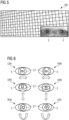
- FIG 5 a moving chessboard MC used as optokinetic stimulus for the eyes of a patient, is shown.
- the eyeballs E of the patient are shown, wherein each of the eyeballs E is directed to a different direction which may be a sign of a nystagmus caused by vertigo.
- FIG 6 a schematic illustration of different types of a nystagmus movement of the eyes E of a patient P in different directions is depicted.
- a horizontal nystagmus is illustrated, wherein there is a horizontal difference between the positions of the centres of the eyes E of the patient which means that a movement of the eyeballs in the horizontal direction is shown.
- the pupil with the sparser dashing symbolizes the displaced eyeball EY and the pupil with the denser dashing (from left down to right up) symbolizes the original position of the eyeball EY.
- a left beating nystagmus LBN is shown, wherein the direction of the view of the eye E is displaced to the left side.
- a right-beating nystagmus RBN is shown, wherein the direction of the view of the eye E is displaced to the right side.
- the second line there is a vertical Nystagmus shown, wherein there is a vertical difference between the positions of the centres of the eyes E of the patient.
- an up-beating nystagmus UBN is shown and on the right side of the second line, a down-beating nystagmus DBN is shown.
- a rotary nystagmus is illustrated, wherein there is a diagonal difference between the eyeballs EY of the patient.
- a right rotating nystagmus RRN is shown and on the right side of the third line, a left rotating nystagmus LRN is shown.
- FIG 7 a schematic sectional view onto a brain and the eyeballs EY of patient is illustrated.
- the eyeballs EY of the patient are both directed in a straight direction SD and the optic nerves ON are directed in a divergent direction DD.
- FIG 8 a schematic sectional view onto a brain and the eyeballs EY of a patient, wherein the eyeballs EY are turned to the left side, is illustrated.
- a displacement DON of the optic nerve ON, in particular of the left eye EY compared to FIG 7 is shown.
- the displacement D-ON is represented by the angle ⁇ .
- the angle ⁇ is defined by the different, i.e. divergent directions DD, D-ON of the optic nerve ON in the straight view direction SD of the eyes as shown FIG 6 and the view direction LD to the left as shown in FIG 7 .
- FIG 9 a schematic sectional view onto a brain and the eyeballs EY of a patient and a grid G laid over the sectional view are depicted.
- the cells C of the grid G, including the optic nerve ON1, ON2 of the left eye EY1 and the right eye EY2 are marked.
- the optic nerve ON1 of the left eye only includes three cells C and the optic nerve ON2 of the right eye includes five cells C.
- an overall bounding box for each optic nerve ON1, ON2 first, in each cell C including a part of an optic nerve ON1, ON2, an elementary bounding box is marked.
- FIG 10 for the right eye EY2, a plurality of part bounding boxes PBB for each cell C of the grid G and expanding from each cell C of the grid G shown in FIG 9 which contains the right optic nerve ON2 are illustrated. In contrast thereto, for the left eye EY1, these bounding boxes PBB have been replaced by an overall bounding box BB.
- the part bounding boxes PBB and the overall bounding box BB are generated based on "Intersection over Union" (IoU).
- FIG 11 two fused bounding boxes, each related to a single optic nerve, are shown.
- the width W and the height H of the bounding box BB of the left eye EY1 are indicated.
- a coordinate system (x, y) including an origin (0,0) in the lower left corner of the bounding box BB of the left eye EY1 and a point (1,1) in the upper right corner of the bounding box BB of the left eye EY1 are depicted.
- FIG 12 a schematic illustration of an eye movement determination device 10 according to an embodiment of the invention is illustrated.
- the eye movement determination device 10 includes a stimulation unit 11 for providing a patient P (shown in FIG 13 ) with an optokinetic stimulus OS for a nystagmus.
- the eye movement determination device 10 comprises an image interface unit 12 for receiving a medical image BD of the brain of a patient P and a time stamp TS assigned to each medical image BD from a reconstruction unit or a scan unit of a medical imaging system (not shown in FIG 12 ).
- Part of the eye movement determination device 10 is also a displacement determination unit 13 for AI-based determining an amount A-DP of a displacement of the optic nerves ON of the eyes of a patient based on the medical image BD.
- the eye movement determination device 10 also comprises an analysis unit 14 for determining if a nystagmus is detected based on the determined amount A-DP of displacement of the optic nerve ON and of what type the detected nystagmus is. For example, the velocity v of the eye movement related to the nystagmus is determined and based on the value of the velocity v, it is determined if the eye movement is related to a special physiological phenomenon like a nystagmus N caused by vestibular migraine.
- FIG 13 a schematic illustration of a medical imaging system 1 according to an embodiment of the invention is represented.
- the medical imaging system 1 comprises a scan unit 2, in which medical images BD are recorded from a patient P.
- the medical imaging system 1 also comprises a stimulation unit 11.
- the stimulation unit 11 generates an optokinetic stimulus OS for stimulating an eye movement of the patient P, for example a moving chessboard MC (shown in FIG 5 ) and applies the optokinetic stimulus OS to the patient P.
- Part of the medical imaging system 1 is also an eye movement determination device 10 as it is shown in FIG 12 .
Landscapes
- Engineering & Computer Science (AREA)
- Health & Medical Sciences (AREA)
- Life Sciences & Earth Sciences (AREA)
- Physics & Mathematics (AREA)
- Computer Vision & Pattern Recognition (AREA)
- Theoretical Computer Science (AREA)
- General Physics & Mathematics (AREA)
- Medical Informatics (AREA)
- General Health & Medical Sciences (AREA)
- Molecular Biology (AREA)
- Heart & Thoracic Surgery (AREA)
- Surgery (AREA)
- Animal Behavior & Ethology (AREA)
- Biomedical Technology (AREA)
- Public Health (AREA)
- Veterinary Medicine (AREA)
- Biophysics (AREA)
- Nuclear Medicine, Radiotherapy & Molecular Imaging (AREA)
- Radiology & Medical Imaging (AREA)
- Pathology (AREA)
- Quality & Reliability (AREA)
- Multimedia (AREA)
- Ophthalmology & Optometry (AREA)
- Developmental Disabilities (AREA)
- Educational Technology (AREA)
- Neurology (AREA)
- Neurosurgery (AREA)
- Physiology (AREA)
- Child & Adolescent Psychology (AREA)
- Human Computer Interaction (AREA)
- Signal Processing (AREA)
- Hospice & Palliative Care (AREA)
- Psychiatry (AREA)
- Psychology (AREA)
- Social Psychology (AREA)
- High Energy & Nuclear Physics (AREA)
- Eye Examination Apparatus (AREA)
- Image Analysis (AREA)
Abstract
Description
- The invention relates to a method for determining a type of characteristic eye movement of a patient. The invention also concerns a method for training an AI-based algorithm (AI = artificial intelligence) for determining an amount of a displacement of an optic nerve of a patient. Furthermore, the invention relates to an eye movement determination device. The invention also relates to a medical imaging system.
- Eye movement or oculomotor function is the term used to describe the movements of the bulbus oculi (eyeball) caused by the external eye muscles, which in humans are physiologically conjugated with the contralateral eye. In the type of eye following movement, a moving object is fixed. The eye follows the movements of the object by slow eye following movements. Saccades are rapid movements of the eye with a constant change of fixation point. Only the images from the time of fixation are perceived, the image shifts are suppressed. Nystagmus is the combination of saccades and eye following movements. The indication of the direction of the nystagmus depends on the rapid recoil movement. Knowledge of the brain structures responsible for eye movements makes it possible in many cases to recognize the underlying disease process or to localize the disease anatomically. Therefore, the examination of ocular motility is an important diagnostic procedure for many neurological and neuro-ophthalmological diseases.
- Vestibular migraines as a type of neurological disease are a phenomenon which affects millions of people across the world. The term "migraine" is more or less familiar to everyone these days. Scientists assume that migraine symptoms are caused by a rapid drop in blood flow to the brain and the resulting change in activity in the affected brain regions. If the brain regions that belong to the vestibular system are affected, dizziness, and/ or vertigo, can be the main symptom.
- Attack-like permanent spinning or swaying dizziness occurs, which can last for minutes, hours - sometimes even several days. As with other types of migraine, some sufferers complain of increased sensitivity to light, noise or movement and therefore have an increased need for rest during the attack. In addition, there is often severe nausea as well as gait, balance and vision disorders. Headaches can occur before, during or after an attack, but are absent in good 30% of the cases. This often makes it difficult to distinguish it from the clinical picture of Meniere's disease, which causes similar symptoms.
- Further, vestibular migraines may show symptoms that appear also in many other diseases. Hence, vestibular migraines are hard to diagnose and are not accurately diagnosed using the current methods that exist. Further, the time taken to declare that a patient has vestibular migraine is high and at times uncertain without conclusive evidence. Hence, sometimes, patients are put on clinical correlation on a trial basis for years to a lifetime.
- Until today, in case a person suffers from vertigo, a magnetic resonance image of brain is normally generated, however the magnetic resonance image of brain is generated for ruling out disorders such as a tumor, epilepsy or multiple sclerosis. However, vestibular migraines are not detected based on a magnetic resonance image of brain. Hence, a diagnose of vestibular migraines is relatively uncertain. Therefore, a patient is merely put under clinical correlation and trial medicines, which may or may not help the patient, are applied to the patient.
- Further, a test called video nystagmography, which is almost unheard of in many countries across the world, is used to detect the disorders causing vertigo. A video mask with an integrated camera is placed on the patient. With darkening (removal of the optical fixation), the movements of the pupils can be tracked automatically with an infrared camera and thus spontaneous or induced nystagmus can be recorded.
- However, the reliability of such a test completely depends on the correlation experience or a technician conducting the test. Hence, it is not 100% accurate.
- Thus, there is a problem of providing reliably measurement data of a patient used as a basis for unambiguously determining if the patient suffers from a certain neurological disease, in particular vestibular migraines, or not.
- The before-mentioned problem is solved by a method for determining a type of a characteristic eye movement of a patient according to
claim 1, by a method for training an AI-based algorithm for determining an amount of a displacement of an optic nerve of a patient according toclaim 11, by an eye movement determination device according toclaim 12 and by a medical imaging system according toclaim 13. - In the method for determining a type of eye movement of a patient, the patient is provided with an optokinetic stimulus for a characteristic eye movement, preferably a nystagmus. Further, a medical image of the brain of a patient, also including the eyes of the patient, is generated. A patient comprises a human person. The medical image is recorded timed with the generation of the optokinetic stimulus, such that the medical image represents the reaction of the eyes of the patient to the optical stimulus. Preferably, the medical image is recorded simultaneously to the optical stimulation or directly or short after generation of the optical stimulus. As later described in detail, "a medical image" may comprise exactly one medical image, preferably more than one medical image and particularly preferred a whole sequence of a plurality of medical images such that a kind of video sequence of the eyes of the patient is achieved.
- In other words, the medical image preferably comprises a sectional internal view of the head of the patient. More preferably, as sectional plane, a transversal plane relatively to the longitudinal axis of the body of the patient is used which is positioned at the height position of the eyes of the patient such that in addition to the brain, also a sectional view of the eyes including eyeballs and the optic nerves of the patient is shown. Hence, a medical image means an image of the interior of the head of the patient, preferably a sectional view, which also comprises a sectional depiction of the eyes of the patient. The medical image may include a plurality of such sectional views in different planes with the same or with different orientations such that a direction of the optic nerve in a two dimensional direction or even in a three dimensional direction can be derived from the medical image. Based on the medical image, an amount of a displacement of an optic nerve and velocity of the displacement of the optic nerve of the patient is determined.
- An optokinetic stimulus comprises a video, which displays one or a plurality of moving objects for a patient, wherein the moving object exerts a movement stimulus to the eyes of the patient.
- Then it is determined if the characteristic eye movement, preferably a nystagmus, is detected based on the determined amount of displacement of the optic nerve and preferably the velocity of displacement of the optic nerve. For a normal healthy patient, when optokinetic stimuli are shown, optic nerve displacement may not happen at all. In only happens if the healthy patient moves the eye intentionally. Normally, optokinetic stimuli do not have any effect on a healthy patient. The determination of the eye movement based on the optic nerve is highly deterministic since medical images of the optic nerve clearly record the optic nerve displacement. The displacement of the optic nerve and preferably the velocity of the displaced optic nerve can be measured by comparing a medical image recorded without optokinetic stimulation and a medical image recorded simultaneously with such an optokinetic stimulation. The velocity of displacement of the optic nerve is higher in patients with vestibular migraine. Since such medical images may be part of routine scans for example based on symptoms like vertigo, such a determination may reduce the turnaround time for a diagnosis of a neurological disease, in particular a diagnosis of vestibular migraine in case of the appearance of vertigo. Further, a perturbation of the patient by trial and error medicine can be avoided due to the reliable determination of the type of characteristic movement, in particular a nystagmus movement, of the eyes.
- It has to be mentioned that there are a plurality of different neurological disorders that can be determined based on a characteristic eye movement. For example, in the case of the slow following eye movements, one checks whether these are smooth or saccadic. The latter speaks for central oculomotor dysfunction. A slightly saccaded vertical downward gaze is also often found in healthy subjects. Physiological visual fixation suppression of the vestibular-ocular reflex is also an important test and is impaired in central cerebellar lesions.
- Attention should be paid to the speed and accuracy of the saccades and to whether both eyes move in parallel. Hypermetric saccades are found in cerebellar disorders, hypometric saccades mostly in brainstem lesions and neuro-degenerative diseases. In the case of progressive supranuclear gaze palsy - an important differential diagnosis for idiopathic Parkinson's syndrome - there is usually initially a slowing down of the vertical saccades, then in the course of the disease also the horizontal saccades and finally a general gaze palsy.
- The eye movement determination device according to the invention comprises a stimulation unit for providing the patient with an optokinetic stimulus for a predetermined type of characteristic eye movement, preferably a nystagmus. Further, the eye movement determination device comprises an image interface unit for receiving a medical image of the brain and the eyes of a patient from a medical imaging system or a scan unit of a medical imaging system. A part of the eye movement determination device is also a displacement determination unit for determining an amount of a displacement of the optic nerve(s) and velocity of the displacement of the optic nerve(s) of the patient based on the medical image and an analysis unit for determining if the predetermined characteristic eye movement, preferably a nystagmus movement, is detected based on the determined amount of displacement of the optic nerve(s) and the velocity of the displaced optic nerve(s) of the patient. The eye movement determination device shares the advantages of the method for determining a type of an eye movement of a patient.
- The medical imaging system according to the invention comprises a scan unit for acquiring medical imaging data from a patient and an eye movement determination device according to the invention. The medical imaging system shares the advantages of the eye movement determination device according to the invention and the method for determining a type of an eye movement of a patient according to the invention.
- Some units or modules of the eye movement determination device mentioned above can be completely or partially realized as software modules running on a processor of a respective computing system, e.g. of a control device of a medical imaging system. A realization largely in the form of software modules can have the advantage that applications already installed on an existing computing system can be updated, with relatively little effort, to install and run these units of the present application. The object of the invention is also achieved by a computer program product with a computer program that is directly loadable into the memory of a computing system, and which comprises program units to perform the steps of the inventive method, at least those steps that could be executed by a computer, especially the steps of determining an amount of a displacement of the optic nerve and velocity of the displacement of the optic nerve of the patient based on the medical image and determining if a characteristic eye movement, preferably a nystagmus movement, is detected based on the determined amount of displacement of the optic nerve and the velocity of the displacement of the optic nerve, when the program is executed by the computing system. In addition to the computer program, such a computer program product can also comprise further parts such as documentation and/or additional components, also hardware components such as a hardware key (dongle etc.) to facilitate access to the software.
- A computer readable medium such as a memory stick, a harddisk or other transportable or permanently-installed carrier can serve to transport and/or to store the executable parts of the computer program product so that these can be read from a processor unit of a computing system. A processor unit can comprise one or more microprocessors or their equivalents.
- The dependent claims and the following description each contain particularly advantageous embodiments and developments of the invention. In particular, the claims of one claim category can also be further developed analogously to the dependent claims of another claim category. In addition, within the scope of the invention, the various features of different exemplary embodiments and claims can also be combined to form new exemplary embodiments.
- Preferably, the medical image or the medical images are recorded using a magnetic resonance imaging system. As mentioned above, in case a person suffers from vertigo, a magnetic resonance image of brain is normally generated, wherein the magnetic resonance image of brain is generated for ruling out disorders such as a tumor, epilepsy or multiple sclerosis. Preferably, the method according to the invention can advantageously be combined with such a routine medical imaging.
- Hence, particularly preferred, the medical image comprises a magnetic resonance image or even a sequence of magnetic resonance images. Advantageously, such magnetic resonance images, which are recorded for ruling out the above-mentioned severe disorders, can be additionally used for detecting and identifying a type of a characteristic eye movement to find out, in case the above-mentioned severe diseases are ruled out, from which disease the patient suffers indeed. Hence, there is no need for additional separate examinations and time and medical resources can be saved. Particularly preferred, the magnetic resonance images are recorded using a dynamic sequence, in particular a Cine sequence. Advantageously, a movement of the eyes of a patient can be easily recorded.
- In case the routine medical image of the brain is without any noticeable finding, the method for determining a type of characteristic eye movement of a patient, wherein an optic nerve displacement is detected using an optokinetic stimulus can be performed afterwards according to a new routine protocol and based on a new sequence. As later discussed, for detecting and identifying a special type of a characteristic eye movement, a dynamic medical imaging method and in particular a dynamic magnetic resonance imaging method is preferred. In case a magnetic resonance imaging method is used, which is advantageous due to an absence of radiation, preferably a Cine sequence is used. Such a dynamic magnetic resonance imaging method, in particular a Cine sequence, including a plurality of subsequently recorded frames, enables a real time recording of movements of internal organs of a patient, in particular the optic nerves of the eyes of a patient. Advantageously, also a time stamp of all the recorded frames is determined such that an exact place-time behaviour of the optic nerves of a patient can be recorded.
- Hence, preferably, in the method according to the invention, the medical image comprises a series or sequence of medical images. Advantageously, a movement of the eyes of the patent can be recorded and compared with predetermined movement patterns for determining a characteristic eye movement. Based on the characteristic eye movement, a specific neurological disease can be determined.
- Preferably, in the method according to the invention, the optokinetic stimulus comprises a movement of an object in a predetermined direction. The predetermined direction of the movement of the object comprises at least one of:
- a movement from left to right,
- a movement from right to left,
- a movement up and down,
- a rotatory motion.
- Advantageously, different movement patterns can be stimulated by the optokinetic stimulus. Optokinetic stimuli comprise many patterns which trigger nystagmus. This could be horizontal (left or right) or vertical (up or down). Hence, it is important to show all sorts of patterns in optokinetic stimuli because we do not know what kind of nystagmus a patient with a specific disease, in particular vestibular migraines, can get.
- Preferably, the optokinetic stimulus comprises a video image of a distorted moving chessboard. Advantageously, such a moving pattern stimulates eye movements, in particular nystagmus movements of the eyes. Such a nystagmus comprises at least one of horizontal (left or right movement), vertical (up or down movement) or rotatory movements.
- Preferably, the step of determining an amount of a displacement of the optic nerve comprises an AI-based determination (AI = artificial intelligence). Advantageously, such an AI-based determination of a quantity of a displacement of the optic nerve enables to avoid an error prone and non-deterministic manual determination by a radiologist, which can often lead to a misdiagnosis, because a patient can move his/her eyes during the display of the optokinetic stimulus. For generating an AI-based determination of an amount of displacement of the optic nerve, an AI model or algorithm is created and trained to detect a specific type of an eye displacement or even movement which is characteristic for a special neurological phenomenon, in particular a special neurological disease. As later described in detail, such a type of characteristic eye movement preferably comprises a type of nystagmus. Such a nystagmus may include at least one of the following types:
- a left beating nystagmus,
- a right beating nystagmus,
- an up beating nystagmus,
- a down beating nystagmus.
- All these nystagmus movements may be indications of a vestibular migraine. If patient sees optokinetic stimuli and gets a nystagmus, then it means patient has vestibular migraines. Now that nystagmus can be anything, for example left horizontal or right horizontal or up vertical or down vertical.
- The method for training an AI-based algorithm for determining an amount of a displacement of an optic nerve of a patient based on at least one medical image, comprises the steps of receiving labelled medical image data. The corresponding time stamp of these medical images are also recorded. With the amount of displacement of optic nerve and the time stamps of medical images, velocity of displayed optic nerve is calculated through the formula velocity equals distance divided by time. This velocity will be higher for patients with vestibular migraines.
- For training an artificial intelligence model, as labelled medical image data a dataset with two different sets of medical images is needed: a first training set with medical images related to a patient with regular eye movement and a second training set with medical images of patients with the type of characteristic eye movement to be detected. As mentioned-above, such a characteristic eye movement may be related to a special type of neurological disease, in particular a vestibular migraine.
- For generating labelled training data, each training medical image is manually provided with a bounding box which marks the location of the optic nerve. Further a simple text is added giving the classification of the marked object, in this case the optic nerve, as well as the coordinates and the size of its bounding box. The more medical images the training data set contains, the more effective the training will be. The accuracy of the training can be tested by providing a labelled test dataset. The outcome of this test is a score called mean average precision, which ranges from 0 to 1 (i.e. 0 to 100 %).
- After the training is completed and a high enough mean average precision score has been achieved, the output is a trained model or algorithm, which can be delivered to the eye movement determination device and be used to detect live data. A mean average precision score nearing 1 will allow for reliant and accurate localization of the optic nerve, thus providing accurate diagnosis.
- Preferably, the detection of the optic nerve is realized by using a "You Only Look Once"-algorithm (by short YOLO-algorithm). Such a YOLO-algorithm is a performant algorithm allowing for real time analysis of the medical image. A YOLO-algorithm is described in Joseph Redmon, Santosh Divvala, Ross Girshick, Ali Fahrhadi, "You Only Look Once: unified, Real-time Object Detection", University of Washington, Allen Institute for AI, Facebook AI Research. The YOLO-algorithm is useful to analyse a high quantity of images. As above-mentioned, in case a whole sequence of images is analysed and a pathologic neurological phenomenon exists, in particular vestibular migraine, each frame or image of such a sequence of images contains a differently localized part of the optic nerve compared to a localization of an optic nerve related to a normal or regular eye movement. Advantageously, a YOLO-algorithm is a performant algorithm to detect the optic nerve reliably and quickly such that it allows for real time analysis of the medical image(s).
- Preferably, in an analogue manner to the above-mentioned step in the training of the method according to the invention, the step of AI-based determining an amount of a displacement of the optic nerve comprises the sub-step of segmenting the optic nerve in the medical image. As later described in detail, identification, localization and segmentation of the optic nerve is preferably achieved using the above-mentioned YOLO-algorithm.
- Preferably, the sub-step of segmenting the optic nerve comprises the following steps:
- splitting the medical image into a grid including a plurality of cells,
- detecting the most likely object within each cell of the grid,
- marking each cell in which the optic nerve is detected by a bounding box,
- using of "Intersection over Union" for creating a unified single bounding box of the detected optic nerve.
- Preferably, the medical image is split into a grid of equal dimension, typically in a grid of 7*7 rectangle cells. For each cell, the detection of the most likely object within each cell includes the determination if there is a part of an optic nerve.
- For each cell which contains an optic nerve or a part of the optic nerve a bounding box, predicting the borders of the optic nerve, is generated. That process results in multiple bounding boxes surrounding different portions of the complete area of the optic nerve.
- For providing just one single bounding box for one single optic nerve, a unification process is used, preferably based on "Intersection over Union". With a "single bounding box" one overall bounding box for one single optic nerve is meant. Intersection over Union (IoU by short), also named Jaccard index or Jaccard similarity coefficient, is a metric that allows us to evaluate how similar our predicted bounding box is to the ground truth bounding box. The idea is that we want to compare the ratio of the area where the two bounding boxes overlap to the total combined area of the two boxes. Principles of "Intersection over Union" and also a generalized version is described in Hamid Rezatofighi et al. "Generalized Intersection over Union: A Metric and A Loss for Bounding Box Regression", Computer Science Department, Stanford University, United States School of Computer Science, The University of Adelaide, Australia Aibee Inc, USA.
- The score related to Intersection over Union results from dividing the area of overlap between the ground truth bounding box and the predicted bounding box through the area of union.
- Each grid cell gives a prediction of its bounding box together with the confidence score. Confidence score is one parameter for the IoU calculation. In case the confidence score is equal to 1, the bounding box represents the real overall bounding box. This mechanism eliminates bounding boxes that are not equal to the real bounding box of an optic nerve. Hence, we get a final detection result as a bounding box for each detected optic nerve. That bounding box is also assigned to attributes such as the dimension and the position of the centre of the bounding box. The dimension in particular comprises values related to the height and the width of the bounding box.
- Preferably, an angle of the orientation of the detected optic nerve is calculated based on the height and width of the bounding box and the amount of a displacement of the optic nerve is determined based on the calculated angle. Using the diagonal of the bounding box as hypotenuse of a right angled triangle comprising the width and the height of the bounding box, the hypotenuse can be calculated based on the height and the width of the bounding box based on Pythagorean theorem. Then the angle α can be calculated as:
- α = arccos (height/hypotenuse) or
- α = arcsin (width/hypotenuse).
- In the method according to the invention, preferably, a series of medical images is generated and a velocity of an eye movement is determined based on the amount of displacement and a time stamp of each of the medical images. Further, the eye movement pattern, preferably a nystagmus, is detected based on the determined velocity. If the angle α is calculated for each frame of the recorded medical images, a live tracking is enabled on how and how fast the optic nerve has moved during the medical imaging under various situations. Based on the time stamps, it is well known for an accuracy of milliseconds, when these medical images have been taken. Hence, also a velocity of the recorded eye movement is determined. Such a velocity of the recorded eye movement can be compared with reference values for determining if an eye movement has been recorded which is characteristic for a specific physiologic condition of the patient.
- In particular, if a nystagmus is detected and the velocity is greater than the velocity captured during a regular eye movement, then it is determined that the patient is considered to experience vertigo based on an optical stimulus. Such a physiological condition may be used for finding that the patient suffers from vestibular migraines. Hence, based on a sequence of medical images, preferably magnetic resonance images, it is enabled to find out if a patient suffers from a neurologic disease, in particular vestibular migraines.
- The invention is explained below with reference to the figures enclosed once again. The same components are provided with identical reference numbers in the various figures. The figures are usually not to scale.
-
FIG 1 shows a flow chart diagram illustrating the method for determining a type of a characteristic eye movement of a patient according to an embodiment of the invention, -
FIG 2 shows a flow chart diagram illustrating details of step 1.III and 1.IV of the method for determining a type of a characteristic eye movement of a patient shown inFIG 1 , -
FIG 3 shows a flow chart diagram illustrating a method for training an AI-based algorithm for determining an amount of a displacement of an optic nerve of a patient according to the invention, -
FIG 4 shows a table showing parameter values of labelled medical image data used for training the AI-based algorithm, -
FIG 5 shows a moving chessboard used as optokinetic stimulus for the eyes of a patient, -
FIG 6 shows a schematic illustration of a nystagmus movement of the eyes of a patient in different directions, -
FIG 7 shows a schematic sectional view onto a brain and the eyes of a patient looking straight at the centre, -
FIG 8 shows a schematic sectional view onto a brain and the eyes of a patient with a left beating nystagmus while viewing optokinetic stimuli, -
FIG 9 shows a schematic sectional view onto a brain and the eyes of a patient and a grid laid over the sectional view, -
FIG 10 shows a plurality of bounding boxes for each cell of the grid shown inFIG 9 that contains the optic nerve, -
FIG 11 shows two fused bounding boxes, each related to a single optic nerve, -
FIG 12 shows a schematic illustration of an eye movement determination device according to an embodiment of the invention, -
FIG 13 shows a schematic illustration of a medical imaging system according to an embodiment of the invention. - In
FIG 1 , a flow chart diagram 100 illustrating the method for determining a type of a characteristic eye movement of a patient P according to an embodiment of the invention, is shown. - In step 1.1, an optokinetic stimulus OS for a nystagmus is shown to the patient P. Such an optokinetic stimulus OS may include a moving chessboard, which is shown in
FIG 5 . The optokinetic stimulus OS is generated simultaneously or directly before a generation of a sequence of magnetic resonance images BD from the patient P, such that a reaction of the patient P, in particular a movement of the optic nerves ON of the patient P is recorded on the magnetic resonance images BD. - In step 1.II, the mentioned sequence of magnetic resonance images BD using a dynamic Cine magnetic resonance imaging pulse sequence is generated from a brain of a patient P. Vividly spoken, a video of a sectional view of the head of the patient P is generated. As sectional view in a transversal plane relative to the longitudinal axis of the body of the patient P is used which is positioned at the height position of the eyes of the patient P such that in addition to the brain, also a sectional view of the eyes of the patient P is shown in the medical images BD. Such a sectional view is illustrated in
FIG 5 to FIG 9 . To each of these recorded medical images BD, a time stamp TS is assigned which indicates the time at which the medical image has been recorded. - In step 1.III, an amount A-DP of a displacement of the optic nerves ON is measured based on an AI-based displacement determination process.
- In step 1.IV, it is determined if a nystagmus correlated to a vertigo phenomenon is detected based on the determined amount A-DP of displacement of the optic nerve ON and the velocity with which this displacement of the optic nerve was achieved. Based on the determination of such a nystagmus a vestibular migraine is enabled to be diagnosed.
- In
FIG 2 , a flow chart diagram depicting details of step 1.III and 1.IV of the method for determining a characteristic eye movement of a patient shown inFIG 1 is illustrated. - In step 1.IIIa, a medical image BD recorded in step 1.1, is overlayed by a grid G. Such a grid G may include cells C of equal dimension (7*7) as shown in
FIG 9 . - In step 1.IIIb, the most likely object MLO within each cell C of the grid G is detected, i.e. it is determined in which cell at least a part of the optic nerve ON is localized. For detecting and localizing the optic nerves ON reliably and quickly, a YOLO algorithm is used.
- In step 1.IIIc, each cell in which the optic nerve ON is detected is marked by a part bounding box PBB.
- In step 1.IIId, an Intersection over Union IoU metric is used for creating a unified single bounding box BB. Hence, the part bounding boxes PBB are unified to form together for each optic nerve ON a single overall bounding box.
- In step 1.IIIe, an angel α representing the orientation of the detected optic nerve ON is calculated based on the height H and width W of the bounding box BB. Then, based on the detected angle α, the amount A-DP of displacement of the optic nerve ON is determined based on the calculated angle α.
- Further, the method returns to step 1.1 for recording additional medical images BD from the head of the patient P and assigning to each of these medical images BD and to the related amount of displacement A-DP a time stamp TS.
- Then, in step 1.IVa, a velocity v of the eye movement of the patient P is calculated based on each determined amount of displacement A-DP and each time stamp TS of each of the medical images BD.
- In step 1.IVb, it is determined based on the calculated velocity v of the patient's eyes if the patient P suffers from a vertigo caused nystagmus N or not.
- In
FIG 3 , a flow chart is shown illustrating a method for training an AI-based algorithm A for determining an amount A-DP of a displacement of an optic nerve ON of a patient P based on at least one medical image BD. The algorithm A can be used for performing step 1.III illustrated inFIG 1 and FIG 2 . Such a training method is performed before the algorithm A is used in step 1.III of the method for determining a type of a characteristic eye movement of a patient P. - In step 3.1, labelled medical image data L-BD comprising an optic nerve ON of a patient P are received. These labelled medical image data L-BD comprise a dataset with two different sets S1, S2 of medical images, a first training set S1 with medical images BD related to a patient P with regular eye movement and a second training set S2 with medical images BD of patients P with a specified type of characteristic eye movements to be determined. The specified type of characteristic eye movement differs from the regular eye movement and may be related to a specified physiological state, in particular to a disease like vestibular migraine. Such labelled medical image data L-BD are symbolized in the table T shown in
FIG 4 . Labelled medical image data L-BD means that the images comprise correct results as reference results. These reference results can be compared with results generated by the algorithm to be trained based on the labelled medical image data L-BD. Based on the comparison, the architecture of the algorithm can be modified such that the results of the algorithm are brought closer to the reference results related to the training data, i.e. the labelled medical image data L-BD. In that case, reference results comprise bounding boxes BB related to optical nerves and the dimensions of these bounding boxes, further also an angle α related to the bounding boxes BB and a velocity v of eye movement calculated based on a sequence of a plurality of medical images. - In step 3.II, the algorithm is trained based on the labelled medical image data L-BD. Such a "training" includes the step of feeding the algorithm A to be trained, including a modifiable algorithm architecture, for example an artificial neural network, with the labelled medical image data L-BD and modify the algorithm architecture based on a comparison between the result generated by the algorithm A and the above-mentioned reference results.
- In step 3.III, the trained algorithm A is provided for performing step 1.III illustrated in
FIG 1 and FIG 2 . - In
FIG 4 , a table T, illustrating sets S of different data sets S1, S2 used for the training method of an algorithm shown inFIG 3 is depicted. As mentioned above, such different data sets S1, S2 include a first training set S1 with medical images BD related to patients P with regular eye movement and a second training set S2 with medical images BD of patients P with a specified type of characteristic eye movement to be determined, in that special case a nystagmus stimulated by an optokinetic stimulus. As mentioned-above, the detection of such a nystagmus is an indicator for vertigo related to vestibular migraines. - The table T shows reference data, which are used for labelling these training data sets S1, S2. In the first column, an angle α of the detected eye movement is represented. The movement angle α of the recorded movement of the eyes of a patient P is always 30° for both sets S1, S2 and all images of these sets, since the optokinetic stimulus used for each patient P is related to a movement of 30°. In the second column of the table T, the time t (in seconds s) which is needed for completing the recorded eye movement of the patient P is displayed. Further, for each type of an optokinetic stimulus OS, in neighboured lines of table T, times t related to the first set S1 and times t related to the corresponding second set S2 are shown as it is indicated by the fifth column. As can be taken from the table T, the values of times t for the regular eye movement stored in the first set S1 is longer than the time of the second set S2, which corresponds to the same optokinetic stimulus. In the third column, the eye movements EM related to different optokinetic stimuli and to the sets S1, S2 are depicted. As can be taken from the table T, related to the first set S1 regular eye movements to the left LEM, to the right REM, up UEM and down DEM are shown. Further corresponding characteristic types of eye movements i.e. a left beating nystagmus LBN, a right beating nystagmus RBN, an up beating nystagmus UBN and a down beating nystagmus DBN are shown in the table T represented by the second training set S2. In the fourth column, the number IN of images or image sequences, i.e. 100 images or sequences of images, corresponding to these different types of eye movements are depicted.
- In
FIG 5 , a moving chessboard MC used as optokinetic stimulus for the eyes of a patient, is shown. In the lower right corner ofFIG 3 , the eyeballs E of the patient are shown, wherein each of the eyeballs E is directed to a different direction which may be a sign of a nystagmus caused by vertigo. - In
FIG 6 , a schematic illustration of different types of a nystagmus movement of the eyes E of a patient P in different directions is depicted. In the first line, a horizontal nystagmus is illustrated, wherein there is a horizontal difference between the positions of the centres of the eyes E of the patient which means that a movement of the eyeballs in the horizontal direction is shown. The pupil with the sparser dashing (from left up to right down) symbolizes the displaced eyeball EY and the pupil with the denser dashing (from left down to right up) symbolizes the original position of the eyeball EY. On the left side of the first line, a left beating nystagmus LBN is shown, wherein the direction of the view of the eye E is displaced to the left side. On the right side of the first line, a right-beating nystagmus RBN is shown, wherein the direction of the view of the eye E is displaced to the right side. - In the second line, there is a vertical Nystagmus shown, wherein there is a vertical difference between the positions of the centres of the eyes E of the patient. On the left side of the second line, an up-beating nystagmus UBN is shown and on the right side of the second line, a down-beating nystagmus DBN is shown. In the lower line of
FIG 6 , a rotary nystagmus is illustrated, wherein there is a diagonal difference between the eyeballs EY of the patient. On the left side of the third line, a right rotating nystagmus RRN is shown and on the right side of the third line, a left rotating nystagmus LRN is shown. - In
FIG 7 , a schematic sectional view onto a brain and the eyeballs EY of patient is illustrated. As can be taken fromFIG 7 , the eyeballs EY of the patient are both directed in a straight direction SD and the optic nerves ON are directed in a divergent direction DD. - In
FIG 8 , a schematic sectional view onto a brain and the eyeballs EY of a patient, wherein the eyeballs EY are turned to the left side, is illustrated. InFIG 8 , a displacement DON of the optic nerve ON, in particular of the left eye EY compared toFIG 7 is shown. The displacement D-ON is represented by the angle δ. The angle δ is defined by the different, i.e. divergent directions DD, D-ON of the optic nerve ON in the straight view direction SD of the eyes as shownFIG 6 and the view direction LD to the left as shown inFIG 7 . - In
FIG 9 , a schematic sectional view onto a brain and the eyeballs EY of a patient and a grid G laid over the sectional view are depicted. The cells C of the grid G, including the optic nerve ON1, ON2 of the left eye EY1 and the right eye EY2 are marked. As you can take fromFIG 9 , the optic nerve ON1 of the left eye only includes three cells C and the optic nerve ON2 of the right eye includes five cells C. For achieving an overall bounding box for each optic nerve ON1, ON2, first, in each cell C including a part of an optic nerve ON1, ON2, an elementary bounding box is marked. - In
FIG 10 , for the right eye EY2, a plurality of part bounding boxes PBB for each cell C of the grid G and expanding from each cell C of the grid G shown inFIG 9 which contains the right optic nerve ON2 are illustrated. In contrast thereto, for the left eye EY1, these bounding boxes PBB have been replaced by an overall bounding box BB. The part bounding boxes PBB and the overall bounding box BB are generated based on "Intersection over Union" (IoU). - In
FIG 11 , two fused bounding boxes, each related to a single optic nerve, are shown. InFIG 11 , also the width W and the height H of the bounding box BB of the left eye EY1 are indicated. Further, a coordinate system (x, y) including an origin (0,0) in the lower left corner of the bounding box BB of the left eye EY1 and a point (1,1) in the upper right corner of the bounding box BB of the left eye EY1 are depicted. - In
FIG 12 , a schematic illustration of an eyemovement determination device 10 according to an embodiment of the invention is illustrated. - The eye
movement determination device 10 includes astimulation unit 11 for providing a patient P (shown inFIG 13 ) with an optokinetic stimulus OS for a nystagmus. - Further, the eye
movement determination device 10 comprises animage interface unit 12 for receiving a medical image BD of the brain of a patient P and a time stamp TS assigned to each medical image BD from a reconstruction unit or a scan unit of a medical imaging system (not shown inFIG 12 ). - Part of the eye
movement determination device 10 is also adisplacement determination unit 13 for AI-based determining an amount A-DP of a displacement of the optic nerves ON of the eyes of a patient based on the medical image BD. - The eye
movement determination device 10 also comprises ananalysis unit 14 for determining if a nystagmus is detected based on the determined amount A-DP of displacement of the optic nerve ON and of what type the detected nystagmus is. For example, the velocity v of the eye movement related to the nystagmus is determined and based on the value of the velocity v, it is determined if the eye movement is related to a special physiological phenomenon like a nystagmus N caused by vestibular migraine. - In
FIG 13 , a schematic illustration of amedical imaging system 1 according to an embodiment of the invention is represented. Themedical imaging system 1 comprises ascan unit 2, in which medical images BD are recorded from a patient P. Themedical imaging system 1 also comprises astimulation unit 11. Thestimulation unit 11 generates an optokinetic stimulus OS for stimulating an eye movement of the patient P, for example a moving chessboard MC (shown inFIG 5 ) and applies the optokinetic stimulus OS to the patient P. Part of themedical imaging system 1 is also an eyemovement determination device 10 as it is shown inFIG 12 . - The above descriptions are merely preferred embodiments of the present disclosure, but not intended to limit the present disclosure, and any modifications, equivalent replacements, improvements, etc. made within the spirit and principle of the present disclosure should be included within the scope of protection of the present disclosure.
- Further, the use of the undefined article "a" or "one" does not exclude that the referred features can also be present several times. Likewise, the term "unit" or "device" does not exclude that it consists of several components, which may also be spatially distributed.
Claims (15)
- Method for determining a type of characteristic eye movement of a patient (P), comprising the steps of:- providing the patient (P) with an optokinetic stimulus (OS) for stimulating the characteristic eye movement,- generating at least one medical image (BD) of the brain of the patient (P) also including the eyes (E) of the patient (P),- determining an amount (A-DP) of a displacement of an optic nerve (ON) of the patient (P) based on the at least one medical image (BD),- determining a type of characteristic movement based on the determined amount (A-DP) of displacement of the optic nerve (ON) .
- Method according to claim 1, wherein the step of determining an amount (A-DP) of a displacement of an optic nerve (ON) of the patient (P) is performed based on artificial intelligence.
- Method according to claim 2, wherein the step of AI-based determining an amount (A-DP) of a displacement of the optic nerve (ON) comprises the sub-step of segmenting the optic nerve (ON) in the medical image (BD).
- Method according to claim 3, wherein the sub-step of segmenting the optic nerve (ON) in the medical image (BD) comprises a detection by using a "You Only Look Once"-algorithm.
- Method according to claim 3 or 4, wherein the sub-step of segmenting the optic nerve (ON) comprises the following steps:- splitting the medical image (BD) into a grid (G) including a plurality of cells (C),- detecting the most likely object (MLO) within each cell (C) of the grid (G),- marking each cell (C) in which the optic nerve (ON) is detected by a bounding box (PBB),- using "Intersection over Union" (IoU) for creating a unified single bounding box (BB) of the detected optic nerve (ON) .
- Method according to claim 5, wherein- an angle (α) of the orientation of the detected optic nerve (ON) is calculated based on the height (H) and width (W) of the single bounding box (BB) and- the amount (A-DP) of a displacement of the optic nerve (ON) is determined based on the calculated angle (α).
- Method according to any of the preceding claims, wherein- the optokinetic stimulus (OS) is designed for stimulating a nystagmus movement of the patient (P) in case the patient (P) suffers from vestibular migraine and- the type of characteristic movement comprises a nystagmus movement of the patient's eyes (EY1, EY2).
- Method according to any of the preceding claims, wherein the optokinetic stimulus (OS) comprises a movement of an object in a predetermined direction.
- Method according to any of the preceding claims, wherein the at least one medical image (BD) comprises a series of medical images (BD).
- Method according to claim 9, wherein- a velocity (v) of an eye movement is determined based on the amount (A-DP) of displacement and a time stamp (TS) of each of the medical images (BD),- the type of characteristic eye movement of the patient (P) is detected based on the determined velocity (v).
- Method for training an artificial intelligence based algorithm (A) for determining an amount (A-DP) of a displacement of an optic nerve (ON) of a patient (P) based on at least one medical image (BD), comprising the steps:- receiving labelled medical image data (L-BD) comprising an optic nerve (ON) of a patient (P), wherein the labelled medical image data (L-BD) comprise a dataset with two different sets (S1, S2) of medical images, a first training set (S1) with medical images (BD) related to a patient (P) with regular eye movement and a second training set (S2) with medical images (BD) of patients (P) with the type of characteristic eye movement to be determined,- training the algorithm (A) based on the labelled medical image data (L-BD),- providing the trained algorithm (A) for determining an amount (A-DP) of a displacement of an optic nerve (ON) of a patient (P) based on the at least one medical image (BD).
- Eye movement determination device (10), comprising:- a stimulation unit (11) for providing a patient (P) with an optokinetic stimulus (OS) for a type of characteristic eye movement of the patient (P),- an image interface unit (12) for receiving a medical image (BD) of the brain and the eyes (E) of the patient (P) from a medical imaging system (1),- a displacement determination unit (13) for determining an amount (A-DP) of a displacement of an optic nerve (ON) of the patient (P) based on the medical image (BD),- an analysis unit (14) for determining a type of characteristic movement based on the determined amount (A-DP) of displacement of the optic nerve (ON).
- Medical imaging system (1), comprising:- a scan unit (2) for acquiring medical imaging data (BD) from a patient (P),- an eye movement determination device (10) according to claim 12.
- A computer program comprising instructions which, when the program is executed by a computer, cause the computer to carry out the steps of the method according to any of the claims 1 to 11.
- A computer readable storage medium comprising instructions which, when executed by a computer, cause the computer to carry out the steps of the method of any of the claims 1 to 11.
Priority Applications (3)
| Application Number | Priority Date | Filing Date | Title |
|---|---|---|---|
| EP22189027.0A EP4316350B1 (en) | 2022-08-05 | 2022-08-05 | Image based detection of characteristic eye movements |
| US18/230,300 US11983876B2 (en) | 2022-08-05 | 2023-08-04 | Image based detection of characteristic eye movements |
| CN202310980941.5A CN117522770A (en) | 2022-08-05 | 2023-08-04 | Image-based detection of characteristic eye movements |
Applications Claiming Priority (1)
| Application Number | Priority Date | Filing Date | Title |
|---|---|---|---|
| EP22189027.0A EP4316350B1 (en) | 2022-08-05 | 2022-08-05 | Image based detection of characteristic eye movements |
Publications (2)
| Publication Number | Publication Date |
|---|---|
| EP4316350A1 true EP4316350A1 (en) | 2024-02-07 |
| EP4316350B1 EP4316350B1 (en) | 2026-01-07 |
Family
ID=82846299
Family Applications (1)
| Application Number | Title | Priority Date | Filing Date |
|---|---|---|---|
| EP22189027.0A Active EP4316350B1 (en) | 2022-08-05 | 2022-08-05 | Image based detection of characteristic eye movements |
Country Status (3)
| Country | Link |
|---|---|
| US (1) | US11983876B2 (en) |
| EP (1) | EP4316350B1 (en) |
| CN (1) | CN117522770A (en) |
Families Citing this family (1)
| Publication number | Priority date | Publication date | Assignee | Title |
|---|---|---|---|---|
| CN119606421B (en) * | 2025-02-12 | 2025-06-13 | 中国科学技术大学 | Method and device for evaluating ejection function based on semi-supervised deep segmentation model |
Citations (6)
| Publication number | Priority date | Publication date | Assignee | Title |
|---|---|---|---|---|
| US20190328231A1 (en) * | 2018-04-27 | 2019-10-31 | Washington University | Imaging nerve function and pathologies |
| CN110415252A (en) * | 2018-04-26 | 2019-11-05 | 北京连心医疗科技有限公司 | A kind of eye circumference organ segmentation method, equipment and storage medium based on CNN |
| CN113662659A (en) * | 2021-08-25 | 2021-11-19 | 中山大学中山眼科中心 | Eye parameter acquisition system based on 3D-MRI eyeball model |
| CN113744271A (en) * | 2021-11-08 | 2021-12-03 | 四川大学 | Neural network-based automatic optic nerve segmentation and compression degree measurement and calculation method |
| US20220058796A1 (en) * | 2018-09-12 | 2022-02-24 | Auckland Uniservices Limited | Methods and systems for ocular imaging, diagnosis and prognosis |
| US20220133145A1 (en) * | 2019-03-05 | 2022-05-05 | Centre Hospitalier Universitaire Vaudois (Chuv) | Magnetic resonance imaging method and device |
Family Cites Families (1)
| Publication number | Priority date | Publication date | Assignee | Title |
|---|---|---|---|---|
| TWI616188B (en) * | 2015-06-04 | 2018-03-01 | 神經元科技股份有限公司 | Rapid screening device for brain disease |
-
2022
- 2022-08-05 EP EP22189027.0A patent/EP4316350B1/en active Active
-
2023
- 2023-08-04 US US18/230,300 patent/US11983876B2/en active Active
- 2023-08-04 CN CN202310980941.5A patent/CN117522770A/en active Pending
Patent Citations (6)
| Publication number | Priority date | Publication date | Assignee | Title |
|---|---|---|---|---|
| CN110415252A (en) * | 2018-04-26 | 2019-11-05 | 北京连心医疗科技有限公司 | A kind of eye circumference organ segmentation method, equipment and storage medium based on CNN |
| US20190328231A1 (en) * | 2018-04-27 | 2019-10-31 | Washington University | Imaging nerve function and pathologies |
| US20220058796A1 (en) * | 2018-09-12 | 2022-02-24 | Auckland Uniservices Limited | Methods and systems for ocular imaging, diagnosis and prognosis |
| US20220133145A1 (en) * | 2019-03-05 | 2022-05-05 | Centre Hospitalier Universitaire Vaudois (Chuv) | Magnetic resonance imaging method and device |
| CN113662659A (en) * | 2021-08-25 | 2021-11-19 | 中山大学中山眼科中心 | Eye parameter acquisition system based on 3D-MRI eyeball model |
| CN113744271A (en) * | 2021-11-08 | 2021-12-03 | 四川大学 | Neural network-based automatic optic nerve segmentation and compression degree measurement and calculation method |
Non-Patent Citations (2)
| Title |
|---|
| HAMID REZATOFIGHI ET AL.: "Generalized Intersection over Union: A Metric and A Loss for Bounding Box Regression", THE UNIVERSITY OF ADELAIDE |
| JOSEPH REDMONSANTOSH DIVVALAROSS GIRSHICKALI FAHRHADI: "Facebook AI Research", UNIVERSITY OF WASHINGTON, article "You Only Look Once: unified, Real-time Object Detection" |
Also Published As
| Publication number | Publication date |
|---|---|
| CN117522770A (en) | 2024-02-06 |
| EP4316350B1 (en) | 2026-01-07 |
| US11983876B2 (en) | 2024-05-14 |
| US20240046475A1 (en) | 2024-02-08 |
Similar Documents
| Publication | Publication Date | Title |
|---|---|---|
| US10376139B2 (en) | Systems and methods for improved ease and accuracy of gaze tracking | |
| US9439592B2 (en) | Eye tracking headset and system for neuropsychological testing including the detection of brain damage | |
| CN112890815B (en) | Autism assisted assessment system and method based on deep learning | |
| US6381339B1 (en) | Image system evaluation method and apparatus using eye motion tracking | |
| Beltrán et al. | Computational techniques for eye movements analysis towards supporting early diagnosis of Alzheimer’s disease: a review | |
| US20210174959A1 (en) | System and method for detecting neurological disorders and for measuring general cognitive performance | |
| US20140364761A1 (en) | An apparatus and method for psychiatric evaluation | |
| WO2019015567A1 (en) | Brain function testing system and device thereof | |
| US10888263B2 (en) | Procedure of non-invasive video-oculographic measurement of eye movements as a diagnostic tool for (early) detection of neuropsychiatric diseases | |
| US20240029886A1 (en) | Method For Training And Quantifying Specific Motor Skills And Cognitive Processes In Persons By Analysing Oculomotor Patterns W Using A 3-D Virtual Reality Device With Embedded Eye-Tracking Technology, Specific Visual Stimuli, Sensors To Show The Movement Of Limbs | |
| CN118415588B (en) | Parkinson's disease diagnostic systems and devices based on virtual reality and eye-tracking technology | |
| CN111317448A (en) | A method and system for analyzing visual-spatial cognition | |
| EP4316350B1 (en) | Image based detection of characteristic eye movements | |
| US20230284962A1 (en) | Systems and methods for diagnosing, assessing, and quantifying brain trauma | |
| Sarker et al. | Analysis of smooth pursuit assessment in virtual reality and concussion detection using bilstm | |
| EP4561443A2 (en) | Identifying disease-specific oculomotor patterns using visual stimuli, drug or treatment | |
| JP7540711B2 (en) | Automatic eye movement recording system, calculation device, calculation method and program | |
| Takegami et al. | An algorithm for model‐based stable pupil detection for eye tracking system | |
| WO2025247410A1 (en) | Detection method and system for determining whether a patient suffers from strabismus and/or convergence insufficiency | |
| US20230368919A1 (en) | Method for identifying specific alterations in subjects with defined diseases analyzing oculomotor patterns when using specific visual stimuli, where a specific drug or treatment would enhance visual processing, cognitive performance and related brain activities | |
| Formisano et al. | AI-driven eye-tracking: Enhancing understanding and diagnosis of autism spectrum disorder | |
| Gienko et al. | Neurophysiological features of human visual system in augmented photogrammetric technologies | |
| KR20260049247A (en) | Method and system for determining the presence of traumatic brain injury | |
| Zhao et al. | 3DeepVOG: An open-source framework for real-time, accurate 3D gaze tracking with deep learning | |
| Vega | Self-calibrating eye tracker using imagesaliency: Självkalibrerande ögonspårare medhjälp av image saliency |
Legal Events
| Date | Code | Title | Description |
|---|---|---|---|
| PUAI | Public reference made under article 153(3) epc to a published international application that has entered the european phase |
Free format text: ORIGINAL CODE: 0009012 |
|
| STAA | Information on the status of an ep patent application or granted ep patent |
Free format text: STATUS: REQUEST FOR EXAMINATION WAS MADE |
|
| 17P | Request for examination filed |
Effective date: 20230918 |
|
| AK | Designated contracting states |
Kind code of ref document: A1 Designated state(s): AL AT BE BG CH CY CZ DE DK EE ES FI FR GB GR HR HU IE IS IT LI LT LU LV MC MK MT NL NO PL PT RO RS SE SI SK SM TR |
|
| RAP1 | Party data changed (applicant data changed or rights of an application transferred) |
Owner name: SIEMENS HEALTHINEERS AG |
|
| GRAP | Despatch of communication of intention to grant a patent |
Free format text: ORIGINAL CODE: EPIDOSNIGR1 |
|
| STAA | Information on the status of an ep patent application or granted ep patent |
Free format text: STATUS: GRANT OF PATENT IS INTENDED |
|
| RIC1 | Information provided on ipc code assigned before grant |
Ipc: A61B 3/00 20060101AFI20250829BHEP Ipc: A61B 3/113 20060101ALI20250829BHEP Ipc: A61B 5/055 20060101ALI20250829BHEP Ipc: A61B 5/00 20060101ALI20250829BHEP Ipc: G06T 7/00 20170101ALI20250829BHEP Ipc: G06T 7/20 20170101ALI20250829BHEP Ipc: A61B 5/16 20060101ALI20250829BHEP Ipc: G06T 7/246 20170101ALI20250829BHEP |
|
| INTG | Intention to grant announced |
Effective date: 20250917 |
|
| GRAS | Grant fee paid |
Free format text: ORIGINAL CODE: EPIDOSNIGR3 |
|
| GRAA | (expected) grant |
Free format text: ORIGINAL CODE: 0009210 |
|
| STAA | Information on the status of an ep patent application or granted ep patent |
Free format text: STATUS: THE PATENT HAS BEEN GRANTED |
|
| AK | Designated contracting states |
Kind code of ref document: B1 Designated state(s): AL AT BE BG CH CY CZ DE DK EE ES FI FR GB GR HR HU IE IS IT LI LT LU LV MC MK MT NL NO PL PT RO RS SE SI SK SM TR |
|
| REG | Reference to a national code |
Ref country code: CH Ref legal event code: F10 Free format text: ST27 STATUS EVENT CODE: U-0-0-F10-F00 (AS PROVIDED BY THE NATIONAL OFFICE) Effective date: 20260107 Ref country code: GB Ref legal event code: FG4D |
|
| REG | Reference to a national code |
Ref country code: DE Ref legal event code: R096 Ref document number: 602022028282 Country of ref document: DE |
|
| REG | Reference to a national code |
Ref country code: IE Ref legal event code: FG4D |
|
| REG | Reference to a national code |
Ref country code: NL Ref legal event code: MP Effective date: 20260107 |
|
| PG25 | Lapsed in a contracting state [announced via postgrant information from national office to epo] |
Ref country code: NL Free format text: LAPSE BECAUSE OF FAILURE TO SUBMIT A TRANSLATION OF THE DESCRIPTION OR TO PAY THE FEE WITHIN THE PRESCRIBED TIME-LIMIT Effective date: 20260107 |
|
| REG | Reference to a national code |
Ref country code: AT Ref legal event code: MK05 Ref document number: 1875082 Country of ref document: AT Kind code of ref document: T Effective date: 20260107 |
|
| PG25 | Lapsed in a contracting state [announced via postgrant information from national office to epo] |
Ref country code: RS Free format text: LAPSE BECAUSE OF FAILURE TO SUBMIT A TRANSLATION OF THE DESCRIPTION OR TO PAY THE FEE WITHIN THE PRESCRIBED TIME-LIMIT Effective date: 20260407 |
|
| PG25 | Lapsed in a contracting state [announced via postgrant information from national office to epo] |
Ref country code: ES Free format text: LAPSE BECAUSE OF FAILURE TO SUBMIT A TRANSLATION OF THE DESCRIPTION OR TO PAY THE FEE WITHIN THE PRESCRIBED TIME-LIMIT Effective date: 20260107 |
|
| PG25 | Lapsed in a contracting state [announced via postgrant information from national office to epo] |
Ref country code: PL Free format text: LAPSE BECAUSE OF FAILURE TO SUBMIT A TRANSLATION OF THE DESCRIPTION OR TO PAY THE FEE WITHIN THE PRESCRIBED TIME-LIMIT Effective date: 20260107 |
|
| PG25 | Lapsed in a contracting state [announced via postgrant information from national office to epo] |
Ref country code: GR Free format text: LAPSE BECAUSE OF FAILURE TO SUBMIT A TRANSLATION OF THE DESCRIPTION OR TO PAY THE FEE WITHIN THE PRESCRIBED TIME-LIMIT Effective date: 20260408 |
|
| REG | Reference to a national code |
Ref country code: LT Ref legal event code: MG9D |
|
| PG25 | Lapsed in a contracting state [announced via postgrant information from national office to epo] |
Ref country code: LT Free format text: LAPSE BECAUSE OF FAILURE TO SUBMIT A TRANSLATION OF THE DESCRIPTION OR TO PAY THE FEE WITHIN THE PRESCRIBED TIME-LIMIT Effective date: 20260107 |
|
| PG25 | Lapsed in a contracting state [announced via postgrant information from national office to epo] |
Ref country code: NO Free format text: LAPSE BECAUSE OF FAILURE TO SUBMIT A TRANSLATION OF THE DESCRIPTION OR TO PAY THE FEE WITHIN THE PRESCRIBED TIME-LIMIT Effective date: 20260407 |
|
| PG25 | Lapsed in a contracting state [announced via postgrant information from national office to epo] |
Ref country code: SE Free format text: LAPSE BECAUSE OF FAILURE TO SUBMIT A TRANSLATION OF THE DESCRIPTION OR TO PAY THE FEE WITHIN THE PRESCRIBED TIME-LIMIT Effective date: 20260107 Ref country code: AT Free format text: LAPSE BECAUSE OF FAILURE TO SUBMIT A TRANSLATION OF THE DESCRIPTION OR TO PAY THE FEE WITHIN THE PRESCRIBED TIME-LIMIT Effective date: 20260107 Ref country code: FI Free format text: LAPSE BECAUSE OF FAILURE TO SUBMIT A TRANSLATION OF THE DESCRIPTION OR TO PAY THE FEE WITHIN THE PRESCRIBED TIME-LIMIT Effective date: 20260107 |
|
| PG25 | Lapsed in a contracting state [announced via postgrant information from national office to epo] |
Ref country code: HR Free format text: LAPSE BECAUSE OF FAILURE TO SUBMIT A TRANSLATION OF THE DESCRIPTION OR TO PAY THE FEE WITHIN THE PRESCRIBED TIME-LIMIT Effective date: 20260107 |
|
| PG25 | Lapsed in a contracting state [announced via postgrant information from national office to epo] |
Ref country code: IS Free format text: LAPSE BECAUSE OF FAILURE TO SUBMIT A TRANSLATION OF THE DESCRIPTION OR TO PAY THE FEE WITHIN THE PRESCRIBED TIME-LIMIT Effective date: 20260507 Ref country code: PT Free format text: LAPSE BECAUSE OF FAILURE TO SUBMIT A TRANSLATION OF THE DESCRIPTION OR TO PAY THE FEE WITHIN THE PRESCRIBED TIME-LIMIT Effective date: 20260507 |
|
| PG25 | Lapsed in a contracting state [announced via postgrant information from national office to epo] |
Ref country code: LV Free format text: LAPSE BECAUSE OF FAILURE TO SUBMIT A TRANSLATION OF THE DESCRIPTION OR TO PAY THE FEE WITHIN THE PRESCRIBED TIME-LIMIT Effective date: 20260107 |
|
| PG25 | Lapsed in a contracting state [announced via postgrant information from national office to epo] |
Ref country code: BG Free format text: LAPSE BECAUSE OF FAILURE TO SUBMIT A TRANSLATION OF THE DESCRIPTION OR TO PAY THE FEE WITHIN THE PRESCRIBED TIME-LIMIT Effective date: 20260107 |