EP4293100A1 - Organ-on-chip and biological function reproduction method - Google Patents
Organ-on-chip and biological function reproduction method Download PDFInfo
- Publication number
- EP4293100A1 EP4293100A1 EP21925650.0A EP21925650A EP4293100A1 EP 4293100 A1 EP4293100 A1 EP 4293100A1 EP 21925650 A EP21925650 A EP 21925650A EP 4293100 A1 EP4293100 A1 EP 4293100A1
- Authority
- EP
- European Patent Office
- Prior art keywords
- channel
- organ
- cross
- trap portion
- cell mass
- Prior art date
- Legal status (The legal status is an assumption and is not a legal conclusion. Google has not performed a legal analysis and makes no representation as to the accuracy of the status listed.)
- Pending
Links
Images



Classifications
-
- C—CHEMISTRY; METALLURGY
- C12—BIOCHEMISTRY; BEER; SPIRITS; WINE; VINEGAR; MICROBIOLOGY; ENZYMOLOGY; MUTATION OR GENETIC ENGINEERING
- C12M—APPARATUS FOR ENZYMOLOGY OR MICROBIOLOGY; APPARATUS FOR CULTURING MICROORGANISMS FOR PRODUCING BIOMASS, FOR GROWING CELLS OR FOR OBTAINING FERMENTATION OR METABOLIC PRODUCTS, i.e. BIOREACTORS OR FERMENTERS
- C12M23/00—Constructional details, e.g. recesses, hinges
- C12M23/02—Form or structure of the vessel
- C12M23/16—Microfluidic devices; Capillary tubes
-
- B—PERFORMING OPERATIONS; TRANSPORTING
- B01—PHYSICAL OR CHEMICAL PROCESSES OR APPARATUS IN GENERAL
- B01L—CHEMICAL OR PHYSICAL LABORATORY APPARATUS FOR GENERAL USE
- B01L3/00—Containers or dishes for laboratory use, e.g. laboratory glassware; Droppers
- B01L3/50—Containers for the purpose of retaining a material to be analysed, e.g. test tubes
- B01L3/502—Containers for the purpose of retaining a material to be analysed, e.g. test tubes with fluid transport, e.g. in multi-compartment structures
- B01L3/5027—Containers for the purpose of retaining a material to be analysed, e.g. test tubes with fluid transport, e.g. in multi-compartment structures by integrated microfluidic structures, i.e. dimensions of channels and chambers are such that surface tension forces are important, e.g. lab-on-a-chip
- B01L3/502761—Containers for the purpose of retaining a material to be analysed, e.g. test tubes with fluid transport, e.g. in multi-compartment structures by integrated microfluidic structures, i.e. dimensions of channels and chambers are such that surface tension forces are important, e.g. lab-on-a-chip specially adapted for handling suspended solids or molecules independently from the bulk fluid flow, e.g. for trapping or sorting beads or physically stretching molecules
-
- C—CHEMISTRY; METALLURGY
- C12—BIOCHEMISTRY; BEER; SPIRITS; WINE; VINEGAR; MICROBIOLOGY; ENZYMOLOGY; MUTATION OR GENETIC ENGINEERING
- C12M—APPARATUS FOR ENZYMOLOGY OR MICROBIOLOGY; APPARATUS FOR CULTURING MICROORGANISMS FOR PRODUCING BIOMASS, FOR GROWING CELLS OR FOR OBTAINING FERMENTATION OR METABOLIC PRODUCTS, i.e. BIOREACTORS OR FERMENTERS
- C12M21/00—Bioreactors or fermenters specially adapted for specific uses
- C12M21/08—Bioreactors or fermenters specially adapted for specific uses for producing artificial tissue or for ex-vivo cultivation of tissue
-
- C—CHEMISTRY; METALLURGY
- C12—BIOCHEMISTRY; BEER; SPIRITS; WINE; VINEGAR; MICROBIOLOGY; ENZYMOLOGY; MUTATION OR GENETIC ENGINEERING
- C12M—APPARATUS FOR ENZYMOLOGY OR MICROBIOLOGY; APPARATUS FOR CULTURING MICROORGANISMS FOR PRODUCING BIOMASS, FOR GROWING CELLS OR FOR OBTAINING FERMENTATION OR METABOLIC PRODUCTS, i.e. BIOREACTORS OR FERMENTERS
- C12M25/00—Means for supporting, enclosing or fixing the microorganisms, e.g. immunocoatings
- C12M25/02—Membranes; Filters
- C12M25/04—Membranes; Filters in combination with well or multiwell plates, i.e. culture inserts
-
- C—CHEMISTRY; METALLURGY
- C12—BIOCHEMISTRY; BEER; SPIRITS; WINE; VINEGAR; MICROBIOLOGY; ENZYMOLOGY; MUTATION OR GENETIC ENGINEERING
- C12M—APPARATUS FOR ENZYMOLOGY OR MICROBIOLOGY; APPARATUS FOR CULTURING MICROORGANISMS FOR PRODUCING BIOMASS, FOR GROWING CELLS OR FOR OBTAINING FERMENTATION OR METABOLIC PRODUCTS, i.e. BIOREACTORS OR FERMENTERS
- C12M29/00—Means for introduction, extraction or recirculation of materials, e.g. pumps
- C12M29/10—Perfusion
-
- C—CHEMISTRY; METALLURGY
- C12—BIOCHEMISTRY; BEER; SPIRITS; WINE; VINEGAR; MICROBIOLOGY; ENZYMOLOGY; MUTATION OR GENETIC ENGINEERING
- C12M—APPARATUS FOR ENZYMOLOGY OR MICROBIOLOGY; APPARATUS FOR CULTURING MICROORGANISMS FOR PRODUCING BIOMASS, FOR GROWING CELLS OR FOR OBTAINING FERMENTATION OR METABOLIC PRODUCTS, i.e. BIOREACTORS OR FERMENTERS
- C12M35/00—Means for application of stress for stimulating the growth of microorganisms or the generation of fermentation or metabolic products; Means for electroporation or cell fusion
- C12M35/08—Chemical, biochemical or biological means, e.g. plasma jet, co-culture
-
- C—CHEMISTRY; METALLURGY
- C12—BIOCHEMISTRY; BEER; SPIRITS; WINE; VINEGAR; MICROBIOLOGY; ENZYMOLOGY; MUTATION OR GENETIC ENGINEERING
- C12N—MICROORGANISMS OR ENZYMES; COMPOSITIONS THEREOF; PROPAGATING, PRESERVING, OR MAINTAINING MICROORGANISMS; MUTATION OR GENETIC ENGINEERING; CULTURE MEDIA
- C12N5/00—Undifferentiated human, animal or plant cells, e.g. cell lines; Tissues; Cultivation or maintenance thereof; Culture media therefor
- C12N5/06—Animal cells or tissues; Human cells or tissues
- C12N5/0602—Vertebrate cells
- C12N5/069—Vascular Endothelial cells
-
- C—CHEMISTRY; METALLURGY
- C12—BIOCHEMISTRY; BEER; SPIRITS; WINE; VINEGAR; MICROBIOLOGY; ENZYMOLOGY; MUTATION OR GENETIC ENGINEERING
- C12N—MICROORGANISMS OR ENZYMES; COMPOSITIONS THEREOF; PROPAGATING, PRESERVING, OR MAINTAINING MICROORGANISMS; MUTATION OR GENETIC ENGINEERING; CULTURE MEDIA
- C12N5/00—Undifferentiated human, animal or plant cells, e.g. cell lines; Tissues; Cultivation or maintenance thereof; Culture media therefor
- C12N5/06—Animal cells or tissues; Human cells or tissues
- C12N5/0602—Vertebrate cells
- C12N5/069—Vascular Endothelial cells
- C12N5/0691—Vascular smooth muscle cells; 3D culture thereof, e.g. models of blood vessels
-
- C—CHEMISTRY; METALLURGY
- C12—BIOCHEMISTRY; BEER; SPIRITS; WINE; VINEGAR; MICROBIOLOGY; ENZYMOLOGY; MUTATION OR GENETIC ENGINEERING
- C12N—MICROORGANISMS OR ENZYMES; COMPOSITIONS THEREOF; PROPAGATING, PRESERVING, OR MAINTAINING MICROORGANISMS; MUTATION OR GENETIC ENGINEERING; CULTURE MEDIA
- C12N5/00—Undifferentiated human, animal or plant cells, e.g. cell lines; Tissues; Cultivation or maintenance thereof; Culture media therefor
- C12N5/06—Animal cells or tissues; Human cells or tissues
- C12N5/0602—Vertebrate cells
- C12N5/0693—Tumour cells; Cancer cells
-
- C—CHEMISTRY; METALLURGY
- C12—BIOCHEMISTRY; BEER; SPIRITS; WINE; VINEGAR; MICROBIOLOGY; ENZYMOLOGY; MUTATION OR GENETIC ENGINEERING
- C12N—MICROORGANISMS OR ENZYMES; COMPOSITIONS THEREOF; PROPAGATING, PRESERVING, OR MAINTAINING MICROORGANISMS; MUTATION OR GENETIC ENGINEERING; CULTURE MEDIA
- C12N5/00—Undifferentiated human, animal or plant cells, e.g. cell lines; Tissues; Cultivation or maintenance thereof; Culture media therefor
- C12N5/06—Animal cells or tissues; Human cells or tissues
- C12N5/0697—Artificial constructs associating cells of different lineages, e.g. tissue equivalents
-
- B—PERFORMING OPERATIONS; TRANSPORTING
- B01—PHYSICAL OR CHEMICAL PROCESSES OR APPARATUS IN GENERAL
- B01L—CHEMICAL OR PHYSICAL LABORATORY APPARATUS FOR GENERAL USE
- B01L2200/00—Solutions for specific problems relating to chemical or physical laboratory apparatus
- B01L2200/06—Fluid handling related problems
- B01L2200/0647—Handling flowable solids, e.g. microscopic beads, cells, particles
- B01L2200/0668—Trapping microscopic beads
-
- B—PERFORMING OPERATIONS; TRANSPORTING
- B01—PHYSICAL OR CHEMICAL PROCESSES OR APPARATUS IN GENERAL
- B01L—CHEMICAL OR PHYSICAL LABORATORY APPARATUS FOR GENERAL USE
- B01L2300/00—Additional constructional details
- B01L2300/08—Geometry, shape and general structure
- B01L2300/0809—Geometry, shape and general structure rectangular shaped
- B01L2300/0816—Cards, e.g. flat sample carriers usually with flow in two horizontal directions
-
- C—CHEMISTRY; METALLURGY
- C12—BIOCHEMISTRY; BEER; SPIRITS; WINE; VINEGAR; MICROBIOLOGY; ENZYMOLOGY; MUTATION OR GENETIC ENGINEERING
- C12N—MICROORGANISMS OR ENZYMES; COMPOSITIONS THEREOF; PROPAGATING, PRESERVING, OR MAINTAINING MICROORGANISMS; MUTATION OR GENETIC ENGINEERING; CULTURE MEDIA
- C12N2513/00—3D culture
-
- C—CHEMISTRY; METALLURGY
- C12—BIOCHEMISTRY; BEER; SPIRITS; WINE; VINEGAR; MICROBIOLOGY; ENZYMOLOGY; MUTATION OR GENETIC ENGINEERING
- C12N—MICROORGANISMS OR ENZYMES; COMPOSITIONS THEREOF; PROPAGATING, PRESERVING, OR MAINTAINING MICROORGANISMS; MUTATION OR GENETIC ENGINEERING; CULTURE MEDIA
- C12N2521/00—Culture process characterised by the use of hydrostatic pressure, flow or shear forces
-
- C—CHEMISTRY; METALLURGY
- C12—BIOCHEMISTRY; BEER; SPIRITS; WINE; VINEGAR; MICROBIOLOGY; ENZYMOLOGY; MUTATION OR GENETIC ENGINEERING
- C12N—MICROORGANISMS OR ENZYMES; COMPOSITIONS THEREOF; PROPAGATING, PRESERVING, OR MAINTAINING MICROORGANISMS; MUTATION OR GENETIC ENGINEERING; CULTURE MEDIA
- C12N2535/00—Supports or coatings for cell culture characterised by topography
-
- C—CHEMISTRY; METALLURGY
- C12—BIOCHEMISTRY; BEER; SPIRITS; WINE; VINEGAR; MICROBIOLOGY; ENZYMOLOGY; MUTATION OR GENETIC ENGINEERING
- C12N—MICROORGANISMS OR ENZYMES; COMPOSITIONS THEREOF; PROPAGATING, PRESERVING, OR MAINTAINING MICROORGANISMS; MUTATION OR GENETIC ENGINEERING; CULTURE MEDIA
- C12N2535/00—Supports or coatings for cell culture characterised by topography
- C12N2535/10—Patterned coating
Definitions
- the present invention relates to an organ-on-a-chip for reproducing a biological process between a cell mass and a blood vessel, and a method for reproducing a biological function using the organ-on-a-chip.
- Non-Patent Document 1 a form in which a first channel and a second flow channel are coupled to each other via a third channel is reported.
- an in-vivo reaction can be reproduced as a response of a blood vessel model to a substance released from cancer cells by forming a blood vessel model in the first channel through 3D culture, filling the second channel with cancer cells, and then culturing the cancer cells.
- Non-Patent Document 1 is anonymnis K. Zervantonakis et al., PNAS, August 21, 2012, vol. 109, no. 34, 13515-13520
- the second channel is filled with cancer cells as described above in the above-mentioned organ-on-a-chip in which a blood vessel model is formed, cancer cells can hardly be considered to exist independently of one another in an actual living organism. Accordingly, a technique that achieves a state closer to the in-vivo state in a chip is required.
- an organ-on-a-chip of the present invention is a chip for reproducing a biological process between a cell mass and a blood vessel, the chip including:
- a biological function reproduction method of the present invention uses the above-mentioned organ-on-a-chip, and includes:
- the inventors of the present invention found that use of a cell mass rather than cells that exist independently of one another is suitable for reproducing a state closer to the in-vivo state in an organ-on-a-chip for forming a blood vessel model.
- the structure of the above-described organ-on-a-chip is based on the premise that the second channel is filled with many free cells, and therefore, even if this organ-on-a-chip is used, it is difficult to generate, for example, an influence of a single cell mass on a blood vessel model under conditions close to the in-vivo conditions.
- the present inventors found that conditions closer to the in-vivo conditions can be mimicked in the above-mentioned organ-on-a-chip by trapping the cell mass at a desired position in the second channel.
- the second channel includes the trap portion, and therefore, the cell mass can be trapped in the trap portion by being introduced as a sample, and thus it can be said that conditions closer to the in-vivo conditions can be reproduced.
- the trap portion in the second channel traps the cell mass and allows single cells to pass through, for example.
- a cross-sectional area of an inner space of the trap portion is smaller than the minimum cross-sectional area of cross sections of the cell mass passing through the center of the cell mass.
- a cross-sectional area of an inner space of the trap portion is larger than the maximum cross-sectional area of single cells.
- a cross section of the trap portion when an inner space has a circular cross section, a cross section of the trap portion has a diameter of 10 to 300 pm, a cross section of a portion located upstream of the trap portion has a diameter of 200 to 1000 pm, and the diameter of the trap portion is 0.1 to 0.9 times as large as the diameter of the portion located upstream of the trap portion, and when an inner space has a quadrilateral cross section, a cross section of the trap portion has a width and a height of 10 to 300 pm, a cross section of a portion located upstream of the trap portion has a width and a height of 200 to 1000 pm, and the width and the height of the trap portion are 0.1 to 0.9 times as large as the width and the height of the portion located upstream of the trap portion.
- the trap portion in the second channel has a columnar body that prevents the cell mass from moving toward the downstream side.
- a height of the columnar body of the trap portion is shorter than a height of the inner space of the trap portion.
- the inner space of the trap portion has a quadrilateral cross section, and lengths of at least a pair of sides of two pairs of opposite sides of the cross section are smaller than a diameter of the cell mass.
- a height of the columnar body of the trap portion is shorter than or equal to half of a height of the inner space of the trap portion.
- the columnar body is disposed at a position 100 to 1000 pm away from a portion of the second channel that communicates with the third channel group.
- the third channel group is disposed upstream of the trap portion in the second channel.
- the third channel group is disposed at a position 0 to 1000 pm away from an end on an upstream side of the trap portion in the second channel.
- a length of an inner circumference of the first channel corresponds to a length of an outer circumference of a blood vessel.
- the cross section has a diameter of 100 to 500 pm, and when an inner space of the first channel has a quadrilateral cross section, the cross section has a width and a height of 100 to 500 ⁇ m.
- the inner wall of the first channel has a coating layer with an affinity for vascular endothelial cells.
- the cross section when an inner space of each of the third channels has a circular cross section in a direction perpendicular to an axial direction of the third channel, the cross section has a diameter of 10 to 100 pm, and when an inner space of each of the third channels has a quadrilateral cross section in the direction perpendicular to the axial direction of the third channel, the cross section has a width and a height of 10 to 100 pm.
- the organ-on-a-chip of the present invention includes a pair of substrates, and one of the substrates is placed on the other substrate.
- An opposed surface of the one substrate is provided with recessed portions serving as inner spaces of the first channel, the second channel, and the third channel group, and the recessed portions on the opposed surface of the one substrate and an opposed surface of the other substrate form the first channel, the second channel, and the third channel group.
- the organ-on-a-chip of the present invention includes a pair of substrates. Opposed surfaces of one of the substrates and the other substrate are each provided with recessed portions serving as inner spaces of the first channel, the second channel, and the third channel group, and the recessed portions on the opposed surface of the first substrate and the recessed portions on the opposed surface of the second substrate form the first channel, the second channel, and the third channel group.
- the vascular endothelial cells are HUVEC cells.
- the vascular endothelial cells are introduced together with a culture medium in the first culturing.
- the cell mass is a cell mass of cancer cells.
- the sample in the second culturing contains the cell mass and a culture medium.
- the biological function reproduction method of the present invention further includes performing coating for forming a coating layer with an affinity for vascular endothelial cells on an inner wall of the first channel prior to the first culturing.
- a coating liquid that contains a coating agent for fixing the vascular endothelial cells and a solvent that does not contain the coating agent are introduced to the first channel and the second channel, respectively, with a flow rate balance that prevents flow in the third channels.
- a culture medium that contains the vascular endothelial cells and a solvent that does not contain the cells are introduced to the first channel and the second channel, respectively, with a flow rate balance that prevents flow in the third channels.
- the organ-on-a-chip of the present invention may be in a state in which a blood vessel model has not been formed in the first channel yet or in a state in which a blood vessel model has already been formed in the first channel.
- the same reference numeral denotes the same portion.
- the present invention is in no way limited or restricted to the following embodiments. The description of each embodiment can be applied to the other embodiments unless otherwise stated. In the embodiments, examples of the sizes of portions are shown, but the sizes can be changed as appropriate depending on, for example, the type of vascular endothelial cells, the target size of a blood vessel model formed, the type and the size of a cell mass, and the like.
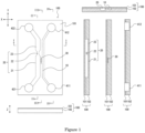
- FIG. 1 shows an example of an organ-on-a-chip of this embodiment.
- FIG. 1 shows schematic diagrams of an organ-on-a-chip 100.
- the left upper diagram is a top view
- the left lower diagram is a side view
- the right upper diagram is a cross-sectional view taken along line I-I
- the three right lower diagrams are a cross-sectional view taken along line II-II, a cross-sectional view taken along line III-III, and a cross-sectional view taken along line IV-IV in the order from left to right.
- the organ-on-a-chip 100 includes a first channel 10, a second channel 20, and a plurality of third channels 30 (third channel group), which are hollow, and inlets 411 and 421 and outlets 412 and 422, which are openings.
- An end on the upstream side and an end on the downstream side of the first channel 10 communicate with the inlet 411 and the outlet 412, respectively, and an end on the upstream side and an end on the downstream side of the second channel 20 communicate with the inlet 421 and the outlet 422, respectively.
- the organ-on-a-chip 100 is a laminate formed by laminating an upper substrate 101 and a lower substrate 102. Recessed portions on an opposed surface of the lower substrate 102, together with an opposed surface of the upper substrate, form voids (inner spaces) of the first channel 10, the second channel 20, and the third channels 30.
- the inlets 411 and 412 are formed by the recessed portions on the opposed surface of the lower substrate 102 and through holes in the upper substrate 101 located at positions corresponding to the above-mentioned recessed portions
- the outlets 421 and 422 are formed by the recessed portions on the opposed surface of the lower substrate 102 and through holes in the upper substrate 101 located at positions corresponding to the above-mentioned recessed portions.
- a biological process between a blood vessel and a cell mass can be reproduced in the organ-on-a-chip 100 by forming a blood vessel model in the first channel 10 and introducing a sample containing a cell mass to the second channel 20, in the organ-on-a-chip 100.
- the third channels 30 communicate between the first channel 10 in which a blood vessel model is to be formed and the second channel 20 to which the sample is to be introduced. This makes it possible to reproduce, in the organ-on-a-chip 100, reactions such as where the blood vessel model detects a substance derived from the cell mass, and sprouts (buds for vascular branching) from the blood vessel model extend toward the second channel 20 through the third channels 30 (among other reactions), for example.
- the blood vessel model in the present invention is, for example, a 3D structure formed in a tubular shape through culture of the vascular endothelial cells.
- vascular pericytes which are constructional elements of a blood vessel, and the like may be used together with the vascular endothelial cells to form the blood vessel model.
- the width in the X direction is 10 to 100 mm
- the length in the Y direction is 10 to 100 mm
- the thickness in the Z direction is 0.5 to 3 mm.
- the axial direction of the first channel 10 is a direction in which a liquid introduced to the first channel 10 flows
- the axial direction of the second channel 20 is a direction in which a liquid introduced to the second channel 20 flows
- the axial direction of each third channel 30 is a direction in which a liquid introduced to the third channel 30 flows.
- the first channel 10 is an introduction channel for introducing vascular endothelial cells for forming a blood vessel model on the inner wall of the first channel 10 through cell culture.
- the shape and the size of the inner space of the first channel 10 are determined such that the inner wall of the first channel can be used as a scaffold to form a blood vessel model that is equivalent or similar to a blood vessel in vivo. It is preferable that, in a direction perpendicular to the axial direction of the first channel 10, the shape and the size of the inner circumference (a cross section of the inner space) of the first channel 10 are the same as or similar to the shape and the size of the outer circumference of a blood vessel.
- the inner space has, for example, a circular cross section in a direction perpendicular to the axial direction of the first channel 10, and the cross section has, for example, a diameter of 100 to 500 pm, 150 to 400 pm, or 200 to 300 pm.
- the circular cross section may have, for example, a perfect circular shape or an elliptic shape.
- the inner space may have, for example, a quadrilateral cross section in a direction perpendicular to the axial direction of the first channel, and the cross section has, for example, a width and a height of 100 to 500 pm, 150 to 400 pm, or 200 to 300 pm.
- the quadrilateral cross section may have, for example, a square shape or a rectangular shape.
- the cross section may have a polygonal shape other than a quadrilateral shape.
- the length of the first channel 10 in the axial direction is 0.5 to 80 mm.
- a blood vessel model is formed on the inner wall of the first channel 10 by introducing the vascular endothelial cells to the first channel 10 and culturing the cells as described later.
- the inner wall of the first channel 10 may have an affinity for the vascular endothelial cells for the purpose of, for example, further facilitating fixing of the vascular endothelial cells, and specifically, the inner wall may have, for example, a coating layer with an affinity for the vascular endothelial cells.
- the coating layer contains, for example, a coating agent with an affinity for the blood endothelial cells, and examples of the coating agent include agents such as those described later.
- the inner walls of the second channel 20 and the third channels 30 may have no affinity for the vascular endothelial cells for the purpose of, for example, suppressing fixing of the vascular endothelial cells even if the vascular endothelial cells introduced to the first channel 10 to form the blood vessel model also flow into the second channel 20 through the third channels 30.
- the inner walls do not have a coating layer with an affinity for the vascular endothelial cells.
- the second channel 20 is an introduction channel for introducing a sample containing a cell mass from the upstream side toward the downstream side, and includes a trap portion 22.
- the trap portion 22 is formed between an upstream region 21 and a downstream region 23 of the second channel 20.
- the trap portion 22 allows single cells to pass through for the reason that, for example, the trap portion 22 traps the cell mass but does not trap vascular endothelial cells that flow thereinto from the first channel 10 through the third channels 30 or unwanted cells. It is preferable that the trap portion 22 has a shape and a size as described below.
- the cross section of the inner space of the trap portion 22 is made smaller (narrower) than that of the upstream region 21, as shown in FIG. 1 .
- the cross-sectional area of the inner space of the trap portion 22 is smaller than the cross-sectional area of the cross section of the cell mass.
- the above-mentioned cross section of the cell mass is, for example, a cross section of the cell mass passing through the center of the cell mass, and it is preferable that the cross-sectional area of the inner space of the trap portion 22 is smaller than, for example, the minimum cross-sectional area of the cross sections of the cell mass passing through the center of the cell mass.
- the cross-sectional area of the inner space of the trap portion 22 is larger than the maximum cross-sectional area of cross sections of a single cell.
- the above-mentioned cross section of the cell is, for example, a cross section having the maximum cross-sectional area, and may or may not pass through the center of the cell. It is preferable that the cross-sectional area of the inner space of the trap portion 22 is, for example, smaller than the minimum cross-sectional area of cross sections of the cell mass passing through the center of the cell mass and larger than the maximum cross-sectional area of cross sections of a single cell.
- the upstream region 21 need only have such a size that allows the cell mass introduced from the inlet 421 to move to the trap portion 22 on the downstream side, and specifically, it is preferable that, for example, the cross-sectional area of the inner space of the upstream region 21 is larger than the maximum cross-sectional area of the cell mass.
- the cross-sectional area of the inner space of the downstream region 23 there is no particular limitation on the cross-sectional area of the inner space of the downstream region 23.
- the cross-sectional area of the inner space of the downstream region 23 may be the same as the cross-sectional area of the inner space of the trap portion 22 or the cross-sectional area of the inner space of the upstream region 21.
- the inner space has, for example, a circular cross section in a direction perpendicular to the axial direction of the second channel 20.
- a cross section of the trap portion 22 has, for example, a diameter of 10 to 300 pm, 50 to 250 pm, or 100 to 200 pm
- a cross section of the upstream region 21 has, for example, a diameter of 200 to 1000 pm, 300 to 800 pm, or 400 to 700 pm
- the diameter of the trap portion 22 is, for example, 0.1 to 0.9 times as large as the diameter of the upstream region 21.
- the circular shape may be, for example, a perfect circular shape or an elliptic shape.
- the inner space may have, for example, a quadrilateral cross section in a direction perpendicular to the axial direction of the second channel 20.
- a cross section of the trap portion 22 has, for example, a width (in the X direction in FIG. 1 ) and a height (in the Z direction in FIG.
- a cross section of the upstream region 21 has, for example, a width and a height of 200 to 1000 pm, 300 to 800 pm, or 400 to 700 pm, and the width and the height of the trap portion 22 are, for example, 0.1 to 0.9 times as large as the width and the height of the upstream region 21.
- the quadrilateral cross section may have, for example, a square shape or a rectangular shape.
- the cross section may have a polygonal shape other than a quadrilateral shape.
- the length of the second channel 20 in the axial direction there is no particular limitation on the length of the second channel 20 in the axial direction.
- the length from the upstream end to the downstream end is 0.5 to 80 mm
- the length from the upstream end of the second channel 20 to the upstream end of the trap portion 22 is 0.25 to 40 mm
- the length of the trap portion 22 is 10 to 100 pm
- the length from the downstream end of the trap portion 22 to the downstream end of the second channel 20 is 0.25 to 40 mm.
- the shape of the trap portion 22 is not limited to this example, and for example, in the second channel 20, the cross-sectional area of the inner space may be larger than the minimum cross-sectional area of cross sections of the cell mass.
- Exemplary conditions for such an example are as follows. In a direction perpendicular to the axial direction of the second channel 20, the inner space of the trap portion 22 has, for example, a quadrilateral cross section, and the lengths of at least a pair of sides of two pairs of opposite sides of the cross section are preferably smaller than the diameter of the cell mass, and more preferably smaller than the minimum diameter of the cell mass.
- the trap portion 22 can trap the cell mass.
- the minimum diameter of the cross section is smaller than the minimum diameter of the cell mass, for example.
- the third channel group includes a plurality of third channels 30, and the third channels 30 are channels that communicate between the first channel 10 and the second channel 20.
- the third channels 30 of the organ-on-a-chip 100 are channels that communicate between the first channel 10 and the second channel 20 to enable interaction between a blood vessel model formed in the first channel 10 and a cell mass introduced to the second channel 20 when the organ-on-a-chip 100 is used. Accordingly, it is preferable that the sizes of cross sections of the third channels 30 in a direction perpendicular to the axial direction of the third channels 30 are determined such that, for example, a substance released from the cell mass, a substance or a cell released from the blood vessel model, a cell (e.g., immunocyte) passing through the blood vessel model, a sprout extending from the blood vessel model, and the like can pass through the third channels 30, and the cell mass cannot pass through the third channels 30.
- a substance released from the cell mass e.g., a substance or a cell released from the blood vessel model, a cell (e.g., immunocyte) passing through the blood vessel model, a sprout extending from the blood vessel model, and the like can
- coupling holes are formed at portions where the first channel 10 is coupled to the third channels 30.
- the cross sections of the inner spaces of the coupling holes correspond to the cross sections of the inner spaces of the third channels 30, and preferably have a size that allows a tubular blood vessel model to be formed covering the coupling holes inside the first channel 10.
- the inner spaces have, for example, a circular cross section in a direction perpendicular to the axial direction of the third channels 30, and the cross section has, for example, a diameter of 10 to 100 pm, 20 to 90 pm, or 30 to 80 pm.
- the circular shape may be, for example, a perfect circular shape or an elliptic shape.
- the inner space may have, for example, a quadrilateral cross section in a direction perpendicular to the axial direction of the third channels 30, and the cross section has, for example, a width (in the Y direction in FIG. 1 ) and a height (in the Z direction in FIG.
- the quadrilateral cross section may have, for example, a square shape or a rectangular shape. Moreover, the cross section may have a polygonal shape other than a quadrilateral shape.
- FIG. 2 is a partial top view of the organ-on-a-chip 100.
- a length L (in the X direction in FIG. 2 ) of the third channels 30 is, for example, 10 to 500 pm, 50 to 300 pm, or 100 to 200 pm.
- the number of the third channels 30 included in the third channel group is not particularly limited, and is, for example, 2 to 500, 2 to 300, or 2 to 100.
- the length of a region where the third channel group couples the first channel 10 and the second channel 20, that is, a distant W (in the Y direction in FIG. 2 ) from the third channel 30 on the upstream side to the third channel 30 on the downstream side is, for example, 30 to 10000 pm, 30 to 5000 pm, or 30 to 1000 pm.
- the pitch between the third channels 30, that is, a distance (in the Y direction in FIG. 2 ) between the adjacent third channels 30, is, for example, 5 to 100 pm, 10 to 50 pm, or 15 to 30 pm.
- a distance between the third channel 30 on the downstream side of the third channel group and an end on the upstream side of the trap portion 22 in the second channel 20 is, for example, 0 to 1000 pm, 0 to 500 pm, or 0 to 200 pm.
- a living organism for example, a substance released from the cell mass is detected, via the third channels 30, by a blood vessel model formed in the first channel 10. Accordingly, it is preferable that a portion of the second channel 20 to which the third channel group is coupled is located, for example, upstream of the trap portion 22 in the second channel 20 as shown in FIGS. 1 and 2 .
- the third channel group is disposed, for example, at a position that is a certain distance away from an end on the upstream side of the trap portion 22 in the second channel 20, and the certain distance (D in FIG. 2 ) is, for example, larger than the diameter of the cell mass.
- the distance is, for example, more than 0 pm and 1000 pm or less, more than 0 pm and 500 pm or less, or more than 0 pm and 200 pm or less.
- a blood vessel model is formed on the inner wall of the first channel 10 by introducing the vascular endothelial cells to the first channel 10 through the inlet 411 of the organ-on-a-chip 100 and culturing the cells. It is preferable to introduce the vascular endothelial cells and a culture medium together, for example, as a cell-containing liquid.
- vascular endothelial cell There is no particular limitation on the type of vascular endothelial cell, and examples thereof include umbilical vein endothelial cells such as HUVEC cells.
- the culture medium is not particularly limited and can be selected, for example, depending on the type of vascular cell. Specific examples thereof include EBM2 and the like.
- the introduction conditions and the culture conditions are not particularly limited, and are as follows, for example. That is to say, a suspension containing HUVEC cells at a concentration of 10 ⁇ 6 cells/mL is introduced through the inlet 411 of the organ-on-a-chip 100 at 37°C until the first channel 10 is filled with the cell suspension, and then the cell suspension is left to stand for 4 hours to fix the HUVEC cells on the inner surface of the first channel 10. Thereafter, the cells are cultured with a culture medium (EBM2) delivered through the inlet 411 at 1000 pL/minute for 12 hours using a syringe pump. With such first culturing, a HUVEC-cell tube (blood vessel model) with a thickness of about 20 pm can be formed on the entire inner circumferential surface of the first channel 10, for example.
- a culture medium EBM2
- a sample containing the cell mass is introduced through the inlet 421 of the organ-on-a-chip 100 and is cultured.
- the number of cell masses included in the sample There is no particular limitation on the number of cell masses included in the sample.
- one cell mass per organ-on-a-chip 100 is preferable because it is important to examine a phenomenon caused by a single cell mass.
- the sample contains, for example, the cell mass and a culture medium.
- the type of culture medium and examples thereof include EBM2 and the like.
- the type of cell mass is not particularly limited, and is preferably a cell mass of cancer cells, for example.
- the size of the cell mass is, for example, 100 to 1000 pm, 200 to 800 pm, or 300 to 500 pm.
- the introduction conditions and the culture conditions are not particularly limited, and are as follows, for example. That is to say, for example, after a sample containing a single cell mass is introduced to the inlet 421 of the organ-on-a-chip 100 using a pipette chip at 37°C, the sample liquid is delivered from the inlet 421 toward the outlet 422 of the second channel 20 using a syringe pump.
- the cell mass arrives at the trap portion 22 and is trapped in the trap portion 22. Then, when the blood vessel model formed in the first channel 10 detects, through the third channels 30, a substance released from the cell mass trapped and cultured in the trap portion 22 in the second channel 20, sprouts appear from the blood vessel model, and then the sprouts can grow inside the third channels 30 toward the second channel 20 via portions (coupling holes) where the third channels 30 are coupled to the first channel 10.
- the organ-on-a-chip 100 of this embodiment makes it possible to reproduce a biological process between a cell mass and a blood vessel.
- the trap portion in the first channel includes columnar bodies (pillars) that prevent the cell mass from moving toward the downstream side.
- the organ-on-a-chip of this embodiment is the same as the organ-on-a-chip of Embodiment 1, except that the trap portion is changed.
- FIGS. 1 and 2 described above can be applied to the basic structure of the organ-on-a-chip of this embodiment.
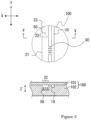
- FIG. 3 partially enlarged views illustrating a region that includes a trap portion in the first channel of the organ-on-a-chip are shown in FIG. 3 .
- the upper diagram is a partial top view of the organ-on-a-chip 100 as in FIG. 2 described above, and the lower diagram is a cross-sectional view taken along line V-V
- the columnar bodies 50 trap the cell mass, and therefore, in the second channel 20, the size of the cross-sectional area of the inner space of the trap portion 20 may be the same as that of the cross-sectional area of the inner space of the upstream region 21, for example, unlike Embodiment 1 above.
- the number and the size of the columnar bodies 50 in the trap portion 22 are not particularly limited, and can be determined as appropriate depending on the size of a cell mass to be trapped, the size of the cross section of the inner space of the trap portion 22, and the like.
- the number of the columnar bodies 50 is, for example, 1 to 10, 2 to 8, or 3 to 5.
- each columnar body 50 is not particularly limited, and may be a cylindrical shape, for example, as shown in FIG. 3 , or a prismatic shape. It is preferable that the area of contact between the cell mass and the columnar body 50 is small, for example, because conditions close to the in-vivo conditions can be achieved.
- the size of each columnar body 50 for example, the height in the Z direction is 10 to 1000 pm or 50 to 500 pm, the width in the X direction is 5 to 200 pm or 10 to 100 pm, and the width in the Y direction is 5 to 200 pm or 10 to 100 pm.
- the columnar body 50 is, for example, shorter than or equal to half of the height of the inner space of the second channel 20. Also, it is preferable that the height of the columnar body 50 is, for example, 0.1 times or more as long as the diameter of the cell mass.
- the columnar bodies 50 in the trap portion 22 there is no particular limitation on the positions of the columnar bodies 50 in the trap portion 22, and it is preferable that, for example, in a direction (e.g., the X direction in FIG. 3 ) perpendicular to the axial direction of the second channel 20, the columnar bodies 50 are disposed on the inner wall on a side opposite to portions that communicate with the third channels 30.
- the trap portion 22 in the second channel 20 can trap the cell mass at a position away from the portions that communicate with the third channels 30.
- a substance released from the cell mass is detected, via the third channels 30, by a blood vessel model formed in the first channel 10. At this time, securing a distance from the cell mass to the third channels 30 makes it possible to form a gradation of the concentration of the released substance in the region therebetween and achieve a state closer to the in-vivo state.
- FIG. 4 As another example of the trap portion 22 in which the columnar bodies 50 are disposed, partially enlarged views illustrating a region that includes a trap portion in the first channel of the organ-on-a-chip are shown in FIG. 4 .
- the upper diagram is a partial top view of the organ-on-a-chip 100
- the lower diagram is a cross-sectional view taken along line VI-VI.
- FIG. 3 The description of FIG. 3 can be applied to this example unless otherwise stated.
- the four columnar bodies 50 are disposed inside the second channel 20, and the region in which the columnar bodies 50 are disposed serves as the trap portion 22.
- the four columnar bodies 50 include two columnar bodies 50H that have a relatively large height in the Z direction, and two columnar bodies 50L that have relatively low height in the Z direction.
- the two columnar bodies 50H are the same as the columnar bodies 50 in FIG. 3 described above and can trap the cell mass.
- the columnar bodies 50H may also be referred to as "trapping columnar bodies”.
- the two columnar bodies 50L with a lower height may also be referred to as "basal columnar bodies" for preventing the cell mass from coming into contact with the inner wall of the second channel 20.
- a state in the organ-on-a-chip is closer to the in-vivo state, and therefore, it is desired that, when the trap portion 22 traps the cell mass, the cell mass floats in the inner space of the second channel 20, that is, the cell mass is not in contact with the inner wall. Accordingly, due to the basal columnar bodies 50L with a relatively low height being disposed, a cell mass that flows into the trap portion 22 in the second channel 20 gets on tops of the basal columnar bodies 50L, and the trapping columnar bodies 50H can trap the cell mass so as to prevent the cell mass from moving toward the downstream side therefrom. With this aspect, an obstacle to the formation of a gradation of a substance released from the cell mass as described above is further reduced, thus making it possible to achieve a state even closer to the in-vivo state.
- the numbers and the sizes of the columnar bodies 50H and 50L in the trap portion 22 are not particularly limited, and can be determined as appropriate depending on the size of a cell mass to be trapped, the size of the cross section of the inner space of the trap portion 22, and the like. It is preferable that the area of contact between the cell mass and each of the columnar bodies 50H and 50L is small, for example, because conditions close to the in-vivo conditions can be achieved.
- the number of the trapping columnar bodies 50H is, for example, 1 to 10, 2 to 8, or 3 to 5, and the number of the basal columnar bodies 50L is, for example, 1 to 10, 1 to 5, or 1 to 3.
- each trapping columnar body 50H are not particularly limited, and are, for example, the same as those in the description of FIG. 3 .
- the shape of each basal columnar body 50L may be, for example, a cylindrical shape or a prismatic shape.
- the size of each columnar body 50L for example, the height in the Z direction is 5 to 100 pm or 10 to 50 pm, the width in the X direction is 5 to 200 pm or 10 to 100 pm, and the width in the Y direction is 5 to 200 pm or 10 to 100 pm.
- the columnar bodies 50 are disposed at positions away from a portion of the second channel 20 that communicates with the third channel group. Specifically, it is preferable that the columnar bodies 50 are disposed at positions 100 to 1000 pm away from a portion of the second channel 20 that communicates with the third channel group.
- the inner spaces of the first channel, the second channel, the third channels, and the like are formed by the recessed portions on the opposed surface of the lower substrate and the opposed surfaces of the upper substrate that covers the recessed portions.
- the organ-on-a-chip is not limited to these aspects, and an aspect may also be employed in which, for example, recessed portions are formed on both the opposed surface of the lower substrate and the opposed surface of the upper substrate and these substrates are laminated.
- an organ-on-a-chip of this embodiment includes a pair of substrates (an upper substrate and a lower substrate), opposed surfaces of one of the substrates and the other substrate are each provided with recessed portions serving as inner spaces of the first channel, the second channel, and the third channel group, and the recessed portions on the opposed surface of the first substrate and the recessed portions on the opposed surface of the second substrate form the first channel, the second channel, and the third channel group.
- FIG. 5 shows an example of an organ-on-a-chip of this embodiment. Note that, in FIG. 5 , the upper diagram is a partial top view of the organ-on-a-chip 100, and the lower diagram is a cross-sectional view taken along line VII-VII. The description of FIG. 4 can be applied to this example unless otherwise stated.
- the inner space of the trap portion 22 in the second channel 20 is formed by the recessed portion on the upper substrate 101 and the recessed portion on the lower substrate 102.
- the trapping columnar bodies 50H are disposed at corresponding three positions (apexes of a triangle) so that the trapping columnar bodies 50H on the lower substrate and the trapping columnar bodies 50H on the upper substrate make a pair
- the basal columnar bodies 50L are disposed at positions corresponding to the center of the base of the above-mentioned triangle such that the basal columnar body 50L on the lower substrate and the basal columnar body 50L on the upper substrate make a pair.
- the trapping columnar bodies 50H and the basal columnar bodies 50L are disposed on both the upper side and the lower side in the inner space of the trap portion 22, thus making it possible to achieve conditions close to the in-vivo conditions as in FIG. 4 described in Embodiment 2.
- the first channel 10 and the second channel 20 communicate with each other via the third channels 30 located at the center in the height direction of the first channel 10 and the center in the height direction of the second channel 20. From this viewpoint as well, it is possible to, for example, form a gradation of a substance released from the cell mass and achieve conditions even closer to the in-vivo conditions.
- the biological function reproduction method of the present invention further includes performing coating for forming a coating layer with an affinity for vascular endothelial cells on an inner wall of the first channel prior to the first culturing for forming a blood vessel model in the first channel of the organ-on-a-chip of the present invention.
- coating for forming a coating layer with an affinity for vascular endothelial cells on an inner wall of the first channel prior to the first culturing for forming a blood vessel model in the first channel of the organ-on-a-chip of the present invention.
- a coating liquid that contains a coating agent for fixing the vascular endothelial cells and a solvent that does not contain the coating agent are introduced to the first channel 10 and the second channel 20, respectively, with a flow rate balance that prevents flow in the third channels 30.
- This adjustment makes it possible to introduce the coating liquid to only the first channel 10 to form a coating layer with an affinity for the vascular endothelial cells in only the first channel 10. With this aspect, it is possible to more efficiently fix the vascular endothelial cells introduced to the first channel 10 on the inner wall of the first channel 10.
- the coating agent examples include collagens such as Collagen I.
- the coating liquid contains, for example the coating agent and a solvent, and examples of the solvent include buffer solutions and buffered saline solutions such as PBS.
- the treatment conditions such as a temperature and a period of time, and the like, and for example, 100 pL of the coating liquid is used and incubated at 25°C for 1 hour.
- a culture medium that contains the vascular endothelial cells and a solvent that does not contain the cells are introduced to the first channel 10 and the second channel 20, respectively, with a flow rate balance that prevents flow in the third channels 30.
- This adjustment makes it possible to prevent the vascular endothelial cells introduced to the first channel 10 from flowing into the second channel 20 through the third channels 30.
- the coating layer is formed only in the first channel 10 and is not formed in the second channel 20 in the coating, and therefore, even if the vascular endothelial cells flow into the second channel 20, the vascular endothelial cells are less likely to be fixed to the second channel 20 compared with the first channel 10 and are likely to be discharged to the outlet 422.
- the solvent introduced to the second channel 20 includes culture mediums, buffer solutions, and buffered saline solutions such as PBS.
- Conditions where the coating liquid and the solvent are introduced to the first channel 10 and the second channel 20, respectively, in the coating with a flow rate balance that prevents flow in the third channels 30 can be determined as follows, for example. That is to say, for example, the flow rate in the first channel 10 and the flow rate in the second channel 20 are determined such that the ratio between the flow rates is the same as the ratio between the cross-sectional area of the first channel 10 and the cross-sectional area of the second channel 20, and the coating liquid and the solvent are delivered at thus determined flow rates using syringe pumps.
- the first culturing may also include introducing a culture medium containing the vascular endothelial cells to the overall channel that includes the first channel 10, the second channel 20, and the third channels 30.
- the coating layer is formed only in the first channel 10 and is not formed in the second channel 20 in the coating, and therefore, the vascular endothelial cells are less likely to be fixed to the second channel 20 compared with the first channel 10 and are likely to be discharged to the outlet 422.
- the coating layer is formed only in the first channel 10 and is not formed in the second channel 20 in the coating, and therefore, even if the vascular endothelial cells flow into the second channel 20, the vascular endothelial cells are less likely to be fixed to the second channel 20 compared with the first channel 10 and are likely to be discharged to the outlet.
- the second channel includes the trap portion, and therefore, the cell mass can be trapped in the trap portion when being introduced as a sample, and thus it can be said that conditions closer to the in-vivo conditions can be reproduced.
Landscapes
- Health & Medical Sciences (AREA)
- Engineering & Computer Science (AREA)
- Life Sciences & Earth Sciences (AREA)
- Chemical & Material Sciences (AREA)
- Biomedical Technology (AREA)
- Organic Chemistry (AREA)
- Bioinformatics & Cheminformatics (AREA)
- Wood Science & Technology (AREA)
- Zoology (AREA)
- Genetics & Genomics (AREA)
- Biotechnology (AREA)
- General Health & Medical Sciences (AREA)
- General Engineering & Computer Science (AREA)
- Microbiology (AREA)
- Biochemistry (AREA)
- Sustainable Development (AREA)
- Cell Biology (AREA)
- Molecular Biology (AREA)
- Dispersion Chemistry (AREA)
- Clinical Laboratory Science (AREA)
- Vascular Medicine (AREA)
- Immunology (AREA)
- General Chemical & Material Sciences (AREA)
- Physics & Mathematics (AREA)
- Fluid Mechanics (AREA)
- Analytical Chemistry (AREA)
- Hematology (AREA)
- Chemical Kinetics & Catalysis (AREA)
- Oncology (AREA)
- Physiology (AREA)
- Apparatus Associated With Microorganisms And Enzymes (AREA)
Abstract
Description
- The present invention relates to an organ-on-a-chip for reproducing a biological process between a cell mass and a blood vessel, and a method for reproducing a biological function using the organ-on-a-chip.
- In recent years, organs-on-chips that reproduce biological functions on chips have been attracting attention. One of the organs to be reproduced on a chip is a blood vessel. As an example of the organ-on-a-chip that reproduces a blood vessel, a form in which a first channel and a second flow channel are coupled to each other via a third channel is reported (Non-Patent Document 1). For example, with this organ-on-a-chip, an in-vivo reaction can be reproduced as a response of a blood vessel model to a substance released from cancer cells by forming a blood vessel model in the first channel through 3D culture, filling the second channel with cancer cells, and then culturing the cancer cells.
- Non-Patent Document 1: Ioannis K. Zervantonakis et al., PNAS, August 21, 2012, vol. 109, no. 34, 13515-13520
- However, although the second channel is filled with cancer cells as described above in the above-mentioned organ-on-a-chip in which a blood vessel model is formed, cancer cells can hardly be considered to exist independently of one another in an actual living organism. Accordingly, a technique that achieves a state closer to the in-vivo state in a chip is required.
- Therefore, it is an object of the present invention to provide a novel organ-on-a-chip for forming a blood vessel model, for example.
- To achieve the above-mentioned object, an organ-on-a-chip of the present invention is a chip for reproducing a biological process between a cell mass and a blood vessel, the chip including:
- a first channel;
- a second channel; and
- a third channel group,
- wherein the first channel is an introduction channel for introducing vascular endothelial cells for forming a blood vessel model on an inner wall of the first channel through cell culture,
- the second channel is an introduction channel for introducing a sample containing a cell mass from an upstream side toward a downstream side, and includes a trap portion for trapping the cell mass and located between the upstream side and the downstream side inside the second channel, and
- the third channel group includes a plurality of third channels, and the third channels are channels that communicate between the first channel and the second channel.
- A biological function reproduction method of the present invention uses the above-mentioned organ-on-a-chip, and includes:
- performing first culturing for forming a blood vessel model on an inner wall of the first channel by introducing vascular endothelial cells to the first channel and culturing the cells; and
- performing second culturing for introducing a sample containing a cell mass to the second channel from an upstream side of the second channel and culturing the cell mass in a state in which the cell mass is trapped in the trap portion in the second channel.
- The inventors of the present invention found that use of a cell mass rather than cells that exist independently of one another is suitable for reproducing a state closer to the in-vivo state in an organ-on-a-chip for forming a blood vessel model. However, the structure of the above-described organ-on-a-chip is based on the premise that the second channel is filled with many free cells, and therefore, even if this organ-on-a-chip is used, it is difficult to generate, for example, an influence of a single cell mass on a blood vessel model under conditions close to the in-vivo conditions. Accordingly, as a result of extensive research, the present inventors found that conditions closer to the in-vivo conditions can be mimicked in the above-mentioned organ-on-a-chip by trapping the cell mass at a desired position in the second channel. With the present invention, the second channel includes the trap portion, and therefore, the cell mass can be trapped in the trap portion by being introduced as a sample, and thus it can be said that conditions closer to the in-vivo conditions can be reproduced.
-
- [
FIG. 1] FIG. 1 shows schematic diagrams illustrating an example of an organ-on-a-chip ofEmbodiment 1. - [
FIG. 2] FIG. 2 is a partial plan view of an example of an organ-on-a-chip ofEmbodiment 1. - [
FIG. 3] FIG. 3 shows schematic diagrams illustrating an example of a portion of an organ-on-a-chip of Embodiment 2. - [
FIG. 4] FIG. 4 shows schematic diagrams illustrating an example of a portion of an organ-on-a-chip of Embodiment 2. - [
FIG. 5] FIG. 5 shows schematic diagrams illustrating an example of an organ-on-a-chip of Embodiment 3. - In the organ-on-a-chip of the present invention, the trap portion in the second channel traps the cell mass and allows single cells to pass through, for example.
- In the organ-on-a-chip of the present invention, for example, in a direction perpendicular to an axial direction of the second channel, a cross-sectional area of an inner space of the trap portion is smaller than the minimum cross-sectional area of cross sections of the cell mass passing through the center of the cell mass.
- In the organ-on-a-chip of the present invention, for example, in the direction perpendicular to the axial direction of the second channel, a cross-sectional area of an inner space of the trap portion is larger than the maximum cross-sectional area of single cells.
- In the organ-on-a-chip of the present invention, for example, in the direction perpendicular to the axial direction of the second channel, when an inner space has a circular cross section, a cross section of the trap portion has a diameter of 10 to 300 pm, a cross section of a portion located upstream of the trap portion has a diameter of 200 to 1000 pm, and the diameter of the trap portion is 0.1 to 0.9 times as large as the diameter of the portion located upstream of the trap portion, and when an inner space has a quadrilateral cross section, a cross section of the trap portion has a width and a height of 10 to 300 pm, a cross section of a portion located upstream of the trap portion has a width and a height of 200 to 1000 pm, and the width and the height of the trap portion are 0.1 to 0.9 times as large as the width and the height of the portion located upstream of the trap portion.
- In the organ-on-a-chip of the present invention, for example, in the direction perpendicular to the axial direction of the second channel, the trap portion in the second channel has a columnar body that prevents the cell mass from moving toward the downstream side.
- In the organ-on-a-chip of the present invention, for example, in the direction perpendicular to the axial direction of the second channel, a height of the columnar body of the trap portion is shorter than a height of the inner space of the trap portion.
- In the organ-on-a-chip of the present invention, for example, in the direction perpendicular to the axial direction of the second channel, the inner space of the trap portion has a quadrilateral cross section, and lengths of at least a pair of sides of two pairs of opposite sides of the cross section are smaller than a diameter of the cell mass.
- In the organ-on-a-chip of the present invention, for example, a height of the columnar body of the trap portion is shorter than or equal to half of a height of the inner space of the trap portion.
- In the organ-on-a-chip of the present invention, for example, the columnar body is disposed at a
position 100 to 1000 pm away from a portion of the second channel that communicates with the third channel group. - In the organ-on-a-chip of the present invention, for example, the third channel group is disposed upstream of the trap portion in the second channel.
- In the organ-on-a-chip of the present invention, for example, the third channel group is disposed at a position 0 to 1000 pm away from an end on an upstream side of the trap portion in the second channel.
- In the organ-on-a-chip of the present invention, for example, in a direction perpendicular to an axial direction of the first channel, a length of an inner circumference of the first channel corresponds to a length of an outer circumference of a blood vessel.
- In the organ-on-a-chip of the present invention, for example, in the direction perpendicular to the axial direction of the first channel, when an inner space of the first channel has a circular cross section, the cross section has a diameter of 100 to 500 pm, and when an inner space of the first channel has a quadrilateral cross section, the cross section has a width and a height of 100 to 500 µm.
- In the organ-on-a-chip of the present invention, for example, the inner wall of the first channel has a coating layer with an affinity for vascular endothelial cells.
- In the organ-on-a-chip of the present invention, for example, when an inner space of each of the third channels has a circular cross section in a direction perpendicular to an axial direction of the third channel, the cross section has a diameter of 10 to 100 pm, and when an inner space of each of the third channels has a quadrilateral cross section in the direction perpendicular to the axial direction of the third channel, the cross section has a width and a height of 10 to 100 pm.
- For example, the organ-on-a-chip of the present invention includes a pair of substrates, and one of the substrates is placed on the other substrate. An opposed surface of the one substrate is provided with recessed portions serving as inner spaces of the first channel, the second channel, and the third channel group, and the recessed portions on the opposed surface of the one substrate and an opposed surface of the other substrate form the first channel, the second channel, and the third channel group.
- For example, the organ-on-a-chip of the present invention includes a pair of substrates. Opposed surfaces of one of the substrates and the other substrate are each provided with recessed portions serving as inner spaces of the first channel, the second channel, and the third channel group, and the recessed portions on the opposed surface of the first substrate and the recessed portions on the opposed surface of the second substrate form the first channel, the second channel, and the third channel group.
- In the biological function reproduction method of the present invention, for example, the vascular endothelial cells are HUVEC cells.
- In the biological function reproduction method of the present invention, for example, the vascular endothelial cells are introduced together with a culture medium in the first culturing.
- In the biological function reproduction method of the present invention, for example, the cell mass is a cell mass of cancer cells.
- In the biological function reproduction method of the present invention, for example, the sample in the second culturing contains the cell mass and a culture medium.
- For example, the biological function reproduction method of the present invention further includes performing coating for forming a coating layer with an affinity for vascular endothelial cells on an inner wall of the first channel prior to the first culturing.
- In the biological function reproduction method of the present invention, for example, in the coating, a coating liquid that contains a coating agent for fixing the vascular endothelial cells and a solvent that does not contain the coating agent are introduced to the first channel and the second channel, respectively, with a flow rate balance that prevents flow in the third channels. In the first culturing, a culture medium that contains the vascular endothelial cells and a solvent that does not contain the cells are introduced to the first channel and the second channel, respectively, with a flow rate balance that prevents flow in the third channels.
- Specifically, the organ-on-a-chip of the present invention may be in a state in which a blood vessel model has not been formed in the first channel yet or in a state in which a blood vessel model has already been formed in the first channel.
- Specific examples of the organ-on-a-chip of the present invention and a method using the same, namely the biological function reproduction method of the present invention, will be described with reference to the drawings. In the diagrams, the same reference numeral denotes the same portion. The present invention is in no way limited or restricted to the following embodiments. The description of each embodiment can be applied to the other embodiments unless otherwise stated. In the embodiments, examples of the sizes of portions are shown, but the sizes can be changed as appropriate depending on, for example, the type of vascular endothelial cells, the target size of a blood vessel model formed, the type and the size of a cell mass, and the like.
-
FIG. 1 shows an example of an organ-on-a-chip of this embodiment.FIG. 1 shows schematic diagrams of an organ-on-a-chip 100. The left upper diagram is a top view, the left lower diagram is a side view, the right upper diagram is a cross-sectional view taken along line I-I, and the three right lower diagrams are a cross-sectional view taken along line II-II, a cross-sectional view taken along line III-III, and a cross-sectional view taken along line IV-IV in the order from left to right. - The organ-on-
a-chip 100 includes afirst channel 10, asecond channel 20, and a plurality of third channels 30 (third channel group), which are hollow, andinlets outlets first channel 10 communicate with theinlet 411 and theoutlet 412, respectively, and an end on the upstream side and an end on the downstream side of thesecond channel 20 communicate with theinlet 421 and theoutlet 422, respectively. - The organ-on-
a-chip 100 is a laminate formed by laminating anupper substrate 101 and alower substrate 102. Recessed portions on an opposed surface of thelower substrate 102, together with an opposed surface of the upper substrate, form voids (inner spaces) of thefirst channel 10, thesecond channel 20, and thethird channels 30. In the organ-on-a-chip 100, theinlets lower substrate 102 and through holes in theupper substrate 101 located at positions corresponding to the above-mentioned recessed portions, and theoutlets lower substrate 102 and through holes in theupper substrate 101 located at positions corresponding to the above-mentioned recessed portions. - For example, as described later, a biological process between a blood vessel and a cell mass can be reproduced in the organ-on-
a-chip 100 by forming a blood vessel model in thefirst channel 10 and introducing a sample containing a cell mass to thesecond channel 20, in the organ-on-a-chip 100. Specifically, thethird channels 30 communicate between thefirst channel 10 in which a blood vessel model is to be formed and thesecond channel 20 to which the sample is to be introduced. This makes it possible to reproduce, in the organ-on-a-chip 100, reactions such as where the blood vessel model detects a substance derived from the cell mass, and sprouts (buds for vascular branching) from the blood vessel model extend toward thesecond channel 20 through the third channels 30 (among other reactions), for example. Note that the blood vessel model in the present invention is, for example, a 3D structure formed in a tubular shape through culture of the vascular endothelial cells. For example, vascular pericytes, which are constructional elements of a blood vessel, and the like may be used together with the vascular endothelial cells to form the blood vessel model. - There is no particular limitation on the overall size of the organ-on-
a-chip 100. For example, inFIG. 1 , the width in the X direction is 10 to 100 mm, the length in the Y direction is 10 to 100 mm, and the thickness in the Z direction is 0.5 to 3 mm. - In this embodiment, the axial direction of the
first channel 10 is a direction in which a liquid introduced to thefirst channel 10 flows, the axial direction of thesecond channel 20 is a direction in which a liquid introduced to thesecond channel 20 flows, and the axial direction of eachthird channel 30 is a direction in which a liquid introduced to thethird channel 30 flows. - The
first channel 10 is an introduction channel for introducing vascular endothelial cells for forming a blood vessel model on the inner wall of thefirst channel 10 through cell culture. - There is no particular limitation on the shape and the size of the inner space of the
first channel 10. For example, the shape and the size are determined such that the inner wall of the first channel can be used as a scaffold to form a blood vessel model that is equivalent or similar to a blood vessel in vivo. It is preferable that, in a direction perpendicular to the axial direction of thefirst channel 10, the shape and the size of the inner circumference (a cross section of the inner space) of thefirst channel 10 are the same as or similar to the shape and the size of the outer circumference of a blood vessel. - In a specific example, in terms of the shape of the
first channel 10, the inner space has, for example, a circular cross section in a direction perpendicular to the axial direction of thefirst channel 10, and the cross section has, for example, a diameter of 100 to 500 pm, 150 to 400 pm, or 200 to 300 pm. The circular cross section may have, for example, a perfect circular shape or an elliptic shape. Also, in terms of the shape of thefirst channel 10, the inner space may have, for example, a quadrilateral cross section in a direction perpendicular to the axial direction of the first channel, and the cross section has, for example, a width and a height of 100 to 500 pm, 150 to 400 pm, or 200 to 300 pm. The quadrilateral cross section may have, for example, a square shape or a rectangular shape. Moreover, the cross section may have a polygonal shape other than a quadrilateral shape. - There is no particular limitation on the length of the
first channel 10 in the axial direction. For example, the length from the upstream end to the downstream end is 0.5 to 80 mm. - When the organ-on-
a-chip 100 is used, a blood vessel model is formed on the inner wall of thefirst channel 10 by introducing the vascular endothelial cells to thefirst channel 10 and culturing the cells as described later. Accordingly, the inner wall of thefirst channel 10 may have an affinity for the vascular endothelial cells for the purpose of, for example, further facilitating fixing of the vascular endothelial cells, and specifically, the inner wall may have, for example, a coating layer with an affinity for the vascular endothelial cells. The coating layer contains, for example, a coating agent with an affinity for the blood endothelial cells, and examples of the coating agent include agents such as those described later. Meanwhile, in the organ-on-a-chip 100, the inner walls of thesecond channel 20 and thethird channels 30 may have no affinity for the vascular endothelial cells for the purpose of, for example, suppressing fixing of the vascular endothelial cells even if the vascular endothelial cells introduced to thefirst channel 10 to form the blood vessel model also flow into thesecond channel 20 through thethird channels 30. Specifically, it is preferable that, for example, the inner walls do not have a coating layer with an affinity for the vascular endothelial cells. - The
second channel 20 is an introduction channel for introducing a sample containing a cell mass from the upstream side toward the downstream side, and includes atrap portion 22. Thetrap portion 22 is formed between anupstream region 21 and adownstream region 23 of thesecond channel 20. There is no particular limitation on the shape of thetrap portion 22 as long as thetrap portion 22 can trap the cell mass when the sample is introduced from the upstream side toward the downstream side in thesecond channel 20. - It is preferable that the
trap portion 22 allows single cells to pass through for the reason that, for example, thetrap portion 22 traps the cell mass but does not trap vascular endothelial cells that flow thereinto from thefirst channel 10 through thethird channels 30 or unwanted cells. It is preferable that thetrap portion 22 has a shape and a size as described below. - As an example of the shape of the
trap portion 22, in thesecond channel 20, the cross section of the inner space of thetrap portion 22 is made smaller (narrower) than that of theupstream region 21, as shown inFIG. 1 . Specifically, it is preferable that in thesecond channel 20, for example, the cross-sectional area of the inner space of thetrap portion 22 is smaller than the cross-sectional area of the cross section of the cell mass. The above-mentioned cross section of the cell mass is, for example, a cross section of the cell mass passing through the center of the cell mass, and it is preferable that the cross-sectional area of the inner space of thetrap portion 22 is smaller than, for example, the minimum cross-sectional area of the cross sections of the cell mass passing through the center of the cell mass. In addition, it is preferable that the cross-sectional area of the inner space of thetrap portion 22 is larger than the maximum cross-sectional area of cross sections of a single cell. The above-mentioned cross section of the cell is, for example, a cross section having the maximum cross-sectional area, and may or may not pass through the center of the cell. It is preferable that the cross-sectional area of the inner space of thetrap portion 22 is, for example, smaller than the minimum cross-sectional area of cross sections of the cell mass passing through the center of the cell mass and larger than the maximum cross-sectional area of cross sections of a single cell. Moreover, in thesecond channel 20, theupstream region 21 need only have such a size that allows the cell mass introduced from theinlet 421 to move to thetrap portion 22 on the downstream side, and specifically, it is preferable that, for example, the cross-sectional area of the inner space of theupstream region 21 is larger than the maximum cross-sectional area of the cell mass. In thesecond channel 20, there is no particular limitation on the cross-sectional area of the inner space of thedownstream region 23. For example, the cross-sectional area of the inner space of thedownstream region 23 may be the same as the cross-sectional area of the inner space of thetrap portion 22 or the cross-sectional area of the inner space of theupstream region 21. - In a specific example, in terms of the shape of the
second channel 20, the inner space has, for example, a circular cross section in a direction perpendicular to the axial direction of thesecond channel 20. In this case, in thesecond channel 20, a cross section of thetrap portion 22 has, for example, a diameter of 10 to 300 pm, 50 to 250 pm, or 100 to 200 pm, a cross section of theupstream region 21 has, for example, a diameter of 200 to 1000 pm, 300 to 800 pm, or 400 to 700 pm, and the diameter of thetrap portion 22 is, for example, 0.1 to 0.9 times as large as the diameter of theupstream region 21. The circular shape may be, for example, a perfect circular shape or an elliptic shape. Also, in terms of the shape of thesecond channel 20, the inner space may have, for example, a quadrilateral cross section in a direction perpendicular to the axial direction of thesecond channel 20. In this case, in thesecond channel 20, a cross section of thetrap portion 22 has, for example, a width (in the X direction inFIG. 1 ) and a height (in the Z direction inFIG. 1 ) of 10 to 300 pm, 50 to 250 pm, or 100 to 200 pm, a cross section of theupstream region 21 has, for example, a width and a height of 200 to 1000 pm, 300 to 800 pm, or 400 to 700 pm, and the width and the height of thetrap portion 22 are, for example, 0.1 to 0.9 times as large as the width and the height of theupstream region 21. The quadrilateral cross section may have, for example, a square shape or a rectangular shape. Moreover, the cross section may have a polygonal shape other than a quadrilateral shape. - There is no particular limitation on the length of the
second channel 20 in the axial direction. For example, the length from the upstream end to the downstream end is 0.5 to 80 mm, the length from the upstream end of thesecond channel 20 to the upstream end of thetrap portion 22 is 0.25 to 40 mm, the length of thetrap portion 22 is 10 to 100 pm, and the length from the downstream end of thetrap portion 22 to the downstream end of thesecond channel 20 is 0.25 to 40 mm. - The shape of the
trap portion 22 is not limited to this example, and for example, in thesecond channel 20, the cross-sectional area of the inner space may be larger than the minimum cross-sectional area of cross sections of the cell mass. Exemplary conditions for such an example are as follows. In a direction perpendicular to the axial direction of thesecond channel 20, the inner space of thetrap portion 22 has, for example, a quadrilateral cross section, and the lengths of at least a pair of sides of two pairs of opposite sides of the cross section are preferably smaller than the diameter of the cell mass, and more preferably smaller than the minimum diameter of the cell mass. In this case, for example, irrespective of the cross-sectional area of the inner space as described above, that is, even when the cross-sectional area of the inner space is smaller than the minimum cross-sectional area of the cell mass, thetrap portion 22 can trap the cell mass. In the case where the inner space of thetrap portion 22 has a circular cross section, it is preferable that the minimum diameter of the cross section is smaller than the minimum diameter of the cell mass, for example. - The third channel group includes a plurality of
third channels 30, and thethird channels 30 are channels that communicate between thefirst channel 10 and thesecond channel 20. - The
third channels 30 of the organ-on-a-chip 100 are channels that communicate between thefirst channel 10 and thesecond channel 20 to enable interaction between a blood vessel model formed in thefirst channel 10 and a cell mass introduced to thesecond channel 20 when the organ-on-a-chip 100 is used. Accordingly, it is preferable that the sizes of cross sections of thethird channels 30 in a direction perpendicular to the axial direction of thethird channels 30 are determined such that, for example, a substance released from the cell mass, a substance or a cell released from the blood vessel model, a cell (e.g., immunocyte) passing through the blood vessel model, a sprout extending from the blood vessel model, and the like can pass through thethird channels 30, and the cell mass cannot pass through thethird channels 30. - In the organ-on-
a-chip 100, coupling holes are formed at portions where thefirst channel 10 is coupled to thethird channels 30. The cross sections of the inner spaces of the coupling holes correspond to the cross sections of the inner spaces of thethird channels 30, and preferably have a size that allows a tubular blood vessel model to be formed covering the coupling holes inside thefirst channel 10. - From this viewpoint, in terms of the shape of the
third channels 30, the inner spaces have, for example, a circular cross section in a direction perpendicular to the axial direction of thethird channels 30, and the cross section has, for example, a diameter of 10 to 100 pm, 20 to 90 pm, or 30 to 80 pm. The circular shape may be, for example, a perfect circular shape or an elliptic shape. Also, in terms of the shape of thethird channels 30, the inner space may have, for example, a quadrilateral cross section in a direction perpendicular to the axial direction of thethird channels 30, and the cross section has, for example, a width (in the Y direction inFIG. 1 ) and a height (in the Z direction inFIG. 1 ) of 10 to 100 pm, 20 to 90 pm, or 30 to 80 pm. The quadrilateral cross section may have, for example, a square shape or a rectangular shape. Moreover, the cross section may have a polygonal shape other than a quadrilateral shape. - A partially enlarged view illustrating a region of the third channel group (which includes a plurality of third channels 30) in the organ-on-
a-chip 100 inFIG. 1 is shown inFIG. 2. FIG. 2 is a partial top view of the organ-on-a-chip 100. A length L (in the X direction inFIG. 2 ) of thethird channels 30 is, for example, 10 to 500 pm, 50 to 300 pm, or 100 to 200 pm. - The number of the
third channels 30 included in the third channel group is not particularly limited, and is, for example, 2 to 500, 2 to 300, or 2 to 100. The length of a region where the third channel group couples thefirst channel 10 and thesecond channel 20, that is, a distant W (in the Y direction inFIG. 2 ) from thethird channel 30 on the upstream side to thethird channel 30 on the downstream side, is, for example, 30 to 10000 pm, 30 to 5000 pm, or 30 to 1000 pm. The pitch between thethird channels 30, that is, a distance (in the Y direction inFIG. 2 ) between the adjacentthird channels 30, is, for example, 5 to 100 pm, 10 to 50 pm, or 15 to 30 pm. - There is no particular limitation on the position of the third channel group, and a distance between the
third channel 30 on the downstream side of the third channel group and an end on the upstream side of thetrap portion 22 in thesecond channel 20 is, for example, 0 to 1000 pm, 0 to 500 pm, or 0 to 200 pm. Regarding how a living organism is reproduced in the organ-on-a-chip 100, for example, a substance released from the cell mass is detected, via thethird channels 30, by a blood vessel model formed in thefirst channel 10. Accordingly, it is preferable that a portion of thesecond channel 20 to which the third channel group is coupled is located, for example, upstream of thetrap portion 22 in thesecond channel 20 as shown inFIGS. 1 and2 . Specifically, it is preferable that the third channel group is disposed, for example, at a position that is a certain distance away from an end on the upstream side of thetrap portion 22 in thesecond channel 20, and the certain distance (D inFIG. 2 ) is, for example, larger than the diameter of the cell mass. The distance is, for example, more than 0 pm and 1000 pm or less, more than 0 pm and 500 pm or less, or more than 0 pm and 200 pm or less. - Next, a method of using the organ-on-
a-chip 100 of Embodiment 1 (biological function reproduction method) will be described. - First, as first culturing, a blood vessel model is formed on the inner wall of the
first channel 10 by introducing the vascular endothelial cells to thefirst channel 10 through theinlet 411 of the organ-on-a-chip 100 and culturing the cells. It is preferable to introduce the vascular endothelial cells and a culture medium together, for example, as a cell-containing liquid. There is no particular limitation on the type of vascular endothelial cell, and examples thereof include umbilical vein endothelial cells such as HUVEC cells. The culture medium is not particularly limited and can be selected, for example, depending on the type of vascular cell. Specific examples thereof include EBM2 and the like. - The introduction conditions and the culture conditions are not particularly limited, and are as follows, for example. That is to say, a suspension containing HUVEC cells at a concentration of 10×6 cells/mL is introduced through the
inlet 411 of the organ-on-a-chip 100 at 37°C until thefirst channel 10 is filled with the cell suspension, and then the cell suspension is left to stand for 4 hours to fix the HUVEC cells on the inner surface of thefirst channel 10. Thereafter, the cells are cultured with a culture medium (EBM2) delivered through theinlet 411 at 1000 pL/minute for 12 hours using a syringe pump. With such first culturing, a HUVEC-cell tube (blood vessel model) with a thickness of about 20 pm can be formed on the entire inner circumferential surface of thefirst channel 10, for example. - Next, as second culturing, a sample containing the cell mass is introduced through the
inlet 421 of the organ-on-a-chip 100 and is cultured. There is no particular limitation on the number of cell masses included in the sample. For example, one cell mass per organ-on-a-chip 100 is preferable because it is important to examine a phenomenon caused by a single cell mass. It is preferable that the sample contains, for example, the cell mass and a culture medium. There is no particular limitation on the type of culture medium, and examples thereof include EBM2 and the like. - The type of cell mass is not particularly limited, and is preferably a cell mass of cancer cells, for example. The size of the cell mass is, for example, 100 to 1000 pm, 200 to 800 pm, or 300 to 500 pm.
- The introduction conditions and the culture conditions are not particularly limited, and are as follows, for example. That is to say, for example, after a sample containing a single cell mass is introduced to the
inlet 421 of the organ-on-a-chip 100 using a pipette chip at 37°C, the sample liquid is delivered from theinlet 421 toward theoutlet 422 of thesecond channel 20 using a syringe pump. - In the second culturing, while the sample flows from the
inlet 421 toward theoutlet 422, the cell mass arrives at thetrap portion 22 and is trapped in thetrap portion 22. Then, when the blood vessel model formed in thefirst channel 10 detects, through thethird channels 30, a substance released from the cell mass trapped and cultured in thetrap portion 22 in thesecond channel 20, sprouts appear from the blood vessel model, and then the sprouts can grow inside thethird channels 30 toward thesecond channel 20 via portions (coupling holes) where thethird channels 30 are coupled to thefirst channel 10. Thus, using the organ-on-a-chip 100 of this embodiment makes it possible to reproduce a biological process between a cell mass and a blood vessel. - In an organ-on-a-chip of this embodiment, the trap portion in the first channel includes columnar bodies (pillars) that prevent the cell mass from moving toward the downstream side. The organ-on-a-chip of this embodiment is the same as the organ-on-a-chip of
Embodiment 1, except that the trap portion is changed.FIGS. 1 and2 described above can be applied to the basic structure of the organ-on-a-chip of this embodiment. - As an example, partially enlarged views illustrating a region that includes a trap portion in the first channel of the organ-on-a-chip are shown in
FIG. 3 . Note that, inFIG. 3 , the upper diagram is a partial top view of the organ-on-a-chip 100 as inFIG. 2 described above, and the lower diagram is a cross-sectional view taken along line V-V - As shown in
FIG. 3 , threecolumnar bodies 50 are disposed inside thesecond channel 20, and the region in which thecolumnar bodies 50 are disposed serves as thetrap portion 22. With this embodiment, thecolumnar bodies 50 trap the cell mass, and therefore, in thesecond channel 20, the size of the cross-sectional area of the inner space of thetrap portion 20 may be the same as that of the cross-sectional area of the inner space of theupstream region 21, for example, unlikeEmbodiment 1 above. - The number and the size of the
columnar bodies 50 in thetrap portion 22 are not particularly limited, and can be determined as appropriate depending on the size of a cell mass to be trapped, the size of the cross section of the inner space of thetrap portion 22, and the like. The number of thecolumnar bodies 50 is, for example, 1 to 10, 2 to 8, or 3 to 5. - The shape of each
columnar body 50 is not particularly limited, and may be a cylindrical shape, for example, as shown inFIG. 3 , or a prismatic shape. It is preferable that the area of contact between the cell mass and thecolumnar body 50 is small, for example, because conditions close to the in-vivo conditions can be achieved. In terms of the size of eachcolumnar body 50, for example, the height in the Z direction is 10 to 1000 pm or 50 to 500 pm, the width in the X direction is 5 to 200 pm or 10 to 100 pm, and the width in the Y direction is 5 to 200 pm or 10 to 100 pm. It is preferable that thecolumnar body 50 is, for example, shorter than or equal to half of the height of the inner space of thesecond channel 20. Also, it is preferable that the height of thecolumnar body 50 is, for example, 0.1 times or more as long as the diameter of the cell mass. - There is no particular limitation on the positions of the
columnar bodies 50 in thetrap portion 22, and it is preferable that, for example, in a direction (e.g., the X direction inFIG. 3 ) perpendicular to the axial direction of thesecond channel 20, thecolumnar bodies 50 are disposed on the inner wall on a side opposite to portions that communicate with thethird channels 30. Thus, thetrap portion 22 in thesecond channel 20 can trap the cell mass at a position away from the portions that communicate with thethird channels 30. Regarding how the organ-on-a-chip is used, as described above, for example, a substance released from the cell mass is detected, via thethird channels 30, by a blood vessel model formed in thefirst channel 10. At this time, securing a distance from the cell mass to thethird channels 30 makes it possible to form a gradation of the concentration of the released substance in the region therebetween and achieve a state closer to the in-vivo state. - As another example of the
trap portion 22 in which thecolumnar bodies 50 are disposed, partially enlarged views illustrating a region that includes a trap portion in the first channel of the organ-on-a-chip are shown inFIG. 4 . Note that, inFIG. 4 , the upper diagram is a partial top view of the organ-on-a-chip 100, and the lower diagram is a cross-sectional view taken along line VI-VI. The description ofFIG. 3 can be applied to this example unless otherwise stated. - As shown in
FIG. 4 , fourcolumnar bodies 50 are disposed inside thesecond channel 20, and the region in which thecolumnar bodies 50 are disposed serves as thetrap portion 22. The fourcolumnar bodies 50 include twocolumnar bodies 50H that have a relatively large height in the Z direction, and twocolumnar bodies 50L that have relatively low height in the Z direction. In this aspect, the twocolumnar bodies 50H are the same as thecolumnar bodies 50 inFIG. 3 described above and can trap the cell mass. Thecolumnar bodies 50H may also be referred to as "trapping columnar bodies". Meanwhile, the twocolumnar bodies 50L with a lower height may also be referred to as "basal columnar bodies" for preventing the cell mass from coming into contact with the inner wall of thesecond channel 20. That is to say, as described above, it is preferable that a state in the organ-on-a-chip is closer to the in-vivo state, and therefore, it is desired that, when thetrap portion 22 traps the cell mass, the cell mass floats in the inner space of thesecond channel 20, that is, the cell mass is not in contact with the inner wall. Accordingly, due to the basalcolumnar bodies 50L with a relatively low height being disposed, a cell mass that flows into thetrap portion 22 in thesecond channel 20 gets on tops of the basalcolumnar bodies 50L, and the trappingcolumnar bodies 50H can trap the cell mass so as to prevent the cell mass from moving toward the downstream side therefrom. With this aspect, an obstacle to the formation of a gradation of a substance released from the cell mass as described above is further reduced, thus making it possible to achieve a state even closer to the in-vivo state. - The numbers and the sizes of the
columnar bodies trap portion 22 are not particularly limited, and can be determined as appropriate depending on the size of a cell mass to be trapped, the size of the cross section of the inner space of thetrap portion 22, and the like. It is preferable that the area of contact between the cell mass and each of thecolumnar bodies columnar bodies 50H is, for example, 1 to 10, 2 to 8, or 3 to 5, and the number of the basalcolumnar bodies 50L is, for example, 1 to 10, 1 to 5, or 1 to 3. - In a specific example, the shape and the size of each trapping
columnar body 50H are not particularly limited, and are, for example, the same as those in the description ofFIG. 3 . The shape of each basalcolumnar body 50L may be, for example, a cylindrical shape or a prismatic shape. In terms of the size of eachcolumnar body 50L, for example, the height in the Z direction is 5 to 100 pm or 10 to 50 pm, the width in the X direction is 5 to 200 pm or 10 to 100 pm, and the width in the Y direction is 5 to 200 pm or 10 to 100 pm. - There is no particular limitation on the positions of the
columnar bodies 50 in a direction perpendicular to the axial direction of thesecond channel 20, and it is preferable that, for example, thecolumnar bodies 50 are disposed at positions away from a portion of thesecond channel 20 that communicates with the third channel group. Specifically, it is preferable that thecolumnar bodies 50 are disposed atpositions 100 to 1000 pm away from a portion of thesecond channel 20 that communicates with the third channel group. - In the examples of the organs-on-a-chip shown in the embodiments described above, the inner spaces of the first channel, the second channel, the third channels, and the like are formed by the recessed portions on the opposed surface of the lower substrate and the opposed surfaces of the upper substrate that covers the recessed portions. The organ-on-a-chip is not limited to these aspects, and an aspect may also be employed in which, for example, recessed portions are formed on both the opposed surface of the lower substrate and the opposed surface of the upper substrate and these substrates are laminated.
- That is to say, for example, an organ-on-a-chip of this embodiment includes a pair of substrates (an upper substrate and a lower substrate), opposed surfaces of one of the substrates and the other substrate are each provided with recessed portions serving as inner spaces of the first channel, the second channel, and the third channel group, and the recessed portions on the opposed surface of the first substrate and the recessed portions on the opposed surface of the second substrate form the first channel, the second channel, and the third channel group.
FIG. 5 shows an example of an organ-on-a-chip of this embodiment. Note that, inFIG. 5 , the upper diagram is a partial top view of the organ-on-a-chip 100, and the lower diagram is a cross-sectional view taken along line VII-VII. The description ofFIG. 4 can be applied to this example unless otherwise stated. - As shown in
FIG. 5 , the inner space of thetrap portion 22 in thesecond channel 20 is formed by the recessed portion on theupper substrate 101 and the recessed portion on thelower substrate 102. In thetrap portion 22, on the bottom surface of the recessed portion on thelower substrate 102 and the bottom surface of the recessed portion on theupper substrate 101, the trappingcolumnar bodies 50H are disposed at corresponding three positions (apexes of a triangle) so that the trappingcolumnar bodies 50H on the lower substrate and the trappingcolumnar bodies 50H on the upper substrate make a pair, and the basalcolumnar bodies 50L are disposed at positions corresponding to the center of the base of the above-mentioned triangle such that the basalcolumnar body 50L on the lower substrate and the basalcolumnar body 50L on the upper substrate make a pair. Accordingly, with the organ-on-a-chip of this embodiment, the trappingcolumnar bodies 50H and the basalcolumnar bodies 50L are disposed on both the upper side and the lower side in the inner space of thetrap portion 22, thus making it possible to achieve conditions close to the in-vivo conditions as inFIG. 4 described in Embodiment 2. - Furthermore, as shown in
FIG. 5 , in the organ-on-a-chip of this embodiment, thefirst channel 10 and thesecond channel 20 communicate with each other via thethird channels 30 located at the center in the height direction of thefirst channel 10 and the center in the height direction of thesecond channel 20. From this viewpoint as well, it is possible to, for example, form a gradation of a substance released from the cell mass and achieve conditions even closer to the in-vivo conditions. - For example, the biological function reproduction method of the present invention further includes performing coating for forming a coating layer with an affinity for vascular endothelial cells on an inner wall of the first channel prior to the first culturing for forming a blood vessel model in the first channel of the organ-on-a-chip of the present invention. Hereinafter, this example will be described with reference to
FIG. 1 described above, but the present invention is not limited to this example. - (4-1)
In the coating, for example, a coating liquid that contains a coating agent for fixing the vascular endothelial cells and a solvent that does not contain the coating agent are introduced to thefirst channel 10 and thesecond channel 20, respectively, with a flow rate balance that prevents flow in thethird channels 30. This adjustment makes it possible to introduce the coating liquid to only thefirst channel 10 to form a coating layer with an affinity for the vascular endothelial cells in only thefirst channel 10. With this aspect, it is possible to more efficiently fix the vascular endothelial cells introduced to thefirst channel 10 on the inner wall of thefirst channel 10. - Examples of the coating agent include collagens such as Collagen I. The coating liquid contains, for example the coating agent and a solvent, and examples of the solvent include buffer solutions and buffered saline solutions such as PBS. In the coating, there is no particular limitation on the amount of the coating agent used, the treatment conditions such as a temperature and a period of time, and the like, and for example, 100 pL of the coating liquid is used and incubated at 25°C for 1 hour.
- In this aspect, it is preferable that, in the following first culturing, for example, a culture medium that contains the vascular endothelial cells and a solvent that does not contain the cells are introduced to the
first channel 10 and thesecond channel 20, respectively, with a flow rate balance that prevents flow in thethird channels 30. This adjustment makes it possible to prevent the vascular endothelial cells introduced to thefirst channel 10 from flowing into thesecond channel 20 through thethird channels 30. Note that the coating layer is formed only in thefirst channel 10 and is not formed in thesecond channel 20 in the coating, and therefore, even if the vascular endothelial cells flow into thesecond channel 20, the vascular endothelial cells are less likely to be fixed to thesecond channel 20 compared with thefirst channel 10 and are likely to be discharged to theoutlet 422. There is no particular limitation on the solvent introduced to thesecond channel 20, and examples thereof include culture mediums, buffer solutions, and buffered saline solutions such as PBS. - Conditions where the coating liquid and the solvent are introduced to the
first channel 10 and thesecond channel 20, respectively, in the coating with a flow rate balance that prevents flow in thethird channels 30 can be determined as follows, for example. That is to say, for example, the flow rate in thefirst channel 10 and the flow rate in thesecond channel 20 are determined such that the ratio between the flow rates is the same as the ratio between the cross-sectional area of thefirst channel 10 and the cross-sectional area of thesecond channel 20, and the coating liquid and the solvent are delivered at thus determined flow rates using syringe pumps. Note that conditions where the culture medium that contains the vascular endothelial cells and the solvent that does not contain the cells are introduced to thefirst channel 10 and thesecond channel 20, respectively, in the first culturing with a flow rate balance that prevents flow in thethird channels 30 can be determined in the same manner. - (4-2)
After the coating in (4-1) above, for example, the first culturing may also include introducing a culture medium containing the vascular endothelial cells to the overall channel that includes thefirst channel 10, thesecond channel 20, and thethird channels 30. As described above, the coating layer is formed only in thefirst channel 10 and is not formed in thesecond channel 20 in the coating, and therefore, the vascular endothelial cells are less likely to be fixed to thesecond channel 20 compared with thefirst channel 10 and are likely to be discharged to theoutlet 422. - Note that the coating layer is formed only in the
first channel 10 and is not formed in thesecond channel 20 in the coating, and therefore, even if the vascular endothelial cells flow into thesecond channel 20, the vascular endothelial cells are less likely to be fixed to thesecond channel 20 compared with thefirst channel 10 and are likely to be discharged to the outlet. - As described above, the present invention has been described with reference to the embodiments, but the present invention is not limited to the above-described embodiments. Various modifications that can be understood by a person skilled in the art can be made in the configurations and details of the present invention without departing from the scope of the present invention.
- As described above, with the present invention, the second channel includes the trap portion, and therefore, the cell mass can be trapped in the trap portion when being introduced as a sample, and thus it can be said that conditions closer to the in-vivo conditions can be reproduced.
-
- 100
- Organ-on-a-chip
- 101
- Upper substrate
- 102
- Lower substrate
- 10
- First channel
- 20
- Second channel
- 21
- Upstream region
- 22
- Trap portion
- 23
- Downstream region
- 30
- Third channel
- 411, 421
- Inlet
- 412, 422
- Outlet
- 50
- Columnar body
- 50H
- Trapping columnar body
- 50L
- Basal columnar body
Claims (25)
- An organ-on-a-chip for reproducing a biological process between a cell mass and a blood vessel, comprising:a first channel;a second channel; anda third channel group,wherein the first channel is an introduction channel for introducing vascular endothelial cells for forming a blood vessel model on an inner wall of the first channel through cell culture,the second channel is an introduction channel for introducing a sample containing a cell mass from an upstream side toward a downstream side, and includes a trap portion for trapping the cell mass and located between the upstream side and the downstream side inside the second channel, andthe third channel group includes a plurality of third channels, and the third channels are channels that communicate between the first channel and the second channel.
- The organ-on-a-chip according to claim 1,
wherein the trap portion in the second channel traps the cell mass and allows single cells to pass through. - The organ-on-a-chip according to claim 1 or 2,wherein, in a direction perpendicular to an axial direction of the second channel,a cross-sectional area of an inner space of the trap portion is smaller than the minimum cross-sectional area of cross sections of the cell mass passing through the center of the cell mass.
- The organ-on-a-chip according to claim 3,wherein, in a direction perpendicular to the axial direction of the second channel,the cross-sectional area of the inner space of the trap portion is larger than the maximum cross-sectional area of cross sections of single cells.
- The organ-on-a-chip according to any one of claims 1 to 4,wherein, in a direction perpendicular to an axial direction of the second channel,when an inner space has a circular cross section, a cross section of the trap portion has a diameter of 10 to 300 pm, a cross section of a portion located upstream of the trap portion has a diameter of 200 to 1000 pm, and the diameter of the trap portion is 0.1 to 0.9 times as large as the diameter of the portion located upstream of the trap portion, andwhen an inner space has a quadrilateral cross section, a cross section of the trap portion has a width and a height of 10 to 300 pm, a cross section of a portion located upstream of the trap portion has a width and a height of 200 to 1000 pm, and the width and the height of the trap portion are 0.1 to 0.9 times as large as the width and the height of the portion located upstream of the trap portion.
- The organ-on-a-chip according to any one of claims 1 to 5,wherein, in a direction perpendicular to an axial direction of the second channel,an inner space of the trap portion has a quadrilateral cross section, andlengths of at least a pair of sides of two pairs of opposite sides of the cross section are smaller than a diameter of the cell mass.
- The organ-on-a-chip according to any one of claims 1 to 6,wherein, in a direction perpendicular to an axial direction of the second channel,the trap portion in the second channel has a columnar body that prevents the cell mass from moving toward the downstream side.
- The organ-on-a-chip according to claim 7,wherein, in a direction perpendicular to the axial direction of the second channel,a height of the columnar body of the trap portion is shorter than a height of an inner space of the trap portion.
- The organ-on-a-chip according to claim 8,
wherein the height of the columnar body of the trap portion is shorter than or equal to half of the height of the inner space of the trap portion. - The organ-on-a-chip according to any one of claims 7 to 9,
wherein the columnar body is disposed at a position 100 to 1000 pm away from a portion of the second channel that communicates with the third channel group. - The organ-on-a-chip according to any one of claims 1 to 10,
wherein the third channel group is disposed upstream of the trap portion in the second channel. - The organ-on-a-chip according to claim 11,
wherein the third channel group is disposed at a position 0 to 1000 pm away from an end on an upstream side of the trap portion in the second channel. - The organ-on-a-chip according to any one of claims 1 to 12,wherein, in a direction perpendicular to an axial direction of the first channel,a length of an inner circumference of the first channel corresponds to a length of an outer circumference of a blood vessel.
- The organ-on-a-chip according to any one of claims 1 to 13,wherein, in a direction perpendicular to an axial direction of the first channel,when an inner space of the first channel has a circular cross section, the cross section has a diameter of 100 to 500 pm, andwhen an inner space of the first channel has a quadrilateral cross section, the cross section has a width and a height of 100 to 500 pm.
- The organ-on-a-chip according to any one of claims 1 to 14,
wherein the inner wall of the first channel has a coating layer with an affinity for vascular endothelial cells. - The organ-on-a-chip according to any one of claims 1 to 15,wherein, when an inner space of each of the third channels has a circular cross section in a direction perpendicular to an axial direction of the third channel, the cross section has a diameter of 10 to 100 pm, andwhen an inner space of each of the third channels has a quadrilateral cross section in a direction perpendicular to the axial direction of the third channel, the cross section has a width and a height of 10 to 100 pm.
- The organ-on-a-chip according to any one of claims 1 to 16, comprisinga pair of substrates,wherein one of the substrates is placed on the other substrate,an opposed surface of the one substrate is provided with recessed portions serving as inner spaces of the first channel, the second channel, and the third channel group, andthe recessed portions on the opposed surface of the one substrate and an opposed surface of the other substrate form the first channel, the second channel, and the third channel group.
- The organ-on-a-chip according to any one of claims 1 to 16, comprisinga pair of substrates,wherein opposed surfaces of one of the substrates and the other substrate are each provided with recessed portions serving as inner spaces of the first channel, the second channel, and the third channel group, andthe recessed portions on the opposed surface of the first substrate and the recessed portions on the opposed surface of the second substrate form the first channel, the second channel, and the third channel group.
- A biological function reproduction method for causing a cell mass and a blood vessel model interact with each other, the method using the organ-ona-chip according to any one of claims 1 to 18, and comprising:performing first culturing for forming a blood vessel model on an inner wall of the first channel by introducing vascular endothelial cells to the first channel and culturing the cells; andperforming second culturing for introducing a sample containing a cell mass to the second channel from an upstream side of the second channel and culturing the cell mass in a state in which the cell mass is trapped in the trap portion in the second channel.
- The biological function reproduction method according to claim 19,
wherein the vascular endothelial cells are HUVEC cells. - The biological function reproduction method according to claim 19 or 20,
wherein the vascular endothelial cells are introduced together with a culture medium in the first culturing. - The biological function reproduction method according to any one of claims 19 to 21,
wherein the cell mass is a cell mass of cancer cells. - The biological function reproduction method according to any one of claims 19 to 22,
wherein the sample contains the cell mass and a culture medium in the second culturing. - The biological function reproduction method according to any one of claims 19 to 23, further comprising
performing coating for forming a coating layer with an affinity for the vascular endothelial cells on the inner wall of the first channel prior to the first culturing. - The biological function reproduction method according to claim 26,wherein, in the coating, a coating liquid that contains a coating agent for fixing the vascular endothelial cells and a solvent that does not contain the coating agent are introduced to the first channel and the second channel, respectively, with a flow rate balance that prevents flow in the third channels, andin the first culturing, a culture medium that contains the vascular endothelial cells and a solvent that does not contain the cells are introduced to the first channel and the second channel, respectively, with a flow rate balance that prevents flow in the third channels.
Applications Claiming Priority (1)
| Application Number | Priority Date | Filing Date | Title |
|---|---|---|---|
| PCT/JP2021/005258 WO2022172397A1 (en) | 2021-02-12 | 2021-02-12 | Organ-on-chip and biological function reproduction method |
Publications (2)
| Publication Number | Publication Date |
|---|---|
| EP4293100A1 true EP4293100A1 (en) | 2023-12-20 |
| EP4293100A4 EP4293100A4 (en) | 2025-09-10 |
Family
ID=82838552
Family Applications (1)
| Application Number | Title | Priority Date | Filing Date |
|---|---|---|---|
| EP21925650.0A Pending EP4293100A4 (en) | 2021-02-12 | 2021-02-12 | ORGAN-ON-CHIP AND METHODS FOR REPRODUCTION OF BIOLOGICAL FUNCTIONS |
Country Status (3)
| Country | Link |
|---|---|
| US (1) | US20240110136A1 (en) |
| EP (1) | EP4293100A4 (en) |
| WO (1) | WO2022172397A1 (en) |
Family Cites Families (12)
| Publication number | Priority date | Publication date | Assignee | Title |
|---|---|---|---|---|
| WO2004101743A2 (en) * | 2003-05-06 | 2004-11-25 | Bellbrook Labs, Llc | Three dimensional cell cultures in a microscale fluid handling system |
| JP2007275689A (en) * | 2004-06-30 | 2007-10-25 | Bussan Nanotech Research Institute Inc | Hydrophobic compound reactor and reaction method |
| JP2008519598A (en) * | 2004-11-11 | 2008-06-12 | エージェンシー フォー サイエンス,テクノロジー アンド リサーチ | Cell culture devices |
| US20110081664A1 (en) * | 2008-10-17 | 2011-04-07 | University Of Massachusetts | Multipurpose microfluidic device for mimicking a microenvironment within a tumor |
| EP2914379A1 (en) * | 2012-11-01 | 2015-09-09 | The Charles Stark Draper Laboratory, Inc. | Ex vivo microfluidic analysis of biologic samples |
| US10105700B2 (en) * | 2013-07-17 | 2018-10-23 | The Johns Hopkins University | Microfluidic chip for analysis of cell motility and methods for using same |
| WO2015013210A1 (en) * | 2013-07-22 | 2015-01-29 | The Regents Of The University Of California | Microfluidics cell culture device |
| CN104630059B (en) * | 2015-01-16 | 2017-07-25 | 中国科学院深圳先进技术研究院 | Microfluidic chip and method for establishing in vitro co-culture model of three types of cells |
| US10767149B2 (en) * | 2016-06-13 | 2020-09-08 | Massachusetts Institute Of Technology | Microfluidic device for three dimensional and compartmentalized coculture of neuronal and muscle cells, with functional force readout |
| CN106544271A (en) * | 2016-12-07 | 2017-03-29 | 清华大学深圳研究生院 | A kind of many cells 3D co-culture devices and method of research tumor invasion blood vessel |
| CN111218401A (en) * | 2018-11-26 | 2020-06-02 | 中国科学院大连化学物理研究所 | A new blood vessel formation and drug evaluation method based on tumor chip |
| JP7264667B2 (en) * | 2019-02-28 | 2023-04-25 | 浜松ホトニクス株式会社 | LIQUID SUPPLY APPARATUS, MICRO DEVICE SYSTEM, AND LIQUID SUPPLY METHOD |
-
2021
- 2021-02-12 US US18/264,691 patent/US20240110136A1/en active Pending
- 2021-02-12 EP EP21925650.0A patent/EP4293100A4/en active Pending
- 2021-02-12 WO PCT/JP2021/005258 patent/WO2022172397A1/en not_active Ceased
Also Published As
| Publication number | Publication date |
|---|---|
| WO2022172397A1 (en) | 2022-08-18 |
| US20240110136A1 (en) | 2024-04-04 |
| EP4293100A4 (en) | 2025-09-10 |
Similar Documents
| Publication | Publication Date | Title |
|---|---|---|
| US8491561B2 (en) | Micromachined bilayer unit of engineered tissues | |
| Zhang et al. | Recent advances in microfluidic platforms for programming cell‐based living materials | |
| CN105950469B (en) | Cell screening chip and micro-fluidic chip joint | |
| WO2003104411A2 (en) | Bioartificial liver system | |
| CN117229916A (en) | A multi-organoid co-culture microfluidic chip and its use method | |
| CN106414703A (en) | Systems and methods for biomimetic fluid processing | |
| JP2010523268A5 (en) | ||
| US20150087004A1 (en) | Microfabricated 3D Cell Culture System | |
| EP1815244A1 (en) | Cell culture device | |
| WO2009099066A1 (en) | Biodevice | |
| CN113174332A (en) | Bionic kidney organ chip structure and bionic liver and kidney chip structure | |
| US20240189822A1 (en) | Three-dimensional microfluidic metastasis array | |
| EP3873557B1 (en) | Microfluidic gas exchange devices and methods for making same | |
| CN221028484U (en) | Double organoid co-culture microfluidic chip | |
| CN118374354A (en) | Vascularized kidney organ chip and its preparation method and application | |
| US20120183990A1 (en) | Microfluidic system and method for producing same | |
| US5948655A (en) | Method of applying hepatocytes to hollow fibers | |
| CN117721019A (en) | Vascularized organ chip and application thereof | |
| US20050227350A1 (en) | Device and method for studying cell migration and deformation | |
| US20240110136A1 (en) | Organ-on-a-chip and biological function reproduction method | |
| CN118516234A (en) | Multi-flow-field controlled vascularized organ chip and method | |
| CN104513798B (en) | A kind of micro-fluidic chip for Micro-CPE neutralization test | |
| CN223813514U (en) | A microfluidic device for cell experiments | |
| CN121046202A (en) | A circular chamber organ-on-a-chip and method for high-efficiency culture of tumor spheres | |
| CN121574819A (en) | A vascularized organ-on-a-chip capable of repeatedly collecting metabolites from multi-cell co-culture or organoids in situ. |
Legal Events
| Date | Code | Title | Description |
|---|---|---|---|
| STAA | Information on the status of an ep patent application or granted ep patent |
Free format text: STATUS: THE INTERNATIONAL PUBLICATION HAS BEEN MADE |
|
| PUAI | Public reference made under article 153(3) epc to a published international application that has entered the european phase |
Free format text: ORIGINAL CODE: 0009012 |
|
| STAA | Information on the status of an ep patent application or granted ep patent |
Free format text: STATUS: REQUEST FOR EXAMINATION WAS MADE |
|
| 17P | Request for examination filed |
Effective date: 20230831 |
|
| AK | Designated contracting states |
Kind code of ref document: A1 Designated state(s): AL AT BE BG CH CY CZ DE DK EE ES FI FR GB GR HR HU IE IS IT LI LT LU LV MC MK MT NL NO PL PT RO RS SE SI SK SM TR |
|
| P01 | Opt-out of the competence of the unified patent court (upc) registered |
Effective date: 20240115 |
|
| DAV | Request for validation of the european patent (deleted) | ||
| DAX | Request for extension of the european patent (deleted) | ||
| A4 | Supplementary search report drawn up and despatched |
Effective date: 20250812 |
|
| RIC1 | Information provided on ipc code assigned before grant |
Ipc: C12M 1/00 20060101AFI20250806BHEP Ipc: C12M 3/00 20060101ALI20250806BHEP |