EP4290261A1 - Magnetic resonance system and corresponding method - Google Patents
Magnetic resonance system and corresponding method Download PDFInfo
- Publication number
- EP4290261A1 EP4290261A1 EP22177656.0A EP22177656A EP4290261A1 EP 4290261 A1 EP4290261 A1 EP 4290261A1 EP 22177656 A EP22177656 A EP 22177656A EP 4290261 A1 EP4290261 A1 EP 4290261A1
- Authority
- EP
- European Patent Office
- Prior art keywords
- magnetic resonance
- magnet
- field
- coils
- patient
- Prior art date
- Legal status (The legal status is an assumption and is not a legal conclusion. Google has not performed a legal analysis and makes no representation as to the accuracy of the status listed.)
- Pending
Links
Images


Classifications
-
- G—PHYSICS
- G01—MEASURING; TESTING
- G01R—MEASURING ELECTRIC VARIABLES; MEASURING MAGNETIC VARIABLES
- G01R33/00—Arrangements or instruments for measuring magnetic variables
- G01R33/20—Arrangements or instruments for measuring magnetic variables involving magnetic resonance
- G01R33/44—Arrangements or instruments for measuring magnetic variables involving magnetic resonance using nuclear magnetic resonance [NMR]
- G01R33/48—NMR imaging systems
- G01R33/54—Signal processing systems, e.g. using pulse sequences ; Generation or control of pulse sequences; Operator console
- G01R33/56—Image enhancement or correction, e.g. subtraction or averaging techniques, e.g. improvement of signal-to-noise ratio and resolution
- G01R33/563—Image enhancement or correction, e.g. subtraction or averaging techniques, e.g. improvement of signal-to-noise ratio and resolution of moving material, e.g. flow contrast angiography
- G01R33/56341—Diffusion imaging
-
- G—PHYSICS
- G01—MEASURING; TESTING
- G01R—MEASURING ELECTRIC VARIABLES; MEASURING MAGNETIC VARIABLES
- G01R33/00—Arrangements or instruments for measuring magnetic variables
- G01R33/20—Arrangements or instruments for measuring magnetic variables involving magnetic resonance
- G01R33/44—Arrangements or instruments for measuring magnetic variables involving magnetic resonance using nuclear magnetic resonance [NMR]
- G01R33/48—NMR imaging systems
-
- G—PHYSICS
- G01—MEASURING; TESTING
- G01R—MEASURING ELECTRIC VARIABLES; MEASURING MAGNETIC VARIABLES
- G01R33/00—Arrangements or instruments for measuring magnetic variables
- G01R33/20—Arrangements or instruments for measuring magnetic variables involving magnetic resonance
- G01R33/28—Details of apparatus provided for in groups G01R33/44 - G01R33/64
- G01R33/30—Sample handling arrangements, e.g. sample cells, spinning mechanisms
- G01R33/307—Sample handling arrangements, e.g. sample cells, spinning mechanisms specially adapted for moving the sample relative to the MR system, e.g. spinning mechanisms, flow cells or means for positioning the sample inside a spectrometer
-
- G—PHYSICS
- G01—MEASURING; TESTING
- G01R—MEASURING ELECTRIC VARIABLES; MEASURING MAGNETIC VARIABLES
- G01R33/00—Arrangements or instruments for measuring magnetic variables
- G01R33/20—Arrangements or instruments for measuring magnetic variables involving magnetic resonance
- G01R33/28—Details of apparatus provided for in groups G01R33/44 - G01R33/64
- G01R33/32—Excitation or detection systems, e.g. using radio frequency signals
- G01R33/34—Constructional details, e.g. resonators, specially adapted to MR
- G01R33/34007—Manufacture of RF coils, e.g. using printed circuit board technology; additional hardware for providing mechanical support to the RF coil assembly or to part thereof, e.g. a support for moving the coil assembly relative to the remainder of the MR system
-
- G—PHYSICS
- G01—MEASURING; TESTING
- G01R—MEASURING ELECTRIC VARIABLES; MEASURING MAGNETIC VARIABLES
- G01R33/00—Arrangements or instruments for measuring magnetic variables
- G01R33/20—Arrangements or instruments for measuring magnetic variables involving magnetic resonance
- G01R33/28—Details of apparatus provided for in groups G01R33/44 - G01R33/64
- G01R33/32—Excitation or detection systems, e.g. using radio frequency signals
- G01R33/34—Constructional details, e.g. resonators, specially adapted to MR
- G01R33/34084—Constructional details, e.g. resonators, specially adapted to MR implantable coils or coils being geometrically adaptable to the sample, e.g. flexible coils or coils comprising mutually movable parts
-
- G—PHYSICS
- G01—MEASURING; TESTING
- G01R—MEASURING ELECTRIC VARIABLES; MEASURING MAGNETIC VARIABLES
- G01R33/00—Arrangements or instruments for measuring magnetic variables
- G01R33/20—Arrangements or instruments for measuring magnetic variables involving magnetic resonance
- G01R33/28—Details of apparatus provided for in groups G01R33/44 - G01R33/64
- G01R33/38—Systems for generation, homogenisation or stabilisation of the main or gradient magnetic field
- G01R33/383—Systems for generation, homogenisation or stabilisation of the main or gradient magnetic field using permanent magnets
-
- G—PHYSICS
- G01—MEASURING; TESTING
- G01R—MEASURING ELECTRIC VARIABLES; MEASURING MAGNETIC VARIABLES
- G01R33/00—Arrangements or instruments for measuring magnetic variables
- G01R33/20—Arrangements or instruments for measuring magnetic variables involving magnetic resonance
- G01R33/28—Details of apparatus provided for in groups G01R33/44 - G01R33/64
- G01R33/38—Systems for generation, homogenisation or stabilisation of the main or gradient magnetic field
- G01R33/385—Systems for generation, homogenisation or stabilisation of the main or gradient magnetic field using gradient magnetic field coils
-
- G—PHYSICS
- G01—MEASURING; TESTING
- G01R—MEASURING ELECTRIC VARIABLES; MEASURING MAGNETIC VARIABLES
- G01R33/00—Arrangements or instruments for measuring magnetic variables
- G01R33/20—Arrangements or instruments for measuring magnetic variables involving magnetic resonance
- G01R33/44—Arrangements or instruments for measuring magnetic variables involving magnetic resonance using nuclear magnetic resonance [NMR]
- G01R33/445—MR involving a non-standard magnetic field B0, e.g. of low magnitude as in the earth's magnetic field or in nanoTesla spectroscopy, comprising a polarizing magnetic field for pre-polarisation, B0 with a temporal variation of its magnitude or direction such as field cycling of B0 or rotation of the direction of B0, or spatially inhomogeneous B0 like in fringe-field MR or in stray-field imaging
-
- G—PHYSICS
- G01—MEASURING; TESTING
- G01R—MEASURING ELECTRIC VARIABLES; MEASURING MAGNETIC VARIABLES
- G01R33/00—Arrangements or instruments for measuring magnetic variables
- G01R33/20—Arrangements or instruments for measuring magnetic variables involving magnetic resonance
- G01R33/44—Arrangements or instruments for measuring magnetic variables involving magnetic resonance using nuclear magnetic resonance [NMR]
- G01R33/48—NMR imaging systems
- G01R33/50—NMR imaging systems based on the determination of relaxation times, e.g. T1 measurement by IR sequences; T2 measurement by multiple-echo sequences
-
- G—PHYSICS
- G01—MEASURING; TESTING
- G01R—MEASURING ELECTRIC VARIABLES; MEASURING MAGNETIC VARIABLES
- G01R33/00—Arrangements or instruments for measuring magnetic variables
- G01R33/20—Arrangements or instruments for measuring magnetic variables involving magnetic resonance
- G01R33/44—Arrangements or instruments for measuring magnetic variables involving magnetic resonance using nuclear magnetic resonance [NMR]
- G01R33/48—NMR imaging systems
- G01R33/54—Signal processing systems, e.g. using pulse sequences ; Generation or control of pulse sequences; Operator console
- G01R33/543—Control of the operation of the MR system, e.g. setting of acquisition parameters prior to or during MR data acquisition, dynamic shimming, use of one or more scout images for scan plane prescription
-
- G—PHYSICS
- G01—MEASURING; TESTING
- G01R—MEASURING ELECTRIC VARIABLES; MEASURING MAGNETIC VARIABLES
- G01R33/00—Arrangements or instruments for measuring magnetic variables
- G01R33/20—Arrangements or instruments for measuring magnetic variables involving magnetic resonance
- G01R33/28—Details of apparatus provided for in groups G01R33/44 - G01R33/64
- G01R33/283—Intercom or optical viewing arrangements, structurally associated with NMR apparatus
Definitions
- the invention relates to a magnetic resonance system, a unit of one or several RF coils, and a method of acquiring mixed contrast magnetic resonance data.
- a common prostate cancer screening method is a targeted clinical test that measures a prostate specific antigen (PSA) level in the patient blood.
- PSA prostate specific antigen
- This method has the advantages of being relatively cheap and that it may be carried out at the point-of-care (POC), such as a physician's office.
- POC point-of-care
- the PSA results tend to have poor specificity and the PSA test will not find all prostate cancers.
- tissue biopsies guided by either ultrasound or magnetic resonance imaging are used.
- Ultrasound guided biopsy is relatively cheap and tends to be largely available.
- ultrasound tissue contrasts are typically poor and there tends to be a high risk of missing prostate tumors.
- magnetic resonance imaging is typically much better, in particular more reliable and precise.
- a magnetic resonance system configured to acquire magnetic resonance data of an object in an field-of-view, wherein the magnetic resonance system comprises
- the magnetic resonance (MR) system may be a magnetic resonance imaging system, though it may also be used for NMR measurements from a single voxel or from the whole field-of-view.
- the object may be an organ and/or part of a patient, e.g. the pelvis region or the liver region of a patient.
- the object may be a prostate.
- the field-of-view is preferably a region, where the object is located during an MR examination, and the MR system is configured to acquire MR data from within this region.
- the field-of-view may be a region in which the static magnetic field generated by the magnet is sufficiently high and has a gradient of sufficient magnitude and homogeneity to carry out NMR measurements, in particular diffusion-weighted measurements.
- the unit of RF coils may be adapted to support and/or flexibly attach to the patient body.
- the magnetic resonance system may comprise a positioning element, e.g. a seat or a patient table, that is configured to guide the position of the patient to be such that the object is located in the field-of-view during the examination. Guiding the position may for example be provided by the geometry of the positioning element.
- a positioning element e.g. a seat or a patient table
- the magnet is preferably a permanent magnet.
- the MR system may use a relatively small magnet, about the same size or smaller than the body part to investigate.
- the magnet may for example be in at least one extension smaller than the overall size of the pelvis region.
- the magnet may have a longest extension of 5 to 30 cm, preferably 8 to 20 cm.
- the MR system is in particular a single-sided NMR device, wherein the static magnetic field required for NMR measurements is generated by a magnet on one side of the field-of-view.
- the magnet is configured to create a magnetic field at the field-of-view, which has an essentially linear gradient in its field strength in at least one direction.
- the at least one direction may in particular be directed from the magnet to the field-of-view and/or through the field-of-view.
- the at least one direction may be orthogonal to the magnet's surface, in particular the surface facing the field-of-view.
- the gradient field may be focused on the field-of-view.
- the magnet may provide a strong inhomogeneity along a direction orthogonal to the magnet surface. Therewith the static gradient in the magnetic field may generate a diffusion contrast.
- the direction of the magnetic field at the field-of-view may be the same or another direction than the direction of the gradient, it may be parallel to the surface of the magnet.
- the magnet may be configured to create the gradient field due to its geometric shape.
- the magnet may be curved on a side facing the field-of-view, in particular such that the magnet is concave with respect to the field-of-view.
- the surface of the magnet facing the field-of-view may be curved such that the magnet partially surrounds the field-of-view.
- a focus point of the curved surface of the magnet may be located on a straight line from the center of the magnet and/or the center of the curved surface to the field-of-view. The focus point may in particular be located in the field-of-view and/or in the area of the field-of-view.
- the MR system may comprise a displacement element configured to displace the magnet, in particular in two non-parallel directions.
- the displacement may be such that the gradient field remains at the field-of-view, wherein at least one portion of the gradient field remains at the same location, in particular a position within the field-of-view, when the magnet is displaced in any or both of two orthogonal directions.
- the direction of the magnetic field gradient preferably changes when the magnet is displaced.
- the MR system is able to generate gradient fields with varying directions of the magnetic field gradient without gradient coils.
- diffusion-weighted NMR may be carried out to achieve noncollinear and independent diffusion directions for the DWI contrasts.
- the MR system may therefore be able to detect diffusion within the object in several independent directions.
- the displacement element the magnet is movable, preferably along two orthogonal directions, while staying focused onto the target anatomy, i.e. the object.
- the displacement element may be configured to displace the magnet along a trajectory.
- the trajectory may preferably be a curved trajectory.
- the trajectory may by extending in two dimensions, such that the magnet can be displaced on a surface.
- the displacement element may be configured such that it can displace the magnet across a bidimensional curved surface trajectory.
- the trajectory may extend from -45° to +45° in the left-right direction and -30° to +90° in the sagittal plane, in relation to the patient to be examined.
- the displacement element may optionally be configured to provide a rotation of the magnet.
- a providing a rotation of the magnet may allow a more flexible adjustment of the direction of the gradient field.
- the control unit may, for example, be a processing unit such as a CPU, a GPU, a computer or part of a computer.
- the computer may be a PC, a server, and/or a console of the magnetic resonance system.
- the computer may also be a mobile device, such as a laptop, tablet computer or mobile phone.
- the control unit is in particular configured to cause the magnetic resonance system to utilize the magnet's gradient field for magnetic resonance measurements.
- the control unit and/or the system may be configured to use the gradient field both as main magnetic field, B 0 , and as gradient field, G.
- the control unit and/or the magnetic resonance system is configured not to require an additional gradient coil arrangement and/or an additional main magnet.
- the magnetic resonance system may be such that it does not comprise a separate main magnet and/or separate gradient coil arrangement.
- the system may be less complex and/or expensive.
- the system may thus require less space and, thus, be suitable for the use in a point-of-care office.
- Diffusion weighted imaging uses the diffusion of molecules to generate contrast in magnetic resonance imaging.
- diffusion contrast is generated by applying a diffusion-weighting gradient field during a spin-echo sequence, so that the signal is attenuated for spins that have moved in the direction of the diffusion-weighting gradient during the echo time.
- ADC apparent diffusion coefficient
- each voxel may be assigned an apparent diffusion coefficient (ADC)and a preferred direction of diffusion.
- ADC apparent diffusion coefficient
- the system and in particular the control unit may be adapted to displace the magnet during an NMR measurement session.
- an NMR measurement session may mean while the object/patient is positioned in the field-of-view. It may mean in between data acquisitions. The displacement may be carried out in between NMR sequences or during a sequence, as explained in more detail below.
- a CPMG (Carr-Purcell-Meiboom-Gill) pulse train magnetic resonance sequence may be used. This sequence uses a train of refocusing 180° RF pulses to acquire an echo train which decays essentially at T2.
- T2-weighted contrast and diffusion weighted contrasts are acquired in a single magnetic resonance sequence.
- a CPMG (Carr-Purcell-Meiboom-Gill) pulse train magnetic resonance measuring sequence, or a SE-CPMG sequence may be used.
- Diffusion-weighting and T2-weighting may also be applied in single-voxel NMR experiments, for example a diffusion coefficient and T2 may be determined in a bi-parametric magnetic resonance measurement. It has been found that prostate cancers and in particular clinically significant prostate cancers may be detected by such a bi-parametric NMR measurement. Therefore, diffusion-weighting contrast and T2-contrast is especially useful in MR examinations of the pelvis and the screening for prostate cancer.
- the MR system of the invention may be adapted to acquire diffusion-weighted and optionally T2-weighted NMR data. It may be adapted to acquire mixed contrast NMR data, comprising diffusion-weighted and T2-weighted data, in particular separately to provide bi-parametric data.
- the MR system of the invention may be adapted to acquire NMR data from a single voxel within the field-of-view. For example, bi-parametric NMR measurements providing only a diffusion coefficient and T2 may be carried out. Corresponding sequences are, for example, described in Ziyi Pan et al. "D-T2 Distribution Obtained Using CPMG-only Sequence Compared with Traditional SE-CPMG Sequence on the Single-sided NMR Device", Proc. Intl.
- the MR system may also be used to carry out MR imaging to acquire MR image data from the field-of-view, in particular DWI and/or T2-weighted imaging.
- Corresponding sequences are, for example, described in the international publications WO 2021/184470 A1 and WO 2021/184471 A1 .
- the above-mentioned sequences may be adapted for the MR system of the invention.
- the sequences disclosed in Siyi Pan et al. may be carried out several times with different gradient directions, for example 3 to 6 independent gradient directions, each repetition with the magnet in a different displacement position and/or orientation.
- the independent gradient directions may be noncollinear, and may be at least approximately orthogonal to one another. Diffusion coefficients may thereby be obtained not only in one direction, but in 3 to 6 or more than 6 directions.
- the diffusion coefficient may thus be a tensor.
- the MR system of the invention may be adapted to carry out scanning sequences which incorporate displacements/movements of the magnet during the sequence.
- the sequence may include scanning or data acquisition while the magnet is being moved.
- the sequence may also include displacing the magnet in between several data acquisitions. It may thus be possible to include spatial encoding into the sequence, and/or to measure the apparent diffusion coefficient in several directions. When moving the magnet during the data acquisition, it may be required to include a correction of motion artefacts caused by said movement.
- the unit of one or several RF coils is configured to acquire magnetic resonance data, in particular from the object within the field-of-view. Additionally and/or alternatively, the unit may be configured to generate RF pulses, in particular in order to excite spins in the object, in particular during an acquisition sequence.
- the unit may be an array of RF coils-.
- the RF coils may comprise RF receive coils and/or RF transmit coils and/or transmit-receive coils.
- the RF coils may each have at least one loop element, preferably multiple loop elements.
- the system and/or the control unit may be configured to operate each loop element of the RF coil array in transmit (TX) and/or receive (RX) mode.
- the system and/or the control unit may be configured to use the sensitivity profiles of the individual loop elements to focus, in particular dynamically focus, the unit of RF coils on the field-of-view and/or the object.
- the field strength of the gradient field in the field-of-view decreases linearly with the spatial distance to the magnet, in particular with a linearity better than 10%, preferably better than 5%, more preferably better than 1%.
- the decrease may preferably be along a direction orthogonal to a surface of the magnet, in particular the surface facing the field-of-view.
- “better than 10%” (or 5% or 1%) may in particular mean that the change of field strength deviates by less than 10% of an ideal linear function.
- a linear decrease of the field strength may advantageously be used to reliably generate a diffusion contrast, in particular by enabling a constant directional diffusion encoding for magnetic resonance signals providing a diffusion weighted imaging contrast.
- the linear decrease may in particular provide an essentially constant field gradient.
- "essentially” may mean a deviation of less than 10%, preferably less than 5%. It has been found that with a linearity better than 10% a diffusion weighted imaging contrast can already be achieved with the inventive system. A linearity of better than 5% may provide a less crude spatial resolution which can be beneficial for some applications. A linearity better than 1% may even allow magnetic resonance imaging with relatively small voxel sizes, e.g. down to 0.5cm 3.
- the magnet comprises, in particular consists of, multiple small magnet pieces that are arranged together to form the magnet.
- the small magnet pieces may be arranged, in particular located and/or tilted, such that they provide a targeted spatial distribution of a magnetic field, in particular the gradient field.
- "small” may in particular mean that the magnet pieces are smaller than the magnet itself, in particular 5 to 50 times, preferably 10 to 30 times smaller than the magnet.
- the small magnet pieces may be small magnetic blocks.
- the small magnet pieces may be arranged according to a calculation, in particular carried out via a genetic algorithm.
- the small magnet pieces may allow to provide a particularly precise arrangement of the gradient field as required for the inventive application and/or as required for specific embodiments of the inventive MR system, e.g. for specific applications of the system.
- the magnetic resonance system comprises a displacement element configured to displace the magnet, in particular in two non-parallel directions.
- the two non-parallel directions may be orthogonal with respect to each other.
- one or both of the directions may for example follow a curved trajectory.
- both trajectories are orthogonal to each other at any point.
- the displacement may advantageously allow to create various different gradient fields by changing the position of the magnet.
- the displacement element may eliminate the need for a gradient coil and thus further reduce the costs of the system.
- the displacement element may be a mechanical displacement element.
- the displacement element may be a shuttle-like element, in particular guided by a guiding structure.
- the guiding structure may for example be raillike.
- the magnet may be mounted on a carriage which is moveable along a guiding structure, in particular rails, which are arranged on a curved surface.
- the magnet may also be displaced by means of a cable mechanism.
- the displacement of the magnet may be caused by an actuator, for example by one or several electromotors, which are controlled by the control unit.
- the control unit may be configured to decompose the magnetic resonance signals into small frequency bands that corresponds to regions of the magnetic field, in particular to regions having the magnetic field corresponding to the frequency band.
- the strength of the magnetic field may be almost constant within the frequency bands. Almost constant may mean that it deviates no more than within 5%, preferably 2%, more preferably 1%.
- the frequency bands may in particular be curvilinear projections throughout the measurement volume, which may be larger than the field-of-view, and/or throughout the field-of-view. It has been found that it is possible to generate curvilinear projections throughout the measurement volume in a similar way to the generation of fan-beam projections in CT (computer tomography) X-ray imaging.
- the focusing capability of the unit of RF coils in particular an RF coil
- the NMR signals by decomposing the NMR signals into small frequency bands that correspond to the magnetic field regions wherein the strength of the magnetic field is almost constant, it is possible to generate curvilinear projections throughout the measurement volume.
- this makes possible to generate crude MR images of the field-of-view and/or a measurement volume surrounding the field-of-view.
- Curvilinear projections in this context may mean that the spatial resolution of the MR image is 0.5 cm 3 to 7 cm 3 .
- curvilinear projections may be acquired in at least three nonparallel directions by moving the magnet by the displacement element into three different positions and repeating the same NMR sequence.
- the data acquired from the at least three magnet positions may also be reconstructed together to form a complete image. Since each acquisition corresponds to a projection, the reconstruction may be performed using a back-projection algorithm, for example filtered back-projection as used for the reconstruction of CT images.
- the displacement element and/or the control unit may be configured to allow a moving "sweet spot" within a restricted sensitivity or measurement volume to enable taking a tissue sample guided by the magnetic resonance system (e.g. in the context of virtual biopsies). Therein, unique signature of magnetic resonance signals may be analyzed.
- the magnetic resonance signals may emerge out of dedicated tissue voxel, e.g. a 0.5cm 3 to 2cm 3 voxel. Signals from this voxel may, for example, be modulated by various encoding patterns (e.g. including RF modulation) and may be processed to extract significant clinical information.
- the trajectory of the magnet in particular two orthogonal directions, span an area that is bowl-shaped.
- a bowl-shaped trajectory may allow a particularly efficient focusing of the gradient field on the field-of-view and/or on the object to be examined.
- the system is configured to displace the magnet, in particular during a diffusion weighted imaging or single-voxel sequence, such that multiple gradient field directions, in particular six gradient field directions, are created during a measurement sequence.
- Multiple gradient field directions allow to take full advantage of the diffusion contrast. Multiple directions may allow to assign a three-dimensional direction of diffusion to each voxel as well as additional information such as a rate of diffusion.
- the multiple gradient field directions may preferably be nonparallel with respect to each other.
- the control unit and/or the displacement element may be configured to create at least six noncollinear gradient field directions via displacement of the magnet.
- the system does therefore not require dynamic gradient coils to encode the diffusion directions.
- the system may be configured to mechanically move the magnet with respect to the target anatomy.
- Six directions may in particular be advantageous to allow an effective measurement of the diffusion tensor.
- it may be beneficial to use fewer gradient field directions, e.g. to save time during the examination.
- the field strength of the permanent magnet is smaller than 1 Tesla, preferably smaller than 0.3 Tesla, more preferably smaller than 0.1 Tesla, at the field-of-view.
- a small field strength can be achieved already with a relatively small and/or cost-efficient producible magnet.
- the field strength is preferably higher than 0.01 T, more preferred higher than 0.04 T, in order to allow sufficient SNR at coarse resolution.
- the magnet is smaller than object to be investigated in at least one direction and/or dimension.
- a small magnet may allow to lower and/or minimize the overall costs of the system.
- the magnet may be smaller in at least one dimension and/or direction than the overall size of the pelvis region of a patient, wherein in particular inside the pelvis region the prostate is located.
- the system is configured to acquire signals with a coarse resolution, wherein a matrix size of the resolution is smaller than 64 ⁇ 64 pixels and/or smaller than 32 ⁇ 32 pixels and/or wherein a voxel size is greater than 0,5cm 3 and/or greater than 3cm 3 , in particular between 3cm 3 and 7cm 3 .
- a coarse resolution may allow to acquire sufficient information to allow the guidance of a biopsy, in particular of the prostate.
- a smaller magnet and/or a more cost-efficient system may be used.
- the system is configured to provide a magnetic resonance signal from at least one prostate biomarker and/or two magnetic resonance imaging contrasts, in particular T2-weighted and diffusion weighted contrasts.
- the system is configured to acquire the T2-weighted and the diffusion weighted contrast by means of a single magnetic resonance sequence.
- the T2-weighted and the diffusion weighted contrast can be important biomarkers in order to detect prostate cancer, in particular clinically significant prostate cancer.
- the two magnetic resonance imaging contrasts may increase the sensitivity for disease detection.
- the unit of RF receive coils is configured to carry a human patient's weight, the patient comprising the object, wherein the unit is suspended from a frame, in particular a frame of the magnetic resonance system, wherein the unit of one or several RF coils is in particular a seat or is part of a seat which is adapted for a patient to sit on it during a magnetic resonance examination.
- the seat may for example be the seat of a chair, in particular an examination chair.
- the unit is configured to be strong enough to carry a patient's weight.
- the unit of one or several RF coils may comprise several parts which are connected together by mechanical joints and/or pivoting articulations. Thereby, the unit of RF coils can adapt to the body part to be examined.
- the unit of one or several RF coils and/or parts thereof may be attached to springs.
- the springs may be further attached to the system and/or to a chair of the system.
- the unit being configured to carry a patient's weight may allow a particularly space-saving solution that may also allow an easy application in a point-of-care office.
- the RF coils may thus naturally be positioned close to the object to examined and thus allow to maximize the magnitude of the received magnetic resonance signals and therewith the signal-to-noise ratio.
- Providing springs may additionally allow a flexible adaption to the size and/or shape of a patient body.
- the unit of RF receive coils is flexible such that it can adapt to the geometry of a patient being placed on, in particular sitting on, the unit of receive coils.
- the RF coils may thus come particularly close to the object to be examined.
- the RF coils are not extensible.
- the RF coils not being extensible may allow a more reliable and/or more robust tuning and matching of the RF coils.
- the unit and/or the RF coils of the unit may be designed such that they adapt to a patient geometry in the region of the object to be examined.
- the unit of RF receive coils are flexible, so that they come as close as possible to the target anatomy and at the same time are strong enough to support the patient weight.
- the system comprises a camera, in particular a 3D camera, wherein the camera is configured to scan a patient's body, in particular from multiple directions, wherein the system is configured to determine the position of the object to be examined based on the camera's scan.
- the system and/or the control unit may be configured to create a patient avatar.
- the camera may be configured to scan the patient from multiple different perspectives in order to create the patient avatar.
- the system and/or the control unit may be configured to estimate the location of the patient and/or of the object within the patient relative to the position of the magnetic resonance system and/or a coordinate system of the magnetic resonance system.
- the system and/or the control unit may be configured to calculate the position of the object based on the scanned data of the patient and/or based on the patient avatar. Calculating the position of the object may be further based on reference data related to the position of the object, e.g. a prostate, within a patient's body.
- the camera may be configured to continuously acquire image data, in particular 3D image data, of the patient, in particular in order to allow refining the patient avatar.
- the control unit and/or the system may be configured to refine the patient avatar based on the continuously acquired image data.
- the system and/or the control unit may be configured to automatically morph the patient avatar according to the current patient's pose.
- the system and/or the control unit may be configured to adjust the estimated location of the object based on the morphed patient avatar.
- a unit of one or several RF receive coils wherein the unit in particular comprises an array of RF coils, wherein the one or several RF coils are configured to acquire magnetic resonance data from an object and optionally to generate RF pulses, wherein the unit is configured to carry a human patient's weight, the patient comprising the object, wherein the unit is suspended from a frame, in particular a frame of the magnetic resonance system, wherein the unit of one or several receive coils is in particular a seat or is part of a seat which is adapted for a patient to sit on it during a magnetic resonance examination. All features and advantages of the magnetic resonance system may be adapted to the unit and vice versa.
- the unit of RF receive coils is flexible such that it can adapt to the geometry of a patient being placed on, in particular sitting on, the unit of receive coils.
- the flexibility of the unit may allow for the RF coils to come closer to an object to be examined and/or to allow for a more comfortable sitting experience of the patient.
- a method for acquiring diffusion-weighted and optionally T2-weighted contrast magnetic resonance data, in particular mixed contrast magnetic resonance data, of an object placed at an field-of-view is provided, wherein in particular the system according to any one of the preceding claims is used, wherein the method comprises the steps:
- the method may in particular used for examining the pelvis area of a patient. All features and advantages of the magnetic resonance system and the unit of one or several RF receive coils may be adapted to the method and vice versa.
- the unit of one or several RF coils may in particular be the unit as described herein.
- a position of the object is determined via a camera, in particular a 3D camera.
- the patient anatomy is scanned by the camera before the patient is placed in the magnetic resonance system for the examination and/or when the patient approaches the magnetic resonance system.
- the camera may scan a patient's body, in particular from multiple directions and the position of the object to be examined may be determined and/or calculated based on the camera's scan.
- a patient avatar may be created based on the camera's scan, in particular by scanning the patient from multiple different perspectives. Determining the position of the object may be further based on reference data related to the position of the object, e.g. a prostate, within a patient's body.
- Image data, in particular 3D image data, of the patient may be acquired continuously, in particular in order to continuously refine the patient avatar.
- the patient avatar may be automatically morphed according to the current patient's pose.
- the estimated location of the object may be adjusted based on the morphed patient avatar.
- the invention is also direct to the use a magnetic resonance system as described herein for carrying out examinations of the human pelvis, in particular for acquiring diffusion-weighted and optionally T2-weighted magnetic resonance data.
- the use is particular suited to examine the prostate of a patient.
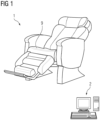
- Figure 1 shows a magnetic resonance system 1 according to an embodiment of the invention.
- the MR system 1 has the overall dimensions of an armchair and therefore fits easily into a POC office.
- the magnetic resonance system 1 comprises a control unit 2 that controls displacement of a magnet 3 and a unit of one or several RF coils 6 that is integrated into a seat 9 of the magnetic resonance system 1.
- the geometry of the magnet 3 according to an embodiment of the invention is shown in Figure 2 .
- the magnet has a curved surface facing the object 5 to be examined, the object being in the field-of-view of the MR system 1.
- Magnetic field lines 4 visualize the spatial distribution of the magnetic field created by the magnet 3 in the area of the object 5. Due to the geometry of the magnet, the magnetic field strength in the area of the object 5 decreases linearly with increasing distance to the magnet 3 thereby creating a gradient field at the position of the object 5.
- the magnet may comprise or consist of multiple small magnet pieces which are assembled according to genetic algorithms that calculate the location and the tilting of these small magnet pieces in order to achieve a targeted spatial distribution of the magnetic field 4.
- an essentially constant built-in field gradient orthogonal to the magnet surface may be created, wherein the field strength decreases linearly with the distance to the magnet surface.
- Figure 3 shows a part of the magnetic resonance system 3 comprising the magnet 3 and a unit 6 of RF coils 7.
- the magnet 3 is movable by a displacement element (not shown) as indicated by arrows showing a possible motion trajectory.
- the magnet 3 is movable to at least six different, noncollinear positions.
- the magnet 3 can be displaced from left to right spanning an angle from -45° to +45°.
- Figure 4 shows a side view of the same set-up as shown in figure 3 .
- the magnet can further be displaced along another motion trajectory, namely in the sagittal plane spanning an angle from -30° to +90°.
- the unit 6 of RF coils 7 serves as a seat for the patient 8 and is suspended via springs 14 to the chair 9.
- the unit comprises an array RF coils 7 that are flexible.
- the RF coils 7 may deform and attach closely to the patient 8 in order to maximize the magnitude of received magnetic resonance signals and therewith the signal-to-noise ratio.
- the RF coils may be connected to each other via mechanical joints.
- a camera 12, in particular a 3D camera 12, is configured to scan the patient 8 in order to determine the position of the object 5 within the patient 8.
- the camera 12 scans the patient 8 from multiple perspectives to create patient avatar, in particular a 3D patient avatar. For example, when the patient enters the examination room, the camera 12 continuously acquires views, in particular 3D views, of the patient 8 and the control unit 2 interactively refines the patient avatar. When the patient finally reaches the field-of-view, the avatar is morphed according to the patient pose and the relative location of the object 5 is calculated.
- Figure 5 shows three different exemplary positions the magnet 3 may be in during an examination allowing to achieve noncollinear and independent diffusion directions for diffusion weighted imaging contrasts.
- FIG. 6 shows a flow diagram of the method according to an embodiment of the invention.
- a position of an object 5 to be examined for example a prostate
- a camera 12 in particular a 3D camera.
- a patient 8 anatomy is scanned by the camera 12 before the patient 8 is placed in the magnetic resonance system 1 for the examination and/or when the patient 8 approaches the magnetic resonance system.
- a permanent magnet 3 is used to create a linear gradient field at the field-of-view where the object 5 is placed in order to create a diffusion contrast and/or to achieve a spatial encoding.
- the magnet 3 is mechanically displace to six different positions in order to measure diffusion magnetic resonance signals in multiple nonparallel directions.
- magnetic resonance data are acquired via an array of RF coils 7.
Landscapes
- Physics & Mathematics (AREA)
- Condensed Matter Physics & Semiconductors (AREA)
- General Physics & Mathematics (AREA)
- High Energy & Nuclear Physics (AREA)
- Health & Medical Sciences (AREA)
- Nuclear Medicine, Radiotherapy & Molecular Imaging (AREA)
- Engineering & Computer Science (AREA)
- Signal Processing (AREA)
- General Health & Medical Sciences (AREA)
- Radiology & Medical Imaging (AREA)
- Vascular Medicine (AREA)
- Spectroscopy & Molecular Physics (AREA)
- Magnetic Resonance Imaging Apparatus (AREA)
Abstract
Description
- The invention relates to a magnetic resonance system, a unit of one or several RF coils, and a method of acquiring mixed contrast magnetic resonance data.
- In the case of various diseases, for example prostate cancer, an early detection can significantly improve the chances of a successful therapy and/or the chance to avoid or minimize possible post-treatment sequels and secondary disorders. For example, a common prostate cancer screening method is a targeted clinical test that measures a prostate specific antigen (PSA) level in the patient blood. This method has the advantages of being relatively cheap and that it may be carried out at the point-of-care (POC), such as a physician's office. However, the PSA results tend to have poor specificity and the PSA test will not find all prostate cancers.
- Hence, in order to detect clinically significant prostate cancers, additional medical examinations are required. For example, according to the state of the art, tissue biopsies guided by either ultrasound or magnetic resonance imaging are used. Ultrasound guided biopsy is relatively cheap and tends to be largely available. However, ultrasound tissue contrasts are typically poor and there tends to be a high risk of missing prostate tumors. On the other hand, magnetic resonance imaging is typically much better, in particular more reliable and precise.
- Some prior art solutions have attempted to provide nuclear magnetic resonance (NMR) measurements at the POC by trying to squeeze the hardware of a full body magnetic resonance (MR) scanner into a more compact solution. However, this poses new problems, since state-of-the-art magnetic resonance imaging (MRI) techniques require high-field magnets to achieve good signal-to-noise ratio, strong gradient coils, used e.g. for diffusion-weighted contrast, as well as a highly homogeneous magnetic field and linear gradients across the field-of-view (FOV) to avoid image artefacts. All these requirements increase the costs and complexity, as well as the installation footprint (e.g. due to large stray magnetic field of strong magnets), and increase the operational costs, e.g. due to power & cooling water consumption. Consequently, these requirements prevent a wide deployment at POC of these otherwise very valuable medical devices. Accordingly, in summary, solutions according to the state of the art that include magnetic resonance imaging are expensive and tend not to be readily available at a POC office.
- It is therefore an object of the invention to provide an MR system that is small in size, has low maintenance costs and can be readily supplied at a point of care. It is a further object of the invention to provide an MR system that can provide NMR data of the pelvis and in particular the prostate.
- These objects are met or exceeded by a magnetic resonance system according to
claim 1, a unit of one or several RF coils according toclaim 12, and a method according to claim 13. Further advantages and features result from the dependent claims, the description and the attached figures. - According to a first aspect of the invention, a magnetic resonance system configured to acquire magnetic resonance data of an object in an field-of-view is provided, wherein the magnetic resonance system comprises
- a magnet, in particular a permanent magnet, that is configured such that it creates a gradient field, which is a static magnetic field having a gradient in its field strength at the field-of-view;
- a unit of one or several RF coils, the unit in particular comprising an array of RF coils, wherein the RF coils are configured to generate RF pulses and to acquire magnetic resonance data from the object;
- a control unit (2) configured to cause the magnetic resonance system to utilize the magnet's gradient field for diffusion weighted and optionally T2-weighted NMR measurements and/or for spatial encoding of the magnetic resonance data.
- The magnetic resonance (MR) system may be a magnetic resonance imaging system, though it may also be used for NMR measurements from a single voxel or from the whole field-of-view. The object may be an organ and/or part of a patient, e.g. the pelvis region or the liver region of a patient. For example, the object may be a prostate. The field-of-view is preferably a region, where the object is located during an MR examination, and the MR system is configured to acquire MR data from within this region. The field-of-view may be a region in which the static magnetic field generated by the magnet is sufficiently high and has a gradient of sufficient magnitude and homogeneity to carry out NMR measurements, in particular diffusion-weighted measurements. The unit of RF coils may be adapted to support and/or flexibly attach to the patient body.
- The magnetic resonance system may comprise a positioning element, e.g. a seat or a patient table, that is configured to guide the position of the patient to be such that the object is located in the field-of-view during the examination. Guiding the position may for example be provided by the geometry of the positioning element.
- The magnet is preferably a permanent magnet. To minimize the overall costs, the MR system may use a relatively small magnet, about the same size or smaller than the body part to investigate. In cases where the object is the prostate, the magnet may for example be in at least one extension smaller than the overall size of the pelvis region. For example, the magnet may have a longest extension of 5 to 30 cm, preferably 8 to 20 cm. The MR system is in particular a single-sided NMR device, wherein the static magnetic field required for NMR measurements is generated by a magnet on one side of the field-of-view.
- The magnet is configured to create a magnetic field at the field-of-view, which has an essentially linear gradient in its field strength in at least one direction. The at least one direction may in particular be directed from the magnet to the field-of-view and/or through the field-of-view. The at least one direction may be orthogonal to the magnet's surface, in particular the surface facing the field-of-view. The gradient field may be focused on the field-of-view. The magnet may provide a strong inhomogeneity along a direction orthogonal to the magnet surface. Therewith the static gradient in the magnetic field may generate a diffusion contrast. The direction of the magnetic field at the field-of-view may be the same or another direction than the direction of the gradient, it may be parallel to the surface of the magnet.
- The magnet may be configured to create the gradient field due to its geometric shape. The magnet may be curved on a side facing the field-of-view, in particular such that the magnet is concave with respect to the field-of-view. In other words, the surface of the magnet facing the field-of-view may be curved such that the magnet partially surrounds the field-of-view. A focus point of the curved surface of the magnet may be located on a straight line from the center of the magnet and/or the center of the curved surface to the field-of-view. The focus point may in particular be located in the field-of-view and/or in the area of the field-of-view.
- According to an embodiment, the MR system may comprise a displacement element configured to displace the magnet, in particular in two non-parallel directions. The displacement may be such that the gradient field remains at the field-of-view, wherein at least one portion of the gradient field remains at the same location, in particular a position within the field-of-view, when the magnet is displaced in any or both of two orthogonal directions. The direction of the magnetic field gradient preferably changes when the magnet is displaced. Thereby, the MR system is able to generate gradient fields with varying directions of the magnetic field gradient without gradient coils. For example, when the magnet is positioned at three locations by the displacement element, diffusion-weighted NMR may be carried out to achieve noncollinear and independent diffusion directions for the DWI contrasts. The MR system may therefore be able to detect diffusion within the object in several independent directions. By means of the displacement element, the magnet is movable, preferably along two orthogonal directions, while staying focused onto the target anatomy, i.e. the object.
- The displacement element may be configured to displace the magnet along a trajectory. The trajectory may preferably be a curved trajectory. The trajectory may by extending in two dimensions, such that the magnet can be displaced on a surface. The displacement element may be configured such that it can displace the magnet across a bidimensional curved surface trajectory. The trajectory may extend from -45° to +45° in the left-right direction and -30° to +90° in the sagittal plane, in relation to the patient to be examined. The displacement element may optionally be configured to provide a rotation of the magnet. Advantageously a providing a rotation of the magnet may allow a more flexible adjustment of the direction of the gradient field.
- The control unit may, for example, be a processing unit such as a CPU, a GPU, a computer or part of a computer. The computer may be a PC, a server, and/or a console of the magnetic resonance system. The computer may also be a mobile device, such as a laptop, tablet computer or mobile phone. The control unit is in particular configured to cause the magnetic resonance system to utilize the magnet's gradient field for magnetic resonance measurements. Preferably the control unit and/or the system may be configured to use the gradient field both as main magnetic field, B0, and as gradient field, G. In other words, the control unit and/or the magnetic resonance system is configured not to require an additional gradient coil arrangement and/or an additional main magnet. Hence the magnetic resonance system may be such that it does not comprise a separate main magnet and/or separate gradient coil arrangement. Correspondingly, the system may be less complex and/or expensive. Furthermore, the system may thus require less space and, thus, be suitable for the use in a point-of-care office.
- Diffusion weighted imaging (DWI) uses the diffusion of molecules to generate contrast in magnetic resonance imaging. Generally, diffusion contrast is generated by applying a diffusion-weighting gradient field during a spin-echo sequence, so that the signal is attenuated for spins that have moved in the direction of the diffusion-weighting gradient during the echo time. By changing the direction of the diffusion-weighting gradient field, the preferred direction of diffusion can also be detected. In DWI, each voxel may be assigned an apparent diffusion coefficient (ADC)and a preferred direction of diffusion. Thus, the system and in particular the control unit, may be adapted to displace the magnet during an NMR measurement session. During an NMR measurement session may mean while the object/patient is positioned in the field-of-view. It may mean in between data acquisitions. The displacement may be carried out in between NMR sequences or during a sequence, as explained in more detail below.
- For T2-weighted imaging, a CPMG (Carr-Purcell-Meiboom-Gill) pulse train magnetic resonance sequence may be used. This sequence uses a train of refocusing 180° RF pulses to acquire an echo train which decays essentially at T2.
- Mixed contrast imaging is in particular a combination of T2-weighted contrast and diffusion weighted contrasts. Preferably the T2-weighted contrast and the diffusion weighted contrast are acquired in a single magnetic resonance sequence. For this purpose, for example, a CPMG (Carr-Purcell-Meiboom-Gill) pulse train magnetic resonance measuring sequence, or a SE-CPMG sequence may be used.
- Diffusion-weighting and T2-weighting may also be applied in single-voxel NMR experiments, for example a diffusion coefficient and T2 may be determined in a bi-parametric magnetic resonance measurement. It has been found that prostate cancers and in particular clinically significant prostate cancers may be detected by such a bi-parametric NMR measurement. Therefore, diffusion-weighting contrast and T2-contrast is especially useful in MR examinations of the pelvis and the screening for prostate cancer.
- The MR system of the invention may be adapted to acquire diffusion-weighted and optionally T2-weighted NMR data. It may be adapted to acquire mixed contrast NMR data, comprising diffusion-weighted and T2-weighted data, in particular separately to provide bi-parametric data. The MR system of the invention may be adapted to acquire NMR data from a single voxel within the field-of-view. For example, bi-parametric NMR measurements providing only a diffusion coefficient and T2 may be carried out. Corresponding sequences are, for example, described in Ziyi Pan et al. "D-T2 Distribution Obtained Using CPMG-only Sequence Compared with Traditional SE-CPMG Sequence on the Single-sided NMR Device", Proc. Intl. Soc. Mag. Reson. Med. 29 (2021), 3104. The MR system may also be used to carry out MR imaging to acquire MR image data from the field-of-view, in particular DWI and/or T2-weighted imaging. Corresponding sequences are, for example, described in the international publications
WO 2021/184470 A1 andWO 2021/184471 A1 . The above-mentioned sequences may be adapted for the MR system of the invention. For example, the sequences disclosed in Siyi Pan et al. may be carried out several times with different gradient directions, for example 3 to 6 independent gradient directions, each repetition with the magnet in a different displacement position and/or orientation. The independent gradient directions may be noncollinear, and may be at least approximately orthogonal to one another. Diffusion coefficients may thereby be obtained not only in one direction, but in 3 to 6 or more than 6 directions. The diffusion coefficient may thus be a tensor. The MR system of the invention may be adapted to carry out scanning sequences which incorporate displacements/movements of the magnet during the sequence. For example, the sequence may include scanning or data acquisition while the magnet is being moved. The sequence may also include displacing the magnet in between several data acquisitions. It may thus be possible to include spatial encoding into the sequence, and/or to measure the apparent diffusion coefficient in several directions. When moving the magnet during the data acquisition, it may be required to include a correction of motion artefacts caused by said movement. - The unit of one or several RF coils is configured to acquire magnetic resonance data, in particular from the object within the field-of-view. Additionally and/or alternatively, the unit may be configured to generate RF pulses, in particular in order to excite spins in the object, in particular during an acquisition sequence. The unit may be an array of RF coils-. The RF coils may comprise RF receive coils and/or RF transmit coils and/or transmit-receive coils. The RF coils may each have at least one loop element, preferably multiple loop elements. The system and/or the control unit may be configured to operate each loop element of the RF coil array in transmit (TX) and/or receive (RX) mode. The system and/or the control unit may be configured to use the sensitivity profiles of the individual loop elements to focus, in particular dynamically focus, the unit of RF coils on the field-of-view and/or the object.
- According to an embodiment, the field strength of the gradient field in the field-of-view decreases linearly with the spatial distance to the magnet, in particular with a linearity better than 10%, preferably better than 5%, more preferably better than 1%. The decrease may preferably be along a direction orthogonal to a surface of the magnet, in particular the surface facing the field-of-view. In this context, "better than 10%" (or 5% or 1%) may in particular mean that the change of field strength deviates by less than 10% of an ideal linear function. A linear decrease of the field strength may advantageously be used to reliably generate a diffusion contrast, in particular by enabling a constant directional diffusion encoding for magnetic resonance signals providing a diffusion weighted imaging contrast. It may further allow to implement Spatial signal encoding from crude to coarse spatial resolution, for example 5cm3 voxels down to 0.5cm3 voxels. The linear decrease may in particular provide an essentially constant field gradient. Therein, "essentially" may mean a deviation of less than 10%, preferably less than 5%. It has been found that with a linearity better than 10% a diffusion weighted imaging contrast can already be achieved with the inventive system. A linearity of better than 5% may provide a less crude spatial resolution which can be beneficial for some applications. A linearity better than 1% may even allow magnetic resonance imaging with relatively small voxel sizes, e.g. down to 0.5cm3.
- According to an embodiment the magnet comprises, in particular consists of, multiple small magnet pieces that are arranged together to form the magnet. The small magnet pieces may be arranged, in particular located and/or tilted, such that they provide a targeted spatial distribution of a magnetic field, in particular the gradient field. In this context, "small" may in particular mean that the magnet pieces are smaller than the magnet itself, in particular 5 to 50 times, preferably 10 to 30 times smaller than the magnet. The small magnet pieces may be small magnetic blocks. The small magnet pieces may be arranged according to a calculation, in particular carried out via a genetic algorithm. Advantageously, the small magnet pieces may allow to provide a particularly precise arrangement of the gradient field as required for the inventive application and/or as required for specific embodiments of the inventive MR system, e.g. for specific applications of the system.
- According to an embodiment, the magnetic resonance system comprises a displacement element configured to displace the magnet, in particular in two non-parallel directions. The two non-parallel directions may be orthogonal with respect to each other. In this context one or both of the directions may for example follow a curved trajectory. Preferably both trajectories are orthogonal to each other at any point. Advantageously the displacement may advantageously allow to create various different gradient fields by changing the position of the magnet. Hence the displacement element may eliminate the need for a gradient coil and thus further reduce the costs of the system.
- The displacement element may be a mechanical displacement element. In an embodiment, the displacement element may be a shuttle-like element, in particular guided by a guiding structure. The guiding structure may for example be raillike. For example, the magnet may be mounted on a carriage which is moveable along a guiding structure, in particular rails, which are arranged on a curved surface. The magnet may also be displaced by means of a cable mechanism. The displacement of the magnet may be caused by an actuator, for example by one or several electromotors, which are controlled by the control unit.
- The frequency of the magnetic resonance signals from the field-of-view may correlate with the local field strength by the gyromagnetic constant:

- The control unit may be configured to decompose the magnetic resonance signals into small frequency bands that corresponds to regions of the magnetic field, in particular to regions having the magnetic field corresponding to the frequency band. Preferably, the strength of the magnetic field may be almost constant within the frequency bands. Almost constant may mean that it deviates no more than within 5%, preferably 2%, more preferably 1%. The frequency bands may in particular be curvilinear projections throughout the measurement volume, which may be larger than the field-of-view, and/or throughout the field-of-view. It has been found that it is possible to generate curvilinear projections throughout the measurement volume in a similar way to the generation of fan-beam projections in CT (computer tomography) X-ray imaging. By also using the focusing capability of the unit of RF coils, in particular an RF coil, to focus the MR excitation to a region within the measurement volume or the field-of-view, in particular comprising an RF coil array, it may thus be possible to generate MR images of the object. Therewith, by decomposing the NMR signals into small frequency bands that correspond to the magnetic field regions wherein the strength of the magnetic field is almost constant, it is possible to generate curvilinear projections throughout the measurement volume. In combination with the focusing capability of the RF coil array, this makes possible to generate crude MR images of the field-of-view and/or a measurement volume surrounding the field-of-view. "Crude" in this context may mean that the spatial resolution of the MR image is 0.5 cm3 to 7 cm3. In an embodiment, such curvilinear projections may be acquired in at least three nonparallel directions by moving the magnet by the displacement element into three different positions and repeating the same NMR sequence. The data acquired from the at least three magnet positions may also be reconstructed together to form a complete image. Since each acquisition corresponds to a projection, the reconstruction may be performed using a back-projection algorithm, for example filtered back-projection as used for the reconstruction of CT images.
- The displacement element and/or the control unit may be configured to allow a moving "sweet spot" within a restricted sensitivity or measurement volume to enable taking a tissue sample guided by the magnetic resonance system (e.g. in the context of virtual biopsies). Therein, unique signature of magnetic resonance signals may be analyzed. The magnetic resonance signals may emerge out of dedicated tissue voxel, e.g. a 0.5cm3 to 2cm3 voxel. Signals from this voxel may, for example, be modulated by various encoding patterns (e.g. including RF modulation) and may be processed to extract significant clinical information.
- According to an embodiment, the trajectory of the magnet, in particular two orthogonal directions, span an area that is bowl-shaped. Advantageously, a bowl-shaped trajectory may allow a particularly efficient focusing of the gradient field on the field-of-view and/or on the object to be examined.
- According to an embodiment, the system is configured to displace the magnet, in particular during a diffusion weighted imaging or single-voxel sequence, such that multiple gradient field directions, in particular six gradient field directions, are created during a measurement sequence. Multiple gradient field directions allow to take full advantage of the diffusion contrast. Multiple directions may allow to assign a three-dimensional direction of diffusion to each voxel as well as additional information such as a rate of diffusion. The multiple gradient field directions may preferably be nonparallel with respect to each other. In particular, the control unit and/or the displacement element may be configured to create at least six noncollinear gradient field directions via displacement of the magnet. Advantageously the system does therefore not require dynamic gradient coils to encode the diffusion directions. Instead, the system may be configured to mechanically move the magnet with respect to the target anatomy. Six directions may in particular be advantageous to allow an effective measurement of the diffusion tensor. In some circumstances, e.g. when some data are already known from previous measurements, it may be beneficial to use fewer gradient field directions, e.g. to save time during the examination.
- According to an embodiment, the field strength of the permanent magnet is smaller than 1 Tesla, preferably smaller than 0.3 Tesla, more preferably smaller than 0.1 Tesla, at the field-of-view. Advantageously a small field strength can be achieved already with a relatively small and/or cost-efficient producible magnet. The field strength is preferably higher than 0.01 T, more preferred higher than 0.04 T, in order to allow sufficient SNR at coarse resolution.
- According to an embodiment, the magnet is smaller than object to be investigated in at least one direction and/or dimension. A small magnet may allow to lower and/or minimize the overall costs of the system. For example, the magnet may be smaller in at least one dimension and/or direction than the overall size of the pelvis region of a patient, wherein in particular inside the pelvis region the prostate is located.
- According to an embodiment, the system is configured to acquire signals with a coarse resolution, wherein a matrix size of the resolution is smaller than 64×64 pixels and/or smaller than 32×32 pixels and/or wherein a voxel size is greater than 0,5cm3 and/or greater than 3cm3, in particular between 3cm3 and 7cm3. It has been found that a coarse resolution may allow to acquire sufficient information to allow the guidance of a biopsy, in particular of the prostate. Advantageously, by aiming only at a coarse resolution and by implementing the design of the system as described herein, a smaller magnet and/or a more cost-efficient system may be used.
- According to an embodiment, the system is configured to provide a magnetic resonance signal from at least one prostate biomarker and/or two magnetic resonance imaging contrasts, in particular T2-weighted and diffusion weighted contrasts. Preferably the system is configured to acquire the T2-weighted and the diffusion weighted contrast by means of a single magnetic resonance sequence. Advantageously, the T2-weighted and the diffusion weighted contrast can be important biomarkers in order to detect prostate cancer, in particular clinically significant prostate cancer. Accordingly, the two magnetic resonance imaging contrasts may increase the sensitivity for disease detection.
- According to an embodiment, the unit of RF receive coils is configured to carry a human patient's weight, the patient comprising the object, wherein the unit is suspended from a frame, in particular a frame of the magnetic resonance system, wherein the unit of one or several RF coils is in particular a seat or is part of a seat which is adapted for a patient to sit on it during a magnetic resonance examination. The seat may for example be the seat of a chair, in particular an examination chair. In particular, the unit is configured to be strong enough to carry a patient's weight. The unit of one or several RF coils may comprise several parts which are connected together by mechanical joints and/or pivoting articulations. Thereby, the unit of RF coils can adapt to the body part to be examined. The unit of one or several RF coils and/or parts thereof may be attached to springs. The springs may be further attached to the system and/or to a chair of the system. Advantageously, the unit being configured to carry a patient's weight may allow a particularly space-saving solution that may also allow an easy application in a point-of-care office. Furthermore, e.g. in the case of a prostate examination, the RF coils may thus naturally be positioned close to the object to examined and thus allow to maximize the magnitude of the received magnetic resonance signals and therewith the signal-to-noise ratio. Providing springs may additionally allow a flexible adaption to the size and/or shape of a patient body.
- According to an embodiment, the unit of RF receive coils is flexible such that it can adapt to the geometry of a patient being placed on, in particular sitting on, the unit of receive coils. Advantageously, due to the flexibility, the RF coils may thus come particularly close to the object to be examined. Preferably the RF coils are not extensible. The RF coils not being extensible may allow a more reliable and/or more robust tuning and matching of the RF coils. The unit and/or the RF coils of the unit may be designed such that they adapt to a patient geometry in the region of the object to be examined. In an embodiment, the unit of RF receive coils are flexible, so that they come as close as possible to the target anatomy and at the same time are strong enough to support the patient weight.
- According to an embodiment, the system comprises a camera, in particular a 3D camera, wherein the camera is configured to scan a patient's body, in particular from multiple directions, wherein the system is configured to determine the position of the object to be examined based on the camera's scan. The system and/or the control unit may be configured to create a patient avatar. The camera may be configured to scan the patient from multiple different perspectives in order to create the patient avatar. Alternatively and/or additionally, the system and/or the control unit may be configured to estimate the location of the patient and/or of the object within the patient relative to the position of the magnetic resonance system and/or a coordinate system of the magnetic resonance system. The system and/or the control unit may be configured to calculate the position of the object based on the scanned data of the patient and/or based on the patient avatar. Calculating the position of the object may be further based on reference data related to the position of the object, e.g. a prostate, within a patient's body. Advantageously, there may be no need for a high-resolution magnetic resonance localizer. The camera may be configured to continuously acquire image data, in particular 3D image data, of the patient, in particular in order to allow refining the patient avatar. The control unit and/or the system may be configured to refine the patient avatar based on the continuously acquired image data. The system and/or the control unit may be configured to automatically morph the patient avatar according to the current patient's pose. The system and/or the control unit may be configured to adjust the estimated location of the object based on the morphed patient avatar.
- According to a further aspect of the invention, a unit of one or several RF receive coils is provided, wherein the unit in particular comprises an array of RF coils, wherein the one or several RF coils are configured to acquire magnetic resonance data from an object and optionally to generate RF pulses, wherein the unit is configured to carry a human patient's weight, the patient comprising the object, wherein the unit is suspended from a frame, in particular a frame of the magnetic resonance system, wherein the unit of one or several receive coils is in particular a seat or is part of a seat which is adapted for a patient to sit on it during a magnetic resonance examination. All features and advantages of the magnetic resonance system may be adapted to the unit and vice versa.
- According to an embodiment, the unit of RF receive coils is flexible such that it can adapt to the geometry of a patient being placed on, in particular sitting on, the unit of receive coils. Advantageously, the flexibility of the unit may allow for the RF coils to come closer to an object to be examined and/or to allow for a more comfortable sitting experience of the patient.
- According to a further aspect of the invention, a method for acquiring diffusion-weighted and optionally T2-weighted contrast magnetic resonance data, in particular mixed contrast magnetic resonance data, of an object placed at an field-of-view is provided, wherein in particular the system according to any one of the preceding claims is used, wherein the method comprises the steps:
- using a permanent magnet to create a gradient field, in particular a linear gradient field, at the field-of-view in order to create a diffusion contrast and/or to achieve a spatial encoding;
- mechanically displacing the magnet to multiple positions, in particular to six different positions, in order to measure diffusion magnetic resonance signals in multiple nonparallel directions;
- acquiring magnetic resonance data via RF coils, in particular via a unit of RF receive and/or transmit coils.
- The method may in particular used for examining the pelvis area of a patient. All features and advantages of the magnetic resonance system and the unit of one or several RF receive coils may be adapted to the method and vice versa. The unit of one or several RF coils may in particular be the unit as described herein.
- According to an embodiment, a position of the object is determined via a camera, in particular a 3D camera. According to a preferred embodiment, the patient anatomy is scanned by the camera before the patient is placed in the magnetic resonance system for the examination and/or when the patient approaches the magnetic resonance system. The camera may scan a patient's body, in particular from multiple directions and the position of the object to be examined may be determined and/or calculated based on the camera's scan. A patient avatar may be created based on the camera's scan, in particular by scanning the patient from multiple different perspectives. Determining the position of the object may be further based on reference data related to the position of the object, e.g. a prostate, within a patient's body. Image data, in particular 3D image data, of the patient may be acquired continuously, in particular in order to continuously refine the patient avatar. The patient avatar may be automatically morphed according to the current patient's pose. The estimated location of the object may be adjusted based on the morphed patient avatar.
- All embodiments mentioned herein may be combined with each other. The invention is also direct to the use a magnetic resonance system as described herein for carrying out examinations of the human pelvis, in particular for acquiring diffusion-weighted and optionally T2-weighted magnetic resonance data. The use is particular suited to examine the prostate of a patient.
- The accompanying drawings illustrate various exemplary embodiments and methods of various aspects of the invention.
- Fig. 1
- shows a magnetic resonance system according to an embodiment of the invention;
- Fig. 2
- shows a magnet according to the invention;
- Fig. 3
- shows part of a magnetic resonance system according to an embodiment of the invention;
- Fig. 4
- shows part of a magnetic resonance system according to an embodiment of the invention;
- Fig. 5
- shows part of a magnetic resonance system with different magnet positions according to an embodiment of the invention;
- Fig. 6
- shows a flow diagram of a method according to an embodiment of the invention.
-
Figure 1 shows amagnetic resonance system 1 according to an embodiment of the invention. TheMR system 1 has the overall dimensions of an armchair and therefore fits easily into a POC office. Themagnetic resonance system 1 comprises acontrol unit 2 that controls displacement of amagnet 3 and a unit of one orseveral RF coils 6 that is integrated into aseat 9 of themagnetic resonance system 1. - The geometry of the
magnet 3 according to an embodiment of the invention is shown inFigure 2 . The magnet has a curved surface facing theobject 5 to be examined, the object being in the field-of-view of theMR system 1.Magnetic field lines 4 visualize the spatial distribution of the magnetic field created by themagnet 3 in the area of theobject 5. Due to the geometry of the magnet, the magnetic field strength in the area of theobject 5 decreases linearly with increasing distance to themagnet 3 thereby creating a gradient field at the position of theobject 5. The magnet may comprise or consist of multiple small magnet pieces which are assembled according to genetic algorithms that calculate the location and the tilting of these small magnet pieces in order to achieve a targeted spatial distribution of themagnetic field 4. Thus, an essentially constant built-in field gradient orthogonal to the magnet surface may be created, wherein the field strength decreases linearly with the distance to the magnet surface. -
Figure 3 shows a part of themagnetic resonance system 3 comprising themagnet 3 and aunit 6 of RF coils 7. Themagnet 3 is movable by a displacement element (not shown) as indicated by arrows showing a possible motion trajectory. Preferably themagnet 3 is movable to at least six different, noncollinear positions. In this embodiment themagnet 3 can be displaced from left to right spanning an angle from -45° to +45°.Figure 4 shows a side view of the same set-up as shown infigure 3 . As indicated by arrows, the magnet can further be displaced along another motion trajectory, namely in the sagittal plane spanning an angle from -30° to +90°. - Due to the movement of the
magnet 3 the magnetic field gradient created by themagnet 3 can change its direction at the position of theobject 5, for example a prostate of apatient 8. Furthermore, theunit 6 of RF coils 7 serves as a seat for thepatient 8 and is suspended viasprings 14 to thechair 9. In particular the unit comprises an array RF coils 7 that are flexible. The RF coils 7 may deform and attach closely to thepatient 8 in order to maximize the magnitude of received magnetic resonance signals and therewith the signal-to-noise ratio. The RF coils may be connected to each other via mechanical joints. Acamera 12, in particular a3D camera 12, is configured to scan thepatient 8 in order to determine the position of theobject 5 within thepatient 8. Thecamera 12 scans thepatient 8 from multiple perspectives to create patient avatar, in particular a 3D patient avatar. For example, when the patient enters the examination room, thecamera 12 continuously acquires views, in particular 3D views, of thepatient 8 and thecontrol unit 2 interactively refines the patient avatar. When the patient finally reaches the field-of-view, the avatar is morphed according to the patient pose and the relative location of theobject 5 is calculated. -
Figure 5 shows three different exemplary positions themagnet 3 may be in during an examination allowing to achieve noncollinear and independent diffusion directions for diffusion weighted imaging contrasts. According to the three different positions of themagnet 3, three differentmagnetic field regions 15 are measured corresponding to three narrow frequency bands, since the frequency of the local NMR signals correlates with the local field strength by the gyromagnetic constant as ω(x,y,z)=γ·|B(x,y,z)|. By decomposing the magnetic resonance signals into small frequency bands that corresponds to themagnetic field regions 15, in which the strength of the magnetic field is almost constant, it is possible to generate curvilinear projections throughout the measurement volume. Together with the positioning of themagnet 3, this enables to reconstruct magnetic resonance images of theobject 5. -
Figure 6 shows a flow diagram of the method according to an embodiment of the invention. In afirst step 101, a position of anobject 5 to be examined, for example a prostate, is determined via acamera 12, in particular a 3D camera. For this purpose, apatient 8 anatomy is scanned by thecamera 12 before thepatient 8 is placed in themagnetic resonance system 1 for the examination and/or when thepatient 8 approaches the magnetic resonance system. In thefollowing step 102, apermanent magnet 3 is used to create a linear gradient field at the field-of-view where theobject 5 is placed in order to create a diffusion contrast and/or to achieve a spatial encoding. In afurther step 103, themagnet 3 is mechanically displace to six different positions in order to measure diffusion magnetic resonance signals in multiple nonparallel directions. In yet afurther step 104, magnetic resonance data are acquired via an array of RF coils 7.
Claims (15)
- A magnetic resonance system (1) configured to acquire magnetic resonance data of an object (5) in a field-of-view, wherein the magnetic resonance system (1) comprises- a magnet (3), in particular a permanent magnet, that is configured such that it creates a gradient field, which is a static magnetic field having a gradient in its field strength at the field-of-view;- a unit of one or several RF coils, the unit in particular comprising an array of RF coils, wherein the RF coils are configured to generate RF pulses and to acquire magnetic resonance data from the object;- a control unit (2) configured to cause the magnetic resonance system to utilize the magnet's gradient field for diffusion weighted and optionally T2-weighted NMR measurements and/or for spatial encoding of the magnetic resonance data.
- The system (1) according to claim 1,
wherein the field strength of the gradient field in the field-of-view decreases linearly with a spatial distance to the magnet, in particular with a linearity better than 10%, preferably better than 5%, more preferably better than 1%. - The system (1) according to one of the claims 1 or 2,
wherein the magnetic resonance system (1) comprises a displacement element configured to displace the magnet, in particular in two orthogonal directions. - The system (1) according to claim 3,
wherein the displacement element is configured to displace the magnet (3) on a trajectory such that the gradient field remains at the field-of-view, in particular such that at least one portion of the gradient field remains at the same location when the magnet (3) is displaced in any or both of the two orthogonal directions, and such that the direction of the gradient field changes when the magnet (3) is displaced. - The system (1) according to claim 4,
wherein the trajectory of the magnet, in particular two orthogonal directions, span an area that is bowl-shaped. - The system (1) according to any one of claims 3 to 5,
wherein the system (1) is configured to displace the magnet, in particular during a diffusion weighted NMR measurement session or an imaging session, such that multiple gradient field directions, in particular six gradient field directions, are created during the session. - The system (1) according to any one of the preceding claims,
wherein the field strength of the permanent magnet (3) is smaller than 1 Tesla, preferably smaller than 0.3 Tesla, more preferably smaller than 0.1 Tesla, at the area of the object (5) to be examined. - The system (1) according to any one of the preceding claims,
wherein the system (1) is configured to provide a magnetic resonance signal from at least one prostate biomarker and/or two magnetic resonance imaging contrasts, in particular T2-weighted and diffusion weighted contrasts. - The system (1) according to any one of the preceding claims,wherein the unit (6) of RF receive coils (7) is configured to carry a human patient's (8) weight, the patient (8) comprising the object (5),wherein the unit (6) is suspended from a frame, in particular a frame of the magnetic resonance system (1),wherein the unit (6) of one or several RF coils (7) is in particular a seat (9) or is part of a seat (9) which is adapted for a patient (8) to sit on it during a magnetic resonance examination.
- The system (1) according to any one of the preceding claims,
wherein the unit (6) of RF one or several coils (7) is flexible such that it can adapt to the geometry of a patient (8) being placed on, in particular sitting on, the unit (6) of one or several RF coils (7). - The system (1) according to any one of the preceding claims,wherein the system (1) comprises a camera (12), in particular a 3D camera,
wherein the camera (12) is configured to scan a patient's (8) body, in particular from multiple directions,wherein the system (1) is configured to determine the position of the object (5) to be examined based on the camera's (12) scan. - A unit (6) of one or several RF coils (7),
wherein the unit (6) in particular comprises an array of RF coils (7),wherein the one or several RF coils (7) are configured to generate RF pulses and/or to acquire magnetic resonance data from an object (5),wherein the unit (6) is configured to carry a human patient's (8) weight, the patient (8) comprising the object (5),wherein the unit (6) is suspended from a frame, in particular a frame of the magnetic resonance system (1),wherein the unit (6) of one or several RF coils (7) is in particular a seat (9) or is part of a seat (9) which is adapted for a patient (8) to sit on it during a magnetic resonance examination. - A method for acquiring diffusion-weighted and optionally T2-weighted magnetic resonance data, in particular mixed-contrast data, of an object (5) placed at a field-of-view,wherein in particular the system (1) according to any one of the claims 1 to 11 is used,wherein the method comprises the steps:- using a permanent magnet (3) to create a gradient field, in particular a linear gradient field, at the field-of-view in order to create a diffusion contrast and/or to achieve a spatial encoding;- mechanically displacing the magnet (3) to multiple positions, in particular to six different positions, in order to measure diffusion magnetic resonance signals in multiple nonparallel directions;- acquiring magnetic resonance data via RF coils (79, in particular via a unit (6) of one or several RF coils (7).
- The method according to claim 13,
wherein a position of the object (5) is determined via a camera (12), in particular a 3D camera. - The method according to claim 13 or 14,
wherein patient anatomy is scanned by the camera (12) before the patient (8) is placed in the magnetic resonance system (1) for the examination and/or when the patient (8) approaches the magnetic resonance system (1).
Priority Applications (2)
| Application Number | Priority Date | Filing Date | Title |
|---|---|---|---|
| EP22177656.0A EP4290261A1 (en) | 2022-06-07 | 2022-06-07 | Magnetic resonance system and corresponding method |
| US18/206,664 US12339342B2 (en) | 2022-06-07 | 2023-06-07 | Magnetic resonance system and corresponding method |
Applications Claiming Priority (1)
| Application Number | Priority Date | Filing Date | Title |
|---|---|---|---|
| EP22177656.0A EP4290261A1 (en) | 2022-06-07 | 2022-06-07 | Magnetic resonance system and corresponding method |
Publications (1)
| Publication Number | Publication Date |
|---|---|
| EP4290261A1 true EP4290261A1 (en) | 2023-12-13 |
Family
ID=81975379
Family Applications (1)
| Application Number | Title | Priority Date | Filing Date |
|---|---|---|---|
| EP22177656.0A Pending EP4290261A1 (en) | 2022-06-07 | 2022-06-07 | Magnetic resonance system and corresponding method |
Country Status (2)
| Country | Link |
|---|---|
| US (1) | US12339342B2 (en) |
| EP (1) | EP4290261A1 (en) |
Families Citing this family (1)
| Publication number | Priority date | Publication date | Assignee | Title |
|---|---|---|---|---|
| WO2025230012A1 (en) * | 2024-05-02 | 2025-11-06 | 国立大学法人千葉大学 | Magnetic resonance detector and magnetic resonance probe |
Citations (6)
| Publication number | Priority date | Publication date | Assignee | Title |
|---|---|---|---|---|
| US20140306702A1 (en) * | 2013-04-12 | 2014-10-16 | Razvan Lazar | Magnetic Resonance Scanner and Antenna |
| US20150177343A1 (en) * | 2012-06-15 | 2015-06-25 | The General Hospital Corporation | System and Method for Portable Magnetic Resonance Imaging Using a Rotating Array of Permanent Magnets |
| US20210156938A1 (en) * | 2019-11-27 | 2021-05-27 | Siemens Healthcare Gmbh | Magnetic resonance imaging system with a rotatable magnet |
| WO2021184470A1 (en) | 2020-03-17 | 2021-09-23 | 无锡鸣石峻致医疗科技有限公司 | Magnetic resonance system gradient field measurement method based on diffusion effect |
| WO2021184471A1 (en) | 2020-03-17 | 2021-09-23 | 无锡鸣石峻致医疗科技有限公司 | Non-uniform field magnetic resonance system-based apparent diffusion coefficient measurment method |
| WO2022038603A1 (en) * | 2020-08-17 | 2022-02-24 | Ramot At Tel-Aviv University Ltd. | Mri in an inhomogeneous field with no pulsed gradients |
Family Cites Families (3)
| Publication number | Priority date | Publication date | Assignee | Title |
|---|---|---|---|---|
| CA2884097C (en) * | 2014-03-13 | 2020-04-21 | LT Imaging Inc. | Magnetic resonance imaging (mri) system and method |
| US10823793B2 (en) | 2019-03-27 | 2020-11-03 | Siemens Healthcare Gmbh | Resonance data acquisition scanner with rotating basic field magnet |
| EP4113146B1 (en) * | 2021-06-30 | 2025-05-07 | Siemens Healthineers AG | Magnetic resonance imaging device with a concave-shaped field generation unit |
-
2022
- 2022-06-07 EP EP22177656.0A patent/EP4290261A1/en active Pending
-
2023
- 2023-06-07 US US18/206,664 patent/US12339342B2/en active Active
Patent Citations (6)
| Publication number | Priority date | Publication date | Assignee | Title |
|---|---|---|---|---|
| US20150177343A1 (en) * | 2012-06-15 | 2015-06-25 | The General Hospital Corporation | System and Method for Portable Magnetic Resonance Imaging Using a Rotating Array of Permanent Magnets |
| US20140306702A1 (en) * | 2013-04-12 | 2014-10-16 | Razvan Lazar | Magnetic Resonance Scanner and Antenna |
| US20210156938A1 (en) * | 2019-11-27 | 2021-05-27 | Siemens Healthcare Gmbh | Magnetic resonance imaging system with a rotatable magnet |
| WO2021184470A1 (en) | 2020-03-17 | 2021-09-23 | 无锡鸣石峻致医疗科技有限公司 | Magnetic resonance system gradient field measurement method based on diffusion effect |
| WO2021184471A1 (en) | 2020-03-17 | 2021-09-23 | 无锡鸣石峻致医疗科技有限公司 | Non-uniform field magnetic resonance system-based apparent diffusion coefficient measurment method |
| WO2022038603A1 (en) * | 2020-08-17 | 2022-02-24 | Ramot At Tel-Aviv University Ltd. | Mri in an inhomogeneous field with no pulsed gradients |
Non-Patent Citations (1)
| Title |
|---|
| ZIYI PAN ET AL.: "D-T Distribution Obtained Using CPMG-only Sequence Compared with Traditional SE-CPMG Sequence on the Single-sided NMR Device", PROC. INTL. SOC. MAG. RESON. MED., vol. 29, 2021, pages 3104 |
Also Published As
| Publication number | Publication date |
|---|---|
| US20230393224A1 (en) | 2023-12-07 |
| US12339342B2 (en) | 2025-06-24 |
Similar Documents
| Publication | Publication Date | Title |
|---|---|---|
| JP6084573B2 (en) | MR imaging using multipoint Dixon technology | |
| EP1362550B1 (en) | Whole body MRI scanning with continously moving table and interactive control | |
| JP4602656B2 (en) | Method and system for imaging a large volume by MRI using parallel reception | |
| EP2730935B1 (en) | Motional error correction in functional magnetic resonance imaging | |
| US10302713B2 (en) | Method and magnetic resonance apparatus for determining absolute receive sensitivity maps for reception coils | |
| Walton et al. | Stereotactic localization with magnetic resonance imaging: a phantom study to compare the accuracy obtained using two-dimensional and three-dimensional data acquisitions | |
| CN101680938A (en) | Method for Automatically Collecting Magnetic Resonance Image Data | |
| WO2008114194A1 (en) | Magnetic resonance device and method | |
| JP2003339669A (en) | Multiple channel, cardiac array for sensitivity encoding in magnetic resonance imaging | |
| US12019134B2 (en) | MR electric properties tomography without contrast agent | |
| US20090278536A1 (en) | Wave-propagation based estimation of coil sensitivities | |
| Siegler et al. | Supine breast MRI | |
| Kozlowski et al. | Diffusion-weighted MRI in prostate cancer—comparison between single-shot fast spin echo and echo planar imaging sequences | |
| JPH0565179B2 (en) | ||
| US12339342B2 (en) | Magnetic resonance system and corresponding method | |
| US7429862B2 (en) | PPA MRT method and apparatus with scaled contribution of k-space data to the reconstructed image | |
| JP3615302B2 (en) | Magnetic resonance imaging system | |
| US12298369B2 (en) | Dedicated magnetic resonance device | |
| JP2005168868A (en) | Magnetic resonance imaging apparatus | |
| Ikeda et al. | Performance assessment of phased-array coil in breast MR imaging | |
| Ferrer et al. | Respiratory-and cardiac-triggered three-dimensional sheath inked rapid acquisition with refocused echoes imaging (SHINKEI) of the abdomen for magnetic resonance neurography of the celiac plexus | |
| CN119053877A (en) | MR electrical property tomography | |
| Dumoulin et al. | Moving table imaging at 1.5 and 3.0 Tesla | |
| WO2008041060A1 (en) | Mri-navigator sequence with restored magnetization | |
| JYRKINEN | Fast Catheter Profiling by Controlled Aliasing |
Legal Events
| Date | Code | Title | Description |
|---|---|---|---|
| PUAI | Public reference made under article 153(3) epc to a published international application that has entered the european phase |
Free format text: ORIGINAL CODE: 0009012 |
|
| STAA | Information on the status of an ep patent application or granted ep patent |
Free format text: STATUS: THE APPLICATION HAS BEEN PUBLISHED |
|
| AK | Designated contracting states |
Kind code of ref document: A1 Designated state(s): AL AT BE BG CH CY CZ DE DK EE ES FI FR GB GR HR HU IE IS IT LI LT LU LV MC MK MT NL NO PL PT RO RS SE SI SK SM TR |
|
| RAP1 | Party data changed (applicant data changed or rights of an application transferred) |
Owner name: SIEMENS HEALTHINEERS AG |
|
| STAA | Information on the status of an ep patent application or granted ep patent |
Free format text: STATUS: REQUEST FOR EXAMINATION WAS MADE |
|
| 17P | Request for examination filed |
Effective date: 20240604 |
|
| RBV | Designated contracting states (corrected) |
Designated state(s): AL AT BE BG CH CY CZ DE DK EE ES FI FR GB GR HR HU IE IS IT LI LT LU LV MC MK MT NL NO PL PT RO RS SE SI SK SM TR |
|
| STAA | Information on the status of an ep patent application or granted ep patent |
Free format text: STATUS: EXAMINATION IS IN PROGRESS |
|
| 17Q | First examination report despatched |
Effective date: 20241216 |