EP4282437A1 - Dysprosium nanoparticles, process for obtaining said nanoparticles and use of said nanoparticles as a contrast agent - Google Patents
Dysprosium nanoparticles, process for obtaining said nanoparticles and use of said nanoparticles as a contrast agent Download PDFInfo
- Publication number
- EP4282437A1 EP4282437A1 EP22382507.6A EP22382507A EP4282437A1 EP 4282437 A1 EP4282437 A1 EP 4282437A1 EP 22382507 A EP22382507 A EP 22382507A EP 4282437 A1 EP4282437 A1 EP 4282437A1
- Authority
- EP
- European Patent Office
- Prior art keywords
- ranging
- nanoparticles
- concentration
- moo
- nps
- Prior art date
- Legal status (The legal status is an assumption and is not a legal conclusion. Google has not performed a legal analysis and makes no representation as to the accuracy of the status listed.)
- Withdrawn
Links
- 239000002105 nanoparticle Substances 0.000 title claims abstract description 245
- 238000000034 method Methods 0.000 title claims abstract description 32
- 239000002872 contrast media Substances 0.000 title claims abstract description 29
- 230000008569 process Effects 0.000 title claims abstract description 20
- 229910052692 Dysprosium Inorganic materials 0.000 title description 4
- KBQHZAAAGSGFKK-UHFFFAOYSA-N dysprosium atom Chemical compound [Dy] KBQHZAAAGSGFKK-UHFFFAOYSA-N 0.000 title description 4
- 229920002125 Sokalan® Polymers 0.000 claims abstract description 87
- 229910015667 MoO4 Inorganic materials 0.000 claims abstract description 55
- 239000011248 coating agent Substances 0.000 claims abstract description 39
- 238000000576 coating method Methods 0.000 claims abstract description 39
- 238000002595 magnetic resonance imaging Methods 0.000 claims abstract description 38
- 239000004584 polyacrylic acid Substances 0.000 claims abstract description 20
- 150000002038 dysprosium compounds Chemical class 0.000 claims abstract description 10
- FEWJPZIEWOKRBE-UHFFFAOYSA-N Tartaric acid Natural products [H+].[H+].[O-]C(=O)C(O)C(O)C([O-])=O FEWJPZIEWOKRBE-UHFFFAOYSA-N 0.000 claims description 47
- 239000000243 solution Substances 0.000 claims description 44
- 239000000203 mixture Substances 0.000 claims description 43
- -1 poly(allylamine hydrochloride) Polymers 0.000 claims description 41
- NBIIXXVUZAFLBC-UHFFFAOYSA-N Phosphoric acid Chemical compound OP(O)(O)=O NBIIXXVUZAFLBC-UHFFFAOYSA-N 0.000 claims description 37
- 229920001223 polyethylene glycol Polymers 0.000 claims description 28
- LYCAIKOWRPUZTN-UHFFFAOYSA-N Ethylene glycol Chemical compound OCCO LYCAIKOWRPUZTN-UHFFFAOYSA-N 0.000 claims description 27
- 235000002906 tartaric acid Nutrition 0.000 claims description 27
- 239000011975 tartaric acid Substances 0.000 claims description 27
- LRHPLDYGYMQRHN-UHFFFAOYSA-N N-Butanol Chemical compound CCCCO LRHPLDYGYMQRHN-UHFFFAOYSA-N 0.000 claims description 24
- 230000032683 aging Effects 0.000 claims description 23
- 239000007864 aqueous solution Substances 0.000 claims description 23
- FAPWRFPIFSIZLT-UHFFFAOYSA-M Sodium chloride Chemical compound [Na+].[Cl-] FAPWRFPIFSIZLT-UHFFFAOYSA-M 0.000 claims description 22
- 239000000872 buffer Substances 0.000 claims description 17
- 229920000867 polyelectrolyte Polymers 0.000 claims description 16
- 239000011684 sodium molybdate Substances 0.000 claims description 16
- TVXXNOYZHKPKGW-UHFFFAOYSA-N sodium molybdate (anhydrous) Chemical compound [Na+].[Na+].[O-][Mo]([O-])(=O)=O TVXXNOYZHKPKGW-UHFFFAOYSA-N 0.000 claims description 16
- 239000007900 aqueous suspension Substances 0.000 claims description 15
- 239000006185 dispersion Substances 0.000 claims description 14
- BTBUEUYNUDRHOZ-UHFFFAOYSA-N Borate Chemical compound [O-]B([O-])[O-] BTBUEUYNUDRHOZ-UHFFFAOYSA-N 0.000 claims description 13
- 229910000147 aluminium phosphate Inorganic materials 0.000 claims description 13
- 238000003384 imaging method Methods 0.000 claims description 13
- 239000000725 suspension Substances 0.000 claims description 12
- 229920002518 Polyallylamine hydrochloride Polymers 0.000 claims description 11
- 239000011780 sodium chloride Substances 0.000 claims description 11
- QXPQVUQBEBHHQP-UHFFFAOYSA-N 5,6,7,8-tetrahydro-[1]benzothiolo[2,3-d]pyrimidin-4-amine Chemical compound C1CCCC2=C1SC1=C2C(N)=NC=N1 QXPQVUQBEBHHQP-UHFFFAOYSA-N 0.000 claims description 10
- 230000017531 blood circulation Effects 0.000 claims description 9
- 238000002156 mixing Methods 0.000 claims description 9
- LMDZBCPBFSXMTL-UHFFFAOYSA-N 1-ethyl-3-(3-dimethylaminopropyl)carbodiimide Chemical compound CCN=C=NCCCN(C)C LMDZBCPBFSXMTL-UHFFFAOYSA-N 0.000 claims description 8
- FEWJPZIEWOKRBE-JCYAYHJZSA-N Dextrotartaric acid Chemical compound OC(=O)[C@H](O)[C@@H](O)C(O)=O FEWJPZIEWOKRBE-JCYAYHJZSA-N 0.000 claims description 8
- 239000005457 ice water Substances 0.000 claims description 8
- 235000015393 sodium molybdate Nutrition 0.000 claims description 8
- 150000001450 anions Chemical class 0.000 claims description 7
- MAYVZUQEFSJDHA-UHFFFAOYSA-N 1,5-bis(methylsulfanyl)naphthalene Chemical compound C1=CC=C2C(SC)=CC=CC2=C1SC MAYVZUQEFSJDHA-UHFFFAOYSA-N 0.000 claims description 6
- 230000003068 static effect Effects 0.000 claims description 6
- 239000002202 Polyethylene glycol Substances 0.000 claims description 3
- NQTADLQHYWFPDB-UHFFFAOYSA-N N-Hydroxysuccinimide Chemical compound ON1C(=O)CCC1=O NQTADLQHYWFPDB-UHFFFAOYSA-N 0.000 claims description 2
- 239000002245 particle Substances 0.000 description 45
- 210000004027 cell Anatomy 0.000 description 40
- 230000001965 increasing effect Effects 0.000 description 26
- XLYOFNOQVPJJNP-UHFFFAOYSA-N water Substances O XLYOFNOQVPJJNP-UHFFFAOYSA-N 0.000 description 26
- LOKCTEFSRHRXRJ-UHFFFAOYSA-I dipotassium trisodium dihydrogen phosphate hydrogen phosphate dichloride Chemical compound P(=O)(O)(O)[O-].[K+].P(=O)(O)([O-])[O-].[Na+].[Na+].[Cl-].[K+].[Cl-].[Na+] LOKCTEFSRHRXRJ-UHFFFAOYSA-I 0.000 description 25
- 239000002953 phosphate buffered saline Substances 0.000 description 25
- 238000007306 functionalization reaction Methods 0.000 description 24
- 238000002296 dynamic light scattering Methods 0.000 description 22
- 239000000523 sample Substances 0.000 description 22
- 230000015572 biosynthetic process Effects 0.000 description 18
- 238000003786 synthesis reaction Methods 0.000 description 14
- 238000001157 Fourier transform infrared spectrum Methods 0.000 description 13
- 239000000126 substance Substances 0.000 description 13
- 238000001556 precipitation Methods 0.000 description 12
- 230000000694 effects Effects 0.000 description 11
- 238000010438 heat treatment Methods 0.000 description 11
- 239000002616 MRI contrast agent Substances 0.000 description 10
- 238000003917 TEM image Methods 0.000 description 10
- 238000002441 X-ray diffraction Methods 0.000 description 10
- 238000004458 analytical method Methods 0.000 description 10
- 238000005033 Fourier transform infrared spectroscopy Methods 0.000 description 9
- 239000012901 Milli-Q water Substances 0.000 description 9
- 229910004619 Na2MoO4 Inorganic materials 0.000 description 9
- RKTYLMNFRDHKIL-UHFFFAOYSA-N copper;5,10,15,20-tetraphenylporphyrin-22,24-diide Chemical compound [Cu+2].C1=CC(C(=C2C=CC([N-]2)=C(C=2C=CC=CC=2)C=2C=CC(N=2)=C(C=2C=CC=CC=2)C2=CC=C3[N-]2)C=2C=CC=CC=2)=NC1=C3C1=CC=CC=C1 RKTYLMNFRDHKIL-UHFFFAOYSA-N 0.000 description 9
- BOXVSFHSLKQLNZ-UHFFFAOYSA-K dysprosium(iii) chloride Chemical compound Cl[Dy](Cl)Cl BOXVSFHSLKQLNZ-UHFFFAOYSA-K 0.000 description 9
- 239000002904 solvent Substances 0.000 description 9
- 238000006243 chemical reaction Methods 0.000 description 8
- 239000000463 material Substances 0.000 description 8
- 239000002243 precursor Substances 0.000 description 8
- 229940095064 tartrate Drugs 0.000 description 8
- 239000010410 layer Substances 0.000 description 7
- 239000006228 supernatant Substances 0.000 description 7
- 238000002411 thermogravimetry Methods 0.000 description 7
- KBPLFHHGFOOTCA-UHFFFAOYSA-N 1-Octanol Chemical compound CCCCCCCCO KBPLFHHGFOOTCA-UHFFFAOYSA-N 0.000 description 6
- FWBHETKCLVMNFS-UHFFFAOYSA-N 4',6-Diamino-2-phenylindol Chemical compound C1=CC(C(=N)N)=CC=C1C1=CC2=CC=C(C(N)=N)C=C2N1 FWBHETKCLVMNFS-UHFFFAOYSA-N 0.000 description 6
- 231100000002 MTT assay Toxicity 0.000 description 6
- 238000000134 MTT assay Methods 0.000 description 6
- HEMHJVSKTPXQMS-UHFFFAOYSA-M Sodium hydroxide Chemical compound [OH-].[Na+] HEMHJVSKTPXQMS-UHFFFAOYSA-M 0.000 description 6
- QHNORJFCVHUPNH-UHFFFAOYSA-L To-Pro-3 Chemical compound [I-].[I-].S1C2=CC=CC=C2[N+](C)=C1C=CC=C1C2=CC=CC=C2N(CCC[N+](C)(C)C)C=C1 QHNORJFCVHUPNH-UHFFFAOYSA-L 0.000 description 6
- 238000004090 dissolution Methods 0.000 description 6
- 239000012530 fluid Substances 0.000 description 6
- 239000002609 medium Substances 0.000 description 6
- MEFBJEMVZONFCJ-UHFFFAOYSA-N molybdate Chemical compound [O-][Mo]([O-])(=O)=O MEFBJEMVZONFCJ-UHFFFAOYSA-N 0.000 description 6
- 238000001757 thermogravimetry curve Methods 0.000 description 6
- 231100000331 toxic Toxicity 0.000 description 6
- 150000007942 carboxylates Chemical group 0.000 description 5
- 238000012512 characterization method Methods 0.000 description 5
- 210000002950 fibroblast Anatomy 0.000 description 5
- 150000002500 ions Chemical class 0.000 description 5
- 229920000642 polymer Polymers 0.000 description 5
- ZCYVEMRRCGMTRW-UHFFFAOYSA-N 7553-56-2 Chemical compound [I] ZCYVEMRRCGMTRW-UHFFFAOYSA-N 0.000 description 4
- 229910016468 DyF3 Inorganic materials 0.000 description 4
- LFQSCWFLJHTTHZ-UHFFFAOYSA-N Ethanol Chemical compound CCO LFQSCWFLJHTTHZ-UHFFFAOYSA-N 0.000 description 4
- 239000003153 chemical reaction reagent Substances 0.000 description 4
- 239000013078 crystal Substances 0.000 description 4
- 239000011630 iodine Substances 0.000 description 4
- 229910052740 iodine Inorganic materials 0.000 description 4
- 238000001000 micrograph Methods 0.000 description 4
- 125000000896 monocarboxylic acid group Chemical group 0.000 description 4
- 239000002077 nanosphere Substances 0.000 description 4
- 239000002244 precipitate Substances 0.000 description 4
- IAZDPXIOMUYVGZ-UHFFFAOYSA-N Dimethylsulphoxide Chemical compound CS(C)=O IAZDPXIOMUYVGZ-UHFFFAOYSA-N 0.000 description 3
- 229910004650 HoF3 Inorganic materials 0.000 description 3
- FEWJPZIEWOKRBE-XIXRPRMCSA-N Mesotartaric acid Chemical compound OC(=O)[C@@H](O)[C@@H](O)C(O)=O FEWJPZIEWOKRBE-XIXRPRMCSA-N 0.000 description 3
- 230000001640 apoptogenic effect Effects 0.000 description 3
- 238000003556 assay Methods 0.000 description 3
- 230000008859 change Effects 0.000 description 3
- 150000001875 compounds Chemical class 0.000 description 3
- 238000002447 crystallographic data Methods 0.000 description 3
- 238000000354 decomposition reaction Methods 0.000 description 3
- NLQFUUYNQFMIJW-UHFFFAOYSA-N dysprosium(III) oxide Inorganic materials O=[Dy]O[Dy]=O NLQFUUYNQFMIJW-UHFFFAOYSA-N 0.000 description 3
- 238000002474 experimental method Methods 0.000 description 3
- 238000001727 in vivo Methods 0.000 description 3
- WTFXARWRTYJXII-UHFFFAOYSA-N iron(2+);iron(3+);oxygen(2-) Chemical compound [O-2].[O-2].[O-2].[O-2].[Fe+2].[Fe+3].[Fe+3] WTFXARWRTYJXII-UHFFFAOYSA-N 0.000 description 3
- 230000005415 magnetization Effects 0.000 description 3
- 230000002438 mitochondrial effect Effects 0.000 description 3
- 239000013642 negative control Substances 0.000 description 3
- 230000006911 nucleation Effects 0.000 description 3
- 238000010899 nucleation Methods 0.000 description 3
- 230000004962 physiological condition Effects 0.000 description 3
- 239000013641 positive control Substances 0.000 description 3
- 239000000843 powder Substances 0.000 description 3
- 238000001878 scanning electron micrograph Methods 0.000 description 3
- 238000010186 staining Methods 0.000 description 3
- 230000002588 toxic effect Effects 0.000 description 3
- FDIFPFNHNADKFC-UHFFFAOYSA-K trifluoroholmium Chemical compound F[Ho](F)F FDIFPFNHNADKFC-UHFFFAOYSA-K 0.000 description 3
- AZKSAVLVSZKNRD-UHFFFAOYSA-M 3-(4,5-dimethylthiazol-2-yl)-2,5-diphenyltetrazolium bromide Chemical compound [Br-].S1C(C)=C(C)N=C1[N+]1=NC(C=2C=CC=CC=2)=NN1C1=CC=CC=C1 AZKSAVLVSZKNRD-UHFFFAOYSA-M 0.000 description 2
- 101100274801 Caenorhabditis elegans dyf-3 gene Proteins 0.000 description 2
- 238000012307 MRI technique Methods 0.000 description 2
- 229910002651 NO3 Inorganic materials 0.000 description 2
- 206010028980 Neoplasm Diseases 0.000 description 2
- 229910019142 PO4 Inorganic materials 0.000 description 2
- 229910052775 Thulium Inorganic materials 0.000 description 2
- 239000000654 additive Substances 0.000 description 2
- 230000002776 aggregation Effects 0.000 description 2
- 238000004220 aggregation Methods 0.000 description 2
- 238000013459 approach Methods 0.000 description 2
- 239000012736 aqueous medium Substances 0.000 description 2
- 239000008280 blood Substances 0.000 description 2
- 210000004369 blood Anatomy 0.000 description 2
- 239000007853 buffer solution Substances 0.000 description 2
- 150000001768 cations Chemical class 0.000 description 2
- 230000003833 cell viability Effects 0.000 description 2
- 238000005119 centrifugation Methods 0.000 description 2
- 239000000084 colloidal system Substances 0.000 description 2
- 230000000052 comparative effect Effects 0.000 description 2
- 239000008139 complexing agent Substances 0.000 description 2
- 239000010949 copper Substances 0.000 description 2
- 231100000135 cytotoxicity Toxicity 0.000 description 2
- 230000003013 cytotoxicity Effects 0.000 description 2
- 230000007423 decrease Effects 0.000 description 2
- 230000003247 decreasing effect Effects 0.000 description 2
- 239000012154 double-distilled water Substances 0.000 description 2
- 230000009881 electrostatic interaction Effects 0.000 description 2
- 230000029142 excretion Effects 0.000 description 2
- 210000003953 foreskin Anatomy 0.000 description 2
- 239000012737 fresh medium Substances 0.000 description 2
- 210000000987 immune system Anatomy 0.000 description 2
- 238000011534 incubation Methods 0.000 description 2
- 230000006698 induction Effects 0.000 description 2
- 238000002354 inductively-coupled plasma atomic emission spectroscopy Methods 0.000 description 2
- 238000002347 injection Methods 0.000 description 2
- 239000007924 injection Substances 0.000 description 2
- 238000003760 magnetic stirring Methods 0.000 description 2
- 238000005259 measurement Methods 0.000 description 2
- 230000007246 mechanism Effects 0.000 description 2
- 230000003278 mimic effect Effects 0.000 description 2
- 230000004660 morphological change Effects 0.000 description 2
- 230000017074 necrotic cell death Effects 0.000 description 2
- 230000001338 necrotic effect Effects 0.000 description 2
- 238000000879 optical micrograph Methods 0.000 description 2
- 239000008188 pellet Substances 0.000 description 2
- NBIIXXVUZAFLBC-UHFFFAOYSA-K phosphate Chemical compound [O-]P([O-])([O-])=O NBIIXXVUZAFLBC-UHFFFAOYSA-K 0.000 description 2
- 239000010452 phosphate Substances 0.000 description 2
- 239000000047 product Substances 0.000 description 2
- 238000011160 research Methods 0.000 description 2
- 239000013557 residual solvent Substances 0.000 description 2
- 229910052708 sodium Inorganic materials 0.000 description 2
- 239000011734 sodium Substances 0.000 description 2
- 241000894007 species Species 0.000 description 2
- 238000001228 spectrum Methods 0.000 description 2
- 238000010561 standard procedure Methods 0.000 description 2
- 230000004083 survival effect Effects 0.000 description 2
- 230000001839 systemic circulation Effects 0.000 description 2
- 230000004580 weight loss Effects 0.000 description 2
- PRDFBSVERLRRMY-UHFFFAOYSA-N 2'-(4-ethoxyphenyl)-5-(4-methylpiperazin-1-yl)-2,5'-bibenzimidazole Chemical compound C1=CC(OCC)=CC=C1C1=NC2=CC=C(C=3NC4=CC(=CC=C4N=3)N3CCN(C)CC3)C=C2N1 PRDFBSVERLRRMY-UHFFFAOYSA-N 0.000 description 1
- AACILMLPSLEQMF-UHFFFAOYSA-N 2,2-dichloroethenyl 2-ethylsulfinylethyl methyl phosphate Chemical compound CCS(=O)CCOP(=O)(OC)OC=C(Cl)Cl AACILMLPSLEQMF-UHFFFAOYSA-N 0.000 description 1
- GVJXGCIPWAVXJP-UHFFFAOYSA-N 2,5-dioxo-1-oxoniopyrrolidine-3-sulfonate Chemical compound ON1C(=O)CC(S(O)(=O)=O)C1=O GVJXGCIPWAVXJP-UHFFFAOYSA-N 0.000 description 1
- 241000238876 Acari Species 0.000 description 1
- RYGMFSIKBFXOCR-UHFFFAOYSA-N Copper Chemical compound [Cu] RYGMFSIKBFXOCR-UHFFFAOYSA-N 0.000 description 1
- 238000000116 DAPI staining Methods 0.000 description 1
- 208000005189 Embolism Diseases 0.000 description 1
- KRHYYFGTRYWZRS-UHFFFAOYSA-M Fluoride anion Chemical compound [F-] KRHYYFGTRYWZRS-UHFFFAOYSA-M 0.000 description 1
- 229910052688 Gadolinium Inorganic materials 0.000 description 1
- 229910052689 Holmium Inorganic materials 0.000 description 1
- PWHULOQIROXLJO-UHFFFAOYSA-N Manganese Chemical compound [Mn] PWHULOQIROXLJO-UHFFFAOYSA-N 0.000 description 1
- 229920004890 Triton X-100 Polymers 0.000 description 1
- 239000013504 Triton X-100 Substances 0.000 description 1
- 238000002835 absorbance Methods 0.000 description 1
- 239000013543 active substance Substances 0.000 description 1
- 230000000996 additive effect Effects 0.000 description 1
- 150000001408 amides Chemical class 0.000 description 1
- 125000003277 amino group Chemical group 0.000 description 1
- 230000006907 apoptotic process Effects 0.000 description 1
- QZPSXPBJTPJTSZ-UHFFFAOYSA-N aqua regia Chemical compound Cl.O[N+]([O-])=O QZPSXPBJTPJTSZ-UHFFFAOYSA-N 0.000 description 1
- 230000008901 benefit Effects 0.000 description 1
- 239000012620 biological material Substances 0.000 description 1
- 230000033228 biological regulation Effects 0.000 description 1
- 230000005540 biological transmission Effects 0.000 description 1
- 230000000903 blocking effect Effects 0.000 description 1
- 201000011510 cancer Diseases 0.000 description 1
- 239000006143 cell culture medium Substances 0.000 description 1
- 230000030833 cell death Effects 0.000 description 1
- 230000001413 cellular effect Effects 0.000 description 1
- 230000004700 cellular uptake Effects 0.000 description 1
- 239000007795 chemical reaction product Substances 0.000 description 1
- 230000004087 circulation Effects 0.000 description 1
- 239000011247 coating layer Substances 0.000 description 1
- 230000000536 complexating effect Effects 0.000 description 1
- 238000002591 computed tomography Methods 0.000 description 1
- 238000013170 computed tomography imaging Methods 0.000 description 1
- 238000013270 controlled release Methods 0.000 description 1
- 150000004696 coordination complex Chemical class 0.000 description 1
- 229910052802 copper Inorganic materials 0.000 description 1
- 230000008878 coupling Effects 0.000 description 1
- 238000010168 coupling process Methods 0.000 description 1
- 238000005859 coupling reaction Methods 0.000 description 1
- 231100000433 cytotoxic Toxicity 0.000 description 1
- 230000001472 cytotoxic effect Effects 0.000 description 1
- 230000007402 cytotoxic response Effects 0.000 description 1
- 230000001419 dependent effect Effects 0.000 description 1
- 230000008021 deposition Effects 0.000 description 1
- 238000013461 design Methods 0.000 description 1
- 238000003745 diagnosis Methods 0.000 description 1
- 238000009792 diffusion process Methods 0.000 description 1
- 238000007865 diluting Methods 0.000 description 1
- CDMADVZSLOHIFP-UHFFFAOYSA-N disodium;3,7-dioxido-2,4,6,8,9-pentaoxa-1,3,5,7-tetraborabicyclo[3.3.1]nonane;decahydrate Chemical compound O.O.O.O.O.O.O.O.O.O.[Na+].[Na+].O1B([O-])OB2OB([O-])OB1O2 CDMADVZSLOHIFP-UHFFFAOYSA-N 0.000 description 1
- 239000012153 distilled water Substances 0.000 description 1
- 239000003814 drug Substances 0.000 description 1
- UIUJMPLGCICBOO-UHFFFAOYSA-K dysprosium(3+);phosphate Chemical compound [Dy+3].[O-]P([O-])([O-])=O UIUJMPLGCICBOO-UHFFFAOYSA-K 0.000 description 1
- DSEBQRTVZFLVND-UHFFFAOYSA-N dysprosium(3+);trinitrate;hydrate Chemical compound O.[Dy+3].[O-][N+]([O-])=O.[O-][N+]([O-])=O.[O-][N+]([O-])=O DSEBQRTVZFLVND-UHFFFAOYSA-N 0.000 description 1
- 230000002708 enhancing effect Effects 0.000 description 1
- 238000011156 evaluation Methods 0.000 description 1
- 238000002073 fluorescence micrograph Methods 0.000 description 1
- 150000002222 fluorine compounds Chemical class 0.000 description 1
- UIWYJDYFSGRHKR-UHFFFAOYSA-N gadolinium atom Chemical compound [Gd] UIWYJDYFSGRHKR-UHFFFAOYSA-N 0.000 description 1
- 238000004896 high resolution mass spectrometry Methods 0.000 description 1
- KJZYNXUDTRRSPN-UHFFFAOYSA-N holmium atom Chemical compound [Ho] KJZYNXUDTRRSPN-UHFFFAOYSA-N 0.000 description 1
- 238000000338 in vitro Methods 0.000 description 1
- 238000009616 inductively coupled plasma Methods 0.000 description 1
- 238000002329 infrared spectrum Methods 0.000 description 1
- 230000001788 irregular Effects 0.000 description 1
- 238000002372 labelling Methods 0.000 description 1
- 239000007788 liquid Substances 0.000 description 1
- 229910052748 manganese Inorganic materials 0.000 description 1
- 239000011572 manganese Substances 0.000 description 1
- VMGAPWLDMVPYIA-HIDZBRGKSA-N n'-amino-n-iminomethanimidamide Chemical compound N\N=C\N=N VMGAPWLDMVPYIA-HIDZBRGKSA-N 0.000 description 1
- 239000002114 nanocomposite Substances 0.000 description 1
- 239000002159 nanocrystal Substances 0.000 description 1
- 239000002086 nanomaterial Substances 0.000 description 1
- 238000012634 optical imaging Methods 0.000 description 1
- 230000006320 pegylation Effects 0.000 description 1
- 239000008363 phosphate buffer Substances 0.000 description 1
- 230000004537 potential cytotoxicity Effects 0.000 description 1
- 230000003389 potentiating effect Effects 0.000 description 1
- 238000000634 powder X-ray diffraction Methods 0.000 description 1
- 230000007425 progressive decline Effects 0.000 description 1
- 230000000750 progressive effect Effects 0.000 description 1
- BOLDJAUMGUJJKM-LSDHHAIUSA-N renifolin D Natural products CC(=C)[C@@H]1Cc2c(O)c(O)ccc2[C@H]1CC(=O)c3ccc(O)cc3O BOLDJAUMGUJJKM-LSDHHAIUSA-N 0.000 description 1
- 230000004044 response Effects 0.000 description 1
- 230000035945 sensitivity Effects 0.000 description 1
- 235000010339 sodium tetraborate Nutrition 0.000 description 1
- 238000004729 solvothermal method Methods 0.000 description 1
- 238000001179 sorption measurement Methods 0.000 description 1
- 239000012798 spherical particle Substances 0.000 description 1
- 238000003756 stirring Methods 0.000 description 1
- 238000001356 surgical procedure Methods 0.000 description 1
- 230000008685 targeting Effects 0.000 description 1
- 230000002123 temporal effect Effects 0.000 description 1
- 238000012360 testing method Methods 0.000 description 1
- 238000002560 therapeutic procedure Methods 0.000 description 1
- 238000004627 transmission electron microscopy Methods 0.000 description 1
- 230000032258 transport Effects 0.000 description 1
- HFEOHRWLEGXZHW-UHFFFAOYSA-K trichlorodysprosium;hexahydrate Chemical compound O.O.O.O.O.O.[Cl-].[Cl-].[Cl-].[Dy+3] HFEOHRWLEGXZHW-UHFFFAOYSA-K 0.000 description 1
- LSGOVYNHVSXFFJ-UHFFFAOYSA-N vanadate(3-) Chemical compound [O-][V]([O-])([O-])=O LSGOVYNHVSXFFJ-UHFFFAOYSA-N 0.000 description 1
- 229910052727 yttrium Inorganic materials 0.000 description 1
- VWQVUPCCIRVNHF-UHFFFAOYSA-N yttrium atom Chemical compound [Y] VWQVUPCCIRVNHF-UHFFFAOYSA-N 0.000 description 1
Images

















Classifications
-
- A—HUMAN NECESSITIES
- A61—MEDICAL OR VETERINARY SCIENCE; HYGIENE
- A61K—PREPARATIONS FOR MEDICAL, DENTAL OR TOILETRY PURPOSES
- A61K49/00—Preparations for testing in vivo
- A61K49/06—Nuclear magnetic resonance [NMR] contrast preparations; Magnetic resonance imaging [MRI] contrast preparations
- A61K49/18—Nuclear magnetic resonance [NMR] contrast preparations; Magnetic resonance imaging [MRI] contrast preparations characterised by a special physical form, e.g. emulsions, microcapsules, liposomes
- A61K49/1818—Nuclear magnetic resonance [NMR] contrast preparations; Magnetic resonance imaging [MRI] contrast preparations characterised by a special physical form, e.g. emulsions, microcapsules, liposomes particles, e.g. uncoated or non-functionalised microparticles or nanoparticles
- A61K49/1821—Nuclear magnetic resonance [NMR] contrast preparations; Magnetic resonance imaging [MRI] contrast preparations characterised by a special physical form, e.g. emulsions, microcapsules, liposomes particles, e.g. uncoated or non-functionalised microparticles or nanoparticles coated or functionalised microparticles or nanoparticles
- A61K49/1824—Nuclear magnetic resonance [NMR] contrast preparations; Magnetic resonance imaging [MRI] contrast preparations characterised by a special physical form, e.g. emulsions, microcapsules, liposomes particles, e.g. uncoated or non-functionalised microparticles or nanoparticles coated or functionalised microparticles or nanoparticles coated or functionalised nanoparticles
- A61K49/1827—Nuclear magnetic resonance [NMR] contrast preparations; Magnetic resonance imaging [MRI] contrast preparations characterised by a special physical form, e.g. emulsions, microcapsules, liposomes particles, e.g. uncoated or non-functionalised microparticles or nanoparticles coated or functionalised microparticles or nanoparticles coated or functionalised nanoparticles having a (super)(para)magnetic core, being a solid MRI-active material, e.g. magnetite, or composed of a plurality of MRI-active, organic agents, e.g. Gd-chelates, or nuclei, e.g. Eu3+, encapsulated or entrapped in the core of the coated or functionalised nanoparticle
- A61K49/1851—Nuclear magnetic resonance [NMR] contrast preparations; Magnetic resonance imaging [MRI] contrast preparations characterised by a special physical form, e.g. emulsions, microcapsules, liposomes particles, e.g. uncoated or non-functionalised microparticles or nanoparticles coated or functionalised microparticles or nanoparticles coated or functionalised nanoparticles having a (super)(para)magnetic core, being a solid MRI-active material, e.g. magnetite, or composed of a plurality of MRI-active, organic agents, e.g. Gd-chelates, or nuclei, e.g. Eu3+, encapsulated or entrapped in the core of the coated or functionalised nanoparticle having a (super)(para)magnetic core coated or functionalised with an organic macromolecular compound, i.e. oligomeric, polymeric, dendrimeric organic molecule
- A61K49/1854—Nuclear magnetic resonance [NMR] contrast preparations; Magnetic resonance imaging [MRI] contrast preparations characterised by a special physical form, e.g. emulsions, microcapsules, liposomes particles, e.g. uncoated or non-functionalised microparticles or nanoparticles coated or functionalised microparticles or nanoparticles coated or functionalised nanoparticles having a (super)(para)magnetic core, being a solid MRI-active material, e.g. magnetite, or composed of a plurality of MRI-active, organic agents, e.g. Gd-chelates, or nuclei, e.g. Eu3+, encapsulated or entrapped in the core of the coated or functionalised nanoparticle having a (super)(para)magnetic core coated or functionalised with an organic macromolecular compound, i.e. oligomeric, polymeric, dendrimeric organic molecule the organic macromolecular compound being obtained by reactions only involving carbon-to-carbon unsaturated bonds, e.g. poly(meth)acrylate, polyacrylamide, polyvinylpyrrolidone, polyvinylalcohol
-
- C—CHEMISTRY; METALLURGY
- C01—INORGANIC CHEMISTRY
- C01G—COMPOUNDS CONTAINING METALS NOT COVERED BY SUBCLASSES C01D OR C01F
- C01G39/00—Compounds of molybdenum
- C01G39/006—Compounds containing molybdenum, with or without oxygen or hydrogen, and containing two or more other elements
-
- C—CHEMISTRY; METALLURGY
- C01—INORGANIC CHEMISTRY
- C01P—INDEXING SCHEME RELATING TO STRUCTURAL AND PHYSICAL ASPECTS OF SOLID INORGANIC COMPOUNDS
- C01P2002/00—Crystal-structural characteristics
- C01P2002/70—Crystal-structural characteristics defined by measured X-ray, neutron or electron diffraction data
- C01P2002/72—Crystal-structural characteristics defined by measured X-ray, neutron or electron diffraction data by d-values or two theta-values, e.g. as X-ray diagram
-
- C—CHEMISTRY; METALLURGY
- C01—INORGANIC CHEMISTRY
- C01P—INDEXING SCHEME RELATING TO STRUCTURAL AND PHYSICAL ASPECTS OF SOLID INORGANIC COMPOUNDS
- C01P2002/00—Crystal-structural characteristics
- C01P2002/80—Crystal-structural characteristics defined by measured data other than those specified in group C01P2002/70
- C01P2002/82—Crystal-structural characteristics defined by measured data other than those specified in group C01P2002/70 by IR- or Raman-data
-
- C—CHEMISTRY; METALLURGY
- C01—INORGANIC CHEMISTRY
- C01P—INDEXING SCHEME RELATING TO STRUCTURAL AND PHYSICAL ASPECTS OF SOLID INORGANIC COMPOUNDS
- C01P2002/00—Crystal-structural characteristics
- C01P2002/80—Crystal-structural characteristics defined by measured data other than those specified in group C01P2002/70
- C01P2002/88—Crystal-structural characteristics defined by measured data other than those specified in group C01P2002/70 by thermal analysis data, e.g. TGA, DTA, DSC
-
- C—CHEMISTRY; METALLURGY
- C01—INORGANIC CHEMISTRY
- C01P—INDEXING SCHEME RELATING TO STRUCTURAL AND PHYSICAL ASPECTS OF SOLID INORGANIC COMPOUNDS
- C01P2004/00—Particle morphology
- C01P2004/01—Particle morphology depicted by an image
- C01P2004/04—Particle morphology depicted by an image obtained by TEM, STEM, STM or AFM
-
- C—CHEMISTRY; METALLURGY
- C01—INORGANIC CHEMISTRY
- C01P—INDEXING SCHEME RELATING TO STRUCTURAL AND PHYSICAL ASPECTS OF SOLID INORGANIC COMPOUNDS
- C01P2004/00—Particle morphology
- C01P2004/30—Particle morphology extending in three dimensions
- C01P2004/32—Spheres
-
- C—CHEMISTRY; METALLURGY
- C01—INORGANIC CHEMISTRY
- C01P—INDEXING SCHEME RELATING TO STRUCTURAL AND PHYSICAL ASPECTS OF SOLID INORGANIC COMPOUNDS
- C01P2004/00—Particle morphology
- C01P2004/51—Particles with a specific particle size distribution
-
- C—CHEMISTRY; METALLURGY
- C01—INORGANIC CHEMISTRY
- C01P—INDEXING SCHEME RELATING TO STRUCTURAL AND PHYSICAL ASPECTS OF SOLID INORGANIC COMPOUNDS
- C01P2004/00—Particle morphology
- C01P2004/60—Particles characterised by their size
- C01P2004/64—Nanometer sized, i.e. from 1-100 nanometer
Definitions
- the invention relates to a nanoparticles comprising a core consisting of a dysprosium compound, said dysprosium compound is selected from DyPO 4 or NaDy(MoO 4 ) 2 and a coating of polyacrylic acid. Furthermore, the present invention refers to the process for obtaining said nanoparticles and to the use of the nanoparticles as a contrast agent particularly for ultra-high-field magnetic resonance imaging.
- the present invention refers to the medical field and is of interest for the industry and users of magnetic resonance imaging techniques.
- Magnetic resonance imaging is a well-established non-invasive technique commonly used for medical diagnosis.
- a powerful magnet is used to create a net magnetization of the endogenous water proton spins in the direction longitudinal to the magnetic field (z), and a radiofrequency current is applied to alter the population balance of this magnetization, shifting it towards the plane transverse to the external magnetic field (xy).
- the net magnetization (Mz) is recovered by a double relaxation process (longitudinal and transverse).
- T 1 for longitudinal relaxation
- T 2 for transverse relaxation
- Gadolinium-based nanoscale MRI contrast agents for tumor imaging Journal of Materials Chemistry B 2017, 5 (19), 3431-3461 ] .
- a contrast between the different analyzed zones is based on the differences in the density of protons between different tissues and the differences in their longitudinal (T 1 ) and transverse (T 2 ) relaxation times [ Peng, E.; Wang, F.; Xue, J. M. Nanostructured magnetic nanocomposites as MRI contrast agents. Journal of Materials Chemistry B 2015, 3 (11), 2241-2276 ] .
- T 1 -weighted or T 2 -weighted images are obtained by using acquisition schemes that potentiate the effect of T 1 or T 2 , respectively.
- CAs contrast agents
- SPIO superparamagnetic iron oxide
- NPs nanoparticles
- nanoparticulate contrast agents For the use of these nanoparticulate contrast agents, they must fulfill certain requirements such as uniform particle shape and size (always below 100 nm to avoid embolism and facilitate the renal release) [ Duan, X.; Li, Y. Physicochemical Characteristics of Nanoparticles Affect Circulation, Biodistribution, Cellular Internalization, and Trafficking. Small 2013, 9 (9-10), 1521-1532 ], high chemical and colloidal stability in physiological media, high biocompatibility (absence of cytotoxicity) and in the case of MRI, high relaxivity.
- the present invention discloses novel nanoparticles comprising a core consisting of a dysprosium compound, said dysprosium compound is selected from DyPO 4 , or NaDy(MoO 4 ) 2 and a coating of polyacrylic acid. Said nanoparticles are suitable for use as a contrast agent for ultra-high-field (UHF) magnetic resonance imaging (MRI) working at fields ⁇ 7 T.
- UHF ultra-high-field
- MRI magnetic resonance imaging
- a first aspect of the invention refers to nanoparticles (herein the nanoparticles of the present invention) comprising
- Said nanoparticles exhibit biocompatibility and notably improved colloidal stability in water and in phosphate-buffered saline PBS (a buffer solution commonly used in biological research to simulate physiological fluid).
- PBS phosphate-buffered saline
- compounds, materials or particles intended to be applied in nanomedicine should be chemically stable, i.e., nanoparticles dissolution should not take place in the physiological fluid during the period between the injection and the excretion of the nanoparticles to hinder potential toxic ions to be released to the blood stream.
- the nanoparticles of the present invention show an excellent degree of chemical stability in PBS. Furthermore, the nanoparticles of the present invention exhibit negligible toxicity effects under the assayed conditions, and, therefore, are suitable as bioimaging probes.
- a preferred embodiment of the nanoparticles of the present invention refers to nanoparticles having an average value of the face diagonal ranging from 23 nm to 57 and comprising
- nanoparticles of the present invention refers to nanoparticles having a size ranging from 10 nm and 30 nm and comprising
- the spherical NaDy(MoO 4 ) 2 nanoparticles have a size ranging from 16 nm and 23 nm.
- nanoparticles of the present invention refers to nanoparticles having a first axis ranging from 40 nm to 95 nm and a second axis ranging from 10 nm to 30 nm, and comprising
- the ellipsoidal NaDy(MoO 4 ) 2 have a first axis ranging from 50 nm to 87 nm and a second axis ranging from 16 nm to 22 nm.
- Another aspect of the invention refers to process for obtaining the nanoparticles having an average value of the face diagonal ranging from 23 nm to 57 nm and comprising
- Step (a) refers to the aging, in a microwave oven, a mixture of a butanol solution of dysprosium nitrate and phosphoric acid at a temperature ranging from 120 oC and 180 oC, preferably between 140 oC and 160 oC, for a period of time ranging between 45 min and 90 min, wherein the concentration of phosphoric acid is ranging between 0.075 M and 0.30 M in the mixture. It is observed that at a temperature higher than 180 oC the size of the nanoparticles increases and at a temperature lower than 120 oC a precipitation of heterogeneous nanoparticles.
- Another aspect of the invention refers to the process for obtaining the nanoparticles having an average value of the face diagonal ranging from 23 nm to 57 and comprising
- step (b) the concentration of poly(acrylic acid) ranges preferably between 1 mg/ml and 20 mg/ml, more preferably in a concentration between 2 mg/ml and 10 mg/ml, to ensure the formation of an increased number of carboxylate groups on the nanoparticles surface for PEG grafting in step (c).
- This step (b) is indispensable to create an amide link between carboxylate groups of PAA and the amine group of PEG.
- the purpose of the PEG functionalization is improving the NPs' systemic circulation time and avoiding their clearance by the immune system.
- Another aspect of the invention refers to the process for obtaining the nanoparticles having a size ranging from 10 nm and 30 nm, preferably having a size ranging from 16 nm and 23 nm, and comprising
- Another aspect of the invention refers to the process for obtaining nanoparticles having a first axis ranging from 40 nm to 95 nm and a second axis ranging from 10 nm to 30 nm, preferably having a first axis ranging from 50 nm to 87 nm and a second axis ranging from 16 nm to 22 nm, and comprising
- Another aspect of the present invention relates to the nanoparticles of the present invention for use as a contrast agent for imaging blood flow in magnetic resonance imaging, preferably for use as a contrast agent for imaging blood flow in ultra-high-field magnetic resonance imaging operating at static magnetic fields of 7 T or higher.
- Another aspect of the invention refers to the use of the nanoparticles of the present invention as a contrast agent for imaging blood flow in magnetic resonance imaging, preferably in ultra-high-field magnetic resonance imaging operating at static magnetic fields of 7 T or higher.
- the last aspect of the invention refers to a method of imaging blood flow in magnetic resonance imaging, preferably in ultra-high-field magnetic resonance imaging operating at static magnetic fields of 7 T or higher, using the nanoparticles of the present invention.
- Dysprosium nitrate hydrate Dy(NO 3 ) 3 ⁇ 5H 2 O, Sigma Aldrich, 99.99%
- phosphoric acid Sigma Aldrich, 85%
- butanol Sigma Aldrich, >99.5%
- 1-octanol Merck, >99%
- PAA Poly(acrylic acid)
- NPs functionalization with PEG the following reagents were used as received: 1-(3-Dimethylaminopropyl)-3-ethyl carbodiimide (EDC - Sigma Aldrich, USA), N-Hydroxysulfosuccinimide (s-NHS -Sigma Aldrich, USA) and ⁇ -Methoxy- ⁇ -amino PEG of 5000 Da (PEG, RappPolymer, Germany).
- NPs Colloidal stability of NPs was evaluated using an aqueous solution of Phosphate Buffered Saline (PBS, Sigma Aldrich) that was prepared as follows: one tablet of PBS was dissolved in 200 mL water to obtain 137 mM NaCl, 2.7 mM KCI and 10 mM phosphate buffer, pH 7.4 at 25 oC). An aqueous solution of sodium tetraborate decahydrate (Sigma Aldrich, > 99.5 %) 10 mM was prepared and its pH was adjusted to 9 with NaOH. We call this solution borate buffer from now on.
- PBS Phosphate Buffered Saline
- Dysprosium nitrate was dissolved in 30 mL of butanol at 50 oC for 1 h under magnetic stirring to facilitate dissolution in order to get a 0.02 M dysprosium nitrate solution. Once dissolved, the solution was admixed with 616 microliters of H 3 PO 4 to obtain a final solution with 0.30 M H 3 PO 4 concentration.
- the mixture was aged in tightly closed test tubes using a microwave oven at 150 oC for 1 h.
- the resulting dispersion was cooled down to RT, centrifuged to remove the supernatants, and washed, twice with ethanol and once with double distilled water.
- the NPs were finally stored either in distilled water or dried at 50 °C for some analyses.
- the NPs showing the optimum magnetic relaxivity value in this study were further functionalized with PEG.
- the NPs were firstly functionalized with PAA following the same procedure described above but using 10 mg/mL PAA to increase the number of carboxylate groups on the NPs surface for PEG grafting.
- the obtained NPs were then suspended in borate buffer (6 mg NPs/ml).
- a volume of 0.94 ml of an EDC solution in borate buffer (3.2 mg/ml) was admixed with 0.85 ml of s-NHS solution in borate buffer (6 mg/ml). The mixture was shaken at RT for 10 min in the end-over-end roller.
- a volume of 161 ⁇ l of the NPs@10PAA suspension was added to the resulting solution.
- the three mentioned volumes were chosen in order to get the following molar ratio: 1 COOH:10 EDC:15 NHS (COOH refers to the carboxylic groups in PAA attached to the NPs).
- the resultant suspension was stirred at RT for 2h in the end-over-end roller and admixed with 1.56 ml of a PEG solution in borate buffer (10 mg/ml) (in accordance with the ratio 1 COOH:2 PEG to block the unreacted COOH groups and to increase the colloidal stability of the NPs).
- NPs were separated by centrifugation at 14500 rpm for 5 minutes. The resulting NPs were washed twice with borate buffer and they were suspended in 1 mL of fresh borate buffer and stored at 4°C. This protocol was repeated ten times to have enough NPs for the rest of the analyses.
- Transmission electron microscopy (TEM, JEOL 2100Plus) was used to determine particles shape and size.
- TEM Transmission electron microscopy
- a droplet of the NPs aqueous suspension was deposited on a copper grid coated with a transparent polymer.
- Particle size distribution curves were obtained by counting about one hundred of particles on the TEM micrographs using the ImageJ open-source image processing program.
- the colloidal stability of the NPs before and after functionalization was assessed by Dynamic Light Scattering (DLS, Malvern Zetasizer Nano-ZS90). DLS curves were obtained both in aqueous and PBS suspensions with ⁇ 0.5 mg NPs/ml.
- ICP Ion Coupling Plasma
- X-ray powder diffraction was used for phase identification.
- the XRD patterns were recorded with a Panalytical X'Pert Pro diffractometer (Cu K ⁇ ) with an X-Celerator detector in the angular range of 10 o ⁇ 2 ⁇ ⁇ 80 ° using a 2 ⁇ step width of 0.02 ° and 10 s counting time.
- the crystallite size was estimated using the Scherrer formula from the full width at half maximum (FWHM) of a single reflection.
- FTIR Fourier Transform Infrared
- TGA Thermogravimetric analyses
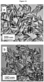
- TEM micrographs in Figure 1 show the NPs obtained after aging, at 150 oC for 1 hour in a microwave oven (MW), three butanol solutions containing dysprosium nitrate (0.02 M) and phosphoric acid (0.075 M, 0.15 M, and 0.30 M). All three micrographs showed uniform NPs whose size increased with increasing H 3 PO 4 concentration, as observed in the histograms plotted in the same Figure 1 . Specifically, the use of very dilute (0.075 M) H 3 PO 4 resulted in cube-like NPs with an average value of the face diagonal of 23 nm, while doubling H 3 PO 4 concentration to 0.15 M gave also rise to cube-like NPs showing a larger face diagonal size (37 nm).
- Figure 2 shows the TEM micrographs of the precipitates obtained by changing just one of the experimental conditions used to synthesize the NPs shown in Figure 1c (Dy37). It can be observed that the solvent nature produced a drastic change in the precipitate obtained, so that using octanol instead of butanol resulted in the precipitation of heterogeneous, fully aggregated tiny NPs ( Figure 2a ).
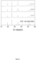
- the crystal phase of the Dy23, Dy37, and Dy57 NPs was analyzed by XRD ( Figure 3 ). All three patterns exhibited the same set of reflections, compatible with tetragonal DyPO 4 (Powder Diffraction File of the International Center for Diffraction Data (ICDD) PDF 00-026-0593). No additional reflections were observed in any of the patterns, indicating the absence of other crystalline phases. It can be noted that the width of the reflections decreased with increasing particle size, indicating that the crystallite size increased correspondingly. The crystallite size was estimated from the half-width at half maximum of the reflection located at -35 °2 ⁇ using the Scherrer formula.
- Crystallite size values of 26 nm, 34 nm, and 52 nm were obtained for the Dy23, Dy37, and Dy57 NPs, respectively.
- NPs with PAA Functionalization of the NPs with PAA was carried out to try to reduce such aggregation.
- the NPs obtained after such functionalization process will be called Dy23@PAA, Dy37@PAA and Dy57@PAA from now on.
- the success of the functionalization process was monitored through FTIR spectroscopy and thermogravimetry curves.
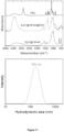
- Figure 4b shows the FTIR spectra of Dy57 NPs before and after PAA coating.
- the corresponding FTIR spectra of Dy23 and Dy37 NPs are plotted in Figure 5 .
- the spectra of the three NPs before functionalization showed the characteristic bands associated with the phosphate vibrational modes (located at wavenumbers ⁇ 1250 cm -1 ) and adsorbed water (at 3400 cm -1 and 1625 cm -1 ) plus a low-intensity band at around 1400 cm -1 (marked with an asterisk) that can be assigned to the presence of residual solvent molecules on the NPs surface.
- the FTIR spectra of all three NPs after functionalization showed two additional bands at ⁇ 1460 cm -1 and 1560 cm -1 (labelled with arrows) that can be assigned to the vibrations of the carboxylate groups of the PAA molecules.
- the TG curve of Dy57 NPs before PAA functionalization (Figure 4c) showed a first weight loss of around 4 % at T ⁇ 100 oC, corresponding to the removal of adsorbed water and a second, progressive one at T > 100 oC that could be assigned to the decomposition of the residual solvent molecules.
- the curve corresponding to the Dy57@PAA NPs showed, in addition to both weigh losses, a steeper loss of around 4 % at ⁇ 300 oC that can be due to the decomposition of PAA entities.
- the colloidal stability of the functionalized NPs suspended in water was then reevaluated using DLS.
- the DLS curves of the functionalized NPs significantly shifted towards lower hydrodynamic values with respect to those recorded before functionalization, as observed in Figure 4a for Dy57@PAA NPs and in Figure 7 for Dy23@PAA and Dy37@PAA. It can be therefore concluded that the functionalization process notably improved the colloidal stability of the NPs in water.
- Figure 9a shows the T 2 -weighted MR images obtained in aqueous suspensions with different concentrations of Dy23@PAA, Dy37@PAA, and Dy57@PAA NPs.
- the images became darker as the NPs concentration increased for all three samples, and they turned practically black at concentrations as low as ⁇ 0.3 mM, which indicates that the NPs behaved as good negative MRI contrast agents.
- the Dy57@PAA NPs were coated with PEG, with the purpose of improving their systemic circulation time and avoiding their clearance by the immune system.
- the NPs were successfully PEGylated, as demonstrated by the FTIR spectra shown in Figure 10 .
- the same figure also contains the DLS curve of the corresponding NPs suspended in PBS, showing a hydrodynamic size of 164 nm.
- the PEGylated sample was submitted as well to magnetic resonance studies, obtaining a value for r 2 of 504 mM -1 ⁇ s -1 , very close to that of the NPs before PEGylation (516 mM -1 ⁇ s -1 ), which indicates a negligible effect of the PEG shell on the relaxivity capacity.
- PBS Phosphate buffered saline
- the spherical NaDy(MoO 4 ) 2 NPs were prepared as follows. Sodium molybdate was dissolved in 1 mL Milli-Q water and after that, 1.5 mL of EG was added. In another vial, dysprosium chloride was dissolved in 2.5 mL of EG at -80 °C under magnetic stirring. Once the latter was cooled down to room temperature, both solutions were admixed. The concentrations of Dy 3+ and MoO 4 2- in the final solution were 0.02 M and 0.2 M, respectively. After the mixture was homogenized, it was aged for 20 hours in a conventional oven preheated at 120 oC. The total concentration of precursors in the starting solutions and the Dy/Mo molar ratio were modified aiming to modulate the size of the precipitated particles.
- the mixture was homogenized, it was aged for 30 minutes at 220 oC in a microwave-assisted oven (Anton Paar Monowave 300).
- a microwave-assisted oven Anton Paar Monowave 300.
- the solvent nature was modified by the addition of different quantities EG to the water solutions, and the total concentration of precursors, the Dy/Mo molar ratio, and the TA concentration were systematically varied.
- the precipitates obtained after aging were cooled down to room temperature. Then, they were centrifuged to remove the supernatants and washed, twice with ethanol and once with double distilled water. The obtained particles were finally stored in Milli-Q water and, for some analyses, they were dried at 50 oC.
- PAH Polyelectrolytes of opposite electrical charge were adsorbed by electrostatic interactions on the surface of the nanoparticles in a sequential and alternating way.
- PAH was used as the positively charged polyelectrolyte and PAA as the negatively charged one.
- the first positive PAH layer was formed/deposited as follows: 5 mL of an 0.05 M NaCl aqueous solution at pH 6.5 containing 10 mg mL -1 of PAH were added to 1.5 mL of a water dispersion containing 5 mg of the NPs. The resulting mixture was first sonicated in iced water for 5 minutes and then kept for 15 minutes at room temperature in an end-over-end shaker.
- the dispersion was centrifuged to remove the supernatant and the NPs washed twice with Milli-Q water.
- the NPs washed twice with Milli-Q water.
- PAH was substituted by PAA.
- the resulting functionalized NPs were dispersed and stored in Milli-Q water.
- the chemical stability of the samples was studied by aging a NPs dispersion (3 mg mL -1 ) in PBS buffer at 37 oC to mimic the physiological conditions. After aging, the dispersion was centrifuged and the supernatants analyzed by ICP.
- TEM Transmission electron
- SEM scanning electron micrographs were obtained using a JEOL 2100Plus (200 kV) or a Hitachi S4800 microscope, respectively.
- the ImageJ software was employed to obtain the size distribution histograms from the micrographs.
- X-ray diffraction (XRD) patterns were acquired using a Panalytical, X'Pert Pro diffractometer (CuK ⁇ ) with an X-Celerator detector over an angular range (2 ⁇ ) from 10 ° to 90 °, 2 ⁇ step width of 0.03 °, and 10 s counting time.
- FTIR infrared spectra
- TGA Thermogravimetric analyses
- the hydrodynamic diameter of the NPs in aqueous or PBS suspensions (0.5 mg ⁇ mL -1 ) before and after functionalization was analyzed by Dynamic Light Scattering (DLS) using a Malvern Zetasizer Nano-ZS90 equipment. This instrument was also used to measure Zeta potential.
- DLS Dynamic Light Scattering
- ICP analyses were carried out in an iCAP 7200 ICP-OES Duo (ThermoFisher Scientific) apparatus.
- Transverse magnetic relaxivity was determined at 9.4 T using a Bruker Biospec MRI system. T 2 values were determined in aqueous suspensions of NPs with different concentrations of Dy, ranging from 0.03 mM to 0.5 mM, using the Carl-Purcell-Meiboom-Gill (CPMG) sequence. Then, r 2 values were obtained from the slope of the linear fit of 1/T 2 vs. the concentration of Dy expressed in mM.
- CPMG Carl-Purcell-Meiboom-Gill
- HFF-1 cells were cultured with a medium containing the Dy nanoparticles in varying concentrations. After 24 h, Triton X-100 was added to the positive control wells. After 15 min, all the wells were stained with DAPI (4',6-Diamidino-2-phenylindole) to label nuclei in all cells, although with stronger labeling in live cells, and TO-PRO-3 Iodine to only label dead cells.
- DAPI 4,6-Diamidino-2-phenylindole
- the cell morphology images were acquired using a Perkin Elmer Operetta High Content Imaging System. For each field, fluorescence images for DAPI and TO-PRO-3, plus a brightfield image were captured. Cell mortality percentages were calculated automatically by Operetta Harmony software, whereby all nuclei (dead and alive) were identified from the DAPI staining and the percentage of dead cells then determined by the number of nuclei also possessing high levels of TO-PRO-3 staining. For the "MTT assay" the HFF-1 cells were cultured with medium containing the Dy nanoparticles in varying concentrations.
- ICP-HRMS Inductively Coupled Plasma High-Resolution Mass Spectroscopy
- a strategy consisting of the addition of a complexing agent to the starting solutions was implemented, since the formation of a metal complex before precipitation alters the reaction kinetics through changes in the nature of the soluble species precursor to precipitation, which may induce changes in particle shape.
- TA tartaric acid
- the complexing anions may also selectively adsorb on certain crystallographic planes blocking their growth, which may also result in anisometric particles.
- FTIR spectroscopy In order to gain information on the role played by the tartrate anions in the formation of our ellipsoidal particles, FTIR spectroscopy, and TGA measurements were conducted.
- the FTIR spectrum of the latter particles displayed bands in the 1630-1300 cm -1 region ( Figure 16 top), which can be attributed to the vibrations of carboxylate groups of tartrate anions. This assignment is also supported by the absence of these bands in the spectrum of the spherical NPs, which are synthesized in the absence of such anions ( Figure 16 top).
- TGA measurements revealed a 1.1 % weight loss in the 250-400 oC range for the ellipsoidal particles ( Figure 16 bottom), which may be ascribed to the decomposition of the tartrate species. Therefore, the presence of these entities most probably adsorbed on the surface of the ellipsoids may be also responsible for the role that the tartrate anions play in determining particle morphology.
- the NPs size required for in vivo applications must be lower than 100 nm. Therefore, several experiments were conducted to decrease the size of the ellipsoidal particles. Under the same aging conditions and TA/Dy ratio, we varied the molybdate/Dy ratio or the total concentration of the Dy and molybdate precursors keeping constant the molybdate/Dy ratio, resulting in more heterogeneous precipitates ( Figure 17 ).
- the second designed strategy was to increase the viscosity of the solvent by adding different amounts of EG to water aiming to slow down the diffusion processes required for nucleation and particle growth.
- the NPs size could also be modulated by changing the amount of TA added.
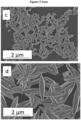
- an increase of the dimensions of the ellipsoid from 68 x 20 nm to 87 x 22 nm was observed when lowering the TA concentration (from 0.04 to 0.02 M), whereas the size decreased to 50 x 16 nm when the amount of TA was increased up to 0.06 M ( Figure 19 ).
- these samples will be named as "Ellipsoids 87 nm” and "Ellipsoids 50 nm", respectively.
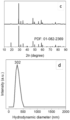
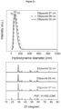
- the NPs obtained using different TA amounts were well dispersed in aqueous suspension (pH ⁇ 6.5) as indicated by their d h as determined by DLS, which was ⁇ 100 nm ( Figure 21 , top) and consisted of tetragonal NaDy(MoO 4 ) 2 , as indicated by XRD ( Figure 21 , bottom). Since the size of these three samples was below 100 nm, they were selected to evaluate the effects of NPs size on their relaxivity.
- the values of the transverse relaxivity estimated as the slope of the straight lines resulting from such plots are shown in Table 1, along with the NPs dimensions and d h values.
- Table 1 Values of the transverse relaxivity obtained for aqueous suspensions of the spherical and ellipsoidal NaDy(MoO 4 ) 2 NPs with different sizes and hydrodynamic diameter (d h ).
- r 2 increased from 160 to 220 mM -1 S -1 as increasing size from 50x16 to 87x22 nm.
- the r 2 value measured for our spherical NPs is higher (230 mM -1 s -1 ) than that of the ellipsoidal ones ( ⁇ 220 mM -1 S -1 ) in spite of the smaller size of the former (Table 1).
- sample Spheres 23 nm the sample having the highest relaxivity, which was that consisting of spheres (sample Spheres 23 nm) as well as that composed by ellipsoids with the size closer to that of the spheres (sample Ellipsoids 50 nm) for comparative purposes.
- the zeta potential value of the PAH coated NPs changed to +49 mV, for the spheres, and +57 mV, for the ellipsoids.
- This charge reversal suggests the success of the coating procedure.
- a negatively charged polymer (PAA) was selected for the second layer and, as expected, a negative zeta potential value was then measured for both, spheres (-40 mV) and ellipsoids (-30 mV). The presence of both polymers on the nanospheres surface was confirmed by FTIR spectroscopy.
- the FTIR spectra obtained for the coated particles displayed bands in the 1400-1650 cm -1 region overlapped with that due to water at about 1620 cm -1 , which can be attributed to the vibrational modes of the N-H (1624 cm -1 and 1455 cm -1 ) and COO- groups (1553 cm -1 ) of the PAH and PAA molecules, respectively, confirming the adsorption of these polymers on the NPs surface.
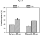
- the NPs colloidal stability of both, the spherical and ellipsoidal NPs in the selected physiological medium was markedly improved since the d h values obtained for the NPs in PBS suspensions (187 nm for the spheres and 218 nm for the ellipsoids) ( Figure 23 ) were much lower than those obtained for the uncoated NPs (> 800 nm).
Landscapes
- Chemical & Material Sciences (AREA)
- Health & Medical Sciences (AREA)
- Nanotechnology (AREA)
- Engineering & Computer Science (AREA)
- Life Sciences & Earth Sciences (AREA)
- Organic Chemistry (AREA)
- Nuclear Medicine, Radiotherapy & Molecular Imaging (AREA)
- Radiology & Medical Imaging (AREA)
- Molecular Biology (AREA)
- Epidemiology (AREA)
- Animal Behavior & Ethology (AREA)
- General Health & Medical Sciences (AREA)
- Public Health (AREA)
- Veterinary Medicine (AREA)
- Inorganic Chemistry (AREA)
- Immunology (AREA)
- Medicines Containing Antibodies Or Antigens For Use As Internal Diagnostic Agents (AREA)
Abstract
The invention relates to nanoparticles comprising a core consisting of a dysprosium compound, said dysprosium compound being selected from DyPO4 or NaDy(MoO4)2, and a coating of polyacrylic acid. Furthermore, the present invention refers to processes for obtaining said nanoparticles and to the uses of the nanoparticles as contrast agents for ultra-high-field magnetic resonance imaging.
Description
- The invention relates to a nanoparticles comprising a core consisting of a dysprosium compound, said dysprosium compound is selected from DyPO4 or NaDy(MoO4)2 and a coating of polyacrylic acid. Furthermore, the present invention refers to the process for obtaining said nanoparticles and to the use of the nanoparticles as a contrast agent particularly for ultra-high-field magnetic resonance imaging.
- Therefore, the present invention refers to the medical field and is of interest for the industry and users of magnetic resonance imaging techniques.
- Magnetic resonance imaging (MRI) is a well-established non-invasive technique commonly used for medical diagnosis. To record MRI images, a powerful magnet is used to create a net magnetization of the endogenous water proton spins in the direction longitudinal to the magnetic field (z), and a radiofrequency current is applied to alter the population balance of this magnetization, shifting it towards the plane transverse to the external magnetic field (xy). Once the current ceases, the net magnetization (Mz) is recovered by a double relaxation process (longitudinal and transverse). The times associated with this relaxation process are called T1 (for longitudinal relaxation) and T2 (for transverse relaxation) [Cao, Y.; Xu, L.; Kuang, Y.; Xiong, D.; Pei, R. Gadolinium-based nanoscale MRI contrast agents for tumor imaging. Journal of Materials Chemistry B 2017, 5 (19), 3431-3461]. In an MRI scan, a contrast between the different analyzed zones is based on the differences in the density of protons between different tissues and the differences in their longitudinal (T1) and transverse (T2) relaxation times [Peng, E.; Wang, F.; Xue, J. M. Nanostructured magnetic nanocomposites as MRI contrast agents. Journal of Materials Chemistry B 2015, 3 (11), 2241-2276]. T1-weighted or T2-weighted images are obtained by using acquisition schemes that potentiate the effect of T1 or T2, respectively.
- To increase the contrast between the regions of interest and the surrounding tissue, contrast agents (CAs) consisting of magnetically active substances that shorten the T1 and T2 relaxation times of protons are often used. The ability of a CA to shorten T1 and T2 is expressed through the longitudinal (r1 ) and transverse (r2 ) relaxivity, respectively. In current clinical trials, superparamagnetic iron oxide (SPIO) nanoparticles (NPs) are used as T2 Cas [Wang, Y.-X. J. Superparamagnetic iron oxide based MRI contrast agents: Current status of clinical application. Quantitative imaging in medicine and surgery 2011, 1 (1), 35-40 ] and Gd3+ chelates as T1 Cas [Pan, D.; Schmieder, A. H.; Wickline, S. A.; Lanza, G. M. Manganese-based MRI contrast agents: past, present, and future. Tetrahedron 2011, 67 (44), 8431-8444 ]. Different Gd-based inorganic NPs have also been proposed as MRI CAsc because of their easy functionalization and bioconjugation, which allows targeting, thus reducing the needed doses [Na, H. B.; Song, I. C.; Hyeon, T. Inorganic Nanoparticles for MRI Contrast Agents. Advanced Materials 2009, 21 (21), 2133-2148] and their controlled release through particle size selection [Barreto, J. A.; O'Malley, W.; Kubeil, M.; Graham, B.; Stephan, H.; Spiccia, L. Nanomaterials: Applications in Cancer Imaging and Therapy. Advanced Materials 2011, 23 (12), H18-H40]. For the use of these nanoparticulate contrast agents, they must fulfill certain requirements such as uniform particle shape and size (always below 100 nm to avoid embolism and facilitate the renal release) [Duan, X.; Li, Y. Physicochemical Characteristics of Nanoparticles Affect Circulation, Biodistribution, Cellular Internalization, and Trafficking. Small 2013, 9 (9-10), 1521-1532], high chemical and colloidal stability in physiological media, high biocompatibility (absence of cytotoxicity) and in the case of MRI, high relaxivity.
- Another recently proposed strategy to improve magnetic resonance images is to increase the magnetic field strength to values higher than those used today in clinical trials (usually ≤ 3 T), which results in a better signal-to-noise ratio and greater spatial/temporal resolution, thus enhancing the sensitivity of the MRI technique [ Norek, M.; Kampert, E.; Zeitler, U.; Peters, J. A. Tuning of the size of DY2O3 nanoparticles for optimal performance as an MRI contrast agent. Journal of the American Chemical Society 2008, 130 (15), 5335-5340 ]. The MRI CAs mentioned above are not effective at high magnetic fields [Lisjak, D.; Plohl, O.; Vidmar, J.; Majaron, B.; Ponikvar-Svet, M. Dissolution Mechanism of Upconverting AYF(4):Yb,Tm (A = Na or K) Nanoparticles in Aqueous Media. Langmuir 2016, 32 (32), 8222-8229]. The most promising candidates are Dy3+ containing compounds due to the very high magnetic moment of this cation (10.6 µB) and short electronic relaxation times, which favors their r2 relaxivity at high field. In response to the high field (≥ 7 T) MRI CAs demand, several Dy3+ based systems have been reported, consisting of NaDyF4 [Zhang, Y.; Vijayaragavan, V.; Das, G. K.; Bhakoo, K. K.; Tan, T. T. Y. Single-Phase NaDyF4:Tb3+ Nanocrystals as Multifunctional Contrast Agents in High-Field Magnetic Resonance and Optical Imaging. European Journal of Inorganic Chemistry 2012, (12), 2044-2048], DyF3 [ Gonzalez-Mancebo, D.; Becerro, A. I.; Rojas, T. C.; Garcia-Martin, M. L.; de la Fuente, J. M.; Ocana, M. HoF3 and DyF3 Nanoparticles as Contrast Agents for High-Field Magnetic Resonance Imaging. Particle & Particle Systems Characterization 2017, 34 (10 )], Dy2O3 and DyVO4 [ Gomez-Gonzalez, E.; Nunez, N. O.; Caro, C.; Garcia-Martin, M. L.; Fernandez-Afonso, Y.; de la Fuente, J. M.; Balcerzyk, M.; Ocana, M. Dysprosium and Holmium Vanadate Nanoprobes as High- Performance Contrast Agents for High-Field Magnetic Resonance and Computed Tomography Imaging. Inorganic Chemistry 2021, 60 (1), 152-160]. However, flanthanide (Ln) fluorides have been shown to be chemically unstable in aqueous solution, since they slowly dissolve and release potentially toxic ions [Lisjak, D.; Plohl, O.; Vidmar, J.; Majaron, B.; Ponikvar-Svet, M. Dissolution Mechanism of Upconverting AYF(4):Yb,Tm (A = Na or K) Nanoparticles in Aqueous Media. Langmuir 2016, 32 (32), 8222-8229].
- Therefore, developing MRI contrast agents that show optimum contrast at high magnetic fields is an urgent task.
- The present invention discloses novel nanoparticles comprising a core consisting of a dysprosium compound, said dysprosium compound is selected from DyPO4, or NaDy(MoO4)2 and a coating of polyacrylic acid. Said nanoparticles are suitable for use as a contrast agent for ultra-high-field (UHF) magnetic resonance imaging (MRI) working at fields ≥ 7 T.
- Therefore, a first aspect of the invention refers to nanoparticles (herein the nanoparticles of the present invention) comprising
- a core consisting of a dysprosium compound, said dysprosium compound is selected from DyPO4 or NaDy(MoO4)2,
- and a coating of polyacrylic acid.
- wherein the DyPO4 nanoparticles are cubic-like and have an average value of the face diagonal ranging from 23 nm to 57 nm, and
- wherein the NaDy(MoO4)2 nanoparticles
- are spherical and have a size ranging from 10 nm and 30 nm,
- or are ellipsoidal and have a first axis ranging from 40 nm to 95 nm and a second axis ranging from 10 nm to 30 nm.
- Said nanoparticles exhibit biocompatibility and notably improved colloidal stability in water and in phosphate-buffered saline PBS (a buffer solution commonly used in biological research to simulate physiological fluid). In addition to their colloidal stability in physiological fluid, compounds, materials or particles intended to be applied in nanomedicine should be chemically stable, i.e., nanoparticles dissolution should not take place in the physiological fluid during the period between the injection and the excretion of the nanoparticles to hinder potential toxic ions to be released to the blood stream. The nanoparticles of the present invention show an excellent degree of chemical stability in PBS. Furthermore, the nanoparticles of the present invention exhibit negligible toxicity effects under the assayed conditions, and, therefore, are suitable as bioimaging probes.
- A preferred embodiment of the nanoparticles of the present invention refers to nanoparticles having an average value of the face diagonal ranging from 23 nm to 57 and comprising
- a core of cubic-like DyPO4,
- a first coating of polyacrylic acid on the surface of the core of cubic-like DyPO4,
- a second coating of polyethylene glycol on the surface of the first coating.
- Another preferred embodiment of the nanoparticles of the present invention refers to nanoparticles having a size ranging from 10 nm and 30 nm and comprising
- a core of spherical NaDy(MoO4)2
- a first coating of poly(allylamine hydrochloride) positively charged polyelectrolyte on the surface of the core of spherical NaDy(MoO4)2
- and a second coating of polyacrylic acid negatively charged polyelectrolyte on the surface of the first coating.
- More preferably the spherical NaDy(MoO4)2 nanoparticles have a size ranging from 16 nm and 23 nm.
- Another preferred embodiment of the nanoparticles of the present invention refers to nanoparticles having a first axis ranging from 40 nm to 95 nm and a second axis ranging from 10 nm to 30 nm, and comprising
- a core of ellipsoidal NaDy(MoO4)2,
- tartrate anions adsorbed at the surface of the core of NaDy(MoO4)2,
- a first coating of poly(allylamine hydrochloride) positively charged polyelectrolyte on the surface of the tartaric anions,
- and a second coating of polyacrylic acid negatively charged polyelectrolyte on the surface of the first coating,
- Preferably the ellipsoidal NaDy(MoO4)2 have a first axis ranging from 50 nm to 87 nm and a second axis ranging from 16 nm to 22 nm.
- Another aspect of the invention refers to process for obtaining the nanoparticles having an average value of the face diagonal ranging from 23 nm to 57 nm and comprising
- a core of cubic-like DyPO4 and
- and a coating of polyacrylic acid,
- a) aging, in a microwave oven, a mixture of a butanol solution of dysprosium nitrate and phosphoric acid at a temperature ranging between 120 ºC and 180 ºC for a period of time ranging between 45 min and 90 min, wherein the concentration of phosphoric acid is ranging between 0.075 M and 0.30 M in the mixture, and
- b) preparing an aqueous suspension of the product obtained in step (a) and adding poly(acrylic acid) in a concentration between 2 mg/ml and 10 mg/ml adjusting the pH to a value ranging between 9 and 11.
- Step (a) refers to the aging, in a microwave oven, a mixture of a butanol solution of dysprosium nitrate and phosphoric acid at a temperature ranging from 120 ºC and 180 ºC, preferably between 140 ºC and 160 ºC, for a period of time ranging between 45 min and 90 min, wherein the concentration of phosphoric acid is ranging between 0.075 M and 0.30 M in the mixture. It is observed that at a temperature higher than 180 ºC the size of the nanoparticles increases and at a temperature lower than 120 ºC a precipitation of heterogeneous nanoparticles.
- Another aspect of the invention refers to the process for obtaining the nanoparticles having an average value of the face diagonal ranging from 23 nm to 57 and comprising
- a core of cubic-like DyPO4,
- a first coating of polyacrylic acid on the surface of the core of cubic-like DyPO4,
- and a second coating of polyethylene glycol on the surface of the first coating, characterized in that it comprises the following steps:
- a) aging, in a microwave oven, a mixture of a butanol solution of dysprosium nitrate and phosphoric acid at a temperature ranging between 120 ºC and 180 ºC, preferably between 140 ºC and 160 ºC, for a period of time ranging between 45 min and 90 min, wherein the concentration of phosphoric acid is ranging between 0.075 M and 0.30 M in the mixture, and
- b) preparing an aqueous suspension of the product obtained in step (a) and adding poly(acrylic acid) in a concentration between 1 mg/ml and 20 mg/ml, preferably in a concentration between 2 mg/ml and 10 mg/ml, adjusting the pH to a value ranging between 9 and 11, and
- c) preparing a suspension of the nanoparticles obtained in step (b) in a borate buffer, wherein said borate buffer comprises an N-ethyl-N'-(3-(dimethylamino)propyl)carbodiimide (EDC) solution and an N-hydroxysuccinimide (s-NHS) solution, and adding a borate buffer PEG solution in a concentration ranging between 5 mg/ml and 20 mg/ml, preferably in a concentration of 10 mg/ml.
- In step (b) the concentration of poly(acrylic acid) ranges preferably between 1 mg/ml and 20 mg/ml, more preferably in a concentration between 2 mg/ml and 10 mg/ml, to ensure the formation of an increased number of carboxylate groups on the nanoparticles surface for PEG grafting in step (c). This step (b) is indispensable to create an amide link between carboxylate groups of PAA and the amine group of PEG. The purpose of the PEG functionalization is improving the NPs' systemic circulation time and avoiding their clearance by the immune system.
- Another aspect of the invention refers to the process for obtaining the nanoparticles having a size ranging from 10 nm and 30 nm, preferably having a size ranging from 16 nm and 23 nm, and comprising
- a core of spherical NaDy(MoO4)2,
- a first coating of poly(allylamine hydrochloride) positive charged polyelectrolyte on the surface of the core of spherical NaDy(MoO4)2,
- and a second coating of polyacrylic acid negative charged polyelectrolyte on the surface of the first coating,
- a) aging, in a conventional oven, a mixture of an aqueous and ethylene glycol solution of sodium molybdate and an ethylene glycol solution of dysprosium chloride at a temperature ranging between 110 ºC and 130 ºC for a period of time ranging between 12 h and 25 h, wherein the concentration of Dy+3 is ranging between 0.01 M and 0.04 M in the mixture, preferably at a concentration of Dy+3 of 0.02 M in the mixture, and wherein the concentration of MoO4 2- is ranging between 0.01 M and 0.04 M in the mixture, preferably at a concentration of MoO4 2- of 0.02 M in the mixture.
- b) mixing an aqueous solution comprising NaCl aqueous solution and poly(allylamine hydrochloride) with an aqueous dispersion of the nanoparticles obtained in step (a) and sonicating in ice water, and
- c) mixing an aqueous solution comprising NaCl aqueous solution and polyacrylic acid with the nanoparticles obtained in step (b) and sonicating in ice water.
- Another aspect of the invention refers to the process for obtaining nanoparticles having a first axis ranging from 40 nm to 95 nm and a second axis ranging from 10 nm to 30 nm, preferably having a first axis ranging from 50 nm to 87 nm and a second axis ranging from 16 nm to 22 nm, and comprising
- a core of ellipsoidal NaDy (MoO4)2,
- and tartrate anions adsorbed at the surface of the core of ellipsoidal NaDy (MoO4)2,
- a first coating of poly(allylamine hydrochloride) positive charged polyelectrolyte on the surface of the tartaric anions,
- and a second coating of polyacrylic acid negative charged polyelectrolyte on the surface of the first coating,
- a) aging, in a microwave oven, a mixture of an aqueous and ethylene glycol solution of sodium molybdate, an ethylene glycol solution of dysprosium chloride and tartaric acid at a temperature ranging between 210 ºC and 230 ºC for a period of time ranging between 20 min and 60 min, wherein the concentration of Dy+3 is ranging between 0.01 M and 0.04 M in the mixture, preferably at a concentration of Dy+3 of 0.02 M in the mixture, wherein the concentration of MoO4 2- is ranging between 0.1 M and 0.4 M in the mixture, preferably at a concentration of M0O4 2- of 0.2 M in the mixture and wherein the concentration of tartaric acid is ranging between 0.01 M and 0.1 M in the mixture, preferably the concentration of tartaric acid is ranging between 0.02 M and 0.06 M in the mixture,
- b) mixing an aqueous solution comprising NaCl aqueous solution and poly(allylamine hydrochloride) with an aqueous dispersion of the nanoparticles obtained in step (a) and sonicating in ice water, and
- c) mixing an aqueous solution comprising NaCl aqueous solution and polyacrylic acid with the nanoparticles obtained in step (b) and sonicating in ice water.
- Another aspect of the present invention relates to the nanoparticles of the present invention for use as a contrast agent for imaging blood flow in magnetic resonance imaging, preferably for use as a contrast agent for imaging blood flow in ultra-high-field magnetic resonance imaging operating at static magnetic fields of 7 T or higher.
- Another aspect of the invention refers to the use of the nanoparticles of the present invention as a contrast agent for imaging blood flow in magnetic resonance imaging, preferably in ultra-high-field magnetic resonance imaging operating at static magnetic fields of 7 T or higher.
- The last aspect of the invention refers to a method of imaging blood flow in magnetic resonance imaging, preferably in ultra-high-field magnetic resonance imaging operating at static magnetic fields of 7 T or higher, using the nanoparticles of the present invention.
- Unless otherwise defined, all technical and scientific terms used herein have the same meaning as commonly understood by one of ordinary skilled in the art to which this invention belongs. Methods and materials similar or equivalent to those described herein can be used in the practice of the present invention. Throughout the description and claims the word "comprise" and its variations are not intended to exclude other technical features, additives, components, or steps. Additional objects, advantages and features of the invention will become apparent to those skilled in the art upon examination of the description or may be learned by practice of the invention. The following examples, drawings are provided by way of illustration and are not intended to be limiting of the present invention.
-
-
Figure 1 . TEM micrographs and corresponding size distribution histograms of DyPO4 NPs synthesized by solvothermal reaction of butanol solutions of dysprosium nitrate (0.02 M) and different H3PO4 concentrations in a microwave oven at 150 º C for 1 h. [H3PO4] = 0.075 M (a and b, Dy23 NPs), 0.15 M (c and d, Dy37 NPs), 0.30 M (e and f, Dy57 NPs). -
Figure 2 : TEM micrographs of DyPO4 NPs obtained in the experimental conditions used for Dy37 NPs (aging at 150 ºC for 1 hour in a microwave oven a dysprosium nitrate (0.02 M) and H3PO4 (0.15M) solution in butanol), changing just one of the parameters as follows: a) Solvent used was octanol, b) Reaction temperature was 120 ºC, c) Reaction temperature was 180 ºC, d) Heating source was a conventional oven. -
Figure 3 : XRD patterns of Dy23, Dy37 and Dy57 NPs synthesized in the conditions described inFigure 1 . Ticks at the bottom of the figure correspond to tetragonal DyPO4 (Powder Diffraction File of the International Center for Diffraction Data (ICDD) PDF 00-026-0593). -
Figure 4 : a) DLS size distribution curve in water, b) FTIR spectra and c) thermogravimetry curves of Dy57NPs and Dy57@PAA NPs. -
Figure 5 : FTIR spectra of Dy23 (a) and Dy37 (b) NPs before and after functionalization with PAA. -
Figure 6 : Thermogravimetry curves of Dy23 (a) and Dy37 (b) NPs before and after functionalization with PAA. -
Figure 7 : DLS curves of Dy23 (a) and Dy37 (b) NPs before and after functionalization with PAA. -
Figure 8 : DLS curves and hydrodynamic sizes of Dy23@PAA, Dy37@PAA, and Dy57@PAA NPs suspended in PBS. -
Figure 9 : a) Concentration-dependent T2 weighted phantom image of the Dy23@PAA, Dy37@PAA and Dy57@PAA NPs at 9.4 T. b) T2 relaxation rates as a function of Dy concentration of Dy23@PAA, Dy37@PAA and Dy57@PAA aqueous suspensions at 9.4 T. -
Figure 10 : Top: FTIR spectra of the Dy57 NPs functionalized with PAA (Dy57@10PAA) and of the same NPs after coated with PEG (Dy57@10PAA@PEG). The FTIR spectrum of pure PEG is also given with comparative purpose. -
Figure 11 : Optical microscopy images of HFF-1 fibroblasts resulting from the merge of bright field (grey), DAPI (blue) and TO-PRO-3 Iodine (red) images: a) negative control, b) positive control, c) cells exposed to 100 mM Dy of Dy57@PAA@PEG NPs. Scale bar corresponds to 50 µm. d) Total number of cells per well after exposure to increasing concentration of Dy57@PAA@PEG NPs. e) Percentage of dead cells after exposure to increasing concentration of Dy57@PAA@PEG NPs. e) MTT assay of cells exposed to increasing concentration of Dy57@PAA@PEG NPs -
Figure 12 . TEM micrograph (a), size distribution histogram (b), X-ray diffraction patterns (c) and DLS curve (d) obtained for the sample synthesized by aging at 120 ºC (in a conventional oven) for 20 h, solutions containing Dy3+ (0.02 M) and molybdate (0.2 M) precursors in EG/water (4/1 by vol.) mixtures. The PDF file in (c) corresponds to tetragonal NaY(MoO4)2. -
Figure 13 . TEM micrographs and corresponding particle size distribution histograms obtained for the samples synthesized under the same conditions used for the sample. shown inFigure 1 except: (a and d) Na2MoO4/DyCl3 mole ratio = 5; (b and e) 0.01M DyCl3 and 0.1M Na2MoO4; and (c and f) 0.04M DyCl3 and 0.4M Na2MoO4. -
Figure 14 . SEM micrograph (a), particle size distribution histogram (b) and DLS curve (c) obtained for the sample synthesized by aging at 120ºC for 20 h in a conventional oven, solutions containing 0.04M DyCl3, 0.4M Na2MoO4 and 0.04 M tartaric acid. -
Figure 15 . TEM micrograph (a), size distribution histogram (b), X-ray diffraction patterns (c) and DLS curve (d) obtained for the sample synthesized by aging at 220ºC (in a microwave oven) for 1 h, aqueous solutions containing the Dy3+ (0.02 M) and molybdate (0.2 M) precursors and TA (0.04 M). The Powder Diffraction File of the International Center for Diffraction Data (ICDD) PDF file in (c) corresponds to tetragonal NaY(MoO4)2. -
Figure 16 . Top: FTIR spectra of the spherical (Figure 1a ) and ellipsoidal particles (Figure 2a ); Bottom: TGA curve obtained for the ellipsoidal particles. -
Figure 17 . SEM micrographs obtained for the samples synthesized under the same conditions used for the sample shown inFigure 2 except: (a) 0.01 M DyCl3, 0.1 M Na2MoO4 and (mole ratio=10) and 0.02 M tartaric acid; (b) 0.06 M DyCl3, 0.6 M Na2MoO4 and (mole ratio=10) and 0.12 M tartaric acid; (c) 0,02 M DyCl3 y 0,1 M Na2MoO4 and (mole ratio =5); and (d) 0,02 M DyCl3 y 0,4 M Na2MoO4 and (mole ratio =20). -
Figure 18 . SEM micrographs (left) and corresponding histograms (right) obtained for the samples synthesized by keeping the same experimental conditions involved in the synthesis of the sample shown infigure 2 but using water-EG mixtures as solvent with different volumetric water/EG ratios: 6/4 (a), 5/5 (b) and 4/6 (c). -
Figure 19 . TEM micrographs (left) and corresponding histograms (right) obtained for the samples synthesized by keeping the same experimental conditions involved in the synthesis of the sample shown in Figure 4c but using different amounts of TA: 0.02 M (a) and 0.06 M (b). -
Figure 20 . FTIR spectra (top) and TGA curves (bottom) obtained for ellipsoidal NPs having different size. -
Figure 21 . DLS curves (top) and X-ray diffraction patterns (bottom) obtained for the ellipsoidal NPs having different size. The PDF file corresponding to tetragonal NaY(MoO4)2 is included. -
Figure 22 . T2-weighted phantom images (top) and T2 relaxation rates as a function of Dy concentration (bottom) obtained for aqueous suspensions of the spherical and ellipsoidal NaDy(MoO4)2 NPs with different sizes. -
Figure 23 . DLS curves obtained for uncoated and coated NaDy(MoO4)2 spheres and ellipsoids dispersed in water and PBS buffer. -
Figure 24 . FTIR spectra obtained for the coated samples:Spheres 23 nm andEllipsoids 50 nm. -
Figure 25 . T2 relaxation rates as a function of Dy concentration obtained for aqueous suspensions of the coated NaDy(MoO4)2 nanospheres and ellipsoids. -
Figure 26 . Optical microscopy images of HFF-1 fibroblasts resulting from the merge of bright field (grey), DAPI (blue), and TO-PRO-3 Iodine (red) images: negative control (a); positive control (b); cells exposed to 100 mM Dy ofSpheres 23 nm (c) and cells exposed to 100 mM Dy of Ellipsoids 50 nm (d). Scale bar corresponds to 100 mm. -
Figure 27 . Total number of cells per well (left column), percentage of dead cells (middle column), and MTT assay of cells (right column) after exposure to increasing concentration of spherical (23 nm) (a, b, c), ellipsoidal (50 nm) (d, e, f) NPs. -
Figure 28 . Cell uptake for coated NaDy(MoO4)2 nanoparticles with spherical (sample Spheres 23 nm) and ellipsoidal (sample Ellipsoids 50 nm) shape in HFF-1 fibroblasts after 1 and 24 h of exposure determined by ICP-HRMS. - Reagents: Dysprosium nitrate hydrate (Dy(NO3)3·5H2O, Sigma Aldrich, 99.99%), phosphoric acid (Sigma Aldrich, 85%), butanol (Sigma Aldrich, >99.5%), and 1-octanol (Merck, >99%) were used for NPs synthesis. Poly(acrylic acid) (PAA, average Mw ~ 1800, Sigma-Aldrich) was used for NPs functionalization with PAA. For NPs functionalization with PEG, the following reagents were used as received: 1-(3-Dimethylaminopropyl)-3-ethyl carbodiimide (EDC - Sigma Aldrich, USA), N-Hydroxysulfosuccinimide (s-NHS -Sigma Aldrich, USA) and α-Methoxy-ω-amino PEG of 5000 Da (PEG, RappPolymer, Germany).
- Colloidal stability of NPs was evaluated using an aqueous solution of Phosphate Buffered Saline (PBS, Sigma Aldrich) that was prepared as follows: one tablet of PBS was dissolved in 200 mL water to obtain 137 mM NaCl, 2.7 mM KCI and 10 mM phosphate buffer, pH 7.4 at 25 ºC). An aqueous solution of sodium tetraborate decahydrate (Sigma Aldrich, > 99.5 %) 10 mM was prepared and its pH was adjusted to 9 with NaOH. We call this solution borate buffer from now on.
- Synthesis: For the synthesis of uniform dysprosium phosphate nanoparticles, we used a homogeneous precipitation reaction. Essentially, the method consists of aging a mixture of Dy(NO3)3 with phosphoric acid in butanol at 150 ºC in a microwave oven. The standard procedure is as follows: Dysprosium nitrate was dissolved in 30 mL of butanol at 50 ºC for 1 h under magnetic stirring to facilitate dissolution in order to get a 0.02 M dysprosium nitrate solution. Once dissolved, the solution was admixed with 616 microliters of H3PO4 to obtain a final solution with 0.30 M H3PO4 concentration. The mixture was aged in tightly closed test tubes using a microwave oven at 150 ºC for 1 h. The resulting dispersion was cooled down to RT, centrifuged to remove the supernatants, and washed, twice with ethanol and once with double distilled water.
- The NPs were finally stored either in distilled water or dried at 50 °C for some analyses.
- Additional experiments were conducted to analyze the effect of synthesis parameters (solvent nature, H3PO4 concentration, aging temperature, and heating source) on the characteristics of the precipitated nanoparticles.
- Functionalization with PAA: The aqueous suspension of NPs obtained as described above was diluted in Milli-Q water to get a concentration of 1 mg/mL and its pH was adjusted to 10 with 1M NaOH solution. PAA (2 mg/mL) was then added to the suspension and, again, the pH was adjusted to 10. The resulting suspension was magnetically stirred for 1 h at RT and centrifuged at 14000 rpm for 20 min to remove the supernatant. The resulting NPs were washed twice with Milli-Q water through centrifugation in the same conditions and stored in Milli-Q water. The pH of the resultant suspension was close to 7.
- Further Functionalization with PEG: The NPs showing the optimum magnetic relaxivity value in this study were further functionalized with PEG. For PEG coating, the NPs were firstly functionalized with PAA following the same procedure described above but using 10 mg/mL PAA to increase the number of carboxylate groups on the NPs surface for PEG grafting. The obtained NPs were then suspended in borate buffer (6 mg NPs/ml). A volume of 0.94 ml of an EDC solution in borate buffer (3.2 mg/ml) was admixed with 0.85 ml of s-NHS solution in borate buffer (6 mg/ml). The mixture was shaken at RT for 10 min in the end-over-end roller. A volume of 161 µl of the NPs@10PAA suspension was added to the resulting solution. The three mentioned volumes were chosen in order to get the following molar ratio: 1 COOH:10 EDC:15 NHS (COOH refers to the carboxylic groups in PAA attached to the NPs). The resultant suspension was stirred at RT for 2h in the end-over-end roller and admixed with 1.56 ml of a PEG solution in borate buffer (10 mg/ml) (in accordance with the
ratio 1 COOH:2 PEG to block the unreacted COOH groups and to increase the colloidal stability of the NPs). Finally, after 45 minutes of stirring at RT in the end-over-end roller, NPs were separated by centrifugation at 14500 rpm for 5 minutes. The resulting NPs were washed twice with borate buffer and they were suspended in 1 mL of fresh borate buffer and stored at 4°C. This protocol was repeated ten times to have enough NPs for the rest of the analyses. - NPs functionalized with PAA were suspended in PBS (pH= 7.4) and the suspension was magnetically stirred at 37 ºC for several weeks. A portion of the suspension was extracted and centrifuged after 1, 3, and 5 weeks and the supernatants were analyzed by ICP to determine the fraction of dissolved dysprosium.
- Transmission electron microscopy (TEM, JEOL 2100Plus) was used to determine particles shape and size. To prepare the sample for the TEM analyses, a droplet of the NPs aqueous suspension was deposited on a copper grid coated with a transparent polymer. Particle size distribution curves were obtained by counting about one hundred of particles on the TEM micrographs using the ImageJ open-source image processing program.
- The colloidal stability of the NPs before and after functionalization was assessed by Dynamic Light Scattering (DLS, Malvern Zetasizer Nano-ZS90). DLS curves were obtained both in aqueous and PBS suspensions with ~ 0.5 mg NPs/ml.
- Ion Coupling Plasma (ICP) analyses were carried out in an iCAP 7200 ICP-OES Duo (ThermoFisher Scientific) apparatus.
- X-ray powder diffraction (XRD) was used for phase identification. The XRD patterns were recorded with a Panalytical X'Pert Pro diffractometer (Cu Kα) with an X-Celerator detector in the angular range of 10 º < 2θ < 80 ° using a 2θ step width of 0.02 ° and 10 s counting time. The crystallite size was estimated using the Scherrer formula from the full width at half maximum (FWHM) of a single reflection. A shape factor of k = 0.9 and an instrumental broadening factor of 0.112 were used for the calculation.
- The success of the functionalization processes was proved using Fourier Transform Infrared (FTIR) spectroscopy and Thermogravimetric analyses (TGA). FTIR (Jasco FT/IR-6200) was conducted on NPs pellets diluted in KBr while TGA curves were recorded in a Q600TA Instrument at a heating rate of 10°C·min-1 in air.
- TEM micrographs in
Figure 1 show the NPs obtained after aging, at 150 ºC for 1 hour in a microwave oven (MW), three butanol solutions containing dysprosium nitrate (0.02 M) and phosphoric acid (0.075 M, 0.15 M, and 0.30 M). All three micrographs showed uniform NPs whose size increased with increasing H3PO4 concentration, as observed in the histograms plotted in the sameFigure 1 . Specifically, the use of very dilute (0.075 M) H3PO4 resulted in cube-like NPs with an average value of the face diagonal of 23 nm, while doubling H3PO4 concentration to 0.15 M gave also rise to cube-like NPs showing a larger face diagonal size (37 nm). Finally, when the concentration of H3PO4 was increased to 0.30 M, more elongated NPs were precipitated with a long axis length of 57 nm. The H3PO4 concentration allowed therefore tuning the NPs size from 23 nm to 57 nm while keeping their uniform shape. The NPs shown inFigure 1 will from now on be called Dy23, Dy37 and Dy57, in reference to their size. - The effect of other experimental parameters such as solvent nature, aging temperature, and heating source on the NPs morphology was subsequently analyzed.
Figure 2 shows the TEM micrographs of the precipitates obtained by changing just one of the experimental conditions used to synthesize the NPs shown inFigure 1c (Dy37). It can be observed that the solvent nature produced a drastic change in the precipitate obtained, so that using octanol instead of butanol resulted in the precipitation of heterogeneous, fully aggregated tiny NPs (Figure 2a ). - Another key factor for the synthesis of uniform NPs was the reaction temperature. When the reaction was carried out at 120 ºC (
Figure 2b ) the coexistence of both, heterogeneous rods and cubic-like NPs was observed in the reaction product while increasing the temperature to 180 ºC gave rise to uniform, cubic-like NPs with -70 nm edge size (Figure 2c ). A minimum reaction temperature of 150 ºC was, therefore, necessary to obtain homogeneous NPs, while increasing the temperature to 180 ºC gave rise to an increase in particle size. - Finally, when conventional heating was used instead of MW heating, aggregated, cubic-like NPs with -65 nm edge size were observed (
Figure 2d ). Conventional heating resulted, therefore, in the precipitation of larger particles compared to MW heating. All these changes must be assigned to variations in the kinetics of precipitation which also affect the nucleation and growth processes. - The crystal phase of the Dy23, Dy37, and Dy57 NPs was analyzed by XRD (
Figure 3 ). All three patterns exhibited the same set of reflections, compatible with tetragonal DyPO4 (Powder Diffraction File of the International Center for Diffraction Data (ICDD) PDF 00-026-0593). No additional reflections were observed in any of the patterns, indicating the absence of other crystalline phases. It can be noted that the width of the reflections decreased with increasing particle size, indicating that the crystallite size increased correspondingly. The crystallite size was estimated from the half-width at half maximum of the reflection located at -35 °2θ using the Scherrer formula. Crystallite size values of 26 nm, 34 nm, and 52 nm were obtained for the Dy23, Dy37, and Dy57 NPs, respectively. The value of the crystallite size, very close to the NPs size in each case, suggesting that the NPs were single crystal in nature. - The colloidal stability of Dy23, Dy37 and Dy57 NPs suspended in water was evaluated from DLS curves (Figure 4a and
Figure 7 ). Mean hydrodynamic diameters of 240 nm, 164 nm, and 142 nm, respectively, were obtained for Dy23, Dy37, and Dy57. These values, higher than the dimensions of the particles obtained from TEM micrographs, indicated that all three types of NPs were aggregated in water. - Functionalization of the NPs with PAA was carried out to try to reduce such aggregation. The NPs obtained after such functionalization process will be called Dy23@PAA, Dy37@PAA and Dy57@PAA from now on. The success of the functionalization process was monitored through FTIR spectroscopy and thermogravimetry curves.
- Figure 4b shows the FTIR spectra of Dy57 NPs before and after PAA coating. The corresponding FTIR spectra of Dy23 and Dy37 NPs are plotted in
Figure 5 . The spectra of the three NPs before functionalization showed the characteristic bands associated with the phosphate vibrational modes (located at wavenumbers < 1250 cm-1) and adsorbed water (at 3400 cm-1 and 1625 cm-1) plus a low-intensity band at around 1400 cm-1 (marked with an asterisk) that can be assigned to the presence of residual solvent molecules on the NPs surface. - On the other hand, the FTIR spectra of all three NPs after functionalization showed two additional bands at ~ 1460 cm-1 and 1560 cm-1 (labelled with arrows) that can be assigned to the vibrations of the carboxylate groups of the PAA molecules.
- On the other hand, the TG curve of Dy57 NPs before PAA functionalization (Figure 4c) showed a first weight loss of around 4 % at T < 100 ºC, corresponding to the removal of adsorbed water and a second, progressive one at T > 100 ºC that could be assigned to the decomposition of the residual solvent molecules. The curve corresponding to the Dy57@PAA NPs showed, in addition to both weigh losses, a steeper loss of around 4 % at ~300 ºC that can be due to the decomposition of PAA entities.
- Similar TG curves were observed for Dy23 and Dy37 before and after functionalization (
Figure 6 ). The FTIR and TG results demonstrate, therefore, that all three types of NPs were successfully coated with PAA. - The colloidal stability of the functionalized NPs suspended in water was then reevaluated using DLS. The DLS curves of the functionalized NPs significantly shifted towards lower hydrodynamic values with respect to those recorded before functionalization, as observed in Figure 4a for Dy57@PAA NPs and in
Figure 7 for Dy23@PAA and Dy37@PAA. It can be therefore concluded that the functionalization process notably improved the colloidal stability of the NPs in water. - Finally, the PAA-functionalized NPs were suspended in PBS (pH= 7.4) to assess their colloidal stability in a buffer solution commonly used in biological research to simulate physiological fluid. All three NPs functionalized with PAA could be easily re-dispersed in the buffer, as demonstrated by the DLS curves shown in
Figure 8 , with hydrodynamic sizes between 80 nm and 100 nm. - It is well known that, in addition to their colloidal stability in physiological fluid, materials intended to be applied in nanomedicine should be chemically stable, i.e., nanoparticles dissolution should not take place in the physiological fluid during the period between the injection and the excretion of the NPs to hinder potential toxic ions to be released to the blood stream. As a proof of concept, we have studied the chemical stability at 37 ºC of the Dy57@PAA NPs suspended in PBS (pH= 7.4) with time. The value of Dy fraction dissolved after 1-, 3-, and 5-weeks incubation was 0.15 %, 0.10 %, and 0.72 %, respectively, which indicates a negligible degree of dissolution of DyPO4 in PBS. This value was, in addition, significantly lower than those reported for fluoride-based NPs (NaYF4 and KYF4) incubated in the same conditions for just 3 days, which showed between 30 % - 45 %, respectively, of fluor dissolved. In conclusion, DyPO4@PAA NPs show an excellent degree of chemical stability in PBS.
- The performance of the three different NPs@PAA as UHF MRI contrast agents was evaluated using a preclinical 9.4 T MRI scanner at room temperature (RT, 23°C). Figure 9a shows the T2-weighted MR images obtained in aqueous suspensions with different concentrations of Dy23@PAA, Dy37@PAA, and Dy57@PAA NPs. The images became darker as the NPs concentration increased for all three samples, and they turned practically black at concentrations as low as ~ 0.3 mM, which indicates that the NPs behaved as good negative MRI contrast agents.
- The inverse of the experimentally determined transverse relaxation rates (1/T2) of water protons was plotted against the molar concentration of Dy3+ in the aqueous suspensions (Figure 9b). The r2 values, determined from the slope of the fitted line, were as high as 395 mM-1s-1, 432 mM-1s-1, and 516 mM-1s-1 for Dy23@PAA, Dy37@PAA, and Dy57@PAA NPs, respectively. The r2 values increased, therefore, with increasing particle size.
- This trend agrees well with the literature for other nanoparticulated systems such as HoF3(Baek, A.; Kim, H. K.; Yang, J. U.; Choi, G.; Kim, M.; Cho, A. E.; Kim, Y. H.; Kim, S.; Sung, B.; Yang, B. W.; et al. High-performance hepatobiliary dysprosium contrast agent for ultra-high-field magnetic resonance imaging. Journal of Industrial and ) NaHoF4 [Ni, D. L.; Zhang, J. W.; Bu, W. B.; Zhang, C.; Yao, Z. W.; Xing, H. Y.; Wang, J.; Duan, F.; Liu, Y. Y.; Fan, W. P.; et al. PEGylated NaHoF4 nanoparticles as contrast agents for both X-ray computed tomography and ultra-high field magnetic resonance imaging. Biomaterials 2016, 76, 218-225], NaDyF4 [Gonzalez-Mancebo, D.; Becerro, A. I.; Rojas, T. C.; Garcia-Martin, M. L.; de la Fuente, J. M.; Ocana, M. HoF3 and DyF3 Nanoparticles as Contrast Agents for High-Field Magnetic Resonance Imaging. Particle & Particle Systems Characterization 2017, 34 (10 )], Dy2O3 [Das, G. K.; Johnson, N. J. J.; Cramen, J.; Blasiak, B.; Latta, P.; Tomanek, B.; van Veggel, F. NaDyF4 Nanoparticles as T-2 Contrast Agents for Ultrahigh Field Magnetic Resonance Imaging. Journal of Physical Chemistry Letters 2012, 3 (4), 524-529], and HoPO4 [Becerro, A. I.; Ocana, M. Quick synthesis, functionalization and properties of uniform, luminescent LuPO4-based nanoparticles. Rsc Advances 2015, 5 (44), 34517-34524 ], where an increase in transversal relaxivity was observed with increasing nanoparticle size. This effect is due to the dependence of the proton relaxation enhancement on spins correlation times. The relaxivity values reported for our DyPO4@PAA NPs were, to the best of our knowledge, the highest r2 values ever reported at 9.4 T for any Dy-based nanometric particles in the literature, such as DyF3, NaDyF4, or Dy2O3, which showed r2 values between 100 mM-1s-1 and 380 mM-1s-1.
- Sample Dy57@PAA, showing the highest r2 value (516 mM-1s-1) among the three NPs of this study, was subsequently submitted to the analysis of colloidal stability in physiological media and it was used as well for in vivo experiments.
- Previous to biocompatibility studies, the Dy57@PAA NPs were coated with PEG, with the purpose of improving their systemic circulation time and avoiding their clearance by the immune system. The NPs were successfully PEGylated, as demonstrated by the FTIR spectra shown in
Figure 10 . The same figure also contains the DLS curve of the corresponding NPs suspended in PBS, showing a hydrodynamic size of 164 nm. The PEGylated sample was submitted as well to magnetic resonance studies, obtaining a value for r2 of 504 mM-1·s-1, very close to that of the NPs before PEGylation (516 mM-1·s-1), which indicates a negligible effect of the PEG shell on the relaxivity capacity. - Biosafety studies of Dy57@PAA@PEG NPs were evaluated in HFF-1 human foreskin fibroblasts. Several parameters were examined to get a complete assessment of the cytotoxic behaviour, such as cell morphology, induction of necrotic/late apoptotic cells, and mitochondrial activity. When merging brightfield, DNA staining (DAPI or Hoechst 33342), and nuclei staining (TO-PRO-3 Iodine) to get microscopy images, it was found that cells exposed to a NPs concentration up to 100 mM (referred to Dy3+), did not show any appreciable morphological change (
Figure 11a-c ). - On the other hand, cellular necrosis and apoptosis were assessed in the "live-dead" assay. The total number of cells per well remained unaltered for all the NPs concentrations tested (
Figure 11d ) while these NP concentrations did not give rise to a relevant increase in cell death, which remained very close to zero in all cases (Figure 11e ). - Finally, no statistically significant effect on mitochondrial activity was observed with the MTT assay, which showed a cell survival close to 100 % for any NPs concentration up to 100 mM Dy (
Figure 11f ). - These results clearly indicate negligible toxicity effects under the assayed conditions, and, therefore, the suitability of the DyPO4@PAA@PEG NPs as bioimaging probes.
- Reagents: Dysprosium (III) chloride hexahydrate (DyCl3. 6H2O, Sigma Aldrich, 99.9%), sodium molybdate (Na2MoO4, Sigma Aldrich, ≥ 98 %), ethylene glycol (EG, Sigma Aldrich, 99.8 %), (2R, 3R)-(+)-Tartaric acid (TA, C4H6O6, Sigma Aldrich), poly(allylamine hydrochloride) (PAH, average Mw ~ 17500 Da), and polyacrylic acid (PAA, Sigma Aldrich, Mw 1800) were used as received. Phosphate buffered saline (PBS) consisting of a 137 mM NaCl, 2.7 mM KCI and 10 mM phosphate solution with pH = 7.4 at 25 ºC, was prepared by dissolving a PBS tablet (Sigma-Aldrich) in 200 mL Milli-Q water.
- Synthesis: The spherical NaDy(MoO4)2 NPs were prepared as follows. Sodium molybdate was dissolved in 1 mL Milli-Q water and after that, 1.5 mL of EG was added. In another vial, dysprosium chloride was dissolved in 2.5 mL of EG at -80 °C under magnetic stirring. Once the latter was cooled down to room temperature, both solutions were admixed. The concentrations of Dy3+ and MoO4 2- in the final solution were 0.02 M and 0.2 M, respectively. After the mixture was homogenized, it was aged for 20 hours in a conventional oven preheated at 120 ºC. The total concentration of precursors in the starting solutions and the Dy/Mo molar ratio were modified aiming to modulate the size of the precipitated particles.
- For the synthesis of the ellipsoidal NPs, a novel procedure was developed. It essentially consisted of a homogeneous precipitation reaction from Dy3+ and MoO4 2-containing solutions in different solvents and in the presence of tartaric acid used as a Dy3+ complexing agent. The standard procedure in the case of using water as solvent was as follows. Weighted amounts of sodium molybdate, dysprosium chloride, and TA were separately dissolved in 5 mL, 2.5 mL, and 2.5 mL of Milli-Q water, respectively. These solutions were admixed so that the final concentrations of Dy3+, MoO4 2- and TA were 0.02 M, 0.2 M, and 0.04 M, respectively. After the mixture was homogenized, it was aged for 30 minutes at 220 ºC in a microwave-assisted oven (Anton Paar Monowave 300). In order to modify the size of the precipitated ellipsoidal particles, the solvent nature was modified by the addition of different quantities EG to the water solutions, and the total concentration of precursors, the Dy/Mo molar ratio, and the TA concentration were systematically varied.
- In all cases, the precipitates obtained after aging were cooled down to room temperature. Then, they were centrifuged to remove the supernatants and washed, twice with ethanol and once with double distilled water. The obtained particles were finally stored in Milli-Q water and, for some analyses, they were dried at 50 ºC.
- Functionalization: Polyelectrolytes of opposite electrical charge were adsorbed by electrostatic interactions on the surface of the nanoparticles in a sequential and alternating way. PAH was used as the positively charged polyelectrolyte and PAA as the negatively charged one. The first positive PAH layer was formed/deposited as follows: 5 mL of an 0.05 M NaCl aqueous solution at pH 6.5 containing 10 mg mL-1 of PAH were added to 1.5 mL of a water dispersion containing 5 mg of the NPs. The resulting mixture was first sonicated in iced water for 5 minutes and then kept for 15 minutes at room temperature in an end-over-end shaker. Afterward, the dispersion was centrifuged to remove the supernatant and the NPs washed twice with Milli-Q water. For the forming/deposition of the second PAA negative layer on the spherical NPs, a similar procedure was followed, except that PAH was substituted by PAA. The resulting functionalized NPs were dispersed and stored in Milli-Q water.
- The chemical stability of the samples was studied by aging a NPs dispersion (3 mg mL-1) in PBS buffer at 37 ºC to mimic the physiological conditions. After aging, the dispersion was centrifuged and the supernatants analyzed by ICP.
- Transmission electron (TEM) and scanning electron (SEM) micrographs were obtained using a JEOL 2100Plus (200 kV) or a Hitachi S4800 microscope, respectively. The ImageJ software was employed to obtain the size distribution histograms from the micrographs.
- X-ray diffraction (XRD) patterns were acquired using a Panalytical, X'Pert Pro diffractometer (CuKα) with an X-Celerator detector over an angular range (2θ) from 10 ° to 90 °, 2θ step width of 0.03 °, and 10 s counting time.
- The infrared spectra (FTIR) of the NPs dispersed in KBr pellets were recorded in a Jasco FT/IR-6200 Fourier transform spectrometer. Thermogravimetric analyses (TGA) were conducted in an air atmosphere at a heating rate of 10 °C min-1, using a TA Instruments-TGA Discovery apparatus.
- The hydrodynamic diameter of the NPs in aqueous or PBS suspensions (0.5 mg·mL-1) before and after functionalization was analyzed by Dynamic Light Scattering (DLS) using a Malvern Zetasizer Nano-ZS90 equipment. This instrument was also used to measure Zeta potential.
- ICP analyses were carried out in an iCAP 7200 ICP-OES Duo (ThermoFisher Scientific) apparatus.
- Transverse magnetic relaxivity (r2) was determined at 9.4 T using a Bruker Biospec MRI system. T2 values were determined in aqueous suspensions of NPs with different concentrations of Dy, ranging from 0.03 mM to 0.5 mM, using the Carl-Purcell-Meiboom-Gill (CPMG) sequence. Then, r2 values were obtained from the slope of the linear fit of 1/T2 vs. the concentration of Dy expressed in mM.
- Cytotoxicity was assessed in HFF-1 cells by Live-Dead and MTT assays (Journal of Colloid and Interface Science 2021, 587, 131-140. Briefly, the "live-dead assay" was as follows: The HFF-1 cells were cultured with a medium containing the Dy nanoparticles in varying concentrations. After 24 h, Triton X-100 was added to the positive control wells. After 15 min, all the wells were stained with DAPI (4',6-Diamidino-2-phenylindole) to label nuclei in all cells, although with stronger labeling in live cells, and TO-PRO-3 Iodine to only label dead cells. The cell morphology images were acquired using a Perkin Elmer Operetta High Content Imaging System. For each field, fluorescence images for DAPI and TO-PRO-3, plus a brightfield image were captured. Cell mortality percentages were calculated automatically by Operetta Harmony software, whereby all nuclei (dead and alive) were identified from the DAPI staining and the percentage of dead cells then determined by the number of nuclei also possessing high levels of TO-PRO-3 staining. For the "MTT assay" the HFF-1 cells were cultured with medium containing the Dy nanoparticles in varying concentrations. After 24 h, the supernatant was replaced by fresh medium containing 3-[4,5-dimethylthiazol-2-yl]-2,5-diphenyl tetrazolium bromide (MTT). After 2 h of incubation the medium was removed and the formazan crystals were solubilized with DMSO. The absorbance of each well was read on a microplate reader (Dynatech MR7000 instruments) at 550 nm. The relative cell viability (%) and its error related to control wells containing cell culture medium without nanoparticles were calculated by the equations given in Journal of Colloid and Interface Science 2021, 587, 131-140.
- Cellular uptake was determined quantitatively by Inductively Coupled Plasma High-Resolution Mass Spectroscopy (ICP-HRMS, Perkin-Elmer NexION) in HFF-1 cells as described previously by some of us. Briefly, cells were plated at a density of 1 x 10⁴ cells per well in a 96-well plate. After 24 h, the medium in the wells was replaced with 0.1 mL of a fresh medium containing NPs at a concentration of 100 µg mL-1 (total amount of NPs = 10 µg), which corresponds to 0.2 mM Dy. The uptake was evaluated at 1 h and 24 h. Once this time had elapsed, wells were washed with PBS, and 200 µL of aqua regia were added, recovering the liquid after 8 h and diluting it with 19.8 mL of water.
- The method previously developed by us for the synthesis of NaY(MoO4)2 nanospheres consisting of aging at 120 º C (in a conventional oven) for 20 h, solutions containing Y3+ (0.02 M) and molybdate (0.2 M) precursors in EG/water (4/1 by vol.) mixtures, was found to be successful when adapted to the NaDy(MoO4)2 system by replacing Y3+ with Dy3+. Thus, almost spherical particles (
Figure 12a ) having a mean diameter of 23 nm and a rather narrow size distribution (standard deviation, α = 4) (Figure 12b ) were obtained, which consisted of tetragonal NaDy(MoO4)2 as indicated by their XRD pattern, which was quite similar to that reported for tetragonal the yttrium member of the same family of compounds (NaY(MoO4)2) (Figure 12c ). From here, this sample will be named as "Spheres 23 nm". - DLS measurements conducted for an aqueous dispersion of such NPs (pH = 4.6) indicated a mean hydrodynamic diameter (dh) of 59 nm (
Figure 12d ), which suggested that the particles were slightly aggregated. Nevertheless, such value of dh was much lower than 100 nm, which is considered as the upper limit for in vivo applications. Attempts to change particle size by altering the precipitation kinetic either changing the MoO4 2-/Dy3+mole ratio or the starting precursors total concentration or failed since although a certain variation of mean size (in the 16-27 nm range) was detected, the obtained NPs presented more irregular shape and/or broader size distribution (Figure 13 ). - In order to obtain NPs with a shape different from spheres, a strategy consisting of the addition of a complexing agent to the starting solutions was implemented, since the formation of a metal complex before precipitation alters the reaction kinetics through changes in the nature of the soluble species precursor to precipitation, which may induce changes in particle shape. In particular, we selected tartrate anions supplied by tartaric acid (TA) for such purpose, given the proved ability of these anions to form complexes with Ln cations having different stoichiometry in aqueous solutions. As a starting point, we prepared an aqueous solution with the same Dy3+ and MoO4 2-concentrations used for the synthesis of the nanospheres, to which we added a certain amount of TA (0.04 M), and this solution was aged under the same conditions (120 ºC for 20 h in a conventional oven). In effect, by so doing, the morphology of the particles changed to rod-like (Figure 14a), although the long rod dimension was above 100 nm and the size distribution was broad (Figure 14b). Moreover, these particles were strongly aggregated in aqueous dispersions as revealed by the higher dh value (Figure 14c) as compared with particle size. We found, however, that by increasing aging temperature to 220 ºC and changing the heating mode (from a conventional oven to a microwave-assisted reactor), highly uniform ellipsoidal particles were obtained (
Figure 15a ) whose mean longest and shortest axes were 393 nm (α = 138) and 117 nm (α = 3), respectively (Figure 15b ). - These ellipsoids consisted of tetragonal NaDy(MoO4)2 (
Figure 15c ) and were free of aggregation in aqueous dispersions (pH = 7.2) as indicated by their dh (302 nm), which was in between the longest and shortest particle dimensions (Figure 15d ). It should be noted that after mixing the different reagents, the solutions were transparent in the presence of TA, whereas in the absence of this additive, they appeared slightly turbid, indicating an instantaneous precipitation process. This finding clearly suggests the formation of the Dy3+-tartrate complex, which has to be thermally decomposed on heating to induce precipitation. Such kinetics effect may account for the observed particle shape change when adding TA. Nevertheless, the complexing anions may also selectively adsorb on certain crystallographic planes blocking their growth, which may also result in anisometric particles. In order to gain information on the role played by the tartrate anions in the formation of our ellipsoidal particles, FTIR spectroscopy, and TGA measurements were conducted. The FTIR spectrum of the latter particles displayed bands in the 1630-1300 cm-1 region (Figure 16 top), which can be attributed to the vibrations of carboxylate groups of tartrate anions. This assignment is also supported by the absence of these bands in the spectrum of the spherical NPs, which are synthesized in the absence of such anions (Figure 16 top). Moreover, TGA measurements revealed a 1.1 % weight loss in the 250-400 ºC range for the ellipsoidal particles (Figure 16 bottom), which may be ascribed to the decomposition of the tartrate species. Therefore, the presence of these entities most probably adsorbed on the surface of the ellipsoids may be also responsible for the role that the tartrate anions play in determining particle morphology. - As discussed above, the NPs size required for in vivo applications must be lower than 100 nm. Therefore, several experiments were conducted to decrease the size of the ellipsoidal particles. Under the same aging conditions and TA/Dy ratio, we varied the molybdate/Dy ratio or the total concentration of the Dy and molybdate precursors keeping constant the molybdate/Dy ratio, resulting in more heterogeneous precipitates (
Figure 17 ). The second designed strategy was to increase the viscosity of the solvent by adding different amounts of EG to water aiming to slow down the diffusion processes required for nucleation and particle growth. This approach was successful since it gave rise to a progressive decrease of the size of the ellipsoid as increasing the amount of EG, finally reaching the nanometer size range (68 x 20 nm) for a water/EG volumetric ratio of 4/6 (Figure 18 ). From here, the later sample will be named as "Ellipsoids 68 nm". - The NPs size could also be modulated by changing the amount of TA added. Thus, for the later water/EG ratio, an increase of the dimensions of the ellipsoid from 68 x 20 nm to 87 x 22 nm was observed when lowering the TA concentration (from 0.04 to 0.02 M), whereas the size decreased to 50 x 16 nm when the amount of TA was increased up to 0.06 M (
Figure 19 ). Hereafter, these samples will be named as "Ellipsoids 87 nm" and "Ellipsoids 50 nm", respectively. It should be noticed that the amount of TA adsorbed on the NPs surface only increased slightly as increasing the raw TA amount as shown by FTIR and TGA (Figure 20 ), which might suggests that its role in tailoring particle size is not related to its capping ability but rather to the influence of TA concentration on the relative amount of the different Ln-tartrate complexes of a given stoichiometry formed previously to precipitation, which must also affect to nucleation and particle growth processes. - In all cases, the NPs obtained using different TA amounts were well dispersed in aqueous suspension (pH ~ 6.5) as indicated by their dh as determined by DLS, which was < 100 nm (
Figure 21 , top) and consisted of tetragonal NaDy(MoO4)2, as indicated by XRD (Figure 21 , bottom). Since the size of these three samples was below 100 nm, they were selected to evaluate the effects of NPs size on their relaxivity. - In vitro T2- weighted phantom MRI images measured at 9.4 T for aqueous dispersions of our NaDy(MoO4)2 NPs having different morphology and size are shown in
Figure 22 (top) as a function of the NPs content. It can be clearly observed that the images become progressively darker as increasing NPs concentration, indicating that all samples act as very efficient high field MRI T2 CAs. For a deeper analysis of the relaxometric properties of these samples, the inverse of the T2 values obtained from these images is plotted inFigure 22 (bottom) vs. Dy concentration. - The values of the transverse relaxivity estimated as the slope of the straight lines resulting from such plots are shown in Table 1, along with the NPs dimensions and dh values.
Table 1. Values of the transverse relaxivity obtained for aqueous suspensions of the spherical and ellipsoidal NaDy(MoO4)2 NPs with different sizes and hydrodynamic diameter (dh). Shape Size/TEM (nm) dh/DLS (nm) r2 (mM-1s-1) Spherical 23 59 230 Ellipsoidal 50x16 92 160 Ellipsoidal 68x20 70 198 Ellipsoidal 87x22 96 220 - It can be first observed that for the ellipsoidal particles, r 2 increased from 160 to 220 mM-1S-1 as increasing size from 50x16 to 87x22 nm. The r2 value measured for our spherical NPs is higher (230 mM-1s-1) than that of the ellipsoidal ones (≤ 220 mM-1S-1) in spite of the smaller size of the former (Table 1).
- Finally, it is worth mentioning that the r2 values obtained for our NPs are higher or similar to those previously reported for other Dy-based systems, (Norek, M.; Kampert, E.; Zeitler, U.; Peters, J. A. Tuning of the size of DY2O3 nanoparticles for optimal performance as an MRI contrast agent. Journal of the American Chemical Society 2008, 130 (15), 5335-5340; Zhang, X.; Blasiak, B.; Marenco, A. J.; Trudel, S.; Tomanek, B.; van Veggel, F. C. J. M. Design and Regulation of NaHoF4 and NaDyF4 Nanoparticles for High-Field Magnetic Resonance Imaging. Chemistry of Materials 2016, 28 (9), 3060-3072; and Das, G. K.; Johnson, N. J. J.; Cramen, J.; Blasiak, B.; Latta, P.; Tomanek, B.; van Veggel, F. NaDyF4 Nanoparticles as T-2 Contrast Agents for Ultrahigh Field Magnetic Resonance Imaging. Journal of Physical Chemistry Letters 2012, 3 (4), 524-529), which supports its suitability as high field MRI CAs.
- To assess the colloidal stability of the developed probes under physiological conditions (PBS at pH =7.4), we selected the sample having the highest relaxivity, which was that consisting of spheres (
sample Spheres 23 nm) as well as that composed by ellipsoids with the size closer to that of the spheres (sample Ellipsoids 50 nm) for comparative purposes. - As observed in
Figure 23 , the dh value obtained from the DLS curve for these samples in PBS was much higher (> 800 nm) than in water. These results indicated that the particles were strongly aggregated under such physiological conditions. To improve colloidal stability, a strategy was envisaged consisting of coating them with two polyelectrolyte layers using the layer-by-layer approach according to the protocol described in the experimental section. Since the value of zeta potential for the spheres (-30 mV) and the ellipsoids (-28 mV) in aqueous suspensions was negative, we selected a positively charged polymer (PAH) as the first layer to favour the electrostatic interaction between both entities. In effect, the zeta potential value of the PAH coated NPs changed to +49 mV, for the spheres, and +57 mV, for the ellipsoids. This charge reversal suggests the success of the coating procedure. With the same strategy, a negatively charged polymer (PAA) was selected for the second layer and, as expected, a negative zeta potential value was then measured for both, spheres (-40 mV) and ellipsoids (-30 mV). The presence of both polymers on the nanospheres surface was confirmed by FTIR spectroscopy. Thus, the FTIR spectra obtained for the coated particles (Figure 24 ) displayed bands in the 1400-1650 cm-1 region overlapped with that due to water at about 1620 cm-1, which can be attributed to the vibrational modes of the N-H (1624 cm-1 and 1455 cm-1) and COO- groups (1553 cm-1) of the PAH and PAA molecules, respectively, confirming the adsorption of these polymers on the NPs surface. Consequently, the NPs colloidal stability of both, the spherical and ellipsoidal NPs in the selected physiological medium was markedly improved since the dh values obtained for the NPs in PBS suspensions (187 nm for the spheres and 218 nm for the ellipsoids) (Figure 23 ) were much lower than those obtained for the uncoated NPs (> 800 nm). - The effects of the coating layers on the relaxometric properties were also evaluated. It was found the r2 value increased (from 230 mM-1s-1 to 290 mM-1s-1 for the spheres and from 160 mM-1s-1 to 232 mM-1s-1 for the ellipsoids) (
Figure 25 ) suggesting a better performance of the coated NPs as high field MRI CAs. This behaviour may be ascribed to the higher value of dh obtained for aqueous dispersions of the coated NPs used for relaxivity measurements (175 nm for the spheres and 218 nm for the ellipsoids) (Figure 23 ) when compared with the uncoated ones (59 nm) (Figure 12d ), since, as predicted by the outer-sphere relaxation theory, r2 must increase with dh. - The biosafety studies were performed for the coated samples meeting the colloidal stability criteria as above discussed, using HFF-1 human foreskin fibroblasts. In order to get a deep evaluation of the cytotoxic response, several parameters were tested, such as cell morphology, induction of necrotic/late apoptotic cells mitochondrial activity, and cell uptake. First, it was found that when cells were exposed to both, spherical and ellipsoidal NPs at concentrations up to 100 mM (referred to Dy3+), no appreciable morphological change was appreciated (
Figure 26 a-d ). - Moreover, for both kinds of NPs, cellular necrosis, evaluated by counting the total number of cells in the "live-dead" assay (
Figure 27a ,d ), can also be discarded since no statistically significant decrease (p < 0.05) in cell number was produced for any of the concentrations tested (≤ 100 mM). Likewise, no statistically significant difference (p < 0.05) was observed between cells exposed to any concentration of NPs and negative control cells, meaning that there were no apoptotic cells (Figure 27b ,e ). Finally, the potential cytotoxicity of the NPs was also evaluated using the MTT assay. Cell survival was close to 100 % for every NPs concentration up to 100 mM Dy (Fig. 27c ,f ). These results clearly indicate negligible toxicity effects under the assayed conditions for the here synthesized and functionalized NPs, irrespective of their size and shape. - Cell uptake was also quantitatively evaluated in HFF-1 (
Figure 28 ). One hour after the addition of 10 µg of NPs, which corresponds to 0.2 mM Dy, to culture wells containing 1 x 10⁴ cells, approximately 40% of the NPs had been internalized, increasing up to ~ 60 % after 24h. No significant differences in uptake were observed between spheres and ellipsoids, which may be related to the similar Dh exhibited by both NPs after coating. These results indicate that the NPs are well tolerated by cells, giving stronger support to their high biocompatibility. - Finally, we analyzed the chemical stability under conditions that may mimic the physiological medium (in PBS buffer at 37ºC) of the spherical NPs colloidally stabilized by coating with polyelectrolyte layers, taken as a case of study. It was found that this sample was chemically stable at least for 1 day, since only a 2% of Mo ions was released from the NPs after this treatment according to the ICP analyses. Therefore, the here developed NaDy(MoO4)2 based CAs show the required chemical stability for their use as high field MRI CAs.
Claims (15)
- Nanoparticles comprising• a core consisting of a dysprosium compound, said dysprosium compound is selected from DyPO4 or NaDy(MoO4)2,• and a coating of polyacrylic acid,wherein the DyPO4 nanoparticles are cubic-like and have an average value of the face diagonal ranging from 23 nm to 57 nm, andwherein the NaDy(MoO4)2 nanoparticles• are spherical and have a size ranging from 10 nm and 30 nm,• or are ellipsoidal and have a first axis ranging from 40 nm to 95 nm and a second axis ranging from 10 nm to 30 nm.
- Nanoparticles according to claim 1, which have an average value of the face diagonal ranging from 23 nm to 57 nm and comprise• a core of cubic-like DyPO4,• a first coating of polyacrylic acid on the surface of the core of cubic-like DyPO4,• and a second coating of polyethylene glycol on the surface of the first coating.
- Nanoparticles according to claim 1, which have a size ranging from 10 nm and 30 nm and comprise• a core of spherical NaDy(MoO4)2,• a first coating of poly(allylamine hydrochloride) positive charged polyelectrolyte on the surface of the core of spherical NaDy(MoO4)2,• and a second coating of polyacrylic acid negative charged polyelectrolyte on the surface of the first coating.
- Nanoparticles according to any of claims 1 or 3, wherein the spherical NaDy(MoO4)2 nanoparticles have a size ranging from 16 nm and 23 nm.
- Nanoparticles according to claim 1, which have a first axis ranging from 40 nm to 95 nm and a second axis ranging from 10 nm to 30 nm and comprise• a core of ellipsoidal NaDy (MoO4)2,• tartaric anions adsorbed in the surface of the core of NaDy(MoO4)2• a first coating of poly(allylamine hydrochloride) positive charged polyelectrolyte on the surface of the tartaric anions,• and a second coating of polyacrylic acid negative charged polyelectrolyte on the surface of the first coating.
- Nanoparticles according to claim 5, wherein the ellipsoidal NaDy(MoO4)2 nanoparticles have a first axis ranging from 50 nm to 87 nm and a second axis ranging from 16 nm to 22 nm.
- A process for obtaining the cubic-like DyPO4 nanoparticles according to claim 1, characterized in that it comprises the following steps:a) aging, in a microwave oven, a mixture of a butanol solution of dysprosium nitrate and phosphoric acid at a temperature ranging between 120 ºC and 180 ºC for a period of time ranging between 45 min and 90 min, wherein the concentration of phosphoric acid is ranging between 0.075 M and 0.30 M in the mixture, andb) preparing an aqueous suspension of the product obtained in step (a) and adding poly(acrylic acid) in a concentration between 2 mg/ml and 10 mg/ml adjusting the pH to a value ranging between 9 and 11.
- A process for obtaining the cubic-like DyPO4 nanoparticles according to claim 2, characterized in that it comprises the following steps:a) aging, in a microwave oven, a mixture of a butanol solution of dysprosium nitrate and phosphoric acid at a temperature ranging between 120 ºC and 180 ºC, preferably between 140 ºC and 160 ºC, for a period of time ranging between 45 min and 90 min, wherein the concentration of phosphoric acid is ranging between 0.075 M and 0.30 M in the mixture, andb) preparing an aqueous suspension of the product obtained in step (a) and adding poly(acrylic acid) in a concentration between 1 mg/ml and 20 mg/ml, preferably in a concentration between 2 mg/ml and 10 mg/ml, adjusting the pH to a value ranging between 9 and 11, andc) preparing a suspension of the nanoparticles obtained in step (b) in a borate buffer, wherein said borate buffer comprises an N-ethyl-N'-(3-(dimethylamino)propyl)carbodiimide (EDC) solution and an N-hydroxysuccinimide (s-NHS) solution, and adding a borate buffer PEG solution in a concentration ranging between 5 mg/ml and 20 mg/ml, preferably in a concentration of 10 mg/ml.
- A process for obtaining the spherical NaDy(MoO4)2 nanoparticles according to any of claims 3 or 4 characterized in that it comprises the following steps:a) aging, in a conventional oven, a mixture of an aqueous and ethylene glycol solution of sodium molybdate and an ethylene glycol solution of dysprosium chloride at a temperature ranging between 110 ºC and 130 ºC for a period of time ranging between 12 h and 25 h, wherein the concentration of Dy+3 is between 0.01 M and 0.04 M in the mixture, preferably at a concentration of Dy+3 of 0.02 M in the mixture, and wherein the concentration of MoO4 2- is ranging between 0.01 M and 0.04 M in the mixture, preferably at a concentration of MoO4 2- of 0.02 M in the mixture,b) mixing an aqueous solution comprising NaCl aqueous solution and poly(allylamine hydrochloride) with an aqueous dispersion of the nanoparticles obtained in step (a) and sonicating in ice water, andc) mixing an aqueous solution comprising NaCl aqueous solution and polyacrylic acid with the nanoparticles obtained in step (b) and sonicating in ice water.
- A process for obtaining the ellipsoidal NaDy(MoO4)2 nanoparticles according to any of claims 5 or 6 characterized in that it comprises the following steps:a) aging, in a microwave oven, a mixture of an aqueous and ethylene glycol solution of sodium molybdate, an ethylene glycol solution of dysprosium chloride and tartaric acid at a temperature ranging between 210 ºC and 230 ºC for a period of time ranging between 20 min and 60 min, wherein the concentration of Dy+3 is ranging between 0.01 M and 0.04 M in the mixture, preferably at a concentration of Dy+3 of 0.02 M in the mixture, wherein the concentration of MoO4 2- is ranging between 0.1 M and 0.4 M in the mixture, preferably at a concentration of MoO4 2- of 0.2 M in the mixture and wherein the concentration of tartaric acid is between 0.01 M and 0.1 M in the mixture, preferably the concentration of tartaric acid is ranging between 0.02 M and 0.06 M in the mixture,b) mixing an aqueous solution comprising NaCl aqueous solution and poly(allylamine hydrochloride) with an aqueous dispersion of the nanoparticles obtained in step (a) and sonicating in ice water, andc) mixing an aqueous solution comprising NaCl aqueous solution and polyacrylic acid with the nanoparticles obtained in step (b) and sonicating in ice water.
- Nanoparticles according to any of claims 1 to 6 for use as a contrast agent for imaging blood flow in magnetic resonance imaging.
- Nanoparticles according to claim 11 for use as a contrast agent for imaging blood flow in ultra-high-field magnetic resonance imaging operating at static magnetic fields of 7 T or higher.
- Use of the nanoparticles according to any of claims 1 to 6, as a contrast agent for imaging blood flow in magnetic resonance imaging.
- Use of the nanoparticles according to claim 13 as a contrast agent for imaging blood flow in ultra-high-field magnetic resonance imaging operating at static magnetic fields of 7 T or higher.
- A method of imaging blood flow in magnetic resonance imaging, preferably in ultra-high-field magnetic resonance imaging operating at static magnetic fields of 7 T or higher, using the nanoparticles according to any of claims 1 to 6.
Priority Applications (1)
| Application Number | Priority Date | Filing Date | Title |
|---|---|---|---|
| EP22382507.6A EP4282437A1 (en) | 2022-05-26 | 2022-05-26 | Dysprosium nanoparticles, process for obtaining said nanoparticles and use of said nanoparticles as a contrast agent |
Applications Claiming Priority (1)
| Application Number | Priority Date | Filing Date | Title |
|---|---|---|---|
| EP22382507.6A EP4282437A1 (en) | 2022-05-26 | 2022-05-26 | Dysprosium nanoparticles, process for obtaining said nanoparticles and use of said nanoparticles as a contrast agent |
Publications (1)
| Publication Number | Publication Date |
|---|---|
| EP4282437A1 true EP4282437A1 (en) | 2023-11-29 |
Family
ID=81941067
Family Applications (1)
| Application Number | Title | Priority Date | Filing Date |
|---|---|---|---|
| EP22382507.6A Withdrawn EP4282437A1 (en) | 2022-05-26 | 2022-05-26 | Dysprosium nanoparticles, process for obtaining said nanoparticles and use of said nanoparticles as a contrast agent |
Country Status (1)
| Country | Link |
|---|---|
| EP (1) | EP4282437A1 (en) |
-
2022
- 2022-05-26 EP EP22382507.6A patent/EP4282437A1/en not_active Withdrawn
Non-Patent Citations (21)
| Title |
|---|
| BAEK, A.KIM, H. K.YANG, J. U.CHOI, G.KIM, M.CHO, A. E.KIM, Y. H.KIM, S.SUNG, B.YANG, B. W. ET AL.: "High-performance hepatobiliary dysprosium contrast agent for ultra-high-field magnetic resonance imaging", JOURNAL OF INDUSTRIAL AND ENGINEERING CHEMISTRY, vol. 85, 2020, pages 297 - 307, XP086110814, DOI: 10.1016/j.jiec.2020.02.015 |
| BARRETO, J. A.O'MALLEY, W.KUBEIL, M.GRAHAM, B.STEPHAN, H.SPICCIA, L: "Nanomaterials: Applications in Cancer Imaging and Therapy", ADVANCED MATERIALS, vol. 23, no. 12, 2011, pages H18 - H40 |
| BECERRO, A. I.OCANA, M: "Quick synthesis, functionalization and properties of uniform, luminescent L PO -based nanoparticles", RSC ADVANCES, vol. 5, no. 44, 2015, pages 34517 - 34524 |
| CAO, Y.XU, L.KUANG, Y.XIONG, D.PEI, R: "Gadolinium-based nanoscale MRI contrast agents for tumor imaging", JOURNAL OF MATERIALS CHEMISTRY B, vol. 5, no. 19, 2017, pages 3431 - 3461, XP055861335, DOI: 10.1039/C7TB00382J |
| COROIU I ET AL: "POTENTIAL CONTRAST AGENTS FOR MAGNETIC RESONANCE IMAGING", APPLIED MAGNETIC RESONANCE, SPRINGER VERLAG, VIENNA, AU, vol. 15, no. 3/04, 1 January 1998 (1998-01-01), pages 531 - 538, XP001063904, ISSN: 0937-9347 * |
| DAS, G. K.JOHNSON, N. J. J.CRAMEN, J.BLASIAK, B.LATTA, P.TOMANEK, B.VAN VEGGEL, F: "NaDyF Nanoparticles as T-2 Contrast Agents for Ultrahigh Field Magnetic Resonance Imaging", JOURNAL OF PHYSICAL CHEMISTRY LETTERS, vol. 3, no. 4, 2012, pages 524 - 529, XP055427161, DOI: 10.1021/jz201664h |
| DUAN, X.LI, Y: "Physicochemical Characteristics of Nanoparticles Affect Circulation, Biodistribution, Cellular Internalization, and Trafficking", SMALL, vol. 9, 2013, pages 1521 - 1532 |
| GOMEZ-GONZALEZ, E.NUNEZ, N. O.CARO, C.GARCIA-MARTIN, M. L.FERNANDEZ-AFONSO, Y.DE LA FUENTE, J. M.BALCERZYK, M.OCANA, M.: "Dysprosium and Holmium Vanadate Nanoprobes as High- Performance Contrast Agents for High-Field Magnetic Resonance and Computed Tomography Imaging", INORGANIC CHEMISTRY, vol. 60, no. 1, 2021, pages 152 - 160 |
| GONZALEZ-MANCEBO, D.BECERRO, A. I.ROJAS, T. C.GARCIA-MARTIN, M. L.DE LA FUENTE, J. M.OCANA, M.: "HoF3 and DyF3 Nanoparticles as Contrast Agents for High-Field Magnetic Resonance Imaging", PARTICLE & PARTICLE SYSTEMS CHARACTERIZATION, vol. 34, 2017, pages 10 |
| GONZALEZ-MANCEBO, D.BECERRO, A. I.ROJAS, T. C.GARCIA-MARTIN, M. L.DE LA FUENTE, J. M.OCANA, M: "HoF and DyF Nanoparticles as Contrast Agents for High-Field Magnetic Resonance Imaging", PARTICLE & PARTICLE SYSTEMS CHARACTERIZATION, vol. 34, 2017, pages 10 |
| JOURNAL OF COLLOID AND INTERFACE SCIENCE, vol. 587, 2021, pages 131 - 140 |
| LISJAK, D.PLOHL, O.VIDMAR, J.MAJARON, B.PONIKVAR-SVET, M.: "Dissolution Mechanism of Upconverting A YF(4):Yb, Tm (A = Na or K) Nanoparticles in Aqueous Media", LANGMUIR, vol. 32, no. 32, 2016, pages 8222 - 8229 |
| LISJAK, D.PLOHL, O.VIDMAR, J.MAJARON, B.PONIKVAR-SVET, M: "Dissolution Mechanism of Upconverting AYF(4):Yb,Tm (A = Na or K) Nanoparticles in Aqueous Media", LANGMUIR, vol. 32, no. 32, 2016, pages 8222 - 8229 |
| NA, H. B.SONG, I. C.HYEON, T: "Inorganic Nanoparticles for MRI Contrast Agents", ADVANCED MATERIALS, vol. 21, no. 21, 2009, pages 2133 - 2143, XP055251674, DOI: 10.1002/adma.200802366 |
| NI, D. L.ZHANG, J. W.BU, W. B.ZHANG, C.YAO, Z. W.XING, H. Y.WANG, J.DUAN, F.LIU, Y. Y.FAN, W. P. ET AL.: "PEGylated NaHoF4 nanoparticles as contrast agents for both X-ray computed tomography and ultra-high field magnetic resonance imaging", BIOMATERIALS, vol. 76, 2016, pages 218 - 225, XP029317320, DOI: 10.1016/j.biomaterials.2015.10.063 |
| NOREK, M.KAMPERT, E.ZEITLER, U.PETERS, J. A.: "Tuning of the size of DY 0 nanoparticles for optimal performance as an MRI contrast agent", JOURNAL OF THE AMERICAN CHEMICAL SOCIETY, vol. 130, no. 15, 2008, pages 5335 - 5340 |
| PAN, D.SCHMIEDER, A. H.WICKLINE, S. A.LANZA, G. M.: "Manganese-based MRI contrast agents: past, present, and future", TETRAHEDRON, vol. 67, no. 44, 2011, pages 8431 - 8444, XP028299887, DOI: 10.1016/j.tet.2011.07.076 |
| PENG, E.WANG, F.XUE, J. M.: "Nanostructured magnetic nanocomposites as MRI contrast agents", JOURNAL OF MATERIALS CHEMISTRY B, vol. 3, no. 11, 2015, pages 2241 - 2276 |
| WANG, Y.-X. J.: "Superparamagnetic iron oxide based MRI contrast agents: Current status of clinical application", QUANTITATIVE IMAGING IN MEDICINE AND SURGERY, vol. 1, no. 1, 2011, pages 35 - 40 |
| ZHANG, X.BLASIAK, B.MARENCO, A. J.TRUDEL, S.TOMANEK, B.VAN VEGGEL, F. C. J. M.: "Design and Regulation of NaHoF and NaDyF Nanoparticles for High-Field Magnetic Resonance Imaging", CHEMISTRY OF MATERIALS, vol. 28, no. 9, 2016, pages 3060 - 3072 |
| ZHANG, Y.VIJAYARAGAVAN, V.DAS, G. K.BHAKOO, K. K.TAN, T. T. Y.: "Single-Phase NaDyF :Tb3+ Nanocrystals as Multifunctional Contrast Agents in High-Field Magnetic Resonance and Optical Imaging", EUROPEAN JOURNAL OF INORGANIC CHEMISTRY, no. 12, 2012, pages 2044 - 2048 |
Similar Documents
| Publication | Publication Date | Title |
|---|---|---|
| Gutiérrez et al. | Synthesis methods to prepare single-and multi-core iron oxide nanoparticles for biomedical applications | |
| Shokouhimehr et al. | Dual purpose Prussian blue nanoparticles for cellular imaging and drug delivery: a new generation of T 1-weighted MRI contrast and small molecule delivery agents | |
| Sundar et al. | Synthesis and characterization of amine modified magnetite nanoparticles as carriers of curcumin-anticancer drug | |
| Togashi et al. | Surfactant-assisted one-pot synthesis of superparamagnetic magnetite nanoparticle clusters with tunable cluster size and magnetic field sensitivity | |
| Gao et al. | One-step solvothermal synthesis of highly water-soluble, negatively charged superparamagnetic Fe 3 O 4 colloidal nanocrystal clusters | |
| JP5569837B2 (en) | Method for producing surface-coated inorganic particles | |
| Yazdani et al. | Synthesis of functionalized magnetite nanoparticles to use as liver targeting MRI contrast agent | |
| KR101642903B1 (en) | Preparation of hydrophilic material coated iron oxide nanoparticles and magnetic resonance contrast agent using thereof | |
| Qu et al. | In situ surface functionalization of magnetic nanoparticles with hydrophilic natural amino acids | |
| Zeng et al. | Ultrasmall water-soluble metal-iron oxide nanoparticles as T 1-weighted contrast agents for magnetic resonance imaging | |
| US9127168B2 (en) | Magnetic iron oxide fine particles, and magnetic particle-containing water dispersion and process for producing the same | |
| Köçkar et al. | Effects of biocompatible surfactants on structural and corresponding magnetic properties of iron oxide nanoparticles coated by hydrothermal process | |
| Jing et al. | One-step reverse precipitation synthesis of water-dispersible superparamagnetic magnetite nanoparticles | |
| Thomas et al. | One-step continuous synthesis of functionalized magnetite nanoflowers | |
| US20120201760A1 (en) | Metal oxide particles coated with polyethylene glycol and their synthesis | |
| Gómez-González et al. | Holmium phosphate nanoparticles as negative contrast agents for high-field magnetic resonance imaging: Synthesis, magnetic relaxivity study and in vivo evaluation | |
| Yue et al. | Ultrasmall europium, gadolinium, and dysprosium oxide nanoparticles: Polyol synthesis, properties, and biomedical imaging applications | |
| Liang et al. | Ultrasmall gadolinium hydrated carbonate nanoparticle: an advanced T 1 MRI contrast agent with large longitudinal relaxivity | |
| Barbosa-Barros et al. | Formation and characterization of biobased magnetic nanoparticles double coated with dextran and chitosan by layer-by-layer deposition | |
| Oghabian et al. | Effect of coating materials on lymph nodes detection using magnetite nanoparticles | |
| US20080057001A1 (en) | Contrast agents for imaging | |
| Chaleawlert-umpon et al. | Morphology-controlled magnetite nanoclusters via polyethyleneimine-mediated solvothermal process | |
| US9775919B2 (en) | Curcumin coated magnetite nanoparticles for biomedical applications | |
| Gómez-González et al. | Outstanding MRI contrast with dysprosium phosphate nanoparticles of tuneable size | |
| CN106913885A (en) | A kind of magnetic nano-particle and its preparation method and application |
Legal Events
| Date | Code | Title | Description |
|---|---|---|---|
| PUAI | Public reference made under article 153(3) epc to a published international application that has entered the european phase |
Free format text: ORIGINAL CODE: 0009012 |
|
| STAA | Information on the status of an ep patent application or granted ep patent |
Free format text: STATUS: THE APPLICATION HAS BEEN PUBLISHED |
|
| AK | Designated contracting states |
Kind code of ref document: A1 Designated state(s): AL AT BE BG CH CY CZ DE DK EE ES FI FR GB GR HR HU IE IS IT LI LT LU LV MC MK MT NL NO PL PT RO RS SE SI SK SM TR |
|
| STAA | Information on the status of an ep patent application or granted ep patent |
Free format text: STATUS: THE APPLICATION IS DEEMED TO BE WITHDRAWN |
|
| 18D | Application deemed to be withdrawn |
Effective date: 20240530 |