EP4266245A1 - A method and system for automated central vein sign assessment - Google Patents
A method and system for automated central vein sign assessment Download PDFInfo
- Publication number
- EP4266245A1 EP4266245A1 EP22168961.5A EP22168961A EP4266245A1 EP 4266245 A1 EP4266245 A1 EP 4266245A1 EP 22168961 A EP22168961 A EP 22168961A EP 4266245 A1 EP4266245 A1 EP 4266245A1
- Authority
- EP
- European Patent Office
- Prior art keywords
- class
- cvs
- lesion
- lesions
- images
- Prior art date
- Legal status (The legal status is an assumption and is not a legal conclusion. Google has not performed a legal analysis and makes no representation as to the accuracy of the status listed.)
- Withdrawn
Links
Images

Classifications
-
- G—PHYSICS
- G06—COMPUTING OR CALCULATING; COUNTING
- G06T—IMAGE DATA PROCESSING OR GENERATION, IN GENERAL
- G06T7/00—Image analysis
- G06T7/0002—Inspection of images, e.g. flaw detection
- G06T7/0012—Biomedical image inspection
-
- G—PHYSICS
- G06—COMPUTING OR CALCULATING; COUNTING
- G06V—IMAGE OR VIDEO RECOGNITION OR UNDERSTANDING
- G06V10/00—Arrangements for image or video recognition or understanding
- G06V10/70—Arrangements for image or video recognition or understanding using pattern recognition or machine learning
- G06V10/77—Processing image or video features in feature spaces; using data integration or data reduction, e.g. principal component analysis [PCA] or independent component analysis [ICA] or self-organising maps [SOM]; Blind source separation
- G06V10/776—Validation; Performance evaluation
-
- G—PHYSICS
- G06—COMPUTING OR CALCULATING; COUNTING
- G06V—IMAGE OR VIDEO RECOGNITION OR UNDERSTANDING
- G06V10/00—Arrangements for image or video recognition or understanding
- G06V10/20—Image preprocessing
- G06V10/26—Segmentation of patterns in the image field; Cutting or merging of image elements to establish the pattern region, e.g. clustering-based techniques; Detection of occlusion
- G06V10/267—Segmentation of patterns in the image field; Cutting or merging of image elements to establish the pattern region, e.g. clustering-based techniques; Detection of occlusion by performing operations on regions, e.g. growing, shrinking or watersheds
-
- G—PHYSICS
- G06—COMPUTING OR CALCULATING; COUNTING
- G06V—IMAGE OR VIDEO RECOGNITION OR UNDERSTANDING
- G06V10/00—Arrangements for image or video recognition or understanding
- G06V10/70—Arrangements for image or video recognition or understanding using pattern recognition or machine learning
- G06V10/764—Arrangements for image or video recognition or understanding using pattern recognition or machine learning using classification, e.g. of video objects
-
- G—PHYSICS
- G06—COMPUTING OR CALCULATING; COUNTING
- G06V—IMAGE OR VIDEO RECOGNITION OR UNDERSTANDING
- G06V10/00—Arrangements for image or video recognition or understanding
- G06V10/70—Arrangements for image or video recognition or understanding using pattern recognition or machine learning
- G06V10/82—Arrangements for image or video recognition or understanding using pattern recognition or machine learning using neural networks
-
- G—PHYSICS
- G06—COMPUTING OR CALCULATING; COUNTING
- G06V—IMAGE OR VIDEO RECOGNITION OR UNDERSTANDING
- G06V10/00—Arrangements for image or video recognition or understanding
- G06V10/70—Arrangements for image or video recognition or understanding using pattern recognition or machine learning
- G06V10/87—Arrangements for image or video recognition or understanding using pattern recognition or machine learning using selection of the recognition techniques, e.g. of a classifier in a multiple classifier system
-
- G—PHYSICS
- G16—INFORMATION AND COMMUNICATION TECHNOLOGY [ICT] SPECIALLY ADAPTED FOR SPECIFIC APPLICATION FIELDS
- G16H—HEALTHCARE INFORMATICS, i.e. INFORMATION AND COMMUNICATION TECHNOLOGY [ICT] SPECIALLY ADAPTED FOR THE HANDLING OR PROCESSING OF MEDICAL OR HEALTHCARE DATA
- G16H50/00—ICT specially adapted for medical diagnosis, medical simulation or medical data mining; ICT specially adapted for detecting, monitoring or modelling epidemics or pandemics
- G16H50/70—ICT specially adapted for medical diagnosis, medical simulation or medical data mining; ICT specially adapted for detecting, monitoring or modelling epidemics or pandemics for mining of medical data, e.g. analysing previous cases of other patients
-
- G—PHYSICS
- G06—COMPUTING OR CALCULATING; COUNTING
- G06T—IMAGE DATA PROCESSING OR GENERATION, IN GENERAL
- G06T2207/00—Indexing scheme for image analysis or image enhancement
- G06T2207/10—Image acquisition modality
- G06T2207/10072—Tomographic images
- G06T2207/10088—Magnetic resonance imaging [MRI]
-
- G—PHYSICS
- G06—COMPUTING OR CALCULATING; COUNTING
- G06T—IMAGE DATA PROCESSING OR GENERATION, IN GENERAL
- G06T2207/00—Indexing scheme for image analysis or image enhancement
- G06T2207/20—Special algorithmic details
- G06T2207/20081—Training; Learning
-
- G—PHYSICS
- G06—COMPUTING OR CALCULATING; COUNTING
- G06T—IMAGE DATA PROCESSING OR GENERATION, IN GENERAL
- G06T2207/00—Indexing scheme for image analysis or image enhancement
- G06T2207/20—Special algorithmic details
- G06T2207/20084—Artificial neural networks [ANN]
-
- G—PHYSICS
- G06—COMPUTING OR CALCULATING; COUNTING
- G06T—IMAGE DATA PROCESSING OR GENERATION, IN GENERAL
- G06T2207/00—Indexing scheme for image analysis or image enhancement
- G06T2207/30—Subject of image; Context of image processing
- G06T2207/30004—Biomedical image processing
- G06T2207/30016—Brain
-
- G—PHYSICS
- G06—COMPUTING OR CALCULATING; COUNTING
- G06T—IMAGE DATA PROCESSING OR GENERATION, IN GENERAL
- G06T2207/00—Indexing scheme for image analysis or image enhancement
- G06T2207/30—Subject of image; Context of image processing
- G06T2207/30004—Biomedical image processing
- G06T2207/30096—Tumor; Lesion
-
- G—PHYSICS
- G06—COMPUTING OR CALCULATING; COUNTING
- G06T—IMAGE DATA PROCESSING OR GENERATION, IN GENERAL
- G06T2207/00—Indexing scheme for image analysis or image enhancement
- G06T2207/30—Subject of image; Context of image processing
- G06T2207/30004—Biomedical image processing
- G06T2207/30101—Blood vessel; Artery; Vein; Vascular
Definitions
- the present invention falls within the field of medical imaging systems and methods, notably magnetic resonance imaging (MRI) systems and methods.
- MRI magnetic resonance imaging
- the present invention relates to a method for automated central vein sign assessment, and to a device for carrying out said method.
- CVS excluded lesions has proven to be challenging, resulting in limited classification accuracy of automated approaches.
- the selection of lesions to include or exclude is an essential requirement to determine the fraction of CVS positive lesions, which is then used as metric for differential diagnosis.
- the present invention proposes notably a method, the method comprising the following steps:
- the first ML algorithm e.g. the first CNN
- the first ML algorithm is configured for performing a classification of the 3D MR brain lesion images comprised in said first subset into a first class or a second class, wherein the first class comprises or refers to CVS-positive (CVS+) lesions, as well as CVS-negative (CVS-) lesions (i.e. lesions identified as CVS+ lesions are classified in said first class and lesions identified as CVS- lesions are also classified in said first class), and the second class comprises or refers to CVS excluded (CVSe) lesions, wherein the first CNN is configured for outputting, for each first subset received as input and each class (i.e.
- CVS+ lesions are lesions that exhibit a single central vein (only one and not more)
- CVS- lesions are lesions that exhibit no vein
- CVSe lesions are lesions that should be excluded from the CVS assessment.
- CVSe lesions comprise confluent lesions (without vein) and/or lesions with multiple veins and/or lesions with eccentric veins and/or confluent lesions with vein(s). Such lesions are categorized in CVSe lesions.
- the second ML algorithm e.g. the second CNN
- the second ML algorithm is configured for performing a classification of the 3D MR brain lesion images comprised in said second subset into a third class or a fourth class, wherein the third class comprises or refers to CVS+ lesions and the fourth class comprises or refers to CVS- lesions
- the second ML algorithm e.g. second CNN
- the second ML algorithm is configured for outputting for each second subset received as input and each class (i.e. said third class and said fourth class), a probability that the subset belongs to said class
- said two ML algorithms e.g. said first and second CNNS
- said two ML algorithms are preferentially both CVSNet CNN [3] or based on the CVSNet architecture, and;
- the task of lesion exclusion i.e. distinguishing CVS+/- from CVSe lesion types
- the task of vein detection i.e. distinguishing CVS+ from CVS-
- two individual tasks i.e. are completely independent, each being performed by a dedicated individual classifier, i.e. said first and second ML algorithms which are preferentially and respectively said first CNN and said second CNN, specifically trained for that purpose.
- the output probabilities of these classifiers are then used as input to a second level classifier, i.e. said final classifier, e.g. random forest classifier, that performs the final classification into CVS+, CVS- and CVSe lesion classes.
- This task separation allows to train the different classifiers more specifically for their corresponding task resulting in better performance.
- the above-mentioned method is then preferentially repeated sequentially for all lesions identified/detected, e.g. during the segmentation process, in a 3D MR image of a whole brain.
- the present invention concerns also a system or apparatus configured for carrying out the steps of the previously described method.
- Said system comprises for instance an MRI system or a connection to an MRI system or to a database for acquiring said brain lesion images, a processing unit and a memory, wherein the processing unit is configured for automatically carrying out the above-mentioned method, and an interface, e.g. a display, for outputting, for each image that has been processed according to said method, the class associated to the highest probability, and/or for outputting a calculated fraction of CVS+ lesions.
- an interface e.g. a display
- Figure 1 schematically illustrates a workflow of a preferred embodiment of the method according to the invention.
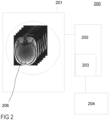
- Figure 2 schematically illustrates a system 200 for automated assessment of the CVS according to the invention.
- Said system 200 comprises:
- the system 200 according to the invention is characterized in that its processing unit 202 is configured for carrying out the steps of the method according to the invention, wherein said interface is configured for providing, for each brain lesion image set, said class associated to the highest probability.
- the method will be now described in more details with reference to Figure 1 .
- the system 200 acquires or receives a set of one or several 3D MR images of a brain lesion.
- an MRI system 201 is configured for performing a 3D T1 weighted MPRAGE imaging before a contrast agent injection, followed by a 3D T2 * weighted segmented EPI imaging during injection, and a 3D T2-weighted FLAIR imaging after injection, wherein a FLAIR * image is then generated by combining the T2-weighted FLAIR image and T2 * -weighted segmented EPI image.
- the image processing for generating FLAIR * images is described for instance in reference [8] and is known in the art.
- the system according to the invention is then further configured for automatically segmenting brain lesions in the acquired or processed images, e.g. in said FLAIR * images.
- the images acquired by the MRI system might undergo different image processing for improving the detection, location, and classification of MS lesions performed in the following steps.
- the system 200 acquires said set of one or several images of a brain lesion by performing a segmentation process on at least one of the MR image acquired by the MRI system, from said segmentation, creating a mask for the concerned lesion, and using said mask for extracting said brain lesion image patch (i.e.
- the acquired 3D brain lesion images are preferentially image patches of said lesion, each patch representing a different MRI contrast, wherein at least one patch results from, or is based on, a T2 * -based contrast imaging and another patch results from, or is based on, a WM lesion contrast MRI imaging, or wherein at least one patch results from, or is based on, a combination of an image obtained using said T2 * -based contrast imaging and another image obtained using a WM lesion contrast MRI imaging.
- the system uses said one or several 3D brain lesion images acquired at step 110 as input to two different CNNs, namely a first CNN 121 and a second CNN 122.
- Each of said CNN may receive a different subset of said set of images as input, e.g. the first CNN receives a first subset and the second CNN receives a second subset.
- said first and second CNN receive the same input.
- said two CNNs are used "in parallel". For instance, they can receive the same input, but provide different outputs since the training they received has been different.
- the first CNN 121 has been trained for detecting a presence of, or identifying, a lesion that has to be excluded from CVS assessment (i.e.
- the second CNN 122 has been trained for detecting a presence of, or identifying, a lesion that comprises a single central vein (i.e. CVS+), classifying each set of images received as input either in the class CVS+ or in the class CVS-, and outputting 124, for each received set, the probability that the concerned set belongs to each class.
- Both the first and second CNNs are preferentially trained using a set of lesion samples (e.g. a set of image patches) as training input, preferentially a first set for the first CNN and a second set for the second CNN, wherein the first and second sets are different, and, as training output, for each lesion sample used as training input, its classification into one of the two classes associated to the concerned CNN.
- a set of lesion samples e.g. a set of image patches
- the system uses the probabilities (or class weights) previously obtained for the classification of each set of 3D MR brain lesion images used as input to respectively the first and second CNN, as input to a final classifier.
- the final classifier has been trained for classifying each set in one of the three classes of lesions, CVS+, CVS-, and CVSe, using as input said probabilities values, and outputting the class (called hereafter "final class") for which the brain lesion image set got the highest probability to belong to.
- the system 200 is configured for providing, via said interface 204, the obtained final class.
- the system 200 automatically calculates a CVS+ lesion fraction and automatically determines whether said fraction exceeds a predefined threshold, e.g. 40% of the total number of eligible (i.e. non CVSe) lesions, and if exceeding said threshold is detected, then the system 200 preferentially automatically triggers a warning.
- a predefined threshold e.g. 40% of the total number of eligible (i.e. non CVSe) lesions
- the final classifier might be replaced by a final CNN using, in addition to the probability/weights results of the first and second CNN, said set of brain lesion images as input, wherein said brain lesion images are preferentially, as explained earlier, 3D patches extracted around the lesion from at least one of the following contrasts and received as separate input channels for the first, second, and optionally the final CNN: FLAIR * , and/or T2 * , and/or lesion mask, and/or CSF, and/or gray/white matter concentration maps, obtained for instance from a partial-volume estimation algorithm as described in Roche et al. [9].
- the present invention proposes a multi-level classification architecture for automated CVS assessment, wherein the task of classifying lesion types is separated into two sub-tasks of identifying CVSe lesions and distinguishing CVS+ and CVS-, followed by a third classification step that uses the results of the first two classifiers as input.
Landscapes
- Engineering & Computer Science (AREA)
- Theoretical Computer Science (AREA)
- Medical Informatics (AREA)
- Health & Medical Sciences (AREA)
- General Physics & Mathematics (AREA)
- Physics & Mathematics (AREA)
- General Health & Medical Sciences (AREA)
- Computer Vision & Pattern Recognition (AREA)
- Evolutionary Computation (AREA)
- Databases & Information Systems (AREA)
- Multimedia (AREA)
- Computing Systems (AREA)
- Artificial Intelligence (AREA)
- Software Systems (AREA)
- Data Mining & Analysis (AREA)
- Public Health (AREA)
- Nuclear Medicine, Radiotherapy & Molecular Imaging (AREA)
- Quality & Reliability (AREA)
- Radiology & Medical Imaging (AREA)
- Biomedical Technology (AREA)
- Primary Health Care (AREA)
- Epidemiology (AREA)
- Pathology (AREA)
- Magnetic Resonance Imaging Apparatus (AREA)
Abstract
Description
- The present invention falls within the field of medical imaging systems and methods, notably magnetic resonance imaging (MRI) systems and methods. In particular, the present invention relates to a method for automated central vein sign assessment, and to a device for carrying out said method.
- Misdiagnosis with potentially harmful consequences for patients is a common problem in multiple sclerosis (MS) and was estimated to affect up to 20% of patients [1]. Assessment of the fraction of white matter (WM) lesions exhibiting a central vein, referred to as the central vein sign (CVS) - i.e. a small vein located at the center of the WM lesion -, has shown the potential to distinguish MS from other mimicking diseases and thereby potentially reduce misdiagnoses. Manual CVS assessment can, however, be tedious and very time-consuming, rendering it unfeasible in clinical routine. To address this problem, automated approaches have been proposed [2,3], but the task remains non-trivial. In particular, the selection of lesions that should be excluded from the assessment per the NAIMS criteria [4] - said lesions being called hereafter CVS excluded (CVSe) lesions - has proven to be challenging, resulting in limited classification accuracy of automated approaches. The selection of lesions to include or exclude is an essential requirement to determine the fraction of CVS positive lesions, which is then used as metric for differential diagnosis.
- These limitations have so far hindered broader clinical assessment and evaluation of the CVS as an imaging biomarker for differential diagnosis.
- Automated CVS assessment using probabilistic or deep learning-based approaches are known in the art. For instance, Dworkin et al. [5] uses a Frangi vesselness filter to detect veins combined with a textural analysis to identify lesion centers. In that approach all periventricular lesions are excluded as they are considered to be confluent or contain multiple veins. The CVS+ probabilities (i.e. the probabilities of having a lesion comprising one and only one vein that is centrally located within the lesion) are then weighted by image noise to account for artifacts. Another technique proposes to use ensembles of convolutional neural networks for CVS assessment (CVSNet, CVSNet2, see ref. [3] and [6]), but either requires a manual lesion exclusion step or classifies all three CVS lesion types (CVS+, CVS- and CVSe) at the same time.
- While overall the classification performance of these methods is considered good, it is still not sufficient to allow confidently applying them for fully automated CVS assessment in an unsupervised setting, which hinders broader clinical application.
- It is thus an objective of the present invention to find a system and method capable of automated CVS assessment that are suitable for clinical applications.
- Said objective is achieved according to the present invention by a system and method for automatically detecting in a MR image a WM lesion exhibiting a CVS according to the object of the independent claims. Dependent claims present further advantages of the invention.
- The present invention proposes notably a method, the method comprising the following steps:
- receiving or acquiring one or several 3D MR images of a brain lesion, i.e. a set of 3D MR images of a brain lesion, wherein each 3D MR image represents preferentially a different MR image contrast of said brain lesion. Each of said 3D MR images might be an image of a single brain lesion, or might be an image of multiple brain lesions. Preferentially, for each brain lesion, said set comprises at least a first image acquired using a T2*-based contrast imaging technique, or, more generally speaking, using an MRI method capable of imaging the magnetic properties of blood, and optionally a second image acquired using an MRI method configured for enabling an identification of WM lesions. Preferentially, instead of said first image and said optional second image, or in addition to said first and optional second image, at least one of said 3D MR brain lesion images of the set is obtained by combining said first image and said second image. In other words and preferentially, for each brain lesion, at least a 3D image combining an MR contrast configured for imaging or detecting veins and a MR contrast configured for imaging or identifying WM lesions might be acquired. Preferentially, said set of 3D MR images of a brain lesion are image patches, i.e. 3D images comprising a selected set of voxels representing only the concerned brain lesion plus a predefined margin surrounding the latter, e.g. centered on the brain lesion (in other words, they represent different contrasts of a same (selected) set of voxels representing the brain lesion). Alternatively, said set of 3D MR images might be whole brain images. For instance, said first and second images typically represent brain slices, e.g. axial slices, of a brain of a patient. Preferentially, said 3D MR brain lesion images are spatially registered onto a common space. In particular, said second image is, or is based on, a fluid-attenuated inversion recovery (FLAIR) image of said brain. Preferentially, said MR images of a brain lesion comprise a FLAIR* image of the lesion. Preferentially, a 3D T1-weighted magnetization-prepared rapid gradient echo (MPRAGE) image, a 3D T2-weighted FLAIR image, and a 3D T2*-weighted segmented echo-planar image (EPI) of a same part of the brain, e.g. representing the same slice or volume of said brain, can be acquired by an MRI system and then registered to a common space, e.g. the FLAIR space, and combined in order to generate said FLAIR* image. Optionally, each 3D MR brain lesion image is obtained after performing, or results from, an automatic segmentation of an MRI brain image, wherein said MRI brain image is for instance the first image, or the second image, or the result of the combination of said first and second image (e.g. said MRI brain image is the FLAIR* image of the brain), and wherein said segmentation aims to delineate each WM lesion in the MRI brain image, each delineated WM lesions being then extracted from the different contrasts acquired for said lesion, e.g. from the first, second, and combination of the first and second image, in order to create said image patch for the different contrasts acquired for said lesion. For this purpose, a known in the art technique or segmentation algorithm might be used, as described in the paper of Roy S. et al. [7]. According to the present invention and in particular, each MR brain lesion image comprises thus a selected set of voxels (e.g. representing a cube with an edge length of 21 voxels) representing a single WM lesion, preferentially surrounded by a predefined margin configured for delineating or identifying a (predefined, e.g. cubical) contour of the WM lesion. Each MR brain lesion image is thus preferentially an image patch extracted from a 3D MRI image of a brain by applying a segmentation mask onto said 3D MRI image, the latter resulting for instance from said combination of the first and second image, said segmentation mask enabling to select said set of voxels representing the WM lesion identified by the segmentation process. For instance, a segmentation algorithm configured for extracting each lesion in the form of a segmentation mask might be used for said extraction. Preferentially, for each lesion, the center of mass of said lesion is determined and the segmentation mask is automatically centered on said center of mass, enabling to extract for instance a cube of size 21x21x21 voxels around the center of mass of the considered lesion for creating, for each acquired contrast, said image patch;
- feeding a first subset of said set of the received or acquired 3D MR brain lesion images into a first machine learning (ML) algorithm, e.g. a first convolutional neural network (CNN), and a second subset of said set into a second ML algorithm, e.g. a second CNN, wherein the first and second ML algorithms are two different ML algorithms, e.g. wherein the first CNN and second CNN are two different CNNs. Preferentially, said first subset is identical to the second subset. Preferentially, said first subset and second subset are identical to said set. According to the present invention, and in particular, each of said first ML algorithm (e.g. CNN) and second ML algorithm (e.g. CNN) might thus receive as input a different subset of said set of 3D MR images.
- According to the present invention, the first ML algorithm, e.g. the first CNN, is configured for performing a classification of the 3D MR brain lesion images comprised in said first subset into a first class or a second class, wherein the first class comprises or refers to CVS-positive (CVS+) lesions, as well as CVS-negative (CVS-) lesions (i.e. lesions identified as CVS+ lesions are classified in said first class and lesions identified as CVS- lesions are also classified in said first class), and the second class comprises or refers to CVS excluded (CVSe) lesions, wherein the first CNN is configured for outputting, for each first subset received as input and each class (i.e. said first class and said second class), a probability that the brain lesion image first subset belongs to said class. A standard radiological definition of a central vein is commonly used and notably defined by Sati et al. in reference [4]: CVS+ lesions are lesions that exhibit a single central vein (only one and not more), CVS- lesions are lesions that exhibit no vein, CVSe lesions are lesions that should be excluded from the CVS assessment. CVSe lesions comprise confluent lesions (without vein) and/or lesions with multiple veins and/or lesions with eccentric veins and/or confluent lesions with vein(s). Such lesions are categorized in CVSe lesions.
- According to the present invention, the second ML algorithm, e.g. the second CNN, is configured for performing a classification of the 3D MR brain lesion images comprised in said second subset into a third class or a fourth class, wherein the third class comprises or refers to CVS+ lesions and the fourth class comprises or refers to CVS- lesions, wherein the second ML algorithm, e.g. second CNN, is configured for outputting for each second subset received as input and each class (i.e. said third class and said fourth class), a probability that the subset belongs to said class,
wherein said two ML algorithms (e.g. said first and second CNNS) are preferentially both CVSNet CNN [3] or based on the CVSNet architecture, and; - for each first subset and second subset that has been fed into respectively said first and second ML algorithm, e.g. CNNs, using the probability results obtained from the first and second ML algorithms, e.g. CNNs, as inputs to a final classifier configured for performing a final classification of the brain lesion image set into the second, third, or fourth class, wherein for each of said classes (i.e. second, third and fourth classes), the final classifier is configured for outputting a final probability that the image set belongs to the concerned class. Preferentially, the final classifier uses a random forest classifier or algorithm;
- providing via an interface, e.g. a display, the class, among the second, third and fourth class, that is characterized by the highest probability value or result. Optionally, the method comprises automatically calculating a fraction F of the CVS+ lesions, wherein F = number of CVS+ lesions divided by the sum of the number of CVS+ and number of CVS- lesions, and if the latter exceeds a predefined threshold, automatically suggesting to a user, or starting, an additional examination, e.g. by means of the MRI system, or automatically launching an additional imaging of one or several lesions identified as CVS+ lesions, or automatically sending a signal to a warning device, said signal being configured for triggering a warning, e.g. visual and/or auditive warning, by the warning device.
- In the proposed approach, the task of lesion exclusion (i.e. distinguishing CVS+/- from CVSe lesion types) and the task of vein detection (i.e. distinguishing CVS+ from CVS-) are separated in two individual tasks, i.e. are completely independent, each being performed by a dedicated individual classifier, i.e. said first and second ML algorithms which are preferentially and respectively said first CNN and said second CNN, specifically trained for that purpose. The output probabilities of these classifiers are then used as input to a second level classifier, i.e. said final classifier, e.g. random forest classifier, that performs the final classification into CVS+, CVS- and CVSe lesion classes. This task separation allows to train the different classifiers more specifically for their corresponding task resulting in better performance. The above-mentioned method is then preferentially repeated sequentially for all lesions identified/detected, e.g. during the segmentation process, in a 3D MR image of a whole brain.
- The present invention concerns also a system or apparatus configured for carrying out the steps of the previously described method.
- Said system comprises for instance an MRI system or a connection to an MRI system or to a database for acquiring said brain lesion images, a processing unit and a memory, wherein the processing unit is configured for automatically carrying out the above-mentioned method, and an interface, e.g. a display, for outputting, for each image that has been processed according to said method, the class associated to the highest probability, and/or for outputting a calculated fraction of CVS+ lesions.
- The foregoing has broadly outlined the features and technical advantages of the present disclosure so that those skilled in the art may better understand the detailed description that follows.
- Additional features and advantages of the disclosure will be described hereinafter that form the object of the claims. Those skilled in the art will appreciate that they may readily use the concept and the specific embodiment disclosed as a basis for modifying or designing other structures for carrying out the same purposes of the present disclosure.
- For a more complete understanding of the present disclosure, and the advantages thereof, reference is now made to the following descriptions taken in conjunction with the accompanying drawings, wherein like numbers designate like objects, and in which:
- Figure 1
- illustrates a flowchart of a method according to the invention;
- Figure 2
- is a schematic illustration of a system according to the invention.
-
Figure 1 schematically illustrates a workflow of a preferred embodiment of the method according to the invention.Figure 2 schematically illustrates asystem 200 for automated assessment of the CVS according to the invention. Saidsystem 200 comprises: - optionally, an
imaging system 201, e.g. a CT imaging system, e.g. an MRI apparatus, for acquiringimages 206 of a brain lesion. Said images might also be acquired from another imaging system, or from a database, or uploaded in the system according to the invention; - a
memory 203 for storing the acquiredimages 206; - a
processing unit 202 comprising a processor, theprocessing unit 202 being configured for processing the acquiredimages 206. Theprocessing unit 202 is typically connected to theimaging system 201 and to thememory 203, and/or comprises an acquisition interface for acquiring or downloading said images. Theprocessing unit 202 preferentially comprises saidmemory 203; - an
interface 204, e.g. a display for displaying the acquiredimages 206 and/or reporting a calculated fraction of CVS+ lesions over all eligible (i.e. not CVSe) WM lesions. - The
system 200 according to the invention is characterized in that itsprocessing unit 202 is configured for carrying out the steps of the method according to the invention, wherein said interface is configured for providing, for each brain lesion image set, said class associated to the highest probability. The method will be now described in more details with reference toFigure 1 . - At
step 110, thesystem 200 according to the invention acquires or receives a set of one or several 3D MR images of a brain lesion. For instance, anMRI system 201 is configured for performing a 3D T1 weighted MPRAGE imaging before a contrast agent injection, followed by a 3D T2* weighted segmented EPI imaging during injection, and a 3D T2-weighted FLAIR imaging after injection, wherein a FLAIR* image is then generated by combining the T2-weighted FLAIR image and T2*-weighted segmented EPI image. The image processing for generating FLAIR* images is described for instance in reference [8] and is known in the art. Preferentially, the system according to the invention is then further configured for automatically segmenting brain lesions in the acquired or processed images, e.g. in said FLAIR* images. In other words, the images acquired by the MRI system might undergo different image processing for improving the detection, location, and classification of MS lesions performed in the following steps. In particular, thesystem 200 acquires said set of one or several images of a brain lesion by performing a segmentation process on at least one of the MR image acquired by the MRI system, from said segmentation, creating a mask for the concerned lesion, and using said mask for extracting said brain lesion image patch (i.e. said mask enabling to select in each image the same set of voxels) from one or several of said images acquired by theMRI system 201, and/or one or several images resulting from a combination of said images acquired by the MRI system. Therefore, for a same lesion, the acquired 3D brain lesion images are preferentially image patches of said lesion, each patch representing a different MRI contrast, wherein at least one patch results from, or is based on, a T2*-based contrast imaging and another patch results from, or is based on, a WM lesion contrast MRI imaging, or wherein at least one patch results from, or is based on, a combination of an image obtained using said T2*-based contrast imaging and another image obtained using a WM lesion contrast MRI imaging. - At
step 120, the system uses said one or several 3D brain lesion images acquired atstep 110 as input to two different CNNs, namely afirst CNN 121 and asecond CNN 122. Each of said CNN may receive a different subset of said set of images as input, e.g. the first CNN receives a first subset and the second CNN receives a second subset. Preferentially, said first and second CNN receive the same input. As shown inFig. 1 , said two CNNs are used "in parallel". For instance, they can receive the same input, but provide different outputs since the training they received has been different. Indeed, thefirst CNN 121 has been trained for detecting a presence of, or identifying, a lesion that has to be excluded from CVS assessment (i.e. for detecting or identifying a CVSe lesion), classifying each set of images received as input either in the class CVSe or in the class CVS+/-, and outputting 123, for each set of 3D brain lesion images received as input, the probability that the received set belongs to each class. Thesecond CNN 122 has been trained for detecting a presence of, or identifying, a lesion that comprises a single central vein (i.e. CVS+), classifying each set of images received as input either in the class CVS+ or in the class CVS-, and outputting 124, for each received set, the probability that the concerned set belongs to each class. - Both the first and second CNNs are preferentially trained using a set of lesion samples (e.g. a set of image patches) as training input, preferentially a first set for the first CNN and a second set for the second CNN, wherein the first and second sets are different, and, as training output, for each lesion sample used as training input, its classification into one of the two classes associated to the concerned CNN.
- At
step 130, the system uses the probabilities (or class weights) previously obtained for the classification of each set of 3D MR brain lesion images used as input to respectively the first and second CNN, as input to a final classifier. Indeed, fromstep 120, each received or acquired set can be associated to four probability values: from thefirst CNN 121 result the probability P11 that the lesion represented in the images of the set belongs to the class CVS+/- and the probability P12=1-P11 that it belongs to the class CVSe; from thesecond CNN 122 result the probably P21 that the lesion belongs to the class CVS+ and the probability P22=1-P21 that it belongs to the class CVS-. Said probability values are used as input in the final classifier. Advantageously, using said probabilities makes sure that all information of the first classification stage can be fully exploited for the final classification. For the latter, the final classifier has been trained for classifying each set in one of the three classes of lesions, CVS+, CVS-, and CVSe, using as input said probabilities values, and outputting the class (called hereafter "final class") for which the brain lesion image set got the highest probability to belong to. - At
step 140, thesystem 200 is configured for providing, via saidinterface 204, the obtained final class. - At
step 150, and optionally, thesystem 200 automatically calculates a CVS+ lesion fraction and automatically determines whether said fraction exceeds a predefined threshold, e.g. 40% of the total number of eligible (i.e. non CVSe) lesions, and if exceeding said threshold is detected, then thesystem 200 preferentially automatically triggers a warning. - Preferentially, the final classifier might be replaced by a final CNN using, in addition to the probability/weights results of the first and second CNN, said set of brain lesion images as input, wherein said brain lesion images are preferentially, as explained earlier, 3D patches extracted around the lesion from at least one of the following contrasts and received as separate input channels for the first, second, and optionally the final CNN: FLAIR*, and/or T2*, and/or lesion mask, and/or CSF, and/or gray/white matter concentration maps, obtained for instance from a partial-volume estimation algorithm as described in Roche et al. [9].
- To summarize, the present invention proposes a multi-level classification architecture for automated CVS assessment, wherein the task of classifying lesion types is separated into two sub-tasks of identifying CVSe lesions and distinguishing CVS+ and CVS-, followed by a third classification step that uses the results of the first two classifiers as input.
-
- CVS
- central vein sign
- MRI
- magnetic resonance imaging
- MR
- magnetic resonance
- MS
- multiple sclerosis
- CSF
- cerebrospinal fluid
- MPRAGE
- magnetization prepared rapid gradient-echo
- FLAIR
- fluid-attenuated inversion recovery
- CNN
- convolutional neural network
- EPI
- echo planar imaging
- ML
- machine learning
-
- [1] Solomon AJ, Bourdette DN, Cross AH, et al. The contemporary spectrum of multiple sclerosis misdiagnosis. Neurology 2016 (87): 1393-1399.
- [2] Fartaria MJ. Cvsnet: A deep learning-based approach for automated detection of the central vein sign. Mult. Scler.J. 2019.
- [3] Maggi P, Fartaria MJ, Jorge J, et al. CVSnet: A machine learning approach for automated central vein sign assessment in multiple sclerosis, NMR Biomed. 2020.
- [4] Sati P, Oh J, Todd Constable R, et al. The central vein sign and its clinical evaluation for the diagnosis of multiple sclerosis: A consensus statement from the North American Imaging in Multiple Sclerosis Cooperative. Nat. Rev. Neurol. 2016.
- [5] Dworkin JD, Sati P, Solomon A, et al., Automated integration of multimodal MRI for the probabilistic detection of the central vein sign in white matter lesions, Am. J. Neuroradiol. 2018.
- [6] Huelnhagen et al., Toward Fully Automated Assessment of the Central Vein Sign Using Deep Learning, Proc. Intl. Soc. Mag. Reson. Med. 29 (2021), 0488.
- [7] Roy S, Butman JA, Reich DS, Calabresi PA, Pham DL. Multiple sclerosis lesion segmentation from brain MRI via fully convolutional neural networks. arXiv: 1803.09172 2018.
- [8] Sati P, George IC, Shea CD, Gaitan Ml, Reich DS. FLAIR*: A combined MR contrast technique for visualizing white matter lesions and parenchymal veins. Radiology 2012, doi: 10.1148/radiol.12120208.
- [9] Roche A, Forbes F. Partial Volume Estimation in Brain MRI Revisited BT - Medical Image Computing and Computer-Assisted Intervention - MICCAI 2014: 17th International Conference, Boston, MA, USA, September 14-18, 2014, Proceedings, Part I. In: Golland P, Hata N, Barillot C, Hornegger J, Howe R, editors. Cham: Springer International Publishing; 2014. pp. 771-778.
Claims (10)
- Method for automatically detecting, in a magnetic resonance , hereafter "MR", image, a white matter, hereafter "WM", lesion exhibiting a central vein, hereafter referred to as central vein sign "CVS", the method comprising the following steps:- acquiring (110) a set comprising one or several 3D MR images of a brain lesion;- using (120) a first and second subset of said set as input respectively to a first and second ML algorithm, wherein the first ML algorithm and the second ML algorithm are two different ML algorithms, and whereinthe first ML algorithm is configured for performing a classification of the first subset into a first class or a second class, wherein the first class comprises CVS+ lesions and CVS- lesions, and the second class comprises CVSe lesions, the first ML algorithm being further configured for outputting, for said first subset received as input and each class, a probability (123) that the subset belongs to said class, andthe second ML algorithm is configured for performing a classification of the second subset into a third class or a fourth class, wherein the third class comprises CVS+ lesions and the fourth class comprises CVS-lesions, the second ML algorithm being further configured for outputting for said second subset received as input and each class, a probability (124) that the second subset belongs to said class;- for each set, using (130) the probability results obtained from the first and second ML algorithm as inputs to a final classifier configured for performing a final classification of the set into the second, third, or fourth class, wherein for each of said classes, the final classifier is configured for outputting a final probability that the set belongs to said class;- providing (140) via an interface the class, among the second, third and fourth class, that is characterized by the highest probability value.
- The method according to claim 1, wherein at least one image of said set is acquired using a T2*-based contrast imaging technique.
- The method according to claim 2, wherein said at least one image is a FLAIR* image.
- The method according to one of the claims 1 to 3, comprising acquiring one or several brain images by means of an MRI system 201, automatically performing a brain lesion segmentation in at least one of said acquired images for segmenting said brain lesion, creating a segmentation mask for said brain lesion from said segmentation, and using said segmentation mask for acquiring, from one or several of said images acquired by the MRI system 201, said set of one or several 3D MR images of the brain lesion.
- The method according to one of the claims 1 to 4, wherein said first and second ML algorithms are CVSNet CNN or based on a CVSNet architecture.
- The method according to one of the claims 1 to 5, characterized in that the final classifier uses a random forest classifier or algorithm.
- The method according to one of the claims 1 to 6, wherein the 3D MR images used as input to the first and/or the second ML algorithms are 3D patches extracted around the brain lesion, wherein the set comprises at least one or several 3D patches extracted from one of the following modalities: FLAIR*, and/or T2*, and/or lesion mask, and/or CSF, and/or gray matter concentration map, and/or white matter concentration map.
- The method according to one of the claims 1 to 7, comprising automatically calculating a fraction of the CVS+ images, determining whether said fraction exceeds a predefined threshold.
- The method according to claim 8, comprising automatically triggering a visual and/or auditive warning if said fraction exceeds the predefined threshold.
- System (200) configured for carrying out the method according to claim 1 to 9.
Priority Applications (2)
| Application Number | Priority Date | Filing Date | Title |
|---|---|---|---|
| EP22168961.5A EP4266245A1 (en) | 2022-04-20 | 2022-04-20 | A method and system for automated central vein sign assessment |
| US18/303,724 US12597240B2 (en) | 2022-04-20 | 2023-04-20 | Method and system for automated central vein sign assessment |
Applications Claiming Priority (1)
| Application Number | Priority Date | Filing Date | Title |
|---|---|---|---|
| EP22168961.5A EP4266245A1 (en) | 2022-04-20 | 2022-04-20 | A method and system for automated central vein sign assessment |
Publications (1)
| Publication Number | Publication Date |
|---|---|
| EP4266245A1 true EP4266245A1 (en) | 2023-10-25 |
Family
ID=81344345
Family Applications (1)
| Application Number | Title | Priority Date | Filing Date |
|---|---|---|---|
| EP22168961.5A Withdrawn EP4266245A1 (en) | 2022-04-20 | 2022-04-20 | A method and system for automated central vein sign assessment |
Country Status (2)
| Country | Link |
|---|---|
| US (1) | US12597240B2 (en) |
| EP (1) | EP4266245A1 (en) |
Family Cites Families (2)
| Publication number | Priority date | Publication date | Assignee | Title |
|---|---|---|---|---|
| US10761171B2 (en) * | 2015-06-22 | 2020-09-01 | Case Western Reserve University | Systems and methods for free-breathing three-dimensional magnetic resonance fingerprinting |
| US11436724B2 (en) * | 2020-10-30 | 2022-09-06 | International Business Machines Corporation | Lesion detection artificial intelligence pipeline computing system |
-
2022
- 2022-04-20 EP EP22168961.5A patent/EP4266245A1/en not_active Withdrawn
-
2023
- 2023-04-20 US US18/303,724 patent/US12597240B2/en active Active
Non-Patent Citations (12)
| Title |
|---|
| "Cham", 2014, SPRINGER INTERNATIONAL PUBLISHING, article "Proceedings", pages: 771 - 778 |
| DWORKIN JDSATI PSOLOMON A ET AL.: "Automated integration of multimodal MRI for the probabilistic detection of the central vein sign in white matter lesions", AM. J. NEURORADIOL, 2018 |
| FARTARIA MJ: "Cvsnet: A deep learning-based approach for automated detection of the central vein sign", MULT. SCLER.J., 2019 |
| HUELNHAGEN ET AL.: "Toward Fully Automated Assessment of the Central Vein Sign Using Deep Learning", PROC. INTL. SOC. MAG. RESON. MED., vol. 29, 2021, pages 0488 |
| MAAYAN FRID-ADAR ET AL: "Modeling the Intra-class Variability for Liver Lesion Detection using a Multi-class Patch-based CNN", ARXIV.ORG, CORNELL UNIVERSITY LIBRARY, 201 OLIN LIBRARY CORNELL UNIVERSITY ITHACA, NY 14853, 19 July 2017 (2017-07-19), XP080777941, DOI: 10.1007/978-3-319-67434-6_15 * |
| MAGGI PFARTARIA MJJORGE J ET AL.: "CVSnet: A machine learning approach for automated central vein sign assessment in multiple sclerosis", NMR BIOMED, 2020 |
| MAGGI PIETRO ET AL: "CVSnet: A machine learning approach for automated central vein sign assessment in multiple sclerosis", NMR IN BIOMEDICINE., vol. 33, no. 5, 3 March 2020 (2020-03-03), GB, XP055971389, ISSN: 0952-3480, Retrieved from the Internet <URL:https://onlinelibrary.wiley.com/doi/full-xml/10.1002/nbm.4283> DOI: 10.1002/nbm.4283 * |
| ROCHE AFORBES F, PARTIAL VOLUME ESTIMATION IN BRAIN MRI REVISITED BT - MEDICAL IMAGE COMPUTING AND COMPUTER-ASSISTED INTERVENTION - MICCAI 2014: 17TH INTERNATIONAL CONFERENCE, BOSTON, MA, USA, 2014 |
| ROY SBUTMAN JAREICH DSCALABRESI PAPHAM DL: "Multiple sclerosis lesion segmentation from brain MRI via fully convolutional neural networks", ARXIV: 1803.09172, 2018 |
| SATI POH JTODD CONSTABLE R ET AL.: "The central vein sign and its clinical evaluation for the diagnosis of multiple sclerosis: A consensus statement from the North American Imaging in Multiple Sclerosis Cooperative", NAT. REV. NEUROL., 2016 |
| SNEHASHIS ROY ET AL: "Multiple Sclerosis Lesion Segmentation from Brain MRI via Fully Convolutional Neural Networks", ARXIV.ORG, CORNELL UNIVERSITY LIBRARY, 201 OLIN LIBRARY CORNELL UNIVERSITY ITHACA, NY 14853, 24 March 2018 (2018-03-24), XP080862608 * |
| TILL HUELNHAGEN ET AL: "Toward Fully Automated Assessment of the Central Vein Sign Using Deep Learning", PROCEEDINGS OF THE 2021 ISMRM & SMRT ANNUAL MEETING & EXHIBITION, 15-20 MAY 2021, ISMRM, 2030 ADDISON STREET, 7TH FLOOR, BERKELEY, CA 94704 USA, no. 488, 30 April 2021 (2021-04-30), XP040722508 * |
Also Published As
| Publication number | Publication date |
|---|---|
| US20230343077A1 (en) | 2023-10-26 |
| US12597240B2 (en) | 2026-04-07 |
Similar Documents
| Publication | Publication Date | Title |
|---|---|---|
| EP3703007B1 (en) | Tumor tissue characterization using multi-parametric magnetic resonance imaging | |
| Costafreda et al. | Automated hippocampal shape analysis predicts the onset of dementia in mild cognitive impairment | |
| US6950544B2 (en) | Automated measurement of anatomical structures in medical imaging | |
| US10335106B2 (en) | Computing system and method for identifying and visualizing cerebral thrombosis based on medical images | |
| WO2011040473A1 (en) | Method, device and program for medical image processing | |
| Ghafaryasl et al. | A computer aided detection system for cerebral microbleeds in brain MRI | |
| CN114119546A (en) | Method and device for detecting MRI images | |
| Zhou et al. | Survival time prediction of patients with glioblastoma multiforme tumors using spatial distance measurement | |
| US12597240B2 (en) | Method and system for automated central vein sign assessment | |
| Razavi et al. | Towards accurate segmentation of fibroglandular tissue in breast MRI using fuzzy c-means and skin-folds removal | |
| Ferrer et al. | Deep learning-based assessment of cerebral microbleeds in COVID-19 | |
| CN117635559B (en) | Image processing method and system for locating gray cortex connected with abnormal white matter | |
| Fiçici et al. | Fully automated brain tumor segmentation and volume estimation based on symmetry analysis in MR images | |
| Rivas et al. | Unique MS lesion identification from MRI | |
| US20190318197A1 (en) | Method and system for abnormal tissue detection using z-scores in a joint histogram | |
| Somasundaram et al. | Brain extraction method for T1-weighted magnetic resonance scans | |
| Kawata et al. | Computer-aided evaluation method of white matter hyperintensities related to subcortical vascular dementia based on magnetic resonance imaging | |
| Saad et al. | Automated stroke lesion detection and diagnosis system | |
| Wang et al. | Voxel-based discriminant map classification on brain ventricles for Alzheimer's disease | |
| Malkanthi et al. | Brain tumor boundary segmentation of MR imaging using spatial domain image processing | |
| Basaran et al. | MSSEG-2 Challenge, Team New Brain: Cascaded networks for new MS lesion detection | |
| CN114463351B (en) | Method and system for processing brain image data | |
| EP4332883B1 (en) | Detecting artifacts in medical images | |
| Wahab et al. | ConvSE for Multiple Sclerosis Lesion Segmentation | |
| Zhang | Improving the diagnostic process of multiple sclerosis by medical image processing and machine learning |
Legal Events
| Date | Code | Title | Description |
|---|---|---|---|
| STAA | Information on the status of an ep patent application or granted ep patent |
Free format text: STATUS: THE APPLICATION HAS BEEN PUBLISHED |
|
| PUAI | Public reference made under article 153(3) epc to a published international application that has entered the european phase |
Free format text: ORIGINAL CODE: 0009012 |
|
| AK | Designated contracting states |
Kind code of ref document: A1 Designated state(s): AL AT BE BG CH CY CZ DE DK EE ES FI FR GB GR HR HU IE IS IT LI LT LU LV MC MK MT NL NO PL PT RO RS SE SI SK SM TR |
|
| RAP1 | Party data changed (applicant data changed or rights of an application transferred) |
Owner name: CENTRE HOSPITALIER UNIVERSITAIRE VAUDOIS Owner name: SIEMENS HEALTHINEERS AG |
|
| STAA | Information on the status of an ep patent application or granted ep patent |
Free format text: STATUS: REQUEST FOR EXAMINATION WAS MADE |
|
| 17P | Request for examination filed |
Effective date: 20240418 |
|
| RBV | Designated contracting states (corrected) |
Designated state(s): AL AT BE BG CH CY CZ DE DK EE ES FI FR GB GR HR HU IE IS IT LI LT LU LV MC MK MT NL NO PL PT RO RS SE SI SK SM TR |
|
| GRAP | Despatch of communication of intention to grant a patent |
Free format text: ORIGINAL CODE: EPIDOSNIGR1 |
|
| STAA | Information on the status of an ep patent application or granted ep patent |
Free format text: STATUS: GRANT OF PATENT IS INTENDED |
|
| RIC1 | Information provided on ipc code assigned before grant |
Ipc: G06T 7/00 20170101AFI20241002BHEP |
|
| INTG | Intention to grant announced |
Effective date: 20241015 |
|
| STAA | Information on the status of an ep patent application or granted ep patent |
Free format text: STATUS: THE APPLICATION IS DEEMED TO BE WITHDRAWN |
|
| 18D | Application deemed to be withdrawn |
Effective date: 20250218 |