EP4265264A1 - Bacteriophages suitable for treating a bacterial infection caused by klebsiella pneumoniae - Google Patents
Bacteriophages suitable for treating a bacterial infection caused by klebsiella pneumoniae Download PDFInfo
- Publication number
- EP4265264A1 EP4265264A1 EP22382362.6A EP22382362A EP4265264A1 EP 4265264 A1 EP4265264 A1 EP 4265264A1 EP 22382362 A EP22382362 A EP 22382362A EP 4265264 A1 EP4265264 A1 EP 4265264A1
- Authority
- EP
- European Patent Office
- Prior art keywords
- bacteriophage
- phage
- klebsiella pneumoniae
- pneumoniae
- pharmaceutical composition
- Prior art date
- Legal status (The legal status is an assumption and is not a legal conclusion. Google has not performed a legal analysis and makes no representation as to the accuracy of the status listed.)
- Withdrawn
Links
Images


Classifications
-
- A—HUMAN NECESSITIES
- A61—MEDICAL OR VETERINARY SCIENCE; HYGIENE
- A61P—SPECIFIC THERAPEUTIC ACTIVITY OF CHEMICAL COMPOUNDS OR MEDICINAL PREPARATIONS
- A61P31/00—Antiinfectives, i.e. antibiotics, antiseptics, chemotherapeutics
- A61P31/04—Antibacterial agents
-
- A—HUMAN NECESSITIES
- A61—MEDICAL OR VETERINARY SCIENCE; HYGIENE
- A61K—PREPARATIONS FOR MEDICAL, DENTAL OR TOILETRY PURPOSES
- A61K35/00—Medicinal preparations containing materials or reaction products thereof with undetermined constitution
- A61K35/66—Microorganisms or materials therefrom
- A61K35/76—Viruses; Subviral particles; Bacteriophages
-
- A—HUMAN NECESSITIES
- A61—MEDICAL OR VETERINARY SCIENCE; HYGIENE
- A61K—PREPARATIONS FOR MEDICAL, DENTAL OR TOILETRY PURPOSES
- A61K38/00—Medicinal preparations containing peptides
- A61K38/16—Peptides having more than 20 amino acids; Gastrins; Somatostatins; Melanotropins; Derivatives thereof
- A61K38/164—Peptides having more than 20 amino acids; Gastrins; Somatostatins; Melanotropins; Derivatives thereof from bacteria
-
- C—CHEMISTRY; METALLURGY
- C12—BIOCHEMISTRY; BEER; SPIRITS; WINE; VINEGAR; MICROBIOLOGY; ENZYMOLOGY; MUTATION OR GENETIC ENGINEERING
- C12N—MICROORGANISMS OR ENZYMES; COMPOSITIONS THEREOF; PROPAGATING, PRESERVING, OR MAINTAINING MICROORGANISMS; MUTATION OR GENETIC ENGINEERING; CULTURE MEDIA
- C12N7/00—Viruses; Bacteriophages; Compositions thereof; Preparation or purification thereof
-
- C—CHEMISTRY; METALLURGY
- C12—BIOCHEMISTRY; BEER; SPIRITS; WINE; VINEGAR; MICROBIOLOGY; ENZYMOLOGY; MUTATION OR GENETIC ENGINEERING
- C12N—MICROORGANISMS OR ENZYMES; COMPOSITIONS THEREOF; PROPAGATING, PRESERVING, OR MAINTAINING MICROORGANISMS; MUTATION OR GENETIC ENGINEERING; CULTURE MEDIA
- C12N2795/00—Bacteriophages
- C12N2795/00011—Details
- C12N2795/00021—Viruses as such, e.g. new isolates, mutants or their genomic sequences
-
- C—CHEMISTRY; METALLURGY
- C12—BIOCHEMISTRY; BEER; SPIRITS; WINE; VINEGAR; MICROBIOLOGY; ENZYMOLOGY; MUTATION OR GENETIC ENGINEERING
- C12N—MICROORGANISMS OR ENZYMES; COMPOSITIONS THEREOF; PROPAGATING, PRESERVING, OR MAINTAINING MICROORGANISMS; MUTATION OR GENETIC ENGINEERING; CULTURE MEDIA
- C12N2795/00—Bacteriophages
- C12N2795/00011—Details
- C12N2795/00032—Use of virus as therapeutic agent, other than vaccine, e.g. as cytolytic agent
Definitions
- the present invention refers to the medial field.
- the present invention refers to a bacteriophage (F13) deposited at the German Collection of Microorganisms and Cell Cultures GmbH (Leibniz Institute) (DSMZ) with deposit number DSM 34222 on 17 th March 2022 and to the bacteriophage F12A characterized by comprising a nucleic acid consisting of SEQ ID NO: 1.
- the bacteriophage/s are used for treating bacterial infections caused by Klebsiella pneumoniae.
- CRE carbapenem-resistant Enterobacteriaceae
- MRSA methicillin-resistant Staphylococcus aureus
- ESBL extended-spectrum betalactamase
- VRE vancomycin-resistant Enterococci
- MDR multidrug-resistant Pseudomonas aeruginosa
- MDR Acinetobacter sp MDR Acinetobacter sp.
- Bacteriophages are viruses that can infect and multiply inside bacteria. They are biological systems ubiquitous in nature and very diverse from the genetic point of view; they were discovered by Felix d'Herelle in 1917. However, the first suspicions of the existence of microbes antagonistic to some bacteria were made by the British bacteriologist Frederick Twort. Phages are classified as virulent or temperate depending on the biological cycle they perform, lytic or lysogenic, respectively. Lysins are enzymes encoded by phages responsible for the bacterial cell wall lysis at the end of the lytic cycle and are interesting for their ability to disrupt biofilms.
- Virulent phages are the most desirable for therapeutic use against bacterial infections.
- the first investigations were carried out analyzing the possible role of these viruses in medicine.
- the discovery of penicillin by Alexander Fleming in 1928 and the subsequent development of clinically successful antibiotics in the following decades of the 20th century brought these lines of research to a standstill. Phage therapy has been poorly investigated.
- K. pneumoniae is a Gram-negative bacillus that belongs to the Enterobacteriaceae family and is an opportunistic pathogen responsible for a wide range of nosocomial infections, including urinary tract infections (UTIs), respiratory tract infections, blood-stream-associated infections, surgical-site infections, and also is responsible for liver abscesses in community acquired infections.
- UMIs urinary tract infections
- the intestine of patients acts as a reservoir for Klebsiella pneumoniae within a hospital. This pathogen causes severe morbidity and mortality in ICUs, premature ICUs and medical, pediatric and surgical wards.
- K. pneumoniae strains are intrinsically resistant to penicillins and can acquire resistance to third and fourth-generation cephalosporins.
- cephalosporins have been used as monotherapy and in combination with aminoglycosides against K. pneumoniae infections.
- Fosfomycin is mainly used in UTIs. This practice accelerated the emergence of fluoroquinolone-resistant and ESBL-producing K . pneumoniae strains which are treated with carbapenems.
- Carbapenem use has also increased the appearance of CRE bacteria which are resistant to carbapenems, penicillins, cephalosporins, fluoroquinolones, and aminoglycosides.
- Carbapenemases are a class of enzymes that can be transferred rapidly through conjugative plasmids among the Enterobacteriaceae family. Treatment options against carbapenem-resistant K .
- the present invention refers to bacteriophages named as F13 and F12A in the present invention.
- F13 has been deposited at the German Collection of Microorganisms and Cell Cultures GmbH (Leibniz Institute) (DSMZ) with deposit number DSM34222 on 17 th March 2022.
- F12A is characterized by comprising a nucleic acid consisting of SEQ ID NO: 1. These bacteriophages are characterized by their specificity for Klebsiella pneumoniae strains.
- the bacteriophage/s of the invention are able to inhibit the growth of clinical strainsof K. pneumoniae (including carbapenem resistant strains), both in liquid and in mature biofilms. Growth curves showed that the phage exhibited antimicrobial effect alone.
- the first embodiment of the present invention refers to a bacteriophage (hereinafter bacteriophage of the invention) deposited at the German Collection of Microorganisms and Cell Cultures GmbH (DSMZ) with deposit number DSM34222 on 17 th March 2022, or characterized by comprising a nucleic acid consisting of SEQ ID NO: 1, or a bacteriophage comprising a nucleic acid having at least 95% of identity with the nucleic acid of the bacteriophage DSM 34222 or the SEQ ID NO: 1.
- DSMZ German Collection of Microorganisms and Cell Cultures GmbH
- the SEQ ID NO: 1 of bacteriophage F12A is as follows:
- the second embodiment of the invention refers to the bacteriophage/s of the invention for use as a medicament, preferably for use in the prevention or treatment of a bacterial infection caused by K. pneumoniae.
- the third embodiment of the invention refers to a pharmaceutical composition comprising the bacteriophage/s of the invention, and, optionally, pharmaceutically acceptable excipients or carriers.
- the fourth embodiment of the present invention refers to the pharmaceutical composition comprising the bacteriophage/s of the invention, for use as a medicament, preferably for use in the prevention or treatment of a bacterial infection caused by K. pneumoniae.
- the present invention refers to a method for preventing or treating a bacterial infection caused by K. pneumoniaewhich comprises the administration of a therapeutically effective dose or amount of the bacteriophage/s of the invention or a pharmaceutical composition comprising thereof.
- the bacteriophages of the invention are administered along with one or more antibiotics, wherein the antibiotics are administered before, after or the same time than the bacteriophages of the invention.
- the antibiotics to be administered in combination with the bacteriophage/s of the invention are preferably selected from: Beta-lactam antibiotics such as penicillin or cephalosporin, aminoglycoside, carbapenems such as imipenem or meropenem, quinolones, or combinations thereof, preferably polymyxin B and/or colistin.
- the last embodiment of the present invention refers to a method of detecting the presence of K. pneumoniae in an in vitro sample, for instance obtained from a subject, wherein the subject is preferably a patient, comprising contacting said sample with the bacteriophage/s of the invention and determining whether said bacteriophage/s kills bacteria in said sample.
- Example 1.1 Bacterial strains and growth conditions
- the K. pneumoniae clinical isolates were stored in Difco TM skimmed milk (East Rutherford, NJ, United States) at -80 °C.
- the clinical isolates were plated from frozen skimmed milk stocks onto Mueller Hinton E agar 90 mm plates and broth cultures were grown in tryptic soy broth (TSB) (BioMerieux, France). Agar plates and broth cultures were incubated at 37 °C.
- the phages isolated against K. pneumoniae strain KP5 were obtained from HUFJD sewage (Madrid, Spain). Samples of 50 ml were centrifuged at 4000 x g for 10 min to remove cellular debris and faecal matter. The supernatant was filtered using 0.45 ⁇ m and 0.22 ⁇ m filters to remove the bacteria and debris. An amount of 100 ⁇ l of filtered solution was combined with 100 ⁇ l of the KP5 overnight culture and 3 mL of agar 0.2% (W/V) and plated on MHE plates via the double-layer agar method. Following overnight incubation, the formation of plaques confirmed the presence of phage.
- Example 1.3 Bacteriophage propagation
- a two-step propagation was applied to amplify and purify the isolated phage.
- 100 ⁇ l of KP5 overnight culture was added into 10 ml TSB and incubated at 37°C with 200 rpm shaking for 1.5 h to reach exponential growth.
- An amount of 100 ⁇ l of phage in SM buffer, 150 ⁇ l of MgSO 4 and CaCl 2 (200 mM) was added into the coculture and incubated for 4-5h at 37°C with 200 rpm shaking.
- the supernatant containing phage was harvested after centrifugation (4000 x g, 15 min) and filtration (0.45 ⁇ m and 0.22 ⁇ m syringe filter). Phage titration was performed.
- the small-scale amplification was applied according to the prior art.
- phage lysis was harvested as described above.
- the large-scale amplification was applied according to the prior art.
- Example 1.4 Bacteriophage titration
- the phage titer was determined via a double-layer agar method. Briefly, 100 ⁇ l of the KP5 overnight broth culture was added to 3 ml of agar (0.2% (W/V)) and overlaid onto a MHE plate. Phages were serially diluted in SM buffer. The plates were incubated at 37°C overnight. The phage titer was counted and calculated.
- K. pneumoniae phage was tested against a wide pH (1-8) and temperature (-80°C to 60°C) range using a working stock in TSB broth with an initial phage titer of around 10 8 PFU ⁇ ml -1 . Briefly, 100 ⁇ l working stock of each phage was suspended in 1 ml SM buffer previously adjusted with 1M NaOH or HCl (Sigma-Aldrich, Castle Hill, NSW, Australia) to yield a pH range of 1-8. The samples were incubated at room temperature for 1 h. The pH stability testing was studied according to the methods described in the prior art.
- Host range of F13 phage was determined against the different clinical Klebsiella pneumoniae strains obtained from Hospital Universitario Fundaconstrutive Jiménez Diaz (HUFJD) using spot testing. Briefly, 3 ⁇ l of the purified phage suspension (10 8 PFU/ml) was spotted onto the surface of double-layer agar plate previously inoculated with the tested strains. After adsorption of phage suspension, plates were incubated overnight at 37°C. Host range was determined by plaque formation. Three replicates were tested for each bacterial strain. The host range test was applied according to methodology described in the prior art.
- the infectivity profile of the K. pneumoniae bacteriophages was assessed at multiplicity of infection (MOI) 0.1, 1 and 10 using inhibition assays in liquid. Briefly, the McFarland inoculation standards (0.5 ⁇ 0.02) of each clinical isolate ( ⁇ 10 8 UFC ⁇ ml -1 ) were prepared and the required volume of phage stock solution (10 8 PFU ⁇ ml -1 ) was added to the corresponding suspension to achieve MOI 0.1, 1 and 10. The samples were incubated at 37°C with shaking orbital amplitude of 5 mm. Every 5min for 15 h the OD 595nm value was measured.
- MOI multiplicity of infection
- the isolated phage effect on K. pneumoniae biofilm was determined using a modified methodology previously described in the prior art. Briefly, biofilm formation on the bottom of a MicroWell 96-well flat-bottom plate (Thermo Fisher Scientific, Waltham, MA, USA) was induced by inoculating 100 ⁇ L of MHB containing ⁇ 10 7 CFU ⁇ ml -1 of bacteria per well and the plate was incubated at 37°C and 5% CO 2 for at least 18 h. The next day, the wells were aspirated, and biofilm formation was treated with bacteriophages at different concentrations (10 7 ,10 6 , 10 5 and 10 4 pfu/mL) in order to study the inhibition of biofilm by phages.
- Planktonic bacterial viability was determined by addition of 20 ⁇ l of 5 mg/mL of 3-(4,5-dimethylthiazol-2-yl)-2,5- diphenyltetrazolium bromide (MTT) (Sigma Aldrich, Merck, Darmstadt, Germany) (2) while biofilm viability was determined by adding 1mL of MTT to a commercial 9mL TSB tube and incubating for 1 h at 37°C and 5% CO 2 . The absorbance was measured at 570 nm for MTT using a microplate reader (TECAN). This experiment was performed in duplicate.
- MTT 3-(4,5-dimethylthiazol-2-yl)-2,5- diphenyltetrazolium bromide
- Antibacterial synergistic activity for F12A and F13 phages was assigned by modified broth microdilution method in flat-bottomed 96 multi-well microplates (Thermo-Fischer). Experimental conditions were designed for antibiotic-phage mix and were tested for colistin, polymyxin B, doripenem, ciprofloxacin and bacterial control at the final volume of 200 ⁇ l. Briefly, the culture was allowed to grow until an OD 600 of 0.44 reached 4 ⁇ 10 7 CFU. Then 50 ⁇ l of antibiotic at different concentrations was added to each well containing 50 ⁇ l of Mueller Hinton Broth II (Millipore, Sigma Aldrich, Merck) supplemented with 10 mM CaCl 2 and 10 mM MgSO 4 .
- phage dilutions 10 7 PFU
- 100 ⁇ l of bacterial suspension was added to the mix wells. Plates were incubated at 37°C overnight and FICI was calculated according to bacterial growth.
- F12A and F13 were stable at -80°C, -20°C, 4°C and 21°C after seven days of exposition.
- a certain reduction of phage viability was observed after seven days at 37°C.
- the main reduction of viability was observed at 60°C after 24 hours.
- the host range against 58 K. pneumoniae clinical isolates for phages F7, F8, F12A, F12B, F13 and F14 showed 10 susceptible (+) strains including a pan-resistant strain (Table 1) . All tested strains were multi-drug resistant (MDR) K. pneumoniae strains including KP5 strain.
- MDR multi-drug resistant
- the phages planktonic infective properties were determined through inhibition assays.
- the phage inhibition of KP5 clinical isolate was assessed for 15 h when infected at MOI 10, 1 and 0.1 for each phage ( Fig. 3 ).
- the concentration of KP5 in planktonic state decreased by 35% at 10 10 PFU/ml concentration of bacteriophage F12A (p-value ⁇ 0.05) after 6h of treatment (Fig. F12A 4a), and was reduced by 32% at 10 8 PFU/mL after 24h of treatment (Fig. F12A 4b). Biofilm formation was reduced by 28% at 6 h and 10 10 PFU/mL of F12A (Fig. F12A 4c).
- the concentration of KP5 bacteria in planktonic state and biofilm formation showed a significant decrease by 15% (Fig. F13 4a) and 22% (Fig. F13 4b), respectively, with 10 6 PFU/ml of F13 (p-value ⁇ 0.05) after 6 h of treatment.
- the concentration of planktonic bacteria was reduced by 60% at 10 6 PFU/ml after 24h of treatment ( Fig. 4c ) and biofilm formation was reduced 35% after 24 h of treatment with 10 6 PFU/ml of F13 ( Fig. 4d ).
- Phage-antibiotic checkerboard assays showed synergy between F13 and polymyxin B (FIC index 0.135) and an additive effect with colistin (FIC index 0.501) (Table 2).
- Polymyxin B was very effective against KP5, 8 ⁇ g/ml of polymyxin B was able to lyse the cells in combination with F13 at phage titer of 10 5 and 10 6 PFU/ml demonstrating synergistic effect.
- F12A was very effective in combination with polymyxin B (FIC index 0.26), doripenem (FIC index 0.251), colistin (FIC index 0.015) and ciprofloxacin (FIC index 0.35) at phage titer of 10 8 PFU/ml.
- Table 2 Antibiotic FIC index F12A F13 Effect Polymyxin B 0.26 0.135 Synergy / Synergy Colistin 0.015 0.501 Synergy / Addition Ciprofloxacin 0.35 Synergy Doripenem 0.251 Synergy Summary of synergistic effect between F12A, F13 and antibiotics against KP5 K . pneumoniae strain.
Landscapes
- Health & Medical Sciences (AREA)
- Life Sciences & Earth Sciences (AREA)
- Chemical & Material Sciences (AREA)
- General Health & Medical Sciences (AREA)
- Medicinal Chemistry (AREA)
- Bioinformatics & Cheminformatics (AREA)
- Engineering & Computer Science (AREA)
- Veterinary Medicine (AREA)
- Public Health (AREA)
- Organic Chemistry (AREA)
- Pharmacology & Pharmacy (AREA)
- Animal Behavior & Ethology (AREA)
- Virology (AREA)
- Genetics & Genomics (AREA)
- Epidemiology (AREA)
- Immunology (AREA)
- Zoology (AREA)
- Wood Science & Technology (AREA)
- Microbiology (AREA)
- Communicable Diseases (AREA)
- General Engineering & Computer Science (AREA)
- Biomedical Technology (AREA)
- Nuclear Medicine, Radiotherapy & Molecular Imaging (AREA)
- Oncology (AREA)
- Chemical Kinetics & Catalysis (AREA)
- Biochemistry (AREA)
- Biotechnology (AREA)
- Gastroenterology & Hepatology (AREA)
- Proteomics, Peptides & Aminoacids (AREA)
- General Chemical & Material Sciences (AREA)
- Mycology (AREA)
- Micro-Organisms Or Cultivation Processes Thereof (AREA)
- Measuring Or Testing Involving Enzymes Or Micro-Organisms (AREA)
Abstract
Description
- The present invention refers to the medial field. Particularly, the present invention refers to a bacteriophage (F13) deposited at the German Collection of Microorganisms and Cell Cultures GmbH (Leibniz Institute) (DSMZ) with deposit number DSM 34222 on 17th March 2022 and to the bacteriophage F12A characterized by comprising a nucleic acid consisting of SEQ ID NO: 1. In a preferred embodiment, the bacteriophage/s are used for treating bacterial infections caused by Klebsiella pneumoniae.
- The increase of multiresistance in bacteria is becoming a major public health problem, especially in hospital settings with increased dissemination of resistant strains due to the globalized world. The most important bacteria in this regard are carbapenem-resistant Enterobacteriaceae (CRE), methicillin-resistant Staphylococcus aureus (MRSA), extended-spectrum betalactamase (ESBL) producing Enterobacteriaceae, vancomycin-resistant Enterococci (VRE), multidrug-resistant (MDR) Pseudomonas aeruginosa, and MDR Acinetobacter sp. According to the latest reports from the Center for Disease Control and prevention (CDC) one out of four healthcare-associated infections will be caused by one of the above-mentioned bacteria. In 2019, the European CDC reported that multiresistance is mainly worrisome in Acinetobacter baumannii (43.6%), Klebsiella pneumoniae (15.6%), Escherichia coli (7.3%) and P. aeruginosa (3.9%). Moreover, the WHO has published the list of pathogens with critical priority in research, development and innovation of new antimicrobial therapies. This list includes carbapenem-resistant A. baumannii, carbapenem-resistant P. aeruginosa and carbapenem-resistant and ESBL-producing Enterobacteriaceae. The worldwide dissemination of these pathogens, together with the appearance of new forms of resistance (including structures like biofilms) has led to the development of the "post-antibiotic era" concept.
- The shortage of new antibiotics in the market leads to find alternative therapies, such as phage therapy, trying to eliminate these MDR pathogens and avoid a global medical crisis. Bacteriophages are viruses that can infect and multiply inside bacteria. They are biological systems ubiquitous in nature and very diverse from the genetic point of view; they were discovered by Felix d'Herelle in 1917. However, the first suspicions of the existence of microbes antagonistic to some bacteria were made by the British bacteriologist Frederick Twort. Phages are classified as virulent or temperate depending on the biological cycle they perform, lytic or lysogenic, respectively. Lysins are enzymes encoded by phages responsible for the bacterial cell wall lysis at the end of the lytic cycle and are interesting for their ability to disrupt biofilms. Virulent phages are the most desirable for therapeutic use against bacterial infections. The first investigations were carried out analyzing the possible role of these viruses in medicine. However, the discovery of penicillin by Alexander Fleming in 1928 and the subsequent development of clinically successful antibiotics in the following decades of the 20th century brought these lines of research to a standstill. Phage therapy has been poorly investigated.
- A PubMed query in April 2021 with the terminus "phage therapy" revealed 1664 publications, most of them (96.69%) from the last 10 years. During decades, this field has suffered from a significant lack of data about many aspects such as limited knowledge about tolerance, immune response, pharmacokinetics, pharmacodynamics, and proper experiments using animal infection models. Recently, phage therapy research has been reintroduced in Western countries such as France, Belgium, the Netherlands, the United Kingdom, Switzerland, and the USA, among others. Moreover, in 2014, the US National Institute of Allergy and Infectious Diseases included phage therapy as one of seven strategies to tackle antibiotic resistance. Some Eastern countries have traditionally used phage therapy under compassionate use in patients (after failure of conventional treatments) with two main Centers providing bacteriophages: The Eliava Institute in Tbilisi (Georgia), and the Ludwik Hirszfeld Institute of Immunology and Experimental Therapy in Wroclaw (Poland). Currently, the process and conditions of compassionate use are regulated by different agencies depending on the country: The Food and Drug Administration (FDA) in the USA, the Therapeutic Goods Administration (TGA) in Australia, or the European Medicines Agency (EMA) in the European Union with additional regional regulations.
- K. pneumoniae is a Gram-negative bacillus that belongs to the Enterobacteriaceae family and is an opportunistic pathogen responsible for a wide range of nosocomial infections, including urinary tract infections (UTIs), respiratory tract infections, blood-stream-associated infections, surgical-site infections, and also is responsible for liver abscesses in community acquired infections. Importantly, the intestine of patients acts as a reservoir for Klebsiella pneumoniae within a hospital. This pathogen causes severe morbidity and mortality in ICUs, premature ICUs and medical, pediatric and surgical wards. K. pneumoniae strains are intrinsically resistant to penicillins and can acquire resistance to third and fourth-generation cephalosporins. Third-generation cephalosporins have been used as monotherapy and in combination with aminoglycosides against K. pneumoniae infections. Fosfomycin is mainly used in UTIs. This practice accelerated the emergence of fluoroquinolone-resistant and ESBL-producing K. pneumoniae strains which are treated with carbapenems. Carbapenem use has also increased the appearance of CRE bacteria which are resistant to carbapenems, penicillins, cephalosporins, fluoroquinolones, and aminoglycosides. Carbapenemases are a class of enzymes that can be transferred rapidly through conjugative plasmids among the Enterobacteriaceae family. Treatment options against carbapenem-resistant K. pneumoniae are limited to colistin and tigecycline. Unfortunately, resistance to these antibiotics has also been described. Moreover, carbapenem-resistant K. pneumoniae is associated with 50% of mortality rates in Europe. A systematic review and meta-analysis of mortality concluded that patients infected with carbapenem-resistant K. pneumoniae strains exhibited higher mortality rates (42.14%) than those infected with carbapenem-sensitive strains (21.16%), especially in association with bloodstream infection, ICU admission, and solid organ transplantation.
- So, there is an unmet clinical need of finding reliable therapeutic strategies, particularly reliable bacteriophage candidates, able to inhibit the growth of clinical strains of K. pneumoniae (including carbapenem resistant strains), both in liquid and in mature biofilms. The present invention is thus focused on solving this problem and new strategy is herein provided.
- The present invention refers to bacteriophages named as F13 and F12A in the present invention. F13 has been deposited at the German Collection of Microorganisms and Cell Cultures GmbH (Leibniz Institute) (DSMZ) with deposit number DSM34222 on 17th March 2022. F12A is characterized by comprising a nucleic acid consisting of SEQ ID NO: 1. These bacteriophages are characterized by their specificity for Klebsiella pneumoniae strains.
- It is of utmost importance to consider that, according the results provided in the present invention (see Example 2 of the present invention), the bacteriophage/s of the invention are able to inhibit the growth of clinical strainsof K. pneumoniae (including carbapenem resistant strains), both in liquid and in mature biofilms. Growth curves showed that the phage exhibited antimicrobial effect alone.
- So, the first embodiment of the present invention refers to a bacteriophage (hereinafter bacteriophage of the invention) deposited at the German Collection of Microorganisms and Cell Cultures GmbH (DSMZ) with deposit number DSM34222 on 17th March 2022, or characterized by comprising a nucleic acid consisting of SEQ ID NO: 1, or a bacteriophage comprising a nucleic acid having at least 95% of identity with the nucleic acid of the bacteriophage DSM 34222 or the SEQ ID NO: 1.
- The SEQ ID NO: 1 of bacteriophage F12A is as follows:


- The second embodiment of the invention refers to the bacteriophage/s of the invention for use as a medicament, preferably for use in the prevention or treatment of a bacterial infection caused by K. pneumoniae.
- The third embodiment of the invention refers to a pharmaceutical composition comprising the bacteriophage/s of the invention, and, optionally, pharmaceutically acceptable excipients or carriers.
- The fourth embodiment of the present invention refers to the pharmaceutical composition comprising the bacteriophage/s of the invention, for use as a medicament, preferably for use in the prevention or treatment of a bacterial infection caused by K. pneumoniae.
- Alternatively, the present invention refers to a method for preventing or treating a bacterial infection caused by K. pneumoniaewhich comprises the administration of a therapeutically effective dose or amount of the bacteriophage/s of the invention or a pharmaceutical composition comprising thereof.
- In a preferred embodiment the bacteriophages of the invention are administered along with one or more antibiotics, wherein the antibiotics are administered before, after or the same time than the bacteriophages of the invention. The antibiotics to be administered in combination with the bacteriophage/s of the invention are preferably selected from: Beta-lactam antibiotics such as penicillin or cephalosporin, aminoglycoside, carbapenems such as imipenem or meropenem, quinolones, or combinations thereof, preferably polymyxin B and/or colistin.
- The last embodiment of the present invention refers to a method of detecting the presence of K. pneumoniaein an in vitro sample, for instance obtained from a subject, wherein the subject is preferably a patient, comprising contacting said sample with the bacteriophage/s of the invention and determining whether said bacteriophage/s kills bacteria in said sample.
- For the purpose of the present invention the following terms are defined:
- The term "comprising" is meant including, but not limited to, whatever follows the word "comprising". Thus, use of the term "comprising" indicates that the listed elements are required or mandatory, but that other elements are optional and may or may not be present.
- By "consisting of' is meant including, and limited to, whatever follows the phrase "consisting of'. Thus, the phrase "consisting of' indicates that the listed elements are required or mandatory, and that no other elements may be present.
- By "therapeutically effective dose or amount" is intended an amount that, when administered as described herein, brings about a positive therapeutic response in the patient. The exact amount required will vary from subject to subject, depending on (non-exhaustive list): the species, age, general condition of the subject, the severity of the condition being treated or the mode of administration. An appropriate "effective" amount in any individual case may be determined by one of ordinary skill in the art using routine experimentation, based upon the information provided herein.
-
-
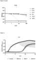
Figure 1 . pH stability of bacteriophage F12A and F13. Incubation for 1h at pH of 1, 4.5, 7.4, and 8. The bars represent the median and the interquartile range. Discontinued line denotes limit detection of quantification (10 PFU/ml). -
Figure 2 . Phage temperature stability. Incubation for 1 h, 24 h and 168 h at temperatures of - 80°C, -20°C, 4°C, 21°C, 37°C and 60°C. The bars represent the median and the interquartile range. Discontinued line denotes limit detection of quantification (10 PFU/ml). -
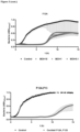
Figure 3 . Bacteriophage Inhibition Assay. Inhibition of KP5 standard clinical strain after treatment with individual phages at MOI (10, 1 and 0.1). Grey bars represent the interquartile range. -
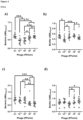
Figure 4 .F12A and F13 effect on KP5 clinical strain biofilm growth in both planktonic state (a, c) and biofilm form (c, d) at 6h (a-b) and 24h (c-d). The bars represent the median and the interquartile range. ∗: p-value<0.05, ∗∗: p-value<0.01, ∗∗∗: p-value<0.001, ∗∗∗∗: p-value<0.0001 for Dunn's test pairwise test with a Benjamini-Hochberg's procedure. - The present invention is illustrated by means of the Examples set below without the intention of limiting its scope of protection.
- Klebsiella pneumoniae clinical strain KP5 and other clinical isolates were obtained from patients (n=58) and donated by the Department of Microbiology, Hospital Universitario Fundación Jimenez Diaz (HUFJD). The presence of K. pneumoniae on clinical isolates was confirmed using MALDI-TOF (Bruker, VIC, Australia). The K. pneumoniae clinical isolates were stored in Difco™ skimmed milk (East Rutherford, NJ, United States) at -80 °C. The clinical isolates were plated from frozen skimmed milk stocks onto Mueller Hinton E agar 90 mm plates and broth cultures were grown in tryptic soy broth (TSB) (BioMerieux, France). Agar plates and broth cultures were incubated at 37 °C.
- The phages isolated against K. pneumoniae strain KP5 were obtained from HUFJD sewage (Madrid, Spain). Samples of 50 ml were centrifuged at 4000 x g for 10 min to remove cellular debris and faecal matter. The supernatant was filtered using 0.45 µm and 0.22 µm filters to remove the bacteria and debris. An amount of 100 µl of filtered solution was combined with 100 µl of the KP5 overnight culture and 3 mL of agar 0.2% (W/V) and plated on MHE plates via the double-layer agar method. Following overnight incubation, the formation of plaques confirmed the presence of phage. Individual plaques with different morphology were picked using pipette tips and placed into 1.5 ml Eppendorf tubes (Eppendorf, Hamburg, Germany) containing sodium magnesium buffer (SM; 100 mM sodium chloride (Panreac Química, Illinois, United States); 10 mM magnesium sulfate (Thermo Fisher Scientific, Waltham, MA, USA); 10 mM calcium chloride (Thermo Fisher Scientific, Waltham, MA, USA);50 mM Tris HCl (Sigma-Aldrich, Castle Hill, NSW, Australia), pH 7.5), and vortexed vigorously before storage at 4°C.
- A two-step propagation was applied to amplify and purify the isolated phage. For small-scale amplification of phage, 100 µl of KP5 overnight culture was added into 10 ml TSB and incubated at 37°C with 200 rpm shaking for 1.5 h to reach exponential growth. An amount of 100 µl of phage in SM buffer, 150 µl of MgSO4 and CaCl2 (200 mM) was added into the coculture and incubated for 4-5h at 37°C with 200 rpm shaking. The supernatant containing phage was harvested after centrifugation (4000 x g, 15 min) and filtration (0.45 µm and 0.22 µm syringe filter). Phage titration was performed. The small-scale amplification was applied according to the prior art.
- For large-scale amplification of phage, 500 µl of the KP5 overnight culture was incubated with 50 ml of TSB for 20 min. After incubation, 100 µl of phage and 2.5 ml of MgSO4 and CaCl2 were then added into the coculture and incubated overnight at 37°C with 200 rpm shaking. The phage lysis was harvested as described above. The large-scale amplification was applied according to the prior art.
- The phage titer was determined via a double-layer agar method. Briefly, 100 µl of the KP5 overnight broth culture was added to 3 ml of agar (0.2% (W/V)) and overlaid onto a MHE plate. Phages were serially diluted in SM buffer. The plates were incubated at 37°C overnight. The phage titer was counted and calculated.
- The stability of K. pneumoniae phage was tested against a wide pH (1-8) and temperature (-80°C to 60°C) range using a working stock in TSB broth with an initial phage titer of around 108 PFU·ml-1. Briefly, 100 µl working stock of each phage was suspended in 1 ml SM buffer previously adjusted with 1M NaOH or HCl (Sigma-Aldrich, Castle Hill, NSW, Australia) to yield a pH range of 1-8. The samples were incubated at room temperature for 1 h. The pH stability testing was studied according to the methods described in the prior art. For thermal stability, 100 µl of working stock phage was suspended in 1 ml SM buffer and incubated at - 80°C, -20°C, 4°C, 21°C, 37°C, and 60°C for 1, 24 and 168 h (1 week). The phage stability for each experiment was determined by measuring the phage titration. The thermal stability testing was studied according to the methods described in the prior art.
- Host range of F13 phage was determined against the different clinical Klebsiella pneumoniae strains obtained from Hospital Universitario Fundación Jiménez Diaz (HUFJD) using spot testing. Briefly, 3 µl of the purified phage suspension (108 PFU/ml) was spotted onto the surface of double-layer agar plate previously inoculated with the tested strains. After adsorption of phage suspension, plates were incubated overnight at 37°C. Host range was determined by plaque formation. Three replicates were tested for each bacterial strain. The host range test was applied according to methodology described in the prior art.
- The infectivity profile of the K. pneumoniae bacteriophages was assessed at multiplicity of infection (MOI) 0.1, 1 and 10 using inhibition assays in liquid. Briefly, the McFarland inoculation standards (0.5±0.02) of each clinical isolate (~108 UFC·ml-1) were prepared and the required volume of phage stock solution (108PFU·ml-1) was added to the corresponding suspension to achieve MOI 0.1, 1 and 10. The samples were incubated at 37°C with shaking orbital amplitude of 5 mm. Every 5min for 15 h the OD595nm value was measured.
- The isolated phage effect on K. pneumoniae biofilm was determined using a modified methodology previously described in the prior art. Briefly, biofilm formation on the bottom of a MicroWell 96-well flat-bottom plate (Thermo Fisher Scientific, Waltham, MA, USA) was induced by inoculating 100 µL of MHB containing ~107 CFU·ml-1 of bacteria per well and the plate was incubated at 37°C and 5% CO2 for at least 18 h. The next day, the wells were aspirated, and biofilm formation was treated with bacteriophages at different concentrations (107,106, 105 and 104 pfu/mL) in order to study the inhibition of biofilm by phages. This study was done at two different incubation times with the treatment: 6h and 24h. After 6h and 24h of treatment with phages, 100 µl from the biofilm formation were transferred to a new 96-well untreated microtiter plate in order to separate planktonic bacteria from biofilm. From the biofilm plate, wells were aspirated, and each well was washed one time with 200 µl of physiological serum (0.9% W/V). Planktonic bacterial viability was determined by addition of 20 µl of 5 mg/mL of 3-(4,5-dimethylthiazol-2-yl)-2,5- diphenyltetrazolium bromide (MTT) (Sigma Aldrich, Merck, Darmstadt, Germany) (2) while biofilm viability was determined by adding 1mL of MTT to a commercial 9mL TSB tube and incubating for 1 h at 37°C and 5% CO2. The absorbance was measured at 570 nm for MTT using a microplate reader (TECAN). This experiment was performed in duplicate.
- Antibacterial synergistic activity for F12A and F13 phages was assigned by modified broth microdilution method in flat-bottomed 96 multi-well microplates (Thermo-Fischer). Experimental conditions were designed for antibiotic-phage mix and were tested for colistin, polymyxin B, doripenem, ciprofloxacin and bacterial control at the final volume of 200 µl. Briefly, the culture was allowed to grow until an OD600 of 0.44 reached 4×10⁷ CFU. Then 50 µl of antibiotic at different concentrations was added to each well containing 50 µl of Mueller Hinton Broth II (Millipore, Sigma Aldrich, Merck) supplemented with 10 mM CaCl2 and 10 mM MgSO4. In addition, 50 µl of the phage dilutions (107 PFU) were added to the suspension and 100 µl of bacterial suspension was added to the mix wells. Plates were incubated at 37°C overnight and FICI was calculated according to bacterial growth.
- Data distribution was evaluated using Shapiro-Wilk or Kolgomorov-Smirnov statistics. Descriptive statistics are cited as median and interquartile range (non-normal distribution) for each variable that were calculated. Non-parametric Mann-Whitney test considering equality of variances was used to compare two groups and non-parametric Kruskal-Wallis's test was used to compared more than two groups. To determine the effect of temperature on the phage viability data was analyzed using linear regression. Bacteriophage inhibition of bacterial biofilm was analyzed by Dunn's pairwise test with a Benjamini-Hochberg's procedure. The possible relation between the bacterial biofilm or planktonic concentration and the concentration of phage was determined by using Spearman rank correlation coefficient. Significance level was established at α=0.05.
- All statistical analysis was performed using R (R Core Team, 2017) with R commander, except for linear regressions that were carried out using GraphPad Prism v.8 (GraphPad Prism, version 8.0.1 (86); Windows Version by Software MacKiev © 2020-2018 GraphPad Software, LLC.; San Diego, CA, USA) and STATA statistical software, release 11 (StataCorp, 2009, StataCorp LP., Texas, USA).
- Departing from these materials and methods, and the common general knowledge, the present invention could be reproduced by the person skilled in the art departing from the genetic material of the bacteriophages (for instance SEQ ID NO: 1). For instance, the reference [Jiang S, Tang Y, Xiang L, Zhu X, Cai Z, Li L, Chen Y, Chen P, Feng Y, Lin X, Li G, SharifJ, Dai J. Efficient de novo assembly and modification of large DNA fragments. Sci China Life Sci. 2021 Dec 16. doi: 10.1007/s11427-021-2029-0. Epub ahead of print. PMID: 34939159 ] explains how to synthesise de novo long DNA from sequences. On the other hand, the reference [Diana P. Pires et al., 2021.Designing P. aeruginosa synthetic phages with reduced genomes. Scientific Reports | (2021) 11:2164 | https://doi.org/10.1038/s41598-021-81580-2] explains how phages can be synthesized and a general scheme is provided therein (see
Figure 2 of this paper). - Phages were tested against a wide pH range (
pH 1, 4.5, 7.4 and 8) after 1h incubation period, to establish their stability under neutral, acidic and alkaline conditions (Fig. 1 ). After 1 h incubation atpH 1, no plaques were detected in 100 ul of sample, which suggested that the bacteriophage is unstable at this pH (p-value<0.05). The rest of pH tested did not produce logarithmic reductions after 1 hour of treatment in F12A and F13. - Phages were also tested after treatments with different temperatures: -80°C, -20°C, 4°C, 21°C and 37°C and 60°C (
Fig.2 ). F12A and F13 were stable at -80°C, -20°C, 4°C and 21°C after seven days of exposition. A certain reduction of phage viability was observed after seven days at 37°C. The main reduction of viability was observed at 60°C after 24 hours. - The host range against 58 K. pneumoniae clinical isolates for phages F7, F8, F12A, F12B, F13 and F14 showed 10 susceptible (+) strains including a pan-resistant strain (Table 1). All tested strains were multi-drug resistant (MDR) K. pneumoniae strains including KP5 strain.
Table 1 Strain Resistance I/C Origin Susceptible to 6 phages KP5 MDR I Unknown + KP21 MDR I Urine + KP22 MDR I Urine + KP29 MDR I Urine + KP32 MDR I Urine + KP52 MDR I Urine + KP74 MDR I Urine + A7 MDR I Wound + A29 MDR I Urine + KP4940164 MDR I Bronchial + Summary of phages Host Range. K. pneumoniae strains susceptible (+) to 6 phages against K.pneumoniae based on the degree of lysis on the bacterial lawn by counting the spots. I: Infection; C: colonization. - The phages planktonic infective properties were determined through inhibition assays. The phage inhibition of KP5 clinical isolate was assessed for 15 h when infected at
MOI Fig. 3 ). - The concentration of KP5 in planktonic state decreased by 35% at 1010 PFU/ml concentration of bacteriophage F12A (p-value<0.05) after 6h of treatment (Fig. F12A 4a), and was reduced by 32% at 108 PFU/mL after 24h of treatment (Fig. F12A 4b). Biofilm formation was reduced by 28% at 6 h and 1010 PFU/mL of F12A (Fig. F12A 4c).
- The concentration of KP5 bacteria in planktonic state and biofilm formation showed a significant decrease by 15% (Fig. F13 4a) and 22% (Fig. F13 4b), respectively, with 106 PFU/ml of F13 (p-value<0.05) after 6 h of treatment. The concentration of planktonic bacteria was reduced by 60% at 106 PFU/ml after 24h of treatment (
Fig. 4c ) and biofilm formation was reduced 35% after 24 h of treatment with 106 PFU/ml of F13 (Fig. 4d ). - Phage-antibiotic checkerboard assays showed synergy between F13 and polymyxin B (FIC index 0.135) and an additive effect with colistin (FIC index 0.501) (Table 2). Polymyxin B was very effective against KP5, 8 µg/ml of polymyxin B was able to lyse the cells in combination with F13 at phage titer of 105 and 106 PFU/ml demonstrating synergistic effect. F12A was very effective in combination with polymyxin B (FIC index 0.26), doripenem (FIC index 0.251), colistin (FIC index 0.015) and ciprofloxacin (FIC index 0.35) at phage titer of 108 PFU/ml.
Table 2 Antibiotic FIC index F12A F13 Effect Polymyxin B 0.26 0.135 Synergy / Synergy Colistin 0.015 0.501 Synergy / Addition Ciprofloxacin 0.35 Synergy Doripenem 0.251 Synergy Summary of synergistic effect between F12A, F13 and antibiotics against KP5 K. pneumoniae strain.
Claims (10)
- Bacteriophage, characterized by their specificity for Klebsiella pneumoniae strains, selected from a bacteriophage deposited at the German Collection of Microorganisms and Cell Cultures GmbH (DSMZ) with deposit number DSM34222; a bacteriophage characterized by comprising a nucleic acid consisting of SEQ ID NO: 1; or a bacteriophage comprising a nucleic acid having at least 95% of identity with the nucleic acid of the bacteriophage DSM 34222 or the SEQ ID NO: 1.
- Bacteriophage, according to claim 1, deposited at the German Collection of Microorganisms and Cell Cultures GmbH (DSMZ) with deposit number DSM34222.
- Bacteriophage, according to any of the claims 1 or 2, for use as a medicament.
- Bacteriophage for use, according to claim 3, in the prevention or treatment of a bacterial infection caused by Klebsiella pneumoniae.
- Bacteriophage for use, according to any of the claims 3 or 4, in combination with an antibiotic, preferably selected from: polymyxin B and/or colistin.
- Pharmaceutical composition comprising a bacteriophage according to claims 1 or 2 and, optionally, pharmaceutically acceptable excipients or carriers.
- Pharmaceutical composition, according to claim 6, further comprising an antibiotic preferably selected from: polymyxin B and/or colistin.
- Pharmaceutical composition according to claims 6 or 7 for use as a medicament.
- Pharmaceutical composition for use, according to claim 8, in the prevention or treatment of a bacterial infection caused by Klebsiella pneumoniae.
- A method of detecting the presence of Klebsiella pneumoniaein an in vitro sample, comprising contacting said sample with a bacteriophage according to claims1 or 2, and determining whether said bacteriophages kill bacteria in said sample.
Priority Applications (1)
| Application Number | Priority Date | Filing Date | Title |
|---|---|---|---|
| EP22382362.6A EP4265264A1 (en) | 2022-04-19 | 2022-04-19 | Bacteriophages suitable for treating a bacterial infection caused by klebsiella pneumoniae |
Applications Claiming Priority (1)
| Application Number | Priority Date | Filing Date | Title |
|---|---|---|---|
| EP22382362.6A EP4265264A1 (en) | 2022-04-19 | 2022-04-19 | Bacteriophages suitable for treating a bacterial infection caused by klebsiella pneumoniae |
Publications (1)
| Publication Number | Publication Date |
|---|---|
| EP4265264A1 true EP4265264A1 (en) | 2023-10-25 |
Family
ID=81344413
Family Applications (1)
| Application Number | Title | Priority Date | Filing Date |
|---|---|---|---|
| EP22382362.6A Withdrawn EP4265264A1 (en) | 2022-04-19 | 2022-04-19 | Bacteriophages suitable for treating a bacterial infection caused by klebsiella pneumoniae |
Country Status (1)
| Country | Link |
|---|---|
| EP (1) | EP4265264A1 (en) |
Citations (4)
| Publication number | Priority date | Publication date | Assignee | Title |
|---|---|---|---|---|
| WO2017081709A1 (en) * | 2015-11-09 | 2017-05-18 | Universita' Degli Studi Di Siena | Bacteriophage able to lyse klebsiella pneumoniae expressing a cpskkbo-4 capsular polysaccharide and related medical uses thereof |
| WO2018106135A2 (en) * | 2016-12-05 | 2018-06-14 | Technophage, Investigação E Desenvolvimento Em Biotecnologia, Sa | Bacteriophage compositions comprising respiratory antibacterial phages and methods of use thereof |
| US20210187046A1 (en) * | 2019-12-19 | 2021-06-24 | Technophage, Investigação E Desenvolvimento Em Biotecnologia, Sa | Cocktail compositions comprising respiratory antibacterial phages and methods of use thereof |
| EP3928782A1 (en) * | 2020-03-20 | 2021-12-29 | Phagegroup LLC | Bacteriophage based therapy |
-
2022
- 2022-04-19 EP EP22382362.6A patent/EP4265264A1/en not_active Withdrawn
Patent Citations (4)
| Publication number | Priority date | Publication date | Assignee | Title |
|---|---|---|---|---|
| WO2017081709A1 (en) * | 2015-11-09 | 2017-05-18 | Universita' Degli Studi Di Siena | Bacteriophage able to lyse klebsiella pneumoniae expressing a cpskkbo-4 capsular polysaccharide and related medical uses thereof |
| WO2018106135A2 (en) * | 2016-12-05 | 2018-06-14 | Technophage, Investigação E Desenvolvimento Em Biotecnologia, Sa | Bacteriophage compositions comprising respiratory antibacterial phages and methods of use thereof |
| US20210187046A1 (en) * | 2019-12-19 | 2021-06-24 | Technophage, Investigação E Desenvolvimento Em Biotecnologia, Sa | Cocktail compositions comprising respiratory antibacterial phages and methods of use thereof |
| EP3928782A1 (en) * | 2020-03-20 | 2021-12-29 | Phagegroup LLC | Bacteriophage based therapy |
Non-Patent Citations (5)
| Title |
|---|
| DIANA P. PIRES ET AL.: "Designing P. aeruginosa synthetic phages with reduced genomes", SCIENTIFIC REPORTS, vol. 11, 2021, pages 2164, Retrieved from the Internet <URL:https://doi.org/10.1038/s41598-021-81580-2> |
| JIANG STANG YXIANG LZHU XCAI ZLI LCHEN YCHEN PFENG YLIN X: "Efficient de novo assembly and modification of large DNA fragments", SCI CHINA LIFE SCI, 16 December 2021 (2021-12-16) |
| KORTRIGHT KAITLYN E ET AL: "Phage Therapy: A Renewed Approach to Combat Antibiotic-Resistant Bacteria", CELL HOST & MICROBE, vol. 25, no. 2, 13 February 2019 (2019-02-13), pages 219 - 232, XP085602331, ISSN: 1931-3128, DOI: 10.1016/J.CHOM.2019.01.014 * |
| LI MIN ET AL: "Isolation and Characterization of Novel Lytic Bacteriophages Infecting Epidemic Carbapenem-Resistant Klebsiella pneumoniae Strains", FRONTIERS IN MICROBIOLOGY, vol. 11, 21 July 2020 (2020-07-21), XP055959281, Retrieved from the Internet <URL:https://com-mendeley-prod-publicsharing-pdfstore.s3.eu-west-1.amazonaws.com/7775-PUBMED/10.3389/fmicb.2020.01554/fmicb_11_01554_pdf.pdf?X-Amz-Security-Token=IQoJb3JpZ2luX2VjEKj//////////wEaCWV1LXdlc3QtMSJGMEQCIAT3tB06atxgkmyOrR5wCLcN2Uw019FcAqejhVO4Q8tdAiBlJZ0rWugrSksKFG2VrrNlVe0qlYe93neS0/eJRo4cECq> DOI: 10.3389/fmicb.2020.01554 * |
| MOUSAVI SEYED MOHAMMAD ET AL: "Bacteriophage as a Novel Therapeutic Weapon for Killing Colistin-Resistant Multi-Drug-Resistant and Extensively Drug-Resistant Gram-Negative Bacteria", CURRENT MICROBIOLOGY, SPRINGER-VERLAG, NEW YORK, vol. 78, no. 12, 11 October 2021 (2021-10-11), pages 4023 - 4036, XP037620127, ISSN: 0343-8651, [retrieved on 20211011], DOI: 10.1007/S00284-021-02662-Y * |
Similar Documents
| Publication | Publication Date | Title |
|---|---|---|
| US10357522B2 (en) | Bacteriophage compositions and methods of selection of components against specific bacteria | |
| CN104140957B (en) | Cleavable multiple-drug resistant pseudomonas aeruginosa bacteriophage and application thereof in infection treatment | |
| Moradpour et al. | Synergistic bactericidal activity of a naturally isolated phage and ampicillin against urinary tract infecting Escherichia coli O157 | |
| US12268720B2 (en) | Bacteriophage for treatment and prevention of E. coli and B. fragilis infections | |
| US20250170195A1 (en) | Bacteriophage compositions for treating pseudomonas infections | |
| JP7812496B2 (en) | Cocktail compositions containing respiratory antibacterial phages and methods of using the same | |
| Winzig et al. | Inhaled bacteriophage therapy for multi-drug resistant Achromobacter | |
| US11826391B2 (en) | Antibacterial formulation comprising a mixture of bacteriophages; use and method for preventing or treating diseases caused by salmonella spp. in farm animals by oral administration of the formulation | |
| Elshamy et al. | In vitro meropenem/antibiotic and meropenem/bacteriophage combinations against carbapenem-resistant gram-negative uropathogens | |
| US20230227789A1 (en) | Methods for treating bacterial infections | |
| CN115029322A (en) | A multidrug-resistant Klebsiella pneumoniae phage and its application | |
| Jaglan et al. | Isolation and characterization of a novel Escherichia phage phiEco273 and its efficacy as an antibiofilm agent | |
| Ma et al. | vB_PaeP_PZH3, a novel bacteriophage for the treatment of MDR Pseudomonas aeruginosa in a mouse wound infection model | |
| Karamoddini et al. | Antibacterial Efficacy of Lytic Bacteriophages against Antibiotic‐Resistant Klebsiella Species | |
| Singh et al. | In vitro efficacy of bacteriophage and colistin combinations in eradicating biofilm formed by colistin-resistant Enterobacter cloacae complex. | |
| US20220184155A1 (en) | Bacteriophages for inhibiting bacteria | |
| Peters | Isolation and characterization of novel cocktail phages of multidrug-resistant (MDR) Acinetobacter baumannii | |
| EP4265263A1 (en) | Bacteriophage suitable for treating a bacterial infection caused by pseudomonas aeruginosa | |
| KR20230038491A (en) | Synergistic compositions of phages and methods for their formation | |
| Nair et al. | The prospects of a highly virulent biofilm dispersing bacteriophage isolated from environment for controlling Enterobacter cloacae in clinical medicine | |
| US20250002869A1 (en) | Burkholderia pseudomallei (Bp) phage vB BpP HN01 and use thereof | |
| WO2025262649A1 (en) | Compositions comprising antibacterial bacteriophages and methods of use thereof | |
| Arnaud | Optimizing high-throughput detection of temperate phages that infect Enterococcus spp. and Staphylococcus spp. | |
| Mohammed et al. | Isolation of various local Bacteriophages via Simple Methods and their Effects against Multidrug Resistance Bacterial Isolates | |
| Gomes | Antimicrobial activity of bacteriophages towards Pseudomonas aeruginosa isolates obtained from companion animals |
Legal Events
| Date | Code | Title | Description |
|---|---|---|---|
| STAA | Information on the status of an ep patent application or granted ep patent |
Free format text: STATUS: THE APPLICATION HAS BEEN PUBLISHED |
|
| PUAI | Public reference made under article 153(3) epc to a published international application that has entered the european phase |
Free format text: ORIGINAL CODE: 0009012 |
|
| AK | Designated contracting states |
Kind code of ref document: A1 Designated state(s): AL AT BE BG CH CY CZ DE DK EE ES FI FR GB GR HR HU IE IS IT LI LT LU LV MC MK MT NL NO PL PT RO RS SE SI SK SM TR |
|
| STAA | Information on the status of an ep patent application or granted ep patent |
Free format text: STATUS: REQUEST FOR EXAMINATION WAS MADE |
|
| 17P | Request for examination filed |
Effective date: 20240424 |
|
| RBV | Designated contracting states (corrected) |
Designated state(s): AL AT BE BG CH CY CZ DE DK EE ES FI FR GB GR HR HU IE IS IT LI LT LU LV MC MK MT NL NO PL PT RO RS SE SI SK SM TR |
|
| STAA | Information on the status of an ep patent application or granted ep patent |
Free format text: STATUS: EXAMINATION IS IN PROGRESS |
|
| 17Q | First examination report despatched |
Effective date: 20250110 |
|
| STAA | Information on the status of an ep patent application or granted ep patent |
Free format text: STATUS: THE APPLICATION IS DEEMED TO BE WITHDRAWN |
|
| 18D | Application deemed to be withdrawn |
Effective date: 20250513 |