EP4265195A1 - Ultrasonic diagnostic apparatus and control method for ultrasonic diagnostic apparatus - Google Patents
Ultrasonic diagnostic apparatus and control method for ultrasonic diagnostic apparatus Download PDFInfo
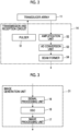
- Publication number
- EP4265195A1 EP4265195A1 EP21906158.7A EP21906158A EP4265195A1 EP 4265195 A1 EP4265195 A1 EP 4265195A1 EP 21906158 A EP21906158 A EP 21906158A EP 4265195 A1 EP4265195 A1 EP 4265195A1
- Authority
- EP
- European Patent Office
- Prior art keywords
- blood vessel
- artery
- vein
- probability
- ultrasound
- Prior art date
- Legal status (The legal status is an assumption and is not a legal conclusion. Google has not performed a legal analysis and makes no representation as to the accuracy of the status listed.)
- Granted
Links
Images



Classifications
-
- A—HUMAN NECESSITIES
- A61—MEDICAL OR VETERINARY SCIENCE; HYGIENE
- A61B—DIAGNOSIS; SURGERY; IDENTIFICATION
- A61B8/00—Diagnosis using ultrasonic, sonic or infrasonic waves
- A61B8/08—Clinical applications
- A61B8/0833—Clinical applications involving detecting or locating foreign bodies or organic structures
- A61B8/085—Clinical applications involving detecting or locating foreign bodies or organic structures for locating body or organic structures, e.g. tumours, calculi, blood vessels, nodules
-
- A—HUMAN NECESSITIES
- A61—MEDICAL OR VETERINARY SCIENCE; HYGIENE
- A61B—DIAGNOSIS; SURGERY; IDENTIFICATION
- A61B8/00—Diagnosis using ultrasonic, sonic or infrasonic waves
- A61B8/08—Clinical applications
- A61B8/0891—Clinical applications for diagnosis of blood vessels
-
- A—HUMAN NECESSITIES
- A61—MEDICAL OR VETERINARY SCIENCE; HYGIENE
- A61B—DIAGNOSIS; SURGERY; IDENTIFICATION
- A61B8/00—Diagnosis using ultrasonic, sonic or infrasonic waves
- A61B8/46—Ultrasonic, sonic or infrasonic diagnostic devices with special arrangements for interfacing with the operator or the patient
- A61B8/461—Displaying means of special interest
- A61B8/463—Displaying means of special interest characterised by displaying multiple images or images and diagnostic data on one display
-
- A—HUMAN NECESSITIES
- A61—MEDICAL OR VETERINARY SCIENCE; HYGIENE
- A61B—DIAGNOSIS; SURGERY; IDENTIFICATION
- A61B8/00—Diagnosis using ultrasonic, sonic or infrasonic waves
- A61B8/48—Diagnostic techniques
- A61B8/488—Diagnostic techniques involving Doppler signals
-
- A—HUMAN NECESSITIES
- A61—MEDICAL OR VETERINARY SCIENCE; HYGIENE
- A61B—DIAGNOSIS; SURGERY; IDENTIFICATION
- A61B8/00—Diagnosis using ultrasonic, sonic or infrasonic waves
- A61B8/52—Devices using data or image processing specially adapted for diagnosis using ultrasonic, sonic or infrasonic waves
- A61B8/5215—Devices using data or image processing specially adapted for diagnosis using ultrasonic, sonic or infrasonic waves involving processing of medical diagnostic data
- A61B8/5223—Devices using data or image processing specially adapted for diagnosis using ultrasonic, sonic or infrasonic waves involving processing of medical diagnostic data for extracting a diagnostic or physiological parameter from medical diagnostic data
-
- A—HUMAN NECESSITIES
- A61—MEDICAL OR VETERINARY SCIENCE; HYGIENE
- A61B—DIAGNOSIS; SURGERY; IDENTIFICATION
- A61B8/00—Diagnosis using ultrasonic, sonic or infrasonic waves
- A61B8/06—Measuring blood flow
-
- A—HUMAN NECESSITIES
- A61—MEDICAL OR VETERINARY SCIENCE; HYGIENE
- A61B—DIAGNOSIS; SURGERY; IDENTIFICATION
- A61B8/00—Diagnosis using ultrasonic, sonic or infrasonic waves
- A61B8/48—Diagnostic techniques
- A61B8/485—Diagnostic techniques involving measuring strain or elastic properties
Definitions
- the present invention relates to an ultrasound diagnostic apparatus, and a control method of the ultrasound diagnostic apparatus which have a function of determining a type (an artery or a vein) of a blood vessel.
- an ultrasound image is observed by using an ultrasound diagnostic apparatus to check the type, size, position, depth, and traveling of the blood vessel, and the presence or absence of an obstacle such as nerves.
- an ultrasound diagnostic apparatus to check the type, size, position, depth, and traveling of the blood vessel, and the presence or absence of an obstacle such as nerves.
- JP2008-545502A and JP2018-202147A as the documents in the related art that are references for the present invention.
- JP2008-545502A discloses that arteries and veins are distinguished by applying a color Doppler technique to detect the direction and pulsation of the blood flow in the arteries and the veins.
- JP2008-545502A discloses that arteries and veins are distinguished by using the fact that the veins collapse in a case where a mechanical pressure is applied to a tissue with blood vessels, or by measuring and analyzing the absorption of two wavelengths on the basis of the oxygen content in the blood.
- JP2018-202147A discloses that arteries and veins are decided by analyzing image data using anatomical prior information indicating typical positions of the arteries and the veins.
- JP2008-545502A in a case where the type of the blood vessel is determined on the basis of data other than the image data, additional work is always required to determine the type of the blood vessel, which causes the deterioration of the usability.
- An object of the present invention is to provide an ultrasound diagnostic apparatus and a control method of the ultrasound diagnostic apparatus which can prevent the type of the blood vessel from being erroneously determined, and determine the type of the blood vessel without the deterioration of the usability.
- an aspect of the present invention provides an ultrasound diagnostic apparatus including an ultrasound probe; an image generation unit that generates an ultrasound image from a reception signal obtained by performing transmission and reception of an ultrasound beam with respect to a subject using the ultrasound probe; a probability calculation unit that calculates each of a probability that a blood vessel in the ultrasound image is an artery and a probability that the blood vessel is a vein by analyzing the ultrasound image; a blood vessel determination unit that determines whether the blood vessel is the artery or the vein or whether or not confirmation processing for confirming whether the blood vessel is the artery or the vein is required on the basis of the probability that the blood vessel is the artery and the probability that the blood vessel is the vein; and a blood vessel confirmation unit that executes the confirmation processing in a case where it is determined that the confirmation processing is required, and confirms whether the blood vessel is the artery or the vein on the basis of an execution result of the confirmation processing.
- the probability calculation unit detects a blood vessel region with a predetermined shape, including a blood vessel in the ultrasound image, in a case of calculating a probability of being the artery and a probability of being the vein.
- the blood vessel region is an artery region including the artery or a vein region including the vein
- the probability calculation unit detects the artery region including the blood vessel in the ultrasound image, calculates a probability that the blood vessel in the artery region is the artery, detects the vein region including the same blood vessel, and calculates a probability that the same blood vessel in the vein region is the vein.
- the probability calculation unit detects a single blood vessel region with the predetermined shape, including a blood vessel in the ultrasound image, and further calculates a probability that the blood vessel in the single blood vessel region is the artery and a probability that the blood vessel in the single blood vessel region is the vein.
- the blood vessel determination unit determines that the blood vessel is one of the artery and the vein, and in a second case other than the first case, the blood vessel determination unit determines that the confirmation processing is required.
- the probability calculation unit calculates, for each pixel of the ultrasound image, each of a probability that the pixel is a pixel of the artery, a probability that the pixel is a pixel of the vein, and a probability that the pixel is a pixel of background other than the blood vessel.
- the blood vessel determination unit decides, for each pixel of the ultrasound image, whether the pixel is the pixel of the artery, the pixel is the pixel of the vein, or the pixel is the pixel of the background on the basis of the probability that the pixel is the pixel of the artery, the probability that the pixel is the pixel of the vein, and the probability that the pixel is the pixel of the background, decides a blood vessel region including the blood vessel on the basis of the pixel of the artery and the pixel of the vein, determines that the blood vessel is one of the artery and the vein in a case where a ratio of one of an area of the pixel of the artery and an area of the pixel of the vein to an area of the blood vessel in the blood vessel region is equal to or greater than a third threshold value, and determines that the confirmation processing is required in a case where the ratio is less than the third threshold value.
- the blood vessel confirmation unit prompts a user to press an examination location of the subject with the ultrasound probe, calculates an aspect ratio of a cross section of the blood vessel on the basis of an ultrasound image generated while the examination location is pressed with the ultrasound probe, and confirms whether the blood vessel is the artery or the vein on the basis of the aspect ratio of the cross section of the blood vessel.
- the ultrasound probe has a pressure sensor, and the blood vessel confirmation unit determines whether or not the examination location is pressed on the basis of a pressure detected by the pressure sensor.
- the blood vessel confirmation unit detects motion in the examination location by analyzing the ultrasound images of a plurality of frames, and determines whether or not the examination location is pressed on the basis of the motion in the examination location.
- the blood vessel confirmation unit determines presence or absence of a change in the aspect ratio of the cross section of the blood vessel only for a predetermined period of time after prompting the user to press the examination location with the ultrasound probe, and determines whether or not the examination location is pressed on the basis of the presence or absence of the change in the aspect ratio of the blood vessel in the predetermined period of time.
- the blood vessel confirmation unit prompts a user to incline the ultrasound probe at an examination location of the subject, and confirms whether the blood vessel is the artery or the vein on the basis of a direction of a blood flow in an ultrasound image generated while the ultrasound probe is inclined at the examination location in a color Doppler mode.
- the blood vessel confirmation unit searches for an ultrasound image of a frame in which a probability that the same blood vessel is the artery or a probability that the same blood vessel is the vein is equal to or greater than a fourth threshold value among ultrasound images of past frames, and confirms whether the blood vessel is the artery or the vein on the basis of the probability that the same blood vessel in the ultrasound image of the searched frame is the artery or the probability that the same blood vessel is the vein.
- the blood vessel confirmation unit detects the blood vessel in ultrasound images of adjacent preceding and succeeding frames, and determines that the blood vessels are the same in a case where an overlapping ratio of the blood vessels in the ultrasound images of the preceding and succeeding frames is equal to or greater than the fourth threshold value.
- the ultrasound diagnostic apparatus further includes a monitor; and a display control unit that superimposes a graphic including the blood vessel on the ultrasound image to be displayed on the monitor on the basis of at least one of a determination result on whether the blood vessel is the artery or the vein or a confirmation result on whether the blood vessel is the artery or the vein.
- the display control unit displays each of the probability that the blood vessel is the artery and the probability that the blood vessel is the vein on the monitor in addition to the graphic.
- the display control unit displays the same graphic for the same blood vessel on the monitor once whether the blood vessel is the artery or the vein has been decided.
- the display control unit displays characters, blood vessel, on the monitor in addition to the graphic in a case where deciding whether the blood vessel is the artery or the vein is not possible.
- Another aspect of the present invention provides a control method of an ultrasound diagnostic apparatus, the control method including generating an ultrasound image from a reception signal obtained by performing transmission and reception of an ultrasound beam with respect to a subject using an ultrasound probe; calculating a probability that a blood vessel in the ultrasound image is an artery and a probability that the blood vessel is a vein by analyzing the ultrasound image; determining whether the blood vessel is the artery or the vein or whether or not confirmation processing for confirming whether the blood vessel is the artery or the vein is required on the basis of the probability that the blood vessel is the artery and the probability that the blood vessel is the vein; and executing the confirmation processing in a case where it is determined that the confirmation processing is required, and confirming whether the blood vessel is the artery or the vein on the basis of an execution result of the confirmation processing.
- the type of the blood vessel is determined on the basis of the probability that the blood vessel is an artery and the probability that the blood vessel is a vein as described above, and in a case where the type of the blood vessel cannot be determined, the confirmation processing is executed to confirm whether the blood vessel is an artery or a vein. Therefore, it is possible to prevent the type of the blood vessel from being erroneously determined, and to greatly improve the determination accuracy of the type of the blood vessel. Further, in the present invention, since the confirmation processing is executed only in a case where the type of the blood vessel cannot be determined, it is possible to greatly suppress the deterioration of the usability.
- Fig. 1 is a block diagram of an embodiment illustrating a configuration of an ultrasound diagnostic apparatus of the present invention.
- An ultrasound diagnostic apparatus 20 illustrated in Fig. 1 includes an ultrasound probe 1, and an apparatus main body 3 connected to the ultrasound probe 1.
- the ultrasound probe 1 scans an examination location of a subject using an ultrasound beam, and outputs a sound ray signal corresponding to an ultrasound image of the examination location.
- the ultrasound probe 1 includes a transducer array 11, and a transmission and reception circuit 14.
- the transducer array 11 and the transmission and reception circuit 14 are bidirectionally connected to each other.
- an apparatus control unit 36, which will be described later, of the apparatus main body 3 is connected to the transmission and reception circuit 14.
- the transducer array 11 has a plurality of ultrasonic transducers arranged in a one-dimensional or two-dimensional manner. According to a drive signal supplied from the transmission and reception circuit 14, each of the transducers transmits an ultrasonic wave and receives a reflected wave from the subject to output an analog reception signal.
- each transducer is formed by using an element in which electrodes are formed at both ends of a piezoelectric body consisting of piezoelectric ceramic represented by lead zirconate titanate (PZT), a polymer piezoelectric element represented by poly vinylidene di fluoride (PVDF), piezoelectric single crystal represented by lead magnesium niobate-lead titanate (PMN-PT), or the like.
- PZT lead zirconate titanate
- PVDF polymer piezoelectric element represented by poly vinylidene di fluoride
- PMN-PT lead magnesium niobate-lead titanate
- the transmission and reception circuit 14 causes the transducer array 11 to transmit the ultrasonic wave, and performs reception focusing processing on the reception signal output from the transducer array 11 that has received the ultrasound echo to generate a sound ray signal, under the control of the apparatus control unit 36.
- the transmission and reception circuit 14 has a pulser 51 connected to the transducer array 11, and an amplification unit 52, an analog to digital (AD) conversion unit 53, and a beam former 54 that are sequentially connected in series from the transducer array 11.
- the pulser 51 includes, for example, a plurality of pulse generators, and the pulser 51 adjusts the amount of delay of each drive signal so that ultrasonic waves transmitted from the plurality of transducers of the transducer array 11 form an ultrasound beam on the basis of a transmission delay pattern selected by the apparatus control unit 36, and supplies the obtained signals to the plurality of transducers.
- a pulsed or continuous-wave voltage is applied to the electrodes of the transducers of the transducer array 11
- the piezoelectric body expands and contracts to generate pulsed or continuous-wave ultrasonic waves from each transducer. From the combined wave of these ultrasonic waves, an ultrasound beam is formed.
- the transmitted ultrasound beam is reflected by a target, for example, a site of the subject, and propagates toward the transducer array 11 of the ultrasound probe 1.
- Each transducer constituting the transducer array 11 expands and contracts by receiving the ultrasound echo propagating toward the transducer array 11 in this manner, to generate the reception signal that is an electric signal, and outputs the reception signal to the amplification unit 52.
- the amplification unit 52 amplifies the signals input from each transducer constituting the transducer array 11, and transmits the amplified signals to the AD conversion unit 53.
- the AD conversion unit 53 converts the analog signal transmitted from the amplification unit 52 into digital reception data, and outputs the reception data to the beam former 54.
- the beam former 54 performs so-called reception focusing processing in which addition is performed by giving delays to respective pieces of the reception data converted by the AD conversion unit 53 according to a sound speed distribution or a sound speed set on the basis of a reception delay pattern selected by the apparatus control unit 36.
- reception focusing processing a sound ray signal in which each piece of the reception data converted by the AD conversion unit 53 is phased and added and the focus of the ultrasound echo is narrowed is generated.
- the apparatus main body 3 generates an ultrasound image of the examination location of the subject on the basis of the sound ray signal generated by the ultrasound probe 1, and displays the ultrasound image of the examination location of the subject.
- the apparatus main body 3 includes an image generation unit 31, an image memory 32, a blood vessel processing unit 35, a display control unit 33, a monitor (display unit) 34, an input device 37, and the apparatus control unit 36.
- the image generation unit 31 is connected to the transmission and reception circuit 14, and the display control unit 33 and the monitor 34 are sequentially connected in series to the image generation unit 31.
- Each of the image memory 32 and the blood vessel processing unit 35 is connected to the image generation unit 31, and the display control unit 33 is connected to the image memory 32 and the blood vessel processing unit 35.
- the apparatus control unit 36 is connected to the transmission and reception circuit 14, the image generation unit 31, the display control unit 33, the image memory 32, and the blood vessel processing unit 35, and the input device 37 is connected to the apparatus control unit 36.
- the image generation unit 31 generates the ultrasound image (ultrasound image signal) of the examination location of the subject, from the reception signal obtained by performing transmission and reception of the ultrasound beams with respect to the examination location of the subject using the ultrasound probe 1 (more precisely, transducer array 11), in other words, from the sound ray signal generated from the reception signal by the transmission and reception circuit 14, under the control of the apparatus control unit 36.
- the image generation unit 31 has a configuration in which a signal processing unit 16, a digital scan converter (DSC) 18, and an image processing unit 17 are sequentially connected in series.
- DSC digital scan converter
- the signal processing unit 16 generates image information data corresponding to the ultrasound image on the basis of the sound ray signal generated by the transmission and reception circuit 14. More specifically, the signal processing unit 16 generates the image information data representing tomographic image information regarding tissues inside the subject, by performing envelope detection processing after signal processing, for example, correcting the attenuation of the sound ray signal generated by the beam former 54 of the transmission and reception circuit 14, which is caused by the propagation distance according to the depth of the reflection position of the ultrasonic wave.
- the DSC 18 raster-converts the image information data generated by the signal processing unit 16 into an image signal according to a normal television signal scanning method.
- the image processing unit 17 performs various kinds of image processing such as brightness correction, gradation correction, sharpness correction, image size correction, refresh rate correction, scanning frequency correction, and color correction according to a display format of the monitor 34, on the image signal input from the DSC 18 to generate the ultrasound image (ultrasound image signal), and then outputs the ultrasound image on which the image processing has been performed, to the image memory 32, the blood vessel processing unit 35, and the display control unit 33.
- image processing such as brightness correction, gradation correction, sharpness correction, image size correction, refresh rate correction, scanning frequency correction, and color correction according to a display format of the monitor 34, on the image signal input from the DSC 18 to generate the ultrasound image (ultrasound image signal), and then outputs the ultrasound image on which the image processing has been performed, to the image memory 32, the blood vessel processing unit 35, and the display control unit 33.
- the image memory 32 is a memory that stores ultrasound images (ultrasound image signal) of the series of a plurality of frames, which are generated for each examination location by the image generation unit 31, under the control of the apparatus control unit 36.
- recording media such as a flash memory, a hard disk drive (HDD), a solid state drive (SSD), a flexible disc (FD), a magneto-optical disc (MO disc), a magnetic tape (MT), a random access memory (RAM), a compact disc (CD), a digital versatile disc (DVD), a secure digital card (SD card), and a universal serial bus memory (USB memory), an external server, or the like can be used.
- the blood vessel processing unit 35 performs various kinds of processing for determining the type of the blood vessel in the ultrasound image of the examination location of the subject, that is, whether the blood vessel is an artery or a vein, under the control of the apparatus control unit 36.
- the blood vessel processing unit 35 includes a probability calculation unit 61, a blood vessel determination unit 62, and a blood vessel confirmation unit 63.
- the blood vessel determination unit 62 and the blood vessel confirmation unit 63 are connected in series to the probability calculation unit 61.
- the probability calculation unit 61 calculates each of a probability that the blood vessel in the ultrasound image is an artery and a probability that the blood vessel in the ultrasound image is a vein by analyzing the ultrasound image of the examination location of the subject.
- the probability calculation unit 61 can calculate the probability that the blood vessel is an artery and the probability that the blood vessel is a vein by analyzing the ultrasound image using at least one of a machine learning model trained using a machine learning technique such as deep learning, template matching, and an image analysis technique using feature amounts such as Adaptive Boosting (Adaboost), support vector machines (SVM) or scale-invariant feature transform (SIFT).
- a machine learning technique such as deep learning, template matching, and an image analysis technique using feature amounts such as Adaptive Boosting (Adaboost), support vector machines (SVM) or scale-invariant feature transform (SIFT).
- Adaboost Adaptive Boosting
- SVM support vector machines
- SIFT scale-invariant feature transform
- the blood vessel determination unit 62 determines whether the blood vessel is an artery or a vein, or whether or not additional confirmation processing for confirming whether the blood vessel is an artery or a vein is required, on the basis of the probability that the blood vessel is an artery and the probability that the blood vessel is a vein. In other words, the blood vessel determination unit 62 determines whether the type of the blood vessel can be decided or cannot be decided, only on the basis of the probability that the blood vessel is an artery or the probability that the blood vessel is a vein.
- the confirmation processing will be specifically described later.
- the blood vessel confirmation unit 63 executes the confirmation processing, and confirms that the blood vessel in the ultrasound image is an artery or a vein on the basis of an execution result of the confirmation processing.
- the display control unit 33 displays various kinds of information on the monitor 34 under the control of the apparatus control unit 36. For example, the display control unit 33 performs predetermined processing on the ultrasound image held in the image memory 32, and displays the processed ultrasound image on the monitor 34. Further, the display control unit 33 displays a determination result on whether the blood vessel is an artery or a vein, a confirmation result on whether the blood vessel is an artery or a vein, and the like on the monitor 34.
- the monitor 34 displays various kinds of information described above under the control of the display control unit 33.
- Examples of the monitor 34 include a display device such as a liquid crystal display (LCD), and an organic electroluminescence (EL) display.
- LCD liquid crystal display
- EL organic electroluminescence
- the input device 37 receives various instructions input from the user (examiner) of the ultrasound diagnostic apparatus.
- the input device 37 includes various buttons, and a touch panel or the like through which various instructions are input by the user performing a touch operation.
- the apparatus control unit 36 controls the ultrasound probe 1 and each unit of the apparatus main body 3 on the basis of a program stored in advance and an instruction or the like of the user input from the input device 37.
- the image generation unit 31, the blood vessel processing unit 35, the display control unit 33, and the apparatus control unit 36 constitute a terminal-side processor 39.
- Step S 1 in a state where the ultrasound probe 1 is in contact with the examination location of the subject, under the control of the apparatus control unit 36, the transmission of the ultrasonic waves is started by the transmission and reception circuit 14, and the sound ray signal is generated (Step S 1).
- the ultrasound beams are transmitted to the examination location of the subject from a plurality of transducers of the transducer array 11 according to the drive signals from the pulser 51.
- Ultrasound echoes from the examination location based on the ultrasound beams transmitted from the pulser 51 are received by each transducer of the transducer array 11, and the reception signal as an analog signal is output from each transducer of the transducer array 11, which has received the ultrasound echo.
- the reception signal output from each transducer of the transducer array 11 is amplified by the amplification unit 52, and is subjected to AD conversion by the AD conversion unit 53, and thereby the reception data is acquired.
- the sound ray signal is generated.
- the ultrasound image (ultrasound image signal) of the examination location of the subject is generated by the image generation unit 31 on the basis of the sound ray signal generated by the beam former 54 of the transmission and reception circuit 14 (Step S2).
- the sound ray signal generated by the beam former 54 is subjected to various kinds of signal processing by the signal processing unit 16, and the image information data representing tomographic image information regarding tissues inside the subject is generated.
- the image information data generated by the signal processing unit 16 is raster-converted by the DSC 18, and is further subjected to various kinds of image processing by the image processing unit 17, and thus the ultrasound image (ultrasound image signal) is generated.
- the ultrasound image generated by the image processing unit 17 is held in the image memory 32.
- Step S3 under the control of the apparatus control unit 36, predetermined processing is performed on the ultrasound image held in the image memory 32 by the display control unit 33, and the processed ultrasound image is displayed on the monitor 34 (Step S3).
- the operation of the ultrasound diagnostic apparatus in a case of determining the type of the blood vessel in the ultrasound image will be described.
- the operation of the ultrasound diagnostic apparatus in a case of calculating the probability of being an artery and the probability of being a vein in a case of calculating the probability of being an artery and the probability of being a vein, the operation of the ultrasound diagnostic apparatus in a case of detecting a blood vessel region (artery region, vein region) with a predetermined shape such as a rectangular shape, a circular shape, or a polygonal shape, including the blood vessel in the ultrasound image, and determining the type of the blood vessel in the ultrasound image will be described.
- the probability calculation unit 61 calculates the probability that the blood vessel in the blood vessel region is an artery and the probability that the blood vessel in the blood vessel region is a vein (Step S 11).
- the blood vessel region is one of an artery region including an artery or a vein region including a vein.
- the probability calculation unit 61 detects the artery region including the blood vessel in the ultrasound image, and calculates the probability that the blood vessel in the artery region is an artery, by using the machine learning model. Further, the probability calculation unit 61 detects the vein region including the same blood vessel, and calculates the probability that the blood vessel in the vein region is a vein. That is, the probability calculation unit 61 performs the calculation of the probability that the blood vessel is an artery and the calculation of the probability that the blood vessel is a vein, for the same single blood vessel in two steps.
- the machine learning model of the artery is a trained model in which learning ultrasound images and ground truth data of the artery region including the blood vessel in the learning ultrasound image and the probability that the blood vessel in the artery region is an artery are used as teacher data, and which has been trained for a relationship between the learning ultrasound image and the ground truth data of the artery region and the probability that the blood vessel in the artery region is an artery in the learning ultrasound image, for a plurality of pieces of the teacher data.
- the ultrasound image of the examination location of the subject at the time of examination is input, and an estimation result of estimating the artery region in the ultrasound image and the probability that the blood vessel in the artery region is an artery is output.
- the probability calculation unit 61 calculates the probability that the blood vessel in the artery region is an artery on the basis of the estimation result by the machine learning model of the artery.
- the machine learning model of the vein is a trained model in which learning ultrasound images and ground truth data of the vein region in the learning ultrasound image and the probability of being a vein are used as teacher data, and which has been trained for a relationship between the learning ultrasound image and the ground truth data of the vein region and the probability of being a vein in the learning ultrasound image, for a plurality of pieces of the teacher data.
- the ultrasound image of the examination location of the subject at the time of examination is input, and an estimation result of estimating the vein region in the ultrasound image and the probability of being a vein is output.
- the probability calculation unit 61 calculates the vein region in the ultrasound image, and the probability that the blood vessel in the vein region is a vein is calculated on the basis of the estimation result by the machine learning model of the vein.
- the probability calculation unit 61 may detect a single blood vessel region with a predetermined shape, including the blood vessel in the ultrasound image, and calculate the probability that the blood vessel in the single blood vessel region is an artery and the probability that the blood vessel in the single blood vessel region is a vein, by using the machine learning model.
- the single blood vessel region means a region including a single blood vessel without distinguishing the artery region or the vein region. That is, the probability calculation unit 61 performs the calculation of the probability that the blood vessel is an artery and the probability that the blood vessel is a vein at once.
- the machine learning model is a trained model in which learning ultrasound images and ground truth data of the single blood vessel region with a predetermined shape, including the blood vessel in the learning ultrasound image, the probability that the blood vessel in the single blood vessel region is an artery, and the probability that the blood vessel in the single blood vessel region is a vein are used as teacher data, and which has been trained for a relationship between the learning ultrasound image and the ground truth data of the single blood vessel region in the learning ultrasound image, the probability that the blood vessel in the single blood vessel region is an artery, and the probability that the blood vessel in the single blood vessel region is a vein, for a plurality of pieces of the teacher data.
- the ultrasound image of the examination location of the subject at the time of examination is input, and an estimation result of estimating the single blood vessel region in the ultrasound image, the probability that the blood vessel in the single blood vessel region is an artery, and the probability that the blood vessel in the single blood vessel region is a vein is output.
- the probability calculation unit 61 calculates the probability that the blood vessel in the single blood vessel region is an artery and the probability that the blood vessel in the single blood vessel region is a vein on the basis of the estimation result by the machine learning model.
- Step S12 whether the blood vessel is an artery or a vein, or whether or not the confirmation processing is required is determined on the basis of the probability that the blood vessel is an artery and the probability that the blood vessel is a vein.
- Step S 13 it is determined that the blood vessel is one of an artery or a vein by the blood vessel determination unit 62 (Step S 13).
- the graphic including the blood vessel is superimposed on the ultrasound image to be displayed on the monitor 34 on the basis of the determination result by the display control unit 33 (Step S14).
- the graphic a rectangular enclosing line surrounding the region of the cross section of the blood vessel is superimposed on the ultrasound image to be displayed on the monitor 34.
- the probability that the blood vessel is an artery and the probability that the blood vessel is a vein are respectively displayed on the monitor 34.
- displaying the probability that the blood vessel is an artery and the probability that the blood vessel is a vein on the monitor 34 is not essential. That is, the probability that the blood vessel is an artery and the probability that the blood vessel is a vein may be displayed or may not be displayed on the monitor 34.
- a rectangular enclosing line 71 including an artery is superimposed and displayed on the ultrasound image, and in addition, characters "80%" are displayed as the probability of being an artery.
- a rectangular enclosing line 72 including a vein is superimposed and displayed on the ultrasound image, and in addition, characters "70%" are displayed as the probability of being a vein.
- the probability that the blood vessel in the central portion of the ultrasound image is an artery is 80%
- the probability that the blood vessel in the central portion of the ultrasound image is a vein is 0%
- the first threshold value is 80%
- the second threshold value is 20%.
- the blood vessel determination unit 62 determines that the blood vessel is an artery.
- the enclosing line 71 including the artery is displayed by a solid line
- the enclosing line 72 including the vein is displayed by a dotted line, but the brightness, color, line type, shape, and the like of the enclosing lines 71 and 72 are not particularly limited.
- Step S15 in a second case other than the first case described above (No in Step S12), that is, in a case other than the case in which the probability that the blood vessel is one of an artery and a vein is equal to or greater than the first threshold value, and the probability that the blood vessel is the other of an artery and a vein is less than the second threshold value smaller than the first threshold value, it is determined that the additional confirmation processing is required by the blood vessel determination unit 62 (Step S15).
- the rectangular enclosing line 71 representing the artery is superimposed and displayed on the ultrasound image, and in addition, characters "80%" are displayed as the probability of being an artery.
- the rectangular enclosing line 72 representing the vein is superimposed and displayed on the ultrasound image, and in addition, characters "90%" are displayed as the probability of being a vein.
- the probability that the blood vessel in the central portion of the ultrasound image is an artery is 80%
- the probability that the blood vessel in the central portion of the ultrasound image is a vein is 90%
- the first threshold value is 80%
- the second threshold value is 20%.
- the blood vessel determination unit 62 determines that the confirmation processing is required.
- the ultrasound image illustrated in Fig. 9 is a conceptual diagram for describing a state of a case where, for the same single blood vessel, both the probability of being an artery and the probability of being a vein are not 0%, and is not displayed on the monitor 34.
- Step S 16 the confirmation processing is executed (Step S 16), and the type of the blood vessel, that is, whether the blood vessel is an artery or a vein is confirmed on the basis of the execution result (Step S 17).
- the graphic including the blood vessel is superimposed on the ultrasound image to be displayed on the monitor 34 on the basis of the confirmation result by the display control unit 33 (Step S18).
- the graphic a rectangular enclosing line surrounding the region of the cross section of the blood vessel is superimposed on the ultrasound image to be displayed on the monitor 34.
- the operation described above is repeatedly performed for each frame of the ultrasound images and for each blood vessel region.
- each of the probability that the pixel is a pixel of an artery, the probability that the pixel is a pixel of a vein, and the probability that the pixel is a pixel of the background other than the blood vessel is calculated for each pixel of the ultrasound image (Step S21).
- the probability calculation unit 61 calculates each of the probability that the pixel is a pixel of an artery, the probability that the pixel is a pixel of a vein, and the probability that the pixel is a pixel of the background other than the blood vessel by using the machine learning model for the probability estimation.
- the machine learning model is a trained model in which learning ultrasound images and ground truth data on whether the pixel is a pixel of an artery, is a pixel of a vein, or is a pixel of the background for each pixel of the learning ultrasound image are used as teacher data, and which has been trained for a relationship between the learning ultrasound images and the ground truth data on whether the pixel is a pixel of an artery, is a pixel of a vein, or is a pixel of the background for each pixel of the learning ultrasound image, for a plurality of pieces of the teacher data.
- the ultrasound image of the examination location of the subject at the time of examination is input, and an estimation result of estimating the probability that the pixel is a pixel of an artery, the probability that the pixel is a pixel of a vein, and the probability that the pixel is a pixel of the background is output for each pixel of the ultrasound image.
- the probability calculation unit 61 for each pixel of the ultrasound image, the probability that the pixel is a pixel of an artery, the probability that the pixel is a pixel of a vein, and the probability that the pixel is a pixel of the background are calculated on the basis of the estimation result by the machine learning model.
- the type of the pixel that is, that the pixel is a pixel of an artery, is a pixel of a vein, or is a pixel of the background is decided on the basis of the probability that the pixel is a pixel of an artery, the probability that the pixel is a pixel of a vein, and the probability that the pixel is a pixel of the background (Step S22).
- the blood vessel determination unit 62 decides that the same single pixel is a type of the pixel with the highest probability. That is, in a case where the probability of being a pixel of an artery is highest, it is decided that the pixel is a pixel of an artery. Similarly, in a case where the probability of being a pixel of a vein is highest, it is decided that the pixel is a pixel of a vein, and in a case where the probability of being a pixel of the background is highest, it is decided that the pixel is a pixel of the background.
- the blood vessel region including the blood vessel is decided on the basis of the pixel of the artery and the pixel of the vein (Step S23). For example, the blood vessel determination unit 62 decides that the region represented by a collection (cluster) of the pixel of the artery and the pixel of the vein is a blood vessel region.
- Step S24 whether the blood vessel in the blood vessel region is an artery or a vein, or whether or not the confirmation processing is required is determined on the basis of the area of the pixel of the artery and the area of the pixel of the vein with respect to the area of the blood vessel in the blood vessel region.
- Step S25 it is determined that the blood vessel is an artery or a vein by the blood vessel determination unit 62 (Step S25).
- the graphic including the blood vessel is superimposed on the ultrasound image to be displayed on the monitor 34 on the basis of the determination result by the display control unit 33 (Step S26).
- a graphic representing the region of the cross section of the blood vessel is superimposed on the ultrasound image to be displayed on the monitor 34.
- a graphic 73 representing the region of the pixel of the artery is superimposed and displayed on the ultrasound image. Further, on the left side in the central lower portion of the ultrasound image, a graphic 74 representing the region of the pixel of the vein is superimposed and displayed on the ultrasound image, and in the right side thereof, the graphic 73 representing the region of the pixel of the artery is superimposed and displayed on the ultrasound image.
- the area of the blood vessel in the blood vessel region in the central portion of the ultrasound image illustrated in Fig. 10 is 100
- the area of the pixel of the artery in the blood vessel region is 100
- the area of the pixel of the vein is 0,
- the third threshold value is 0.8.
- Fig. 10 as the graphics 73 and 74, predetermined hatching is applied to the region of the pixel of the artery and the region of the pixel of the vein, but the present invention is not limited thereto, and for example, the regions may be colored in a translucent color, or may be displayed with diagonal lines.
- the enclosing line described above may be displayed, or only the enclosing line may be displayed. The same applies to the example illustrated in Fig. 8 described above.
- Step S27 it is determined that the additional confirmation processing is required by the blood vessel determination unit 62 (Step S27).
- the graphic 73 representing the region of the pixel of the artery is superimposed and displayed on the ultrasound image, and in both sides thereof, the graphics 74 representing the regions of the pixels of the vein are superimposed and displayed on the ultrasound image. That is, both the pixel of the artery and the pixel of the vein coexist in the same single blood vessel region.
- the blood vessel determination unit 62 determines that the confirmation processing is required.
- the ultrasound image illustrated in Fig. 11 is a conceptual diagram for describing a state of a case where, for the same single blood vessel region, both the area of the pixel of the artery and the area of the pixel of the vein are not 0, and is not displayed on the monitor 34.
- Step S28 the confirmation processing is executed (Step S28), and the type of the blood vessel, that is, whether the blood vessel is an artery or a vein is confirmed on the basis of the execution result (Step S29).
- the graphic including the blood vessel is superimposed on the ultrasound image to be displayed on the monitor 34 on the basis of the confirmation result by the display control unit 33 (Step S30).
- the graphic representing the region of the cross section of the blood vessel is superimposed on the ultrasound image to be displayed on the monitor 34.
- the confirmation processing is not particularly limited, but can be exemplified by a method of pressing the examination location, a method of using a color Doppler mode, and a method of using the ultrasound image of the past frame.
- the blood vessel confirmation unit 63 prompts the user to press the examination location of the subject with the ultrasound probe 1.
- the blood vessel confirmation unit 63 causes the display control unit 33 to display a message for prompting the user on the monitor 34.
- a schematic diagram representing pressing the ultrasound probe is displayed on the right portion of the ultrasound image, and on the lower side thereof, characters "press! are displayed.
- the blood vessel confirmation unit 63 may be provided with a speaker and output sound for prompting the user from the speaker, and may prompt the user by using both the message and the sound.
- the blood vessel confirmation unit 63 calculates the aspect ratio of the cross section of the blood vessel on the basis of the ultrasound image generated while the examination location is pressed with the ultrasound probe 1.
- the artery represented by the enclosing line 71 hardly collapses due to a high internal pressure of the artery, but the vein represented by the enclosing line 72 greatly collapses due to a low internal pressure of the vein, and thus the aspect ratio of the cross section of the vein is changed.
- the length in the lateral direction/the length in the vertical direction is increased.
- the blood vessel confirmation unit 63 confirms whether the blood vessel is an artery or a vein on the basis of the aspect ratio of the cross section of the blood vessel. That is, the case where the aspect ratio of the cross section of the blood vessel is not changed means that the blood vessel has not collapsed, the blood vessel confirmation unit 63 confirms that the blood vessel is an artery. Conversely, the case where the aspect ratio of the cross section of the blood vessel is changed means that the blood vessel has collapsed, the blood vessel confirmation unit 63 confirms that the blood vessel is a vein.
- the blood vessel confirmation unit 63 can determine whether or not the examination location is pressed on the basis of the pressure detected by the pressure sensor.
- the blood vessel confirmation unit 63 can detect the motion in the examination location by analyzing the ultrasound images of a plurality of temporally consecutive frames such as the optical flow, and determine whether or not the examination location is pressed on the basis of the motion in the examination location.
- the blood vessel confirmation unit 63 can determine the presence or absence of the change in the aspect ratio of the cross section of the blood vessel for a predetermined period of time after prompting the user to press the examination location with the ultrasound probe 1, and determine whether or not the examination location is pressed on the basis of the presence or absence of the change in the aspect ratio of the blood vessel in the predetermined period of time. This determination method does not require the above-described pressure sensor and the image analysis such as the detection of the motion.
- the blood vessel confirmation unit 63 causes the display control unit 33 to display, on the monitor 34, the above-described predetermined period of time, that is, the remaining time for determining the presence or absence of the change in the aspect ratio of the cross section of the blood vessel.
- the user can understand the time during which the examination location is pressed with the ultrasound probe 1, in other words, the timing at which pressing the examination location with the ultrasound probe 1 is ended.
- the blood vessel confirmation unit 63 prompts the user to incline the ultrasound probe 1 to the peripheral side or the central side at the examination location of the subject.
- the blood vessel confirmation unit 63 causes the display control unit 33 to display a message for prompting the user on the monitor 34.
- a schematic diagram representing inclining the ultrasound probe is displayed on the right portion of the ultrasound image, and on the lower side thereof, characters "incline! are displayed.
- the blood vessel confirmation unit 63 may output sound for prompting the user from the speaker, and may use both the message and the sound.
- the blood vessel confirmation unit 63 confirms whether the blood vessel is an artery or a vein on the basis of the direction of the blood flow in the ultrasound image generated while the ultrasound probe 1 is inclined at the examination location in the color Doppler mode. For example, in a case where the ultrasound probe 1 is inclined to the peripheral side, the blood vessel confirmation unit 63 confirms that the blood flow is an artery in a case where the direction of the blood flow is a direction approaching the ultrasound probe 1, and the blood vessel confirmation unit 63 confirms that the blood vessel is a vein in a case where the direction of the blood flow is a direction away from the ultrasound probe 1. On the other hand, in a case where the ultrasound probe 1 is inclined to the central side, the blood vessel confirmation unit 63 confirms results opposite to the results in a case where the ultrasound probe 1 is inclined to the peripheral side.
- the blood vessel confirmation unit 63 ends the color Doppler mode.
- the blood vessel confirmation unit 63 searches for an ultrasound image of a frame in which the probability that the same blood vessel as the blood vessel in the ultrasound image at the time of examination is an artery or the probability that the same blood vessel is a vein is equal to or greater than a fourth threshold value, among the ultrasound images of the past frames stored in the image memory 32.
- the blood vessel confirmation unit 63 searches for an ultrasound image of a frame with the highest probability that the same blood vessel is an artery or the highest probability that the same blood vessel is a vein, among the ultrasound images of the past frames.
- the blood vessel confirmation unit 63 can detect the blood vessel in the ultrasound images of the adjacent preceding and succeeding frames, and determine that the blood vessels are the same in a case where an overlapping ratio of the blood vessels in the ultrasound images of the preceding and succeeding frames is equal to or greater than the fourth threshold value.
- the blood vessel confirmation unit 63 confirms that the blood vessel in the ultrasound image at the time of examination is an artery or a vein on the basis of the probability that the same blood vessel in the ultrasound image of the frame searched from the ultrasound images of the past frames is an artery or the probability that the same blood vessel is a vein. That is, as the probability that the same blood vessel in the ultrasound image of the current frame is an artery or the probability that the same blood vessel is a vein, the probability that the same blood vessel in the ultrasound image of the past frame is an artery or the probability that the same blood vessel is a vein is used as is.
- the ultrasound diagnostic apparatus determines the type of the blood vessel on the basis of the probability that the blood vessel is an artery and the probability that the blood vessel is a vein as described above, and in a case where the type of the blood vessel cannot be determined, the confirmation processing is executed to confirm whether the blood vessel is an artery or a vein. Therefore, it is possible to prevent the type of the blood vessel from being erroneously determined, and to greatly improve the determination accuracy of the type of the blood vessel. In addition, since the ultrasound diagnostic apparatus executes the confirmation processing only in a case where the type of the blood vessel cannot be determined, it is possible to greatly suppress the deterioration of the usability.
- the user In a case of puncturing the blood vessel using the ultrasound diagnostic apparatus, the user is required to be skilled in scanning the examination location of the subject and interpreting the ultrasound image, and it is not easy for an unskilled person to scan the examination location and interpret the ultrasound image. Accordingly, by determining the region including the blood vessel and the type of the blood vessel, and displaying the determination result on the monitor 34 as described above, it is possible to assist the user in scanning the examination location and interpreting the ultrasound image.
- the display control unit 33 can superimpose the graphic including the blood vessel on the ultrasound image to be displayed on the monitor 34 on the basis of at least one of the determination result on whether the blood vessel is an artery or a vein or the confirmation result on whether the blood vessel is an artery or a vein. That is, the graphic including the blood vessel may be superimposed on the ultrasound image to be displayed on the monitor 34 on the basis of both the determination result and the confirmation result.
- the probability calculation unit 61 calculates a score according to the aspect ratio of the cross section of the blood vessel in the ultrasound image generated while the examination location is pressed with the ultrasound probe 1. Next, the probability calculation unit 61 weights and averages the probability that the blood vessel is an artery and the probability that the blood vessel is a vein, with the score of the aspect ratio of the cross section of the blood vessel to calculate a final score that the blood vessel is an artery and a final score that the blood vessel is a vein.
- the display control unit 33 superimposes the final score that the blood vessel is an artery and the final score that the blood vessel is a vein on the ultrasound image to be displayed on the monitor 34, instead of the probability that the blood vessel is an artery and the probability that the blood vessel is a vein.
- the user can know the final score that the blood vessel is an artery and the final score that the blood vessel is a vein which are more accurate than the probability that the blood vessel is an artery and the probability that the blood vessel is a vein.
- the blood vessel determination unit 62 may determine whether the blood vessel is an artery or a vein or whether or not the confirmation processing is required on the basis of the final score that the blood vessel is an artery and the final score that the blood vessel is a vein instead of the probability that the blood vessel is an artery and the probability that the blood vessel is a vein.
- the display control unit 33 may display the same graphic for the same blood vessel on the monitor 34 once whether the blood vessel is an artery or a vein has been decided. Thereby, it is possible to greatly reduce the processing for deciding the type of the blood vessel.
- the display control unit 33 may display characters "blood vessel” instead of the graphics, on the monitor 34 in a case where whether the blood vessel is an artery or a vein cannot be decided even by the confirmation processing. Thereby, it is possible for the user to know that there is a blood vessel in the region of the graphic.
- two or more kinds of confirmation processing may be used in combination.
- the method of pressing the examination location requires the user's operation such as pressing the examination location with the ultrasound probe 1
- the method of using the color Doppler mode requires the user's operation such as inclining the ultrasound probe 1 at the examination location.
- the method of using the ultrasound image of the past frame does not require the user's operation. Therefore, it is desirable to execute the confirmation processing step by step such that, in a case where the method of using the ultrasound image of the past frame is executed first, and the type of the blood vessel cannot be confirmed by the execution result, the method of pressing the examination location, the method of using the color Doppler mode, and the like are sequentially executed.
- the present invention is not limited to a stationary ultrasound diagnostic apparatus, and can be similarly applied to a portable ultrasound diagnostic apparatus in which an apparatus main body 3 is realized by a laptop terminal device, and a handheld ultrasound diagnostic apparatus in which an apparatus main body 3 is realized by a handheld terminal device such as a smartphone or a tablet personal computer (PC).
- the ultrasound probe 1 and the apparatus main body 3 may be connected in a wired or wireless manner. Further, the entire image generation unit 31 or only the signal processing unit 16 may be provided on the ultrasound probe 1 side, or provided on the apparatus main body 3 side.
- the hardware configurations of the processing units executing various kinds of processing such as the transmission and reception circuit 14, the image generation unit 31, the display control unit 33, the blood vessel processing unit 35, and the apparatus control unit 36 may be dedicated hardware, or may be various processors or computers that execute programs.
- the various processors include a central processing unit (CPU) as a general-purpose processor executing software (program) and functioning as various processing units, a programmable logic device (PLD) as a processor of which the circuit configuration can be changed after manufacturing such as a field programmable gate array (FPGA), and a dedicated electric circuit as a processor having a circuit configuration designed exclusively for executing specific processing such as an application specific integrated circuit (ASIC).
- CPU central processing unit
- PLD programmable logic device
- FPGA field programmable gate array
- ASIC application specific integrated circuit
- One processing unit may be configured by one of the various processors or may be configured by a combination of the same or different kinds of two or more processors, for example, a combination of a plurality of FPGAs or a combination of an FPGA and a CPU). Further, a plurality of processing units may be configured by one of various processors, or two or more of a plurality of processing units may be collectively configured by using one processor.
- one processor is configured by a combination of one or more CPUs and software as typified by a computer, such as a server and a client, and this processor functions as a plurality of processing units.
- a processor realizing the functions of the entire system including a plurality of processing units by one integrated circuit (IC) chip as typified by a system on chip (SoC) or the like is used.
- IC integrated circuit
- SoC system on chip
- the method of the present invention can be carried out, for example, by a program for causing a computer to execute each step of the method. Further, a computer-readable recording medium in which this program is recorded can also be provided.
Landscapes
- Health & Medical Sciences (AREA)
- Life Sciences & Earth Sciences (AREA)
- Engineering & Computer Science (AREA)
- Biomedical Technology (AREA)
- Medical Informatics (AREA)
- Veterinary Medicine (AREA)
- Biophysics (AREA)
- Nuclear Medicine, Radiotherapy & Molecular Imaging (AREA)
- Pathology (AREA)
- Radiology & Medical Imaging (AREA)
- Public Health (AREA)
- Heart & Thoracic Surgery (AREA)
- Physics & Mathematics (AREA)
- Molecular Biology (AREA)
- Surgery (AREA)
- Animal Behavior & Ethology (AREA)
- General Health & Medical Sciences (AREA)
- Vascular Medicine (AREA)
- Physiology (AREA)
- Computer Vision & Pattern Recognition (AREA)
- Ultra Sonic Daignosis Equipment (AREA)
Abstract
Description
- The present invention relates to an ultrasound diagnostic apparatus, and a control method of the ultrasound diagnostic apparatus which have a function of determining a type (an artery or a vein) of a blood vessel.
- For example, in a case of puncturing a blood vessel of a subject, in order to determine a puncture position and the thickness of a puncture needle, an ultrasound image is observed by using an ultrasound diagnostic apparatus to check the type, size, position, depth, and traveling of the blood vessel, and the presence or absence of an obstacle such as nerves. In particular, since mistakes in puncturing arteries and veins directly lead to serious medical accidents, it is important to determine the type of the blood vessel, that is, whether the blood vessel is an artery or a vein.
- Here, there are
JP2008-545502A JP2018-202147A -
JP2008-545502A JP2008-545502A -
JP2018-202147A - As in
JP2018-202147A - On the other hand, as in
JP2008-545502A - An object of the present invention is to provide an ultrasound diagnostic apparatus and a control method of the ultrasound diagnostic apparatus which can prevent the type of the blood vessel from being erroneously determined, and determine the type of the blood vessel without the deterioration of the usability.
- In order to achieve the object, an aspect of the present invention provides an ultrasound diagnostic apparatus including an ultrasound probe; an image generation unit that generates an ultrasound image from a reception signal obtained by performing transmission and reception of an ultrasound beam with respect to a subject using the ultrasound probe; a probability calculation unit that calculates each of a probability that a blood vessel in the ultrasound image is an artery and a probability that the blood vessel is a vein by analyzing the ultrasound image; a blood vessel determination unit that determines whether the blood vessel is the artery or the vein or whether or not confirmation processing for confirming whether the blood vessel is the artery or the vein is required on the basis of the probability that the blood vessel is the artery and the probability that the blood vessel is the vein; and a blood vessel confirmation unit that executes the confirmation processing in a case where it is determined that the confirmation processing is required, and confirms whether the blood vessel is the artery or the vein on the basis of an execution result of the confirmation processing.
- Here, it is preferable that the probability calculation unit detects a blood vessel region with a predetermined shape, including a blood vessel in the ultrasound image, in a case of calculating a probability of being the artery and a probability of being the vein.
- Further, it is preferable that the blood vessel region is an artery region including the artery or a vein region including the vein, and the probability calculation unit detects the artery region including the blood vessel in the ultrasound image, calculates a probability that the blood vessel in the artery region is the artery, detects the vein region including the same blood vessel, and calculates a probability that the same blood vessel in the vein region is the vein.
- It is preferable that the probability calculation unit detects a single blood vessel region with the predetermined shape, including a blood vessel in the ultrasound image, and further calculates a probability that the blood vessel in the single blood vessel region is the artery and a probability that the blood vessel in the single blood vessel region is the vein.
- It is preferable that, in a first case in which a probability of being one of the artery and the vein is equal to or greater than a first threshold value and a probability of being the other of the artery and the vein is less than a second threshold value smaller than the first threshold value, the blood vessel determination unit determines that the blood vessel is one of the artery and the vein, and in a second case other than the first case, the blood vessel determination unit determines that the confirmation processing is required.
- It is preferable that the probability calculation unit calculates, for each pixel of the ultrasound image, each of a probability that the pixel is a pixel of the artery, a probability that the pixel is a pixel of the vein, and a probability that the pixel is a pixel of background other than the blood vessel.
- It is preferable that the blood vessel determination unit decides, for each pixel of the ultrasound image, whether the pixel is the pixel of the artery, the pixel is the pixel of the vein, or the pixel is the pixel of the background on the basis of the probability that the pixel is the pixel of the artery, the probability that the pixel is the pixel of the vein, and the probability that the pixel is the pixel of the background, decides a blood vessel region including the blood vessel on the basis of the pixel of the artery and the pixel of the vein, determines that the blood vessel is one of the artery and the vein in a case where a ratio of one of an area of the pixel of the artery and an area of the pixel of the vein to an area of the blood vessel in the blood vessel region is equal to or greater than a third threshold value, and determines that the confirmation processing is required in a case where the ratio is less than the third threshold value.
- It is preferable that the blood vessel confirmation unit prompts a user to press an examination location of the subject with the ultrasound probe, calculates an aspect ratio of a cross section of the blood vessel on the basis of an ultrasound image generated while the examination location is pressed with the ultrasound probe, and confirms whether the blood vessel is the artery or the vein on the basis of the aspect ratio of the cross section of the blood vessel.
- It is preferable that the ultrasound probe has a pressure sensor, and the blood vessel confirmation unit determines whether or not the examination location is pressed on the basis of a pressure detected by the pressure sensor.
- It is preferable that the blood vessel confirmation unit detects motion in the examination location by analyzing the ultrasound images of a plurality of frames, and determines whether or not the examination location is pressed on the basis of the motion in the examination location.
- It is preferable that the blood vessel confirmation unit determines presence or absence of a change in the aspect ratio of the cross section of the blood vessel only for a predetermined period of time after prompting the user to press the examination location with the ultrasound probe, and determines whether or not the examination location is pressed on the basis of the presence or absence of the change in the aspect ratio of the blood vessel in the predetermined period of time.
- It is preferable that the blood vessel confirmation unit prompts a user to incline the ultrasound probe at an examination location of the subject, and confirms whether the blood vessel is the artery or the vein on the basis of a direction of a blood flow in an ultrasound image generated while the ultrasound probe is inclined at the examination location in a color Doppler mode.
- It is preferable that the blood vessel confirmation unit searches for an ultrasound image of a frame in which a probability that the same blood vessel is the artery or a probability that the same blood vessel is the vein is equal to or greater than a fourth threshold value among ultrasound images of past frames, and confirms whether the blood vessel is the artery or the vein on the basis of the probability that the same blood vessel in the ultrasound image of the searched frame is the artery or the probability that the same blood vessel is the vein.
- It is preferable that the blood vessel confirmation unit detects the blood vessel in ultrasound images of adjacent preceding and succeeding frames, and determines that the blood vessels are the same in a case where an overlapping ratio of the blood vessels in the ultrasound images of the preceding and succeeding frames is equal to or greater than the fourth threshold value.
- It is preferable that the ultrasound diagnostic apparatus further includes a monitor; and a display control unit that superimposes a graphic including the blood vessel on the ultrasound image to be displayed on the monitor on the basis of at least one of a determination result on whether the blood vessel is the artery or the vein or a confirmation result on whether the blood vessel is the artery or the vein.
- It is preferable that the display control unit displays each of the probability that the blood vessel is the artery and the probability that the blood vessel is the vein on the monitor in addition to the graphic.
- It is preferable that the display control unit displays the same graphic for the same blood vessel on the monitor once whether the blood vessel is the artery or the vein has been decided.
- It is preferable that the display control unit displays characters, blood vessel, on the monitor in addition to the graphic in a case where deciding whether the blood vessel is the artery or the vein is not possible.
- Another aspect of the present invention provides a control method of an ultrasound diagnostic apparatus, the control method including generating an ultrasound image from a reception signal obtained by performing transmission and reception of an ultrasound beam with respect to a subject using an ultrasound probe; calculating a probability that a blood vessel in the ultrasound image is an artery and a probability that the blood vessel is a vein by analyzing the ultrasound image; determining whether the blood vessel is the artery or the vein or whether or not confirmation processing for confirming whether the blood vessel is the artery or the vein is required on the basis of the probability that the blood vessel is the artery and the probability that the blood vessel is the vein; and executing the confirmation processing in a case where it is determined that the confirmation processing is required, and confirming whether the blood vessel is the artery or the vein on the basis of an execution result of the confirmation processing.
- In the present invention, the type of the blood vessel is determined on the basis of the probability that the blood vessel is an artery and the probability that the blood vessel is a vein as described above, and in a case where the type of the blood vessel cannot be determined, the confirmation processing is executed to confirm whether the blood vessel is an artery or a vein. Therefore, it is possible to prevent the type of the blood vessel from being erroneously determined, and to greatly improve the determination accuracy of the type of the blood vessel. Further, in the present invention, since the confirmation processing is executed only in a case where the type of the blood vessel cannot be determined, it is possible to greatly suppress the deterioration of the usability.
-
-
Fig. 1 is a block diagram of an embodiment illustrating a configuration of an ultrasound diagnostic apparatus. -
Fig. 2 is a block diagram of an embodiment illustrating a configuration of a transmission and reception circuit. -
Fig. 3 is a block diagram of an embodiment illustrating a configuration of an image generation unit. -
Fig. 4 is a block diagram of an embodiment illustrating a configuration of a blood vessel processing unit. -
Fig. 5 is a flowchart of an embodiment illustrating an operation of an ultrasound diagnostic apparatus in a case of capturing an ultrasound image. -
Fig. 6 is a flowchart of an embodiment illustrating an operation of an ultrasound diagnostic apparatus in a case of determining the type of the blood vessel in an ultrasound image. -
Fig. 7 is a flowchart of another embodiment illustrating an operation of an ultrasound diagnostic apparatus in a case of determining the type of the blood vessel in an ultrasound image. -
Fig. 8 is a conceptual diagram of an embodiment illustrating an ultrasound image in which an enclosing line enclosing a region of a cross section of a blood vessel is superimposed and displayed. -
Fig. 9 is a conceptual diagram of another embodiment illustrating an ultrasound image in which an enclosing line enclosing a region of a cross section of a blood vessel is superimposed and displayed. -
Fig. 10 is a conceptual diagram of an embodiment illustrating an ultrasound image on which a graphic representing a region of a cross section of a blood vessel is superimposed and displayed. -
Fig. 11 is a conceptual diagram of another embodiment illustrating an ultrasound image on which a graphic representing a region of a cross section of a blood vessel is superimposed and displayed. -
Fig. 12 is a conceptual diagram of an embodiment illustrating a message prompting a user to press an examination location of a subject with an ultrasound probe. -
Fig. 13A is a conceptual diagram of an embodiment illustrating an ultrasound image in a state where an examination location is not pressed by an ultrasound probe. -
Fig. 13B is a conceptual diagram of an embodiment illustrating an ultrasound image in a state where an examination location is pressed by an ultrasound probe. -
Fig. 14 is a conceptual diagram of an embodiment illustrating a message prompting a user to incline an ultrasound probe at an examination location of a subject. - Hereinafter, an ultrasound diagnostic apparatus and a control method of the ultrasound diagnostic apparatus according to the present invention will be described in detail on the basis of preferred embodiments illustrated in the accompanying drawings.
-
Fig. 1 is a block diagram of an embodiment illustrating a configuration of an ultrasound diagnostic apparatus of the present invention. An ultrasound diagnostic apparatus 20 illustrated inFig. 1 includes an ultrasound probe 1, and an apparatusmain body 3 connected to the ultrasound probe 1. - The ultrasound probe 1 scans an examination location of a subject using an ultrasound beam, and outputs a sound ray signal corresponding to an ultrasound image of the examination location. As illustrated in
Fig. 1 , the ultrasound probe 1 includes atransducer array 11, and a transmission andreception circuit 14. Thetransducer array 11 and the transmission andreception circuit 14 are bidirectionally connected to each other. Further, anapparatus control unit 36, which will be described later, of the apparatusmain body 3 is connected to the transmission andreception circuit 14. - The
transducer array 11 has a plurality of ultrasonic transducers arranged in a one-dimensional or two-dimensional manner. According to a drive signal supplied from the transmission andreception circuit 14, each of the transducers transmits an ultrasonic wave and receives a reflected wave from the subject to output an analog reception signal. - For example, each transducer is formed by using an element in which electrodes are formed at both ends of a piezoelectric body consisting of piezoelectric ceramic represented by lead zirconate titanate (PZT), a polymer piezoelectric element represented by poly vinylidene di fluoride (PVDF), piezoelectric single crystal represented by lead magnesium niobate-lead titanate (PMN-PT), or the like.
- The transmission and
reception circuit 14 causes thetransducer array 11 to transmit the ultrasonic wave, and performs reception focusing processing on the reception signal output from thetransducer array 11 that has received the ultrasound echo to generate a sound ray signal, under the control of theapparatus control unit 36. As illustrated inFig. 2 , the transmission andreception circuit 14 has apulser 51 connected to thetransducer array 11, and anamplification unit 52, an analog to digital (AD)conversion unit 53, and a beam former 54 that are sequentially connected in series from thetransducer array 11. - The
pulser 51 includes, for example, a plurality of pulse generators, and thepulser 51 adjusts the amount of delay of each drive signal so that ultrasonic waves transmitted from the plurality of transducers of thetransducer array 11 form an ultrasound beam on the basis of a transmission delay pattern selected by theapparatus control unit 36, and supplies the obtained signals to the plurality of transducers. Thus, in a case where a pulsed or continuous-wave voltage is applied to the electrodes of the transducers of thetransducer array 11, the piezoelectric body expands and contracts to generate pulsed or continuous-wave ultrasonic waves from each transducer. From the combined wave of these ultrasonic waves, an ultrasound beam is formed. - The transmitted ultrasound beam is reflected by a target, for example, a site of the subject, and propagates toward the
transducer array 11 of the ultrasound probe 1. Each transducer constituting thetransducer array 11 expands and contracts by receiving the ultrasound echo propagating toward thetransducer array 11 in this manner, to generate the reception signal that is an electric signal, and outputs the reception signal to theamplification unit 52. - The
amplification unit 52 amplifies the signals input from each transducer constituting thetransducer array 11, and transmits the amplified signals to theAD conversion unit 53. TheAD conversion unit 53 converts the analog signal transmitted from theamplification unit 52 into digital reception data, and outputs the reception data to the beam former 54. - The beam former 54 performs so-called reception focusing processing in which addition is performed by giving delays to respective pieces of the reception data converted by the
AD conversion unit 53 according to a sound speed distribution or a sound speed set on the basis of a reception delay pattern selected by theapparatus control unit 36. Through the reception focusing processing, a sound ray signal in which each piece of the reception data converted by theAD conversion unit 53 is phased and added and the focus of the ultrasound echo is narrowed is generated. - Next, the apparatus
main body 3 generates an ultrasound image of the examination location of the subject on the basis of the sound ray signal generated by the ultrasound probe 1, and displays the ultrasound image of the examination location of the subject. As illustrated inFig. 1 , the apparatusmain body 3 includes animage generation unit 31, animage memory 32, a bloodvessel processing unit 35, adisplay control unit 33, a monitor (display unit) 34, aninput device 37, and theapparatus control unit 36. - The
image generation unit 31 is connected to the transmission andreception circuit 14, and thedisplay control unit 33 and themonitor 34 are sequentially connected in series to theimage generation unit 31. Each of theimage memory 32 and the bloodvessel processing unit 35 is connected to theimage generation unit 31, and thedisplay control unit 33 is connected to theimage memory 32 and the bloodvessel processing unit 35. Theapparatus control unit 36 is connected to the transmission andreception circuit 14, theimage generation unit 31, thedisplay control unit 33, theimage memory 32, and the bloodvessel processing unit 35, and theinput device 37 is connected to theapparatus control unit 36. - The
image generation unit 31 generates the ultrasound image (ultrasound image signal) of the examination location of the subject, from the reception signal obtained by performing transmission and reception of the ultrasound beams with respect to the examination location of the subject using the ultrasound probe 1 (more precisely, transducer array 11), in other words, from the sound ray signal generated from the reception signal by the transmission andreception circuit 14, under the control of theapparatus control unit 36. As illustrated inFig. 3 , theimage generation unit 31 has a configuration in which asignal processing unit 16, a digital scan converter (DSC) 18, and animage processing unit 17 are sequentially connected in series. - The
signal processing unit 16 generates image information data corresponding to the ultrasound image on the basis of the sound ray signal generated by the transmission andreception circuit 14. More specifically, thesignal processing unit 16 generates the image information data representing tomographic image information regarding tissues inside the subject, by performing envelope detection processing after signal processing, for example, correcting the attenuation of the sound ray signal generated by the beam former 54 of the transmission andreception circuit 14, which is caused by the propagation distance according to the depth of the reflection position of the ultrasonic wave. - The
DSC 18 raster-converts the image information data generated by thesignal processing unit 16 into an image signal according to a normal television signal scanning method. - The
image processing unit 17 performs various kinds of image processing such as brightness correction, gradation correction, sharpness correction, image size correction, refresh rate correction, scanning frequency correction, and color correction according to a display format of themonitor 34, on the image signal input from theDSC 18 to generate the ultrasound image (ultrasound image signal), and then outputs the ultrasound image on which the image processing has been performed, to theimage memory 32, the bloodvessel processing unit 35, and thedisplay control unit 33. - The
image memory 32 is a memory that stores ultrasound images (ultrasound image signal) of the series of a plurality of frames, which are generated for each examination location by theimage generation unit 31, under the control of theapparatus control unit 36. Here, as theimage memory 32, recording media such as a flash memory, a hard disk drive (HDD), a solid state drive (SSD), a flexible disc (FD), a magneto-optical disc (MO disc), a magnetic tape (MT), a random access memory (RAM), a compact disc (CD), a digital versatile disc (DVD), a secure digital card (SD card), and a universal serial bus memory (USB memory), an external server, or the like can be used. - The blood
vessel processing unit 35 performs various kinds of processing for determining the type of the blood vessel in the ultrasound image of the examination location of the subject, that is, whether the blood vessel is an artery or a vein, under the control of theapparatus control unit 36. As illustrated inFig. 4 , the bloodvessel processing unit 35 includes aprobability calculation unit 61, a bloodvessel determination unit 62, and a bloodvessel confirmation unit 63. The bloodvessel determination unit 62 and the bloodvessel confirmation unit 63 are connected in series to theprobability calculation unit 61. - The
probability calculation unit 61 calculates each of a probability that the blood vessel in the ultrasound image is an artery and a probability that the blood vessel in the ultrasound image is a vein by analyzing the ultrasound image of the examination location of the subject. - Although not particularly limited, the
probability calculation unit 61 can calculate the probability that the blood vessel is an artery and the probability that the blood vessel is a vein by analyzing the ultrasound image using at least one of a machine learning model trained using a machine learning technique such as deep learning, template matching, and an image analysis technique using feature amounts such as Adaptive Boosting (Adaboost), support vector machines (SVM) or scale-invariant feature transform (SIFT). - The blood
vessel determination unit 62 determines whether the blood vessel is an artery or a vein, or whether or not additional confirmation processing for confirming whether the blood vessel is an artery or a vein is required, on the basis of the probability that the blood vessel is an artery and the probability that the blood vessel is a vein. In other words, the bloodvessel determination unit 62 determines whether the type of the blood vessel can be decided or cannot be decided, only on the basis of the probability that the blood vessel is an artery or the probability that the blood vessel is a vein. - The confirmation processing will be specifically described later.
- In a case where it is determined that the confirmation processing is required by the blood
vessel determination unit 62, the bloodvessel confirmation unit 63 executes the confirmation processing, and confirms that the blood vessel in the ultrasound image is an artery or a vein on the basis of an execution result of the confirmation processing. - The
display control unit 33 displays various kinds of information on themonitor 34 under the control of theapparatus control unit 36. For example, thedisplay control unit 33 performs predetermined processing on the ultrasound image held in theimage memory 32, and displays the processed ultrasound image on themonitor 34. Further, thedisplay control unit 33 displays a determination result on whether the blood vessel is an artery or a vein, a confirmation result on whether the blood vessel is an artery or a vein, and the like on themonitor 34. - The
monitor 34 displays various kinds of information described above under the control of thedisplay control unit 33. Examples of themonitor 34 include a display device such as a liquid crystal display (LCD), and an organic electroluminescence (EL) display. - The
input device 37 receives various instructions input from the user (examiner) of the ultrasound diagnostic apparatus. Although not particularly limited, theinput device 37 includes various buttons, and a touch panel or the like through which various instructions are input by the user performing a touch operation. - The
apparatus control unit 36 controls the ultrasound probe 1 and each unit of the apparatusmain body 3 on the basis of a program stored in advance and an instruction or the like of the user input from theinput device 37. - The
image generation unit 31, the bloodvessel processing unit 35, thedisplay control unit 33, and theapparatus control unit 36 constitute a terminal-side processor 39. - Next, the operation of the ultrasound diagnostic apparatus in a case where the ultrasound image is captured will be described with reference to the flowchart of
Fig. 5 . - First, in a state where the ultrasound probe 1 is in contact with the examination location of the subject, under the control of the
apparatus control unit 36, the transmission of the ultrasonic waves is started by the transmission andreception circuit 14, and the sound ray signal is generated (Step S 1). - That is, the ultrasound beams are transmitted to the examination location of the subject from a plurality of transducers of the
transducer array 11 according to the drive signals from thepulser 51. - Ultrasound echoes from the examination location based on the ultrasound beams transmitted from the
pulser 51 are received by each transducer of thetransducer array 11, and the reception signal as an analog signal is output from each transducer of thetransducer array 11, which has received the ultrasound echo. - The reception signal output from each transducer of the
transducer array 11 is amplified by theamplification unit 52, and is subjected to AD conversion by theAD conversion unit 53, and thereby the reception data is acquired. - By performing the reception focusing processing on the reception data by the beam former 54, the sound ray signal is generated.
- Next, under the control of the
apparatus control unit 36, the ultrasound image (ultrasound image signal) of the examination location of the subject is generated by theimage generation unit 31 on the basis of the sound ray signal generated by the beam former 54 of the transmission and reception circuit 14 (Step S2). - That is, the sound ray signal generated by the beam former 54 is subjected to various kinds of signal processing by the
signal processing unit 16, and the image information data representing tomographic image information regarding tissues inside the subject is generated. - The image information data generated by the
signal processing unit 16 is raster-converted by theDSC 18, and is further subjected to various kinds of image processing by theimage processing unit 17, and thus the ultrasound image (ultrasound image signal) is generated. - The ultrasound image generated by the
image processing unit 17 is held in theimage memory 32. - Next, under the control of the
apparatus control unit 36, predetermined processing is performed on the ultrasound image held in theimage memory 32 by thedisplay control unit 33, and the processed ultrasound image is displayed on the monitor 34 (Step S3). - Next, the operation of the ultrasound diagnostic apparatus in a case of determining the type of the blood vessel in the ultrasound image will be described. First, with reference to the flowchart of
Fig. 6 , in a case of calculating the probability of being an artery and the probability of being a vein, the operation of the ultrasound diagnostic apparatus in a case of detecting a blood vessel region (artery region, vein region) with a predetermined shape such as a rectangular shape, a circular shape, or a polygonal shape, including the blood vessel in the ultrasound image, and determining the type of the blood vessel in the ultrasound image will be described. - First, by the
probability calculation unit 61, the blood vessel region in the ultrasound image is detected, and the probability that the blood vessel in the blood vessel region is an artery and the probability that the blood vessel in the blood vessel region is a vein are calculated (Step S 11). - The blood vessel region is one of an artery region including an artery or a vein region including a vein.
- For example, the
probability calculation unit 61 detects the artery region including the blood vessel in the ultrasound image, and calculates the probability that the blood vessel in the artery region is an artery, by using the machine learning model. Further, theprobability calculation unit 61 detects the vein region including the same blood vessel, and calculates the probability that the blood vessel in the vein region is a vein. That is, theprobability calculation unit 61 performs the calculation of the probability that the blood vessel is an artery and the calculation of the probability that the blood vessel is a vein, for the same single blood vessel in two steps. - Here, the machine learning model of the artery is a trained model in which learning ultrasound images and ground truth data of the artery region including the blood vessel in the learning ultrasound image and the probability that the blood vessel in the artery region is an artery are used as teacher data, and which has been trained for a relationship between the learning ultrasound image and the ground truth data of the artery region and the probability that the blood vessel in the artery region is an artery in the learning ultrasound image, for a plurality of pieces of the teacher data.
- In this case, in the machine learning model of the artery, the ultrasound image of the examination location of the subject at the time of examination is input, and an estimation result of estimating the artery region in the ultrasound image and the probability that the blood vessel in the artery region is an artery is output.
- Then, by the
probability calculation unit 61, the artery region in the ultrasound image is detected, and the probability that the blood vessel in the artery region is an artery is calculated on the basis of the estimation result by the machine learning model of the artery. - Further, the machine learning model of the vein is a trained model in which learning ultrasound images and ground truth data of the vein region in the learning ultrasound image and the probability of being a vein are used as teacher data, and which has been trained for a relationship between the learning ultrasound image and the ground truth data of the vein region and the probability of being a vein in the learning ultrasound image, for a plurality of pieces of the teacher data.
- In this case, in the machine learning model of the vein, the ultrasound image of the examination location of the subject at the time of examination is input, and an estimation result of estimating the vein region in the ultrasound image and the probability of being a vein is output.
- Then, by the
probability calculation unit 61, the vein region in the ultrasound image is detected, and the probability that the blood vessel in the vein region is a vein is calculated on the basis of the estimation result by the machine learning model of the vein. - Similarly, the
probability calculation unit 61 may detect a single blood vessel region with a predetermined shape, including the blood vessel in the ultrasound image, and calculate the probability that the blood vessel in the single blood vessel region is an artery and the probability that the blood vessel in the single blood vessel region is a vein, by using the machine learning model. The single blood vessel region means a region including a single blood vessel without distinguishing the artery region or the vein region. That is, theprobability calculation unit 61 performs the calculation of the probability that the blood vessel is an artery and the probability that the blood vessel is a vein at once. - Here, the machine learning model is a trained model in which learning ultrasound images and ground truth data of the single blood vessel region with a predetermined shape, including the blood vessel in the learning ultrasound image, the probability that the blood vessel in the single blood vessel region is an artery, and the probability that the blood vessel in the single blood vessel region is a vein are used as teacher data, and which has been trained for a relationship between the learning ultrasound image and the ground truth data of the single blood vessel region in the learning ultrasound image, the probability that the blood vessel in the single blood vessel region is an artery, and the probability that the blood vessel in the single blood vessel region is a vein, for a plurality of pieces of the teacher data.
- In this case, in the machine learning model, the ultrasound image of the examination location of the subject at the time of examination is input, and an estimation result of estimating the single blood vessel region in the ultrasound image, the probability that the blood vessel in the single blood vessel region is an artery, and the probability that the blood vessel in the single blood vessel region is a vein is output.
- Then, by the
probability calculation unit 61, the single blood vessel region in the ultrasound image is detected, and the probability that the blood vessel in the single blood vessel region is an artery and the probability that the blood vessel in the single blood vessel region is a vein are calculated on the basis of the estimation result by the machine learning model. - Subsequently, by the blood
vessel determination unit 62, whether the blood vessel is an artery or a vein, or whether or not the confirmation processing is required is determined on the basis of the probability that the blood vessel is an artery and the probability that the blood vessel is a vein (Step S12). - Here, in a first case in which the probability of being one of an artery and a vein is equal to or greater than a first threshold value, and the probability of being the other of an artery and a vein is less than a second threshold value smaller than the first threshold value (Yes in Step S12), it is determined that the blood vessel is one of an artery or a vein by the blood vessel determination unit 62 (Step S 13).
- In a case where it is determined that the blood vessel is one of an artery or a vein, the graphic including the blood vessel is superimposed on the ultrasound image to be displayed on the
monitor 34 on the basis of the determination result by the display control unit 33 (Step S14). For example, as illustrated inFig. 8 , as the graphic, a rectangular enclosing line surrounding the region of the cross section of the blood vessel is superimposed on the ultrasound image to be displayed on themonitor 34. Further, in addition to the enclosing line, the probability that the blood vessel is an artery and the probability that the blood vessel is a vein are respectively displayed on themonitor 34. - In the present invention, displaying the probability that the blood vessel is an artery and the probability that the blood vessel is a vein on the
monitor 34 is not essential. That is, the probability that the blood vessel is an artery and the probability that the blood vessel is a vein may be displayed or may not be displayed on themonitor 34. - In the example illustrated in
Fig. 8 , in a central portion of the ultrasound image, arectangular enclosing line 71 including an artery is superimposed and displayed on the ultrasound image, and in addition, characters "80%" are displayed as the probability of being an artery. Further, in a central lower portion of the ultrasound image, arectangular enclosing line 72 including a vein is superimposed and displayed on the ultrasound image, and in addition, characters "70%" are displayed as the probability of being a vein. - As illustrated in
Fig. 8 , the probability that the blood vessel in the central portion of the ultrasound image is an artery is 80%, the probability that the blood vessel in the central portion of the ultrasound image is a vein is 0%, and for example, it is assumed that the first threshold value is 80% and the second threshold value is 20%. In this case, since 80% of the probability that the blood vessel is an artery is equal to or greater than 80% of the first threshold value, and 0% of the probability that the blood vessel is a vein is less than 20% of the second threshold value, the bloodvessel determination unit 62 determines that the blood vessel is an artery. - In
Fig. 8 , the enclosingline 71 including the artery is displayed by a solid line, and the enclosingline 72 including the vein is displayed by a dotted line, but the brightness, color, line type, shape, and the like of theenclosing lines - On the other hand, in a second case other than the first case described above (No in Step S12), that is, in a case other than the case in which the probability that the blood vessel is one of an artery and a vein is equal to or greater than the first threshold value, and the probability that the blood vessel is the other of an artery and a vein is less than the second threshold value smaller than the first threshold value, it is determined that the additional confirmation processing is required by the blood vessel determination unit 62 (Step S15).
- For example, in the example illustrated in
Fig. 9 , in the blood vessel in the central portion of the ultrasound image, therectangular enclosing line 71 representing the artery is superimposed and displayed on the ultrasound image, and in addition, characters "80%" are displayed as the probability of being an artery. Further, in the same single blood vessel, therectangular enclosing line 72 representing the vein is superimposed and displayed on the ultrasound image, and in addition, characters "90%" are displayed as the probability of being a vein. - As illustrated in
Fig. 9 , the probability that the blood vessel in the central portion of the ultrasound image is an artery is 80%, the probability that the blood vessel in the central portion of the ultrasound image is a vein is 90%, and similar to the example inFig. 8 , it is assumed that the first threshold value is 80% and the second threshold value is 20%. In this case, since 80% of the probability that the blood vessel is an artery is equal to or greater than 80% of the first threshold value, but 90% of the probability that the blood vessel is a vein is not less than 20% of the second threshold value, the bloodvessel determination unit 62 determines that the confirmation processing is required. - The ultrasound image illustrated in
Fig. 9 is a conceptual diagram for describing a state of a case where, for the same single blood vessel, both the probability of being an artery and the probability of being a vein are not 0%, and is not displayed on themonitor 34. - In a case where it is determined that the confirmation processing is required, by the blood
vessel confirmation unit 63, the confirmation processing is executed (Step S 16), and the type of the blood vessel, that is, whether the blood vessel is an artery or a vein is confirmed on the basis of the execution result (Step S 17). - In a case where it is confirmed whether the blood vessel is an artery or a vein, the graphic including the blood vessel is superimposed on the ultrasound image to be displayed on the
monitor 34 on the basis of the confirmation result by the display control unit 33 (Step S18). For example, similar to the example illustrated inFig. 8 , as the graphic, a rectangular enclosing line surrounding the region of the cross section of the blood vessel is superimposed on the ultrasound image to be displayed on themonitor 34. - The operation described above is repeatedly performed for each frame of the ultrasound images and for each blood vessel region.
- Next, with reference to the flowchart of
Fig. 7 , the operation of the ultrasound diagnostic apparatus in a case of deciding whether the pixel is a pixel of an artery or a pixel of a vein for each pixel in the ultrasound image, and determining the type of the blood vessel on the basis of the result will be described. - First, by the
probability calculation unit 61, each of the probability that the pixel is a pixel of an artery, the probability that the pixel is a pixel of a vein, and the probability that the pixel is a pixel of the background other than the blood vessel is calculated for each pixel of the ultrasound image (Step S21). For example, theprobability calculation unit 61 calculates each of the probability that the pixel is a pixel of an artery, the probability that the pixel is a pixel of a vein, and the probability that the pixel is a pixel of the background other than the blood vessel by using the machine learning model for the probability estimation. - Here, the machine learning model is a trained model in which learning ultrasound images and ground truth data on whether the pixel is a pixel of an artery, is a pixel of a vein, or is a pixel of the background for each pixel of the learning ultrasound image are used as teacher data, and which has been trained for a relationship between the learning ultrasound images and the ground truth data on whether the pixel is a pixel of an artery, is a pixel of a vein, or is a pixel of the background for each pixel of the learning ultrasound image, for a plurality of pieces of the teacher data.
- In this case, in the machine learning model, the ultrasound image of the examination location of the subject at the time of examination is input, and an estimation result of estimating the probability that the pixel is a pixel of an artery, the probability that the pixel is a pixel of a vein, and the probability that the pixel is a pixel of the background is output for each pixel of the ultrasound image.
- Then, by the
probability calculation unit 61, for each pixel of the ultrasound image, the probability that the pixel is a pixel of an artery, the probability that the pixel is a pixel of a vein, and the probability that the pixel is a pixel of the background are calculated on the basis of the estimation result by the machine learning model. - Subsequently, by the blood
vessel determination unit 62, for each pixel of the ultrasound image, the type of the pixel, that is, that the pixel is a pixel of an artery, is a pixel of a vein, or is a pixel of the background is decided on the basis of the probability that the pixel is a pixel of an artery, the probability that the pixel is a pixel of a vein, and the probability that the pixel is a pixel of the background (Step S22). - For example, the blood
vessel determination unit 62 decides that the same single pixel is a type of the pixel with the highest probability. That is, in a case where the probability of being a pixel of an artery is highest, it is decided that the pixel is a pixel of an artery. Similarly, in a case where the probability of being a pixel of a vein is highest, it is decided that the pixel is a pixel of a vein, and in a case where the probability of being a pixel of the background is highest, it is decided that the pixel is a pixel of the background. - Subsequently, by the blood
vessel determination unit 62, the blood vessel region including the blood vessel is decided on the basis of the pixel of the artery and the pixel of the vein (Step S23). For example, the bloodvessel determination unit 62 decides that the region represented by a collection (cluster) of the pixel of the artery and the pixel of the vein is a blood vessel region. - Subsequently, by the blood
vessel determination unit 62, whether the blood vessel in the blood vessel region is an artery or a vein, or whether or not the confirmation processing is required is determined on the basis of the area of the pixel of the artery and the area of the pixel of the vein with respect to the area of the blood vessel in the blood vessel region (Step S24). - Here, in a case where a ratio of one of the area of the pixel of the artery or the area of the pixel of the vein to the area of the blood vessel in the blood vessel region is equal to or greater than a third threshold value (Yes in Step S24), it is determined that the blood vessel is an artery or a vein by the blood vessel determination unit 62 (Step S25).
- In a case where it is determined that the blood vessel is an artery or a vein, the graphic including the blood vessel is superimposed on the ultrasound image to be displayed on the
monitor 34 on the basis of the determination result by the display control unit 33 (Step S26). For example, as illustrated inFig. 10 , a graphic representing the region of the cross section of the blood vessel is superimposed on the ultrasound image to be displayed on themonitor 34. - In the example illustrated in
Fig. 10 , in the central portion of the ultrasound image, a graphic 73 representing the region of the pixel of the artery is superimposed and displayed on the ultrasound image. Further, on the left side in the central lower portion of the ultrasound image, a graphic 74 representing the region of the pixel of the vein is superimposed and displayed on the ultrasound image, and in the right side thereof, the graphic 73 representing the region of the pixel of the artery is superimposed and displayed on the ultrasound image. - For example, it is assumed that the area of the blood vessel in the blood vessel region in the central portion of the ultrasound image illustrated in
Fig. 10 is 100, the area of the pixel of the artery in the blood vessel region is 100, the area of the pixel of the vein is 0, and the third threshold value is 0.8. In this case, the area of the pixel of the artery, which is 100, with respect to the area of the blood vessel in the blood vessel region, which is 100, is 100/100 = 1, and is equal to or greater than 0.8 as the third threshold value, and thus, the bloodvessel determination unit 62 determines that the blood vessel is an artery. - In
Fig. 10 , as thegraphics Fig. 8 described above. - On the other hand, in a case where a ratio of one of the area of the pixel of the artery or the area of the pixel of the vein to the area of the blood vessel in the blood vessel region is less than the third threshold value (No in Step S24), it is determined that the additional confirmation processing is required by the blood vessel determination unit 62 (Step S27).
- For example, in the example illustrated in
Fig. 11 , in the blood vessel region in the central portion of the ultrasound image, in the central portion in the left and right direction, the graphic 73 representing the region of the pixel of the artery is superimposed and displayed on the ultrasound image, and in both sides thereof, thegraphics 74 representing the regions of the pixels of the vein are superimposed and displayed on the ultrasound image. That is, both the pixel of the artery and the pixel of the vein coexist in the same single blood vessel region. - For example, it is assumed that the area of the blood vessel in the blood vessel region in the central portion of the ultrasound image illustrated in
Fig. 11 is 100, the area of the pixel of the artery in the blood vessel region is 40, the area of the pixel of the vein is 60, and the third threshold value is 0.8. In this case, the area of the pixel of the artery, which is 40, with respect to the area of the blood vessel in the blood vessel region, which is 100, is 40/100 = 0.4, the area of the pixel of the vein, which is 60, with respect to the area of the blood vessel in the blood vessel region, which is 100, is 60/100 = 0.6, and none of the ratios is equal to or greater than 0.8 as the third threshold value. Therefore, the bloodvessel determination unit 62 determines that the confirmation processing is required. - The ultrasound image illustrated in
Fig. 11 is a conceptual diagram for describing a state of a case where, for the same single blood vessel region, both the area of the pixel of the artery and the area of the pixel of the vein are not 0, and is not displayed on themonitor 34. - In a case where it is determined that the confirmation processing is required, by the blood
vessel confirmation unit 63, the confirmation processing is executed (Step S28), and the type of the blood vessel, that is, whether the blood vessel is an artery or a vein is confirmed on the basis of the execution result (Step S29). - In a case where it is confirmed whether the blood vessel is an artery or a vein, the graphic including the blood vessel is superimposed on the ultrasound image to be displayed on the
monitor 34 on the basis of the confirmation result by the display control unit 33 (Step S30). For example, similar to the example illustrated inFig. 10 , the graphic representing the region of the cross section of the blood vessel is superimposed on the ultrasound image to be displayed on themonitor 34. - Similarly, the operation described above is repeatedly performed for each frame of the ultrasound images and for each blood vessel region.
- Next, the confirmation processing will be described. The confirmation processing is not particularly limited, but can be exemplified by a method of pressing the examination location, a method of using a color Doppler mode, and a method of using the ultrasound image of the past frame.
- First, the method of pressing the examination location will be described.
- In this case, the blood
vessel confirmation unit 63 prompts the user to press the examination location of the subject with the ultrasound probe 1. For example, as illustrated inFig. 12 , the bloodvessel confirmation unit 63 causes thedisplay control unit 33 to display a message for prompting the user on themonitor 34. InFig. 12 , a schematic diagram representing pressing the ultrasound probe is displayed on the right portion of the ultrasound image, and on the lower side thereof, characters "press!" are displayed. Alternatively, the bloodvessel confirmation unit 63 may be provided with a speaker and output sound for prompting the user from the speaker, and may prompt the user by using both the message and the sound. - Subsequently, the blood
vessel confirmation unit 63 calculates the aspect ratio of the cross section of the blood vessel on the basis of the ultrasound image generated while the examination location is pressed with the ultrasound probe 1. - Here, in a case where a state where the examination location is not pressed as illustrated in
Fig. 13A is changed to a state where the examination location is pressed as illustrated inFig. 13B , in the examination location, the artery represented by the enclosingline 71 hardly collapses due to a high internal pressure of the artery, but the vein represented by the enclosingline 72 greatly collapses due to a low internal pressure of the vein, and thus the aspect ratio of the cross section of the vein is changed. As illustrated inFig. 13B , for example, the length in the lateral direction/the length in the vertical direction is increased. - Then, the blood
vessel confirmation unit 63 confirms whether the blood vessel is an artery or a vein on the basis of the aspect ratio of the cross section of the blood vessel. That is, the case where the aspect ratio of the cross section of the blood vessel is not changed means that the blood vessel has not collapsed, the bloodvessel confirmation unit 63 confirms that the blood vessel is an artery. Conversely, the case where the aspect ratio of the cross section of the blood vessel is changed means that the blood vessel has collapsed, the bloodvessel confirmation unit 63 confirms that the blood vessel is a vein. - By providing a pressure sensor that detects a pressure applied to a contact surface with the examination location of the subject, to the ultrasound probe 1, the blood
vessel confirmation unit 63 can determine whether or not the examination location is pressed on the basis of the pressure detected by the pressure sensor. - The blood
vessel confirmation unit 63 can detect the motion in the examination location by analyzing the ultrasound images of a plurality of temporally consecutive frames such as the optical flow, and determine whether or not the examination location is pressed on the basis of the motion in the examination location. - Further, the blood
vessel confirmation unit 63 can determine the presence or absence of the change in the aspect ratio of the cross section of the blood vessel for a predetermined period of time after prompting the user to press the examination location with the ultrasound probe 1, and determine whether or not the examination location is pressed on the basis of the presence or absence of the change in the aspect ratio of the blood vessel in the predetermined period of time. This determination method does not require the above-described pressure sensor and the image analysis such as the detection of the motion. - In this case, it is preferable that the blood
vessel confirmation unit 63 causes thedisplay control unit 33 to display, on themonitor 34, the above-described predetermined period of time, that is, the remaining time for determining the presence or absence of the change in the aspect ratio of the cross section of the blood vessel. Thereby, the user can understand the time during which the examination location is pressed with the ultrasound probe 1, in other words, the timing at which pressing the examination location with the ultrasound probe 1 is ended. - Next, the method of using the color Doppler mode will be described.
- In this case, the blood
vessel confirmation unit 63 prompts the user to incline the ultrasound probe 1 to the peripheral side or the central side at the examination location of the subject. For example, as illustrated inFig. 14 , the bloodvessel confirmation unit 63 causes thedisplay control unit 33 to display a message for prompting the user on themonitor 34. InFig. 14 , a schematic diagram representing inclining the ultrasound probe is displayed on the right portion of the ultrasound image, and on the lower side thereof, characters "incline!" are displayed. Similarly, the bloodvessel confirmation unit 63 may output sound for prompting the user from the speaker, and may use both the message and the sound. - Subsequently, the blood
vessel confirmation unit 63 confirms whether the blood vessel is an artery or a vein on the basis of the direction of the blood flow in the ultrasound image generated while the ultrasound probe 1 is inclined at the examination location in the color Doppler mode. For example, in a case where the ultrasound probe 1 is inclined to the peripheral side, the bloodvessel confirmation unit 63 confirms that the blood flow is an artery in a case where the direction of the blood flow is a direction approaching the ultrasound probe 1, and the bloodvessel confirmation unit 63 confirms that the blood vessel is a vein in a case where the direction of the blood flow is a direction away from the ultrasound probe 1. On the other hand, in a case where the ultrasound probe 1 is inclined to the central side, the bloodvessel confirmation unit 63 confirms results opposite to the results in a case where the ultrasound probe 1 is inclined to the peripheral side. - Since it is only necessary to determine the direction of the blood flow, it is not essential to display the direction of the blood flow in color in the ultrasound image.
- Then, after it is confirmed whether the blood vessel is an artery or a vein, the blood
vessel confirmation unit 63 ends the color Doppler mode. - Next, the method of using the ultrasound images of the past frames will be described.
- In this case, the blood
vessel confirmation unit 63 searches for an ultrasound image of a frame in which the probability that the same blood vessel as the blood vessel in the ultrasound image at the time of examination is an artery or the probability that the same blood vessel is a vein is equal to or greater than a fourth threshold value, among the ultrasound images of the past frames stored in theimage memory 32. For example, the bloodvessel confirmation unit 63 searches for an ultrasound image of a frame with the highest probability that the same blood vessel is an artery or the highest probability that the same blood vessel is a vein, among the ultrasound images of the past frames. - For example, the blood
vessel confirmation unit 63 can detect the blood vessel in the ultrasound images of the adjacent preceding and succeeding frames, and determine that the blood vessels are the same in a case where an overlapping ratio of the blood vessels in the ultrasound images of the preceding and succeeding frames is equal to or greater than the fourth threshold value. - Then, the blood
vessel confirmation unit 63 confirms that the blood vessel in the ultrasound image at the time of examination is an artery or a vein on the basis of the probability that the same blood vessel in the ultrasound image of the frame searched from the ultrasound images of the past frames is an artery or the probability that the same blood vessel is a vein. That is, as the probability that the same blood vessel in the ultrasound image of the current frame is an artery or the probability that the same blood vessel is a vein, the probability that the same blood vessel in the ultrasound image of the past frame is an artery or the probability that the same blood vessel is a vein is used as is. - The ultrasound diagnostic apparatus determines the type of the blood vessel on the basis of the probability that the blood vessel is an artery and the probability that the blood vessel is a vein as described above, and in a case where the type of the blood vessel cannot be determined, the confirmation processing is executed to confirm whether the blood vessel is an artery or a vein. Therefore, it is possible to prevent the type of the blood vessel from being erroneously determined, and to greatly improve the determination accuracy of the type of the blood vessel. In addition, since the ultrasound diagnostic apparatus executes the confirmation processing only in a case where the type of the blood vessel cannot be determined, it is possible to greatly suppress the deterioration of the usability.
- In a case of puncturing the blood vessel using the ultrasound diagnostic apparatus, the user is required to be skilled in scanning the examination location of the subject and interpreting the ultrasound image, and it is not easy for an unskilled person to scan the examination location and interpret the ultrasound image. Accordingly, by determining the region including the blood vessel and the type of the blood vessel, and displaying the determination result on the
monitor 34 as described above, it is possible to assist the user in scanning the examination location and interpreting the ultrasound image. - The
display control unit 33 can superimpose the graphic including the blood vessel on the ultrasound image to be displayed on themonitor 34 on the basis of at least one of the determination result on whether the blood vessel is an artery or a vein or the confirmation result on whether the blood vessel is an artery or a vein. That is, the graphic including the blood vessel may be superimposed on the ultrasound image to be displayed on themonitor 34 on the basis of both the determination result and the confirmation result. - In this case, for example, the
probability calculation unit 61 calculates a score according to the aspect ratio of the cross section of the blood vessel in the ultrasound image generated while the examination location is pressed with the ultrasound probe 1. Next, theprobability calculation unit 61 weights and averages the probability that the blood vessel is an artery and the probability that the blood vessel is a vein, with the score of the aspect ratio of the cross section of the blood vessel to calculate a final score that the blood vessel is an artery and a final score that the blood vessel is a vein. - Then, the
display control unit 33 superimposes the final score that the blood vessel is an artery and the final score that the blood vessel is a vein on the ultrasound image to be displayed on themonitor 34, instead of the probability that the blood vessel is an artery and the probability that the blood vessel is a vein. Thereby, the user can know the final score that the blood vessel is an artery and the final score that the blood vessel is a vein which are more accurate than the probability that the blood vessel is an artery and the probability that the blood vessel is a vein. - Similarly, the blood
vessel determination unit 62 may determine whether the blood vessel is an artery or a vein or whether or not the confirmation processing is required on the basis of the final score that the blood vessel is an artery and the final score that the blood vessel is a vein instead of the probability that the blood vessel is an artery and the probability that the blood vessel is a vein. - The
display control unit 33 may display the same graphic for the same blood vessel on themonitor 34 once whether the blood vessel is an artery or a vein has been decided. Thereby, it is possible to greatly reduce the processing for deciding the type of the blood vessel. - Furthermore, the
display control unit 33 may display characters "blood vessel" instead of the graphics, on themonitor 34 in a case where whether the blood vessel is an artery or a vein cannot be decided even by the confirmation processing. Thereby, it is possible for the user to know that there is a blood vessel in the region of the graphic. - Among the kinds of confirmation processing such as the method of pressing the examination location, the method of using the color Doppler mode, and the method of using the ultrasound image of the past frame, two or more kinds of confirmation processing may be used in combination.
- Here, the method of pressing the examination location requires the user's operation such as pressing the examination location with the ultrasound probe 1, and the method of using the color Doppler mode requires the user's operation such as inclining the ultrasound probe 1 at the examination location. However, the method of using the ultrasound image of the past frame does not require the user's operation. Therefore, it is desirable to execute the confirmation processing step by step such that, in a case where the method of using the ultrasound image of the past frame is executed first, and the type of the blood vessel cannot be confirmed by the execution result, the method of pressing the examination location, the method of using the color Doppler mode, and the like are sequentially executed.
- The present invention is not limited to a stationary ultrasound diagnostic apparatus, and can be similarly applied to a portable ultrasound diagnostic apparatus in which an apparatus
main body 3 is realized by a laptop terminal device, and a handheld ultrasound diagnostic apparatus in which an apparatusmain body 3 is realized by a handheld terminal device such as a smartphone or a tablet personal computer (PC). The ultrasound probe 1 and the apparatusmain body 3 may be connected in a wired or wireless manner. Further, the entireimage generation unit 31 or only thesignal processing unit 16 may be provided on the ultrasound probe 1 side, or provided on the apparatusmain body 3 side. - In the apparatus of the present invention, the hardware configurations of the processing units executing various kinds of processing such as the transmission and
reception circuit 14, theimage generation unit 31, thedisplay control unit 33, the bloodvessel processing unit 35, and theapparatus control unit 36 may be dedicated hardware, or may be various processors or computers that execute programs. - The various processors include a central processing unit (CPU) as a general-purpose processor executing software (program) and functioning as various processing units, a programmable logic device (PLD) as a processor of which the circuit configuration can be changed after manufacturing such as a field programmable gate array (FPGA), and a dedicated electric circuit as a processor having a circuit configuration designed exclusively for executing specific processing such as an application specific integrated circuit (ASIC).
- One processing unit may be configured by one of the various processors or may be configured by a combination of the same or different kinds of two or more processors, for example, a combination of a plurality of FPGAs or a combination of an FPGA and a CPU). Further, a plurality of processing units may be configured by one of various processors, or two or more of a plurality of processing units may be collectively configured by using one processor.
- For example, there is a form where one processor is configured by a combination of one or more CPUs and software as typified by a computer, such as a server and a client, and this processor functions as a plurality of processing units. Further, there is a form where a processor realizing the functions of the entire system including a plurality of processing units by one integrated circuit (IC) chip as typified by a system on chip (SoC) or the like is used.
- Furthermore, the hardware configurations of these various processors are more specifically electric circuitry where circuit elements, such as semiconductor elements, are combined.
- The method of the present invention can be carried out, for example, by a program for causing a computer to execute each step of the method. Further, a computer-readable recording medium in which this program is recorded can also be provided.
- The present invention has been described in detail, but the present invention is not limited to the above-described embodiments, and various improvements and changes may be made within a range not departing from the scope of the present invention.
-
- 1: ultrasound probe
- 3: apparatus main body
- 11: transducer array
- 14: transmission and reception circuit
- 16: signal processing unit
- 17: image processing unit
- 18: DSC
- 31: image generation unit
- 32: image memory
- 33: display control unit
- 34: monitor
- 35: blood vessel processing unit
- 36: apparatus control unit
- 37: input device
- 39: processor
- 51: pulser
- 52: amplification unit
- 53: AD conversion unit
- 54: beam former
- 61: probability calculation unit
- 62: blood vessel determination unit
- 63: blood vessel confirmation unit
- 71, 72: enclosing line
- 73, 74: graphic
Claims (19)
- An ultrasound diagnostic apparatus comprising:an ultrasound probe;an image generation unit that generates an ultrasound image from a reception signal obtained by performing transmission and reception of an ultrasound beam with respect to a subject using the ultrasound probe;a probability calculation unit that calculates each of a probability that a blood vessel in the ultrasound image is an artery and a probability that the blood vessel is a vein by analyzing the ultrasound image;a blood vessel determination unit that determines whether the blood vessel is the artery or the vein or whether or not confirmation processing for confirming whether the blood vessel is the artery or the vein is required on the basis of the probability that the blood vessel is the artery and the probability that the blood vessel is the vein; anda blood vessel confirmation unit that executes the confirmation processing in a case where it is determined that the confirmation processing is required, and confirms whether the blood vessel is the artery or the vein on the basis of an execution result of the confirmation processing.
- The ultrasound diagnostic apparatus according to claim 1,
wherein the probability calculation unit detects a blood vessel region with a predetermined shape, including a blood vessel in the ultrasound image, in a case of calculating a probability of being the artery and a probability of being the vein. - The ultrasound diagnostic apparatus according to claim 2,wherein the blood vessel region is an artery region including the artery or a vein region including the vein, andthe probability calculation unit detects the artery region including the blood vessel in the ultrasound image, calculates a probability that the blood vessel in the artery region is the artery, detects the vein region including the same blood vessel, and calculates a probability that the same blood vessel in the vein region is the vein.
- The ultrasound diagnostic apparatus according to claim 2,
wherein the probability calculation unit detects a single blood vessel region with the predetermined shape, including a blood vessel in the ultrasound image, and further calculates a probability that the blood vessel in the single blood vessel region is the artery and a probability that the blood vessel in the single blood vessel region is the vein. - The ultrasound diagnostic apparatus according to any one of claims 2 to 4,
wherein in a first case in which a probability of being one of the artery and the vein is equal to or greater than a first threshold value and a probability of being the other of the artery and the vein is less than a second threshold value smaller than the first threshold value, the blood vessel determination unit determines that the blood vessel is one of the artery and the vein, and in a second case other than the first case, the blood vessel determination unit determines that the confirmation processing is required. - The ultrasound diagnostic apparatus according to claim 1,
wherein the probability calculation unit calculates, for each pixel of the ultrasound image, each of a probability that the pixel is a pixel of the artery, a probability that the pixel is a pixel of the vein, and a probability that the pixel is a pixel of background other than the blood vessel. - The ultrasound diagnostic apparatus according to claim 6,
wherein the blood vessel determination unit decides, for each pixel of the ultrasound image, whether the pixel is the pixel of the artery, the pixel is the pixel of the vein, or the pixel is the pixel of the background on the basis of the probability that the pixel is the pixel of the artery, the probability that the pixel is the pixel of the vein, and the probability that the pixel is the pixel of the background, decides a blood vessel region including the blood vessel on the basis of the pixel of the artery and the pixel of the vein, determines that the blood vessel is one of the artery and the vein in a case where a ratio of one of an area of the pixel of the artery and an area of the pixel of the vein to an area of the blood vessel in the blood vessel region is equal to or greater than a third threshold value, and determines that the confirmation processing is required in a case where the ratio is less than the third threshold value. - The ultrasound diagnostic apparatus according to any one of claims 1 to 7,
wherein the blood vessel confirmation unit prompts a user to press an examination location of the subject with the ultrasound probe, calculates an aspect ratio of a cross section of the blood vessel on the basis of an ultrasound image generated while the examination location is pressed with the ultrasound probe, and confirms whether the blood vessel is the artery or the vein on the basis of the aspect ratio of the cross section of the blood vessel. - The ultrasound diagnostic apparatus according to claim 8,wherein the ultrasound probe has a pressure sensor, andthe blood vessel confirmation unit determines whether or not the examination location is pressed on the basis of a pressure detected by the pressure sensor.
- The ultrasound diagnostic apparatus according to claim 8,
wherein the blood vessel confirmation unit detects motion in the examination location by analyzing the ultrasound images of a plurality of frames, and determines whether or not the examination location is pressed on the basis of the motion in the examination location. - The ultrasound diagnostic apparatus according to claim 8,
wherein the blood vessel confirmation unit determines presence or absence of a change in the aspect ratio of the cross section of the blood vessel only for a predetermined period of time after prompting the user to press the examination location with the ultrasound probe, and determines whether or not the examination location is pressed on the basis of the presence or absence of the change in the aspect ratio of the blood vessel in the predetermined period of time. - The ultrasound diagnostic apparatus according to any one of claims 1 to 11,
wherein the blood vessel confirmation unit prompts a user to incline the ultrasound probe at an examination location of the subject, and confirms whether the blood vessel is the artery or the vein on the basis of a direction of a blood flow in an ultrasound image generated while the ultrasound probe is inclined at the examination location in a color Doppler mode. - The ultrasound diagnostic apparatus according to any one of claims 1 to 12,
wherein the blood vessel confirmation unit searches for an ultrasound image of a frame in which a probability that the same blood vessel is the artery or a probability that the same blood vessel is the vein is equal to or greater than a fourth threshold value among ultrasound images of past frames, and confirms whether the blood vessel is the artery or the vein on the basis of the probability that the same blood vessel in the ultrasound image of the searched frame is the artery or the probability that the same blood vessel is the vein. - The ultrasound diagnostic apparatus according to claim 13,
wherein the blood vessel confirmation unit detects the blood vessel in ultrasound images of adjacent preceding and succeeding frames, and determines that the blood vessels are the same in a case where an overlapping ratio of the blood vessels in the ultrasound images of the preceding and succeeding frames is equal to or greater than the fourth threshold value. - The ultrasound diagnostic apparatus according to any one of claims 1 to 14, further comprising:a monitor; anda display control unit that superimposes a graphic including the blood vessel on the ultrasound image to be displayed on the monitor on the basis of at least one of a determination result on whether the blood vessel is the artery or the vein or a confirmation result on whether the blood vessel is the artery or the vein.
- The ultrasound diagnostic apparatus according to claim 15,
wherein the display control unit displays each of the probability that the blood vessel is the artery and the probability that the blood vessel is the vein on the monitor in addition to the graphic. - The ultrasound diagnostic apparatus according to claim 15 or 16,
wherein the display control unit displays the same graphic for the same blood vessel on the monitor once whether the blood vessel is the artery or the vein has been decided. - The ultrasound diagnostic apparatus according to any one of claims 15 to 17,
wherein the display control unit displays characters, blood vessel, on the monitor in addition to the graphic in a case where deciding whether the blood vessel is the artery or the vein is not possible. - A control method of an ultrasound diagnostic apparatus, the control method comprising:generating an ultrasound image from a reception signal obtained by performing transmission and reception of an ultrasound beam with respect to a subject using an ultrasound probe;calculating a probability that a blood vessel in the ultrasound image is an artery and a probability that the blood vessel is a vein by analyzing the ultrasound image;determining whether the blood vessel is the artery or the vein or whether or not confirmation processing for confirming whether the blood vessel is the artery or the vein is required on the basis of the probability that the blood vessel is the artery and the probability that the blood vessel is the vein; andexecuting the confirmation processing in a case where it is determined that the confirmation processing is required, and confirming whether the blood vessel is the artery or the vein on the basis of an execution result of the confirmation processing.
Applications Claiming Priority (2)
| Application Number | Priority Date | Filing Date | Title |
|---|---|---|---|
| JP2020209315 | 2020-12-17 | ||
| PCT/JP2021/039685 WO2022130794A1 (en) | 2020-12-17 | 2021-10-27 | Ultrasonic diagnostic apparatus and control method for ultrasonic diagnostic apparatus |
Publications (3)
| Publication Number | Publication Date |
|---|---|
| EP4265195A1 true EP4265195A1 (en) | 2023-10-25 |
| EP4265195A4 EP4265195A4 (en) | 2024-06-19 |
| EP4265195B1 EP4265195B1 (en) | 2025-04-16 |
Family
ID=82057525
Family Applications (1)
| Application Number | Title | Priority Date | Filing Date |
|---|---|---|---|
| EP21906158.7A Active EP4265195B1 (en) | 2020-12-17 | 2021-10-27 | Ultrasonic diagnostic apparatus and control method for ultrasonic diagnostic apparatus |
Country Status (5)
| Country | Link |
|---|---|
| US (1) | US20230293144A1 (en) |
| EP (1) | EP4265195B1 (en) |
| JP (1) | JP7667801B2 (en) |
| CN (1) | CN116568222B (en) |
| WO (1) | WO2022130794A1 (en) |
Families Citing this family (2)
| Publication number | Priority date | Publication date | Assignee | Title |
|---|---|---|---|---|
| KR102641346B1 (en) * | 2022-10-14 | 2024-02-27 | 주식회사 에어스메디컬 | Vessel detection method and computer program performing the same |
| WO2026057467A1 (en) * | 2024-09-12 | 2026-03-19 | Compremium Ag | Method of obtaining a value of an occlusion pressure of a compartment |
Family Cites Families (12)
| Publication number | Priority date | Publication date | Assignee | Title |
|---|---|---|---|---|
| US8047993B2 (en) * | 2004-12-08 | 2011-11-01 | Industrial Technology Research Institute | Quantitative non-invasive method for detecting degree of malignancy in tumors and application thereof |
| EP1893093A1 (en) | 2005-06-10 | 2008-03-05 | Koninklijke Philips Electronics N.V. | System for guiding a probe over the surface of the skin of a patient or an animal |
| US10765400B2 (en) * | 2015-04-24 | 2020-09-08 | The Government Of The United States, As Represented By The Secretary Of The Army | Vascular targeting system |
| JP2018171177A (en) * | 2017-03-31 | 2018-11-08 | 大日本印刷株式会社 | Fundus image processing device |
| JP7134704B2 (en) | 2017-06-01 | 2022-09-12 | キヤノン株式会社 | Image processing device, image processing method, and program |
| GB201716606D0 (en) * | 2017-10-10 | 2017-11-22 | Heriot-Watt Univ | Tracking in ultrasound imaging |
| JP7282564B2 (en) * | 2018-03-30 | 2023-05-29 | キヤノンメディカルシステムズ株式会社 | Medical diagnostic device, medical image processing device, and image processing program |
| EP3845133B1 (en) * | 2018-08-27 | 2023-01-18 | FUJIFILM Corporation | Ultrasonic diagnostic device and ultrasonic diagnostic device control method |
| US11596384B2 (en) * | 2018-10-26 | 2023-03-07 | Philips Image Guided Therapy Corporation | Intraluminal ultrasound vessel border selection and associated devices, systems, and methods |
| CN111134727B (en) * | 2018-11-02 | 2022-12-20 | 无锡祥生医疗科技股份有限公司 | Puncture guiding system for vein and artery identification based on neural network |
| JP7330705B2 (en) * | 2019-01-17 | 2023-08-22 | キヤノンメディカルシステムズ株式会社 | Image analysis device |
| EP3719808B1 (en) * | 2019-04-04 | 2024-09-11 | Optos plc | Determining levels of hypertension from retinal vasculature images |
-
2021
- 2021-10-27 CN CN202180082422.6A patent/CN116568222B/en active Active
- 2021-10-27 WO PCT/JP2021/039685 patent/WO2022130794A1/en not_active Ceased
- 2021-10-27 JP JP2022569750A patent/JP7667801B2/en active Active
- 2021-10-27 EP EP21906158.7A patent/EP4265195B1/en active Active
-
2023
- 2023-05-22 US US18/321,418 patent/US20230293144A1/en active Pending
Also Published As
| Publication number | Publication date |
|---|---|
| CN116568222A (en) | 2023-08-08 |
| EP4265195B1 (en) | 2025-04-16 |
| EP4265195A4 (en) | 2024-06-19 |
| CN116568222B (en) | 2026-04-21 |
| US20230293144A1 (en) | 2023-09-21 |
| WO2022130794A1 (en) | 2022-06-23 |
| JP7667801B2 (en) | 2025-04-23 |
| JPWO2022130794A1 (en) | 2022-06-23 |
Similar Documents
| Publication | Publication Date | Title |
|---|---|---|
| US11559283B2 (en) | Ultrasound diagnosis apparatus and method of controlling ultrasound diagnosis apparatus | |
| US20210219960A1 (en) | Ultrasound diagnostic apparatus and control method of ultrasound diagnostic apparatus | |
| US20230293144A1 (en) | Ultrasound diagnostic apparatus and control method of ultrasound diagnostic apparatus | |
| US11116481B2 (en) | Ultrasound diagnostic apparatus and control method of ultrasound diagnostic apparatus | |
| CN112367920A (en) | Acoustic wave diagnostic apparatus and method for controlling acoustic wave diagnostic apparatus | |
| US12364464B2 (en) | Ultrasound diagnostic apparatus and control method of ultrasound diagnostic apparatus | |
| US20240065671A1 (en) | Ultrasound diagnostic apparatus and control method of ultrasound diagnostic apparatus | |
| US12521093B2 (en) | Ultrasound diagnostic apparatus and control method of ultrasound diagnostic apparatus | |
| US12303336B2 (en) | Ultrasound diagnostic apparatus and control method of ultrasound diagnostic apparatus | |
| JP7313446B2 (en) | ULTRASOUND DIAGNOSTIC SYSTEM AND CONTROL METHOD OF ULTRASOUND DIAGNOSTIC SYSTEM | |
| US12611164B2 (en) | Ultrasound diagnostic apparatus with function for highlight-displaying excrement region in ultrasound image and control method for ultrasound diagnostic apparatus | |
| US20230240655A1 (en) | Ultrasound diagnostic apparatus and display method of ultrasound diagnostic apparatus | |
| EP4360565A1 (en) | Ultrasonic diagnostic device and method for controlling ultrasonic diagnostic device | |
| US12383231B2 (en) | Ultrasound diagnostic apparatus and control method of ultrasound diagnostic apparatus | |
| EP4230148B1 (en) | Ultrasound diagnostic apparatus and display method for ultrasound diagnostic apparatus | |
| US12414757B2 (en) | Ultrasound diagnostic apparatus and control method for ultrasound diagnostic apparatus |
Legal Events
| Date | Code | Title | Description |
|---|---|---|---|
| STAA | Information on the status of an ep patent application or granted ep patent |
Free format text: STATUS: THE INTERNATIONAL PUBLICATION HAS BEEN MADE |
|
| PUAI | Public reference made under article 153(3) epc to a published international application that has entered the european phase |
Free format text: ORIGINAL CODE: 0009012 |
|
| STAA | Information on the status of an ep patent application or granted ep patent |
Free format text: STATUS: REQUEST FOR EXAMINATION WAS MADE |
|
| 17P | Request for examination filed |
Effective date: 20230615 |
|
| AK | Designated contracting states |
Kind code of ref document: A1 Designated state(s): AL AT BE BG CH CY CZ DE DK EE ES FI FR GB GR HR HU IE IS IT LI LT LU LV MC MK MT NL NO PL PT RO RS SE SI SK SM TR |
|
| DAV | Request for validation of the european patent (deleted) | ||
| DAX | Request for extension of the european patent (deleted) | ||
| A4 | Supplementary search report drawn up and despatched |
Effective date: 20240517 |
|
| RIC1 | Information provided on ipc code assigned before grant |
Ipc: A61B 8/06 20060101ALI20240513BHEP Ipc: A61B 8/14 20060101AFI20240513BHEP |
|
| REG | Reference to a national code |
Ref legal event code: R079 Free format text: PREVIOUS MAIN CLASS: A61B0008140000 Ipc: A61B0008080000 Ref country code: DE Ref legal event code: R079 Ref document number: 602021029406 Country of ref document: DE Free format text: PREVIOUS MAIN CLASS: A61B0008140000 Ipc: A61B0008080000 |
|
| GRAP | Despatch of communication of intention to grant a patent |
Free format text: ORIGINAL CODE: EPIDOSNIGR1 |
|
| STAA | Information on the status of an ep patent application or granted ep patent |
Free format text: STATUS: GRANT OF PATENT IS INTENDED |
|
| RIC1 | Information provided on ipc code assigned before grant |
Ipc: A61B 8/08 20060101AFI20241112BHEP |
|
| INTG | Intention to grant announced |
Effective date: 20241206 |
|
| P01 | Opt-out of the competence of the unified patent court (upc) registered |
Free format text: CASE NUMBER: APP_3526/2025 Effective date: 20250122 |
|
| GRAS | Grant fee paid |
Free format text: ORIGINAL CODE: EPIDOSNIGR3 |
|
| GRAA | (expected) grant |
Free format text: ORIGINAL CODE: 0009210 |
|
| STAA | Information on the status of an ep patent application or granted ep patent |
Free format text: STATUS: THE PATENT HAS BEEN GRANTED |
|
| AK | Designated contracting states |
Kind code of ref document: B1 Designated state(s): AL AT BE BG CH CY CZ DE DK EE ES FI FR GB GR HR HU IE IS IT LI LT LU LV MC MK MT NL NO PL PT RO RS SE SI SK SM TR |
|
| REG | Reference to a national code |
Ref country code: GB Ref legal event code: FG4D |
|
| REG | Reference to a national code |
Ref country code: CH Ref legal event code: EP Ref country code: DE Ref legal event code: R096 Ref document number: 602021029406 Country of ref document: DE |
|
| REG | Reference to a national code |
Ref country code: IE Ref legal event code: FG4D |
|
| REG | Reference to a national code |
Ref country code: NL Ref legal event code: MP Effective date: 20250416 |
|
| PG25 | Lapsed in a contracting state [announced via postgrant information from national office to epo] |
Ref country code: NL Free format text: LAPSE BECAUSE OF FAILURE TO SUBMIT A TRANSLATION OF THE DESCRIPTION OR TO PAY THE FEE WITHIN THE PRESCRIBED TIME-LIMIT Effective date: 20250416 |
|
| REG | Reference to a national code |
Ref country code: AT Ref legal event code: MK05 Ref document number: 1784980 Country of ref document: AT Kind code of ref document: T Effective date: 20250416 |
|
| PG25 | Lapsed in a contracting state [announced via postgrant information from national office to epo] |
Ref country code: PT Free format text: LAPSE BECAUSE OF FAILURE TO SUBMIT A TRANSLATION OF THE DESCRIPTION OR TO PAY THE FEE WITHIN THE PRESCRIBED TIME-LIMIT Effective date: 20250818 Ref country code: ES Free format text: LAPSE BECAUSE OF FAILURE TO SUBMIT A TRANSLATION OF THE DESCRIPTION OR TO PAY THE FEE WITHIN THE PRESCRIBED TIME-LIMIT Effective date: 20250416 Ref country code: FI Free format text: LAPSE BECAUSE OF FAILURE TO SUBMIT A TRANSLATION OF THE DESCRIPTION OR TO PAY THE FEE WITHIN THE PRESCRIBED TIME-LIMIT Effective date: 20250416 |
|
| REG | Reference to a national code |
Ref country code: LT Ref legal event code: MG9D |
|
| PG25 | Lapsed in a contracting state [announced via postgrant information from national office to epo] |
Ref country code: GR Free format text: LAPSE BECAUSE OF FAILURE TO SUBMIT A TRANSLATION OF THE DESCRIPTION OR TO PAY THE FEE WITHIN THE PRESCRIBED TIME-LIMIT Effective date: 20250717 Ref country code: NO Free format text: LAPSE BECAUSE OF FAILURE TO SUBMIT A TRANSLATION OF THE DESCRIPTION OR TO PAY THE FEE WITHIN THE PRESCRIBED TIME-LIMIT Effective date: 20250716 |
|
| PG25 | Lapsed in a contracting state [announced via postgrant information from national office to epo] |
Ref country code: PL Free format text: LAPSE BECAUSE OF FAILURE TO SUBMIT A TRANSLATION OF THE DESCRIPTION OR TO PAY THE FEE WITHIN THE PRESCRIBED TIME-LIMIT Effective date: 20250416 |
|
| PG25 | Lapsed in a contracting state [announced via postgrant information from national office to epo] |
Ref country code: BG Free format text: LAPSE BECAUSE OF FAILURE TO SUBMIT A TRANSLATION OF THE DESCRIPTION OR TO PAY THE FEE WITHIN THE PRESCRIBED TIME-LIMIT Effective date: 20250416 |
|
| PGFP | Annual fee paid to national office [announced via postgrant information from national office to epo] |
Ref country code: GB Payment date: 20250904 Year of fee payment: 5 |
|
| PG25 | Lapsed in a contracting state [announced via postgrant information from national office to epo] |
Ref country code: HR Free format text: LAPSE BECAUSE OF FAILURE TO SUBMIT A TRANSLATION OF THE DESCRIPTION OR TO PAY THE FEE WITHIN THE PRESCRIBED TIME-LIMIT Effective date: 20250416 |
|
| PG25 | Lapsed in a contracting state [announced via postgrant information from national office to epo] |
Ref country code: AT Free format text: LAPSE BECAUSE OF FAILURE TO SUBMIT A TRANSLATION OF THE DESCRIPTION OR TO PAY THE FEE WITHIN THE PRESCRIBED TIME-LIMIT Effective date: 20250416 |
|
| PGFP | Annual fee paid to national office [announced via postgrant information from national office to epo] |
Ref country code: FR Payment date: 20250908 Year of fee payment: 5 |
|
| PG25 | Lapsed in a contracting state [announced via postgrant information from national office to epo] |
Ref country code: RS Free format text: LAPSE BECAUSE OF FAILURE TO SUBMIT A TRANSLATION OF THE DESCRIPTION OR TO PAY THE FEE WITHIN THE PRESCRIBED TIME-LIMIT Effective date: 20250716 |
|
| PG25 | Lapsed in a contracting state [announced via postgrant information from national office to epo] |
Ref country code: IS Free format text: LAPSE BECAUSE OF FAILURE TO SUBMIT A TRANSLATION OF THE DESCRIPTION OR TO PAY THE FEE WITHIN THE PRESCRIBED TIME-LIMIT Effective date: 20250816 |
|
| PG25 | Lapsed in a contracting state [announced via postgrant information from national office to epo] |
Ref country code: LV Free format text: LAPSE BECAUSE OF FAILURE TO SUBMIT A TRANSLATION OF THE DESCRIPTION OR TO PAY THE FEE WITHIN THE PRESCRIBED TIME-LIMIT Effective date: 20250416 |
|
| PGFP | Annual fee paid to national office [announced via postgrant information from national office to epo] |
Ref country code: DE Payment date: 20250902 Year of fee payment: 5 |
|
| PG25 | Lapsed in a contracting state [announced via postgrant information from national office to epo] |
Ref country code: SM Free format text: LAPSE BECAUSE OF FAILURE TO SUBMIT A TRANSLATION OF THE DESCRIPTION OR TO PAY THE FEE WITHIN THE PRESCRIBED TIME-LIMIT Effective date: 20250416 Ref country code: DK Free format text: LAPSE BECAUSE OF FAILURE TO SUBMIT A TRANSLATION OF THE DESCRIPTION OR TO PAY THE FEE WITHIN THE PRESCRIBED TIME-LIMIT Effective date: 20250416 |
|
| REG | Reference to a national code |
Ref country code: DE Ref legal event code: R097 Ref document number: 602021029406 Country of ref document: DE |
|
| PG25 | Lapsed in a contracting state [announced via postgrant information from national office to epo] |
Ref country code: CZ Free format text: LAPSE BECAUSE OF FAILURE TO SUBMIT A TRANSLATION OF THE DESCRIPTION OR TO PAY THE FEE WITHIN THE PRESCRIBED TIME-LIMIT Effective date: 20250416 |
|
| PG25 | Lapsed in a contracting state [announced via postgrant information from national office to epo] |
Ref country code: EE Free format text: LAPSE BECAUSE OF FAILURE TO SUBMIT A TRANSLATION OF THE DESCRIPTION OR TO PAY THE FEE WITHIN THE PRESCRIBED TIME-LIMIT Effective date: 20250416 |
|
| PG25 | Lapsed in a contracting state [announced via postgrant information from national office to epo] |
Ref country code: SK Free format text: LAPSE BECAUSE OF FAILURE TO SUBMIT A TRANSLATION OF THE DESCRIPTION OR TO PAY THE FEE WITHIN THE PRESCRIBED TIME-LIMIT Effective date: 20250416 |
|
| PG25 | Lapsed in a contracting state [announced via postgrant information from national office to epo] |
Ref country code: IT Free format text: LAPSE BECAUSE OF FAILURE TO SUBMIT A TRANSLATION OF THE DESCRIPTION OR TO PAY THE FEE WITHIN THE PRESCRIBED TIME-LIMIT Effective date: 20250416 |
|
| PLBE | No opposition filed within time limit |
Free format text: ORIGINAL CODE: 0009261 |
|
| STAA | Information on the status of an ep patent application or granted ep patent |
Free format text: STATUS: NO OPPOSITION FILED WITHIN TIME LIMIT |
|
| REG | Reference to a national code |
Ref country code: CH Ref legal event code: L10 Free format text: ST27 STATUS EVENT CODE: U-0-0-L10-L00 (AS PROVIDED BY THE NATIONAL OFFICE) Effective date: 20260225 |
|
| PG25 | Lapsed in a contracting state [announced via postgrant information from national office to epo] |
Ref country code: RO Free format text: LAPSE BECAUSE OF FAILURE TO SUBMIT A TRANSLATION OF THE DESCRIPTION OR TO PAY THE FEE WITHIN THE PRESCRIBED TIME-LIMIT Effective date: 20250416 |
|
| 26N | No opposition filed |
Effective date: 20260119 |