EP4261556A1 - A method and system for t1 mapping for tissue characterized by short-t2 relaxation in mri - Google Patents
A method and system for t1 mapping for tissue characterized by short-t2 relaxation in mri Download PDFInfo
- Publication number
- EP4261556A1 EP4261556A1 EP22168302.2A EP22168302A EP4261556A1 EP 4261556 A1 EP4261556 A1 EP 4261556A1 EP 22168302 A EP22168302 A EP 22168302A EP 4261556 A1 EP4261556 A1 EP 4261556A1
- Authority
- EP
- European Patent Office
- Prior art keywords
- mri
- signal
- readout
- short
- mapping
- Prior art date
- Legal status (The legal status is an assumption and is not a legal conclusion. Google has not performed a legal analysis and makes no representation as to the accuracy of the status listed.)
- Pending
Links
- 238000000034 method Methods 0.000 title claims abstract description 40
- 238000013507 mapping Methods 0.000 title claims abstract description 28
- 238000002595 magnetic resonance imaging Methods 0.000 claims abstract description 94
- 230000005415 magnetization Effects 0.000 claims abstract description 32
- 238000001208 nuclear magnetic resonance pulse sequence Methods 0.000 claims abstract description 27
- 238000002360 preparation method Methods 0.000 claims abstract description 27
- 238000011084 recovery Methods 0.000 claims abstract description 23
- 230000004044 response Effects 0.000 claims abstract description 5
- 238000002592 echocardiography Methods 0.000 claims description 12
- 230000005284 excitation Effects 0.000 claims description 12
- 210000001519 tissue Anatomy 0.000 description 26
- 238000002075 inversion recovery Methods 0.000 description 8
- 238000005070 sampling Methods 0.000 description 6
- 238000013459 approach Methods 0.000 description 5
- 238000003384 imaging method Methods 0.000 description 5
- 238000012545 processing Methods 0.000 description 4
- 238000001514 detection method Methods 0.000 description 3
- 238000011002 quantification Methods 0.000 description 3
- 241000894007 species Species 0.000 description 3
- 230000002123 temporal effect Effects 0.000 description 3
- 210000000988 bone and bone Anatomy 0.000 description 2
- 238000002474 experimental method Methods 0.000 description 2
- 230000006698 induction Effects 0.000 description 2
- 238000005259 measurement Methods 0.000 description 2
- 238000004088 simulation Methods 0.000 description 2
- 210000002435 tendon Anatomy 0.000 description 2
- XLYOFNOQVPJJNP-UHFFFAOYSA-N water Substances O XLYOFNOQVPJJNP-UHFFFAOYSA-N 0.000 description 2
- 238000012307 MRI technique Methods 0.000 description 1
- 102000006386 Myelin Proteins Human genes 0.000 description 1
- 108010083674 Myelin Proteins Proteins 0.000 description 1
- 238000010521 absorption reaction Methods 0.000 description 1
- 238000010420 art technique Methods 0.000 description 1
- 239000012620 biological material Substances 0.000 description 1
- 230000015572 biosynthetic process Effects 0.000 description 1
- 238000004364 calculation method Methods 0.000 description 1
- 230000008859 change Effects 0.000 description 1
- 238000012937 correction Methods 0.000 description 1
- 230000001054 cortical effect Effects 0.000 description 1
- 238000013480 data collection Methods 0.000 description 1
- 230000001419 dependent effect Effects 0.000 description 1
- 238000003745 diagnosis Methods 0.000 description 1
- 201000010099 disease Diseases 0.000 description 1
- 208000037265 diseases, disorders, signs and symptoms Diseases 0.000 description 1
- 230000009977 dual effect Effects 0.000 description 1
- 238000011156 evaluation Methods 0.000 description 1
- 230000003993 interaction Effects 0.000 description 1
- 230000002452 interceptive effect Effects 0.000 description 1
- 238000002955 isolation Methods 0.000 description 1
- 210000003041 ligament Anatomy 0.000 description 1
- 239000007788 liquid Substances 0.000 description 1
- 230000004807 localization Effects 0.000 description 1
- 210000004072 lung Anatomy 0.000 description 1
- 229920002521 macromolecule Polymers 0.000 description 1
- 210000005012 myelin Anatomy 0.000 description 1
- 238000012805 post-processing Methods 0.000 description 1
- 230000008569 process Effects 0.000 description 1
- 238000013102 re-test Methods 0.000 description 1
- 150000003384 small molecules Chemical class 0.000 description 1
- 210000004872 soft tissue Anatomy 0.000 description 1
- 239000000126 substance Substances 0.000 description 1
- 238000002560 therapeutic procedure Methods 0.000 description 1
- 238000012546 transfer Methods 0.000 description 1
- 238000013519 translation Methods 0.000 description 1
Images


Classifications
-
- G—PHYSICS
- G01—MEASURING; TESTING
- G01R—MEASURING ELECTRIC VARIABLES; MEASURING MAGNETIC VARIABLES
- G01R33/00—Arrangements or instruments for measuring magnetic variables
- G01R33/20—Arrangements or instruments for measuring magnetic variables involving magnetic resonance
- G01R33/44—Arrangements or instruments for measuring magnetic variables involving magnetic resonance using nuclear magnetic resonance [NMR]
- G01R33/48—NMR imaging systems
- G01R33/50—NMR imaging systems based on the determination of relaxation times, e.g. T1 measurement by IR sequences; T2 measurement by multiple-echo sequences
-
- A—HUMAN NECESSITIES
- A61—MEDICAL OR VETERINARY SCIENCE; HYGIENE
- A61B—DIAGNOSIS; SURGERY; IDENTIFICATION
- A61B5/00—Measuring for diagnostic purposes; Identification of persons
- A61B5/05—Detecting, measuring or recording for diagnosis by means of electric currents or magnetic fields; Measuring using microwaves or radio waves
- A61B5/055—Detecting, measuring or recording for diagnosis by means of electric currents or magnetic fields; Measuring using microwaves or radio waves involving electronic [EMR] or nuclear [NMR] magnetic resonance, e.g. magnetic resonance imaging
-
- G—PHYSICS
- G01—MEASURING; TESTING
- G01R—MEASURING ELECTRIC VARIABLES; MEASURING MAGNETIC VARIABLES
- G01R33/00—Arrangements or instruments for measuring magnetic variables
- G01R33/20—Arrangements or instruments for measuring magnetic variables involving magnetic resonance
- G01R33/44—Arrangements or instruments for measuring magnetic variables involving magnetic resonance using nuclear magnetic resonance [NMR]
- G01R33/48—NMR imaging systems
- G01R33/4816—NMR imaging of samples with ultrashort relaxation times such as solid samples, e.g. MRI using ultrashort TE [UTE], single point imaging, constant time imaging
-
- G—PHYSICS
- G01—MEASURING; TESTING
- G01R—MEASURING ELECTRIC VARIABLES; MEASURING MAGNETIC VARIABLES
- G01R33/00—Arrangements or instruments for measuring magnetic variables
- G01R33/20—Arrangements or instruments for measuring magnetic variables involving magnetic resonance
- G01R33/44—Arrangements or instruments for measuring magnetic variables involving magnetic resonance using nuclear magnetic resonance [NMR]
- G01R33/48—NMR imaging systems
- G01R33/54—Signal processing systems, e.g. using pulse sequences ; Generation or control of pulse sequences; Operator console
- G01R33/56—Image enhancement or correction, e.g. subtraction or averaging techniques, e.g. improvement of signal-to-noise ratio and resolution
- G01R33/5608—Data processing and visualization specially adapted for MR, e.g. for feature analysis and pattern recognition on the basis of measured MR data, segmentation of measured MR data, edge contour detection on the basis of measured MR data, for enhancing measured MR data in terms of signal-to-noise ratio by means of noise filtering or apodization, for enhancing measured MR data in terms of resolution by means for deblurring, windowing, zero filling, or generation of gray-scaled images, colour-coded images or images displaying vectors instead of pixels
-
- G—PHYSICS
- G01—MEASURING; TESTING
- G01R—MEASURING ELECTRIC VARIABLES; MEASURING MAGNETIC VARIABLES
- G01R33/00—Arrangements or instruments for measuring magnetic variables
- G01R33/20—Arrangements or instruments for measuring magnetic variables involving magnetic resonance
- G01R33/44—Arrangements or instruments for measuring magnetic variables involving magnetic resonance using nuclear magnetic resonance [NMR]
- G01R33/48—NMR imaging systems
- G01R33/4818—MR characterised by data acquisition along a specific k-space trajectory or by the temporal order of k-space coverage, e.g. centric or segmented coverage of k-space
- G01R33/4824—MR characterised by data acquisition along a specific k-space trajectory or by the temporal order of k-space coverage, e.g. centric or segmented coverage of k-space using a non-Cartesian trajectory
- G01R33/4826—MR characterised by data acquisition along a specific k-space trajectory or by the temporal order of k-space coverage, e.g. centric or segmented coverage of k-space using a non-Cartesian trajectory in three dimensions
Definitions
- the present invention falls within the field of magnetic resonance imaging (MRI) systems and methods.
- the present invention relates to a method and system for magnetic resonance imaging in which a magnetization preparation radio frequency pulse sequence is used together with a 3D image sampling readout sensitive to short time constant T2 or T2* (i.e. the time constant for the decay of transverse magnetization arising from natural interactions at the atomic or molecular levels), and to a device for carrying out said method.
- T2 or T2* i.e. the time constant for the decay of transverse magnetization arising from natural interactions at the atomic or molecular levels
- T2 or T2* short time constant for the decay of transverse magnetization arising from natural interactions at the atomic or molecular levels
- the many considerations hold equally for different types of signal decay and therefore, unless stated otherwise, all kinds of rapid loss of transverse magnetization (whether best characterized by T2 or T2*) will be jointly referred to as short-T2 [1] decay (or simply "short-T2") thereafter.
- MRI is a powerful technique that is capable of providing detailed images of a biological body, such as a human body.
- the signal detected in medical MRI is proportional to the transversal 1H nuclear spin magnetization. Once generated, it decays approximately exponentially with time constants T2* (transverse magnetization decay effectively observed during MRI experiments, e.g. in gradient-echo (GRE) or free-induction-decay sequences, with T2* ⁇ T2).
- T2* transverse magnetization decay effectively observed during MRI experiments, e.g. in gradient-echo (GRE) or free-induction-decay sequences, with T2* ⁇ T2).
- GRE gradient-echo
- T2* ⁇ T2 free-induction-decay sequences
- a much faster signal decay can be observed in molecules with more restricted mobility - caused by a geometrically restricted environment - such as observed with water protons in tendons or bone - or by a larger molecular size and mass and concomitantly slowed molecular rotation and translation - such as in macro-molecules.
- the acquisition of short-T2 signals can be achieved by a very fast spatial encoding and by the omission of an echo generation, i.e., by direct sampling of the free induction decay (FID) after the application of a radiofrequency excitation pulse.
- MRI sequences defined by different use of radio frequency (RF) pulses and magnetic gradient fields (gradients) used for spatial encoding are well-known to the persons skilled in the art.
- Techniques that enable the detection of short-T2 signal components include among others e.g. ultra-short echo time (UTE), zero echo time (ZTE) or single-point imaging (SPI) methods.
- UTE ultra-short echo time
- ZTE zero echo time
- SPI single-point imaging
- the contrast and signal intensity in a conventional MR image depend on a variety of physical, chemical, and physiological parameters of the observed tissue, as well as on the acquisition technique: a complex and - typically unknown and ill-controlled - mix of these factors can produce different signal intensities or even contrast weightings and preclude reliable reproduction.
- the mean signal value within a region of interest within the same tissue may vary across values measured on scanners from different vendors at different sites, but even in immediate test-retest repeated acquisitions of the same individual on the same scanner.
- a more quantitative approach at characterizing biological tissue by MRI is to directly measure one or more of the physical tissue properties in isolation, separating its/their influence on the complex mix that determines signal intensity and contrast in traditional images.
- Such techniques are usually referred to as “parametric mapping” or “quantitative imaging / qMRI” methods. They can produce quantitative parametric tissue maps that less strongly depend on the employed hardware, the applied imaging technique, and the image-acquisition parameters.
- the move from relative contrast information that depends on a variety of factors to a single, absolute measure based on physical tissue properties is a prerequisite, e.g., for the comparability of values (i) measured at different points in time in a single individuum in longitudinal studies, e.g., to monitor and quantify disease progress or therapy response or (ii) measured with different scanners, at different institutions and/or in different individuals. It may enable the establishment of databases of normal quantitative tissue values, to which a newly scanned patient dataset can be compared, and even contribute to an improved single-individual's diagnosis based on a single scan.
- T1 tissue parameter of interest
- IR inversion-recovery
- SE spin-echo
- T1 tissue parameter of interest
- TI inversion times
- TR repetition time
- the MP2RAGE sequence [3] was originally developed to generate T1 weighted images free from bias introduced by B1-field inhomogeneity. It implements a 2-point inversion-recovery approach with significantly shortened scan time compared to an extended inversion-recovery sampling. It periodically samples partial image data in 2 phase-encoded GRE trains that start at 2 different TI times after periodically applied 180-degree inversion pulses. During the image reconstruction, these data sets can be combined in a way that cancels the B1-strength bias and can produce images whose signal intensity directly, but non-linearly depends on a tissue's T1 time. Based on well-established simulations of the spin behavior, i.e., by Bloch simulations, such images can be converted into quantitative T1 maps.
- components in the MR-signal from - at microscopic level - structurally rigid biological objects such as cortical and trabecular bone, tendon, ligament, or myelin, as well as from regions with locally strongly varying magnetic susceptibility, e.g., in the lung, exhibit a fast decay (i.e. short-T2 time) that prevents detection and localization with traditional, echo-formation based MRI sequences, and, thus, that their associated quantitative properties cannot be measured.
- short-T2 time i.e. short-T2 time
- T1 mapping techniques for tissues with short-T2 signal components are strongly sensitive to B1-inhomogeneity, which can result in distortion and bias of the quantitative measurements and in increased coefficients of variation for reproduction attempts.
- Another problem is to selectively quantify T1 times of short-T2 signal components, without interference and/or admixture from longer-T2 components.
- it is an interfering problem - but also a source of information - that the T1 times measured in the above-mentioned structures and tissues depend on the echo time at which signals are measured.
- mapping T1 of tissues with short-T2 signal components maps to 3D mappings.
- mapping T1 in short-T2 tissue has been shown in time consuming 2D acquisitions (look-locker) for single slices, but not in 3D.
- Most traditional T1 mapping methods measure signal echoes, generated by gradient refocusing alone or in co-incidence with RF-induced spin echoes. The echo formation takes a minimum time, e.g., ca.
- Saturation-Recovery UTE [4,5], IR-UTE [6,7], 2D IR-UTE at varying TE [8,9], SPI [10], and 3D UTE-AFI-STR [11] are strongly hampered by inefficient data acquisition and long scan times not suitable for clinical applications, as well as by B1-inhomogeneity.
- Other existing solutions are even more strongly affected by B1-inhomogeneity, which requires calibration measurements and RF flip angles with high specific absorption rate (SAR), which acerbates another potential problem, i.e., high SAR values.
- SAR specific absorption rate
- the correction for spatially varying actual flip angle makes such techniques especially fragile at high or ultra-high field strength.
- VFA UTE-variable flip angle
- AFI UTE-actual flip angle
- 3D UTE-AFI-STR 3D UTE-AFI-STR
- 2D UTE-Fingerprinting 15].
- the present invention concerns notably an MRI method for mapping T1 relaxation times of a biological object comprising a part characterized by a short-T2 relaxation time (i.e. smaller than 1ms), the method comprising the steps of:
- each readout block R_i and thus respectively its corresponding acquired signal S_i, is separated from a next readout block R_i+1, and thus respectively from a next acquired signal S_i+1, by a time interval (strictly) greater than 0.
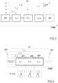
- the N readout blocks form a continuous readout excitation pulse sequence R_cont configured for being executed by the MRI system, wherein each readout block is immediately followed by a next readout block to create continuity, the MRI signals S_i, which can in this case temporally overlap (e.g. using a time-shifting reconstruction process over the continuously sampled data for R_cont, wherein temporal windows for the different MRI signals S_i may overlap - see Fig. 4 ), being acquired at said different recovery times T_i in order to enable the creation or reconstruction of said images of said part at different recovery times T_i.
- R_cont configured for being executed by the MRI system, wherein each readout block is immediately followed by a next readout block to create continuity
- the MRI signals S_i which can in this case temporally overlap (e.g. using a time-shifting reconstruction process over the continuously sampled data for R_cont, wherein temporal windows for the different MRI signals S_i may overlap - see Fig. 4 ), being
- Said continuous readout excitation pulse sequence R_cont might be defined as a merging of the N readout blocks into a single continuous block, wherein a temporal succession of two directly successive readout blocks is configured for creating a continuity over the so-called continuous readout excitation pulse sequence.
- different T1 maps might be generated from at least two MRI signals S_i, i.e. from at least two readout blocks R_i, wherein each readout block R_i comprises a collection of data at multiple echo times TE_j, enabling each to estimate one of said different T1 maps.
- each signal S_i might comprise one or several signals S_i,j, each acquired at a different echo time TE.
- the present invention concerns also an MRI system or apparatus configured for mapping T1 relaxation times of a biological object comprising at least one part characterized by a short-T2 relaxation time, and configured for carrying out the steps of the previously described method.
- FIG. 1 illustrates a preferred embodiment of an MRI system 100 according to the invention, i.e. that is capable of imaging biological material characterized by a short-T2 relaxation time during clinical operation.
- the MRI system 100 comprises notably different coils 101 and respective coil controllers configured for generating magnetic fields and RF pulses in order to acquire an MRI signal from a biological object 106 comprising at least one part characterized by said short-T2 relaxation time.
- the MRI system 100 comprises:
- the MRI system 100 is configured for carrying out the following method described in connection with Figures 2 to 5 .
- the MRI system 100 performs one or several magnetization preparation RF pulse sequences 301.
- the performed magnetization preparation RF pulse sequence is an inversion or saturation recovery pulse sequence.
- two successive magnetization preparation RF pulse sequences 301 are separated by a repetition time interval TR (see for instance Fig. 3-5 ).
- TR repetition time interval
- another magnetization preparation e.g., Magnetization Transfer preparation or T2 preparation
- the magnetization preparation RF pulse sequence 301 may comprise a tailored preparation pulse to suppress long-T2 signal components and, thus, selectively observe properties of short-T2 components.
- the recovery time T_i might also be measured from the center of the magnetization preparation pulse sequence to the center of the readout block R_i.
- the readout blocks R_i take thus place during each repetition time interval TR, after a magnetization preparation RF pulse sequence and before a directly next magnetization preparation RF pulse sequence.
- an MRI signal S_i is acquired by the MRI system 100.
- Said MRI signals S_i are thus acquired at the different recovery times T_i and enable to reconstruct, for each readout block, at least one MRI image from the acquired MRI signal.
- each readout block R_i according to the invention is configured for being sensitive to short-T2 signal.
- the MRI system 100 e.g. its processing unit 102 or a stand-alone computer device connected to the MRI system 100, is configured for reconstructing, from each MRI signal S_i, an image of said part of the biological object 106 and for using one or several of the reconstructed images for mapping T1 values for said part.
- an acquisition of two or more images might be interleaved in a single repetition time interval TR.
- a first MRI signal S_1 is acquired at T_1
- a second MRI signal S_2 is acquired at T_2, T_i being the starting time or center time of the acquisition.
- the signals S_1 and S_2 can then be separately reconstructed to yield "original" or "directly measured” 3D images of the observed part of the biological object 106.
- Said original images and/or their raw data might be suitably combined to yield quantitative estimates of localized T1 and proton-density values of the observed part (i.e. T1 and PD maps).
- Figure 3 shows a generalization of the preferred embodiment using two readout blocks, by extending the sampling to N ⁇ 2 readout blocks after a magnetization preparation 301.
- T1 relaxation times might be estimated by directly fitting a model on two or more reconstructed images or by combining two or more reconstructed images and comparing the resulting voxel intensities to simulated intensities.
- the different readout blocks R_i take place, e.g. start, one after the other in order to form a continuous readout block R_cont.
- MRI data might be continuously sampled throughout the whole of continuous readout block R_cont with many subsequent excitation-pulses.
- R_cont is a continuous 3D radial readout employed during the whole recovery period separating two successive magnetization preparations, which - combined with a sliding-window or time-shifting reconstruction for acquiring each MRI signal S_i (i.e.
- each MRI dataset at different recovery times T_i - might allow a reconstruction of an arbitrary number of original image data sets. Since each MRI signal is acquired using a short-T2 sensitive readout block, like UTE, ZTE, or single-point readout, the short-T2 MRI signal components can be detected and quantitatively mapped. Preferentially, an arbitrary number greater than or equal to two of such original data sets (MRI signal S_i) can be used to reconstruct quantitative T1 and PD maps of the observed biological part, including short-T2 signals. More specifically, Fig.
- FIG. 4 shows 3 temporal windows W1, W2, W3, wherein window W2 overlaps both W1 and W3, wherein for each window W_i, a readout block R_i sensitive to short-T2 signal is used for acquiring the corresponding signal S_i, each signal S_i being acquired at a different inversion time T_i, enabling the creation of 3 different images I_1, I_2, I_3 at the respective inversion times TI_1, TI_2, TI_3.
- a readout block R_i e.g. UTE or ZTE readout
- the T1 difference between these two maps is caused by the T1 of the tissue with short-T2 species under the assumption that the magnetization of short-T2 species decays between the echoes.
- different readout blocks e.g. single echo readout block, dual echo readout block, or multi echo readout block
- These images I_i,j can then be used to estimate the T1 relaxation time of short-T2 signal tissues that are mixed with tissue of long-T2 signal.
- the MP2RAGE method [3] might be used between images I_1,1 and I_2,1, and respectively images I_1,2 and I_2,2 for calculating two T1 maps.
- the differences in T1 between these two T1 maps are dominated by the different signal fraction of short-T2 magnetization in the different echoes.
- multiple compartment models can be directly fitted on the images I_i,j.
- acquiring more than 2 inversions and echoes improves such fitting.
- T2* or phase information can be extracted from the multi-echo readout. More detailed examples are given in the following:
- the present invention proposes to combine the concept of MP2RAGE with a 3D readout sensitive to short-T2 signal to get notably a B1-insensitive quantification of T1 relaxation time in tissue or biological objects characterized by a short-T2 relaxation time. Furthermore, the present invention proposes to use multiple echoes during the readout for improving the distinction between short-T2 and long-T2 contribution of the T1 relaxation within a same voxel. It finally proposes to extended this approach to N inversion- or saturation-recovery times and M echoes to allow for the fitting of multicompartment models.
Landscapes
- Physics & Mathematics (AREA)
- Health & Medical Sciences (AREA)
- High Energy & Nuclear Physics (AREA)
- Nuclear Medicine, Radiotherapy & Molecular Imaging (AREA)
- Condensed Matter Physics & Semiconductors (AREA)
- General Physics & Mathematics (AREA)
- Engineering & Computer Science (AREA)
- Life Sciences & Earth Sciences (AREA)
- Radiology & Medical Imaging (AREA)
- General Health & Medical Sciences (AREA)
- Heart & Thoracic Surgery (AREA)
- Biophysics (AREA)
- Pathology (AREA)
- Medical Informatics (AREA)
- Molecular Biology (AREA)
- Surgery (AREA)
- Animal Behavior & Ethology (AREA)
- Biomedical Technology (AREA)
- Public Health (AREA)
- Veterinary Medicine (AREA)
- Artificial Intelligence (AREA)
- Computer Vision & Pattern Recognition (AREA)
- Signal Processing (AREA)
- Magnetic Resonance Imaging Apparatus (AREA)
Abstract
Description
- The present invention falls within the field of magnetic resonance imaging (MRI) systems and methods. In particular, the present invention relates to a method and system for magnetic resonance imaging in which a magnetization preparation radio frequency pulse sequence is used together with a 3D image sampling readout sensitive to short time constant T2 or T2* (i.e. the time constant for the decay of transverse magnetization arising from natural interactions at the atomic or molecular levels), and to a device for carrying out said method. The many considerations hold equally for different types of signal decay and therefore, unless stated otherwise, all kinds of rapid loss of transverse magnetization (whether best characterized by T2 or T2*) will be jointly referred to as short-T2 [1] decay (or simply "short-T2") thereafter. In particular, the present invention is interested in mapping the longitudinal relaxation time T1 for biological matter characterized by a short-T2 relaxation time.
- MRI is a powerful technique that is capable of providing detailed images of a biological body, such as a human body. The signal detected in medical MRI is proportional to the transversal 1H nuclear spin magnetization. Once generated, it decays approximately exponentially with time constants T2* (transverse magnetization decay effectively observed during MRI experiments, e.g. in gradient-echo (GRE) or free-induction-decay sequences, with T2* ≤ T2). The 1H transverse relaxation times in small molecules in liquids, but also in water molecules in most soft tissues, are relatively long, i.e., in the order of tens of milliseconds up to seconds. A much faster signal decay can be observed in molecules with more restricted mobility - caused by a geometrically restricted environment - such as observed with water protons in tendons or bone - or by a larger molecular size and mass and concomitantly slowed molecular rotation and translation - such as in macro-molecules. The acquisition of short-T2 signals (T2 <1 ms) can be achieved by a very fast spatial encoding and by the omission of an echo generation, i.e., by direct sampling of the free induction decay (FID) after the application of a radiofrequency excitation pulse. Numerous MRI sequences defined by different use of radio frequency (RF) pulses and magnetic gradient fields (gradients) used for spatial encoding are well-known to the persons skilled in the art. Techniques that enable the detection of short-T2 signal components include among others e.g. ultra-short echo time (UTE), zero echo time (ZTE) or single-point imaging (SPI) methods.
- The contrast and signal intensity in a conventional MR image depend on a variety of physical, chemical, and physiological parameters of the observed tissue, as well as on the acquisition technique: a complex and - typically unknown and ill-controlled - mix of these factors can produce different signal intensities or even contrast weightings and preclude reliable reproduction. The mean signal value within a region of interest within the same tissue may vary across values measured on scanners from different vendors at different sites, but even in immediate test-retest repeated acquisitions of the same individual on the same scanner.
- A more quantitative approach at characterizing biological tissue by MRI is to directly measure one or more of the physical tissue properties in isolation, separating its/their influence on the complex mix that determines signal intensity and contrast in traditional images. Such techniques are usually referred to as "parametric mapping" or "quantitative imaging / qMRI" methods. They can produce quantitative parametric tissue maps that less strongly depend on the employed hardware, the applied imaging technique, and the image-acquisition parameters.
- The move from relative contrast information that depends on a variety of factors to a single, absolute measure based on physical tissue properties is a prerequisite, e.g., for the comparability of values (i) measured at different points in time in a single individuum in longitudinal studies, e.g., to monitor and quantify disease progress or therapy response or (ii) measured with different scanners, at different institutions and/or in different individuals. It may enable the establishment of databases of normal quantitative tissue values, to which a newly scanned patient dataset can be compared, and even contribute to an improved single-individual's diagnosis based on a single scan.
- One tissue parameter of interest is T1. The most basic and probably most accurate approach for quantitative T1 mapping are repeated inversion-recovery (IR) experiments in combination with a spin-echo (SE) sequence. After a (adiabatic) inversion pulse, the longitudinal magnetization exponentially approaches the equilibrium magnetization with the time constant T1. The acquisition of multiple spin-echo images, produced by data acquired at different inversion times (TI), with a repetition time (TR) that was at least 4 times as long as the longest T1 time to be measured, allows to obtain an accurate estimate of T1 from a fit of the inversion-recovery signal-model to the experimental data. However, this combination results in long, clinically impractical acquisition times, similar to T2 mapping with a single SE sequence.
- One of the most promising 3D T1-mapping methods [2] is the MP2RAGE sequence [3], which was originally developed to generate T1 weighted images free from bias introduced by B1-field inhomogeneity. It implements a 2-point inversion-recovery approach with significantly shortened scan time compared to an extended inversion-recovery sampling. It periodically samples partial image data in 2 phase-encoded GRE trains that start at 2 different TI times after periodically applied 180-degree inversion pulses. During the image reconstruction, these data sets can be combined in a way that cancels the B1-strength bias and can produce images whose signal intensity directly, but non-linearly depends on a tissue's T1 time. Based on well-established simulations of the spin behavior, i.e., by Bloch simulations, such images can be converted into quantitative T1 maps.
- However, components in the MR-signal from - at microscopic level - structurally rigid biological objects, such as cortical and trabecular bone, tendon, ligament, or myelin, as well as from regions with locally strongly varying magnetic susceptibility, e.g., in the lung, exhibit a fast decay (i.e. short-T2 time) that prevents detection and localization with traditional, echo-formation based MRI sequences, and, thus, that their associated quantitative properties cannot be measured. For instance, quantitative 3D T1 mapping of short-T2 signal components with clinically useful accuracy in clinically practical scan times is an unsolved problem. Also, state-of-the art T1 mapping techniques for tissues with short-T2 signal components are strongly sensitive to B1-inhomogeneity, which can result in distortion and bias of the quantitative measurements and in increased coefficients of variation for reproduction attempts. Another problem is to selectively quantify T1 times of short-T2 signal components, without interference and/or admixture from longer-T2 components. Thus, it is an interfering problem - but also a source of information - that the T1 times measured in the above-mentioned structures and tissues depend on the echo time at which signals are measured.
- To summarize, 3D mapping of T1 of tissues with short-T2 signal components, combined 3D mapping of T1 and T2* of tissues with short-T2 signal components, and combined mapping of T1, T2* and magnetic susceptibility (QSM, quantitative susceptibility mapping) of tissues with short-T2 signal components are unsolved problems. Also, mapping T1 in short-T2 tissue has been shown in time consuming 2D acquisitions (look-locker) for single slices, but not in 3D. Most traditional T1 mapping methods measure signal echoes, generated by gradient refocusing alone or in co-incidence with RF-induced spin echoes. The echo formation takes a minimum time, e.g., ca. 0.7 ms for GRE readouts, and 6 ms for SE readouts. By the time the data is acquired, signal components with short-T2 (< 1 ms) times of the transverse magnetization will have decayed by significant amounts and may not be acquired at all. Therefore, it is not possible to measure T1 of these tissue types with most known in the art sequences. All attempts that have been performed for quantifying T1 times of biological tissues characterized by short-T2 relaxation times have had limited success. In particular, Saturation-Recovery UTE [4,5], IR-UTE [6,7], 2D IR-UTE at varying TE [8,9], SPI [10], and 3D UTE-AFI-STR [11] are strongly hampered by inefficient data acquisition and long scan times not suitable for clinical applications, as well as by B1-inhomogeneity. Other existing solutions are even more strongly affected by B1-inhomogeneity, which requires calibration measurements and RF flip angles with high specific absorption rate (SAR), which acerbates another potential problem, i.e., high SAR values. The correction for spatially varying actual flip angle makes such techniques especially fragile at high or ultra-high field strength. Such techniques are for instance UTE-variable flip angle (VFA) [12], UTE-variable TR [13], UTE-actual flip angle (AFI) [14], 3D UTE-AFI-STR [11], or 2D UTE-Fingerprinting [15].
- It is therefore an objective of the present invention to find a system and method capable of mapping T1 times for biological matter characterized by short-T2 relaxation times and that is suitable for clinical applications.
- Said objective is achieved according to the present invention by a system and method for T1-mapping according to the object of the independent claims. Dependent claims present further advantages of the invention.
- The present invention concerns notably an MRI method for mapping T1 relaxation times of a biological object comprising a part characterized by a short-T2 relaxation time (i.e. smaller than 1ms), the method comprising the steps of:
- performing, by an MRI system, one or several magnetization preparation RF pulse sequences, wherein two successive magnetization preparation RF pulse sequences are separated by a repetition time interval TR;
- acquiring, by the MRI system and during each repetition time interval TR, an MRI signal generated by said part of the biological object in response to N 3D readout blocks R_i generated by the MRI system and applied to said part of the biological object, wherein for each readout block R_i, an MRI signal S_i is acquired by the MRI system at a different recovery time T_i (e.g. inversion- or saturation-recovery time), and wherein each readout block R_i is configured for being sensitive to short-T2 signal, with i=1,...,N, with N ≥ 2. In other words, the MRI signal generated by said part is observed by N readout blocks R_i, each centered at a different recovery time T_i and associated to an MRI signal S_i that is the set of MRI data acquired during said readout block R_i and used then for image reconstruction. The so-called "MRI signal generated by said part" comprises thus all said sets of MRI data that are acquired during the different readout blocks R_i. In particular, "sensitive to short-T2 signal" means that the 3D readout block R_i comprises a data collection of at least one echo time TE, wherein the collected data are sensitive to short-T2 signal, such a 3D readout block R_i being notably configured for acquiring signal with an echo-time TE smaller or equal to 0.7ms. A TE of 0.7ms is able to capture half of the MRI signal amplitude for a T2* relaxation time of 1ms. For shorter T2* relaxation time, even shorter TE are necessary to capture enough signal. Examples of 3D readout blocks sensitive to short-T2 signal are the 3D UTE pulse sequence, or the 3D ZTE pulse sequence, or the SPI pulse sequence;
- reconstructing, from each MRI signal S_i, an image of said part and mapping T1 values for said part from at least two reconstructed images. Thus, two or more reconstructed images, e.g. at least a part of their raw data, might be combined into a T1 map. In particular, instead of, or additionally to, combining two or more of the reconstructed images for extracting information about biological object tissue with short-T2 relaxation time, T1 and/or T2* information might be directly extracted from two or more of said reconstructed images by fitting an analytical model on each of said one or several reconstructed images.
- According to the present invention, and preferentially, each readout block R_i, and thus respectively its corresponding acquired signal S_i, is separated from a next readout block R_i+1, and thus respectively from a next acquired signal S_i+1, by a time interval (strictly) greater than 0.
- According to a preferred embodiment, the N readout blocks form a continuous readout excitation pulse sequence R_cont configured for being executed by the MRI system, wherein each readout block is immediately followed by a next readout block to create continuity, the MRI signals S_i, which can in this case temporally overlap (e.g. using a time-shifting reconstruction process over the continuously sampled data for R_cont, wherein temporal windows for the different MRI signals S_i may overlap - see
Fig. 4 ), being acquired at said different recovery times T_i in order to enable the creation or reconstruction of said images of said part at different recovery times T_i. Said continuous readout excitation pulse sequence R_cont might be defined as a merging of the N readout blocks into a single continuous block, wherein a temporal succession of two directly successive readout blocks is configured for creating a continuity over the so-called continuous readout excitation pulse sequence. - Optionally, different T1 maps might be generated from at least two MRI signals S_i, i.e. from at least two readout blocks R_i, wherein each readout block R_i comprises a collection of data at multiple echo times TE_j, enabling each to estimate one of said different T1 maps. For instance, each readout block R_i comprises a train of RF excitation pulses of predefined flip angle, each followed by M gradient echoes, resulting thus in an additional dimension in the acquired MR signal denoted as S_i,j, with i=1,...,N, with N ≥ 2 and j=1,...,M, with M ≥ 1. In other words, each signal S_i might comprise one or several signals S_i,j, each acquired at a different echo time TE.
- The present invention concerns also an MRI system or apparatus configured for mapping T1 relaxation times of a biological object comprising at least one part characterized by a short-T2 relaxation time, and configured for carrying out the steps of the previously described method.
- The foregoing has broadly outlined the features and technical advantages of the present disclosure so that those skilled in the art may better understand the detailed description that follows.
- Additional features and advantages of the disclosure will be described hereinafter that form the object of the claims. Those skilled in the art will appreciate that they may readily use the concept and the specific embodiment disclosed as a basis for modifying or designing other structures for carrying out the same purposes of the present disclosure.
- For a more complete understanding of the present disclosure, and the advantages thereof, reference is now made to the following descriptions taken in conjunction with the accompanying drawings, wherein like numbers designate like objects, and in which:
- Figure 1
- is a schematic illustration of an MRI system according to the invention;
- Figure 2
- illustrates a flowchart of a method according to the invention;
- Figure 3
- shows a first preferred embodiment of a readout block according to the invention;
- Figure 4
- shows a second preferred embodiment of a readout block according to the invention;
- Figure 5
- shows a third preferred embodiment of a readout block according to the invention.
-
Figure 1 illustrates a preferred embodiment of anMRI system 100 according to the invention, i.e. that is capable of imaging biological material characterized by a short-T2 relaxation time during clinical operation. TheMRI system 100 comprises notablydifferent coils 101 and respective coil controllers configured for generating magnetic fields and RF pulses in order to acquire an MRI signal from abiological object 106 comprising at least one part characterized by said short-T2 relaxation time. For instance, theMRI system 100 comprises: - an axial magnetic field coil and an axial magnetic field coil controller for controlling an axial magnetic field B0 generated by said axial magnetic field coil;
- a gradient coil and a gradient coil controller configured for controlling a magnetic field gradient G generated by said gradient coil;
- a RF coil and a RF coil controller configured for controlling a RF magnetic field B1 produced by the RF coil;
- a receiver coil, which can be the same as the RF coil, configured for detecting changes in a magnetization of the
biological object 106 to be imaged, and for communicating said changes to a receiver coil controller, which can be the same as the RF coil controller, the latter outputting an MRI signal to aprocessing unit 102 comprising a processor and amemory 103, theprocessing unit 102 being configured for processing the outputted MRI signal in order to reconstruct images of said biological object; - a
display 104 for displaying the reconstructed images. - Contrary to prior art MRI systems, the
MRI system 100 according to the invention is configured for carrying out the following method described in connection withFigures 2 to 5 . - At
step 201, theMRI system 100 performs one or several magnetization preparationRF pulse sequences 301. Preferentially, the performed magnetization preparation RF pulse sequence is an inversion or saturation recovery pulse sequence. As known in the art, two successive magnetization preparationRF pulse sequences 301 are separated by a repetition time interval TR (see for instanceFig. 3-5 ). Of course, another magnetization preparation (e.g., Magnetization Transfer preparation or T2 preparation) or a combination of different preparation modules to further manipulate the signal of short-T2 signal components can be used. For example, the magnetization preparationRF pulse sequence 301 may comprise a tailored preparation pulse to suppress long-T2 signal components and, thus, selectively observe properties of short-T2 components. - At
step 202, theMRI system 100 acquires an MRI signal generated by said part of thebiological object 106 in response to N 3D readout blocks R_i generated by theMRI system 101 and applied to said part of the biological object 106 (seeFig. 3 ), wherein i=1,...,N, and N ≥ 2.Fig. 4 shows a preferred embodiment wherein N=3, i.e. 3 readout blocks R_1, R_2, and R_3, are successively generated and applied to thebiological object 106.Fig. 5 shows another preferred embodiment wherein N=2, i.e. two readout blocks R_1 and R_2 are used. As known in the art, the first readout block R_1 starts at the recovery time T_1 after the starting-time of the magnetizationpreparation pulse sequence 301, each additional successive readout block R_k, with k = 2,...,N, starting at a recovery time T_k ≠ T_i for i≠k, the end-time of the Nth readout block R_N being separated from the starting-time of the next magnetizationpreparation pulse sequence 301 by a final recovery time T_final (seeFig. 3 ). As shown inFig. 3 , the recovery time T_i might also be measured from the center of the magnetization preparation pulse sequence to the center of the readout block R_i. - The readout blocks R_i take thus place during each repetition time interval TR, after a magnetization preparation RF pulse sequence and before a directly next magnetization preparation RF pulse sequence. For and during each readout block R_i, an MRI signal S_i is acquired by the
MRI system 100. Said MRI signals S_i are thus acquired at the different recovery times T_i and enable to reconstruct, for each readout block, at least one MRI image from the acquired MRI signal. Preferentially, the readout blocks are separated from each other by a predefined positive period of time, so that each MRI signal S_p is acquired separately from the next MRI signal S_p+1, p=1,...,N-1. Contrary to most traditional MRI techniques, each readout block R_i according to the invention is configured for being sensitive to short-T2 signal. - At
step 203, theMRI system 100, e.g. itsprocessing unit 102 or a stand-alone computer device connected to theMRI system 100, is configured for reconstructing, from each MRI signal S_i, an image of said part of thebiological object 106 and for using one or several of the reconstructed images for mapping T1 values for said part. - In a preferred embodiment of the invention, an acquisition of two or more images might be interleaved in a single repetition time interval TR. For instance, the MRI recovery signal is sampled (using for instance radial sampling) by two (N=2) readout blocks R_1 and R_2 of excitation-pulse/detection period combinations (e.g. 3D UTE or ZTE readout), centered or starting respectively each at a different preparation-recovery time, respectively T_1 and T_2, after the magnetization preparation (e.g. inversion or saturation) pulse sequence. Thus, during the first readout block R_1, a first MRI signal S_1 is acquired at T_1, and during the second readout block R_2, a second MRI signal S_2 is acquired at T_2, T_i being the starting time or center time of the acquisition. The signals S_1 and S_2 can then be separately reconstructed to yield "original" or "directly measured" 3D images of the observed part of the
biological object 106. Said original images and/or their raw data might be suitably combined to yield quantitative estimates of localized T1 and proton-density values of the observed part (i.e. T1 and PD maps).Figure 3 shows a generalization of the preferred embodiment using two readout blocks, by extending the sampling to N≥2 readout blocks after amagnetization preparation 301. Having more readout blocks allows a more precise quantification of T1 values in the tissue of thebiological object 106. Known in the art techniques might be used then to quantify T1 from the reconstructed images. For instance, T1 relaxation times might be estimated by directly fitting a model on two or more reconstructed images or by combining two or more reconstructed images and comparing the resulting voxel intensities to simulated intensities. - According to another preferred embodiment illustrated in
Fig. 4 , the different readout blocks R_i take place, e.g. start, one after the other in order to form a continuous readout block R_cont. In other words, instead of sampling the preparation recovery by small readout blocks temporally separated from each other as in the previous preferred embodiments, MRI data might be continuously sampled throughout the whole of continuous readout block R_cont with many subsequent excitation-pulses. Preferentially, R_cont is a continuous 3D radial readout employed during the whole recovery period separating two successive magnetization preparations, which - combined with a sliding-window or time-shifting reconstruction for acquiring each MRI signal S_i (i.e. each MRI dataset) at different recovery times T_i - might allow a reconstruction of an arbitrary number of original image data sets. Since each MRI signal is acquired using a short-T2 sensitive readout block, like UTE, ZTE, or single-point readout, the short-T2 MRI signal components can be detected and quantitatively mapped. Preferentially, an arbitrary number greater than or equal to two of such original data sets (MRI signal S_i) can be used to reconstruct quantitative T1 and PD maps of the observed biological part, including short-T2 signals. More specifically,Fig. 4 shows 3 temporal windows W1, W2, W3, wherein window W2 overlaps both W1 and W3, wherein for each window W_i, a readout block R_i sensitive to short-T2 signal is used for acquiring the corresponding signal S_i, each signal S_i being acquired at a different inversion time T_i, enabling the creation of 3 different images I_1, I_2, I_3 at the respective inversion times TI_1, TI_2, TI_3. - According to another preferred embodiment of the invention illustrated in
Fig. 5 , M≥ 1 gradient-echoes can be formed after eachexcitation pulse 501 of a readout block R_i (e.g. UTE or ZTE readout) by using M successive gradients or gradient echosequences 502 enabling to acquire, for each gradient echo sequence, a subset S_i,j of MRI data at an echo time TE_j for the signal S_i, with j=1,...,M. Due to the T2* decay after the excitation, every echo at TE_j will have a different signal intensity. This allows to separate image information that comes from tissue with short and long T2* or the quantification of T2*. For example, it becomes possible to estimate T1 maps independently with data collected at a first and second echo. The T1 difference between these two maps is caused by the T1 of the tissue with short-T2 species under the assumption that the magnetization of short-T2 species decays between the echoes. Thanks to this technique, different readout blocks (e.g. single echo readout block, dual echo readout block, or multi echo readout block) can be used to sample different numbers of echoes, each at a different echo time TE_j. - For instance, the present method might comprise two inversion-recovery times T_1 and T_2 (i.e. N = 2) and two echo times TE_1 and TE_2 (i.e. M=2), enabling to reconstruct four images I_i,j:
- a first image I_1,1 (T_1, TE_1) that is characterized by a strong T1 weighting - short-T2 signal sensitive;
- a second image I_1,2 (T_1, TE_2) that is characterized by a strong T1 weighting - short-T2 signal insensitive;
- a third image I_2,1 (T_2, TE_1) that is characterized by a low T1 weighting - short-T2 signal sensitive; and
- a fourth image I_2,2 (T_2, TE_2) that is characterized by a low T1 weighting - short-T2 signal insensitive.
- These images I_i,j can then be used to estimate the T1 relaxation time of short-T2 signal tissues that are mixed with tissue of long-T2 signal. Optionally, the MP2RAGE method [3] might be used between images I_1,1 and I_2,1, and respectively images I_1,2 and I_2,2 for calculating two T1 maps. The differences in T1 between these two T1 maps are dominated by the different signal fraction of short-T2 magnetization in the different echoes. Alternatively, multiple compartment models can be directly fitted on the images I_i,j. Advantageously, acquiring more than 2 inversions and echoes improves such fitting.
- Of course, other methods of post processing the reconstructed images or the acquired MRI signals S_i that use mathematical operations on the different contrasts to extract information about tissue with short-T2 relaxation time might be used. For example, T2* or phase information can be extracted from the multi-echo readout. More detailed examples are given in the following:
- 3D MP-2-UTE (N=2, M>1) with multi-echo readout and additional T2* fitting: If more than one echo is acquired, then the present invention makes the calculation of quantitative T2* maps in addition to different sets of quantitative T1 and PD maps with data acquired at each echo time possible. This enables notably an efficient and B1-corrected T1, PD and T2* mapping.
- 3D MP-2-UTE (N=2, M>1) with multi-echo readout and with additional evaluation of phase information: The change of the complex phase of the images across different echoes contains additional information. The present invention proposes to use the multi-echo readout to extract phase information, and using the latter to estimate variations in the main magnetic field (B0) or tissue susceptibility in species with short T2. This enables notably an efficient and B1-corrected T1, PD, phase, B0, and susceptibility (QSM) mapping.
- To summarize, the present invention proposes to combine the concept of MP2RAGE with a 3D readout sensitive to short-T2 signal to get notably a B1-insensitive quantification of T1 relaxation time in tissue or biological objects characterized by a short-T2 relaxation time. Furthermore, the present invention proposes to use multiple echoes during the readout for improving the distinction between short-T2 and long-T2 contribution of the T1 relaxation within a same voxel. It finally proposes to extended this approach to N inversion- or saturation-recovery times and M echoes to allow for the fitting of multicompartment models.
-
- MRI
- magnetic resonance imaging
- MR
- magnetic resonance
- RF
- radio frequency
- GRE
- gradient echo
- UTE
- ultra-short echo time
- ZTE
- zero echo time
- SPI
- single-point imaging
- qMRI
- quantitative magnetic resonance imaging
- IR
- inversion recovery
- SE
- spin-echo
- TR
- repetition time
- T_i
- recovery time
- QSM
- quantitative susceptibility mapping
- TE
- echo time
- VFA
- variable flip angle
- AFI
- actual flip angle
- FID
- free induction decay
- SP
- spoiler gradient
- RO
- readout gradient
- RW
- rewinding gradient
- MP
- magnetic preparation
-
- [1] Weiger, M., et al., Short-T2 MRI: Principles and recent advances. Prog Nucl Magn Reason Spectrosc, 2019. 114-115: p. 237-270.
- [2] Glasser, M.F., et al., The minimal preprocessing pipelines for the Human Connectome Project. NeuroImage, 2013. 80: p. 105-124.
- [3] Marques, J.P., et al., MP2RAGE, a self bias-field corrected sequence for improved segmentation and T1-mapping at high field. NeuroImage, 2010. 49(2): p. 1271-1281.
- [4] Du, J., et al., Qualitative and quantitative ultrashort echo time (UTE) imaging of cortical bone. J Magn Reson, 2010. 207(2): p. 304-11.
- [5] Du, J. and G.M. Bydder, Qualitative and quantitative ultrashort-TE MRI of cortical bone. NMR in Biomedicine, 2013. 26(5): p. 489-506.
- [6] Wei, Z., et al., To measure T1 of short T2 species using an inversion recovery prepared three-dimensional ultra-short echo time (3D IR-UTE) method: A phantom study. Journal of Magnetic Resonance, 2020. 314: p. 106725.
- [7] Guo, T., et al., T1 measurement of bound water in cortical bone using 3D adiabatic inversion recovery ultrashort echo time (3D IR-UTE) Cones imaging. Magnetic Resonance in Medicine, 2020. 84(2): p. 634-645.
- [8] Triphan, S.M.F., et al., Oxygen enhanced lung MRI by simultaneous measurement of T1 and T2* during free breathing using ultrashort TE. Journal of Magnetic Resonance Imaging, 2015. 41(6): p. 1708-1714.
- [9] Wielpütz, M.O., et al., Outracing Lung Signal Decay - Potential of Ultrashort Echo Time MRI. Rofo, 2019. 191(05): p. 415-423.
- [10] Beyea, S.D., et al., Relaxation Time Mapping of ShortT*2Nuclei with Single-Point Imaging (SPI) Methods. Journal of Magnetic Resonance, 1998. 135(1): p. 156-164.
- [11] Wei, Z., et al., Fast T1 measurement of cortical bone using 3D UTE actual flip angle imaging and single-TR acquisition (3D UTE-AFI-STR). Magnetic Resonance in Medicine, 2021. 85(6): p. 3290-3298.
- [12] Springer, F., et al., Magnetization transfer contrast imaging in bovine and human cortical bone applying an ultrashort echo time sequence at 3 Tesla. Magnetic Resonance in Medicine, 2009. 61(5): p. 1040-1048
- [13] Chen, J., et al., Measurement of bound and pore water T1 relaxation times in cortical bone using three-dimensional ultrashort echo time cones sequences. Magnetic Resonance in Medicine, 2017. 77(6): p. 2136-2145
- [14] Ma, Y.-J., et al., Whole knee joint T1 values measured in vivo at 3T by combined 3D ultrashort echo time cones actual flip angle and variable flip angle methods. Magnetic Resonance in Medicine, 2019. 81(3): p. 1634-1644
- [15] Li, Q., et al., Ultrashort echo time magnetic resonance fingerprinting (UTE-MRF) for simultaneous quantification of long and ultrashort T2 tissues. Magnetic Resonance in Medicine, 2019. 82(4): p. 1359-1372.
Claims (9)
- Magnetic Resonance Imaging (hereafter "MRI") method for mapping, with an MRI system (100), T1 relaxation times of a biological object (106) comprising a part characterized by a short-T2 relaxation time, the method comprising the steps of:- performing (201), by the MRI system (100), one or several magnetization preparation radio frequency (hereafter "RF") pulse sequences (301), wherein two successive magnetization preparation RF pulse sequences (301) are separated by a repetition time interval TR;- acquiring (202), by the MRI system (100) and during each repetition time interval TR, an MRI signal generated by said part of said biological object (106) in response to N 3D readout blocks R_i generated by the MRI system (100) and applied to said part of the biological object (106), wherein for each readout block R_i, an MRI signal S_i is acquired by the MRI system (100) at a different recovery time T_i, and wherein each readout block R_i is configured for being sensitive to short-T2 signal, with i=1,...,N, with N ≥ 2;- reconstructing (203), from each MRI signal S_i at various T_i, an image of said part and mapping T1 values for said part from two or more of the reconstructed images.
- MRI method according to claim 1, wherein each readout block R_i, and thus respectively its corresponding acquired signal S_i, is separated from a next readout block R_i+1, and thus respectively from a next acquired signal S_i+1 by a time interval greater than 0.
- MRI method according to claim 1, wherein the N readout blocks form a continuous readout excitation pulse sequence R_cont configured for being executed by the MRI system (100), wherein the MRI signals S_i are acquired at different recovery times T_i in order to create images with many different T_i.
- MRI method according to one of the claims 1 to 3, wherein each readout block R_i comprises a train of RF excitation pulses of predefined flip angle, each followed by M gradient echoes resulting in an additional dimension in the MR signal denoted as S_i,j, with i=1,...,N, with N ≥ 2 and j=1,...,M, with M ≥ 1.
- MRI method according to one of the claims 1 to 4, wherein each readout block is:- one or more RF excitation(s) with UTE signal readout(s); or- one or more RF excitation(s) with ZTE signal readout(s); or- one or more RF excitation(s) with SPI data-point collection(s) .
- MRI method according to one of the claims 1 to 5, characterized in that the magnetization preparation RF pulse sequence (301) is an inversion or saturation recovery pulse sequence.
- MRI method according to one of the claims 1 to 6, comprising combining said two or more reconstructed images for extracting information about biological object tissue with short-T2 relaxation time.
- MRI method according to one of the claims 1 to 6, where T1 and T2* information is extracted directly from said two or more reconstructed images by a fitting of an analytical model.
- MRI system (100) configured for carrying out the MRI method according to one of the claims 1 to 8.
Priority Applications (2)
| Application Number | Priority Date | Filing Date | Title |
|---|---|---|---|
| EP22168302.2A EP4261556A1 (en) | 2022-04-14 | 2022-04-14 | A method and system for t1 mapping for tissue characterized by short-t2 relaxation in mri |
| US18/298,515 US12339339B2 (en) | 2022-04-14 | 2023-04-11 | Method and system for T1 mapping for tissue characterized by short-T2 relaxation in MRI |
Applications Claiming Priority (1)
| Application Number | Priority Date | Filing Date | Title |
|---|---|---|---|
| EP22168302.2A EP4261556A1 (en) | 2022-04-14 | 2022-04-14 | A method and system for t1 mapping for tissue characterized by short-t2 relaxation in mri |
Publications (1)
| Publication Number | Publication Date |
|---|---|
| EP4261556A1 true EP4261556A1 (en) | 2023-10-18 |
Family
ID=81328175
Family Applications (1)
| Application Number | Title | Priority Date | Filing Date |
|---|---|---|---|
| EP22168302.2A Pending EP4261556A1 (en) | 2022-04-14 | 2022-04-14 | A method and system for t1 mapping for tissue characterized by short-t2 relaxation in mri |
Country Status (2)
| Country | Link |
|---|---|
| US (1) | US12339339B2 (en) |
| EP (1) | EP4261556A1 (en) |
Families Citing this family (1)
| Publication number | Priority date | Publication date | Assignee | Title |
|---|---|---|---|---|
| CN121784631A (en) * | 2026-03-05 | 2026-04-03 | 首都医科大学附属北京天坛医院 | An optimized method, apparatus, device, and medium for magnetic resonance imaging. |
Family Cites Families (7)
| Publication number | Priority date | Publication date | Assignee | Title |
|---|---|---|---|---|
| US6531868B2 (en) * | 1996-12-30 | 2003-03-11 | Halliburton Energy Services, Inc. | System and methods for formation evaluation while drilling |
| US9759795B2 (en) * | 2012-07-19 | 2017-09-12 | Siemens Healthcare Gmbh | System for reducing artifacts in imaging in the presence of a spin-lock radio-frequency field |
| US10132898B2 (en) * | 2013-05-24 | 2018-11-20 | Wisconsin Alumni Research Foundation | MRI double inversion recovery method and system, with different magnetization recovery states providing the MRI image contrast(s) |
| US9618593B2 (en) * | 2014-03-18 | 2017-04-11 | Siemens Healthcare Gmbh | Phase enhanced UTE with improved fat suppression |
| US10338180B2 (en) * | 2015-10-12 | 2019-07-02 | Wisconsin Alumni Research Foundation | System and method for gradient measurement using single-point imaging |
| EP3376939B1 (en) * | 2015-11-18 | 2022-05-11 | University Of Louisville Research Foundation, Inc. | Automated methods for the objective quantification of retinal characteristics by retinal region and diagnosis of retinal pathology |
| US20180231626A1 (en) * | 2017-02-10 | 2018-08-16 | Case Western Reserve University | Systems and methods for magnetic resonance fingerprinting for quantitative breast imaging |
-
2022
- 2022-04-14 EP EP22168302.2A patent/EP4261556A1/en active Pending
-
2023
- 2023-04-11 US US18/298,515 patent/US12339339B2/en active Active
Non-Patent Citations (20)
| Title |
|---|
| BEYEA, S.D. ET AL.: "Relaxation Time Mapping of ShortT*2Nuclei with Single-Point Imaging (SPI) Methods", JOURNAL OF MAGNETIC RESONANCE, vol. 135, no. 1, 1998, pages 156 - 164, XP004407348, DOI: 10.1006/jmre.1998.1537 |
| CHEN, J. ET AL.: "Measurement of bound and pore water T1 relaxation times in cortical bone using three-dimensional ultrashort echo time cones sequences", MAGNETIC RESONANCE IN MEDICINE, vol. 77, no. 6, 2017, pages 2136 - 2145 |
| DU, J. ET AL.: "Qualitative and quantitative ultrashort echo time (UTE) imaging of cortical bone", J MAGN RESON, vol. 207, no. 2, 2010, pages 304 - 11, XP027526051 |
| DU, J.G.M. BYDDER: "Qualitative and quantitative ultrashort-TE MRI of cortical bone", NMR IN BIOMEDICINE, vol. 26, no. 5, 2013, pages 489 - 506 |
| GLASSER, M.F. ET AL.: "The minimal preprocessing pipelines for the Human Connectome Project", NEUROLMAGE, vol. 80, 2013, pages 105 - 124, XP055665201, DOI: 10.1016/j.neuroimage.2013.04.127 |
| GUO, T. ET AL.: "T1 measurement of bound water in cortical bone using 3D adiabatic inversion recovery ultrashort echo time (3D IR-UTE) Cones imaging", MAGNETIC RESONANCE IN MEDICINE, vol. 84, no. 2, 2020, pages 634 - 645 |
| IDIYATULLIN ET AL: "Fast and quiet MRI using a swept radiofrequency", JOURNAL OF MAGNETIC RESONANCE, ACADEMIC PRESS, ORLANDO, FL, US, vol. 181, no. 2, 1 August 2006 (2006-08-01), pages 342 - 349, XP005569620, ISSN: 1090-7807 * |
| LI, Q. ET AL.: "Ultrashort echo time magnetic resonance fingerprinting (UTE-MRF) for simultaneous quantification of long and ultrashort T2 tissues", MAGNETIC RESONANCE IN MEDICINE, vol. 82, no. 4, 2019, pages 1359 - 1372 |
| MA, Y.-J. ET AL.: "Whole knee joint T1 values measured in vivo at 3T by combined 3D ultrashort echo time cones actual flip angle and variable flip angle methods", MAGNETIC RESONANCE IN MEDICINE, vol. 81, no. 3, 2019, pages 1634 - 1644 |
| MARQUES, J.P. ET AL.: "MP2RAGE, a self bias-field corrected sequence for improved segmentation and Tl-mapping at high field", NEUROLMAGE, vol. 49, no. 2, 2010, pages 1271 - 1281, XP026796256 |
| MISUNG HAN ET AL: "Actual Flip Angle Imaging to Improve T1 Measurement for Short T2 Tissues", PROCEEDINGS OF THE INTERNATIONAL SOCIETY FOR MAGNETIC RESONANCE IN MEDICINE, ISMRM, 23RD ANNUAL MEETING AND EXHIBITION, TORONTO, ONTARIO, CANADA, 30 MAY - 5 JUNE 2015, no. 501, 15 May 2015 (2015-05-15), XP040666184 * |
| QI HAIKUN ET AL: "Simultaneous T1 and T2 mapping of the carotid plaque (SIMPLE) with T2 and inversion recovery prepared 3D radial imaging", MAGNETIC RESONANCE IN MEDICINE, vol. 80, no. 6, 25 May 2018 (2018-05-25), US, pages 2598 - 2608, XP055964973, ISSN: 0740-3194, DOI: 10.1002/mrm.27361 * |
| RYAN CHAMBERLAIN ET AL: "Measuring T1 in the presence of very high iron concentrations with SWIFT", PROCEEDINGS OF THE INTERNATIONAL SOCIETY FOR MAGNETIC RESONANCE IN MEDICINE, ISMRM, 20TH ANNUAL MEETING AND EXHIBITION, MELBOURNE, AUSTRALIA, 5-11 MAY 2012, 21 April 2012 (2012-04-21), XP040626789 * |
| SPRINGER, F. ET AL.: "Magnetization transfer contrast imaging in bovine and human cortical bone applying an ultrashort echo time sequence at 3 Tesla", MAGNETIC RESONANCE IN MEDICINE, vol. 61, no. 5, 2009, pages 1040 - 1048 |
| TRIPHAN SIMON M.F. ET AL: "Oxygen enhanced lung MRI by simultaneous measurement of T1 and T2* during free breathing using ultrashort TE", JOURNAL OF MAGNETIC RESONANCE IMAGING, vol. 41, no. 6, 7 July 2014 (2014-07-07), US, pages 1708 - 1714, XP055964985, ISSN: 1053-1807, DOI: 10.1002/jmri.24692 * |
| TRIPHAN, S.M.F. ET AL.: "Oxygen enhanced lung MRI by simultaneous measurement of T1 and T2* during free breathing using ultrashort TE", JOURNAL OF MAGNETIC RESONANCE IMAGING, vol. 41, no. 6, 2015, pages 1708 - 1714 |
| WEI, Z. ET AL.: "Fast T1 measurement of cortical bone using 3D UTE actual flip angle imaging and single-TR acquisition (3D UTE-AFI-STR", MAGNETIC RESONANCE IN MEDICINE, vol. 85, no. 6, 2021, pages 3290 - 3298 |
| WEI, Z. ET AL.: "To measure T1 of short T2 species using an inversion recovery prepared three-dimensional ultra-short echo time (3D IR-UTE) method: A phantom study", JOURNAL OF MAGNETIC RESONANCE, vol. 314, 2020, pages 106725 |
| WEIGER, M. ET AL.: "Short-T2 MRI: Principles and recent advances", PROG NUCL MAGN REASON SPECTROSC, vol. 114-115, 2019, pages 237 - 270, XP085921084, DOI: 10.1016/j.pnmrs.2019.07.001 |
| WIELPTITZ, M.O. ET AL.: "Outracing Lung Signal Decay - Potential of Ultrashort Echo Time MRI", ROFO, vol. 191, no. 05, 2019, pages 415 - 423 |
Also Published As
| Publication number | Publication date |
|---|---|
| US12339339B2 (en) | 2025-06-24 |
| US20230333186A1 (en) | 2023-10-19 |
Similar Documents
| Publication | Publication Date | Title |
|---|---|---|
| Weiger et al. | Advances in MRI of the myelin bilayer | |
| US6603989B1 (en) | T2 contrast in magnetic resonance imaging with gradient echoes | |
| RU2605516C2 (en) | Magnetic resonance (mr) | |
| US9513358B2 (en) | Method and apparatus for magnetic resonance imaging | |
| JP4444413B2 (en) | Apparatus for quantitative MR imaging of water and fat using quad field echo sequence | |
| US8406849B2 (en) | Systems and methods for magnetic resonance imaging | |
| US6064203A (en) | Method and apparatus for determining or imaging longitudinal spin relaxation time or producing images which substantially reflect longitudinal spin relaxation time contrast | |
| RU2683605C1 (en) | Parallel mri with sensitivity mapping using rf coil | |
| US10359489B2 (en) | Phase sensitive inversion recovery MRI with water/fat separation | |
| US8872515B2 (en) | System and method for diffusion-modulated relaxation magnetic resonance imaging | |
| WO2015161156A1 (en) | System and method for correcting intrinsic heterogeneity in magnetic resonance imaging | |
| US20190353731A1 (en) | System and Method for Quantifying T1, T2 and Resonance Frequency Using Rosette Trajectory Acquisition and Read Segmented Reconstruction | |
| EP4261558B1 (en) | A method and system for simultaneous mapping of quantitative mri parameters using a t2 prepared inversion | |
| Finkelstein et al. | Diffusion encoding methods in MRI: perspectives and challenges | |
| US12339339B2 (en) | Method and system for T1 mapping for tissue characterized by short-T2 relaxation in MRI | |
| CN116888489B (en) | Methods for analyzing medical images | |
| WO2004013648A1 (en) | Measurement of the macromolecular proton density involved in magnetization transfer by means of magnetic resonance | |
| US20050256393A1 (en) | System and method for generating t1 and t2 maps | |
| US10114095B2 (en) | Multi-spectral MRI scan with magnetization recovery | |
| EP3385742A1 (en) | Method and apparatus for identifying at least one material comprised in a voxel | |
| Shin et al. | Yet more evidence that non-aqueous myelin lipids can be directly imaged with ultrashort echo time (UTE) MRI on a clinical 3T scanner: a lyophilized red blood cell membrane lipid study | |
| US11768264B2 (en) | System and method for magnetic resonance fingerprinting with relaxation and diffusion data acquisition | |
| EP4462147A1 (en) | Mr imaging using cest contrast enhancement | |
| US20020022778A1 (en) | Imaging technique and magnetic resonance tomograph | |
| JP2003523230A (en) | Image processing method and image data processing device |
Legal Events
| Date | Code | Title | Description |
|---|---|---|---|
| PUAI | Public reference made under article 153(3) epc to a published international application that has entered the european phase |
Free format text: ORIGINAL CODE: 0009012 |
|
| STAA | Information on the status of an ep patent application or granted ep patent |
Free format text: STATUS: THE APPLICATION HAS BEEN PUBLISHED |
|
| AK | Designated contracting states |
Kind code of ref document: A1 Designated state(s): AL AT BE BG CH CY CZ DE DK EE ES FI FR GB GR HR HU IE IS IT LI LT LU LV MC MK MT NL NO PL PT RO RS SE SI SK SM TR |
|
| RAP1 | Party data changed (applicant data changed or rights of an application transferred) |
Owner name: BALGRIST CAMPUS AG Owner name: SIEMENS HEALTHINEERS AG |
|
| STAA | Information on the status of an ep patent application or granted ep patent |
Free format text: STATUS: REQUEST FOR EXAMINATION WAS MADE |
|
| 17P | Request for examination filed |
Effective date: 20240405 |
|
| RBV | Designated contracting states (corrected) |
Designated state(s): AL AT BE BG CH CY CZ DE DK EE ES FI FR GB GR HR HU IE IS IT LI LT LU LV MC MK MT NL NO PL PT RO RS SE SI SK SM TR |
|
| STAA | Information on the status of an ep patent application or granted ep patent |
Free format text: STATUS: EXAMINATION IS IN PROGRESS |
|
| 17Q | First examination report despatched |
Effective date: 20250411 |