EP4261542A1 - Method and device for determining the vitamin status - Google Patents
Method and device for determining the vitamin status Download PDFInfo
- Publication number
- EP4261542A1 EP4261542A1 EP22167551.5A EP22167551A EP4261542A1 EP 4261542 A1 EP4261542 A1 EP 4261542A1 EP 22167551 A EP22167551 A EP 22167551A EP 4261542 A1 EP4261542 A1 EP 4261542A1
- Authority
- EP
- European Patent Office
- Prior art keywords
- plp
- probe
- des
- vitamin
- erythrocytes
- Prior art date
- Legal status (The legal status is an assumption and is not a legal conclusion. Google has not performed a legal analysis and makes no representation as to the accuracy of the status listed.)
- Withdrawn
Links
- 238000000034 method Methods 0.000 title claims abstract description 56
- 235000020812 vitamin status Nutrition 0.000 title claims abstract description 37
- 238000000338 in vitro Methods 0.000 claims abstract description 13
- NGVDGCNFYWLIFO-UHFFFAOYSA-N pyridoxal 5'-phosphate Chemical compound CC1=NC=C(COP(O)(O)=O)C(C=O)=C1O NGVDGCNFYWLIFO-UHFFFAOYSA-N 0.000 claims description 271
- 239000000523 sample Substances 0.000 claims description 196
- 210000003743 erythrocyte Anatomy 0.000 claims description 81
- 239000006166 lysate Substances 0.000 claims description 49
- 102000004190 Enzymes Human genes 0.000 claims description 46
- 108090000790 Enzymes Proteins 0.000 claims description 46
- 230000001419 dependent effect Effects 0.000 claims description 29
- 238000002372 labelling Methods 0.000 claims description 25
- LXNHXLLTXMVWPM-UHFFFAOYSA-N pyridoxine Chemical compound CC1=NC=C(CO)C(CO)=C1O LXNHXLLTXMVWPM-UHFFFAOYSA-N 0.000 claims description 25
- 238000011534 incubation Methods 0.000 claims description 20
- 239000011726 vitamin B6 Substances 0.000 claims description 20
- RADKZDMFGJYCBB-UHFFFAOYSA-N pyridoxal hydrochloride Natural products CC1=NC=C(CO)C(C=O)=C1O RADKZDMFGJYCBB-UHFFFAOYSA-N 0.000 claims description 19
- 210000004369 blood Anatomy 0.000 claims description 16
- 239000008280 blood Substances 0.000 claims description 16
- 108010026552 Proteome Proteins 0.000 claims description 15
- 238000012360 testing method Methods 0.000 claims description 14
- 235000020820 vitamin B6 status Nutrition 0.000 claims description 14
- 102000001554 Hemoglobins Human genes 0.000 claims description 13
- 108010054147 Hemoglobins Proteins 0.000 claims description 13
- 235000019158 vitamin B6 Nutrition 0.000 claims description 13
- 238000011068 loading method Methods 0.000 claims description 12
- 230000002934 lysing effect Effects 0.000 claims description 12
- 229940011671 vitamin b6 Drugs 0.000 claims description 12
- 238000003556 assay Methods 0.000 claims description 11
- 230000029918 bioluminescence Effects 0.000 claims description 10
- 238000005415 bioluminescence Methods 0.000 claims description 10
- 239000000975 dye Substances 0.000 claims description 10
- QYSXJUFSXHHAJI-XFEUOLMDSA-N Vitamin D3 Natural products C1(/[C@@H]2CC[C@@H]([C@]2(CCC1)C)[C@H](C)CCCC(C)C)=C/C=C1\C[C@@H](O)CCC1=C QYSXJUFSXHHAJI-XFEUOLMDSA-N 0.000 claims description 9
- 201000010099 disease Diseases 0.000 claims description 9
- 208000037265 diseases, disorders, signs and symptoms Diseases 0.000 claims description 9
- 239000012528 membrane Substances 0.000 claims description 9
- 239000002105 nanoparticle Substances 0.000 claims description 9
- 238000011282 treatment Methods 0.000 claims description 9
- YBJHBAHKTGYVGT-ZKWXMUAHSA-N (+)-Biotin Chemical compound N1C(=O)N[C@@H]2[C@H](CCCCC(=O)O)SC[C@@H]21 YBJHBAHKTGYVGT-ZKWXMUAHSA-N 0.000 claims description 8
- AUNGANRZJHBGPY-SCRDCRAPSA-N Riboflavin Chemical compound OC[C@@H](O)[C@@H](O)[C@@H](O)CN1C=2C=C(C)C(C)=CC=2N=C2C1=NC(=O)NC2=O AUNGANRZJHBGPY-SCRDCRAPSA-N 0.000 claims description 8
- QYSXJUFSXHHAJI-YRZJJWOYSA-N vitamin D3 Chemical compound C1(/[C@@H]2CC[C@@H]([C@]2(CCC1)C)[C@H](C)CCCC(C)C)=C\C=C1\C[C@@H](O)CCC1=C QYSXJUFSXHHAJI-YRZJJWOYSA-N 0.000 claims description 8
- AUNGANRZJHBGPY-UHFFFAOYSA-N D-Lyxoflavin Natural products OCC(O)C(O)C(O)CN1C=2C=C(C)C(C)=CC=2N=C2C1=NC(=O)NC2=O AUNGANRZJHBGPY-UHFFFAOYSA-N 0.000 claims description 7
- 229930003779 Vitamin B12 Natural products 0.000 claims description 7
- 229930003471 Vitamin B2 Natural products 0.000 claims description 7
- FDJOLVPMNUYSCM-WZHZPDAFSA-L cobalt(3+);[(2r,3s,4r,5s)-5-(5,6-dimethylbenzimidazol-1-yl)-4-hydroxy-2-(hydroxymethyl)oxolan-3-yl] [(2r)-1-[3-[(1r,2r,3r,4z,7s,9z,12s,13s,14z,17s,18s,19r)-2,13,18-tris(2-amino-2-oxoethyl)-7,12,17-tris(3-amino-3-oxopropyl)-3,5,8,8,13,15,18,19-octamethyl-2 Chemical compound [Co+3].N#[C-].N([C@@H]([C@]1(C)[N-]\C([C@H]([C@@]1(CC(N)=O)C)CCC(N)=O)=C(\C)/C1=N/C([C@H]([C@@]1(CC(N)=O)C)CCC(N)=O)=C\C1=N\C([C@H](C1(C)C)CCC(N)=O)=C/1C)[C@@H]2CC(N)=O)=C\1[C@]2(C)CCC(=O)NC[C@@H](C)OP([O-])(=O)O[C@H]1[C@@H](O)[C@@H](N2C3=CC(C)=C(C)C=C3N=C2)O[C@@H]1CO FDJOLVPMNUYSCM-WZHZPDAFSA-L 0.000 claims description 7
- 239000007850 fluorescent dye Substances 0.000 claims description 7
- 235000019163 vitamin B12 Nutrition 0.000 claims description 7
- 239000011715 vitamin B12 Substances 0.000 claims description 7
- 235000019164 vitamin B2 Nutrition 0.000 claims description 7
- 239000011716 vitamin B2 Substances 0.000 claims description 7
- 235000005282 vitamin D3 Nutrition 0.000 claims description 7
- 239000011647 vitamin D3 Substances 0.000 claims description 7
- 238000005119 centrifugation Methods 0.000 claims description 6
- 239000003638 chemical reducing agent Substances 0.000 claims description 6
- 238000012544 monitoring process Methods 0.000 claims description 6
- 238000005406 washing Methods 0.000 claims description 6
- 208000003056 Vitamin B6 deficiency Diseases 0.000 claims description 5
- 229910052737 gold Inorganic materials 0.000 claims description 5
- 239000010931 gold Substances 0.000 claims description 5
- 238000003306 harvesting Methods 0.000 claims description 5
- 208000002720 Malnutrition Diseases 0.000 claims description 4
- 238000002835 absorbance Methods 0.000 claims description 4
- IVRMZWNICZWHMI-UHFFFAOYSA-N azide group Chemical group [N-]=[N+]=[N-] IVRMZWNICZWHMI-UHFFFAOYSA-N 0.000 claims description 4
- 229960002685 biotin Drugs 0.000 claims description 4
- 235000020958 biotin Nutrition 0.000 claims description 4
- 239000011616 biotin Substances 0.000 claims description 4
- 239000003153 chemical reaction reagent Substances 0.000 claims description 4
- 230000009089 cytolysis Effects 0.000 claims description 4
- 235000000824 malnutrition Nutrition 0.000 claims description 4
- 230000001071 malnutrition Effects 0.000 claims description 4
- 208000015380 nutritional deficiency disease Diseases 0.000 claims description 4
- 229960002477 riboflavin Drugs 0.000 claims description 4
- 239000007787 solid Substances 0.000 claims description 4
- 208000022559 Inflammatory bowel disease Diseases 0.000 claims description 3
- DPOPAJRDYZGTIR-UHFFFAOYSA-N Tetrazine Chemical group C1=CN=NN=N1 DPOPAJRDYZGTIR-UHFFFAOYSA-N 0.000 claims description 3
- 150000001345 alkine derivatives Chemical class 0.000 claims description 3
- 125000002355 alkine group Chemical group 0.000 claims description 3
- PCHJSUWPFVWCPO-UHFFFAOYSA-N gold Chemical compound [Au] PCHJSUWPFVWCPO-UHFFFAOYSA-N 0.000 claims description 3
- ANRHNWWPFJCPAZ-UHFFFAOYSA-M thionine Chemical compound [Cl-].C1=CC(N)=CC2=[S+]C3=CC(N)=CC=C3N=C21 ANRHNWWPFJCPAZ-UHFFFAOYSA-M 0.000 claims description 3
- 208000007848 Alcoholism Diseases 0.000 claims description 2
- 239000002250 absorbent Substances 0.000 claims description 2
- 230000002745 absorbent Effects 0.000 claims description 2
- 201000007930 alcohol dependence Diseases 0.000 claims description 2
- 238000000502 dialysis Methods 0.000 claims description 2
- FDJOLVPMNUYSCM-UVKKECPRSA-L cobalt(3+);[(2r,3s,4r,5s)-5-(5,6-dimethylbenzimidazol-1-yl)-4-hydroxy-2-(hydroxymethyl)oxolan-3-yl] [(2r)-1-[3-[(2r,3r,4z,7s,9z,12s,13s,14z,17s,18s,19r)-2,13,18-tris(2-amino-2-oxoethyl)-7,12,17-tris(3-amino-3-oxopropyl)-3,5,8,8,13,15,18,19-octamethyl-2,7, Chemical compound [Co+3].N#[C-].C1([C@H](CC(N)=O)[C@@]2(C)CCC(=O)NC[C@@H](C)OP([O-])(=O)O[C@H]3[C@H]([C@H](O[C@@H]3CO)N3C4=CC(C)=C(C)C=C4N=C3)O)[N-]\C2=C(C)/C([C@H](C\2(C)C)CCC(N)=O)=N/C/2=C\C([C@H]([C@@]/2(CC(N)=O)C)CCC(N)=O)=N\C\2=C(C)/C2=N[C@]1(C)[C@@](C)(CC(N)=O)[C@@H]2CCC(N)=O FDJOLVPMNUYSCM-UVKKECPRSA-L 0.000 claims 1
- 229940045999 vitamin b 12 Drugs 0.000 claims 1
- 229940021056 vitamin d3 Drugs 0.000 claims 1
- 230000036541 health Effects 0.000 abstract description 5
- 229940088594 vitamin Drugs 0.000 abstract description 5
- 229930003231 vitamin Natural products 0.000 abstract description 5
- 235000013343 vitamin Nutrition 0.000 abstract description 5
- 239000011782 vitamin Substances 0.000 abstract description 5
- 239000011589 pyridoxal 5'-phosphate Substances 0.000 description 126
- 235000007682 pyridoxal 5'-phosphate Nutrition 0.000 description 125
- 229960001327 pyridoxal phosphate Drugs 0.000 description 125
- 235000018102 proteins Nutrition 0.000 description 37
- 102000004169 proteins and genes Human genes 0.000 description 37
- 108090000623 proteins and genes Proteins 0.000 description 37
- 101000689365 Homo sapiens Pyridoxal phosphate homeostasis protein Proteins 0.000 description 33
- 102100024487 Pyridoxal phosphate homeostasis protein Human genes 0.000 description 32
- 238000002493 microarray Methods 0.000 description 25
- -1 azide alkyne Chemical class 0.000 description 17
- CSCPPACGZOOCGX-UHFFFAOYSA-N Acetone Chemical compound CC(C)=O CSCPPACGZOOCGX-UHFFFAOYSA-N 0.000 description 12
- OKKJLVBELUTLKV-UHFFFAOYSA-N Methanol Chemical compound OC OKKJLVBELUTLKV-UHFFFAOYSA-N 0.000 description 12
- 239000012279 sodium borohydride Substances 0.000 description 12
- 229910000033 sodium borohydride Inorganic materials 0.000 description 12
- 238000004458 analytical method Methods 0.000 description 10
- 238000004128 high performance liquid chromatography Methods 0.000 description 10
- 230000009467 reduction Effects 0.000 description 10
- 239000011324 bead Substances 0.000 description 9
- 230000007812 deficiency Effects 0.000 description 9
- 238000002360 preparation method Methods 0.000 description 9
- ZUFQODAHGAHPFQ-UHFFFAOYSA-N pyridoxine hydrochloride Chemical compound Cl.CC1=NC=C(CO)C(CO)=C1O ZUFQODAHGAHPFQ-UHFFFAOYSA-N 0.000 description 9
- 102100021225 Serine hydroxymethyltransferase, cytosolic Human genes 0.000 description 7
- 238000001514 detection method Methods 0.000 description 7
- 239000012139 lysis buffer Substances 0.000 description 7
- IAZDPXIOMUYVGZ-UHFFFAOYSA-N Dimethylsulphoxide Chemical compound CS(C)=O IAZDPXIOMUYVGZ-UHFFFAOYSA-N 0.000 description 6
- FAPWRFPIFSIZLT-UHFFFAOYSA-M Sodium chloride Chemical compound [Na+].[Cl-] FAPWRFPIFSIZLT-UHFFFAOYSA-M 0.000 description 6
- 239000000872 buffer Substances 0.000 description 6
- 238000006243 chemical reaction Methods 0.000 description 6
- 210000002381 plasma Anatomy 0.000 description 6
- 101000888214 Flaveria pringlei Serine hydroxymethyltransferase 1, mitochondrial Proteins 0.000 description 5
- 101001040808 Homo sapiens Serine hydroxymethyltransferase, cytosolic Proteins 0.000 description 5
- 239000007983 Tris buffer Substances 0.000 description 5
- 210000004027 cell Anatomy 0.000 description 5
- 230000000694 effects Effects 0.000 description 5
- 238000002474 experimental method Methods 0.000 description 5
- 125000000524 functional group Chemical group 0.000 description 5
- 239000008188 pellet Substances 0.000 description 5
- 238000001556 precipitation Methods 0.000 description 5
- LENZDBCJOHFCAS-UHFFFAOYSA-N tris Chemical compound OCC(N)(CO)CO LENZDBCJOHFCAS-UHFFFAOYSA-N 0.000 description 5
- WEVYAHXRMPXWCK-UHFFFAOYSA-N Acetonitrile Chemical compound CC#N WEVYAHXRMPXWCK-UHFFFAOYSA-N 0.000 description 4
- 239000012148 binding buffer Substances 0.000 description 4
- 238000012650 click reaction Methods 0.000 description 4
- 230000000295 complement effect Effects 0.000 description 4
- 238000004519 manufacturing process Methods 0.000 description 4
- 238000005259 measurement Methods 0.000 description 4
- BDAGIHXWWSANSR-UHFFFAOYSA-N methanoic acid Natural products OC=O BDAGIHXWWSANSR-UHFFFAOYSA-N 0.000 description 4
- 238000012545 processing Methods 0.000 description 4
- 238000002415 sodium dodecyl sulfate polyacrylamide gel electrophoresis Methods 0.000 description 4
- 238000000527 sonication Methods 0.000 description 4
- 239000000126 substance Substances 0.000 description 4
- 239000006228 supernatant Substances 0.000 description 4
- 229920000936 Agarose Polymers 0.000 description 3
- 101710148753 Ornithine aminotransferase Proteins 0.000 description 3
- 102100027177 Ornithine aminotransferase, mitochondrial Human genes 0.000 description 3
- 102000007056 Recombinant Fusion Proteins Human genes 0.000 description 3
- 108010008281 Recombinant Fusion Proteins Proteins 0.000 description 3
- HEMHJVSKTPXQMS-UHFFFAOYSA-M Sodium hydroxide Chemical compound [OH-].[Na+] HEMHJVSKTPXQMS-UHFFFAOYSA-M 0.000 description 3
- 238000013459 approach Methods 0.000 description 3
- 229960000074 biopharmaceutical Drugs 0.000 description 3
- 206010012601 diabetes mellitus Diseases 0.000 description 3
- 239000011521 glass Substances 0.000 description 3
- 238000004895 liquid chromatography mass spectrometry Methods 0.000 description 3
- 238000012123 point-of-care testing Methods 0.000 description 3
- 230000002265 prevention Effects 0.000 description 3
- 229960003581 pyridoxal Drugs 0.000 description 3
- 235000008164 pyridoxal Nutrition 0.000 description 3
- 239000011674 pyridoxal Substances 0.000 description 3
- 239000011780 sodium chloride Substances 0.000 description 3
- 235000020942 vitamer Nutrition 0.000 description 3
- 239000011608 vitamer Substances 0.000 description 3
- WKGZJBVXZWCZQC-UHFFFAOYSA-N 1-(1-benzyltriazol-4-yl)-n,n-bis[(1-benzyltriazol-4-yl)methyl]methanamine Chemical compound C=1N(CC=2C=CC=CC=2)N=NC=1CN(CC=1N=NN(CC=2C=CC=CC=2)C=1)CC(N=N1)=CN1CC1=CC=CC=C1 WKGZJBVXZWCZQC-UHFFFAOYSA-N 0.000 description 2
- OSWFIVFLDKOXQC-UHFFFAOYSA-N 4-(3-methoxyphenyl)aniline Chemical compound COC1=CC=CC(C=2C=CC(N)=CC=2)=C1 OSWFIVFLDKOXQC-UHFFFAOYSA-N 0.000 description 2
- HUDPLKWXRLNSPC-UHFFFAOYSA-N 4-aminophthalhydrazide Chemical compound O=C1NNC(=O)C=2C1=CC(N)=CC=2 HUDPLKWXRLNSPC-UHFFFAOYSA-N 0.000 description 2
- 108010082126 Alanine transaminase Proteins 0.000 description 2
- IJGRMHOSHXDMSA-UHFFFAOYSA-N Atomic nitrogen Chemical compound N#N IJGRMHOSHXDMSA-UHFFFAOYSA-N 0.000 description 2
- 241000894006 Bacteria Species 0.000 description 2
- 208000024172 Cardiovascular disease Diseases 0.000 description 2
- 206010061218 Inflammation Diseases 0.000 description 2
- CKLJMWTZIZZHCS-REOHCLBHSA-N L-aspartic acid Chemical compound OC(=O)[C@@H](N)CC(O)=O CKLJMWTZIZZHCS-REOHCLBHSA-N 0.000 description 2
- PZBFGYYEXUXCOF-UHFFFAOYSA-N TCEP Chemical compound OC(=O)CCP(CCC(O)=O)CCC(O)=O PZBFGYYEXUXCOF-UHFFFAOYSA-N 0.000 description 2
- 108090000340 Transaminases Proteins 0.000 description 2
- 229930003316 Vitamin D Natural products 0.000 description 2
- 150000004705 aldimines Chemical class 0.000 description 2
- 235000003704 aspartic acid Nutrition 0.000 description 2
- OQFSQFPPLPISGP-UHFFFAOYSA-N beta-carboxyaspartic acid Natural products OC(=O)C(N)C(C(O)=O)C(O)=O OQFSQFPPLPISGP-UHFFFAOYSA-N 0.000 description 2
- 239000012472 biological sample Substances 0.000 description 2
- 239000000090 biomarker Substances 0.000 description 2
- 230000015572 biosynthetic process Effects 0.000 description 2
- 201000011529 cardiovascular cancer Diseases 0.000 description 2
- 239000013068 control sample Substances 0.000 description 2
- ARUVKPQLZAKDPS-UHFFFAOYSA-L copper(II) sulfate Chemical compound [Cu+2].[O-][S+2]([O-])([O-])[O-] ARUVKPQLZAKDPS-UHFFFAOYSA-L 0.000 description 2
- 229910000366 copper(II) sulfate Inorganic materials 0.000 description 2
- 238000006114 decarboxylation reaction Methods 0.000 description 2
- 238000011033 desalting Methods 0.000 description 2
- 239000012154 double-distilled water Substances 0.000 description 2
- 239000003814 drug Substances 0.000 description 2
- 238000010828 elution Methods 0.000 description 2
- 235000019253 formic acid Nutrition 0.000 description 2
- 239000007789 gas Substances 0.000 description 2
- 102000057958 human PLPBP Human genes 0.000 description 2
- 102000051938 human pyridoxal 5'-phosphate-binding Human genes 0.000 description 2
- 108700003812 human pyridoxal 5'-phosphate-binding Proteins 0.000 description 2
- 230000004054 inflammatory process Effects 0.000 description 2
- 230000002427 irreversible effect Effects 0.000 description 2
- 239000000463 material Substances 0.000 description 2
- 230000003278 mimic effect Effects 0.000 description 2
- 208000010125 myocardial infarction Diseases 0.000 description 2
- 238000010606 normalization Methods 0.000 description 2
- 235000016709 nutrition Nutrition 0.000 description 2
- 238000000746 purification Methods 0.000 description 2
- NHZMQXZHNVQTQA-UHFFFAOYSA-N pyridoxamine Chemical compound CC1=NC=C(CO)C(CN)=C1O NHZMQXZHNVQTQA-UHFFFAOYSA-N 0.000 description 2
- 238000003908 quality control method Methods 0.000 description 2
- 238000011002 quantification Methods 0.000 description 2
- 238000011160 research Methods 0.000 description 2
- 229920006395 saturated elastomer Polymers 0.000 description 2
- 150000003335 secondary amines Chemical class 0.000 description 2
- 239000000758 substrate Substances 0.000 description 2
- 208000024891 symptom Diseases 0.000 description 2
- 230000008685 targeting Effects 0.000 description 2
- OFVLGDICTFRJMM-WESIUVDSSA-N tetracycline Chemical compound C1=CC=C2[C@](O)(C)[C@H]3C[C@H]4[C@H](N(C)C)C(O)=C(C(N)=O)C(=O)[C@@]4(O)C(O)=C3C(=O)C2=C1O OFVLGDICTFRJMM-WESIUVDSSA-N 0.000 description 2
- 229930101283 tetracycline Natural products 0.000 description 2
- 102000014898 transaminase activity proteins Human genes 0.000 description 2
- 238000005891 transamination reaction Methods 0.000 description 2
- 235000019166 vitamin D Nutrition 0.000 description 2
- 239000011710 vitamin D Substances 0.000 description 2
- 150000003710 vitamin D derivatives Chemical class 0.000 description 2
- 229940046008 vitamin d Drugs 0.000 description 2
- XLYOFNOQVPJJNP-UHFFFAOYSA-N water Substances O XLYOFNOQVPJJNP-UHFFFAOYSA-N 0.000 description 2
- AUTOLBMXDDTRRT-JGVFFNPUSA-N (4R,5S)-dethiobiotin Chemical compound C[C@@H]1NC(=O)N[C@@H]1CCCCCC(O)=O AUTOLBMXDDTRRT-JGVFFNPUSA-N 0.000 description 1
- AQSRRZGQRFFFGS-UHFFFAOYSA-N 2-methylpyridin-3-ol Chemical group CC1=NC=CC=C1O AQSRRZGQRFFFGS-UHFFFAOYSA-N 0.000 description 1
- 108010060511 4-Aminobutyrate Transaminase Proteins 0.000 description 1
- 102100035923 4-aminobutyrate aminotransferase, mitochondrial Human genes 0.000 description 1
- 102100036475 Alanine aminotransferase 1 Human genes 0.000 description 1
- 102100027211 Albumin Human genes 0.000 description 1
- 108010088751 Albumins Proteins 0.000 description 1
- 102000002260 Alkaline Phosphatase Human genes 0.000 description 1
- 108020004774 Alkaline Phosphatase Proteins 0.000 description 1
- 108010006591 Apoenzymes Proteins 0.000 description 1
- 102100038238 Aromatic-L-amino-acid decarboxylase Human genes 0.000 description 1
- 108010088278 Branched-chain-amino-acid transaminase Proteins 0.000 description 1
- 208000015943 Coeliac disease Diseases 0.000 description 1
- 206010010904 Convulsion Diseases 0.000 description 1
- RYGMFSIKBFXOCR-UHFFFAOYSA-N Copper Chemical compound [Cu] RYGMFSIKBFXOCR-UHFFFAOYSA-N 0.000 description 1
- XFXPMWWXUTWYJX-UHFFFAOYSA-N Cyanide Chemical compound N#[C-] XFXPMWWXUTWYJX-UHFFFAOYSA-N 0.000 description 1
- 206010051055 Deep vein thrombosis Diseases 0.000 description 1
- KCXVZYZYPLLWCC-UHFFFAOYSA-N EDTA Chemical compound OC(=O)CN(CC(O)=O)CCN(CC(O)=O)CC(O)=O KCXVZYZYPLLWCC-UHFFFAOYSA-N 0.000 description 1
- 241000588724 Escherichia coli Species 0.000 description 1
- LFQSCWFLJHTTHZ-UHFFFAOYSA-N Ethanol Chemical compound CCO LFQSCWFLJHTTHZ-UHFFFAOYSA-N 0.000 description 1
- 239000005977 Ethylene Substances 0.000 description 1
- 208000005232 Glossitis Diseases 0.000 description 1
- PEDCQBHIVMGVHV-UHFFFAOYSA-N Glycerine Chemical compound OCC(O)CO PEDCQBHIVMGVHV-UHFFFAOYSA-N 0.000 description 1
- 108010043428 Glycine hydroxymethyltransferase Proteins 0.000 description 1
- 241000282412 Homo Species 0.000 description 1
- 238000006736 Huisgen cycloaddition reaction Methods 0.000 description 1
- AVXURJPOCDRRFD-UHFFFAOYSA-N Hydroxylamine Chemical compound ON AVXURJPOCDRRFD-UHFFFAOYSA-N 0.000 description 1
- 102100023162 L-serine dehydratase/L-threonine deaminase Human genes 0.000 description 1
- 240000008790 Musa x paradisiaca Species 0.000 description 1
- 102100021079 Ornithine decarboxylase Human genes 0.000 description 1
- 108700005126 Ornithine decarboxylases Proteins 0.000 description 1
- 229920001213 Polysorbate 20 Polymers 0.000 description 1
- 102000001253 Protein Kinase Human genes 0.000 description 1
- JUJWROOIHBZHMG-UHFFFAOYSA-N Pyridine Chemical group C1=CC=NC=C1 JUJWROOIHBZHMG-UHFFFAOYSA-N 0.000 description 1
- 108010070648 Pyridoxal Kinase Proteins 0.000 description 1
- 102100038517 Pyridoxal kinase Human genes 0.000 description 1
- 101710167692 Pyridoxal phosphate homeostasis protein Proteins 0.000 description 1
- 102000004879 Racemases and epimerases Human genes 0.000 description 1
- 108090001066 Racemases and epimerases Proteins 0.000 description 1
- 206010039793 Seborrhoeic dermatitis Diseases 0.000 description 1
- 101710084636 Serine hydroxymethyltransferase Proteins 0.000 description 1
- 101710197770 Serine hydroxymethyltransferase 1 Proteins 0.000 description 1
- 101710099809 Serine hydroxymethyltransferase, cytosolic Proteins 0.000 description 1
- 101710087362 Serine hydroxymethyltransferase, mitochondrial Proteins 0.000 description 1
- 102000019394 Serine hydroxymethyltransferases Human genes 0.000 description 1
- 244000061456 Solanum tuberosum Species 0.000 description 1
- 235000002595 Solanum tuberosum Nutrition 0.000 description 1
- 108010090804 Streptavidin Proteins 0.000 description 1
- DKGAVHZHDRPRBM-UHFFFAOYSA-N Tert-Butanol Chemical compound CC(C)(C)O DKGAVHZHDRPRBM-UHFFFAOYSA-N 0.000 description 1
- 108010035075 Tyrosine decarboxylase Proteins 0.000 description 1
- 206010047249 Venous thrombosis Diseases 0.000 description 1
- 230000005856 abnormality Effects 0.000 description 1
- 238000009825 accumulation Methods 0.000 description 1
- 230000001476 alcoholic effect Effects 0.000 description 1
- 150000001299 aldehydes Chemical class 0.000 description 1
- 235000001014 amino acid Nutrition 0.000 description 1
- 150000001413 amino acids Chemical class 0.000 description 1
- 230000003321 amplification Effects 0.000 description 1
- 238000003491 array Methods 0.000 description 1
- 150000001540 azides Chemical class 0.000 description 1
- 230000001580 bacterial effect Effects 0.000 description 1
- 235000021015 bananas Nutrition 0.000 description 1
- 238000007068 beta-elimination reaction Methods 0.000 description 1
- 150000001615 biotins Chemical class 0.000 description 1
- 238000004364 calculation method Methods 0.000 description 1
- 238000011088 calibration curve Methods 0.000 description 1
- 230000015556 catabolic process Effects 0.000 description 1
- 230000003915 cell function Effects 0.000 description 1
- 230000001413 cellular effect Effects 0.000 description 1
- 230000000739 chaotic effect Effects 0.000 description 1
- 239000005515 coenzyme Substances 0.000 description 1
- 150000001875 compounds Chemical class 0.000 description 1
- 230000001010 compromised effect Effects 0.000 description 1
- 238000002508 contact lithography Methods 0.000 description 1
- 229910052802 copper Inorganic materials 0.000 description 1
- 239000010949 copper Substances 0.000 description 1
- 238000010168 coupling process Methods 0.000 description 1
- 238000005859 coupling reaction Methods 0.000 description 1
- 230000009615 deamination Effects 0.000 description 1
- 238000006481 deamination reaction Methods 0.000 description 1
- 238000006731 degradation reaction Methods 0.000 description 1
- 238000002405 diagnostic procedure Methods 0.000 description 1
- 235000005911 diet Nutrition 0.000 description 1
- 230000037213 diet Effects 0.000 description 1
- 229940079593 drug Drugs 0.000 description 1
- 239000003596 drug target Substances 0.000 description 1
- 230000002255 enzymatic effect Effects 0.000 description 1
- 238000006911 enzymatic reaction Methods 0.000 description 1
- 238000011067 equilibration Methods 0.000 description 1
- 235000020774 essential nutrients Nutrition 0.000 description 1
- 239000013613 expression plasmid Substances 0.000 description 1
- 238000011049 filling Methods 0.000 description 1
- 235000013305 food Nutrition 0.000 description 1
- 238000002825 functional assay Methods 0.000 description 1
- 230000014509 gene expression Effects 0.000 description 1
- 150000007857 hydrazones Chemical class 0.000 description 1
- 238000003018 immunoassay Methods 0.000 description 1
- 238000013383 initial experiment Methods 0.000 description 1
- 239000000543 intermediate Substances 0.000 description 1
- 230000003834 intracellular effect Effects 0.000 description 1
- 229960003350 isoniazid Drugs 0.000 description 1
- QRXWMOHMRWLFEY-UHFFFAOYSA-N isoniazide Chemical compound NNC(=O)C1=CC=NC=C1 QRXWMOHMRWLFEY-UHFFFAOYSA-N 0.000 description 1
- 150000002576 ketones Chemical class 0.000 description 1
- 238000010801 machine learning Methods 0.000 description 1
- 239000003550 marker Substances 0.000 description 1
- 235000012054 meals Nutrition 0.000 description 1
- 235000013372 meat Nutrition 0.000 description 1
- 238000010208 microarray analysis Methods 0.000 description 1
- 230000035772 mutation Effects 0.000 description 1
- 229910052757 nitrogen Inorganic materials 0.000 description 1
- 238000003199 nucleic acid amplification method Methods 0.000 description 1
- 230000035764 nutrition Effects 0.000 description 1
- 229940127234 oral contraceptive Drugs 0.000 description 1
- 239000003539 oral contraceptive agent Substances 0.000 description 1
- 230000002018 overexpression Effects 0.000 description 1
- 150000002923 oximes Chemical class 0.000 description 1
- 230000007170 pathology Effects 0.000 description 1
- 230000037361 pathway Effects 0.000 description 1
- 208000009670 pellagra-like syndrome Diseases 0.000 description 1
- 208000033808 peripheral neuropathy Diseases 0.000 description 1
- 230000026731 phosphorylation Effects 0.000 description 1
- 238000006366 phosphorylation reaction Methods 0.000 description 1
- 230000036470 plasma concentration Effects 0.000 description 1
- 239000000256 polyoxyethylene sorbitan monolaurate Substances 0.000 description 1
- 235000010486 polyoxyethylene sorbitan monolaurate Nutrition 0.000 description 1
- 235000003784 poor nutrition Nutrition 0.000 description 1
- 235000012015 potatoes Nutrition 0.000 description 1
- 239000002244 precipitate Substances 0.000 description 1
- 238000002203 pretreatment Methods 0.000 description 1
- 230000002035 prolonged effect Effects 0.000 description 1
- 238000000751 protein extraction Methods 0.000 description 1
- 108060006633 protein kinase Proteins 0.000 description 1
- 125000002050 pyridoxal group Chemical group 0.000 description 1
- 235000008151 pyridoxamine Nutrition 0.000 description 1
- 239000011699 pyridoxamine Substances 0.000 description 1
- ZMJGSOSNSPKHNH-UHFFFAOYSA-N pyridoxamine 5'-phosphate Chemical compound CC1=NC=C(COP(O)(O)=O)C(CN)=C1O ZMJGSOSNSPKHNH-UHFFFAOYSA-N 0.000 description 1
- 235000008160 pyridoxine Nutrition 0.000 description 1
- 239000011677 pyridoxine Substances 0.000 description 1
- WHOMFKWHIQZTHY-UHFFFAOYSA-L pyridoxine 5'-phosphate(2-) Chemical compound CC1=NC=C(COP([O-])([O-])=O)C(CO)=C1O WHOMFKWHIQZTHY-UHFFFAOYSA-L 0.000 description 1
- 235000019170 pyridoxine-5-phosphate Nutrition 0.000 description 1
- 239000011763 pyridoxine-5-phosphate Substances 0.000 description 1
- DGZUEIPKRRSMGK-UHFFFAOYSA-N quadricyclane Chemical compound C1C2C3C2C2C3C12 DGZUEIPKRRSMGK-UHFFFAOYSA-N 0.000 description 1
- 230000006340 racemization Effects 0.000 description 1
- 238000004725 rapid separation liquid chromatography Methods 0.000 description 1
- 206010039073 rheumatoid arthritis Diseases 0.000 description 1
- PYWVYCXTNDRMGF-UHFFFAOYSA-N rhodamine B Chemical compound [Cl-].C=12C=CC(=[N+](CC)CC)C=C2OC2=CC(N(CC)CC)=CC=C2C=1C1=CC=CC=C1C(O)=O PYWVYCXTNDRMGF-UHFFFAOYSA-N 0.000 description 1
- 239000001022 rhodamine dye Substances 0.000 description 1
- 108020000318 saccharopine dehydrogenase Proteins 0.000 description 1
- 208000008742 seborrheic dermatitis Diseases 0.000 description 1
- 230000011218 segmentation Effects 0.000 description 1
- 238000007086 side reaction Methods 0.000 description 1
- 125000006850 spacer group Chemical group 0.000 description 1
- 238000001228 spectrum Methods 0.000 description 1
- 238000010561 standard procedure Methods 0.000 description 1
- 238000010972 statistical evaluation Methods 0.000 description 1
- 239000011550 stock solution Substances 0.000 description 1
- 238000003860 storage Methods 0.000 description 1
- 230000009469 supplementation Effects 0.000 description 1
- 239000000725 suspension Substances 0.000 description 1
- 238000003786 synthesis reaction Methods 0.000 description 1
- 238000010998 test method Methods 0.000 description 1
- 238000012800 visualization Methods 0.000 description 1
- 239000011720 vitamin B Substances 0.000 description 1
- 235000019156 vitamin B Nutrition 0.000 description 1
- 239000003643 water by type Substances 0.000 description 1
- 235000021413 well-balanced diet Nutrition 0.000 description 1
Images





Classifications
-
- G—PHYSICS
- G01—MEASURING; TESTING
- G01N—INVESTIGATING OR ANALYSING MATERIALS BY DETERMINING THEIR CHEMICAL OR PHYSICAL PROPERTIES
- G01N33/00—Investigating or analysing materials by specific methods not covered by groups G01N1/00 - G01N31/00
- G01N33/48—Biological material, e.g. blood, urine; Haemocytometers
- G01N33/50—Chemical analysis of biological material, e.g. blood, urine; Testing involving biospecific ligand binding methods; Immunological testing
- G01N33/82—Chemical analysis of biological material, e.g. blood, urine; Testing involving biospecific ligand binding methods; Immunological testing involving vitamins or their receptors
-
- G—PHYSICS
- G01—MEASURING; TESTING
- G01N—INVESTIGATING OR ANALYSING MATERIALS BY DETERMINING THEIR CHEMICAL OR PHYSICAL PROPERTIES
- G01N33/00—Investigating or analysing materials by specific methods not covered by groups G01N1/00 - G01N31/00
- G01N33/48—Biological material, e.g. blood, urine; Haemocytometers
- G01N33/50—Chemical analysis of biological material, e.g. blood, urine; Testing involving biospecific ligand binding methods; Immunological testing
- G01N33/53—Immunoassay; Biospecific binding assay; Materials therefor
- G01N33/543—Immunoassay; Biospecific binding assay; Materials therefor with an insoluble carrier for immobilising immunochemicals
-
- G—PHYSICS
- G01—MEASURING; TESTING
- G01N—INVESTIGATING OR ANALYSING MATERIALS BY DETERMINING THEIR CHEMICAL OR PHYSICAL PROPERTIES
- G01N33/00—Investigating or analysing materials by specific methods not covered by groups G01N1/00 - G01N31/00
- G01N33/48—Biological material, e.g. blood, urine; Haemocytometers
- G01N33/50—Chemical analysis of biological material, e.g. blood, urine; Testing involving biospecific ligand binding methods; Immunological testing
- G01N33/53—Immunoassay; Biospecific binding assay; Materials therefor
- G01N33/543—Immunoassay; Biospecific binding assay; Materials therefor with an insoluble carrier for immobilising immunochemicals
- G01N33/54366—Apparatus specially adapted for solid-phase testing
-
- G—PHYSICS
- G01—MEASURING; TESTING
- G01N—INVESTIGATING OR ANALYSING MATERIALS BY DETERMINING THEIR CHEMICAL OR PHYSICAL PROPERTIES
- G01N33/00—Investigating or analysing materials by specific methods not covered by groups G01N1/00 - G01N31/00
- G01N33/48—Biological material, e.g. blood, urine; Haemocytometers
- G01N33/50—Chemical analysis of biological material, e.g. blood, urine; Testing involving biospecific ligand binding methods; Immunological testing
- G01N33/53—Immunoassay; Biospecific binding assay; Materials therefor
- G01N33/573—Immunoassay; Biospecific binding assay; Materials therefor for enzymes or isoenzymes
-
- G—PHYSICS
- G01—MEASURING; TESTING
- G01N—INVESTIGATING OR ANALYSING MATERIALS BY DETERMINING THEIR CHEMICAL OR PHYSICAL PROPERTIES
- G01N33/00—Investigating or analysing materials by specific methods not covered by groups G01N1/00 - G01N31/00
- G01N33/48—Biological material, e.g. blood, urine; Haemocytometers
- G01N33/50—Chemical analysis of biological material, e.g. blood, urine; Testing involving biospecific ligand binding methods; Immunological testing
- G01N33/58—Chemical analysis of biological material, e.g. blood, urine; Testing involving biospecific ligand binding methods; Immunological testing involving labelled substances
- G01N33/582—Chemical analysis of biological material, e.g. blood, urine; Testing involving biospecific ligand binding methods; Immunological testing involving labelled substances with fluorescent label
Definitions
- the present invention relates to in vitro methods for determining the vitamin status of a subject, in particular health related vitamins, such as B2, B6, B12 and/or D3.
- the present invention further relates to a kit and an immunographic device for in vitro determining the vitamin status of a subject.
- Vitamin B6 refers to a collection of vitamers including the enzyme cofactor pyridoxal phosphate (PLP).
- PPP cofactor pyridoxal phosphate
- B6 is an essential nutrient that has to be taken up by a well-balanced diet. While the supply of B6 in wholesome foods including meat, bananas and potatoes is sufficient, processing of meals can significantly reduce the amount of B6 vitamers due to their limited stability (Gregory and Kirk, 1977).
- B6 deficiency is associated with various symptoms including higher rates of cardiovascular disease, stroke and cancer (Verhoef et al., 1996; Robinson et al ., 1998; Sies et al., 1992; Vanuzzo et al., 2007; Dalery et al., 1995; Rimm et al., 1998; Johansson et al., 2010; Kelly et al., 2004; Lotto et al., 2011).
- Populations at risk to exhibit critically low B6 levels include elderly people, diabetes patients and alcoholics. Additionally, healthy people with increased needs of B6, such as pregnant and lactating women require a stringent and continuous monitoring of B6 levels.
- Plasma PLP is the most prevalent biomarker used (Leklem, 1990), which is detected by high performance liquid chromatography (HPLC) as part of laboratory diagnostics. Major challenges in its determination are the
- erythrocyte PLP determination via HPLC or functional assays have been identified as a more relevant marker of the B6 status also reflecting the fact, that the vitamer is an intracellular cofactor (Leklem, 1990; Ueland et al., 2015; Vermaak et al ., 1990). Blood can be rapidly drawn from patients providing an opportunity to directly monitor effects of B6 supplementation.
- a disadvantage of direct PLP measurement is the binding of PLP to hemoglobin, lowering the overall precision (Ueland et al., 2015; Reynolds et al., 1995).
- functional B6 biomarker tests are based on PLP-dependent enzymes (PLP-DEs) that are supposed to overcome these problems.
- the transaminase test is one of the most mature assay systems monitoring the activity of aspartic acid transaminase or alanine transaminase in erythrocyte extract (Ueland et al., 2015). While this assay overcomes above mentioned limitations of free-PLP detection, a major challenge is the time-consuming processing of fresh blood for these assays limiting the overall stability of PLP-DEs (Ueland et al., 2015; Huang et al., 1998).
- this object is solved by a kit for in vitro the vitamin status, in particular vitamin B6, B2, B12 and/or D3, in a sample of a subject, comprising
- an immunographic device for in vitro determining the vitamin status, in particular vitamin B6, B2, B12 and/or D3, in a sample of a subject, said device comprising a solid carrier or surface coated with anti-PLP-DE-antibodies, wherein the immunographic device is preferably a lateral flow assay (LFA) device.
- LFA lateral flow assay
- the present invention provides an in vitro method for determining the vitamin status of a subject.
- the vitamin status of health related vitamins is determined, in particular vitamin B2, B6, B12 and/or D3.
- Said method comprises the steps of:
- the method comprises the steps of:
- the method comprises the steps of:
- step (a) of the method of the present invention erythrocytes are harvested from a sample obtained from a subject or erythrocytes obtained from a sample of a subject are provided.
- the erythrocytes are harvested from a sample, they are preferably harvested by centrifugation, optionally including washing step(s).
- the sample is preferably whole blood.
- a probe which comprises at least one functionalized cofactor of PLP-dependent enzymes (PLP-DEs) (PLP probe).
- PLP-DEs have been implicated in human disease and are recognized as important drug targets.
- PLP-DEs include aspartic acid transaminase, alanine racemase, serine hydroxymethyltransferase, ornithine decarboxylase, GABA aminotransferase, DOPA decarboxylase, and branched-chain amino acid aminotransferase.
- PLP acts as a coenzyme in all transamination reactions, and in certain decarboxylation, deamination, and racemization reactions of amino acids.
- PLP is also involved in various beta-elimination reactions such as the reactions carried out by serine dehydratase and GDP-4-keto-6-deoxymannose-3-dehydratase (ColD).
- the cofactor(s) of PLP-dependent enzymes are functionalized with a biorthogonal labeling group.
- the cofactor(s) of PLP-dependent enzymes are functionalized with a biorthogonal labeling group and optionally further with a phosphoamidate capping group.
- bioorthogonal chemistry refers to any chemical reactions that can occur inside of living systems or in biological samples in general, that do not interfere with the biological system as well as side reactions in the organism or with the biological sample do not take place making bioorthogonal chemistry extremely selective reactions within complex biological matrices.
- a number of chemical ligation strategies have been developed that fulfill the requirements of bioorthogonality, including 1,3-dipolar cycloaddition between azides and cyclooctynes (also termed copper-free click chemistry), between nitrones and cyclooctynes, oxime/hydrazone formation from aldehydes and ketones, the tetrazine ligation, the isocyanide-based click reaction, and the quadricyclane ligation. Furthermore the copper catalyzed azide alkyne click reaction (CuAAC) and the Staudinger ligation.
- CuAAC copper catalyzed azide alkyne click reaction
- Staudinger ligation the Staudinger ligation.
- bioorthogonal chemistry usually proceeds in two steps. First, a (cellular) substrate is modified with a bioorthogonal functional group ("biorthogonal labeling group"). Secondly, a probe containing the complementary functional group is introduced to react and label the substrate.
- the biorthogonal labeling group is used to modify or functionalize the cofactor(s) of the PLP-DEs.
- the complementary functional group is added to the label.
- the biorthogonal labeling group is preferably selected from an alkyne or azide moiety or a tetrazine functionality.
- the biorthogonal labeling group is an alkyne group.
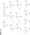
- the alkyne group can be attached directly to the pyridine ring of the cofactor (such as PL1 or PL1P) or with a spacer, such as an ethylene spacer (such as PL2 or PL2P).
- a cofactor such as PL1 or PL1P
- a spacer such as an ethylene spacer (such as PL2 or PL2P).
- the phosphorylated probes can be obtained from the reaction of the respective PLx probe with the enzyme S.
- Aureus protein kinase PLK such as described in Hoegl et al. (2016) or Fux et al. (2019).
- the functionalized cofactors are preferably selected from PL1P, PL2P, PL4P to PL13P.
- the biorthogonal labeling group is an azide group, and the cofactor is preferably PL3P.
- PL3P (6-(azidomethyl)-4-formyl-5-hydroxypyridin-3-yl)methyl phosphate
- the cofactor of PLP-DEs is further functionalized with a phosphoamidate.
- the probe comprises at least two functionalized cofactors of PLP-DEs or it comprises three or more functionalized cofactors.
- the probe comprises a further moiety or tag or further moieties/tags, such as biotin.
- step (c) of the method of the present invention the erythrocytes are lysed and thereby release the proteome including the PLP-DEs.
- the lysis of the erythrocytes comprises the removal of hemoglobin.
- Removal of hemoglobin is preferred, because PLP binds to hemoglobin, which can disturb the result of the method of the present invention.
- Step (c) can further comprise incubation with lysis buffer (preferably on ice) for 5 min and removal of hemoglobin with NiNTA-agarose beads (e.g. ratio ⁇ 10 ⁇ L lysate to 100 ⁇ L beads).
- Step (c) of the method of the present invention further comprises that the lysate obtained is treated with the PLP probe provided in (b).
- the treatment comprises an incubation of the lysate with the PLP probe and optional addition of a reducing agent.
- the PLP probe Under reducing conditions, the PLP probe will bind irreversibly to the PLP DEs.
- reduction can be performed - if necessary - with sodium borohydride NaBH 4 .
- step (c') of the method of the present invention the erythrocytes are treated with the PLP probe provided in (b).
- Said treatment with the PLP probe is carried out before lysing the erythrocytes.
- the PLP probe is a cofactor of PLP-DEs which is (further) functionalized with a phosphoamidate.
- the treatment comprises an incubation of the lysate with the PLP probe and optional addition of a reducing agent.
- step (c') after the treatment, the erythrocytes are lysed and thereby release the proteome including the PLP-DEs.
- the lysis of the erythrocytes comprises the removal of hemoglobin.
- Removal of hemoglobin is preferred, because PLP binds to hemoglobin, which can disturb the result of the method of the present invention.
- Step (c) can further comprise incubation with lysis buffer (preferably on ice) for 5 min and removal of hemoglobin with NiNTA-agarose beads (e.g. ratio ⁇ 10 ⁇ L lysate to 100 ⁇ L beads).
- step (d) of the method of the present invention a label for the PLP-probe is added to the treated lysate of (c) or the lysate of (c').
- the label in step (d) is
- the label is preferably attached to the biorthogonal labeling group of the cofactor(s).
- the label preferably comprises a complementary functional group to the biorthogonal labeling group of the cofactor(s). Both the biorthogonal labeling group and the respective complementary functional group react with each other, such as via click chemistry or Staudinger ligation.
- step (e) of the method of the present invention the labeled PLP-DEs are captured with anti-PLP-DE-antibodies immobilized on a surface.
- the surface is preferably an array or a membrane. Suitable surfaces are known to the skilled artisan.
- the anti-PLP-DE-antibodies can be monoclonal or polyclonal antibodies. Such antibodies are commercially available. For example:
- a second anti-PLP-DE-antibody is added.
- Said second anti-PLP-DE-antibody is not immobilized on a surface.
- Said second anti-PLP-DE-antibody comprises a label, which is different from the label of the PL-P-probe.
- the second anti-PLP-DE-antibody is a polyclonal antibody which can be the same as the capturing anti-PLP-DE-antibody due to its polyclonality.
- step (f) of the method of the present invention the labeled PLP-DEs are detected.
- the method of detection and the readout depend on the label used.
- the fluorescence is detected and preferably fluorescence intensity is determined.
- the fluorescence intensity can be the readout.
- the label is a bioluminescence label
- the bioluminescence is detected and preferably bioluminescence intensity is determined.
- the bioluminescence intensity can be the readout.
- the color can be detected or absorbance can be detected.
- the label of said second antibody is detected as well.
- the PLP-probe signal reflects the loading state of the cofactor (wherein higher PLP probe signal corresponds to a lower loading state of the cofactor) and (2) the antibody signal reflects the total amount of PLP-DE enzyme in the sample.
- the free vitamin b6 status of the patients in blood can be determined via HPLC to calibrate the ratios.
- step (g) of the method of the present invention the vitamin status is determined.
- the vitamin status is determined by comparing the readout of step (f), such as fluorescence intensity, bioluminescence intensity, or absorbance, with a control.
- Preferred controls are recombinant PLP-DEs with defined cofactor loading or reference samples where the PLP status is known, e.g. in form of a calibration curve.
- the vitamin B6 status of the subject is determined.
- the method of the present invention is a point of care method.
- Point-of-care testing is a form of testing in which the analysis is performed where healthcare is provided close to or near the patient. POCT can be undertaken in many locations including: home use and self testing.
- the method of the present invention further comprises the steps of:
- the vitamin status determined in a subject is used for determining the vitamin B6 status of the subject.
- the vitamin status determined in a subject is used for monitoring the PLP status and/or vitamin B6 status of the subject.
- the vitamin status determined in a subject is used for diagnosing vitamin B6-deficiency and vitamin B6-deficiency-associated diseases
- Vitamin B6 comprises six compounds based on a 2-methyl-3-hydroxypyridine structure with differing subunits at positions C4 and C5 that are interconvertible. These are pyridoxine, pyridoxamine, and pyridoxal and their phosphorylated derivatives pyridoxine-5-phosphate, pyridoxamine-5-phosphate, and pyridoxal-5-phosphate (PLP). PLP is a cofactor in > 150 different enzyme reactions including transamination and decarboxylation enzyme systems.
- the symptoms of vitamin B 6 deficiency include peripheral neuropathy, pellagra-like syndrome with seborrheic dermatitis and glossitis. Prolonged deficiency can lead to depression, confusion, and in severe cases causes abnormalities in EEG signals and seizures.
- Vitamin B 6 deficiency is often associated with deficiencies in other B vitamins and can be due to overall poor nutrition caused by a chaotic lifestyle, e.g., in alcoholic subjects. Suboptimal vitamin B 6 status has also been reported in oral contraceptive users, smokers, and patients with celiac disease or diabetes Secondary vitamin B 6 deficiency can occur in treatments with drugs that interact with PLP, e.g., isoniazid or inborn errors in vitamin B 6 salvage pathways or when mutations result in the accumulation of intermediates that react with PLP. Vitamin B 6 deficiency has been reported to be associated with an increased risk of cardiovascular disease, stroke, and cancer. Low plasma PLP has been reported in a number of diseases associated with inflammation including rheumatoid arthritis, inflammatory bowel disease, diabetes, and deep vein thrombosis.
- the present invention provides a kit for in vitro determining the vitamin status in a sample of a subject.
- the vitamin status of health related vitamins is determined, in particular vitamin B2, B6, B12 and/or D3.
- Said kit comprises
- the present invention provides an immunographic device for in vitro determining the vitamin status in a sample of a subject.
- the probe comprising at least one functionalized cofactor of PLP-dependent enzymes (PLP-DEs) (PLP probe) is as defined herein above.
- the label for the PLP-probe is as defined herein above.
- the vitamin status of health related vitamins is determined, in particular vitamin B2, B6, B12 and/or D3.
- Said device comprises a solid carrier or surface coated with anti-PLP-DE-antibodies or a solid carrier or surface coated with nanoparticles comprising a moiety or tag which binds to a probe comprising at least one functionalized cofactor of PLP-dependent enzymes (PLP-DEs) (PLP probe).
- PLP-DEs PLP-dependent enzymes
- the immunographic device is a lateral flow assay (LFA) device, which is more preferably a point-of-care device.
- LFA lateral flow assay
- the immunographic device comprises a porous membrane operably connected to
- the sample is whole blood or erythrocytes.
- the erythrocytes are harvested therefrom, as decribed above
- obtaining a processed sample which is applied to the device of the present invention, comprises:
- obtaining a processed sample which is applied to the device of the present invention, comprises:
- the PLP-probe comprises a biotin tag and the gold nanoparticles in the test portion/line are streptavidin-gold nanoparticles.
- control portion or control line comprises antibodies against streptavidin, or biotin or biotin derivatives to capture the streptavidin-gold nanoparticles.
- the inventors developed chemical PLP probes which successfully captured 75 % of all known bacterial PLP-DEs. These probes largely mimic the pyridoxal core scaffold and bear a biorthogonal alkyne handle for click chemistry to a reporter tag.
- the probes infiltrate cognate pyridoxal uptake into bacteria, get phosphorylated to the corresponding PLP probes by pyridoxal kinases and finally become incorporated into PLP-DEs.
- the inventors also applied this methodology to study the human PLP-ome (published under Fux et al. (Cell Chemical Biology, 2019 ), where they demonstrated the utility of these PLP-cofactor mimics for the identification of human PLP-binding proteins in live cells.
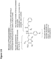
- PLP probes are highly versatile tools to directly determine the PLP status in human erythrocytes. See Figure 2 .
- PLP-dependent enzymes Human erythrocytes express seven known PLP-dependent enzymes (Bryk et al., 2017) and the activity of PLP-DEs is generally dependent on the amount of PLP bound (Ueland et al., 2015).
- the inventors show that a PLP probe can immediately monitor the PLP bound status of all seven enzymes in parallel without the need of performing individual activity assays. This not only enhances the reliability but also provides personalized parameters due to an assessment of multiple enzymes with essential cellular functions. For example, if the PLP status is low more probe binding will be observed to these enzymes and accordingly a higher probe-bound PLP-DE signal will be obtained. If the levels are high, the signal goes down.
- Such a direct assay provides a readout of the overall B6 status (direct parameter) and furthermore yields individual parameters on several signature PLP-DEs which may correlate with individual disease predispositions (personalized parameter).
- the invention provides a method as well as a ready-to-use device, termed B6VitaStat, which is able to directly monitor the PLP status of human erythrocytes based on multiple readout parameters.
- Erythrocytes are treated with our PLP cofactor mimics.
- PLP-DEs are labeled with our PLP probe and subsequently clicked to a fluorescent rhodamine tag.
- Labeled proteins are captured by commercial antibodies against PLP-DEs which are immobilized on a surface, such as glass slides (microarray) or membranes. The array is washed and analyzed in a fluorescent scanner.
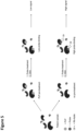
- Recombinant PLP-DEs with defined cofactor loading are added to the lysate as internal standard to provide a quality control and facilitate a quantitative readout by comparison of signal intensities ( Figure 3 ).
- the patient's blood is split into two equivalent samples and one is pre-incubated with a defined PLP standard prior to probe treatment.
- a defined PLP standard prior to probe treatment.
- less PLP probe can bind to the human PLP-DEs resulting in a lower (fluorescent) signal, whereas more probe is able to bind the non-pretreated sample leading to higher signals.
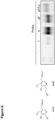
- Comparison of the obtained signals upon (fluorescent) readout on the microarray allows the determination of the patient's PLP status. Implementation of threshold values allows to facilitate the recognition of PLP deficiencies in humans ( Figure 4 ).
- the method of the present invention is able to determine the PLP status immediately, preserves the facile cofactor prior to degradation, provides personalized parameters based on multiple enzymes and is independent from unbound PLP in erythrocytes. Most importantly, this methodology does not require expensive HPLC-instruments and only a much cheaper fluorescent device is needed. Thus, the corresponding test can also be performed easily in developing countries, where malnutrition and B6 deficiency is a big problem.
- Signal detection and amplification can also be adjusted to different settings including smartphone-based sensing methods (Kanchi et al., 2018) paving the way for a ready-to-use device at family doctor offices or at home.
- B6VitaStat platform is innovative as it provides a new approach to overcome limitations of established methods suffering from long processing times, indirect readouts, narrow scope and need for high-end instruments.
- tests comprise direct PLP determination via HPLC analysis or indirect PLP determination based on enzymatic activity (such as disclosed in German patent No. DE 000060037311 T2 ) or bioactivity (e.g. ID-Vit ® -Vitamin B6 Assay from Immundiagnostik, Germany).
- B6VitaStat according to the present invention represents an improved diagnostic platform for continuous and direct monitoring of the vitamin B6 status directly from patient blood without the need of expensive equipment providing rapid processing analysis.
- Vitamin D which can be detected by different immunoassay methods, such as electrochemiluminescent assay Elecsys ® (Roche Diagnostics) or Preventis SmarTest Pro ® Vitamin D
- B6 determination can now be offered on a more frequent basis or even independent of diagnostic laboratories. For example, comparison of blood samples from different patients suffering under diverse B6-deficiency associated diseases will likely provide patterns characteristic for the respective pathologies.
- B6VitaStat thus not only provides rapid and cheap access to the nutritional B6 status but can also serve as a device linked to machine learning with important implications for personalized medicine and diagnostics.
- the microarray can be extended to a sandwich format to enable normalization of signal intensities and to interpret the vitamin status of a subject. Therefore, after incubation of the PLP treated lysate on the microarrays, a second antibody bearing a different fluorescent label than the PLP probe is added and a two-channel fluorescence readout is performed.
- a second antibody bearing a different fluorescent label than the PLP probe is added and a two-channel fluorescence readout is performed.
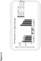
- high ratios of PLP probe/protein amount indicate low vitamin B6 status. Comparison of two healthy volunteers with differences in their vitamin B6 status (as pre-determined via HPLC) verified this theory making this platform a novel platform for vitamin status assessment (see e.g. Figure 12 ).
- Probes were synthesized and phosphorylation of probes was performed by S. aureus pyridoxal kinsa (SaPLK) according to the published procedures in Hoegl et al. (2016) and Pfanzelt et al. (2022). Angewandte Chemie (2022) https://doi.org/10.1002/anie.202117724
- PLPBP and SHMT1 were recombinantly expressed as described previously (Fux et al., 2019).
- E. coli Rosetta2 (DE3) (Merck, Cat# 71400) carrying the expression plasmids were cultured as described and expression was induced through the addition of 0.2 ⁇ g/mL anhydrotetracycline (ATET).
- Bacteria were harvested and washed with PBS (6,000 ⁇ g , 4°C). The cell pellet was resuspended in strep binding buffer and lysed by sonication. The lysate was clarified by centrifugation (36,000 ⁇ g , 30 min, 4°C).
- Concentration of PLPBP and apoSHMT1 was calculated according to Lambert-Beer's law from the absorbance at 280 nm recorded in an InfiniteM200 PRO reader (TECAN, Cat# IN-MNANO). Proteins were stored at -80°C in small aliquots. Intact Protein MS is performed to check protein mass and PLP loading state of the enzyme.
- IP-MS measurements were perfomed on an Ultimate 3000 RSLC system (Thermo Scientific) coupled to a LTQ Orbitrap XL mass spectrometer (Thermo Scientific). Protein desalting was carried out using a MassPREP desalting column (Waters) at 25 °C. Gradient elution was carried out with 0.1% formic acid (LC-MS grade, Fisher Analytics) in water (LC-MS grade, Fisher Analytics) (A) and 0.1% formic acid in acetonitrile (MeCN, LC-MS grade, Fisher Analytics) (B).
- Human erythrocytes were obtained from freshly drawn whole blood in EDTA tubes (4 ml each) via centrifugation (1 ⁇ 3500 rpm, 6 min, 1 ⁇ 3500 rpm, 8 min, 1 ⁇ 3000 rpm, 8 min). Supernatant (blood plasma) and buffy coat is removed and washed with PBS (5 mL) after each centrifugation step. To 3-4 mL erythrocytes are added 3-4 mL lysis buffer (Tris 50 mM, NaCl 150 mM, 1% NP40 (v/v), pH 7.5), incubated on ice for 5 min and frozen at -80°C o/n.
- 3-4 lysis buffer Tris 50 mM, NaCl 150 mM, 1% NP40 (v/v), pH 7.5
- erythrocyte lysate is centrifuged (13 000 rpm, 10 min, 4 °C) and total protein amount is determined by BCA Assay (ROTI ® Quant, Roth). Erythrocyte lysate is stored in small aliquots at -80°C.
- Ni-NTA agarose beads (Qiagen) are carefully resuspended by inversion on an Eppendorf tube wheel at 4 °C and transferred to an Eppendorf tube (X ⁇ 100 ⁇ L + 25% in one LoBind Eppendorf tube with marked filling level before washing steps are performed.
- the beads are washed with PBS (3 ⁇ 1 mL), centrifuged (400 g, 3 min, r.t.) and filled to the original level with PBS.
- To 100 ⁇ L of bead suspension are added 7 ⁇ L erythrocyte lysate and incubated for 30 min at r.t..
- the samples are transferred quantitatively onto biospin columns and centrifuged (1000 g, 2 min, r.t.). Collected flow through is filled up to a final volume of 100 ⁇ L with PBS and further subjected to probe labelling.
- Recombinant protein or erythrocyte lysate in PBS are incubated with 40 ⁇ M PlxP probe (addition of 1-2 ⁇ L of respective stock) for 1h at room temperature.
- Samples are either directly subjected to Click chemistry or if irreversible probe biding is required to reduction with NaBH 4 .
- the internal aldimine is reduced to the respective secondary amine with NaBH 4 .
- Labelled recombinant protein or erythrocyte lysate in PBS are treated with 20 mM NaBH 4 (250 mM stock solution in 0.1 M NaOH) and incubated for 30 min at r.t. Reduction is quenched by adding cold acetone (4 ⁇ volume of sample, -20°C) for at least 1h or o/n. Precipitated protein is pelletized (10 000 rpm, 15 min, 4 °C) and supernatant is discarded. For washing, protein pellet is resuspended in 0.5 mL cold methanol (-80 °C) by mild sonication (10 s, 10% intensity) and centrifuged (10 000 rpm, 10 min, 4 °C). Finally, each protein pellet is resuspended in 100 ⁇ L PBS by sonication (10 s, 10% intensity).
- the samples are subjected to Click reaction by adding 1 ⁇ L rhodamine azide (10 mM in DMSO), 6 ⁇ L TBTA (1.667 mM in t BuOH/DMSO 80:20), 1 ⁇ L TCEP (15 mg/mL in ddH 2 O) and 2 ⁇ L CuSO 4 (50 mM in ddH 2 O) to each sample (100 ⁇ L) and incubated for at least 1 h at r.t. Samples are subsequently analysed on the microarrays.
- Custom microarrays were produced using the automated production facility at Sciomics. Standard production protocols using contact printing on chemically modified glass surfaces (Epoxy-coupling groups) were used and slides were individually quality controlled after production. Produced Slides were stored according to Sciomics SOPs cold and protected from light.
- the secondary antibodies were labelled at an adjusted protein concentration for two hours with scioDye2 (Sciomics, Germany). After two hours the reaction was stopped and the buffer exchanged to PBS. All labelled protein samples were stored at -20° C until use.
- the samples were analysed in a single colour approach (without secondary antibody incubation) or dual-colour approach (with secondary antibody incubation) using custom microarrays containing antibodies against OAT, SHMT and PLPBP or a subset of these antibodies. Each antibody is represented on the array in 16 replicates.
- the arrays were blocked with scioBlock (Sciomics) on a Hybstation 4800 (Tecan, Austria) and afterwards the samples were incubated. After incubation for three hours, the slides were thoroughly washed with 1x PBSTT, rinsed with 0.1x PBS as well as with water and subsequently dried with nitrogen.
- PLPBP Human PLP binding protein
- PL4P Human PLP binding protein
- the antibodies were immobilized on glass sides, which contain several replicates of the antibody for quality control and statistical evaluation.
- Recombinant PLPBP incubated with PL4P and clicked to Cy5 was used. Results see Figure 7 showing that all three immobilized antibodies against PLPBP work.
- the additional background reduction step enabled the successful detection of endogenous PLPBP in human erythrocytes with our PL4P probe proving our overall B6VitaStat concept.
- Spike-in of recombinant PLPBP to the human erythrocyte lysate additionally shows a dependency of the signal intensity on the amount of PLP-DE present in the sample. Results see Figure 9 . This is also visible in the obtained microarray images (see Figure 10 ), where human erthyrocyte lysate was also spiked with PLPBP. This image additionally shows the extension of our microarray format to several PLP-DEs. In total, 7 different antibodies targeting the three PLP-DEs PLPBP, SHMT1 and OAT showed very good to moderate signal intensities proving that the array can be exploited for all PLP-DEs if suitable antibodies are available.
- the microarray is extended to a sandwich format to enable normalization of signal intensities and to interpret the vitamin status of a subject. Therefore, after incubation of the PLP treated lysate on the microarrays, a second antibody bearing a different fluorescent label than the PLP probe is added and a two-channel fluorescence readout is performed.
- a second antibody bearing a different fluorescent label than the PLP probe is added and a two-channel fluorescence readout is performed.
- high ratios of PLP probe/protein amount would indicate low vitamin B6 status.
- Comparison of two healthy volunteers with huge differences in their vitamin B6 status verified this theory making this platform a novel platform for vitamin status assessment (see Figure 12 ).
Landscapes
- Health & Medical Sciences (AREA)
- Life Sciences & Earth Sciences (AREA)
- Engineering & Computer Science (AREA)
- Immunology (AREA)
- Molecular Biology (AREA)
- Biomedical Technology (AREA)
- Chemical & Material Sciences (AREA)
- Hematology (AREA)
- Urology & Nephrology (AREA)
- Cell Biology (AREA)
- Microbiology (AREA)
- Biotechnology (AREA)
- Food Science & Technology (AREA)
- Medicinal Chemistry (AREA)
- Physics & Mathematics (AREA)
- Analytical Chemistry (AREA)
- Biochemistry (AREA)
- General Health & Medical Sciences (AREA)
- General Physics & Mathematics (AREA)
- Pathology (AREA)
- Investigating Or Analysing Biological Materials (AREA)
Abstract
The present invention relates to in vitro methods for determining the vitamin status of a subject, in particular health related vitamins, such as B2, B6, B12 and/or D3. The present invention further relates to a kit and an immunographic device for in vitro determining the vitamin status of a subject.
Description
- The present invention relates to in vitro methods for determining the vitamin status of a subject, in particular health related vitamins, such as B2, B6, B12 and/or D3. The present invention further relates to a kit and an immunographic device for in vitro determining the vitamin status of a subject.
- Vitamin B6 refers to a collection of vitamers including the enzyme cofactor pyridoxal phosphate (PLP). B6 is an essential nutrient that has to be taken up by a well-balanced diet. While the supply of B6 in wholesome foods including meat, bananas and potatoes is sufficient, processing of meals can significantly reduce the amount of B6 vitamers due to their limited stability (Gregory and Kirk, 1977).
- The Centers for Disease Control and Prevention showed in a study that about 10 % of the US population exhibits plasma PLP levels which are below 20 nmol/L, considered as threshold value for sufficient supply, indicating an inadequate B6 status (Center for Disease Control and Prevention, 2012). Malnutrition, as often observed in developing countries, further intensifies the problem. B6 deficiency is associated with various symptoms including higher rates of cardiovascular disease, stroke and cancer (Verhoef et al., 1996; Robinson et al., 1998; Sies et al., 1992; Vanuzzo et al., 2007; Dalery et al., 1995; Rimm et al., 1998; Johansson et al., 2010; Kelly et al., 2004; Lotto et al., 2011). Populations at risk to exhibit critically low B6 levels include elderly people, diabetes patients and alcoholics. Additionally, healthy people with increased needs of B6, such as pregnant and lactating women require a stringent and continuous monitoring of B6 levels. Here, several diagnostic procedures are on the market. Plasma PLP is the most prevalent biomarker used (Leklem, 1990), which is detected by high performance liquid chromatography (HPLC) as part of laboratory diagnostics. Major challenges in its determination are the
- overall stability of PLP, whose plasma concentration declines in samples kept at room temperature and exposed to light (Hustad et al., 2012; Panton et al.,2013; van der Ham et al., 2012);
- inflammation, albumin binding, alkaline phosphatase activity and alcohol consumption can result in a 30 - 40 % variance (Brussaard et al., 1997; Ueland et al., 2015).
- Thus, erythrocyte PLP determination via HPLC or functional assays have been identified as a more relevant marker of the B6 status also reflecting the fact, that the vitamer is an intracellular cofactor (Leklem, 1990; Ueland et al., 2015; Vermaak et al., 1990). Blood can be rapidly drawn from patients providing an opportunity to directly monitor effects of B6 supplementation. However, a disadvantage of direct PLP measurement is the binding of PLP to hemoglobin, lowering the overall precision (Ueland et al., 2015; Reynolds et al., 1995). As an alternative, functional B6 biomarker tests are based on PLP-dependent enzymes (PLP-DEs) that are supposed to overcome these problems. Here, the transaminase test is one of the most mature assay systems monitoring the activity of aspartic acid transaminase or alanine transaminase in erythrocyte extract (Ueland et al., 2015). While this assay overcomes above mentioned limitations of free-PLP detection, a major challenge is the time-consuming processing of fresh blood for these assays limiting the overall stability of PLP-DEs (Ueland et al., 2015; Huang et al., 1998).
- As of yet, there is no diagnostic B6 test available which combines analysis of blood samples, detection of most relevant enzyme based-PLP-cofactor status and speed for a reliable analysis. No personalized parameters which are based on recording the status of multiple enzymes can be obtained via the methods of the prior art.
- There is a need in the art for improved methods and means for determining the vitamin status of a subject.
- According to the present invention this object is solved by an in vitro method for determining the vitamin status of a subject comprising
- (a) harvesting erythrocytes from a sample obtained from a subject or providing erythrocytes obtained from a sample of a subject;
- (b) providing a probe comprising at least one functionalized cofactor of PLP-dependent enzymes (PLP-DEs) (PLP probe);
- (c) lysing the erythrocytes and thereby releasing the proteome including the PLP-DEs and then treating the lysate with the PLP probe provided in (b); or
- (c') treating the erythrocytes with the PLP probe provided in (b) and then lysing the erythrocytes and thereby releasing the proteome including the PLP-DEs;
- (d) adding a label for the PLP-probe to the treated lysate of (c) or to the lysate of (c');
- (e) capturing the labeled PLP-DEs with anti-PLP-DE-antibodies immobilized on a surface, preferably an array or a membrane,
- (f) detecting the labeled PLP-DEs, and
- (g) determining the vitamin status.
- According to the present invention this object is solved by a kit for in vitro the vitamin status, in particular vitamin B6, B2, B12 and/or D3, in a sample of a subject, comprising
- (1) a probe comprising at least one functionalized cofactor of PLP-dependent enzymes (PLP-DEs) (PLP probe);
- (2) a label for the PLP-probe;
- (3) optionally, an array for PLP-DEs, preferably comprising anti-PLP-DE-antibodies,
- (4) optionally, reagents for lysing erythrocytes, reducing agents, and control(s).
- According to the present invention this object is solved by an immunographic device for in vitro determining the vitamin status, in particular vitamin B6, B2, B12 and/or D3, in a sample of a subject,
said device comprising a solid carrier or surface coated with anti-PLP-DE-antibodies, wherein the immunographic device is preferably a lateral flow assay (LFA) device. - Before the present invention is described in more detail below, it is to be understood that this invention is not limited to the particular methodology, protocols and reagents described herein as these may vary. It is also to be understood that the terminology used herein is for the purpose of describing particular embodiments only, and is not intended to limit the scope of the present invention which will be limited only by the appended claims. Unless defined otherwise, all technical and scientific terms used herein have the same meanings as commonly understood by one of ordinary skill in the art. For the purpose of the present invention, all references cited herein are incorporated by reference in their entireties.
- Concentrations, amounts, and other numerical data may be expressed or presented herein in a range format. It is to be understood that such a range format is used merely for convenience and brevity and thus should be interpreted flexibly to include not only the numerical values explicitly recited as the limits of the range, but also to include all the individual numerical values or sub-ranges encompassed within that range as if each numerical value and sub-range is explicitly recited. As an illustration, a numerical range of "1 to 21" should be interpreted to include not only the explicitly recited values of 1 to 21, but also include individual values and sub-ranges within the indicated range. Thus, included in this numerical range are individual values such as 1, 2, 3, 4, 5 .... 17, 18, 19, 20, 21 and sub-ranges such as from 2 to 10, 8 to 15, etc. This same principle applies to ranges reciting only one numerical value, such as "at least 90%". Furthermore, such an interpretation should apply regardless of the breadth of the range or the characteristics being described.
- As outlined above, the present invention provides an in vitro method for determining the vitamin status of a subject.
- Preferably, the vitamin status of health related vitamins is determined, in particular vitamin B2, B6, B12 and/or D3.
- Said method comprises the steps of:
- (a) harvesting erythrocytes from a sample obtained from a subject or providing erythrocytes obtained from a sample of a subject;
- (b) providing a probe comprising at least one functionalized cofactor of PLP-dependent enzymes (PLP-DEs) (PLP probe);
- (c) lysing the erythrocytes and thereby releasing the proteome including the PLP-DEs and then treating the lysate with the PLP probe provided in (b);
or - (c') treating the erythrocytes with the PLP probe provided in (b) and then lysing the erythrocytes and thereby releasing the proteome including the PLP-DEs;
- (d) adding a label for the PLP-probe to the treated lysate of (c) or to the lysate of (c');
- (e) capturing the labeled PLP-DEs with anti-PLP-DE-antibodies immobilized on a surface, preferably an array or a membrane,
- (f) detecting the labeled PLP-DEs, and
- (g) determining the vitamin status.
- In one embodiment, the method comprises the steps of:
- (a) harvesting erythrocytes from a sample obtained from a subject or providing erythrocytes obtained from a sample of a subject;
- (b) providing a probe comprising at least one functionalized cofactor of PLP-dependent enzymes (PLP-DEs) (PLP probe);
- (c) lysing the erythrocytes and thereby releasing the proteome including the PLP-DEs and then treating the lysate with the PLP probe provided in (b);
- (d) adding a label for the PLP-probe to the treated lysate of (c);
- (e) capturing the labeled PLP-DEs with anti-PLP-DE-antibodies immobilized on a surface, preferably an array or a membrane,
- (f) detecting the labeled PLP-DEs, and
- (g) determining the vitamin status.
- In an alternative embodiment, the method comprises the steps of:
- (a) harvesting erythrocytes from a sample obtained from a subject or providing erythrocytes obtained from a sample of a subject;
- (b) providing a probe comprising at least one functionalized cofactor of PLP-dependent enzymes (PLP-DEs) (PLP probe);
- (c') treating the erythrocytes with the PLP probe provided in (b) and then lysing the erythrocytes and thereby releasing the proteome including the PLP-DEs;
- (d) adding a label for the PLP-probe to the lysate of (c');
- (e) capturing the labeled PLP-DEs with anti-PLP-DE-antibodies immobilized on a surface, preferably an array or a membrane,
- (f) detecting the labeled PLP-DEs, and
- (g) determining the vitamin status.
- In step (a) of the method of the present invention, erythrocytes are harvested from a sample obtained from a subject or erythrocytes obtained from a sample of a subject are provided.
- In the embodiment, where the erythrocytes are harvested from a sample, they are preferably harvested by centrifugation, optionally including washing step(s).
- The sample is preferably whole blood.
- In step (b) of the method of the present invention, a probe is provided which comprises at least one functionalized cofactor of PLP-dependent enzymes (PLP-DEs) (PLP probe).
- PLP-DEs have been implicated in human disease and are recognized as important drug targets. PLP-DEs include aspartic acid transaminase, alanine racemase, serine hydroxymethyltransferase, ornithine decarboxylase, GABA aminotransferase, DOPA decarboxylase, and branched-chain amino acid aminotransferase. PLP acts as a coenzyme in all transamination reactions, and in certain decarboxylation, deamination, and racemization reactions of amino acids. PLP is also involved in various beta-elimination reactions such as the reactions carried out by serine dehydratase and GDP-4-keto-6-deoxymannose-3-dehydratase (ColD).
- Preferably, the cofactor(s) of PLP-dependent enzymes (PLP-DEs) are functionalized with a biorthogonal labeling group.
- In one embodiment, the cofactor(s) of PLP-dependent enzymes (PLP-DEs) are functionalized with a biorthogonal labeling group and optionally further with a phosphoamidate capping group.
- The term "bioorthogonal chemistry" refers to any chemical reactions that can occur inside of living systems or in biological samples in general, that do not interfere with the biological system as well as side reactions in the organism or with the biological sample do not take place making bioorthogonal chemistry extremely selective reactions within complex biological matrices.
- A number of chemical ligation strategies have been developed that fulfill the requirements of bioorthogonality, including
1,3-dipolar cycloaddition between azides and cyclooctynes (also termed copper-free click chemistry), between nitrones and cyclooctynes, oxime/hydrazone formation from aldehydes and ketones, the tetrazine ligation, the isocyanide-based click reaction, and the quadricyclane ligation. Furthermore the copper catalyzed azide alkyne click reaction (CuAAC) and the Staudinger ligation. - The use of bioorthogonal chemistry usually proceeds in two steps. First, a (cellular) substrate is modified with a bioorthogonal functional group ("biorthogonal labeling group"). Secondly, a probe containing the complementary functional group is introduced to react and label the substrate.
- In a preferred embodiment, the biorthogonal labeling group is used to modify or functionalize the cofactor(s) of the PLP-DEs. The complementary functional group is added to the label.
- The biorthogonal labeling group is preferably selected from an alkyne or azide moiety or a tetrazine functionality.
- In a preferred embodiment, the biorthogonal labeling group is an alkyne group.
- The alkyne group can be attached directly to the pyridine ring of the cofactor (such as PL1 or PL1P) or with a spacer, such as an ethylene spacer (such as PL2 or PL2P).
- The phosphorylated probes (PLxP) can be obtained from the reaction of the respective PLx probe with the enzyme S. Aureus protein kinase PLK, such as described in Hoegl et al. (2018) or Fux et al. (2019).
- The functionalized cofactors are preferably selected from PL1P, PL2P, PL4P to PL13P.
- PL1P : (6-ethynyl-4-formyl-5-hydroxypyridin-3-yl)methyl phosphate
- PL2P : (4-formyl-6-(hept-6-yn-1-yl)-5-hydroxypyridin-3-yl)methyl phosphate
- PL4P : (6-(but-3-yn-1-yl)-4-formyl-5-hydroxypyridin-3-yl)methyl phosphate
- PL5P : (6-(4-ethynylphenethyl)-4-formyl-5-hydroxypyridin-3-yl)methyl phosphate
- PL6P : (4-formyl-5-hydroxy-6-(pent-4-yn-1-yl)pyridin-3-yl)methyl phosphate
- PL7P : (4-formyl-6-(hex-5-yn-1-yl)-5-hydroxypyridin-3-yl)methyl phosphate
- PL8P : (4-formyl-5-hydroxy-6-(2-(1-(prop-2-yn-1-yl)-1H-1,2,3-triazol-4-yl)ethyl)pyridin-3-yl)methyl phosphate
- PL9P : (6-(2-(1-(but-3-yn-1-yl)-1H-1,2,3-triazol-4-yl)ethyl)-4-formyl-5-hydroxypyridin-3-yl)methyl phosphate
- PL10P : (2-ethynyl-4-formyl-5-hydroxy-6-methylpyridin-3-yl)methyl phosphate
- PL11P: (4-formyl-5-hydroxy-6-methyl-2-(1-(prop-2-yn-1-yl)-1H-1,2,3-triazol-4-yl)pyridin-3-yl)methyl phosphate
- PL12P : (2-(but-3-yn-1-yl)-4-formyl-5-hydroxy-6-methylpyridin-3-yl)methyl phosphate PL13P : (4-formyl-6-methyl-5-(prop-2-yn-1-yloxy)pyridin-3-yl)methyl phosphate,
- In a preferred embodiment, the biorthogonal labeling group is an azide group, and the cofactor is preferably PL3P.
- PL3P: (6-(azidomethyl)-4-formyl-5-hydroxypyridin-3-yl)methyl phosphate
- In a preferred embodiment, the cofactor of PLP-DEs is further functionalized with a phosphoamidate.
- In one embodiment, the probe comprises at least two functionalized cofactors of PLP-DEs or it comprises three or more functionalized cofactors.
- In one embodiment, the probe comprises a further moiety or tag or further moieties/tags, such as biotin.
- In step (c) of the method of the present invention, the erythrocytes are lysed and thereby release the proteome including the PLP-DEs.
- Preferably, the lysis of the erythrocytes comprises the removal of hemoglobin.
- Removal of hemoglobin is preferred, because PLP binds to hemoglobin, which can disturb the result of the method of the present invention.
- In one embodiment, step (c) comprises the addition of a lysis buffer, preferably in a ratio erythrocytes:buffer = 1:1. The lysis buffer can be any suitable buffer to lyse the cells, such as Tris buffer (Tris 50 mM, NaCl 150 mM, 1% NP40 (v/v), pH=7.5). Step (c) can further comprise incubation with lysis buffer (preferably on ice) for 5 min and removal of hemoglobin with NiNTA-agarose beads (e.g. ratio ~ 10 µL lysate to 100 µL beads).
- Step (c) of the method of the present invention further comprises that the lysate obtained is treated with the PLP probe provided in (b).
- Preferably, the treatment comprises an incubation of the lysate with the PLP probe and optional addition of a reducing agent.
- Under reducing conditions, the PLP probe will bind irreversibly to the PLP DEs.
- In one embodiment, reduction can be performed - if necessary - with sodium borohydride NaBH4.
- In step (c') of the method of the present invention, the erythrocytes are treated with the PLP probe provided in (b).
- Said treatment with the PLP probe is carried out before lysing the erythrocytes.
- Preferably, the PLP probe is a cofactor of PLP-DEs which is (further) functionalized with a phosphoamidate.
- Preferably, the treatment comprises an incubation of the lysate with the PLP probe and optional addition of a reducing agent.
- In step (c'), after the treatment, the erythrocytes are lysed and thereby release the proteome including the PLP-DEs.
- Preferably, the lysis of the erythrocytes comprises the removal of hemoglobin.
- Removal of hemoglobin is preferred, because PLP binds to hemoglobin, which can disturb the result of the method of the present invention.
- In one embodiment, step (c') comprises the addition of a lysis buffer, preferably in a ratio erythrocytes:buffer = 1:1. The lysis buffer can be any suitable buffer to lyse the cells, such as Tris buffer (Tris 50 mM, NaCl 150 mM, 1% NP40 (v/v), pH=7.5). Step (c) can further comprise incubation with lysis buffer (preferably on ice) for 5 min and removal of hemoglobin with NiNTA-agarose beads (e.g. ratio ~ 10 µL lysate to 100 µL beads).
- In step (d) of the method of the present invention, a label for the PLP-probe is added to the treated lysate of (c) or the lysate of (c').
- Preferably, the label in step (d) is
- a fluorescent label,
such as rhodamines, cyanine dyes (Cy3, Cy5), fluoresceines, - a bioluminescence or chemiluminescence label,
such as isoluminol and its derivatives, luciferins, - a dye,
or combinations thereof. - The label is preferably attached to the biorthogonal labeling group of the cofactor(s).
- The label preferably comprises a complementary functional group to the biorthogonal labeling group of the cofactor(s). Both the biorthogonal labeling group and the respective complementary functional group react with each other, such as via click chemistry or Staudinger ligation.
- In step (e) of the method of the present invention, the labeled PLP-DEs are captured with anti-PLP-DE-antibodies immobilized on a surface.
- The surface is preferably an array or a membrane. Suitable surfaces are known to the skilled artisan.
- The anti-PLP-DE-antibodies can be monoclonal or polyclonal antibodies. Such antibodies are commercially available. For example:
- Anti PLPHP_1 Thermofischer, cat. nr. PA532036, polyclonal
- Anti PLPBP_2 Novus Biologicals, cat. No. 21730002 polyclonal
- Anti PLPBP _3 Origene, cat. nr. TA505130, monoclonal
- Anti OAT_1 Origene, cat. AM09363PU-S, polyclonal
- Anti OAT_2 Abcam, cat. Ab137679, polyclonal
- Anti OAT_3 Novus Biologicals, cat. NBP1-49312, monoclonal
- Anti SHMT1_1 Abcam, cat. Ab186130, polyclonal
- Anti SHMT1_2 Merck, cat. ABE416, polyclonal
- Anti SHMT1_3 Novus Biologicals, cat. NBP2-32173, polyclonal
- In a preferred embodiment, in or after step (e) a second anti-PLP-DE-antibody is added.
- Said second anti-PLP-DE-antibody is not immobilized on a surface. Said second anti-PLP-DE-antibody comprises a label, which is different from the label of the PL-P-probe.
- Preferably, the label of the second anti-PLP-DE-antibody is
- a fluorescent label,
such as rhodamines, cyanine dyes (Cy3, Cy5), fluoresceines, - a bioluminescence or chemiluminescence label,
such as isoluminol and its derivatives, luciferins, - a dye,
or combinations thereof. - For example, the second anti-PLP-DE-antibody is a polyclonal antibody which can be the same as the capturing anti-PLP-DE-antibody due to its polyclonality.
- In step (f) of the method of the present invention, the labeled PLP-DEs are detected.
- The method of detection and the readout depend on the label used.
- In an embodiment, where the label is a fluorescent label, the fluorescence is detected and preferably fluorescence intensity is determined. The fluorescence intensity can be the readout. In an embodiment, where the label is a bioluminescence label, the bioluminescence is detected and preferably bioluminescence intensity is determined. The bioluminescence intensity can be the readout.
- In an embodiment, where the label is a dye, the color can be detected or absorbance can be detected.
- In an embodiment, where second anti-PLP-DE-antibodies are used, the label of said second antibody is detected as well.
- In this embodiment, two signals are detected, where (1) the PLP-probe signal reflects the loading state of the cofactor (wherein higher PLP probe signal corresponds to a lower loading state of the cofactor) and (2) the antibody signal reflects the total amount of PLP-DE enzyme in the sample. By comparing these to signals, a ratio PLP loading state/total PLP-DE amount is obtained.
- Healthy patients exhibit roughly the same ratio, whereas vitamin B6 compromised patients exihibit higher ratios.
- For example, in case of vitamin B6 deficiency, little PLP is bound to PLP-DE, such that more PLxP probe can bind to the PLP-DEs which results in a higher signal.
- Additionally, the free vitamin b6 status of the patients in blood can be determined via HPLC to calibrate the ratios.
- In step (g) of the method of the present invention, the vitamin status is determined.
- Preferably, the vitamin status is determined by comparing the readout of step (f), such as fluorescence intensity, bioluminescence intensity, or absorbance, with a control.
- Preferred controls are recombinant PLP-DEs with defined cofactor loading or reference samples where the PLP status is known, e.g. in form of a calibration curve.
- Preferably, the vitamin B6 status of the subject is determined.
- In a preferred embodiment, the method of the present invention is a point of care method.
- Point-of-care testing (POCT) is a form of testing in which the analysis is performed where healthcare is provided close to or near the patient. POCT can be undertaken in many locations including: home use and self testing.
- In preferred embodiments, the method of the present invention further comprises the steps of:
- dividing the sample obtained from the subject in two parts and pre-treating one part with a PLP standard prior to step (c),
- performing steps (a) to (f) for both parts in parallel,
- comparing the results of step (f) for both parts and thereby quantifying the PLP status.
- Preferably, the vitamin status determined in a subject is used for determining the vitamin B6 status of the subject.
- Preferably, the vitamin status determined in a subject is used for monitoring the PLP status and/or vitamin B6 status of the subject.
- Preferably, the vitamin status determined in a subject is used for diagnosing vitamin B6-deficiency and vitamin B6-deficiency-associated diseases,
- in particular related to age, malnutrition, and/or alcoholism.
- in dialysis patients,
- inflammatory bowel disease.
- Vitamin B6 comprises six compounds based on a 2-methyl-3-hydroxypyridine structure with differing subunits at positions C4 and C5 that are interconvertible. These are pyridoxine, pyridoxamine, and pyridoxal and their phosphorylated derivatives pyridoxine-5-phosphate, pyridoxamine-5-phosphate, and pyridoxal-5-phosphate (PLP). PLP is a cofactor in > 150 different enzyme reactions including transamination and decarboxylation enzyme systems. The symptoms of vitamin B6 deficiency include peripheral neuropathy, pellagra-like syndrome with seborrheic dermatitis and glossitis. Prolonged deficiency can lead to depression, confusion, and in severe cases causes abnormalities in EEG signals and seizures. Vitamin B6 deficiency is often associated with deficiencies in other B vitamins and can be due to overall poor nutrition caused by a chaotic lifestyle, e.g., in alcoholic subjects. Suboptimal vitamin B6 status has also been reported in oral contraceptive users, smokers, and patients with celiac disease or diabetes Secondary vitamin B6 deficiency can occur in treatments with drugs that interact with PLP, e.g., isoniazid or inborn errors in vitamin B6 salvage pathways or when mutations result in the accumulation of intermediates that react with PLP. Vitamin B6 deficiency has been reported to be associated with an increased risk of cardiovascular disease, stroke, and cancer. Low plasma PLP has been reported in a number of diseases associated with inflammation including rheumatoid arthritis, inflammatory bowel disease, diabetes, and deep vein thrombosis.
- As outlined above, the present invention provides a kit for in vitro determining the vitamin status in a sample of a subject.
- Preferably, the vitamin status of health related vitamins is determined, in particular vitamin B2, B6, B12 and/or D3.
- Said kit comprises
- (1) a probe comprising at least one functionalized cofactor of PLP-dependent enzymes (PLP-DEs) (PLP probe);
- (2) a label for the PLP-probe;
- (3) optionally, an array for PLP-DEs, preferably comprising anti-PLP-DE-antibodies,
- (4) optionally, reagents for lysing erythrocytes, reducing agents, and control(s).
- As outlined above, the present invention provides an immunographic device for in vitro determining the vitamin status in a sample of a subject.
- Preferably, the probe comprising at least one functionalized cofactor of PLP-dependent enzymes (PLP-DEs) (PLP probe) is as defined herein above.
- Preferably, the label for the PLP-probe is as defined herein above.
- Preferably, the vitamin status of health related vitamins is determined, in particular vitamin B2, B6, B12 and/or D3.
- Said device comprises a solid carrier or surface coated with anti-PLP-DE-antibodies or a solid carrier or surface coated with nanoparticles comprising a moiety or tag which binds to a probe comprising at least one functionalized cofactor of PLP-dependent enzymes (PLP-DEs) (PLP probe).
- Preferably, the immunographic device is a lateral flow assay (LFA) device, which is more preferably a point-of-care device.
- In an embodiment, the immunographic device comprises a porous membrane operably connected to
- (a) a portion or pad, where a processed sample is applied,
- (b) a test portion or test line comprising said anti-PLP-DE-antibodies or said nanoparticles, preferably gold nanoparticles,
- (c) a control portion or test line; and
- (d) an absorbent portion/pad.
- Preferably, the sample is whole blood or erythrocytes.
- In an embodiment, when the sample is whole blood, the erythrocytes are harvested therefrom, as decribed above
- In a preferred embodiment, obtaining a processed sample, which is applied to the device of the present invention, comprises:
- the erythrocytes or the harvested erythrocytes are lysed and thereby release the proteome including the PLP-dependent enzymes (PLP-DEs),
- the lysate is treated with a probe comprising at least one functionalized cofactor of PLP-dependent enzymes (PLP-DEs) (PLP probe), said probe being as defined herein above, and
- a label for the PLP-probe, which is as defined herein above, is added to the treated lysate, and
- the processed sample is obtained.
- In an alternative embodiment, obtaining a processed sample, which is applied to the device of the present invention, comprises:
- the erythrocytes or the harvested erythrocytes are treated with a probe comprising at least one functionalized cofactor of PLP-dependent enzymes (PLP-DEs) (PLP probe), said probe being preferably a cofactor of PLP-DEs which is functionalized with a phosphoamidate,
- the treated erythrocytes are then lysed and thereby release the proteome including the PLP-dependent enzymes (PLP-DEs),
- a label for the PLP-probe, which is as defined herein above, is added to the lysate, and
- the processed sample is obtained.
- In an embodiment, the PLP-probe comprises a biotin tag and the gold nanoparticles in the test portion/line are streptavidin-gold nanoparticles.
- Preferably, the control portion or control line comprises antibodies against streptavidin, or biotin or biotin derivatives to capture the streptavidin-gold nanoparticles.
- The inventors developed chemical PLP probes which successfully captured 75 % of all known bacterial PLP-DEs. These probes largely mimic the pyridoxal core scaffold and bear a biorthogonal alkyne handle for click chemistry to a reporter tag. In a previous study, published under Hoegl A. et al. (Nature Chemistry, 2018), we showed that the probes infiltrate cognate pyridoxal uptake into bacteria, get phosphorylated to the corresponding PLP probes by pyridoxal kinases and finally become incorporated into PLP-DEs. The inventors also applied this methodology to study the human PLP-ome (published under Fux et al. (Cell Chemical Biology, 2019), where they demonstrated the utility of these PLP-cofactor mimics for the identification of human PLP-binding proteins in live cells.
- In the present application, it is first shown that PLP probes are highly versatile tools to directly determine the PLP status in human erythrocytes. See
Figure 2 . - Human erythrocytes express seven known PLP-dependent enzymes (Bryk et al., 2017) and the activity of PLP-DEs is generally dependent on the amount of PLP bound (Ueland et al., 2015). The inventors show that a PLP probe can immediately monitor the PLP bound status of all seven enzymes in parallel without the need of performing individual activity assays. This not only enhances the reliability but also provides personalized parameters due to an assessment of multiple enzymes with essential cellular functions. For example, if the PLP status is low more probe binding will be observed to these enzymes and accordingly a higher probe-bound PLP-DE signal will be obtained. If the levels are high, the signal goes down. Such a direct assay provides a readout of the overall B6 status (direct parameter) and furthermore yields individual parameters on several signature PLP-DEs which may correlate with individual disease predispositions (personalized parameter).
- The invention provides a method as well as a ready-to-use device, termed B6VitaStat, which is able to directly monitor the PLP status of human erythrocytes based on multiple readout parameters. Erythrocytes are treated with our PLP cofactor mimics. Upon lysis of erythrocytes, PLP-DEs are labeled with our PLP probe and subsequently clicked to a fluorescent rhodamine tag. Labeled proteins are captured by commercial antibodies against PLP-DEs which are immobilized on a surface, such as glass slides (microarray) or membranes. The array is washed and analyzed in a fluorescent scanner. Recombinant PLP-DEs with defined cofactor loading are added to the lysate as internal standard to provide a quality control and facilitate a quantitative readout by comparison of signal intensities (
Figure 3 ). - Alternatively for relative quantification, the patient's blood is split into two equivalent samples and one is pre-incubated with a defined PLP standard prior to probe treatment. Thus, less PLP probe can bind to the human PLP-DEs resulting in a lower (fluorescent) signal, whereas more probe is able to bind the non-pretreated sample leading to higher signals. Comparison of the obtained signals upon (fluorescent) readout on the microarray allows the determination of the patient's PLP status. Implementation of threshold values allows to facilitate the recognition of PLP deficiencies in humans (
Figure 4 ). - Thus, the method of the present invention is able to determine the PLP status immediately, preserves the facile cofactor prior to degradation, provides personalized parameters based on multiple enzymes and is independent from unbound PLP in erythrocytes. Most importantly, this methodology does not require expensive HPLC-instruments and only a much cheaper fluorescent device is needed. Thus, the corresponding test can also be performed easily in developing countries, where malnutrition and B6 deficiency is a big problem.
- Signal detection and amplification can also be adjusted to different settings including smartphone-based sensing methods (Kanchi et al., 2018) paving the way for a ready-to-use device at family doctor offices or at home.
- Our novel B6VitaStat platform is innovative as it provides a new approach to overcome limitations of established methods suffering from long processing times, indirect readouts, narrow scope and need for high-end instruments. Currently, tests comprise direct PLP determination via HPLC analysis or indirect PLP determination based on enzymatic activity (such as disclosed in German patent No.
DE 000060037311 T2 ) or bioactivity (e.g. ID-Vit®-Vitamin B6 Assay from Immundiagnostik, Germany). In contrast, B6VitaStat according to the present invention represents an improved diagnostic platform for continuous and direct monitoring of the vitamin B6 status directly from patient blood without the need of expensive equipment providing rapid processing analysis. This kind of monitoring is easy and can also be performed by nonmedical experts, e.g. at home or in developing countries. Like Vitamin D which can be detected by different immunoassay methods, such as electrochemiluminescent assay Elecsys® (Roche Diagnostics) or Preventis SmarTest Pro® Vitamin D, B6 determination can now be offered on a more frequent basis or even independent of diagnostic laboratories. For example, comparison of blood samples from different patients suffering under diverse B6-deficiency associated diseases will likely provide patterns characteristic for the respective pathologies. B6VitaStat thus not only provides rapid and cheap access to the nutritional B6 status but can also serve as a device linked to machine learning with important implications for personalized medicine and diagnostics. - Furthermore, due to the dependency of the fluorescence intensity of the amount of protein present in the sample and the saturation state of the enzyme (i.e. the PLP loading status of PLP-DEs), the microarray can be extended to a sandwich format to enable normalization of signal intensities and to interpret the vitamin status of a subject. Therefore, after incubation of the PLP treated lysate on the microarrays, a second antibody bearing a different fluorescent label than the PLP probe is added and a two-channel fluorescence readout is performed. Herein, high ratios of PLP probe/protein amount indicate low vitamin B6 status. Comparison of two healthy volunteers with differences in their vitamin B6 status (as pre-determined via HPLC) verified this theory making this platform a novel platform for vitamin status assessment (see e.g.
Figure 12 ). - The following examples and drawings illustrate the present invention without, however, limiting the same thereto.
-
-
Figure 1 . PLP probes.- A, Chemical structures of phosphorylated PLP probes (PLxP).
- B, Exemplary structure of PLP probes with a further phosphoamidate group.
-
Figure 2 . Labeling strategy to detect PLP-dependent enzymes with PLP probes.
Desired PLP probe is incubated with human erythrocyte lysate, incorporated into human PLP-DEs (big crescent moon = enzyme). Introduction of a fluorescent tag via click chemistry allows visualization of PLP-DEs. -
Figure 3 . Schematic workflow of the method of the present invention.
Erythrocytes are harvested from patient blood by centrifugation and treated with the probe. After introduction of the tag as seen inFigure 1 the labeled proteome is applied to a microarray coated with antibodies against PLP-DEs. Fluorescence readout allows direct determination of the patient's PLP status. -
Figure 4 . Method of the present invention using second anti-PLP-DE antibodies.
The PLP-DE enzymes in the lysate are labelled with the PLP cofactor probes and bioorthogonally attached to a label. After capturing these labelled PLP-DE enzymes, a second polyclonal antibody (that can be the same as the capturing antibody due to its polyclonality) labelled with a second label different to the PLP-probe label. Two signals can be detected, where the PLP-probe signal reflects the loading state of the cofactor and the antibody signal reflects the total amount of PLP-DE enzyme in the sample. By comparing these to signals, a ratio PLP loading state/total PLP-DE amount can be obtained. -
Figure 5 . Schematic workflow of the method of the present invention for relative quantification of PLP status.
Pre-treatment of patient's sample with defined PLP standard prior to probe treatment results in lower overall fluorescent signals compared to the non-pretreated sample. Comparison of the obtained signals upon fluorescent readout on the microarray allows the determination of the patient's PLP status. -
Figure 6 . Recombinant labelling of human PLPBP with different PLP cofactor mimics PL1P and PL4P, and labelling with several PLxPs (all including PL1P, PL4P and PL13P). H.c. = heat control.
Conditions: 1 µM PLPBP, 100 µM probe (incubation 1 h, r.t.), reduction (NaBH4), precipitation (Aceton), wash (MeOH), redissolve in PBS + 0.2% SDS, click to Cy5. Analysis via fluorescence SDS-Page. -
Figure 7 . anti-PLPHP binding of purified PLPHP protein.
Signal intensities after fluorescent readout of the microarray with recombinant PLPBP samples. Sample preparation: 6 µM PLPBP (210 µL → ~ 40 µg/sample), 100 µM PL4P, 1 h, r.t, reduction (NaBH4), precipitation (acetone), wash, redissolve in PBS, Click to Cy5, 1h r.t., precipitation (acetone), wash, redissolve 2% Tween20. (1) and (2): purified recombinant PLPBP (1), (3) and (4): purified recombinant PLPBP + plasma, (5) and (6): purified PLPBP + BSA. Samples are measured in biological duplicates. -
Figure 8 . anti-PLPHP binding of purified PLPHP protein.
Signal intensities after fluorescent readout of the microarray with recombinant PLPBP samples. Facilitated sample preparation: (1): 6 µM PLPBP (210 µL → ~ 40 µg/sample), 100 µM PL4P, 1 h, r.t, Click to Rh-N3, (2): control sample without PL4P, (3): control sample according to initial sample preparation, but click to Rh-N3 instead of Cy5. -
Figure 9 . Detection of PLP homeostasis protein in erythrocyte lysates.
Signal intensities after fluorescent readout of PLPBP in erythrocyte lysate. Sample preparation: sample 5: 7 µL lysate (≈ 14 µg total protein amount after Ni-NTA), sample 6: 14 µL lysate (≈ 28 µg total protein amount after Ni-NTA), sample 7: 7 µL lysate + 2 µg PLPBP spike-in (≈ 16 µg total protein amount after Ni-NTA), Ni-NTA beads (5,7: 100 µL, 6: 200 µL), 30 min r. t., 100 µM PL4P, 1 h, r.t., Click to Rh-N3. -
Figure 10 . Example image of a microarray.
Example of obtained image of a microarray after incubation of sample and scanning of fluorescence intensity of the PLP-Probe signal in the Cy3 channel. Left and right image represent the same experiment. Experiment shows the signals obtained for erythrocyte lysate spiked with recombinant PLPBP. On thisarray 7 different antibodies targeting the PLP-DEs PLPBP, SHMT1 and OAT are immobilized in replicates. Different circles show the positions of the different antibodies on the microarray. The intense signals (lower right and left corner (3 signals) and upper left (2 signals) that are not marked with circles are positional controls to ensure a correct read-out. -
Figure 11 . Signal intensity is dependent on saturation level of PLP-DE with PLP.
Signal intensities after fluorescent readout of recomb. SHMT1 with different PLP loading states. Sample preparation: sample (1): 25 µL recombinant apoSHMT1 (40 µM, ≈ 56 µg total protein amount), sample (2): 25 µL recomb. apoSHMT1 (40 µM, ≈ 56 µg total protein amount) + 0.5 eq. PLP (1 µL of 500 µM PLP), (3) 25 µL recomb. apoSHMT1 (40 µM, ≈ 56 µg total protein amount) + 1.0 eq. PLP (2 µL of 500 µM PLP), (4) 25 µL recomb. apoSHMT1 (40 µM, ≈ 56 µg total protein amount) + 4.0 eq. PLP (2 µL of 2 mM PLP, incubation with PLP 1h, r.t., reduction with NaBH4 (final 20 mM) 30 min, r.t., precipitate with acetone (-80°C), centrifuge, wash and redissolve in PBS, add 100 µM PL4P, 1 h, r.t., Click to Rh-N3. -
Figure 12 . Vitamin status assessment - Comparison via PLPBP sandwich readout. Comparison of two healthy volunteers with differences in their vitamin B6 status (as pre-determined via HPLC) verify the suitability of the method of the present application for vitamin status assessment. Person 1 (HPLC: c(B6) = 28 ng/mL), Person 2 (HPLC: c(B6) = 14 ng/mL). The figure shows the ratio of obtained signal intensity from the probe representing the PL4P level vs. the signal intensity obtained from the secondary antibody representing the total protein amount of the sample. Low ratios indicate that less PLxP probe is bound normalized to the whole protein amount present in the sample ofPerson 1. This refers to a higher saturation of the PLP-DE with PLP/vitamin B6. High ratios indicate that more PLxP probe is bound normalized to the whole protein amount present in the sample ofPerson 2. This refers to a lower saturation of the PLP-DE with PLP/vitamin B6. These obtained results are in accordance with predetermined HPLC values of vitamin B6 status of healthy volunteers. - Probes were synthesized and phosphorylation of probes was performed by S. aureus pyridoxal kinsa (SaPLK) according to the published procedures in Hoegl et al. (2018) and Pfanzelt et al. (2022). Angewandte Chemie (2022) https://doi.org/10.1002/anie.202117724
- Gel-based labelling of recombinant proteins was performed as described previously (Hoegl et al., 2018). In brief, PLPBP (1 µM in PBS, total volume: 50 µL) is labelled with PLxP probe (final conc. 100 µM), incubated for 1h at r.t. and reduced with NaBH4 previous to Click chemistry to attach rhodamine azide. Samples were analyzed by SDS-PAGE and fluorescence scanning.
- PLPBP and SHMT1 were recombinantly expressed as described previously (Fux et al., 2019). In brief, E. coli Rosetta2 (DE3) (Merck, Cat# 71400) carrying the expression plasmids were cultured as described and expression was induced through the addition of 0.2 µg/mL anhydrotetracycline (ATET). Bacteria were harvested and washed with PBS (6,000 × g, 4°C).The cell pellet was resuspended in strep binding buffer and lysed by sonication. The lysate was clarified by centrifugation (36,000 × g, 30 min, 4°C). Supernatant was loaded onto a StrepTrap HP column (5 mL, GE Healthcare, Cat# 28-9075) equilibrated with binding buffer using an Äkta purification system (GE Healthcare). After washing, proteins were eluted in binding buffer containing 2.5 mM desthiobiotin. For preparation of apo-SHMT1, the column was washed with 20 mL binding buffer containing 25 mM hydroxylamine prior to elution. SEC was applied for further purification. Proteins were loaded onto columns equilibrated with SEC buffer. Concentration of PLPBP and apoSHMT1 was calculated according to Lambert-Beer's law from the absorbance at 280 nm recorded in an InfiniteM200 PRO reader (TECAN, Cat# IN-MNANO). Proteins were stored at -80°C in small aliquots. Intact Protein MS is performed to check protein mass and PLP loading state of the enzyme.
- IP-MS measurements were perfomed on an Ultimate 3000 RSLC system (Thermo Scientific) coupled to a LTQ Orbitrap XL mass spectrometer (Thermo Scientific). Protein desalting was carried out using a MassPREP desalting column (Waters) at 25 °C. Gradient elution was carried out with 0.1% formic acid (LC-MS grade, Fisher Analytics) in water (LC-MS grade, Fisher Analytics) (A) and 0.1% formic acid in acetonitrile (MeCN, LC-MS grade, Fisher Analytics) (B). After 2 min pre-equilibration with 6% B, protein samples were injected and eluted with a linear gradient from 6% to 95% B over 1.5 min and 2 min at 95% B at 300 µL/min flow rate. The column was re-equilibrated with 6% B for 1 min. Mass spectrometric measurements were conducted in HESI positive mode (H-ESI-II source, Thermo Scientific) with the following parameters: 4.0 kV capillary voltage, 350 °C capillary temperature, 31 V capillary voltage, 110 V tube lens, 30 L/h sheath gas, 15 L/h aux gas. Full scan measurements were accomplished in a range from 300-2000 m/z in profile mode in the orbitrap at a resolution of 100,000. Raw spectra were processed with UniDec 2.6.7 for deconvolution.
- Human erythrocytes were obtained from freshly drawn whole blood in EDTA tubes (4 ml each) via centrifugation (1 × 3500 rpm, 6 min, 1 × 3500 rpm, 8 min, 1 × 3000 rpm, 8 min). Supernatant (blood plasma) and buffy coat is removed and washed with PBS (5 mL) after each centrifugation step. To 3-4 mL erythrocytes are added 3-4 mL lysis buffer (Tris 50 mM, NaCl 150 mM, 1% NP40 (v/v), pH 7.5), incubated on ice for 5 min and frozen at -80°C o/n. Thawed erythrocyte lysate is centrifuged (13 000 rpm, 10 min, 4 °C) and total protein amount is determined by BCA Assay (ROTI®Quant, Roth). Erythrocyte lysate is stored in small aliquots at -80°C.
- Ni-NTA agarose beads (Qiagen) are carefully resuspended by inversion on an Eppendorf tube wheel at 4 °C and transferred to an Eppendorf tube (X × 100 µL + 25% in one LoBind Eppendorf tube with marked filling level before washing steps are performed. The beads are washed with PBS (3 × 1 mL), centrifuged (400 g, 3 min, r.t.) and filled to the original level with PBS. To 100 µL of bead suspension are added 7 µL erythrocyte lysate and incubated for 30 min at r.t.. The samples are transferred quantitatively onto biospin columns and centrifuged (1000 g, 2 min, r.t.). Collected flow through is filled up to a final volume of 100 µL with PBS and further subjected to probe labelling.
- Recombinant protein or erythrocyte lysate in PBS (total volume: 100 µL) are incubated with 40 µM PlxP probe (addition of 1-2 µL of respective stock) for 1h at room temperature. Samples are either directly subjected to Click chemistry or if irreversible probe biding is required to reduction with NaBH4.
- To irreversible bind the respective PlxP probe to PLP-DE, the internal aldimine is reduced to the respective secondary amine with NaBH4.
- Labelled recombinant protein or erythrocyte lysate in PBS are treated with 20 mM NaBH4 (250 mM stock solution in 0.1 M NaOH) and incubated for 30 min at r.t. Reduction is quenched by adding cold acetone (4 × volume of sample, -20°C) for at least 1h or o/n. Precipitated protein is pelletized (10 000 rpm, 15 min, 4 °C) and supernatant is discarded. For washing, protein pellet is resuspended in 0.5 mL cold methanol (-80 °C) by mild sonication (10 s, 10% intensity) and centrifuged (10 000 rpm, 10 min, 4 °C). Finally, each protein pellet is resuspended in 100 µL PBS by sonication (10 s, 10% intensity).
- The samples are subjected to Click reaction by adding 1 µL rhodamine azide (10 mM in DMSO), 6 µL TBTA (1.667 mM in tBuOH/DMSO 80:20), 1 µL TCEP (15 mg/mL in ddH2O) and 2 µL CuSO4 (50 mM in ddH2O) to each sample (100 µL) and incubated for at least 1 h at r.t. Samples are subsequently analysed on the microarrays.
- For the first experiments (
Figure 7 ), excess rhodamine dye is removed prior to incubation on the microarrays. For this, Click reaction is quenched by adding cold acetone (4 × volume of sample, -20°C) for at least 1h or o/n. Precipitated protein is pelletized (10 000 rpm, 15 min, 4 °C) and supernatant is discarded. For washing, protein pellet is resuspended in 0.5 mL cold methanol (-80 °C) by mild sonication (10 s, 10% intensity) and centrifuged (10 000 rpm, 10 min, 4 °C). Finally, each protein pellet is resuspended in PBS (100 µL). - Custom microarrays were produced using the automated production facility at Sciomics. Standard production protocols using contact printing on chemically modified glass surfaces (Epoxy-coupling groups) were used and slides were individually quality controlled after production. Produced Slides were stored according to Sciomics SOPs cold and protected from light.
- The secondary antibodies were labelled at an adjusted protein concentration for two hours with scioDye2 (Sciomics, Germany). After two hours the reaction was stopped and the buffer exchanged to PBS. All labelled protein samples were stored at -20° C until use.
- The samples were used immediately after performing Click chemistry without any freeze/thaw cycles and only minimal storage times at 4°C.
- The samples were analysed in a single colour approach (without secondary antibody incubation) or dual-colour approach (with secondary antibody incubation) using custom microarrays containing antibodies against OAT, SHMT and PLPBP or a subset of these antibodies. Each antibody is represented on the array in 16 replicates. The arrays were blocked with scioBlock (Sciomics) on a Hybstation 4800 (Tecan, Austria) and afterwards the samples were incubated. After incubation for three hours, the slides were thoroughly washed with 1x PBSTT, rinsed with 0.1x PBS as well as with water and subsequently dried with nitrogen.
- Slide scanning was conducted using a Powerscanner (Tecan, Austria) with constant instrument laser power and PMT settings. Spot segmentation was performed with GenePix Pro 6.0 (Molecular Devices, Union City, CA, USA). Acquired raw data were analysed using Microsoft Excel for all calculations such as average signal values and standard deviations.
- Conditions: 1 µM PLPBP, 100 µM probe (incubation 1 h, r.t.), reduction (NaBH4), precipitation (aceton), wash (MeOH), redissolve in PBS + 0.2% SDS, click to Cy5. Analysis via fluorescence SDS-Page. Results see
Figure 6 . - Human PLP binding protein (PLPBP, UniProt O94903) was used as a model PLP-DE in human erythrocytes. The following two PLP cofactor mimics were successfully tested: PL1P, PL4P, wherein PL4P was identified as the most suitable PLP cofactor mimic for labeling of recombinant PLPBP via SDS-Page.
- To further validate the method of the invention, three different anti-PLPBP antibodies tested:
- anti PLPHP_1: Thermofischer, cat. nr. PA532036, polyclonal
- anti PLPHP_2: Novusbio, cat. nr. 21730002, polyclonal
- anti PLPHP_3: Origene, cat. nr. TA505130, monoclonal
- The antibodies were immobilized on glass sides, which contain several replicates of the antibody for quality control and statistical evaluation. Recombinant PLPBP incubated with PL4P and clicked to Cy5 was used. Results see
Figure 7 showing that all three immobilized antibodies against PLPBP work. - The experimental conditions were further optimized to a sample preparation, where no reduction of the internal aldimine to the secondary amine, no precipitation and removal of excess dye is necessary prior to the incubation on the microarray with subsequent fluorescent analysis. Additionally, negative (sample 2) and positive (sample 3) controls verify the overall concept for recombinant PLPBP detection on our microarray with immobilized antibodies against PLPBP. Results see
Figure 7 and8 . - To further validate the method of invention, it was proven that the obtained fluorescent signal is dependent on the PLP saturation state of the enzyme. Hereby,
serine hydroxymethyltransferase 1 was recombinantly expressed as the apo-enzyme and gradually saturated with different amounts of PLP, that was irreversibly bound to the SHMT1 enzyme via NaBH4 to ensure a fixed saturation state (verified by IP-MS). The observed decrease in signal intensity with increasing PLP saturation shown inFigure 11 is in accordance with our inverse correlation theory that less PL4P probe can bind to a higher PLP saturated enzyme. - With the results of 2.1, we started our test procedures in human erythrocyte lysate.
- Initial experiments revealed the inference of hemoglobin with the microarray. According to literature, hemoglobin and carboanhydrase-1 account for up to 98% of the total soluble protein amount in erythrocytes that are often hindering the analysis of blood containing samples and identification of less abundant proteins like PLP-Des (Ringrose et al., 2008). We adapted a literature procedure by Ringrose et al. (2008) and Yang et al. (2012) and depleted hemoglobin in our erythrocyte lysate with Ni-NTA beads prior to incubation with our PL4P probe. The additional background reduction step enabled the successful detection of endogenous PLPBP in human erythrocytes with our PL4P probe proving our overall B6VitaStat concept. Spike-in of recombinant PLPBP to the human erythrocyte lysate additionally shows a dependency of the signal intensity on the amount of PLP-DE present in the sample. Results see
Figure 9 . This is also visible in the obtained microarray images (seeFigure 10 ), where human erthyrocyte lysate was also spiked with PLPBP. This image additionally shows the extension of our microarray format to several PLP-DEs. In total, 7 different antibodies targeting the three PLP-DEs PLPBP, SHMT1 and OAT showed very good to moderate signal intensities proving that the array can be exploited for all PLP-DEs if suitable antibodies are available. - Lastly, due to the proven dependency of the fluorescence intensity of the amount of protein present in the sample and the saturation state of the enzyme, the microarray is extended to a sandwich format to enable normalization of signal intensities and to interpret the vitamin status of a subject. Therefore, after incubation of the PLP treated lysate on the microarrays, a second antibody bearing a different fluorescent label than the PLP probe is added and a two-channel fluorescence readout is performed. Herein, high ratios of PLP probe/protein amount would indicate low vitamin B6 status. Comparison of two healthy volunteers with huge differences in their vitamin B6 status (determined via HPLC) verified this theory making this platform a novel platform for vitamin status assessment (see
Figure 12 ). - The features disclosed in the foregoing description, in the claims and/or in the accompanying drawings may, both separately and in any combination thereof, be material for realizing the invention in diverse forms thereof.
- The project leading to this application has received funding from the European Research Council (ERC) under the European Union's Horizon 2020 research and innovation program (grant agreement No. 875594).
- Brussaard, J., Löwik, M., Van den Berg, H., Brants, H. & Bemelmans, W. Dietary and other determinants of vitamin B6 parameters. European journal of clinical nutrition 51, S39-45 (1997).
- Bryk, A. H. & Wiśniewski, J. R. Quantitative analysis of human red blood cell proteome. Journal of proteome research 16, 2752-2761 (2017).
- CDC. Second National Report on Biochemical Indicators of Diet and Nutrition in the U.S. Population. 495 (Center for Disease Control and Prevention, 2012).
- Dalery, K. et al. Homocysteine and coronary artery disease in French Canadian subjects: relation with vitamins B12, B6, pyridoxal phosphate, and folate. The American journal of cardiology 75, 1107-1111 (1995).
- Fux, A., Pfanzelt, M., Kirsch, V. C., Hoegl, A. & Sieber, S. A. Customizing Functionalized Cofactor Mimics to Study the Human Pyridoxal 5'-Phosphate-Binding Proteome. Cell Chemical Biology 26, 1461-1468.e1467, doi:https://doi.org/10.1016/j.chembiol.2019.08.003 (2019).
- Gregory, J. & Kirk, J. Vitamin B6 in foods: assessment of stability and bioavailability. (1977).
- Hoegl, A. et al. Mining the cellular inventory of pyridoxal phosphate-dependent enzymes with functionalized cofactor mimics. Nature chemistry 10, 1234-1245 (2018).
- Huang, Y.-C., Chen, W., Evans, M. A., Mitchell, M. E. & Shultz, T. D. Vitamin B-6 requirement and status assessment of young women fed a high-protein diet with various levels of vitamin B-6. The American journal of clinical nutrition 67, 208-220 (1998).
- Hustad, S. et al. Kinetic modeling of storage effects on biomarkers related to B vitamin status and one-carbon metabolism. Clinical chemistry 58, 402-410 (2012).
- Johansson, M. et al. Serum B vitamin levels and risk of lung cancer. Jama 303, 2377-2385 (2010).
- Kanchi, S., Sabela, M. I., Mdluli, P. S., Inamuddin & Bisetty, K. Smartphone based bioanalytical and diagnosis applications: A review. Biosensors and Bioelectronics 102, 136-149, doi:https://doi.org/10.1016/j.bios.2017.11.021 (2018).
- Kelly, P. et al. Inflammation, homocysteine, and vitamin B6 status after ischemic stroke. Stroke 35, 12-15 (2004).
- Leklem, J. E. Vitamin B-6: a status report. The Journal of nutrition 120, 1503-1507 (1990).
- Lotto, V., Choi, S.-W. & Friso, S. Vitamin B6: a challenging link between nutrition and inflammation in CVD. British journal of nutrition 106, 183-195 (2011).
- Panton, K. K., Farup, P. G., Sagen, E., Sirum, U. F. & Åsberg, A. Vitamin B6 in plasma-sample stability and the reference limits. Scandinavian journal of clinical and laboratory investigation 73, 476-479 (2013).
- Pfanzelt, M., Maher, T.E., Absmeier, R.M., Schwarz, M., Sieber, S.A. Tailored Pyridoxal Probes Unravel Novel Cofactor-Dependent Targets and Antibiotic Hits in Critical Bacterial Pathogens. Angewandte Chemie (2022) https://doi.org/10.1002/anie.202117724
- Ringrose, J. H. et al. Highly Efficient Depletion Strategy for the Two Most Abundant Erythrocyte Soluble Proteins Improves Proteome Coverage Dramatically. Journal of ).
- Reynolds, R. D. in Vitamin B-6 Metabolism in Pregnancy, Lactation, and Infancy 41-59 (CRC Press, 1995).
- Rimm, E. B. et al. Folate and vitamin B6 from diet and supplements in relation to risk of coronary heart disease among women. Jama 279, 359-364 (1998).
- Robinson, K. et al. Low circulating folate and vitamin B6 concentrations: risk factors for stroke, peripheral vascular disease, and coronary artery disease. Circulation 97, 437-443 (1998).
- Sies, H., Stahl, W. & Sundquist, A. R. Antioxidant functions of vitamins: Vitamins E and C, Beta-Carotene, and other carotenoids a. Annals of the New York Academy of Sciences 669, 7-20 (1992).
- Ueland, P. M., Ulvik, A., Rios-Avila, L., Midttun, Ø. & Gregory, J. F. Direct and functional biomarkers of vitamin B6 status. Annual review of nutrition 35, 33-70 (2015).
- Van der Ham, M. et al. Quantification of vitamin B6 vitamers in human cerebrospinal fluid by ultra performance liquid chromatography-tandem mass spectrometry. Analytica chimica acta 712, 108-114 (2012).
- Vanuzzo, D. et al. Both vitamin B6 and total homocysteine plasma levels predict long-term atherothrombotic events in healthy subjects. European heart journal 28, 484-491 (2007).
- Verhoef, P. et al. Homocysteine metabolism and risk of myocardial infarction: relation with vitamins B6, B12, and folate. American journal of epidemiology 143, 845-859 (1996).
- Vermaak, W. et al. Vitamin B-6 nutrition status and cigarette smoking. The American journal of clinical nutrition 51, 1058-1061 (1990).
- Yang, H., Zhou, B., Prinz, M. & Siegel, D. Proteomic Analysis of Menstrual Blood∗. Molecular & Cellular Proteomics 11, 1024-1035, doi:https://doi.org/10.1 12.018390 (2012).
Claims (15)
- An in vitro method for determining the vitamin status, in particular vitamin B6, B2, B 12 and/or D3, of a subject comprising(a) harvesting erythrocytes from a sample obtained from a subject or providing erythrocytes obtained from a sample of a subject;(b) providing a probe comprising at least one functionalized cofactor of PLP-dependent enzymes (PLP-DEs) (PLP probe);(c) lysing the erythrocytes and thereby releasing the proteome including the PLP-DEs and then treating the lysate with the PLP probe provided in (b); or(c') treating the erythrocytes with the PLP probe provided in (b) and then lysing the erythrocytes and thereby releasing the proteome including the PLP-DEs;(d) adding a label for the PLP-probe to the treated lysate of (c) or to the lysate of (c');(e) capturing the labeled PLP-DEs with anti-PLP-DE-antibodies immobilized on a surface, preferably an array or a membrane,(f) detecting the labeled PLP-DEs, and(g) determining the vitamin status of the subject.
- The method of claim 1, wherein in step (a) the erythrocytes are harvested by centrifugation, optionally including washing step(s), and the sample is whole blood.
- The method of claim 1 or 2, wherein the cofactor of PLP-DEs is functionalized with a biorthogonal labeling group,
wherein preferably the biorthogonal labeling group is selected from an alkyne or azide moiety or a tetrazine functionality,
wherein the cofactor of PLP-DEs is optionally further functionalized with a phosphoamidate. - The method of claim 3, wherein the biorthogonal labeling group comprises an alkyne group,and the cofactor probe is preferably selected from PL1P, PL2P, PL4P to PL13P,or wherein the biorthogonal labeling group is an azide group, and the cofactor probe is preferably PL3P.
- The method of any one of claims 1 to 4, wherein the probe comprises at least two functionalized cofactors of PLP-DEs or it comprises three or more functionalized cofactors of PLP-DEs,
and/or wherein the probe comprises a further moiety or tag, such as biotin. - The method of any one of claims 1 to 5, wherein the lysis of the erythrocytes in step (c) or in step (c') comprises the removal of hemoglobin.
- The method of any one of claims 1 to 6, wherein the treatment in step (c) comprises an incubation of the lysate with the PLP probe, and optional addition of a reducing agent,
or wherein the treatment in step (c') comprises an incubation of the erythrocytes with the PLP probe. - The method of any one of claims 1 to 7, wherein the label in step (d) isa fluorescent label, such as rhodamines, cyanine dyes (Cy3, Cy5), fluoresceines,a bioluminescence label,a chemiluminescence label,a dye,or combinations thereofwherein the label is attached to the biorthogonal labeling group of the cofactors,
such as via click chemistry or Staudinger ligation. - The method of any one of claims 1 to 8, wherein in step (e) second anti-PLP-DE-antibodies are added, wherein said second anti-PLP-DE-antibodies are labelled,
wherein the label of said second anti-PLP-DE-antibodies is different to the label of step (d) and is selected from a fluorescent label, a bioluminescence label, a chemiluminescence label, a dye, or combinations thereof;
wherein in step (f) the label of said second anti-PLP-DE-antibodies is detected in addition to the signal of the labeled PLP-DEs and a ratio of PLP loading state/total PLP-DE amount is obtained by comparing the signal of the labeled PLP-DEs and the signal of the second antibodies. - The method of any one of claims 1 to 9, wherein in step (g) the vitamin status, preferably the vitamin B6 status, is determined by comparing the readout of step (f), such as fluorescence intensity, bioluminescence intensity, or absorbance, with a control, such as recombinant PLP-DEs with defined cofactor loading or reference samples where the PLP status is known,
and/or wherein the method is a point of care method. - The method of any one of claims 1 to 10, further comprisingdividing the sample obtained from the subject in two parts and pre-treating one part with a PLP standard prior to step (c),performing steps (a) to (f) for both parts in parallel,comparing the results of step (f) for both parts and thereby quantifying the PLP status.
- The method of any one of claims 1 to 11, wherein the vitamin status determined in a subject is used for:- determining the vitamin B6 status of the subject,- monitoring the PLP status and/or vitamin B6 status of the subject,- diagnosing vitamin B6-deficiency and vitamin B6-deficiency-associated diseases,in particular related to age, malnutrition, and/or alcoholism.in dialysis patients,inflammatory bowel disease.
- A kit for in vitro determining the vitamin status, in particular vitamin B6, B2, B12 and/or D3, in a sample of a subject, comprising(1) a probe comprising at least one functionalized cofactor of PLP-dependent enzymes (PLP-DEs) (PLP probe), said probe being preferably as defined in any one of claims 3 to 5;(2) a label for the PLP-probe, which is preferably as defined in claim 8;(3) optionally, an array for PLP-DEs, preferably comprising anti-PLP-DE-antibodies,(4) optionally, reagents for lysing erythrocytes, reducing agents, and control(s).
- An immunographic device for in vitro determining the vitamin status, in particular vitamin B6, B2, B12 and/or D3, in a sample of a subject,said device comprising a solid carrier or surface coated with anti-PLP-DE-antibodies or with nanoparticles comprising a moiety or tag which binds to a probe comprising at least one functionalized cofactor of PLP-dependent enzymes (PLP-DEs) (PLP probe),wherein the immunographic device is preferably a lateral flow assay (LFA) device, more preferably a point-of-care device.
- The immunographic device of claim 14, wherein the immunographic device comprises a porous membrane operably connected to(a) a portion or pad, where a processed sample is applied,(b) a test portion or test line comprising said anti-PLP-DE-antibodies or said nanoparticles, preferably gold nanoparticles,(c) a control portion or control line; and(d) an absorbent portion/pad,wherein, preferably, the sample is whole blood or erythrocytes.wherein, when the sample is more preferably whole blood, the erythrocytes are preferably harvested therefrom,and/or wherein, preferably, obtaining a processed sample comprises:the erythrocytes or the harvested erythrocytes are lysed and thereby release the proteome including the PLP-dependent enzymes (PLP-DEs),the lysate is treated with a probe comprising at least one functionalized cofactor of PLP-dependent enzymes (PLP-DEs) (PLP probe), said probe being as defined in any one of claims 3 to 5, anda label for the PLP-probe, which is as defined in claim 8, is added to the treated lysate, andthe processed sample is obtained,or wherein, preferably, obtaining a processed sample comprises:the erythrocytes or the harvested erythrocytes are treated with a probe comprising at least one functionalized cofactor of PLP-dependent enzymes (PLP-DEs) (PLP probe), said probe being as defined in any one of claims 3 to 5, preferably a cofactor of PLP-DEs which is functionalized with a phosphoamidate,the treated erythrocytes are then lysed and thereby release the proteome including the PLP-dependent enzymes (PLP-DEs),a label for the PLP-probe, which is as defined herein above, is added to the lysate, andthe processed sample is obtained.
Priority Applications (4)
| Application Number | Priority Date | Filing Date | Title |
|---|---|---|---|
| EP22167551.5A EP4261542A1 (en) | 2022-04-11 | 2022-04-11 | Method and device for determining the vitamin status |
| PCT/EP2023/058690 WO2023198496A1 (en) | 2022-04-11 | 2023-04-03 | Method and device for determining the vitamin status |
| US18/834,096 US20250102527A1 (en) | 2022-04-11 | 2023-04-03 | Method and device for determining the vitamin status |
| EP23713396.2A EP4508437A1 (en) | 2022-04-11 | 2023-04-03 | Method and device for determining the vitamin status |
Applications Claiming Priority (1)
| Application Number | Priority Date | Filing Date | Title |
|---|---|---|---|
| EP22167551.5A EP4261542A1 (en) | 2022-04-11 | 2022-04-11 | Method and device for determining the vitamin status |
Publications (1)
| Publication Number | Publication Date |
|---|---|
| EP4261542A1 true EP4261542A1 (en) | 2023-10-18 |
Family
ID=81846617
Family Applications (2)
| Application Number | Title | Priority Date | Filing Date |
|---|---|---|---|
| EP22167551.5A Withdrawn EP4261542A1 (en) | 2022-04-11 | 2022-04-11 | Method and device for determining the vitamin status |
| EP23713396.2A Pending EP4508437A1 (en) | 2022-04-11 | 2023-04-03 | Method and device for determining the vitamin status |
Family Applications After (1)
| Application Number | Title | Priority Date | Filing Date |
|---|---|---|---|
| EP23713396.2A Pending EP4508437A1 (en) | 2022-04-11 | 2023-04-03 | Method and device for determining the vitamin status |
Country Status (3)
| Country | Link |
|---|---|
| US (1) | US20250102527A1 (en) |
| EP (2) | EP4261542A1 (en) |
| WO (1) | WO2023198496A1 (en) |
Citations (4)
| Publication number | Priority date | Publication date | Assignee | Title |
|---|---|---|---|---|
| US4596771A (en) * | 1982-09-27 | 1986-06-24 | Research Corporation | Monoclonal antibodies to vitamin B-6 and immunossay method |
| DE60037311T2 (en) | 1999-02-01 | 2008-10-23 | Anticancer Inc., San Diego | HOMOGENEOUS ENZYMATIC DETERMINATION METHOD FOR VITAMIN B6 |
| EP2155887A1 (en) * | 2007-06-14 | 2010-02-24 | Quest Diagnostics Investments Incorporated | Mass spectrometry method for measuring vitamin b6 in body fluid |
| EP2533053A1 (en) * | 2011-06-09 | 2012-12-12 | Bühlmann Laboratories AG | Method for the determination of the concentration of vitamin B6 in a sample |
-
2022
- 2022-04-11 EP EP22167551.5A patent/EP4261542A1/en not_active Withdrawn
-
2023
- 2023-04-03 EP EP23713396.2A patent/EP4508437A1/en active Pending
- 2023-04-03 US US18/834,096 patent/US20250102527A1/en active Pending
- 2023-04-03 WO PCT/EP2023/058690 patent/WO2023198496A1/en not_active Ceased
Patent Citations (4)
| Publication number | Priority date | Publication date | Assignee | Title |
|---|---|---|---|---|
| US4596771A (en) * | 1982-09-27 | 1986-06-24 | Research Corporation | Monoclonal antibodies to vitamin B-6 and immunossay method |
| DE60037311T2 (en) | 1999-02-01 | 2008-10-23 | Anticancer Inc., San Diego | HOMOGENEOUS ENZYMATIC DETERMINATION METHOD FOR VITAMIN B6 |
| EP2155887A1 (en) * | 2007-06-14 | 2010-02-24 | Quest Diagnostics Investments Incorporated | Mass spectrometry method for measuring vitamin b6 in body fluid |
| EP2533053A1 (en) * | 2011-06-09 | 2012-12-12 | Bühlmann Laboratories AG | Method for the determination of the concentration of vitamin B6 in a sample |
Non-Patent Citations (29)
| Title |
|---|
| ANONYMOUS: "A diagnostic device for personalized determination of vitamin B6 status and health | B6VitaStat Project | H2020 | CORDIS | European Commission", 13 September 2020 (2020-09-13), XP055964117, Retrieved from the Internet <URL:https://web.archive.org/web/20200913042732/https://cordis.europa.eu/project/id/875594> [retrieved on 20220923] * |
| BRUSSAARD, J.LOWIK, M.VAN DEN BERG, H.BRANTS, H.BEMELMANS, W.: "Dietary and other determinants of vitamin B6 parameters", EUROPEAN JOURNAL OF CLINICAL NUTRITION, vol. 51, 1997, pages 39 - 45 |
| BRYK, A. H.WISNIEWSKI, J. R.: "Quantitative analysis of human red blood cell proteome", JOURNAL OF PROTEOME RESEARCH, vol. 16, 2017, pages 2752 - 2761 |
| DALERY, K. ET AL.: "Homocysteine and coronary artery disease in French Canadian subjects: relation with vitamins B 12, B6, pyridoxal phosphate, and folate", THE AMERICAN JOURNAL OF CARDIOLOGY, vol. 75, 1995, pages 1107 - 1111, XP005499735, DOI: 10.1016/S0002-9149(99)80739-5 |
| FUX ANJA ET AL: "Customizing Functionalized Cofactor Mimics to Study the Human Pyridoxal 5'-Phosphate-Binding Proteome", CELL CHEMICAL BIOLOGY , ELSEVIER, AMSTERDAM, NL, vol. 26, no. 10, 22 August 2019 (2019-08-22), pages 1461, XP085865466, ISSN: 2451-9456, [retrieved on 20190822], DOI: 10.1016/J.CHEMBIOL.2019.08.003 * |
| FUX, APFANZELT, M.KIRSCH, V. C.HOEGL, A.SIEBER, S. A.: "Customizing Functionalized Cofactor Mimics to Study the Human Pyridoxal 5'-Phosphate-Binding Proteome", CELL CHEMICAL BIOLOGY, vol. 26, 2019, pages 1461 - 1468, Retrieved from the Internet <URL:https://doi.org/10.1016/j.chembiol.2019.08.003> |
| GREGORY, J.KIRK, J., VITAMIN B6 IN FOODS: ASSESSMENT OF STABILITY AND BIOAVAILABILITY, 1977 |
| HOEGL, A. ET AL.: "Mining the cellular inventory of pyridoxal phosphate-dependent enzymes with functionalized cofactor mimics", NATURE CHEMISTRY, vol. 10, 2018, pages 1234 - 1245, XP036642746, DOI: 10.1038/s41557-018-0144-2 |
| HUANG, Y.-C.CHEN, W.EVANS, M. A.MITCHELL, M. E.SHULTZ, T. D.: "Vitamin B-6 requirement and status assessment of young women fed a high-protein diet with various levels of vitamin B-6", THE AMERICAN JOURNAL OF CLINICAL NUTRITION, vol. 67, 1998, pages 208 - 220 |
| HUSTAD, S ET AL.: "Kinetic modeling of storage effects on biomarkers related to B vitamin status and one-carbon metabolism.", CLINICAL CHEMISTRY, vol. 58, 2012, pages 402 - 410 |
| JOHAN PUTS ET AL: "Simultaneous Determination of Underivatized Vitamin B1 and B6 in Whole Blood by Reversed Phase Ultra High Performance Liquid Chromatography Tandem Mass Spectrometry", PLOS ONE, vol. 10, no. 7, 2 July 2015 (2015-07-02), pages e0132018, XP055354332, DOI: 10.1371/journal.pone.0132018 * |
| JOHANSSON, M. ET AL.: "Serum B vitamin levels and risk of lung cancer", JAMA, vol. 303, 2010, pages 2377 - 2385, XP002633937 |
| KANCHI, S.SABELA, M. I.MDLULI, P. S.INAMUDDINBISETTY, K.: "Smartphone based bioanalytical and diagnosis applications: A review", BIOSENSORS AND BIOELECTRONICS, vol. 102, 2018, pages 136 - 149 |
| KELLY, P. ET AL.: "Inflammation, homocysteine, and vitamin B6 status after ischemic stroke", STROKE, vol. 35, 2004, pages 12 - 15 |
| LEKLEM, J. E.: "Vitamin B-6: a status report", THE JOURNAL OF NUTRITION, vol. 120, 1990, pages 1503 - 1507 |
| LOTTO, V.CHOI, S.-W.FRISO, S: "Vitamin B6: a challenging link between nutrition and inflammation in CVD", BRITISH JOURNAL OF NUTRITION, vol. 106, 2011, pages 183 - 195 |
| PANTON, K. K.FARUP, P. G.SAGEN, E.SIRUM, U. F.ASBERG, A.: "Vitamin B6 in plasma-sample stability and the reference limits", SCANDINAVIAN JOURNAL OF CLINICAL AND LABORATORY INVESTIGATION, vol. 73, 2013, pages 476 - 479 |
| PFANZELT, M.MAHER, T.E.ABSMEIER, R.M.SCHWARZ, M.SIEBER, S.A.: "Tailored Pyridoxal Probes Unravel Novel Cofactor-Dependent Targets and Antibiotic Hits in Critical Bacterial Pathogens", ANGEWANDTE CHEMIE, 2022, Retrieved from the Internet <URL:https://doi.org/10.1002/anie.202117724> |
| REYNOLDS, R. D.: "Vitamin B-6 Metabolism in Pregnancy, Lactation, and Infancy", 1995, CRC PRESS, pages: 41 - 59 |
| RIMM, E. B. ET AL.: "Folate and vitamin B6 from diet and supplements in relation to risk of coronary heart disease among women", JAMA, vol. 279, 1998, pages 359 - 364, XP000965325, DOI: 10.1001/jama.279.5.359 |
| RINGROSE, J. H. ET AL.: "Highly Efficient Depletion Strategy for the Two Most Abundant Erythrocyte Soluble Proteins Improves Proteome Coverage Dramatically", JOURNAL OF PROTEOME RESEARCH, vol. 7, 2008, pages 3060 - 3063 |
| ROBINSON, K. ET AL.: "Low circulating folate and vitamin B6 concentrations: risk factors for stroke, peripheral vascular disease, and coronary artery disease", CIRCULATION, vol. 97, 1998, pages 437 - 443, XP002263808 |
| SIES, H.STAHL, W.SUNDQUIST, A. R.: "Antioxidant functions of vitamins: Vitamins E and C, Beta-Carotene, and other carotenoids a", ANNALS OF THE NEW YORK ACADEMY OF SCIENCES, vol. 669, 1992, pages 7 - 20, XP009100396, DOI: 10.1111/j.1749-6632.1992.tb17085.x |
| UELAND, P. M.ULVIK, A.RIOS-AVILA, L.MIDTTUN, 0.GREGORY, J. F.: "Direct and functional biomarkers of vitamin B6 status", ANNUAL REVIEW OF NUTRITION, vol. 35, 2015, pages 33 - 70 |
| VAN DER HAM, M. ET AL.: "Quantification of vitamin B6 vitamers in human cerebrospinal fluid by ultra performance liquid chromatography-tandem mass spectrometry", ANALYTICA CHIMICA ACTA, vol. 712, 2012, pages 108 - 114 |
| VANUZZO, D. ET AL.: "Both vitamin B6 and total homocysteine plasma levels predict long-term atherothrombotic events in healthy subjects", EUROPEAN HEART JOURNAL, vol. 28, 2007, pages 484 - 491 |
| VERHOEF, P. ET AL.: "Homocysteine metabolism and risk of myocardial infarction: relation with vitamins B6, B12, and folate", AMERICAN JOURNAL OF EPIDEMIOLOGY, vol. 143, 1996, pages 845 - 859 |
| VERMAAK, W. ET AL.: "Vitamin B-6 nutrition status and cigarette smoking", THE AMERICAN JOURNAL OF CLINICAL NUTRITION, vol. 51, 1990, pages 1058 - 1061 |
| YANG, H.ZHOU, B.PRINZ, M.SIEGEL, D.: "Proteomic Analysis of Menstrual Blood", MOLECULAR & CELLULAR PROTEOMICS, vol. 11, 2012, pages 1024 - 1035, XP055126566, Retrieved from the Internet <URL:https://doi.org/10.112.018390> DOI: 10.1074/mcp.M112.018390 |
Also Published As
| Publication number | Publication date |
|---|---|
| WO2023198496A1 (en) | 2023-10-19 |
| EP4508437A1 (en) | 2025-02-19 |
| US20250102527A1 (en) | 2025-03-27 |
Similar Documents
| Publication | Publication Date | Title |
|---|---|---|
| Sanz-Medel et al. | ICP-MS for absolute quantification of proteins for heteroatom-tagged, targeted proteomics | |
| Wang et al. | Site-specific GlcNAcylation of human erythrocyte proteins: potential biomarker (s) for diabetes | |
| CN101467048B (en) | Improved measurement of vitamin D | |
| Liu et al. | Determination of polyamine metabolome in plasma and urine by ultrahigh performance liquid chromatography–tandem mass spectrometry method: Application to identify potential markers for human hepatic cancer | |
| Selevsek et al. | Systematic quantification of peptides/proteins in urine using selected reaction monitoring | |
| US20220283177A1 (en) | Method for detecting interaction and affinity between ligand and protein | |
| Jenčo et al. | Recent trends in determination of thiamine and its derivatives in clinical practice | |
| JP2016500821A (en) | Methylglyoxal as a cancer marker | |
| Hanff et al. | Simultaneous pentafluorobenzyl derivatization and GC-ECNICI-MS measurement of nitrite and malondialdehyde in human urine: Close positive correlation between these disparate oxidative stress biomarkers | |
| Kośliński et al. | Metabolic profiling of pteridines for determination of potential biomarkers in cancer diseases | |
| Klepacki et al. | Development and validation of an LC-MS/MS assay for the quantification of the trans-methylation pathway intermediates S-adenosylmethionine and S-adenosylhomocysteine in human plasma | |
| Kopp et al. | Assessing volumetric absorptive microsampling coupled with stable isotope dilution assay and liquid chromatography–tandem mass spectrometry as potential diagnostic tool for whole blood 5-methyltetrahydrofolic acid | |
| CN104714031A (en) | Rapid detection kit for myocardial infarction | |
| US20230109016A1 (en) | In-vitro diagnosis of histamine intolerance syndrome | |
| US20250102527A1 (en) | Method and device for determining the vitamin status | |
| Modrzejewska et al. | Normalization of metabolic data to total thymine content and its application to determination of 2-hydroxyglutarate | |
| Mishra et al. | Simultaneous estimation of total homocysteine and methylmalonic-acid using LC-MS/MS: Clinical application in adult and pediatric | |
| Gally et al. | Identifying bioactivity of pseudo-natural products using the Cell Painting assay. | |
| CN116165309A (en) | Dilution solvent, homocysteine quality control product and application | |
| CN112595851A (en) | Homocysteine determination kit with strong stability and preparation method thereof | |
| US9464312B2 (en) | Schiff-base conjugate of N, N-dibutyl-P-phenylenediamine with pyridoxal 5′-phosphate for improved homocysteine assays using pyridoxal 5′-phosphate-dependent enzymes | |
| KR101803287B1 (en) | Kit for diagnosis gastric cancer using the change of kynurenine metabolic ratio | |
| Tashlitsky et al. | Analysis of content of 2-oxoacids in rat brain extracts using high-performance liquid chromatography | |
| JP2002535009A (en) | Improvements in homogeneous enzyme assays for vitamin B6 and H2S detection | |
| KR101531835B1 (en) | Mass spectrometry for quantitative determination of serum brain natriuretic peptide by chemical isotope replacement |
Legal Events
| Date | Code | Title | Description |
|---|---|---|---|
| PUAI | Public reference made under article 153(3) epc to a published international application that has entered the european phase |
Free format text: ORIGINAL CODE: 0009012 |
|
| STAA | Information on the status of an ep patent application or granted ep patent |
Free format text: STATUS: THE APPLICATION HAS BEEN PUBLISHED |
|
| AK | Designated contracting states |
Kind code of ref document: A1 Designated state(s): AL AT BE BG CH CY CZ DE DK EE ES FI FR GB GR HR HU IE IS IT LI LT LU LV MC MK MT NL NO PL PT RO RS SE SI SK SM TR |
|
| STAA | Information on the status of an ep patent application or granted ep patent |
Free format text: STATUS: THE APPLICATION IS DEEMED TO BE WITHDRAWN |
|
| 18D | Application deemed to be withdrawn |
Effective date: 20240419 |