EP4258206A1 - Methods and systems for classifying a malignancy risk of a kidney and training thereof - Google Patents
Methods and systems for classifying a malignancy risk of a kidney and training thereof Download PDFInfo
- Publication number
- EP4258206A1 EP4258206A1 EP22167134.0A EP22167134A EP4258206A1 EP 4258206 A1 EP4258206 A1 EP 4258206A1 EP 22167134 A EP22167134 A EP 22167134A EP 4258206 A1 EP4258206 A1 EP 4258206A1
- Authority
- EP
- European Patent Office
- Prior art keywords
- neural network
- imaging data
- kidney
- training
- data
- Prior art date
- Legal status (The legal status is an assumption and is not a legal conclusion. Google has not performed a legal analysis and makes no representation as to the accuracy of the status listed.)
- Granted
Links
Images


Classifications
-
- G—PHYSICS
- G16—INFORMATION AND COMMUNICATION TECHNOLOGY [ICT] SPECIALLY ADAPTED FOR SPECIFIC APPLICATION FIELDS
- G16H—HEALTHCARE INFORMATICS, i.e. INFORMATION AND COMMUNICATION TECHNOLOGY [ICT] SPECIALLY ADAPTED FOR THE HANDLING OR PROCESSING OF MEDICAL OR HEALTHCARE DATA
- G16H50/00—ICT specially adapted for medical diagnosis, medical simulation or medical data mining; ICT specially adapted for detecting, monitoring or modelling epidemics or pandemics
- G16H50/30—ICT specially adapted for medical diagnosis, medical simulation or medical data mining; ICT specially adapted for detecting, monitoring or modelling epidemics or pandemics for calculating health indices; for individual health risk assessment
-
- G—PHYSICS
- G06—COMPUTING OR CALCULATING; COUNTING
- G06V—IMAGE OR VIDEO RECOGNITION OR UNDERSTANDING
- G06V10/00—Arrangements for image or video recognition or understanding
- G06V10/70—Arrangements for image or video recognition or understanding using pattern recognition or machine learning
- G06V10/764—Arrangements for image or video recognition or understanding using pattern recognition or machine learning using classification, e.g. of video objects
-
- G—PHYSICS
- G06—COMPUTING OR CALCULATING; COUNTING
- G06T—IMAGE DATA PROCESSING OR GENERATION, IN GENERAL
- G06T7/00—Image analysis
- G06T7/0002—Inspection of images, e.g. flaw detection
- G06T7/0012—Biomedical image inspection
-
- G—PHYSICS
- G06—COMPUTING OR CALCULATING; COUNTING
- G06N—COMPUTING ARRANGEMENTS BASED ON SPECIFIC COMPUTATIONAL MODELS
- G06N3/00—Computing arrangements based on biological models
- G06N3/02—Neural networks
- G06N3/04—Architecture, e.g. interconnection topology
- G06N3/0464—Convolutional networks [CNN, ConvNet]
-
- G—PHYSICS
- G06—COMPUTING OR CALCULATING; COUNTING
- G06N—COMPUTING ARRANGEMENTS BASED ON SPECIFIC COMPUTATIONAL MODELS
- G06N3/00—Computing arrangements based on biological models
- G06N3/02—Neural networks
- G06N3/08—Learning methods
-
- G—PHYSICS
- G06—COMPUTING OR CALCULATING; COUNTING
- G06T—IMAGE DATA PROCESSING OR GENERATION, IN GENERAL
- G06T7/00—Image analysis
- G06T7/10—Segmentation; Edge detection
- G06T7/11—Region-based segmentation
-
- G—PHYSICS
- G06—COMPUTING OR CALCULATING; COUNTING
- G06V—IMAGE OR VIDEO RECOGNITION OR UNDERSTANDING
- G06V10/00—Arrangements for image or video recognition or understanding
- G06V10/70—Arrangements for image or video recognition or understanding using pattern recognition or machine learning
- G06V10/77—Processing image or video features in feature spaces; using data integration or data reduction, e.g. principal component analysis [PCA] or independent component analysis [ICA] or self-organising maps [SOM]; Blind source separation
- G06V10/774—Generating sets of training patterns; Bootstrap methods, e.g. bagging or boosting
-
- G—PHYSICS
- G16—INFORMATION AND COMMUNICATION TECHNOLOGY [ICT] SPECIALLY ADAPTED FOR SPECIFIC APPLICATION FIELDS
- G16H—HEALTHCARE INFORMATICS, i.e. INFORMATION AND COMMUNICATION TECHNOLOGY [ICT] SPECIALLY ADAPTED FOR THE HANDLING OR PROCESSING OF MEDICAL OR HEALTHCARE DATA
- G16H50/00—ICT specially adapted for medical diagnosis, medical simulation or medical data mining; ICT specially adapted for detecting, monitoring or modelling epidemics or pandemics
- G16H50/20—ICT specially adapted for medical diagnosis, medical simulation or medical data mining; ICT specially adapted for detecting, monitoring or modelling epidemics or pandemics for computer-aided diagnosis, e.g. based on medical expert systems
-
- G—PHYSICS
- G06—COMPUTING OR CALCULATING; COUNTING
- G06T—IMAGE DATA PROCESSING OR GENERATION, IN GENERAL
- G06T2207/00—Indexing scheme for image analysis or image enhancement
- G06T2207/10—Image acquisition modality
- G06T2207/10072—Tomographic images
- G06T2207/10081—Computed x-ray tomography [CT]
-
- G—PHYSICS
- G06—COMPUTING OR CALCULATING; COUNTING
- G06T—IMAGE DATA PROCESSING OR GENERATION, IN GENERAL
- G06T2207/00—Indexing scheme for image analysis or image enhancement
- G06T2207/10—Image acquisition modality
- G06T2207/10072—Tomographic images
- G06T2207/10088—Magnetic resonance imaging [MRI]
-
- G—PHYSICS
- G06—COMPUTING OR CALCULATING; COUNTING
- G06T—IMAGE DATA PROCESSING OR GENERATION, IN GENERAL
- G06T2207/00—Indexing scheme for image analysis or image enhancement
- G06T2207/20—Special algorithmic details
- G06T2207/20081—Training; Learning
-
- G—PHYSICS
- G06—COMPUTING OR CALCULATING; COUNTING
- G06T—IMAGE DATA PROCESSING OR GENERATION, IN GENERAL
- G06T2207/00—Indexing scheme for image analysis or image enhancement
- G06T2207/20—Special algorithmic details
- G06T2207/20084—Artificial neural networks [ANN]
-
- G—PHYSICS
- G06—COMPUTING OR CALCULATING; COUNTING
- G06T—IMAGE DATA PROCESSING OR GENERATION, IN GENERAL
- G06T2207/00—Indexing scheme for image analysis or image enhancement
- G06T2207/30—Subject of image; Context of image processing
- G06T2207/30004—Biomedical image processing
- G06T2207/30084—Kidney; Renal
-
- G—PHYSICS
- G06—COMPUTING OR CALCULATING; COUNTING
- G06T—IMAGE DATA PROCESSING OR GENERATION, IN GENERAL
- G06T2207/00—Indexing scheme for image analysis or image enhancement
- G06T2207/30—Subject of image; Context of image processing
- G06T2207/30004—Biomedical image processing
- G06T2207/30096—Tumor; Lesion
Definitions
- Examples of the invention generally relate to methods and systems for classifying a malignancy risk of a kidney. Further examples of the invention relate to methods and systems for training a machine learning algorithm to classify a malignancy risk of a kidney.
- Determining the malignancy risk for renal lesions is essential for patient management. Especially the distinction of cysts, which are present in more than 50 % of people above 50 years, and cancerous lesions is important. Imaging, especially CT and MRI, plays a crucial part in determining the malignancy risk for complex lesions. However, often the criteria defined are ambiguous and often not consistent between physicians. This leads to suboptimal outcomes for patients.
- the Spanishk criteria defines a cystic renal mass as a mass that, based on subjective visual inspection, is composed of less than approximately 25 % enhancing components. Therefore, masses with approximately 25% or more enhancing components are considered solid and not classifiable by the Lauk classification.
- cystic and solid renal masses are important. Solid masses behave more aggressively and have a higher propensity for local recurrence and metastatic disease than cystic masses. Therefore, their management and prognosis differ. As a corollary, it is important not to mistake a necrotic RCC for a cystic renal cell carcinoma (and vice versa), as the former is aggressive and the latter is typically indolent.
- a problem of the present invention may be stated as how to improve the classification of malignancy risks of a kidney, in particular a human kidney.
- a first aspect of the invention refers to a computer-implemented method for classifying a malignancy risk of a kidney, in particular a human kidney.
- the method therefore comprises the steps of providing imaging data of an anatomy of a subject patient, wherein the imaging data comprises at least partially a representation of a kidney of the subject patient, using a first neural network to segment at least one region of the kidney representation which is based on the imaging data and using a second neural network to detect one or more suspected lesions of the segmented kidney representation. Furthermore, the step of classifying the detected suspected lesion with a malignancy risk using a third neural network is performed, wherein the third neural network is a deep profiler.
- the method can extract complex features from imaging data to create a more accurate risk score.
- classifying the malignancy risk improves the patient management for patients with suspected renal lesions.
- a second aspect of the invention refers to a system for classifying a malignancy risk scoring of a kidney, in particular a human kidney.
- the system comprises an interface which is configured to provide imaging data of an anatomy of a subject patient, wherein the imaging data comprises at least partially a representation of a kidney of the subject patient.
- the system comprises at least a first analyzing unit which is configured to use a first neural network to segment at least one region of the kidney representation which is based on the imaging data, a second analyzing unit which is configured to use a second neural network to detect one or more suspected lesions of the segmented kidney representation, and a deep profiler which is configured to classify the detected suspected lesion with a malignancy risk.
- Deploying a system according the second aspect of the invention may provide efficient and accurate assessment of the malignancy risk. Based on that, such a system can be deployed anywhere and thus the system deployment is not limited to any certain geographical areas where a specific kidney expert is practicing. In addition, this system can be scaled easily, which means that it may not be limited to a low number of classifications per day as it may be the case when kidney imaging data are classified manually.
- the third neural network is configured to classify the malignancy risk based on imaging data and non-imaging data, wherein the non-imaging data comprises at least histopathologic data.
- Histopathologic data can pertain to a result of a histopathologic assessment; a microscopic image of a tissue sample can be inspected for tumor-infested tissue, e.g., using fluorescence microscopy.
- Non-imaging data may additionally support the classification of malignancy risks since imaging data itself may be incomplete or biased by not accurate enough image recording.
- histopathologic data can improve the malignancy risk classification to the extent that false-positive samples can be avoided up to 90 %, preferably up to 95 % and more preferably up to 99 %.
- an unnecessary surgical intervention can be avoided for a subject patient. This may reduce healthcare costs and improved patient satisfaction due to reduction in follow-up exams. Furthermore, this may reduce the number of missed malignant lesions and thus may improve the patient survival probability.
- the deep profiler comprises an encoder for extracting imaging features.
- Utilizing an encoder may support the deep profiler to build at least one task-specific fingerprint.
- An encoder can contract spatial features in one or more steps to determine a latent feature vector.
- the encoder is designed as convolutional neural network (CNN), in particular a three-dimensional CNN.
- CNN convolutional neural network
- Applying a CNN may reduce or even minimize the cost of computing since CNNs make us of local spacial coherence that provides same weight to some of the edges. This is in particular useful when GPU resources are limited which may be often the case in medical environments where standard computers or only handheld devices like tablet PCs or smartphones are used for classification.
- CNNs may reduce the size of memory which is needed for execution due to a reduced number of parameters. Compared to a simple deep neural network, a reduced number of hidden layers and nodes may reduce the allocated memory needed for execution.
- deploying CNNs may help the method or system for classifying a malignancy risk since minor or major changes in the output data may be identified more accurately and retain the reliability of the model. This is based on the fact that convolutions are equivariant to many data transformation operations which may help to identify how a particular change in input will affect the output. Three-dimensional CNNs may benefit even more from the above identified advantages.
- the deep profiler comprises a decoder for estimating at least one malignancy risk indicator.
- a decoder may expand spatial features in one or more steps, based on a latent feature vector.
- the decoder can generate synthetic imaging data which can be compared to real-world imaging data again. Any further information like deviations from real-world imaging data and/or deviations from ideal imaging data can be used for further classifications again.
- the deep profiler comprises a task-specific network for generating at least one image signature for classifying at least one malignancy risk.
- Using one or more image signatures may decrease the false-positive rate of detected lesions of kidneys.
- comparing imaging data of subject patients with image signatures may be more efficient since specific parameters which may be characterizing for a high or low malignancy risk can be emphasized in image signatures.
- the method comprises the additional method step: using a fourth neural network to detect anatomical landmarks based on the provided imaging data.
- This additional step may support the classification since the provided imaging data may be prefiltered. Thus, not-relevant parts of the anatomy of a subject patient, which are incorporated in the imaging data, can be reduced or even minimized.
- the fourth neural network is designed as convolutional neural network for image feature extraction.
- CNNs for image feature extraction may be more beneficial compared to other machine learning algorithms since CNNs are more independent to geometrical transformations like scaling or rotations. Thus, the image feature extraction may be improved.
- the fourth neural network additionally uses at least one universal non-linear function approximator.
- non-linear function approximators are beneficial due to the fact that they may decrease the error rate compared to the number of parameters in the approximant.
- the number of parameters usually correlates with computational efforts.
- nonlinear functions may help to reduce computational efforts, too. It is shown that in many settings the rate of nonlinear approximation can be characterized by certain smoothness conditions which are significantly weaker than required in the linear theory.
- the first neural network is designed as convolutional encoder-decoder architecture.
- This architecture may reduce or even skip connections.
- a convolutional encoder-decoder setup may support a deep supervision scheme within a framework to improve its structure design compared to a U-Net or deep supervised networks. This is in particular beneficial for 3D volumetric datasets which may be the case for the present invention.
- the first neural network is designed as multi-level feature concatenation and deep supervision architecture. Therefore, bridges may be built directly from the encoder layers to the decoder layers. These bridges may pass information from the encoder forward and then concatenate it with decoder feature layers. Based on that, the first neural network may benefit from local and global contextual information and may achieve improved boundary detection and segmentation results.
- the step of detecting one or more suspected lesions is based on a fully convolutional one-stage object detection (FCOS).
- FCOS fully convolutional one-stage object detection
- the FCOS approach may be anchor box free and proposal free. Additionally, by eliminating the predefined set of anchor boxes, FCOS may avoid complicated computation related to anchor boxes such as calculating overlapping. Furthermore, several or even all hyper-parameters related to anchor boxes may be avoided which may be sensitive to a specific detection performance. Moreover, FCOS may surpass previous one-stage detectors with the advantage of being much simpler and more flexible regarding its detection framework which may result in an improved detection accuracy.
- the provided imaging data is based on computer tomography (CT) and/or magnet resonance imaging (MRI). Since image capturing based on CT or MRI is widely applied around the world, a minimum level of quality and accuracy may be guaranteed as inputted imaging data. Furthermore, CT and/or MRI imaging data may be captured in a three-dimensional manner, thus additionally improving and enriching the available imaging data. Since CT and MRI are standard image capturing methods it may be guaranteed that subject patient can be examined with at least one of CT and/or MRI in most of the time.
- CT computer tomography
- MRI magnet resonance imaging
- the imaging data and/or non-imaging data comprises context data relating to data recording information. It may be necessary to access data recording information additionally when e.g. chromaticity parameters of the imaging data differ from ideal data or data which were used for training the method or system. Moreover, imaging data may differ based on the image capturing devices from different manufacturers and thus, it may be necessary to align the different captured imaging data accordingly.
- the context data comprises at least one out of following: data recording device information, contrast configuration information, brightness configuration information, recording direction information, total recording time information, projection type information (e.g. CT: average intensity projection (AIP), maximum intensity projection (MIP) or Minimum intensity projection MinIP)), date and/or time of recording information.
- CT average intensity projection
- MIP maximum intensity projection
- MinIP Minimum intensity projection MinIP
- the non-imaging data of the subject patient comprises at least one out of following: age, weight, size, health condition information, gender, nutritional practice. Based on these data which refer to the subject patient any deviations from at least two different imaging data can be at least partly aligned or corrected to improve the classification additionally.
- the provided imaging data comprises at least partially a 3D illustration of the anatomy of the subject patient. This may improve the classification of a malignancy risk since different layers of a detected kidney lesion can be analyzed individually and thus the scoring may be more accurate.
- the method comprises the additional step: converting the imaging data from at least a partially 3D illustration of the anatomy of the subject patient to a 2D illustration of the anatomy of the subject patient. This step may be necessary whenever limited computational resources are available. When converting the 3D illustration to a 2D illustration, the volume of imaging data can be reduced and thus, less memory space and computational effort for analyzing the data is needed.
- a third aspect refers to a computer-implemented method for training a machine learning algorithm to classify a malignancy risk of a kidney, in particular a human kidney.
- the method comprises the steps of training a first neural network with first training data including imaging data of an anatomy of at least one subject patient, wherein the imaging data comprises at least partially a representation of one or more kidneys, training a second neural network with second training data including one or more detected lesions of one or more segmented kidney representations and training a third neural network with third training data of one or more lesions classified with a malignancy risk.
- a fourth aspect of the invention refers to a system for training a machine learning algorithm to classify a malignancy risk of a kidney, in particular a human kidney, comprising a first analysis unit which is configured to train a first neural network with first training data including imaging data of an anatomy of at least one subject patient, wherein the imaging data comprises at least partially a representation of one or more kidneys, a second analysis unit which is configured to train a second neural network with second training data including one or more detected lesions of one or more segmented kidney representations; and a third analysis unit which is configured to train a third neural network with third training data of one or more lesions classified with a malignancy risk.
- a method or system for training a machine learning algorithm to classify a malignancy risk of a kidney may be beneficial due to the fact that it may reduce the error-rate in patientmanagement of patients which are suspected with renal lesions. In particular, it may increase inconsistency of documented exam information and may decrease the likelihood of user/institution bias. The reason therefore may be seen that a machine learning algorithm according to the invention may be much more accurate compared to a manual training of a single expert or a group of experts. In particular, inexperienced experts like radiographers may not have the experience compared to a trained machine learning algorithm. Furthermore, by training a machine learning algorithm the classification algorithm may be much faster educated compared to a human expert. In addition, it is nearly impossible for a human expert to analyze and learn his/her manual classification accuracy with such a high number of classified examples compared to a computer-based training.
- the third training data are based on imaging data and non-imaging data, wherein the non-imaging data comprises at least histopathologic data.
- At least one ground-truth label of the deep profiler is determined based on histopathologic data.
- the method or system for training may be more precisely if at least one ground-truth label refers at least partly to histopathologic data.
- the method comprises the following additional step of training a fourth neural network with fourth training data using deep reinforcement learning based on training data related to detected anatomical landmarks, in particular landmarks relating to one or more kidney representations.
- the first, second and/or third training data can be improved with more accurate and relevant data.
- irrelevant imaging data information can be reduced or even removed.
- the quality of the training data, in particular imaging data may be improved.
- an adversarial network is utilized for training of the first neural network to discriminate the output from ground truth. Therefore, the first neural network pushes its output towards the distribution of ground truth, sot that it may enhance its performance by refining its output. By doing so, a higher computing efficiency may be achieved since the discrimination does not need to be executed at any inference.
- At least two different training data fragments are based on the same subject patient.
- the two different training data fragments are indicated as training data of the same subject patient.
- the non-imaging data comprise at least one indicator which is configured to serve for patient follow-up diagnosis.
- a fifth aspect of the invention relates to a computer program comprising instructions which, when the program is executed by a computer, cause the computer to carry out at least one of the examples of the method according the first aspect or according the third aspect of the invention.
- a sixth aspect of the invention refers to a machine-readable medium having stored thereon a set of instructions, which if performed by one or more processors, cause the one or more processors to carry out at least one of the examples of the method according the first aspect or according the third aspect of the invention.
- first, second, etc. may be used herein to describe various elements, components, regions, layers, and/or sections, these elements, components, regions, layers, and/or sections, should not be limited by these terms. These terms are only used to distinguish one element from another. For example, a first element could be termed a second element, and, similarly, a second element could be termed a first element, without departing from the scope of example examples of the present invention.
- the term "and/or,” includes any and all combinations of one or more of the associated listed items. The phrase "at least one of" has the same meaning as "and/or”.
- FIG. 1 is a schematic drawing depicting a computer-implemented method 100 for classifying a malignancy risk MR of a kidney.
- imaging data of an anatomy of a subject patient comprises at least partially a representation of a kidney of the subject patient.
- the imaging data ID may be based on image capturing via computer tomography (CT) and/or magnet resonance imaging (MRI) .
- the provided imaging data ID may comprise at least partially a 3D illustration of the anatomy of the subject patient.
- the 3D illustration is layered, which means that at least two layers can be extracted from the imaging data ID defining at least two different depths of the captured anatomy.
- a fourth neural network is used to detect anatomical landmarks based on the provided imaging data ID.
- This step is in particular important since the method may require a precise, automatic detection of anatomical structures, preferably of at least a kidney, to initialize and constrain mathematical models for volumetric organ segmentation. As such, enabling accurate and efficient anatomical landmark detection can support the method 100 for a more effective and streamlined image reading.
- the fourth neural network may be designed as convolutional neural network for image feature extraction. Furthermore, the fourth neural network may additionally use at least one universal non-linear function approximator.
- Convolutional layers may exploit local spatial correlations of image voxels to learn translation-invariant convolutional kernels, which capture discriminative image features. It may be considered a multi-channel signal representation M k in layer k, i.e., a channel-wise concatenation of signal representations M k,c with c ⁇ .
- M k +1 ,l ⁇ (M k ⁇ w k,L + b k,l )
- w k,l ⁇ W may represent a convolutional kernel with the same number of channels as M k
- the value b k,l ⁇ b may represent the bias
- 1 denotes the channel index
- * denotes a convolution operation.
- the function ⁇ may represent the nonlinear activation function, which is applied pointwise. Rectified linear unit (ReLU) activations may be used.
- the final network layers may be typically fully-connected.
- D [(X 1,y1 ), ..., (X N,YN )], i.e., N independent pairs of volumetric image observations with value assignments
- R ⁇ ; ⁇
- L Maximum Likelihood Estimation to estimate the optimal network parameters (L denotes the likelihood):
- ⁇ ⁇ argmax L
- This optimization problem may be solved with stochastic gradient descent (SGD) combined with the backpropagation algorithm to compute the network gradients.
- SGD stochastic gradient descent
- the task may to learn a navigation strategy to p GT in image space, i.e., a voxel grid of the imaging data ID.
- a navigation strategy to seek voxelbased navigation trajectories from any arbitrary starting point p 0 to p k within image I, with the property that ⁇ p k - p GT ⁇ is minimal.
- MDP Markov Decision Process
- S may represent a finite set of states, s t ⁇ S being the state of the agent at time t.
- s t I( p k ), which may denote an axis-aligned box of image intensities extracted from I and centered at the voxelposition p t in image space.
- A may represent a finite set of actions allowing the agent to interact with the environment defined by I, where a t ⁇ A is the action the agent may perform at time t.
- a discrete voxelwise navigation model may be used allowing the agent to move from any voxel position p t to an adjacent voxel position p t +1 in image space.
- T : S ⁇ A ⁇ S ⁇ [0; 1] may be a stochastic transition function, where T s , a s ′ may describe the probability of arriving in state s', after performing action a in state s.
- R : S ⁇ A ⁇ S ⁇ may be a scalar reward function, which drives the behavior of the agent, where R s , a s ′ ⁇ R may denote the expected reward after a state transition.
- R s , a s ′ ⁇ p ⁇ t ⁇ p ⁇ GT ⁇ 2 2 ⁇ ⁇ p ⁇ t + 1 ⁇ p ⁇ GT ⁇ 2 2 .
- This may represent a distancebased feedback, which is positive if the agent gets closer to the target structure and negative otherwise.
- ⁇ may be the discount factor controlling the importance of future versus immediate rewards.
- an optimal action-value function Q*(..,..) may be defined which encodes the maximum expected future discounted reward when starting in state s, performing action a, and acting optimally thereafter:
- Q ⁇ ( s, a) maxE [ R t
- this approach may be amended by deploying a deep Q-network (DQN) which is used as a non-linear approximator for the optimal action-value function.
- DQN deep Q-network
- a deep Q-network can be trained in an RL setup using an iterative approach to minimize the mean squared error based on the Bellman optimality equation.
- it may be possible to approximate an optimal expected target value for the action-value function using a set of reference parameters based on a previous training iteration i' ⁇ i.
- Learning the action-value function Q* may enable the agent to effectively search for objects in the image, as opposed to scanning the volumetric space exhaustively. This learning process may be based on an adequate exploration of the environment, which we ensure through an off-policy ⁇ - greedy approach.
- variable ⁇ ⁇ [0,1] controls the randomness in the exploration. This means that during training, actions are selected either uniformly at random with probability ⁇ , or deterministically using the current policy with probability 1 - ⁇ . Another important strategy to ensure the training stability may be the decorrelation of the training samples using the concept of experience replay.
- an adaptive episode length By gradually reducing the episode length during training using linear decay, the space exploration by sampling increasing numbers of trajectories that are stored in the active memory may be improved.
- a first neural network is used to segment at least one region of the kidney representation which is based on the imaging data ID. Therefore, the first neural network may be designed as convolutional encoder-decoder architecture. Additionally, the first neural network may be designed as multi-level feature concatenation and deep supervision architecture. Compared to non-learning-based approaches like statistical distribution of the intensity, including atlas-based, active shape model (ASM-)based, levelset based or graph-cut-based methods, learning-based approaches may be more beneficial for better segmentation.
- ASM- active shape model
- FCN Fully convolutional networks
- CRF conditional random field
- GAN Generative Adversarial Networks
- the GAN may comprise at least a generator and a discriminator.
- the generator tries to produce the output that is close to the real samples, while the discriminator attempts to distinguish between real and generated samples.
- An advanced approach may be an adversarial image-to-image network (DI2IN-AN), wherein a deep image-to-image network (DI2IN) may serve as a generator to produce a liver segmentation. It may employ a convolutional encoder-decoder architecture combined with multi-level feature concatenation and deep supervision. The network may try to optimize a conventional multi-class cross-entropy loss together with an adversarial term that aims to distinguish between the output of DI2IN and ground truth.
- DI2IN-AN adversarial image-to-image network
- DI2IN deep image-to-image network
- the network may try to optimize a conventional multi-class cross-entropy loss together with an adversarial term that aims to distinguish between the output of DI2IN and ground truth.
- the discriminator pushes the generator's output towards the distribution of ground truth, so that it may have the potential to enhance generator's performance by refining its output. Since the discriminator is usually a CNN which takes the joint configuration of many input variables, it may embed the higher-order potentials into the network (the geometric difference between prediction and ground truth is represented by the trainable network model instead of heuristic hints). The proposed implementation also achieves higher computing efficiency since the discriminator does not need to be executed at inference.
- DI2IN may take the 3D imaging data ID as input, and outputs the probability maps that indicate how likely voxels belongs to the liver region. At least one block, in particular all blocks in DI2IN comprise 3D convolutional and bilinear upscaling layers.
- the decoder of DI2IN may comprise convolutional and bilinear upscaling layers.
- the upscaling layers may be implemented as bilinear interpolation to enlarge the activation maps. All convolutional kernels may be of 3 ⁇ 3 ⁇ 3.
- the upscaling factor in a decoder may be 2 for x; y; z dimension.
- the leaky rectified linear unit (Leaky ReLU) and batch normalization may be adopted in all convolutional layers for proper gradient back-propagation.
- DI2IN In order to further improve the performance of DI2IN, several mainstream technologies may be adopted.
- a feature layer concatenation may be used in DI2IN.
- Fast bridges may be built directly from the encoder layers to the decoder layers. The bridges may pass the information from the encoder forward and then may concatenate it with the decoder feature layers. The combined feature may be used as the input for the next convolution layer.
- DI2IN may benefit from local and global contextual information.
- a second neural network is used to detect one or more suspected lesions of the segmented kidney representation. Therefore, the third neural network may be configured to classify the malignancy risk MR based on imaging data ID and non-imaging data NID, wherein the non-imaging data NID comprises at least histopathologic data.
- the second neural network may be configured at least partly as a fully convolutional one-stage object detection (FCOS). This is preferably performed in a per-pixel prediction fashion, analogue to semantic segmentation.
- FCOS may be anchor box free and/or proposal free.
- the FCOS may avoid any complicated computation related to anchor boxes such as calculating overlapping during training. Additionally, FCOS may avoid hyper-parameters related to anchor boxes, which may be sensitive to a final detection performance.
- An example of a fully convolutional one-stage object decoder may be defined as follows: Let F i ⁇ R H ⁇ W ⁇ C be the feature maps at layer I of a backbone CNN and s be the total stride until the layer.
- x 0 i y 0 i and x 1 i y 1 i may denote the coordinates of the left-top and right-bottom corners of the bounding box.
- c (i) may be the class that the object in the bounding box belongs to.
- C may be the number of classes.
- For each location (x, y) on the feature map F i one may map it back onto the input image as s 2 + xs , s 2 + ys , which is near the center of the receptive field of the location (x, y).
- the target bounding box may be directly regressed at the location.
- the FCOS may leverage as many foreground samples as possible to train the regressor.
- C binary classifiers may be trained.
- at least four convolutional layers may be added after the feature maps of the backbone networks respectively for classification and regression branches. Since regression targets may be positive, exp(x) may be employed to map any real number to (0, ⁇ ) on the top of the regression branch.
- N pos may denote the number of positive samples and ⁇ being in 1 may be the balance weight for L reg .
- the summation may be calculated over all locations on the feature maps F i . 1 c x , y ⁇ > 0 may be the indicator function, being 1 if c i * > 0 and 0 otherwise.
- giving the imaging data ID may be forwarded through the network and may obtain the classification scores p x,y and the regression prediction t x,y for each location on the feature maps F i .
- the required IOU scores for positive anchor boxes may be lowered.
- a single-layer branch may be added additionally, in parallel with a classification branch, in order to predict a "centerness" of a location.
- the centerness may depict the normalized distance from the location to the center of the object that the location is responsible for.
- the centerness may range from 0 to 1 and may be trained with binary cross entropy (BCE) loss.
- BCE binary cross entropy
- the detected suspected lesion may be classified with a malignancy risk MR using a third neural network, wherein the third neural network is a deep profiler.
- the deep profiler may comprise an encoder for extracting imaging features.
- the encoder may be designed as convolutional neural network (CNN), in particular a three-dimensional CNN.
- the deep profiler may comprise a decoder for estimating at least one malignancy risk MR indicator and/or a task-specific network for generating at least one image signature for classifying at least one malignancy risk MR.
- the imaging data ID may be quantified by intensity, geometry, texture and/or wavelet features.
- the geometry features may quantify the 2D or 3D shape characteristics of the kidney, the texture features may describe spatial distribution of voxel or pixel intensities, thereby quantifying a heterogeneity. Any intensity and texture features may be computed after applying wavelet transformations to the imaging data ID.
- the derivative of the final partial likelihood loss with respect to the imaging data ID may be taken and evaluated.
- the imaging data ID and/or non-imaging data NID may comprise context data relating to data recording information.
- the context data may comprise at least one out of following: data recording device information, contrast configuration information, brightness configuration information, recording direction information, total recording time information, projection type information (e.g. CT: average intensity projection (AIP), maximum intensity projection (MIP) or Minimum intensity projection MinIP)), date and/or time of recording information.
- CT average intensity projection
- MIP maximum intensity projection
- MinIP Minimum intensity projection MinIP
- the non-imaging data NID of the subject patient may comprise at least one out of following: age, weight, size, health condition information, gender, nutritional practice.
- a sixth method step 106 may convert the imaging data ID from at least a partially 3D illustration of the anatomy of the subject patient to a 2D illustration of the anatomy of the subject patient. This method step may be performed at any time due to e.g. memory or computational limitations.
- FIG. 2 is a schematic drawing depicting a system 200 for classifying a malignancy risk MR scoring of a kidney.
- the system 200 comprises an interface 201 which is configured to provide imaging data ID of an anatomy of a subject patient, wherein the imaging data ID comprises at least partially a representation of a kidney of the subject patient. Therefore, imaging data ID will be forwarded, in particular together with non-imaging data, to the interface.
- a first analyzing unit 202 of the system 200 is configured to use a first neural network to segment at least one region of the kidney representation which is based on the imaging data.
- a second analyzing unit 203 is also part of the system 200, wherein the second analyzing unit 203 is configured to use a second neural network to detect one or more suspected lesions of the segmented kidney representation.
- the system comprises additionally a deep profiler 204 which is configured to classify the detected suspected lesion with a malignancy risk MR.
- the deep profiler 204 is configured to classify the malignancy risk MR based on imaging data ID and non-imaging data NID, wherein the non-imaging data NID comprises at least histopathologic data.
- the value may be stored in the system 200 or in a cloud-based memory storage system.
- at least parts of the system 200 may be deployed in a cloud-based system, which means that they do not have to be physically integrated in a box.
- the system 200 may be configured that each input data and/or output data can be forwarded to each of its units, namely its interface 201, its first analyzing unit 202, its second analyzing unit 203 and/or its deep profiler 204. It is further denoted that the neural networks of the specific units may be designed analogue to the neural networks disclosed above relating to Fig. 1 .
- FIG. 3 is a schematic drawing depicting a computer-implemented method 300 for training a machine learning algorithm to classify a malignancy risk MR of a kidney.
- the method 300 comprises a first step 301 of training a first neural network with first training data including imaging data ID of an anatomy of at least one subject patient, wherein the imaging data ID comprises at least partially a representation of one or more kidneys.
- An adversarial network may be utilized for training of the first neural network to discriminate the output from ground truth.
- a second neural network is trained with second training data including one or more detected lesions of one or more segmented kidney representations.
- a third neural network is trained with third training data of one or more lesions classified with a malignancy risk MR.
- the third training data are preferably based on imaging data ID and non-imaging data NID, wherein the non-imaging data NID comprises at least histopathologic data. Furthermore, at least one ground-truth label of the deep profiler may be determined based on histopathologic data.
- a fourth neural network is trained using deep reinforcement learning based on fourth training data related to detected anatomical landmarks, in particular landmarks relating to one or more kidney representations.
- At least two different training data fragments of the first, second third or fourth training data may be based on the same subject patient. Furthermore, the two different training data fragments may be indicated as training data of the same subject patient. Additionally, the non-imaging data NID may comprise at least one indicator which is configured to serve for patient follow-up diagnosis.
- FIG. 4 is a schematic drawing depicting a system 400 for training a machine learning algorithm to classify a malignancy risk MR of a kidney.
- the system 400 comprises a first analysis unit 401 which is configured to train a first neural network with first training data including imaging data ID of an anatomy of at least one subject patient, wherein the imaging data ID comprises at least partially a representation of one or more kidneys.
- a second analysis unit 401 of the system 400 is configured to train a second neural network with second training data including one or more detected lesions of one or more segmented kidney representations.
- the system 400 further comprises a third analysis unit 403 which is configured to train a third neural network with third training data of one or more lesions classified with a malignancy risk MR.
- the system may additionally comprise a fourth analysis unit 404 which is configured to train a fourth neural network with fourth training data related to detected anatomical landmarks, in particular landmarks relating to one or more kidney representations.
- a fourth analysis unit 404 which is configured to train a fourth neural network with fourth training data related to detected anatomical landmarks, in particular landmarks relating to one or more kidney representations.
- the system 400 may be configured that each input data and/or output data can be forwarded to each of its units, namely its first analysis unit 401, its second analysis unit 402, its third analysis unit 403 and/or its fourth analysis unit 404. It is further denoted that the neural networks of the specific units may be designed analogue to the neural networks disclosed above relating to Fig. 3 .
Landscapes
- Engineering & Computer Science (AREA)
- Health & Medical Sciences (AREA)
- Theoretical Computer Science (AREA)
- Medical Informatics (AREA)
- General Health & Medical Sciences (AREA)
- Physics & Mathematics (AREA)
- Public Health (AREA)
- Biomedical Technology (AREA)
- General Physics & Mathematics (AREA)
- Databases & Information Systems (AREA)
- Data Mining & Analysis (AREA)
- Evolutionary Computation (AREA)
- Computer Vision & Pattern Recognition (AREA)
- Software Systems (AREA)
- Artificial Intelligence (AREA)
- Computing Systems (AREA)
- Epidemiology (AREA)
- Primary Health Care (AREA)
- Pathology (AREA)
- Biophysics (AREA)
- Molecular Biology (AREA)
- Computational Linguistics (AREA)
- General Engineering & Computer Science (AREA)
- Mathematical Physics (AREA)
- Life Sciences & Earth Sciences (AREA)
- Multimedia (AREA)
- Nuclear Medicine, Radiotherapy & Molecular Imaging (AREA)
- Radiology & Medical Imaging (AREA)
- Quality & Reliability (AREA)
- Image Analysis (AREA)
Abstract
Description
- Examples of the invention generally relate to methods and systems for classifying a malignancy risk of a kidney. Further examples of the invention relate to methods and systems for training a machine learning algorithm to classify a malignancy risk of a kidney.
- Determining the malignancy risk for renal lesions is essential for patient management. Especially the distinction of cysts, which are present in more than 50 % of people above 50 years, and cancerous lesions is important. Imaging, especially CT and MRI, plays a crucial part in determining the malignancy risk for complex lesions. However, often the criteria defined are ambiguous and often not consistent between physicians. This leads to suboptimal outcomes for patients.
- For some of the lesions ultrasound imaging can be used to distinguish cysts. However, this approach only works for "simple cysts" which only have clear fluid inside and are harmless. More complex cysts need to examine with more advanced imaging such as CT and MRI.
- There are several guidelines to examine, and report suspected lesions such as the Bosniak criteria. For example, the Bosniak criteria defines a cystic renal mass as a mass that, based on subjective visual inspection, is composed of less than approximately 25 % enhancing components. Therefore, masses with approximately 25% or more enhancing components are considered solid and not classifiable by the Bosniak classification.
- The distinction between cystic and solid renal masses is important. Solid masses behave more aggressively and have a higher propensity for local recurrence and metastatic disease than cystic masses. Therefore, their management and prognosis differ. As a corollary, it is important not to mistake a necrotic RCC for a cystic renal cell carcinoma (and vice versa), as the former is aggressive and the latter is typically indolent.
- However, the guidelines are not very specific which can lead to large user variability and disagreements between individual readers. Thus, the risk assessment and ultimately the patient management can be quite different depending on who is examining the imaging data. Additionally, as the guidelines are based on "simple" appearances that can be interpreted by physicians the performance of such a classification is limited as much more sophisticated imaging features are not incorporated. And this sometimes leads to sub-optimal patient outcomes.
- Thus, a problem of the present invention may be stated as how to improve the classification of malignancy risks of a kidney, in particular a human kidney.
- A first aspect of the invention refers to a computer-implemented method for classifying a malignancy risk of a kidney, in particular a human kidney. The method therefore comprises the steps of providing imaging data of an anatomy of a subject patient, wherein the imaging data comprises at least partially a representation of a kidney of the subject patient, using a first neural network to segment at least one region of the kidney representation which is based on the imaging data and using a second neural network to detect one or more suspected lesions of the segmented kidney representation. Furthermore, the step of classifying the detected suspected lesion with a malignancy risk using a third neural network is performed, wherein the third neural network is a deep profiler.
- By using a deep profiler to determine the malignancy risk of suspected renal lesions the method can extract complex features from imaging data to create a more accurate risk score. In particular, classifying the malignancy risk improves the patient management for patients with suspected renal lesions.
- A second aspect of the invention refers to a system for classifying a malignancy risk scoring of a kidney, in particular a human kidney. The system comprises an interface which is configured to provide imaging data of an anatomy of a subject patient, wherein the imaging data comprises at least partially a representation of a kidney of the subject patient. Furthermore, the system comprises at least a first analyzing unit which is configured to use a first neural network to segment at least one region of the kidney representation which is based on the imaging data, a second analyzing unit which is configured to use a second neural network to detect one or more suspected lesions of the segmented kidney representation, and a deep profiler which is configured to classify the detected suspected lesion with a malignancy risk.
- Deploying a system according the second aspect of the invention may provide efficient and accurate assessment of the malignancy risk. Based on that, such a system can be deployed anywhere and thus the system deployment is not limited to any certain geographical areas where a specific kidney expert is practicing. In addition, this system can be scaled easily, which means that it may not be limited to a low number of classifications per day as it may be the case when kidney imaging data are classified manually.
- In one example according the first and/or second aspect, the third neural network is configured to classify the malignancy risk based on imaging data and non-imaging data, wherein the non-imaging data comprises at least histopathologic data. Histopathologic data can pertain to a result of a histopathologic assessment; a microscopic image of a tissue sample can be inspected for tumor-infested tissue, e.g., using fluorescence microscopy.
- Non-imaging data may additionally support the classification of malignancy risks since imaging data itself may be incomplete or biased by not accurate enough image recording. In particular, histopathologic data can improve the malignancy risk classification to the extent that false-positive samples can be avoided up to 90 %, preferably up to 95 % and more preferably up to 99 %. Thus, an unnecessary surgical intervention can be avoided for a subject patient. This may reduce healthcare costs and improved patient satisfaction due to reduction in follow-up exams. Furthermore, this may reduce the number of missed malignant lesions and thus may improve the patient survival probability.
- In one example according the first and/or second aspect, the deep profiler comprises an encoder for extracting imaging features. Utilizing an encoder may support the deep profiler to build at least one task-specific fingerprint. An encoder can contract spatial features in one or more steps to determine a latent feature vector.
- In one example according the first and/or second aspect, the encoder is designed as convolutional neural network (CNN), in particular a three-dimensional CNN. Applying a CNN may reduce or even minimize the cost of computing since CNNs make us of local spacial coherence that provides same weight to some of the edges. This is in particular useful when GPU resources are limited which may be often the case in medical environments where standard computers or only handheld devices like tablet PCs or smartphones are used for classification. In addition, CNNs may reduce the size of memory which is needed for execution due to a reduced number of parameters. Compared to a simple deep neural network, a reduced number of hidden layers and nodes may reduce the allocated memory needed for execution.
- Moreover, deploying CNNs may help the method or system for classifying a malignancy risk since minor or major changes in the output data may be identified more accurately and retain the reliability of the model. This is based on the fact that convolutions are equivariant to many data transformation operations which may help to identify how a particular change in input will affect the output. Three-dimensional CNNs may benefit even more from the above identified advantages.
- In one example the deep profiler comprises a decoder for estimating at least one malignancy risk indicator. A decoder may expand spatial features in one or more steps, based on a latent feature vector. By estimating one or more approximated imaging data, the decoder can generate synthetic imaging data which can be compared to real-world imaging data again. Any further information like deviations from real-world imaging data and/or deviations from ideal imaging data can be used for further classifications again.
- In one example according the first and/or second aspect, the deep profiler comprises a task-specific network for generating at least one image signature for classifying at least one malignancy risk. Using one or more image signatures may decrease the false-positive rate of detected lesions of kidneys. Furthermore, comparing imaging data of subject patients with image signatures may be more efficient since specific parameters which may be characterizing for a high or low malignancy risk can be emphasized in image signatures.
- In one example according the first aspect, the method comprises the additional method step: using a fourth neural network to detect anatomical landmarks based on the provided imaging data. This additional step may support the classification since the provided imaging data may be prefiltered. Thus, not-relevant parts of the anatomy of a subject patient, which are incorporated in the imaging data, can be reduced or even minimized.
- In one example according the first and/or second aspect, the fourth neural network is designed as convolutional neural network for image feature extraction. CNNs for image feature extraction may be more beneficial compared to other machine learning algorithms since CNNs are more independent to geometrical transformations like scaling or rotations. Thus, the image feature extraction may be improved.
- In one example according the first and/or second aspect, the fourth neural network additionally uses at least one universal non-linear function approximator. Compared to mainly linear function approximators, non-linear function approximators are beneficial due to the fact that they may decrease the error rate compared to the number of parameters in the approximant. In addition, the number of parameters usually correlates with computational efforts. Thus, nonlinear functions may help to reduce computational efforts, too. It is shown that in many settings the rate of nonlinear approximation can be characterized by certain smoothness conditions which are significantly weaker than required in the linear theory.
- In one example according the first and/or second aspect, the first neural network is designed as convolutional encoder-decoder architecture. This architecture may reduce or even skip connections. Furthermore, a convolutional encoder-decoder setup may support a deep supervision scheme within a framework to improve its structure design compared to a U-Net or deep supervised networks. This is in particular beneficial for 3D volumetric datasets which may be the case for the present invention.
- In one example according the first and/or second aspect, the first neural network is designed as multi-level feature concatenation and deep supervision architecture. Therefore, bridges may be built directly from the encoder layers to the decoder layers. These bridges may pass information from the encoder forward and then concatenate it with decoder feature layers. Based on that, the first neural network may benefit from local and global contextual information and may achieve improved boundary detection and segmentation results.
- In one example the step of detecting one or more suspected lesions is based on a fully convolutional one-stage object detection (FCOS). The FCOS approach may be anchor box free and proposal free. Additionally, by eliminating the predefined set of anchor boxes, FCOS may avoid complicated computation related to anchor boxes such as calculating overlapping. Furthermore, several or even all hyper-parameters related to anchor boxes may be avoided which may be sensitive to a specific detection performance. Moreover, FCOS may surpass previous one-stage detectors with the advantage of being much simpler and more flexible regarding its detection framework which may result in an improved detection accuracy.
- In one example according the first and/or second aspect, the provided imaging data is based on computer tomography (CT) and/or magnet resonance imaging (MRI). Since image capturing based on CT or MRI is widely applied around the world, a minimum level of quality and accuracy may be guaranteed as inputted imaging data. Furthermore, CT and/or MRI imaging data may be captured in a three-dimensional manner, thus additionally improving and enriching the available imaging data. Since CT and MRI are standard image capturing methods it may be guaranteed that subject patient can be examined with at least one of CT and/or MRI in most of the time.
- In one example according the first and/or second aspect, the imaging data and/or non-imaging data comprises context data relating to data recording information. It may be necessary to access data recording information additionally when e.g. chromaticity parameters of the imaging data differ from ideal data or data which were used for training the method or system. Moreover, imaging data may differ based on the image capturing devices from different manufacturers and thus, it may be necessary to align the different captured imaging data accordingly.
- In one example according the first and/or second aspect, the context data comprises at least one out of following:
data recording device information, contrast configuration information, brightness configuration information, recording direction information, total recording time information, projection type information (e.g. CT: average intensity projection (AIP), maximum intensity projection (MIP) or Minimum intensity projection MinIP)), date and/or time of recording information. Thus, any deviations from other imaging data can be standardized and thus corrected based on the context data. - In one example according the first and/or second aspect, the non-imaging data of the subject patient comprises at least one out of following: age, weight, size, health condition information, gender, nutritional practice. Based on these data which refer to the subject patient any deviations from at least two different imaging data can be at least partly aligned or corrected to improve the classification additionally.
- In one example according the first and/or second aspect, the provided imaging data comprises at least partially a 3D illustration of the anatomy of the subject patient. This may improve the classification of a malignancy risk since different layers of a detected kidney lesion can be analyzed individually and thus the scoring may be more accurate.
- In one example according the first aspect, the method comprises the additional step: converting the imaging data from at least a partially 3D illustration of the anatomy of the subject patient to a 2D illustration of the anatomy of the subject patient. This step may be necessary whenever limited computational resources are available. When converting the 3D illustration to a 2D illustration, the volume of imaging data can be reduced and thus, less memory space and computational effort for analyzing the data is needed.
- A third aspect refers to a computer-implemented method for training a machine learning algorithm to classify a malignancy risk of a kidney, in particular a human kidney. The method comprises the steps of training a first neural network with first training data including imaging data of an anatomy of at least one subject patient, wherein the imaging data comprises at least partially a representation of one or more kidneys, training a second neural network with second training data including one or more detected lesions of one or more segmented kidney representations and training a third neural network with third training data of one or more lesions classified with a malignancy risk.
- A fourth aspect of the invention refers to a system for training a machine learning algorithm to classify a malignancy risk of a kidney, in particular a human kidney, comprising a first analysis unit which is configured to train a first neural network with first training data including imaging data of an anatomy of at least one subject patient, wherein the imaging data comprises at least partially a representation of one or more kidneys, a second analysis unit which is configured to train a second neural network with second training data including one or more detected lesions of one or more segmented kidney representations; and a third analysis unit which is configured to train a third neural network with third training data of one or more lesions classified with a malignancy risk.
- A method or system for training a machine learning algorithm to classify a malignancy risk of a kidney may be beneficial due to the fact that it may reduce the error-rate in patientmanagement of patients which are suspected with renal lesions. In particular, it may increase inconsistency of documented exam information and may decrease the likelihood of user/institution bias. The reason therefore may be seen that a machine learning algorithm according to the invention may be much more accurate compared to a manual training of a single expert or a group of experts. In particular, inexperienced experts like radiographers may not have the experience compared to a trained machine learning algorithm. Furthermore, by training a machine learning algorithm the classification algorithm may be much faster educated compared to a human expert. In addition, it is nearly impossible for a human expert to analyze and learn his/her manual classification accuracy with such a high number of classified examples compared to a computer-based training.
- In one example according to the third and/or fourth aspect, the third training data are based on imaging data and non-imaging data, wherein the non-imaging data comprises at least histopathologic data.
- Based on histopathologic data specific information which is inherent for any malignancy risk classification of kidneys can be added to the machine learning algorithm which may result in much higher accurate classifications.
- In one example according to the third and/or fourth aspect, at least one ground-truth label of the deep profiler is determined based on histopathologic data. Thus, the method or system for training may be more precisely if at least one ground-truth label refers at least partly to histopathologic data.
- In one example according to the third aspect, the method comprises the following additional step of training a fourth neural network with fourth training data using deep reinforcement learning based on training data related to detected anatomical landmarks, in particular landmarks relating to one or more kidney representations. Based on this, the first, second and/or third training data can be improved with more accurate and relevant data. In particular, irrelevant imaging data information can be reduced or even removed. Thus, the quality of the training data, in particular imaging data, may be improved.
- In one example according to the third and/or fourth aspect, an adversarial network is utilized for training of the first neural network to discriminate the output from ground truth. Therefore, the first neural network pushes its output towards the distribution of ground truth, sot that it may enhance its performance by refining its output. By doing so, a higher computing efficiency may be achieved since the discrimination does not need to be executed at any inference.
- In one example according to the third and/or fourth aspect, at least two different training data fragments are based on the same subject patient. In one example according to the third and/or fourth aspect, the two different training data fragments are indicated as training data of the same subject patient. In one example according to the third and/or fourth aspect, the non-imaging data comprise at least one indicator which is configured to serve for patient follow-up diagnosis. Thus, it can be achieved that the training method or system is able to track the malignancy risk of a specific subject patient, in particular over time.
- A fifth aspect of the invention relates to a computer program comprising instructions which, when the program is executed by a computer, cause the computer to carry out at least one of the examples of the method according the first aspect or according the third aspect of the invention.
- A sixth aspect of the invention refers to a machine-readable medium having stored thereon a set of instructions, which if performed by one or more processors, cause the one or more processors to carry out at least one of the examples of the method according the first aspect or according the third aspect of the invention.
- Further details and advantages of the present invention can be taken from the following description of preferred examples in conjunction with the drawings, in which:
-
FIG. 1 is a schematic drawing depicting a computer-implemented method for classifying a malignancy risk of a kidney; -
FIG. 2 is a schematic drawing depicting a system for classifying a malignancy risk scoring of a kidney; -
FIG. 3 is a schematic drawing depicting a computer-implemented method for training a machine learning algorithm to classify a malignancy risk of a kidney; and -
FIG. 4 is a schematic drawing depicting a system for training a machine learning algorithm to classify a malignancy risk of a kidney. - The drawings are to be regarded as being schematic representations and elements illustrated in the drawings are not necessarily shown to scale. Rather, the various elements are represented such that their function and general purpose become apparent to a person skilled in the art. Any connection or coupling between functional blocks, devices, components, or other physical or functional units shown in the drawings or described herein may also be implemented by an indirect connection or coupling. A coupling between components may also be established over a wireless connection. Functional blocks may be implemented in hardware, firmware, software, or a combination thereof.
- Various examples will now be described more fully with reference to the accompanying drawings in which only some examples are shown. Specific structural and functional details disclosed herein are merely representative for purposes of describing examples. Examples, however, may be embodied in various different forms, and should not be construed as being limited to only the illustrated examples. Rather, the illustrated examples are provided as examples so that this disclosure will be thorough and complete, and will fully convey the concepts of this disclosure to those skilled in the art. Accordingly, known processes, elements, and techniques, may not be described with respect to some examples. Unless otherwise noted, like reference characters denote like elements throughout the attached drawings and written description, and thus descriptions will not be repeated. The present invention, however, may be embodied in many alternate forms and should not be construed as limited to only the examples set forth herein.
- It will be understood that, although the terms first, second, etc. may be used herein to describe various elements, components, regions, layers, and/or sections, these elements, components, regions, layers, and/or sections, should not be limited by these terms. These terms are only used to distinguish one element from another. For example, a first element could be termed a second element, and, similarly, a second element could be termed a first element, without departing from the scope of example examples of the present invention. As used herein, the term "and/or," includes any and all combinations of one or more of the associated listed items. The phrase "at least one of" has the same meaning as "and/or".
-
FIG. 1 is a schematic drawing depicting a computer-implementedmethod 100 for classifying a malignancy risk MR of a kidney. - In a
first method step 101 imaging data of an anatomy of a subject patient is provided. The imaging data ID comprises at least partially a representation of a kidney of the subject patient. The imaging data ID may be based on image capturing via computer tomography (CT) and/or magnet resonance imaging (MRI) . - Thus, the provided imaging data ID may comprise at least partially a 3D illustration of the anatomy of the subject patient. In particular, the 3D illustration is layered, which means that at least two layers can be extracted from the imaging data ID defining at least two different depths of the captured anatomy.
- In a second method step 102 a fourth neural network is used to detect anatomical landmarks based on the provided imaging data ID. This step is in particular important since the method may require a precise, automatic detection of anatomical structures, preferably of at least a kidney, to initialize and constrain mathematical models for volumetric organ segmentation. As such, enabling accurate and efficient anatomical landmark detection can support the
method 100 for a more effective and streamlined image reading. - Therefore, the fourth neural network may be designed as convolutional neural network for image feature extraction. Furthermore, the fourth neural network may additionally use at least one universal non-linear function approximator.
- The network may be parametrized by Θ = [W, b], where W denotes the inter-neural connection weights organized as (multichannel) filter kernels, and b defines the set of neuron bias values.
- Convolutional layers may exploit local spatial correlations of image voxels to learn translation-invariant convolutional kernels, which capture discriminative image features. It may be considered a multi-channel signal representation Mk in layer k, i.e., a channel-wise concatenation of signal representations Mk,c with c ∈. One can generate a signal representation in layer k + 1 as: M k+1,l = φ(Mk ∗ wk,L + bk,l ) where wk,l ∈ W may represent a convolutional kernel with the same number of channels as Mk, the value bk,l ∈ b may represent the bias, 1 denotes the channel index, and * denotes a convolution operation.

- The function φ may represent the nonlinear activation function, which is applied pointwise. Rectified linear unit (ReLU) activations may be used. The final network layers may be typically fully-connected. In a supervised regression setup, given training data D = [(X1,y1), ..., (XN,YN)], i.e., N independent pairs of volumetric image observations with value assignments, one may define the network response function as R(·;θ), and use Maximum Likelihood Estimation to estimate the optimal network parameters (L denotes the likelihood):

- This optimization problem may be solved with stochastic gradient descent (SGD) combined with the backpropagation algorithm to compute the network gradients.
- It may be beneficial to reformulate the anatomy detection as a cognitive learning task for an artificial agent. Given a volumetric image I :


p GT in image space, i.e., a voxel grid of the imaging data ID. In other words, to seek voxelbased navigation trajectories from any arbitrary starting pointp 0 top k within image I, with the property that ∥p k -p GT ∥ is minimal. With reinforcement learning this problem may be modelled using a Markov Decision Process (MDP) M := (S,A,T,R,γ), where:
S may represent a finite set of states, st ∈ S being the state of the agent at time t. To encode the location of the agent in the imaged volumetric space at time t, it may be defined st = I(p k ), which may denote an axis-aligned box of image intensities extracted from I and centered at the voxelpositionp t in image space. - A may represent a finite set of actions allowing the agent to interact with the environment defined by I, where at ∈ A is the action the agent may perform at time t. A discrete voxelwise navigation model may be used allowing the agent to move from any voxel position
p t to an adjacent voxel positionp t+1 in image space. - T : S × A × S → [0; 1] may be a stochastic transition function, where

- R : S × A × S →may be a scalar reward function, which drives the behavior of the agent, where


p t →p t+1, we define
- Furthermore, an optimal action-value function Q*(..,..) may be defined which encodes the maximum expected future discounted reward when starting in state s, performing action a, and acting optimally thereafter:
Q ∗(s, a) = maxE[Rt |st = s, at = a,π], where π may be an action policy, in other words a probability distribution over actions in any given state. An important relation satisfied by the optimal action-value function Q* may be the Bellman optimality equation, which represents following recursive formulation:

- Learning the action-value function Q* may enable the agent to effectively search for objects in the image, as opposed to scanning the volumetric space exhaustively. This learning process may be based on an adequate exploration of the environment, which we ensure through an off-policy ∈ - greedy approach.
- The variable ∈ ε [0,1] controls the randomness in the exploration. This means that during training, actions are selected either uniformly at random with probability ∈, or deterministically using the current policy with probability 1 - ∈. Another important strategy to ensure the training stability may be the decorrelation of the training samples using the concept of experience replay. During training, the agent maintains an active memory of episodic trajectories M = [T1; T2; ...], which is constantly expanded and uniformly sampled to estimate the learning gradient.
- To further accelerate the training, it may be possible to use an adaptive episode length. By gradually reducing the episode length during training using linear decay, the space exploration by sampling increasing numbers of trajectories that are stored in the active memory may be improved.
- In a third method step 103 a first neural network is used to segment at least one region of the kidney representation which is based on the imaging data ID. Therefore, the first neural network may be designed as convolutional encoder-decoder architecture. Additionally, the first neural network may be designed as multi-level feature concatenation and deep supervision architecture. Compared to non-learning-based approaches like statistical distribution of the intensity, including atlas-based, active shape model (ASM-)based, levelset based or graph-cut-based methods, learning-based approaches may be more beneficial for better segmentation.
- Fully convolutional networks (FCN) with deep supervision may be used, which can perform end-to-end learning and inference. The output of FCN may be refined with a fully connected conditional random field (CRF) approach. Furthermore, cascaded FCNs followed by CRF refinement may be applied.
- However, also Generative Adversarial Networks (GAN) may be a powerful framework for this task. The GAN may comprise at least a generator and a discriminator. The generator tries to produce the output that is close to the real samples, while the discriminator attempts to distinguish between real and generated samples.
- An advanced approach may be an adversarial image-to-image network (DI2IN-AN), wherein a deep image-to-image network (DI2IN) may serve as a generator to produce a liver segmentation. It may employ a convolutional encoder-decoder architecture combined with multi-level feature concatenation and deep supervision. The network may try to optimize a conventional multi-class cross-entropy loss together with an adversarial term that aims to distinguish between the output of DI2IN and ground truth.
- Ideally, the discriminator pushes the generator's output towards the distribution of ground truth, so that it may have the potential to enhance generator's performance by refining its output. Since the discriminator is usually a CNN which takes the joint configuration of many input variables, it may embed the higher-order potentials into the network (the geometric difference between prediction and ground truth is represented by the trainable network model instead of heuristic hints). The proposed implementation also achieves higher computing efficiency since the discriminator does not need to be executed at inference.
- DI2IN may take the 3D imaging data ID as input, and outputs the probability maps that indicate how likely voxels belongs to the liver region. At least one block, in particular all blocks in DI2IN comprise 3D convolutional and bilinear upscaling layers.
- In the encoder part of DI2IN, only the convolution layers are used in all blocks. In order to increase the receptive field of neurons and lower the GPU memory consumption, stride may be set as 2 at some layers and the size of feature maps may be reduced. Moreover, larger receptive field may cover more contextual information and help to preserve liver shape information in the prediction. The decoder of DI2IN may comprise convolutional and bilinear upscaling layers.
- To enable end-to-end prediction and training, the upscaling layers may be implemented as bilinear interpolation to enlarge the activation maps. All convolutional kernels may be of 3 × 3 × 3. The upscaling factor in a decoder may be 2 for x; y; z dimension. The leaky rectified linear unit (Leaky ReLU) and batch normalization may be adopted in all convolutional layers for proper gradient back-propagation.
- In order to further improve the performance of DI2IN, several mainstream technologies may be adopted. First, a feature layer concatenation may be used in DI2IN. Fast bridges may be built directly from the encoder layers to the decoder layers. The bridges may pass the information from the encoder forward and then may concatenate it with the decoder feature layers. The combined feature may be used as the input for the next convolution layer. Following the steps above to explicitly combine advanced and low-level features, DI2IN may benefit from local and global contextual information.
- In a fourth method step 104 a second neural network is used to detect one or more suspected lesions of the segmented kidney representation. Therefore, the third neural network may be configured to classify the malignancy risk MR based on imaging data ID and non-imaging data NID, wherein the non-imaging data NID comprises at least histopathologic data.
- The second neural network may be configured at least partly as a fully convolutional one-stage object detection (FCOS). This is preferably performed in a per-pixel prediction fashion, analogue to semantic segmentation. The FCOS may be anchor box free and/or proposal free. Thus, the FCOS may avoid any complicated computation related to anchor boxes such as calculating overlapping during training. Additionally, FCOS may avoid hyper-parameters related to anchor boxes, which may be sensitive to a final detection performance.
- An example of a fully convolutional one-stage object decoder may be defined as follows: Let


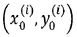


- Location (x, y) may be considered as a positive sample if it falls into any ground-truth box and the class label c* of the location may be the class label of the ground-truth box. Otherwise, it may be a negative sample and c* = 0 (background class). Furthermore, there may be a 4D real vector t* = (1*, t*, r*, b*) being the regression targets for the location. l*, t*, r* and b* may be distances from the location to the four sides of the bounding box. If a location falls into multiple bounding boxes, it may be considered as an ambiguous sample. The bounding box with minimal area may be chosen as its regression target.
- If location (x, y) is associated to a bounding box Bi, the training regression targets for the location may be formulated as, l* = x-x0 (i), t* = y-y0 (i), r* = x1 (i)-x and b* = y1 (i)-y. The FCOS may leverage as many foreground samples as possible to train the regressor.
- Corresponding to the training targets, the final layer of the networks may predict an 80D vector p of classification labels and a 4D vector t = (1, t, r, b) bounding box coordinates. In one example, C binary classifiers may be trained. In one example, at least four convolutional layers may be added after the feature maps of the backbone networks respectively for classification and regression branches. Since regression targets may be positive, exp(x) may be employed to map any real number to (0,∞) on the top of the regression branch.
- A training loss function may be defined as follows:


- Giving the imaging data ID, they may be forwarded through the network and may obtain the classification scores px,y and the regression prediction tx,y for each location on the feature maps Fi. In one example, the required IOU scores for positive anchor boxes may be lowered.
- In one example a single-layer branch may be added additionally, in parallel with a classification branch, in order to predict a "centerness" of a location. The centerness may depict the normalized distance from the location to the center of the object that the location is responsible for. Given the regression targets l*, t*, r* and b* for a location the centerness target may be defined as

- In a
fifth method step 105 the detected suspected lesion may be classified with a malignancy risk MR using a third neural network, wherein the third neural network is a deep profiler. Therefore, the deep profiler may comprise an encoder for extracting imaging features. Furthermore, the encoder may be designed as convolutional neural network (CNN), in particular a three-dimensional CNN. Additionally, the deep profiler may comprise a decoder for estimating at least one malignancy risk MR indicator and/or a task-specific network for generating at least one image signature for classifying at least one malignancy risk MR. - In one example the imaging data ID may be quantified by intensity, geometry, texture and/or wavelet features. The geometry features may quantify the 2D or 3D shape characteristics of the kidney, the texture features may describe spatial distribution of voxel or pixel intensities, thereby quantifying a heterogeneity. Any intensity and texture features may be computed after applying wavelet transformations to the imaging data ID.
- To find voxels of the imaging data ID that contribute the most toward the prediction, the derivative of the final partial likelihood loss with respect to the imaging data ID may be taken and evaluated.
- In one example the imaging data ID and/or non-imaging data NID may comprise context data relating to data recording information. In addition, the context data may comprise at least one out of following: data recording device information, contrast configuration information, brightness configuration information, recording direction information, total recording time information, projection type information (e.g. CT: average intensity projection (AIP), maximum intensity projection (MIP) or Minimum intensity projection MinIP)), date and/or time of recording information.
- In one example, the non-imaging data NID of the subject patient may comprise at least one out of following: age, weight, size, health condition information, gender, nutritional practice.
- A
sixth method step 106 may convert the imaging data ID from at least a partially 3D illustration of the anatomy of the subject patient to a 2D illustration of the anatomy of the subject patient. This method step may be performed at any time due to e.g. memory or computational limitations. -
FIG. 2 is a schematic drawing depicting asystem 200 for classifying a malignancy risk MR scoring of a kidney. - The
system 200 comprises aninterface 201 which is configured to provide imaging data ID of an anatomy of a subject patient, wherein the imaging data ID comprises at least partially a representation of a kidney of the subject patient. Therefore, imaging data ID will be forwarded, in particular together with non-imaging data, to the interface. - A
first analyzing unit 202 of thesystem 200 is configured to use a first neural network to segment at least one region of the kidney representation which is based on the imaging data. Asecond analyzing unit 203 is also part of thesystem 200, wherein thesecond analyzing unit 203 is configured to use a second neural network to detect one or more suspected lesions of the segmented kidney representation. The system comprises additionally adeep profiler 204 which is configured to classify the detected suspected lesion with a malignancy risk MR. - In one example the
deep profiler 204 is configured to classify the malignancy risk MR based on imaging data ID and non-imaging data NID, wherein the non-imaging data NID comprises at least histopathologic data. - Whenever a malignancy risk MR score has been determined by the
system 200, the value may be stored in thesystem 200 or in a cloud-based memory storage system. In one example, at least parts of thesystem 200 may be deployed in a cloud-based system, which means that they do not have to be physically integrated in a box. - The
system 200 may be configured that each input data and/or output data can be forwarded to each of its units, namely itsinterface 201, itsfirst analyzing unit 202, itssecond analyzing unit 203 and/or itsdeep profiler 204. It is further denoted that the neural networks of the specific units may be designed analogue to the neural networks disclosed above relating toFig. 1 . -
FIG. 3 is a schematic drawing depicting a computer-implementedmethod 300 for training a machine learning algorithm to classify a malignancy risk MR of a kidney. - Therefore, the
method 300 comprises afirst step 301 of training a first neural network with first training data including imaging data ID of an anatomy of at least one subject patient, wherein the imaging data ID comprises at least partially a representation of one or more kidneys. An adversarial network may be utilized for training of the first neural network to discriminate the output from ground truth. - In a second method step 302 a second neural network is trained with second training data including one or more detected lesions of one or more segmented kidney representations.
- In a third method step 303 a third neural network is trained with third training data of one or more lesions classified with a malignancy risk MR. The third training data are preferably based on imaging data ID and non-imaging data NID, wherein the non-imaging data NID comprises at least histopathologic data. Furthermore, at least one ground-truth label of the deep profiler may be determined based on histopathologic data.
- In a fourth method step 304 a fourth neural network is trained using deep reinforcement learning based on fourth training data related to detected anatomical landmarks, in particular landmarks relating to one or more kidney representations.
- In one example at least two different training data fragments of the first, second third or fourth training data may be based on the same subject patient. Furthermore, the two different training data fragments may be indicated as training data of the same subject patient. Additionally, the non-imaging data NID may comprise at least one indicator which is configured to serve for patient follow-up diagnosis.
-
FIG. 4 is a schematic drawing depicting asystem 400 for training a machine learning algorithm to classify a malignancy risk MR of a kidney. - Therefore, the
system 400 comprises afirst analysis unit 401 which is configured to train a first neural network with first training data including imaging data ID of an anatomy of at least one subject patient, wherein the imaging data ID comprises at least partially a representation of one or more kidneys. - A
second analysis unit 401 of thesystem 400 is configured to train a second neural network with second training data including one or more detected lesions of one or more segmented kidney representations. Thesystem 400 further comprises athird analysis unit 403 which is configured to train a third neural network with third training data of one or more lesions classified with a malignancy risk MR. - The system may additionally comprise a
fourth analysis unit 404 which is configured to train a fourth neural network with fourth training data related to detected anatomical landmarks, in particular landmarks relating to one or more kidney representations. - The
system 400 may be configured that each input data and/or output data can be forwarded to each of its units, namely itsfirst analysis unit 401, itssecond analysis unit 402, itsthird analysis unit 403 and/or itsfourth analysis unit 404. It is further denoted that the neural networks of the specific units may be designed analogue to the neural networks disclosed above relating toFig. 3 . - According to one embodiment the following the following clause is provided:
- Clause 1: A computer-implemented method (100) for classifying a malignancy risk (MR) of a kidney, in particular a human kidney, comprising following steps:
- providing (101) imaging data (ID) of an anatomy of a subject patient, wherein the imaging data (ID) comprises at least partially a representation of a kidney of the subject patient;
- using (102) a first neural network to segment at least one region of the kidney representation which is based on the imaging data (ID);
- using (103) a second neural network to detect one or more suspected lesions of the segmented kidney representation; and
- classifying (104) the detected suspected lesion with a malignancy risk (MR) using a third neural network,
- 2. The method of clause 1, wherein the imaging data (ID) and/or non-imaging data (NID) comprises context data relating to data recording information.
- 3. The method of clause 1 or 2, wherein the context data comprises at least one out of following:
data recording device information, contrast configuration information, brightness configuration information, recording direction information, total recording time information, projection type information, date and/or time of recording information. - 4. The method of any of clauses 1 to 3, wherein the non-imaging data (NID) of the subject patient comprises at least one out of following:
age, weight, size, health condition information, gender, nutritional practice. - According to a 5th clause, the following is provided:
A computer-implemented method (300) for training a machine learning algorithm to classify a malignancy risk (MR) of a kidney, in particular a human kidney, comprising following steps:- training (301) a first neural network with first training data including imaging data (ID) of an anatomy of at least one subject patient, wherein the imaging data (ID) comprises at least partially a representation of one or more kidneys;
- training (302) a second neural network with second training data including one or more detected lesions of one or more segmented kidney representations; and
- training (303) a third neural network with third training data of one or more lesions classified with a malignancy risk.
- 6. The method of clause 5, wherein the third training data are based on imaging data (ID) and non-imaging data (NID), wherein the non-imaging data (NID) comprises at least histopathologic data.
- 7. The method of clause 5 or 6, wherein at least one ground-truth label of the deep profiler is determined based on histopathologic data.
- 8. The method of any of clauses 5 to 7, comprising following additional step:
- training (304) a fourth neural network using deep reinforcement learning based on fourth training data related to detected anatomical landmarks, in particular landmarks relating to one or more kidney representations.
- 9. The method of any of clauses 5 to 8, wherein an adversarial network is utilized for training of the first neural network to discriminate the output from ground truth.
- 10. The method of any of clauses 5 to 10, wherein at least two different training data fragments are based on the same subject patient.
- 11. The method of clause 10, wherein the two different training data fragments are indicated as training data of the same subject patient.
- 12. The method of any of clauses 5 to 11, wherein the non-imaging data (NID) comprise at least one indicator which is configured to serve for patient follow-up diagnosis. Although the present invention has been described in detail with reference to the preferred example, the present invention is not limited by the disclosed examples from which the skilled person is able to derive other variations without departing from the scope of the invention. Example examples being thus described, it will be obvious that the same may be varied in many ways. Such variations are not to be regarded as a departure from the spirit and scope of the present invention, and all such modifications as would be obvious to one skilled in the art are intended to be included within the scope of the following claims.
Claims (17)
- A computer-implemented method (100) for classifying a malignancy risk (MR) of a kidney, in particular a human kidney, comprising following steps:- providing (101) imaging data (ID) of an anatomy of a subject patient, wherein the imaging data (ID) comprises at least partially a representation of a kidney of the subject patient;- using (102) a first neural network to segment at least one region of the kidney representation which is based on the imaging data (ID);- using (103) a second neural network to detect one or more suspected lesions of the segmented kidney representation; and- classifying (104) the detected suspected lesion with a malignancy risk (MR) using a third neural network,wherein the third neural network is a deep profiler.
- The method (100) of claim 1, wherein the third neural network is configured to classify the malignancy risk (MR) based on imaging data (ID) and non-imaging data (NID), wherein the non-imaging data (NID) comprises at least histopathologic data.
- The method (100) of claim 1 or 2, wherein the deep profiler comprises an encoder for extracting imaging features.
- The method (100) of one of the preceding claims, wherein the encoder is designed as convolutional neural network (CNN), in particular a three-dimensional CNN.
- The method (100) of at least one of the preceding claims, wherein the deep profiler comprises a decoder for estimating at least one malignancy risk (MR) indicator.
- The method (100) of at least one of the preceding claims, wherein the deep profiler comprises a task-specific network for generating at least one image signature for classifying at least one malignancy risk (MR).
- The method (100) of at least one of the preceding claims, comprising the additional method step:- using (105) a fourth neural network to detect anatomical landmarks based on the provided imaging data (ID), and preferably the fourth neural network is designed as convolutional neural network for image feature extraction and more preferably the fourth neural network additionally uses at least one universal non-linear function approximator.
- The method (100) of one of the preceding claims, wherein the first neural network is designed as at least one of the following:- a convolutional encoder-decoder architecture.- a multi-level feature concatenation and deep supervision architecture.
- The method (100) of at least one of the preceding claims, wherein the step of detecting one or more suspected lesions is based on a fully convolutional one-stage object detection.
- The method (100) of at least one of the preceding claims, wherein the provided imaging data (ID) comprises at least one of the following:- is based on computer tomography and/or magnet resonance imaging,- comprises at least partially a 3D illustration of the anatomy of the subject patient.
- The method (100) of at least one of the preceding claims, comprising the additional step:- converting (106) the imaging data (ID) from at least a partially 3D illustration of the anatomy of the subject patient to a 2D illustration of the anatomy of the subject patient.
- A system (200) for classifying a malignancy risk (MR) scoring of a kidney, in particular a human kidney, comprising:- an interface (201) which is configured to provide imaging data (ID) of an anatomy of a subject patient, wherein the imaging data (ID) comprises at least partially a representation of a kidney of the subject patient;- a first analyzing unit (202) which is configured to use a first neural network to segment at least one region of the kidney representation which is based on the imaging data (ID);- a second analyzing unit (203) which is configured to use a second neural network to detect one or more suspected lesions of the segmented kidney representation; and- a deep profiler (204) which is configured to classify the detected suspected lesion with a malignancy risk.
- The system (200) of claim 12, wherein the deep profiler (204) is configured to classify the malignancy risk (MR) based on imaging data (ID) and non-imaging data (NID), wherein the non-imaging data (NID) comprises at least histopathologic data.
- A computer-implemented method (300) for training a machine learning algorithm to classify a malignancy risk (MR) of a kidney, in particular a human kidney, comprising following steps:- training (301) a first neural network with first training data including imaging data (ID) of an anatomy of at least one subject patient, wherein the imaging data (ID) comprises at least partially a representation of one or more kidneys;- training (302) a second neural network with second training data including one or more detected lesions of one or more segmented kidney representations; and- training (303) a third neural network with third training data of one or more lesions classified with a malignancy risk.
- A system (400) for training a machine learning algorithm to classify a malignancy risk (MR) of a kidney, in particular a human kidney, comprising:- a first analysis unit (401) which is configured to train a first neural network with first training data including imaging data (ID) of an anatomy of at least one subject patient, wherein the imaging data (ID) comprises at least partially a representation of one or more kidneys;- a second analysis unit (402) which is configured to train a second neural network with second training data including one or more detected lesions of one or more segmented kidney representations; and- a third analysis unit (403) which is configured to train a third neural network with third training data of one or more lesions classified with a malignancy risk.
- A computer program comprising instructions which, when the program is executed by a computer, cause the computer to carry out the method (100; 300) of at least one of the claims 1 to 11 and/or 14.
- A machine-readable medium having stored thereon a set of instructions, which if performed by one or more processors, cause the one or more processors to carry out a method (100; 300) as mentioned in at least one of the claims 1 to 11 and/or 14.
Priority Applications (3)
| Application Number | Priority Date | Filing Date | Title |
|---|---|---|---|
| EP22167134.0A EP4258206B1 (en) | 2022-04-07 | 2022-04-07 | Methods and systems for classifying a malignancy risk of a kidney and training thereof |
| US18/183,973 US20230326608A1 (en) | 2022-04-07 | 2023-03-15 | Methods and systems for classifying a malignancy risk of a kidney and training thereof |
| CN202310357466.6A CN116894969A (en) | 2022-04-07 | 2023-04-04 | Methods and systems for classifying risk of malignancy in the kidney and training thereof |
Applications Claiming Priority (1)
| Application Number | Priority Date | Filing Date | Title |
|---|---|---|---|
| EP22167134.0A EP4258206B1 (en) | 2022-04-07 | 2022-04-07 | Methods and systems for classifying a malignancy risk of a kidney and training thereof |
Publications (3)
| Publication Number | Publication Date |
|---|---|
| EP4258206A1 true EP4258206A1 (en) | 2023-10-11 |
| EP4258206C0 EP4258206C0 (en) | 2026-02-25 |
| EP4258206B1 EP4258206B1 (en) | 2026-02-25 |
Family
ID=81328356
Family Applications (1)
| Application Number | Title | Priority Date | Filing Date |
|---|---|---|---|
| EP22167134.0A Active EP4258206B1 (en) | 2022-04-07 | 2022-04-07 | Methods and systems for classifying a malignancy risk of a kidney and training thereof |
Country Status (3)
| Country | Link |
|---|---|
| US (1) | US20230326608A1 (en) |
| EP (1) | EP4258206B1 (en) |
| CN (1) | CN116894969A (en) |
Families Citing this family (2)
| Publication number | Priority date | Publication date | Assignee | Title |
|---|---|---|---|---|
| EP4473921A1 (en) * | 2023-06-05 | 2024-12-11 | Storz Medical AG | Lithotripter with detection of kidney stones |
| CN119919407B (en) * | 2025-04-02 | 2025-07-08 | 上海交通大学医学院附属仁济医院 | Kidney hardening proportion analysis method and system based on MRI (magnetic resonance imaging) image |
Family Cites Families (3)
| Publication number | Priority date | Publication date | Assignee | Title |
|---|---|---|---|---|
| CN111354002A (en) * | 2020-02-07 | 2020-06-30 | 天津大学 | Kidney and kidney tumor segmentation method based on deep neural network |
| TWI920110B (en) * | 2020-07-06 | 2026-04-01 | 瑞典商艾西尼診斷公司 | Systems and methods for artificial intelligence-based image analysis for detection and characterization of lesions |
| CN112734723B (en) * | 2021-01-08 | 2023-06-30 | 温州医科大学 | Multi-source data-oriented breast tumor image classification prediction method and device |
-
2022
- 2022-04-07 EP EP22167134.0A patent/EP4258206B1/en active Active
-
2023
- 2023-03-15 US US18/183,973 patent/US20230326608A1/en active Pending
- 2023-04-04 CN CN202310357466.6A patent/CN116894969A/en active Pending
Non-Patent Citations (2)
| Title |
|---|
| DAN ZHAO ET AL: "Deep Learning in Computer-Aided Diagnosis and Treatment of Tumors: A Survey", ARXIV.ORG, CORNELL UNIVERSITY LIBRARY, 201 OLIN LIBRARY CORNELL UNIVERSITY ITHACA, NY 14853, 2 November 2020 (2020-11-02), XP081805486 * |
| JANG HYUN-JONG ET AL: "Applications of deep learning for the analysis of medical data", ARCHIVES OF PHARMACAL RESEARCH, NATL. FISHERIES UNIVERSITY , PUSAN, KR, vol. 42, no. 6, 28 May 2019 (2019-05-28), pages 492 - 504, XP037240316, ISSN: 0253-6269, [retrieved on 20190528], DOI: 10.1007/S12272-019-01162-9 * |
Also Published As
| Publication number | Publication date |
|---|---|
| US20230326608A1 (en) | 2023-10-12 |
| EP4258206C0 (en) | 2026-02-25 |
| CN116894969A (en) | 2023-10-17 |
| EP4258206B1 (en) | 2026-02-25 |
Similar Documents
| Publication | Publication Date | Title |
|---|---|---|
| Raposo | Intelligent imaging: A systematic review of artificial intelligence techniques in disease detection, segmentation, and classification | |
| US10496884B1 (en) | Transformation of textbook information | |
| CN108268870B (en) | Multi-scale feature fusion based on adversarial learning for semantic segmentation of ultrasound images | |
| TWI823897B (en) | System and method for diagnosing gastrointestinal neoplasm | |
| US10691980B1 (en) | Multi-task learning for chest X-ray abnormality classification | |
| Nie et al. | Recent advances in diagnosis of skin lesions using dermoscopic images based on deep learning | |
| Ahamed et al. | Automated detection of colorectal polyp utilizing deep learning methods with explainable AI | |
| CN118657800B (en) | Joint segmentation method of multiple lesions in retinal OCT images based on hybrid network | |
| Ypsilantis et al. | Recurrent convolutional networks for pulmonary nodule detection in CT imaging | |
| Khan et al. | Robust & precise knowledge distillation-based novel context-aware predictor for disease detection in brain and gastrointestinal | |
| Liu et al. | A semi-supervised convolutional transfer neural network for 3D pulmonary nodules detection | |
| US20230326608A1 (en) | Methods and systems for classifying a malignancy risk of a kidney and training thereof | |
| Ali et al. | Self-supervised learning for accurate liver view classification in ultrasound images with minimal labeled data | |
| Rathinam et al. | An Adaptive Fuzzy C-Means segmentation and deep learning model for efficient mammogram classification using VGG-Net | |
| Khan et al. | Temperature-driven robust disease detection in brain and gastrointestinal disorders via context-aware adaptive knowledge distillation | |
| Rao et al. | Multi-class classification of Alzheimer’s disease using deep learning and transfer learning on 3D MRI images | |
| Hiraman et al. | Lung tumor segmentation: a review of the state of the art | |
| US12333773B2 (en) | Explaining a model output of a trained model | |
| Saravanan et al. | Weighted Majority Voting Ensemble for MRI-Based Brain Tumor Classification Using Capsule Networks and XGBoost | |
| Chegireddy et al. | A Novel Method for Human MRI Based Pancreatic Cancer Prediction Using Integration of Harris Hawks Varients & VGG16: A Deep Learning Approach | |
| Lakide et al. | Precise Lung Cancer Prediction using ResNet–50 Deep Neural Network Architecture | |
| Ankireddy | Assistive diagnostic tool for brain tumor detection using computer vision | |
| WO2022170203A1 (en) | System and method for automatic diagnosis of idiopathic pulmonary fibrosis (ipf) using neural networks | |
| US12555230B2 (en) | Regression-based approach for measurement of rotator cuff tears in shoulder MRI images | |
| Rafee et al. | Deep learning approach for thyroid medical image analysis and prediction |
Legal Events
| Date | Code | Title | Description |
|---|---|---|---|
| PUAI | Public reference made under article 153(3) epc to a published international application that has entered the european phase |
Free format text: ORIGINAL CODE: 0009012 |
|
| STAA | Information on the status of an ep patent application or granted ep patent |
Free format text: STATUS: REQUEST FOR EXAMINATION WAS MADE |
|
| 17P | Request for examination filed |
Effective date: 20220407 |
|
| AK | Designated contracting states |
Kind code of ref document: A1 Designated state(s): AL AT BE BG CH CY CZ DE DK EE ES FI FR GB GR HR HU IE IS IT LI LT LU LV MC MK MT NL NO PL PT RO RS SE SI SK SM TR |
|
| RAP1 | Party data changed (applicant data changed or rights of an application transferred) |
Owner name: SIEMENS HEALTHINEERS AG |
|
| RBV | Designated contracting states (corrected) |
Designated state(s): AL AT BE BG CH CY CZ DE DK EE ES FI FR GB GR HR HU IE IS IT LI LT LU LV MC MK MT NL NO PL PT RO RS SE SI SK SM TR |
|
| GRAP | Despatch of communication of intention to grant a patent |
Free format text: ORIGINAL CODE: EPIDOSNIGR1 |
|
| STAA | Information on the status of an ep patent application or granted ep patent |
Free format text: STATUS: GRANT OF PATENT IS INTENDED |
|
| RIC1 | Information provided on ipc code assigned before grant |
Ipc: G06T 7/00 20170101AFI20251008BHEP Ipc: G06T 7/11 20170101ALI20251008BHEP |
|
| INTG | Intention to grant announced |
Effective date: 20251023 |
|
| GRAS | Grant fee paid |
Free format text: ORIGINAL CODE: EPIDOSNIGR3 |
|
| GRAA | (expected) grant |
Free format text: ORIGINAL CODE: 0009210 |
|
| STAA | Information on the status of an ep patent application or granted ep patent |
Free format text: STATUS: THE PATENT HAS BEEN GRANTED |
|
| AK | Designated contracting states |
Kind code of ref document: B1 Designated state(s): AL AT BE BG CH CY CZ DE DK EE ES FI FR GB GR HR HU IE IS IT LI LT LU LV MC MK MT NL NO PL PT RO RS SE SI SK SM TR |
|
| REG | Reference to a national code |
Ref country code: CH Ref legal event code: F10 Free format text: ST27 STATUS EVENT CODE: U-0-0-F10-F00 (AS PROVIDED BY THE NATIONAL OFFICE) Effective date: 20260225 Ref country code: GB Ref legal event code: FG4D |
|
| REG | Reference to a national code |
Ref country code: IE Ref legal event code: FG4D |
|
| U01 | Request for unitary effect filed |
Effective date: 20260225 |
|
| U07 | Unitary effect registered |
Designated state(s): AT BE BG DE DK EE FI FR IT LT LU LV MT NL PT RO SE SI Effective date: 20260302 |
|
| U20 | Renewal fee for the european patent with unitary effect paid |
Year of fee payment: 5 Effective date: 20260420 |
|
| PG25 | Lapsed in a contracting state [announced via postgrant information from national office to epo] |
Ref country code: RS Free format text: LAPSE BECAUSE OF FAILURE TO SUBMIT A TRANSLATION OF THE DESCRIPTION OR TO PAY THE FEE WITHIN THE PRESCRIBED TIME-LIMIT Effective date: 20260525 |
|
| PG25 | Lapsed in a contracting state [announced via postgrant information from national office to epo] |
Ref country code: ES Free format text: LAPSE BECAUSE OF FAILURE TO SUBMIT A TRANSLATION OF THE DESCRIPTION OR TO PAY THE FEE WITHIN THE PRESCRIBED TIME-LIMIT Effective date: 20260225 |
|
| PG25 | Lapsed in a contracting state [announced via postgrant information from national office to epo] |
Ref country code: PL Free format text: LAPSE BECAUSE OF FAILURE TO SUBMIT A TRANSLATION OF THE DESCRIPTION OR TO PAY THE FEE WITHIN THE PRESCRIBED TIME-LIMIT Effective date: 20260225 |
|
| PGFP | Annual fee paid to national office [announced via postgrant information from national office to epo] |
Ref country code: GB Payment date: 20260513 Year of fee payment: 5 |
|
| PG25 | Lapsed in a contracting state [announced via postgrant information from national office to epo] |
Ref country code: GR Free format text: LAPSE BECAUSE OF FAILURE TO SUBMIT A TRANSLATION OF THE DESCRIPTION OR TO PAY THE FEE WITHIN THE PRESCRIBED TIME-LIMIT Effective date: 20260526 |
|
| PG25 | Lapsed in a contracting state [announced via postgrant information from national office to epo] |
Ref country code: NO Free format text: LAPSE BECAUSE OF FAILURE TO SUBMIT A TRANSLATION OF THE DESCRIPTION OR TO PAY THE FEE WITHIN THE PRESCRIBED TIME-LIMIT Effective date: 20260525 |
|
| PG25 | Lapsed in a contracting state [announced via postgrant information from national office to epo] |
Ref country code: HR Free format text: LAPSE BECAUSE OF FAILURE TO SUBMIT A TRANSLATION OF THE DESCRIPTION OR TO PAY THE FEE WITHIN THE PRESCRIBED TIME-LIMIT Effective date: 20260225 |
|
| PG25 | Lapsed in a contracting state [announced via postgrant information from national office to epo] |
Ref country code: IS Free format text: LAPSE BECAUSE OF FAILURE TO SUBMIT A TRANSLATION OF THE DESCRIPTION OR TO PAY THE FEE WITHIN THE PRESCRIBED TIME-LIMIT Effective date: 20260625 |