EP4257703A1 - Method for selecting mesenchymal stem cells having improved self-maintenance ability, and mesenchymal stem cells selected thereby - Google Patents
Method for selecting mesenchymal stem cells having improved self-maintenance ability, and mesenchymal stem cells selected thereby Download PDFInfo
- Publication number
- EP4257703A1 EP4257703A1 EP21903672.0A EP21903672A EP4257703A1 EP 4257703 A1 EP4257703 A1 EP 4257703A1 EP 21903672 A EP21903672 A EP 21903672A EP 4257703 A1 EP4257703 A1 EP 4257703A1
- Authority
- EP
- European Patent Office
- Prior art keywords
- mesenchymal stem
- stem cells
- msmf
- cells
- aurka
- Prior art date
- Legal status (The legal status is an assumption and is not a legal conclusion. Google has not performed a legal analysis and makes no representation as to the accuracy of the status listed.)
- Pending
Links
Images










Classifications
-
- C—CHEMISTRY; METALLURGY
- C12—BIOCHEMISTRY; BEER; SPIRITS; WINE; VINEGAR; MICROBIOLOGY; ENZYMOLOGY; MUTATION OR GENETIC ENGINEERING
- C12Q—MEASURING OR TESTING PROCESSES INVOLVING ENZYMES, NUCLEIC ACIDS OR MICROORGANISMS; COMPOSITIONS OR TEST PAPERS THEREFOR; PROCESSES OF PREPARING SUCH COMPOSITIONS; CONDITION-RESPONSIVE CONTROL IN MICROBIOLOGICAL OR ENZYMOLOGICAL PROCESSES
- C12Q1/00—Measuring or testing processes involving enzymes, nucleic acids or microorganisms; Compositions therefor; Processes of preparing such compositions
- C12Q1/68—Measuring or testing processes involving enzymes, nucleic acids or microorganisms; Compositions therefor; Processes of preparing such compositions involving nucleic acids
- C12Q1/6876—Nucleic acid products used in the analysis of nucleic acids, e.g. primers or probes
- C12Q1/6881—Nucleic acid products used in the analysis of nucleic acids, e.g. primers or probes for tissue or cell typing, e.g. human leukocyte antigen [HLA] probes
-
- A—HUMAN NECESSITIES
- A23—FOODS OR FOODSTUFFS; TREATMENT THEREOF, NOT COVERED BY OTHER CLASSES
- A23L—FOODS, FOODSTUFFS OR NON-ALCOHOLIC BEVERAGES, NOT OTHERWISE PROVIDED FOR; PREPARATION OR TREATMENT THEREOF
- A23L33/00—Modifying nutritive qualities of foods; Dietetic products; Preparation or treatment thereof
- A23L33/10—Modifying nutritive qualities of foods; Dietetic products; Preparation or treatment thereof using additives
-
- A—HUMAN NECESSITIES
- A61—MEDICAL OR VETERINARY SCIENCE; HYGIENE
- A61K—PREPARATIONS FOR MEDICAL, DENTAL OR TOILETRY PURPOSES
- A61K35/00—Medicinal preparations containing materials or reaction products thereof with undetermined constitution
- A61K35/12—Materials from mammals; Compositions comprising non-specified tissues or cells; Compositions comprising non-embryonic stem cells; Genetically modified cells
- A61K35/28—Bone marrow; Haematopoietic stem cells; Mesenchymal stem cells of any origin, e.g. adipose-derived stem cells
-
- A—HUMAN NECESSITIES
- A61—MEDICAL OR VETERINARY SCIENCE; HYGIENE
- A61P—SPECIFIC THERAPEUTIC ACTIVITY OF CHEMICAL COMPOUNDS OR MEDICINAL PREPARATIONS
- A61P21/00—Drugs for disorders of the muscular or neuromuscular system
-
- C—CHEMISTRY; METALLURGY
- C12—BIOCHEMISTRY; BEER; SPIRITS; WINE; VINEGAR; MICROBIOLOGY; ENZYMOLOGY; MUTATION OR GENETIC ENGINEERING
- C12N—MICROORGANISMS OR ENZYMES; COMPOSITIONS THEREOF; PROPAGATING, PRESERVING, OR MAINTAINING MICROORGANISMS; MUTATION OR GENETIC ENGINEERING; CULTURE MEDIA
- C12N5/00—Undifferentiated human, animal or plant cells, e.g. cell lines; Tissues; Cultivation or maintenance thereof; Culture media therefor
- C12N5/06—Animal cells or tissues; Human cells or tissues
- C12N5/0602—Vertebrate cells
- C12N5/0652—Cells of skeletal and connective tissues; Mesenchyme
- C12N5/0662—Stem cells
-
- A—HUMAN NECESSITIES
- A23—FOODS OR FOODSTUFFS; TREATMENT THEREOF, NOT COVERED BY OTHER CLASSES
- A23V—INDEXING SCHEME RELATING TO FOODS, FOODSTUFFS OR NON-ALCOHOLIC BEVERAGES AND LACTIC OR PROPIONIC ACID BACTERIA USED IN FOODSTUFFS OR FOOD PREPARATION
- A23V2002/00—Food compositions, function of food ingredients or processes for food or foodstuffs
-
- A—HUMAN NECESSITIES
- A23—FOODS OR FOODSTUFFS; TREATMENT THEREOF, NOT COVERED BY OTHER CLASSES
- A23V—INDEXING SCHEME RELATING TO FOODS, FOODSTUFFS OR NON-ALCOHOLIC BEVERAGES AND LACTIC OR PROPIONIC ACID BACTERIA USED IN FOODSTUFFS OR FOOD PREPARATION
- A23V2200/00—Function of food ingredients
- A23V2200/30—Foods, ingredients or supplements having a functional effect on health
- A23V2200/316—Foods, ingredients or supplements having a functional effect on health having an effect on regeneration or building of ligaments or muscles
-
- C—CHEMISTRY; METALLURGY
- C12—BIOCHEMISTRY; BEER; SPIRITS; WINE; VINEGAR; MICROBIOLOGY; ENZYMOLOGY; MUTATION OR GENETIC ENGINEERING
- C12N—MICROORGANISMS OR ENZYMES; COMPOSITIONS THEREOF; PROPAGATING, PRESERVING, OR MAINTAINING MICROORGANISMS; MUTATION OR GENETIC ENGINEERING; CULTURE MEDIA
- C12N15/00—Mutation or genetic engineering; DNA or RNA concerning genetic engineering, vectors, e.g. plasmids, or their isolation, preparation or purification; Use of hosts therefor
- C12N15/09—Recombinant DNA-technology
- C12N15/11—DNA or RNA fragments; Modified forms thereof; Non-coding nucleic acids having a biological activity
- C12N15/113—Non-coding nucleic acids modulating the expression of genes, e.g. antisense oligonucleotides; Antisense DNA or RNA; Triplex- forming oligonucleotides; Catalytic nucleic acids, e.g. ribozymes; Nucleic acids used in co-suppression or gene silencing
-
- C—CHEMISTRY; METALLURGY
- C12—BIOCHEMISTRY; BEER; SPIRITS; WINE; VINEGAR; MICROBIOLOGY; ENZYMOLOGY; MUTATION OR GENETIC ENGINEERING
- C12N—MICROORGANISMS OR ENZYMES; COMPOSITIONS THEREOF; PROPAGATING, PRESERVING, OR MAINTAINING MICROORGANISMS; MUTATION OR GENETIC ENGINEERING; CULTURE MEDIA
- C12N15/00—Mutation or genetic engineering; DNA or RNA concerning genetic engineering, vectors, e.g. plasmids, or their isolation, preparation or purification; Use of hosts therefor
- C12N15/09—Recombinant DNA-technology
- C12N15/11—DNA or RNA fragments; Modified forms thereof; Non-coding nucleic acids having a biological activity
- C12N15/113—Non-coding nucleic acids modulating the expression of genes, e.g. antisense oligonucleotides; Antisense DNA or RNA; Triplex- forming oligonucleotides; Catalytic nucleic acids, e.g. ribozymes; Nucleic acids used in co-suppression or gene silencing
- C12N15/1137—Non-coding nucleic acids modulating the expression of genes, e.g. antisense oligonucleotides; Antisense DNA or RNA; Triplex- forming oligonucleotides; Catalytic nucleic acids, e.g. ribozymes; Nucleic acids used in co-suppression or gene silencing against enzymes
-
- C—CHEMISTRY; METALLURGY
- C12—BIOCHEMISTRY; BEER; SPIRITS; WINE; VINEGAR; MICROBIOLOGY; ENZYMOLOGY; MUTATION OR GENETIC ENGINEERING
- C12N—MICROORGANISMS OR ENZYMES; COMPOSITIONS THEREOF; PROPAGATING, PRESERVING, OR MAINTAINING MICROORGANISMS; MUTATION OR GENETIC ENGINEERING; CULTURE MEDIA
- C12N2310/00—Structure or type of the nucleic acid
- C12N2310/10—Type of nucleic acid
- C12N2310/14—Type of nucleic acid interfering nucleic acids [NA]
-
- C—CHEMISTRY; METALLURGY
- C12—BIOCHEMISTRY; BEER; SPIRITS; WINE; VINEGAR; MICROBIOLOGY; ENZYMOLOGY; MUTATION OR GENETIC ENGINEERING
- C12N—MICROORGANISMS OR ENZYMES; COMPOSITIONS THEREOF; PROPAGATING, PRESERVING, OR MAINTAINING MICROORGANISMS; MUTATION OR GENETIC ENGINEERING; CULTURE MEDIA
- C12N2510/00—Genetically modified cells
-
- C—CHEMISTRY; METALLURGY
- C12—BIOCHEMISTRY; BEER; SPIRITS; WINE; VINEGAR; MICROBIOLOGY; ENZYMOLOGY; MUTATION OR GENETIC ENGINEERING
- C12Q—MEASURING OR TESTING PROCESSES INVOLVING ENZYMES, NUCLEIC ACIDS OR MICROORGANISMS; COMPOSITIONS OR TEST PAPERS THEREFOR; PROCESSES OF PREPARING SUCH COMPOSITIONS; CONDITION-RESPONSIVE CONTROL IN MICROBIOLOGICAL OR ENZYMOLOGICAL PROCESSES
- C12Q2600/00—Oligonucleotides characterized by their use
- C12Q2600/158—Expression markers
Definitions
- the present disclosure relates to a method for selecting mesenchymal stem cells having improved self-maintenance ability.
- Mesenchymal stem cells as multipotent cells, are isolated from a variety of tissues such as bone marrow, adipose tissue, placenta, and umbilical cord blood and have the capacity to differentiate into bone, cartilage, muscle, fat, and fibroblasts. Extensive research has been conducted into the use of mesenchymal stem cells as a treatment for various diseases by regenerating non-regenerative tissues or cells by using such characteristics of stem cells.
- mesenchymal stem cells have problems of large donor variation, low production yield, and high manufacturing costs, as well as failure to show long-term therapeutic effects due to a short survival period in the body.
- One aspect is to provide a method for selecting mesenchymal stem cells having improved self-maintenance ability, the method including measuring an expression or activity level of a mesenchymal stem cell (MSC) self-maintenance factor (MSMF) after culturing mesenchymal stem cells.
- MSC mesenchymal stem cell
- MSMF self-maintenance factor
- Another aspect is to provide mesenchymal stem cells selected by the method.
- Another aspect is to provide an apoptosis inhibitor including mesenchymal stem cells genetically engineered to reduce the expression of the MSMF or suppress the expression of the MSMF compared to parent cells, or including the MSMF.
- Another aspect is to provide a pharmaceutical composition for preventing or treating muscle diseases including mesenchymal stem cells genetically engineered to secrete the MSMF or overexpress the MSMF compared to parent cells, or including the MSMF.
- Another aspect is to provide a pharmaceutical composition for preventing or treating muscle diseases including the MSMF as an active ingredient.
- Another aspect is to provide a health functional food composition for preventing or alleviating muscle diseases including the MSMF as an active ingredient.
- Another aspect is to provide a cell therapy product including mesenchymal stem cells genetically engineered to secrete the MSMF or overexpress the MSMF compared to parent cells, or including the MSMF.
- Another aspect is to provide a method for preventing or treating muscle diseases, the method including administering mesenchymal stem cells genetically engineered to secrete the MSMF or overexpress MSMF compared to parent cells, or a pharmaceutically effective dose MSMF, to an individual in need thereof.
- Another aspect is to provide a use of a composition including mesenchymal stem cells genetically engineered to secrete MSMF or overexpress MSMF compared to parent cells, or MSMF for treating muscle diseases.
- Another aspect is to provide a use of a composition including mesenchymal stem cells genetically engineered to secrete the MSMF or overexpress the MSMF compared to parent cells, or the MSMF for preparing a therapeutic agent for muscle diseases.
- One aspect provides a method for selecting mesenchymal stem cells having improved self-maintenance ability, the method including measuring an expression or activity level of a mesenchymal stem cell (MSC) self-maintenance factor (MSMF) after culturing mesenchymal stem cells.
- MSC mesenchymal stem cell
- MSMF self-maintenance factor
- mesenchymal stem cells refers to multipotent stem cells maintaining the ability to self-renew and stemness and having the ability to differentiate into various mesenchymal tissues such as mesodermal cells, e.g., bone, cartilage, fat, and muscle cells, and ectodermal cells, e.g., nerve cells.
- the mesenchymal stem cells may be derived from umbilical cord, umbilical cord blood, bone marrow, fat, muscle nerve, skin, amnion, chorion, decidua, placenta, and the like.
- the mesenchymal stem cells may be derived from humans, fetuses, or non-human mammals.
- MSMF MSC self-maintenance factor
- the MSMF may be, for example, FBLN5, OLR1, TNFAIP6, ANXA3, IL6, POU2F2, TNFAIP2, SERPINE2, INHBA, VEGFA, HMGB1, CSF2, GATA3, PCSK6, SYN1, F2RL1, DOCK2, SLC9A4, STX1B, RARRES2, CXCL1, FGF7, PLAU, SCG2, NR4A3, COR01A, CHRM3, NPR3, BST2, GATA4, CREG1, FGF7, YPEL5, or AURKA.
- FBLN5 OLR1, TNFAIP6, ANXA3, IL6, POU2F2, TNFAIP2, SERPINE2, INHBA, VEGFA, HMGB1, CSF2, GATA3, PCSK6, SYN1, F2RL1, DOCK2, SLC9A4, STX1B, RARRES2, CXCL1, FGF7, PLAU, SCG2, NR4A3, COR01A, CHRM3, NPR
- proteins involved in adhesion may be FBLN5, OLR1, TNFAIP6, ANXA3, and the like
- proteins involved in differentiation may be IL6, POU2F2, TNFAIP2, SERPINE2, INHBA, VEGFA, HMGB1, CSF2, GATA3, PCSK6, SYN1, F2RL1, DOCK2, SLC9A4, STX1B, RARRES2, and the like.
- proteins involved in chemotaxis may be IL6, CXCL1, VEGFA, FGF7, PLAU, HMGB1, SCG2, NR4A3, GATA3, DOCK2, COR01A, RARRES2, and the like, and proteins involved in proliferation may be CXCL1, DOCK2, CHRM3, NPR3, NR4A3, F2RL1, BST2, GATA4, CREG1, FGF7, YPEL5, AURKA, and the like.
- the method includes: culturing the mesenchymal stem cells and treating the cultured mesenchymal stem cells with an expression or activity inhibitor of the MSMF; measuring an expression or activity level of the MSMF in the mesenchymal stem cells in which expression or activity of the MSMF is inhibited; and separating mesenchymal stem cells in which the expression or activity level of the MSMF is reduced compared to a control group untreated with the expression or activity inhibitor of the MSMF.
- the expression or activity inhibitor of the MSMF may be selected from the group consisting of small interference RNA (siRNA), short hairpin RNA (shRNA), microRNA (miRNA), ribozyme, DNAzyme, peptide nucleic acids (PNA), antisense oligonucleotides, antibodies, aptamers, and compounds and natural extracts directly binding to the MSMF protein and inhibiting the activity thereof.
- the expression or activity inhibitor of the MSMF may be a first primer set represented by SEQ ID NOS: 5 and 6 and a second primer set represented by SEQ ID NOS: 9 and 10.
- the measuring of the expression or activity level of the MSMF may be performed by a method selected from the group consisting of reverse transcription polymerase chain reaction, competitive reverse transcription polymerase chain reaction, real-time reverse transcription polymerase chain reaction, RNase protection assay, Northern blotting, and DNA chip assay.
- the method may be selected from the group consisting of protein chip analysis, immunoassay, ligand binding assay, matrix desorption/ionization time of flight mass spectrometry (MALDI-TOF) assay, surface enhanced laser desorption/ionization time of flight mass spectrometry (SELDI-TOF) assay, radioimmunoassay, radial immunodiffusion assay, Ouchterlony immunodiffusion assay, rocket immunoelectrophoresis, immunohistostaining, complement fixation assay, two-dimensional electrophoresis, liquid chromatography-mass spectrometry (LC-MS), liquid chromatography-mass spectrometry/mass spectrometry (LC-MS/MS), Western blotting, and enzyme linked immunosorbent assay (ELISA).
- MALDI-TOF matrix desorption/ionization time of flight mass spectrometry
- SELDI-TOF surface enhanced laser desorption/ionization time of flight mass spectrometry
- Mesenchymal stem cells having improved self-maintenance ability in a medium may be selected by isolating mesenchymal stem cells in which the expression or activity level of the MSMF is reduced compared to a control untreated with the expression or activity inhibitor of the MSMF.
- a doubling time of the mesenchymal stem cells increased. Therefore, since expression or activity of the MSMF is increased in the mesenchymal stem cells with improved self-maintenance ability, desired cells may be obtained by isolating the mesenchymal stem cells in which the expression or activity of the MSMF is maintained or reduced.
- mesenchymal stem cells may be mass-produced as well as manufacturing costs may be reduced by selecting mesenchymal stem cells having high proliferative ability in the early stage.
- Another aspect provides mesenchymal stem cells selected by the above-described method.
- Another aspect provides an apoptosis inhibitor including mesenchymal stem cells genetically engineered to reduce the expression of the MSMF or suppress the expression of the MSMF compared to parent cells, or including the MSMF.
- Another aspect provides a method for inhibiting apoptosis, the method including: brining the mesenchymal stem cells genetically engineered to reduce the expression of the MSMF or suppress the expression of the MSMF compared to parent cells and/or the MSMF into contact with cells in vitro, or administering the genetically engineered mesenchymal stem cells or MSMF to an experimental animal in vivo.
- the mesenchymal stem cells may have an increased expression level of the MSMF that is essential for self-maintenance. That is, the mesenchymal stem cells may have improved stemness, migration ability, colony formation ability, and/or cell proliferative ability. In addition, the mesenchymal stem cells may have a reduced doubling time. That is, MSMF expression and cell doubling time may exhibit a negative correlation.
- the term “genetic engineering” or “genetically engineered” refers to the act of introducing one or more genetic modifications to a cell or a cell produced thereby.
- the mesenchymal stem cells or host cells may be those genetically engineered to increase the expression or activity of the MSMF or an active fragment thereof, for example, those containing an exogenous gene encoding the MSMF or the active fragment thereof.
- the increased activity may mean that the activity of a protein or enzyme of the same type is higher than the activity of an endogenous protein or enzyme possessed or not possessed by parent cells, which are not genetically engineered (e.g., wild-type).
- the exogenous gene may be expressed in an amount sufficient to increase the activity of the above-mentioned protein in the mesenchymal stem cells or host cells compared to the parent cells.
- the exogenous gene may be introduced into parental cells via an expression vector.
- the exogenous gene may be introduced into the parent cells in the form of a linear polynucleotide.
- the exogenous gene may be expressed from an expression vector (e.g., plasmid) in a cell.
- the exogenous gene may be expressed in a form inserted into a genetic material (e.g., chromosome) in a cell for stable expression.
- Another aspect provides a pharmaceutical composition for preventing or treating muscle diseases including mesenchymal stem cells genetically engineered to reduce the expression of the MSMF or suppress the expression of the MSMF compared to parent cells, or including the MSMF.
- Another aspect provides a method for preventing or treating muscle diseases, the method including administering the mesenchymal stem cells and/or MSMF to an individual.
- the mesenchymal stem cells genetically engineered to secrete the MSMF or overexpress the MSMF compared to parental cells or the MSMF are as described above.
- mRNA of AURKA and DCOCK2 and proteins thereof may be used to prevent or treat muscle diseases by inhibiting apoptosis of muscle cells.
- the muscle disease may be, for example, Charcot-Marie-Tooth disease, Spinal Muscular Atrophy (SMA), Lou Gehrig's disease (amyotrophic lateral sclerosis, ALS), Duchenne Muscular Dystrophy, Myotonic Dystrophy, sarcopenia, muscular atrophy, myasthenia, muscular dystrophy, myotonia, hypotonia, muscular weakness, muscular dystrophy, amyotrophic lateral sclerosis, or inflammatory myopathy.
- SMA Spinal Muscular Atrophy
- Lou Gehrig's disease as myotrophic lateral sclerosis, ALS
- Duchenne Muscular Dystrophy Myotonic Dystrophy
- sarcopenia muscular atrophy, myasthenia, muscular dystrophy, myotonia, hypotonia, muscular weakness, muscular dystrophy, amyotrophic lateral sclerosis, or inflammatory myopathy.
- Another aspect provides a pharmaceutical composition for preventing or treating muscle diseases including the MSMF as an active ingredient.
- the MSMF may be isolated from mesenchymal stem cells.
- the composition may further include mesenchymal stem cells.
- the mesenchymal stem cells may secrete the MSMF or an active fragment thereof or may be genetically engineered to secrete the MSMF or the active fragment thereof.
- the mesenchymal stem cells may be those modified by insertion or injection of DNA in cell culture by a method of modifying, enhancing, or supplementing cell functions for induction for structural or therapeutic purposes. Therefore, the MSMF and mesenchymal stem cells may be co-administered.
- the pharmaceutical composition for preventing or treating muscle diseases may be formulated into various forms such as oral formulations such as powders, granules, tablets, capsules, suspensions, emulsions, syrups, and aerosols, formulations for external use, suppositories, and sterile injection solutions, and may include an appropriate carrier, excipient, or diluent commonly used for formulation in preparation of pharmaceutical compositions.
- the carrier, excipient, or diluent may be various compounds or mixtures including lactose, dextrose, sucrose, sorbitol, mannitol, xylitol, erythritol, maltitol, starch, Acacia gum, alginate, gelatin, calcium phosphate, calcium silicate, cellulose, methyl cellulose, amorphous cellulose, polyvinyl pyrrolidone, water, methylhydroxybenzoate, propylhydroxybenzoate, talc, magnesium stearate, and mineral oil.
- the pharmaceutical composition may be formulated using a diluent or excipient commonly used in the art such as a filler, an extender, a binder, a humectant, a disintegrant, and a surfactant.
- a diluent or excipient commonly used in the art such as a filler, an extender, a binder, a humectant, a disintegrant, and a surfactant.
- Solid formulations for oral administration may be prepared by mixing a legume extract with at least one excipient such as starch, calcium carbonate, sucrose or lactose, and gelatin.
- excipients such as starch, calcium carbonate, sucrose or lactose, and gelatin.
- a lubricant such as magnesium stearate and talc may also be used.
- Liquid formulations for oral administration which are suspensions, formulations for internal use, emulsions, and syrups, may include various excipients such as a humectant, a sweetener, a flavoring agent, and a preservative, in addition to simple diluents such as water and liquid paraffin.
- Formulations for parenteral administration include sterile aqueous solutions, nonaqueous solvent, suspensions, emulsions, lyophilizates, and suppositories.
- nonaqueous solvents and suspensions propylene glycol, polyethylene glycol, vegetable oil such as olive oil, injectable ester such as ethyl oleate, and the like may be used.
- injectable ester such as ethyl oleate, and the like
- a base material for suppositories witepsol, macrogol, tween 61, cacao butter, laurin butter, glycerolgelatin, and the like may be used.
- a preferred dosage of the pharmaceutical composition for preventing or treating muscle diseases varies according to the condition and body weight of a patient, severity of disease, form of drug, route and duration of administration, but may be appropriately selected by those skilled in the art. However, for desirable effects, the dosage may be from 0.0001 to 2,000 mg/kg, preferably, from 0.001 to 2,000 mg/kg per day.
- the pharmaceutical composition may be administered once a day or several times a day at divided doses. However, the scope of the present invention is not limited by the dosage.
- the pharmaceutical composition for preventing or treating muscle diseases may be administered to mammals such as rats, mice, livestock, and humans via various routes. Administration may be conducted via any method, for example, orally or rectally, or by intravenous, muscular, subcutaneous, intrauterine subdural, or intracerebroventricular injection.
- Another aspect provides a health functional food composition for preventing or alleviating muscle diseases including mesenchymal stem cells genetically engineered to reduce the expression of the MSMF or suppress the expression of the MSMF compared to parent cells, or including the MSMF.
- the mesenchymal stem cells or the MSMF are as described above.
- the compound when used as an additive to the health functional food, it may be added as it is or used together with other foods or food ingredients and may be appropriately used according to any method commonly used in the art.
- the amount of the active ingredient to be mixed therewith may be appropriately determined according to the intended use, e.g., prevention, health, or treatment.
- the health functional food may be provided in the form of all food or drink types commonly used in the art as well as in the form of powders, granules, pills, tablets, and capsules.
- the type of the food is not particularly limited, and examples of the food to which the substance may be added, may include meats, sausages, breads, chocolates, candies, snacks, cookies, pizzas, ramens, other noodles, gums, dairy products including ice cream, various kinds of soups, beverages, teas, drinks, alcoholic beverages, and vitamin complexes, and may also include all foods that are considered within conventional meaning.
- the compound in general, in the preparation of foods or drinks, may be added in an amount of 15 parts by weight or less, preferably 10 parts by weight or less, based on 100 parts by weight of raw materials.
- the amount in the case of a long-term intake for the purposes of health and hygiene or health control, the amount may be less than the above-described range.
- drinks may further include various flavoring agents or natural carbohydrates as additional ingredients.
- natural carbohydrates may include monosaccharides such as glucose and fructose, disaccharides such as maltose and sucrose, polysaccharides such as dextrin and cyclodextrin, and sugar alcohols such as xylitol, sorbitol and erythritol.
- sweetener may include natural sweeteners such as thaumatin and stevia extract and synthetic sweeteners such as saccharin and aspartame.
- the ratio of the natural carbohydrate may be from about 0.01 to 0.04 g, preferably from about 0.02 to 0.03 g per 100 mL of the drink according to the present invention.
- the health functional food for preventing or alleviating muscle diseases may further include various nutrients, vitamins, electrolytes, flavors, colorants, pectic acid and salts thereof, alginic acid and salts thereof, organic acids, protective colloidal thickeners, and pH adjusters, stabilizers, preservatives, glycerin, alcohols, carbonating agents used in carbonated drinks.
- the composition for improving sleep of the present disclosure may include fruit flesh for production of natural fruit juice, fruit juice-flavored drinks, and vegetable-based beverages. These ingredients may be used independently or in combination. The ratio of these additives is not limited, but is generally selected from the range of 0.01 to 0.1 parts by weight based on 100 parts by weight of the health functional food of the present disclosure.
- Another aspect provides a cell therapy product including the mesenchymal stem cells as an active ingredient.
- the mesenchymal stem cells are as described above.
- the term "cell therapy product” refers to cells and tissues prepared by isolation from an individual, culture, and specific manipulation and used as a medicament for the purpose of treatment, diagnosis, and prevention of a disease (FDA regulations, USA) and means a medicament used for the purpose of treatment, diagnosis, and prevention of a disease through a series of actions such as proliferation and selection of living autologous, allogenic, or xenogenic cells in vitro or changes of biological properties of cells by different methods, in order to restore the functions of cells or tissues.
- the “treatment” refers to all actions involved in alleviating or beneficially changing symptoms of a disease by administering a cell therapy product.
- the mesenchymal stem cells may be those having increased expression of the MSMF, and expression of FBLN5, TNFAIP6, ANXA3, IL6, POU2F2, TNFAIP2, INHBA, VEGFA, CSF2, GATA3, CXCL1, HMGB1, BST2, and AURKA associated with inflammatory diseases, immune diseases, or cancer may be increased. Therefore, in one embodiment, the cell therapy product may be used to treat muscle diseases, inflammatory diseases, immune diseases, or cancer.
- Examples of the inflammatory disease may include atopy, psoriasis, dermatitis, allergy, arthritis, rhinitis, otitis media, sore throat, tonsillitis, cystitis, nephritis, pelvic inflammatory disease, Crohn's disease, ulcerative colitis, ankylosing spondylitis, systemic lupus erythematosus (SLE), asthma, edema, delayed allergy (type IV allergy), graft rejection, graft versus host disease, autoimmune encephalomyelitis, multiple sclerosis, inflammatory bowel disease, cystic fibrosis, diabetic retinopathy, ischemic-reperfusion injury, vascular restenosis, glomerulonephritis, and gastrointestinal allergy.
- SLE systemic lupus erythematosus
- SLE systemic lupus erythematosus
- asthma edema
- type IV allergy delayed allergy
- graft rejection graft
- Examples of the immune disease may include autoimmune diseases such as rheumatoid arthritis, type I diabetes, multiple sclerosis, systemic lupus erythematosus, and inflammatory diseases such as asthma, encephalitis, inflammatory enteritis, chronic obstructive pulmonary disease, allergy, septic shock, pulmonary fibrosis, undifferentiated spondyloarthrosis, undifferentiated arthropathy, arthritis, inflammatory osteolysis, and chronic inflammation caused by chronic viral or bacterial infection.
- autoimmune diseases such as rheumatoid arthritis, type I diabetes, multiple sclerosis, systemic lupus erythematosus
- inflammatory diseases such as asthma, encephalitis, inflammatory enteritis, chronic obstructive pulmonary disease, allergy, septic shock, pulmonary fibrosis, undifferentiated spondyloarthrosis, undifferentiated arthropathy, arthritis, inflammatory osteolysis, and chronic inflammation caused by chronic viral or bacterial infection
- Examples of the cancer may include multiple myeloma, lung cancer, liver cancer, gastric cancer, colorectal cancer, colon cancer, skin cancer, bladder cancer, prostate cancer, breast cancer, ovarian cancer, cervical cancer, thyroid cancer, renal cancer, fibrosarcoma, melanoma, and hematologic malignancy.
- the cell therapy product may be administered after being prepared into a pharmaceutical formulation in a unit dosage form suitable for administration into a patient's body according to any common method in the pharmaceutical field, and the formulation may include an effective amount for a single dose or for divided doses.
- a formulation suitable for this purpose a formulation for parenteral administration such as an injection formulation, e.g., an ampoule for injection, an infusion formulation, e.g., an infusion bag, and a spray formulation, e.g., an aerosol may be used.
- the ampoule for injection may be mixed with an injectable solution immediately before use, and physiological saline, glucose, mannitol, Ringer's solution, or the like may be used as the injectable solution.
- the infusion bag may be made of polyvinyl chloride or polyethylene and those manufactured by Baxter, Becton Dickinson, Medcep, National Hospital Products, or Terumo corporations may be used.
- the pharmaceutical formulation may further include at least one pharmaceutically acceptable inert carrier, e.g., a preservative, an analgesic, a solubilizer, or a stabilizer for injectable formulations and an excipient, a lubricant, or a preservative for topical formulations.
- a pharmaceutically acceptable inert carrier e.g., a preservative, an analgesic, a solubilizer, or a stabilizer for injectable formulations and an excipient, a lubricant, or a preservative for topical formulations.
- the cell therapy product or pharmaceutical preparation of the present disclosure may be administered by a common administration method used in the art, together with other stem cells used for transplantation and other purposes, in the form of a mixture therewith.
- a common administration method used in the art together with other stem cells used for transplantation and other purposes, in the form of a mixture therewith.
- Direct engraftment or transplantation to a lesion of a patient in need of treatment, or direct transplantation or injection into the peritoneal cavity is preferred, without being limited thereto.
- both of a non-surgical administration using a catheter and a surgical administration such as injection or transplantation after incision are possible, but non-surgical administration using a catheter is more preferred.
- parenteral administration according to a common method for example, transplantation by infusion into the blood vessel as a common method is also possible as well as direct administration into a lesion.
- the daily dose of the mesenchymal stem cells may be 1.0 ⁇ 10 4 to 1.0 ⁇ 10 10 cells/kg (body weight), preferably 2.5 ⁇ 10 5 to 5 ⁇ 10 7 cells/kg (body weight) and may be administered once or as several divided doses.
- the actual dose of active ingredients is determined in consideration of various related factors such as a disease to be treated, severity of the disease, route of administration, body weight, age and gender of a patient, and therefore the dose should not be construed as limiting the scope of the present disclosure in any manner.
- the mesenchymal stem cells may be used in various types of treatment protocols for enhancement, treatment, or replacement of tissues or organs of the body by engraftment, transplantation, or infusion of desired cell populations.
- the cell therapy product may be used in an unfrozen state or frozen for future use.
- a standard cryopreservative e.g., DMSO, glycerol, and Epilife ® cell freezing medium (Cascade Biologics)
- DMSO methyl methoxylate
- glycerol glycerol
- Epilife ® cell freezing medium e.g., Epilife ® cell freezing medium
- the mesenchymal stem cells or the MSMF may be used as a preventive or therapeutic agent for various muscle diseases such as sprains, contusions, and spasm by inhibiting apoptosis of muscle cells.
- donor variation may be reduced by selecting mesenchymal stem cells having excellent self-proliferative ability, and mesenchymal stem cells may be mass-produced, and thus the method may be used for development of cell therapy products.
- the mesenchymal stem cells selected according to the above-described method has an increased survival period in vivo, thereby increasing therapeutic effects.
- the mesenchymal stem cells isolated and cultured in Example 1 were classified into mesenchymal stem cells satisfying the criteria of Good Manufacturing Practice (GMP) of Samsung Medical Center and mesenchymal stem cells not satisfying the criteria.
- GMP Good Manufacturing Practice
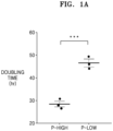
- the mesenchymal stem cells satisfying the criteria had a shorter doubling time than that of the mesenchymal stem cells not satisfying the criteria.
- the cells were classified into a proliferation-high (P-high) group and a proliferation-low (P-low) group according to the doubling time, and the doubling time thereof was identified.
- genes expressed at high levels in the P-high group compared to the P-low group were compared and analyzed. Specifically, after extracting each RNA from the P-low group and the P-high group, RNA quality was measured using an Agilent 2100 Bioanalyzer and migration and peak patterns were analyzed. A microarray was performed using an Illumina Hiseq 2500 to compare and analyze genes whose log2 fold change was three times higher in the P-high group than in the P-low group.
- FIG. 1A is a graph showing doubling time of a P-high group and a P-low group.
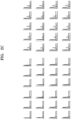
- FIG. 1B shows results of comparison of expression levels of genes involved in self-maintenance in the P-low group and the P-high group.
- Green color represents genes exhibiting lower expression levels in the P-high group compared to the P-low group, and red color represents genes exhibiting higher expression levels in the P-high group compared to the P-low group.
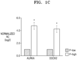
- FIG. 1C is a graph showing comparison of expression levels of AURKA and DOCK2, which are selected as MSMF gene candidates, in the P-low group and the P-high group.
- AURKA and DOCK2 were expressed at about 4 times higher levels in the P-high group compared to the P-low group indicating a statistically significant difference. Therefore, based on the above-described results, AURKA and DOCK2, which exhibited a statistically significant difference in the expression levels of the genes involved in adhesion, differentiation, chemotaxis, or proliferation, were selected as final candidates of the MSMF gene.
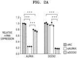
- sequence specificity was identified by comparing relative mRNA expression levels of AURKA and DOCK2 by the selected candidates.
- FIG. 2A is a graph showing comparison of relative expression levels of AURKA and DOCK2 by selected siRNA sequence.
- the selected siRNA sequences may have sequence specificity for each MSAF gene.
- Example 3-1 Western blotting was performed to identify whether protein expression levels of AURKA and DOCK2 were reduced by the siRNA sequence selected in Example 3-1. Specifically, mesenchymal stem cells were transfected with siAURKA (Candidate 3) and siDOCK2 (Candidate 2) respectively and cultured for 72 hours, and then rinsed with PBS. Then, the cells were lysed by a RIPA buffer (BIOSESANG, Sungnam, Gyeonggi, Korea) containing a protease inhibitor cocktail (Amresco, Solon, OH, USA) and centrifuged at 4°C with 15,000 g for 30 minutes to obtain a supernatant.
- RIPA buffer BIOOSESANG, Sungnam, Gyeonggi, Korea
- Amresco Amresco, Solon, OH, USA
- PVDF polyvinylidene difluoride
- the membrane was rinsed three times with TBST for 10 minutes each and treated with an ECL solution (Advansta, USA), and then images of bands were obtained using a gel imaging system (Amersham Imager 600, GE Healthcare, Buckinghamshire, UK).
- the expression levels of the proteins were measured using Image J software and corrected with ⁇ -actin.
- AURKA Invitrogen, CA
- DOCK2 Disanta Cruz Biotechnology, Dallas, TX, USA
- Beta-actin Santa Cruz Biotechnology, Dallas, TX, USA
- FIG. 2B shows protein expression levels of AURKA and DOCK2 after knocking down the MSMF gene by transfecting mesenchymal stem cells with siAURKA and siDOCK2.
- the protein expression levels of AURKA and DOCK2 were statistically significantly decreased in the siAURKA-treated group and the siDOCK2-treated group by 0.51 ⁇ 0.06 times and 0.76 ⁇ 0.04 times, respectively, compared to the control group treated with siNC.
- Mesenchymal stem cells were knocked down by transfection with siAURKA and siDOCK2 using the sequence selected in Example 3-1 and stemness thereof was identified by FACS. Markers of the mesenchymal stem cells were identified by expression of CD44, CD73, CD90, CD105, and CD166, and hematopoietic stem cell lineage markers were compared with MSAF siNC-treated control group in stemness by using CD14, CD11b, HLA-DR (MHCII), CD34, CD45, and CD19 (BD Biosciences, USA). In this regard, 10000 events were acquired and analyzed using a BD FACS Verse flow cytometer.
- FIG. 2C is a graph showing stemness obtained by FACS after knocking down the MSMF gene by transfecting the mesenchymal stem cells with siAURKA and siDOCK2.
- mesenchymal stem cells transfected with siNC, and siAURKA and siDOCK2 selected in Example 3-1 were aliquoted into a 12-well plate at a density of 1 ⁇ 10 5 cells/well and incubated for 48 hours using a minimum essential medium (MEM) Alpha (Invitrogen-Gibco, Rockville, MD) supplemented with 10% FBS.
- MEM minimum essential medium
- the cells were rinsed twice with the MEM Alpha not containing the FBS to inhibit proliferation of the cells and incubated in the MEM Alpha supplemented with 10 ⁇ g/ml mitomycin C (Sigma-Aldrich, St. Louis, MO) for 2 hours. Then, scratches were made using a 200 pipette tip. After rinsing 5 times with the culture solution, images were obtained at 40x magnification using a microscope after 0 and 24 hours. The obtained images were quantitatively analyzed using Image J software of Java, and cell migration ability was expressed as a percent wound closure.
- mitomycin C Sigma-Aldrich, St. Louis, MO
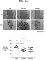
- FIG. 3A shows migration ability of mesenchymal stem cells after knocking down the MSMF gene by transfecting mesenchymal stem cells with siAURKA and siDOCK2.
- the migration ability of the control group treated with siNC was 61.02 ⁇ 1.0%, whereas the migration ability of the siAURKA-treated group and the siDOCK2-treated group were 44.06 ⁇ 1.87% and 47.18 ⁇ 2.93%, respectively. That is, it was confirmed that the migration ability of the mesenchymal stem cells significantly decreased as the expression levels of the AURKA and DOCK2 genes decreased.
- mesenchymal stem cells transfected with siNC, and siAURKA, and siDOCK2 selected in Example 3-1 were aliquoted into a 6-well plate at a density of 1 ⁇ 10 3 cells/well and incubated in a MEM Alpha (Invitrogen-Gibco, Rockville, MD) supplemented with 10% FBS for 14 days. Then, the cells were rinsed with PBS and immobilized with cold 100% methanol for 20 minutes. The immobilized cells were stained with a 1% crystal violet solution (Sigma-Aldrich, St. Louis, MO) for 30 minutes and rinsed three times or more with distilled water, and only colonies consisting of more than 50 cells were counted.
- MEM Alpha Invitrogen-Gibco, Rockville, MD
- FIG. 3B is a graph showing whether mesenchymal stem cells form colonies after knocking down an MSMF gene by transfecting the mesenchymal stem cells with siAURKA and siDOCK2.
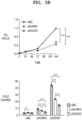
- mesenchymal stem cells transfected with siNC, and siAURKA, and siDOCK2 selected in Example 3-1 were aliquoted into a 12-well plate at a density of 3 ⁇ 10 3 cells/cm 2 and incubated in a MEM Alpha (Invitrogen-Gibco, Rockville, MD) supplemented with 10% FBS. Doubling time was identified by measuring the number of cells after 0, 24, 72, and 144 hours from the aliquoting.
- FIG. 3C is a graph showing doubling time of mesenchymal stem cells after knocking down an MSMF gene by transfecting the mesenchymal stem cells with siAURKA and siDOCK2.
- mesenchymal stem cells transfected with siNC, and siAURKA, and siDOCK2 selected in Example 3-1 were aliquoted into a 96-well plate at a density of 2 ⁇ 10 3 cells/well and incubated in a MEM Alpha (Invitrogen-Gibco, Rockville, MD) supplemented with 10% FBS. Absorbance was measured at 450 nm using a cell counting kit-8 (CCK-8, Dojindo, Japan, Tokyo) after 0, 24, 72, and 144 hours from the aliquoting.
- MEM Alpha Invitrogen-Gibco, Rockville, MD
- FIG. 3D shows cell proliferative ability of mesenchymal stem cells after knocking down an MSMF gene by transfecting the mesenchymal stem cells with siAURKA and siDOCK2.
- FIGS. 3E and 3F show effects of inhibition of expression of AURKA and DOCK2 genes on phosphorylation of AKT and ERK, which are kinases involved in proliferation of cells and phosphorylation of FAK and JNK, which are kinases involved in the migration ability.
- AURKA affects cell proliferation and migration ability of cells via AKT and FAK
- DOCK2 affects cell proliferation via AKT, ERK, and JNK.
- Example 4 By combining the results of Example 4 above, it may be seen that the AURKA and DOCK2 genes are involved in self-maintenance by affecting the migration ability, colony formation ability, and cell proliferation of mesenchymal stem cells.
- mesenchymal stem cells having reduced donor variation Due to a problem of large donor variation of mesenchymal stem cells, attempts have been made to select mesenchymal stem cells having reduced donor variation by identifying correlation between mRNA expression levels of AURKA and DOCK2 of the mesenchymal stem cells and doubling time.
- 10 mesenchymal stem cells obtained from 10 different donors were aliquoted into a 25T flask at a density of 3 ⁇ 10 3 cells/cm 2 and incubated in a MEM Alpha (Invitrogen-Gibco, Rockville, MD) supplemented with 10% FBS. When the cells grew by about 80%, RNA was extracted and qRT-PCR was performed, and then relative mRNA expression levels of AURKA and DOCK2 were compared.
- the correlation between the expression levels of AURKA and DOCK2 genes and the doubling time of cells was identified by Pearson correlation analysis.
- FIG 4A is a graph showing comparison of relative mRNA expression levels between AURKA and DOCK2 in 10 different mesenchymal stem cells.
- MSC_A had the highest mRNA expression level of AURKA among the 10 stem cells
- MSC_C had the highest mRNA expression level of DOCK among the 10 stem cells
- MSC_J had the lowest mRNA expression levels of AURKA and DOCK2.
- FIG. 4B is a graph sequentially showing doubling time of 10 different mesenchymal stem cells.
- the doubling time of MSC_A was the shortest as 20.9 ⁇ 0.6 hours, and the doubling time of MSC_J was the longest as 41.9 ⁇ 0.6 hours.
- An average doubling time of the 10 mesenchymal stem cells was 27.6 ⁇ 1.8 hours.
- FIG. 4C shows graphs of correlation between the mRNA expression levels of AURKA and DOCK2 and doubling time.
- mesenchymal stem cells having high self-proliferative ability may be easily selected by measuring the expression levels of AURKA and DOCK2 genes.
- the correlation between mRNA expression levels of AURKA and DOCK2 and protein expression levels of AURKA and DOCK2 was identified in mesenchymal stem cells. Specifically, Western blotting was performed in the same manner as in Example 3-2, except that mesenchymal stem cells cultured in a growth medium by about 80% were used. Subsequently, MSC_A having the highest mRNA expression level of AURKA, MSC_C having the highest mRNA expression level of DOCK2, and MSC_J having relatively low mRNA expression levels of AURKA and DOCK2 were selected (see FIG. 4A ), and a difference in protein expression levels of AURKA and DOCK2 was identified.
- FIG. 4D shows protein expression levels of AURKA and DOCK2 in mesenchymal stem cells.
- the protein expression levels of AURKA and DOCK2 in MSC_J decreased by 0.31 ⁇ 0.05 times and 0.59 ⁇ 0.02 times, respectively, compared to those of MSC_A and MSC_C. That is, it was confirmed that in the mesenchymal stem cells having low mRNA expression levels of AURKA and DOCK2, the expression levels of the proteins exhibiting the functions thereof also decreased.
- FIGS. 4E and 4F show results of confirming phosphorylation levels of AKT, ERK, FAK, and JNK in mesenchymal stem cells.
- the difference of mRNA expression levels of AURKA and DOCK2 and protein expression levels of AURKA and DOCK2 affects phosphorylation of kinases involved in cell proliferation and migration ability in mesenchymal stem cells, and mesenchymal stem cells having high expression levels of these genes may promote cell proliferation via phosphorylation of AKT and/or ERK and promote the migration ability via phosphorylation of FAK and/or JNK.
- muscle cells C2C12 were aliquoted into a 6-well plate at a density of 8 ⁇ 10 3 cells/cm 2 and incubated in a Dulbecco's Modified Eagle's Medium (DMEM, Biowest SAS, Nuaille, France) supplemented with 10% FBS and 1 U/ml penicillin/streptomycin (Gibco BRL, Grand Island, NY). After 24 hours of incubation, apoptosis was induced by starvation of FBS for another 24 hours.
- DMEM Dulbecco's Modified Eagle's Medium
- FIG. 5A shows results of identifying expression of apoptosis-related proteins according to mRNA expression levels of AURKA and DOCK2, by co-culturing mesenchymal stem cells and muscle cells in which apoptosis is induced.
- the expression levels of the cleaved PARP and the cleaved Caspase 3 decreased in an experimental group co-cultured with the mesenchymal stem cells compared to the control group in which apoptosis was induced. Specifically, compared to the control group in which apoptosis was induced, the expression levels of the cleaved caspase3 decreased by 0.41 ⁇ 0.07 and 0.38 ⁇ 0.07 times, respectively, and the expression levels of the cleaved PARP significantly decreased by 0.37 ⁇ 0.06 and 0.35 ⁇ 0.05 times, respectively, in the experimental groups co-cultured with MSC_A or MSC_C (p ⁇ 0.001).
- the expression level of the cleaved caspase3 was 0.66 ⁇ 0.07 times, and the expression level of the cleaved PARP was 0.78 ⁇ 0.01 times indicating slight differences from the control group in which apoptosis was induced. That is, it may be seen that the ability to inhibit apoptosis of muscle cells deteriorated as the mRNA expression levels of AURKA and DOCK2 in mesenchymal stem cells decrease.
- Live/dead staining was performed using an Apoptosis/Necrosis Detection Kit (Abcam, Cambridge, MA, USA) to identify the degree of reduction in apoptosis of C2C12 cells of Example 6-1.
- the experimental group in which the mesenchymal stem cells and the apoptosis-induced muscle cells were co-cultured in the same manner as in Example 6-1, was rinsed twice with PBS, followed by reaction with an Apopxin Green Indicator capable of detecting apoptosis and a CytoCalcein Violet 450 capable of detecting live cells in a light-blocked condition for 1 hour. Then, the cells were rinsed twice with PBS and images were obtained by using a fluorescence microscope. Among total cells, ratios of apoptotic cells were measured using Image J.
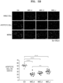
- FIG. 5B shows results of identifying degrees of reduction in apoptosis according to mRNA expression levels of AURKA and DOCK2, by co-culturing mesenchymal stem cells and muscle cells in which apoptosis is induced.
- apoptosis was inhibited in the experimental group co-cultured with the mesenchymal stem cells compared to the control group in which apoptosis was induced. Specifically, while the percentage of apoptotic cells was 52.04 ⁇ 1.93% in the control group in which apoptosis was induced, the percentages of apoptotic cells decreased to 22.50 ⁇ 2.17%, 25.72 ⁇ 1.53%, and 31.87 ⁇ 1.99% in the experimental groups co-cultured with MSC_A, MSC_C, or MSC_J (p ⁇ 0.001).
- mesenchymal stem cells having low mRNA expression levels of AURKA and DOCK2 have decreased inhibitory effects on apoptosis when co-cultured with the apoptosis-induced muscle cells.
- apoptosis-induced muscle cells were co-cultured respectively with mesenchymal stem cells and mesenchymal stem cells treated with siNC, and siAURKA or siDOCK2 selected in Example 3-1, and then protein expression was identified in the same manner as in Example 6-1.
- FIG. 6A shows results of confirming expression of apoptosis-related proteins according to inhibition of expression of AURKA and DOCK2 genes, by co-culturing mesenchymal stem cells in which AURKA and DOCK2 genes are knocked down and muscle cells in which apoptosis is induced.
- protein expression of the cleaved caspase3 and the cleaved PARP was significantly decreased in an experimental group co-cultured with the mesenchymal stem cells compared to a control group (apoptosis-induced muscle cells).
- the expression levels of the cleaved caspase3 and the cleaved PARP were decreased by 0.34 ⁇ 0.04, 0.30 ⁇ 0.02, and 0.52 ⁇ 0.05 and 0.40 ⁇ 0.04 times, respectively in the muscle cells co-cultured with the mesenchymal stem cells, the siNC-treated group, the siAURKA-treated group, and the siDOCK2-treated group compared to the control group in which apoptosis was induced (p ⁇ 0.001).
- the expression levels of the cleaved PARP were decreased by 0.39 ⁇ 0.07, 0.45 ⁇ 0.04, and 0.72 ⁇ 0.043, and 0.54 ⁇ 0.045 times, respectively in the muscle cells co-cultured with the mesenchymal stem cells, the siNC-treated group, the siAURKA-treated group, and the siDOCK2-treated group compared to the control group in which apoptosis was induced (MSC, siNC, and siDOCK2, p ⁇ 0.001; and siAURKA, p ⁇ 0.01).
- Example 3-1 In order to identify the degree of reduction in apoptosis according to inhibition of expression of AURKA and DOCK2 genes, the apoptosis-induced muscle cells were co-cultured respectively with mesenchymal stem cells, and siNC, and siAURKA or siDOCK2 selected in Example 3-1 above, followed by evaluation in the same manner as in Example 6-2.
- FIG. 6B shows results of identifying degrees of reduction in apoptosis according to inhibition of expression of AURKA and DOCK2 genes, by co-culturing mesenchymal stem cells in which AURKA and DOCK2 genes are knocked down and muscle cells in which apoptosis is induced.
- apoptosis was significantly inhibited in the experimental group co-cultured with the mesenchymal stem cells compared to the control group (apoptosis-induced muscle cells). Specifically, while an apoptosis rate was 41.73 ⁇ 1.03% in the control group in which apoptosis was induced, the apoptosis rates were decreased to 17.93 ⁇ 1.40%, 17.79 ⁇ 2.06%, 32.75 ⁇ 2.24%, and 25.62 ⁇ 2.29%, respectively, in the case of the muscle cells co-cultured with the mesenchymal stem cells, the siNC-treated group, the siAURKA-treated group, and the siDOCK2-treated group (p ⁇ 0.001). Particularly, it was confirmed that the apoptosis rates significantly increased in the siAURKA-treated group co-cultured with apoptosis-induced muscle cells compared to the siNC-treated group (p ⁇ 0.001).
- the inhibitory effect on apoptosis more significantly decreases by knocking down AURKA, it is considered that the inhibitory effect of the mesenchymal stem cells on apoptosis of muscle cells is more affected by the AURKA than by the DOCK2.
- FIG. 6C shows protein expression levels of XCL1 of mesenchymal stem cells in which an AURKA gene is knocked down.
- the expression level of XCL1 that is a protein involved in inhibition of apoptosis was decreased.
- the expression level of XCL1 of the siAURKA-treated group decreased by 0.44 ⁇ 0.07 times compared to that of the siNC-treated group (p ⁇ 0.01). That is, the AURKA gene may exhibit the inhibitory effect of the mesenchymal stem cells on apoptosis of muscle cells by directly affecting expression of XCL1.
- Example 8 Identification of Correlation between mRNA Expression Level of AURKA of Mesenchymal Stem Cell and Apoptosis of Muscle Tissue
- An inhibitory effect of mesenchymal stem cells on apoptosis of muscle cells according to a mRNA expression level of AURKA was identified by Annexin V staining. Specifically, mesenchymal stem cells were administered to the tail vein of a muscular dystrophy mouse model (Mdx) once at dose of 1 ⁇ 10 5 cells/100 ⁇ l.
- MCS_A having the highest mRNA expression level of AURKA
- MCS_E havin a median mRNA expression level of AURKA
- MCS_J having the lowest mRNA expression level of AURKA which are determined to directly affecting inhibition of apoptosis were used.
- mice were sacrificed and tissues of calf muscles were separated. Then, expression levels of proteins were identified in the same manner as in Example 3-2 except that annexin V (Abcam, Cambridge, Ma, USA) and Beta-actin (Santa Cruz Biotechnology, Dallas, TX, USA) were used as primary antibodies.
- annexin V Abcam, Cambridge, Ma, USA
- Beta-actin Santa Cruz Biotechnology, Dallas, TX, USA
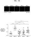
- FIG. 7A shows inhibitory effects on apoptosis of muscle tissue according to a difference in mRNA expression levels of AURKA of mesenchymal stem cells in a muscular dystrophy mouse model.
- the degree of inhibition of apoptosis of muscle tissues according to the mRNA expression level of AURKA was identified by Annexin V staining. Specifically, after separating the thigh and calf muscles of the Mdx mouse model in the same manner as in Example 8-1, tissue cut by paraffin sectioning was rinsed and reacted at room temperature using a 5% blocking solution. Subsequently, the resultant was reacted with Annexin antibody (Abcam, Cambridge, UK) diluted to a 1/500 concentration at 4°C for 18 hours.
- Annexin antibody Abcam, Cambridge, UK
- the tissue was reacted with Alexa Fluor ® 594 AffiniPure Goat Anti-rabbit IgG (H+L) secondary antibody (Thermo fisher scientific, Rockford, IL) at room temperature for 1 hour, followed by counter-staining using Hoechst 33342 (Thermo fisher scientific, Rockford, IL), and then fluorescence images were obtained from the tissue using a Carl Zeiss LSM 700 confocal microscopy system. The obtained images were quantitatively analyzed using Image J software of Java, and relative values to the Mdx control group were obtained.
- Alexa Fluor ® 594 AffiniPure Goat Anti-rabbit IgG (H+L) secondary antibody Thermo fisher scientific, Rockford, IL
- Hoechst 33342 Thermo fisher scientific, Rockford, IL
- FIG. 7B shows differences in inhibitory effects on apoptosis of muscle tissue according to differences in mRNA expression levels of AURKA of mesenchymal stem cells in a muscular dystrophy mouse model.
- the mesenchymal stem cells having a higher mRNA expression level of AURKA have a greater inhibitory effect on apoptosis of damaged muscle cells, and therefore, the mesenchymal stem cells may be used as a therapeutic agent for muscle diseases.
- FIG. 8A is a graph showing the degree of cell survival according to mRNA expression levels of AURKA of mesenchymal stem cells in a muscular dystrophy mouse model.
- the numbers of mesenchymal stem cells remaining in the leg muscles of the muscular dystrophy mouse model administered with MSC_A, MSC_F and MSC_J were 41.23 ⁇ 5.60, 42.68 ⁇ 8.71, and 16.96 ⁇ 8.44, respectively.
- the number of the mesenchymal stem cells remaining in the leg muscles of the muscular dystrophy mouse model was the lowest in the case of administering MSC_J having the lowest mRNA expression level of AURKA. That is, the mesenchymal stem cells having a higher mRNA expression level of AURKA migrate to damaged muscle cells and a larger number of cells remain in the damaged area, thereby assisting recovery of muscles.
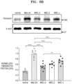
- the inhibitory effect on fibrosis of muscle tissue according to the mRNA expression level of AURKA was identified by using expression of fibronectin. Specifically, expression of proteins was identified in the same manner as in Example 8-2, except that Fibronectin (Abcam, Cambridge, Ma, USA) and Beta-actin (Santa Cruz Biotechnology, Dallas, TX, USA) were used as primary antibodies.
- FIG. 8B shows inhibitory effects on fibrosis of muscle tissue according to mRNA expression levels of AURKA of mesenchymal stem cells in a muscular dystrophy mouse model.
- the expression level of fibronectin was decreased by 0.54 ⁇ 0.04, 0.60 ⁇ 0.03, and 0.71 ⁇ 0.05 times, respectively, in the case of administering MCS_A, MCS_E, and MCS_J, compared to the control group. That is, mesenchymal stem cells having a high mRNA expression level of AURKA may effectively inhibit fibrosis of damaged muscle cells by reducing expression of fibronectin.
- the inhibitory effect on fibrosis of muscle tissue according to the mRNA expression level of AURKA was identified by accumulation of collagen. Specifically, after rinsing the thigh muscle tissue of the Mdx mouse model in the same manner as in Example 8-1, reaction was conducted at room temperature for 1 hour using a picro-sirius red (Solution A). Then, the resultant was rinsed twice with acidified water (Solution B) and mounted, and then images thereof were obtained using a Scanscope. The obtained images were quantitatively analyzed using Image J software of Java to obtain relative values to the Mdx control group.
- FIG. 8C shows degrees of fibrosis of muscle tissue according to mRNA expression level of AURKA of mesenchymal stem cells in a muscular dystrophy mouse model.
- collagen accumulation was reduced by 0.26 ⁇ 0.04, 0.27 ⁇ 0.03, and 0.73 ⁇ 0.04 times, respectively, in the case of administering MCS_A, MCS_E, and MCS_J compared to the control group. That is, mesenchymal stem cells having a high mRNA expression level of AURKA may effectively inhibit fibrosis of damaged muscle cells by reducing collagen accumulation.
Landscapes
- Health & Medical Sciences (AREA)
- Life Sciences & Earth Sciences (AREA)
- Chemical & Material Sciences (AREA)
- Engineering & Computer Science (AREA)
- Organic Chemistry (AREA)
- Zoology (AREA)
- Immunology (AREA)
- General Health & Medical Sciences (AREA)
- Cell Biology (AREA)
- Biotechnology (AREA)
- Wood Science & Technology (AREA)
- Bioinformatics & Cheminformatics (AREA)
- Proteomics, Peptides & Aminoacids (AREA)
- Biomedical Technology (AREA)
- Developmental Biology & Embryology (AREA)
- Genetics & Genomics (AREA)
- Animal Behavior & Ethology (AREA)
- Pharmacology & Pharmacy (AREA)
- Public Health (AREA)
- Veterinary Medicine (AREA)
- Medicinal Chemistry (AREA)
- Analytical Chemistry (AREA)
- Microbiology (AREA)
- General Engineering & Computer Science (AREA)
- Biochemistry (AREA)
- Molecular Biology (AREA)
- Epidemiology (AREA)
- Virology (AREA)
- Biophysics (AREA)
- Neurology (AREA)
- Orthopedic Medicine & Surgery (AREA)
- Nuclear Medicine, Radiotherapy & Molecular Imaging (AREA)
- General Chemical & Material Sciences (AREA)
- Chemical Kinetics & Catalysis (AREA)
- Physical Education & Sports Medicine (AREA)
- Physics & Mathematics (AREA)
- Hematology (AREA)
- Mycology (AREA)
- Rheumatology (AREA)
- Nutrition Science (AREA)
Abstract
Description
- This application claims the benefit of
Korean Patent Application No. 10-2020-0169835 filed on December 7, 2020 - The present disclosure relates to a method for selecting mesenchymal stem cells having improved self-maintenance ability.
- Mesenchymal stem cells, as multipotent cells, are isolated from a variety of tissues such as bone marrow, adipose tissue, placenta, and umbilical cord blood and have the capacity to differentiate into bone, cartilage, muscle, fat, and fibroblasts. Extensive research has been conducted into the use of mesenchymal stem cells as a treatment for various diseases by regenerating non-regenerative tissues or cells by using such characteristics of stem cells. However, as a cell therapy product, mesenchymal stem cells have problems of large donor variation, low production yield, and high manufacturing costs, as well as failure to show long-term therapeutic effects due to a short survival period in the body.
- Therefore, there is a need to select mesenchymal stem cells having high proliferative ability and high therapeutic efficacy to produce stem cells for development of stem cell therapy products.
- One aspect is to provide a method for selecting mesenchymal stem cells having improved self-maintenance ability, the method including measuring an expression or activity level of a mesenchymal stem cell (MSC) self-maintenance factor (MSMF) after culturing mesenchymal stem cells.
- Another aspect is to provide mesenchymal stem cells selected by the method.
- Another aspect is to provide an apoptosis inhibitor including mesenchymal stem cells genetically engineered to reduce the expression of the MSMF or suppress the expression of the MSMF compared to parent cells, or including the MSMF.
- Another aspect is to provide a pharmaceutical composition for preventing or treating muscle diseases including mesenchymal stem cells genetically engineered to secrete the MSMF or overexpress the MSMF compared to parent cells, or including the MSMF.
- Another aspect is to provide a pharmaceutical composition for preventing or treating muscle diseases including the MSMF as an active ingredient.
- Another aspect is to provide a health functional food composition for preventing or alleviating muscle diseases including the MSMF as an active ingredient.
- Another aspect is to provide a cell therapy product including mesenchymal stem cells genetically engineered to secrete the MSMF or overexpress the MSMF compared to parent cells, or including the MSMF.
- Another aspect is to provide a method for preventing or treating muscle diseases, the method including administering mesenchymal stem cells genetically engineered to secrete the MSMF or overexpress MSMF compared to parent cells, or a pharmaceutically effective dose MSMF, to an individual in need thereof.
- Another aspect is to provide a use of a composition including mesenchymal stem cells genetically engineered to secrete MSMF or overexpress MSMF compared to parent cells, or MSMF for treating muscle diseases.
- Another aspect is to provide a use of a composition including mesenchymal stem cells genetically engineered to secrete the MSMF or overexpress the MSMF compared to parent cells, or the MSMF for preparing a therapeutic agent for muscle diseases.
- One aspect provides a method for selecting mesenchymal stem cells having improved self-maintenance ability, the method including measuring an expression or activity level of a mesenchymal stem cell (MSC) self-maintenance factor (MSMF) after culturing mesenchymal stem cells.
- As used herein, the term "mesenchymal stem cells (MSCs)" refers to multipotent stem cells maintaining the ability to self-renew and stemness and having the ability to differentiate into various mesenchymal tissues such as mesodermal cells, e.g., bone, cartilage, fat, and muscle cells, and ectodermal cells, e.g., nerve cells. The mesenchymal stem cells may be derived from umbilical cord, umbilical cord blood, bone marrow, fat, muscle nerve, skin, amnion, chorion, decidua, placenta, and the like. In addition, the mesenchymal stem cells may be derived from humans, fetuses, or non-human mammals.
- As used herein, the term "MSC self-maintenance factor (MSMF)" refers to various regulators secreted by mesenchymal stem cells, essential for self-maintenance of mesenchymal stem cells, and expressed in a time-specific and cell-specific manner. The MSMF may be several proteins or genes thereof involved in adhesion, differentiation, chemotaxis, or proliferation. The MSMF may be, for example, FBLN5, OLR1, TNFAIP6, ANXA3, IL6, POU2F2, TNFAIP2, SERPINE2, INHBA, VEGFA, HMGB1, CSF2, GATA3, PCSK6, SYN1, F2RL1, DOCK2, SLC9A4, STX1B, RARRES2, CXCL1, FGF7, PLAU, SCG2, NR4A3, COR01A, CHRM3, NPR3, BST2, GATA4, CREG1, FGF7, YPEL5, or AURKA. Specifically, proteins involved in adhesion may be FBLN5, OLR1, TNFAIP6, ANXA3, and the like, and proteins involved in differentiation may be IL6, POU2F2, TNFAIP2, SERPINE2, INHBA, VEGFA, HMGB1, CSF2, GATA3, PCSK6, SYN1, F2RL1, DOCK2, SLC9A4, STX1B, RARRES2, and the like. In addition, proteins involved in chemotaxis may be IL6, CXCL1, VEGFA, FGF7, PLAU, HMGB1, SCG2, NR4A3, GATA3, DOCK2, COR01A, RARRES2, and the like, and proteins involved in proliferation may be CXCL1, DOCK2, CHRM3, NPR3, NR4A3, F2RL1, BST2, GATA4, CREG1, FGF7, YPEL5, AURKA, and the like.
- In one embodiment, the method includes: culturing the mesenchymal stem cells and treating the cultured mesenchymal stem cells with an expression or activity inhibitor of the MSMF; measuring an expression or activity level of the MSMF in the mesenchymal stem cells in which expression or activity of the MSMF is inhibited; and separating mesenchymal stem cells in which the expression or activity level of the MSMF is reduced compared to a control group untreated with the expression or activity inhibitor of the MSMF.
- The expression or activity inhibitor of the MSMF may be selected from the group consisting of small interference RNA (siRNA), short hairpin RNA (shRNA), microRNA (miRNA), ribozyme, DNAzyme, peptide nucleic acids (PNA), antisense oligonucleotides, antibodies, aptamers, and compounds and natural extracts directly binding to the MSMF protein and inhibiting the activity thereof. In one embodiment, the expression or activity inhibitor of the MSMF may be a first primer set represented by SEQ ID NOS: 5 and 6 and a second primer set represented by SEQ ID NOS: 9 and 10.
- The measuring of the expression or activity level of the MSMF may be performed by a method selected from the group consisting of reverse transcription polymerase chain reaction, competitive reverse transcription polymerase chain reaction, real-time reverse transcription polymerase chain reaction, RNase protection assay, Northern blotting, and DNA chip assay. In addition, the method may be selected from the group consisting of protein chip analysis, immunoassay, ligand binding assay, matrix desorption/ionization time of flight mass spectrometry (MALDI-TOF) assay, surface enhanced laser desorption/ionization time of flight mass spectrometry (SELDI-TOF) assay, radioimmunoassay, radial immunodiffusion assay, Ouchterlony immunodiffusion assay, rocket immunoelectrophoresis, immunohistostaining, complement fixation assay, two-dimensional electrophoresis, liquid chromatography-mass spectrometry (LC-MS), liquid chromatography-mass spectrometry/mass spectrometry (LC-MS/MS), Western blotting, and enzyme linked immunosorbent assay (ELISA).
- Mesenchymal stem cells having improved self-maintenance ability in a medium may be selected by isolating mesenchymal stem cells in which the expression or activity level of the MSMF is reduced compared to a control untreated with the expression or activity inhibitor of the MSMF. In one embodiment, it was confirmed that migration ability, colony formation ability, and cell proliferative ability of mesenchymal stem cells decreased as the expression of the MSMF decreased. Also, it was confirmed that a doubling time of the mesenchymal stem cells increased. Therefore, since expression or activity of the MSMF is increased in the mesenchymal stem cells with improved self-maintenance ability, desired cells may be obtained by isolating the mesenchymal stem cells in which the expression or activity of the MSMF is maintained or reduced. According to the method, mesenchymal stem cells may be mass-produced as well as manufacturing costs may be reduced by selecting mesenchymal stem cells having high proliferative ability in the early stage.
- Another aspect provides mesenchymal stem cells selected by the above-described method. Another aspect provides an apoptosis inhibitor including mesenchymal stem cells genetically engineered to reduce the expression of the MSMF or suppress the expression of the MSMF compared to parent cells, or including the MSMF. Another aspect provides a method for inhibiting apoptosis, the method including: brining the mesenchymal stem cells genetically engineered to reduce the expression of the MSMF or suppress the expression of the MSMF compared to parent cells and/or the MSMF into contact with cells in vitro, or administering the genetically engineered mesenchymal stem cells or MSMF to an experimental animal in vivo. The mesenchymal stem cells may have an increased expression level of the MSMF that is essential for self-maintenance. That is, the mesenchymal stem cells may have improved stemness, migration ability, colony formation ability, and/or cell proliferative ability. In addition, the mesenchymal stem cells may have a reduced doubling time. That is, MSMF expression and cell doubling time may exhibit a negative correlation.
- As used herein, the term "genetic engineering" or "genetically engineered" refers to the act of introducing one or more genetic modifications to a cell or a cell produced thereby. For example, the mesenchymal stem cells or host cells may be those genetically engineered to increase the expression or activity of the MSMF or an active fragment thereof, for example, those containing an exogenous gene encoding the MSMF or the active fragment thereof. The increased activity may mean that the activity of a protein or enzyme of the same type is higher than the activity of an endogenous protein or enzyme possessed or not possessed by parent cells, which are not genetically engineered (e.g., wild-type). The exogenous gene may be expressed in an amount sufficient to increase the activity of the above-mentioned protein in the mesenchymal stem cells or host cells compared to the parent cells. The exogenous gene may be introduced into parental cells via an expression vector. In addition, the exogenous gene may be introduced into the parent cells in the form of a linear polynucleotide. In addition, the exogenous gene may be expressed from an expression vector (e.g., plasmid) in a cell. In addition, the exogenous gene may be expressed in a form inserted into a genetic material (e.g., chromosome) in a cell for stable expression.
- Another aspect provides a pharmaceutical composition for preventing or treating muscle diseases including mesenchymal stem cells genetically engineered to reduce the expression of the MSMF or suppress the expression of the MSMF compared to parent cells, or including the MSMF. Another aspect provides a method for preventing or treating muscle diseases, the method including administering the mesenchymal stem cells and/or MSMF to an individual. The mesenchymal stem cells genetically engineered to secrete the MSMF or overexpress the MSMF compared to parental cells or the MSMF are as described above. In one embodiment, it was confirmed that apoptosis was inhibited and cell proliferation was promoted by co-culturing muscle cells in which apoptosis was induced and mesenchymal stem cells treated with siAURKA and siDOCK2. Therefore, mRNA of AURKA and DCOCK2 and proteins thereof may be used to prevent or treat muscle diseases by inhibiting apoptosis of muscle cells.
- The muscle disease may be, for example, Charcot-Marie-Tooth disease, Spinal Muscular Atrophy (SMA), Lou Gehrig's disease (amyotrophic lateral sclerosis, ALS), Duchenne Muscular Dystrophy, Myotonic Dystrophy, sarcopenia, muscular atrophy, myasthenia, muscular dystrophy, myotonia, hypotonia, muscular weakness, muscular dystrophy, amyotrophic lateral sclerosis, or inflammatory myopathy.
- Another aspect provides a pharmaceutical composition for preventing or treating muscle diseases including the MSMF as an active ingredient. In one embodiment, the MSMF may be isolated from mesenchymal stem cells.
- In another embodiment, the composition may further include mesenchymal stem cells. The mesenchymal stem cells may secrete the MSMF or an active fragment thereof or may be genetically engineered to secrete the MSMF or the active fragment thereof. Specifically, the mesenchymal stem cells may be those modified by insertion or injection of DNA in cell culture by a method of modifying, enhancing, or supplementing cell functions for induction for structural or therapeutic purposes. Therefore, the MSMF and mesenchymal stem cells may be co-administered.
- The pharmaceutical composition for preventing or treating muscle diseases according to an aspect may be formulated into various forms such as oral formulations such as powders, granules, tablets, capsules, suspensions, emulsions, syrups, and aerosols, formulations for external use, suppositories, and sterile injection solutions, and may include an appropriate carrier, excipient, or diluent commonly used for formulation in preparation of pharmaceutical compositions.
- The carrier, excipient, or diluent may be various compounds or mixtures including lactose, dextrose, sucrose, sorbitol, mannitol, xylitol, erythritol, maltitol, starch, Acacia gum, alginate, gelatin, calcium phosphate, calcium silicate, cellulose, methyl cellulose, amorphous cellulose, polyvinyl pyrrolidone, water, methylhydroxybenzoate, propylhydroxybenzoate, talc, magnesium stearate, and mineral oil.
- The pharmaceutical composition may be formulated using a diluent or excipient commonly used in the art such as a filler, an extender, a binder, a humectant, a disintegrant, and a surfactant.
- Solid formulations for oral administration may be prepared by mixing a legume extract with at least one excipient such as starch, calcium carbonate, sucrose or lactose, and gelatin. In addition to simple excipients, a lubricant such as magnesium stearate and talc may also be used.
- Liquid formulations for oral administration, which are suspensions, formulations for internal use, emulsions, and syrups, may include various excipients such as a humectant, a sweetener, a flavoring agent, and a preservative, in addition to simple diluents such as water and liquid paraffin.
- Formulations for parenteral administration include sterile aqueous solutions, nonaqueous solvent, suspensions, emulsions, lyophilizates, and suppositories. As the nonaqueous solvents and suspensions, propylene glycol, polyethylene glycol, vegetable oil such as olive oil, injectable ester such as ethyl oleate, and the like may be used. As a base material for suppositories, witepsol, macrogol, tween 61, cacao butter, laurin butter, glycerolgelatin, and the like may be used.
- A preferred dosage of the pharmaceutical composition for preventing or treating muscle diseases according to one aspect varies according to the condition and body weight of a patient, severity of disease, form of drug, route and duration of administration, but may be appropriately selected by those skilled in the art. However, for desirable effects, the dosage may be from 0.0001 to 2,000 mg/kg, preferably, from 0.001 to 2,000 mg/kg per day. The pharmaceutical composition may be administered once a day or several times a day at divided doses. However, the scope of the present invention is not limited by the dosage.
- The pharmaceutical composition for preventing or treating muscle diseases according to one aspect may be administered to mammals such as rats, mice, livestock, and humans via various routes. Administration may be conducted via any method, for example, orally or rectally, or by intravenous, muscular, subcutaneous, intrauterine subdural, or intracerebroventricular injection.
- Another aspect provides a health functional food composition for preventing or alleviating muscle diseases including mesenchymal stem cells genetically engineered to reduce the expression of the MSMF or suppress the expression of the MSMF compared to parent cells, or including the MSMF. The mesenchymal stem cells or the MSMF are as described above.
- In the health functional food for preventing or alleviating muscle diseases according to one aspect, when the compound is used as an additive to the health functional food, it may be added as it is or used together with other foods or food ingredients and may be appropriately used according to any method commonly used in the art. The amount of the active ingredient to be mixed therewith may be appropriately determined according to the intended use, e.g., prevention, health, or treatment.
- The health functional food may be provided in the form of all food or drink types commonly used in the art as well as in the form of powders, granules, pills, tablets, and capsules.
- The type of the food is not particularly limited, and examples of the food to which the substance may be added, may include meats, sausages, breads, chocolates, candies, snacks, cookies, pizzas, ramens, other noodles, gums, dairy products including ice cream, various kinds of soups, beverages, teas, drinks, alcoholic beverages, and vitamin complexes, and may also include all foods that are considered within conventional meaning.
- In general, in the preparation of foods or drinks, the compound may be added in an amount of 15 parts by weight or less, preferably 10 parts by weight or less, based on 100 parts by weight of raw materials. However, in the case of a long-term intake for the purposes of health and hygiene or health control, the amount may be less than the above-described range. In addition, there is no problem in terms of safety since fractions from natural substances are used, and thus an amount greater than the above-described range may also be used.
- Among health functional foods according to one aspect, drinks, like conventional drinks, may further include various flavoring agents or natural carbohydrates as additional ingredients. Examples of the above-described natural carbohydrates may include monosaccharides such as glucose and fructose, disaccharides such as maltose and sucrose, polysaccharides such as dextrin and cyclodextrin, and sugar alcohols such as xylitol, sorbitol and erythritol. Examples of the sweetener may include natural sweeteners such as thaumatin and stevia extract and synthetic sweeteners such as saccharin and aspartame. The ratio of the natural carbohydrate may be from about 0.01 to 0.04 g, preferably from about 0.02 to 0.03 g per 100 mL of the drink according to the present invention.
- The health functional food for preventing or alleviating muscle diseases according to one aspect may further include various nutrients, vitamins, electrolytes, flavors, colorants, pectic acid and salts thereof, alginic acid and salts thereof, organic acids, protective colloidal thickeners, and pH adjusters, stabilizers, preservatives, glycerin, alcohols, carbonating agents used in carbonated drinks. In addition, the composition for improving sleep of the present disclosure may include fruit flesh for production of natural fruit juice, fruit juice-flavored drinks, and vegetable-based beverages. These ingredients may be used independently or in combination. The ratio of these additives is not limited, but is generally selected from the range of 0.01 to 0.1 parts by weight based on 100 parts by weight of the health functional food of the present disclosure.
- Another aspect provides a cell therapy product including the mesenchymal stem cells as an active ingredient. The mesenchymal stem cells are as described above.
- As used herein, the term "cell therapy product" refers to cells and tissues prepared by isolation from an individual, culture, and specific manipulation and used as a medicament for the purpose of treatment, diagnosis, and prevention of a disease (FDA regulations, USA) and means a medicament used for the purpose of treatment, diagnosis, and prevention of a disease through a series of actions such as proliferation and selection of living autologous, allogenic, or xenogenic cells in vitro or changes of biological properties of cells by different methods, in order to restore the functions of cells or tissues. In addition, the "treatment" refers to all actions involved in alleviating or beneficially changing symptoms of a disease by administering a cell therapy product. The mesenchymal stem cells may be those having increased expression of the MSMF, and expression of FBLN5, TNFAIP6, ANXA3, IL6, POU2F2, TNFAIP2, INHBA, VEGFA, CSF2, GATA3, CXCL1, HMGB1, BST2, and AURKA associated with inflammatory diseases, immune diseases, or cancer may be increased. Therefore, in one embodiment, the cell therapy product may be used to treat muscle diseases, inflammatory diseases, immune diseases, or cancer.
- Examples of the inflammatory disease may include atopy, psoriasis, dermatitis, allergy, arthritis, rhinitis, otitis media, sore throat, tonsillitis, cystitis, nephritis, pelvic inflammatory disease, Crohn's disease, ulcerative colitis, ankylosing spondylitis, systemic lupus erythematosus (SLE), asthma, edema, delayed allergy (type IV allergy), graft rejection, graft versus host disease, autoimmune encephalomyelitis, multiple sclerosis, inflammatory bowel disease, cystic fibrosis, diabetic retinopathy, ischemic-reperfusion injury, vascular restenosis, glomerulonephritis, and gastrointestinal allergy.
- Examples of the immune disease may include autoimmune diseases such as rheumatoid arthritis, type I diabetes, multiple sclerosis, systemic lupus erythematosus, and inflammatory diseases such as asthma, encephalitis, inflammatory enteritis, chronic obstructive pulmonary disease, allergy, septic shock, pulmonary fibrosis, undifferentiated spondyloarthrosis, undifferentiated arthropathy, arthritis, inflammatory osteolysis, and chronic inflammation caused by chronic viral or bacterial infection.
- Examples of the cancer may include multiple myeloma, lung cancer, liver cancer, gastric cancer, colorectal cancer, colon cancer, skin cancer, bladder cancer, prostate cancer, breast cancer, ovarian cancer, cervical cancer, thyroid cancer, renal cancer, fibrosarcoma, melanoma, and hematologic malignancy.
- The cell therapy product may be administered after being prepared into a pharmaceutical formulation in a unit dosage form suitable for administration into a patient's body according to any common method in the pharmaceutical field, and the formulation may include an effective amount for a single dose or for divided doses. As a formulation suitable for this purpose, a formulation for parenteral administration such as an injection formulation, e.g., an ampoule for injection, an infusion formulation, e.g., an infusion bag, and a spray formulation, e.g., an aerosol may be used. The ampoule for injection may be mixed with an injectable solution immediately before use, and physiological saline, glucose, mannitol, Ringer's solution, or the like may be used as the injectable solution. In addition, the infusion bag may be made of polyvinyl chloride or polyethylene and those manufactured by Baxter, Becton Dickinson, Medcep, National Hospital Products, or Terumo corporations may be used.
- The pharmaceutical formulation may further include at least one pharmaceutically acceptable inert carrier, e.g., a preservative, an analgesic, a solubilizer, or a stabilizer for injectable formulations and an excipient, a lubricant, or a preservative for topical formulations.
- The cell therapy product or pharmaceutical preparation of the present disclosure may be administered by a common administration method used in the art, together with other stem cells used for transplantation and other purposes, in the form of a mixture therewith. Direct engraftment or transplantation to a lesion of a patient in need of treatment, or direct transplantation or injection into the peritoneal cavity is preferred, without being limited thereto. Furthermore, both of a non-surgical administration using a catheter and a surgical administration such as injection or transplantation after incision are possible, but non-surgical administration using a catheter is more preferred. In addition, parenteral administration according to a common method, for example, transplantation by infusion into the blood vessel as a common method is also possible as well as direct administration into a lesion.
- The daily dose of the mesenchymal stem cells may be 1.0×10⁴ to 1.0×10¹⁰ cells/kg (body weight), preferably 2.5×10⁵ to 5×10⁷ cells/kg (body weight) and may be administered once or as several divided doses. However, it should be understood that the actual dose of active ingredients is determined in consideration of various related factors such as a disease to be treated, severity of the disease, route of administration, body weight, age and gender of a patient, and therefore the dose should not be construed as limiting the scope of the present disclosure in any manner.
- The mesenchymal stem cells may be used in various types of treatment protocols for enhancement, treatment, or replacement of tissues or organs of the body by engraftment, transplantation, or infusion of desired cell populations.
- The cell therapy product may be used in an unfrozen state or frozen for future use. In the case of being frozen, a standard cryopreservative (e.g., DMSO, glycerol, and Epilife® cell freezing medium (Cascade Biologics)) may be added to cell populations before being frozen.
- As described above, the mesenchymal stem cells or the MSMF according to one aspect may be used as a preventive or therapeutic agent for various muscle diseases such as sprains, contusions, and spasm by inhibiting apoptosis of muscle cells.
- According to the method of the present disclosure, donor variation may be reduced by selecting mesenchymal stem cells having excellent self-proliferative ability, and mesenchymal stem cells may be mass-produced, and thus the method may be used for development of cell therapy products. In addition, the mesenchymal stem cells selected according to the above-described method has an increased survival period in vivo, thereby increasing therapeutic effects.
-
-
FIG. 1A is a graph showing doubling time of a P-high group and a P-low group. -
FIG. 1B shows results of comparison of expression levels of genes involved in self-maintenance in the P-low group and the P-high group. -
FIG. 1C is a graph showing comparison of expression levels of AURKA and DOCK2, which are selected as MSMF gene candidates, in the P-low group and the P-high group. -
FIG. 2A is a graph showing comparison of relative mRNA expression levels of AURKA and DOCK2 by selected siRNA sequences. -
FIG. 2B shows protein expression levels of AURKA and DOCK2 after knocking down an MSMF gene by transfecting mesenchymal stem cells with siAURKA and siDOCK2. -
FIG. 2C is a graph showing stemness identified by FACS after knocking down an MSMF gene by transfecting mesenchymal stem cells with siAURKA and siDOCK2. -
FIG. 3A shows migration ability of mesenchymal stem cells after knocking down an MSMF gene by transfecting mesenchymal stem cells with siAURKA and siDOCK2. -
FIG. 3B is a graph showing whether mesenchymal stem cells form colonies after knocking down an MSMF gene by transfecting the mesenchymal stem cells with siAURKA and siDOCK2. -
FIG. 3C is a graph showing doubling time of mesenchymal stem cells after knocking down an MSMF gene by transfecting the mesenchymal stem cells with siAURKA and siDOCK2. -
FIG. 3D shows cell proliferative ability of mesenchymal stem cells after knocking down an MSMF gene by transfecting the mesenchymal stem cells with siAURKA and siDOCK2. -
FIGS. 3E and3F show effects of inhibition of expression of AURKA and DOCK2 genes on phosphorylation of AKT and ERK, which are kinases involved in proliferation of cells, and phosphorylation of FAK and JNK, which are kinases involved in migration ability. -
FIG. 4A is a graph showing comparison of relative mRNA expression levels of AURKA and DOCK2 in 10 lots of mesenchymal stem cells. -
FIG. 4B is a graph sequentially showing doubling time in 10 lots of mesenchymal stem cells. -
FIG. 4C shows graphs of correlation between mRNA expression levels of AURKA and DOCK2 and doubling time. -
FIG. 4D shows protein expression levels of AURKA and DOCK2 in mesenchymal stem cells. -
FIGS. 4E and4F show results of confirming phosphorylation levels of AKT, ERK, FAK, and JNK in mesenchymal stem cells. -
FIG. 5A shows results of identifying expression of apoptosis-related proteins according to mRNA expression levels of AURKA and DOCK2, by co-culturing mesenchymal stem cells and muscle cells in which apoptosis is induced. -
FIG. 5B shows results of identifying degrees of reduction in apoptosis according to mRNA expression levels of AURKA and DOCK2, by co-culturing mesenchymal stem cells and muscle cells in which apoptosis is induced. -
FIG. 6A shows results of confirming expression of apoptosis-related proteins according to inhibition of expression of AURKA and DOCK2 genes, by co-culturing mesenchymal stem cells in which AURKA and DOCK2 genes are knocked down and muscle cells in which apoptosis is induced. -
FIG. 6B shows results of identifying degrees of reduction in apoptosis according to inhibition of expression of AURKA and DOCK2 genes, by co-culturing mesenchymal stem cells in which AURKA and DOCK2 genes are knocked down and muscle cells in which apoptosis is induced. -
FIG. 6C shows protein expression levels of XCL1 of mesenchymal stem cells in which an AURKA gene is knocked down. -
FIG. 7A shows inhibitory effects on apoptosis of muscle tissue according to a difference in mRNA expression levels of AURKA of mesenchymal stem cells in a muscular dystrophy mouse model. -
FIG. 7B shows differences in inhibitory effects on apoptosis of muscle tissue according to differences in mRNA expression levels of AURKA of mesenchymal stem cells in a muscular dystrophy mouse model. -
FIG. 8A is a graph showing the degree of cell survival according to mRNA expression levels of AURKA of mesenchymal stem cells in a muscular dystrophy mouse model. -
FIG. 8B shows inhibitory effects on fibrosis of muscle tissue according to mRNA expression levels of AURKA of mesenchymal stem cells in a muscular dystrophy mouse model. -
FIG. 8C shows degrees of fibrosis of muscle tissue according to mRNA expression levels of AURKA of mesenchymal stem cells in a muscular dystrophy mouse model. - Hereinafter, the present disclosure will be described in more detail with reference to the following examples. However, the following examples are merely presented to exemplify the present disclosure, and the scope of the present disclosure is not limited thereto.
- All samples were collected with informed consent of donors under approval (IRB No. 2011-10-134) from the Institutional Review Board (IRB) of Samsung Medical Center. Mesenchymal stem cells were isolated by a conventionally known method. The isolated cells were aliquoted into a minimum essential medium (MEA) alpha (Invitrogen-Gibco, Rockville, MD) supplemented with 10% fetal bovine serum (FBS, Invitrogen-Gibco) and 50 µg/mL gentamycin (Invitrogen-Gibco) at a density of 3 ×10³ /cm2 and incubated at 37°C under a 5% CO2 condition.
- The mesenchymal stem cells isolated and cultured in Example 1 were classified into mesenchymal stem cells satisfying the criteria of Good Manufacturing Practice (GMP) of Samsung Medical Center and mesenchymal stem cells not satisfying the criteria. As a result of measuring the doubling time of each of the mesenchymal stem cells classified according to the above criteria, it was confirmed that the mesenchymal stem cells satisfying the criteria had a shorter doubling time than that of the mesenchymal stem cells not satisfying the criteria. Based on these results, the cells were classified into a proliferation-high (P-high) group and a proliferation-low (P-low) group according to the doubling time, and the doubling time thereof was identified. Subsequently, genes expressed at high levels in the P-high group compared to the P-low group were compared and analyzed. Specifically, after extracting each RNA from the P-low group and the P-high group, RNA quality was measured using an Agilent 2100 Bioanalyzer and migration and peak patterns were analyzed. A microarray was performed using an Illumina Hiseq 2500 to compare and analyze genes whose log2 fold change was three times higher in the P-high group than in the P-low group.
-
FIG. 1A is a graph showing doubling time of a P-high group and a P-low group. - As a result, as shown
FIG. 1A , while the doubling time of the mesenchymal stem cells of the P-high group was 28.4 ± 1.2 hours, the doubling time of the mesenchymal stem cells of the P-low group was 47.9 ± 1.6 hours, which was 1.6 times longer than that of the P-high group. -
FIG. 1B shows results of comparison of expression levels of genes involved in self-maintenance in the P-low group and the P-high group. Green color represents genes exhibiting lower expression levels in the P-high group compared to the P-low group, and red color represents genes exhibiting higher expression levels in the P-high group compared to the P-low group.FIG. 1C is a graph showing comparison of expression levels of AURKA and DOCK2, which are selected as MSMF gene candidates, in the P-low group and the P-high group. - As a result, as shown in
FIG. 1B , among the genes with a three-fold or more difference in the average log2 fold change values in the P-high group compared to the P-low group, 33 genes involved in adhesion, differentiation, chemotaxis, and proliferation were confirmed. Particularly, among the 33 genes, CORO1A, STX1B, HMGB1, DOCK2, AURKA, SLC9A4, CHRM3, NPR3, RARRES2, and ANXA3 were confirmed as genes expressed at higher levels in the P-high group compared to the P-low group. In addition, as shown inFIG. 1c , it was confirmed that AURKA and DOCK2 were expressed at about 4 times higher levels in the P-high group compared to the P-low group indicating a statistically significant difference. Therefore,
based on the above-described results, AURKA and DOCK2, which exhibited a statistically significant difference in the expression levels of the genes involved in adhesion, differentiation, chemotaxis, or proliferation, were selected as final candidates of the MSMF gene. - After verifying knock-down efficacy using siRNA libraries of AURKA and DOCK2 selected as the MSMF genes in Example 2, an optimal sequence was selected. Specifically, mesenchymal stem cells cultured in a growth medium by about 50% were transferred to a serum-free medium and transfected with three candidate sequences (Bioneer Corporation) for each of AURKA and DOCK2 obtained from the siRNA libraries (see Table 1 below) using a Lipofectamine RNAiMax (Invitrogen) at a concentration of 25 nM. After 48 hours, RNA was extracted from mesenchymal stem cells using an AccuPrep® Universal RNA Extraction Kit and qRT-PCR was performed using a 2X Power SYBR Green Master Mix (AB). Subsequently, relative mRNA expression levels were compared with each other and a sequence exhibiting the greatest knock-down effect was selected as an optimal sequence for inhibiting expression of AURKA and DOCK2.
[Table 1] Sequence number siRNA sequences human AURKA Candidate 1 SEQ ID NO: 1 GAGUCAUAGCAUGUGUGUA SEQ ID NO: 2 UACACACAUGCUAUGACUC Candidate 2 SEQ ID NO: 3 CUGUCAUUCGAAGAGAGUU SEQ ID NO: 4 AACUCUCUUCGAAUGACAG Candidate 3 SEQ ID NO: 5 GUGCAAUAACCUUCCUAGU SEQ ID NO: 6 ACUAGGAAGGUUAUUGCAC human DOCK2 candidate 1 SEQ ID NO: 7 CAGAACAAAAUCUGCUUCA SEQ ID NO: 8 UGAAGCAGAUUUUGUUCUG Candidate 2 SEQ ID NO: 9 CUGAGAAUGACUUCCUAC SEQ ID NO: 10 UGUAGGAAGUCAUUCUCAG Candidate 3 SEQ ID NO: 11 CAGAUGAGAGCAAAGACAA SEQ ID NO: 12 UUGUCUUUGCUCUCAUCUG - As a result, it was confirmed that the sequence of
Candidate 3 had the greatest knock-down effect in the case of siAURKA and the sequence ofCandidate 2 had the greatest knock-down effect in the case of siDOCK2. - Therefore, after transfecting the mesenchymal stem cells with the sequence of
Candidate 3 of siAURKA and the sequence ofCandidate 2 of siDOCK2, sequence specificity was identified by comparing relative mRNA expression levels of AURKA and DOCK2 by the selected candidates. -
FIG. 2A is a graph showing comparison of relative expression levels of AURKA and DOCK2 by selected siRNA sequence. - As a result, as shown in
FIG. 2A , in the case of AURKA, the mRNA expression was significantly reduced in the siAURKA-treated group compared to the siNC and siDOCK2-treated groups, and in the case of DOCK2, the mRNA expression was significantly reduced in siDOCK2-treated group compared to the siNC and siAURKA-treated groups. That is, the selected siRNA sequences may have sequence specificity for each MSAF gene. - Western blotting was performed to identify whether protein expression levels of AURKA and DOCK2 were reduced by the siRNA sequence selected in Example 3-1. Specifically, mesenchymal stem cells were transfected with siAURKA (Candidate 3) and siDOCK2 (Candidate 2) respectively and cultured for 72 hours, and then rinsed with PBS. Then, the cells were lysed by a RIPA buffer (BIOSESANG, Sungnam, Gyeonggi, Korea) containing a protease inhibitor cocktail (Amresco, Solon, OH, USA) and centrifuged at 4°C with 15,000 g for 30 minutes to obtain a supernatant. 30 µg of proteins were subjected to electrophoresis by SDS-PAGE for separation by size and then transferred to a polyvinylidene difluoride (PVDF) membrane. The membrane was blocked by TBST containing 5% skim milk at room temperature for 1 hour and incubated overnight with a primary antibody in the TBST containing 5% skim milk at 4°C. Then, the membrane was rinsed three times with TBST for 10 minutes and then incubated with a secondary antibody diluted in the TBST containing 5% skim milk at room temperature for 1 hour. Subsequently, the membrane was rinsed three times with TBST for 10 minutes each and treated with an ECL solution (Advansta, USA), and then images of bands were obtained using a gel imaging system (Amersham Imager 600, GE Healthcare, Buckinghamshire, UK). The expression levels of the proteins were measured using Image J software and corrected with β-actin. AURKA (Invitrogen, CA), DOCK2 (Santa Cruz Biotechnology, Dallas, TX, USA), and Beta-actin (Santa Cruz Biotechnology, Dallas, TX, USA) were used as the primary antibody.
-
FIG. 2B shows protein expression levels of AURKA and DOCK2 after knocking down the MSMF gene by transfecting mesenchymal stem cells with siAURKA and siDOCK2. - As a result, as shown in
FIG. 2B , the protein expression levels of AURKA and DOCK2 were statistically significantly decreased in the siAURKA-treated group and the siDOCK2-treated group by 0.51 ± 0.06 times and 0.76 ± 0.04 times, respectively, compared to the control group treated with siNC. - Mesenchymal stem cells were knocked down by transfection with siAURKA and siDOCK2 using the sequence selected in Example 3-1 and stemness thereof was identified by FACS. Markers of the mesenchymal stem cells were identified by expression of CD44, CD73, CD90, CD105, and CD166, and hematopoietic stem cell lineage markers were compared with MSAF siNC-treated control group in stemness by using CD14, CD11b, HLA-DR (MHCII), CD34, CD45, and CD19 (BD Biosciences, USA). In this regard, 10000 events were acquired and analyzed using a BD FACS Verse flow cytometer.
-
FIG. 2C is a graph showing stemness obtained by FACS after knocking down the MSMF gene by transfecting the mesenchymal stem cells with siAURKA and siDOCK2. - As a result, as shown in
FIG. 2C , all of the cells expressed positive markers CD44, CD73, CD90, CD105, and CD166, but did not express negative markers CD14, CD11b, HLA-DR (MHCII), CD34, CD45, and CD19. That is, it was confirmed that stemness was not changed although AURKA and DOCK2 were knocked down by siRNA in the mesenchymal stem cells. - In order to identify correlation between inhibition of AURKA and DOCK2 expression and the migratory ability of mesenchymal stem cells, a wound healing assay was performed. Specifically, mesenchymal stem cells transfected with siNC, and siAURKA and siDOCK2 selected in Example 3-1 were aliquoted into a 12-well plate at a density of 1 ×10⁵ cells/well and incubated for 48 hours using a minimum essential medium (MEM) Alpha (Invitrogen-Gibco, Rockville, MD) supplemented with 10% FBS. Then, the cells were rinsed twice with the MEM Alpha not containing the FBS to inhibit proliferation of the cells and incubated in the MEM Alpha supplemented with 10 µg/ml mitomycin C (Sigma-Aldrich, St. Louis, MO) for 2 hours. Then, scratches were made using a 200 pipette tip. After rinsing 5 times with the culture solution, images were obtained at 40x magnification using a microscope after 0 and 24 hours. The obtained images were quantitatively analyzed using Image J software of Java, and cell migration ability was expressed as a percent wound closure.
-
FIG. 3A shows migration ability of mesenchymal stem cells after knocking down the MSMF gene by transfecting mesenchymal stem cells with siAURKA and siDOCK2. - As a result, as shown in
FIG. 3A , the migration ability of the control group treated with siNC was 61.02 ± 1.0%, whereas the migration ability of the siAURKA-treated group and the siDOCK2-treated group were 44.06 ± 1.87% and 47.18 ± 2.93%, respectively. That is, it was confirmed that the migration ability of the mesenchymal stem cells significantly decreased as the expression levels of the AURKA and DOCK2 genes decreased. - The correlation between inhibition of AURKA and DOCK2 expression and colony forming ability of mesenchymal stem cells was identified. Specifically, mesenchymal stem cells transfected with siNC, and siAURKA, and siDOCK2 selected in Example 3-1 were aliquoted into a 6-well plate at a density of 1 ×10³ cells/well and incubated in a MEM Alpha (Invitrogen-Gibco, Rockville, MD) supplemented with 10% FBS for 14 days. Then, the cells were rinsed with PBS and immobilized with cold 100% methanol for 20 minutes. The immobilized cells were stained with a 1% crystal violet solution (Sigma-Aldrich, St. Louis, MO) for 30 minutes and rinsed three times or more with distilled water, and only colonies consisting of more than 50 cells were counted.
-
FIG. 3B is a graph showing whether mesenchymal stem cells form colonies after knocking down an MSMF gene by transfecting the mesenchymal stem cells with siAURKA and siDOCK2. - As a result, as shown in
FIG. 3B , while the number of colonies was 30.7 ± 4.0 in the control group treated with siNC, the numbers of colonies were decreased to 13.0 ± 2.7 and 11.0 ± 1.4, respectively in the siRNA-treated group and the siDOCK2-treated group. That is, it was confirmed that the formation of the mesenchymal stem cells significantly decreased as the expression levels of the AURKA and DOCK2 genes decreased. - The correlation between inhibition of AURKA and DOCK2 expression and doubling time of mesenchymal stem cells was identified. Specifically, mesenchymal stem cells transfected with siNC, and siAURKA, and siDOCK2 selected in Example 3-1 were aliquoted into a 12-well plate at a density of 3 ×10³ cells/cm2 and incubated in a MEM Alpha (Invitrogen-Gibco, Rockville, MD) supplemented with 10% FBS. Doubling time was identified by measuring the number of cells after 0, 24, 72, and 144 hours from the aliquoting.
-
FIG. 3C is a graph showing doubling time of mesenchymal stem cells after knocking down an MSMF gene by transfecting the mesenchymal stem cells with siAURKA and siDOCK2. - As a result, as shown in
FIG. 3C , while the doubling time of the control group treated with siNC was 31.1 ± 0.4 hours, the doubling times of the siAURKA-treated group and the siDOCK2-treated group were 55.3 ± 0.8 hours and 59.2 ± 0.8 hours, respectively. That is, it was confirmed that the doubling time of the mesenchymal stem cells significantly increased as the expression levels of the AURKA and DOCK2 genes decreased. - The correlation between inhibition of AURKA and DOCK2 expression and cell proliferative ability of mesenchymal stem cells was confirmed. Specifically, mesenchymal stem cells transfected with siNC, and siAURKA, and siDOCK2 selected in Example 3-1 were aliquoted into a 96-well plate at a density of 2 ×10³ cells/well and incubated in a MEM Alpha (Invitrogen-Gibco, Rockville, MD) supplemented with 10% FBS. Absorbance was measured at 450 nm using a cell counting kit-8 (CCK-8, Dojindo, Japan, Tokyo) after 0, 24, 72, and 144 hours from the aliquoting.
-
FIG. 3D shows cell proliferative ability of mesenchymal stem cells after knocking down an MSMF gene by transfecting the mesenchymal stem cells with siAURKA and siDOCK2. - As a result, as shown in
FIG. 3D , there was no difference in cell proliferative ability between the siAURKA-treated and siDOCK2-treated groups and the control group treated with siNC after 24 hours of incubation of cells. However, it was confirmed that the cell proliferative ability of the siAURKA-treated and siDOCK2-treated groups decreased after 48 hours compared to the control group treated with siNC, and the decrease in the cell proliferative ability was accelerated over time. - The correlation between inhibition of AURKA and DOCK2 expression and phosphorylation of AKT, ERK, FAK and JNK was identified. Specifically, Western blotting was performed in the same manner as in Example 3-2 above, except that AKT, p-AKT, p-ERK (R&D, Minneapolis, MN, USA), ERK, FAK, p-FAK, JNK, p-JNK (Cell Signaling Technology, Danvers, MA) and Beta-actin (Santa Cruz Biotechnology, Dallas, TX, USA) were used as primary antibodies.
-
FIGS. 3E and3F show effects of inhibition of expression of AURKA and DOCK2 genes on phosphorylation of AKT and ERK, which are kinases involved in proliferation of cells and phosphorylation of FAK and JNK, which are kinases involved in the migration ability. - As a result, as shown in
FIGS. 3E and3F , it was confirmed that the phosphorylation of AKT and FAK was inhibited by inhibiting the expression of AURKA, and the phosphorylation of AKT, ERK, and JNK was inhibited by inhibiting the expression of DOCK2. That is, it can be seen that AURKA affects cell proliferation and migration ability of cells via AKT and FAK, and DOCK2 affects cell proliferation via AKT, ERK, and JNK. - By combining the results of Example 4 above, it may be seen that the AURKA and DOCK2 genes are involved in self-maintenance by affecting the migration ability, colony formation ability, and cell proliferation of mesenchymal stem cells.
- Due to a problem of large donor variation of mesenchymal stem cells, attempts have been made to select mesenchymal stem cells having reduced donor variation by identifying correlation between mRNA expression levels of AURKA and DOCK2 of the mesenchymal stem cells and doubling time. Specifically, 10 mesenchymal stem cells obtained from 10 different donors were aliquoted into a 25T flask at a density of 3 ×10³ cells/cm2 and incubated in a MEM Alpha (Invitrogen-Gibco, Rockville, MD) supplemented with 10% FBS. When the cells grew by about 80%, RNA was extracted and qRT-PCR was performed, and then relative mRNA expression levels of AURKA and DOCK2 were compared. In addition, after identifying doubling time of the 10 mesenchymal stem cells, the correlation between the expression levels of AURKA and DOCK2 genes and the doubling time of cells was identified by Pearson correlation analysis.
-
FIG 4A is a graph showing comparison of relative mRNA expression levels between AURKA and DOCK2 in 10 different mesenchymal stem cells. - As a result, as shown in
FIG. 4A , MSC_A had the highest mRNA expression level of AURKA among the 10 stem cells, and MSC_C had the highest mRNA expression level of DOCK among the 10 stem cells. On the other hand, MSC_J had the lowest mRNA expression levels of AURKA and DOCK2. -
FIG. 4B is a graph sequentially showing doubling time of 10 different mesenchymal stem cells. - As a result, as shown in
FIG. 4B , the doubling time of MSC_A was the shortest as 20.9 ± 0.6 hours, and the doubling time of MSC_J was the longest as 41.9 ± 0.6 hours. An average doubling time of the 10 mesenchymal stem cells was 27.6 ± 1.8 hours. -
FIG. 4C shows graphs of correlation between the mRNA expression levels of AURKA and DOCK2 and doubling time. - As a result, as shown in
FIG. 4C , the correlation between the mRNA expression level of AURKA and the doubling time was a very strong negative correlation as r = - 0.757, and the correlation between the mRNA expression level of DOCK2 and the doubling time was a strong negative correlation as r = -0.536 (p < 0.01). - Thus, as the expression levels of AURKA and DOCK2 genes increase in mesenchymal stem cells, the doubling time decreases. Therefore, mesenchymal stem cells having high self-proliferative ability may be easily selected by measuring the expression levels of AURKA and DOCK2 genes.
- The correlation between mRNA expression levels of AURKA and DOCK2 and protein expression levels of AURKA and DOCK2 was identified in mesenchymal stem cells. Specifically, Western blotting was performed in the same manner as in Example 3-2, except that mesenchymal stem cells cultured in a growth medium by about 80% were used. Subsequently, MSC_A having the highest mRNA expression level of AURKA, MSC_C having the highest mRNA expression level of DOCK2, and MSC_J having relatively low mRNA expression levels of AURKA and DOCK2 were selected (see
FIG. 4A ), and a difference in protein expression levels of AURKA and DOCK2 was identified. -
FIG. 4D shows protein expression levels of AURKA and DOCK2 in mesenchymal stem cells. - As shown in
FIG. 4D , the protein expression levels of AURKA and DOCK2 in MSC_J decreased by 0.31 ± 0.05 times and 0.59 ± 0.02 times, respectively, compared to those of MSC_A and MSC_C. That is, it was confirmed that in the mesenchymal stem cells having low mRNA expression levels of AURKA and DOCK2, the expression levels of the proteins exhibiting the functions thereof also decreased. - Western blotting was performed in the same manner as in Examples 4-5 to identify the correlation between mRNA expression levels of AURKA and DOCK2 and phosphorylation of AKT, ERK, FAK, and JNK in mesenchymal stem cells.
-
FIGS. 4E and4F show results of confirming phosphorylation levels of AKT, ERK, FAK, and JNK in mesenchymal stem cells. - As a result, as shown in
FIGS. 4E and4F , in the case of MSC_A and MSC_C, there was no significant difference in the phosphorylation of AKT and ERK, which are kinases involved in cell proliferation, and phosphorylation of FAK and JNK, which are kinases involved in migration ability. However, in the case of MSC_J, it was confirmed that phosphorylation of AKT was reduced by 0.46 ± 0.08 times and phosphorylation of ERK was reduced by 0.37 ± 0.07 times compared to those of MSC_A and MSC_C. In addition, it was confirmed that phosphorylation of FAK was reduced by 0.79 ± 0.03 times and phosphorylation of JNK was reduced by 0.56 ± 0.05 times. - Therefore, the difference of mRNA expression levels of AURKA and DOCK2 and protein expression levels of AURKA and DOCK2 affects phosphorylation of kinases involved in cell proliferation and migration ability in mesenchymal stem cells, and mesenchymal stem cells having high expression levels of these genes may promote cell proliferation via phosphorylation of AKT and/or ERK and promote the migration ability via phosphorylation of FAK and/or JNK.
- In order to identify an inhibitory effect of mesenchymal stem cells on apoptosis, the correlation between expression levels of AURKA and DOCK2 and expression of cleaved PARP and cleaved
Caspase 3 was evaluated. Specifically, muscle cells C2C12 were aliquoted into a 6-well plate at a density of 8 ×10³ cells/cm2 and incubated in a Dulbecco's Modified Eagle's Medium (DMEM, Biowest SAS, Nuaille, France) supplemented with 10% FBS and 1 U/ml penicillin/streptomycin (Gibco BRL, Grand Island, NY). After 24 hours of incubation, apoptosis was induced by starvation of FBS for another 24 hours. Then, the muscle cells in which apoptosis was induced were co-cultured with mesenchymal stem cells by insertion. Western blotting was performed using cleaved Poly ADP ribose polymerase PARP (Cell Signaling Technology, Danvers, MA), cleaved Caspase 3 (Cell Signaling Technology, Danvers, MA), and Beta-actin (Santa Cruz Biotechnology, USA) as primary antibodies and the degree of apoptosis was compared. -
FIG. 5A shows results of identifying expression of apoptosis-related proteins according to mRNA expression levels of AURKA and DOCK2, by co-culturing mesenchymal stem cells and muscle cells in which apoptosis is induced. - As a result, as shown in
FIG. 5A , it was confirmed that the expression levels of the cleaved PARP and the cleavedCaspase 3 decreased in an experimental group co-cultured with the mesenchymal stem cells compared to the control group in which apoptosis was induced. Specifically, compared to the control group in which apoptosis was induced, the expression levels of the cleaved caspase3 decreased by 0.41 ± 0.07 and 0.38 ± 0.07 times, respectively, and the expression levels of the cleaved PARP significantly decreased by 0.37 ± 0.06 and 0.35 ± 0.05 times, respectively, in the experimental groups co-cultured with MSC_A or MSC_C (p<0.001). However, in the experimental group co-cultured with MSC_J, the expression level of the cleaved caspase3 was 0.66 ± 0.07 times, and the expression level of the cleaved PARP was 0.78 ± 0.01 times indicating slight differences from the control group in which apoptosis was induced. That is, it may be seen that the ability to inhibit apoptosis of muscle cells deteriorated as the mRNA expression levels of AURKA and DOCK2 in mesenchymal stem cells decrease. - Live/dead staining was performed using an Apoptosis/Necrosis Detection Kit (Abcam, Cambridge, MA, USA) to identify the degree of reduction in apoptosis of C2C12 cells of Example 6-1. Specifically, the experimental group, in which the mesenchymal stem cells and the apoptosis-induced muscle cells were co-cultured in the same manner as in Example 6-1, was rinsed twice with PBS, followed by reaction with an Apopxin Green Indicator capable of detecting apoptosis and a CytoCalcein Violet 450 capable of detecting live cells in a light-blocked condition for 1 hour. Then, the cells were rinsed twice with PBS and images were obtained by using a fluorescence microscope. Among total cells, ratios of apoptotic cells were measured using Image J.
-
FIG. 5B shows results of identifying degrees of reduction in apoptosis according to mRNA expression levels of AURKA and DOCK2, by co-culturing mesenchymal stem cells and muscle cells in which apoptosis is induced. - As a result, as shown in
FIG. 5B , apoptosis was inhibited in the experimental group co-cultured with the mesenchymal stem cells compared to the control group in which apoptosis was induced. Specifically, while the percentage of apoptotic cells was 52.04 ± 1.93% in the control group in which apoptosis was induced, the percentages of apoptotic cells decreased to 22.50 ± 2.17%, 25.72 ± 1.53%, and 31.87 ± 1.99% in the experimental groups co-cultured with MSC_A, MSC_C, or MSC_J (p<0.001). In particular, in the case of the experimental group co-cultured with MSC_A having the highest mRNA expression level of AURKA, the ratio of apoptotic cells was significantly reduced compared to the experimental group co-cultured with MSC_J (p<0.001). - Therefore, it may be seen that the mesenchymal stem cells having low mRNA expression levels of AURKA and DOCK2 have decreased inhibitory effects on apoptosis when co-cultured with the apoptosis-induced muscle cells.
- In order to identify whether the knock-down of the AURKA and DOCK2 genes affects the apoptosis inhibitory effect, apoptosis-induced muscle cells were co-cultured respectively with mesenchymal stem cells and mesenchymal stem cells treated with siNC, and siAURKA or siDOCK2 selected in Example 3-1, and then protein expression was identified in the same manner as in Example 6-1.
-
FIG. 6A shows results of confirming expression of apoptosis-related proteins according to inhibition of expression of AURKA and DOCK2 genes, by co-culturing mesenchymal stem cells in which AURKA and DOCK2 genes are knocked down and muscle cells in which apoptosis is induced. - As a result, as shown in
FIG. 6A , protein expression of the cleaved caspase3 and the cleaved PARP was significantly decreased in an experimental group co-cultured with the mesenchymal stem cells compared to a control group (apoptosis-induced muscle cells). Specifically, it was confirmed that the expression levels of the cleaved caspase3 and the cleaved PARP were decreased by 0.34±0.04, 0.30±0.02, and 0.52 ± 0.05 and 0.40 ± 0.04 times, respectively in the muscle cells co-cultured with the mesenchymal stem cells, the siNC-treated group, the siAURKA-treated group, and the siDOCK2-treated group compared to the control group in which apoptosis was induced (p<0.001). In addition, it was confirmed that the expression levels of the cleaved PARP were decreased by 0.39 ± 0.07, 0.45 ± 0.04, and 0.72 ± 0.043, and 0.54 ± 0.045 times, respectively in the muscle cells co-cultured with the mesenchymal stem cells, the siNC-treated group, the siAURKA-treated group, and the siDOCK2-treated group compared to the control group in which apoptosis was induced (MSC, siNC, and siDOCK2, p<0.001; and siAURKA, p<0.01). In particular, it was confirmed that the expression levels of the cleaved caspase3 and the cleaved PARP were statistically significantly increased in the siAURKA-treated group co-cultured with the apoptosis-induced muscle cells compared to the siNC-treated group (p<0.01). - In order to identify the degree of reduction in apoptosis according to inhibition of expression of AURKA and DOCK2 genes, the apoptosis-induced muscle cells were co-cultured respectively with mesenchymal stem cells, and siNC, and siAURKA or siDOCK2 selected in Example 3-1 above, followed by evaluation in the same manner as in Example 6-2.
-
FIG. 6B shows results of identifying degrees of reduction in apoptosis according to inhibition of expression of AURKA and DOCK2 genes, by co-culturing mesenchymal stem cells in which AURKA and DOCK2 genes are knocked down and muscle cells in which apoptosis is induced. - As a result, as shown in
FIG. 6B , it was confirmed that apoptosis was significantly inhibited in the experimental group co-cultured with the mesenchymal stem cells compared to the control group (apoptosis-induced muscle cells). Specifically, while an apoptosis rate was 41.73 ± 1.03% in the control group in which apoptosis was induced, the apoptosis rates were decreased to 17.93 ± 1.40%, 17.79 ± 2.06%, 32.75 ± 2.24%, and 25.62 ± 2.29%, respectively, in the case of the muscle cells co-cultured with the mesenchymal stem cells, the siNC-treated group, the siAURKA-treated group, and the siDOCK2-treated group (p<0.001). Particularly, it was confirmed that the apoptosis rates significantly increased in the siAURKA-treated group co-cultured with apoptosis-induced muscle cells compared to the siNC-treated group (p<0.001). - Therefore, because the inhibitory effect on apoptosis more significantly decreases by knocking down AURKA, it is considered that the inhibitory effect of the mesenchymal stem cells on apoptosis of muscle cells is more affected by the AURKA than by the DOCK2.
- It was identified whether the inhibitory effect of the mesenchymal stem cells on apoptosis of muscle cells is directly related to the expression level of AURKA gene. Specifically, after knocking down the AURKA gene in mesenchymal stem cells using the siRNA selected in Example 3-1, the inhibitory effect on apoptosis was evaluated. Western blotting was performed in the same manner as in Example 3-2, except that XCL1 (R&D, Minneapolis, MN, USA) and Beta-actin (Santa Cruz Biotechnology, USA) were used as primary antibodies.
-
FIG. 6C shows protein expression levels of XCL1 of mesenchymal stem cells in which an AURKA gene is knocked down. - As a result, as shown in
FIG. 6C , as expression of AUKRA gene was inhibited, the expression level of XCL1 that is a protein involved in inhibition of apoptosis was decreased. Specifically, the expression level of XCL1 of the siAURKA-treated group decreased by 0.44 ± 0.07 times compared to that of the siNC-treated group (p<0.01). That is, the AURKA gene may exhibit the inhibitory effect of the mesenchymal stem cells on apoptosis of muscle cells by directly affecting expression of XCL1. - An inhibitory effect of mesenchymal stem cells on apoptosis of muscle cells according to a mRNA expression level of AURKA was identified by Annexin V staining. Specifically, mesenchymal stem cells were administered to the tail vein of a muscular dystrophy mouse model (Mdx) once at dose of 1 × 10⁵ cells/100 µl. In this regard, based on the results of Example 7 above, MCS_A having the highest mRNA expression level of AURKA, MCS_E havin a median mRNA expression level of AURKA, and MCS_J having the lowest mRNA expression level of AURKA which are determined to directly affecting inhibition of apoptosis were used. After one week, the mice were sacrificed and tissues of calf muscles were separated. Then, expression levels of proteins were identified in the same manner as in Example 3-2 except that annexin V (Abcam, Cambridge, Ma, USA) and Beta-actin (Santa Cruz Biotechnology, Dallas, TX, USA) were used as primary antibodies.
-
FIG. 7A shows inhibitory effects on apoptosis of muscle tissue according to a difference in mRNA expression levels of AURKA of mesenchymal stem cells in a muscular dystrophy mouse model. - As a result, as shown in
FIG. 7A , although the expression levels of Annexin V decreased by 0.58 ± 0.05 and 0.67 ± 0.04 times, respectively, in the case of administering MCS_A and MCS_E, compared to the Mdx control group, there was no statistically significant difference between the MCS_J-administered group and the control group. That is, it was confirmed that the inhibitory effect on apoptosis decreased as the mRNA expression level of AURKA decreased. - The degree of inhibition of apoptosis of muscle tissues according to the mRNA expression level of AURKA was identified by Annexin V staining. Specifically, after separating the thigh and calf muscles of the Mdx mouse model in the same manner as in Example 8-1, tissue cut by paraffin sectioning was rinsed and reacted at room temperature using a 5% blocking solution. Subsequently, the resultant was reacted with Annexin antibody (Abcam, Cambridge, UK) diluted to a 1/500 concentration at 4°C for 18 hours. After rinsing the resultant tissue, the tissue was reacted with Alexa Fluor® 594 AffiniPure Goat Anti-rabbit IgG (H+L) secondary antibody (Thermo fisher scientific, Rockford, IL) at room temperature for 1 hour, followed by counter-staining using Hoechst 33342 (Thermo fisher scientific, Rockford, IL), and then fluorescence images were obtained from the tissue using a Carl Zeiss LSM 700 confocal microscopy system. The obtained images were quantitatively analyzed using Image J software of Java, and relative values to the Mdx control group were obtained.
-
FIG. 7B shows differences in inhibitory effects on apoptosis of muscle tissue according to differences in mRNA expression levels of AURKA of mesenchymal stem cells in a muscular dystrophy mouse model. - As a result, as shown in
FIG. 7B , in the case of administering MCS_A, MCS_F, and MCS_L, relative intensity of Annexin V decreased by 0.41 ± 0.05, 0.43 ± 0.07, and 0.64 ± 0.06 times compared to the Mdx control group. Particularly, in the case of administering MCS_A having the highest expression of AURKA, the relative intensity of Annexin V was significantly decreased compared to the case of administering MCS_J having the lowest expression level of AURKA. That is, the mesenchymal stem cells having a higher mRNA expression level of AURKA have a greater inhibitory effect on apoptosis of damaged muscle cells, and therefore, the mesenchymal stem cells may be used as a therapeutic agent for muscle diseases. - An inhibitory effect of mesenchymal stem cells remaining on fibrosis of muscle tissue according to the number of mesenchymal stem cells remaining in leg muscles of a muscular dystrophy mouse model (Mdx) was identified. Specifically, after separating the thigh and calf muscles of the Mdx mouse model in the same manner as in Example 8-1, DNA was extracted therefrom using a Gentra Puregene Tissue kit (Qiagen Inc.), andreal-time PCR was performed using human Alu primers.
-
FIG. 8A is a graph showing the degree of cell survival according to mRNA expression levels of AURKA of mesenchymal stem cells in a muscular dystrophy mouse model. - As a result, as shown in
FIG. 8A , the numbers of mesenchymal stem cells remaining in the leg muscles of the muscular dystrophy mouse model administered with MSC_A, MSC_F and MSC_J were 41.23 ± 5.60, 42.68 ± 8.71, and 16.96 ± 8.44, respectively. Particularly, it was confirmed that the number of the mesenchymal stem cells remaining in the leg muscles of the muscular dystrophy mouse model was the lowest in the case of administering MSC_J having the lowest mRNA expression level of AURKA. That is, the mesenchymal stem cells having a higher mRNA expression level of AURKA migrate to damaged muscle cells and a larger number of cells remain in the damaged area, thereby assisting recovery of muscles. - The inhibitory effect on fibrosis of muscle tissue according to the mRNA expression level of AURKA was identified by using expression of fibronectin. Specifically, expression of proteins was identified in the same manner as in Example 8-2, except that Fibronectin (Abcam, Cambridge, Ma, USA) and Beta-actin (Santa Cruz Biotechnology, Dallas, TX, USA) were used as primary antibodies.
-
FIG. 8B shows inhibitory effects on fibrosis of muscle tissue according to mRNA expression levels of AURKA of mesenchymal stem cells in a muscular dystrophy mouse model. - As a result, as shown in
FIG. 8B , the expression level of fibronectin was decreased by 0.54 ± 0.04, 0.60 ± 0.03, and 0.71 ± 0.05 times, respectively, in the case of administering MCS_A, MCS_E, and MCS_J, compared to the control group. That is, mesenchymal stem cells having a high mRNA expression level of AURKA may effectively inhibit fibrosis of damaged muscle cells by reducing expression of fibronectin. - The inhibitory effect on fibrosis of muscle tissue according to the mRNA expression level of AURKA was identified by accumulation of collagen. Specifically, after rinsing the thigh muscle tissue of the Mdx mouse model in the same manner as in Example 8-1, reaction was conducted at room temperature for 1 hour using a picro-sirius red (Solution A). Then, the resultant was rinsed twice with acidified water (Solution B) and mounted, and then images thereof were obtained using a Scanscope. The obtained images were quantitatively analyzed using Image J software of Java to obtain relative values to the Mdx control group.
-
FIG. 8C shows degrees of fibrosis of muscle tissue according to mRNA expression level of AURKA of mesenchymal stem cells in a muscular dystrophy mouse model. - As a result, as shown in
FIG. 8C , collagen accumulation was reduced by 0.26 ± 0.04, 0.27 ± 0.03, and 0.73 ± 0.04 times, respectively, in the case of administering MCS_A, MCS_E, and MCS_J compared to the control group. That is, mesenchymal stem cells having a high mRNA expression level of AURKA may effectively inhibit fibrosis of damaged muscle cells by reducing collagen accumulation. - The above description of the present disclosure is provided for the purpose of illustration, and it would be understood by those skilled in the art that various changes and modifications may be made without changing technical conception and essential features of the present disclosure. Thus, it is clear that the above-described embodiments of the present disclosure are illustrative in all aspects and do not limit the present disclosure.
Claims (17)
- A method for selecting mesenchymal stem cells having improved self-maintenance ability, the method comprising measuring an expression or activity level of a mesenchymal stem cell (MSC) self-maintenance factor (MSMF) after culturing mesenchymal stem cells.
- The method according to claim 1, wherein the mesenchymal stem cells are derived from umbilical cord, umbilical cord blood, bone marrow, fat, muscle nerve, skin, amnion, chorion, decidua, or placenta.
- The method according to claim 1, wherein the MSMF is a gene involved in adhesion, differentiation, chemotaxis, or proliferation.
- The method according to claim 3, wherein the gene is selected from FBKN4, OLR1, TNFAIP6, ANXA3, ILO6, POU2F2, TNFAIP2, SERPINE2, INHBA, VEGRA, HMGB1, CSF2, GATA3, PCSK6, SYN1, F2RL1, DOCK2, SLCOQ4, STX1B, RARRES2, CXCL1, FGF7, PLAU, SCG2, NR4A3, CORD01A, CHRM3, NPR3, BST2M, GATA4M, CREG1M, FGF7M, TPEL5, and AURKA.
- Mesenchymal stem cells selected by the method according to claim 1.
- The mesenchymal stem cells according to claim 5, wherein the mesenchymal stem cells have increased expression of MSMF.
- Mesenchymal stem cells genetically engineered to secrete a mesenchymal stem cell (MSC) self-maintenance factor (MSMF) or to overexpress MSMF compared to parental cells.
- A pharmaceutical composition for preventing or treating muscle diseases, comprising mesenchymal stem cells genetically engineered to secrete a mesenchymal stem cell (MSC) self-maintenance factor (MSMF) or to overexpress MSMF compared to parent cells, or comprising MSMF.
- A pharmaceutical composition for preventing or treating muscle diseases, comprising a mesenchymal stem cell (MSC) self-maintenance factor (MSMF) as an active ingredient.
- The pharmaceutical composition according to claim 9, further comprising mesenchymal stem cells.
- The pharmaceutical composition according to claim 9, wherein the MSMF is isolated from mesenchymal stem cells.
- A health functional food composition for preventing or alleviating muscle diseases, comprising mesenchymal stem cells genetically engineered to secrete a mesenchymal stem cell (MSC) self-maintenance factor (MSMF) or overexpress MSMF compared to parent cells, or comprising MSMF.
- A cell therapy product comprising, as an active ingredient, mesenchymal stem cells genetically engineered to secrete a mesenchymal stem cell (MSC) self-maintenance factor (MSMF) or to overexpress MSMF compared to parent cells.
- The cell therapy product according to claim 13, for treating muscle diseases, inflammatory diseases, immune diseases, or cancer.
- A method for preventing or treating muscle diseases, the method comprising administering mesenchymal stem cells genetically engineered to secrete a mesenchymal stem cell (MSC) self-maintenance factor (MSMF) or overexpress MSMF compared to parent cells, or a pharmaceutically effective dose of MSMF, to an individual in need thereof.
- A use of a composition comprising mesenchymal stem cells genetically engineered to secrete a mesenchymal stem cell (MSC) self-maintenance factor (MSMF) or overexpress MSMF compared to parent cells, or comprising MSMF, for treating muscle diseases.
- A use of a composition comprising mesenchymal stem cells genetically engineered to secrete a mesenchymal stem cell (MSC) self-maintenance factor (MSMF) or overexpress MSMF compared to parent cells, or comprising MSMF, for preparing a therapeutic agent for muscle diseases.
Applications Claiming Priority (2)
| Application Number | Priority Date | Filing Date | Title |
|---|---|---|---|
| KR20200169835 | 2020-12-07 | ||
| PCT/KR2021/017083 WO2022124626A1 (en) | 2020-12-07 | 2021-11-19 | Method for selecting mesenchymal stem cells having improved self-maintenance ability, and mesenchymal stem cells selected thereby |
Publications (2)
| Publication Number | Publication Date |
|---|---|
| EP4257703A1 true EP4257703A1 (en) | 2023-10-11 |
| EP4257703A4 EP4257703A4 (en) | 2025-06-04 |
Family
ID=81973742
Family Applications (1)
| Application Number | Title | Priority Date | Filing Date |
|---|---|---|---|
| EP21903672.0A Pending EP4257703A4 (en) | 2020-12-07 | 2021-11-19 | Method for selecting mesenchymal stem cells having improved self-maintenance ability, and mesenchymal stem cells selected thereby |
Country Status (6)
| Country | Link |
|---|---|
| US (1) | US20240018588A1 (en) |
| EP (1) | EP4257703A4 (en) |
| JP (2) | JP2023552808A (en) |
| KR (2) | KR102580518B1 (en) |
| CN (1) | CN116867909A (en) |
| WO (1) | WO2022124626A1 (en) |
Families Citing this family (1)
| Publication number | Priority date | Publication date | Assignee | Title |
|---|---|---|---|---|
| WO2025220973A1 (en) * | 2024-04-15 | 2025-10-23 | 이엔셀 주식회사 | Method for selecting high-efficiency stem cells |
Family Cites Families (14)
| Publication number | Priority date | Publication date | Assignee | Title |
|---|---|---|---|---|
| US20060166214A1 (en) * | 2003-03-10 | 2006-07-27 | Yukio Kato | Marker for detecting mesenchymal stem cell and method of distinguishing mesenchymal stem cell using the marker |
| EP2270226B1 (en) * | 2005-03-31 | 2016-05-18 | Two Cells Co., Ltd | Method for distinguishing mesenchymal stem cell using molecular marker and use thereof |
| EP2055308B1 (en) * | 2006-10-30 | 2017-05-10 | Genomix Co., Ltd. | Pharmaceutical for promoting functional regeneration of damaged tissue |
| WO2009065093A2 (en) * | 2007-11-17 | 2009-05-22 | University Of Medicine And Dentistry Of New Jersey | Use of stem cells for wound healing |
| KR102113809B1 (en) * | 2011-04-26 | 2020-05-21 | 가부시키가이샤 스템림 | Peptide for inducing regeneration of tissue and use thereof |
| CN103958668A (en) * | 2011-09-23 | 2014-07-30 | 路易斯维尔大学研究基金会公司 | Methods and compositions for expanding cells and improving engraftment |
| JP6463029B2 (en) * | 2013-08-02 | 2019-01-30 | 有未 伊谷 | Monoclonal antibody specifically recognizing human mesenchymal stem cells and method for isolating and / or evaluating quality of human mesenchymal stem cells using the same |
| EP3176253B1 (en) * | 2014-08-01 | 2022-08-24 | Purec Co., Ltd. | Method for evaluating quality of human mesenchymal stem cell, and monoclonal antibody for use in said method |
| KR101833971B1 (en) * | 2016-02-19 | 2018-03-02 | 사회복지법인 삼성생명공익재단 | Pharmaceutical composition for prevention or treatment of muscular disease containing mesenchymal stem cells or XCL1 |
| CA3042360A1 (en) * | 2016-11-03 | 2018-05-11 | Exostem Biotec Ltd. | Mesenchymal stem cells populations, their products, and use thereof |
| CN106957821B (en) * | 2017-04-26 | 2020-04-21 | 南京医科大学第二附属医院 | A method for regulating the directed differentiation of mesenchymal stem cells |
| WO2020012991A1 (en) * | 2018-07-09 | 2020-01-16 | ロート製薬株式会社 | Mesenchymal stem cell and therapeutic agent for neuropathy |
| KR102142956B1 (en) * | 2018-11-09 | 2020-08-10 | 코아스템(주) | Biomarker for prediction of proliferation and migration capacity of mesenchymal stem cell and use thereof |
| CN119033816A (en) * | 2018-12-14 | 2024-11-29 | 日本乐敦制药株式会社 | Mesenchymal stem cells, therapeutic agent for immune disease, and anti-inflammatory agent |
-
2021
- 2021-11-19 CN CN202180093114.3A patent/CN116867909A/en active Pending
- 2021-11-19 WO PCT/KR2021/017083 patent/WO2022124626A1/en not_active Ceased
- 2021-11-19 JP JP2023534748A patent/JP2023552808A/en active Pending
- 2021-11-19 EP EP21903672.0A patent/EP4257703A4/en active Pending
- 2021-11-19 KR KR1020210160705A patent/KR102580518B1/en active Active
- 2021-11-19 US US18/256,352 patent/US20240018588A1/en active Pending
-
2023
- 2023-06-02 KR KR1020230071902A patent/KR102720222B1/en active Active
-
2025
- 2025-10-27 JP JP2025181084A patent/JP2026021410A/en active Pending
Also Published As
| Publication number | Publication date |
|---|---|
| KR102720222B1 (en) | 2024-10-23 |
| WO2022124626A1 (en) | 2022-06-16 |
| JP2023552808A (en) | 2023-12-19 |
| JP2026021410A (en) | 2026-02-10 |
| KR20220080700A (en) | 2022-06-14 |
| US20240018588A1 (en) | 2024-01-18 |
| KR102580518B1 (en) | 2023-09-21 |
| CN116867909A (en) | 2023-10-10 |
| KR20230095040A (en) | 2023-06-28 |
| EP4257703A4 (en) | 2025-06-04 |
Similar Documents
| Publication | Publication Date | Title |
|---|---|---|
| KR101835018B1 (en) | Composition for preventing or treating of hepatic fibrosis comprising exosome or exosomal RNAs | |
| US20190048054A1 (en) | Mesenchymal Stem Cells Expressing Biomarkers that Predict the Effectiveness of Mesenchymal Stem Cells for Treating Diseases and Disorders | |
| US20240117316A1 (en) | Method for preparing mesenchymal stem cell-derived extracellular vesicle, mesenchymal stem cell-derived extracellular vesicle prepared by the method, and use of mesenchymal stem cell-derived extracellular vesicle for reducing adipogenesis and treating osteoarthritis | |
| JP2026021410A (en) | Method for selecting mesenchymal stem cells with improved self-maintenance ability, and mesenchymal stem cells selected thereby | |
| KR20250070017A (en) | Composition for inhibiting coronavirus containing placenta-derived extracellular vesicles | |
| KR101652957B1 (en) | Novel siRNA suppressing ATF3 gene expression and use thereof | |
| KR101516763B1 (en) | Pharmaceutical composition comprising stem cells treated with DNA methyltransferase or culture thereof for prevention and treatment of immune diseases and inflammatory diseases | |
| KR20170129281A (en) | Platelet-derived growth factor-bb production promoter, and mesenchymal stem cell production accelerator, stem cell stabilizer and dermal regenerator comprising the same | |
| EP3909591B1 (en) | Pharmaceutical composition for treating pancreatitis, comprising clonal stem cells | |
| Cho et al. | Crossregulation of β-catenin/Tcf pathway by NF-κB is mediated by lzts2 in human adipose tissue-derived mesenchymal stem cells | |
| KR20230168277A (en) | Composition for preventing or treating muscle disease comprising mesenchymal stem cell | |
| CN113917145B (en) | Uses of human CAPN7 gene and related products | |
| CN112089733A (en) | Application of modified umbilical cord stem cells in preparation of anti-aging pharmaceutical composition or health-care product | |
| EP4245307A1 (en) | Composition for preventing or treating muscle diseases, containing stem cells in which gro-? is expressed or gro-? | |
| EP4335917A1 (en) | Mesenchymal stem cells having enhanced osteogenic differentiation capacity, and use thereof | |
| JP2025500063A (en) | Composition for preventing or treating Duchenne muscular dystrophy comprising miR-499-5p activator | |
| Lu et al. | Human PMSCs derived exosomes alleviate neuropathic pain through miR-26a-5p/Wnt5a in SNI mice model | |
| KR101854322B1 (en) | Composition for treating hyperlipidemia comprising greentea seed extracts | |
| KR20240153521A (en) | Use of let-7 miRNA inhibitors for enhancing mesenchymal stromal cell function | |
| CN121896100A (en) | Lactobacillus paracasei L-30 strain and its uses | |
| KR20250114171A (en) | Pharmaceutical composition for preventing or treating chronic rhinosinusitis comprising an expression or activity inhibitor of ganglioside GD3 as an active ingredient | |
| KR20240135518A (en) | Artificial nucleic acid molecules for treating disease of small animals and uses thereof | |
| KR101784054B1 (en) | Pharmaceutical composition for preventing or treating cell senescence diseases comprising mmu-miR-291a-3p | |
| KR20200122183A (en) | A composition for preventing or treating cancer | |
| HK40061064A (en) | Pharmaceutical composition for treating pancreatitis, comprising clonal stem cells |
Legal Events
| Date | Code | Title | Description |
|---|---|---|---|
| STAA | Information on the status of an ep patent application or granted ep patent |
Free format text: STATUS: THE INTERNATIONAL PUBLICATION HAS BEEN MADE |
|
| PUAI | Public reference made under article 153(3) epc to a published international application that has entered the european phase |
Free format text: ORIGINAL CODE: 0009012 |
|
| STAA | Information on the status of an ep patent application or granted ep patent |
Free format text: STATUS: REQUEST FOR EXAMINATION WAS MADE |
|
| 17P | Request for examination filed |
Effective date: 20230707 |
|
| AK | Designated contracting states |
Kind code of ref document: A1 Designated state(s): AL AT BE BG CH CY CZ DE DK EE ES FI FR GB GR HR HU IE IS IT LI LT LU LV MC MK MT NL NO PL PT RO RS SE SI SK SM TR |
|
| DAV | Request for validation of the european patent (deleted) | ||
| DAX | Request for extension of the european patent (deleted) | ||
| REG | Reference to a national code |
Ref country code: DE Ref legal event code: R079 Free format text: PREVIOUS MAIN CLASS: C12Q0001687400 Ipc: A61K0035280000 |
|
| RIC1 | Information provided on ipc code assigned before grant |
Ipc: A61K 48/00 20060101ALI20250127BHEP Ipc: A23L 33/18 20160101ALI20250127BHEP Ipc: A61P 21/00 20060101ALI20250127BHEP Ipc: C12N 5/0775 20100101ALI20250127BHEP Ipc: C12Q 1/6874 20180101ALI20250127BHEP Ipc: A61K 35/28 20150101AFI20250127BHEP |
|
| A4 | Supplementary search report drawn up and despatched |
Effective date: 20250506 |
|
| RIC1 | Information provided on ipc code assigned before grant |
Ipc: A61K 48/00 20060101ALI20250428BHEP Ipc: A23L 33/18 20160101ALI20250428BHEP Ipc: A61P 21/00 20060101ALI20250428BHEP Ipc: C12N 5/0775 20100101ALI20250428BHEP Ipc: C12Q 1/6874 20180101ALI20250428BHEP Ipc: A61K 35/28 20150101AFI20250428BHEP |