EP4257615A1 - Self-assembled nanoparticle containing gb protein of eb virus, and preparation method therefor and use thereof - Google Patents
Self-assembled nanoparticle containing gb protein of eb virus, and preparation method therefor and use thereof Download PDFInfo
- Publication number
- EP4257615A1 EP4257615A1 EP20964841.9A EP20964841A EP4257615A1 EP 4257615 A1 EP4257615 A1 EP 4257615A1 EP 20964841 A EP20964841 A EP 20964841A EP 4257615 A1 EP4257615 A1 EP 4257615A1
- Authority
- EP
- European Patent Office
- Prior art keywords
- polypeptide
- self
- protein
- nanoparticle
- seq
- Prior art date
- Legal status (The legal status is an assumption and is not a legal conclusion. Google has not performed a legal analysis and makes no representation as to the accuracy of the status listed.)
- Pending
Links
- 239000002105 nanoparticle Substances 0.000 title claims abstract description 115
- 108090000623 proteins and genes Proteins 0.000 title claims abstract description 98
- 102000004169 proteins and genes Human genes 0.000 title claims abstract description 89
- 238000002360 preparation method Methods 0.000 title claims abstract description 25
- 241000700605 Viruses Species 0.000 title abstract description 19
- 108090000765 processed proteins & peptides Proteins 0.000 claims abstract description 82
- 229920001184 polypeptide Polymers 0.000 claims abstract description 64
- 102000004196 processed proteins & peptides Human genes 0.000 claims abstract description 64
- 230000009385 viral infection Effects 0.000 claims abstract description 11
- 201000010099 disease Diseases 0.000 claims abstract description 7
- 208000037265 diseases, disorders, signs and symptoms Diseases 0.000 claims abstract description 7
- 239000013598 vector Substances 0.000 claims description 50
- 239000002671 adjuvant Substances 0.000 claims description 24
- 150000001413 amino acids Chemical class 0.000 claims description 22
- 229960005486 vaccine Drugs 0.000 claims description 12
- 101710189104 Fibritin Proteins 0.000 claims description 11
- 229940079593 drug Drugs 0.000 claims description 7
- 239000003814 drug Substances 0.000 claims description 7
- 239000013638 trimer Substances 0.000 claims description 6
- FWMNVWWHGCHHJJ-SKKKGAJSSA-N 4-amino-1-[(2r)-6-amino-2-[[(2r)-2-[[(2r)-2-[[(2r)-2-amino-3-phenylpropanoyl]amino]-3-phenylpropanoyl]amino]-4-methylpentanoyl]amino]hexanoyl]piperidine-4-carboxylic acid Chemical compound C([C@H](C(=O)N[C@H](CC(C)C)C(=O)N[C@H](CCCCN)C(=O)N1CCC(N)(CC1)C(O)=O)NC(=O)[C@H](N)CC=1C=CC=CC=1)C1=CC=CC=C1 FWMNVWWHGCHHJJ-SKKKGAJSSA-N 0.000 claims description 4
- 101100107610 Arabidopsis thaliana ABCF4 gene Proteins 0.000 claims description 3
- 108010033276 Peptide Fragments Proteins 0.000 claims description 3
- 102000007079 Peptide Fragments Human genes 0.000 claims description 3
- 101100068078 Saccharomyces cerevisiae (strain ATCC 204508 / S288c) GCN4 gene Proteins 0.000 claims description 3
- 125000003275 alpha amino acid group Chemical group 0.000 claims 4
- 239000000427 antigen Substances 0.000 abstract description 26
- 102000036639 antigens Human genes 0.000 abstract description 26
- 108091007433 antigens Proteins 0.000 abstract description 26
- 239000002245 particle Substances 0.000 abstract description 25
- 230000003472 neutralizing effect Effects 0.000 abstract description 8
- 239000000126 substance Substances 0.000 abstract description 8
- 102000019260 B-Cell Antigen Receptors Human genes 0.000 abstract description 5
- 108010012919 B-Cell Antigen Receptors Proteins 0.000 abstract description 5
- 241001465754 Metazoa Species 0.000 abstract description 4
- 230000009286 beneficial effect Effects 0.000 abstract description 4
- 230000004936 stimulating effect Effects 0.000 abstract description 3
- 235000018102 proteins Nutrition 0.000 description 71
- 210000004027 cell Anatomy 0.000 description 44
- 238000010586 diagram Methods 0.000 description 24
- 210000002966 serum Anatomy 0.000 description 18
- 230000008859 change Effects 0.000 description 17
- 241000699670 Mus sp. Species 0.000 description 16
- 239000002539 nanocarrier Substances 0.000 description 16
- 238000010438 heat treatment Methods 0.000 description 15
- 241000701044 Human gammaherpesvirus 4 Species 0.000 description 14
- PEDCQBHIVMGVHV-UHFFFAOYSA-N Glycerine Chemical compound OCC(O)CO PEDCQBHIVMGVHV-UHFFFAOYSA-N 0.000 description 12
- 239000001963 growth medium Substances 0.000 description 12
- 235000014304 histidine Nutrition 0.000 description 11
- 229960000789 guanidine hydrochloride Drugs 0.000 description 10
- PJJJBBJSCAKJQF-UHFFFAOYSA-N guanidinium chloride Chemical compound [Cl-].NC(N)=[NH2+] PJJJBBJSCAKJQF-UHFFFAOYSA-N 0.000 description 10
- 235000001014 amino acid Nutrition 0.000 description 9
- 238000003032 molecular docking Methods 0.000 description 9
- 238000000746 purification Methods 0.000 description 9
- FAPWRFPIFSIZLT-UHFFFAOYSA-M Sodium chloride Chemical compound [Na+].[Cl-] FAPWRFPIFSIZLT-UHFFFAOYSA-M 0.000 description 8
- 238000001514 detection method Methods 0.000 description 8
- HNDVDQJCIGZPNO-UHFFFAOYSA-N histidine Natural products OC(=O)C(N)CC1=CN=CN1 HNDVDQJCIGZPNO-UHFFFAOYSA-N 0.000 description 8
- 239000002808 molecular sieve Substances 0.000 description 8
- URGAHOPLAPQHLN-UHFFFAOYSA-N sodium aluminosilicate Chemical compound [Na+].[Al+3].[O-][Si]([O-])=O.[O-][Si]([O-])=O URGAHOPLAPQHLN-UHFFFAOYSA-N 0.000 description 8
- 239000000872 buffer Substances 0.000 description 7
- 239000003153 chemical reaction reagent Substances 0.000 description 7
- 239000013604 expression vector Substances 0.000 description 7
- 230000014509 gene expression Effects 0.000 description 7
- 239000000463 material Substances 0.000 description 7
- 238000002844 melting Methods 0.000 description 7
- 230000008018 melting Effects 0.000 description 7
- XAGFODPZIPBFFR-UHFFFAOYSA-N aluminium Chemical compound [Al] XAGFODPZIPBFFR-UHFFFAOYSA-N 0.000 description 6
- 229910052782 aluminium Inorganic materials 0.000 description 6
- 238000013461 design Methods 0.000 description 6
- 239000006228 supernatant Substances 0.000 description 6
- 108010076504 Protein Sorting Signals Proteins 0.000 description 5
- 230000003321 amplification Effects 0.000 description 5
- 101150029683 gB gene Proteins 0.000 description 5
- 238000003199 nucleic acid amplification method Methods 0.000 description 5
- 238000010186 staining Methods 0.000 description 5
- QKNYBSVHEMOAJP-UHFFFAOYSA-N 2-amino-2-(hydroxymethyl)propane-1,3-diol;hydron;chloride Chemical compound Cl.OCC(N)(CO)CO QKNYBSVHEMOAJP-UHFFFAOYSA-N 0.000 description 4
- 241000894006 Bacteria Species 0.000 description 4
- 230000002776 aggregation Effects 0.000 description 4
- 238000004220 aggregation Methods 0.000 description 4
- 229960000723 ampicillin Drugs 0.000 description 4
- AVKUERGKIZMTKX-NJBDSQKTSA-N ampicillin Chemical compound C1([C@@H](N)C(=O)N[C@H]2[C@H]3SC([C@@H](N3C2=O)C(O)=O)(C)C)=CC=CC=C1 AVKUERGKIZMTKX-NJBDSQKTSA-N 0.000 description 4
- UQLDLKMNUJERMK-UHFFFAOYSA-L di(octadecanoyloxy)lead Chemical compound [Pb+2].CCCCCCCCCCCCCCCCCC([O-])=O.CCCCCCCCCCCCCCCCCC([O-])=O UQLDLKMNUJERMK-UHFFFAOYSA-L 0.000 description 4
- 230000005847 immunogenicity Effects 0.000 description 4
- 239000011780 sodium chloride Substances 0.000 description 4
- 239000000243 solution Substances 0.000 description 4
- 241000588724 Escherichia coli Species 0.000 description 3
- 102000018897 Membrane Fusion Proteins Human genes 0.000 description 3
- 108010027796 Membrane Fusion Proteins Proteins 0.000 description 3
- 241000699666 Mus <mouse, genus> Species 0.000 description 3
- 206010028980 Neoplasm Diseases 0.000 description 3
- 238000001042 affinity chromatography Methods 0.000 description 3
- 238000012575 bio-layer interferometry Methods 0.000 description 3
- 210000002919 epithelial cell Anatomy 0.000 description 3
- 230000004927 fusion Effects 0.000 description 3
- 150000002411 histidines Chemical class 0.000 description 3
- 230000003053 immunization Effects 0.000 description 3
- 238000002649 immunization Methods 0.000 description 3
- 208000015181 infectious disease Diseases 0.000 description 3
- BPHPUYQFMNQIOC-NXRLNHOXSA-N isopropyl beta-D-thiogalactopyranoside Chemical compound CC(C)S[C@@H]1O[C@H](CO)[C@H](O)[C@H](O)[C@H]1O BPHPUYQFMNQIOC-NXRLNHOXSA-N 0.000 description 3
- 229930027917 kanamycin Natural products 0.000 description 3
- 229960000318 kanamycin Drugs 0.000 description 3
- SBUJHOSQTJFQJX-NOAMYHISSA-N kanamycin Chemical compound O[C@@H]1[C@@H](O)[C@H](O)[C@@H](CN)O[C@@H]1O[C@H]1[C@H](O)[C@@H](O[C@@H]2[C@@H]([C@@H](N)[C@H](O)[C@@H](CO)O2)O)[C@H](N)C[C@@H]1N SBUJHOSQTJFQJX-NOAMYHISSA-N 0.000 description 3
- 229930182823 kanamycin A Natural products 0.000 description 3
- 239000012528 membrane Substances 0.000 description 3
- 238000000034 method Methods 0.000 description 3
- 150000007523 nucleic acids Chemical class 0.000 description 3
- 239000000047 product Substances 0.000 description 3
- 238000001890 transfection Methods 0.000 description 3
- 238000003146 transient transfection Methods 0.000 description 3
- NVKAWKQGWWIWPM-ABEVXSGRSA-N 17-β-hydroxy-5-α-Androstan-3-one Chemical compound C1C(=O)CC[C@]2(C)[C@H]3CC[C@](C)([C@H](CC4)O)[C@@H]4[C@@H]3CC[C@H]21 NVKAWKQGWWIWPM-ABEVXSGRSA-N 0.000 description 2
- 208000023275 Autoimmune disease Diseases 0.000 description 2
- 208000011691 Burkitt lymphomas Diseases 0.000 description 2
- 102000009016 Cholera Toxin Human genes 0.000 description 2
- 108010049048 Cholera Toxin Proteins 0.000 description 2
- 108090000790 Enzymes Proteins 0.000 description 2
- 102000004190 Enzymes Human genes 0.000 description 2
- DHMQDGOQFOQNFH-UHFFFAOYSA-N Glycine Chemical compound NCC(O)=O DHMQDGOQFOQNFH-UHFFFAOYSA-N 0.000 description 2
- 208000017604 Hodgkin disease Diseases 0.000 description 2
- 208000021519 Hodgkin lymphoma Diseases 0.000 description 2
- 208000010747 Hodgkins lymphoma Diseases 0.000 description 2
- 101710175625 Maltose/maltodextrin-binding periplasmic protein Proteins 0.000 description 2
- MSFSPUZXLOGKHJ-UHFFFAOYSA-N Muraminsaeure Natural products OC(=O)C(C)OC1C(N)C(O)OC(CO)C1O MSFSPUZXLOGKHJ-UHFFFAOYSA-N 0.000 description 2
- 208000002454 Nasopharyngeal Carcinoma Diseases 0.000 description 2
- 206010061306 Nasopharyngeal cancer Diseases 0.000 description 2
- 108091028043 Nucleic acid sequence Proteins 0.000 description 2
- 238000012408 PCR amplification Methods 0.000 description 2
- 108010013639 Peptidoglycan Proteins 0.000 description 2
- 210000003719 b-lymphocyte Anatomy 0.000 description 2
- 230000001580 bacterial effect Effects 0.000 description 2
- 230000005540 biological transmission Effects 0.000 description 2
- 238000005119 centrifugation Methods 0.000 description 2
- 238000011960 computer-aided design Methods 0.000 description 2
- NKLPQNGYXWVELD-UHFFFAOYSA-M coomassie brilliant blue Chemical compound [Na+].C1=CC(OCC)=CC=C1NC1=CC=C(C(=C2C=CC(C=C2)=[N+](CC)CC=2C=C(C=CC=2)S([O-])(=O)=O)C=2C=CC(=CC=2)N(CC)CC=2C=C(C=CC=2)S([O-])(=O)=O)C=C1 NKLPQNGYXWVELD-UHFFFAOYSA-M 0.000 description 2
- 230000008878 coupling Effects 0.000 description 2
- 238000010168 coupling process Methods 0.000 description 2
- 238000005859 coupling reaction Methods 0.000 description 2
- 238000010494 dissociation reaction Methods 0.000 description 2
- 230000005593 dissociations Effects 0.000 description 2
- 238000002296 dynamic light scattering Methods 0.000 description 2
- 238000000635 electron micrograph Methods 0.000 description 2
- 238000001962 electrophoresis Methods 0.000 description 2
- 239000000839 emulsion Substances 0.000 description 2
- 229940088598 enzyme Drugs 0.000 description 2
- 238000001976 enzyme digestion Methods 0.000 description 2
- 238000002474 experimental method Methods 0.000 description 2
- 230000006870 function Effects 0.000 description 2
- 206010017758 gastric cancer Diseases 0.000 description 2
- 230000003179 granulation Effects 0.000 description 2
- 238000005469 granulation Methods 0.000 description 2
- 230000001900 immune effect Effects 0.000 description 2
- 230000028993 immune response Effects 0.000 description 2
- 230000002163 immunogen Effects 0.000 description 2
- 230000006698 induction Effects 0.000 description 2
- 201000006747 infectious mononucleosis Diseases 0.000 description 2
- 230000003993 interaction Effects 0.000 description 2
- 210000003734 kidney Anatomy 0.000 description 2
- 210000003292 kidney cell Anatomy 0.000 description 2
- 230000034217 membrane fusion Effects 0.000 description 2
- 239000002086 nanomaterial Substances 0.000 description 2
- 201000011216 nasopharynx carcinoma Diseases 0.000 description 2
- 108020004707 nucleic acids Proteins 0.000 description 2
- 102000039446 nucleic acids Human genes 0.000 description 2
- 239000002244 precipitate Substances 0.000 description 2
- 238000005215 recombination Methods 0.000 description 2
- 230000006798 recombination Effects 0.000 description 2
- 238000012216 screening Methods 0.000 description 2
- 230000003248 secreting effect Effects 0.000 description 2
- 238000002415 sodium dodecyl sulfate polyacrylamide gel electrophoresis Methods 0.000 description 2
- 238000004114 suspension culture Methods 0.000 description 2
- 238000009736 wetting Methods 0.000 description 2
- YYGNTYWPHWGJRM-UHFFFAOYSA-N (6E,10E,14E,18E)-2,6,10,15,19,23-hexamethyltetracosa-2,6,10,14,18,22-hexaene Chemical compound CC(C)=CCCC(C)=CCCC(C)=CCCC=C(C)CCC=C(C)CCC=C(C)C YYGNTYWPHWGJRM-UHFFFAOYSA-N 0.000 description 1
- 229920000936 Agarose Polymers 0.000 description 1
- 108010011170 Ala-Trp-Arg-His-Pro-Gln-Phe-Gly-Gly Proteins 0.000 description 1
- 108010077805 Bacterial Proteins Proteins 0.000 description 1
- 241000282465 Canis Species 0.000 description 1
- 241000283707 Capra Species 0.000 description 1
- OKTJSMMVPCPJKN-UHFFFAOYSA-N Carbon Chemical compound [C] OKTJSMMVPCPJKN-UHFFFAOYSA-N 0.000 description 1
- 241000867607 Chlorocebus sabaeus Species 0.000 description 1
- 208000017667 Chronic Disease Diseases 0.000 description 1
- 206010008874 Chronic Fatigue Syndrome Diseases 0.000 description 1
- 108020004705 Codon Proteins 0.000 description 1
- 208000035473 Communicable disease Diseases 0.000 description 1
- RYGMFSIKBFXOCR-UHFFFAOYSA-N Copper Chemical compound [Cu] RYGMFSIKBFXOCR-UHFFFAOYSA-N 0.000 description 1
- 241000699802 Cricetulus griseus Species 0.000 description 1
- 102000004127 Cytokines Human genes 0.000 description 1
- 108090000695 Cytokines Proteins 0.000 description 1
- 238000002965 ELISA Methods 0.000 description 1
- 239000004471 Glycine Substances 0.000 description 1
- 101710170470 Glycoprotein 42 Proteins 0.000 description 1
- 229920001202 Inulin Polymers 0.000 description 1
- 208000032420 Latent Infection Diseases 0.000 description 1
- 208000003926 Myelitis Diseases 0.000 description 1
- 108091034117 Oligonucleotide Proteins 0.000 description 1
- 108010019160 Pancreatin Proteins 0.000 description 1
- 229920001213 Polysorbate 20 Polymers 0.000 description 1
- 241000241413 Propolis Species 0.000 description 1
- 101710146873 Receptor-binding protein Proteins 0.000 description 1
- PRXRUNOAOLTIEF-ADSICKODSA-N Sorbitan trioleate Chemical compound CCCCCCCC\C=C/CCCCCCCC(=O)OC[C@@H](OC(=O)CCCCCCC\C=C/CCCCCCCC)[C@H]1OC[C@H](O)[C@H]1OC(=O)CCCCCCC\C=C/CCCCCCCC PRXRUNOAOLTIEF-ADSICKODSA-N 0.000 description 1
- 241000256251 Spodoptera frugiperda Species 0.000 description 1
- 208000005718 Stomach Neoplasms Diseases 0.000 description 1
- 108010090804 Streptavidin Proteins 0.000 description 1
- BHEOSNUKNHRBNM-UHFFFAOYSA-N Tetramethylsqualene Natural products CC(=C)C(C)CCC(=C)C(C)CCC(C)=CCCC=C(C)CCC(C)C(=C)CCC(C)C(C)=C BHEOSNUKNHRBNM-UHFFFAOYSA-N 0.000 description 1
- COQLPRJCUIATTQ-UHFFFAOYSA-N Uranyl acetate Chemical compound O.O.O=[U]=O.CC(O)=O.CC(O)=O COQLPRJCUIATTQ-UHFFFAOYSA-N 0.000 description 1
- 238000010171 animal model Methods 0.000 description 1
- 239000011324 bead Substances 0.000 description 1
- 230000008901 benefit Effects 0.000 description 1
- 230000002457 bidirectional effect Effects 0.000 description 1
- 230000015572 biosynthetic process Effects 0.000 description 1
- 210000004369 blood Anatomy 0.000 description 1
- 239000008280 blood Substances 0.000 description 1
- 201000011510 cancer Diseases 0.000 description 1
- 230000000711 cancerogenic effect Effects 0.000 description 1
- 229910052799 carbon Inorganic materials 0.000 description 1
- 231100000315 carcinogenic Toxicity 0.000 description 1
- 229960005091 chloramphenicol Drugs 0.000 description 1
- WIIZWVCIJKGZOK-RKDXNWHRSA-N chloramphenicol Chemical compound ClC(Cl)C(=O)N[C@H](CO)[C@H](O)C1=CC=C([N+]([O-])=O)C=C1 WIIZWVCIJKGZOK-RKDXNWHRSA-N 0.000 description 1
- 238000010276 construction Methods 0.000 description 1
- 229910052802 copper Inorganic materials 0.000 description 1
- 239000010949 copper Substances 0.000 description 1
- 230000007547 defect Effects 0.000 description 1
- 238000004925 denaturation Methods 0.000 description 1
- 230000036425 denaturation Effects 0.000 description 1
- 208000037771 disease arising from reactivation of latent virus Diseases 0.000 description 1
- PRAKJMSDJKAYCZ-UHFFFAOYSA-N dodecahydrosqualene Natural products CC(C)CCCC(C)CCCC(C)CCCCC(C)CCCC(C)CCCC(C)C PRAKJMSDJKAYCZ-UHFFFAOYSA-N 0.000 description 1
- 239000003937 drug carrier Substances 0.000 description 1
- 238000001493 electron microscopy Methods 0.000 description 1
- 238000005516 engineering process Methods 0.000 description 1
- 210000003527 eukaryotic cell Anatomy 0.000 description 1
- 238000002270 exclusion chromatography Methods 0.000 description 1
- 239000013613 expression plasmid Substances 0.000 description 1
- 208000010749 gastric carcinoma Diseases 0.000 description 1
- 150000004676 glycans Chemical class 0.000 description 1
- 238000000703 high-speed centrifugation Methods 0.000 description 1
- 230000036039 immunity Effects 0.000 description 1
- 230000003308 immunostimulating effect Effects 0.000 description 1
- 238000011534 incubation Methods 0.000 description 1
- 239000000411 inducer Substances 0.000 description 1
- 230000001939 inductive effect Effects 0.000 description 1
- 229940029339 inulin Drugs 0.000 description 1
- JYJIGFIDKWBXDU-MNNPPOADSA-N inulin Chemical compound O[C@H]1[C@H](O)[C@@H](CO)O[C@@]1(CO)OC[C@]1(OC[C@]2(OC[C@]3(OC[C@]4(OC[C@]5(OC[C@]6(OC[C@]7(OC[C@]8(OC[C@]9(OC[C@]%10(OC[C@]%11(OC[C@]%12(OC[C@]%13(OC[C@]%14(OC[C@]%15(OC[C@]%16(OC[C@]%17(OC[C@]%18(OC[C@]%19(OC[C@]%20(OC[C@]%21(OC[C@]%22(OC[C@]%23(OC[C@]%24(OC[C@]%25(OC[C@]%26(OC[C@]%27(OC[C@]%28(OC[C@]%29(OC[C@]%30(OC[C@]%31(OC[C@]%32(OC[C@]%33(OC[C@]%34(OC[C@]%35(OC[C@]%36(O[C@@H]%37[C@@H]([C@@H](O)[C@H](O)[C@@H](CO)O%37)O)[C@H]([C@H](O)[C@@H](CO)O%36)O)[C@H]([C@H](O)[C@@H](CO)O%35)O)[C@H]([C@H](O)[C@@H](CO)O%34)O)[C@H]([C@H](O)[C@@H](CO)O%33)O)[C@H]([C@H](O)[C@@H](CO)O%32)O)[C@H]([C@H](O)[C@@H](CO)O%31)O)[C@H]([C@H](O)[C@@H](CO)O%30)O)[C@H]([C@H](O)[C@@H](CO)O%29)O)[C@H]([C@H](O)[C@@H](CO)O%28)O)[C@H]([C@H](O)[C@@H](CO)O%27)O)[C@H]([C@H](O)[C@@H](CO)O%26)O)[C@H]([C@H](O)[C@@H](CO)O%25)O)[C@H]([C@H](O)[C@@H](CO)O%24)O)[C@H]([C@H](O)[C@@H](CO)O%23)O)[C@H]([C@H](O)[C@@H](CO)O%22)O)[C@H]([C@H](O)[C@@H](CO)O%21)O)[C@H]([C@H](O)[C@@H](CO)O%20)O)[C@H]([C@H](O)[C@@H](CO)O%19)O)[C@H]([C@H](O)[C@@H](CO)O%18)O)[C@H]([C@H](O)[C@@H](CO)O%17)O)[C@H]([C@H](O)[C@@H](CO)O%16)O)[C@H]([C@H](O)[C@@H](CO)O%15)O)[C@H]([C@H](O)[C@@H](CO)O%14)O)[C@H]([C@H](O)[C@@H](CO)O%13)O)[C@H]([C@H](O)[C@@H](CO)O%12)O)[C@H]([C@H](O)[C@@H](CO)O%11)O)[C@H]([C@H](O)[C@@H](CO)O%10)O)[C@H]([C@H](O)[C@@H](CO)O9)O)[C@H]([C@H](O)[C@@H](CO)O8)O)[C@H]([C@H](O)[C@@H](CO)O7)O)[C@H]([C@H](O)[C@@H](CO)O6)O)[C@H]([C@H](O)[C@@H](CO)O5)O)[C@H]([C@H](O)[C@@H](CO)O4)O)[C@H]([C@H](O)[C@@H](CO)O3)O)[C@H]([C@H](O)[C@@H](CO)O2)O)[C@@H](O)[C@H](O)[C@@H](CO)O1 JYJIGFIDKWBXDU-MNNPPOADSA-N 0.000 description 1
- -1 inulin (MPI) Chemical class 0.000 description 1
- 239000002502 liposome Substances 0.000 description 1
- 238000011068 loading method Methods 0.000 description 1
- 230000003228 microsomal effect Effects 0.000 description 1
- 239000004005 microsphere Substances 0.000 description 1
- 239000000203 mixture Substances 0.000 description 1
- 238000012986 modification Methods 0.000 description 1
- 230000004048 modification Effects 0.000 description 1
- 239000000178 monomer Substances 0.000 description 1
- 201000006417 multiple sclerosis Diseases 0.000 description 1
- 208000029766 myalgic encephalomeyelitis/chronic fatigue syndrome Diseases 0.000 description 1
- 239000002077 nanosphere Substances 0.000 description 1
- 239000007764 o/w emulsion Substances 0.000 description 1
- 210000001672 ovary Anatomy 0.000 description 1
- 229940055695 pancreatin Drugs 0.000 description 1
- 239000013612 plasmid Substances 0.000 description 1
- 239000000256 polyoxyethylene sorbitan monolaurate Substances 0.000 description 1
- 235000010486 polyoxyethylene sorbitan monolaurate Nutrition 0.000 description 1
- 235000010482 polyoxyethylene sorbitan monooleate Nutrition 0.000 description 1
- 229920001282 polysaccharide Polymers 0.000 description 1
- 239000005017 polysaccharide Substances 0.000 description 1
- 229920000053 polysorbate 80 Polymers 0.000 description 1
- 230000008569 process Effects 0.000 description 1
- 210000001236 prokaryotic cell Anatomy 0.000 description 1
- 230000009465 prokaryotic expression Effects 0.000 description 1
- 229940069949 propolis Drugs 0.000 description 1
- 230000005180 public health Effects 0.000 description 1
- 102000005962 receptors Human genes 0.000 description 1
- 108020003175 receptors Proteins 0.000 description 1
- 238000011084 recovery Methods 0.000 description 1
- 108091008146 restriction endonucleases Proteins 0.000 description 1
- 238000005070 sampling Methods 0.000 description 1
- 238000012163 sequencing technique Methods 0.000 description 1
- 239000001509 sodium citrate Substances 0.000 description 1
- NLJMYIDDQXHKNR-UHFFFAOYSA-K sodium citrate Chemical compound O.O.[Na+].[Na+].[Na+].[O-]C(=O)CC(O)(CC([O-])=O)C([O-])=O NLJMYIDDQXHKNR-UHFFFAOYSA-K 0.000 description 1
- 229940031439 squalene Drugs 0.000 description 1
- TUHBEKDERLKLEC-UHFFFAOYSA-N squalene Natural products CC(=CCCC(=CCCC(=CCCC=C(/C)CCC=C(/C)CC=C(C)C)C)C)C TUHBEKDERLKLEC-UHFFFAOYSA-N 0.000 description 1
- 230000000087 stabilizing effect Effects 0.000 description 1
- 201000011549 stomach cancer Diseases 0.000 description 1
- 201000000498 stomach carcinoma Diseases 0.000 description 1
- 238000007920 subcutaneous administration Methods 0.000 description 1
- 238000006467 substitution reaction Methods 0.000 description 1
- 238000003786 synthesis reaction Methods 0.000 description 1
- 239000012096 transfection reagent Substances 0.000 description 1
- 238000005829 trimerization reaction Methods 0.000 description 1
- 241001529453 unidentified herpesvirus Species 0.000 description 1
- 239000007762 w/o emulsion Substances 0.000 description 1
- 210000005253 yeast cell Anatomy 0.000 description 1
Images



Classifications
-
- C—CHEMISTRY; METALLURGY
- C07—ORGANIC CHEMISTRY
- C07K—PEPTIDES
- C07K14/00—Peptides having more than 20 amino acids; Gastrins; Somatostatins; Melanotropins; Derivatives thereof
- C07K14/005—Peptides having more than 20 amino acids; Gastrins; Somatostatins; Melanotropins; Derivatives thereof from viruses
- C07K14/01—DNA viruses
- C07K14/03—Herpetoviridae, e.g. pseudorabies virus
- C07K14/05—Epstein-Barr virus
-
- C—CHEMISTRY; METALLURGY
- C07—ORGANIC CHEMISTRY
- C07K—PEPTIDES
- C07K14/00—Peptides having more than 20 amino acids; Gastrins; Somatostatins; Melanotropins; Derivatives thereof
- C07K14/005—Peptides having more than 20 amino acids; Gastrins; Somatostatins; Melanotropins; Derivatives thereof from viruses
-
- A—HUMAN NECESSITIES
- A61—MEDICAL OR VETERINARY SCIENCE; HYGIENE
- A61K—PREPARATIONS FOR MEDICAL, DENTAL OR TOILETRY PURPOSES
- A61K38/00—Medicinal preparations containing peptides
- A61K38/16—Peptides having more than 20 amino acids; Gastrins; Somatostatins; Melanotropins; Derivatives thereof
- A61K38/162—Peptides having more than 20 amino acids; Gastrins; Somatostatins; Melanotropins; Derivatives thereof from virus
-
- A—HUMAN NECESSITIES
- A61—MEDICAL OR VETERINARY SCIENCE; HYGIENE
- A61K—PREPARATIONS FOR MEDICAL, DENTAL OR TOILETRY PURPOSES
- A61K39/00—Medicinal preparations containing antigens or antibodies
- A61K39/12—Viral antigens
-
- A—HUMAN NECESSITIES
- A61—MEDICAL OR VETERINARY SCIENCE; HYGIENE
- A61K—PREPARATIONS FOR MEDICAL, DENTAL OR TOILETRY PURPOSES
- A61K39/00—Medicinal preparations containing antigens or antibodies
- A61K39/12—Viral antigens
- A61K39/245—Herpetoviridae, e.g. herpes simplex virus
-
- A—HUMAN NECESSITIES
- A61—MEDICAL OR VETERINARY SCIENCE; HYGIENE
- A61K—PREPARATIONS FOR MEDICAL, DENTAL OR TOILETRY PURPOSES
- A61K9/00—Medicinal preparations characterised by special physical form
- A61K9/48—Preparations in capsules, e.g. of gelatin, of chocolate
- A61K9/50—Microcapsules having a gas, liquid or semi-solid filling; Solid microparticles or pellets surrounded by a distinct coating layer, e.g. coated microspheres, coated drug crystals
- A61K9/51—Nanocapsules; Nanoparticles
-
- A—HUMAN NECESSITIES
- A61—MEDICAL OR VETERINARY SCIENCE; HYGIENE
- A61P—SPECIFIC THERAPEUTIC ACTIVITY OF CHEMICAL COMPOUNDS OR MEDICINAL PREPARATIONS
- A61P31/00—Antiinfectives, i.e. antibiotics, antiseptics, chemotherapeutics
- A61P31/12—Antivirals
- A61P31/20—Antivirals for DNA viruses
- A61P31/22—Antivirals for DNA viruses for herpes viruses
-
- A—HUMAN NECESSITIES
- A61—MEDICAL OR VETERINARY SCIENCE; HYGIENE
- A61K—PREPARATIONS FOR MEDICAL, DENTAL OR TOILETRY PURPOSES
- A61K39/00—Medicinal preparations containing antigens or antibodies
- A61K2039/51—Medicinal preparations containing antigens or antibodies comprising whole cells, viruses or DNA/RNA
- A61K2039/525—Virus
- A61K2039/5258—Virus-like particles
-
- A—HUMAN NECESSITIES
- A61—MEDICAL OR VETERINARY SCIENCE; HYGIENE
- A61K—PREPARATIONS FOR MEDICAL, DENTAL OR TOILETRY PURPOSES
- A61K39/00—Medicinal preparations containing antigens or antibodies
- A61K2039/555—Medicinal preparations containing antigens or antibodies characterised by a specific combination antigen/adjuvant
- A61K2039/55511—Organic adjuvants
- A61K2039/55555—Liposomes; Vesicles, e.g. nanoparticles; Spheres, e.g. nanospheres; Polymers
-
- A—HUMAN NECESSITIES
- A61—MEDICAL OR VETERINARY SCIENCE; HYGIENE
- A61K—PREPARATIONS FOR MEDICAL, DENTAL OR TOILETRY PURPOSES
- A61K39/00—Medicinal preparations containing antigens or antibodies
- A61K2039/555—Medicinal preparations containing antigens or antibodies characterised by a specific combination antigen/adjuvant
- A61K2039/55511—Organic adjuvants
- A61K2039/55566—Emulsions, e.g. Freund's adjuvant, MF59
-
- A—HUMAN NECESSITIES
- A61—MEDICAL OR VETERINARY SCIENCE; HYGIENE
- A61K—PREPARATIONS FOR MEDICAL, DENTAL OR TOILETRY PURPOSES
- A61K39/00—Medicinal preparations containing antigens or antibodies
- A61K2039/57—Medicinal preparations containing antigens or antibodies characterised by the type of response, e.g. Th1, Th2
- A61K2039/575—Medicinal preparations containing antigens or antibodies characterised by the type of response, e.g. Th1, Th2 humoral response
-
- C—CHEMISTRY; METALLURGY
- C07—ORGANIC CHEMISTRY
- C07K—PEPTIDES
- C07K2319/00—Fusion polypeptide
-
- C—CHEMISTRY; METALLURGY
- C12—BIOCHEMISTRY; BEER; SPIRITS; WINE; VINEGAR; MICROBIOLOGY; ENZYMOLOGY; MUTATION OR GENETIC ENGINEERING
- C12N—MICROORGANISMS OR ENZYMES; COMPOSITIONS THEREOF; PROPAGATING, PRESERVING, OR MAINTAINING MICROORGANISMS; MUTATION OR GENETIC ENGINEERING; CULTURE MEDIA
- C12N2710/00—MICROORGANISMS OR ENZYMES; COMPOSITIONS THEREOF; PROPAGATING, PRESERVING, OR MAINTAINING MICROORGANISMS; MUTATION OR GENETIC ENGINEERING; CULTURE MEDIA dsDNA viruses
- C12N2710/00011—Details
- C12N2710/16011—Herpesviridae
- C12N2710/16211—Lymphocryptovirus, e.g. human herpesvirus 4, Epstein-Barr Virus
- C12N2710/16222—New viral proteins or individual genes, new structural or functional aspects of known viral proteins or genes
-
- C—CHEMISTRY; METALLURGY
- C12—BIOCHEMISTRY; BEER; SPIRITS; WINE; VINEGAR; MICROBIOLOGY; ENZYMOLOGY; MUTATION OR GENETIC ENGINEERING
- C12N—MICROORGANISMS OR ENZYMES; COMPOSITIONS THEREOF; PROPAGATING, PRESERVING, OR MAINTAINING MICROORGANISMS; MUTATION OR GENETIC ENGINEERING; CULTURE MEDIA
- C12N2710/00—MICROORGANISMS OR ENZYMES; COMPOSITIONS THEREOF; PROPAGATING, PRESERVING, OR MAINTAINING MICROORGANISMS; MUTATION OR GENETIC ENGINEERING; CULTURE MEDIA dsDNA viruses
- C12N2710/00011—Details
- C12N2710/16011—Herpesviridae
- C12N2710/16211—Lymphocryptovirus, e.g. human herpesvirus 4, Epstein-Barr Virus
- C12N2710/16234—Use of virus or viral component as vaccine, e.g. live-attenuated or inactivated virus, VLP, viral protein
-
- Y—GENERAL TAGGING OF NEW TECHNOLOGICAL DEVELOPMENTS; GENERAL TAGGING OF CROSS-SECTIONAL TECHNOLOGIES SPANNING OVER SEVERAL SECTIONS OF THE IPC; TECHNICAL SUBJECTS COVERED BY FORMER USPC CROSS-REFERENCE ART COLLECTIONS [XRACs] AND DIGESTS
- Y02—TECHNOLOGIES OR APPLICATIONS FOR MITIGATION OR ADAPTATION AGAINST CLIMATE CHANGE
- Y02A—TECHNOLOGIES FOR ADAPTATION TO CLIMATE CHANGE
- Y02A50/00—TECHNOLOGIES FOR ADAPTATION TO CLIMATE CHANGE in human health protection, e.g. against extreme weather
- Y02A50/30—Against vector-borne diseases, e.g. mosquito-borne, fly-borne, tick-borne or waterborne diseases whose impact is exacerbated by climate change
Definitions
- the present disclosure belongs to the field of biotechnology, and more particularly, relates to a self-assembled nanoparticle containing a gB protein of an EB virus and a preparation method and use thereof.
- Epstein-Barr Virus belongs to a herpes virus family.
- EBV has a latent infection ability and is one of the earliest identified carcinogenic viruses.
- EBV infects 95% of the population in the world, and mainly causes infectious mononucleosis in adolescents.
- EBV is closely related to epithelial tumors such as nasopharyngeal carcinoma and gastric cancer, and B-cell tumors such as Burkitt lymphoma and Hodgkin lymphoma in adults. Therefore, it is of great public health significance to develop EBV vaccines.
- EBV As an enveloped virus, EBV infects host cells by membrane fusion. This process is completed by an interaction between membrane fusion proteins gB, gH/gL, gp42, and the like on the surface of EBV and a host cell receptor. At present, it has been found that neutralizing antibodies of these membrane fusion proteins can inhibit EBV from infecting epithelial cells or B cells, therefore, these major membrane fusion proteins are ideal candidate antigens for developing EBV vaccines.
- gH-gL and gB work together to promote the fusion of EBV
- receptor binding proteins such as gp350 and gH-gL are used as targets in almost all EBV vaccines, and gB is rarely used as an immunogen.
- gB with a trimerization conformation has a more complex structure and more diverse functions, so that it is more unknown and challenging to develop vaccines with gB as an immunogen.
- An objective of a second aspect of the present disclosure is to provide a preparation method of the self-assembled nanoparticle containing the gB protein.
- An objective of a third aspect of the present disclosure is to provide use of the self-assembled nanoparticle in the preparation of a drug for preventing EB virus infection.
- An objective of a fifth aspect the present disclosure is to provide use of the self-assembled nanoparticle in the preparation of a drug for treating diseases caused by EB virus infection.
- a self-assembled nanoparticle containing a gB protein which comprises a first polypeptide and a second polypeptide, wherein the first polypeptide comprises a gB protein and a first vector subunit, the second polypeptide comprises a second vector subunit; the first vector subunit is I53-50A1, the second vector subunit is I53-50B.4PT1; and the gB protein is linked to the first vector subunit through a linker peptide, so that the gB protein is displayed outside the assembled nanoparticle, and an immune response of a body is better stimulated.
- the first vector subunit and the second vector subunit are self-assembled to form a nanostructure by a noncovalent interaction
- the first vector subunit is coated on a surface of the second vector subunit
- the gB protein is displayed on a surface of the nanostructure.
- An amino acid sequence of the I53-50B.4PT1 is shown in SEQ ID NO: 2.
- the linker peptide is a polypeptide containing 5 amino acids to 20 amino acids; and preferably, the linker peptide is a polypeptide containing 10 amino acids to 15 amino acids. More preferably, the linker peptide is a polypeptide with an amino acid sequence shown in any one of SEQ ID NO: 4 to SEQ ID NO: 9; and most preferably, the linker peptide is a polypeptide with an amino acid sequence shown in SEQ ID NO: 9.
- the linker peptide is used for linking the antigen gB protein to the vector protein, without affecting the immunogenicity of antigen and the correct folding of protein.
- the first polypeptide further comprises a stable protein.
- the stable protein is a T4 fibritin shown in SEQ ID NO: 10 or a GCN4 peptide fragment shown in SEQ ID NO: 11; and more preferably, the stable protein is the T4 fibritin.
- the first polypeptide is a first polypeptide trimer.
- the second polypeptide is a second polypeptide pentamer.
- the self-assembled nanoparticle containing the gB protein has an icosahedral symmetry.
- a preparation method of the self-assembled nanoparticle containing the gB protein comprising the step of incubating the first polypeptide and the second polypeptide to obtain the self-assembled nanoparticle containing the gB protein.
- the first polypeptide comprises a gB protein and a first vector subunit
- the second polypeptide comprises a second vector subunit;
- the first vector subunit is I53-50A1, and the second vector subunit is I53-50B.4PT1;
- the gB protein is linked to the first vector subunit through a linker peptide, so that the gB protein is displayed outside the assembled nanoparticle, and an immune response of a body is better stimulated.
- An amino acid sequence of the I53-50A1 is shown in SEQ ID NO: 1.
- An amino acid sequence of the I53-50B.4PT1 is shown in SEQ ID NO: 2.
- a molar mass ratio of the first polypeptide to the second polypeptide is preferably 1: 3 to 6; and is more preferably 1:5.
- the incubation is preferably carried out in an assembled buffer for 0.5 hour to 2 hours.
- the assembled buffer comprises 250 mm NaCl, 50 mm Tris-HCl with pH 8.0 and 5% glycerol.
- the linker peptide is a polypeptide containing 5 amino acids to 20 amino acids; and preferably, the linker peptide is a polypeptide containing 10 amino acids to 15 amino acids. More preferably, the linker peptide is a polypeptide with an amino acid sequence shown in any one of SEQ ID NO: 4 to SEQ ID NO: 9; and most preferably, the linker peptide is a polypeptide with an amino acid sequence shown in SEQ ID NO: 9.
- the linker peptide is used for linking the antigen gB protein to a vector protein, without affecting the immunogenicity of antigen and the correct folding of protein.
- the first polypeptide further comprises a stable protein.
- the stable protein is located between the linker peptide and the first vector subunit.
- the stable protein is preferably a T4 fibritin shown in SEQ ID NO: 10 or a GCN4 peptide fragment shown in SEQ ID NO: 11; and more preferably, the stable protein is the T4 fibritin.
- the first polypeptide and the second polypeptide further comprise a purification tag.
- the purification tag is at least one selected from the group consisting of histidine tag (His tag), streptavidin tag (Strep tag) and maltose binding protein (MBP); and more preferably, the purification tag is the histidine tag (His tag).
- the purification tag of the first polypeptide is located between the stable protein and the first vector subunit.
- the purification tag of the second polypeptide is located at a tail end of the second vector subunit.
- the first polypeptide further comprises a signal peptide, so that a target protein can be secreted to a supernatant after expression.
- the signal peptide is a CD5 signal peptide shown in SEQ ID NO: 25.
- the first polypeptide is obtained by the following steps: introducing a nucleic acid expressing the first polypeptide into a first host cell; and incubating the first host cell to express the first polypeptide.
- the first host cell is a eukaryotic cell; more preferably, the first host cell is at least one selected from the group consisting of human embryonic kidney 293 cell ( HEK293F ) , Madin-Daby canine kidney cell (MDCK), Chlorocebus sabaeus kidney cell (VERO), SF9 ( Spodoptera frugiperda 9 ) cell, HighFive cell, CHO ( Chinese Hamster Ovary ) cell, and yeast cell; and most preferably, the first host cell is the human embryonic kidney 293 cell.
- HEK293F human embryonic kidney 293 cell
- MDCK Madin-Daby canine kidney cell
- VEO Chlorocebus sabaeus kidney cell
- SF9 Spodoptera frugiperda 9
- HighFive cell CHO ( Chinese Hamster Ovary ) cell
- yeast cell and most preferably, the first host cell is the human embryonic kidney 293 cell.
- the second polypeptide is obtained by the following steps: introducing a nucleic acid expressing the second polypeptide into a second host cell; and incubating the second host cell to express the second polypeptide.
- the second host cell is a prokaryotic cell; more preferably, the second host cell is Escherichia coli; and most preferably, the second host cell is Rosetta(DE3).
- a vaccine comprising the self-assembled nanoparticle in the first aspect is provided.
- a vaccine comprising the self-assembled nanoparticle containing the gB protein is provided.
- the vaccine further includes an adjuvant.
- the adjuvant is at least one selected from the group consisting of an aluminum adjuvant, an oil emulsion adjuvant such as oil-in-water emulsion, water-in-oil emulsion and bidirectional emulsion, a microorganism-originated adjuvant such as peptidoglycan (PG), lipopolysccharide (LPS) of Gram-negative bacterial outer membrane, mycobacteria and components thereof, GpG oligonucleotide (GpG ODN) and cholera toxin (CT), a microsomal antigen delivery system such as liposome, polymeric microsphere, inert nanosphere, nano aluminum adjuvant, immunostimulating complex (IS-COM), cytokine, a polysaccharide such as inulin (MPI), and a natural source such as propolis and sapoin. More preferably, the adjuvant is at least one selected from the group consisting of aluminum adjuvant and MF59 adjuvant.
- the disease is at least one selected from the group consisting of infectious disease, malignant tumor, chronic disease and autoimmune disease. More preferably, the disease is at least one selected from the group consisting of mononucleosis, nasopharyngeal carcinoma, gastric carcinoma, epithelial tumor, Burkitt lymphoma, Hodgkin lymphoma, chronic fatigue syndrome, multiple sclerosis and ankylosing myelitis.
- the present disclosure has the beneficial effects as follows:
- the self-assembled nanoparticle provided herein, the gB protein of the EB virus is displayed on the surface of the nanoparticle for the first time.
- the particle size of the self-assembled nanoparticle is larger than that of the antigen (gB)
- the heat stability of the self-assembled nanoparticle is equivalent to that of the antigen (gB)
- the chemical stability of the self-assembled nanoparticle is higher than that of the antigen (gB)
- the binding capacity with the neutralizing antibody of the self-assembled nanoparticle is higher than that of the antigen (gB), which are beneficial for prolonging residence time of the self-assembled nanoparticle in the B cell antigen receptor and stimulating generation of the antibody.
- the self-assembled nanoparticle is capable of inducing a higher animal immune antibody titer, and is suitable for preventing EB virus infection and treating diseases caused by EB virus infection.
- heterologous gene is introduced into the self-assembled nanoparticle provided herein, since the heterologous gene is derived from the protein of bacteria, which can avoid causing autoimmune diseases, thus having an advantage of high safety without affecting an immune effect.
- the appropriate fusion distance between the antigen (gB protein, SEQ ID NO: 3) and the nanoparticle carrier (153-50) was determined through computer aided designs such as Sic_axle, Rosetta and the like, so as to determine the length of the linker for linking the antigen to the nanoparticle vector in the sequence, and design the corresponding nanoparticle vector based on the length.
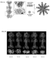
- FIG. 1 The results were shown in FIG. 1 .
- FIG. 1 (A) after docking, the distance between C end of gB and N end of the nanoparticle vector subunit is 56.1 A, so that the appropriate length of the linker shall not be shorter than 56.1 A.
- Linker regions with different lengths are designed (as shown in Table 1), and different vector (I53-50, T3-10, O3-33, T33-15 and T33-31, as shown in Table 2) are selected.
- the self-assembled nanoparticle expected to be constructed was shown FIG. 1 (A), and FIG. 1(B) was a schematic diagram of docking gB with other nanoparticle vector subunits.
- the vector I53-50 comprised subunits I53-50A1 (SEQ ID NO: 1) and I53-50B.4PT1 (SEQ ID NO: 2). Table 1 Sequence of linker Sequence No.
- Expression vector s with different lengths of linkers were constructed for transfection and expression, then the protein concentration was determined after purification and concentration. The specific steps were as follows: (1) The gB gene of the EB virus (SEQ ID NO: 12), the linkers (with a nucleic acid sequence shown in Table 3), T4 fibritin (SEQ ID NO: 19) and I53-50A1 (SEQ ID NO: 20) were inserted into the vector pcDNA3.1(+) by PCR amplification and enzyme digestion recombination, so as to obtain the target gene gB-I53-50A1 expressed by the expression vector.
- the front end of the vector pcDNA3.1(+) was provided with a CD5 signal peptide (SEQ ID NO: 21) for secreting the expressed polypeptide outside cells
- the tail end of the T4 fibritin was provided with the histidine tag (SEQ ID NO: 22) of 8 histidines for convenient purification
- the tail end of the histidine tag was linked to a linking sequence (SEQ ID NO: 26).
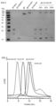
- FIG. 2 shows results of Coomassie brilliant blue staining of SDS-PAGE electrophoresis of the nanoparticle: nanovector subunit I53-50A1 (the preparation method of which is the same as the preparation method of the gB-I53-50A1 subunit in the Point 3, except that in step (1), the gB gene of the EB virus, the linker and the T4 fibritin were not inserted into the vector pcDNA3.1(+)), nanovector subunit I53-50B.4PT1 (the preparation method of which is the same as the preparation method of the I53-50B.4PT1 subunit in Point 3), antigen gB (SEQ ID NO: 3), recombinant nanoparticle gB-I53-50A1 only comprising the nanovector subunit I53-50A1 (the preparation method of which is the same as the preparation method of the gB-I53-50A1 subunit in Point 3), nanoparticle vector I53-50A NP without an antigen
- FIG. 3 is a molecular sieve chromatogram of the nanoparticle. It could be seen from FIG. 3 that, the recombinant vector was successfully constructed, and the high-purity nanoparticle protein (gB-I53-50ANP) could be obtained.
- the molecular mass of gB-I53-50A1 was greater than that of gB, and the size of gB-I53-50A NP after nano granulation is larger than that of the blank particle 153-50 NP, which showed that gB was displayed on the surface of the nanoparticle after assembly.
- the gB self-assembled nanoparticle (gB-I53-50A NP) with 100% display density in Example 2 the nanoparticle vector (I53-50 NP) and the antigen (gB) were diluted to 0.5 mg/mL. 200 ⁇ L of sample was added into a special sample loading slot of Uncle, and stood for 5 minutes, and then the particle size of the nanoparticle was detected by Uncle instrument of Unchained Company.
- the gB self-assembled nanoparticle (gB-I53-50A NP) with 100% display density in Example 2 and the nanoparticle vector (I53-50 NP) were diluted to a concentration ranging from 0.02 mg/mL to 0.2 mg/mL.
- the protein was incubated on a carbon-coated copper grid, then incubated and stained with 2% uranyl acetate for 2 minutes, and dried in air. Then, the size and morphology of the particle were observed by using the 120 KV transmission electron microscope.
- the gB self-assembled nanoparticle (gB-153-50A NP) with 100% display density in Example 2 and the nanoparticle vector (I53-50 NP) were diluted to 0.5 mg/mL.
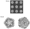
- the thin-layer cryo-electron microscope sample was prepared by using a sampling machine, and then observed by using the 300 KV cryo-electron microscope, and the structure was built by using Relion3.
- the gB self-assembled nanoparticle (gB-I53-50A NP) with 100% display density in Example 2 the nanoparticle vector (I53-50 NP), the antigen (gB) and gB-I53-50A1 were diluted to 0.5 mg/mL firs. Then, heating scanning was carried out from 25°C to 90°C by using Promethus instrument of NanoTemper Company. Changes of the bifluorescence signal ratio and back reflection aggregation signal were recorded, and the Tm value and aggregation temperature were obtained through the first-order derivative.
- the nanoparticle protein was diluted to 0.5 mg/mL first, and then guanidine hydrochloride solutions with the concentration gradient of 0 M to 7 M were added, and incubated overnight at the room temperature. Then, the change of the bifluorescence signal ratio of the protein in different guanidine hydrochloride solutions was detected by using Promethus instrument of NanoTemper Company, and the value of Gibbs free energy change ⁇ G was obtained through the first-order derivative.
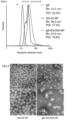
- the gB self-assembled nanoparticle (gB-I53-50A NP) had relatively uniform particle size distribution, and the particle size of the gB self-assembled nanoparticle was significantly larger than that of the nanoparticle vector (I53-50 NP) and that of the antigen (gB), which indicated that gB was successfully displayed on the surface of the particle.
- gB-I53-50A NP and I53-50 NP had good homogeneity under negative staining electron microscope, and the surface of the gB-I53-50A NP particle had an obvious external protrusion compared with the I53-50NP empty particle, which indicated that gB was successfully displayed on the surface of the nanoparticle vector.
- FIG. 7(A) to FIG. 7(C) show results of differential fluorescence scanning of gB, gB-I53-50A1, I53-50 NP and gB-I53-50ANP, wherein FIG. 7 (A) shows the change of the ratio of F330 to F350 of the protein of gB, gB-I53-50A1, I53-50 NP and gB-I53-50A NP during heating; FIG. 7 (B) shows the first-order derivative value of the change of the ratio; and FIG. 7 (C) shows the change of scattered light during heating.
- FIG. 8 shows melting temperature results obtained according to the results of differential fluorescence scanning, wherein Tm onset is the temperature at which melting begins and Tm is the melting temperature.
- the half denaturation concentration of gB was lower than that of gB-I53-50ANP under the guanidine hydrochloride of different concentrations, which mean that the chemical stability of the granulated gB (gB-I53-50A NP) was higher than that of gB.
- the Gibbs free energy of the granulated gB (gB-I53-50ANP) was lower than that of gB, which mean that the nanoparticle (gB-I53-50ANP) not only did not destroy the physical and chemical properties of the antigen gB itself, but also had higher stability than gB on the premise of ensuring the uniformity.
- I53-50 NP has the function of stabilizing gB.
- PBST 0.5% PBST was prepared for kinetic detection. 150 ⁇ L of PBST was added into the pre-wetting plate, and incubated in the proteinA sensor for 10 minutes.
- the antibody AMMOS (please refer to the document Snijder et al., 2018, Immunity 48, 799-811 for its preparation method) was diluted for coupling. After equilibrium, the coupling was started, and then the antigens such as the protein of the nanoparticle (gB, gB-I53-50A1 and gB-I53-50A NP with 100% display density in Example 2) were diluted in gradient (6.25 nM, 12.5 nM, 25 nM, 50 nM and 100 nM), and bound to the sensor. The binding signal and the dissociation signal were recorded, and the sensor was regenerated by using the glycine solution. The binding signal was fitted by using a binding model of 1: 1 to calculate the dynamic parameters.
- the gB nanoparticle (gB-I53-50ANP) had a stronger binding ability to the AMMOS antibody compared to gB, which proved that the antigenicity of the gB nanoparticle is stronger than that of gB.
- the gB nanoparticle (gB-I53-50A NP) had a longer dissociation time. Especially, it is almost difficult for the nanoparticle to dissociate from the antibody, which is beneficial for staying on BCR (B cell antigen receptor) for a long time, and stimulating generation of the antibody.
- nanoparticles with different display densities are immunized at the same time to judge the influence of the display density on the immune effect of the nanoparticle.
- FIG. 11(A) to FIG. 11 (F) in detection of the antibody titer of the serum in the second week and the fifth week, the total antibody titers of the serum induced by the recombinant nanoparticles (gB-I53-50A NP) with the display densities (33%, 67% and 100%) were all higher than that of the monomer gB.
- the total antibody titer of the serum induced by the gB nanoparticle (gB-I53-50ANP) is about 10 times greater than that of gB, and the result is also the same when no adjuvant is used.
- the use of the adjuvants can further enhance the immunogenicity induced by the recombinant nanoparticle.
- gB-I53-50ANP with 33%, 67% and 100% display densities could all effectively produce a large number of neutralizing antibodies.
- the neutralizing antibody produced by gB with 100% display density has a titer that is about 10 times that of gB, which could effectively neutralize EBV infected epithelial cells.
Landscapes
- Health & Medical Sciences (AREA)
- Life Sciences & Earth Sciences (AREA)
- Virology (AREA)
- Chemical & Material Sciences (AREA)
- General Health & Medical Sciences (AREA)
- Medicinal Chemistry (AREA)
- Public Health (AREA)
- Veterinary Medicine (AREA)
- Pharmacology & Pharmacy (AREA)
- Animal Behavior & Ethology (AREA)
- Engineering & Computer Science (AREA)
- Organic Chemistry (AREA)
- Epidemiology (AREA)
- Bioinformatics & Cheminformatics (AREA)
- Molecular Biology (AREA)
- Immunology (AREA)
- Chemical Kinetics & Catalysis (AREA)
- Communicable Diseases (AREA)
- General Chemical & Material Sciences (AREA)
- Gastroenterology & Hepatology (AREA)
- Mycology (AREA)
- Microbiology (AREA)
- Biotechnology (AREA)
- Proteomics, Peptides & Aminoacids (AREA)
- Nuclear Medicine, Radiotherapy & Molecular Imaging (AREA)
- Oncology (AREA)
- Biomedical Technology (AREA)
- Nanotechnology (AREA)
- Optics & Photonics (AREA)
- Physics & Mathematics (AREA)
- Genetics & Genomics (AREA)
- Biophysics (AREA)
- Biochemistry (AREA)
- Tropical Medicine & Parasitology (AREA)
- Peptides Or Proteins (AREA)
- Medicines Containing Antibodies Or Antigens For Use As Internal Diagnostic Agents (AREA)
Abstract
Description
- The present disclosure belongs to the field of biotechnology, and more particularly, relates to a self-assembled nanoparticle containing a gB protein of an EB virus and a preparation method and use thereof.
- An Epstein-Barr Virus (EBV) belongs to a herpes virus family. EBV has a latent infection ability and is one of the earliest identified carcinogenic viruses. EBV infects 95% of the population in the world, and mainly causes infectious mononucleosis in adolescents. Moreover, EBV is closely related to epithelial tumors such as nasopharyngeal carcinoma and gastric cancer, and B-cell tumors such as Burkitt lymphoma and Hodgkin lymphoma in adults. Therefore, it is of great public health significance to develop EBV vaccines.
- As an enveloped virus, EBV infects host cells by membrane fusion. This process is completed by an interaction between membrane fusion proteins gB, gH/gL, gp42, and the like on the surface of EBV and a host cell receptor. At present, it has been found that neutralizing antibodies of these membrane fusion proteins can inhibit EBV from infecting epithelial cells or B cells, therefore, these major membrane fusion proteins are ideal candidate antigens for developing EBV vaccines. As the co-executive proteins during the membrane fusion, gH-gL and gB work together to promote the fusion of EBV At present, receptor binding proteins such as gp350 and gH-gL are used as targets in almost all EBV vaccines, and gB is rarely used as an immunogen. Compared with gH-gL and other proteins, gB with a trimerization conformation has a more complex structure and more diverse functions, so that it is more unknown and challenging to develop vaccines with gB as an immunogen.
- In order to overcome the defects in the prior art, an objective of a first aspect of the present disclosure is to provide a self-assembled nanoparticle containing a gB protein.
- An objective of a second aspect of the present disclosure is to provide a preparation method of the self-assembled nanoparticle containing the gB protein.
- An objective of a third aspect of the present disclosure is to provide use of the self-assembled nanoparticle in the preparation of a drug for preventing EB virus infection.
- An objective of a fourth aspect of the present disclosure is to provide a vaccine comprising the self-assembled nanoparticle.
- An objective of a fifth aspect the present disclosure is to provide use of the self-assembled nanoparticle in the preparation of a drug for treating diseases caused by EB virus infection.
- In order to achieve the objectives above, the technical solutions used in the present disclosure are as follows.
- In the first aspect of the present disclosure, a self-assembled nanoparticle containing a gB protein is provided, which comprises a first polypeptide and a second polypeptide, wherein the first polypeptide comprises a gB protein and a first vector subunit, the second polypeptide comprises a second vector subunit; the first vector subunit is I53-50A1, the second vector subunit is I53-50B.4PT1; and the gB protein is linked to the first vector subunit through a linker peptide, so that the gB protein is displayed outside the assembled nanoparticle, and an immune response of a body is better stimulated.
- Preferably, the first vector subunit and the second vector subunit are self-assembled to form a nanostructure by a noncovalent interaction, the first vector subunit is coated on a surface of the second vector subunit, and the gB protein is displayed on a surface of the nanostructure.
- An amino acid sequence of the I53-50A1 is shown in SEQ ID NO: 1.
- An amino acid sequence of the I53-50B.4PT1 is shown in SEQ ID NO: 2.
- An amino acid sequence of the gB protein is shown in SEQ ID NO: 3.
- The linker peptide is a polypeptide containing 5 amino acids to 20 amino acids; and preferably, the linker peptide is a polypeptide containing 10 amino acids to 15 amino acids. More preferably, the linker peptide is a polypeptide with an amino acid sequence shown in any one of SEQ ID NO: 4 to SEQ ID NO: 9; and most preferably, the linker peptide is a polypeptide with an amino acid sequence shown in SEQ ID NO: 9. The linker peptide is used for linking the antigen gB protein to the vector protein, without affecting the immunogenicity of antigen and the correct folding of protein.
- Preferably, the first polypeptide further comprises a stable protein.
- Preferably, the stable protein is located between the linker peptide and the first vector subunit.
- Preferably, the stable protein is a T4 fibritin shown in SEQ ID NO: 10 or a GCN4 peptide fragment shown in SEQ ID NO: 11; and more preferably, the stable protein is the T4 fibritin.
- Preferably, the first polypeptide is a first polypeptide trimer.
- Preferably, the second polypeptide is a second polypeptide pentamer.
- Preferably, the first polypeptide trimer has a copy number ranging from 18 to 22, and the second polypeptide pentamer has a copy number ranging from 10 to 14. More preferably, the first polypeptide trimer has a copy number of 20, and the second polypeptide pentamer has a copy number of 12.
- Preferably, the self-assembled nanoparticle containing the gB protein has an icosahedral symmetry.
- In the second aspect of the present disclosure, a preparation method of the self-assembled nanoparticle containing the gB protein is provided, comprising the step of incubating the first polypeptide and the second polypeptide to obtain the self-assembled nanoparticle containing the gB protein. The first polypeptide comprises a gB protein and a first vector subunit, and the second polypeptide comprises a second vector subunit; the first vector subunit is I53-50A1, and the second vector subunit is I53-50B.4PT1; and the gB protein is linked to the first vector subunit through a linker peptide, so that the gB protein is displayed outside the assembled nanoparticle, and an immune response of a body is better stimulated.
- An amino acid sequence of the I53-50A1 is shown in SEQ ID NO: 1.
- An amino acid sequence of the I53-50B.4PT1 is shown in SEQ ID NO: 2.
- A molar mass ratio of the first polypeptide to the second polypeptide is preferably 1: 3 to 6; and is more preferably 1:5.
- The incubation is preferably carried out in an assembled buffer for 0.5 hour to 2 hours.
- Preferably, the assembled buffer comprises 250 mm NaCl, 50 mm Tris-HCl with pH 8.0 and 5% glycerol.
- An amino acid sequence of the gB protein is shown in SEQ ID NO: 3.
- The linker peptide is a polypeptide containing 5 amino acids to 20 amino acids; and preferably, the linker peptide is a polypeptide containing 10 amino acids to 15 amino acids. More preferably, the linker peptide is a polypeptide with an amino acid sequence shown in any one of SEQ ID NO: 4 to SEQ ID NO: 9; and most preferably, the linker peptide is a polypeptide with an amino acid sequence shown in SEQ ID NO: 9. The linker peptide is used for linking the antigen gB protein to a vector protein, without affecting the immunogenicity of antigen and the correct folding of protein.
- Preferably, the first polypeptide further comprises a stable protein.
- Preferably, the stable protein is located between the linker peptide and the first vector subunit.
- The stable protein is preferably a T4 fibritin shown in SEQ ID NO: 10 or a GCN4 peptide fragment shown in SEQ ID NO: 11; and more preferably, the stable protein is the T4 fibritin.
- Preferably, the first polypeptide and the second polypeptide further comprise a purification tag.
- Preferably, the purification tag is at least one selected from the group consisting of histidine tag (His tag), streptavidin tag (Strep tag) and maltose binding protein (MBP); and more preferably, the purification tag is the histidine tag (His tag).
- The purification tag of the first polypeptide is located between the stable protein and the first vector subunit.
- The purification tag of the second polypeptide is located at a tail end of the second vector subunit.
- The first polypeptide further comprises a signal peptide, so that a target protein can be secreted to a supernatant after expression.
- The signal peptide is a CD5 signal peptide shown in SEQ ID NO: 25.
- Preferably, the first polypeptide is obtained by the following steps: introducing a nucleic acid expressing the first polypeptide into a first host cell; and incubating the first host cell to express the first polypeptide.
- Preferably, the first host cell is a eukaryotic cell; more preferably, the first host cell is at least one selected from the group consisting of human embryonic kidney 293 cell (HEK293F), Madin-Daby canine kidney cell (MDCK), Chlorocebus sabaeus kidney cell (VERO), SF9 (Spodoptera frugiperda 9) cell, HighFive cell, CHO (Chinese Hamster Ovary) cell, and yeast cell; and most preferably, the first host cell is the human embryonic kidney 293 cell.
- Preferably, the second polypeptide is obtained by the following steps: introducing a nucleic acid expressing the second polypeptide into a second host cell; and incubating the second host cell to express the second polypeptide.
- Preferably, the second host cell is a prokaryotic cell; more preferably, the second host cell is Escherichia coli; and most preferably, the second host cell is Rosetta(DE3).
- In the third aspect of the present disclosure, use of the self-assembled nanoparticle in the first aspect in the preparation of a drug for preventing EB virus infection is provided.
- In the fourth aspect of the present disclosure, a vaccine comprising the self-assembled nanoparticle in the first aspect is provided.
- A vaccine comprising the self-assembled nanoparticle containing the gB protein is provided.
- The vaccine further includes an adjuvant.
- Preferably, the adjuvant is at least one selected from the group consisting of an aluminum adjuvant, an oil emulsion adjuvant such as oil-in-water emulsion, water-in-oil emulsion and bidirectional emulsion, a microorganism-originated adjuvant such as peptidoglycan (PG), lipopolysccharide (LPS) of Gram-negative bacterial outer membrane, mycobacteria and components thereof, GpG oligonucleotide (GpG ODN) and cholera toxin (CT), a microsomal antigen delivery system such as liposome, polymeric microsphere, inert nanosphere, nano aluminum adjuvant, immunostimulating complex (IS-COM), cytokine, a polysaccharide such as inulin (MPI), and a natural source such as propolis and sapoin. More preferably, the adjuvant is at least one selected from the group consisting of aluminum adjuvant and MF59 adjuvant.
- In the fifth aspect of the present disclosure, use of the self-assembled nanoparticle in the first aspect in the preparation of a drug for treating diseases caused by EB virus infection is provided.
- Preferably, the disease is at least one selected from the group consisting of infectious disease, malignant tumor, chronic disease and autoimmune disease. More preferably, the disease is at least one selected from the group consisting of mononucleosis, nasopharyngeal carcinoma, gastric carcinoma, epithelial tumor, Burkitt lymphoma, Hodgkin lymphoma, chronic fatigue syndrome, multiple sclerosis and ankylosing myelitis.
- The drug further comprises a pharmaceutically acceptable carrier.
- The present disclosure has the beneficial effects as follows:
In the self-assembled nanoparticle provided herein, the gB protein of the EB virus is displayed on the surface of the nanoparticle for the first time. The particle size of the self-assembled nanoparticle is larger than that of the antigen (gB), the heat stability of the self-assembled nanoparticle is equivalent to that of the antigen (gB), the chemical stability of the self-assembled nanoparticle is higher than that of the antigen (gB), and the binding capacity with the neutralizing antibody of the self-assembled nanoparticle is higher than that of the antigen (gB), which are beneficial for prolonging residence time of the self-assembled nanoparticle in the B cell antigen receptor and stimulating generation of the antibody. Meanwhile, the self-assembled nanoparticle is capable of inducing a higher animal immune antibody titer, and is suitable for preventing EB virus infection and treating diseases caused by EB virus infection. - Although a heterologous gene is introduced into the self-assembled nanoparticle provided herein, since the heterologous gene is derived from the protein of bacteria, which can avoid causing autoimmune diseases, thus having an advantage of high safety without affecting an immune effect.
-
-
FIG. 1 is a schematic diagram of docking a gB antigen with different nanoparticle skeleton proteins, wherein (A) is a schematic diagram of docking the gB antigen with the nanoparticle skeleton protein I53-50A1, and the self-assembled nanoparticle formed; and (B) is a schematic diagram of docking the gB antigen with different nanoparticle skeleton proteins. -
FIG. 2 is a Coomassie brilliant blue staining graph of SDS-PAGE electrophoresis of the self-assembled nanoparticle. -
FIG. 3 is a molecular sieve chromatogram of the self-assembled nanoparticle. -
FIG. 4 is a result diagram of dynamic light scattering of the self-assembled nanoparticle. -
FIG. 5 is a negative staining electron micrograph of the self-assembled nanoparticle. -
FIG. 6 is a cryo-electron micrograph of the self-assembled nanoparticle, wherein (A) is the 2D classification diagram of the self-assembled nanoparticle under Relion3; and (B) is the electron density map after reconstruction. -
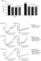
FIG. 7 is a result diagram of differential fluorescence scanning of the self-assembled nanoparticle, wherein (A) is a diagram showing a change of a ratio of F330 to F350 of the protein of the self-assembled nanoparticle during heating; (B) is a diagram of a first-order derivative value of the change of the ratio of F330 to F350 of the protein of the self-assembled nanoparticle during heating; and (C) is a diagram of a change of scattered light of the protein of the self-assembled nanoparticle during heating. -
FIG. 8 is a diagram of Tm onset and Tm of the self-assembled nanoparticle, wherein Tm onset is a temperature at which melting begins, and Tm is a melting temperature. -
FIG. 9 is a diagram showing the change of the ratio of F330 to F350 of proteins of different particles under different concentrations of guanidine hydrochloride during heating and a diagram showing the change of scattered light of the proteins during heating, wherein (A) is a diagram showing the change of the ratio of F330 to F350 of the protein of nanoparticle vector (I53-50ANP) under different concentrations of guanidine hydrochloride during heating; (B) is a diagram showing the change of the ratio of F330 to F350 of the protein of gB under different concentrations of guanidine hydrochloride during heating; (C) is a diagram showing the change of the ratio of F330 to F350 of the protein of self-assembled nanoparticle (gB-I53-50A NP) under different concentrations of guanidine hydrochloride during heating; (D) is a diagram showing the change of scattered light of the protein of the nanoparticle vector (I53-50ANP) under different concentrations of guanidine hydrochloride during heating; (E) is a diagram showing the change of scattered light of the protein of gB under different concentrations of guanidine hydrochloride during heating; and (F) is a diagram showing the change of scattered light of the protein of the self-assembled nanoparticle (gB-I53-50A NP) under different concentrations of guanidine hydrochloride during heating. -
FIG. 10 is a diagram of affinity detection between different particles and an antibody, wherein (A) is a diagram of a binding point between gB and an AMMOS antibody; (B) is a diagram of a binding point between a subunit gB-I53-50A1 and the AMMOS antibody; and (C) is a diagram of a binding point between the self-assembled nanoparticle (gB-I53-50A NP) and the AMMOS antibody. -
FIG. 11 is a graph of anti-gB serum titers of immunized mice, wherein (A) is a graph of anti-gB serum titers of mice immunized with different particles for two weeks; (B) is a graph of anti-gB serum titers of mice immunized with different particles containing an adjuvant MF59 for two weeks; (C) is a graph of anti-gB serum titers of mice immunized with particles that are added or not added with the nanoparticle vector (I53-50A NP) for two weeks; (D) is a graph of anti-gB serum titers of mice immunized with different particles for five weeks; (E) a graph of anti-gB serum titers of mice immunized with different particles containing the adjuvant MF59 for five weeks; (F) is a graph of anti-gB serum titers of mice immunized with particles that are added or not added with the nanoparticle vector (I53-50A NP) for five weeks; wherein, ∗ indicates that P<0.05; ∗∗ indicates that P<0.005; ∗∗∗ indicates that P<0.0005; ∗∗∗∗ indicates that P<0.00005; and ns indicates that P>0.05. -
FIG. 12 is a graph showing influence of serum of mice immunized for five weeks on EBV-infected epithelial cells. - The contents of the present disclosure are further described in detail hereinafter with reference to the specific example and the accompanying drawings.
- It should be understood that these examples are only used for describing the present disclosure and are not intended to limit the scope of the present disclosure.
- In the following examples, if the specific conditions of the experimental methods are not indicated, the conventional conditions are generally used. Various common chemical reagents used in the examples are all commercially available products.
- The preparation method of the nanoparticle vaccine in the present disclosure includes the following steps.
- A. An appropriate fusion distance is determined through computer aided designs such as Sic_axle, Rosetta and the like, so as to determine a length of a linker for nanoparticle design in a sequence, and design a corresponding nanoparticle vector based on the length.
- B. An appropriate quantity of eukaryotic expression vectors are transferred into a host first cell for expression by a transient transfection technology to obtain a nanoparticle subunit protein (a first polypeptide) of gB-I53-50A1. Meanwhile, the expression plasmid of I53-50B.4PT1 is transformed by using a second host cells, and after induction with IPTG, another nanoparticle subunit protein (a second polypeptide) of I53-50B.4PT1 is expressed and obtained.
- C. gB-I53-50A1 and I53-50B.4PT1 subunits are added into an assembling buffer according to a certain proportion, and incubated at a room temperature to obtain an assembled nanoparticle. The assembled nanoparticle is separated by molecular exclusion chromatography, and particle size distribution and stability of the protein is determined by negative staining electron microscopy, dynamic light scattering and differential scanning fluorescence.
- D. An antigenicity of the nanoparticle is determined by bio-layer interferometry (BLI).
- E. The nanoparticle is evenly mixed with an adjuvant, and a Balb/C mouse is immunized to verify an antibody level against gB generated in the mouse and a neutralizing ability of serum of the mouse to an EBV virus.
- The nanoparticle vaccine of the present application is further described in detail hereinafter.
- The appropriate fusion distance between the antigen (gB protein, SEQ ID NO: 3) and the nanoparticle carrier (153-50) was determined through computer aided designs such as Sic_axle, Rosetta and the like, so as to determine the length of the linker for linking the antigen to the nanoparticle vector in the sequence, and design the corresponding nanoparticle vector based on the length.
- The software used in the design were: Sic_axle (Marcandalli et al., 2019, Cell 177, 1420-1431), and Rosetta (Bale et al., 2016, Science 353, 389-394.). https://www.rosettacommons.org/docs/latest/scripting_documentation/RosettaScripts/Mo vers/movers_pages/RelaxScript.
- The results were shown in
FIG. 1 . InFIG. 1 (A) , after docking, the distance between C end of gB and N end of the nanoparticle vector subunit is 56.1 A, so that the appropriate length of the linker shall not be shorter than 56.1 A. Linker regions with different lengths are designed (as shown in Table 1), and different vector (I53-50, T3-10, O3-33, T33-15 and T33-31, as shown in Table 2) are selected. The self-assembled nanoparticle expected to be constructed was shownFIG. 1 (A), and FIG. 1(B) was a schematic diagram of docking gB with other nanoparticle vector subunits. - After comparing the final docking structure model and the docking distance, since the shortest docking distance could be finally obtained when I53-50 was docked with the antigen gB as the vector, the vector I53-50 was finally selected and served as the appropriate vector for further design. The vector I53-50 comprised subunits I53-50A1 (SEQ ID NO: 1) and I53-50B.4PT1 (SEQ ID NO: 2).
Table 1 Sequence of linker Sequence No. Linker SEQ ID NO: 4 GGGGSGGGGS SEQ ID NO: 5 GGGGSGGGGSG SEQ ID NO: 6 GGGGSGGGGSGS SEQ ID NO: 7 GGGGSGGGGSGGS SEQ ID NO: 8 GGGGSGGGGSGGGS SEQ ID NO: 9 GGGGSGGGGSGGGGS Table 2 Information of vectors ID PDB code Particle property Docking distance T3-10 4EGG Tetrahedron 56.9 A O3-33 3VCD Octahedron 52.42 A T33-15 4NWO Co-assembling tetrahedron 59.82 A T33-31 4ZK7 Co-assembling tetrahedron 51.54 A I53-50 6P6F Co-assembling icosahedron 49.5 A -
- (1) Expression vectors: eukaryotic expression vector pcDNA3.1(+) (ThermoFisher), prokaryotic expression vector pET28a(+) (ThermoFisher), and Escherichia coli competent cell DH5α (Tiangen).
- (2) Expression systems: Eukaryotic expression system cell HEK293F(ATCC) and transformed Escherichia coli cell Rosetta (DE3) (Tianen).
- (3) Reagents and materials: PCR enzyme (GeneStar), recombinant enzyme (Vazyme), restriction endonuclease (NEB), gel recovery reagent (GeneStar), plasmid midiprep kit (MN), cell transfection reagent PEI (Polyscience), 293F culture medium (Union), TB culture medium (Xiangbo Bio), purified agarose beads of histidine tag protein (Roche), and other conventional reagents and materials purchased commercially.
- (4) Genes: gB gene of EB virus (M81 strain) and I53-50A1/I53-50B particle subunit gene optimized based on bacterial protein were optimized and synthesized through the OptimumGeneTM codon platform of Nanjing Genscript Biological Co., Ltd..
- Expression vector s with different lengths of linkers were constructed for transfection and expression, then the protein concentration was determined after purification and concentration. The specific steps were as follows: (1) The gB gene of the EB virus (SEQ ID NO: 12), the linkers (with a nucleic acid sequence shown in Table 3), T4 fibritin (SEQ ID NO: 19) and I53-50A1 (SEQ ID NO: 20) were inserted into the vector pcDNA3.1(+) by PCR amplification and enzyme digestion recombination, so as to obtain the target gene gB-I53-50A1 expressed by the expression vector. Wherein, the front end of the vector pcDNA3.1(+) was provided with a CD5 signal peptide (SEQ ID NO: 21) for secreting the expressed polypeptide outside cells, the tail end of the T4 fibritin was provided with the histidine tag (SEQ ID NO: 22) of 8 histidines for convenient purification, and the tail end of the histidine tag was linked to a linking sequence (SEQ ID NO: 26). (2) The recombinant vector gene in pcDNA3.1 was transformed into DH5α competent bacteria, and positive clones were screened by ampicillin resistance. Then, the positive clones were picked into a TB culture medium containing 0.1% ampicillin (0.1 mg/mL) for amplification, and then extracted by the midiprep kit. The specific method could be referred to the instruction of product. (3) 293F cells were subjected to suspension culture and amplification in the 293F culture medium (Union), and were ready for transient transfection after being amplified to a certain quantity. The cells were diluted to 1 L with a density of 1∗106/mL, and then, the transfection system of 1 mg of pcDNA3.1-
target protein vector 5 mg PEI was prepared with a fresh culture medium, added into the diluted 293F cells after standing for 30 minutes, and cultured at 37°C, 80% humidity and 5% CO2 concentration for 7 days under shaking at 120 rpm. The cell precipitate was removed by centrifugation. The supernatant was filtered by the 0.22 µm filter membrane, and then purified by protein affinity chromatography and molecular sieve to obtain a high-purity target protein gB-I53-50A1 subunit. Results were shown in Table 3, which indicated that the gB-I53-50A1 subunit had the highest yield when the linker was GGGGSGGGGSGGGGS (SEQ ID NO: 9).Table 3 Protein yields of vectors with different linkers Linker (nucleic acid sequence) Linker (amino acid sequence) Length of amino acid sequence Yield (mg/L culture medium) 
GGGGSGGGGS (SEQ ID NO: 4) 10 0.1 
GGGGSGGGGSG (SEQ ID NO: 5) 11 0.25 
GGGGSGGGGSGS (SEQ ID NO: 6) 12 0.72 
GGGGSGGGGSGGS (SEQ ID NO: 7) 13 0.44 
GGGGSGGGGSGGGS (SEQ ID NO: 8) 14 0.34 
GGGGSGGGGSGGGGS (SEQ ID NO: 9) 15 1.2 -
- (1) The gB gene of the EB virus (SEQ ID NO: 12), the linker (SEQ ID NO: 18), T4 fibritin (SEQ ID NO: 19) and I53-50A1 (SEQ ID NO: 20) were inserted into the vector pcDNA3.1(+) by PCR amplification and enzyme digestion recombination, so as to obtain the target gene gB-I53-50A1 (SEQ ID NO: 27) expressed by the expression vector. The front end of the vector pcDNA3.1(+) was provided with the CD5 signal peptide (SEQ ID NO: 21) for secreting the expressed polypeptide outside cells, the tail end of the T4 fibritin was provided with the histidine tag (SEQ ID NO: 22) of 8 histidines for convenient purification, and the tail end of the histidine tag was linked to a linking sequence (SEQ ID NO: 26) . Moreover, I53-50B.4PT1 (SEQ ID NO: 23) was directly inserted into the vector pET28a(+) during synthesis, and the tail end of the vector pET28a(+) was provided with the histidine tag (SEQ ID NO: 24) of 6 histidines for convenient purification. After sequencing and comparison, the successfully constructed vector was selected for the next experiment.
- (2) The recombinant vector gene in pcDNA3.1 was transformed into the DH5α competent bacteria, and the positive clones were screened by ampicillin resistance. Then, the positive clones were picked into the TB culture medium containing 0.1% ampicillin (0.1 mg/mL) for amplification, and then extracted by the midiprep kit. The specific method could be referred to the instruction of product.
- (3) The recombinant vector gene in pET28a(+) was transformed into Rosetta (DE3) competent bacteria, and positive clones were screened by kanamycin resistance. Then, the positive clones were picked into the TB culture medium containing 0.1% kanamycin (0.03 g/mL) for amplification, and then further amplified to 1 L in a conical flask, and kanamycin and chloramphenicol were added for screening positive cells. 0.2 mM chemical inducer isopropyl thiogalactoside (IPTG) was added at 18°C to induce expression of the target protein, and after induction for 20 hours, bacterial cells were collected, crushed under a high pressure, and centrifuged to obtain a supernatant. The supernatant was filtered at 0.22 µm, and purified by protein affinity chromatography and molecular sieve to obtain a high-purity target protein I53-50B.4PT1 subunit (SEQ ID NO: 29).
- (4) The 293F cells were subjected to suspension culture and amplification in the 293F culture medium (Union), and were ready for transient transfection after being amplified to a certain quantity. The cells were diluted to 1L with the density of 1∗106/mL, and then, the transfection system of 1 mg of pcDNA3.1-
target protein vector 5 mg PEI was prepared with the fresh culture medium, added into the diluted 293F cells after standing for 30 minutes, and cultured at 37°C, 80% humidity and 5% CO2 concentration for 7 days under shaking at 120 rpm. The cell precipitate was removed by centrifugation. The supernatant was filtered by the 0.22 µm filter membrane, and then purified by protein affinity chromatography and molecular sieve to obtain the high-purity target protein gB-I53-50A1 subunit (SEQ ID NO: 28). - (5) The two subunits (gB-I53-50A1 and I53-50B.4PT1) were added into the assembling buffer (250 mM NaCl, 50 mM Tris-HCl with pH 8.0, and 5% glycerol) according to the molar ratio of 1: 5, and incubated at the room temperature for 1 hour, and then the assembled nanoparticles (gB-I53-50A NP) with 100% display density was separated by using the molecular sieve. The two subunits (gB-I53-50A1 and I53-50B.4PT1) were added into the assembling buffer (250 mm NaCl, 50 mm Tris-HCl with PH 8.0, and 5% glycerol) according to a molar ratio of 1: 2, and incubated at the room temperature for 1 hour, and then the assembled nanoparticle (gB-I53-50ANP) with 33% display density was separated by using the molecular sieve. The two subunits (gB-I53-50A1 and I53-50B.4PT1) were added into the assembling buffer (250 mm NaCl, 50 mm Tris-HCl with pH 8.0, and 5% glycerol) according to a molar ratio of 2: 1, and incubated at the room temperature for 1 hour, and then the assembled nanoparticle (gB-I53-50ANP) with 67% display density was separated by using the molecular sieve.
- As shown in
FIG. 2 and FIG. 3, FIG. 2 shows results of Coomassie brilliant blue staining of SDS-PAGE electrophoresis of the nanoparticle: nanovector subunit I53-50A1 (the preparation method of which is the same as the preparation method of the gB-I53-50A1 subunit in the Point 3, except that in step (1), the gB gene of the EB virus, the linker and the T4 fibritin were not inserted into the vector pcDNA3.1(+)), nanovector subunit I53-50B.4PT1 (the preparation method of which is the same as the preparation method of the I53-50B.4PT1 subunit in Point 3), antigen gB (SEQ ID NO: 3), recombinant nanoparticle gB-I53-50A1 only comprising the nanovector subunit I53-50A1 (the preparation method of which is the same as the preparation method of the gB-I53-50A1 subunit in Point 3), nanoparticle vector I53-50A NP without an antigen (the preparation method of which is the same as the preparation method of the nanoparticle (gB-I53-50ANP) with 100% display density in Point 3, except that in step (1), the gB gene of the EB virus, the linker and the T4 fibritin were not inserted into the vector pcDNA3.1(+)), and recombinant nanoparticle protein gB-I53-50A NP with multiple display densities containing the antigen gB, the I53-50A1 and the I53-50B.4PT1 (the preparation method of which is the same as the preparation method of the nanoparticle (gB-I53-50A NP) with different display densities in Point 3) are shown from left to right.FIG. 3 is a molecular sieve chromatogram of the nanoparticle. It could be seen fromFIG. 3 that, the recombinant vector was successfully constructed, and the high-purity nanoparticle protein (gB-I53-50ANP) could be obtained. The molecular mass of gB-I53-50A1 was greater than that of gB, and the size of gB-I53-50A NP after nano granulation is larger than that of the blank particle 153-50 NP, which showed that gB was displayed on the surface of the nanoparticle after assembly. -
- (1) Unchained Uncle high-throughput protein stability analyzer (Unchained Labs).
- (2) NanoTemper Promethus 48 protein stability analyzer (Nanotemper).
- (3)120 KV transmission electron microscope (FEI).
- (4) 300 KV cryoelectron microscope (Thermo).
- The gB self-assembled nanoparticle (gB-I53-50A NP) with 100% display density in Example 2, the nanoparticle vector (I53-50 NP) and the antigen (gB) were diluted to 0.5 mg/mL. 200 µL of sample was added into a special sample loading slot of Uncle, and stood for 5 minutes, and then the particle size of the nanoparticle was detected by Uncle instrument of Unchained Company.
- The gB self-assembled nanoparticle (gB-I53-50A NP) with 100% display density in Example 2 and the nanoparticle vector (I53-50 NP) were diluted to a concentration ranging from 0.02 mg/mL to 0.2 mg/mL. The protein was incubated on a carbon-coated copper grid, then incubated and stained with 2% uranyl acetate for 2 minutes, and dried in air. Then, the size and morphology of the particle were observed by using the 120 KV transmission electron microscope.
- The gB self-assembled nanoparticle (gB-153-50A NP) with 100% display density in Example 2 and the nanoparticle vector (I53-50 NP) were diluted to 0.5 mg/mL. The thin-layer cryo-electron microscope sample was prepared by using a sampling machine, and then observed by using the 300 KV cryo-electron microscope, and the structure was built by using Relion3.
- The gB self-assembled nanoparticle (gB-I53-50A NP) with 100% display density in Example 2, the nanoparticle vector (I53-50 NP), the antigen (gB) and gB-I53-50A1 were diluted to 0.5 mg/mL firs. Then, heating scanning was carried out from 25°C to 90°C by using Promethus instrument of NanoTemper Company. Changes of the bifluorescence signal ratio and back reflection aggregation signal were recorded, and the Tm value and aggregation temperature were obtained through the first-order derivative.
- The nanoparticle protein was diluted to 0.5 mg/mL first, and then guanidine hydrochloride solutions with the concentration gradient of 0 M to 7 M were added, and incubated overnight at the room temperature. Then, the change of the bifluorescence signal ratio of the protein in different guanidine hydrochloride solutions was detected by using Promethus instrument of NanoTemper Company, and the value of Gibbs free energy change ΔG was obtained through the first-order derivative.
- As shown in
FIG. 4 , the gB self-assembled nanoparticle (gB-I53-50A NP) had relatively uniform particle size distribution, and the particle size of the gB self-assembled nanoparticle was significantly larger than that of the nanoparticle vector (I53-50 NP) and that of the antigen (gB), which indicated that gB was successfully displayed on the surface of the particle. - As shown in
FIG. 5 , it could be seen that gB-I53-50A NP and I53-50 NP had good homogeneity under negative staining electron microscope, and the surface of the gB-I53-50A NP particle had an obvious external protrusion compared with the I53-50NP empty particle, which indicated that gB was successfully displayed on the surface of the nanoparticle vector. - As shown in
FIG. 6, FIG. 6(A) is a 2D classification diagram of the nanoparticle under Relion3. It was obvious that the nanoparticle showed regular icosahedron symmetry.FIG. 6(B) is an electron density map after reconstruction. It could be seen that there were obvious nanoparticles formed by 20 copies of trimer I53-50A1 and 12 copies of pentamer I53-50B4.PT1. - As shown in
FIG. 7 andFIG. 8 ,FIG. 7(A) to FIG. 7(C) show results of differential fluorescence scanning of gB, gB-I53-50A1, I53-50 NP and gB-I53-50ANP, whereinFIG. 7 (A) shows the change of the ratio of F330 to F350 of the protein of gB, gB-I53-50A1, I53-50 NP and gB-I53-50A NP during heating;FIG. 7 (B) shows the first-order derivative value of the change of the ratio; andFIG. 7 (C) shows the change of scattered light during heating. It could be seen that the melting temperatures of gB in gB, gB-I53-50A1 and gB-I53-50A NP were almost the same, which proved that granulation had no obvious influence on the structure of gB. In addition, in terms of aggregation information (increase in scattered light), the gB-I53-50A NP particle had no obvious aggregation during heating compared with gB and gB-I53-50A1, which indicated that the heat stability of gB-I53-50A NP particle is better.FIG. 8 shows melting temperature results obtained according to the results of differential fluorescence scanning, wherein Tm onset is the temperature at which melting begins and Tm is the melting temperature. It could be seen that the melting temperatures of gB, gB-I53-50A1 and gB-I53-50A NP were similar, which proved that gB, gB-I53-50A1 and gB-I53-50A NP were highly similar in the heat stability structure, and the structural change did not destroy physical and chemical properties of gB. - As shown in
FIG. 9 (A) to FIG. 9 (F) , the half denaturation concentration of gB was lower than that of gB-I53-50ANP under the guanidine hydrochloride of different concentrations, which mean that the chemical stability of the granulated gB (gB-I53-50A NP) was higher than that of gB. Moreover, the Gibbs free energy of the granulated gB (gB-I53-50ANP) was lower than that of gB, which mean that the nanoparticle (gB-I53-50ANP) not only did not destroy the physical and chemical properties of the antigen gB itself, but also had higher stability than gB on the premise of ensuring the uniformity. In addition, it could be proved that I53-50 NP has the function of stabilizing gB. -
- (1) ProteinA sensor (Fortebio), PBS, and Tween 20 (Sigma-Aldrich).
- (2)
Fortebio Octet 96 instrument. - (3) Pre-wetting plate, 96-well plate, and other commercial and conventional consumables.
-
- (1) The affinity between the nanoparticle and the neutralizing antibody was detected by using BLI.
- 0.5% PBST was prepared for kinetic detection. 150 µL of PBST was added into the pre-wetting plate, and incubated in the proteinA sensor for 10 minutes. The antibody AMMOS (please refer to the document Snijder et al., 2018, Immunity 48, 799-811 for its preparation method) was diluted for coupling. After equilibrium, the coupling was started, and then the antigens such as the protein of the nanoparticle (gB, gB-I53-50A1 and gB-I53-50A NP with 100% display density in Example 2) were diluted in gradient (6.25 nM, 12.5 nM, 25 nM, 50 nM and 100 nM), and bound to the sensor. The binding signal and the dissociation signal were recorded, and the sensor was regenerated by using the glycine solution. The binding signal was fitted by using a binding model of 1: 1 to calculate the dynamic parameters.
- As shown in
FIG. 10(A) to FIG. 10 (C) and Table 4, the gB nanoparticle (gB-I53-50ANP) had a stronger binding ability to the AMMOS antibody compared to gB, which proved that the antigenicity of the gB nanoparticle is stronger than that of gB. In addition, the gB nanoparticle (gB-I53-50A NP) had a longer dissociation time. Especially, it is almost difficult for the nanoparticle to dissociate from the antibody, which is beneficial for staying on BCR (B cell antigen receptor) for a long time, and stimulating generation of the antibody.Table 4 Kinetic parameters of antibody affinities of gB, gB-I53-50A1 and gB-I53-50A NP KD (M) kon (1/Ms) Kdis (1/s) gB 1.82E-09 2.12E +05 3.87E-04 gB-I53-50A1 1.63E-09 1.24E +05 2.03E-04 gB-I53-50ANP 1.24E-09 8.95E +05 1.11E-04 -
- (1) Mice: BalB/C mice, female, 6 weeks to 8 weeks of age (Beijing Charles River Laboratory Animal Technology Co., Ltd.).
- (2) Adjuvant: Inject A aluminum adjuvant (ThermoFisher), and MF59 adjuvant {0.5%(v/v)
Tween 80, 0.5%(v/v)Span 85, 4.3%(v/v) squalene, 10 nM sodium citrate buffer}. - (3) Other commercial and conventional reagent.
-
- (1) 0.5 µg of empty nanovector (empty-NP, and I53-50 NP in Example 2), 5 µg of gB protein of EB virus, and particles with 33%, 67% and 100% display densities containing gB of equal molar mass (gB-I53-50ANP in Example 2) were respectively mixed with the above two adjuvants (Inject A aluminum adjuvant and MF59 adjuvant) or respectively diluted with PBST, that was, the adjuvants or PBST was mixed with the antigen according to the mass ratio of 1: 1, and incubated under shaking overnight at 4°C. The mice were immunized by subcutaneous immunization.
- (2) The mice were immunized again in the third week after immunization. In the second week and the fifth week after immunization, the orbital blood of the mice was collected, and separated to collect serum. The total antibody titer of gB in the serum of the mice was detected through indirect enzyme-linked immunosorbent assay.
- Since there is a positive relationship between the display density and the BCR affinity, in order to detect the relationship between the display density of gB and the induced antibody titer, nanoparticles with different display densities are immunized at the same time to judge the influence of the display density on the immune effect of the nanoparticle. As shown in
FIG. 11(A) to FIG. 11 (F) , in detection of the antibody titer of the serum in the second week and the fifth week, the total antibody titers of the serum induced by the recombinant nanoparticles (gB-I53-50A NP) with the display densities (33%, 67% and 100%) were all higher than that of the monomer gB. The higher the display density was, the higher the total antibody titer of the serum was. Whether the aluminum adjuvant or the MF59 adjuvant is used, the total antibody titer of the serum induced by the gB nanoparticle (gB-I53-50ANP) is about 10 times greater than that of gB, and the result is also the same when no adjuvant is used. The use of the adjuvants can further enhance the immunogenicity induced by the recombinant nanoparticle. -
- (1) Reagents: Goat anti-human IgG (ThermoFisher), and other commercial and conventional reagents and consumables.
- (2) Cell lines: Akata-EBV-P (Vicbio (Beijing) Biotechnology Co., Ltd.), and HNE1(ATCC).
- (3) Virus: Akata-EBV-GFP virus induced by cell strain Akata-EBV
-
- (1) Akata-EBV was induced by IgG to produce the Akata-EBV-GFP virus, which was enriched by high-speed centrifugation, resuspended with serum-free 1640, and stored at -80°C.
- (2) The Akata virus was diluted by the serum-free 1640 culture medium (Gibco) with a ratio of 1: 5, and added into 96-well plate, with 50 µL per well. The serum of the mice collected in the fifth week in Example 5 was diluted with a ratio of 1: 5, then diluted into the virus with multiple proportions of 10, and incubated at 37°C for 2 hours.
- (3) The mixture of the serum and the virus was added into spread HNE1 cells (7000 cells/well) and infected at 37°C for 3.5 hours.
- (4) After infection, the supernatant was removed and replaced by 5% FBS 1640 culture medium. After culture for 48 hours, the cells were digested with pancreatin, and then the ratio of GFP positive cells to total cells was detected to judge the cell infection efficiency.
- As shown in
FIG. 12 , gB-I53-50ANP with 33%, 67% and 100% display densities could all effectively produce a large number of neutralizing antibodies. Especially, the neutralizing antibody produced by gB with 100% display density has a titer that is about 10 times that of gB, which could effectively neutralize EBV infected epithelial cells. In addition, there was a more obvious difference in the ability to induce the neutralizing antibody in the adjuvant-free group. - The above examples are the preferred examples of the present disclosure, but the embodiments of the present disclosure are not limited by the above examples. Any other changes, modifications, substitutions, combinations, and simplifications made without departing from the spirit and principle of the present disclosure should be equivalent substitute modes, and should be included in the scope of protection of the present disclosure.
Claims (10)
- A self-assembled nanoparticle, comprising a first polypeptide and a second polypeptide, wherein the first polypeptide comprises a gB protein and a first vector subunit, the second polypeptide comprises a second vector subunit; the first vector subunit is I53-50A1, and the second vector subunit is I53-50B.4PT1; and the gB protein is linked to the first vector subunit through a linker peptide.
- The self-assembled nanoparticle according to claim 1, wherein, an amino acid sequence of the I53-50A1 is shown in SEQ ID NO: 1; an amino acid sequence of the I53-50B.4PT1 is shown in SEQ ID NO: 2; and the linker peptide is preferably a polypeptide containing 5 amino acids to 20 amino acids; and more preferably, the linker peptide is a polypeptide with an amino acid sequence shown in any one of SEQ ID NO: 4 to SEQ ID NO: 9.
- The self-assembled nanoparticle according to claim 2, wherein, the first polypeptide further comprises a stable protein; the stable protein is located between the first vector subunit and the linker peptide; and the stable protein is preferably a T4 fibritin shown in SEQ ID NO: 10 or a GCN4 peptide fragment shown in SEQ ID NO: 11.
- The self-assembled nanoparticle according to claim 3, wherein, the first polypeptide is a first polypeptide trimer, and the second polypeptide is a second polypeptide pentamer.
- The self-assembled nanoparticle according to claim 4, wherein: the first polypeptide trimer has a copy number ranging from 18 to 22, and the second polypeptide pentamer has a copy number ranging from 10 to 14.
- The self-assembled nanoparticle according to any one of claims 1 to 5, wherein: an amino acid sequence of the gB protein is shown in SEQ ID NO: 3.
- A preparation method of the self-assembled nanoparticle according to any one of claims 1 to 6, comprising incubating the first polypeptide and the second polypeptide to obtain the self-assembled nanoparticle, wherein, a molar mass ratio of the first polypeptide to the second polypeptide is preferably 1: 3 to 6.
- Use of the self-assembled nanoparticle according to any one of claims 1 to 6 in the preparation of a drug for preventing EB virus infection.
- A vaccine, comprising the self-assembled nanoparticle according to any one of claims 1 to 6; wherein the vaccine further comprises an adjuvant preferably.
- Use of the self-assembled nanoparticle according to any one of claims 1 to 6 in the preparation of a drug for treating diseases caused by EB virus infection.
Applications Claiming Priority (2)
| Application Number | Priority Date | Filing Date | Title |
|---|---|---|---|
| CN202011417091.0A CN112521511B (en) | 2020-12-07 | 2020-12-07 | Self-assembled nano-particles containing EB virus gB protein and preparation method and application thereof |
| PCT/CN2020/136752 WO2022120908A1 (en) | 2020-12-07 | 2020-12-16 | Self-assembled nanoparticle containing gb protein of eb virus, and preparation method therefor and use thereof |
Publications (2)
| Publication Number | Publication Date |
|---|---|
| EP4257615A1 true EP4257615A1 (en) | 2023-10-11 |
| EP4257615A4 EP4257615A4 (en) | 2024-09-04 |
Family
ID=74997265
Family Applications (1)
| Application Number | Title | Priority Date | Filing Date |
|---|---|---|---|
| EP20964841.9A Pending EP4257615A4 (en) | 2020-12-07 | 2020-12-16 | SELF-ASSEMBLED NANOPARTICLES CONTAINING GB PROTEIN OF THE EB VIRUS, PRODUCTION PROCESS AND USE THEREOF |
Country Status (4)
| Country | Link |
|---|---|
| US (1) | US12098167B2 (en) |
| EP (1) | EP4257615A4 (en) |
| CN (1) | CN112521511B (en) |
| WO (1) | WO2022120908A1 (en) |
Families Citing this family (8)
| Publication number | Priority date | Publication date | Assignee | Title |
|---|---|---|---|---|
| CN112521511B (en) | 2020-12-07 | 2023-03-14 | 中山大学 | Self-assembled nano-particles containing EB virus gB protein and preparation method and application thereof |
| CN112778404B (en) * | 2021-01-28 | 2022-11-29 | 中山大学 | Self-assembled nano-particles containing EB virus gHgL protein and preparation method and application thereof |
| JP2024520730A (en) * | 2021-06-07 | 2024-05-24 | アイコサバックス, インコーポレイテッド | Virus-like particle vaccines against coronaviruses |
| CN116284348B (en) * | 2022-08-03 | 2025-09-23 | 中山大学肿瘤防治中心(中山大学附属肿瘤医院、中山大学肿瘤研究所) | A monoclonal antibody for recognizing Epstein-Barr virus gp42 protein and its application |
| CN117462666B (en) * | 2022-09-30 | 2024-07-30 | 烟台派诺生物技术有限公司 | An immune composition product for preventing or treating varicella-zoster virus-related diseases and its preparation method |
| CN116333168A (en) * | 2023-02-17 | 2023-06-27 | 岭南现代农业科学与技术广东省实验室肇庆分中心 | Porcine epidemic diarrhea virus S protein trimer nano-particle and preparation method thereof |
| CN116874600B (en) * | 2023-07-13 | 2024-07-05 | 浙江大学 | Preparation method and application of short peptides from nanoantibodies that can target CD163 receptors |
| CN119798422B (en) * | 2024-12-12 | 2025-12-09 | 广州珺柏诺医疗科技有限公司 | Monoclonal antibody of EB virus gB antigen targeting and application thereof |
Family Cites Families (15)
| Publication number | Priority date | Publication date | Assignee | Title |
|---|---|---|---|---|
| US20050228074A1 (en) | 2004-04-05 | 2005-10-13 | Bridgestone Corporation | Amphiphilic polymer micelles and use thereof |
| US7465348B1 (en) * | 2007-06-07 | 2008-12-16 | Xerox Corporation | Nanosized particles of monoazo laked pigment |
| CA2917512C (en) | 2007-10-12 | 2025-08-05 | President And Fellows Of Harvard College | Vaccine nanotechnology |
| CN102108336A (en) * | 2010-06-13 | 2011-06-29 | 中山大学肿瘤防治中心 | Recombinant adenovirus vaccine containing EB (Epstein-Barr) virus membrane protein gp350/220, gp85, gp78 or gp25 gene |
| KR20160002848A (en) * | 2013-03-24 | 2016-01-08 | 오이신 바이오테크놀로지스 | Systems and methods for the targeted production of a therapeutic protein within a target cell |
| EP3906939A1 (en) * | 2013-10-11 | 2021-11-10 | The United States of America, represented by the Secretary, Department of Health and Human Services | Epstein-barr virus vaccines |
| US11525146B2 (en) * | 2017-01-09 | 2022-12-13 | Oisin Biotechnologies, Inc. | Expression constructs, fusogenic lipid-based nanoparticles and methods of use thereof |
| CN111511405A (en) * | 2017-10-16 | 2020-08-07 | 阿迪根有限公司 | Peptides and nanoparticles for intracellular delivery of mRNA |
| US11395851B2 (en) * | 2018-02-16 | 2022-07-26 | The Wistar Institute Of Anatomy And Biology | Epstein-Barr virus nucleic acid constructs and vaccines made therefrom, and methods of using same |
| CA3095216A1 (en) * | 2018-02-28 | 2019-09-06 | University Of Washington | Self-asssembling nanostructure vaccines |
| US20220265804A1 (en) | 2019-04-29 | 2022-08-25 | Academy Of Military Medical Sciences | Fusion protein capable of self-assembling into protein nanoparticles and application thereof |
| CN110615848B (en) * | 2019-10-08 | 2021-05-14 | 中山大学肿瘤防治中心(中山大学附属肿瘤医院、中山大学肿瘤研究所) | A Chimeric Particle Containing the Dominant Epitope Peptide of Epstein-Barr Virus Membrane Surface Glycoprotein gp350 and Its Encoding Gene and Application |
| CN110922488A (en) * | 2019-11-08 | 2020-03-27 | 中山大学肿瘤防治中心(中山大学附属肿瘤医院、中山大学肿瘤研究所) | Self-assembled nano-particles containing EB virus gp350, and preparation method and application thereof |
| CN111991556B (en) * | 2020-10-29 | 2021-03-02 | 中山大学 | SARS-CoV-2 RBD conjugated nano particle vaccine |
| CN112521511B (en) | 2020-12-07 | 2023-03-14 | 中山大学 | Self-assembled nano-particles containing EB virus gB protein and preparation method and application thereof |
-
2020
- 2020-12-07 CN CN202011417091.0A patent/CN112521511B/en active Active
- 2020-12-15 US US17/762,308 patent/US12098167B2/en active Active
- 2020-12-16 WO PCT/CN2020/136752 patent/WO2022120908A1/en not_active Ceased
- 2020-12-16 EP EP20964841.9A patent/EP4257615A4/en active Pending
Also Published As
| Publication number | Publication date |
|---|---|
| CN112521511B (en) | 2023-03-14 |
| US20240034755A1 (en) | 2024-02-01 |
| WO2022120908A1 (en) | 2022-06-16 |
| EP4257615A4 (en) | 2024-09-04 |
| CN112521511A (en) | 2021-03-19 |
| US12098167B2 (en) | 2024-09-24 |
Similar Documents
| Publication | Publication Date | Title |
|---|---|---|
| US12098167B2 (en) | Self-assembled nanoparticle containing gB protein of EB virus and preparation method and use thereof | |
| Lua et al. | Bioengineering virus‐like particles as vaccines | |
| EP4265632A1 (en) | Self-assembled nanoparticle containing ghgl protein of eb virus, and preparation method therefor and use thereof | |
| Vragniau et al. | Synthetic self-assembling ADDomer platform for highly efficient vaccination by genetically encoded multiepitope display | |
| WO2022121322A1 (en) | Recombinant subunit vaccine of novel coronavirus and application thereof | |
| JP2003530123A (en) | Hepatitis B core antigen fusion protein | |
| CN107921115A (en) | Novel baculovirus vectors and methods of use | |
| Wang et al. | Self-assembling ferritin nanoparticles coupled with linear sequences from canine distemper virus haemagglutinin protein elicit robust immune responses | |
| CN115960259B (en) | Preparation method and application of modularized assembled bi-component nano particles | |
| WO2023087555A1 (en) | Novel coronavirus recombinant protein with broad-spectrum neutralizing activity and preparation method therefor | |
| CN112089831A (en) | Preparation method of recombinant novel coronavirus subunit vaccine | |
| Dong et al. | Nanoparticles of conformation-stabilized canine distemper virus hemagglutinin are highly immunogenic and induce robust immunity | |
| US10125175B2 (en) | Method for enhancing immunogenicity of epitope peptide of HPV antigen, virus-like particle, and method for preparing HPV vaccine | |
| Wu et al. | Recombinant T7 phage with FMDV AKT-III strain VP1 protein is a potential FMDV vaccine | |
| AU2009216696A1 (en) | Protein nanocarriers, process for obtaining them and applications | |
| CN113144187B (en) | Self-assembled nanoparticle containing EB virus gHgLgp42 protein and preparation method and application thereof | |
| CN106928372A (en) | Hepatitis B recombinant antigen and its expressing gene, construction method, virus-like particle and preparation method thereof, using with vaccine | |
| Kimura et al. | Efficient Neutralizing Antibodies Induction by Human Parvovirus B19 Epitope‐Presenting Protein Nanoparticles | |
| CN116444623A (en) | A nano-vaccine based on a nanoparticle scaffold and its preparation method and application | |
| CN102181461A (en) | Method for expressing target polypeptide on filobactivirus at high density and application thereof | |
| Zhang et al. | A shared N-terminal hydrophobic tail for the formation of nanoparticulates | |
| Zhu et al. | Construction and Immunogenicity Detection of Canine Par vovirus-like Particles Fusion with Canine Febrile Antigen Epitopes | |
| Scheller et al. | Human cytomegalovirus protein pp65: an efficient protein carrier system into human dendritic cells | |
| CN120392994A (en) | Preparation and application of a nanoparticle vaccine of Epstein-Barr virus gp350 protein polypeptide and its combination | |
| CN121851117A (en) | HMPV Pre-fusion F Protein Mutant and Its Application |
Legal Events
| Date | Code | Title | Description |
|---|---|---|---|
| STAA | Information on the status of an ep patent application or granted ep patent |
Free format text: STATUS: THE INTERNATIONAL PUBLICATION HAS BEEN MADE |
|
| PUAI | Public reference made under article 153(3) epc to a published international application that has entered the european phase |
Free format text: ORIGINAL CODE: 0009012 |
|
| STAA | Information on the status of an ep patent application or granted ep patent |
Free format text: STATUS: REQUEST FOR EXAMINATION WAS MADE |
|
| 17P | Request for examination filed |
Effective date: 20230706 |
|
| AK | Designated contracting states |
Kind code of ref document: A1 Designated state(s): AL AT BE BG CH CY CZ DE DK EE ES FI FR GB GR HR HU IE IS IT LI LT LU LV MC MK MT NL NO PL PT RO RS SE SI SK SM TR |
|
| DAV | Request for validation of the european patent (deleted) | ||
| DAX | Request for extension of the european patent (deleted) | ||
| A4 | Supplementary search report drawn up and despatched |
Effective date: 20240802 |
|
| RIC1 | Information provided on ipc code assigned before grant |
Ipc: A61P 35/00 20060101ALI20240729BHEP Ipc: A61P 31/22 20060101ALI20240729BHEP Ipc: A61K 9/14 20060101ALI20240729BHEP Ipc: A61K 39/245 20060101ALI20240729BHEP Ipc: C12N 15/62 20060101ALI20240729BHEP Ipc: C07K 19/00 20060101AFI20240729BHEP |