EP4249591A1 - Production method for microglia - Google Patents
Production method for microglia Download PDFInfo
- Publication number
- EP4249591A1 EP4249591A1 EP23159583.6A EP23159583A EP4249591A1 EP 4249591 A1 EP4249591 A1 EP 4249591A1 EP 23159583 A EP23159583 A EP 23159583A EP 4249591 A1 EP4249591 A1 EP 4249591A1
- Authority
- EP
- European Patent Office
- Prior art keywords
- microglia
- cells
- gene
- production method
- differentiation
- Prior art date
- Legal status (The legal status is an assumption and is not a legal conclusion. Google has not performed a legal analysis and makes no representation as to the accuracy of the status listed.)
- Pending
Links
- 210000000274 microglia Anatomy 0.000 title claims abstract description 60
- 238000004519 manufacturing process Methods 0.000 title claims abstract description 24
- 108091023040 Transcription factor Proteins 0.000 claims abstract description 65
- 102000040945 Transcription factor Human genes 0.000 claims abstract description 64
- 230000014509 gene expression Effects 0.000 claims abstract description 38
- 108090000623 proteins and genes Proteins 0.000 claims abstract description 31
- 102100034808 CCAAT/enhancer-binding protein alpha Human genes 0.000 claims abstract description 19
- 101000601664 Homo sapiens Paired box protein Pax-8 Proteins 0.000 claims abstract description 16
- 102100037502 Paired box protein Pax-8 Human genes 0.000 claims abstract description 16
- 210000001778 pluripotent stem cell Anatomy 0.000 claims abstract description 16
- 108010018650 MEF2 Transcription Factors Proteins 0.000 claims abstract description 15
- 102100034798 CCAAT/enhancer-binding protein beta Human genes 0.000 claims abstract description 11
- 102100020679 Krueppel-like factor 6 Human genes 0.000 claims abstract description 11
- 108700026244 Open Reading Frames Proteins 0.000 claims abstract description 9
- 102000039446 nucleic acids Human genes 0.000 claims abstract description 9
- 108020004707 nucleic acids Proteins 0.000 claims abstract description 9
- 150000007523 nucleic acids Chemical class 0.000 claims abstract description 9
- 108700023400 Platelet-activating factor receptors Proteins 0.000 claims abstract description 8
- 102000030769 platelet activating factor receptor Human genes 0.000 claims abstract description 8
- 102100037799 DNA-binding protein Ikaros Human genes 0.000 claims abstract description 7
- 102100023227 E3 SUMO-protein ligase EGR2 Human genes 0.000 claims abstract description 7
- 102100039247 ETS-related transcription factor Elf-4 Human genes 0.000 claims abstract description 7
- 102100030334 Friend leukemia integration 1 transcription factor Human genes 0.000 claims abstract description 7
- 101000599038 Homo sapiens DNA-binding protein Ikaros Proteins 0.000 claims abstract description 7
- 101001049692 Homo sapiens E3 SUMO-protein ligase EGR2 Proteins 0.000 claims abstract description 7
- 101000813135 Homo sapiens ETS-related transcription factor Elf-4 Proteins 0.000 claims abstract description 7
- 101001062996 Homo sapiens Friend leukemia integration 1 transcription factor Proteins 0.000 claims abstract description 7
- 101001109689 Homo sapiens Nuclear receptor subfamily 4 group A member 3 Proteins 0.000 claims abstract description 7
- 102100022673 Nuclear receptor subfamily 4 group A member 3 Human genes 0.000 claims abstract description 7
- 102100024894 PR domain zinc finger protein 1 Human genes 0.000 claims abstract description 7
- 102100025373 Runt-related transcription factor 1 Human genes 0.000 claims abstract description 7
- 102000016918 Complement C3 Human genes 0.000 claims abstract description 6
- 108010028780 Complement C3 Proteins 0.000 claims abstract description 6
- 101001139126 Homo sapiens Krueppel-like factor 6 Proteins 0.000 claims abstract description 3
- 101000945515 Homo sapiens CCAAT/enhancer-binding protein alpha Proteins 0.000 claims abstract 4
- 108010043471 Core Binding Factor Alpha 2 Subunit Proteins 0.000 claims abstract 2
- 101000945963 Homo sapiens CCAAT/enhancer-binding protein beta Proteins 0.000 claims abstract 2
- 101000973200 Homo sapiens Nuclear factor 1 C-type Proteins 0.000 claims abstract 2
- 101000893493 Homo sapiens Protein flightless-1 homolog Proteins 0.000 claims abstract 2
- 101000611183 Homo sapiens Tumor necrosis factor Proteins 0.000 claims abstract 2
- 102100021148 Myocyte-specific enhancer factor 2A Human genes 0.000 claims abstract 2
- 102100039229 Myocyte-specific enhancer factor 2C Human genes 0.000 claims abstract 2
- 102100022162 Nuclear factor 1 C-type Human genes 0.000 claims abstract 2
- 101150084398 PTAFR gene Proteins 0.000 claims abstract 2
- 108010009975 Positive Regulatory Domain I-Binding Factor 1 Proteins 0.000 claims abstract 2
- 102100040247 Tumor necrosis factor Human genes 0.000 claims abstract 2
- 101100365087 Arabidopsis thaliana SCRA gene Proteins 0.000 claims description 2
- 210000004027 cell Anatomy 0.000 description 56
- 230000004069 differentiation Effects 0.000 description 42
- 238000000034 method Methods 0.000 description 30
- 239000002299 complementary DNA Substances 0.000 description 29
- 239000002773 nucleotide Substances 0.000 description 27
- 125000003729 nucleotide group Chemical group 0.000 description 27
- 239000001963 growth medium Substances 0.000 description 25
- 239000003550 marker Substances 0.000 description 20
- 230000002025 microglial effect Effects 0.000 description 20
- 108010065459 CCAAT-Enhancer-Binding Protein-alpha Proteins 0.000 description 15
- 238000001890 transfection Methods 0.000 description 15
- 238000003559 RNA-seq method Methods 0.000 description 12
- 108091032973 (ribonucleotides)n+m Proteins 0.000 description 11
- 102000055120 MEF2 Transcription Factors Human genes 0.000 description 11
- 238000011156 evaluation Methods 0.000 description 11
- 108010064535 CCAAT-Enhancer-Binding Protein-beta Proteins 0.000 description 9
- 108010049058 Kruppel-Like Factor 6 Proteins 0.000 description 9
- 102100026171 P2Y purinoceptor 12 Human genes 0.000 description 9
- 108010014270 Purinergic P2Y12 Receptors Proteins 0.000 description 9
- 238000004458 analytical method Methods 0.000 description 9
- 108020004999 messenger RNA Proteins 0.000 description 9
- 108010023243 NFI Transcription Factors Proteins 0.000 description 8
- 102000011178 NFI Transcription Factors Human genes 0.000 description 8
- 230000006698 induction Effects 0.000 description 8
- 239000000243 solution Substances 0.000 description 8
- 230000000242 pagocytic effect Effects 0.000 description 7
- 101000687344 Homo sapiens PR domain zinc finger protein 1 Proteins 0.000 description 6
- 101000857677 Homo sapiens Runt-related transcription factor 1 Proteins 0.000 description 6
- 239000002609 medium Substances 0.000 description 6
- 238000010899 nucleation Methods 0.000 description 6
- 102100022820 Disintegrin and metalloproteinase domain-containing protein 28 Human genes 0.000 description 5
- 101000756756 Homo sapiens Disintegrin and metalloproteinase domain-containing protein 28 Proteins 0.000 description 5
- 238000002123 RNA extraction Methods 0.000 description 5
- 102100027264 Solute carrier organic anion transporter family member 2B1 Human genes 0.000 description 5
- 101710095541 Solute carrier organic anion transporter family member 2B1 Proteins 0.000 description 5
- 108060008682 Tumor Necrosis Factor Proteins 0.000 description 5
- 210000004443 dendritic cell Anatomy 0.000 description 5
- 238000000556 factor analysis Methods 0.000 description 5
- 239000000203 mixture Substances 0.000 description 5
- 210000001616 monocyte Anatomy 0.000 description 5
- 238000000513 principal component analysis Methods 0.000 description 5
- 102000003390 tumor necrosis factor Human genes 0.000 description 5
- 102100033312 Alpha-2-macroglobulin Human genes 0.000 description 4
- 101150071258 C3 gene Proteins 0.000 description 4
- 102100039196 CX3C chemokine receptor 1 Human genes 0.000 description 4
- IAZDPXIOMUYVGZ-UHFFFAOYSA-N Dimethylsulphoxide Chemical compound CS(C)=O IAZDPXIOMUYVGZ-UHFFFAOYSA-N 0.000 description 4
- 101000746022 Homo sapiens CX3C chemokine receptor 1 Proteins 0.000 description 4
- 101001064779 Homo sapiens Plexin domain-containing protein 2 Proteins 0.000 description 4
- 101000651211 Homo sapiens Transcription factor PU.1 Proteins 0.000 description 4
- 206010057249 Phagocytosis Diseases 0.000 description 4
- 108010015078 Pregnancy-Associated alpha 2-Macroglobulins Proteins 0.000 description 4
- 102100027654 Transcription factor PU.1 Human genes 0.000 description 4
- 239000006285 cell suspension Substances 0.000 description 4
- 210000003169 central nervous system Anatomy 0.000 description 4
- LOKCTEFSRHRXRJ-UHFFFAOYSA-I dipotassium trisodium dihydrogen phosphate hydrogen phosphate dichloride Chemical compound P(=O)(O)(O)[O-].[K+].P(=O)(O)([O-])[O-].[Na+].[Na+].[Cl-].[K+].[Cl-].[Na+] LOKCTEFSRHRXRJ-UHFFFAOYSA-I 0.000 description 4
- 210000002540 macrophage Anatomy 0.000 description 4
- 230000008782 phagocytosis Effects 0.000 description 4
- 239000002953 phosphate buffered saline Substances 0.000 description 4
- 101001051291 Homo sapiens Lysosomal-associated transmembrane protein 5 Proteins 0.000 description 3
- 101000916644 Homo sapiens Macrophage colony-stimulating factor 1 receptor Proteins 0.000 description 3
- 102100033485 Lymphocyte antigen 86 Human genes 0.000 description 3
- 101710158190 Lymphocyte antigen 86 Proteins 0.000 description 3
- 102100024625 Lysosomal-associated transmembrane protein 5 Human genes 0.000 description 3
- 102100028198 Macrophage colony-stimulating factor 1 receptor Human genes 0.000 description 3
- 102100031889 Plexin domain-containing protein 2 Human genes 0.000 description 3
- 238000011530 RNeasy Mini Kit Methods 0.000 description 3
- 230000001464 adherent effect Effects 0.000 description 3
- 238000004113 cell culture Methods 0.000 description 3
- 239000011248 coating agent Substances 0.000 description 3
- 238000000576 coating method Methods 0.000 description 3
- 238000011534 incubation Methods 0.000 description 3
- 210000004263 induced pluripotent stem cell Anatomy 0.000 description 3
- 230000001939 inductive effect Effects 0.000 description 3
- 239000000126 substance Substances 0.000 description 3
- HJCMDXDYPOUFDY-WHFBIAKZSA-N Ala-Gln Chemical compound C[C@H](N)C(=O)N[C@H](C(O)=O)CCC(N)=O HJCMDXDYPOUFDY-WHFBIAKZSA-N 0.000 description 2
- CIWBSHSKHKDKBQ-JLAZNSOCSA-N Ascorbic acid Chemical compound OC[C@H](O)[C@H]1OC(=O)C(O)=C1O CIWBSHSKHKDKBQ-JLAZNSOCSA-N 0.000 description 2
- 239000006144 Dulbecco’s modified Eagle's medium Substances 0.000 description 2
- 102100020997 Fractalkine Human genes 0.000 description 2
- 101000854520 Homo sapiens Fractalkine Proteins 0.000 description 2
- 102000007651 Macrophage Colony-Stimulating Factor Human genes 0.000 description 2
- 108010046938 Macrophage Colony-Stimulating Factor Proteins 0.000 description 2
- 239000012124 Opti-MEM Substances 0.000 description 2
- FAPWRFPIFSIZLT-UHFFFAOYSA-M Sodium chloride Chemical compound [Na+].[Cl-] FAPWRFPIFSIZLT-UHFFFAOYSA-M 0.000 description 2
- 102000004887 Transforming Growth Factor beta Human genes 0.000 description 2
- 108090001012 Transforming Growth Factor beta Proteins 0.000 description 2
- 239000012574 advanced DMEM Substances 0.000 description 2
- 201000010099 disease Diseases 0.000 description 2
- 208000037265 diseases, disorders, signs and symptoms Diseases 0.000 description 2
- 239000013604 expression vector Substances 0.000 description 2
- JVTAAEKCZFNVCJ-UHFFFAOYSA-N lactic acid Chemical compound CC(O)C(O)=O JVTAAEKCZFNVCJ-UHFFFAOYSA-N 0.000 description 2
- 238000002156 mixing Methods 0.000 description 2
- 210000004498 neuroglial cell Anatomy 0.000 description 2
- 239000008188 pellet Substances 0.000 description 2
- DAEPDZWVDSPTHF-UHFFFAOYSA-M sodium pyruvate Chemical compound [Na+].CC(=O)C([O-])=O DAEPDZWVDSPTHF-UHFFFAOYSA-M 0.000 description 2
- ZRKFYGHZFMAOKI-QMGMOQQFSA-N tgfbeta Chemical compound C([C@H](NC(=O)[C@H](C(C)C)NC(=O)CNC(=O)[C@H](CCC(O)=O)NC(=O)[C@H](CCCNC(N)=N)NC(=O)[C@H](CC(N)=O)NC(=O)[C@H](CC(C)C)NC(=O)[C@H]([C@@H](C)O)NC(=O)[C@H](CCC(O)=O)NC(=O)[C@H]([C@@H](C)O)NC(=O)[C@H](CC(C)C)NC(=O)CNC(=O)[C@H](C)NC(=O)[C@H](CO)NC(=O)[C@H](CCC(N)=O)NC(=O)[C@@H](NC(=O)[C@H](C)NC(=O)[C@H](C)NC(=O)[C@@H](NC(=O)[C@H](CC(C)C)NC(=O)[C@@H](N)CCSC)C(C)C)[C@@H](C)CC)C(=O)N[C@@H]([C@@H](C)O)C(=O)N[C@@H](C(C)C)C(=O)N[C@@H](CC=1C=CC=CC=1)C(=O)N[C@@H](C)C(=O)N1[C@@H](CCC1)C(=O)N[C@@H]([C@@H](C)O)C(=O)N[C@@H](CC(N)=O)C(=O)N[C@@H](CCC(O)=O)C(=O)N[C@@H](C)C(=O)N[C@@H](CC=1C=CC=CC=1)C(=O)N[C@@H](CCCNC(N)=N)C(=O)N[C@@H](C)C(=O)N[C@@H](CC(C)C)C(=O)N1[C@@H](CCC1)C(=O)N1[C@@H](CCC1)C(=O)N[C@@H](CCCNC(N)=N)C(=O)N[C@@H](CCC(O)=O)C(=O)N[C@@H](CCCNC(N)=N)C(=O)N[C@@H](CO)C(=O)N[C@@H](CCCNC(N)=N)C(=O)N[C@@H](CC(C)C)C(=O)N[C@@H](CC(C)C)C(O)=O)C1=CC=C(O)C=C1 ZRKFYGHZFMAOKI-QMGMOQQFSA-N 0.000 description 2
- YBJHBAHKTGYVGT-UHFFFAOYSA-N 5-(2-oxo-1,3,3a,4,6,6a-hexahydrothieno[3,4-d]imidazol-4-yl)pentanoic acid Chemical compound N1C(=O)NC2C(CCCCC(=O)O)SCC21 YBJHBAHKTGYVGT-UHFFFAOYSA-N 0.000 description 1
- 101150037123 APOE gene Proteins 0.000 description 1
- 208000024827 Alzheimer disease Diseases 0.000 description 1
- 102000013455 Amyloid beta-Peptides Human genes 0.000 description 1
- 108010090849 Amyloid beta-Peptides Proteins 0.000 description 1
- 102100029470 Apolipoprotein E Human genes 0.000 description 1
- 102100024119 CDK5 and ABL1 enzyme substrate 1 Human genes 0.000 description 1
- 102100026190 Class E basic helix-loop-helix protein 41 Human genes 0.000 description 1
- 108050001049 Extracellular proteins Proteins 0.000 description 1
- 101100489763 Homo sapiens A2M gene Proteins 0.000 description 1
- 101100215477 Homo sapiens ADAM28 gene Proteins 0.000 description 1
- 101100218714 Homo sapiens BHLHE41 gene Proteins 0.000 description 1
- 101100328540 Homo sapiens C3 gene Proteins 0.000 description 1
- 101100135226 Homo sapiens CD200 gene Proteins 0.000 description 1
- 101000910461 Homo sapiens CDK5 and ABL1 enzyme substrate 1 Proteins 0.000 description 1
- 101100439100 Homo sapiens CEBPA gene Proteins 0.000 description 1
- 101100439105 Homo sapiens CEBPB gene Proteins 0.000 description 1
- 101100168714 Homo sapiens CSF1R gene Proteins 0.000 description 1
- 101100115243 Homo sapiens CX3CR1 gene Proteins 0.000 description 1
- 101100444906 Homo sapiens EGR2 gene Proteins 0.000 description 1
- 101100011485 Homo sapiens ELF4 gene Proteins 0.000 description 1
- 101100281204 Homo sapiens FLI1 gene Proteins 0.000 description 1
- 101100452380 Homo sapiens IKZF1 gene Proteins 0.000 description 1
- 101100181395 Homo sapiens LAPTM5 gene Proteins 0.000 description 1
- 101100236298 Homo sapiens LY86 gene Proteins 0.000 description 1
- 101100456626 Homo sapiens MEF2A gene Proteins 0.000 description 1
- 101100290704 Homo sapiens MEF2C gene Proteins 0.000 description 1
- 101100026247 Homo sapiens NFIC gene Proteins 0.000 description 1
- 101001098352 Homo sapiens OX-2 membrane glycoprotein Proteins 0.000 description 1
- 101001086535 Homo sapiens Olfactomedin-like protein 3 Proteins 0.000 description 1
- 101100242428 Homo sapiens P2RY12 gene Proteins 0.000 description 1
- 101100351035 Homo sapiens PAX8 gene Proteins 0.000 description 1
- 101100138493 Homo sapiens PTAFR gene Proteins 0.000 description 1
- 101100310795 Homo sapiens SPI1 gene Proteins 0.000 description 1
- 101000836226 Homo sapiens Solute carrier organic anion transporter family member 2B1 Proteins 0.000 description 1
- 101100099880 Homo sapiens TNF gene Proteins 0.000 description 1
- 101000598051 Homo sapiens Transmembrane protein 119 Proteins 0.000 description 1
- 101000795117 Homo sapiens Triggering receptor expressed on myeloid cells 2 Proteins 0.000 description 1
- 101000577630 Homo sapiens Vitamin K-dependent protein S Proteins 0.000 description 1
- 206010020880 Hypertrophy Diseases 0.000 description 1
- 102100038235 Large neutral amino acids transporter small subunit 2 Human genes 0.000 description 1
- 239000012580 N-2 Supplement Substances 0.000 description 1
- 101710092556 Nuclear receptor subfamily 4 group A member 3 Proteins 0.000 description 1
- 102100037589 OX-2 membrane glycoprotein Human genes 0.000 description 1
- 102100032750 Olfactomedin-like protein 3 Human genes 0.000 description 1
- 108091006238 SLC7A8 Proteins 0.000 description 1
- 102100037029 Transmembrane protein 119 Human genes 0.000 description 1
- 102100029678 Triggering receptor expressed on myeloid cells 2 Human genes 0.000 description 1
- 241000700605 Viruses Species 0.000 description 1
- 102100028885 Vitamin K-dependent protein S Human genes 0.000 description 1
- 210000001642 activated microglia Anatomy 0.000 description 1
- 238000007792 addition Methods 0.000 description 1
- 235000010323 ascorbic acid Nutrition 0.000 description 1
- 229960005070 ascorbic acid Drugs 0.000 description 1
- 239000011668 ascorbic acid Substances 0.000 description 1
- 210000004556 brain Anatomy 0.000 description 1
- 210000005056 cell body Anatomy 0.000 description 1
- 230000024245 cell differentiation Effects 0.000 description 1
- 210000000170 cell membrane Anatomy 0.000 description 1
- 230000004663 cell proliferation Effects 0.000 description 1
- 208000015114 central nervous system disease Diseases 0.000 description 1
- 238000005119 centrifugation Methods 0.000 description 1
- 230000008859 change Effects 0.000 description 1
- 238000006243 chemical reaction Methods 0.000 description 1
- 238000007796 conventional method Methods 0.000 description 1
- 238000012258 culturing Methods 0.000 description 1
- 239000003145 cytotoxic factor Substances 0.000 description 1
- 238000007865 diluting Methods 0.000 description 1
- 210000001671 embryonic stem cell Anatomy 0.000 description 1
- 238000010195 expression analysis Methods 0.000 description 1
- 238000000605 extraction Methods 0.000 description 1
- 210000003958 hematopoietic stem cell Anatomy 0.000 description 1
- 210000005260 human cell Anatomy 0.000 description 1
- 238000003384 imaging method Methods 0.000 description 1
- 210000002865 immune cell Anatomy 0.000 description 1
- 238000001727 in vivo Methods 0.000 description 1
- 230000002757 inflammatory effect Effects 0.000 description 1
- 235000014655 lactic acid Nutrition 0.000 description 1
- 239000004310 lactic acid Substances 0.000 description 1
- 230000003902 lesion Effects 0.000 description 1
- 238000013507 mapping Methods 0.000 description 1
- 230000007246 mechanism Effects 0.000 description 1
- 210000003716 mesoderm Anatomy 0.000 description 1
- 238000012986 modification Methods 0.000 description 1
- 230000004048 modification Effects 0.000 description 1
- 210000000653 nervous system Anatomy 0.000 description 1
- 210000002569 neuron Anatomy 0.000 description 1
- 235000003170 nutritional factors Nutrition 0.000 description 1
- 210000001539 phagocyte Anatomy 0.000 description 1
- 239000013600 plasmid vector Substances 0.000 description 1
- 238000002360 preparation method Methods 0.000 description 1
- 102000005962 receptors Human genes 0.000 description 1
- 108020003175 receptors Proteins 0.000 description 1
- 238000010079 rubber tapping Methods 0.000 description 1
- 210000002966 serum Anatomy 0.000 description 1
- 238000012174 single-cell RNA sequencing Methods 0.000 description 1
- 239000011780 sodium chloride Substances 0.000 description 1
- 229940054269 sodium pyruvate Drugs 0.000 description 1
- 229960005322 streptomycin Drugs 0.000 description 1
- 238000006467 substitution reaction Methods 0.000 description 1
- 239000006228 supernatant Substances 0.000 description 1
- 239000006188 syrup Substances 0.000 description 1
- 235000020357 syrup Nutrition 0.000 description 1
- 238000011222 transcriptome analysis Methods 0.000 description 1
- 239000013598 vector Substances 0.000 description 1
- 230000035899 viability Effects 0.000 description 1
- XLYOFNOQVPJJNP-UHFFFAOYSA-N water Substances O XLYOFNOQVPJJNP-UHFFFAOYSA-N 0.000 description 1
Images



Classifications
-
- C—CHEMISTRY; METALLURGY
- C12—BIOCHEMISTRY; BEER; SPIRITS; WINE; VINEGAR; MICROBIOLOGY; ENZYMOLOGY; MUTATION OR GENETIC ENGINEERING
- C12N—MICROORGANISMS OR ENZYMES; COMPOSITIONS THEREOF; PROPAGATING, PRESERVING, OR MAINTAINING MICROORGANISMS; MUTATION OR GENETIC ENGINEERING; CULTURE MEDIA
- C12N5/00—Undifferentiated human, animal or plant cells, e.g. cell lines; Tissues; Cultivation or maintenance thereof; Culture media therefor
- C12N5/10—Cells modified by introduction of foreign genetic material
-
- C—CHEMISTRY; METALLURGY
- C12—BIOCHEMISTRY; BEER; SPIRITS; WINE; VINEGAR; MICROBIOLOGY; ENZYMOLOGY; MUTATION OR GENETIC ENGINEERING
- C12N—MICROORGANISMS OR ENZYMES; COMPOSITIONS THEREOF; PROPAGATING, PRESERVING, OR MAINTAINING MICROORGANISMS; MUTATION OR GENETIC ENGINEERING; CULTURE MEDIA
- C12N5/00—Undifferentiated human, animal or plant cells, e.g. cell lines; Tissues; Cultivation or maintenance thereof; Culture media therefor
- C12N5/06—Animal cells or tissues; Human cells or tissues
- C12N5/0602—Vertebrate cells
- C12N5/0618—Cells of the nervous system
-
- C—CHEMISTRY; METALLURGY
- C12—BIOCHEMISTRY; BEER; SPIRITS; WINE; VINEGAR; MICROBIOLOGY; ENZYMOLOGY; MUTATION OR GENETIC ENGINEERING
- C12N—MICROORGANISMS OR ENZYMES; COMPOSITIONS THEREOF; PROPAGATING, PRESERVING, OR MAINTAINING MICROORGANISMS; MUTATION OR GENETIC ENGINEERING; CULTURE MEDIA
- C12N5/00—Undifferentiated human, animal or plant cells, e.g. cell lines; Tissues; Cultivation or maintenance thereof; Culture media therefor
- C12N5/06—Animal cells or tissues; Human cells or tissues
- C12N5/0602—Vertebrate cells
- C12N5/0618—Cells of the nervous system
- C12N5/0622—Glial cells, e.g. astrocytes, oligodendrocytes; Schwann cells
-
- C—CHEMISTRY; METALLURGY
- C12—BIOCHEMISTRY; BEER; SPIRITS; WINE; VINEGAR; MICROBIOLOGY; ENZYMOLOGY; MUTATION OR GENETIC ENGINEERING
- C12N—MICROORGANISMS OR ENZYMES; COMPOSITIONS THEREOF; PROPAGATING, PRESERVING, OR MAINTAINING MICROORGANISMS; MUTATION OR GENETIC ENGINEERING; CULTURE MEDIA
- C12N2501/00—Active agents used in cell culture processes, e.g. differentation
- C12N2501/60—Transcription factors
-
- C—CHEMISTRY; METALLURGY
- C12—BIOCHEMISTRY; BEER; SPIRITS; WINE; VINEGAR; MICROBIOLOGY; ENZYMOLOGY; MUTATION OR GENETIC ENGINEERING
- C12N—MICROORGANISMS OR ENZYMES; COMPOSITIONS THEREOF; PROPAGATING, PRESERVING, OR MAINTAINING MICROORGANISMS; MUTATION OR GENETIC ENGINEERING; CULTURE MEDIA
- C12N2506/00—Differentiation of animal cells from one lineage to another; Differentiation of pluripotent cells
-
- C—CHEMISTRY; METALLURGY
- C12—BIOCHEMISTRY; BEER; SPIRITS; WINE; VINEGAR; MICROBIOLOGY; ENZYMOLOGY; MUTATION OR GENETIC ENGINEERING
- C12N—MICROORGANISMS OR ENZYMES; COMPOSITIONS THEREOF; PROPAGATING, PRESERVING, OR MAINTAINING MICROORGANISMS; MUTATION OR GENETIC ENGINEERING; CULTURE MEDIA
- C12N2506/00—Differentiation of animal cells from one lineage to another; Differentiation of pluripotent cells
- C12N2506/02—Differentiation of animal cells from one lineage to another; Differentiation of pluripotent cells from embryonic cells
-
- C—CHEMISTRY; METALLURGY
- C12—BIOCHEMISTRY; BEER; SPIRITS; WINE; VINEGAR; MICROBIOLOGY; ENZYMOLOGY; MUTATION OR GENETIC ENGINEERING
- C12N—MICROORGANISMS OR ENZYMES; COMPOSITIONS THEREOF; PROPAGATING, PRESERVING, OR MAINTAINING MICROORGANISMS; MUTATION OR GENETIC ENGINEERING; CULTURE MEDIA
- C12N2506/00—Differentiation of animal cells from one lineage to another; Differentiation of pluripotent cells
- C12N2506/03—Differentiation of animal cells from one lineage to another; Differentiation of pluripotent cells from non-embryonic pluripotent stem cells
-
- C—CHEMISTRY; METALLURGY
- C12—BIOCHEMISTRY; BEER; SPIRITS; WINE; VINEGAR; MICROBIOLOGY; ENZYMOLOGY; MUTATION OR GENETIC ENGINEERING
- C12N—MICROORGANISMS OR ENZYMES; COMPOSITIONS THEREOF; PROPAGATING, PRESERVING, OR MAINTAINING MICROORGANISMS; MUTATION OR GENETIC ENGINEERING; CULTURE MEDIA
- C12N2506/00—Differentiation of animal cells from one lineage to another; Differentiation of pluripotent cells
- C12N2506/45—Differentiation of animal cells from one lineage to another; Differentiation of pluripotent cells from artificially induced pluripotent stem cells
Definitions
- the present invention relates to a production method for microglia.
- Priority is claimed on Japanese Patent Application No. 2022-046010, filed March 22, 2022 , the content of which is incorporated herein by reference.
- iPSC induced pluripotent stem cells
- disease model cells can also be produced by differentiating pluripotent stem cells derived from a patient into cells of interest.
- Microglia are one kind of glial cells of the central nervous system. Glial cells are a general term for cells that are not nerve cells that constitute the nervous system. Microglia are known as central immunocompetent cells. Unlike cells of the central nervous system, microglia are conceived to be derived from the mesoderm and play a role as immune cells in the brain. At the time in a disease state, microglia become activated, which involves cell body hypertrophy and cell proliferation.
- microglia change the expression of various molecules including receptors in the cell membrane, migrate to a lesion part, phagocytize damaged cells or extracellular proteins such as amyloid ⁇ protein, and produce humoral factors (an inflammatory factor, a cytotoxic factor, a nutritional factor, and the like).
- humoral factors an inflammatory factor, a cytotoxic factor, a nutritional factor, and the like.
- an object of the present invention is to provide a technique for producing microglia from pluripotent stem cells in a short period of time.
- a production method for microglia according to the present invention includes a step of introducing, into pluripotent stem cells, a nucleic acid including an open reading frame of a transcription factor that increases expression of a Complement C3 (C3) gene.
- the present invention provides a production method for microglia, which includes a step of introducing, into pluripotent stem cells, a nucleic acid including an open reading frame of a transcription factor that increases expression of a Complement C3 (C3) gene.
- the inventors of the present invention revealed that the production of microglia, which conventionally takes 30 days or longer, can be carried out within 14 days by the production method of the present embodiment.
- the microglia produced by the production method of the present embodiment have phagocytic activity and thus have a function of microglia.
- microglial marker genes are expressed, and a function of microglia, such as phagocytic activity, is exhibited.
- the microglial marker gene include Purinergic Receptor P2Y12 (P2RY12), Complement C3 (C3), C-X3-C Motif Chemokine Receptor 1 (CX3CR1), Colony Stimulating Factor 1 Receptor (CSF1R), Alpha-2-Macroglobulin (A2M), ADAM Metallopeptidase Domain 28 (ADAM28), Plexin Domain Containing 2 (PLXDC2), Lysosomal Protein Transmembrane 5 (LAPTM5), Solute Carrier Organic Anion Transporter Family Member 2B1 (SLCO2B1), and Lymphocyte Antigen 86 (LY86), which will be described later in Examples.
- P2Y12 Purinergic Receptor P2Y12
- C3 C3
- CX3-C Motif Chemokine Receptor 1 CSF1
- the NCBI accession number of the nucleotide sequence of cDNA of the human P2RY12 gene is NM_022788.5, NM_176876.3, or the like.
- the NCBI accession number of the nucleotide sequence of cDNA of the human C3 gene is NM_000064.4.
- the NCBI accession number of the nucleotide sequence of cDNA of the human CX3CR1 gene is NM_001171171.2, NM_001171172.2, NM_001171174.1, or the like.
- the NCBI accession number of the nucleotide sequence of cDNA of the human CSF1R gene is NM_001288705.3, NM_001349736.2, NM_001375320.1, or the like.
- the NCBI accession number of the nucleotide sequence of cDNA of the human A2M gene is NM_000014.6, NM_001347423.2, NM_001347424.2, or the like.
- the NCBI accession number of the nucleotide sequence of cDNA of the human ADAM28 gene is NM_001304351.2, NM_014265.6, NM_021777.5, or the like.
- the NCBI accession number of the nucleotide sequence of cDNA of the human PLXDC2 gene is NM_001282736.2, NM_032812.9, or the like.
- the NCBI accession number of the nucleotide sequence of cDNA of the human LAPTM5 gene is NM_006762.3.
- NCBI accession number of the nucleotide sequence of cDNA of the human SLCO2B1 gene is NM_001145211.3, NM_001145212.3, NM_007256.5, or the like.
- the NCBI accession number of the nucleotide sequence of cDNA of the human LY86 gene is NM_004271.4.
- examples of the pluripotent stem cell include an embryonic stem cell (an ES cell) and an induced pluripotent stem cell (an iPS cell).
- the pluripotent stem cell is preferably a human cell.
- transcription factor that increases the expression of the C3 gene
- SPI1 Spi-1 Proto-Oncogene
- CEBPA CCAAT Enhancer Binding Protein Alpha
- PTAFR Platelet Activating Factor Receptor
- Fli-1 Proto-Oncogene ETS Transcription Factor
- FLI1 Myocyte Enhancer Factor 2C
- EGR2 Early Growth Response 2
- RUNX Family Transcription Factor 1 RUNX1
- Tumor Necrosis Factor TNF
- CEBPB CCAAT Enhancer Binding Protein Beta
- IKAROS Family Zinc Finger 1 IKZF1
- KLF6 Kruppel Like Factor 6
- KLF6 Nuclear Factor I C
- E74 Like ETS Transcription Factor 4 ELF4
- Paired Box 8 PAX8
- PRDM1 PRDM1
- Myocyte Enhancer Factor 2A MEF2A
- NFR Myocyte Enhancer Binding Protein Alpha
- PTAFR Platelet Activating Factor Receptor
- the NCBI accession number of the nucleotide sequence of cDNA of the human SPI1 gene is NM_001080547.2, NM_003120.3, or the like.
- the NCBI accession number of the nucleotide sequence of cDNA of the human CEBPA gene is NM_001285829.1, NM_001287424.2, NM_001287435.1, or the like.
- the NCBI accession number of the nucleotide sequence of cDNA of the human PTAFR gene is NM_000952.5, NM_001164721.2, NM_001164722.3, or the like.
- the NCBI accession number of the nucleotide sequence of cDNA of the human FLI1 gene is NM_001167681.3, NM_001271010.2, NM_001271012.2, or the like.
- the NCBI accession number of the nucleotide sequence of cDNA of the human MEF2C gene is NM_001131005.2, NM_001193347.1, NM_001193348.1, or the like.
- the NCBI accession number of the nucleotide sequence of cDNA of the human EGR2 gene is NM_000399.5, NM_001136177.3, NM_001136178.2, or the like.
- the NCBI accession number of the nucleotide sequence of cDNA of the human RUNX1 gene is NM_001001890.3, NM_001122607.2, NM_001754.5, or the like.
- the NCBI accession number of the nucleotide sequence of cDNA of the human TNF gene is NM_000594.4.
- the NCBI accession number of the nucleotide sequence of cDNA of the human CEBPB gene is NM_001285878.1, NM_001285879.1, NM_005194.4, or the like.
- the NCBI accession number of the nucleotide sequence of cDNA of the human IKZF1 gene is NM_001220765.3, NM_001220767.2, NM_001220768.2, or the like.
- the NCBI accession number of the nucleotide sequence of cDNA of the human KLF6 gene is NM_001160124.2, NM_001160125.2, NM_001300.6, or the like.
- the NCBI accession number of the nucleotide sequence of cDNA of the human NFIC gene is NM_001245002.2, NM_001245004.2, NM_001245005.2, or the like.
- the NCBI accession number of the nucleotide sequence of cDNA of the human ELF4 gene is NM_001127197.2, NM_001421.4, or the like.
- the NCBI accession number of the nucleotide sequence of cDNA of the human PAX8 gene is NM_003466.4, NM_013951.3, NM_013952.4, or the like.
- the NCBI accession number of the nucleotide sequence of cDNA of the human PRDM1 gene is NM_001198.4, NM_182907.3, or the like.
- NCBI accession number of the nucleotide sequence of cDNA of the human MEF2A gene is NM_001130926.5, NM_001130927.5, NM_001130928.4, or the like.
- the NCBI accession number of the nucleotide sequence of cDNA of the human NR4A3 gene is NM_006981.4, NM_173199.4, or the like.
- nucleic acids containing open reading frames of these transcription factors in a case of introducing nucleic acids containing open reading frames of these transcription factors into pluripotent stem cells, it is possible to induce differentiation into microglia. It is noted that the method of transfecting transcription factors into pluripotent stem cells to induce differentiation is also called forward programming. Nucleic acids containing open reading frames of these transcription factors may be used singly or in a combination of two or more thereof.
- the nucleic acids containing open reading frames of these transcription factors may be mRNA or an expression vector.
- the expression vector may be a virus vector or may be a plasmid vector.
- the transcription factor that increases the expression of the C3 gene preferably includes at least CEBPA, and it is preferably two or more transcription factors including at least CEBPA.
- Examples of the more specific combination of transcription factors that increase the expression of the C3 gene include combinations of 2 to 5 transcription factors, which include CEBPA, CEBPB, KLF6, NFIC, or PAX8 among SP11, CEBPA, PTAFR, FLI1, MEF2C, EGR2, RUNX1, TNF, CEBPB, IKZF1, KLF6, NFIC, ELF4, PAX8, PRDM1, MEF2A, NR4A3.
- 2 to 5 transcription factors which include CEBPA, CEBPB, KLF6, NFIC, or PAX8 among SP11, CEBPA, PTAFR, FLI1, MEF2C, EGR2, RUNX1, TNF, CEBPB, IKZF1, KLF6, NFIC, ELF4, PAX8, PRDM1, MEF2A, NR4A3.
- Examples of the still more specific combination of transcription factors that increase the expression of the C3 gene include a combination of CEBPA, MEF2C, and PAX8, a combination of CEBPA, CEBPB, and PAX8, a combination of CEBPA, NFIC, and PAX8, a combination of CEBPA and PAX8, a combination of CEBPB, KLF6, and PAX8, a combination of KLF6, NFIC, and PAX8, a combination of PTAFR, CEBPB, and PAX8, and a combination of CEBPA, KLF6, and PAX8.
- the culture medium for culturing pluripotent stem cells introduced with a nucleic acid containing an open reading frame of a transcription factor is not particularly limited.
- the present invention provides microglia produced by the production method described above. Due to being characterized by the production method, the microglia of the present embodiment are conceived to have a different gene expression profile and the like as compared with microglia obtained by inducing differentiation by other methods or microglia in vivo. However, it is very difficult and impractical to identify such a difference and identify whether or not it is the microglia of the present embodiment based on the difference in gene expression profile or the like. For this reason, it is realistic to carry out the identification by the production method.
- Table 1 shows the set of microglial marker genes selected by the inventors of the present invention. [Table 1] No. Gene name 1 P2RY12 2 C3 3 CX3CR1 4 CSFIR 5 A2M 6 ADAM28 7 PLXDC2 8 LAPTM5 9 SLCO2B1 10 LY86
- transcription factors for microglial differentiation were selected, where the transcription factors were conceived to be suitable for inducing differentiation into microglia. More specifically, transcription factors were selected, where the transcription factors were conceived to promote the expression of the microglial marker genes shown in Table 1 above in a case of being introduced into pluripotent stem cells. Table 2 below shows a list of transcription factors for microglial differentiation selected by the inventors of the present invention. For simplification, each gene is represented by one letter of the alphabet from "A" to "Q".
- HPS1005 1383D2 (RIKEN BRC) was used as an iPS cell.
- StemFit AK02N (Ajinomoto Co., Inc., AJ100, hereinafter may be referred to as "StemFit") was prepared according to the manufacturer's instructions.
- CultureSure Y-27632 (FUJIFILM Wako Pure Chemical Corporation, 036-24023) was prepared to 10 mM with dimethylsulfoxide (DMSO).
- a coating solution was prepared by diluting iMatrix-511 silk (Takara Bio Inc., 892021) to 0.5 ⁇ g/cm 2 with phosphate-buffered saline (PBS) (-), and 20 mL of the coating solution was added to each of four 150 mm dishes and precoated at 37°C for 1 hour.
- PBS phosphate-buffered saline
- the coating solution was aspirated, Cells were seeded to 5.0 ⁇ 10 5 cells per dish using a culture solution obtained by adding a 1/1,000 volume of 10 mM CultureSure Y-27632 to StemFit (hereinafter, may be referred to as "Medium SY”), and the culture was carried out at 37°C in 5% CO 2 until an appropriate cell number was reached. From the day after seeding, the culture medium was replaced using StemFit.
- Medium SY a culture solution obtained by adding a 1/1,000 volume of 10 mM CultureSure Y-27632 to StemFit
- a 96-well plate was used for iPS cell culture. All wells of twelve 96-well plates were precoated with iMatrix-511 silk (Takara Bio Inc., 892021) at a concentration of 0.5 ⁇ g/cm 2 , and aspiration was carried out immediately before seeding iPS cells.
- the iPS cells cultured in a 150 mm dish were washed with PBS (-), a solution obtained by mixing equal volumes of TrypLE Select (1X) (Thermo Fisher Scientific, Inc., 12563-029) and 0.5 mmol/L-EDTA/PBS solution (Nacalai Tesque, Inc., 13567-84) was added incubated at 37°C for 5 minutes, and then the cells were carefully stirred with a 1,000 ⁇ L micropipette to detach the cells from the dish and collected in a centrifuge tube.
- PBS -
- TrypLE Select (1X) Thermo Fisher Scientific, Inc., 12563-029
- 0.5 mmol/L-EDTA/PBS solution Nacalai Tesque, Inc., 13567-84
- Viability and cell number were measured by Countess (Thermo Fisher Scientific, Inc.), and cell suspensions having three concentration levels of 1.79 ⁇ 10 5 cells/mL, 3.04 ⁇ 10 5 cells/mL, and 4.30 ⁇ 10 5 cells/mL were prepared with Medium SY to prepare a confluency condition proper for transfection.
- Transcription factors were transfected in the form of mRNA.
- the mRNA was thawed on ice and prepared to a concentration of 0.1 ⁇ g/ ⁇ L, and four sets of mRNA mix stocks were prepared by mixing 0.5 ⁇ L with the combinations shown in Table 3 above to be 1.5 ⁇ L and kept frozen at -80°C until the used in the transfection.
- iPS cell seeding (Day 1), four plates having a concentration level of 70% to 90% confluency were selected, the culture medium was replaced with StemFit, and the culture was carried out at 37°C in a 5% CO 2 environment until transfection was carried out.
- Opti-MEM I Reduced Serum Medium (Thermo Fisher Scientific, Inc., 31985062, hereinafter may be referred to as "Opti-MEM") was returned to room temperature, and the mRNA mix stock was thawed on ice over 30 minutes.
- a Mix 1 solution was prepared by mixing, per well of a 96-well plate, 1.5 ⁇ L of Lipofectamine Messenger MAX (Thermo Fisher Scientific, Inc., LMRNA001) in 36.7 ⁇ L of Opti-MEM and incubated at room temperature for 10 minutes.
- a Mix 2 solution was prepared by adding 1.5 ⁇ L of the mRNA mix to 36.7 ⁇ L of Opti-MEM and carrying out mixing by tapping.
- RNA-seq was carried out according to the Smart-seq2 method ( Picelli S., et al., Full-length RNA-seq from single cells using Smart-seq2, Nat Protoc., 9 (1), 171-181, 2014 .) and the Quartz-seq2 method ( Sasagawa Y, et al., Quartz-Seq2: a high-throughput single-cell RNA-sequencing method that effectively uses limited sequence reads, Genome Biol., 19 (1), 29, 2018 .).
- Transcriptome data worthy of analysis were obtained from 354 samples obtained by the Smart-seq2 method and 297 samples obtained by the Quartz-seq2 method, collectively from 379 samples among the 384 samples.
- TPM Transcripts per million
- CPM Counters per million, a value obtained by normalizing the number of UMI counts by 1,000,000
- Quartz-seq2 were subjected to heat mapping with log 2 , and the expression levels of seven genes of SLCO2B1, ADAM28, A2M, CSF1R, CX3CR1, C3, and P2RY12 were checked among the set of microglial marker genes shown in Table 1 above.
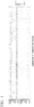
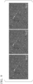
- FIG. 1 is a heat map showing the expression state of microglial marker genes.
- the horizontal axis indicates the combination of transcription factors introduced into iPS cells
- the vertical axis indicates seven microglial marker genes.
- the upper part shows a heat map of log 2 (CPM + 1) of gene expression levels obtained by the Quartz-seq2 method
- the lower part shows a heat map of log 2 (TPM + 1) of gene expression levels obtained by the Smart-seq2 method.
- the upper limit of the color scale is 6.
- FIG. 2 is a heat map in which combinations of transcription factors introduced into iPS cells are arranged from the left in descending order of the expression levels of the above seven genes.
- the horizontal axis indicates the combination of transcription factors
- the vertical axis indicates seven microglial marker genes.
- the upper part shows a heat map of log 2 (CPM + 1) of gene expression levels obtained by the Quartz-seq2 method (upper limit of color scale: 6), and the lower part shows a heat map of log 2 (TPM + 1) of gene expression levels obtained by the Smart-seq2 method (upper limit of color scale: 2).
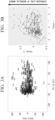
- FIG. 3A shows the results of the principal component analysis of transcriptome data obtained by the Smart-seq2 method.
- the horizontal axis indicates PC1, and the vertical axis indicates PC2.
- FIG. 3B shows the results of the factor analysis of transcriptome data obtained by the Smart-seq2 method.
- the horizontal axis indicates Factor 1
- the vertical axis indicates Factor 2.
- the expression level of the microglial marker gene contributes to the positive direction of Factor 2.
- iPS cells were subjected to forward programming by transcription factor transfection with the combinations of transcription factors shown in Table 4, which had been determined to have a high ability to induce differentiation into microglia and were induced to differentiate with a culture medium for differentiation to induce differentiation into microglia, and the transcriptome analysis by RNA-seq was carried out.
- a combination of transcription factors was transfected into iPS cells, which were subsequently induced to differentiate into microglia for 4 days in a culture medium for differentiation.
- culture media for differentiation the following three kinds of basal media were used: Neurobasal Media, DMEM/F-12, and Advanced DMEM/F-12.
- IL-34, M-CSF, GlutaMAX, and Component P were added to each of the basal media to obtain culture media designated as M1, M2, and M3.
- CD200, CX3CL1, and TGF-beta were added to M1, M2, and M3 to obtain culture media each designated as M4, M5, and M6.
- Table 5 shows the compositions of the six kinds of the culture media M1 to M6.
- Thermo Fisher Scientific, Inc. FUJIFILM Wako 1 mM 1 1 0 0 0 0 Biotin Pure Chemical Corporation 3.5 ng/mL 1 1 0 0 0 0 lipidated BSA (Albumax I) Thermo Fisher Scientific, Inc. 2 mg/mL 1 1 0 0 0 0 0 Penicillin-Streptomycin Nacalai Tesque, Inc. 1 % 1 1 1 1 1 1 1 1 Ascorbic Acid Sigma-Aldrich Co. LLC 2.5 ⁇ g/mL 0 0 1 1 0 0 NEAA Thermo Fisher Scientific, Inc. 1 % 0 0 1 1 0 0 0
- differentiation culture was carried out in triplicate with respect to the six different culture media for differentiation under the 32 conditions of the 31 kinds of combinations of transcription factors shown in Table 4 above and ntfc (no transcription factor control) with culture medium for differentiation alone for checking the differentiation induction, where Nuclease-free Water was used instead of the mRNA mix in the ntfc.
- Transfection was carried out according to the same protocol as in Experimental Example 3. A combination of two kinds of transcription factors of 0.75 ⁇ L was mixed at a concentration of 0.1 ⁇ g/ ⁇ L to obtain 1.5 ⁇ L of an mRNA mix. In addition, in order to investigate a case of transfection with only one kind of transcription factor, 1.5 ⁇ L of the mRNA in Table 2 above was transfected at a concentration of 0.1 ⁇ g/ ⁇ L. siPOU5F1 (Thermo Fisher Scientific, Inc., s10873) was added at 20 nM at the time of the preparation of all the first transfection solutions.
- siPOU5F1 Thermo Fisher Scientific, Inc., s10873
- the culture medium was changed from StemFit to the culture medium for differentiation shown in Table 5 above.
- the culture medium for differentiation was replaced every other day, and all samples were subjected to RNA extraction on Day 7.
- RNA-seq was carried out by the Quartz-seq2 method.
- total RNA extracted after the culture and total RNA extracted from the iPS cells collected at the time of seeding were used, and the following cells were used as controls: Human Microglia Primary Cell Culture Total RNA: 50 ⁇ g (Celprogen, 37089 RNA), Total RNA Human Monocytes, 1 ⁇ g (3H Biomedical, 3H100-30-10), Total RNA Human Dendritic Cells (MoDCs), 1 ⁇ g (3H Biomedical, 3H100-70-10), and Human Macrophage Primary Cell Culture-Frozen Vial (Celprogen, 36070-01).
- RNeasy mini kit (Qiagen, 74004) was used for the extraction of total RNA.
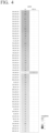
- FIG. 4 is a view summarizing the results of RNA-seq analysis.
- the numbers in the cells indicate the UMI counts.
- the top 50 samples arranged in descending order of expression levels of C3 and P2RY12 are shown after normalizing the variation in the UMI counts between samples by 1,000,000.
- the microglial control is positioned at the 13th raw. No expression of P2RY12 was confirmed in the microglia control.
- BLN was included in the top 50 samples regardless of the kind of culture medium for differentiation.
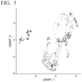
- FIG. 5 is a view showing the results of carrying out clustering by UMAP. As a result of the analysis, 13 clusters were confirmed.
- FIG. 6 is a view showing the results of classifying UMAP clustering for M0 which is a sample subjected to RNA extraction on Day 3 after the introduction of only one kind of transcription factor, culture media for differentiation M1 to M6, and Mz which is various controls.
- clusters 9 and 12 in FIG. 5 are samples in which only one kind of transcription factor has been introduced and RNA has been extracted on Day 3.
- RNA has been extracted on Day 3.
- microglia are distributed in the cluster 1
- monocytes, dendritic cells, and macrophages are distributed in the cluster 10.
- samples distributed close to monocytes, dendritic cells, and macrophages were ABD, AEI, BDF, DIJ, and DIP.
- the secondary evaluation using the culture media for differentiation revealed that a suitable method of carrying out differentiation into microglia is selecting a combination including B, I, K, L, and N, such as BLN, KLN, or IKN, from the transcription factor candidates in Table 2 above.
- iPS cell differentiation into microglia was evaluated.
- a combination of transcription factors AB, ABD, AEI, DIJ, or DIP which provided a transcriptome state similar to that of the control hematopoietic cells, was also used to induce differentiation.
- the differentiation induction conditions of Experimental Example 4 were scaled up to those for a 24-well plate, and the differentiation induction was carried out at N4 with each combination of transcription factors.
- M6 was used as the culture medium for differentiation. Similar to Experimental Example 4, differentiation was induced in the culture medium for differentiation M6 after four transfections. Subsequently, RNA was extracted from samples for N3 on Day 7 using the RNeasy mini kit, and RNA-seq was carried out by the Smart-seq2 method.
- iCell microglia (FUJIFILM Wako Pure Chemical Corporation, C1110), in addition to the iPS cells at the time of seeding, the ntfc, and the microglia, the monocytes, and the dendritic cells the same as those used in Experimental Example 4, were cultured, and each total RNA was extracted as a control with the RNeasy mini kit.
- the remaining N1 was continuously cultured in the culture medium for differentiation M6 from Day 7, subcultured on Day 11, and cultured in the M6 culture medium to which Component P was not added. Subsequently, a time-lapse movie of 13 hours was taken overnight on Day 14 to check the phagocytic activity of the differentiated cells.
- adherent cells were few in AB, ABD, AEI, DIJ, and DIP, and ntfc. Therefore, floating cells were centrifuged to precipitate the cells and subjected together with the adherent cells to RNA extraction.
- C3 and P2RY12 which are conceived to be important markers, in addition to APOE, BHLHE41, CABLES1, OLFML3, PROS1, SLC7A8, SLCO2B1, TMEM119, and TREM2, which have been confirmed as genes specifically expressed microglia.
- FIG. 7 is a view showing expression levels of the respective microglia marker genes.
- the horizontal axis indicates the expression level.
- P2RY12 which is conceived to be an important marker, was slightly expressed even in the iCell microglia, and AB, AEI, DIJ, DIP, and KLN were confirmed in the differentiation-induced samples only for N1.
- C3 was highly expressed in the microglia (Celprogen) and the iCell microglia.
- the combination of BN, BEN, BIN, BLN, IKN, or KLN which was suggested to have a high ability to induce differentiation into microglia in Experimental Examples 3 and 4, reproduced a high expression level.
- FIG. 8 shows a representative phagocytosis scene in the time-lapse movie of BN
- FIG. 9 shows a representative phagocytosis scene of BLN.
- time elapses from left to right In addition, arrows indicate phagocytic cells having phagocytic activity.
- the cells having phagocytic activity actively migrated had relatively large cell sizes of about 30 ⁇ m to 100 ⁇ m, and phagocytosed cells one after another.
- BEN, BIN, BLN, BN, IKN, and KLN which could be subcultured, were confirmed to have phagocytic activity in the time-lapse movie.
- iPS cells can be induced to differentiate into microglia in 14 days by transfecting a combination of transcription factors that increase the expression of C3 and other microglia-specific marker genes.
- the present invention includes the following aspects.
Landscapes
- Health & Medical Sciences (AREA)
- Engineering & Computer Science (AREA)
- Biomedical Technology (AREA)
- Life Sciences & Earth Sciences (AREA)
- Genetics & Genomics (AREA)
- Chemical & Material Sciences (AREA)
- Biotechnology (AREA)
- Organic Chemistry (AREA)
- Bioinformatics & Cheminformatics (AREA)
- Wood Science & Technology (AREA)
- Zoology (AREA)
- Microbiology (AREA)
- Biochemistry (AREA)
- General Engineering & Computer Science (AREA)
- General Health & Medical Sciences (AREA)
- Cell Biology (AREA)
- Neurology (AREA)
- Neurosurgery (AREA)
- Micro-Organisms Or Cultivation Processes Thereof (AREA)
Abstract
There is provided a production method for microglia, including a step of introducing, into pluripotent stem cells, a nucleic acid including an open reading frame of a transcription factor that increases expression of a Complement C3 (C3) gene. In the production method, the transcription factor is selected from the group consisting of SPll, CEBPA, PTAFR, FLI1, MEF2C, EGR2, RUNX1, TNF, CEBPB, IKZF1, KLF6, NFIC, ELF4, PAX8, PRDM1, MEF2A, and NR4A3.
Description
- The present invention relates to a production method for microglia. Priority is claimed on
Japanese Patent Application No. 2022-046010, filed March 22, 2022 - Cells of the central nervous system are difficult to obtain. On the other hand, various cells including cells of the central nervous system cells have become available by differentiating pluripotent stem cells such as induced pluripotent stem cells (iPSC) into cells of interest. Further, disease model cells can also be produced by differentiating pluripotent stem cells derived from a patient into cells of interest.
- Microglia are one kind of glial cells of the central nervous system. Glial cells are a general term for cells that are not nerve cells that constitute the nervous system. Microglia are known as central immunocompetent cells. Unlike cells of the central nervous system, microglia are conceived to be derived from the mesoderm and play a role as immune cells in the brain. At the time in a disease state, microglia become activated, which involves cell body hypertrophy and cell proliferation. It is known that activated microglia change the expression of various molecules including receptors in the cell membrane, migrate to a lesion part, phagocytize damaged cells or extracellular proteins such as amyloid β protein, and produce humoral factors (an inflammatory factor, a cytotoxic factor, a nutritional factor, and the like). Microglia play a major role in the mechanisms of central nervous system diseases and thus have been suggested to be associated with Alzheimer's disease.
- However, a conventional method of producing microglia from pluripotent stem cells requires 30 or more days of culture in order to obtain microglia. Accordingly, an object of the present invention is to provide a technique for producing microglia from pluripotent stem cells in a short period of time.
- A production method for microglia according to the present invention includes a step of introducing, into pluripotent stem cells, a nucleic acid including an open reading frame of a transcription factor that increases expression of a Complement C3 (C3) gene.
- According to the present invention, it is possible to provide a technique for producing microglia from pluripotent stem cells in a short period of time.
-
-
FIG. 1 is a heat map showing the expression state of microglial marker genes in Experimental Example 3. -
FIG. 2 is a heat map in which combinations of transcription factors introduced into iPS cells in Experimental Example 3 are arranged from the left in descending order of the expression levels of microglial marker genes. -
FIG. 3A shows results of a principal component analysis of transcriptome data obtained by a Smart-seq2 method in Experimental Example 3. -
FIG. 3B shows results of a factor analysis of transcriptome data obtained by a Smart-seq2 method in Experimental Example 3. -
FIG. 4 is a view summarizing results of an RNA-seq analysis in Experimental Example 4. -
FIG. 5 is a view showing results of carrying out clustering by UMAP in Experimental Example 4. -
FIG. 6 is a view showing results of carrying out clustering by UMAP in Experimental Example 4. -
FIG. 7 is a view showing expression levels of respective microglia marker genes in Experimental Example 5. -
FIG. 8 is microscopic images showing a part of a time-lapse movie of a representative phagocytosis scene of microglia in Experimental Example 5. -
FIG. 9 is microscopic images showing a part of a time-lapse movie of a representative phagocytosis scene of microglia in Experimental Example 5. - In one embodiment, the present invention provides a production method for microglia, which includes a step of introducing, into pluripotent stem cells, a nucleic acid including an open reading frame of a transcription factor that increases expression of a Complement C3 (C3) gene.
- As will be described later in Examples, the inventors of the present invention revealed that the production of microglia, which conventionally takes 30 days or longer, can be carried out within 14 days by the production method of the present embodiment. In addition, as will be described later in Examples, the microglia produced by the production method of the present embodiment have phagocytic activity and thus have a function of microglia.
- In microglia, microglial marker genes are expressed, and a function of microglia, such as phagocytic activity, is exhibited. Examples of the microglial marker gene include Purinergic Receptor P2Y12 (P2RY12), Complement C3 (C3), C-X3-C Motif Chemokine Receptor 1 (CX3CR1), Colony
Stimulating Factor 1 Receptor (CSF1R), Alpha-2-Macroglobulin (A2M), ADAM Metallopeptidase Domain 28 (ADAM28), Plexin Domain Containing 2 (PLXDC2), Lysosomal Protein Transmembrane 5 (LAPTM5), Solute Carrier Organic Anion Transporter Family Member 2B1 (SLCO2B1), and Lymphocyte Antigen 86 (LY86), which will be described later in Examples. - The NCBI accession number of the nucleotide sequence of cDNA of the human P2RY12 gene is NM_022788.5, NM_176876.3, or the like. The NCBI accession number of the nucleotide sequence of cDNA of the human C3 gene is NM_000064.4. The NCBI accession number of the nucleotide sequence of cDNA of the human CX3CR1 gene is NM_001171171.2, NM_001171172.2, NM_001171174.1, or the like. The NCBI accession number of the nucleotide sequence of cDNA of the human CSF1R gene is NM_001288705.3, NM_001349736.2, NM_001375320.1, or the like. The NCBI accession number of the nucleotide sequence of cDNA of the human A2M gene is NM_000014.6, NM_001347423.2, NM_001347424.2, or the like. The NCBI accession number of the nucleotide sequence of cDNA of the human ADAM28 gene is NM_001304351.2, NM_014265.6, NM_021777.5, or the like. The NCBI accession number of the nucleotide sequence of cDNA of the human PLXDC2 gene is NM_001282736.2, NM_032812.9, or the like. The NCBI accession number of the nucleotide sequence of cDNA of the human LAPTM5 gene is NM_006762.3. The NCBI accession number of the nucleotide sequence of cDNA of the human SLCO2B1 gene is NM_001145211.3, NM_001145212.3, NM_007256.5, or the like. The NCBI accession number of the nucleotide sequence of cDNA of the human LY86 gene is NM_004271.4.
- In the production method of the present embodiment, examples of the pluripotent stem cell include an embryonic stem cell (an ES cell) and an induced pluripotent stem cell (an iPS cell). The pluripotent stem cell is preferably a human cell.
- Examples of the transcription factor that increases the expression of the C3 gene include Spi-1 Proto-Oncogene (SPI1), CCAAT Enhancer Binding Protein Alpha (CEBPA), Platelet Activating Factor Receptor (PTAFR), Fli-1 Proto-Oncogene, ETS Transcription Factor (FLI1), Myocyte Enhancer Factor 2C (MEF2C), Early Growth Response 2 (EGR2), RUNX Family Transcription Factor 1 (RUNX1), Tumor Necrosis Factor (TNF), CCAAT Enhancer Binding Protein Beta (CEBPB), IKAROS Family Zinc Finger 1 (IKZF1), Kruppel Like Factor 6 (KLF6), Nuclear Factor I C (NFIC), E74 Like ETS Transcription Factor 4 (ELF4), Paired Box 8 (PAX8), PR/SET Domain 1 (PRDM1), Myocyte Enhancer Factor 2A (MEF2A), and
Nuclear Receptor Subfamily 4 Group A Member 3 (NR4A3). - The NCBI accession number of the nucleotide sequence of cDNA of the human SPI1 gene is NM_001080547.2, NM_003120.3, or the like. The NCBI accession number of the nucleotide sequence of cDNA of the human CEBPA gene is NM_001285829.1, NM_001287424.2, NM_001287435.1, or the like. The NCBI accession number of the nucleotide sequence of cDNA of the human PTAFR gene is NM_000952.5, NM_001164721.2, NM_001164722.3, or the like. The NCBI accession number of the nucleotide sequence of cDNA of the human FLI1 gene is NM_001167681.3, NM_001271010.2, NM_001271012.2, or the like. The NCBI accession number of the nucleotide sequence of cDNA of the human MEF2C gene is NM_001131005.2, NM_001193347.1, NM_001193348.1, or the like. The NCBI accession number of the nucleotide sequence of cDNA of the human EGR2 gene is NM_000399.5, NM_001136177.3, NM_001136178.2, or the like. The NCBI accession number of the nucleotide sequence of cDNA of the human RUNX1 gene is NM_001001890.3, NM_001122607.2, NM_001754.5, or the like. The NCBI accession number of the nucleotide sequence of cDNA of the human TNF gene is NM_000594.4. The NCBI accession number of the nucleotide sequence of cDNA of the human CEBPB gene is NM_001285878.1, NM_001285879.1, NM_005194.4, or the like. The NCBI accession number of the nucleotide sequence of cDNA of the human IKZF1 gene is NM_001220765.3, NM_001220767.2, NM_001220768.2, or the like. The NCBI accession number of the nucleotide sequence of cDNA of the human KLF6 gene is NM_001160124.2, NM_001160125.2, NM_001300.6, or the like. The NCBI accession number of the nucleotide sequence of cDNA of the human NFIC gene is NM_001245002.2, NM_001245004.2, NM_001245005.2, or the like. The NCBI accession number of the nucleotide sequence of cDNA of the human ELF4 gene is NM_001127197.2, NM_001421.4, or the like. The NCBI accession number of the nucleotide sequence of cDNA of the human PAX8 gene is NM_003466.4, NM_013951.3, NM_013952.4, or the like. The NCBI accession number of the nucleotide sequence of cDNA of the human PRDM1 gene is NM_001198.4, NM_182907.3, or the like. The NCBI accession number of the nucleotide sequence of cDNA of the human MEF2A gene is NM_001130926.5, NM_001130927.5, NM_001130928.4, or the like. The NCBI accession number of the nucleotide sequence of cDNA of the human NR4A3 gene is NM_006981.4, NM_173199.4, or the like.
- As will be described later in Examples, in a case of introducing nucleic acids containing open reading frames of these transcription factors into pluripotent stem cells, it is possible to induce differentiation into microglia. It is noted that the method of transfecting transcription factors into pluripotent stem cells to induce differentiation is also called forward programming. Nucleic acids containing open reading frames of these transcription factors may be used singly or in a combination of two or more thereof.
- The nucleic acids containing open reading frames of these transcription factors may be mRNA or an expression vector. In addition, the expression vector may be a virus vector or may be a plasmid vector.
- In the production method of the present embodiment, the transcription factor that increases the expression of the C3 gene preferably includes at least CEBPA, and it is preferably two or more transcription factors including at least CEBPA.
- Examples of the more specific combination of transcription factors that increase the expression of the C3 gene include combinations of 2 to 5 transcription factors, which include CEBPA, CEBPB, KLF6, NFIC, or PAX8 among SP11, CEBPA, PTAFR, FLI1, MEF2C, EGR2, RUNX1, TNF, CEBPB, IKZF1, KLF6, NFIC, ELF4, PAX8, PRDM1, MEF2A, NR4A3.
- Examples of the still more specific combination of transcription factors that increase the expression of the C3 gene include a combination of CEBPA, MEF2C, and PAX8, a combination of CEBPA, CEBPB, and PAX8, a combination of CEBPA, NFIC, and PAX8, a combination of CEBPA and PAX8, a combination of CEBPB, KLF6, and PAX8, a combination of KLF6, NFIC, and PAX8, a combination of PTAFR, CEBPB, and PAX8, and a combination of CEBPA, KLF6, and PAX8.
- As will be described later in Examples, in the production method of the present embodiment, the culture medium for culturing pluripotent stem cells introduced with a nucleic acid containing an open reading frame of a transcription factor is not particularly limited.
- In one embodiment, the present invention provides microglia produced by the production method described above. Due to being characterized by the production method, the microglia of the present embodiment are conceived to have a different gene expression profile and the like as compared with microglia obtained by inducing differentiation by other methods or microglia in vivo. However, it is very difficult and impractical to identify such a difference and identify whether or not it is the microglia of the present embodiment based on the difference in gene expression profile or the like. For this reason, it is realistic to carry out the identification by the production method.
- With reference to published papers, a unique weighting was carried out to select a gene set capable of serving as microglia markers. Table 1 shows the set of microglial marker genes selected by the inventors of the present invention.
[Table 1] No. Gene name 1 P2RY12 2 C3 3 CX3CR1 4 CSFIR 5 A2M 6 ADAM28 7 PLXDC2 8 LAPTM5 9 SLCO2B1 10 LY86 - With reference to published papers, transcription factors for microglial differentiation were selected, where the transcription factors were conceived to be suitable for inducing differentiation into microglia. More specifically, transcription factors were selected, where the transcription factors were conceived to promote the expression of the microglial marker genes shown in Table 1 above in a case of being introduced into pluripotent stem cells. Table 2 below shows a list of transcription factors for microglial differentiation selected by the inventors of the present invention. For simplification, each gene is represented by one letter of the alphabet from "A" to "Q".
[Table 2] Symbol Name of transcription factor gene A SPI1 B CEBPA C PTAFR D FLI1 E MEF2C F EGR2 G RUNX1 H TNF I CEBPB J IKZF1 K KLF6 L NFIC M ELF4 N PAX8 O PRDM1 P MEF2A Q NR4A3 - Using the 17 kinds of transcription factors shown in Table 2, it was checked whether or not iPS cells could be induced to differentiate into microglia by forward programming of transfecting transcription factors into iPS cells. First, as a primary evaluation, the transcription factors shown in Table 2 above were transfected into iPS cells in 384 combinations shown in Table 3 below, and gene expression analysis was carried out.

- HPS1005 1383D2 (RIKEN BRC) was used as an iPS cell. StemFit AK02N (Ajinomoto Co., Inc., AJ100, hereinafter may be referred to as "StemFit") was prepared according to the manufacturer's instructions. CultureSure Y-27632 (FUJIFILM Wako Pure Chemical Corporation, 036-24023) was prepared to 10 mM with dimethylsulfoxide (DMSO). A coating solution was prepared by diluting iMatrix-511 silk (Takara Bio Inc., 892021) to 0.5 µg/cm2 with phosphate-buffered saline (PBS) (-), and 20 mL of the coating solution was added to each of four 150 mm dishes and precoated at 37°C for 1 hour.
- After 1 hour, the coating solution was aspirated, Cells were seeded to 5.0 × 10⁵ cells per dish using a culture solution obtained by adding a 1/1,000 volume of 10 mM CultureSure Y-27632 to StemFit (hereinafter, may be referred to as "Medium SY"), and the culture was carried out at 37°C in 5% CO2 until an appropriate cell number was reached. From the day after seeding, the culture medium was replaced using StemFit.
- A 96-well plate was used for iPS cell culture. All wells of twelve 96-well plates were precoated with iMatrix-511 silk (Takara Bio Inc., 892021) at a concentration of 0.5 µg/cm2, and aspiration was carried out immediately before seeding iPS cells.
- The iPS cells cultured in a 150 mm dish were washed with PBS (-), a solution obtained by mixing equal volumes of TrypLE Select (1X) (Thermo Fisher Scientific, Inc., 12563-029) and 0.5 mmol/L-EDTA/PBS solution (Nacalai Tesque, Inc., 13567-84) was added incubated at 37°C for 5 minutes, and then the cells were carefully stirred with a 1,000 µL micropipette to detach the cells from the dish and collected in a centrifuge tube.
- The same amount of Medium SY as that of the cell suspension was added into the centrifuge tube, in which the cell suspension had been collected, to stop the TrypLE Select reaction. After centrifugation at 200 × g for 3 minutes, the supernatant was aspirated to prepare a soft pellet, the pellet was loosened by tapping, and then Medium SY was added thereto to resuspend the cells.
- Viability and cell number were measured by Countess (Thermo Fisher Scientific, Inc.), and cell suspensions having three concentration levels of 1.79 × 10⁵ cells/mL, 3.04 × 10⁵ cells/mL, and 4.30 × 10⁵ cells/mL were prepared with Medium SY to prepare a confluency condition proper for transfection.
- Aliquots of 100 µL of the cell suspension at each concentration were dispensed into the four coated 96-well plates to prepare four 96-well plates per concentration level for three concentration levels, and the culture was carried out at 37°C in a 5% CO2 environment.
- Transcription factors were transfected in the form of mRNA. The mRNA was thawed on ice and prepared to a concentration of 0.1 µg/µL, and four sets of mRNA mix stocks were prepared by mixing 0.5 µL with the combinations shown in Table 3 above to be 1.5 µL and kept frozen at -80°C until the used in the transfection.
- One day after iPS cell seeding (Day 1), four plates having a concentration level of 70% to 90% confluency were selected, the culture medium was replaced with StemFit, and the culture was carried out at 37°C in a 5% CO2 environment until transfection was carried out.
- The temperature of Opti-MEM I Reduced Serum Medium (Thermo Fisher Scientific, Inc., 31985062, hereinafter may be referred to as "Opti-MEM") was returned to room temperature, and the mRNA mix stock was thawed on ice over 30 minutes.
- A
Mix 1 solution was prepared by mixing, per well of a 96-well plate, 1.5 µL of Lipofectamine Messenger MAX (Thermo Fisher Scientific, Inc., LMRNA001) in 36.7 µL of Opti-MEM and incubated at room temperature for 10 minutes. Next, aMix 2 solution was prepared by adding 1.5 µL of the mRNA mix to 36.7 µL of Opti-MEM and carrying out mixing by tapping. - Subsequently, 12 µL of the
Mix 2 solution was mixed with 12 µL of the incubatedMix 1 solution by pipetting to prepare a transfection solution, which was subsequently incubated for 5 minutes at room temperature. Subsequently, 20.8 µL of the transfection solution prepared according to the combination of mRNAs shown in Table 3 above was added dropwise to each well of the four 96-well plates seeded with iPS cells, and incubation was carried out for 3 hours at 37°C in a 5% CO2 environment. After 3 hours, the whole amount of StemFit was replaced, and incubation was carried out for 2 hours at 37°C in a 5% CO2 environment. - After 2 hours, the same transfection operation was carried out once again, incubation was carried out overnight at 37°C in a 5% CO2 environment, and two further times of transfection were carried out on
Day 2, whereby transfection was carried out four times in total. - On
Day 3, all 384 wells of the four 96-well plates that had been subjected to the differentiation induction were subjected to RNA-seq. RNA-seq was carried out according to the Smart-seq2 method (Picelli S., et al., Full-length RNA-seq from single cells using Smart-seq2, Nat Protoc., 9 (1), 171-181, 2014.) and the Quartz-seq2 method (Sasagawa Y, et al., Quartz-Seq2: a high-throughput single-cell RNA-sequencing method that effectively uses limited sequence reads, Genome Biol., 19 (1), 29, 2018.). - Transcriptome data worthy of analysis were obtained from 354 samples obtained by the Smart-seq2 method and 297 samples obtained by the Quartz-seq2 method, collectively from 379 samples among the 384 samples.
- From the obtained transcriptome data, TPM (Transcripts per million) in Smart-seq2 and CPM (Counts per million, a value obtained by normalizing the number of UMI counts by 1,000,000) in Quartz-seq2 were subjected to heat mapping with log2, and the expression levels of seven genes of SLCO2B1, ADAM28, A2M, CSF1R, CX3CR1, C3, and P2RY12 were checked among the set of microglial marker genes shown in Table 1 above.
-
FIG. 1 is a heat map showing the expression state of microglial marker genes. - In
FIG. 1 , the horizontal axis indicates the combination of transcription factors introduced into iPS cells, and the vertical axis indicates seven microglial marker genes. Here, the upper part shows a heat map of log2(CPM + 1) of gene expression levels obtained by the Quartz-seq2 method, and the lower part shows a heat map of log2(TPM + 1) of gene expression levels obtained by the Smart-seq2 method. The upper limit of the color scale is 6. - As a result of the analysis, according to the results of the Quartz-seq2 method, it was confirmed that C3 is expressed in almost all samples, followed by ADAM28 which is expressed in a large number of samples. In addition, from the results of the Smart-seq2 method, it was confirmed that although the observed TPM values are smaller than those of the Quartz-seq2 method, a large number of samples express C3. The above results suggested that iPS cells have differentiated into microglia-like cells in almost all combinations of transcription factors shown in Table 3 above.
-
FIG. 2 is a heat map in which combinations of transcription factors introduced into iPS cells are arranged from the left in descending order of the expression levels of the above seven genes. InFIG. 2 , the horizontal axis indicates the combination of transcription factors, and the vertical axis indicates seven microglial marker genes. - Here, the upper part shows a heat map of log2(CPM + 1) of gene expression levels obtained by the Quartz-seq2 method (upper limit of color scale: 6), and the lower part shows a heat map of log2(TPM + 1) of gene expression levels obtained by the Smart-seq2 method (upper limit of color scale: 2).
- As a result of the analysis, it was confirmed that, among the 384 combinations, a combination containing transcription factors such as B, E, I, L, and N, in terms of transcription factor symbol, has high expression levels of microglial marker genes.
- Subsequently, each of the transcriptome data, which had been obtained by the Smart-seq2 method and the Quartz-seq2 method, were subjected to principal component analysis and factor analysis.
FIG. 3A shows the results of the principal component analysis of transcriptome data obtained by the Smart-seq2 method. The horizontal axis indicates PC1, and the vertical axis indicates PC2.FIG. 3B shows the results of the factor analysis of transcriptome data obtained by the Smart-seq2 method. The horizontal axis indicatesFactor 1, and the vertical axis indicatesFactor 2. The expression level of the microglial marker gene contributes to the positive direction ofFactor 2. - As a result of the analysis, it was confirmed that combinations of transcription factors with high expression of microglial marker genes are distributed on the positive side of PC1 by the principal component analysis and the positive side of
Factor 2 by the factor analysis. - Based on each of the results of the principal component analysis and the factor analysis by the Smart-seq2 method and the Quartz-seq2 method and based on the expression levels of the marker genes in
FIG. 2 , combinations of transcription factors more suitable for inducing differentiation into microglia were selected. Table 4 shows the selected combinations of transcription factors having a high ability to induce differentiation into microglia.[Table 4] No. Combination No. Combination 1 BI 17 BNQ 2 BN 18 CIN 3 BCF 19 DIJ 4 BDF 20 DIP 5 BDN 21 BIN 6 BEI 22 IKN 7 BEN 23 BKO 8 BFH 24 AOQ 9 BFK 25 GKN 10 BHP 26 ENQ 11 BIK 27 ABD 12 BIN 28 AEI 13 BKN 29 AGM 14 BKP 30 BIP 15 BLN 31 KLN 16 BNP - - - Differentiation culture conditions were further investigated regarding the combination conditions of transcription factors. iPS cells were subjected to forward programming by transcription factor transfection with the combinations of transcription factors shown in Table 4, which had been determined to have a high ability to induce differentiation into microglia and were induced to differentiate with a culture medium for differentiation to induce differentiation into microglia, and the transcriptome analysis by RNA-seq was carried out.
- A combination of transcription factors was transfected into iPS cells, which were subsequently induced to differentiate into microglia for 4 days in a culture medium for differentiation. As culture media for differentiation, the following three kinds of basal media were used: Neurobasal Media, DMEM/F-12, and Advanced DMEM/F-12. IL-34, M-CSF, GlutaMAX, and Component P were added to each of the basal media to obtain culture media designated as M1, M2, and M3. In addition, CD200, CX3CL1, and TGF-beta were added to M1, M2, and M3 to obtain culture media each designated as M4, M5, and M6. Table 5 below shows the compositions of the six kinds of the culture media M1 to M6.
[Table 5] Contents Vendor Final Cone. M1 M4 M2 M5 M3 M6 Neurobasal Medium Thermo Fisher Scientific, Inc. - 1 1 0 0 0 0 DMEM/F12 Thermo Fisher Scientific, Inc. - 0 0 1 1 0 0 Advanced DMEM/F12 Thermo Fisher Scientific, Inc. - 0 0 0 0 1 1 N2 Supplement (100X) Thermo Fisher Scientific, Inc. 1 % 1 1 1 1 1 1 IL-34 PeproTech, Inc. 0.1 µg/ mL 1 1 1 1 1 1 M-CSF PeproTech, Inc. 0.025 µg/ mL 1 1 1 1 1 1 GlutaMAX Thermo Fisher Scientific, Inc. 1 % 1 1 1 1 1 1 Component P Elixirgen Scientific, Inc. 0.1 % 1 1 1 1 1 1 Recombinant Human CD200 R&D Systems Inc. 0.1 µg/ mL 0 1 0 1 0 1 CX3CL1 PeproTech, Inc. 0.1 µg/ mL 0 1 0 1 0 1 TGF-beta PeproTech, Inc. 0.05 µg/ mL 0 1 0 1 0 1 NaCl 5M Sigma-Aldrich Co. LLC 50 mM 1 1 0 0 0 0 lactic acid (85% syrup stock) Sigma-Aldrich Co. LLC 0.017 % 1 1 0 0 0 0 Sodium Pyruvate (100 mM) Thermo Fisher Scientific, Inc. FUJIFILM Wako 1 mM 1 1 0 0 0 0 Biotin Pure Chemical Corporation 3.5 ng/ mL 1 1 0 0 0 0 lipidated BSA (Albumax I) Thermo Fisher Scientific, Inc. 2 mg/ mL 1 1 0 0 0 0 Penicillin-Streptomycin Nacalai Tesque, Inc. 1 % 1 1 1 1 1 1 Ascorbic Acid Sigma-Aldrich Co. LLC 2.5 µg/ mL 0 0 1 1 0 0 NEAA Thermo Fisher Scientific, Inc. 1 % 0 0 1 1 0 0 - Until transfection, differentiation culture was carried out in triplicate with respect to the six different culture media for differentiation under the 32 conditions of the 31 kinds of combinations of transcription factors shown in Table 4 above and ntfc (no transcription factor control) with culture medium for differentiation alone for checking the differentiation induction, where Nuclease-free Water was used instead of the mRNA mix in the ntfc.
- Transfection was carried out according to the same protocol as in Experimental Example 3. A combination of two kinds of transcription factors of 0.75 µL was mixed at a concentration of 0.1 µg/µL to obtain 1.5 µL of an mRNA mix. In addition, in order to investigate a case of transfection with only one kind of transcription factor, 1.5 µL of the mRNA in Table 2 above was transfected at a concentration of 0.1 µg/µL. siPOU5F1 (Thermo Fisher Scientific, Inc., s10873) was added at 20 nM at the time of the preparation of all the first transfection solutions.
- At the time of the culture medium replacement after the fourth transfection on
Day 2, the culture medium was changed from StemFit to the culture medium for differentiation shown in Table 5 above. The culture medium for differentiation was replaced every other day, and all samples were subjected to RNA extraction onDay 7. - Samples transfected with only one kind of transcription factor were subjected to culture medium exchange to StemFit and then subjected to RNA extraction on
Day 3. - Subsequently, RNA-seq was carried out by the Quartz-seq2 method. In addition, at the time of RNA-seq, total RNA extracted after the culture and total RNA extracted from the iPS cells collected at the time of seeding were used, and the following cells were used as controls: Human Microglia Primary Cell Culture Total RNA: 50 µg (Celprogen, 37089 RNA), Total RNA Human Monocytes, 1 µg (3H Biomedical, 3H100-30-10), Total RNA Human Dendritic Cells (MoDCs), 1 µg (3H Biomedical, 3H100-70-10), and Human Macrophage Primary Cell Culture-Frozen Vial (Celprogen, 36070-01). RNeasy mini kit (Qiagen, 74004) was used for the extraction of total RNA.
- The expression levels of C3 and P2RY12 were checked regarding the UMI counts obtained by the Quartz-seq2 method.
FIG. 4 is a view summarizing the results of RNA-seq analysis. InFIG. 4 , the numbers in the cells indicate the UMI counts. Here, the top 50 samples arranged in descending order of expression levels of C3 and P2RY12 are shown after normalizing the variation in the UMI counts between samples by 1,000,000. In addition, the microglial control is positioned at the 13th raw. No expression of P2RY12 was confirmed in the microglia control. - In addition, in the iPS cells introduced with a combination of transcription factors including B, 1, K, L, and N, such as BLN, KLN, or IKN, C3 was highly expressed, and BLN was included in the top 50 samples regardless of the kind of culture medium for differentiation.
-
FIG. 5 is a view showing the results of carrying out clustering by UMAP. As a result of the analysis, 13 clusters were confirmed.FIG. 6 is a view showing the results of classifying UMAP clustering for M0 which is a sample subjected to RNA extraction onDay 3 after the introduction of only one kind of transcription factor, culture media for differentiation M1 to M6, and Mz which is various controls. - As a result of the classification, it was revealed that
clusters FIG. 5 are samples in which only one kind of transcription factor has been introduced and RNA has been extracted onDay 3. Regarding the control, it was revealed that microglia are distributed in thecluster 1, and monocytes, dendritic cells, and macrophages are distributed in thecluster 10. - When the culture media for differentiation were compared, the
clusters - When the samples distributed at positions close to the control of microglia were checked, BIN, CIN, BN, and BKN were frequently distributed.
- In addition, the samples distributed close to monocytes, dendritic cells, and macrophages were ABD, AEI, BDF, DIJ, and DIP.
- From the above results, the secondary evaluation using the culture media for differentiation revealed that a suitable method of carrying out differentiation into microglia is selecting a combination including B, I, K, L, and N, such as BLN, KLN, or IKN, from the transcription factor candidates in Table 2 above.
- It was checked whether the phagocytic activity, which is a function of microglia, was exhibited in the combinations of transcription factors that were suggested to have a high microglia differentiation potential in the secondary evaluation.
- Based on the results of the primary evaluation and the secondary evaluation, the induction of iPS cell differentiation into microglia with a combination of transcription factors BN, BEN, BIN, BLN, IKN, or KLN was evaluated. In addition, for comparison, a combination of transcription factors, AB, ABD, AEI, DIJ, or DIP, which provided a transcriptome state similar to that of the control hematopoietic cells, was also used to induce differentiation.
- The differentiation induction conditions of Experimental Example 4 were scaled up to those for a 24-well plate, and the differentiation induction was carried out at N4 with each combination of transcription factors. M6 was used as the culture medium for differentiation. Similar to Experimental Example 4, differentiation was induced in the culture medium for differentiation M6 after four transfections. Subsequently, RNA was extracted from samples for N3 on
Day 7 using the RNeasy mini kit, and RNA-seq was carried out by the Smart-seq2 method. - In addition, iCell microglia (FUJIFILM Wako Pure Chemical Corporation, C1110), in addition to the iPS cells at the time of seeding, the ntfc, and the microglia, the monocytes, and the dendritic cells the same as those used in Experimental Example 4, were cultured, and each total RNA was extracted as a control with the RNeasy mini kit. The remaining N1 was continuously cultured in the culture medium for differentiation M6 from
Day 7, subcultured on Day 11, and cultured in the M6 culture medium to which Component P was not added. Subsequently, a time-lapse movie of 13 hours was taken overnight onDay 14 to check the phagocytic activity of the differentiated cells. - At the time of the RNA extraction on
Day 7, adherent cells were few in AB, ABD, AEI, DIJ, and DIP, and ntfc. Therefore, floating cells were centrifuged to precipitate the cells and subjected together with the adherent cells to RNA extraction. - In the cDNA quality check by the Smart-seq2 method, it was confirmed that in the described above combinations of transcription factors and the microglial control (Celprogen), the chain length of cDNA is short, and thus the RNA quality is poor.
- Among the microglia markers shown in Table 1 above, the expression levels of the following genes were confirmed: C3 and P2RY12, which are conceived to be important markers, in addition to APOE, BHLHE41, CABLES1, OLFML3, PROS1, SLC7A8, SLCO2B1, TMEM119, and TREM2, which have been confirmed as genes specifically expressed microglia.
-
FIG. 7 is a view showing expression levels of the respective microglia marker genes. InFIG. 7 , the horizontal axis indicates the expression level. As a result, the expression of almost all marker genes was confirmed in the iCell microglia. P2RY12, which is conceived to be an important marker, was slightly expressed even in the iCell microglia, and AB, AEI, DIJ, DIP, and KLN were confirmed in the differentiation-induced samples only for N1. - C3 was highly expressed in the microglia (Celprogen) and the iCell microglia. In addition, the combination of BN, BEN, BIN, BLN, IKN, or KLN, which was suggested to have a high ability to induce differentiation into microglia in Experimental Examples 3 and 4, reproduced a high expression level.
- Further, in the microglial markers shown in
FIG. 7 , it was confirmed that the total level of expression levels is high in the combination of BN, BEN, BIN, BLN, IKN, KLN, or DIP. - At the time of subculture on Day 11, adherent cells were few in AB, ABD, AEI, DIJ, DIP, and ntfc, and thus the subculture was carried out only on BEN, BIN, BLN, BN, IKN, and KLN. On
Day 14, time-lapse imaging was carried out overnight for 13 hours using CQ1 (Yokogawa Electric Corporation). -
FIG. 8 shows a representative phagocytosis scene in the time-lapse movie of BN, andFIG. 9 shows a representative phagocytosis scene of BLN. InFIG. 8 andFIG. 9 , time elapses from left to right. In addition, arrows indicate phagocytic cells having phagocytic activity. As a result, the cells having phagocytic activity actively migrated, had relatively large cell sizes of about 30 µm to 100 µm, and phagocytosed cells one after another. BEN, BIN, BLN, BN, IKN, and KLN, which could be subcultured, were confirmed to have phagocytic activity in the time-lapse movie. - From the above results, it was confirmed that iPS cells can be induced to differentiate into microglia in 14 days by transfecting a combination of transcription factors that increase the expression of C3 and other microglia-specific marker genes.
- The present invention includes the following aspects.
- [1] A production method for microglia, including a step of introducing, into pluripotent stem cells, a nucleic acid including an open reading frame of a transcription factor that increases expression of a Complement C3 (C3) gene.
- [2] The production method according to [1], in which the transcription factor is selected from the group consisting of SPI1, CEBPA, PTAFR, FLI1, MEF2C, EGR2, RUNX1, TNF, CEBPB, IKZF1, KLF6, NFIC, ELF4, PAX8, PRDM1, MEF2A, and NR4A3.
- [3] The production method according to [2], in which the transcription factor includes at least CEBPA.
- [4] The production method according to [2] or [3], in which the transcription factor is two or more transcription factors including at least CEBPA.
-
- [Patent Document 1]
PCT International Publication No. WO2020/252477 - [Patent Document 2]
United States Patent Application, Publication No. 2020/0063105 - While preferred embodiments of the invention have been described and illustrated above, it should be understood that these are exemplary of the invention and are not to be considered as limiting. Additions, omissions, substitutions, and other modifications can be made without departing from the scope of the invention. Accordingly, the invention is not to be considered as being limited by the foregoing description and is only limited by the scope of the appended claims.
Claims (4)
- A production method for microglia, comprising:
a step of introducing, into pluripotent stem cells, a nucleic acid including an open reading frame of a transcription factor that increases expression of a Complement C3 (C3) gene. - The production method according to claim 1,
wherein the transcription factor is selected from the group consisting of SP11, CEBPA, PTAFR, FLI1, MEF2C, EGR2, RUNX1, TNF, CEBPB, IKZF1, KLF6, NFIC, ELF4, PAX8, PRDM1, MEF2A, and NR4A3. - The production method according to claim 2,
wherein the transcription factor includes at least CEBPA. - The production method according to claim 2 or 3,
wherein the transcription factor is two or more transcription factors including at least CEBPA.
Applications Claiming Priority (1)
| Application Number | Priority Date | Filing Date | Title |
|---|---|---|---|
| JP2022046010A JP2023140135A (en) | 2022-03-22 | 2022-03-22 | Microglia production method |
Publications (1)
| Publication Number | Publication Date |
|---|---|
| EP4249591A1 true EP4249591A1 (en) | 2023-09-27 |
Family
ID=85415237
Family Applications (1)
| Application Number | Title | Priority Date | Filing Date |
|---|---|---|---|
| EP23159583.6A Pending EP4249591A1 (en) | 2022-03-22 | 2023-03-02 | Production method for microglia |
Country Status (3)
| Country | Link |
|---|---|
| US (1) | US20230323295A1 (en) |
| EP (1) | EP4249591A1 (en) |
| JP (1) | JP2023140135A (en) |
Cited By (1)
| Publication number | Priority date | Publication date | Assignee | Title |
|---|---|---|---|---|
| EP4511478A4 (en) * | 2022-04-22 | 2026-04-08 | Harvard College | METHOD AND COMPOSITIONS FOR THE PREPARATION OF MICROGLIA |
Citations (4)
| Publication number | Priority date | Publication date | Assignee | Title |
|---|---|---|---|---|
| US20200063105A1 (en) | 2017-05-01 | 2020-02-27 | President And Fellows Of Harvard College | Transcription factors controlling differentiation of stem cells |
| WO2020239807A1 (en) * | 2019-05-27 | 2020-12-03 | Westfälische Wilhelms-Universität Münster | Rapid and deterministic generation of microglia from human pluripotent stem cells |
| WO2020252477A1 (en) | 2019-06-14 | 2020-12-17 | FUJIFILM Cellular Dynamics, Inc. | Methods for the production of multiple lineages from induced pluripotent stem cells using charged surfaces |
| JP2022046010A (en) | 2020-09-10 | 2022-03-23 | コクヨ株式会社 | desk |
-
2022
- 2022-03-22 JP JP2022046010A patent/JP2023140135A/en active Pending
-
2023
- 2023-03-01 US US18/176,959 patent/US20230323295A1/en active Pending
- 2023-03-02 EP EP23159583.6A patent/EP4249591A1/en active Pending
Patent Citations (4)
| Publication number | Priority date | Publication date | Assignee | Title |
|---|---|---|---|---|
| US20200063105A1 (en) | 2017-05-01 | 2020-02-27 | President And Fellows Of Harvard College | Transcription factors controlling differentiation of stem cells |
| WO2020239807A1 (en) * | 2019-05-27 | 2020-12-03 | Westfälische Wilhelms-Universität Münster | Rapid and deterministic generation of microglia from human pluripotent stem cells |
| WO2020252477A1 (en) | 2019-06-14 | 2020-12-17 | FUJIFILM Cellular Dynamics, Inc. | Methods for the production of multiple lineages from induced pluripotent stem cells using charged surfaces |
| JP2022046010A (en) | 2020-09-10 | 2022-03-23 | コクヨ株式会社 | desk |
Non-Patent Citations (9)
| Title |
|---|
| "NCB1", Database accession no. NM_001193348.1 |
| "NCBI", Database accession no. NM_001130928.4 |
| CHEN SHIH-WEI ET AL: "Efficient conversion of human induced pluripotent stem cells into microglia by defined transcription factors", STEM CELL REPORTS, vol. 16, no. 5, 1 May 2021 (2021-05-01), United States, pages 1363 - 1380, XP093071169, ISSN: 2213-6711, DOI: 10.1016/j.stemcr.2021.03.010 * |
| DRÄGER NINA M. ET AL: "A CRISPRi/a platform in human iPSC-derived microglia uncovers regulators of disease states", NATURE NEUROSCIENCE, vol. 25, no. 9, 11 August 2022 (2022-08-11), New York, pages 1149 - 1162, XP093071157, ISSN: 1097-6256, Retrieved from the Internet <URL:https://www.nature.com/articles/s41593-022-01131-4> DOI: 10.1038/s41593-022-01131-4 * |
| PICELLI S ET AL.: "Full-length RNA-seq from single cells using Smart-seq2", NAT PROTOC, vol. 9, no. 1, 2014, pages 171 - 181, XP002742134, DOI: 10.1038/nprot.2014.006 |
| POCOCK JENNIFER M ET AL: "Modelling microglial function with induced pluripotent stem cells: an update", NATURE REVIEWS. NEUROSCIENCE, NATURE PUBLISHING GROUP, GB, vol. 19, no. 8, 5 July 2018 (2018-07-05), pages 445 - 452, XP036544213, ISSN: 1471-003X, [retrieved on 20180705], DOI: 10.1038/S41583-018-0030-3 * |
| SASAGAWA Y ET AL.: "Quartz-Seq2: a high-throughput single-cell RNA-sequencing method that effectively uses limited sequence reads", GENOME BIOL, vol. 19, no. 1, 2018, pages 29 |
| SONN IKI ET AL: "Single transcription factor efficiently leads human induced pluripotent stem cells to functional microglia", INFLAMMATION AND REGENERATION, vol. 42, no. 1, 1 December 2022 (2022-12-01), XP093020432, Retrieved from the Internet <URL:https://www.ncbi.nlm.nih.gov/pmc/articles/PMC9248164/pdf/41232_2022_Article_201.pdf> DOI: 10.1186/s41232-022-00201-1 * |
| WONG YU-HUI ET AL: "DIRECT CONVERSION OF HUMAN-INDUCED PLURIPOTENT STEM CELLS INTO FUNCTIONAL MICROGLIA-LIKE CELLS BY DEFINED TRANSCRIPTION FACTORS", ALZHEIMER'S & DEMENTIA, ELSEVIER, NEW YORK, NY, US, vol. 15, no. 7, 1 July 2019 (2019-07-01), XP085870141, ISSN: 1552-5260, [retrieved on 20191018], DOI: 10.1016/J.JALZ.2019.08.024 * |
Cited By (1)
| Publication number | Priority date | Publication date | Assignee | Title |
|---|---|---|---|---|
| EP4511478A4 (en) * | 2022-04-22 | 2026-04-08 | Harvard College | METHOD AND COMPOSITIONS FOR THE PREPARATION OF MICROGLIA |
Also Published As
| Publication number | Publication date |
|---|---|
| US20230323295A1 (en) | 2023-10-12 |
| JP2023140135A (en) | 2023-10-04 |
Similar Documents
| Publication | Publication Date | Title |
|---|---|---|
| EP2072618A1 (en) | Use of RNA for reprogramming somatic cells | |
| EP2354227B1 (en) | Genes with ES cell-specific expression | |
| US20190264223A1 (en) | Novel method | |
| US20190365818A1 (en) | Genetically-tagged stem cell lines and methods of use | |
| WO2021172542A1 (en) | Mature-cardiomyocyte production method | |
| Lei et al. | Cultured pluripotent planarian stem cells retain potency and express proteins from exogenously introduced mRNAs | |
| CN102395685B (en) | For the method identifying the mammalian cell subgroup with distinctive ribosome translation profiles spectrum | |
| WO2019236893A2 (en) | Stem cell lines containing endogenous, differentially-expressed tagged proteins, methods of production, and use thereof | |
| Domingo-Muelas et al. | The rates of adult neurogenesis and oligodendrogenesis are linked to cell cycle regulation through p27-dependent gene repression of SOX2 | |
| JP2023140135A (en) | Microglia production method | |
| US20240117431A1 (en) | Stem-cell based biomarker discovery | |
| CN109022546A (en) | A kind of verification method of Nanos2 promoter nucleus key transcription factor | |
| US20250263655A1 (en) | Methods and compositions for producing microglia | |
| Faubert et al. | Complementary and independent function for Hoxb4 and Bmi1 in HSC activity | |
| Hertz et al. | Comparative study of enriched dopaminergic neurons from siblings with Gaucher disease discordant for parkinsonism | |
| JP7762970B2 (en) | Method for selecting cardiomyocytes with high proliferation potential | |
| CN114591915B (en) | Method for inducing pluripotent stem cells of large yellow croaker in vitro | |
| Matsuda et al. | Modeling the human segmentation clock with pluripotent stem cells | |
| JP2006502723A5 (en) | ||
| CN110257340A (en) | Dnmt3b gene-deficient CHO cell line and preparation method and application thereof, recombinant protein expression system | |
| WO2026086823A1 (en) | In vitro production of expanded potential stem cells | |
| WO2024182558A1 (en) | Fluorescent cone reporter ips cells, retinal organoids and uses thereof | |
| US11149284B2 (en) | Transgenic cloned piglet expressing human proinsulin and method of producing the same | |
| WO2012029316A1 (en) | Method for screening differentiation-responsive induced pluripotent stem cell | |
| Matalkah et al. | The Musashi proteins are post-transcriptional activators of protein expression and alternative exon splicing in vertebrate photoreceptors |
Legal Events
| Date | Code | Title | Description |
|---|---|---|---|
| PUAI | Public reference made under article 153(3) epc to a published international application that has entered the european phase |
Free format text: ORIGINAL CODE: 0009012 |
|
| STAA | Information on the status of an ep patent application or granted ep patent |
Free format text: STATUS: REQUEST FOR EXAMINATION WAS MADE |
|
| 17P | Request for examination filed |
Effective date: 20230302 |
|
| AK | Designated contracting states |
Kind code of ref document: A1 Designated state(s): AL AT BE BG CH CY CZ DE DK EE ES FI FR GB GR HR HU IE IS IT LI LT LU LV MC ME MK MT NL NO PL PT RO RS SE SI SK SM TR |