EP4234578A1 - Ferritin nanocage fused with pd-l1-binding peptide 1 and use thereof as anticancer immunotherapy agent - Google Patents
Ferritin nanocage fused with pd-l1-binding peptide 1 and use thereof as anticancer immunotherapy agent Download PDFInfo
- Publication number
- EP4234578A1 EP4234578A1 EP21883129.5A EP21883129A EP4234578A1 EP 4234578 A1 EP4234578 A1 EP 4234578A1 EP 21883129 A EP21883129 A EP 21883129A EP 4234578 A1 EP4234578 A1 EP 4234578A1
- Authority
- EP
- European Patent Office
- Prior art keywords
- cancer
- ferritin
- nanocage
- pharmaceutical composition
- ppnf
- Prior art date
- Legal status (The legal status is an assumption and is not a legal conclusion. Google has not performed a legal analysis and makes no representation as to the accuracy of the status listed.)
- Granted
Links
Images




Classifications
-
- C—CHEMISTRY; METALLURGY
- C07—ORGANIC CHEMISTRY
- C07K—PEPTIDES
- C07K14/00—Peptides having more than 20 amino acids; Gastrins; Somatostatins; Melanotropins; Derivatives thereof
- C07K14/435—Peptides having more than 20 amino acids; Gastrins; Somatostatins; Melanotropins; Derivatives thereof from animals; from humans
- C07K14/79—Transferrins, e.g. lactoferrins, ovotransferrins
-
- C—CHEMISTRY; METALLURGY
- C07—ORGANIC CHEMISTRY
- C07K—PEPTIDES
- C07K16/00—Immunoglobulins [IG], e.g. monoclonal or polyclonal antibodies
- C07K16/18—Immunoglobulins [IG], e.g. monoclonal or polyclonal antibodies against material from animals or humans
-
- A—HUMAN NECESSITIES
- A61—MEDICAL OR VETERINARY SCIENCE; HYGIENE
- A61K—PREPARATIONS FOR MEDICAL, DENTAL OR TOILETRY PURPOSES
- A61K31/00—Medicinal preparations containing organic active ingredients
- A61K31/70—Carbohydrates; Sugars; Derivatives thereof
- A61K31/7028—Compounds having saccharide radicals attached to non-saccharide compounds by glycosidic linkages
- A61K31/7034—Compounds having saccharide radicals attached to non-saccharide compounds by glycosidic linkages attached to a carbocyclic compound, e.g. phloridzin
- A61K31/704—Compounds having saccharide radicals attached to non-saccharide compounds by glycosidic linkages attached to a carbocyclic compound, e.g. phloridzin attached to a condensed carbocyclic ring system, e.g. sennosides, thiocolchicosides, escin, daunorubicin
-
- A—HUMAN NECESSITIES
- A61—MEDICAL OR VETERINARY SCIENCE; HYGIENE
- A61K—PREPARATIONS FOR MEDICAL, DENTAL OR TOILETRY PURPOSES
- A61K39/00—Medicinal preparations containing antigens or antibodies
- A61K39/39—Medicinal preparations containing antigens or antibodies characterised by the immunostimulating additives, e.g. chemical adjuvants
-
- A—HUMAN NECESSITIES
- A61—MEDICAL OR VETERINARY SCIENCE; HYGIENE
- A61K—PREPARATIONS FOR MEDICAL, DENTAL OR TOILETRY PURPOSES
- A61K45/00—Medicinal preparations containing active ingredients not provided for in groups A61K31/00 - A61K41/00
- A61K45/06—Mixtures of active ingredients without chemical characterisation, e.g. antiphlogistics and cardiaca
-
- A—HUMAN NECESSITIES
- A61—MEDICAL OR VETERINARY SCIENCE; HYGIENE
- A61K—PREPARATIONS FOR MEDICAL, DENTAL OR TOILETRY PURPOSES
- A61K9/00—Medicinal preparations characterised by special physical form
- A61K9/48—Preparations in capsules, e.g. of gelatin, of chocolate
- A61K9/50—Microcapsules having a gas, liquid or semi-solid filling; Solid microparticles or pellets surrounded by a distinct coating layer, e.g. coated microspheres, coated drug crystals
- A61K9/51—Nanocapsules; Nanoparticles
- A61K9/5107—Excipients; Inactive ingredients
- A61K9/513—Organic macromolecular compounds; Dendrimers
- A61K9/5169—Proteins, e.g. albumin, gelatin
-
- A—HUMAN NECESSITIES
- A61—MEDICAL OR VETERINARY SCIENCE; HYGIENE
- A61P—SPECIFIC THERAPEUTIC ACTIVITY OF CHEMICAL COMPOUNDS OR MEDICINAL PREPARATIONS
- A61P35/00—Antineoplastic agents
-
- C—CHEMISTRY; METALLURGY
- C07—ORGANIC CHEMISTRY
- C07K—PEPTIDES
- C07K14/00—Peptides having more than 20 amino acids; Gastrins; Somatostatins; Melanotropins; Derivatives thereof
- C07K14/435—Peptides having more than 20 amino acids; Gastrins; Somatostatins; Melanotropins; Derivatives thereof from animals; from humans
- C07K14/46—Peptides having more than 20 amino acids; Gastrins; Somatostatins; Melanotropins; Derivatives thereof from animals; from humans from vertebrates
- C07K14/47—Peptides having more than 20 amino acids; Gastrins; Somatostatins; Melanotropins; Derivatives thereof from animals; from humans from vertebrates from mammals
-
- C—CHEMISTRY; METALLURGY
- C07—ORGANIC CHEMISTRY
- C07K—PEPTIDES
- C07K14/00—Peptides having more than 20 amino acids; Gastrins; Somatostatins; Melanotropins; Derivatives thereof
- C07K14/435—Peptides having more than 20 amino acids; Gastrins; Somatostatins; Melanotropins; Derivatives thereof from animals; from humans
- C07K14/705—Receptors; Cell surface antigens; Cell surface determinants
- C07K14/70503—Immunoglobulin superfamily
- C07K14/70521—CD28, CD152
-
- C—CHEMISTRY; METALLURGY
- C07—ORGANIC CHEMISTRY
- C07K—PEPTIDES
- C07K16/00—Immunoglobulins [IG], e.g. monoclonal or polyclonal antibodies
- C07K16/18—Immunoglobulins [IG], e.g. monoclonal or polyclonal antibodies against material from animals or humans
- C07K16/28—Immunoglobulins [IG], e.g. monoclonal or polyclonal antibodies against material from animals or humans against receptors, cell surface antigens or cell surface determinants
- C07K16/2803—Immunoglobulins [IG], e.g. monoclonal or polyclonal antibodies against material from animals or humans against receptors, cell surface antigens or cell surface determinants against the immunoglobulin superfamily
- C07K16/2827—Immunoglobulins [IG], e.g. monoclonal or polyclonal antibodies against material from animals or humans against receptors, cell surface antigens or cell surface determinants against the immunoglobulin superfamily against B7 molecules, e.g. CD80, CD86
-
- C—CHEMISTRY; METALLURGY
- C07—ORGANIC CHEMISTRY
- C07K—PEPTIDES
- C07K7/00—Peptides having 5 to 20 amino acids in a fully defined sequence; Derivatives thereof
- C07K7/04—Linear peptides containing only normal peptide links
- C07K7/06—Linear peptides containing only normal peptide links having 5 to 11 amino acids
-
- C—CHEMISTRY; METALLURGY
- C12—BIOCHEMISTRY; BEER; SPIRITS; WINE; VINEGAR; MICROBIOLOGY; ENZYMOLOGY; MUTATION OR GENETIC ENGINEERING
- C12N—MICROORGANISMS OR ENZYMES; COMPOSITIONS THEREOF; PROPAGATING, PRESERVING, OR MAINTAINING MICROORGANISMS; MUTATION OR GENETIC ENGINEERING; CULTURE MEDIA
- C12N15/00—Mutation or genetic engineering; DNA or RNA concerning genetic engineering, vectors, e.g. plasmids, or their isolation, preparation or purification; Use of hosts therefor
- C12N15/09—Recombinant DNA-technology
- C12N15/11—DNA or RNA fragments; Modified forms thereof; Non-coding nucleic acids having a biological activity
- C12N15/62—DNA sequences coding for fusion proteins
-
- A—HUMAN NECESSITIES
- A61—MEDICAL OR VETERINARY SCIENCE; HYGIENE
- A61K—PREPARATIONS FOR MEDICAL, DENTAL OR TOILETRY PURPOSES
- A61K39/00—Medicinal preparations containing antigens or antibodies
- A61K2039/505—Medicinal preparations containing antigens or antibodies comprising antibodies
-
- A—HUMAN NECESSITIES
- A61—MEDICAL OR VETERINARY SCIENCE; HYGIENE
- A61K—PREPARATIONS FOR MEDICAL, DENTAL OR TOILETRY PURPOSES
- A61K39/00—Medicinal preparations containing antigens or antibodies
- A61K2039/555—Medicinal preparations containing antigens or antibodies characterised by a specific combination antigen/adjuvant
- A61K2039/55511—Organic adjuvants
- A61K2039/55555—Liposomes; Vesicles, e.g. nanoparticles; Spheres, e.g. nanospheres; Polymers
-
- A—HUMAN NECESSITIES
- A61—MEDICAL OR VETERINARY SCIENCE; HYGIENE
- A61K—PREPARATIONS FOR MEDICAL, DENTAL OR TOILETRY PURPOSES
- A61K39/00—Medicinal preparations containing antigens or antibodies
- A61K2039/60—Medicinal preparations containing antigens or antibodies characteristics by the carrier linked to the antigen
- A61K2039/6031—Proteins
-
- A—HUMAN NECESSITIES
- A61—MEDICAL OR VETERINARY SCIENCE; HYGIENE
- A61K—PREPARATIONS FOR MEDICAL, DENTAL OR TOILETRY PURPOSES
- A61K39/00—Medicinal preparations containing antigens or antibodies
- A61K2039/62—Medicinal preparations containing antigens or antibodies characterised by the link between antigen and carrier
-
- A—HUMAN NECESSITIES
- A61—MEDICAL OR VETERINARY SCIENCE; HYGIENE
- A61K—PREPARATIONS FOR MEDICAL, DENTAL OR TOILETRY PURPOSES
- A61K9/00—Medicinal preparations characterised by special physical form
- A61K9/0012—Galenical forms characterised by the site of application
- A61K9/0019—Injectable compositions; Intramuscular, intravenous, arterial, subcutaneous administration; Compositions to be administered through the skin in an invasive manner
-
- A—HUMAN NECESSITIES
- A61—MEDICAL OR VETERINARY SCIENCE; HYGIENE
- A61K—PREPARATIONS FOR MEDICAL, DENTAL OR TOILETRY PURPOSES
- A61K9/00—Medicinal preparations characterised by special physical form
- A61K9/48—Preparations in capsules, e.g. of gelatin, of chocolate
- A61K9/50—Microcapsules having a gas, liquid or semi-solid filling; Solid microparticles or pellets surrounded by a distinct coating layer, e.g. coated microspheres, coated drug crystals
- A61K9/51—Nanocapsules; Nanoparticles
- A61K9/5107—Excipients; Inactive ingredients
- A61K9/5123—Organic compounds, e.g. fats, sugars
-
- B—PERFORMING OPERATIONS; TRANSPORTING
- B82—NANOTECHNOLOGY
- B82Y—SPECIFIC USES OR APPLICATIONS OF NANOSTRUCTURES; MEASUREMENT OR ANALYSIS OF NANOSTRUCTURES; MANUFACTURE OR TREATMENT OF NANOSTRUCTURES
- B82Y5/00—Nanobiotechnology or nanomedicine, e.g. protein engineering or drug delivery
-
- C—CHEMISTRY; METALLURGY
- C07—ORGANIC CHEMISTRY
- C07K—PEPTIDES
- C07K14/00—Peptides having more than 20 amino acids; Gastrins; Somatostatins; Melanotropins; Derivatives thereof
- C07K14/435—Peptides having more than 20 amino acids; Gastrins; Somatostatins; Melanotropins; Derivatives thereof from animals; from humans
- C07K14/705—Receptors; Cell surface antigens; Cell surface determinants
- C07K14/70503—Immunoglobulin superfamily
- C07K14/70532—B7 molecules, e.g. CD80, CD86
-
- C—CHEMISTRY; METALLURGY
- C07—ORGANIC CHEMISTRY
- C07K—PEPTIDES
- C07K2317/00—Immunoglobulins specific features
- C07K2317/70—Immunoglobulins specific features characterized by effect upon binding to a cell or to an antigen
- C07K2317/76—Antagonist effect on antigen, e.g. neutralization or inhibition of binding
-
- C—CHEMISTRY; METALLURGY
- C07—ORGANIC CHEMISTRY
- C07K—PEPTIDES
- C07K2319/00—Fusion polypeptide
Definitions
- the present disclosure relates to a ferritin nanocage fused with a PD-L1-binding peptide 1 and a use thereof as an anticancer immunotherapy agent, and to a use of a ferritin nanocage fused with a PD-L1-binding peptide 1 for immune checkpoint control and drug delivery complex anticancer immunotherapy.
- Cage proteins are proteins capable of forming macromolecules tens to hundreds of times the molecular weight of monomers due to precise self-assembly properties of low molecular weight monomers. In nature, it includes viral capsid proteins, ferritins, heat shock proteins, and DPS proteins, and each monomers constituting a cage show very regular and precise interactions with neighboring monomers, while the inside of the cage is empty. Container-like properties of the cage protein as described above make the inside and the outside isolated to ensure high applicability in the medical field as a drug delivery.
- Ferritin protein a type of intracellular protein, that functions to store and release iron.
- Ferritin is generally in the form of a hollow spherical cage in vivo and composed of 24 subunits that are divided into heavy chains and light chains depending on the structure.
- PD-1 Programmed cell death
- T cells T cells upon activation and abundant in exhausted T cells commonly seen in tumor-infiltrating lymphocytes. Blocking the interaction of PD-1 with its ligand, excellent antitumor response and clinical effects were observed in some patients.
- An object of the present disclosure is to provide a fusion polypeptide in which a PD-L1-binding peptide having an amino acid sequence represented by SEQ ID NO: 2 is conjugated to a C-terminal or N-terminal of a human ferritin heavy chain fragment having an amino acid sequence represented by SEQ ID NO: 1, and a ferritin nanocage including the fusion polypeptide.
- an object of the present disclosure is to provide a polynucleotide encoding the fusion polypeptide, an expression vector including the polynucleotide, and a transformant transformed with the expression vector (except for humans).
- an object of the present disclosure is to provide a pharmaceutical composition for preventing or treating cancer including the ferritin nanocage as an active ingredient.
- an object of the present disclosure is to provide a pharmaceutical composition for preventing or treating cancer including the ferritin nanocage and an anticancer agent as active ingredients.
- an object of the present disclosure is to provide a composition for drug delivery, including the ferritin nanocage as an active ingredient.
- the present disclosure provides a fusion polypeptide in which a PD-L1-binding peptide having an amino acid sequence represented by SEQ ID NO: 2 is conjugated to a C-terminal or N-terminal of a human ferritin heavy chain fragment having an amino acid sequence represented by SEQ ID NO: 1, and a ferritin nanocage including the fusion polypeptide.
- the present disclosure provides a polynucleotide encoding the fusion polypeptide.
- the present disclosure provides an expression vector including the polynucleotide.
- the present disclosure provides a transformant transformed with the expression vector (except for humans).
- the present disclosure provides a pharmaceutical composition for preventing or treating cancer including the ferritin nanocage as an active ingredient.
- the present disclosure provides a pharmaceutical composition for preventing or treating cancer including the ferritin nanocage and an anticancer agent as active ingredients.
- the present disclosure provides a composition for drug delivery, including the ferritin nanocage as an active ingredient.
- the present disclosure relates to a ferritin nanocage fused with a PD-L1-binding peptide 1 and a use thereof as an anticancer immunotherapy agent.
- a nanocage was prepared by fusing a human ferritin monomer with a peptide 1 (PD-L1pep1: CLQKTPKQC) that binds to immune checkpoint receptor PD-L1 overexpressed in many cancers such as breast cancer, colorectal cancer, renal cancer, and glioblastoma, such that 24 Pd-L1prep1s are displayed when the cage is formed.
- the nanocage of the present disclosure exhibited an anticancer effect by effectively targeting colorectal cancer tissues, and when doxorubicin, an anticancer agent, is loaded in the nanocage, higher anticancer effects than PD-L1 antibodies in the same dose were shown. Accordingly, the nanocage is expected as a next-generation drug that surmounts the limitation of immune checkpoint blocking therapy using antibodies.
- the present disclosure provides a fusion polypeptide in which a PD-L1-binding peptide having an amino acid sequence represented by SEQ ID NO: 2 is conjugated to a C-terminal or N-terminal of a human ferritin heavy chain fragment having an amino acid sequence represented by SEQ ID NO: 1.
- the fusion polypeptide may further include a linker between the human ferritin heavy chain fragment and the PD-L1-binding peptide. More preferably, the linker may have an amino acid sequence represented by SEQ ID NO: 3 (GG) or SEQ ID NO: 4 (GGGSGGGGSG), but is not limited thereto.
- the present disclosure provides a ferritin nanocage including the fusion polypeptide.
- the ferritin nanocage may be in a form in which the PD-L1-binding peptide is fused to an outer surface of ferritin.
- the term 'ferritin' is a type of intracellular proteins that function to store and release iron.
- Ferritin is generally in the form of a hollow spherical cage in vivo, and the cage is composed of 24 ferritin monomers which are divided into heavy chains and light chains depending on the structure.
- the ferritin protein may be used without limitation if each of the proteins has an activity to form a cage-like complex protein as a monomer.
- human ferritin heavy chain fragments (1-161) having amino acid sequences represented by SEQ ID NO: 1 were used.
- 'PD-L1-binding peptide' refers to a peptide having an amino acid sequence (CLQKTPKQC) represented by SEQ ID NO: 2 and is indicated herein as “PD-L1-binding peptide 1" or "PD-L1pep1".
- the term “nanocage” refers to a cage that is formed by precise self-assembly properties of low molecular weight monomers and made of proteins having space inside. These include viral capsid proteins, ferritin, heat shock proteins, and DPS proteins.
- the nanocage of the present disclosure includes the fusion polypeptide of the present disclosure as a monomer constituting the nanocage.
- self-assembly refers to the property that certain molecules form a specific nanostructure by themselves without external special stimuli or artificial induction.
- the nanocage of the present disclosure may be a complex protein in which the fusion polypeptides of the present disclosure are regularly arranged as monomers, and more preferably, the nanocage may be formed as the 24 fusion polypeptides of the present disclosure are regularly arranged in three dimensions.
- the present disclosure provides a polynucleotide encoding the fusion polypeptide.
- polynucleotide refers to a polymer of deoxyribonucleotides or ribonucleotides that exist in a single-stranded or double-stranded form.
- the polynucleotide includes RNA genomic sequences, DNA (gDNA and cDNA), and RNA sequences transcribed therefrom and also includes analogs of natural polynucleotides unless otherwise specified.
- the polynucleotide includes not only the nucleotide sequence encoding the fusion peptide, but also a sequence complementary to the sequence.
- the complementary sequence includes not only perfectly complementary sequences, but also substantially complementary sequences.
- polynucleotide may be modified. Such modifications include additions, deletions, or non-conservative substitutions or conservative substitutions of nucleotides.
- the polynucleotide encoding the amino acid sequence is construed to include a nucleotide sequence showing substantial identity to the nucleotide sequence. The substantial identity may refer to, when the nucleotide sequence and any other sequence are aligned to the maximum correspondence and the aligned sequence is analyzed using an algorithm commonly used in the art, a sequence showing at least 80% homology, at least 90% homology, or at least 95% homology.
- the present disclosure provides an expression vector including the polynucleotide.
- the present disclosure provides a transformant transformed with the expression vector (except for humans).
- vector refers to a self-replicating DNA molecule used to carry a clonal gene (or another fragment of clonal DNA).
- vector may refer to a plasmid, viral vector or other media known in the art capable of expressing the inserted nucleic acid in a host cell, and one that polynucleotides encoding the fusion polypeptide of the present disclosure are operably linked to a conventional expression vector known in the art.
- the expression vector may include an origin of replication to generally enable proliferation in a host cell, and one or more expression regulatory sequences (e.g., promoter, enhancer, etc.) to regulate expression, a selective marker, and a polynucleotide encoding the fusion polypeptide of the present disclosure operably linked to an expression regulatory sequence.
- the transformant may be transformed with the expression vector.
- the transformant may be obtained by introducing the expression vector including the polynucleotide encoding the fusion polypeptide of the present disclosure into a host cell by a method known in the art, for example, but not limited to, transient transfection, microinjection, transduction, cell fusion, calcium phosphate precipitation, liposome-mediated transfection, DEAE dextran-mediated transfection, polybrene-mediated transfection, electroporation, gene gun, and other known methods for introducing a nucleic acid into the cell ( Wu et al., J. Bio. Chem., 267:963-967, 1992 ; Wu and Wu, J. Bio. Chem., 263:14621-14624, 1988 ).
- the present disclosure provides a pharmaceutical composition for preventing or treating cancer including the ferritin nanocage as an active ingredient.
- the cancer may include breast cancer, colon cancer, rectal cancer, brain tumor, lung cancer, liver cancer, skin cancer, esophageal cancer, testicular cancer, kidney cancer, stomach cancer, bladder cancer, ovarian cancer, cholangiocarcinoma, gallbladder cancer, uterine cancer, cervical cancer, prostate cancer, head and neck cancer, pancreatic cancer, and squamous cell carcinoma, but is not limited thereto.
- the pharmaceutical composition may block binding of PD-1 and PD-L1 and inhibit immune checkpoints.
- the present disclosure provides a pharmaceutical composition for preventing or treating cancer including the ferritin nanocage and an anticancer agent as active ingredients.
- the anticancer agent may be any one or more selected from the group consisting of doxorubicin, paclitaxel, vincristine, daunorubicin, vinblastine, actinomycin-D, docetaxel, etoposide, teniposide, bisantrene, homoharringtonine, Gleevec (STI-571), cisplatin, 5-fluorouracil, adriamycin, methotrexate, busulfan, chlorambucil, cyclophosphamide, melphalan, nitrogen mustard, and nitrosourea, but is not limited thereto.
- the cancer may include breast cancer, colon cancer, rectal cancer, brain tumor, lung cancer, liver cancer, skin cancer, esophageal cancer, testicular cancer, kidney cancer, stomach cancer, bladder cancer, ovarian cancer, cholangiocarcinoma, gallbladder cancer, uterine cancer, cervical cancer, prostate cancer, head and neck cancer, pancreatic cancer, and squamous cell carcinoma, but is not limited thereto.
- the pharmaceutical composition of the present disclosure may be prepared using a pharmaceutically suitable and physiologically acceptable adjuvant in addition to the active ingredient, and a solubilizing agent such as an excipient, a disintegrant, a sweetener, a binder, a coating agent, a swelling agent, a lubricant, a polishing agent, or a flavoring agent may be used as the adjuvant.
- a solubilizing agent such as an excipient, a disintegrant, a sweetener, a binder, a coating agent, a swelling agent, a lubricant, a polishing agent, or a flavoring agent may be used as the adjuvant.
- the pharmaceutical composition of the present disclosure may be preferably formulated into a pharmaceutical composition by including one or more pharmaceutically acceptable carriers in addition to the active ingredient for administration.
- composition formulated as a liquid solution pharmaceutically acceptable carriers may be sterile and biocompatible and used by mixing saline, sterile water, Ringer's solution, buffered saline, albumin injection, dextrose solution, maltodextrin solution, glycerol, ethanol and one or more of these components, while other conventional additives such as antioxidants, buffers, and bacteriostats may be added as needed.
- diluents, dispersants, surfactants, binders, and lubricants may be additionally added to formulate into an injectable formulation such as an aqueous solution, suspension, and emulsion as well as pills, capsules, granules, or tablets.
- the pharmaceutical formulation of the pharmaceutical composition of the present disclosure may be granules, powder, coated tablets, tablets, capsules, suppositories, syrups, juices, suspensions, emulsions, drops or injectable solutions, and sustained-release preparations of the active compound.
- the pharmaceutical composition of the present disclosure may be administered in a conventional manner via intravenous, intraarterial, intraperitoneal, intramuscular, intraarterial, intraperitoneal, intrasternal, transdermal, intranasal, inhalational, topical, rectal, oral, intraocular or intradermal routes.
- the effective amount of the active ingredient of the pharmaceutical composition of the present disclosure refers to an amount required for preventing or treating a disease.
- the effective amount may be controlled by various factors including the type of disease, the severity of the disease, the type and content of the active ingredient and other ingredients included in the composition, the type of formulation and the age, weight, general health status, sex and diet of a patient, administration time, administration route and secretion rate of the composition, treatment period, and drugs used in combination with.
- the composition of the present disclosure may be administered at a dose of 0.1 ng/kg to 10 g/kg in the case of a compound when administered once to several times a day.
- the present disclosure provides a composition for drug delivery, including the ferritin nanocage as an active ingredient.
- the ferritin nanocage according to the present disclosure may be used as an intelligent drug delivery that selectively delivers drugs to cancer tissues. If the previously known drug is loaded in the ferritin nanocage of the present disclosure to be used for cancer treatment, since the drug is selectively delivered only to cancer tissues and cancer cells by the ferritin nanocage of the present disclosure, it is possible to increase effectiveness of the drug while side effects of the drug on normal tissues may be significantly reduced.
- the drug is an anticancer agent, and, as an anticancer agent that may be loaded in the ferritin nanocage of the present disclosure, there is no limitation in the use if it is for the conventional cancer treatment.
- it may include doxorubicin, paclitaxel, vincristine, daunorubicin, vinblastine, actinomycin-D, docetaxel, etoposide, teniposide, bisantrene, homoharringtonine, Gleevec (STI-571), cisplatin, 5-fluorouracil, adriamycin, methotrexate, busulfan, chlorambucil, cyclophosphamide, melphalan, nitrogen mustard, and nitrosourea.
- a gene sequence of human ferritin heavy chain fragment (1-161) or that of human ferritin heavy chain (1-183) was obtained by amplifying by PCR in a cDNA library that was reverse-transcribed from human mRNA and inserted into pET-28a plasmids using a gene recombinant method.
- the gene sequence of PD-L1-binding peptide 1 was chemically synthesized and inserted into the nitrogen terminal of the human ferritin heavy chain fragment (1-161) using restriction enzymes KpnI and NheI or into the carbon terminal of the human ferritin heavy chain fragment (1-161) using restriction enzymes SpeI and XhoI.
- the peptide having GG sequence was inserted therebetween to increase the degree of freedom, and when inserting into the carbon terminal of ferritin, a longer flexible linker (GGGSGGGGSG) was inserted to increase the degree of freedom while preventing the peptide from being hidden inside the ferritin.
- the gene sequence of PD-L1-binding peptide 1 was inserted using the restriction enzymes BamHI and ApaI between genes corresponding to helix 4 and 5 of the human ferritin heavy chain (1-183) gene.
- a construct into which the peptide was inserted at the nitrogen terminal of ferritin was named PpNF, that inserted between helix 4 and 5 was named PpLF, and that inserted into the nitrogen terminal of ferritin was named PpCF.
- E. coli (BL21) transformed with each plasmid into which the sequences of PpNF, PpLF, and PpCF were inserted was inoculated into LB media and grown at 37°C in a shake incubator. When a value of OD 600 became 0.5, IPTG was added to the media to be 100 uM so as to induce protein overexpression.
- lysis buffer was added into E. coli which was then disrupted by ultrasound. The lytic solution (supernatant) collected by centrifugation was mixed with Ni-NTA beads at 4°C for 1 hour.
- the lytic solution that did not bind to the bead was filtered out by a chromatographic column and the beads were washed with a wash buffer containing 30 mM imidazole. Endotoxins of E. coli were removed with a wash buffer containing 0.1% Triton X-114 which was then removed with a wash buffer. The protein was eluted and purified using elution buffers each containing 100 mM, 200 mM, 300 mM, and 500 mM imidazole. For PpLF, a lysis buffer was added to E. coli, disruption was performed by ultrasound, and then proteins were collected from the inclusion body collected by centrifugation.
- the proteins were fragmented into monomers with an extraction buffer containing 8 M urea and extracted from the inclusion body, and the isolated proteins (supernatant) were mixed with Ni-NTA beads at 4°C for 2 hours as above. Subsequently, for the wash buffer and elution buffer, 8 M urea was added to allow the protein to remain in a monomeric state.
- the lytic solution that did not bind to the beads was filtered out by a chromatographic column, and the beads were washed with the wash buffer containing 30 mM imidazole. Endotoxins of E. coli were removed with the wash buffer containing 0.1% Triton X-114 which was then removed by the wash buffer.
- the protein was eluted and purified using the elution buffer containing 300 mM imidazole.
- 8 M urea was slowly removed by dialysis to induce protein folding and formation of a ferritin nanocage again.
- Human ferritin heavy chain fragments have three junction points, capable of peptide display on the surface when 24 monomers undergoes polymerization to form a cage, present on the N-terminal, a loop connecting the 4 th and 5 th helixes, and the C-terminal.
- fusion proteins PpNF, PpLF, PpCF
- PD-L1-binding peptide 1 PD-L1pep1
- FIG. 1A The diagrams of gene sequences for PpNF, PpLF, and PpCF are shown in FIG. 1A .
- each protein was purified using Ni-NTA beads, and transmission electron microscopy (TEM) analysis was performed to determine whether the proteins form ferritin nanocages ( FIG. 2A ). It was found that each protein formed the nanocage, and by performing dynamic light scattering (DLS) analysis, it was found that each cage size was about 24 nm for PpNF, about 13 nm for PpLF, and about 39 nm for PpCF ( FIG. 2B ).
- TEM transmission electron microscopy
- Confocal laser scanning microscopy was performed. After culturing triple negative breast cancer cells (MDA-MB 231) or colorectal cancer cells (CT26), a group treated with 25 ng/ml of interferon- ⁇ and a group without treatment were cultured at 37°C for 24 hours. These cells are cancer cells expressing the PD-L1 receptor and are known to have expression increased upon interferon- ⁇ treatment. MCF-7, a breast cancer cell that does not express PD-L1, was observed as a control.
- the PD-L1 protein was attached to the surface of a gold sensor chip for surface plasmon resonance activated by EDC-NHS and blocked with ethanolamine (pH 8.5).
- PpNF and PpCF proteins dissolved in 20 mM Tris-HCl (pH 8.0), 150 mM NaCl, and 2 mM DTT solutions were each flowed over the gold sensor chip bound with PD-L1 proteins respectively to analyze binding kinetics.
- CD8 + T cells were extracted from the spleen obtained by sacrificing BALB/C mice. Extracted CD8 + T cells were treated with interleukin-2/interleukin-15 and anti-CD-3 antibody/anti-CD-28 antibody each for 24 hours for activation. Prior to treating activated CD8 + T cells with CFSE and performing symbiotic culture of tumor cells (CT26, mouse colon cancer), tumor cells were pretreated with anti-PD-L1 antibody, PpNF (25 nM or 50 nM), and PD-L1pep1 (50 nM), respectively, and interactions of PD-L1 of tumor cells and PD-L1 of activated CD8 + T cells were inhibited, respectively.
- CT26 symbiotic culture of tumor cells
- FIG. 4A After symbiotic culture for 24 hours, CD8 + T cells were isolated ( FIG. 4A ). The degree of CFSE binding to isolated T cells was measured with a flow cytometer ( FIG. 4B ). In addition, the amount of interferon gamma secreted by isolated T cells was measured using an enzyme-linked immunosorbent assay ( FIG. 4C ).
- CD8 + T cells isolated from mice were activated (IL-2/IL-15 treatment and anti-CD3/anti-CD28 treatment), labeled with fluorescence (CFSE staining), and cultured with CT26 colorectal cancer cells.
- CFSE staining fluorescence staining
- PD-L1 of cancer cells and PD-1 of T-cells were bound and showed an immunosuppressive effect.
- Treatment with PpNF or PD-L1 antibodies has shown immunoactive effects, i.e., T-cell proliferation and IFN- ⁇ increment, by inhibiting PD-1/PD-L1 binding.
- IVIS fluorescence imaging systems were used to investigate the in vivo distribution of proteins 1, 4, 8, 12, and 24 hours after injection. Then, after 24 hours, the mice were sacrificed and tumors, hearts, lungs, liver, kidneys, and spleen were taken out to investigate how many proteins remained in the organs.
- glioblastoma targeting effect of the immune checkpoint blocking nanocage was identified.
- GL26 a glioblastoma cell line
- PBP1-N-FtH fluorescently labeled PBP1-N-FtH was injected into the tail vein [control: saline, ferritin].
- a fluorescence signal was observed in the brain, and it was observed that PBP1-N-FtH was targeted to the tumor site on the left side of the brain ( FIG. 6 ).
- CT26 cells were injected subcutaneously into the upper right thigh of BALB/c wildtype mice.
- mice were randomly divided into four groups. Saline, PpNF (10 mg/kg) dissolved in saline, and wildtype ferritin (10 mg/kg) were injected into the tail vein of each mouse, and therapeutic anti-PD-L1 antibody (10 mg/kg) was injected intraperitoneally ( FIG. 7A ).
- a total of 10 injections were performed once every two days (once every 4 days, repeated 6 times in total for anti-PD-L1 antibodies) to measure tumor size and weight of mice ( FIGS. 7B and 7D ). After 3 weeks, the tumor was removed by sacrificing mice and the tumor was weighed ( FIG. 7C ).
- the extracellular matrix of the removed tumor was degraded with collagenase to form single cells, and antibodies against labeled proteins of CD4 + T cells, CD8 + T cells, and regulatory T cells were each treated to analyze how many T cells are each present in the tumor by flow cytometry ( FIGS. 7E to 7G ).
- PpNF had the effect of inhibiting the immune checkpoint in vivo, increasing CD8 + T cells, and eventually inhibiting tumor growth.
- CT26 cells were injected subcutaneously into the upper right thigh of BALB/c wildtype mice. When the tumor was about 50 mm 3 in size, they were randomly divided into five groups. PBS, PpNF (10 mg/kg) dissolved in PBS, PpNF (Dox) (10 mg/kg, 1.04 mg/kg for doxorubicin) in which doxorubicin was loaded in ferritin, and doxorubicin (1.04 mg/kg) were injected into the tail vein of each mouse, and therapeutic anti-PD-L1 antibody (10 mg/kg) was injected intraperitoneally ( FIG. 8A ).
- FIGS. 8B and 8D A total of 10 injections (once every 3 days, repeated 6 times in total for anti-PD-L1 antibodies,) were performed 3 times a week to measure tumor size and weight of mice.
- the tumor was removed by sacrificing the mice, and the tumor was weighed ( FIG. 8C ).
- the extracellular matrix of the removed tumor was degraded with collagenase to form single cells, and antibodies against labeled proteins of CD4 + T cells, CD8 + T cells, and regulatory T cells were treated respectively to analyze how many T cells are each present in the tumor by flow cytometry ( FIGS. 8E to 8G ).
Landscapes
- Health & Medical Sciences (AREA)
- Chemical & Material Sciences (AREA)
- Life Sciences & Earth Sciences (AREA)
- Organic Chemistry (AREA)
- General Health & Medical Sciences (AREA)
- Genetics & Genomics (AREA)
- Medicinal Chemistry (AREA)
- Molecular Biology (AREA)
- Engineering & Computer Science (AREA)
- Biophysics (AREA)
- Biochemistry (AREA)
- Pharmacology & Pharmacy (AREA)
- Zoology (AREA)
- Proteomics, Peptides & Aminoacids (AREA)
- Veterinary Medicine (AREA)
- Public Health (AREA)
- Animal Behavior & Ethology (AREA)
- Immunology (AREA)
- Bioinformatics & Cheminformatics (AREA)
- Biomedical Technology (AREA)
- Epidemiology (AREA)
- General Engineering & Computer Science (AREA)
- Biotechnology (AREA)
- Gastroenterology & Hepatology (AREA)
- Wood Science & Technology (AREA)
- Toxicology (AREA)
- Chemical Kinetics & Catalysis (AREA)
- General Chemical & Material Sciences (AREA)
- Physics & Mathematics (AREA)
- Nanotechnology (AREA)
- Nuclear Medicine, Radiotherapy & Molecular Imaging (AREA)
- Microbiology (AREA)
- Plant Pathology (AREA)
- Optics & Photonics (AREA)
- Crystallography & Structural Chemistry (AREA)
- Medical Informatics (AREA)
- Cell Biology (AREA)
- Mycology (AREA)
- Peptides Or Proteins (AREA)
- Medicines That Contain Protein Lipid Enzymes And Other Medicines (AREA)
Abstract
Description
- The present disclosure relates to a ferritin nanocage fused with a PD-L1-binding peptide 1 and a use thereof as an anticancer immunotherapy agent, and to a use of a ferritin nanocage fused with a PD-L1-binding peptide 1 for immune checkpoint control and drug delivery complex anticancer immunotherapy.
- Cage proteins are proteins capable of forming macromolecules tens to hundreds of times the molecular weight of monomers due to precise self-assembly properties of low molecular weight monomers. In nature, it includes viral capsid proteins, ferritins, heat shock proteins, and DPS proteins, and each monomers constituting a cage show very regular and precise interactions with neighboring monomers, while the inside of the cage is empty. Container-like properties of the cage protein as described above make the inside and the outside isolated to ensure high applicability in the medical field as a drug delivery.
- In the field of cage protein-applied substance transport, studies on viral vectors and non-viral vectors are actively underway. To date, adenoviruses as the viral vectors have been studied, and ferritins and heat shock proteins as the non-viral vectors. However, in the case of conventional viral vectors, there have been raised issues concerning in vivo safety due to genes possessed by virus itself.
- Ferritin protein, a type of intracellular protein, that functions to store and release iron. Ferritin is generally in the form of a hollow spherical cage in vivo and composed of 24 subunits that are divided into heavy chains and light chains depending on the structure.
- Meanwhile, the development in cancer immunotherapy has reached an important point of inflection in the history of cancer treatment. Programmed cell death (PD-1), one of the immune checkpoint molecules, has become an attractive therapeutic target for several cancers. PD-1 is upregulated in T cells upon activation and abundant in exhausted T cells commonly seen in tumor-infiltrating lymphocytes. Blocking the interaction of PD-1 with its ligand, excellent antitumor response and clinical effects were observed in some patients.
- Therefore, it is necessary to develop a drug delivery platform that binds to PD-1 to increase the immune checkpoint inhibitory function and stability of peptides that block the interaction of PD-1 and PD-L1, and is capable of effectively transporting to cancer tissues.
- An object of the present disclosure is to provide a fusion polypeptide in which a PD-L1-binding peptide having an amino acid sequence represented by SEQ ID NO: 2 is conjugated to a C-terminal or N-terminal of a human ferritin heavy chain fragment having an amino acid sequence represented by SEQ ID NO: 1, and a ferritin nanocage including the fusion polypeptide.
- In addition, an object of the present disclosure is to provide a polynucleotide encoding the fusion polypeptide, an expression vector including the polynucleotide, and a transformant transformed with the expression vector (except for humans).
- In addition, an object of the present disclosure is to provide a pharmaceutical composition for preventing or treating cancer including the ferritin nanocage as an active ingredient.
- In addition, an object of the present disclosure is to provide a pharmaceutical composition for preventing or treating cancer including the ferritin nanocage and an anticancer agent as active ingredients.
- In addition, an object of the present disclosure is to provide a composition for drug delivery, including the ferritin nanocage as an active ingredient.
- In order to solve the above problem, the present disclosure provides a fusion polypeptide in which a PD-L1-binding peptide having an amino acid sequence represented by SEQ ID NO: 2 is conjugated to a C-terminal or N-terminal of a human ferritin heavy chain fragment having an amino acid sequence represented by SEQ ID NO: 1, and a ferritin nanocage including the fusion polypeptide.
- In addition, the present disclosure provides a polynucleotide encoding the fusion polypeptide.
- In addition, the present disclosure provides an expression vector including the polynucleotide.
- In addition, the present disclosure provides a transformant transformed with the expression vector (except for humans).
- In addition, the present disclosure provides a pharmaceutical composition for preventing or treating cancer including the ferritin nanocage as an active ingredient.
- In addition, the present disclosure provides a pharmaceutical composition for preventing or treating cancer including the ferritin nanocage and an anticancer agent as active ingredients.
- In addition, the present disclosure provides a composition for drug delivery, including the ferritin nanocage as an active ingredient.
- The present disclosure relates to a ferritin nanocage fused with a PD-L1-binding peptide 1 and a use thereof as an anticancer immunotherapy agent. A nanocage was prepared by fusing a human ferritin monomer with a peptide 1 (PD-L1pep1: CLQKTPKQC) that binds to immune checkpoint receptor PD-L1 overexpressed in many cancers such as breast cancer, colorectal cancer, renal cancer, and glioblastoma, such that 24 Pd-L1prep1s are displayed when the cage is formed. The nanocage of the present disclosure exhibited an anticancer effect by effectively targeting colorectal cancer tissues, and when doxorubicin, an anticancer agent, is loaded in the nanocage, higher anticancer effects than PD-L1 antibodies in the same dose were shown. Accordingly, the nanocage is expected as a next-generation drug that surmounts the limitation of immune checkpoint blocking therapy using antibodies.
-
-
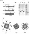
FIG. 1 shows results of designing and preparing fusion proteins (PpNF, PpLF, PpCF) in which PD-L1-binding peptide 1 (PD- L 1 pep 1) is inserted into each of three junction points to identify differences in structure, biophysical properties, and binding force, wherein A) shows diagrams of gene sequences for PpNF, PpLF, and PpCF, B) shows results of verifying stable expression and purification of PpNF, PpLF, and PpCF in E. coli, and C) shows results of tertiary structural modeling of PpNF, PpLF, and PpCF. -
FIG. 2 shows results of observing whether cages are formed and particle sizes of each prepared fusion protein by TEM and DLS, wherein A) shows results of transmission electron microscopy (TEM) analysis, and B) shows results of dynamic light scattering (DLS) analysis. -
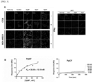
FIG. 3 shows results of comparative analysis of a binding force for each prepared fusion proteins, wherein A) shows results of culture cell binding assay, and B) shows results of protein binding assay by means of a surface plasmon resonance device. -
FIG. 4 shows results of identifying an immune checkpoint inhibitory function of PpNF in vitro, wherein A) shows a diagram for observing an activity of PpNF following co-culture of cancer cells and immune cells, B) shows results of flow cytometer measurement to determine an immune checkpoint inhibitory effect of PpNF, and C) shows a result of measuring an amount of interferon gamma secreted by isolated T cells using an enzyme-linked immunosorbent assay to identify an immune checkpoint inhibitory effect of PpNF. -
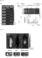
FIG. 5 shows results of identifying a tumor targeting ability of PpNF. -
FIG. 6 shows results of identifying a glioblastoma targeting effect of an immune checkpoint blocking nanocage. -
FIG. 7 shows results of analyzing anticancer and immune checkpoint inhibitory effects of PpNF in vivo. -
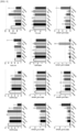
FIG. 8 shows results of analyzing anticancer and immune checkpoint inhibitory effects in vivo in a combined therapy using PpNF and doxorubicin. -
FIG. 9 shows results of determining biotoxicity of PpNF via blood and serologic analysis. - The present disclosure provides a fusion polypeptide in which a PD-L1-binding peptide having an amino acid sequence represented by SEQ ID NO: 2 is conjugated to a C-terminal or N-terminal of a human ferritin heavy chain fragment having an amino acid sequence represented by SEQ ID NO: 1.
- Preferably, the fusion polypeptide may further include a linker between the human ferritin heavy chain fragment and the PD-L1-binding peptide. More preferably, the linker may have an amino acid sequence represented by SEQ ID NO: 3 (GG) or SEQ ID NO: 4 (GGGSGGGGSG), but is not limited thereto.
- In addition, the present disclosure provides a ferritin nanocage including the fusion polypeptide. Specifically, the ferritin nanocage may be in a form in which the PD-L1-binding peptide is fused to an outer surface of ferritin.
- As used herein, the term 'ferritin' is a type of intracellular proteins that function to store and release iron. Ferritin is generally in the form of a hollow spherical cage in vivo, and the cage is composed of 24 ferritin monomers which are divided into heavy chains and light chains depending on the structure. In the present disclosure, the ferritin protein may be used without limitation if each of the proteins has an activity to form a cage-like complex protein as a monomer.
- In the present disclosure, human ferritin heavy chain fragments (1-161) having amino acid sequences represented by SEQ ID NO: 1 were used.
- As used herein, the term 'PD-L1-binding peptide' refers to a peptide having an amino acid sequence (CLQKTPKQC) represented by SEQ ID NO: 2 and is indicated herein as "PD-L1-binding peptide 1" or "PD-L1pep1".
- As used herein, the term "nanocage" refers to a cage that is formed by precise self-assembly properties of low molecular weight monomers and made of proteins having space inside. These include viral capsid proteins, ferritin, heat shock proteins, and DPS proteins. The nanocage of the present disclosure includes the fusion polypeptide of the present disclosure as a monomer constituting the nanocage. The term "self-assembly" as used herein refers to the property that certain molecules form a specific nanostructure by themselves without external special stimuli or artificial induction.
- The nanocage of the present disclosure may be a complex protein in which the fusion polypeptides of the present disclosure are regularly arranged as monomers, and more preferably, the nanocage may be formed as the 24 fusion polypeptides of the present disclosure are regularly arranged in three dimensions.
- In addition, the present disclosure provides a polynucleotide encoding the fusion polypeptide.
- The term "polynucleotide" as used herein refers to a polymer of deoxyribonucleotides or ribonucleotides that exist in a single-stranded or double-stranded form. The polynucleotide includes RNA genomic sequences, DNA (gDNA and cDNA), and RNA sequences transcribed therefrom and also includes analogs of natural polynucleotides unless otherwise specified.
- The polynucleotide includes not only the nucleotide sequence encoding the fusion peptide, but also a sequence complementary to the sequence. The complementary sequence includes not only perfectly complementary sequences, but also substantially complementary sequences.
- In addition, the polynucleotide may be modified. Such modifications include additions, deletions, or non-conservative substitutions or conservative substitutions of nucleotides. The polynucleotide encoding the amino acid sequence is construed to include a nucleotide sequence showing substantial identity to the nucleotide sequence. The substantial identity may refer to, when the nucleotide sequence and any other sequence are aligned to the maximum correspondence and the aligned sequence is analyzed using an algorithm commonly used in the art, a sequence showing at least 80% homology, at least 90% homology, or at least 95% homology.
- In addition, the present disclosure provides an expression vector including the polynucleotide.
- In addition, the present disclosure provides a transformant transformed with the expression vector (except for humans).
- The term "vector" as used herein refers to a self-replicating DNA molecule used to carry a clonal gene (or another fragment of clonal DNA).
- The term "vector" as used herein may refer to a plasmid, viral vector or other media known in the art capable of expressing the inserted nucleic acid in a host cell, and one that polynucleotides encoding the fusion polypeptide of the present disclosure are operably linked to a conventional expression vector known in the art. The expression vector may include an origin of replication to generally enable proliferation in a host cell, and one or more expression regulatory sequences (e.g., promoter, enhancer, etc.) to regulate expression, a selective marker, and a polynucleotide encoding the fusion polypeptide of the present disclosure operably linked to an expression regulatory sequence. The transformant may be transformed with the expression vector.
- Preferably, the transformant may be obtained by introducing the expression vector including the polynucleotide encoding the fusion polypeptide of the present disclosure into a host cell by a method known in the art, for example, but not limited to, transient transfection, microinjection, transduction, cell fusion, calcium phosphate precipitation, liposome-mediated transfection, DEAE dextran-mediated transfection, polybrene-mediated transfection, electroporation, gene gun, and other known methods for introducing a nucleic acid into the cell (Wu et al., J. Bio. Chem., 267:963-967, 1992; Wu and Wu, J. Bio. Chem., 263:14621-14624, 1988).
- In addition, the present disclosure provides a pharmaceutical composition for preventing or treating cancer including the ferritin nanocage as an active ingredient.
- Preferably, the cancer may include breast cancer, colon cancer, rectal cancer, brain tumor, lung cancer, liver cancer, skin cancer, esophageal cancer, testicular cancer, kidney cancer, stomach cancer, bladder cancer, ovarian cancer, cholangiocarcinoma, gallbladder cancer, uterine cancer, cervical cancer, prostate cancer, head and neck cancer, pancreatic cancer, and squamous cell carcinoma, but is not limited thereto.
- Preferably, the pharmaceutical composition may block binding of PD-1 and PD-L1 and inhibit immune checkpoints.
- In addition, the present disclosure provides a pharmaceutical composition for preventing or treating cancer including the ferritin nanocage and an anticancer agent as active ingredients.
- Preferably, the anticancer agent may be any one or more selected from the group consisting of doxorubicin, paclitaxel, vincristine, daunorubicin, vinblastine, actinomycin-D, docetaxel, etoposide, teniposide, bisantrene, homoharringtonine, Gleevec (STI-571), cisplatin, 5-fluorouracil, adriamycin, methotrexate, busulfan, chlorambucil, cyclophosphamide, melphalan, nitrogen mustard, and nitrosourea, but is not limited thereto.
- Preferably, the cancer may include breast cancer, colon cancer, rectal cancer, brain tumor, lung cancer, liver cancer, skin cancer, esophageal cancer, testicular cancer, kidney cancer, stomach cancer, bladder cancer, ovarian cancer, cholangiocarcinoma, gallbladder cancer, uterine cancer, cervical cancer, prostate cancer, head and neck cancer, pancreatic cancer, and squamous cell carcinoma, but is not limited thereto.
- The pharmaceutical composition of the present disclosure may be prepared using a pharmaceutically suitable and physiologically acceptable adjuvant in addition to the active ingredient, and a solubilizing agent such as an excipient, a disintegrant, a sweetener, a binder, a coating agent, a swelling agent, a lubricant, a polishing agent, or a flavoring agent may be used as the adjuvant. The pharmaceutical composition of the present disclosure may be preferably formulated into a pharmaceutical composition by including one or more pharmaceutically acceptable carriers in addition to the active ingredient for administration. In the composition formulated as a liquid solution, pharmaceutically acceptable carriers may be sterile and biocompatible and used by mixing saline, sterile water, Ringer's solution, buffered saline, albumin injection, dextrose solution, maltodextrin solution, glycerol, ethanol and one or more of these components, while other conventional additives such as antioxidants, buffers, and bacteriostats may be added as needed. In addition, diluents, dispersants, surfactants, binders, and lubricants may be additionally added to formulate into an injectable formulation such as an aqueous solution, suspension, and emulsion as well as pills, capsules, granules, or tablets.
- The pharmaceutical formulation of the pharmaceutical composition of the present disclosure may be granules, powder, coated tablets, tablets, capsules, suppositories, syrups, juices, suspensions, emulsions, drops or injectable solutions, and sustained-release preparations of the active compound. The pharmaceutical composition of the present disclosure may be administered in a conventional manner via intravenous, intraarterial, intraperitoneal, intramuscular, intraarterial, intraperitoneal, intrasternal, transdermal, intranasal, inhalational, topical, rectal, oral, intraocular or intradermal routes. The effective amount of the active ingredient of the pharmaceutical composition of the present disclosure refers to an amount required for preventing or treating a disease. Therefore, the effective amount may be controlled by various factors including the type of disease, the severity of the disease, the type and content of the active ingredient and other ingredients included in the composition, the type of formulation and the age, weight, general health status, sex and diet of a patient, administration time, administration route and secretion rate of the composition, treatment period, and drugs used in combination with. Although not limited thereto, for example, in the case of adults, when administered once to several times a day, the composition of the present disclosure may be administered at a dose of 0.1 ng/kg to 10 g/kg in the case of a compound when administered once to several times a day.
- In addition, the present disclosure provides a composition for drug delivery, including the ferritin nanocage as an active ingredient.
- The ferritin nanocage according to the present disclosure may be used as an intelligent drug delivery that selectively delivers drugs to cancer tissues. If the previously known drug is loaded in the ferritin nanocage of the present disclosure to be used for cancer treatment, since the drug is selectively delivered only to cancer tissues and cancer cells by the ferritin nanocage of the present disclosure, it is possible to increase effectiveness of the drug while side effects of the drug on normal tissues may be significantly reduced.
- The drug is an anticancer agent, and, as an anticancer agent that may be loaded in the ferritin nanocage of the present disclosure, there is no limitation in the use if it is for the conventional cancer treatment. For example, it may include doxorubicin, paclitaxel, vincristine, daunorubicin, vinblastine, actinomycin-D, docetaxel, etoposide, teniposide, bisantrene, homoharringtonine, Gleevec (STI-571), cisplatin, 5-fluorouracil, adriamycin, methotrexate, busulfan, chlorambucil, cyclophosphamide, melphalan, nitrogen mustard, and nitrosourea.
- Hereinafter, the present disclosure will be described in detail according to an example embodiment that does not limit the present disclosure. It is clear that the following example embodiments of the present disclosure are intended to embody the present disclosure and do not limit or restrict the scope of the present disclosure. Therefore, what may be easily inferred by an expert in the art to which the present disclosure pertains from the detailed description and example embodiments of the present disclosure is interpreted as falling within the scope of the present disclosure.
- A gene sequence of human ferritin heavy chain fragment (1-161) or that of human ferritin heavy chain (1-183) was obtained by amplifying by PCR in a cDNA library that was reverse-transcribed from human mRNA and inserted into pET-28a plasmids using a gene recombinant method. The gene sequence of PD-L1-binding peptide 1 was chemically synthesized and inserted into the nitrogen terminal of the human ferritin heavy chain fragment (1-161) using restriction enzymes KpnI and NheI or into the carbon terminal of the human ferritin heavy chain fragment (1-161) using restriction enzymes SpeI and XhoI. When inserting the peptide into the nitrogen terminal of ferritin, the peptide having GG sequence was inserted therebetween to increase the degree of freedom, and when inserting into the carbon terminal of ferritin, a longer flexible linker (GGGSGGGGSG) was inserted to increase the degree of freedom while preventing the peptide from being hidden inside the ferritin. In addition, the gene sequence of PD-L1-binding peptide 1 was inserted using the restriction enzymes BamHI and ApaI between genes corresponding to
helix 4 and 5 of the human ferritin heavy chain (1-183) gene. A construct into which the peptide was inserted at the nitrogen terminal of ferritin was named PpNF, that inserted betweenhelix 4 and 5 was named PpLF, and that inserted into the nitrogen terminal of ferritin was named PpCF. - E. coli (BL21) transformed with each plasmid into which the sequences of PpNF, PpLF, and PpCF were inserted was inoculated into LB media and grown at 37°C in a shake incubator. When a value of OD600 became 0.5, IPTG was added to the media to be 100 uM so as to induce protein overexpression. For PpNF and PpCF, lysis buffer was added into E. coli which was then disrupted by ultrasound. The lytic solution (supernatant) collected by centrifugation was mixed with Ni-NTA beads at 4°C for 1 hour. The lytic solution that did not bind to the bead was filtered out by a chromatographic column and the beads were washed with a wash buffer containing 30 mM imidazole. Endotoxins of E. coli were removed with a wash buffer containing 0.1% Triton X-114 which was then removed with a wash buffer. The protein was eluted and purified using elution buffers each containing 100 mM, 200 mM, 300 mM, and 500 mM imidazole. For PpLF, a lysis buffer was added to E. coli, disruption was performed by ultrasound, and then proteins were collected from the inclusion body collected by centrifugation. The proteins were fragmented into monomers with an extraction buffer containing 8 M urea and extracted from the inclusion body, and the isolated proteins (supernatant) were mixed with Ni-NTA beads at 4°C for 2 hours as above. Subsequently, for the wash buffer and elution buffer, 8 M urea was added to allow the protein to remain in a monomeric state. The lytic solution that did not bind to the beads was filtered out by a chromatographic column, and the beads were washed with the wash buffer containing 30 mM imidazole. Endotoxins of E. coli were removed with the wash buffer containing 0.1% Triton X-114 which was then removed by the wash buffer. The protein was eluted and purified using the elution buffer containing 300 mM imidazole. For the purified monomer protein, 8 M urea was slowly removed by dialysis to induce protein folding and formation of a ferritin nanocage again. For each protein of PpNF, PpLF, and PpCF, more than 95% homogeneous purified protein was collected by SDS PAGE.
- Human ferritin heavy chain fragments have three junction points, capable of peptide display on the surface when 24 monomers undergoes polymerization to form a cage, present on the N-terminal, a loop connecting the 4th and 5th helixes, and the C-terminal. Hereby, fusion proteins (PpNF, PpLF, PpCF) into which PD-L1-binding peptide 1 (PD-L1pep1) was inserted into each of these three junction points were designed and prepared to identify differences in structure, biophysical properties, and binding force (
FIG. 1 ). - The diagrams of gene sequences for PpNF, PpLF, and PpCF are shown in
FIG. 1A . - When electrophoresis of each protein of PpNF, PpLF, PpCF, and human ferritin heavy chain (FTH) was performed by SDS-polyacrylamide gel electrophoresis and then the protein bands were detected by staining the gel with Coomassie brilliant blue, stable expression and purification in E. coli were verified by detecting the protein band at the same position as the theoretical monomer size of each protein (
FIG. 1B ). - Through the tertiary structural models of PpNF, PpLF, and PpCF using MODELLAR v12 as a modeling program, it was found that the peptide was freely displayed on the surface in the case of PpNF, while the conformation of the peptide was implemented in the restricted range for PpLF. It was determined that PpCF had a degree of freedom higher than PpLF but not higher than that of PpNF (
FIG. 1C ). - In E. coli in which PpNF, PpLF, and PpCF were overexpressed, each protein was purified using Ni-NTA beads, and transmission electron microscopy (TEM) analysis was performed to determine whether the proteins form ferritin nanocages (
FIG. 2A ). It was found that each protein formed the nanocage, and by performing dynamic light scattering (DLS) analysis, it was found that each cage size was about 24 nm for PpNF, about 13 nm for PpLF, and about 39 nm for PpCF (FIG. 2B ). - Confocal laser scanning microscopy (Confocal) was performed. After culturing triple negative breast cancer cells (MDA-MB 231) or colorectal cancer cells (CT26), a group treated with 25 ng/ml of interferon-γ and a group without treatment were cultured at 37°C for 24 hours. These cells are cancer cells expressing the PD-L1 receptor and are known to have expression increased upon interferon-γ treatment. MCF-7, a breast cancer cell that does not express PD-L1, was observed as a control. In order to block nonspecific binding of PD-L1 proteins, blocking was performed using 1% BSA dissolved in PBS at room temperature for 1 hour, then BSA was removed, and PpNF, PpLF, and PpCF (100 nM) were reacted at 4°C for 1 hour. After washing with PBS, a reaction was performed using anti-human ferritin heavy chain antibodies (1: 200 ratio) for 1 hour, followed by staining (green). After fixing with 4% paraformaldehyde, observation was performed under a confocal microscope. To observe the nucleus, DAPI was dissolved in PBS in a ratio of 1: 1000, and staining was performed at room temperature for 5-10 minutes (blue).
- It was found that PpNF and PpCF fusion proteins bound in MDA-MB231 cells and CT26 cells expressing PD-L1. On the contrary, the binding did not take place in MCF-7 cells that do not express PD-L1, indicating that the binding was due to PD-L1. PpLF did not bind in MDA-MB231 cells and CT26 cells expressing PD-L1, despite the fact that the cage was well formed. Through the structural model, it was found that the peptide displayed on PpNF and PpCF was present on the surface in a conformation with a higher degree of freedom compared to PpLF, and a difference in the display pattern determined whether the binding takes place and the binding force (
FIG. 3A ). - The PD-L1 protein was attached to the surface of a gold sensor chip for surface plasmon resonance activated by EDC-NHS and blocked with ethanolamine (pH 8.5). PpNF and PpCF proteins dissolved in 20 mM Tris-HCl (pH 8.0), 150 mM NaCl, and 2 mM DTT solutions were each flowed over the gold sensor chip bound with PD-L1 proteins respectively to analyze binding kinetics. The right channel, without PD-L1 protein attached, was used as a reference to correct the resonance measurements.
- As a result of direct measurement of the binding of PpNF and PpCF with PD-L1, PpNF directly bound to PD-L1 (Kd = 39 nM), while PpCF showed no binding at all even at a concentration of 200 nM. Therefore, it was revealed that PpNF displayed PD-L1-binding peptide 1 in a structure capable of binding to PD-L1, which was used in future experiments (
FIG. 3B ). - CD8+ T cells were extracted from the spleen obtained by sacrificing BALB/C mice. Extracted CD8+ T cells were treated with interleukin-2/interleukin-15 and anti-CD-3 antibody/anti-CD-28 antibody each for 24 hours for activation. Prior to treating activated CD8+ T cells with CFSE and performing symbiotic culture of tumor cells (CT26, mouse colon cancer), tumor cells were pretreated with anti-PD-L1 antibody, PpNF (25 nM or 50 nM), and PD-L1pep1 (50 nM), respectively, and interactions of PD-L1 of tumor cells and PD-L1 of activated CD8+ T cells were inhibited, respectively. After symbiotic culture for 24 hours, CD8+ T cells were isolated (
FIG. 4A ). The degree of CFSE binding to isolated T cells was measured with a flow cytometer (FIG. 4B ). In addition, the amount of interferon gamma secreted by isolated T cells was measured using an enzyme-linked immunosorbent assay (FIG. 4C ). - CD8+ T cells isolated from mice were activated (IL-2/IL-15 treatment and anti-CD3/anti-CD28 treatment), labeled with fluorescence (CFSE staining), and cultured with CT26 colorectal cancer cells. At this time, PD-L1 of cancer cells and PD-1 of T-cells were bound and showed an immunosuppressive effect. Treatment with PpNF or PD-L1 antibodies has shown immunoactive effects, i.e., T-cell proliferation and IFN-γ increment, by inhibiting PD-1/PD-L1 binding.
- Thereby, it was possible to verify that PpNF had an immune checkpoint inhibitory effect, and it was found that the effect of PpNF is better than that of PD-L1 antibody or PD-L1pep1 (PD-L1-binding peptide 1).
- 1×10⁶ CT26 colorectal cancer cells contained in 100 µl of PBS were injected subcutaneously into the right thigh of BLAB/c mice aged 6 weeks. When the tumor was about 200 mm3 in size, 200 µl each of PpNF (10 mg/kg) that was labeled with Flamma-675 and dissolved in saline as well as ferritin wildtype (10 mg/kg) proteins was injected into each tail vein of the mice.
- IVIS fluorescence imaging systems were used to investigate the in vivo distribution of
proteins 1, 4, 8, 12, and 24 hours after injection. Then, after 24 hours, the mice were sacrificed and tumors, hearts, lungs, liver, kidneys, and spleen were taken out to investigate how many proteins remained in the organs. - It was found that PpNF had increased tumor targeting ability than ferritin wildtype (tumor target by the EPR effect) (
FIG. 5 ). - In addition, the glioblastoma targeting effect of the immune checkpoint blocking nanocage was identified. GL26, a glioblastoma cell line, was injected into the left side of the mouse brain to create an orthotopic GBM model and fluorescently labeled PBP1-N-FtH was injected into the tail vein [control: saline, ferritin]. After 24 hours, a fluorescence signal was observed in the brain, and it was observed that PBP1-N-FtH was targeted to the tumor site on the left side of the brain (
FIG. 6 ). - CT26 cells were injected subcutaneously into the upper right thigh of BALB/c wildtype mice. When the tumor was about 100 mm3 in size, mice were randomly divided into four groups. Saline, PpNF (10 mg/kg) dissolved in saline, and wildtype ferritin (10 mg/kg) were injected into the tail vein of each mouse, and therapeutic anti-PD-L1 antibody (10 mg/kg) was injected intraperitoneally (
FIG. 7A ). A total of 10 injections were performed once every two days (once every 4 days, repeated 6 times in total for anti-PD-L1 antibodies) to measure tumor size and weight of mice (FIGS. 7B and 7D ). After 3 weeks, the tumor was removed by sacrificing mice and the tumor was weighed (FIG. 7C ). The extracellular matrix of the removed tumor was degraded with collagenase to form single cells, and antibodies against labeled proteins of CD4+ T cells, CD8+ T cells, and regulatory T cells were each treated to analyze how many T cells are each present in the tumor by flow cytometry (FIGS. 7E to 7G ). - PpNF alone significantly inhibited tumor growth compared to the control (p<0.05) (
FIG. 7B ). The weight of the tumor was much smaller (FIG. 7C ), and by increasing the proportion of CD8+ T cells in the tumor (FIG. 7F ), the ratio of CD8+ T cells and regulatory T cells in the tumor was significantly increased compared to the control (FIG. 7H ). Both PpNF and wildtype ferritin showed no change in body weight compared to control mice (FIG. 7D ). - Through the results, it was found that PpNF had the effect of inhibiting the immune checkpoint in vivo, increasing CD8+ T cells, and eventually inhibiting tumor growth.
- CT26 cells were injected subcutaneously into the upper right thigh of BALB/c wildtype mice. When the tumor was about 50 mm3 in size, they were randomly divided into five groups. PBS, PpNF (10 mg/kg) dissolved in PBS, PpNF (Dox) (10 mg/kg, 1.04 mg/kg for doxorubicin) in which doxorubicin was loaded in ferritin, and doxorubicin (1.04 mg/kg) were injected into the tail vein of each mouse, and therapeutic anti-PD-L1 antibody (10 mg/kg) was injected intraperitoneally (
FIG. 8A ). A total of 10 injections (once every 3 days, repeated 6 times in total for anti-PD-L1 antibodies,) were performed 3 times a week to measure tumor size and weight of mice (FIGS. 8B and 8D ). After 3 weeks, the tumor was removed by sacrificing the mice, and the tumor was weighed (FIG. 8C ). The extracellular matrix of the removed tumor was degraded with collagenase to form single cells, and antibodies against labeled proteins of CD4+ T cells, CD8+ T cells, and regulatory T cells were treated respectively to analyze how many T cells are each present in the tumor by flow cytometry (FIGS. 8E to 8G ). - When PpNF was loaded with doxorubicin and treated to the tumor in combined therapy, not only was the tumor growth significantly inhibited (p<0.001) compared to the control (p0.001), but also was the tumor growth significantly inhibited also compared to the same amount of PpNF (p<0.001), to the same amount of doxorubicin (p<0.01), and to therapeutic anti-PD-L1 antibodies (p<0.01) (
FIG. 8B ). The weight of tumor was also reduced (FIG. 8C ), and by reducing the proportion of regulatory T cells in the tumor (FIG. 8G ), the ratio of CD8+ T cells and regulatory T cells in the tumor increased compared to the control (FIG. 8H ). - From the results, it was found that the combined therapy with doxorubicin loaded in PpNF had the effect of inhibiting the immune checkpoint in vivo, increasing the ratio of CD8+ T cells to regulatory T cells in the tumor, and eventually inhibiting the tumor growth.
- After antitumor animal experiments, blood and serum were collected from each group when sacrificing mice, and the amounts of red blood cells, hemoglobin, hematocrit, white blood cells, and neutrophils in the blood, blood urea nitrogen in serum, creatinine, alanine aminotransferase, and aspartate aminotransferase were measured and compared.
- No hepatotoxicity or nephrotoxicity was observed in the mouse group treated with PpNF and PpNF with doxorubicin loaded, and there was no significant hematologic change other than an increase in neutrophils in PpNF loaded with doxorubicin (
FIG. 9 ).
Claims (17)
- A fusion polypeptide, wherein a PD-L1-binding peptide having an amino acid sequence represented by SEQ ID NO: 2 is conjugated to a C-terminal or N-terminal of a human ferritin heavy chain fragment having an amino acid sequence represented by SEQ ID NO: 1.
- The fusion polypeptide of claim 1, wherein the fusion polypeptide further comprises a linker between the human ferritin heavy chain fragment and the PD-L1-binding peptide.
- The fusion polypeptide of claim 2, wherein the linker has an amino acid sequence represented by SEQ ID NO: 3 or SEQ ID NO: 4.
- A ferritin nanocage, comprising the fusion polypeptide of any one of claims 1 to 3.
- The ferritin nanocage of claim 4, wherein the ferritin nanocage is in a form in which the PD-L1-binding peptide is fused to an outer surface of ferritin.
- A polynucleotide encoding the fusion polypeptide of any one of claims 1 to 3.
- An expression vector, comprising the polynucleotide of claim 6.
- A transformant transformed with the expression vector of claim 7 (except for humans).
- A pharmaceutical composition for preventing or treating cancer, comprising the ferritin nanocage of claim 4 as an active ingredient.
- The pharmaceutical composition of claim 9, wherein the cancer comprises any one selected from the group consisting of breast cancer, colon cancer, rectal cancer, brain tumor, lung cancer, liver cancer, skin cancer, esophageal cancer, testicular cancer, kidney cancer, stomach cancer, bladder cancer, ovarian cancer, cholangiocarcinoma, gallbladder cancer, uterine cancer, cervical cancer, prostate cancer, head and neck cancer, pancreatic cancer, and squamous cell carcinoma.
- The pharmaceutical composition of claim 9, wherein the pharmaceutical composition blocks binding of PD-1 and PD-L1 and inhibits immune checkpoints.
- A pharmaceutical composition for preventing or treating cancer, comprising the ferritin nanocage of claim 4 and an anticancer agent as active ingredients.
- The pharmaceutical composition of claim 12, wherein the anticancer agent is any one or more selected from the group consisting of doxorubicin, paclitaxel, vincristine, daunorubicin, vinblastine, actinomycin-D, docetaxel, etoposide, teniposide, bisantrene, homoharringtonine, Gleevec (STI-571), cisplatin, 5-fluorouracil, adriamycin, methotrexate, busulfan, chlorambucil, cyclophosphamide, melphalan, nitrogen mustard, and nitrosourea.
- The pharmaceutical composition of claim 12, wherein the cancer is any one selected from the group consisting of breast cancer, colon cancer, rectal cancer, brain tumor, lung cancer, liver cancer, skin cancer, esophageal cancer, testicular cancer, kidney cancer, stomach cancer, bladder cancer, ovarian cancer, cholangiocarcinoma, gallbladder cancer, uterine cancer, cervical cancer, prostate cancer, head and neck cancer, pancreatic cancer, and squamous cell carcinoma.
- A composition for drug delivery, comprising the ferritin nanocage of claim 4 as an active ingredient.
- The composition of claim 15, wherein the drug is an anticancer agent.
- The composition of claim 15, wherein the anticancer agent is any one or more selected from the group consisting of doxorubicin, paclitaxel, vincristine, daunorubicin, vinblastine, actinomycin-D, docetaxel, etoposide, teniposide, bisantrene, homoharringtonine, Gleevec (STI-571), cisplatin, 5-fluorouracil, adriamycin, methotrexate, busulfan, chlorambucil, cyclophosphamide, melphalan, nitrogen mustard, and nitrosourea.
Applications Claiming Priority (2)
| Application Number | Priority Date | Filing Date | Title |
|---|---|---|---|
| KR1020200138075A KR102658825B1 (en) | 2020-10-23 | 2020-10-23 | The immunotherapeutic nanocage displaying PD-L1 binding peptide 1 and use in anti-cancer immunotherapeutic agent thereof |
| PCT/KR2021/014261 WO2022086058A1 (en) | 2020-10-23 | 2021-10-14 | Ferritin nanocage fused with pd-l1-binding peptide 1 and use thereof as anticancer immunotherapy agent |
Publications (3)
| Publication Number | Publication Date |
|---|---|
| EP4234578A1 true EP4234578A1 (en) | 2023-08-30 |
| EP4234578A4 EP4234578A4 (en) | 2024-08-28 |
| EP4234578B1 EP4234578B1 (en) | 2025-11-12 |
Family
ID=81290704
Family Applications (1)
| Application Number | Title | Priority Date | Filing Date |
|---|---|---|---|
| EP21883129.5A Active EP4234578B1 (en) | 2020-10-23 | 2021-10-14 | Ferritin nanocage fused with pd-l1-binding peptide 1 and use thereof as anticancer immunotherapy agent |
Country Status (4)
| Country | Link |
|---|---|
| US (1) | US20230374113A1 (en) |
| EP (1) | EP4234578B1 (en) |
| KR (1) | KR102658825B1 (en) |
| WO (1) | WO2022086058A1 (en) |
Cited By (1)
| Publication number | Priority date | Publication date | Assignee | Title |
|---|---|---|---|---|
| WO2025177045A1 (en) * | 2024-02-23 | 2025-08-28 | Thena Biotech S.r.l. | Fusion-proteins based on human ferritin and their use as multiple bioactive peptides delivery system |
Families Citing this family (3)
| Publication number | Priority date | Publication date | Assignee | Title |
|---|---|---|---|---|
| KR102934900B1 (en) | 2023-05-04 | 2026-03-10 | 경북대학교 산학협력단 | The immunotherapeutic nanocage displaying LAG3 binding peptide and use in anti-cancer immunotherapeutic agent thereof |
| CN118221832B (en) * | 2024-04-10 | 2025-02-18 | 合肥工业大学 | A kind of antifreeze ferritin precisely conjugated with polypeptide and preparation method thereof |
| WO2026010421A1 (en) * | 2024-07-04 | 2026-01-08 | (주)셀레메디 | Dual target ferritin-protein complex and use thereof |
Family Cites Families (6)
| Publication number | Priority date | Publication date | Assignee | Title |
|---|---|---|---|---|
| EP1456652A4 (en) * | 2001-11-13 | 2005-11-02 | Dana Farber Cancer Inst Inc | AGENTS MODULATING IMMUNE CELL ACTIVITY AND METHODS OF USE THEREOF |
| KR101811050B1 (en) | 2015-09-02 | 2017-12-21 | 경북대학교 산학협력단 | Fusion-polypeptide of anti-inflammatory polypeptide and ferritin monomer fragment and Pharmaceutical Composition comprising the same |
| KR101888675B1 (en) * | 2015-09-02 | 2018-08-14 | 경북대학교 산학협력단 | Fragment of human ferritin monomer and fusion polypeptide using thereof |
| KR101953617B1 (en) * | 2016-07-15 | 2019-05-23 | 한국과학기술연구원 | A novel nanocage and use thereof |
| JP2019533645A (en) * | 2016-09-16 | 2019-11-21 | ザ・ジョンズ・ホプキンス・ユニバーシティー | Increased mucosal penetration protein nanocages for targeted tissue and intracellular delivery |
| US11306119B2 (en) * | 2017-12-15 | 2022-04-19 | Kyungpook National University Industry-Academic Cooperation Foundation | Peptide bound to PD-L1 and use thereof |
-
2020
- 2020-10-23 KR KR1020200138075A patent/KR102658825B1/en active Active
-
2021
- 2021-10-14 WO PCT/KR2021/014261 patent/WO2022086058A1/en not_active Ceased
- 2021-10-14 EP EP21883129.5A patent/EP4234578B1/en active Active
-
2023
- 2023-04-23 US US18/138,118 patent/US20230374113A1/en active Pending
Cited By (1)
| Publication number | Priority date | Publication date | Assignee | Title |
|---|---|---|---|---|
| WO2025177045A1 (en) * | 2024-02-23 | 2025-08-28 | Thena Biotech S.r.l. | Fusion-proteins based on human ferritin and their use as multiple bioactive peptides delivery system |
Also Published As
| Publication number | Publication date |
|---|---|
| EP4234578B1 (en) | 2025-11-12 |
| EP4234578A4 (en) | 2024-08-28 |
| US20230374113A1 (en) | 2023-11-23 |
| KR20220053848A (en) | 2022-05-02 |
| KR102658825B1 (en) | 2024-04-19 |
| WO2022086058A1 (en) | 2022-04-28 |
Similar Documents
| Publication | Publication Date | Title |
|---|---|---|
| EP4234578B1 (en) | Ferritin nanocage fused with pd-l1-binding peptide 1 and use thereof as anticancer immunotherapy agent | |
| JP6076963B2 (en) | Anti-epidermal growth factor receptor variant III chimeric antigen receptor and its use for the treatment of cancer | |
| US9358308B2 (en) | Compositions of a peptide targeting system for treating cancer | |
| UA124532C2 (en) | Transfected t-cells and t-cell receptors for use in immunotherapy against cancers | |
| WO2017039382A1 (en) | Human-derived ferritin monomer fragment and fusion polypeptide using same | |
| KR101286721B1 (en) | Recombinant albumins fused with poly-cysteine peptide and the methods for preparing the same | |
| TWI868739B (en) | T cell receptor targeting KRAS G12V mutant polypeptide and its use | |
| KR102348838B1 (en) | Active TRAIL trimer and tumor targeting peptide multi-displayed on ferritin nanocage and use in anti-cancer agent thereof | |
| JP6612063B2 (en) | Malignant glioma molecule targeting peptide | |
| CN117750970A (en) | Compositions comprising NKG2D, CXCR2 and DAP10/DAP12 fusion polypeptides and methods of use thereof | |
| WO2021190550A1 (en) | Chimeric antigen receptor containing protective peptide, and use thereof | |
| WO2026067755A1 (en) | Self-assembled trimeric protein and preparation method therefor | |
| US12577292B2 (en) | Ferritin nanocage for multi-displaying trail trimer and cancer-targeting peptide and use thereof as anticancer agent | |
| Wang et al. | Synergistic HER2-scFv mediated immunotherapy with targeted gemcitabine delivery harnessing biomimetic composite nanoparticles for enhanced metastatic tumor therapy | |
| KR102726136B1 (en) | The immunotherapeutic nanocage displaying PD-L1 binding peptide 2 and use in anti-cancer immunotherapeutic agent thereof | |
| US20220409550A1 (en) | Pd-1-decorated nanocages and uses thereof | |
| CN110016074B (en) | MAGE-A3 humanized T cell receptor | |
| JP2026514918A (en) | High-affinity T cell receptor for identifying KRAS mutations and its applications | |
| WO2025217954A1 (en) | Bifunctional fusion protein comprising anti-pd-1 antibody and il-2 mutant | |
| KR102934900B1 (en) | The immunotherapeutic nanocage displaying LAG3 binding peptide and use in anti-cancer immunotherapeutic agent thereof | |
| CN120302987A (en) | Chimeric antigen receptor system, preparation method and use thereof | |
| KR20220120910A (en) | EGFR-GST-Ulixertinib complex, and pharmaceutical composition for preventing or treating cancer comprising the same | |
| WO2024228598A1 (en) | Ferritin nanocage fused with lag3 binding peptide and use thereof as anticancer immunotherapeutic agent | |
| CN121293363B (en) | CD14 and CD3 targeting dual-function T cell adapter and application thereof | |
| US20250304718A1 (en) | Her2 single domain antibody and uses thereof |
Legal Events
| Date | Code | Title | Description |
|---|---|---|---|
| STAA | Information on the status of an ep patent application or granted ep patent |
Free format text: STATUS: THE INTERNATIONAL PUBLICATION HAS BEEN MADE |
|
| PUAI | Public reference made under article 153(3) epc to a published international application that has entered the european phase |
Free format text: ORIGINAL CODE: 0009012 |
|
| STAA | Information on the status of an ep patent application or granted ep patent |
Free format text: STATUS: REQUEST FOR EXAMINATION WAS MADE |
|
| 17P | Request for examination filed |
Effective date: 20230511 |
|
| AK | Designated contracting states |
Kind code of ref document: A1 Designated state(s): AL AT BE BG CH CY CZ DE DK EE ES FI FR GB GR HR HU IE IS IT LI LT LU LV MC MK MT NL NO PL PT RO RS SE SI SK SM TR |
|
| DAV | Request for validation of the european patent (deleted) | ||
| DAX | Request for extension of the european patent (deleted) | ||
| REG | Reference to a national code |
Ref country code: DE Ref legal event code: R079 Free format text: PREVIOUS MAIN CLASS: C07K0016180000 Ipc: C07K0014470000 Ref country code: DE Ref legal event code: R079 Ref document number: 602021042428 Country of ref document: DE Free format text: PREVIOUS MAIN CLASS: C07K0016180000 Ipc: C07K0014470000 |
|
| A4 | Supplementary search report drawn up and despatched |
Effective date: 20240731 |
|
| RIC1 | Information provided on ipc code assigned before grant |
Ipc: C07K 14/705 20060101ALN20240725BHEP Ipc: B82Y 5/00 20110101ALN20240725BHEP Ipc: A61K 39/00 20060101ALN20240725BHEP Ipc: A61K 9/51 20060101ALN20240725BHEP Ipc: A61K 9/00 20060101ALN20240725BHEP Ipc: C07K 19/00 20060101ALN20240725BHEP Ipc: A61P 35/00 20060101ALI20240725BHEP Ipc: A61K 31/704 20060101ALI20240725BHEP Ipc: A61K 39/39 20060101ALI20240725BHEP Ipc: C12N 15/62 20060101ALI20240725BHEP Ipc: C07K 16/28 20060101ALI20240725BHEP Ipc: C07K 7/06 20060101ALI20240725BHEP Ipc: C07K 14/47 20060101AFI20240725BHEP |
|
| STAA | Information on the status of an ep patent application or granted ep patent |
Free format text: STATUS: EXAMINATION IS IN PROGRESS |
|
| 17Q | First examination report despatched |
Effective date: 20250228 |
|
| RIC1 | Information provided on ipc code assigned before grant |
Ipc: C07K 14/47 20060101AFI20250624BHEP Ipc: C07K 7/06 20060101ALI20250624BHEP Ipc: C07K 16/28 20060101ALI20250624BHEP Ipc: C12N 15/62 20060101ALI20250624BHEP Ipc: A61K 39/39 20060101ALI20250624BHEP Ipc: A61K 31/704 20060101ALI20250624BHEP Ipc: A61P 35/00 20060101ALI20250624BHEP Ipc: C07K 19/00 20060101ALN20250624BHEP Ipc: A61K 9/00 20060101ALN20250624BHEP Ipc: A61K 9/51 20060101ALN20250624BHEP Ipc: A61K 39/00 20060101ALN20250624BHEP Ipc: B82Y 5/00 20110101ALN20250624BHEP Ipc: C07K 14/705 20060101ALN20250624BHEP |
|
| GRAP | Despatch of communication of intention to grant a patent |
Free format text: ORIGINAL CODE: EPIDOSNIGR1 |
|
| STAA | Information on the status of an ep patent application or granted ep patent |
Free format text: STATUS: GRANT OF PATENT IS INTENDED |
|
| RIC1 | Information provided on ipc code assigned before grant |
Ipc: C07K 14/47 20060101AFI20250710BHEP Ipc: C07K 7/06 20060101ALI20250710BHEP Ipc: C07K 16/28 20060101ALI20250710BHEP Ipc: C12N 15/62 20060101ALI20250710BHEP Ipc: A61K 39/39 20060101ALI20250710BHEP Ipc: A61K 31/704 20060101ALI20250710BHEP Ipc: A61P 35/00 20060101ALI20250710BHEP Ipc: C07K 19/00 20060101ALN20250710BHEP Ipc: A61K 9/00 20060101ALN20250710BHEP Ipc: A61K 9/51 20060101ALN20250710BHEP Ipc: A61K 39/00 20060101ALN20250710BHEP Ipc: B82Y 5/00 20110101ALN20250710BHEP Ipc: C07K 14/705 20060101ALN20250710BHEP |
|
| INTG | Intention to grant announced |
Effective date: 20250811 |
|
| GRAS | Grant fee paid |
Free format text: ORIGINAL CODE: EPIDOSNIGR3 |
|
| GRAA | (expected) grant |
Free format text: ORIGINAL CODE: 0009210 |
|
| STAA | Information on the status of an ep patent application or granted ep patent |
Free format text: STATUS: THE PATENT HAS BEEN GRANTED |
|
| AK | Designated contracting states |
Kind code of ref document: B1 Designated state(s): AL AT BE BG CH CY CZ DE DK EE ES FI FR GB GR HR HU IE IS IT LI LT LU LV MC MK MT NL NO PL PT RO RS SE SI SK SM TR |
|
| REG | Reference to a national code |
Ref country code: CH Ref legal event code: F10 Free format text: ST27 STATUS EVENT CODE: U-0-0-F10-F00 (AS PROVIDED BY THE NATIONAL OFFICE) Effective date: 20251112 Ref country code: GB Ref legal event code: FG4D |
|
| REG | Reference to a national code |
Ref country code: IE Ref legal event code: FG4D |
|
| REG | Reference to a national code |
Ref country code: DE Ref legal event code: R096 Ref document number: 602021042428 Country of ref document: DE |
|
| REG | Reference to a national code |
Ref country code: NL Ref legal event code: MP Effective date: 20251112 |
|
| PG25 | Lapsed in a contracting state [announced via postgrant information from national office to epo] |
Ref country code: ES Free format text: LAPSE BECAUSE OF FAILURE TO SUBMIT A TRANSLATION OF THE DESCRIPTION OR TO PAY THE FEE WITHIN THE PRESCRIBED TIME-LIMIT Effective date: 20251112 |
|
| REG | Reference to a national code |
Ref country code: LT Ref legal event code: MG9D |
|
| PG25 | Lapsed in a contracting state [announced via postgrant information from national office to epo] |
Ref country code: NO Free format text: LAPSE BECAUSE OF FAILURE TO SUBMIT A TRANSLATION OF THE DESCRIPTION OR TO PAY THE FEE WITHIN THE PRESCRIBED TIME-LIMIT Effective date: 20260212 |
|
| PG25 | Lapsed in a contracting state [announced via postgrant information from national office to epo] |
Ref country code: HR Free format text: LAPSE BECAUSE OF FAILURE TO SUBMIT A TRANSLATION OF THE DESCRIPTION OR TO PAY THE FEE WITHIN THE PRESCRIBED TIME-LIMIT Effective date: 20251112 Ref country code: AT Free format text: LAPSE BECAUSE OF FAILURE TO SUBMIT A TRANSLATION OF THE DESCRIPTION OR TO PAY THE FEE WITHIN THE PRESCRIBED TIME-LIMIT Effective date: 20251112 Ref country code: FI Free format text: LAPSE BECAUSE OF FAILURE TO SUBMIT A TRANSLATION OF THE DESCRIPTION OR TO PAY THE FEE WITHIN THE PRESCRIBED TIME-LIMIT Effective date: 20251112 |
|
| REG | Reference to a national code |
Ref country code: AT Ref legal event code: MK05 Ref document number: 1856688 Country of ref document: AT Kind code of ref document: T Effective date: 20251112 |
|
| PG25 | Lapsed in a contracting state [announced via postgrant information from national office to epo] |
Ref country code: NL Free format text: LAPSE BECAUSE OF FAILURE TO SUBMIT A TRANSLATION OF THE DESCRIPTION OR TO PAY THE FEE WITHIN THE PRESCRIBED TIME-LIMIT Effective date: 20251112 |
|
| PG25 | Lapsed in a contracting state [announced via postgrant information from national office to epo] |
Ref country code: RS Free format text: LAPSE BECAUSE OF FAILURE TO SUBMIT A TRANSLATION OF THE DESCRIPTION OR TO PAY THE FEE WITHIN THE PRESCRIBED TIME-LIMIT Effective date: 20260212 |
|
| PG25 | Lapsed in a contracting state [announced via postgrant information from national office to epo] |
Ref country code: IS Free format text: LAPSE BECAUSE OF FAILURE TO SUBMIT A TRANSLATION OF THE DESCRIPTION OR TO PAY THE FEE WITHIN THE PRESCRIBED TIME-LIMIT Effective date: 20260312 |
|
| PG25 | Lapsed in a contracting state [announced via postgrant information from national office to epo] |
Ref country code: PT Free format text: LAPSE BECAUSE OF FAILURE TO SUBMIT A TRANSLATION OF THE DESCRIPTION OR TO PAY THE FEE WITHIN THE PRESCRIBED TIME-LIMIT Effective date: 20260312 |
|
| PG25 | Lapsed in a contracting state [announced via postgrant information from national office to epo] |
Ref country code: PL Free format text: LAPSE BECAUSE OF FAILURE TO SUBMIT A TRANSLATION OF THE DESCRIPTION OR TO PAY THE FEE WITHIN THE PRESCRIBED TIME-LIMIT Effective date: 20251112 |
|
| PG25 | Lapsed in a contracting state [announced via postgrant information from national office to epo] |
Ref country code: LV Free format text: LAPSE BECAUSE OF FAILURE TO SUBMIT A TRANSLATION OF THE DESCRIPTION OR TO PAY THE FEE WITHIN THE PRESCRIBED TIME-LIMIT Effective date: 20251112 |
|
| PG25 | Lapsed in a contracting state [announced via postgrant information from national office to epo] |
Ref country code: BG Free format text: LAPSE BECAUSE OF FAILURE TO SUBMIT A TRANSLATION OF THE DESCRIPTION OR TO PAY THE FEE WITHIN THE PRESCRIBED TIME-LIMIT Effective date: 20251112 |
|
| PG25 | Lapsed in a contracting state [announced via postgrant information from national office to epo] |
Ref country code: LT Free format text: LAPSE BECAUSE OF FAILURE TO SUBMIT A TRANSLATION OF THE DESCRIPTION OR TO PAY THE FEE WITHIN THE PRESCRIBED TIME-LIMIT Effective date: 20251112 |
|
| PG25 | Lapsed in a contracting state [announced via postgrant information from national office to epo] |
Ref country code: SM Free format text: LAPSE BECAUSE OF FAILURE TO SUBMIT A TRANSLATION OF THE DESCRIPTION OR TO PAY THE FEE WITHIN THE PRESCRIBED TIME-LIMIT Effective date: 20251112 |
|
| PG25 | Lapsed in a contracting state [announced via postgrant information from national office to epo] |
Ref country code: DK Free format text: LAPSE BECAUSE OF FAILURE TO SUBMIT A TRANSLATION OF THE DESCRIPTION OR TO PAY THE FEE WITHIN THE PRESCRIBED TIME-LIMIT Effective date: 20251112 |
|
| PG25 | Lapsed in a contracting state [announced via postgrant information from national office to epo] |
Ref country code: IT Free format text: LAPSE BECAUSE OF FAILURE TO SUBMIT A TRANSLATION OF THE DESCRIPTION OR TO PAY THE FEE WITHIN THE PRESCRIBED TIME-LIMIT Effective date: 20251112 |
|
| PG25 | Lapsed in a contracting state [announced via postgrant information from national office to epo] |
Ref country code: EE Free format text: LAPSE BECAUSE OF FAILURE TO SUBMIT A TRANSLATION OF THE DESCRIPTION OR TO PAY THE FEE WITHIN THE PRESCRIBED TIME-LIMIT Effective date: 20251112 |
|
| PG25 | Lapsed in a contracting state [announced via postgrant information from national office to epo] |
Ref country code: CZ Free format text: LAPSE BECAUSE OF FAILURE TO SUBMIT A TRANSLATION OF THE DESCRIPTION OR TO PAY THE FEE WITHIN THE PRESCRIBED TIME-LIMIT Effective date: 20251112 |
|
| PG25 | Lapsed in a contracting state [announced via postgrant information from national office to epo] |
Ref country code: SK Free format text: LAPSE BECAUSE OF FAILURE TO SUBMIT A TRANSLATION OF THE DESCRIPTION OR TO PAY THE FEE WITHIN THE PRESCRIBED TIME-LIMIT Effective date: 20251112 |
|
| PG25 | Lapsed in a contracting state [announced via postgrant information from national office to epo] |
Ref country code: SE Free format text: LAPSE BECAUSE OF FAILURE TO SUBMIT A TRANSLATION OF THE DESCRIPTION OR TO PAY THE FEE WITHIN THE PRESCRIBED TIME-LIMIT Effective date: 20251112 |