EP4230227A1 - Contrast agent for detecting cartilage damage, and method and program for detecting cartilage damage using said contrast agent - Google Patents
Contrast agent for detecting cartilage damage, and method and program for detecting cartilage damage using said contrast agent Download PDFInfo
- Publication number
- EP4230227A1 EP4230227A1 EP21886309.0A EP21886309A EP4230227A1 EP 4230227 A1 EP4230227 A1 EP 4230227A1 EP 21886309 A EP21886309 A EP 21886309A EP 4230227 A1 EP4230227 A1 EP 4230227A1
- Authority
- EP
- European Patent Office
- Prior art keywords
- contrast agent
- cartilage
- mri
- administration
- damage
- Prior art date
- Legal status (The legal status is an assumption and is not a legal conclusion. Google has not performed a legal analysis and makes no representation as to the accuracy of the status listed.)
- Pending
Links
Images


Classifications
-
- A—HUMAN NECESSITIES
- A61—MEDICAL OR VETERINARY SCIENCE; HYGIENE
- A61P—SPECIFIC THERAPEUTIC ACTIVITY OF CHEMICAL COMPOUNDS OR MEDICINAL PREPARATIONS
- A61P19/00—Drugs for skeletal disorders
- A61P19/02—Drugs for skeletal disorders for joint disorders, e.g. arthritis, arthrosis
-
- A—HUMAN NECESSITIES
- A61—MEDICAL OR VETERINARY SCIENCE; HYGIENE
- A61B—DIAGNOSIS; SURGERY; IDENTIFICATION
- A61B5/00—Measuring for diagnostic purposes; Identification of persons
- A61B5/05—Detecting, measuring or recording for diagnosis by means of electric currents or magnetic fields; Measuring using microwaves or radio waves
- A61B5/055—Detecting, measuring or recording for diagnosis by means of electric currents or magnetic fields; Measuring using microwaves or radio waves involving electronic [EMR] or nuclear [NMR] magnetic resonance, e.g. magnetic resonance imaging
-
- A—HUMAN NECESSITIES
- A61—MEDICAL OR VETERINARY SCIENCE; HYGIENE
- A61B—DIAGNOSIS; SURGERY; IDENTIFICATION
- A61B5/00—Measuring for diagnostic purposes; Identification of persons
- A61B5/45—For evaluating or diagnosing the musculoskeletal system or teeth
- A61B5/4514—Cartilage
-
- A—HUMAN NECESSITIES
- A61—MEDICAL OR VETERINARY SCIENCE; HYGIENE
- A61K—PREPARATIONS FOR MEDICAL, DENTAL OR TOILETRY PURPOSES
- A61K49/00—Preparations for testing in vivo
- A61K49/06—Nuclear magnetic resonance [NMR] contrast preparations; Magnetic resonance imaging [MRI] contrast preparations
-
- G—PHYSICS
- G01—MEASURING; TESTING
- G01R—MEASURING ELECTRIC VARIABLES; MEASURING MAGNETIC VARIABLES
- G01R33/00—Arrangements or instruments for measuring magnetic variables
- G01R33/20—Arrangements or instruments for measuring magnetic variables involving magnetic resonance
- G01R33/44—Arrangements or instruments for measuring magnetic variables involving magnetic resonance using nuclear magnetic resonance [NMR]
- G01R33/48—NMR imaging systems
- G01R33/50—NMR imaging systems based on the determination of relaxation times, e.g. T1 measurement by IR sequences; T2 measurement by multiple-echo sequences
-
- G—PHYSICS
- G01—MEASURING; TESTING
- G01R—MEASURING ELECTRIC VARIABLES; MEASURING MAGNETIC VARIABLES
- G01R33/00—Arrangements or instruments for measuring magnetic variables
- G01R33/20—Arrangements or instruments for measuring magnetic variables involving magnetic resonance
- G01R33/44—Arrangements or instruments for measuring magnetic variables involving magnetic resonance using nuclear magnetic resonance [NMR]
- G01R33/48—NMR imaging systems
- G01R33/54—Signal processing systems, e.g. using pulse sequences ; Generation or control of pulse sequences; Operator console
- G01R33/56—Image enhancement or correction, e.g. subtraction or averaging techniques, e.g. improvement of signal-to-noise ratio and resolution
- G01R33/5601—Image enhancement or correction, e.g. subtraction or averaging techniques, e.g. improvement of signal-to-noise ratio and resolution involving use of a contrast agent for contrast manipulation, e.g. a paramagnetic, super-paramagnetic, ferromagnetic or hyperpolarised contrast agent
-
- G—PHYSICS
- G01—MEASURING; TESTING
- G01R—MEASURING ELECTRIC VARIABLES; MEASURING MAGNETIC VARIABLES
- G01R33/00—Arrangements or instruments for measuring magnetic variables
- G01R33/20—Arrangements or instruments for measuring magnetic variables involving magnetic resonance
- G01R33/44—Arrangements or instruments for measuring magnetic variables involving magnetic resonance using nuclear magnetic resonance [NMR]
- G01R33/48—NMR imaging systems
- G01R33/54—Signal processing systems, e.g. using pulse sequences ; Generation or control of pulse sequences; Operator console
- G01R33/56—Image enhancement or correction, e.g. subtraction or averaging techniques, e.g. improvement of signal-to-noise ratio and resolution
- G01R33/5602—Image enhancement or correction, e.g. subtraction or averaging techniques, e.g. improvement of signal-to-noise ratio and resolution by filtering or weighting based on different relaxation times within the sample, e.g. T1 weighting using an inversion pulse
Definitions
- the present invention relates to a contrast agent for detecting cartilage damage, containing 17 O-labeled water and a method and a program for examining cartilage damage using the contrast agent.
- Osteoarthritis is a disease causing an inflammation localized within joints accompanied by degeneration and fracture of articular cartilage, proliferative changes in joint margin and subchondral bone, and synovitis.
- patients with osteoarthritis there is a problem in that pain interferes with daily life activities including walking, resulting in reduced quality of life (QOL).
- QOL quality of life
- Articular cartilage contains water, which makes up 70% of its components, and this water is held by proteoglycans, in about 10%, present in intercellular spaces. Osteoarthritis, which progresses following overloads or injuries during exercise, is thought to be caused by disruption of the hydrostatic pressure maintenance mechanism of cartilage tissue in an early stage of damage, leading to induction of chondrocyte apoptosis. Thus, a method of diagnosis focusing on the water-retaining function of cartilage is important.
- T2 mapping is a method of imaging capable of evaluating irregular collagen arrangement and increased water content.
- evaluation is required to be performed with consideration on a difference in T2 values due to the different arrangement directions of collagen between the surface layer, the intermediate layer, and the deep layer of the cartilage surface.
- caution is also required for the possibility of normal cartilage being interpreted as degenerated cartilage due to elongation of the T2 value by what is called magic angle effect.
- T1 ⁇ mapping is capable of detecting a proteoglycan concentration, which reduces along with cartilage degeneration, and is regarded as being less affected by the magic angle effect, but it is required to be taken into account that T1 ⁇ mapping is also affected by a water content.
- Delayed gadolinium enhanced magnetic resonance imaging of cartilage which is a contrast imaging examination, is a method of imaging using gadolinium and has high specificity to a proteoglycan concentration change in cartilage, but a gadolinium contrast agent is required to be administered in an amount double a normal administration amount, causing an invasiveness problem.
- Water labeled with oxygen-17 ( 17 O), which is a stable isotope of oxygen, (also referred as oxygen-17 stable isotope-labeled water, H 2 - 17 O-containing water, and 17 O-labeled water, and denoted as 17 O-labeled water in the present specification) is known as an effective component of a nuclear magnetic resonance image diagnostic agent (Patent Literature 1), and methods of MRI imaging for evaluating a cerebral blood flow disorder using 17 O-labeled water are being developed (Patent Literature 2 and Non Patent Literature 1). Owing to its safety, 17 O-labeled water is expected to provide further clinical applications.
- Non Patent Literature 1 Kudo K. et al., Magn Reson Med Sci 2018 17(3): 223-230 .
- An object of the present invention is to provide new means useful for detecting cartilage damage and performing an image diagnosis of early osteoarthritis.
- the inventors of the present invention have found that administration of 17 O-labeled water promptly increases the 17 O concentration in a damaged cartilage region, but such an increase in the 17 O concentration does not occur in a normal cartilage region to complete the following invention.
- Item 1 An MRI contrast agent for detecting cartilage damage, the MRI contrast agent including 17 O-labeled water.
- a method for examining cartilage damage including:
- Item 3 The method according to Item 2, wherein the cartilage is articular cartilage.
- Item 4 The method according to Item 2 or 3, wherein the contrast agent is administered into a joint cavity of the subject.
- Item 5 The method according to any one of Items 2 to 4, wherein the MRI images are T2-weighted images taken with a proton MRI apparatus, and the change in the signal value is a reduction in the signal value.
- a program for examining cartilage damage for executing processing to acquire a signal value derived from 17 O from MRI images of cartilage of a subject to which an MRI contrast agent including 17 O-labeled water has been administered before and after administration of the contrast agent, processing to optionally calculate a 17 O concentration from the signal value, and processing to compare the signal values or the 17 O concentrations calculated from the signal values between before and after administration of the contrast agent.
- Item 7 The program according to Item 6, wherein the cartilage is articular cartilage.
- Item 8 The program according to Item 6 or 7, wherein the contrast agent is administered into a joint cavity of the subject.
- Item 9 The program according to any one of Items 6 to 8, wherein the MRI images are T2-weighted images taken with a proton MRI apparatus, and the change in the signal value is a reduction in the signal value.
- the present invention by using 17 O-labeled water as a contrast agent, a highly safe method of image diagnosis that can detect cartilage damage, especially slight damage to the cartilage surface layer, which is an early lesion of osteoarthritis, which has been difficult to detect.
- the present invention provides an MRI contrast agent for detecting cartilage damage, the MRI contrast agent including 17 O-labeled water.
- 17 O-labeled water is water containing a higher concentration of H 2 - 17 O than naturally occurring water does. It suffices if the concentration of H 2 - 17 O in 17 O-labeled water is a concentration that can provide a sufficient concentration of H 2 - 17 O for detection by MRI at a site in need of a cartilage damage examination, but a higher concentration is preferred in order to reduce the volume of liquid to be administered to a subject.
- the H 2 - 17 O concentration in 17 O-labeled water is, for example, 5 mol% or more, preferably 10 mol% or more, and more preferably 15 mol% or more.
- H 2 - 17 O concentration in 17 O-labeled water there is no limit to the upper limit of the H 2 - 17 O concentration in 17 O-labeled water, which can theoretically be 100 mol%.
- the mol% representing the H 2 - 17 O concentration in 17 O-labeled water is synonymous with atom% (the ratio of oxygen atoms).
- 17 O-labeled water can be produced by methods known to those skilled in the art, such as the method described in Japanese Patent Application Laid-open No. 2000-218134 , for example, in which 17 O is concentrated in advance by low-temperature distillation of raw oxygen containing 17 O and then hydrogen is added to the concentrate to cause them to react with each other.
- 17 O-labeled water commercially available products, such as those sold by Taiyo Nippon Sanso Corporation, can be used.
- the contrast agent can contain components such as isotonic agents such as sodium chloride, potassium chloride, glucose, D-sorbitol, and glycerin; preservatives such as benzyl alcohol, para-hydroxybenzoate, and chlorobutanol; antioxidants such as ascorbic acid, ⁇ -tocopherol, and sulfites; and buffers such as phosphates, carbonates, acetates, and citrates.
- the contrast agent containing such other components as exemplified above in addition to 17 O-labeled water can be represented as a contrast composition.
- the contrast agent can be administered by any route of administration to a subject in need of a cartilage damage examination so long as the administered contrast agent is accessible to the cartilage in the subject's body, but local administration near the cartilage is preferred in order to perform the examination with a smaller amount of the contrast agent.
- the contrast agent is particularly preferably administered locally into a joint cavity.
- the contrast agent is administered in a dose giving an H 2 - 17 O concentration around the cartilage, or an H 2 - 17 O concentration in the joint cavity in the case of the local administration to the joint cavity, for example, of 0.5 mol% or more and preferably 0.8 mol% or more, or an amount giving around 1 mol%, for example.
- the dose can be set as appropriate by those skilled in the art by considering factors such as an H 2 - 17 O concentration in the contrast agent, the volume of the joint cavity, and the route of administration.
- the subject is subjected to MRI imaging of the cartilage before and after administration of the contrast agent, and MRI images are acquired.
- MRI imaging of cartilage a direct method using the nuclear magnetic resonance phenomenon of 17 O itself may be used, or an indirect method using a T2 shortening occurring in protons in H 2 - 17 O molecules may be used.
- the indirect method uses the nuclear magnetic resonance phenomenon of protons, thus allowing use of a proton MRI apparatus in current clinical use, and also allowing detection of 17 O with higher sensitivity than that of the direct method.
- the following further describes a method of 17 O detection with the indirect method as an example.
- T2-weighted images are preferably acquired by employing a T2-weighted sequence by the fast spin echo method or the spin echo method, especially the fast spin echo method.
- Imaging parameters such as a time of repetition (TR) and a time of echo (TE) may be set as appropriate within ranges generally used to acquire T2-weighted images in accordance with an imaging sequence.
- TR time of repetition
- TE time of echo
- a favorable image can be acquired with one phase of 3 minutes 39 seconds with a TR of 1,600 ms, a TE of 129 ms, an FA of 150°, an echo train (ETL) of 12, and a number of times of addition of six times, for example.
- Imaging is performed one or a plurality of times each before and after administration of the contrast agent. Imaging before administration of the contrast agent may be performed at any point in time before administration, and imaging after administration of the contrast agent may be performed at any point in time from immediately after administration to 3 days after administration. In a preferred embodiment, imaging before administration of the contrast agent is performed at any point in time from 30 minutes before administration to immediately before administration, and imaging after administration of the contrast agent is performed at any point in time from immediately after administration to 60 minutes after administration. In a more preferred embodiment, imaging before administration of the contrast agent is performed immediately before administration, and imaging after administration of the contrast agent is performed continuously from immediately after administration to 10 minutes after administration.
- signal values (signal intensity) are acquired from the acquired MRI images by image analysis, and the signal values are compared between before and after administration of the contrast agent.
- any region of interest is set in a region corresponding to the cartilage in the MRI images, and signal values are acquired from respective pixels included in the ROI.
- Identification of the cartilage in the MRI images may be made with reference to other images such as proton density-weighted images. Comparison of the signal values between before and after administration of the contrast agent can be made using a value obtained by averaging the signal values of the respective pixels included in the ROI.
- the T2 shortening occurs in protons in H 2 - 17 O molecules, and thus the signal values reduce.
- the H 2 - 17 O concentration has increased with the administration, and the cartilage contained in the ROI can be evaluated as having damage or being likely to have damage.
- signal values are acquired from respective pixels in the MRI image, and the differences in the signal values between before and after administration of the contrast agent are displayed on a map.
- the H 2 - 17 O concentration has increased with the administration, and the cartilage contained in that region can be evaluated as having damage or being likely to have damage.
- a 17 O concentration calculated from the signal values may be compared.
- the 17 O concentration can be calculated based on the relation between the signal values and the 17 O concentration obtained in advance.
- the signal value is a signal value of the T2-weighted image
- the 17 O concentration can be calculated using the following equation.
- 17 O concentration ( mol % ) ⁇ 1 / ( TE ⁇ R 2 17 O ) ⁇ log ( S / S 0 ) + 0.038
- TE is the time of echo
- R 2 17 O is the transverse relaxation capacity of protons in H 2 - 17 O molecules (3.33 mol% -1 s -1 )
- S is a signal value acquired from the T2-weighted image after administration of the contrast agent
- S0 is a baseline signal value acquired from the T2-weighted image before administration of the contrast agent.
- the value 0.038 is the natural abundance of 17 O.
- an average 17 O concentration in the ROI is calculated from the average of the signal values of the respective pixels included in the ROI, and the average 17 O concentrations are compared between before and after administration of the contrast agent.
- the cartilage contained in the ROI can be evaluated as having damage or being likely to have damage.
- 17 O concentrations are calculated from the signal values of the respective pixels in the MRI image, and the differences in the 17 O concentrations between before and after administration of the contrast agent are displayed on a map.
- the cartilage contained in a region with an increased 17 O concentration with administration of the contrast agent displayed on the map can be evaluated as having damage or being likely to have damage.
- an ROI as a control may be set in a normal cartilage part.
- the averages of the signal values or the 17 O concentrations in each of the test ROI and the control ROI are compared between before and after administration of the contrast agent, and if the degree of a reduction in the signal values or the degree of an increase in the 17 O concentration with administration is greater in the test ROI than in the control ROI, or if the rate of reduction in the signal values or rate of increase in the 17 O concentration with administration is greater in the test ROI than in the control ROI, the cartilage contained in the test ROI can be evaluated as having damage or being likely to have damage.
- cartilage damage can be examined in a similar way by acquiring signal values derived from 17 O from MRI images and based on a change in the signal values or an increase in the 17 O concentration calculated from the signal values.
- the change in the signal values is an increase in the signal values.
- the present invention provides a method for examining cartilage damage, the method including acquiring a signal value derived from 17 O from MRI images of cartilage of a subject to which an MRI contrast agent including 17 O-labeled water has been administered before and after administration of the contrast agent, comparing the signal values or 17 O concentrations calculated from the signal values between before and after administration of the contrast agent, and determining a change in the signal value or an increase in the 17 O concentration with administration of the contrast agent.
- the above method of examination can also be represented as a method for detecting cartilage damage or a method for evaluating the possibility of cartilage damage.
- the present invention also provides a method for collecting data for diagnosis of cartilage damage, the method including acquiring a signal value derived from 17 O from MRI images of cartilage of a subject to which an MRI contrast agent including 17 O-labeled water has been administered before and after administration of the contrast agent.
- the present invention also provides a method for diagnosing cartilage damage, the method including administering an MRI contrast agent including 17 O-labeled water to a subject, acquiring MRI images of the subject's cartilage before and after administration of the contrast agent, acquiring a signal value derived from 17 O from the MRI images, comparing the signal values or 17 O concentrations calculated from the signal values between before and after administration of the contrast agent, determining a change in the signal value or an increase in the 17 O concentration with administration of the contrast agent, and diagnosing that, when it is determined that the change in the signal value or the increase in the 17 O concentration is present, the cartilage has damage or is likely to have damage.
- an MRI contrast agent including 17 O-labeled water
- the present invention further provides a program for examining cartilage damage for causing a computer to execute processing to acquire a signal value derived from 17 O from MRI images of cartilage of a subject to which an MRI contrast agent including 17 O-labeled water has been administered before and after administration of the contrast agent, processing to optionally calculate a 17 O concentration from the signal value, and processing to compare the signal values or the 17 O concentrations calculated from the signal values between before and after administration of the contrast agent.
- This program can be used to perform the method of examination described above, and the details of each processing are as described in the description of the method of examination.
- a computer-readable storage medium storing therein the program is also provided by the present invention.
- the recording medium may be a portable medium such as a compact disc (CD), a digital versatile disc (DVD), or a USB flash drive or a storage device such as a hard disk incorporated in a computer.
- the program recorded on the recording medium may be for implementing all the functions described above or for implementing part of them. In the latter case, the functions described above can be implemented by a combination with a program already recorded on the computer.
- the present invention further provides a system for examining cartilage damage, the system including means for acquiring a signal value derived from 17 O from MRI images of cartilage of a subject to which an MRI contrast agent including 17 O-labeled water has been administered before and after administration of the contrast agent, means for optionally calculating a 17 O concentration from the signal value, and means for comparing the signal values or the 17 O concentrations calculated from the signal values between before and after administration of the contrast agent.
- the present invention further provides an image analysis apparatus including a signal value acquisition unit acquiring a signal value derived from 17 O from MRI images of cartilage of a subject to which an MRI contrast agent including 17 O-labeled water has been administered before and after administration of the contrast agent, a 17 O concentration calculation unit optionally calculating a 17 O concentration from the signal value, and a comparison unit comparing the signal values or the 17 O concentrations calculated from the signal values between before and after administration of the contrast agent.
- a signal value acquisition unit acquiring a signal value derived from 17 O from MRI images of cartilage of a subject to which an MRI contrast agent including 17 O-labeled water has been administered before and after administration of the contrast agent
- a 17 O concentration calculation unit optionally calculating a 17 O concentration from the signal value
- a comparison unit comparing the signal values or the 17 O concentrations calculated from the signal values between before and after administration of the contrast agent.
- the present invention further provides an MRI apparatus including a nuclear magnetic resonance imaging unit imaging cartilage of a subject to which an MRI contrast agent including 17 O-labeled water has been administered, an image generating unit generating an MRI image from echo data acquired by the nuclear magnetic resonance imaging unit, a signal value acquisition unit acquiring a signal value derived from 17 O from the MRI image, a 17 O concentration calculation unit optionally calculating a 17 O concentration from the signal value, and a comparison unit comparing the signal values or the 17 O concentrations calculated from the signal values between before and after administration of the contrast agent.
- MRI apparatus including a nuclear magnetic resonance imaging unit imaging cartilage of a subject to which an MRI contrast agent including 17 O-labeled water has been administered, an image generating unit generating an MRI image from echo data acquired by the nuclear magnetic resonance imaging unit, a signal value acquisition unit acquiring a signal value derived from 17 O from the MRI image, a 17 O concentration calculation unit optionally calculating a 17 O concentration from the signal value, and a comparison unit comparing
- the details of the program, the system, the image analysis apparatus, and the MRI apparatus are as described in the description of the method of examination described above.
- the program, the storage medium, the system, the image analysis apparatus, and the MRI apparatus can be used in the implementation of the method of examination and the method of diagnosis described above.
- the 17 O concentration promptly increases in a damaged cartilage region, whereas such a concentration increase does not occur in a normal cartilage region.
- the contrast agent including 17 O-labeled water is administered to a subject in need of a cartilage damage examination, and the change in the signal value derived from 17 O in the cartilage is detected through MRI imaging, and thereby image diagnosis of cartilage damage can be performed.
- the increase in the 17 O concentration is thought to be due to H 2 - 17 O molecules in the contrast agent penetrating into the cartilage from damaged sites of cartilage.
- the penetration of water molecules is considered possible even if the degree of cartilage damage is slight, and thus the present invention can detect even the slightest damage such as degeneration of the cartilage surface layer, which is an early lesion of osteoarthritis, which has been difficult to detect.
- the present invention is applied to a subject in need of a cartilage damage examination.
- the subject include mammals such as rodents including mice, rats, hamsters, guinea pigs, and rabbits; primates including humans, chimpanzees, and rhesus monkeys; domestic animals including pigs, cattle, goats, horses, and sheep; and pet animals including dogs and cats.
- the subject is preferably a human.
- the subject in need of a cartilage damage examination is a subject that has or may have some damage to the cartilage.
- the subject include a subject that has or may have a disease or symptom with cartilage damage or caused by cartilage damage.
- the disease or symptom include osteoarthritis (the knee joint, the elbow joint, the ankle joint, the shoulder joint, and the like), autoimmune arthritis such as rheumatoid arthritis, intra-articular fractures, meniscus injuries, knee ligament injuries, patellar dislocation, chronic knee disorders due to sports, intervertebral disc disorders, and osteochondritis dissecans (the knee joint, the elbow joint, the ankle joint, and the like).
- An ACLT model was created using 14-week-old male Japanese white rabbits to induce cartilage damage in the knee joint.
- the rabbits were anesthetized intravenously with pentobarbital (30 mg/kg) and ketamine (50 mg/kg), followed by inhalation anesthesia with isoflurane (1 to 4%) for maintenance anesthesia.
- the anterior surface of the knee joint on the operative side was shaved, was disinfected with povidone-iodine, and then an analgesic (buprenorphine 0.02 mg/kg) was subcutaneously administered by injection. Skin incision of about 5 cm was placed on the medial side of the patella, and the joint capsule was incised in a similar position.
- the patella was dislocated laterally, and the knee joint was deeply flexed to expose the anterior cruciate ligament of the femoral intercondylar part, which was separated using a No. 11 scalpel. After washing the inside of the joint with a physiological saline solution, the joint capsule and the skin were sutured using 3-0 nylon thread. Postoperatively, euthanasia was performed if wound infection was observed or in the case of an inability to stand.
- a proton density-weighted image was taken as a reference image for identifying the articular cartilage under the following conditions: TE, 21 ms; TR, 1,600; FOV, 60 ⁇ 80 mm; matrix, 269 ⁇ 448; slice thickness, 2 mm; slice gap, 2.6 mm; number of excitations, 6; and scan time, 3 min 39 s.
- T2-weighted images using the 2D-fast spin echo method were successively taken under the following imaging conditions: TE, 129 ms; TR, 1,600; echo train length, 12; FOV, 60 ⁇ 80 mm; matrix, 269 ⁇ 448; slice thickness, 2 mm; slice gap, 2.6 mm; number of excitations, 6; scan time, 3 min 39 s; number of repetitions, 18; and total scan time, 65 min 42 s.
- the first three images out of 18 images taken consecutively were used as baseline images before administration.
- a physiological saline solution containing a 20-mol% 17 O-labeled water (Taiyo Nippon Sanso Corporation) was rapidly administered into the knee joint through the indwelled Surflo and was flushed with 0.5 ml of a physiological saline solution.
- the articular cartilage in each slice section was identified from the proton density-weighted image, and a plurality of small regions of interest (ROIs) were set in the articular cartilage of both condyles.
- ROIs small regions of interest
- the 17 O concentration at each point in time in each set ROI was calculated from the following equation using the signal value of the T2-weighted image, and changes over time in the 17 O concentration in each ROI were compared with the histological grade of cartilage damage described below.
- 17 O concentration ( mol % ) in each ROI ⁇ 1 / ( TE ⁇ R 2 17 O ) ⁇ log ( S / S 0 ) + 0.038
- TE is a time of echo
- R 2 17 O is 3.33
- S is the signal value in the ROI at a certain point in time after administration of 17 O-labeled water
- S0 is an average signal value in the ROI before administration of 17 O-labeled water (the average of the signal values in the ROI in the three images taken as the baseline images before administration).
- the value 0.038 corresponds to the natural abundance of 17 O.
- cartilage damage was evaluated using Osteoarthritis Research Society International (OARSI) scoring ( S. Laverty et al. The OARSI histopathology initiative - recommendations for histological assessments of osteoarthritis in the rabbit. Osteoarthritis and Cartilage, 2010, 18 Suppl 3: S53-S65 ) with Indian ink.
- both femoral condyles were first fixed in 10% formalin for 24 hours and were then decalcified in formic acid with a ratio of formic acid 5 : formalin stock solution 1 : distilled water 15 for 20 days.
- Specimens were prepared and evaluated for three slices including, as a central slice, the most distal part of both femoral condyles in a coronal section anteverted 45 degrees to the bone axis so as to match the slice width of MRI and slices 2.6 mm anterior and posterior to the central slice.
- Safranin-O staining was performed, and the degree of cartilage damage was evaluated by OARSI grading ( H.J. Mankin et al.
- FIG. 1A is an appearance photograph after India ink staining.
- the macroscopic evaluation allowed observation of cartilage damage with an OARSI score 3 on the lateral side of the medial condyle and cartilage damage with an OARSI score 2 on the medial side of the lateral condyle.
- FIG. 1B and FIG. 1C illustrate an HE-stained image and a Safranin-O-stained image of the sliced specimen, respectively.
- the histological evaluation allowed observation of cartilage damage with an OARSI grade 3 (cracks from the cartilage surface layer to the intermediate layer are present, the black arrows in the drawings) on the lateral side of the medial condyle and cartilage damage with an OARSI grade 2 (cracks in the cartilage intermediate layer, the white arrows in the drawings) on the medial side of the lateral condyle in both the HE-stained image and the Safranin-O-stained image, which were consistent with the damage parts observed in the macroscopic evaluation.
- FIG. 2B illustrates a T2-weighted image of both femoral condyles in a coronal section at the central dotted line out of the five dotted lines illustrated in FIG. 2A
- FIG. 2C illustrates a 17 O map obtained by calculating 17 O concentrations from signal values of the respective pixels of a T2-weighted image taken about 11 minutes after administration of 17 O-labeled water (the sixth imaging) and mapping them, superimposed on a proton density-weighted image. It was confirmed that the 17 O concentration was higher in the region where cartilage damage was observed.
- FIG. 3A illustrates the set positions of the ROIs
- FIG. 3B illustrates changes over time in the 17 O concentration in the respective ROls.
- ROI 1 and ROI 2 which were set in the damaged cartilage region, showed an increase in the 17 O concentration immediately after administration of 17 O-labeled water (the fourth imaging), whereas ROI 3 and ROI 4, which were set in the normal cartilage region, showed no tendency of an increase in the 17 O concentration with administration of 17 O-labeled water.
Landscapes
- Health & Medical Sciences (AREA)
- Life Sciences & Earth Sciences (AREA)
- Physics & Mathematics (AREA)
- Nuclear Medicine, Radiotherapy & Molecular Imaging (AREA)
- General Health & Medical Sciences (AREA)
- Animal Behavior & Ethology (AREA)
- Public Health (AREA)
- Veterinary Medicine (AREA)
- Radiology & Medical Imaging (AREA)
- Engineering & Computer Science (AREA)
- High Energy & Nuclear Physics (AREA)
- Surgery (AREA)
- Biophysics (AREA)
- Biomedical Technology (AREA)
- Heart & Thoracic Surgery (AREA)
- Medical Informatics (AREA)
- Molecular Biology (AREA)
- Pathology (AREA)
- Epidemiology (AREA)
- Condensed Matter Physics & Semiconductors (AREA)
- General Physics & Mathematics (AREA)
- Rheumatology (AREA)
- Orthopedic Medicine & Surgery (AREA)
- Bioinformatics & Cheminformatics (AREA)
- Organic Chemistry (AREA)
- Physical Education & Sports Medicine (AREA)
- Chemical & Material Sciences (AREA)
- Chemical Kinetics & Catalysis (AREA)
- General Chemical & Material Sciences (AREA)
- Medicinal Chemistry (AREA)
- Immunology (AREA)
- Pharmacology & Pharmacy (AREA)
- Dentistry (AREA)
- Oral & Maxillofacial Surgery (AREA)
- Signal Processing (AREA)
- Medicines Containing Antibodies Or Antigens For Use As Internal Diagnostic Agents (AREA)
- Magnetic Resonance Imaging Apparatus (AREA)
Abstract
Description
- The present invention relates to a contrast agent for detecting cartilage damage, containing 17O-labeled water and a method and a program for examining cartilage damage using the contrast agent.
- Osteoarthritis is a disease causing an inflammation localized within joints accompanied by degeneration and fracture of articular cartilage, proliferative changes in joint margin and subchondral bone, and synovitis. In patients with osteoarthritis, there is a problem in that pain interferes with daily life activities including walking, resulting in reduced quality of life (QOL).
- Articular cartilage contains water, which makes up 70% of its components, and this water is held by proteoglycans, in about 10%, present in intercellular spaces. Osteoarthritis, which progresses following overloads or injuries during exercise, is thought to be caused by disruption of the hydrostatic pressure maintenance mechanism of cartilage tissue in an early stage of damage, leading to induction of chondrocyte apoptosis. Thus, a method of diagnosis focusing on the water-retaining function of cartilage is important.
- In an early image diagnosis of articular cartilage, various imaging sequences of magnetic resonance imaging (MRI) are often used. T2 mapping is a method of imaging capable of evaluating irregular collagen arrangement and increased water content. In making a diagnosis based on T2 mapping, evaluation is required to be performed with consideration on a difference in T2 values due to the different arrangement directions of collagen between the surface layer, the intermediate layer, and the deep layer of the cartilage surface. In addition, caution is also required for the possibility of normal cartilage being interpreted as degenerated cartilage due to elongation of the T2 value by what is called magic angle effect. T1ρ mapping is capable of detecting a proteoglycan concentration, which reduces along with cartilage degeneration, and is regarded as being less affected by the magic angle effect, but it is required to be taken into account that T1ρ mapping is also affected by a water content. Delayed gadolinium enhanced magnetic resonance imaging of cartilage (dGEMRIC), which is a contrast imaging examination, is a method of imaging using gadolinium and has high specificity to a proteoglycan concentration change in cartilage, but a gadolinium contrast agent is required to be administered in an amount double a normal administration amount, causing an invasiveness problem.
- None of the above methods has enough sensitivity and specificity to evaluate metabolic dynamic changes in cartilage matrix, and no appropriate modalities for diagnosing early osteoarthritis have been developed.
- Water labeled with oxygen-17 (17O), which is a stable isotope of oxygen, (also referred as oxygen-17 stable isotope-labeled water, H2-17O-containing water, and 17O-labeled water, and denoted as 17O-labeled water in the present specification) is known as an effective component of a nuclear magnetic resonance image diagnostic agent (Patent Literature 1), and methods of MRI imaging for evaluating a cerebral blood flow disorder using 17O-labeled water are being developed (
Patent Literature 2 and Non Patent Literature 1). Owing to its safety, 17O-labeled water is expected to provide further clinical applications. -
- Patent Literature 1:
JP 2003-102698 A - Patent Literature 2:
JP 2013-255586 A - Non Patent Literature 1: Kudo K. et al., Magn Reson Med Sci 2018 17(3): 223-230.
- An object of the present invention is to provide new means useful for detecting cartilage damage and performing an image diagnosis of early osteoarthritis.
- The inventors of the present invention have found that administration of 17O-labeled water promptly increases the 17O concentration in a damaged cartilage region, but such an increase in the 17O concentration does not occur in a normal cartilage region to complete the following invention.
-
Item 1. An MRI contrast agent for detecting cartilage damage, the MRI contrast agent including 17O-labeled water. -
Item 2. A method for examining cartilage damage, the method including: - acquiring a signal value derived from 17O from MRI images of cartilage of a subject to which an MRI contrast agent including 17O-labeled water has been administered before and after administration of the contrast agent;
- comparing the signal values or 17O concentrations calculated from the signal values between before and after administration of the contrast agent; and
- determining a change in the signal value or an increase in the 17O concentration with administration of the contrast agent.
-
Item 3. The method according toItem 2, wherein the cartilage is articular cartilage. -
Item 4. The method according toItem -
Item 5. The method according to any one ofItems 2 to 4, wherein the MRI images are T2-weighted images taken with a proton MRI apparatus, and the change in the signal value is a reduction in the signal value. - Item 6. A program for examining cartilage damage for executing processing to acquire a signal value derived from 17O from MRI images of cartilage of a subject to which an MRI contrast agent including 17O-labeled water has been administered before and after administration of the contrast agent, processing to optionally calculate a 17O concentration from the signal value, and processing to compare the signal values or the 17O concentrations calculated from the signal values between before and after administration of the contrast agent.
-
Item 7. The program according to Item 6, wherein the cartilage is articular cartilage.Item 8. The program according toItem 6 or 7, wherein the contrast agent is administered into a joint cavity of the subject. -
Item 9. The program according to any one of Items 6 to 8, wherein the MRI images are T2-weighted images taken with a proton MRI apparatus, and the change in the signal value is a reduction in the signal value. - According to the present invention, by using 17O-labeled water as a contrast agent, a highly safe method of image diagnosis that can detect cartilage damage, especially slight damage to the cartilage surface layer, which is an early lesion of osteoarthritis, which has been difficult to detect.
-
-
FIG. 1A is an appearance photograph of rabbit bothfemoral condyles 4 weeks after anterior cruciate ligament transection stained with India ink,FIG. 1B is an HE-stained image of a sliced specimen of both femoral condyles, andFIG. 1C is a Safranin-O-stained image of the sliced specimen of both femoral condyles. -
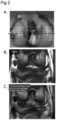
FIG. 2A is a diagram in which the positions of MRI imaging sections are indicated by five dotted lines on the India ink-stained image of the rabbit bothfemoral condyles 4 weeks after anterior cruciate ligament transection.FIG. 2B is a T2-weighted image (before administration of 17O-labeled water) on the imaging section corresponding to the central dotted line out of the five dotted lines inFIG. 2A, and FIG. 2C is a diagram in which a 17O concentration map of articular cartilage based on the T2-weighted image is fused with a proton density-weighted image (an anatomical image). -
FIG. 3A is a diagram indicating the positions of regions of interest (ROIs) analyzed in the T2-weighted image, andFIG. 3B is a graph indicating changes over time in the 17O concentration in the respective ROls. The phase on the horizontal axis corresponds to the number of times of imaging of MRI. The scan time is 3 minutes 39 seconds. - The description of the present invention described below may be based on representative embodiments or specific examples, but the present invention is not limited to such embodiments or specific examples. In the present specification, the upper limit value and the lower limit value of each numerical value range can be combined as desired. In the present specification, a numerical range represented using "to" or "-" means a range including numerical values at both ends thereof as an upper limit value and a lower limit value, unless otherwise noted.
- The present invention provides an MRI contrast agent for detecting cartilage damage, the MRI contrast agent including 17O-labeled water.
- 17O-labeled water is water containing a higher concentration of H2-17O than naturally occurring water does. It suffices if the concentration of H2-17O in 17O-labeled water is a concentration that can provide a sufficient concentration of H2-17O for detection by MRI at a site in need of a cartilage damage examination, but a higher concentration is preferred in order to reduce the volume of liquid to be administered to a subject. The H2-17O concentration in 17O-labeled water is, for example, 5 mol% or more, preferably 10 mol% or more, and more preferably 15 mol% or more. There is no limit to the upper limit of the H2-17O concentration in 17O-labeled water, which can theoretically be 100 mol%. The mol% representing the H2-17O concentration in 17O-labeled water is synonymous with atom% (the ratio of oxygen atoms).
- 17O-labeled water can be produced by methods known to those skilled in the art, such as the method described in
Japanese Patent Application Laid-open No. 2000-218134 - In addition to 17O-labeled water, the contrast agent can contain components such as isotonic agents such as sodium chloride, potassium chloride, glucose, D-sorbitol, and glycerin; preservatives such as benzyl alcohol, para-hydroxybenzoate, and chlorobutanol; antioxidants such as ascorbic acid, α-tocopherol, and sulfites; and buffers such as phosphates, carbonates, acetates, and citrates. The contrast agent containing such other components as exemplified above in addition to 17O-labeled water can be represented as a contrast composition.
- The contrast agent can be administered by any route of administration to a subject in need of a cartilage damage examination so long as the administered contrast agent is accessible to the cartilage in the subject's body, but local administration near the cartilage is preferred in order to perform the examination with a smaller amount of the contrast agent. When the cartilage to be examined is articular cartilage, the contrast agent is particularly preferably administered locally into a joint cavity.
- It suffices if the contrast agent is administered in a dose giving an H2-17O concentration around the cartilage, or an H2-17O concentration in the joint cavity in the case of the local administration to the joint cavity, for example, of 0.5 mol% or more and preferably 0.8 mol% or more, or an amount giving around 1 mol%, for example. The dose can be set as appropriate by those skilled in the art by considering factors such as an H2-17O concentration in the contrast agent, the volume of the joint cavity, and the route of administration.
- For the cartilage damage examination, the subject is subjected to MRI imaging of the cartilage before and after administration of the contrast agent, and MRI images are acquired. For the MRI imaging of cartilage, a direct method using the nuclear magnetic resonance phenomenon of 17O itself may be used, or an indirect method using a T2 shortening occurring in protons in H2-17O molecules may be used. The indirect method uses the nuclear magnetic resonance phenomenon of protons, thus allowing use of a proton MRI apparatus in current clinical use, and also allowing detection of 17O with higher sensitivity than that of the direct method. The following further describes a method of 17O detection with the indirect method as an example.
- In the indirect method, to detect the T2 shortening with high sensitivity, T2-weighted images (T2WI) are preferably acquired by employing a T2-weighted sequence by the fast spin echo method or the spin echo method, especially the fast spin echo method. Imaging parameters such as a time of repetition (TR) and a time of echo (TE) may be set as appropriate within ranges generally used to acquire T2-weighted images in accordance with an imaging sequence. In the case of the fast spin echo method, a favorable image can be acquired with one phase of 3 minutes 39 seconds with a TR of 1,600 ms, a TE of 129 ms, an FA of 150°, an echo train (ETL) of 12, and a number of times of addition of six times, for example.
- Imaging (scanning) is performed one or a plurality of times each before and after administration of the contrast agent. Imaging before administration of the contrast agent may be performed at any point in time before administration, and imaging after administration of the contrast agent may be performed at any point in time from immediately after administration to 3 days after administration. In a preferred embodiment, imaging before administration of the contrast agent is performed at any point in time from 30 minutes before administration to immediately before administration, and imaging after administration of the contrast agent is performed at any point in time from immediately after administration to 60 minutes after administration. In a more preferred embodiment, imaging before administration of the contrast agent is performed immediately before administration, and imaging after administration of the contrast agent is performed continuously from immediately after administration to 10 minutes after administration.
- Next, signal values (signal intensity) are acquired from the acquired MRI images by image analysis, and the signal values are compared between before and after administration of the contrast agent.
- In one embodiment, any region of interest (ROI) is set in a region corresponding to the cartilage in the MRI images, and signal values are acquired from respective pixels included in the ROI. Identification of the cartilage in the MRI images may be made with reference to other images such as proton density-weighted images. Comparison of the signal values between before and after administration of the contrast agent can be made using a value obtained by averaging the signal values of the respective pixels included in the ROI.
- In the indirect method, the T2 shortening occurs in protons in H2-17O molecules, and thus the signal values reduce. Thus, in an ROI in which the average of the signal values has reduced after administration compared to that before administration of the contrast agent, the H2-17O concentration has increased with the administration, and the cartilage contained in the ROI can be evaluated as having damage or being likely to have damage.
- In another embodiment, signal values are acquired from respective pixels in the MRI image, and the differences in the signal values between before and after administration of the contrast agent are displayed on a map. In a region in which the signal value has decreased with administration of the contrast agent displayed on the map, the H2-17O concentration has increased with the administration, and the cartilage contained in that region can be evaluated as having damage or being likely to have damage.
- Instead of comparing the signal values between before and after administration of the contrast agent as described above, a 17O concentration calculated from the signal values may be compared. The 17O concentration can be calculated based on the relation between the signal values and the 17O concentration obtained in advance. When the signal value is a signal value of the T2-weighted image, for example, the 17O concentration can be calculated using the following equation.

- In one embodiment, for example, an average 17O concentration in the ROI is calculated from the average of the signal values of the respective pixels included in the ROI, and the average 17O concentrations are compared between before and after administration of the contrast agent. When the 17O concentration in the ROI increases after administration, the cartilage contained in the ROI can be evaluated as having damage or being likely to have damage.
- In another embodiment, 17O concentrations are calculated from the signal values of the respective pixels in the MRI image, and the differences in the 17O concentrations between before and after administration of the contrast agent are displayed on a map. The cartilage contained in a region with an increased 17O concentration with administration of the contrast agent displayed on the map can be evaluated as having damage or being likely to have damage.
- When setting ROls in the MRI image, in addition to an ROI specifying a region to be examined (a test ROI), an ROI as a control (a control ROI) may be set in a normal cartilage part. The averages of the signal values or the 17O concentrations in each of the test ROI and the control ROI are compared between before and after administration of the contrast agent, and if the degree of a reduction in the signal values or the degree of an increase in the 17O concentration with administration is greater in the test ROI than in the control ROI, or if the rate of reduction in the signal values or rate of increase in the 17O concentration with administration is greater in the test ROI than in the control ROI, the cartilage contained in the test ROI can be evaluated as having damage or being likely to have damage.
- The above describes MRI imaging using the indirect method as an example. Also when using MRI imaging using the direct method, in which 17O itself is to be detected, cartilage damage can be examined in a similar way by acquiring signal values derived from 17O from MRI images and based on a change in the signal values or an increase in the 17O concentration calculated from the signal values. In this context, the change in the signal values is an increase in the signal values.
- Thus, the present invention provides a method for examining cartilage damage, the method including acquiring a signal value derived from 17O from MRI images of cartilage of a subject to which an MRI contrast agent including 17O-labeled water has been administered before and after administration of the contrast agent, comparing the signal values or 17O concentrations calculated from the signal values between before and after administration of the contrast agent, and determining a change in the signal value or an increase in the 17O concentration with administration of the contrast agent. The above method of examination can also be represented as a method for detecting cartilage damage or a method for evaluating the possibility of cartilage damage. The present invention also provides a method for collecting data for diagnosis of cartilage damage, the method including acquiring a signal value derived from 17O from MRI images of cartilage of a subject to which an MRI contrast agent including 17O-labeled water has been administered before and after administration of the contrast agent.
- The present invention also provides a method for diagnosing cartilage damage, the method including administering an MRI contrast agent including 17O-labeled water to a subject, acquiring MRI images of the subject's cartilage before and after administration of the contrast agent, acquiring a signal value derived from 17O from the MRI images, comparing the signal values or 17O concentrations calculated from the signal values between before and after administration of the contrast agent, determining a change in the signal value or an increase in the 17O concentration with administration of the contrast agent, and diagnosing that, when it is determined that the change in the signal value or the increase in the 17O concentration is present, the cartilage has damage or is likely to have damage.
- The present invention further provides a program for examining cartilage damage for causing a computer to execute processing to acquire a signal value derived from 17O from MRI images of cartilage of a subject to which an MRI contrast agent including 17O-labeled water has been administered before and after administration of the contrast agent, processing to optionally calculate a 17O concentration from the signal value, and processing to compare the signal values or the 17O concentrations calculated from the signal values between before and after administration of the contrast agent. This program can be used to perform the method of examination described above, and the details of each processing are as described in the description of the method of examination. A computer-readable storage medium storing therein the program is also provided by the present invention. The recording medium may be a portable medium such as a compact disc (CD), a digital versatile disc (DVD), or a USB flash drive or a storage device such as a hard disk incorporated in a computer. The program recorded on the recording medium may be for implementing all the functions described above or for implementing part of them. In the latter case, the functions described above can be implemented by a combination with a program already recorded on the computer.
- The present invention further provides a system for examining cartilage damage, the system including means for acquiring a signal value derived from 17O from MRI images of cartilage of a subject to which an MRI contrast agent including 17O-labeled water has been administered before and after administration of the contrast agent, means for optionally calculating a 17O concentration from the signal value, and means for comparing the signal values or the 17O concentrations calculated from the signal values between before and after administration of the contrast agent.
- The present invention further provides an image analysis apparatus including a signal value acquisition unit acquiring a signal value derived from 17O from MRI images of cartilage of a subject to which an MRI contrast agent including 17O-labeled water has been administered before and after administration of the contrast agent, a 17O concentration calculation unit optionally calculating a 17O concentration from the signal value, and a comparison unit comparing the signal values or the 17O concentrations calculated from the signal values between before and after administration of the contrast agent.
- The present invention further provides an MRI apparatus including a nuclear magnetic resonance imaging unit imaging cartilage of a subject to which an MRI contrast agent including 17O-labeled water has been administered, an image generating unit generating an MRI image from echo data acquired by the nuclear magnetic resonance imaging unit, a signal value acquisition unit acquiring a signal value derived from 17O from the MRI image, a 17O concentration calculation unit optionally calculating a 17O concentration from the signal value, and a comparison unit comparing the signal values or the 17O concentrations calculated from the signal values between before and after administration of the contrast agent.
- The details of the program, the system, the image analysis apparatus, and the MRI apparatus are as described in the description of the method of examination described above. The program, the storage medium, the system, the image analysis apparatus, and the MRI apparatus can be used in the implementation of the method of examination and the method of diagnosis described above.
- As described in examples below, after administration of 17O-labeled water, the 17O concentration promptly increases in a damaged cartilage region, whereas such a concentration increase does not occur in a normal cartilage region. Thus, according to the present invention, the contrast agent including 17O-labeled water is administered to a subject in need of a cartilage damage examination, and the change in the signal value derived from 17O in the cartilage is detected through MRI imaging, and thereby image diagnosis of cartilage damage can be performed.
- The increase in the 17O concentration is thought to be due to H2-17O molecules in the contrast agent penetrating into the cartilage from damaged sites of cartilage. The penetration of water molecules is considered possible even if the degree of cartilage damage is slight, and thus the present invention can detect even the slightest damage such as degeneration of the cartilage surface layer, which is an early lesion of osteoarthritis, which has been difficult to detect.
- The present invention is applied to a subject in need of a cartilage damage examination. Examples of the subject include mammals such as rodents including mice, rats, hamsters, guinea pigs, and rabbits; primates including humans, chimpanzees, and rhesus monkeys; domestic animals including pigs, cattle, goats, horses, and sheep; and pet animals including dogs and cats. The subject is preferably a human.
- The subject in need of a cartilage damage examination is a subject that has or may have some damage to the cartilage. Examples of the subject include a subject that has or may have a disease or symptom with cartilage damage or caused by cartilage damage. Examples of the disease or symptom include osteoarthritis (the knee joint, the elbow joint, the ankle joint, the shoulder joint, and the like), autoimmune arthritis such as rheumatoid arthritis, intra-articular fractures, meniscus injuries, knee ligament injuries, patellar dislocation, chronic knee disorders due to sports, intervertebral disc disorders, and osteochondritis dissecans (the knee joint, the elbow joint, the ankle joint, and the like).
- The following describes the present invention in more detail by the following examples, but the invention is not limited to these examples.
- An ACLT model was created using 14-week-old male Japanese white rabbits to induce cartilage damage in the knee joint. The rabbits were anesthetized intravenously with pentobarbital (30 mg/kg) and ketamine (50 mg/kg), followed by inhalation anesthesia with isoflurane (1 to 4%) for maintenance anesthesia. The anterior surface of the knee joint on the operative side was shaved, was disinfected with povidone-iodine, and then an analgesic (buprenorphine 0.02 mg/kg) was subcutaneously administered by injection. Skin incision of about 5 cm was placed on the medial side of the patella, and the joint capsule was incised in a similar position. The patella was dislocated laterally, and the knee joint was deeply flexed to expose the anterior cruciate ligament of the femoral intercondylar part, which was separated using a No. 11 scalpel. After washing the inside of the joint with a physiological saline solution, the joint capsule and the skin were sutured using 3-0 nylon thread. Postoperatively, euthanasia was performed if wound infection was observed or in the case of an inability to stand.
- It has been reported that in the ACLT model, degeneration of cartilage surface begins at 4 weeks postoperatively and occurs in the entire cartilage layer from 8 weeks postoperatively (Makoto Yoshioka et al. Characterization of a model of osteoarthritis in the rabbit knee. Osteoarthritis and Cartilage, 1996, 4 (2): 87-98), and thus five rabbits were slaughtered and euthanized with pentobarbital (200 mg/kg) each at 4 weeks and 8 weeks postoperatively, and cartilage damage was evaluated by MRI and macroscopically and histologically.
- Using a 3T-MRI apparatus (MAGNETOM Prisma, Siemens), imaging of the rabbit knee joint on the operative side was performed. The affected rabbit limb was disarticulated at the thigh and was fixed at the knee joint extension to avoid the influence of body movement during imaging. A 24G Surflo needle was inserted from the anterior surface of the knee joint through the patellar tendon and was indwelled in the joint. By cutting the locking connector side of an extension tube and coupling the lumen of the cut surface and a narrow polyethylene tube (PE50) to each other with curable resin (Shofu Quick Resin), a self-made administration route with a narrow lumen was created and was connected to the indwelled Surflo needle.
- To evaluate both femoral condyles, five slices were set as MRI imaging cross sections in such a manner that the most distal part of both femoral condyles was the central slice in a coronal section anteverted 45 degrees to the bone axis (indicated by the five dotted lines in
FIG. 2A ). First, a proton density-weighted image (PDWI) was taken as a reference image for identifying the articular cartilage under the following conditions: TE, 21 ms; TR, 1,600; FOV, 60 × 80 mm; matrix, 269 × 448; slice thickness, 2 mm; slice gap, 2.6 mm; number of excitations, 6; and scan time, 3 min 39 s. Subsequently, T2-weighted images using the 2D-fast spin echo method were successively taken under the following imaging conditions: TE, 129 ms; TR, 1,600; echo train length, 12; FOV, 60 × 80 mm; matrix, 269 × 448; slice thickness, 2 mm; slice gap, 2.6 mm; number of excitations, 6; scan time, 3 min 39 s; number of repetitions, 18; and total scan time, 65 min 42 s. The first three images out of 18 images taken consecutively were used as baseline images before administration. Immediately after the third imaging ended, 0.5 ml of a physiological saline solution containing a 20-mol% 17O-labeled water (Taiyo Nippon Sanso Corporation) was rapidly administered into the knee joint through the indwelled Surflo and was flushed with 0.5 ml of a physiological saline solution. - The articular cartilage in each slice section was identified from the proton density-weighted image, and a plurality of small regions of interest (ROIs) were set in the articular cartilage of both condyles. The 17O concentration at each point in time in each set ROI was calculated from the following equation using the signal value of the T2-weighted image, and changes over time in the 17O concentration in each ROI were compared with the histological grade of cartilage damage described below.

- After MRI, the rabbit knee joint was incised and expanded, and cartilage damage in the femoral condyle was macroscopically and histologically evaluated according to the MRI slice (Gabriel GN et al. Evaluation of multiphase implants for repair of focal osteochondral defects in goats. Biomaterials, 2000, 21 (24): 2561-74).
- In the macroscopic evaluation, cartilage damage was evaluated using Osteoarthritis Research Society International (OARSI) scoring (S. Laverty et al. The OARSI histopathology initiative - recommendations for histological assessments of osteoarthritis in the rabbit. Osteoarthritis and Cartilage, 2010, 18 Suppl 3: S53-S65) with Indian ink. An
OARSI score 2 or more, which shows accumulation of Indian ink on the joint surface, was defined as "macroscopic cartilage damage being present," while anOARSI score 1, which shows no accumulation, was defined as "macroscopic cartilage damage being absent." - In the histological evaluation, both femoral condyles were first fixed in 10% formalin for 24 hours and were then decalcified in formic acid with a ratio of formic acid 5 : formalin stock solution 1 : distilled water 15 for 20 days. Specimens were prepared and evaluated for three slices including, as a central slice, the most distal part of both femoral condyles in a coronal section anteverted 45 degrees to the bone axis so as to match the slice width of MRI and slices 2.6 mm anterior and posterior to the central slice. Safranin-O staining was performed, and the degree of cartilage damage was evaluated by OARSI grading (H.J. Mankin et al. Biochemical and metabolic abnormalities in articular cartilage from osteoarthritic human hips. II. Correlation of morphology with biochemical and metabolic data. J Bone Joint Surg, 1971, 53 (3): 523-37). An
OARSI grade 1 or more, which shows fibrosis of the cartilage surface layer, was defined as "histological cartilage damage being present," while an OARSI grade 0, which shows normal cartilage, was defined as "histological cartilage damage being absent." Scoring was performed twice independently by two examiners to evaluate inter-examiner errors and intra-examiner errors. - All animal experiments were performed according to a protocol approved by the Institutional Animal Care and Use Committee of the Graduate School of Medicine, Hokkaido University.
- For a representative case at 4 weeks postoperatively,
FIG. 1A is an appearance photograph after India ink staining. The macroscopic evaluation allowed observation of cartilage damage with anOARSI score 3 on the lateral side of the medial condyle and cartilage damage with anOARSI score 2 on the medial side of the lateral condyle.FIG. 1B and FIG. 1C illustrate an HE-stained image and a Safranin-O-stained image of the sliced specimen, respectively. The histological evaluation allowed observation of cartilage damage with an OARSI grade 3 (cracks from the cartilage surface layer to the intermediate layer are present, the black arrows in the drawings) on the lateral side of the medial condyle and cartilage damage with an OARSI grade 2 (cracks in the cartilage intermediate layer, the white arrows in the drawings) on the medial side of the lateral condyle in both the HE-stained image and the Safranin-O-stained image, which were consistent with the damage parts observed in the macroscopic evaluation. - For a representative case at 4 weeks postoperatively,
FIG. 2B illustrates a T2-weighted image of both femoral condyles in a coronal section at the central dotted line out of the five dotted lines illustrated inFIG. 2A, and FIG. 2C illustrates a 17O map obtained by calculating 17O concentrations from signal values of the respective pixels of a T2-weighted image taken about 11 minutes after administration of 17O-labeled water (the sixth imaging) and mapping them, superimposed on a proton density-weighted image. It was confirmed that the 17O concentration was higher in the region where cartilage damage was observed. - Two ROIs were set in each of a region corresponding to the damaged cartilage in the T2-weighted image and a region corresponding to the normal cartilage, and the 17O concentration was calculated based on the average of signal values in each ROI.
FIG. 3A illustrates the set positions of the ROIs, andFIG. 3B illustrates changes over time in the 17O concentration in the respective ROls.ROI 1 andROI 2, which were set in the damaged cartilage region, showed an increase in the 17O concentration immediately after administration of 17O-labeled water (the fourth imaging), whereasROI 3 andROI 4, which were set in the normal cartilage region, showed no tendency of an increase in the 17O concentration with administration of 17O-labeled water.
Claims (9)
- A magnetic resonance imaging (MRI) contrast agent for detecting cartilage damage, the MRI contrast agent comprising 17O-labeled water.
- A method for examining cartilage damage, the method comprising:acquiring a signal value derived from 17O from magnetic resonance imaging (MRI) images of cartilage of a subject to which an MRI contrast agent including 17O-labeled water has been administered before and after administration of the contrast agent;comparing the signal values or 17O concentrations calculated from the signal values between before and after administration of the contrast agent; anddetermining a change in the signal value or an increase in the 17O concentration with administration of the contrast agent.
- The method according to claim 2, wherein the cartilage is articular cartilage.
- The method according to claim 2 or 3, wherein the contrast agent is administered into a joint cavity of the subject.
- The method according to any one of claims 2 to 4, whereinthe MRI images are T2-weighted images taken with a proton MRI apparatus, andthe change in the signal value is a reduction in the signal value.
- A program for examining cartilage damage for executing:processing to acquire a signal value derived from 17O from magnetic resonance imaging (MRI) images of cartilage of a subject to which an MRI contrast agent including 17O-labeled water has been administered before and after administration of the contrast agent;processing to optionally calculate a 17O concentration from the signal value; andprocessing to compare the signal values or the 17O concentrations calculated from the signal values between before and after administration of the contrast agent.
- The program according to claim 6, wherein the cartilage is articular cartilage.
- The program according to claim 6 or 7, wherein the contrast agent is administered into a joint cavity of the subject.
- The program according to any one of claims 6 to 8, whereinthe MRI images are T2-weighted images taken with a proton MRI apparatus, andthe change in the signal value is a reduction in the signal value.
Applications Claiming Priority (2)
| Application Number | Priority Date | Filing Date | Title |
|---|---|---|---|
| JP2020180918A JP7624170B2 (en) | 2020-10-28 | 2020-10-28 | Contrast agent for detecting cartilage damage, and method and program for inspecting cartilage damage using said contrast agent |
| PCT/JP2021/039798 WO2022092192A1 (en) | 2020-10-28 | 2021-10-28 | Contrast agent for detecting cartilage damage, and method and program for detecting cartilage damage using said contrast agent |
Publications (2)
| Publication Number | Publication Date |
|---|---|
| EP4230227A1 true EP4230227A1 (en) | 2023-08-23 |
| EP4230227A4 EP4230227A4 (en) | 2025-06-18 |
Family
ID=81382662
Family Applications (1)
| Application Number | Title | Priority Date | Filing Date |
|---|---|---|---|
| EP21886309.0A Pending EP4230227A4 (en) | 2020-10-28 | 2021-10-28 | Contrast agent for detecting cartilage damage, as well as method and program for detecting cartilage damage using this contrast agent |
Country Status (4)
| Country | Link |
|---|---|
| US (1) | US20240066152A1 (en) |
| EP (1) | EP4230227A4 (en) |
| JP (1) | JP7624170B2 (en) |
| WO (1) | WO2022092192A1 (en) |
Family Cites Families (4)
| Publication number | Priority date | Publication date | Assignee | Title |
|---|---|---|---|---|
| JP4327287B2 (en) | 1999-01-29 | 2009-09-09 | 大陽日酸株式会社 | Method and apparatus for producing heavy oxygen water |
| JP2003102698A (en) | 2001-09-28 | 2003-04-08 | Nihon Medi Physics Co Ltd | Nuclear magnetic resonance imaging agent |
| JP2008239566A (en) * | 2007-03-28 | 2008-10-09 | Taiyo Nippon Sanso Corp | Drug for analysis of water transport function of membrane protein in living tissue |
| JP5958898B2 (en) | 2012-06-11 | 2016-08-02 | 学校法人 岩手医科大学 | Method for operating MRI apparatus in imaging of cerebral blood flow |
-
2020
- 2020-10-28 JP JP2020180918A patent/JP7624170B2/en active Active
-
2021
- 2021-10-28 US US18/250,591 patent/US20240066152A1/en active Pending
- 2021-10-28 EP EP21886309.0A patent/EP4230227A4/en active Pending
- 2021-10-28 WO PCT/JP2021/039798 patent/WO2022092192A1/en not_active Ceased
Also Published As
| Publication number | Publication date |
|---|---|
| US20240066152A1 (en) | 2024-02-29 |
| JP7624170B2 (en) | 2025-01-30 |
| EP4230227A4 (en) | 2025-06-18 |
| JP2022071772A (en) | 2022-05-16 |
| WO2022092192A1 (en) | 2022-05-05 |
Similar Documents
| Publication | Publication Date | Title |
|---|---|---|
| Pappas et al. | Magnetic resonance imaging of asymptomatic knees in collegiate basketball players: the effect of one season of play | |
| Gillis et al. | Magnetic resonance imaging of relative glycosaminoglycan distribution in patients with autologous chondrocyte transplants | |
| Ma et al. | Graft maturity of the reconstructed anterior cruciate ligament 6 months postoperatively: a magnetic resonance imaging evaluation of quadriceps tendon with bone block and hamstring tendon autografts | |
| D'ANJOU et al. | Osteophytosis, subchondral bone sclerosis, joint effusion and soft tissue thickening in canine experimental stifle osteoarthritis: comparison between 1.5 T magnetic resonance imaging and computed radiography | |
| Koff et al. | Correlation of meniscal T2* with multiphoton microscopy, and change of articular cartilage T2 in an ovine model of meniscal repair | |
| Multanen et al. | Reproducibility of imaging human knee cartilage by delayed gadolinium-enhanced MRI of cartilage (dGEMRIC) at 1.5 Tesla | |
| Hirvasniemi et al. | In vivo comparison of delayed gadolinium-enhanced MRI of cartilage and delayed quantitative CT arthrography in imaging of articular cartilage | |
| Xiao et al. | Tracking osteoarthritis progress through cationic nanoprobe-enhanced photoacoustic imaging of cartilage | |
| US20190159697A1 (en) | Method, system, and computer-accessible medium for assessment of glycosaminoglycan concentration in vivo by chemical exchange saturation transfer | |
| Pauvert et al. | MRI study of the ligamentization of ACL grafts in children with open growth plates | |
| Lin et al. | Differential regional perfusion of the human anterior cruciate ligament: quantitative magnetic resonance imaging assessment | |
| Shimodaira et al. | Effect of a new remnant-preserving technique with anatomical double-bundle anterior cruciate ligament reconstruction on MRI-based graft maturity: a comparison cohort study | |
| Deng et al. | Incomplete histologic healing and diminished biomechanical strength of meniscus-bone interface after medial meniscus posterior root transosseous repair in a goat model | |
| Miller | MR imaging of the knee | |
| US20240066152A1 (en) | Contrast agent for detecting cartilage damage and method and program for examining cartilage damage using said contrast agent | |
| Ley et al. | Evaluation of osteochondral sample collection guided by computed tomography and magnetic resonance imaging for early detection of osteoarthritis in centrodistal joints of young Icelandic horses | |
| Bischofberger et al. | Use of a 3-telsa magnet to perform delayed gadolinium-enhanced magnetic resonance imaging of the distal interphalangeal joint of horses with and without naturally occurring osteoarthritis | |
| Fu et al. | Anatomic ACL Reconstruction, An Issue of Clinics in Sports Medicine | |
| Accadbled et al. | Knee anterior laxity and graft maturation after transphyseal anterior cruciate ligament reconstruction | |
| Wang et al. | Research on quantitative measurement method of articular cartilage thickness change based on MR image | |
| Tanaka et al. | MRI analysis of the attachment of the anteromedial and posterolateral bundles of anterior cruciate ligament using coronal oblique images | |
| McCartney et al. | Use of low-field MRA to presurgically screen for medial meniscus lesions in 30 dogs with cranial cruciate deficient stifles | |
| Terracciano et al. | Quantitative high-resolution 7T MRI to assess longitudinal changes in articular cartilage after anterior cruciate ligament injury in a rabbit model of post-traumatic osteoarthritis | |
| Tomura et al. | Walking speed is associated with MRI-detected structural changes of the knee osteoarthritis in healthy elderlies-the bunkyo health study | |
| Bouchgua et al. | 311 BONE MINERAL DENSITY ASSESSED BY COMPUTED TOMOGRAPHY IN AN IN VIVO RABBIT MODEL OF OSTEOARTHRITIS |
Legal Events
| Date | Code | Title | Description |
|---|---|---|---|
| STAA | Information on the status of an ep patent application or granted ep patent |
Free format text: STATUS: THE INTERNATIONAL PUBLICATION HAS BEEN MADE |
|
| PUAI | Public reference made under article 153(3) epc to a published international application that has entered the european phase |
Free format text: ORIGINAL CODE: 0009012 |
|
| STAA | Information on the status of an ep patent application or granted ep patent |
Free format text: STATUS: REQUEST FOR EXAMINATION WAS MADE |
|
| 17P | Request for examination filed |
Effective date: 20230516 |
|
| AK | Designated contracting states |
Kind code of ref document: A1 Designated state(s): AL AT BE BG CH CY CZ DE DK EE ES FI FR GB GR HR HU IE IS IT LI LT LU LV MC MK MT NL NO PL PT RO RS SE SI SK SM TR |
|
| DAV | Request for validation of the european patent (deleted) | ||
| DAX | Request for extension of the european patent (deleted) | ||
| A4 | Supplementary search report drawn up and despatched |
Effective date: 20250516 |
|
| RIC1 | Information provided on ipc code assigned before grant |
Ipc: G01R 33/56 20060101ALI20250512BHEP Ipc: A61P 19/02 20060101ALI20250512BHEP Ipc: A61B 5/055 20060101ALI20250512BHEP Ipc: A61B 5/00 20060101ALI20250512BHEP Ipc: A61K 49/06 20060101AFI20250512BHEP |