EP4224420A1 - A computer-implemented method for determining scar segmentation - Google Patents
A computer-implemented method for determining scar segmentation Download PDFInfo
- Publication number
- EP4224420A1 EP4224420A1 EP22155242.5A EP22155242A EP4224420A1 EP 4224420 A1 EP4224420 A1 EP 4224420A1 EP 22155242 A EP22155242 A EP 22155242A EP 4224420 A1 EP4224420 A1 EP 4224420A1
- Authority
- EP
- European Patent Office
- Prior art keywords
- contrast agent
- dose
- scar
- segmentation
- neural network
- Prior art date
- Legal status (The legal status is an assumption and is not a legal conclusion. Google has not performed a legal analysis and makes no representation as to the accuracy of the status listed.)
- Granted
Links
Images


Classifications
-
- G—PHYSICS
- G06—COMPUTING OR CALCULATING; COUNTING
- G06T—IMAGE DATA PROCESSING OR GENERATION, IN GENERAL
- G06T7/00—Image analysis
- G06T7/10—Segmentation; Edge detection
- G06T7/11—Region-based segmentation
-
- A—HUMAN NECESSITIES
- A61—MEDICAL OR VETERINARY SCIENCE; HYGIENE
- A61B—DIAGNOSIS; SURGERY; IDENTIFICATION
- A61B5/00—Measuring for diagnostic purposes; Identification of persons
- A61B5/05—Detecting, measuring or recording for diagnosis by means of electric currents or magnetic fields; Measuring using microwaves or radio waves
- A61B5/055—Detecting, measuring or recording for diagnosis by means of electric currents or magnetic fields; Measuring using microwaves or radio waves involving electronic [EMR] or nuclear [NMR] magnetic resonance, e.g. magnetic resonance imaging
-
- G—PHYSICS
- G06—COMPUTING OR CALCULATING; COUNTING
- G06V—IMAGE OR VIDEO RECOGNITION OR UNDERSTANDING
- G06V10/00—Arrangements for image or video recognition or understanding
- G06V10/20—Image preprocessing
- G06V10/26—Segmentation of patterns in the image field; Cutting or merging of image elements to establish the pattern region, e.g. clustering-based techniques; Detection of occlusion
-
- G—PHYSICS
- G06—COMPUTING OR CALCULATING; COUNTING
- G06V—IMAGE OR VIDEO RECOGNITION OR UNDERSTANDING
- G06V10/00—Arrangements for image or video recognition or understanding
- G06V10/70—Arrangements for image or video recognition or understanding using pattern recognition or machine learning
- G06V10/82—Arrangements for image or video recognition or understanding using pattern recognition or machine learning using neural networks
-
- G—PHYSICS
- G06—COMPUTING OR CALCULATING; COUNTING
- G06T—IMAGE DATA PROCESSING OR GENERATION, IN GENERAL
- G06T2207/00—Indexing scheme for image analysis or image enhancement
- G06T2207/10—Image acquisition modality
- G06T2207/10072—Tomographic images
- G06T2207/10081—Computed x-ray tomography [CT]
-
- G—PHYSICS
- G06—COMPUTING OR CALCULATING; COUNTING
- G06T—IMAGE DATA PROCESSING OR GENERATION, IN GENERAL
- G06T2207/00—Indexing scheme for image analysis or image enhancement
- G06T2207/10—Image acquisition modality
- G06T2207/10072—Tomographic images
- G06T2207/10088—Magnetic resonance imaging [MRI]
-
- G—PHYSICS
- G06—COMPUTING OR CALCULATING; COUNTING
- G06T—IMAGE DATA PROCESSING OR GENERATION, IN GENERAL
- G06T2207/00—Indexing scheme for image analysis or image enhancement
- G06T2207/10—Image acquisition modality
- G06T2207/10116—X-ray image
-
- G—PHYSICS
- G06—COMPUTING OR CALCULATING; COUNTING
- G06T—IMAGE DATA PROCESSING OR GENERATION, IN GENERAL
- G06T2207/00—Indexing scheme for image analysis or image enhancement
- G06T2207/20—Special algorithmic details
- G06T2207/20081—Training; Learning
-
- G—PHYSICS
- G06—COMPUTING OR CALCULATING; COUNTING
- G06T—IMAGE DATA PROCESSING OR GENERATION, IN GENERAL
- G06T2207/00—Indexing scheme for image analysis or image enhancement
- G06T2207/20—Special algorithmic details
- G06T2207/20084—Artificial neural networks [ANN]
-
- G—PHYSICS
- G06—COMPUTING OR CALCULATING; COUNTING
- G06T—IMAGE DATA PROCESSING OR GENERATION, IN GENERAL
- G06T2207/00—Indexing scheme for image analysis or image enhancement
- G06T2207/20—Special algorithmic details
- G06T2207/20092—Interactive image processing based on input by user
- G06T2207/20104—Interactive definition of region of interest [ROI]
-
- G—PHYSICS
- G06—COMPUTING OR CALCULATING; COUNTING
- G06T—IMAGE DATA PROCESSING OR GENERATION, IN GENERAL
- G06T2207/00—Indexing scheme for image analysis or image enhancement
- G06T2207/30—Subject of image; Context of image processing
- G06T2207/30004—Biomedical image processing
- G06T2207/30048—Heart; Cardiac
-
- G—PHYSICS
- G06—COMPUTING OR CALCULATING; COUNTING
- G06V—IMAGE OR VIDEO RECOGNITION OR UNDERSTANDING
- G06V2201/00—Indexing scheme relating to image or video recognition or understanding
- G06V2201/03—Recognition of patterns in medical or anatomical images
Definitions
- the invention relates to a method for training an artificial neural network for determining scar segmentation, in particular of a myocardial scar, a computer-implemented method for determining scar segmentation, an imaging method for determining a scar area, as well as a corresponding computer program, non-transitory computer-readable medium, and system.
- contrast agents are used in medical imaging, in particular in magnetic resonance imaging (MRI), in order to accentuate different areas or types of tissue in a medical image of a subject or object.
- MRI magnetic resonance imaging
- structures and functions may be traced more easily and/or efficiently.
- LGE gadolinium enhancement
- GBCA gadolinium-based contrast agents
- LGE imaging is currently performed with standard doses, which are quite high, wherein the exact dose depends on the choice of the agent and other factors.
- the enhanced region is then segmented using one of several established methods. In many cases, assessment of the extent of LGE in these patients is used as a key biomarker for treatment and prognosis.
- a method for training an artificial neural network for determining scar segmentation, in particular segmentation of a myocardial scar, on contrast agent enhanced medical images with reduced contrast agent dose comprises:
- the artificial neural network may be a convolutional, in particular fully convolutional, neural network.
- the neural network may comprise multiple layers. Training the neural network may comprise feeding the input training data into the neural network and comparing the output of the neural network with the output training data. During training, based on the comparison, weights of the neural network may be adjusted trying to achieve a closer match between the output of the neural network and the provided output training data.
- the neural network may be trained thus to determine scar segmentation on images with reduced contrast agent in a way or similarly to a way that the scar would be segmented on standard full-dose images using a standard segmentation method. In this context, similarly may mean that, during training, the weights of the neural network are adjusted such that the output of the neural network approaches the full-dose output training data as closely as possible.
- the training may be done by a back-propagation algorithm.
- the medical images of the input training data namely the medical images of the first set, as well as the medical images on which the output training data is based, i.e. the medical images of the second set, are in particular contrast agent enhanced. Due to the contrast agent, in particular the visibility of scar tissue on the medical images is enhanced.
- the first set of medical images and the second set of medical images may be a set of two-dimensional (2D) images, for example represented by a 2D-matrix of grayscale values.
- the first and second set of medical images may each be a set of three-dimensional (3D) images.
- the individual medical images of the first set and/or of the second set may be taken from different subjects, in particular patients, but from the same or corresponding part, organ and/or area of the different subjects.
- each medical image of the first set there is a corresponding medical image of the second set from the same subject and object.
- the medical images of each pair may be measured one after the other, in particular under the same or similar circumstances apart from the amount of contrast agent administered.
- the pairs may be used together to train the artificial neural network, i.e., during training, the output of the neural network corresponding to the input of the first of the pair of medical images may be compared to the second of the pair of medical images.
- the images of the first and second set are preferably registered with each other and/or have the same dimensions, i.e. pixel size, such as 256 x 256 pixel for a 2D image.
- the medical images may be magnetic resonance (MR) images and the contrast agent may be an MR contrast agent.
- the medical images may be reconstructed from MR data, in particular as magnitude inversion recovery (MagIR) and/or phase-sensitive inversion recovery (PSIR images).
- the MR data may in principle be obtained by any MR imaging sequence, for example by a gradient echo, spin-echo or fast spin-echo sequence, wherein inversion recovery are preferred, because of the strong T1-weighting.
- the object may be part of a human or animal patient, in particular comprising scar tissue, such as an organ, part of an organ or an area comprising the organ.
- the medical images may comprise the heart or part of the heart, in particular the myocardium.
- the scar or scars to be segmented may be myocardial scar tissue, for example scar tissue resulting from infarction.
- the scar segmentation mask may be data and/or an image denoting the area of scar tissue on a medical image and/or within an area of the medical image.
- the output training data namely the reference scar segmentation masks, may be determined by a state-of-the-art scar segmentation method. The determining of the reference scar segmentation masks does not have to be part of the inventive method, e.g. existing data may be used for this method. However, optionally, determining the reference scar segmentation mask on the second set of medical images may be an additional step of the method applied prior to the step of receiving output training data.
- the amount of contrast agent of the full-dose may for example depend on the type of contrast agent that is used, the object to be observed/segmented and/or the individual patient, e.g. the patient's size and/or weight.
- the standard full-dose of contrast agent may in particular be determined by the manufacturer's recommendation and/or the amount that is usually administered for a specific examination.
- the low-dose of contrast agent is lower than the standard full-dose, in particular lower than the manufacturer's recommendation and/or lower than the amount that is usually administered for the specific examination.
- the standard full-dose may correspond to 0.10 to 0.20 mmol/kg of Gadolinium (Gd)-based contrast agent (GBCA), preferably 0.12 to 1.15 mmol/kg, where kg refers to the patient weight, and the dose in mmol refers to the GBCA, wherein usually each Gd atom is included in a ligand.
- Gd Gadolinium
- GBCA Gadolinium-based contrast agent
- the inventive method may be carried out on a processing unit, such as a GPU (graphics processing unit), CPU (central processing unit) or other digital data processing equipment.
- a processing unit such as a GPU (graphics processing unit), CPU (central processing unit) or other digital data processing equipment.
- the method may be executed by a processing unit which is part of or connected to a corresponding imaging system, e.g. an MRI system, used in diagnostic imaging.
- the invention may allow to provide a medical imaging and evaluation system which can quantify the scar extent on medical images, wherein reduced amounts of contrast agent have been applied. I.e., after training the artificial neural network according to the inventive method, only a low-dose medical image, which would otherwise not be sufficient to produce reliable results, is required for scar segmentation. Hence, patient safety may be increased and material costs may be saved since less contrast agent needs to be administered.
- the contrast agent is a gadolinium-based contrast agent (GBCA), wherein the medical images are in particular acquired via magnetic resonance (MR) imaging with late gadolinium enhancement (LGE).
- GBCA may typically decrease the T1 relaxation time in MR imaging.
- areas where the GBCA accumulates may appear brighter in T1-weighted sequences, such as T1 weighted inversion recovery sequences.
- GBCA may typically accumulate in scar regions and be present in these regions for a longer time than in the area around the scar tissue.
- late gadolinium enhancement wherein the measurement is taken a certain time after the administering of GBCA may enhance the scar tissue area in the medical image.
- LGE is in particular beneficial for a segmentation of myocardial scars. Therein, assessment of the extent of LGE in these patients may be key biomarker for treatment and prognosis. It has turned out that the inventive method works well with LGE on myocardial scars. Hence, the inventive method using LGE may be applied to examine patients with chronic myocardial infarction.
- the GBCA may be injected about 2 to 20 minutes, preferably 3 to 5 minutes, before imaging. It has turned out that the method may work particularly well, when imaging is carried out 3 to 5 minutes after the administering of the GBCA.
- the acquired data may for example be reconstructed as magnitude inversion recovery (MagIR) and/or phase-sensitive inversion recovery (PSRI).
- the reference scar segmentation masks are determined via a full width at half maximum (FWHM) method on the second set of medical images.
- a reference area may be chosen in the medical image, automatically or, preferably, manually.
- the reference area may be an area of the enhanced scar tissue area, in particular an area where the scar tissue is clearly recognizable, an area that represents the contrast of the scar tissue area particularly well and/or an area without significant imaging artifacts.
- the maximum signal e.g. per pixel or per average of a cluster of several adjacent pixels, is preferably taken as reference.
- a scar segmentation mask is an image having one value (e.g. 1) at the pixels considered to be scar tissue and another value (e.g. 0) at all other pixels.
- a binary image It may have the same dimensions as the corresponding image from the second set.
- the FWHM method may be carried out manually, automatically or partially automatically. For example, some steps, such as choosing the reference area and/or choosing a general area within the medical image where the FWMH for scar segmentation may be applied, may be carried out manually, while some other steps, e.g. determining the maximum signal within the reference area and/or searching for pixels that have a value higher than a fraction of the reference value, i.e. that are considered scar tissue, may be carried out automatically.
- a reference scar segmentation mask created by the FWHM method has turned out to be a particularly good basis for training the artificial neural network.
- the scar segmentation of the second set of medical images may be carried out completely manually based on the visual impression of an expert of the medical image and/or based on taking a reference standard deviation within the reference area and determining scar tissue by assuming a multiple of the reference standard deviation.
- the low-dose comprises 20% to 90%, preferably 50% to 75%, more preferably 60% to 70% of the amount of contrast agent of the full-dose.
- a dose of 50% to 75% may provide a significant reduction of applied contrast agent, while at the same time the reliability of scar segmentation of the thus trained neural network may be in about the same order of magnitude as in the case of a scar segmentation according to the state of the art for a full-dose medical image.
- a Dice score of up to 0,776 could be achieved, wherein the Dice score (also called S ⁇ rensen-Dice coefficient) is a statistic used to gauge the similarity of two samples - in this case the segmentation masks obtained from full-dose and from low-dose medical images, respectively.
- the Dice score also called S ⁇ rensen-Dice coefficient
- borders and/or contours of the objects to be segmented are provided on the first set of medical images, wherein the determination of the scar segmentation by the artificial neural network is restricted to said borders.
- the borders may in particular be provided manually, e.g. by an experienced reader/clinician.
- the quality of the scar segmentation of the reduced contrast agent medical images via the trained neural network may be increased noticeable by applying this step, in particular by also applying this step correspondingly when using the trained neural network for scar segmentation.
- a U-Net based artificial neural network in particular with Batch Normalization and one or several ReLU activation functions is used.
- the Batch Normalization may stabilize the training.
- a loss function preferably a Dice loss function
- the Dice loss function may be used for the training, namely for comparing the output of the artificial neural network with the output training data.
- another loss function may be applied, such as based on a cross entropy loss.
- the U-Net may be optimized using an Adam optimizer.
- another optimization algorithm preferably based on gradient descent, may be used as well. For example, a learning rate of 10-4 has worked well.
- the artificial neural network may comprise copying operations, wherein states may be copied from earlier layers to later layers and then concatenated, thereby skipping layers in between.
- the U-Net may comprise four levels and/or three pooling operations. It has turned out that this configuration may be efficient for the purpose of training and utilizing the neural network for scar segmentation with reduced contrast agent.
- the U-net may be based on the teachings in the publication of Ronneberger et al. in "U-Net: Convolutional networks for biomedical image segmentation", Medical image computing and computer-assisted intervention - MICCAI 2015. 2015; doi: 10.1007/978-3-319-24574-4_28 .
- the input training data and/or the output training data comprise real medical images and simulated medical images.
- the simulated medical images may for example be created by an imaging physics simulator, in particular an MR physics simulator.
- an imaging physics simulator in particular an MR physics simulator.
- a virtual phantom such as the XCAT virtual phantom as described by Segars et al. in "4D XCAT phantom for multimodality imaging research", Medical Physics, 2010; 37:4902-4915. doi: 10.1118/1.3480985 , may be used.
- the simulated medical images may allow to efficiently train the neural network, even if only a small number of real medical images is available.
- the simulated images may be created with varying amounts and/or types of low-dose contrast agent, e.g. to train the neural network to be more flexible with respect to the amount and/or type of contrast agent.
- the number of real medical images may be increased by oversampling and/or augmentation comprising horizontal flipping, vertical flipping, rotation, translation, scaling, and/or blurring.
- a real image may be modified to create another "real" image to be used for training.
- This augmentation may help to train the neural network to be more robust and reliable with respect to varying imaging conditions and/or parameters.
- the number of real images may be increased such that they appear equally or about equally often as simulated images. For example, the weighting or impact of the real medical images on the training may thus be greater than that of the simulated medical images.
- the method comprises the additional step of performing an image registration of the first set of medical images and the second set of medical images in order to transform them into a common coordinate system.
- An image registration may for example improve the comparing of output training data to the output of the neural network during training.
- the image registration may be performed based on the borders of the objects to be segmented as described above.
- the method including the image registration may thus be more reliable and/or more focused on the purpose of scar segmentation.
- a low-dose medical image may be acquired after the application of a first dose of contrast agent, e.g. GBCA, corresponding to the low-dose.
- the medical image may, for example, be acquired 3-5 minutes after the injection of the contrast agent.
- the additional dose of contrast agent may be applied to accumulate a full dose of contrast agent.
- the second, full-dose, image may be acquired about 15-20 minutes after the first injection.
- the amount of contrast agent for the training data can thus be kept low and the comparability of the medical images from the first and second set may be improved, therefore improving the efficiency of the training.
- these steps may not be necessary, e.g. if corresponding images are already available.
- a computer-implemented method for determining scar segmentation, in particular segmentation of a myocardial scar comprises the following steps:
- the ratio of the amount of the low-dose to the amount of the standard full dose may be the same or in the same order of magnitude and/or range as the ratio that was used during training the neural network.
- this method may allow to use less contrast agent for the purpose of scar segmentation while still achieving a comparable reliability of the scar segmentation.
- the neural network in particular if trained as described herein, may be a key algorithm to achieve this.
- the described method may thus provide a scar segmentation for low-dose medical images, wherein the low-dose is in particular so low that a reliable scar segmentation with a method according to the state of the art would not be possible in a satisfactory and/or reliable way.
- the method comprises the additional step of determining borders of the objects to be segmented, in particular myocardial borders, on the medical image prior to determining the scar segmentation mask, wherein the determination of the scar segmentation by the artificial neural network is restricted to said determined borders. Determining the borders may be carried out before feeding the medical image to the neural network.
- the borders may be determined in the same way as determining the borders for training the neural network, as described above.
- the quality and/or reliability may be increased noticeably, when applying this additional step.
- the contrast agent is a gadolinium-based contrast agent
- the medical image has been in particular acquired via MR imaging with late gadolinium enhancement.
- a U-Net based artificial neural network in particular with Batch Normalization, and/or one or several ReLU activation functions is/are used.
- an imaging method for determining a scar area, in particular of a myocardial scar, of an object comprises the following steps:
- the trained artificial neural network is trained for scar segmentation, in particular segmentation of a myocardial scar, on contrast agent enhanced medical images with reduced contrast agent dose.
- the trained artificial neural network may in particular be trained as described herein.
- the contrast agent is a gadolinium-based contrast agent (GBCA).
- GBCA gadolinium-based contrast agent
- the medical image may have been acquired via MR imaging with late gadolinium enhancement.
- the GBCA may be injected about 2 to 20 minutes, preferably 3 to 5 minutes, before imaging. It has turned out that the method may work particularly well, when imaging is carried out 3 to 5 minutes after the administering of the GBCA.
- the acquired data may for example be reconstructed as magnitude inversion recovery (MagIR) and/or phase-sensitive inversion recovery (PSRI).
- MagIR magnitude inversion recovery
- PSRI phase-sensitive inversion recovery
- the low-dose comprises 20% to 90%, preferably 50% to 75%, more preferably 60% to 70% of the amount of contrast agent of the full-dose.
- the method of the invention may include the step of applying the low-dose of contrast agent.
- a U-Net based artificial neural network in particular with Batch Normalization, and/or one or several ReLU activation functions is/are used.
- a U-Net based artificial neural network as described above may be used.
- the imaging method comprises the additional step of determining borders of the objects to be segmented, in particular myocardial borders, on the medical image prior to determining the scar segmentation mask, wherein the determination of the scar segmentation by the artificial neural network is restricted to said determined borders.
- This additional step may in particular be carried out in between the steps of acquiring the medical image of the object and determining the scar segmentation mask and may be carried out as described above.
- a computer program comprises instructions which, when the program is executed by a processing unit of a medical imaging device, in particular a magnetic resonance imaging device, causes the processing unit to carry out the method or one of the methods as described herein. All features and advantages of the method for training an artificial neural network, the computer-implemented method for determining scar segmentation, and the imaging method for determining a scar area may be adapted to the computer program and vice versa.
- a non-transitory computer-readable medium having stored thereon the computer program as described above is provided.
- the computer-readable medium may be any digital storage medium, for example a hard disk, a server, a cloud, a computer, an optical or a magnetic digital storage medium, a CD-ROM, an SSD-card, an SD-card, a DVD or a USB or other memory stick. All features and advantages of the method for training an artificial neural network, the computer-implemented method for determining scar segmentation, the imaging method for determining a scar area, and the computer program may be adapted to the non-transitory computer-readable medium and vice versa.
- a system for determining a scar area, in particular of a myocardial scar, of an object.
- the system comprises
- the processing unit may be a computer or may be part of a computer.
- the computer may be a PC, a server, a console of an MRI apparatus.
- the computer may also be a mobile device, such as a laptop, tablet computer or mobile phone. All features and advantages of the method for training an artificial neural network, the computer-implemented method for determining scar segmentation, the imaging method for determining a scar area, the computer program, and the non-transitory computer-readable medium may be adapted to the system and vice versa.
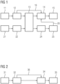
- Figure 1 shows a flow diagram of a method for training an artificial neural network for determining scar segmentation according to the invention.
- the neural network may preferably be a fully convolutional neural network.
- the neural network may in particular be trained to segment myocardial scar on images with reduced gadolinium-based contrast agent (GBCA) dose in the way that the scar would be segmented on standard full-dose images using a standard segmentation method, e.g. a FWHM method.
- GBCA gadolinium-based contrast agent
- a first set of medical images 5 is acquired after applying a low-dose of contrast agent.
- the images from the first set may in particular be low low-dose motion corrected late gadolinium enhanced (LGE) images acquired via magnetic resonance imaging.
- LGE low low-dose motion corrected late gadolinium enhanced
- a second set of medical images 6 is acquired after applying a standard full-dose of contrast agent.
- the images from the second set may in particular be reference full-dose motion corrected LGE images acquired via magnetic resonance imaging.
- the low-dose of contrast agent comprises less contrast agent than the standard full-dose of contrast agent.
- an additional dose of contrast agent may be applied such that the low-dose and the additional dose add up to the full-dose which is then used for acquiring images of the second.
- the low-dose may comprise 0.1 mmol/kg of contrast agent and the imaging may be timed to take place about 3-5 minutes after the administering of the contrast agent.
- the additional dose may comprise 0.05 mmol/kg of contrast agent and the imaging of the image from the second set of images may take place about 15-20 minutes in total after administering the low dose, i.e. the first dose.
- the medical images 5, 6 of the first and second set may be supplemented with simulated images. Oversampling and augmentation may be applied to the real patients' images, in particular such that they occur equally often as the simulated images during training. Augmentation may for example include horizontal and vertical flipping, rotation, translation, scaling, and blurring. Afterwards, borders or contours of the objects to be segmented are provided both on the images from the first set (step 12) and the images from the second set (step 22).
- the images may have the purpose to examine patients with chronic myocardial infarction.
- the borders may be myocardial borders.
- an image registration of each image from the first set of images together witch each corresponding image from the second set of images is performed in order to transform the images into a common coordinate system.
- the corresponding images for each patient are transformed into a common coordinate system.
- the image registration may in particular be based on the borders of the objects to be segmented.
- the first set of images is input into a neural network 14, in particular a convolutional neural network. The neural network predicts a scar segmentation mask 15, while being restricted to the determined borders of the object 16 as determined in step 12.
- the scar area of the images from the second set of images is determined with a standard method, such as the FWHM method.
- a standard method such as the FWHM method.
- reference scar segmentation masks are created 25.
- the reference scar segmentation masks are compared to the scar segmentation masks 15 predicted by the neural network and the weights of the neural network are adjusted accordingly (step 20), i.e. the reference scar segmentation masks 25 serve as ground truth 20 during training of the neural network 14.
- Figure 2 shows a flow diagram of an imaging method for determining a scar area according to the invention.
- the imaging method may in particular have corresponding details as the method for training shown in figure 1 .
- a medical image 5 is acquired after applying a low-dose of contrast agent.
- the low-dose of contrast agent comprises less contrast agent than a standard full-dose of contrast agent.
- borders or contours 32 of the objects to be segmented are provided on the image.
- the borders may for example be myocardial borders of a myocardial scar.
- the image is input into a neural network 34, in particular a convolutional neural network.
- the neural network 34 may in particular be a neural network that is trained as shown in figure 1 .
- the neural network predicts a scar segmentation mask 35, while being restricted to the determined borders 32 of the object, as indicated by arrow 36.
- the output of the imaging method is thus a scar segmentation mask 44, for example a segmentation of myocardial scar, that is based on imaging with a reduced amount of contrast agent but has a similar reliability as a scar segmentation based on a standard full-dose of contrast agent.
- Figure 3 shows an exemplary low-dose image 5 (left) and an exemplary full-dose image 6 (right) where myocardial borders have been drawn.
- the myocardium is thereby contoured by inner borders 42 and outer borders 41 in each image.
- a reference area 43 is drawn within the myocardial borders 41, 42.
- the maximum signal e.g. per pixel, is taken as reference value for the FWHM method for the full-dose image (right). All pixels within the myocardial borders that have a value higher than half the reference value are considered to be scar tissue and are segmented.
- the myocardial borders 41, 42 are also used in the neural network to restrict the determination of the scar segmentation to the area within these borders 41, 42.
- LGE gadolinium enhanced
- a low-dose motion corrected (MOCO) LGE image in short axis position was acquired after application of a first bolus of gadolinium-based contrast agent (GBCA) (0.1 mmol/kg Gadoteridol, imaged ⁇ 3-5 minutes post-injection).
- GBCA gadolinium-based contrast agent
- a reference full-dose MOCO LGE image was obtained after additional injection of GBCA (0.05 mmol/kg Gadoteridol injected immediately after low-dose LGE, imaged ⁇ 15-20 minutes after first injection).
- MagnIR magnitude inversion recovery
- PSIR phase-sensitive inversion recovery
- the data set was supplemented with simulated images using an MR physics simulator and a virtual phantom, namely the XCAT virtual phantom as described by Segars et al. in "4D XCAT phantom for multimodality imaging research", Medical Physics, 2010; 37:4902-4915, doi: 10.1118/1.3480985 .
- Oversampling and augmentation were applied to real patients' images such that they occurred equally often as simulated images during training. Augmentation included horizontal and vertical flipping, rotation, translation, scaling, and blurring.
- Image registration was performed based on the contours as described above. Images were cropped to 128x128 pixels around the center of the left ventricle.
- a 2-D U-Net with Batch Normalization, ReLU activation functions, and the Dice loss function was trained to segment the scarred area on the low-dose images.
- the U-Net contained four levels, i.e. three pooling operations, and was optimized using the Adam optimizer and a learning rate of 10-4. After inference, the network output was restricted to lie within the previously contoured myocardium. Of the 23 patients, ten were used for training the network (in addition to 340 simulated images), five for validation, and eight for testing. A dice score of unseen test data for individual images reached values up to 0.777. Predicted and ground-truth scar sizes, where correlated with a Pearson's r of 0.87. The trained neural network was able to locate and estimate the size of myocardial scars.
- Figure 4 shows an exemplary low-dose LGE image 5 (top) with a corresponding scar segmentation mask 44 (bottom) from a test data set.
- Figure 5 shows a U-Net based neural network architecture according to the invention.
- the numbers above the layers indicate the depths of the layers.
- Arrows pointing downwards denote max pooling
- arrows pointing upwards denote an upsampling followed by a convolution
- arrows pointing to the right denote a convolution, Batch Normalization and a rectified linear unit (ReLU).
- the dashed arrows denote copying, wherein states are to be copied from earlier layers to later layers and then concatenated thereby skipping layers in between. Thereby, the empty boxes denote copied states.
- Figure 6 shows a system 1 for determining a scar area according to the invention.
- the system 1 comprises a first interface 2 configured for receiving a medical image 5 of an object to be segmented acquired after an application of a low-dose of contrast agent.
- the system 1 further comprises a processing unit 3 configured for determining a scar segmentation mask by applying a trained artificial neural network and a second interface 4, configured for outputting the scar segmentation mask 44.
- the second interface 4 is a screen, however the second interface 4 may also be a data interface configured to transfer the scar segmentation mask 44 as data.
Landscapes
- Engineering & Computer Science (AREA)
- Health & Medical Sciences (AREA)
- Physics & Mathematics (AREA)
- Theoretical Computer Science (AREA)
- General Physics & Mathematics (AREA)
- Life Sciences & Earth Sciences (AREA)
- Computer Vision & Pattern Recognition (AREA)
- Nuclear Medicine, Radiotherapy & Molecular Imaging (AREA)
- General Health & Medical Sciences (AREA)
- Medical Informatics (AREA)
- Evolutionary Computation (AREA)
- Multimedia (AREA)
- Pathology (AREA)
- Radiology & Medical Imaging (AREA)
- Animal Behavior & Ethology (AREA)
- Molecular Biology (AREA)
- Public Health (AREA)
- Veterinary Medicine (AREA)
- Heart & Thoracic Surgery (AREA)
- Biomedical Technology (AREA)
- Biophysics (AREA)
- Surgery (AREA)
- High Energy & Nuclear Physics (AREA)
- Artificial Intelligence (AREA)
- Computing Systems (AREA)
- Databases & Information Systems (AREA)
- Software Systems (AREA)
- Magnetic Resonance Imaging Apparatus (AREA)
- Apparatus For Radiation Diagnosis (AREA)
Abstract
Description
- The invention relates to a method for training an artificial neural network for determining scar segmentation, in particular of a myocardial scar, a computer-implemented method for determining scar segmentation, an imaging method for determining a scar area, as well as a corresponding computer program, non-transitory computer-readable medium, and system.
- According to the state of the art, contrast agents are used in medical imaging, in particular in magnetic resonance imaging (MRI), in order to accentuate different areas or types of tissue in a medical image of a subject or object. Thus, for example, structures and functions may be traced more easily and/or efficiently. For example, MRI with late gadolinium enhancement (LGE) is used for patients with acute and chronic myocardial infarction in order to indicate scarred myocardium. Typically, for this purpose, gadolinium-based contrast agents (GBCA) are injected before image acquisition. LGE imaging is currently performed with standard doses, which are quite high, wherein the exact dose depends on the choice of the agent and other factors. For accurate quantification of the scar extent, the enhanced region is then segmented using one of several established methods. In many cases, assessment of the extent of LGE in these patients is used as a key biomarker for treatment and prognosis.
- In order to automatize the process of scar segmentation, to save time and to achieve a more objective way of data evaluation it has been proposed to utilize a deep convolutional neural network to automatically quantify the scar volume on magnetic resonance images acquired with LGE. In particular, this has been proposed by Fahmy et al. in "Automated cardiac MR scar quantification in hypertrophic cardiomyopathy using deep convolutional neural networks", JACC: Cardiovascular Imaging, 2018; 11:1917-1918. doi: 10.1016/j.jcmg.2018.04.030 and by Moccia et al. in "Development and testing of a deep learning-based strategy for scar segmentation on CMR-LGE images", Magnetic Resonance Materials in Physics, Biology and Medicine, 2019; 32:187-195, doi: 10.1007/s10334-018-0718-4.
- For a long time, it was assumed that the application of GBCA does not pose any danger to a patient and the contrast medium will be completely removed from a patient's body quite soon after its application. However, recently it has been found that, after an application of a contrast agent, increased signal values were detected via MRI suggesting the presence of traces of gadolinium in brain regions some considerable time, e.g. months, after the application of the contrast agents. Accordingly, concerns about the safety of GBCA have led to an interest in reducing the amounts of administered GBCA. However, for the purpose of scar segmentation, simply reducing the GBCA dose would lead to inaccurate and unreliable assessment of the scar.
- In
US 10,997,716 B2 - It is therefore an object of the invention to provide a method for carrying out scar segmentation that allows using a reduced amount of contrast agent during imaging while still maintaining an accurate and reliable assessment of the scar.
- This object is met or exceeded by a method according to
claim 1, a method according to claim 10, a method according toclaim 12, a computer program according toclaim 13, a non-transitory computer-readable medium according toclaim 14, and a system according toclaim 15. - According to a first aspect of the invention a method for training an artificial neural network for determining scar segmentation, in particular segmentation of a myocardial scar, on contrast agent enhanced medical images with reduced contrast agent dose is proposed. The method comprises:
- (a) Receiving input training data, namely a first set of medical images of objects to be segmented acquired after an application of a low-dose of contrast agent, wherein the low-dose of contrast agent comprises less contrast agent than a standard full-dose of contrast agent;
- (b) Receiving output training data, namely reference scar segmentation masks as determined from a second set of medical images of said objects acquired after an application of a full-dose of contrast agent;
- (c) Training the artificial neural network by using the input training data and the output training data to determine the scar segmentation on the contrast agent enhanced medical images with reduced contrast agent dose.
- The artificial neural network may be a convolutional, in particular fully convolutional, neural network. The neural network may comprise multiple layers. Training the neural network may comprise feeding the input training data into the neural network and comparing the output of the neural network with the output training data. During training, based on the comparison, weights of the neural network may be adjusted trying to achieve a closer match between the output of the neural network and the provided output training data. The neural network may be trained thus to determine scar segmentation on images with reduced contrast agent in a way or similarly to a way that the scar would be segmented on standard full-dose images using a standard segmentation method. In this context, similarly may mean that, during training, the weights of the neural network are adjusted such that the output of the neural network approaches the full-dose output training data as closely as possible. The training may be done by a back-propagation algorithm.
- The medical images of the input training data, namely the medical images of the first set, as well as the medical images on which the output training data is based, i.e. the medical images of the second set, are in particular contrast agent enhanced. Due to the contrast agent, in particular the visibility of scar tissue on the medical images is enhanced. The first set of medical images and the second set of medical images may be a set of two-dimensional (2D) images, for example represented by a 2D-matrix of grayscale values. Alternatively, the first and second set of medical images may each be a set of three-dimensional (3D) images. The individual medical images of the first set and/or of the second set may be taken from different subjects, in particular patients, but from the same or corresponding part, organ and/or area of the different subjects. It has turned out that data from 10 to 30 patients may be sufficient to apply the inventive method and get satisfying results. Preferably, for each medical image of the first set there is a corresponding medical image of the second set from the same subject and object. Thus, there may be pairs of medical images taken from the same subject and object. The medical images of each pair may be measured one after the other, in particular under the same or similar circumstances apart from the amount of contrast agent administered. The pairs may be used together to train the artificial neural network, i.e., during training, the output of the neural network corresponding to the input of the first of the pair of medical images may be compared to the second of the pair of medical images. The images of the first and second set are preferably registered with each other and/or have the same dimensions, i.e. pixel size, such as 256 x 256 pixel for a 2D image.
- The medical images may be magnetic resonance (MR) images and the contrast agent may be an MR contrast agent. The medical images may be reconstructed from MR data, in particular as magnitude inversion recovery (MagIR) and/or phase-sensitive inversion recovery (PSIR images). The MR data may in principle be obtained by any MR imaging sequence, for example by a gradient echo, spin-echo or fast spin-echo sequence, wherein inversion recovery are preferred, because of the strong T1-weighting. The object may be part of a human or animal patient, in particular comprising scar tissue, such as an organ, part of an organ or an area comprising the organ. For example, the medical images may comprise the heart or part of the heart, in particular the myocardium. Accordingly, the scar or scars to be segmented may be myocardial scar tissue, for example scar tissue resulting from infarction. The scar segmentation mask may be data and/or an image denoting the area of scar tissue on a medical image and/or within an area of the medical image. The output training data, namely the reference scar segmentation masks, may be determined by a state-of-the-art scar segmentation method. The determining of the reference scar segmentation masks does not have to be part of the inventive method, e.g. existing data may be used for this method. However, optionally, determining the reference scar segmentation mask on the second set of medical images may be an additional step of the method applied prior to the step of receiving output training data.
- The amount of contrast agent of the full-dose may for example depend on the type of contrast agent that is used, the object to be observed/segmented and/or the individual patient, e.g. the patient's size and/or weight. In this context the standard full-dose of contrast agent may in particular be determined by the manufacturer's recommendation and/or the amount that is usually administered for a specific examination. Correspondingly, the low-dose of contrast agent is lower than the standard full-dose, in particular lower than the manufacturer's recommendation and/or lower than the amount that is usually administered for the specific examination. The standard full-dose may correspond to 0.10 to 0.20 mmol/kg of Gadolinium (Gd)-based contrast agent (GBCA), preferably 0.12 to 1.15 mmol/kg, where kg refers to the patient weight, and the dose in mmol refers to the GBCA, wherein usually each Gd atom is included in a ligand.
- The inventive method may be carried out on a processing unit, such as a GPU (graphics processing unit), CPU (central processing unit) or other digital data processing equipment. Preferably, the method may be executed by a processing unit which is part of or connected to a corresponding imaging system, e.g. an MRI system, used in diagnostic imaging.
- Advantageously, the invention may allow to provide a medical imaging and evaluation system which can quantify the scar extent on medical images, wherein reduced amounts of contrast agent have been applied. I.e., after training the artificial neural network according to the inventive method, only a low-dose medical image, which would otherwise not be sufficient to produce reliable results, is required for scar segmentation. Hence, patient safety may be increased and material costs may be saved since less contrast agent needs to be administered.
- According to an embodiment, the contrast agent is a gadolinium-based contrast agent (GBCA), wherein the medical images are in particular acquired via magnetic resonance (MR) imaging with late gadolinium enhancement (LGE). GBCA may typically decrease the T1 relaxation time in MR imaging. Hence, areas where the GBCA accumulates may appear brighter in T1-weighted sequences, such as T1 weighted inversion recovery sequences. Advantageously, GBCA may typically accumulate in scar regions and be present in these regions for a longer time than in the area around the scar tissue. Accordingly late gadolinium enhancement, wherein the measurement is taken a certain time after the administering of GBCA may enhance the scar tissue area in the medical image. LGE is in particular beneficial for a segmentation of myocardial scars. Therein, assessment of the extent of LGE in these patients may be key biomarker for treatment and prognosis. It has turned out that the inventive method works well with LGE on myocardial scars. Hence, the inventive method using LGE may be applied to examine patients with chronic myocardial infarction. The GBCA may be injected about 2 to 20 minutes, preferably 3 to 5 minutes, before imaging. It has turned out that the method may work particularly well, when imaging is carried out 3 to 5 minutes after the administering of the GBCA. The acquired data may for example be reconstructed as magnitude inversion recovery (MagIR) and/or phase-sensitive inversion recovery (PSRI).
- According to an embodiment, the reference scar segmentation masks are determined via a full width at half maximum (FWHM) method on the second set of medical images. For this, a reference area may be chosen in the medical image, automatically or, preferably, manually. The reference area may be an area of the enhanced scar tissue area, in particular an area where the scar tissue is clearly recognizable, an area that represents the contrast of the scar tissue area particularly well and/or an area without significant imaging artifacts. On the image, preferably within the reference area, the maximum signal, e.g. per pixel or per average of a cluster of several adjacent pixels, is preferably taken as reference. Advantageously, using the reference area to determine the maximum signal may lead to more robust results and may in particular avoid choosing a faulty, in particular too high, reference value, e.g. due to image artifacts and/or unprecise myocardial segmentation. Further, all pixels in the medical image and/or in an area of the medical image, that have a value higher than a fraction of, in particular half, the reference value, are considered to be scar tissue and are segmented. Hence, a scar segmentation mask is an image having one value (e.g. 1) at the pixels considered to be scar tissue and another value (e.g. 0) at all other pixels. Thus, it may be termed a binary image. It may have the same dimensions as the corresponding image from the second set. Thus, it preferably is also registered with and/or has the same dimensions as the corresponding image from the first set of medical images. The FWHM method may be carried out manually, automatically or partially automatically. For example, some steps, such as choosing the reference area and/or choosing a general area within the medical image where the FWMH for scar segmentation may be applied, may be carried out manually, while some other steps, e.g. determining the maximum signal within the reference area and/or searching for pixels that have a value higher than a fraction of the reference value, i.e. that are considered scar tissue, may be carried out automatically. A reference scar segmentation mask created by the FWHM method has turned out to be a particularly good basis for training the artificial neural network. However, other methods may be applied as well to determine the reference scar segmentation mask. For example, the scar segmentation of the second set of medical images may be carried out completely manually based on the visual impression of an expert of the medical image and/or based on taking a reference standard deviation within the reference area and determining scar tissue by assuming a multiple of the reference standard deviation.
- According to an embodiment, the low-dose comprises 20% to 90%, preferably 50% to 75%, more preferably 60% to 70% of the amount of contrast agent of the full-dose. A dose of 50% to 75% may provide a significant reduction of applied contrast agent, while at the same time the reliability of scar segmentation of the thus trained neural network may be in about the same order of magnitude as in the case of a scar segmentation according to the state of the art for a full-dose medical image. In particular, for 60% to 70% of the amount of contrast agent of the full-dose, a Dice score of up to 0,776 could be achieved, wherein the Dice score (also called Sørensen-Dice coefficient) is a statistic used to gauge the similarity of two samples - in this case the segmentation masks obtained from full-dose and from low-dose medical images, respectively.
- According to an embodiment, as part of the input training data, borders and/or contours of the objects to be segmented, in particular myocardial borders, are provided on the first set of medical images, wherein the determination of the scar segmentation by the artificial neural network is restricted to said borders. The borders may in particular be provided manually, e.g. by an experienced reader/clinician. Advantageously, the quality of the scar segmentation of the reduced contrast agent medical images via the trained neural network may be increased noticeable by applying this step, in particular by also applying this step correspondingly when using the trained neural network for scar segmentation.
- According to an embodiment, a U-Net based artificial neural network, in particular with Batch Normalization and one or several ReLU activation functions is used. Advantageously, the Batch Normalization may stabilize the training. A loss function, preferably a Dice loss function, may be used. The Dice loss function may be used for the training, namely for comparing the output of the artificial neural network with the output training data. Alternatively, another loss function may be applied, such as based on a cross entropy loss. The U-Net may be optimized using an Adam optimizer. However, alternatively, another optimization algorithm, preferably based on gradient descent, may be used as well. For example, a learning rate of 10-4 has worked well. The artificial neural network may comprise copying operations, wherein states may be copied from earlier layers to later layers and then concatenated, thereby skipping layers in between. According to a preferred embodiment, the U-Net may comprise four levels and/or three pooling operations. It has turned out that this configuration may be efficient for the purpose of training and utilizing the neural network for scar segmentation with reduced contrast agent. The U-net may be based on the teachings in the publication of Ronneberger et al. in "U-Net: Convolutional networks for biomedical image segmentation", Medical image computing and computer-assisted intervention - MICCAI 2015. 2015; doi: 10.1007/978-3-319-24574-4_28.
- According to an embodiment, the input training data and/or the output training data comprise real medical images and simulated medical images. The simulated medical images may for example be created by an imaging physics simulator, in particular an MR physics simulator. In the case of simulated MR medical images, a virtual phantom, such as the XCAT virtual phantom as described by Segars et al. in "4D XCAT phantom for multimodality imaging research", Medical Physics, 2010; 37:4902-4915. doi: 10.1118/1.3480985, may be used. The simulated medical images may allow to efficiently train the neural network, even if only a small number of real medical images is available. The simulated images may be created with varying amounts and/or types of low-dose contrast agent, e.g. to train the neural network to be more flexible with respect to the amount and/or type of contrast agent. Additionally and/or alternatively, the number of real medical images may be increased by oversampling and/or augmentation comprising horizontal flipping, vertical flipping, rotation, translation, scaling, and/or blurring. Thereby, a real image may be modified to create another "real" image to be used for training. This augmentation may help to train the neural network to be more robust and reliable with respect to varying imaging conditions and/or parameters. Via oversampling and/or augmentation the number of real images may be increased such that they appear equally or about equally often as simulated images. For example, the weighting or impact of the real medical images on the training may thus be greater than that of the simulated medical images.
- According to an embodiment, the method comprises the additional step of performing an image registration of the first set of medical images and the second set of medical images in order to transform them into a common coordinate system. An image registration may for example improve the comparing of output training data to the output of the neural network during training. The image registration may be performed based on the borders of the objects to be segmented as described above. Advantageously the method including the image registration may thus be more reliable and/or more focused on the purpose of scar segmentation.
- According to an embodiment the method comprises the additional steps of:
- acquiring each image of the first set of medical images after the low-dose of contrast agent has been applied;
- acquiring each of the second set of medical images after an additional dose of contrast agent has been applied such that the low-dose and the additional dose add up to the full-dose, respectively.
- These steps are in particular carried out prior to the steps (steps (a), (b), (c)) as described above. For example, a low-dose medical image may be acquired after the application of a first dose of contrast agent, e.g. GBCA, corresponding to the low-dose. The medical image may, for example, be acquired 3-5 minutes after the injection of the contrast agent. Afterwards, the additional dose of contrast agent may be applied to accumulate a full dose of contrast agent. For example, the second, full-dose, image may be acquired about 15-20 minutes after the first injection. Advantageously the amount of contrast agent for the training data can thus be kept low and the comparability of the medical images from the first and second set may be improved, therefore improving the efficiency of the training. Alternatively, these steps may not be necessary, e.g. if corresponding images are already available.
- According to a further aspect of the invention, a computer-implemented method for determining scar segmentation, in particular segmentation of a myocardial scar, is provided. The method comprises the following steps:
- (a) Receiving a medical image of an object to be segmented acquired after an application of a low-dose of contrast agent, wherein the low-dose of contrast agent comprises less contrast agent than a standard full-dose of contrast agent;
- (b) Determining a scar segmentation mask by applying a trained artificial neural network, in particular trained to determine a scar segmentation on an image acquired after an application of a reduced contrast agent dose, such as trained as described herein, to the medical image.
- Preferably the ratio of the amount of the low-dose to the amount of the standard full dose may be the same or in the same order of magnitude and/or range as the ratio that was used during training the neural network. Advantageously, this method may allow to use less contrast agent for the purpose of scar segmentation while still achieving a comparable reliability of the scar segmentation. Therein the neural network, in particular if trained as described herein, may be a key algorithm to achieve this. The described method may thus provide a scar segmentation for low-dose medical images, wherein the low-dose is in particular so low that a reliable scar segmentation with a method according to the state of the art would not be possible in a satisfactory and/or reliable way. Furthermore, the automated evaluation via the neural network may provide a more objective, reproduceable and time-saving way compared to a more manual evaluation. All features and advantages of the method for training an artificial neural network may be adapted to the computer-implemented method for determining scar segmentation and vice versa.
- According to an embodiment, the method comprises the additional step of determining borders of the objects to be segmented, in particular myocardial borders, on the medical image prior to determining the scar segmentation mask, wherein the determination of the scar segmentation by the artificial neural network is restricted to said determined borders. Determining the borders may be carried out before feeding the medical image to the neural network. The borders may be determined in the same way as determining the borders for training the neural network, as described above. Advantageously, the quality and/or reliability may be increased noticeably, when applying this additional step.
- According to an embodiment, the contrast agent is a gadolinium-based contrast agent, and the medical image has been in particular acquired via MR imaging with late gadolinium enhancement. According to a further embodiment described in more detail above in relation to the training method, a U-Net based artificial neural network, in particular with Batch Normalization, and/or one or several ReLU activation functions is/are used.
- According to a further aspect of the invention, an imaging method for determining a scar area, in particular of a myocardial scar, of an object is provided. The method comprises the following steps:
- (a) Acquiring a medical image of the object, in particular via magnetic resonance imaging after a low-dose of contrast agent has been applied to the object, wherein the low-dose of contrast agent comprises less contrast agent than a standard full-dose of contrast agent;
- (b) Determining a scar segmentation mask by applying a trained artificial neural network, in particular trained to determine a scar segmentation on an image acquired after an application of a reduced contrast agent dose, such as trained as described herein, to the medical image. The magnetic resonance imaging may comprise an inversion recovery pulse sequence. The imaging may take
place 2 to 20 minutes, preferably 3 to 10 minutes after the application of the contrast agent. The imaging may in particular be carried out with late gadolinium enhancement. All features and advantages of the method for training an artificial neural network and the computer-implemented method for determining scar segmentation may be adapted to the imaging method for determining a scar area and vice versa. - According to an embodiment, the trained artificial neural network is trained for scar segmentation, in particular segmentation of a myocardial scar, on contrast agent enhanced medical images with reduced contrast agent dose. The trained artificial neural network may in particular be trained as described herein.
- According to an embodiment, the contrast agent is a gadolinium-based contrast agent (GBCA). Advantageously, the medical image may have been acquired via MR imaging with late gadolinium enhancement. The GBCA may be injected about 2 to 20 minutes, preferably 3 to 5 minutes, before imaging. It has turned out that the method may work particularly well, when imaging is carried out 3 to 5 minutes after the administering of the GBCA. The acquired data may for example be reconstructed as magnitude inversion recovery (MagIR) and/or phase-sensitive inversion recovery (PSRI).
- According to an embodiment, the low-dose comprises 20% to 90%, preferably 50% to 75%, more preferably 60% to 70% of the amount of contrast agent of the full-dose. The method of the invention may include the step of applying the low-dose of contrast agent.
- According to an embodiment, a U-Net based artificial neural network, in particular with Batch Normalization, and/or one or several ReLU activation functions is/are used. In particular, a U-Net based artificial neural network as described above may be used.
- According to an embodiment the imaging method comprises the additional step of determining borders of the objects to be segmented, in particular myocardial borders, on the medical image prior to determining the scar segmentation mask, wherein the determination of the scar segmentation by the artificial neural network is restricted to said determined borders. This additional step may in particular be carried out in between the steps of acquiring the medical image of the object and determining the scar segmentation mask and may be carried out as described above.
- According to a further aspect of the invention a computer program is provided that comprises instructions which, when the program is executed by a processing unit of a medical imaging device, in particular a magnetic resonance imaging device, causes the processing unit to carry out the method or one of the methods as described herein. All features and advantages of the method for training an artificial neural network, the computer-implemented method for determining scar segmentation, and the imaging method for determining a scar area may be adapted to the computer program and vice versa.
- According to a further aspect of the invention, a non-transitory computer-readable medium having stored thereon the computer program as described above is provided. The computer-readable medium may be any digital storage medium, for example a hard disk, a server, a cloud, a computer, an optical or a magnetic digital storage medium, a CD-ROM, an SSD-card, an SD-card, a DVD or a USB or other memory stick. All features and advantages of the method for training an artificial neural network, the computer-implemented method for determining scar segmentation, the imaging method for determining a scar area, and the computer program may be adapted to the non-transitory computer-readable medium and vice versa.
- According to a further aspect of the invention, a system, in particular a magnetic resonance imaging system, for determining a scar area, in particular of a myocardial scar, of an object is provided. The system comprises
- (a) a first interface configured for receiving a medical image of an object to be segmented acquired after an application of a low-dose of contrast agent, wherein the low-dose of contrast agent comprises less contrast agent than a standard full-dose of contrast agent;
- (b) a processing unit configured for determining a scar segmentation mask by applying a trained artificial neural network, in particular trained to determine a scar segmentation on an image acquired after an application of a reduced contrast agent dose, such as trained as described herein, to the medical image;
- (c) a second interface, configured for outputting the scar segmentation mask.
- The processing unit may be a computer or may be part of a computer. The computer may be a PC, a server, a console of an MRI apparatus. The computer may also be a mobile device, such as a laptop, tablet computer or mobile phone. All features and advantages of the method for training an artificial neural network, the computer-implemented method for determining scar segmentation, the imaging method for determining a scar area, the computer program, and the non-transitory computer-readable medium may be adapted to the system and vice versa.
- All embodiments mentioned herein may be combined with each other.
- The accompanying drawings illustrate various example methods and other example embodiments of various aspects of the invention.
- Fig. 1
- shows a flow diagram of a method for training an artificial neural network for determining scar segmentation according to the invention;
- Fig. 2
- shows a flow diagram of an imaging method for determining a scar area according to the invention;
- Fig. 3
- shows exemplary low-dose (left) and full-dose (right) images with myocardial borders;
- Fig. 4
- shows an exemplary low-dose LGE image (top) with a corresponding scar segmentation mask (bottom);
- Fig. 5
- shows a U-Net based neural network architecture according to the invention; and
- Fig. 6
- shows a system for determining a scar area according to the invention.
- Similar elements are designated with the same reference signs in the drawings.
-
Figure 1 shows a flow diagram of a method for training an artificial neural network for determining scar segmentation according to the invention. The neural network may preferably be a fully convolutional neural network. The neural network may in particular be trained to segment myocardial scar on images with reduced gadolinium-based contrast agent (GBCA) dose in the way that the scar would be segmented on standard full-dose images using a standard segmentation method, e.g. a FWHM method. Instep 11, a first set ofmedical images 5 is acquired after applying a low-dose of contrast agent. The images from the first set may in particular be low low-dose motion corrected late gadolinium enhanced (LGE) images acquired via magnetic resonance imaging. Instep 21, a second set ofmedical images 6 is acquired after applying a standard full-dose of contrast agent. The images from the second set may in particular be reference full-dose motion corrected LGE images acquired via magnetic resonance imaging. The low-dose of contrast agent comprises less contrast agent than the standard full-dose of contrast agent. After acquisition of each of the first set of images an additional dose of contrast agent may be applied such that the low-dose and the additional dose add up to the full-dose which is then used for acquiring images of the second. For example, the low-dose may comprise 0.1 mmol/kg of contrast agent and the imaging may be timed to take place about 3-5 minutes after the administering of the contrast agent. Further, the additional dose may comprise 0.05 mmol/kg of contrast agent and the imaging of the image from the second set of images may take place about 15-20 minutes in total after administering the low dose, i.e. the first dose. Themedical images further step 13, an image registration of each image from the first set of images together witch each corresponding image from the second set of images is performed in order to transform the images into a common coordinate system. In other words, the corresponding images for each patient are transformed into a common coordinate system. The image registration may in particular be based on the borders of the objects to be segmented. In a further step, the first set of images is input into aneural network 14, in particular a convolutional neural network. The neural network predicts ascar segmentation mask 15, while being restricted to the determined borders of theobject 16 as determined instep 12. Additionally, instep 24, the scar area of the images from the second set of images is determined with a standard method, such as the FWHM method. Thereby, reference scar segmentation masks are created 25. For the training the reference scar segmentation masks are compared to the scar segmentation masks 15 predicted by the neural network and the weights of the neural network are adjusted accordingly (step 20), i.e. the reference scar segmentation masks 25 serve asground truth 20 during training of theneural network 14. -
Figure 2 shows a flow diagram of an imaging method for determining a scar area according to the invention. The imaging method may in particular have corresponding details as the method for training shown infigure 1 . Instep 31, amedical image 5 is acquired after applying a low-dose of contrast agent. Therein, the low-dose of contrast agent comprises less contrast agent than a standard full-dose of contrast agent. Afterwards, borders orcontours 32 of the objects to be segmented are provided on the image. The borders may for example be myocardial borders of a myocardial scar. In a further step, the image is input into aneural network 34, in particular a convolutional neural network. Theneural network 34 may in particular be a neural network that is trained as shown infigure 1 . The neural network predicts ascar segmentation mask 35, while being restricted to thedetermined borders 32 of the object, as indicated byarrow 36. The output of the imaging method is thus ascar segmentation mask 44, for example a segmentation of myocardial scar, that is based on imaging with a reduced amount of contrast agent but has a similar reliability as a scar segmentation based on a standard full-dose of contrast agent. -
Figure 3 shows an exemplary low-dose image 5 (left) and an exemplary full-dose image 6 (right) where myocardial borders have been drawn. The myocardium is thereby contoured byinner borders 42 andouter borders 41 in each image. Furthermore, areference area 43 is drawn within themyocardial borders reference area 43, the maximum signal, e.g. per pixel, is taken as reference value for the FWHM method for the full-dose image (right). All pixels within the myocardial borders that have a value higher than half the reference value are considered to be scar tissue and are segmented. The myocardial borders 41, 42 are also used in the neural network to restrict the determination of the scar segmentation to the area within theseborders - The data set was supplemented with simulated images using an MR physics simulator and a virtual phantom, namely the XCAT virtual phantom as described by Segars et al. in "4D XCAT phantom for multimodality imaging research", Medical Physics, 2010; 37:4902-4915, doi: 10.1118/1.3480985. Oversampling and augmentation were applied to real patients' images such that they occurred equally often as simulated images during training. Augmentation included horizontal and vertical flipping, rotation, translation, scaling, and blurring. Image registration was performed based on the contours as described above. Images were cropped to 128x128 pixels around the center of the left ventricle. A 2-D U-Net with Batch Normalization, ReLU activation functions, and the Dice loss function was trained to segment the scarred area on the low-dose images. The U-Net contained four levels, i.e. three pooling operations, and was optimized using the Adam optimizer and a learning rate of 10-4. After inference, the network output was restricted to lie within the previously contoured myocardium. Of the 23 patients, ten were used for training the network (in addition to 340 simulated images), five for validation, and eight for testing. A dice score of unseen test data for individual images reached values up to 0.777. Predicted and ground-truth scar sizes, where correlated with a Pearson's r of 0.87. The trained neural network was able to locate and estimate the size of myocardial scars.
-
Figure 4 shows an exemplary low-dose LGE image 5 (top) with a corresponding scar segmentation mask 44 (bottom) from a test data set. -
Figure 5 shows a U-Net based neural network architecture according to the invention. The numbers above the layers indicate the depths of the layers. Arrows pointing downwards denote max pooling, arrows pointing upwards denote an upsampling followed by a convolution, and arrows pointing to the right denote a convolution, Batch Normalization and a rectified linear unit (ReLU). The dashed arrows denote copying, wherein states are to be copied from earlier layers to later layers and then concatenated thereby skipping layers in between. Thereby, the empty boxes denote copied states. -
Figure 6 shows asystem 1 for determining a scar area according to the invention. Thesystem 1 comprises afirst interface 2 configured for receiving amedical image 5 of an object to be segmented acquired after an application of a low-dose of contrast agent. Thesystem 1 further comprises aprocessing unit 3 configured for determining a scar segmentation mask by applying a trained artificial neural network and a second interface 4, configured for outputting thescar segmentation mask 44. In this example the second interface 4 is a screen, however the second interface 4 may also be a data interface configured to transfer thescar segmentation mask 44 as data.
Claims (15)
- A method for training an artificial neural network for determining scar segmentation, in particular segmentation of a myocardial scar, on contrast agent enhanced medical images (5) with reduced contrast agent dose, the method comprising:(a) Receiving input training data (11), namely a first set of medical images (5) of objects to be segmented acquired after an application of a low-dose of contrast agent, wherein the low-dose of contrast agent comprises less contrast agent than a standard full-dose of contrast agent;(b) Receiving output training data (25), namely reference scar segmentation masks (25) as determined from a second set of medical images (6) of said objects acquired after an application of a full-dose of contrast medium;(c) Training the artificial neural network (14, 34) by using the input training data (11) and the output training data (25) to determine the scar segmentation on the contrast agent enhanced medical images 5 with reduced contrast agent dose.
- The method according to claim 1,
wherein the reference scar segmentation masks (25) are determined via a full width at half maximum method (24) on the second set of medical images (6). - The method according to any one of the preceding claims,wherein the input training data (11) and/or the output training data (25) comprise real medical images (5) and simulated medical images,wherein in particular the number of real medical images (5) is increased by oversampling and/or augmentation comprising horizontal flipping, vertical flipping, rotation, translation, scaling, and/or blurring.
- The method according to any one of the preceding claims,
comprising the additional step of performing an image registration (13) of the first set of medical images (5, 11, 12) and the second set of medical images (6, 21, 22) in order to transform them into a common coordinate system. - The method according to any one of the preceding claims, comprising the additional steps of:- acquiring each image of the first set of medical images (5) after the low-dose of contrast agent has been applied;- acquiring each of the second set of medical images (6) after an additional dose of contrast agent has been applied such that the low-dose and the additional dose add up to the full-dose, respectively.
- A computer-implemented method for determining scar segmentation, in particular segmentation of a myocardial scar, comprising the following steps:(a) Receiving a medical image (5) of an object to be segmented acquired after an application of a low-dose of contrast agent, wherein the low-dose of contrast agent comprises less contrast agent than a standard full-dose of contrast agent;(b) Determining a scar segmentation mask (44) by applying a trained artificial neural network (14) to the medical image (5).
- The method according to claim 6, wherein the trained artificial neural network (14) is trained for scar segmentation, in particular segmentation of a myocardial scar, on contrast agent enhanced medical images (5) with reduced contrast agent dose,
wherein the trained artificial neural network (14) is trained according to any one of claims 1-5. - The method according to claim 6 or 7,wherein the contrast agent is a gadolinium-based contrast agent,wherein the medical image (5) has been in particular acquired via MR imaging with late gadolinium enhancement.
- The method according to any one of claims 6 to 8,
wherein the low-dose comprises 20% to 90%, preferably 50% to 75%, more preferably 60% to 70% of the amount of contrast agent of the full-dose. - The method according to any one of claims 6 to 9,
wherein a U-Net based artificial neural network, in particular with Batch Normalization, and/or one or several ReLU activation functions is/are used. - The method according to any one of claims 6 to 10 comprising the additional step of- determining borders (32) of the objects to be segmented, in particular myocardial borders, on the medical image (5) prior to determining the scar segmentation mask (44),
wherein the determination of the scar segmentation by the artificial neural network (34) is restricted (36) to said determined borders. - An imaging method for determining a scar area, in particular of a myocardial scar, of an object comprising the following steps:(a) Acquiring a medical image (5) of the object, in particular via magnetic resonance imaging, after a low-dose of contrast agent has been applied to the object, wherein the low-dose of contrast agent comprises less contrast agent than a standard full-dose of contrast agent;(b) Determining a scar segmentation mask (44) by applying a trained artificial neural network (34), in particular trained according to any one of claims 1-5, to the medical image.
- A computer program comprising instructions which, when the program is executed by a processing unit of a medical imaging device, in particular a magnetic resonance imaging device, causes the processing unit to carry out the method of any one of claims 1 to 12.
- A non-transitory computer-readable medium having stored thereon the computer program according to claim 13.
- A system (1), in particular a magnetic resonance imaging system, for determining a scar area, in particular of a myocardial scar, of an object comprising(a) a first interface (2) configured for receiving a medical image (5) of an object to be segmented acquired after an application of a low-dose of contrast agent, wherein the low-dose of contrast agent comprises less contrast agent than a standard full-dose of contrast agent;(b) a processing unit (3) configured for determining a scar segmentation mask (44) by applying a trained artificial neural network (14, 34), in particular trained according to any one of claims 1-9, to the medical image;(c) a second interface (4), configured for outputting the scar segmentation mask (44).
Priority Applications (2)
| Application Number | Priority Date | Filing Date | Title |
|---|---|---|---|
| EP22155242.5A EP4224420B1 (en) | 2022-02-04 | 2022-02-04 | A computer-implemented method for determining scar segmentation |
| US18/162,852 US12511754B2 (en) | 2022-02-04 | 2023-02-01 | Computer-implemented method for determining scar segmentation |
Applications Claiming Priority (1)
| Application Number | Priority Date | Filing Date | Title |
|---|---|---|---|
| EP22155242.5A EP4224420B1 (en) | 2022-02-04 | 2022-02-04 | A computer-implemented method for determining scar segmentation |
Publications (3)
| Publication Number | Publication Date |
|---|---|
| EP4224420A1 true EP4224420A1 (en) | 2023-08-09 |
| EP4224420C0 EP4224420C0 (en) | 2025-11-12 |
| EP4224420B1 EP4224420B1 (en) | 2025-11-12 |
Family
ID=80218576
Family Applications (1)
| Application Number | Title | Priority Date | Filing Date |
|---|---|---|---|
| EP22155242.5A Active EP4224420B1 (en) | 2022-02-04 | 2022-02-04 | A computer-implemented method for determining scar segmentation |
Country Status (2)
| Country | Link |
|---|---|
| US (1) | US12511754B2 (en) |
| EP (1) | EP4224420B1 (en) |
Cited By (1)
| Publication number | Priority date | Publication date | Assignee | Title |
|---|---|---|---|---|
| CN120876497A (en) * | 2025-09-29 | 2025-10-31 | 中国人民解放军空军军医大学 | Intelligent myocardial fibrosis region identification method based on magnetic resonance image |
Citations (1)
| Publication number | Priority date | Publication date | Assignee | Title |
|---|---|---|---|---|
| US10997716B2 (en) | 2017-10-09 | 2021-05-04 | The Board Of Trustees Of The Leland Stanford Junior University | Contrast dose reduction for medical imaging using deep learning |
Family Cites Families (28)
| Publication number | Priority date | Publication date | Assignee | Title |
|---|---|---|---|---|
| DE60139457D1 (en) * | 2000-02-02 | 2009-09-17 | Gen Hospital Corp | UNGEN USING A TISSUE HAZARD CARD |
| WO2010023618A1 (en) * | 2008-09-01 | 2010-03-04 | Koninklijke Philips Electronics N.V. | Segmentation of the long-axis late-enhancement cardiac mri |
| GB201121307D0 (en) * | 2011-12-12 | 2012-01-25 | Univ Stavanger | Probability mapping for visualisation of biomedical images |
| US9311570B2 (en) * | 2013-12-06 | 2016-04-12 | Kabushiki Kaisha Toshiba | Method of, and apparatus for, segmentation of structures in medical images |
| US10379189B2 (en) * | 2014-07-28 | 2019-08-13 | Case Western Reserve University | Simultaneous magnetic resonance angiography and perfusion with nuclear magnetic resonance fingerprinting |
| US11094058B2 (en) * | 2015-08-14 | 2021-08-17 | Elucid Bioimaging Inc. | Systems and method for computer-aided phenotyping (CAP) using radiologic images |
| US10902598B2 (en) * | 2017-01-27 | 2021-01-26 | Arterys Inc. | Automated segmentation utilizing fully convolutional networks |
| DE102017203313A1 (en) * | 2017-03-01 | 2018-09-06 | Friedrich-Alexander-Universität Erlangen-Nürnberg | Method for evaluating a contrast-enhanced magnetic resonance tomographic image of a heart, image processing device, computer program and electronically readable data carrier |
| US10096109B1 (en) * | 2017-03-31 | 2018-10-09 | The Board Of Trustees Of The Leland Stanford Junior University | Quality of medical images using multi-contrast and deep learning |
| US9968257B1 (en) * | 2017-07-06 | 2018-05-15 | Halsa Labs, LLC | Volumetric quantification of cardiovascular structures from medical imaging |
| EP3652747B1 (en) * | 2017-07-13 | 2023-09-27 | Pie Medical Imaging BV | Methods and systems for guidance in cardiac resynchronization therapy |
| EP3675956B1 (en) * | 2017-08-31 | 2026-01-07 | Mayo Foundation for Medical Education and Research | System for carbon particle therapy for treatment of cardiac arrhythmias and other diseases |
| US11551353B2 (en) * | 2017-11-22 | 2023-01-10 | Arterys Inc. | Content based image retrieval for lesion analysis |
| EP4579590A1 (en) * | 2017-12-13 | 2025-07-02 | Washington University | System and method for determining segments for ablation |
| US10896508B2 (en) * | 2018-02-07 | 2021-01-19 | International Business Machines Corporation | System for segmentation of anatomical structures in cardiac CTA using fully convolutional neural networks |
| US11234666B2 (en) * | 2018-05-31 | 2022-02-01 | Canon Medical Systems Corporation | Apparatus and method for medical image reconstruction using deep learning to improve image quality in position emission tomography (PET) |
| WO2019241155A1 (en) * | 2018-06-11 | 2019-12-19 | Arterys Inc. | Simulating abnormalities in medical images with generative adversarial networks |
| US11398026B2 (en) * | 2019-03-28 | 2022-07-26 | The Board Of Trustees Of The Leland Stanford Junior University | Systems and methods for synthetic medical image generation |
| US11540798B2 (en) * | 2019-08-30 | 2023-01-03 | The Research Foundation For The State University Of New York | Dilated convolutional neural network system and method for positron emission tomography (PET) image denoising |
| BR112022005559A2 (en) * | 2019-09-25 | 2022-06-21 | Subtle Medical Inc | Systems and methods to improve low-dose MRI with enhanced volumetric contrast |
| CN112770838B (en) * | 2019-10-01 | 2023-08-25 | 长沙微妙医疗科技有限公司 | System and method for image enhancement using self-focused deep learning |
| WO2021081626A1 (en) * | 2019-10-29 | 2021-05-06 | Elekta Ltd. | Cardiac ablation using an mr linac |
| US11562482B2 (en) * | 2020-03-30 | 2023-01-24 | Varian Medical Systems International Ag | Systems and methods for pseudo image data augmentation for training machine learning models |
| EP4226316A4 (en) * | 2020-10-07 | 2024-11-13 | Hyperfine Operations, Inc. | DEEP LEARNING METHODS FOR NOISE REDUCTION IN MEDICAL IMAGING |
| US12670598B2 (en) * | 2020-10-20 | 2026-06-30 | The Johns Hopkins University | Anatomically-informed deep learning on contrast-enhanced cardiac MRI for scar segmentation and clinical feature extraction |
| US11837354B2 (en) * | 2020-12-30 | 2023-12-05 | London Health Sciences Centre Research Inc. | Contrast-agent-free medical diagnostic imaging |
| JP2024536959A (en) * | 2021-10-15 | 2024-10-10 | ブラッコ・イメージング・ソシエタ・ペル・アチオニ | Training a machine learning model to simulate images with high dose contrast agents in medical imaging applications |
| US20250147136A1 (en) * | 2023-11-06 | 2025-05-08 | Siemens Healthineers Ag | Automatic inversion time selection for flow-independent dark blood delayed enhancement |
-
2022
- 2022-02-04 EP EP22155242.5A patent/EP4224420B1/en active Active
-
2023
- 2023-02-01 US US18/162,852 patent/US12511754B2/en active Active
Patent Citations (1)
| Publication number | Priority date | Publication date | Assignee | Title |
|---|---|---|---|---|
| US10997716B2 (en) | 2017-10-09 | 2021-05-04 | The Board Of Trustees Of The Leland Stanford Junior University | Contrast dose reduction for medical imaging using deep learning |
Non-Patent Citations (9)
| Title |
|---|
| FAHMY ET AL.: "Automated cardiac MR scar quantification in hypertrophic cardiomyopathy using deep convolutional neural networks", JACC: CARDIOVASCULAR IM AGING, vol. 11, 2018, pages 1917 - 1918, XP085552501, DOI: 10.1016/j.jcmg.2018.04.030 |
| GALEA NICOLA ET AL: "Ultra low-dose of gadobenate dimeglumine for late gadolinium enhancement (LGE) imaging in acute myocardial infarction: A feasibility study", EUROPEAN JOURNAL OF RADIOLOGY, ELSEVIER SCIENCE, NL, vol. 83, no. 12, 28 September 2014 (2014-09-28), pages 2151 - 2158, XP029019755, ISSN: 0720-048X, DOI: 10.1016/J.EJRAD.2014.09.002 * |
| MOCCIA ET AL.: "Development and testing of a deep learning-based strategy for scar segmentation on CMR-LGE images", MAGNETIC RESONANCE MATERIALS IN PHYSICS, BIOLOGY AND MEDICINE, vol. 32, 2019, pages 187 - 195, XP036735452, DOI: 10.1007/s10334-018-0718-4 |
| MOCCIA SARA ET AL: "Development and testing of a deep learning-based strategy for scar segmentation on CMR-LGE images", MAGNETIC RESONANCE MATERIALS IN PHYSICS, BIOLOGY AND MEDICINE, SPRINGER, DE, GB, vol. 32, no. 2, 20 November 2018 (2018-11-20), pages 187 - 195, XP036735452, ISSN: 0968-5243, [retrieved on 20181120], DOI: 10.1007/S10334-018-0718-4 * |
| RONNEBERGER ET AL.: "U-Net: Convolutional networks for biomedical image segmentation", MEDICAL IMAGE COMPUTING AND COMPUTER-ASSISTED INTERVENTION, 2015, pages 2015 |
| ROSENDAHL LENE ET AL: "Late gadolinium uptake demonstrated with magnetic resonance in patients where automated PERFIT analysis of myocardial SPECT suggests irreversible perfusion defect", BMC MEDICAL IMAGING, BIOMED CENTRAL, LONDON, GB, vol. 8, no. 1, 12 December 2008 (2008-12-12), pages 17, XP021048785, ISSN: 1471-2342, DOI: 10.1186/1471-2342-8-17 * |
| SEGARS ET AL.: "4D XCAT phantom for multimodality imaging research", MEDICAL PHYSICS, vol. 37, 2010, pages 4902 - 4915, XP012144964, DOI: 10.1118/1.3480985 |
| XU CHENCHU ET AL: "Direct Detection of Pixel-Level Myocardial Infarction Areas via a Deep-Learning Algorithm", 4 September 2017, SAT 2015 18TH INTERNATIONAL CONFERENCE, AUSTIN, TX, USA, SEPTEMBER 24-27, 2015; [LECTURE NOTES IN COMPUTER SCIENCE; LECT.NOTES COMPUTER], SPRINGER, BERLIN, HEIDELBERG, PAGE(S) 240 - 249, ISBN: 978-3-540-74549-5, XP047429093 * |
| YINZHE WU ET AL: "Recent Advances in Fibrosis and Scar Segmentation from Cardiac MRI: A State-of-the-Art Review and Future Perspectives", ARXIV.ORG, CORNELL UNIVERSITY LIBRARY, 201 OLIN LIBRARY CORNELL UNIVERSITY ITHACA, NY 14853, 28 June 2021 (2021-06-28), XP081996041 * |
Cited By (2)
| Publication number | Priority date | Publication date | Assignee | Title |
|---|---|---|---|---|
| CN120876497A (en) * | 2025-09-29 | 2025-10-31 | 中国人民解放军空军军医大学 | Intelligent myocardial fibrosis region identification method based on magnetic resonance image |
| CN120876497B (en) * | 2025-09-29 | 2025-12-12 | 中国人民解放军空军军医大学 | Intelligent myocardial fibrosis region identification method based on magnetic resonance image |
Also Published As
| Publication number | Publication date |
|---|---|
| EP4224420C0 (en) | 2025-11-12 |
| US12511754B2 (en) | 2025-12-30 |
| US20230252640A1 (en) | 2023-08-10 |
| EP4224420B1 (en) | 2025-11-12 |
Similar Documents
| Publication | Publication Date | Title |
|---|---|---|
| Chen et al. | Fully automated multiorgan segmentation in abdominal magnetic resonance imaging with deep neural networks | |
| Liu et al. | Motion artifacts reduction in brain MRI by means of a deep residual network with densely connected multi-resolution blocks (DRN-DCMB) | |
| Liu et al. | Deep convolutional neural network and 3D deformable approach for tissue segmentation in musculoskeletal magnetic resonance imaging | |
| JP2023540910A (en) | Connected Machine Learning Model with Collaborative Training for Lesion Detection | |
| EP3367331A1 (en) | Deep convolutional encoder-decoder for prostate cancer detection and classification | |
| WO2021030629A1 (en) | Three dimensional object segmentation of medical images localized with object detection | |
| JP2020517946A (en) | Dose reduction for medical imaging using deep convolutional neural networks | |
| CN109993726A (en) | Medical image detection method, device, equipment and storage medium | |
| US20240407663A1 (en) | Synthetic contrast-enhanced mr images | |
| Gourdeau et al. | On the proper use of structural similarity for the robust evaluation of medical image synthesis models | |
| CN107492097A (en) | A kind of method and device for identifying MRI image area-of-interest | |
| Wang et al. | Automatic segmentation of prostate magnetic resonance imaging using generative adversarial networks | |
| CN114450716B (en) | Image processing for stroke characterization | |
| RU2752690C2 (en) | Detecting changes in medical imaging | |
| CN117115185B (en) | A tumor segmentation method and system based on multimodal generative adversarial network | |
| CN114926487A (en) | Multi-modal image brain glioma target area segmentation method, system and equipment | |
| JP2022552484A (en) | Speeding up MRI examinations | |
| CN111723836A (en) | Image similarity calculation method and device, electronic equipment and storage medium | |
| KR20260061166A (en) | Medical image processing device | |
| US12511754B2 (en) | Computer-implemented method for determining scar segmentation | |
| CN120182421A (en) | Method, system and storage medium for simulating structures in an image | |
| US20250049405A1 (en) | Method for processing at least a pre-contrast image and a contrast image respectively depicting a body part prior to and after an injection of a first dose of contrast agent | |
| Shen et al. | Automated segmentation of biventricular contours in tissue phase mapping using deep learning | |
| JP7807656B2 (en) | IMAGE IDENTIFICATION PROGRAM, IMAGE IDENTIFICATION METHOD, IMAGE IDENTIFICATION DEVICE, AND INFORMATION PROCESSING SYSTEM | |
| Huang et al. | Deep learning-based deformable registration of dynamic contrast-enhanced MR images of the kidney |
Legal Events
| Date | Code | Title | Description |
|---|---|---|---|
| PUAI | Public reference made under article 153(3) epc to a published international application that has entered the european phase |
Free format text: ORIGINAL CODE: 0009012 |
|
| STAA | Information on the status of an ep patent application or granted ep patent |
Free format text: STATUS: THE APPLICATION HAS BEEN PUBLISHED |
|
| AK | Designated contracting states |
Kind code of ref document: A1 Designated state(s): AL AT BE BG CH CY CZ DE DK EE ES FI FR GB GR HR HU IE IS IT LI LT LU LV MC MK MT NL NO PL PT RO RS SE SI SK SM TR |
|
| STAA | Information on the status of an ep patent application or granted ep patent |
Free format text: STATUS: REQUEST FOR EXAMINATION WAS MADE |
|
| RAP1 | Party data changed (applicant data changed or rights of an application transferred) |
Owner name: SIEMENS HEALTHINEERS AG |
|
| 17P | Request for examination filed |
Effective date: 20240205 |
|
| RBV | Designated contracting states (corrected) |
Designated state(s): AL AT BE BG CH CY CZ DE DK EE ES FI FR GB GR HR HU IE IS IT LI LT LU LV MC MK MT NL NO PL PT RO RS SE SI SK SM TR |
|
| STAA | Information on the status of an ep patent application or granted ep patent |
Free format text: STATUS: EXAMINATION IS IN PROGRESS |
|
| 17Q | First examination report despatched |
Effective date: 20250527 |
|
| GRAP | Despatch of communication of intention to grant a patent |
Free format text: ORIGINAL CODE: EPIDOSNIGR1 |
|
| STAA | Information on the status of an ep patent application or granted ep patent |
Free format text: STATUS: GRANT OF PATENT IS INTENDED |
|
| INTG | Intention to grant announced |
Effective date: 20250709 |
|
| GRAS | Grant fee paid |
Free format text: ORIGINAL CODE: EPIDOSNIGR3 |
|
| GRAA | (expected) grant |
Free format text: ORIGINAL CODE: 0009210 |
|
| STAA | Information on the status of an ep patent application or granted ep patent |
Free format text: STATUS: THE PATENT HAS BEEN GRANTED |
|
| AK | Designated contracting states |
Kind code of ref document: B1 Designated state(s): AL AT BE BG CH CY CZ DE DK EE ES FI FR GB GR HR HU IE IS IT LI LT LU LV MC MK MT NL NO PL PT RO RS SE SI SK SM TR |
|
| REG | Reference to a national code |
Ref country code: CH Ref legal event code: F10 Free format text: ST27 STATUS EVENT CODE: U-0-0-F10-F00 (AS PROVIDED BY THE NATIONAL OFFICE) Effective date: 20251112 Ref country code: GB Ref legal event code: FG4D |
|
| REG | Reference to a national code |
Ref country code: IE Ref legal event code: FG4D |
|
| U01 | Request for unitary effect filed |
Effective date: 20251112 |
|
| U07 | Unitary effect registered |
Designated state(s): AT BE BG DE DK EE FI FR IT LT LU LV MT NL PT RO SE SI Effective date: 20251118 |
|
| U20 | Renewal fee for the european patent with unitary effect paid |
Year of fee payment: 5 Effective date: 20260219 |
|
| PGFP | Annual fee paid to national office [announced via postgrant information from national office to epo] |
Ref country code: GB Payment date: 20260310 Year of fee payment: 5 |
|
| PG25 | Lapsed in a contracting state [announced via postgrant information from national office to epo] |
Ref country code: ES Free format text: LAPSE BECAUSE OF FAILURE TO SUBMIT A TRANSLATION OF THE DESCRIPTION OR TO PAY THE FEE WITHIN THE PRESCRIBED TIME-LIMIT Effective date: 20251112 |
|
| PG25 | Lapsed in a contracting state [announced via postgrant information from national office to epo] |
Ref country code: NO Free format text: LAPSE BECAUSE OF FAILURE TO SUBMIT A TRANSLATION OF THE DESCRIPTION OR TO PAY THE FEE WITHIN THE PRESCRIBED TIME-LIMIT Effective date: 20260212 |
|
| PG25 | Lapsed in a contracting state [announced via postgrant information from national office to epo] |
Ref country code: HR Free format text: LAPSE BECAUSE OF FAILURE TO SUBMIT A TRANSLATION OF THE DESCRIPTION OR TO PAY THE FEE WITHIN THE PRESCRIBED TIME-LIMIT Effective date: 20251112 |
|
| PG25 | Lapsed in a contracting state [announced via postgrant information from national office to epo] |
Ref country code: RS Free format text: LAPSE BECAUSE OF FAILURE TO SUBMIT A TRANSLATION OF THE DESCRIPTION OR TO PAY THE FEE WITHIN THE PRESCRIBED TIME-LIMIT Effective date: 20260212 |
|
| PG25 | Lapsed in a contracting state [announced via postgrant information from national office to epo] |
Ref country code: IS Free format text: LAPSE BECAUSE OF FAILURE TO SUBMIT A TRANSLATION OF THE DESCRIPTION OR TO PAY THE FEE WITHIN THE PRESCRIBED TIME-LIMIT Effective date: 20260312 |
|
| PG25 | Lapsed in a contracting state [announced via postgrant information from national office to epo] |
Ref country code: PL Free format text: LAPSE BECAUSE OF FAILURE TO SUBMIT A TRANSLATION OF THE DESCRIPTION OR TO PAY THE FEE WITHIN THE PRESCRIBED TIME-LIMIT Effective date: 20251112 |
|
| PG25 | Lapsed in a contracting state [announced via postgrant information from national office to epo] |
Ref country code: SM Free format text: LAPSE BECAUSE OF FAILURE TO SUBMIT A TRANSLATION OF THE DESCRIPTION OR TO PAY THE FEE WITHIN THE PRESCRIBED TIME-LIMIT Effective date: 20251112 |
|
| PG25 | Lapsed in a contracting state [announced via postgrant information from national office to epo] |
Ref country code: CZ Free format text: LAPSE BECAUSE OF FAILURE TO SUBMIT A TRANSLATION OF THE DESCRIPTION OR TO PAY THE FEE WITHIN THE PRESCRIBED TIME-LIMIT Effective date: 20251112 |
|
| PG25 | Lapsed in a contracting state [announced via postgrant information from national office to epo] |
Ref country code: SK Free format text: LAPSE BECAUSE OF FAILURE TO SUBMIT A TRANSLATION OF THE DESCRIPTION OR TO PAY THE FEE WITHIN THE PRESCRIBED TIME-LIMIT Effective date: 20251112 |