EP4210583B1 - Method of performing ultrasound measurements on a human or animal body - Google Patents
Method of performing ultrasound measurements on a human or animal body Download PDFInfo
- Publication number
- EP4210583B1 EP4210583B1 EP21772802.1A EP21772802A EP4210583B1 EP 4210583 B1 EP4210583 B1 EP 4210583B1 EP 21772802 A EP21772802 A EP 21772802A EP 4210583 B1 EP4210583 B1 EP 4210583B1
- Authority
- EP
- European Patent Office
- Prior art keywords
- elastic element
- ultrasound
- measurements
- measurement
- compression
- Prior art date
- Legal status (The legal status is an assumption and is not a legal conclusion. Google has not performed a legal analysis and makes no representation as to the accuracy of the status listed.)
- Active
Links
Images
Classifications
-
- A—HUMAN NECESSITIES
- A61—MEDICAL OR VETERINARY SCIENCE; HYGIENE
- A61B—DIAGNOSIS; SURGERY; IDENTIFICATION
- A61B8/00—Diagnosis using ultrasonic, sonic or infrasonic waves
- A61B8/42—Details of probe positioning or probe attachment to the patient
- A61B8/4272—Details of probe positioning or probe attachment to the patient involving the acoustic interface between the transducer and the tissue
-
- A—HUMAN NECESSITIES
- A61—MEDICAL OR VETERINARY SCIENCE; HYGIENE
- A61B—DIAGNOSIS; SURGERY; IDENTIFICATION
- A61B8/00—Diagnosis using ultrasonic, sonic or infrasonic waves
- A61B8/42—Details of probe positioning or probe attachment to the patient
- A61B8/4272—Details of probe positioning or probe attachment to the patient involving the acoustic interface between the transducer and the tissue
- A61B8/429—Details of probe positioning or probe attachment to the patient involving the acoustic interface between the transducer and the tissue characterised by determining or monitoring the contact between the transducer and the tissue
-
- A—HUMAN NECESSITIES
- A61—MEDICAL OR VETERINARY SCIENCE; HYGIENE
- A61B—DIAGNOSIS; SURGERY; IDENTIFICATION
- A61B8/00—Diagnosis using ultrasonic, sonic or infrasonic waves
- A61B8/48—Diagnostic techniques
- A61B8/485—Diagnostic techniques involving measuring strain or elastic properties
Definitions
- the present invention relates to a method of performing ultrasound measurements on a human or animal body, and in particular it relates to such a method which can be used to obtain the pressure applied via an ultrasound probe used to perform the measurements.
- ultrasound measurements are used for many different types of investigations on a human or animal body. Examples are for the investigation of abdominal aortic aneurisms (AAA) or for monitoring of the well-being and expected time of birth of a child.
- AAA abdominal aortic aneurisms
- Such measurements are typically performed by manually pressing an ultrasound transducer towards the skin of the body and moving the transducer over a region of interest while a resulting ultrasound image is studied on a display and possibly also recorded for further assessment.
- the pressure applied to the ultrasound transducer causes a compression of e.g. the AAA between the transducer and the spine, potentially resulting in an underestimation of AAA anterior-posterior diameter.
- the applied pressure will depend on the person performing the investigation, and it may also vary between two different investigations performed at different points in time. This can lead to erroneous conclusions being taken and for some types of investigations, this may cause erroneous or delayed treatment being initiated. It may also result in a conclusion that no treatment is needed even if it was.
- US2012016238 may be considered to disclose a method according to the preamble of claim 1.
- ultrasound compatible is meant that ultrasound can be sent through the material without any significant scattering or dampening, and with a speed of sound resembling what it will be in human tissues.
- compressible is meant that a force applied by the ultrasound transducer will cause a deformation of the elastic element which is measureable on the ultrasound acquisition with a precision which is high enough to be used for the subsequent method steps.
- an elastic element with predetermined elastic properties is preferably meant that it is made from a material having a known relationship between compressive stress and strain.
- the elastic element may e.g. have a linear relationship between stress and strain.
- the elastic element is arranged freely on the tissue surface of the body.
- freely is meant that it is unconstrained at all outer surfaces, with no adjacent frames or means of fixing the elastic element to e.g. electronic sensors.
- This provides the benefit that the elastic element can be used without extra sensors, but using the ultrasound probe without any add-ons, such as frames and mechanisms for measuring deformation, such as compression.
- the compression of the elastic element is determined directly on the ultrasound acquisition which is a simple and reliable way of determining the applied pressure.
- the deformation determined is the local deformation beneath the footprint of the ultrasound probe and thus not attenuated to the outside of the immediate footprint of the probe.
- the method further comprises a step of automatically correcting the part of the ultrasound acquisition being the second region to obtain standardized ultrasound measurement results which are independent on the applied pressure.
- Such automatic correction may take place in a computing unit connected to or forming part of the system used.
- the automatic correction will typically be based on numerical calculations possibly involving parameter characteristics for the type of investigations and/or measures related to the body under examination, such as weight or body mass index.
- the obtained measure of the pressure may be presented visually or audibly during the performance of the ultrasound measurements. It could e.g. be presented on a display via a row of LEDs, a number, or a scale. Hereby it will be possible for the person performing the measurements to get an indication of the actual applied pressure and based thereon continuously adjust this to aim at a desired value.
- a desired value could e.g. be a record of a pressure used in a former measurement performed on the same body so that the results can be directly compared with larger precision than with known methods.
- a similar method can also be used during training of persons performing ultrasound measurements.
- Such training could e.g. be used to give persons under education an interactive way of practising the performance of ultrasound measurements and which pressures that are appropriate. Hereby good skills within the field can be obtained faster than would normally be required.
- the elastic element is made from a material in which the speed of sound is in the range of 1200 to 1700 m/s, such as in the range of 1400 to 1600 m/s. This is around the speed of sound in human tissue which is about 1540 m/s.
- the elastic element is made from a material in which the attenuation of sound is below 3 dB/[mHz x cm], such as below 2 dB/[mHz x cm]. This is around the attenuation of sound in human tissue.
- the speed of sound as well as the attenuation of sound depend on the frequency of the ultrasound, but these values apply to the medical scanners typical used for the investigation of human bodies.
- the elastic element may be made from a solid material selected from:
- any type of elastic material that can be traversed by ultrasound.
- the choice of material for a given application may depend on other parameter, such as price, how easy it is to clean if re-used, how stable the elastic properties are during longer term storage, and requirements related to disposal after use.
- hydrogels based on gelatine, agar, Psyllium husk, or Chia seeds will be biodegradable.
- Cryogel based on poly vinyl alcohol has the advantage that such a material is biodegrable (i.e. easily disposable), has stable mechanical properties, and the mechanical properties can be changed without additives.
- elements made from cryogel is easy to manufacture. Silicones are very non-biodegrable.
- the elastic element may comprise an inner cavity filled with liquid.
- liquid may be water, less waste material is obtained, as it does not require any special treatment after use.
- liquids are physiological saline (H2O with NaCl 0.9%), glycerine, and polyethylene glycol. If the elastic element is provided with a closable and openable opening into the inner cavity, it is possible to adjust the volume and type of the liquid to a given application.
- a method as described above may be at least a part of one of the following types of investigations: measurement of anatomy in the body, measurement of a foetus in the body, measurement of pathology in the body, measurement of abnormal/deformity in the body, and measurement of motion in the body, such as liquid motion.
- the present invention relates to a system for performing ultrasound measurements on a human or animal body, the system comprising:
- processing unit is meant a unit which can be used to determine the applied pressure based on measures of the compression of the elastic element and information about the predetermined elastic properties of the elastic element.
- the elastic element is configured to be arranged freely on the tissue surface of the body during the performance of the ultrasound measurements.
- the elastic element may be made from a material in which the speed of sound is in the range of 1200 to 1700 m/s, such as in the range of 1400 to 1600 m/s.
- the elastic element may be made from a material in which the attenuation of sound is below 3 dB/[mHz x cm], such as below 2 dB/[mHz x cm].
- the elastic element may be made from a solid material selected from:
- the elastic element may comprise an inner cavity filled with liquid.
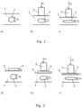
- Figure 1 schematically shows how results from ultrasound measurements performed on a human or animal body 1 can be influenced by the pressure applied with the ultrasound transducer 2.
- Figure 1 shows a simple spherical cavity 3 and measurements of the size of that cavity 3. The deformations are exaggerated in the figures to better illustrate the invention.
- Figure 1.a shows a cross-sectional view of the spherical cavity 3 having a diameter d 0 .
- Figure 1.b shows how measurements with an ultrasound transducer 2 with an applied pressure P 1 results in a measured diameter d 1 of the cavity 3 being deformed by the pressure P 1 .
- Figure 1.c shows how measurements with an ultrasound transducer 2 with an applied pressure P 2 results in a measured diameter d 2 of the cavity 3 being deformed by the pressure P 2 .
- the different pressures P 1 and P 2 could e.g. be applied by two different persons, or it could be applied by the same person at two different points in time. In both situations, this means that different conclusions may be made which can have serious consequences e.g. if the progress of a growing defect inside a patient is monitored.
- Figure 2 schematically shows the measurements in figure 1 adjusted in accordance with the present invention by interpositioning of an ultrasound compatible and compressible elastic element 4 between a tissue surface 5 of the body 1 and the ultrasound probe 2.
- the elastic element 4 has predetermined elastic properties, and the ultrasound acquisition is obtained by sending ultrasound signals from the ultrasound probe 2 into the body 1 through the elastic element 4.
- the elastic element 4 is typically made from a material in which the speed of sound is in the range of 1200 to 1700 m/s, such as in the range of 1400 to 1600 m/s.
- the attenuation of sound is below 3 dB/[mHz x cm], such as below 2 dB/[mHz x cm].
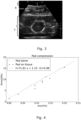
- Figure 3 shows an example of an ultrasound image obtained in accordance with the present invention.
- the image is seen to include the first region 6 representing the thickness of the compressed elastic element 4 and the second region 7 representing a part of a human body 1 in the form of the region around and including a part of the aorta.
- the compression of the elastic element 4 on the ultrasound acquisition is measured, and based on the measured compression of the elastic element 4 and a known correlation between elastic element compression and applied pressure, it is possible to determine the pressure applied with the ultrasound transducer 2.
- the measurements on the second region 7 can hereby be standardized to provide results that are independent on the applied pressure.
- the obtained measure of the pressure may be presented visually or audibly during the performance of the ultrasound measurements.
- FIG. 4 shows results of measurements of the elastic properties of an elastic element 4 for use in a method according to the present invention.
- the elastic element is referred to as "pad”.
- the relationships between the stress and the strain were measured under conditions corresponding to real ultrasound measurements.
- the crosses represent measurements where the elastic element was placed on a table, i.e.
- the dots represent measurements where the elastic element was placed on a pad of animal tissue.
- the line in the figure is a linear approximation of the results from the measurements made with the elastic elements placed on a hard surface.
- the slope of the line is the elastic modulus of the material, and as seen in the figure, this value will also be valid for the similar measurements with the softer surface in the form of tissue.
- a method of performing ultrasound measurements on a human or animal body 1 in accordance with the present invention comprises the following steps:
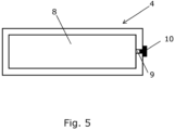
- FIG. 5 schematically shows an example of an elastic element 4 that comprises an inner cavity 8 filled with liquid.
- a channel 9 extends from the inner cavity 8 towards an exterior surface of the elastic element 4.
- the channel 9 is provided with a plug 10 so that it is possible to remove the liquid, such as for replacing it with another liquid whereby it is possible to adjust the elastic properties of the elastic element 4 for a given application.
- the elastic properties of the elastic element 4 can also be varied by filling different amounts of liquid into the cavity as described above.
Landscapes
- Life Sciences & Earth Sciences (AREA)
- Health & Medical Sciences (AREA)
- Physics & Mathematics (AREA)
- Engineering & Computer Science (AREA)
- Heart & Thoracic Surgery (AREA)
- Nuclear Medicine, Radiotherapy & Molecular Imaging (AREA)
- Pathology (AREA)
- Radiology & Medical Imaging (AREA)
- Veterinary Medicine (AREA)
- Biomedical Technology (AREA)
- Biophysics (AREA)
- Medical Informatics (AREA)
- Molecular Biology (AREA)
- Surgery (AREA)
- Animal Behavior & Ethology (AREA)
- General Health & Medical Sciences (AREA)
- Public Health (AREA)
- Acoustics & Sound (AREA)
- Ultra Sonic Daignosis Equipment (AREA)
Description
- The present invention relates to a method of performing ultrasound measurements on a human or animal body, and in particular it relates to such a method which can be used to obtain the pressure applied via an ultrasound probe used to perform the measurements.
- Within the field of medical technology, ultrasound measurements are used for many different types of investigations on a human or animal body. Examples are for the investigation of abdominal aortic aneurisms (AAA) or for monitoring of the well-being and expected time of birth of a child.
- Such measurements are typically performed by manually pressing an ultrasound transducer towards the skin of the body and moving the transducer over a region of interest while a resulting ultrasound image is studied on a display and possibly also recorded for further assessment. However, the pressure applied to the ultrasound transducer causes a compression of e.g. the AAA between the transducer and the spine, potentially resulting in an underestimation of AAA anterior-posterior diameter. Furthermore, since the transducer is held manually, the applied pressure will depend on the person performing the investigation, and it may also vary between two different investigations performed at different points in time. This can lead to erroneous conclusions being taken and for some types of investigations, this may cause erroneous or delayed treatment being initiated. It may also result in a conclusion that no treatment is needed even if it was.
- Hence, an improved method of performing ultrasound measurements on a human or animal body would be advantageous.
-
US2012016238 may be considered to disclose a method according to the preamble ofclaim 1. - It is an object of the present invention to provide a method of performing ultrasound measurements on a human or animal body which gives more reliable results than known methods.
- It is another object of the present invention to provide a method of obtaining a measure of the pressure of the ultrasound transducer on the object being scanned when performing ultrasound measurements on a human or animal body.
- It is an object of at least some embodiments of the present invention to provide a method of performing ultrasound measurements on a human or animal body with which the final results are independent on the pressure applied to the ultrasound probe during the performance of the measurements.
- It is another object of at least some embodiments of the present invention to provide a method of performing ultrasound measurements on a human or animal body with which the results are independent on the person performing the measurements.
- It is a further object of the present invention to provide an alternative to the prior art.
- In particular, it may be seen as an object of the present invention to provide a method of performing ultrasound measurements that solves the above mentioned problems of the prior art.
- Thus, the above-described object and several other objects are intended to be obtained in a first aspect of the invention by providing a method of performing ultrasound measurements on a human or animal body by use of an ultrasound probe, the method comprising:
- interpositioning of an ultrasound compatible and compressible elastic element between a tissue surface of the body and the ultrasound probe, the elastic element having predetermined elastic properties,
- performing the ultrasound measurements to obtain at least one ultrasound acquisition by sending ultrasound signals from the ultrasound probe into the body through the elastic element, the at least one ultrasound acquisition including:
- a first region representing the compression of the elastic element being compressed by pressure applied with the ultrasound probe during the performance of the ultrasound measurements, and
- a second region representing at least a part of the body under investigation,
- measuring the compression of the elastic element on the ultrasound acquisition, and
- based on the measured compression of the elastic element and a known correlation between elastic element compression and applied pressure, determining the pressure applied with the ultrasound transducer.
- By "ultrasound compatible" is meant that ultrasound can be sent through the material without any significant scattering or dampening, and with a speed of sound resembling what it will be in human tissues.
- By "compressible" is meant that a force applied by the ultrasound transducer will cause a deformation of the elastic element which is measureable on the ultrasound acquisition with a precision which is high enough to be used for the subsequent method steps.
- By "an elastic element with predetermined elastic properties" is preferably meant that it is made from a material having a known relationship between compressive stress and strain. Hereby the measured elastic element deformation can be correlated to the applied pressure. The elastic element may e.g. have a linear relationship between stress and strain.
- By such a method it is possible to adjust for the fact that different people may apply different amount of pressure on the ultrasound probe (interobserver variability), and that a person may not always apply the same pressure (intraobserver variability). Hereby it is possible to obtain measurement results which are standardized to provide results that are more reliable than what is possible with traditional ultrasound measurements on a human or animal body.
- The elastic element is arranged freely on the tissue surface of the body. By "freely" is meant that it is unconstrained at all outer surfaces, with no adjacent frames or means of fixing the elastic element to e.g. electronic sensors. This provides the benefit that the elastic element can be used without extra sensors, but using the ultrasound probe without any add-ons, such as frames and mechanisms for measuring deformation, such as compression. The compression of the elastic element is determined directly on the ultrasound acquisition which is a simple and reliable way of determining the applied pressure. There is a further benefit that the deformation determined is the local deformation beneath the footprint of the ultrasound probe and thus not attenuated to the outside of the immediate footprint of the probe.
- In some embodiments of the invention, the method further comprises a step of automatically correcting the part of the ultrasound acquisition being the second region to obtain standardized ultrasound measurement results which are independent on the applied pressure. Such automatic correction may take place in a computing unit connected to or forming part of the system used. The automatic correction will typically be based on numerical calculations possibly involving parameter characteristics for the type of investigations and/or measures related to the body under examination, such as weight or body mass index.
- The obtained measure of the pressure may be presented visually or audibly during the performance of the ultrasound measurements. It could e.g. be presented on a display via a row of LEDs, a number, or a scale. Hereby it will be possible for the person performing the measurements to get an indication of the actual applied pressure and based thereon continuously adjust this to aim at a desired value. Such a desired value could e.g. be a record of a pressure used in a former measurement performed on the same body so that the results can be directly compared with larger precision than with known methods.
- A similar method can also be used during training of persons performing ultrasound measurements. Such training could e.g. be used to give persons under education an interactive way of practising the performance of ultrasound measurements and which pressures that are appropriate. Hereby good skills within the field can be obtained faster than would normally be required.
- In presently preferred embodiments of the invention, the elastic element is made from a material in which the speed of sound is in the range of 1200 to 1700 m/s, such as in the range of 1400 to 1600 m/s. This is around the speed of sound in human tissue which is about 1540 m/s.
- In presently preferred embodiments of the invention, the elastic element is made from a material in which the attenuation of sound is below 3 dB/[mHz x cm], such as below 2 dB/[mHz x cm]. This is around the attenuation of sound in human tissue.
- In general, the speed of sound as well as the attenuation of sound depend on the frequency of the ultrasound, but these values apply to the medical scanners typical used for the investigation of human bodies. By choosing a material for the elastic element with similar properties for the propagation of the ultrasound waves, imaging through the elastic element is enabled.
- The elastic element may be made from a solid material selected from:
- ballistic gel and synthetic ballistic gel;
- hydrogels based on e.g. gelatine, agar, Psyllium husk, or Chia seeds;
- cryogels based on poly vinyl alcohol;
- polyurethane;
- poly vinyl chloride;
- silicones;
- glycerine softened polymers, such as thermoplastic starch.
- It is possible to use any type of elastic material that can be traversed by ultrasound. In addition to the elastic properties of the material, the choice of material for a given application may depend on other parameter, such as price, how easy it is to clean if re-used, how stable the elastic properties are during longer term storage, and requirements related to disposal after use. E.g., hydrogels based on gelatine, agar, Psyllium husk, or Chia seeds will be biodegradable. Cryogel based on poly vinyl alcohol has the advantage that such a material is biodegrable (i.e. easily disposable), has stable mechanical properties, and the mechanical properties can be changed without additives. Furthermore, elements made from cryogel is easy to manufacture. Silicones are very non-biodegrable. They are very stable, and thus, counter-intuitively, considered one of the most "green" oil based polymers. Furthermore, silicones are easily manufactured and maintains the shape regardless of storage conditions within typical storage conditions. Glycerine softened polymers, such as thermoplastic starch, are biodegradable (i.e. easily disposable), and the plasticiser (glycerine) is very safe to humans as there is a low risk of negative influences, such as skin irritation, even with prolonged contact.
- In alternative embodiments of the invention, the elastic element may comprise an inner cavity filled with liquid. Hereby it may be easier to avoid viscoelastic creep of the elastic element due to the applied pressure, whereby more precise measurements can be obtained. It may also be easier to find a material with a desired attenuation of sound and speed of sound for a given type of investigation. If the liquid is water, less waste material is obtained, as it does not require any special treatment after use. Other examples of liquids are physiological saline (H2O with NaCl 0.9%), glycerine, and polyethylene glycol. If the elastic element is provided with a closable and openable opening into the inner cavity, it is possible to adjust the volume and type of the liquid to a given application.
- A method as described above may be at least a part of one of the following types of investigations: measurement of anatomy in the body, measurement of a foetus in the body, measurement of pathology in the body, measurement of abnormal/deformity in the body, and measurement of motion in the body, such as liquid motion.
- In a second aspect, the present invention relates to a system for performing ultrasound measurements on a human or animal body, the system comprising:
- an ultrasound probe,
- an ultrasound compatible and compressible elastic element,
- a computing unit having access to known correlations between elastic element compression and applied pressure, and
- By "computing unit" is meant a unit which can be used to determine the applied pressure based on measures of the compression of the elastic element and information about the predetermined elastic properties of the elastic element.
- The elastic element is configured to be arranged freely on the tissue surface of the body during the performance of the ultrasound measurements.
- The elastic element may be made from a material in which the speed of sound is in the range of 1200 to 1700 m/s, such as in the range of 1400 to 1600 m/s.
- The elastic element may be made from a material in which the attenuation of sound is below 3 dB/[mHz x cm], such as below 2 dB/[mHz x cm].
- The elastic element may be made from a solid material selected from:
- ballistic gel and synthetic ballistic gel;
- hydrogels based on e.g. gelatine, agar, Psyllium husk, or Chia seeds;
- cryogels based on poly vinyl alcohol;
- polyurethane;
- poly vinyl chloride;
- silicones;
- glycerine softened polymers, such as thermoplastic starch.
- The elastic element may comprise an inner cavity filled with liquid.
- The first and second aspects of the present invention may be combined. These and other aspects of the invention will be apparent from and elucidated with reference to the embodiments described hereinafter.
- The method of performing ultrasound measurements on a human or animal body as well as a system for use in such a method according to the invention will now be described in more detail with regard to the accompanying figures. The figures show one way of implementing the present invention and is not to be construed as being limiting to other possible embodiments falling within the scope of the claims.
-
Figure 1 schematically shows how results from ultrasound measurements performed on a human or animal body can be influenced by the pressure applied with the ultrasound transducer. -
Figure 2 schematically shows the measurements infigure 1 adjusted in accordance with the present invention. -
Figure 3 shows an example of an ultrasound image obtained in accordance with the present invention. -
Figure 4 shows results of measurements of the elastic properties of an elastic element for use in a method according to the present invention. -
Figure 5 schematically shows an example of an elastic element that comprises an inner cavity filled with liquid. -
Figure 1 schematically shows how results from ultrasound measurements performed on a human oranimal body 1 can be influenced by the pressure applied with theultrasound transducer 2. Many different types of investigations can be performed by the invention. However, in order to better illustrate the overall principle,figure 1 shows a simplespherical cavity 3 and measurements of the size of thatcavity 3. The deformations are exaggerated in the figures to better illustrate the invention.Figure 1.a shows a cross-sectional view of thespherical cavity 3 having a diameter d0.Figure 1.b shows how measurements with anultrasound transducer 2 with an applied pressure P1 results in a measured diameter d1 of thecavity 3 being deformed by the pressure P1.Figure 1.c shows how measurements with anultrasound transducer 2 with an applied pressure P2 results in a measured diameter d2 of thecavity 3 being deformed by the pressure P2. The different pressures P1 and P2 could e.g. be applied by two different persons, or it could be applied by the same person at two different points in time. In both situations, this means that different conclusions may be made which can have serious consequences e.g. if the progress of a growing defect inside a patient is monitored. -
Figure 2 schematically shows the measurements infigure 1 adjusted in accordance with the present invention by interpositioning of an ultrasound compatible and compressibleelastic element 4 between atissue surface 5 of thebody 1 and theultrasound probe 2. Theelastic element 4 has predetermined elastic properties, and the ultrasound acquisition is obtained by sending ultrasound signals from theultrasound probe 2 into thebody 1 through theelastic element 4. Theelastic element 4 is typically made from a material in which the speed of sound is in the range of 1200 to 1700 m/s, such as in the range of 1400 to 1600 m/s. The attenuation of sound is below 3 dB/[mHz x cm], such as below 2 dB/[mHz x cm]. These parameters are frequency dependent, and the numbers are here given in relation to the types of ultrasound transducers typically used for medical assessments. - For simplicity, the same deformations of the
cavity 3 as infigure 1 are shown to take place even though in practise they may be slightly smaller since a part of the pressure is used for the compression of theelastic element 4. As seen fromfigures 2.b and 2.c , the differences in applied pressure also cause corresponding different compressions, t1 and t2, of theelastic element 4 during the performance of the ultrasound measurements. -
Figure 3 shows an example of an ultrasound image obtained in accordance with the present invention. The image is seen to include thefirst region 6 representing the thickness of the compressedelastic element 4 and thesecond region 7 representing a part of ahuman body 1 in the form of the region around and including a part of the aorta. The compression of theelastic element 4 on the ultrasound acquisition is measured, and based on the measured compression of theelastic element 4 and a known correlation between elastic element compression and applied pressure, it is possible to determine the pressure applied with theultrasound transducer 2. As described above, for some embodiments of the invention, the measurements on thesecond region 7 can hereby be standardized to provide results that are independent on the applied pressure. Alternatively or in combination herewith, the obtained measure of the pressure may be presented visually or audibly during the performance of the ultrasound measurements. It could e.g. be presented on a display via a row of LEDs, a number, or a scale. Hereby it will be possible for the person performing the measurements to get an indication of the actual applied pressure and based thereon continuously adjust this to aim at a desired value as described in further details above. - The elastic properties of a given
elastic element 4 to be used in the measurements as described above are predetermined so that this information can be used in the determination of the final results. As part of the studies leading to the present invention, it has been tested whether the type of surface on which theelastic element 4 is placed influences the elastic properties in a way that would be compromising to the quality of the results of the ultrasound measurements being performed.Figure 4 shows results of measurements of the elastic properties of anelastic element 4 for use in a method according to the present invention. Infigure 4 , the elastic element is referred to as "pad". The relationships between the stress and the strain were measured under conditions corresponding to real ultrasound measurements. Infigure 4 , the crosses represent measurements where the elastic element was placed on a table, i.e. a hard surface, and the dots represent measurements where the elastic element was placed on a pad of animal tissue. The line in the figure is a linear approximation of the results from the measurements made with the elastic elements placed on a hard surface. The slope of the line is the elastic modulus of the material, and as seen in the figure, this value will also be valid for the similar measurements with the softer surface in the form of tissue. - In summary, a method of performing ultrasound measurements on a human or
animal body 1 in accordance with the present invention comprises the following steps: - interpositioning of an ultrasound compatible and compressible
elastic element 4 between atissue surface 5 of thebody 1 and theultrasound probe 2, theelastic element 4 having predetermined elastic properties, - performing the ultrasound measurements to obtain at least one ultrasound acquisition by sending ultrasound signals from the
ultrasound probe 2 into thebody 1 through theelastic element 4, the at least one ultrasound acquisition including: - a
first region 6 representing the compression of theelastic element 4 being compressed by pressure applied with theultrasound probe 2 during the performance of the ultrasound measurements, and - a
second region 7 representing at least a part of thebody 1 under investigation, - measuring the compression of the
elastic element 4 on the ultrasound acquisition, and - based on the measured compression of the
elastic element 4 and a known correlation between elastic element compression and applied pressure, determining the pressure applied with theultrasound transducer 2, so that measurements on thesecond region 7 can be standardized to provide results that are independent on the applied pressure. The following is a list of examples of investigations that can be performed by the invention: measurement of anatomy in the body, measurement of a foetus in the body, measurement of pathology in the body, measurement of abnormal/deformity in the body, and measurement of motion in the body, such as liquid motion. -
Figure 5 schematically shows an example of anelastic element 4 that comprises aninner cavity 8 filled with liquid. In this embodiment, achannel 9 extends from theinner cavity 8 towards an exterior surface of theelastic element 4. Thechannel 9 is provided with aplug 10 so that it is possible to remove the liquid, such as for replacing it with another liquid whereby it is possible to adjust the elastic properties of theelastic element 4 for a given application. The elastic properties of theelastic element 4 can also be varied by filling different amounts of liquid into the cavity as described above. - Although the present invention has been described in connection with the specified embodiments, it should not be construed as being in any way limited to the presented examples. The scope of the present invention is set out by the accompanying claim set. In the context of the claims, the terms "comprising" or "comprises" do not exclude other possible elements or steps. Furthermore, the mentioning of references such as "a" or "an" etc. should not be construed as excluding a plurality. The use of reference signs in the claims with respect to elements indicated in the figures shall also not be construed as limiting the scope of the invention. Furthermore, individual features mentioned in different claims, may possibly be advantageously combined, and the mentioning of these features in different claims does not exclude that a combination of features is not possible and advantageous.
Claims (13)
- Method of performing ultrasound measurements on a human or animal body (1) by use of an ultrasound probe (2), the method comprising:- interpositioning of an ultrasound compatible and compressible elastic element (4) between a tissue surface (5) of the body (1) and the ultrasound probe (2), the elastic element (4) having predetermined elastic properties,- performing the ultrasound measurements to obtain at least one ultrasound acquisition by sending ultrasound signals from the ultrasound probe (2) into the body (1) through the elastic element (4), the at least one ultrasound acquisition including:- a first region (6) representing the compression of the elastic element (4) being compressed by pressure applied with the ultrasound probe (2) during the performance of the ultrasound measurements, and- a second region (7) representing at least a part of the body (1) under investigation,- measuring the compression of the elastic element (4) on the ultrasound acquisition, and- based on the measured compression of the elastic element (4) and a known correlation between elastic element compression and applied pressure, obtaining a measure of the pressure applied with the ultrasound transducer (2),characterised in that the elastic element (4) is arranged freely on the tissue surface (5) of the body (1), meaning that the elastic element (4) is unconstrained at all outer surfaces thereof, with no adjacent frames or means of fixing the elastic element (4) to electronic sensors.
- Method according to claim 1, further comprising a step of automatically correcting the part of the ultrasound acquisition being the second region (7) to obtain standardized ultrasound measurement results which are independent of the applied pressure.
- Method according to any of the preceding claims, wherein the obtained measure of the pressure is presented visually or audibly during the performance of the ultrasound measurements.
- Method according to any of the preceding claims, wherein the elastic element (4) is made from a material in which the speed of sound is in the range of 1200 to 1700 m/s, such as in the range of 1400 to 1600 m/s.
- Method according to any of the preceding claims, wherein the elastic element (4) is made from a material in which the attenuation of sound is below 3 dB/[mHz x cm], such as below 2 dB/[mHz x cm].
- Method according to any of the preceding claims, wherein the elastic element (4) is made from a solid material selected from:- ballistic gel and synthetic ballistic gel;- hydrogels based on e.g. gelatine, agar, Psyllium husk, or Chia seeds;- cryogels based on poly vinyl alcohol;- polyurethane;- poly vinyl chloride;- silicones;- glycerine softened polymers, such as thermoplastic starch.
- Method according to any of claims 1 to 5, wherein the elastic element (4) comprises an inner cavity (8) filled with liquid.
- Method according to any of the preceding claims, wherein the method is at least a part of one of the following types of investigations: measurement of anatomy in the body, measurement of a foetus in the body, measurement of pathology in the body, measurement of abnormal/deformity in the body, and measurement of motion in the body, such as liquid motion.
- System for performing ultrasound measurements on a human or animal body (1), the system comprising:- an ultrasound probe (2),- an ultrasound compatible and compressible elastic element (4) which is configured to be arranged freely on the tissue surface (5) of the body (1) during the performance of the ultrasound measurements so that the elastic element (4) is unconstrained at all outer surfaces thereof, with no adjacent frames or means of fixing the elastic element (4) to electronic sensors,- a computing unit having access to known correlations between elastic element (4) compression and applied pressure, andwherein the system is adapted to perform a method according to any of the preceding claims.
- System according to claim 9, wherein the elastic element (4) is made from a material in which the speed of sound is in the range of 1200 to 1700 m/s, such as in the range of 1400 to 1600 m/s.
- System according to any of claims 9 to 10, wherein the elastic element (4) is made from a material in which the attenuation of sound is below 3 dB/[mHz x cm], such as below 2 dB/[mHz x cm].
- System according to any of claims 9 to 11, wherein the elastic element (4) is made from a solid material selected from:- ballistic gel and synthetic ballistic gel;- hydrogels based on e.g. gelatine, agar, Psyllium husk, or Chia seeds;- cryogels based on poly vinyl alcohol;- polyurethane;- poly vinyl chloride;- silicones;- glycerine softened polymers, such as thermoplastic starch.
- System according to any of claims 9 to 11, wherein the elastic element (4) comprises an inner cavity (8) filled with liquid.
Applications Claiming Priority (2)
| Application Number | Priority Date | Filing Date | Title |
|---|---|---|---|
| EP20194832.0A EP3964135A1 (en) | 2020-09-07 | 2020-09-07 | Method of performing ultrasound measurements on a human or animal body |
| PCT/EP2021/074559 WO2022049302A1 (en) | 2020-09-07 | 2021-09-07 | Method of performing ultrasound measurements on a human or animal body |
Publications (3)
| Publication Number | Publication Date |
|---|---|
| EP4210583A1 EP4210583A1 (en) | 2023-07-19 |
| EP4210583C0 EP4210583C0 (en) | 2024-11-06 |
| EP4210583B1 true EP4210583B1 (en) | 2024-11-06 |
Family
ID=72428142
Family Applications (2)
| Application Number | Title | Priority Date | Filing Date |
|---|---|---|---|
| EP20194832.0A Withdrawn EP3964135A1 (en) | 2020-09-07 | 2020-09-07 | Method of performing ultrasound measurements on a human or animal body |
| EP21772802.1A Active EP4210583B1 (en) | 2020-09-07 | 2021-09-07 | Method of performing ultrasound measurements on a human or animal body |
Family Applications Before (1)
| Application Number | Title | Priority Date | Filing Date |
|---|---|---|---|
| EP20194832.0A Withdrawn EP3964135A1 (en) | 2020-09-07 | 2020-09-07 | Method of performing ultrasound measurements on a human or animal body |
Country Status (2)
| Country | Link |
|---|---|
| EP (2) | EP3964135A1 (en) |
| WO (1) | WO2022049302A1 (en) |
Family Cites Families (4)
| Publication number | Priority date | Publication date | Assignee | Title |
|---|---|---|---|---|
| WO2005120358A1 (en) * | 2004-06-09 | 2005-12-22 | Hitachi Medical Corporation | Elastic image display method and ultrasonographic device |
| CN101489490B (en) * | 2006-07-31 | 2011-03-23 | 株式会社日立医药 | Compression device, ultrasonic probe and ultrasonic diagnostic device using the same |
| CN101553174B (en) * | 2006-12-20 | 2011-06-15 | 株式会社日立医药 | Ultrasonic diagnostic device |
| US8845538B2 (en) * | 2008-04-25 | 2014-09-30 | Hitachi Medical Corporation | Ultrasonic diagnostic apparatus |
-
2020
- 2020-09-07 EP EP20194832.0A patent/EP3964135A1/en not_active Withdrawn
-
2021
- 2021-09-07 WO PCT/EP2021/074559 patent/WO2022049302A1/en not_active Ceased
- 2021-09-07 EP EP21772802.1A patent/EP4210583B1/en active Active
Also Published As
| Publication number | Publication date |
|---|---|
| WO2022049302A1 (en) | 2022-03-10 |
| EP3964135A1 (en) | 2022-03-09 |
| EP4210583C0 (en) | 2024-11-06 |
| EP4210583A1 (en) | 2023-07-19 |
Similar Documents
| Publication | Publication Date | Title |
|---|---|---|
| CN111386070B (en) | Pressure measuring device, pressure measuring system and method | |
| Kähn | Sonographic fetometry in the bovine | |
| Zheng et al. | An ultrasound indentation system for biomechanical properties assessment of soft tissues in-vivo | |
| US10028700B2 (en) | Method and system for non-invasive determination of human body fat | |
| Buntin et al. | Noninvasive assessment of mechanical properties of peripheral arteries | |
| US20240138810A1 (en) | Method and apparatus for evaluating contact state of ultrasound probe on basis of soft tissue morphology | |
| Boroffka et al. | Intraobserver and interobserver repeatability of ocular biometric measurements obtained by means of B-mode ultrasonography in dogs | |
| CN103845074A (en) | Ultrasonic elastography system and method | |
| WO2021023051A1 (en) | Tissue evaluation method, apparatus and device, and computer-readable storage medium | |
| US20070167819A1 (en) | Method for in-vivo measurement of biomechanical properties of internal tissues | |
| Stanger et al. | Fetal movement measurement and technology: a narrative review | |
| Hee | Overview of the methods available for biomechanical testing of the uterine cervix in vivo | |
| EP1959824A1 (en) | Device for, in-vivo measurement of biomechanical properties of internal tissues | |
| Carson et al. | Fetal depth and ultrasound path lengths through overlying tissues | |
| EP3964135A1 (en) | Method of performing ultrasound measurements on a human or animal body | |
| Gao et al. | A pilot study on ultrasound elastography for evaluation of mechanical characteristics and oral strategy of gels | |
| Kriewall et al. | Mechanical behavior of fetal dura mater under large axisymmetric inflation | |
| US5535750A (en) | Method and apparatus for evaluating the progress of osteoporosis by ultrasonic signals | |
| O'Hara et al. | Shear wave elastography of the maternal cervix: A transabdominal technique | |
| US12564372B2 (en) | Tactile ultrasound method and probe for predicting preterm birth | |
| Bluth | Ultrasound of the abdominal aorta | |
| US20190159751A1 (en) | Automated time-domain determination of tissue viscoelasticity in a sub-hertz frequency range | |
| CN113116379B (en) | Fetal heart monitor integrating pregnancy monitoring and working method thereof | |
| KR20200125150A (en) | Breast Phantom for breast radiographic mammography education and practic | |
| WO2015149805A1 (en) | Reference material element for ultrasound scanning probe |
Legal Events
| Date | Code | Title | Description |
|---|---|---|---|
| STAA | Information on the status of an ep patent application or granted ep patent |
Free format text: STATUS: UNKNOWN |
|
| STAA | Information on the status of an ep patent application or granted ep patent |
Free format text: STATUS: THE INTERNATIONAL PUBLICATION HAS BEEN MADE |
|
| PUAI | Public reference made under article 153(3) epc to a published international application that has entered the european phase |
Free format text: ORIGINAL CODE: 0009012 |
|
| STAA | Information on the status of an ep patent application or granted ep patent |
Free format text: STATUS: REQUEST FOR EXAMINATION WAS MADE |
|
| 17P | Request for examination filed |
Effective date: 20230404 |
|
| AK | Designated contracting states |
Kind code of ref document: A1 Designated state(s): AL AT BE BG CH CY CZ DE DK EE ES FI FR GB GR HR HU IE IS IT LI LT LU LV MC MK MT NL NO PL PT RO RS SE SI SK SM TR |
|
| DAV | Request for validation of the european patent (deleted) | ||
| DAX | Request for extension of the european patent (deleted) | ||
| GRAP | Despatch of communication of intention to grant a patent |
Free format text: ORIGINAL CODE: EPIDOSNIGR1 |
|
| STAA | Information on the status of an ep patent application or granted ep patent |
Free format text: STATUS: GRANT OF PATENT IS INTENDED |
|
| INTG | Intention to grant announced |
Effective date: 20240514 |
|
| GRAS | Grant fee paid |
Free format text: ORIGINAL CODE: EPIDOSNIGR3 |
|
| GRAA | (expected) grant |
Free format text: ORIGINAL CODE: 0009210 |
|
| STAA | Information on the status of an ep patent application or granted ep patent |
Free format text: STATUS: THE PATENT HAS BEEN GRANTED |
|
| AK | Designated contracting states |
Kind code of ref document: B1 Designated state(s): AL AT BE BG CH CY CZ DE DK EE ES FI FR GB GR HR HU IE IS IT LI LT LU LV MC MK MT NL NO PL PT RO RS SE SI SK SM TR |
|
| REG | Reference to a national code |
Ref country code: GB Ref legal event code: FG4D |
|
| REG | Reference to a national code |
Ref country code: CH Ref legal event code: EP |
|
| REG | Reference to a national code |
Ref country code: DE Ref legal event code: R096 Ref document number: 602021021481 Country of ref document: DE |
|
| REG | Reference to a national code |
Ref country code: IE Ref legal event code: FG4D |
|
| U01 | Request for unitary effect filed |
Effective date: 20241128 |
|
| U07 | Unitary effect registered |
Designated state(s): AT BE BG DE DK EE FI FR IT LT LU LV MT NL PT RO SE SI Effective date: 20241205 |
|
| PG25 | Lapsed in a contracting state [announced via postgrant information from national office to epo] |
Ref country code: HR Free format text: LAPSE BECAUSE OF FAILURE TO SUBMIT A TRANSLATION OF THE DESCRIPTION OR TO PAY THE FEE WITHIN THE PRESCRIBED TIME-LIMIT Effective date: 20241106 Ref country code: IS Free format text: LAPSE BECAUSE OF FAILURE TO SUBMIT A TRANSLATION OF THE DESCRIPTION OR TO PAY THE FEE WITHIN THE PRESCRIBED TIME-LIMIT Effective date: 20250306 |
|
| PG25 | Lapsed in a contracting state [announced via postgrant information from national office to epo] |
Ref country code: ES Free format text: LAPSE BECAUSE OF FAILURE TO SUBMIT A TRANSLATION OF THE DESCRIPTION OR TO PAY THE FEE WITHIN THE PRESCRIBED TIME-LIMIT Effective date: 20241106 |
|
| PG25 | Lapsed in a contracting state [announced via postgrant information from national office to epo] |
Ref country code: NO Free format text: LAPSE BECAUSE OF FAILURE TO SUBMIT A TRANSLATION OF THE DESCRIPTION OR TO PAY THE FEE WITHIN THE PRESCRIBED TIME-LIMIT Effective date: 20250206 |
|
| PG25 | Lapsed in a contracting state [announced via postgrant information from national office to epo] |
Ref country code: GR Free format text: LAPSE BECAUSE OF FAILURE TO SUBMIT A TRANSLATION OF THE DESCRIPTION OR TO PAY THE FEE WITHIN THE PRESCRIBED TIME-LIMIT Effective date: 20250207 |
|
| PG25 | Lapsed in a contracting state [announced via postgrant information from national office to epo] |
Ref country code: PL Free format text: LAPSE BECAUSE OF FAILURE TO SUBMIT A TRANSLATION OF THE DESCRIPTION OR TO PAY THE FEE WITHIN THE PRESCRIBED TIME-LIMIT Effective date: 20241106 |
|
| PG25 | Lapsed in a contracting state [announced via postgrant information from national office to epo] |
Ref country code: RS Free format text: LAPSE BECAUSE OF FAILURE TO SUBMIT A TRANSLATION OF THE DESCRIPTION OR TO PAY THE FEE WITHIN THE PRESCRIBED TIME-LIMIT Effective date: 20250206 |
|
| PG25 | Lapsed in a contracting state [announced via postgrant information from national office to epo] |
Ref country code: SM Free format text: LAPSE BECAUSE OF FAILURE TO SUBMIT A TRANSLATION OF THE DESCRIPTION OR TO PAY THE FEE WITHIN THE PRESCRIBED TIME-LIMIT Effective date: 20241106 |
|
| PG25 | Lapsed in a contracting state [announced via postgrant information from national office to epo] |
Ref country code: SK Free format text: LAPSE BECAUSE OF FAILURE TO SUBMIT A TRANSLATION OF THE DESCRIPTION OR TO PAY THE FEE WITHIN THE PRESCRIBED TIME-LIMIT Effective date: 20241106 |
|
| PG25 | Lapsed in a contracting state [announced via postgrant information from national office to epo] |
Ref country code: CZ Free format text: LAPSE BECAUSE OF FAILURE TO SUBMIT A TRANSLATION OF THE DESCRIPTION OR TO PAY THE FEE WITHIN THE PRESCRIBED TIME-LIMIT Effective date: 20241106 |
|
| PLBE | No opposition filed within time limit |
Free format text: ORIGINAL CODE: 0009261 |
|
| STAA | Information on the status of an ep patent application or granted ep patent |
Free format text: STATUS: NO OPPOSITION FILED WITHIN TIME LIMIT |
|
| 26N | No opposition filed |
Effective date: 20250807 |
|
| U20 | Renewal fee for the european patent with unitary effect paid |
Year of fee payment: 5 Effective date: 20250924 |
|
| REG | Reference to a national code |
Ref country code: CH Ref legal event code: H13 Free format text: ST27 STATUS EVENT CODE: U-0-0-H10-H13 (AS PROVIDED BY THE NATIONAL OFFICE) Effective date: 20260504 |