EP4209176A1 - Medical image display apparatus - Google Patents
Medical image display apparatus Download PDFInfo
- Publication number
- EP4209176A1 EP4209176A1 EP21863926.8A EP21863926A EP4209176A1 EP 4209176 A1 EP4209176 A1 EP 4209176A1 EP 21863926 A EP21863926 A EP 21863926A EP 4209176 A1 EP4209176 A1 EP 4209176A1
- Authority
- EP
- European Patent Office
- Prior art keywords
- adjustment value
- medical image
- display device
- image display
- attribute
- Prior art date
- Legal status (The legal status is an assumption and is not a legal conclusion. Google has not performed a legal analysis and makes no representation as to the accuracy of the status listed.)
- Granted
Links
Images



Classifications
-
- G—PHYSICS
- G06—COMPUTING OR CALCULATING; COUNTING
- G06T—IMAGE DATA PROCESSING OR GENERATION, IN GENERAL
- G06T7/00—Image analysis
- G06T7/0002—Inspection of images, e.g. flaw detection
- G06T7/0012—Biomedical image inspection
-
- A—HUMAN NECESSITIES
- A61—MEDICAL OR VETERINARY SCIENCE; HYGIENE
- A61B—DIAGNOSIS; SURGERY; IDENTIFICATION
- A61B6/00—Apparatus or devices for radiation diagnosis; Apparatus or devices for radiation diagnosis combined with radiation therapy equipment
- A61B6/46—Arrangements for interfacing with the operator or the patient
- A61B6/461—Displaying means of special interest
- A61B6/462—Displaying means of special interest characterised by constructional features of the display
-
- A—HUMAN NECESSITIES
- A61—MEDICAL OR VETERINARY SCIENCE; HYGIENE
- A61B—DIAGNOSIS; SURGERY; IDENTIFICATION
- A61B5/00—Measuring for diagnostic purposes; Identification of persons
-
- A—HUMAN NECESSITIES
- A61—MEDICAL OR VETERINARY SCIENCE; HYGIENE
- A61B—DIAGNOSIS; SURGERY; IDENTIFICATION
- A61B6/00—Apparatus or devices for radiation diagnosis; Apparatus or devices for radiation diagnosis combined with radiation therapy equipment
-
- G—PHYSICS
- G09—EDUCATION; CRYPTOGRAPHY; DISPLAY; ADVERTISING; SEALS
- G09G—ARRANGEMENTS OR CIRCUITS FOR CONTROL OF INDICATING DEVICES USING STATIC MEANS TO PRESENT VARIABLE INFORMATION
- G09G3/00—Control arrangements or circuits, of interest only in connection with visual indicators other than cathode-ray tubes
- G09G3/20—Control arrangements or circuits, of interest only in connection with visual indicators other than cathode-ray tubes for presentation of an assembly of a number of characters, e.g. a page, by composing the assembly by combination of individual elements arranged in a matrix no fixed position being assigned to or needed to be assigned to the individual characters or partial characters
- G09G3/34—Control arrangements or circuits, of interest only in connection with visual indicators other than cathode-ray tubes for presentation of an assembly of a number of characters, e.g. a page, by composing the assembly by combination of individual elements arranged in a matrix no fixed position being assigned to or needed to be assigned to the individual characters or partial characters by control of light from an independent source
- G09G3/3406—Control of illumination source
-
- G—PHYSICS
- G09—EDUCATION; CRYPTOGRAPHY; DISPLAY; ADVERTISING; SEALS
- G09G—ARRANGEMENTS OR CIRCUITS FOR CONTROL OF INDICATING DEVICES USING STATIC MEANS TO PRESENT VARIABLE INFORMATION
- G09G3/00—Control arrangements or circuits, of interest only in connection with visual indicators other than cathode-ray tubes
- G09G3/20—Control arrangements or circuits, of interest only in connection with visual indicators other than cathode-ray tubes for presentation of an assembly of a number of characters, e.g. a page, by composing the assembly by combination of individual elements arranged in a matrix no fixed position being assigned to or needed to be assigned to the individual characters or partial characters
- G09G3/34—Control arrangements or circuits, of interest only in connection with visual indicators other than cathode-ray tubes for presentation of an assembly of a number of characters, e.g. a page, by composing the assembly by combination of individual elements arranged in a matrix no fixed position being assigned to or needed to be assigned to the individual characters or partial characters by control of light from an independent source
- G09G3/36—Control arrangements or circuits, of interest only in connection with visual indicators other than cathode-ray tubes for presentation of an assembly of a number of characters, e.g. a page, by composing the assembly by combination of individual elements arranged in a matrix no fixed position being assigned to or needed to be assigned to the individual characters or partial characters by control of light from an independent source using liquid crystals
-
- G—PHYSICS
- G16—INFORMATION AND COMMUNICATION TECHNOLOGY [ICT] SPECIALLY ADAPTED FOR SPECIFIC APPLICATION FIELDS
- G16H—HEALTHCARE INFORMATICS, i.e. INFORMATION AND COMMUNICATION TECHNOLOGY [ICT] SPECIALLY ADAPTED FOR THE HANDLING OR PROCESSING OF MEDICAL OR HEALTHCARE DATA
- G16H30/00—ICT specially adapted for the handling or processing of medical images
- G16H30/20—ICT specially adapted for the handling or processing of medical images for handling medical images, e.g. DICOM, HL7 or PACS
-
- G—PHYSICS
- G16—INFORMATION AND COMMUNICATION TECHNOLOGY [ICT] SPECIALLY ADAPTED FOR SPECIFIC APPLICATION FIELDS
- G16H—HEALTHCARE INFORMATICS, i.e. INFORMATION AND COMMUNICATION TECHNOLOGY [ICT] SPECIALLY ADAPTED FOR THE HANDLING OR PROCESSING OF MEDICAL OR HEALTHCARE DATA
- G16H30/00—ICT specially adapted for the handling or processing of medical images
- G16H30/40—ICT specially adapted for the handling or processing of medical images for processing medical images, e.g. editing
-
- G—PHYSICS
- G16—INFORMATION AND COMMUNICATION TECHNOLOGY [ICT] SPECIALLY ADAPTED FOR SPECIFIC APPLICATION FIELDS
- G16H—HEALTHCARE INFORMATICS, i.e. INFORMATION AND COMMUNICATION TECHNOLOGY [ICT] SPECIALLY ADAPTED FOR THE HANDLING OR PROCESSING OF MEDICAL OR HEALTHCARE DATA
- G16H40/00—ICT specially adapted for the management or administration of healthcare resources or facilities; ICT specially adapted for the management or operation of medical equipment or devices
- G16H40/60—ICT specially adapted for the management or administration of healthcare resources or facilities; ICT specially adapted for the management or operation of medical equipment or devices for the operation of medical equipment or devices
- G16H40/63—ICT specially adapted for the management or administration of healthcare resources or facilities; ICT specially adapted for the management or operation of medical equipment or devices for the operation of medical equipment or devices for local operation
-
- G—PHYSICS
- G16—INFORMATION AND COMMUNICATION TECHNOLOGY [ICT] SPECIALLY ADAPTED FOR SPECIFIC APPLICATION FIELDS
- G16H—HEALTHCARE INFORMATICS, i.e. INFORMATION AND COMMUNICATION TECHNOLOGY [ICT] SPECIALLY ADAPTED FOR THE HANDLING OR PROCESSING OF MEDICAL OR HEALTHCARE DATA
- G16H40/00—ICT specially adapted for the management or administration of healthcare resources or facilities; ICT specially adapted for the management or operation of medical equipment or devices
- G16H40/60—ICT specially adapted for the management or administration of healthcare resources or facilities; ICT specially adapted for the management or operation of medical equipment or devices for the operation of medical equipment or devices
- G16H40/67—ICT specially adapted for the management or administration of healthcare resources or facilities; ICT specially adapted for the management or operation of medical equipment or devices for the operation of medical equipment or devices for remote operation
-
- A—HUMAN NECESSITIES
- A61—MEDICAL OR VETERINARY SCIENCE; HYGIENE
- A61B—DIAGNOSIS; SURGERY; IDENTIFICATION
- A61B6/00—Apparatus or devices for radiation diagnosis; Apparatus or devices for radiation diagnosis combined with radiation therapy equipment
- A61B6/50—Apparatus or devices for radiation diagnosis; Apparatus or devices for radiation diagnosis combined with radiation therapy equipment specially adapted for specific body parts; specially adapted for specific clinical applications
- A61B6/502—Apparatus or devices for radiation diagnosis; Apparatus or devices for radiation diagnosis combined with radiation therapy equipment specially adapted for specific body parts; specially adapted for specific clinical applications for diagnosis of breast, i.e. mammography
-
- G—PHYSICS
- G09—EDUCATION; CRYPTOGRAPHY; DISPLAY; ADVERTISING; SEALS
- G09G—ARRANGEMENTS OR CIRCUITS FOR CONTROL OF INDICATING DEVICES USING STATIC MEANS TO PRESENT VARIABLE INFORMATION
- G09G2320/00—Control of display operating conditions
- G09G2320/02—Improving the quality of display appearance
- G09G2320/0271—Adjustment of the gradation levels within the range of the gradation scale, e.g. by redistribution or clipping
- G09G2320/0276—Adjustment of the gradation levels within the range of the gradation scale, e.g. by redistribution or clipping for the purpose of adaptation to the characteristics of a display device, i.e. gamma correction
-
- G—PHYSICS
- G09—EDUCATION; CRYPTOGRAPHY; DISPLAY; ADVERTISING; SEALS
- G09G—ARRANGEMENTS OR CIRCUITS FOR CONTROL OF INDICATING DEVICES USING STATIC MEANS TO PRESENT VARIABLE INFORMATION
- G09G2320/00—Control of display operating conditions
- G09G2320/06—Adjustment of display parameters
- G09G2320/0613—The adjustment depending on the type of the information to be displayed
- G09G2320/062—Adjustment of illumination source parameters
-
- G—PHYSICS
- G09—EDUCATION; CRYPTOGRAPHY; DISPLAY; ADVERTISING; SEALS
- G09G—ARRANGEMENTS OR CIRCUITS FOR CONTROL OF INDICATING DEVICES USING STATIC MEANS TO PRESENT VARIABLE INFORMATION
- G09G2320/00—Control of display operating conditions
- G09G2320/06—Adjustment of display parameters
- G09G2320/0626—Adjustment of display parameters for control of overall brightness
-
- G—PHYSICS
- G09—EDUCATION; CRYPTOGRAPHY; DISPLAY; ADVERTISING; SEALS
- G09G—ARRANGEMENTS OR CIRCUITS FOR CONTROL OF INDICATING DEVICES USING STATIC MEANS TO PRESENT VARIABLE INFORMATION
- G09G2320/00—Control of display operating conditions
- G09G2320/08—Arrangements within a display terminal for setting, manually or automatically, display parameters of the display terminal
-
- G—PHYSICS
- G09—EDUCATION; CRYPTOGRAPHY; DISPLAY; ADVERTISING; SEALS
- G09G—ARRANGEMENTS OR CIRCUITS FOR CONTROL OF INDICATING DEVICES USING STATIC MEANS TO PRESENT VARIABLE INFORMATION
- G09G2380/00—Specific applications
- G09G2380/08—Biomedical applications
Definitions
- the present disclosure relates to a medical image display device.
- a medical image based on medical image data such as chest X-ray (radiograph) image data or mammography image data is displayed on a medical image display device, and a doctor may diagnose a patient by viewing the medical image.
- various types of medical images are stored in a picture archiving and communication system (PACS) server.
- a workstation reads a medical image from the PACS server, and the medical image display device displays the medical image on a display.
- PACS picture archiving and communication system
- Medical image display devices are not necessarily classified into image display devices dedicated to displaying mammography images and image display devices dedicated to displaying chest X-ray images. Thus, a single medical image display device may display a mammography image or a chest X-ray image on a display.
- the adjustment values of a medical image display device for displaying mammography images with suitable image quality are different from those of a medical image display device for displaying chest X-ray images with suitable image quality. For example, if a chest X-ray image is displayed while a medical image display device is set to display mammography images with suitable image quality, the doctor may not be able to make an accurate diagnosis.
- Mammography and chest X-ray images are monochrome images, while endoscopic and ultrasound diagnostic images are color images.
- a medical image display device includes a display (color panel) that can display color images, and the adjustment value that provides suitable image quality for displaying monochrome images on the color panel and the adjustment value that provides suitable image quality for displaying color images on the color panel are different. For example, if a monochrome image is displayed on the medical image display device while the medical image display device is set to display color images with suitable image quality for displaying color images, the doctor may not be able to make an accurate diagnosis.
- the adjustment value set in the medical image display device is not suitable for the medical image to be displayed, the displayed image quality may not be optimal and the doctor may not be able to make an accurate diagnosis. Therefore, it is desirable to develop a medical image display device that can avoid a mismatch between the adjustment value set in the medical image display device and the adjustment value suitable for the medical image to be displayed.
- the object of one or more embodiments is to provide a medical image display device that can avoid a mismatch between the adjustment value set in the medical image display device and the adjustment value suitable for the medical image to be displayed.
- An aspect of one or more embodiments provides a medical image display device including: an adjustment value holding unit configured to hold an adjustment value selected for displaying medical image data on a medical image display device with a predetermined image quality; an image attribute detector configured to detect an attribute of the medical image data; a determiner configured to determine whether or not the adjustment value held in the adjustment value holding unit matches an adjustment value corresponding to the attribute detected by the image attribute detector; and a notification unit configured to visually or audibly notify that the adjustment value held in the adjustment value holding unit does not match an adjustment value suitable for the medical image to be displayed on the medical image display device, when the determiner determines that the adjustment value held in the adjustment value holding unit does not match the adjustment value corresponding to the attribute detected by the image attribute detector.
- the medical image display device it is possible to avoid a mismatch between the adjustment value set in the medical image display device and the adjustment value suitable for the medical image to be displayed.
- FIG. 1 medical image data read from a PACS server by an unillustrated workstation is supplied to an image attribute detector 15 and a display 20.
- An adjustment value storage unit 13 stores adjustment values for adjusting image quality when various types of medical images are to be displayed on the display 20.
- the adjustment value storage unit 13 is a ROM, for example.
- an adjustment value is a combination of a gradation characteristic and a maximum luminance
- the adjustment value storage unit 13 stores three adjustment values, adjustment values 1 to 3, for example.
- the adjustment value 1 is an adjustment value for mammography images
- the adjustment value 2 is an adjustment value for chest X-ray images
- the adjustment value 3 is an adjustment value for color images.
- the adjustment value 3 is not required.
- DICOM GSDF shown in FIG. 2 is a gradation characteristic using a grayscale standard display function (GSDF) conforming to the digital imaging and communications in medicine (DICOM) standard.
- Gamma 2.2 shown in FIG. 2 is a gradation characteristic having a gamma value of 2.2. Assuming that the medical image data is 8 bits, DICOM GSDF and gamma 2.2 have gradation characteristics indicating the input/output characteristics shown in FIG. 3 .
- an operator such as a doctor or nurse operates an operation unit 11 to select an adjustment value according to the medical image to be displayed on the display 20.
- a plurality of adjustment values may be toggled by operating the operation unit 11, or a menu for selecting an adjustment value may be displayed on the display 20 by operating the operation unit 11, and one of the adjustment values may be selected.
- an adjustment value setter 12 reads the selected adjustment value from the adjustment value storage unit 13 and causes an adjustment value holding unit 14 to hold the selected adjustment value.
- the adjustment value holding unit 14 is a non-volatile memory, for example.
- the display 20 includes a gamma corrector 21, a luminance adjuster 22, the liquid crystal panel 23, and a backlight 24.
- the gamma corrector 21 performs a gamma correction on the input medical image data with the gradation characteristic shown in FIG. 3 .
- the luminance adjuster 22 controls the luminance of the backlight 24 to adjust the maximum luminance of the medical image displayed on the liquid crystal panel 23.
- the maximum luminance of the medical image displayed on the liquid crystal panel 23 is determined by the luminance of the backlight 24.
- the gamma corrector 21 performs a gamma correction on the input medical image data so as to have the gradation characteristic of DICOM GSDF.
- the luminance adjuster 22 controls the luminance of the backlight 24 so that the maximum luminance is 500 cd/m 2 .
- the image attribute detector 15 detects an attribute indicating the type of medical image by analyzing a tag (DICOM tag) attached to the medical image data.
- the image attribute detector 15 analyzes the tag to detect whether the medical image data is a mammography image or a chest X-ray image.
- the DICOM tag includes "(0008, 0060)", which stores the type of device (modality) that generated the image, what is called the modality code.
- the modality code is represented by two letters of the alphabet, and MG indicates mammography, for example. If the modality code described in (0008, 0060) of the DICOM tag is MG, it can be detected that the device that generated the medical image data is a mammography device and the medical image data is a mammography image.
- the image attribute detector 15 may detect the attribute of the medical image data by combining the DICOM tag (0008, 0060) and other tag information.
- a determiner 16 determines whether or not the adjustment value held in the adjustment value holding unit 14 is an adjustment value corresponding to the attribute detected by the image attribute detector 15.
- the determiner 16 supplies the determination result to a notification unit 17.
- the fact that the adjustment value held in the adjustment value holding unit 14 does not correspond to the attribute detected by the image attribute detector 15 means that the image quality set in the medical image display device does not match the image quality suitable for the medical image to be displayed on the display 20.
- the notification unit 17 visually or audibly informs the user of the medical image display device that the adjustment value held in the adjustment value holding unit 14 is not suitable for the medical image to be displayed on the display 20.
- the notification unit 17 may be a warning light such as an LED, or may be a speaker.
- the notification unit 17 may output a notification such as "please adjust the maximum luminance to 350 cd/m2" or "please select the adjustment value 2".
- the user who has received the notification from the notification unit 17 can operate the operation unit 11 to select an appropriate adjustment value according to the medical image to be displayed on the display 20.
- the operation unit 11 it is possible to avoid a mismatch between the image quality set in the medical image display device and the image quality suitable for the medical image to be displayed on the medical image display device.
- an on-screen display (OSD) superimposing unit 27 included in the display 20 may function as the notification unit 17.
- OSD on-screen display
- the OSD superimposing unit 27 superimposes text information or image information indicating that the adjustment value is inappropriate, on the gamma-corrected medical image data output from the gamma corrector 21, and supplies it to the liquid crystal panel 23.
- the user can recognize that the adjustment value is not appropriate due to the text information or image information superimposed on the medical image displayed on the liquid crystal panel 23, and can select an appropriate adjustment value.
- the purpose of the second embodiment shown in FIG. 5 is to adjust the image quality to be more appropriate according to the brightness of the ambient light surrounding the medical image display device.
- the same parts as those in FIG. 1 are denoted by the same reference numerals, and the description thereof may be omitted.
- the adjustment value storage unit 13 stores three adjustment values 1 to 3 as adjustment values that can be recognized by operators such as doctors and nurses, for example.
- each of the adjustment values 1 to 3 consists of two adjustment values, one suitable for bright surroundings and one suitable for dark surroundings.
- the adjustment value 1 for mammography images includes an adjustment value 1A suitable for bright surroundings and an adjustment value 1B suitable for dark surroundings.
- the adjustment value 2 for chest X-ray images includes an adjustment value 2A suitable for bright surroundings and an adjustment value 2B suitable for dark surroundings.
- the adjustment value 3 for color images includes an adjustment value 3A suitable for bright surroundings and an adjustment value 3B suitable for dark surroundings.
- Bright surroundings are defined as the surrounding brightness being equal to or higher than a predetermined brightness
- dark surroundings are defined as the surrounding brightness being less than the predetermined brightness.
- the maximum luminance is made to be different between when the surroundings are bright and when the surroundings are dark.
- the brightness of the surroundings may be divided into three or more levels, and the maximum luminance may be varied in three or more levels.
- an ambient light sensor 18 detects whether the ambient brightness is equal to or greater than a predetermined brightness, or less than the predetermined brightness.
- the operator operates the operation unit 11 to select one of the adjustment values 1 to 3 according to the medical image to be displayed on the display 20.
- the adjustment value setter 12 selects an adjustment value corresponding to the selection value selected by the operator and according to the brightness detected by the ambient light sensor 18.
- the adjustment value setter 12 reads the selected adjustment value from the adjustment value storage unit 13 and causes the adjustment value holding unit 14 to hold the selected adjustment value.
- the adjustment value holding unit 14 holds the adjustment value 1A or 1B when the operator selects the adjustment value 1, the adjustment value 2A or 2B when the operator selects the adjustment value 2, and the adjustment value 3A or 3B when the operator selects the adjustment value 3.
- a medical image display device may be configured by combining the first embodiment and the second embodiment.
- the present invention is not limited to the first and second embodiments described above, and various modifications can be made without departing from the scope of the present invention.
- the display 20 is not limited to a configuration including the liquid crystal panel 23, and may be configured to include an organic EL panel. In this case, the backlight 24 is not necessary, and the luminance adjuster 22 adjusts the maximum luminance when the organic EL panel emits light.
- the adjustment value setter 12, the image attribute detector 15, and the determiner 16 may be configured by means of a microcomputer or microprocessor.
- a part of the medical image display device may be configured by means of software (computer program). The use of hardware or software is optional.
- some of the functions of the medical image display device may be realized by a means of a computer device connected to the medical image display device.
Landscapes
- Engineering & Computer Science (AREA)
- Health & Medical Sciences (AREA)
- Medical Informatics (AREA)
- Life Sciences & Earth Sciences (AREA)
- General Health & Medical Sciences (AREA)
- Public Health (AREA)
- Biomedical Technology (AREA)
- Physics & Mathematics (AREA)
- Radiology & Medical Imaging (AREA)
- Nuclear Medicine, Radiotherapy & Molecular Imaging (AREA)
- Epidemiology (AREA)
- Primary Health Care (AREA)
- Theoretical Computer Science (AREA)
- General Physics & Mathematics (AREA)
- Molecular Biology (AREA)
- Heart & Thoracic Surgery (AREA)
- Veterinary Medicine (AREA)
- Animal Behavior & Ethology (AREA)
- Surgery (AREA)
- Biophysics (AREA)
- Pathology (AREA)
- Computer Hardware Design (AREA)
- High Energy & Nuclear Physics (AREA)
- Optics & Photonics (AREA)
- General Business, Economics & Management (AREA)
- Business, Economics & Management (AREA)
- Chemical & Material Sciences (AREA)
- Crystallography & Structural Chemistry (AREA)
- Human Computer Interaction (AREA)
- Quality & Reliability (AREA)
- Computer Vision & Pattern Recognition (AREA)
- Apparatus For Radiation Diagnosis (AREA)
- Control Of Indicators Other Than Cathode Ray Tubes (AREA)
- Measuring And Recording Apparatus For Diagnosis (AREA)
Abstract
Description
- The present disclosure relates to a medical image display device.
- In the medical field, a medical image based on medical image data such as chest X-ray (radiograph) image data or mammography image data is displayed on a medical image display device, and a doctor may diagnose a patient by viewing the medical image. Generally, various types of medical images are stored in a picture archiving and communication system (PACS) server. A workstation reads a medical image from the PACS server, and the medical image display device displays the medical image on a display.
- Medical image display devices are not necessarily classified into image display devices dedicated to displaying mammography images and image display devices dedicated to displaying chest X-ray images. Thus, a single medical image display device may display a mammography image or a chest X-ray image on a display.
-
- Patent Literature 1:
Japanese Unexamined Patent Application Publication No. 2004-57592 - Patent Literature 2:
Japanese Unexamined Patent Application Publication No. 2015-19815 - The adjustment values of a medical image display device for displaying mammography images with suitable image quality are different from those of a medical image display device for displaying chest X-ray images with suitable image quality. For example, if a chest X-ray image is displayed while a medical image display device is set to display mammography images with suitable image quality, the doctor may not be able to make an accurate diagnosis.
- Mammography and chest X-ray images are monochrome images, while endoscopic and ultrasound diagnostic images are color images. A medical image display device includes a display (color panel) that can display color images, and the adjustment value that provides suitable image quality for displaying monochrome images on the color panel and the adjustment value that provides suitable image quality for displaying color images on the color panel are different. For example, if a monochrome image is displayed on the medical image display device while the medical image display device is set to display color images with suitable image quality for displaying color images, the doctor may not be able to make an accurate diagnosis.
- In this way, if the adjustment value set in the medical image display device is not suitable for the medical image to be displayed, the displayed image quality may not be optimal and the doctor may not be able to make an accurate diagnosis. Therefore, it is desirable to develop a medical image display device that can avoid a mismatch between the adjustment value set in the medical image display device and the adjustment value suitable for the medical image to be displayed.
- The object of one or more embodiments is to provide a medical image display device that can avoid a mismatch between the adjustment value set in the medical image display device and the adjustment value suitable for the medical image to be displayed.
- An aspect of one or more embodiments provides a medical image display device including: an adjustment value holding unit configured to hold an adjustment value selected for displaying medical image data on a medical image display device with a predetermined image quality; an image attribute detector configured to detect an attribute of the medical image data; a determiner configured to determine whether or not the adjustment value held in the adjustment value holding unit matches an adjustment value corresponding to the attribute detected by the image attribute detector; and a notification unit configured to visually or audibly notify that the adjustment value held in the adjustment value holding unit does not match an adjustment value suitable for the medical image to be displayed on the medical image display device, when the determiner determines that the adjustment value held in the adjustment value holding unit does not match the adjustment value corresponding to the attribute detected by the image attribute detector.
- In accordance with the medical image display device according to one or more embodiments, it is possible to avoid a mismatch between the adjustment value set in the medical image display device and the adjustment value suitable for the medical image to be displayed.
-
-
FIG. 1 is a block diagram illustrating a medical image display device according to a first embodiment. -
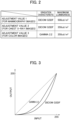
FIG. 2 is a table illustrating an example of adjustment values for image quality stored in an adjustment value storage unit ofFIG. 1 . -
FIG. 3 is a characteristic diagram illustrating gradation characteristics of DICOM GSDF and gamma 2.2. -
FIG. 4 is a block diagram illustrating a modified example of the medical image display device according to the first embodiment. -
FIG. 5 is a block diagram illustrating a medical image display device according to a second embodiment. -
FIG. 6 is a table illustrating an example of adjustment values for image quality stored in an adjustment value storage unit ofFIG. 5 . - Hereinafter, the medical image display device according to each embodiment will be described with reference to the accompanying drawings.
- In
FIG. 1 , medical image data read from a PACS server by an unillustrated workstation is supplied to animage attribute detector 15 and adisplay 20. - An adjustment
value storage unit 13 stores adjustment values for adjusting image quality when various types of medical images are to be displayed on thedisplay 20. The adjustmentvalue storage unit 13 is a ROM, for example. As shown inFIG. 2 , an adjustment value is a combination of a gradation characteristic and a maximum luminance, and the adjustmentvalue storage unit 13 stores three adjustment values, adjustment values 1 to 3, for example. The adjustment value 1 is an adjustment value for mammography images, the adjustment value 2 is an adjustment value for chest X-ray images, and the adjustment value 3 is an adjustment value for color images. When aliquid crystal panel 23 is a monochrome panel, the adjustment value 3 is not required. - DICOM GSDF shown in
FIG. 2 is a gradation characteristic using a grayscale standard display function (GSDF) conforming to the digital imaging and communications in medicine (DICOM) standard. Gamma 2.2 shown inFIG. 2 is a gradation characteristic having a gamma value of 2.2. Assuming that the medical image data is 8 bits, DICOM GSDF and gamma 2.2 have gradation characteristics indicating the input/output characteristics shown inFIG. 3 . - Returning to
FIG. 1 , an operator such as a doctor or nurse operates anoperation unit 11 to select an adjustment value according to the medical image to be displayed on thedisplay 20. A plurality of adjustment values may be toggled by operating theoperation unit 11, or a menu for selecting an adjustment value may be displayed on thedisplay 20 by operating theoperation unit 11, and one of the adjustment values may be selected. When an operation to select an adjustment value is performed by theoperation unit 11, anadjustment value setter 12 reads the selected adjustment value from the adjustmentvalue storage unit 13 and causes an adjustmentvalue holding unit 14 to hold the selected adjustment value. The adjustmentvalue holding unit 14 is a non-volatile memory, for example. - The
display 20 includes agamma corrector 21, aluminance adjuster 22, theliquid crystal panel 23, and abacklight 24. Thegamma corrector 21 performs a gamma correction on the input medical image data with the gradation characteristic shown inFIG. 3 . The luminance adjuster 22 controls the luminance of thebacklight 24 to adjust the maximum luminance of the medical image displayed on theliquid crystal panel 23. The maximum luminance of the medical image displayed on theliquid crystal panel 23 is determined by the luminance of thebacklight 24. - For example, if the operator selects the adjustment value 1 and the adjustment
value holding unit 14 holds the adjustment value 1, thegamma corrector 21 performs a gamma correction on the input medical image data so as to have the gradation characteristic of DICOM GSDF. The luminance adjuster 22 controls the luminance of thebacklight 24 so that the maximum luminance is 500 cd/m2. - The
image attribute detector 15 detects an attribute indicating the type of medical image by analyzing a tag (DICOM tag) attached to the medical image data. Theimage attribute detector 15 analyzes the tag to detect whether the medical image data is a mammography image or a chest X-ray image. - To explain the DICOM tag specifically, the DICOM tag includes "(0008, 0060)", which stores the type of device (modality) that generated the image, what is called the modality code. The modality code is represented by two letters of the alphabet, and MG indicates mammography, for example. If the modality code described in (0008, 0060) of the DICOM tag is MG, it can be detected that the device that generated the medical image data is a mammography device and the medical image data is a mammography image.
- The
image attribute detector 15 may detect the attribute of the medical image data by combining the DICOM tag (0008, 0060) and other tag information. - A
determiner 16 determines whether or not the adjustment value held in the adjustmentvalue holding unit 14 is an adjustment value corresponding to the attribute detected by theimage attribute detector 15. Thedeterminer 16 supplies the determination result to anotification unit 17. The fact that the adjustment value held in the adjustmentvalue holding unit 14 does not correspond to the attribute detected by theimage attribute detector 15 means that the image quality set in the medical image display device does not match the image quality suitable for the medical image to be displayed on thedisplay 20. - Therefore, when the determination result by the
determiner 16 indicates a mismatch, thenotification unit 17 visually or audibly informs the user of the medical image display device that the adjustment value held in the adjustmentvalue holding unit 14 is not suitable for the medical image to be displayed on thedisplay 20. Thenotification unit 17 may be a warning light such as an LED, or may be a speaker. When the medical image data is image data of a chest X-ray image and the adjustment value 1 is selected, thenotification unit 17 may output a notification such as "please adjust the maximum luminance to 350 cd/m2" or "please select the adjustment value 2". - The user who has received the notification from the
notification unit 17 can operate theoperation unit 11 to select an appropriate adjustment value according to the medical image to be displayed on thedisplay 20. Thus, according to the first embodiment, it is possible to avoid a mismatch between the image quality set in the medical image display device and the image quality suitable for the medical image to be displayed on the medical image display device. - As shown in
FIG. 4 , an on-screen display (OSD) superimposingunit 27 included in thedisplay 20 may function as thenotification unit 17. When the determination result by thedeterminer 16 indicates a mismatch, theOSD superimposing unit 27 superimposes text information or image information indicating that the adjustment value is inappropriate, on the gamma-corrected medical image data output from thegamma corrector 21, and supplies it to theliquid crystal panel 23. - The user can recognize that the adjustment value is not appropriate due to the text information or image information superimposed on the medical image displayed on the
liquid crystal panel 23, and can select an appropriate adjustment value. - The purpose of the second embodiment shown in
FIG. 5 is to adjust the image quality to be more appropriate according to the brightness of the ambient light surrounding the medical image display device. InFIG. 5 , the same parts as those inFIG. 1 are denoted by the same reference numerals, and the description thereof may be omitted. - As shown in
FIG. 6 , the adjustmentvalue storage unit 13 stores three adjustment values 1 to 3 as adjustment values that can be recognized by operators such as doctors and nurses, for example. In the second embodiment, each of the adjustment values 1 to 3 consists of two adjustment values, one suitable for bright surroundings and one suitable for dark surroundings. - The adjustment value 1 for mammography images includes an adjustment value 1A suitable for bright surroundings and an
adjustment value 1B suitable for dark surroundings. The adjustment value 2 for chest X-ray images includes anadjustment value 2A suitable for bright surroundings and anadjustment value 2B suitable for dark surroundings. The adjustment value 3 for color images includes an adjustment value 3A suitable for bright surroundings and anadjustment value 3B suitable for dark surroundings. - Bright surroundings are defined as the surrounding brightness being equal to or higher than a predetermined brightness, and dark surroundings are defined as the surrounding brightness being less than the predetermined brightness. In the example shown in
FIG. 6 , the maximum luminance is made to be different between when the surroundings are bright and when the surroundings are dark. The brightness of the surroundings may be divided into three or more levels, and the maximum luminance may be varied in three or more levels. Although one of the adjustment values 1 to 3 can be selected as the adjustment value, the adjustment values 1A, 1B, 2A, 2B, 3A, and 3B cannot be directly selected. - In
FIG. 5 , an ambientlight sensor 18 detects whether the ambient brightness is equal to or greater than a predetermined brightness, or less than the predetermined brightness. The operator operates theoperation unit 11 to select one of the adjustment values 1 to 3 according to the medical image to be displayed on thedisplay 20. Theadjustment value setter 12 selects an adjustment value corresponding to the selection value selected by the operator and according to the brightness detected by the ambientlight sensor 18. Theadjustment value setter 12 reads the selected adjustment value from the adjustmentvalue storage unit 13 and causes the adjustmentvalue holding unit 14 to hold the selected adjustment value. - The adjustment
value holding unit 14 holds theadjustment value 1A or 1B when the operator selects the adjustment value 1, theadjustment value adjustment value 3A or 3B when the operator selects the adjustment value 3. - According to the second embodiment, it is possible to adjust the image quality to be more appropriate according to the brightness of the ambient light surrounding the medical image display device. A medical image display device may be configured by combining the first embodiment and the second embodiment.
- The present invention is not limited to the first and second embodiments described above, and various modifications can be made without departing from the scope of the present invention. The
display 20 is not limited to a configuration including theliquid crystal panel 23, and may be configured to include an organic EL panel. In this case, thebacklight 24 is not necessary, and theluminance adjuster 22 adjusts the maximum luminance when the organic EL panel emits light. - In
FIG. 1 or5 , theadjustment value setter 12, theimage attribute detector 15, and thedeterminer 16 may be configured by means of a microcomputer or microprocessor. A part of the medical image display device may be configured by means of software (computer program). The use of hardware or software is optional. Furthermore, some of the functions of the medical image display device may be realized by a means of a computer device connected to the medical image display device. - This application claims priority under
Japanese Patent Application No. 2020-148151 filed with the Japan Patent Office on September 3, 2020
Claims (3)
- A medical image display device comprising:an adjustment value holding unit configured to hold an adjustment value selected for displaying medical image data on a medical image display device with a predetermined image quality;an image attribute detector configured to detect an attribute of the medical image data;a determiner configured to determine whether or not the adjustment value held in the adjustment value holding unit matches an adjustment value corresponding to the attribute detected by the image attribute detector; anda notification unit configured to visually or audibly notify that the adjustment value held in the adjustment value holding unit does not match an adjustment value suitable for the medical image to be displayed on the medical image display device, when the determiner determines that the adjustment value held in the adjustment value holding unit does not match the adjustment value corresponding to the attribute detected by the image attribute detector.
- The medical image display device according to claim 1,further comprising an adjustment value storage unit storing a plurality of different adjustment values associated with attributes,wherein the adjustment value includes at least a predetermined gradation characteristic and a maximum luminance, andan adjustment value corresponding to the attribute detected by the image attribute detector is selected from the adjustment values stored in the adjustment value storage unit.
- The medical image display device according to claim 2, wherein the adjustment value held in the adjustment value holding unit is an adjustment value selected from among the plurality of adjustment values stored in the adjustment value storage unit.
Applications Claiming Priority (2)
| Application Number | Priority Date | Filing Date | Title |
|---|---|---|---|
| JP2020148151A JP7505338B2 (en) | 2020-09-03 | 2020-09-03 | Medical image display device |
| PCT/JP2021/023748 WO2022049868A1 (en) | 2020-09-03 | 2021-06-23 | Medical image display apparatus |
Publications (3)
| Publication Number | Publication Date |
|---|---|
| EP4209176A1 true EP4209176A1 (en) | 2023-07-12 |
| EP4209176A4 EP4209176A4 (en) | 2023-07-12 |
| EP4209176B1 EP4209176B1 (en) | 2024-10-23 |
Family
ID=80491059
Family Applications (1)
| Application Number | Title | Priority Date | Filing Date |
|---|---|---|---|
| EP21863926.8A Active EP4209176B1 (en) | 2020-09-03 | 2021-06-23 | Medical image display apparatus |
Country Status (5)
| Country | Link |
|---|---|
| US (1) | US12303309B2 (en) |
| EP (1) | EP4209176B1 (en) |
| JP (1) | JP7505338B2 (en) |
| CN (1) | CN115397315B (en) |
| WO (1) | WO2022049868A1 (en) |
Family Cites Families (16)
| Publication number | Priority date | Publication date | Assignee | Title |
|---|---|---|---|---|
| US6323869B1 (en) * | 1998-01-09 | 2001-11-27 | Eastman Kodak Company | Method and system for modality dependent tone scale adjustment |
| JP2004057592A (en) | 2002-07-30 | 2004-02-26 | Secom Co Ltd | Hospital terminal device and remote reading system |
| JP2004159986A (en) * | 2002-11-14 | 2004-06-10 | Fuji Photo Film Co Ltd | Liquid crystal display device |
| US8896619B2 (en) * | 2011-03-09 | 2014-11-25 | Mckesson Financial Holdings | Apparatus, method and computer-readable storage medium for compensating for image-quality discrepancies |
| CN105307571B (en) * | 2013-06-18 | 2018-10-26 | 佳能株式会社 | Control device, photographic device, camera system and control method |
| JP6229342B2 (en) | 2013-07-18 | 2017-11-15 | 富士通株式会社 | Program, display adjustment support method, and display adjustment support device |
| JP6014001B2 (en) * | 2013-09-26 | 2016-10-25 | 京セラドキュメントソリューションズ株式会社 | Image forming apparatus and image forming program |
| JP2015173890A (en) * | 2014-03-17 | 2015-10-05 | キヤノン株式会社 | Image quality adjustment supporting system, image quality adjustment supporting method, and program |
| US9626476B2 (en) * | 2014-03-27 | 2017-04-18 | Change Healthcare Llc | Apparatus, method and computer-readable storage medium for transforming digital images |
| GB2528283B (en) * | 2014-07-16 | 2020-08-05 | Barco Nv | Image colour calibration with multiple colour scales |
| JP2016024362A (en) * | 2014-07-22 | 2016-02-08 | キヤノン株式会社 | Image display device, image output device, control method of image display device, control method of image output device and program |
| JP2016042954A (en) * | 2014-08-21 | 2016-04-04 | キヤノン株式会社 | Display control device, display system, and display control method |
| JP6699197B2 (en) * | 2016-01-25 | 2020-05-27 | コニカミノルタ株式会社 | Medical image display device, medical image adjusting method and program |
| JP6855279B2 (en) * | 2017-02-28 | 2021-04-07 | ゼネラル・エレクトリック・カンパニイ | Medical image device, control program for medical image device, ultrasonic image display device, control program for ultrasonic image display device, and image display device |
| CN107145730A (en) * | 2017-04-27 | 2017-09-08 | 广州慧扬健康科技有限公司 | DICOM medical images file reads adaption system |
| JP2020148151A (en) | 2019-03-14 | 2020-09-17 | ダイハツ工業株式会社 | Reserve tank |
-
2020
- 2020-09-03 JP JP2020148151A patent/JP7505338B2/en active Active
-
2021
- 2021-06-23 WO PCT/JP2021/023748 patent/WO2022049868A1/en not_active Ceased
- 2021-06-23 EP EP21863926.8A patent/EP4209176B1/en active Active
- 2021-06-23 CN CN202180027206.1A patent/CN115397315B/en active Active
-
2023
- 2023-03-02 US US18/116,474 patent/US12303309B2/en active Active
Also Published As
| Publication number | Publication date |
|---|---|
| JP2022042663A (en) | 2022-03-15 |
| JP7505338B2 (en) | 2024-06-25 |
| EP4209176B1 (en) | 2024-10-23 |
| US20230200766A1 (en) | 2023-06-29 |
| WO2022049868A1 (en) | 2022-03-10 |
| US12303309B2 (en) | 2025-05-20 |
| CN115397315B (en) | 2025-07-29 |
| EP4209176A4 (en) | 2023-07-12 |
| CN115397315A (en) | 2022-11-25 |
Similar Documents
| Publication | Publication Date | Title |
|---|---|---|
| US8890906B2 (en) | Method and system for remotely calibrating display of image data | |
| US6977670B2 (en) | Method and apparatus for selective registration of endoscopes with database | |
| US8872753B2 (en) | Adjusting brightness of a display image in a display having an adjustable intensity light source | |
| US6577753B2 (en) | Medical network system | |
| US20170047022A1 (en) | Display apparatus and control method thereof | |
| US20020039084A1 (en) | Medical image display system | |
| US20080123918A1 (en) | Image processing apparatus | |
| JPWO2010013308A1 (en) | Gray scale display device | |
| US20160054969A1 (en) | Display control apparatus controlling gradation characteristics of display apparatus, display system, and display control method | |
| US9795277B2 (en) | Monitor apparatus | |
| JP6064084B2 (en) | Image processing device | |
| CN109064990A (en) | A kind of display device and its image regulating method | |
| US10235968B2 (en) | Medical image display apparatus, medical image adjustment method and recording medium | |
| JP2012235807A (en) | Image processor, image display system, radiographic image photographing system, image processing program, and image processing method | |
| JP5231841B2 (en) | Electronic endoscope device | |
| US12303309B2 (en) | Medical image display device | |
| JP2004159986A (en) | Liquid crystal display device | |
| US11298094B2 (en) | Radiography system, portable information terminal, radiography method, and computer-readable storage medium | |
| JP2016024362A (en) | Image display device, image output device, control method of image display device, control method of image output device and program | |
| US9437162B2 (en) | Image output apparatus, control method therefor, image display apparatus, control method therefor, and storage medium | |
| US20100128049A1 (en) | Method and system for visualizing monochromatic images in color hue | |
| JP2015215482A (en) | Image display system and image display method | |
| JP2019133007A (en) | Medical image creation device | |
| US20220108787A1 (en) | Device and method for managing medical image data and treatment data | |
| JP2022129407A (en) | Image display system, server device, and image display method |
Legal Events
| Date | Code | Title | Description |
|---|---|---|---|
| STAA | Information on the status of an ep patent application or granted ep patent |
Free format text: STATUS: THE INTERNATIONAL PUBLICATION HAS BEEN MADE |
|
| PUAI | Public reference made under article 153(3) epc to a published international application that has entered the european phase |
Free format text: ORIGINAL CODE: 0009012 |
|
| STAA | Information on the status of an ep patent application or granted ep patent |
Free format text: STATUS: REQUEST FOR EXAMINATION WAS MADE |
|
| 17P | Request for examination filed |
Effective date: 20230302 |
|
| A4 | Supplementary search report drawn up and despatched |
Effective date: 20230512 |
|
| AK | Designated contracting states |
Kind code of ref document: A1 Designated state(s): AL AT BE BG CH CY CZ DE DK EE ES FI FR GB GR HR HU IE IS IT LI LT LU LV MC MK MT NL NO PL PT RO RS SE SI SK SM TR |
|
| DAV | Request for validation of the european patent (deleted) | ||
| DAX | Request for extension of the european patent (deleted) | ||
| GRAP | Despatch of communication of intention to grant a patent |
Free format text: ORIGINAL CODE: EPIDOSNIGR1 |
|
| STAA | Information on the status of an ep patent application or granted ep patent |
Free format text: STATUS: GRANT OF PATENT IS INTENDED |
|
| INTG | Intention to grant announced |
Effective date: 20240510 |
|
| GRAS | Grant fee paid |
Free format text: ORIGINAL CODE: EPIDOSNIGR3 |
|
| GRAA | (expected) grant |
Free format text: ORIGINAL CODE: 0009210 |
|
| STAA | Information on the status of an ep patent application or granted ep patent |
Free format text: STATUS: THE PATENT HAS BEEN GRANTED |
|
| RIN1 | Information on inventor provided before grant (corrected) |
Inventor name: TAKESHITA, HIROSHI Inventor name: HATANAKA, MASAO |
|
| AK | Designated contracting states |
Kind code of ref document: B1 Designated state(s): AL AT BE BG CH CY CZ DE DK EE ES FI FR GB GR HR HU IE IS IT LI LT LU LV MC MK MT NL NO PL PT RO RS SE SI SK SM TR |
|
| RAP3 | Party data changed (applicant data changed or rights of an application transferred) |
Owner name: JVCKENWOOD CORPORATION |
|
| REG | Reference to a national code |
Ref country code: GB Ref legal event code: FG4D |
|
| REG | Reference to a national code |
Ref country code: CH Ref legal event code: EP |
|
| REG | Reference to a national code |
Ref country code: DE Ref legal event code: R096 Ref document number: 602021020815 Country of ref document: DE |
|
| REG | Reference to a national code |
Ref country code: IE Ref legal event code: FG4D |
|
| REG | Reference to a national code |
Ref country code: LT Ref legal event code: MG9D |
|
| REG | Reference to a national code |
Ref country code: NL Ref legal event code: MP Effective date: 20241023 |
|
| REG | Reference to a national code |
Ref country code: AT Ref legal event code: MK05 Ref document number: 1734148 Country of ref document: AT Kind code of ref document: T Effective date: 20241023 |
|
| PG25 | Lapsed in a contracting state [announced via postgrant information from national office to epo] |
Ref country code: NL Free format text: LAPSE BECAUSE OF FAILURE TO SUBMIT A TRANSLATION OF THE DESCRIPTION OR TO PAY THE FEE WITHIN THE PRESCRIBED TIME-LIMIT Effective date: 20241023 |
|
| PG25 | Lapsed in a contracting state [announced via postgrant information from national office to epo] |
Ref country code: NL Free format text: LAPSE BECAUSE OF FAILURE TO SUBMIT A TRANSLATION OF THE DESCRIPTION OR TO PAY THE FEE WITHIN THE PRESCRIBED TIME-LIMIT Effective date: 20241023 |
|
| PG25 | Lapsed in a contracting state [announced via postgrant information from national office to epo] |
Ref country code: PT Free format text: LAPSE BECAUSE OF FAILURE TO SUBMIT A TRANSLATION OF THE DESCRIPTION OR TO PAY THE FEE WITHIN THE PRESCRIBED TIME-LIMIT Effective date: 20250224 Ref country code: IS Free format text: LAPSE BECAUSE OF FAILURE TO SUBMIT A TRANSLATION OF THE DESCRIPTION OR TO PAY THE FEE WITHIN THE PRESCRIBED TIME-LIMIT Effective date: 20250223 Ref country code: HR Free format text: LAPSE BECAUSE OF FAILURE TO SUBMIT A TRANSLATION OF THE DESCRIPTION OR TO PAY THE FEE WITHIN THE PRESCRIBED TIME-LIMIT Effective date: 20241023 |
|
| PG25 | Lapsed in a contracting state [announced via postgrant information from national office to epo] |
Ref country code: FI Free format text: LAPSE BECAUSE OF FAILURE TO SUBMIT A TRANSLATION OF THE DESCRIPTION OR TO PAY THE FEE WITHIN THE PRESCRIBED TIME-LIMIT Effective date: 20241023 |
|
| PG25 | Lapsed in a contracting state [announced via postgrant information from national office to epo] |
Ref country code: BG Free format text: LAPSE BECAUSE OF FAILURE TO SUBMIT A TRANSLATION OF THE DESCRIPTION OR TO PAY THE FEE WITHIN THE PRESCRIBED TIME-LIMIT Effective date: 20241023 |
|
| PG25 | Lapsed in a contracting state [announced via postgrant information from national office to epo] |
Ref country code: ES Free format text: LAPSE BECAUSE OF FAILURE TO SUBMIT A TRANSLATION OF THE DESCRIPTION OR TO PAY THE FEE WITHIN THE PRESCRIBED TIME-LIMIT Effective date: 20241023 |
|
| PG25 | Lapsed in a contracting state [announced via postgrant information from national office to epo] |
Ref country code: NO Free format text: LAPSE BECAUSE OF FAILURE TO SUBMIT A TRANSLATION OF THE DESCRIPTION OR TO PAY THE FEE WITHIN THE PRESCRIBED TIME-LIMIT Effective date: 20250123 |
|
| PG25 | Lapsed in a contracting state [announced via postgrant information from national office to epo] |
Ref country code: LV Free format text: LAPSE BECAUSE OF FAILURE TO SUBMIT A TRANSLATION OF THE DESCRIPTION OR TO PAY THE FEE WITHIN THE PRESCRIBED TIME-LIMIT Effective date: 20241023 Ref country code: AT Free format text: LAPSE BECAUSE OF FAILURE TO SUBMIT A TRANSLATION OF THE DESCRIPTION OR TO PAY THE FEE WITHIN THE PRESCRIBED TIME-LIMIT Effective date: 20241023 Ref country code: GR Free format text: LAPSE BECAUSE OF FAILURE TO SUBMIT A TRANSLATION OF THE DESCRIPTION OR TO PAY THE FEE WITHIN THE PRESCRIBED TIME-LIMIT Effective date: 20250124 |
|
| PG25 | Lapsed in a contracting state [announced via postgrant information from national office to epo] |
Ref country code: PL Free format text: LAPSE BECAUSE OF FAILURE TO SUBMIT A TRANSLATION OF THE DESCRIPTION OR TO PAY THE FEE WITHIN THE PRESCRIBED TIME-LIMIT Effective date: 20241023 |
|
| PG25 | Lapsed in a contracting state [announced via postgrant information from national office to epo] |
Ref country code: RS Free format text: LAPSE BECAUSE OF FAILURE TO SUBMIT A TRANSLATION OF THE DESCRIPTION OR TO PAY THE FEE WITHIN THE PRESCRIBED TIME-LIMIT Effective date: 20250123 |
|
| PG25 | Lapsed in a contracting state [announced via postgrant information from national office to epo] |
Ref country code: SM Free format text: LAPSE BECAUSE OF FAILURE TO SUBMIT A TRANSLATION OF THE DESCRIPTION OR TO PAY THE FEE WITHIN THE PRESCRIBED TIME-LIMIT Effective date: 20241023 |
|
| PGFP | Annual fee paid to national office [announced via postgrant information from national office to epo] |
Ref country code: DE Payment date: 20250429 Year of fee payment: 5 |
|
| PG25 | Lapsed in a contracting state [announced via postgrant information from national office to epo] |
Ref country code: DK Free format text: LAPSE BECAUSE OF FAILURE TO SUBMIT A TRANSLATION OF THE DESCRIPTION OR TO PAY THE FEE WITHIN THE PRESCRIBED TIME-LIMIT Effective date: 20241023 |
|
| PGFP | Annual fee paid to national office [announced via postgrant information from national office to epo] |
Ref country code: GB Payment date: 20250501 Year of fee payment: 5 |
|
| PG25 | Lapsed in a contracting state [announced via postgrant information from national office to epo] |
Ref country code: EE Free format text: LAPSE BECAUSE OF FAILURE TO SUBMIT A TRANSLATION OF THE DESCRIPTION OR TO PAY THE FEE WITHIN THE PRESCRIBED TIME-LIMIT Effective date: 20241023 |
|
| PGFP | Annual fee paid to national office [announced via postgrant information from national office to epo] |
Ref country code: FR Payment date: 20250508 Year of fee payment: 5 |
|
| PG25 | Lapsed in a contracting state [announced via postgrant information from national office to epo] |
Ref country code: RO Free format text: LAPSE BECAUSE OF FAILURE TO SUBMIT A TRANSLATION OF THE DESCRIPTION OR TO PAY THE FEE WITHIN THE PRESCRIBED TIME-LIMIT Effective date: 20241023 |
|
| REG | Reference to a national code |
Ref country code: DE Ref legal event code: R097 Ref document number: 602021020815 Country of ref document: DE |
|
| PG25 | Lapsed in a contracting state [announced via postgrant information from national office to epo] |
Ref country code: SK Free format text: LAPSE BECAUSE OF FAILURE TO SUBMIT A TRANSLATION OF THE DESCRIPTION OR TO PAY THE FEE WITHIN THE PRESCRIBED TIME-LIMIT Effective date: 20241023 |
|
| PG25 | Lapsed in a contracting state [announced via postgrant information from national office to epo] |
Ref country code: CZ Free format text: LAPSE BECAUSE OF FAILURE TO SUBMIT A TRANSLATION OF THE DESCRIPTION OR TO PAY THE FEE WITHIN THE PRESCRIBED TIME-LIMIT Effective date: 20241023 |
|
| PG25 | Lapsed in a contracting state [announced via postgrant information from national office to epo] |
Ref country code: IT Free format text: LAPSE BECAUSE OF FAILURE TO SUBMIT A TRANSLATION OF THE DESCRIPTION OR TO PAY THE FEE WITHIN THE PRESCRIBED TIME-LIMIT Effective date: 20241023 |
|
| PLBE | No opposition filed within time limit |
Free format text: ORIGINAL CODE: 0009261 |
|
| STAA | Information on the status of an ep patent application or granted ep patent |
Free format text: STATUS: NO OPPOSITION FILED WITHIN TIME LIMIT |
|
| PG25 | Lapsed in a contracting state [announced via postgrant information from national office to epo] |
Ref country code: SE Free format text: LAPSE BECAUSE OF FAILURE TO SUBMIT A TRANSLATION OF THE DESCRIPTION OR TO PAY THE FEE WITHIN THE PRESCRIBED TIME-LIMIT Effective date: 20241023 |
|
| 26N | No opposition filed |
Effective date: 20250724 |
|
| REG | Reference to a national code |
Ref country code: CH Ref legal event code: H13 Free format text: ST27 STATUS EVENT CODE: U-0-0-H10-H13 (AS PROVIDED BY THE NATIONAL OFFICE) Effective date: 20260127 |
|
| PG25 | Lapsed in a contracting state [announced via postgrant information from national office to epo] |
Ref country code: MC Free format text: LAPSE BECAUSE OF FAILURE TO SUBMIT A TRANSLATION OF THE DESCRIPTION OR TO PAY THE FEE WITHIN THE PRESCRIBED TIME-LIMIT Effective date: 20241023 |
|
| PG25 | Lapsed in a contracting state [announced via postgrant information from national office to epo] |
Ref country code: LU Free format text: LAPSE BECAUSE OF NON-PAYMENT OF DUE FEES Effective date: 20250623 |
|
| REG | Reference to a national code |
Ref country code: BE Ref legal event code: MM Effective date: 20250630 |
|
| PG25 | Lapsed in a contracting state [announced via postgrant information from national office to epo] |
Ref country code: IE Free format text: LAPSE BECAUSE OF NON-PAYMENT OF DUE FEES Effective date: 20250623 |
|
| PG25 | Lapsed in a contracting state [announced via postgrant information from national office to epo] |
Ref country code: BE Free format text: LAPSE BECAUSE OF NON-PAYMENT OF DUE FEES Effective date: 20250630 |
|
| PG25 | Lapsed in a contracting state [announced via postgrant information from national office to epo] |
Ref country code: CH Free format text: LAPSE BECAUSE OF NON-PAYMENT OF DUE FEES Effective date: 20250630 |