EP4202855A1 - Method for processing at least a pre-contrast image and a contrast image respectively depicting a body part prior to and after an injection of a dose of contrast agent - Google Patents
Method for processing at least a pre-contrast image and a contrast image respectively depicting a body part prior to and after an injection of a dose of contrast agent Download PDFInfo
- Publication number
- EP4202855A1 EP4202855A1 EP21306909.9A EP21306909A EP4202855A1 EP 4202855 A1 EP4202855 A1 EP 4202855A1 EP 21306909 A EP21306909 A EP 21306909A EP 4202855 A1 EP4202855 A1 EP 4202855A1
- Authority
- EP
- European Patent Office
- Prior art keywords
- dose
- contrast
- contrast image
- training
- injection
- Prior art date
- Legal status (The legal status is an assumption and is not a legal conclusion. Google has not performed a legal analysis and makes no representation as to the accuracy of the status listed.)
- Withdrawn
Links
Images



Classifications
-
- A—HUMAN NECESSITIES
- A61—MEDICAL OR VETERINARY SCIENCE; HYGIENE
- A61B—DIAGNOSIS; SURGERY; IDENTIFICATION
- A61B6/00—Apparatus or devices for radiation diagnosis; Apparatus or devices for radiation diagnosis combined with radiation therapy equipment
- A61B6/48—Diagnostic techniques
- A61B6/481—Diagnostic techniques involving the use of contrast agents
-
- A—HUMAN NECESSITIES
- A61—MEDICAL OR VETERINARY SCIENCE; HYGIENE
- A61B—DIAGNOSIS; SURGERY; IDENTIFICATION
- A61B6/00—Apparatus or devices for radiation diagnosis; Apparatus or devices for radiation diagnosis combined with radiation therapy equipment
- A61B6/52—Devices using data or image processing specially adapted for radiation diagnosis
-
- G—PHYSICS
- G01—MEASURING; TESTING
- G01R—MEASURING ELECTRIC VARIABLES; MEASURING MAGNETIC VARIABLES
- G01R33/00—Arrangements or instruments for measuring magnetic variables
- G01R33/20—Arrangements or instruments for measuring magnetic variables involving magnetic resonance
- G01R33/44—Arrangements or instruments for measuring magnetic variables involving magnetic resonance using nuclear magnetic resonance [NMR]
- G01R33/48—NMR imaging systems
- G01R33/54—Signal processing systems, e.g. using pulse sequences ; Generation or control of pulse sequences; Operator console
- G01R33/56—Image enhancement or correction, e.g. subtraction or averaging techniques, e.g. improvement of signal-to-noise ratio and resolution
- G01R33/5601—Image enhancement or correction, e.g. subtraction or averaging techniques, e.g. improvement of signal-to-noise ratio and resolution involving use of a contrast agent for contrast manipulation, e.g. a paramagnetic, super-paramagnetic, ferromagnetic or hyperpolarised contrast agent
-
- G—PHYSICS
- G01—MEASURING; TESTING
- G01R—MEASURING ELECTRIC VARIABLES; MEASURING MAGNETIC VARIABLES
- G01R33/00—Arrangements or instruments for measuring magnetic variables
- G01R33/20—Arrangements or instruments for measuring magnetic variables involving magnetic resonance
- G01R33/44—Arrangements or instruments for measuring magnetic variables involving magnetic resonance using nuclear magnetic resonance [NMR]
- G01R33/48—NMR imaging systems
- G01R33/54—Signal processing systems, e.g. using pulse sequences ; Generation or control of pulse sequences; Operator console
- G01R33/56—Image enhancement or correction, e.g. subtraction or averaging techniques, e.g. improvement of signal-to-noise ratio and resolution
- G01R33/5608—Data processing and visualization specially adapted for MR, e.g. for feature analysis and pattern recognition on the basis of measured MR data, segmentation of measured MR data, edge contour detection on the basis of measured MR data, for enhancing measured MR data in terms of signal-to-noise ratio by means of noise filtering or apodization, for enhancing measured MR data in terms of resolution by means for deblurring, windowing, zero filling, or generation of gray-scaled images, colour-coded images or images displaying vectors instead of pixels
-
- G—PHYSICS
- G06—COMPUTING OR CALCULATING; COUNTING
- G06N—COMPUTING ARRANGEMENTS BASED ON SPECIFIC COMPUTATIONAL MODELS
- G06N3/00—Computing arrangements based on biological models
- G06N3/02—Neural networks
- G06N3/04—Architecture, e.g. interconnection topology
- G06N3/0464—Convolutional networks [CNN, ConvNet]
-
- G—PHYSICS
- G06—COMPUTING OR CALCULATING; COUNTING
- G06T—IMAGE DATA PROCESSING OR GENERATION, IN GENERAL
- G06T12/00—Tomographic reconstruction from projections
- G06T12/10—Image preprocessing, e.g. calibration, positioning of sources or scatter correction
-
- G—PHYSICS
- G06—COMPUTING OR CALCULATING; COUNTING
- G06V—IMAGE OR VIDEO RECOGNITION OR UNDERSTANDING
- G06V10/00—Arrangements for image or video recognition or understanding
- G06V10/70—Arrangements for image or video recognition or understanding using pattern recognition or machine learning
- G06V10/77—Processing image or video features in feature spaces; using data integration or data reduction, e.g. principal component analysis [PCA] or independent component analysis [ICA] or self-organising maps [SOM]; Blind source separation
- G06V10/774—Generating sets of training patterns; Bootstrap methods, e.g. bagging or boosting
-
- G—PHYSICS
- G06—COMPUTING OR CALCULATING; COUNTING
- G06V—IMAGE OR VIDEO RECOGNITION OR UNDERSTANDING
- G06V10/00—Arrangements for image or video recognition or understanding
- G06V10/70—Arrangements for image or video recognition or understanding using pattern recognition or machine learning
- G06V10/82—Arrangements for image or video recognition or understanding using pattern recognition or machine learning using neural networks
-
- G—PHYSICS
- G16—INFORMATION AND COMMUNICATION TECHNOLOGY [ICT] SPECIALLY ADAPTED FOR SPECIFIC APPLICATION FIELDS
- G16H—HEALTHCARE INFORMATICS, i.e. INFORMATION AND COMMUNICATION TECHNOLOGY [ICT] SPECIALLY ADAPTED FOR THE HANDLING OR PROCESSING OF MEDICAL OR HEALTHCARE DATA
- G16H30/00—ICT specially adapted for the handling or processing of medical images
- G16H30/20—ICT specially adapted for the handling or processing of medical images for handling medical images, e.g. DICOM, HL7 or PACS
-
- G—PHYSICS
- G16—INFORMATION AND COMMUNICATION TECHNOLOGY [ICT] SPECIALLY ADAPTED FOR SPECIFIC APPLICATION FIELDS
- G16H—HEALTHCARE INFORMATICS, i.e. INFORMATION AND COMMUNICATION TECHNOLOGY [ICT] SPECIALLY ADAPTED FOR THE HANDLING OR PROCESSING OF MEDICAL OR HEALTHCARE DATA
- G16H30/00—ICT specially adapted for the handling or processing of medical images
- G16H30/40—ICT specially adapted for the handling or processing of medical images for processing medical images, e.g. editing
-
- G—PHYSICS
- G16—INFORMATION AND COMMUNICATION TECHNOLOGY [ICT] SPECIALLY ADAPTED FOR SPECIFIC APPLICATION FIELDS
- G16H—HEALTHCARE INFORMATICS, i.e. INFORMATION AND COMMUNICATION TECHNOLOGY [ICT] SPECIALLY ADAPTED FOR THE HANDLING OR PROCESSING OF MEDICAL OR HEALTHCARE DATA
- G16H50/00—ICT specially adapted for medical diagnosis, medical simulation or medical data mining; ICT specially adapted for detecting, monitoring or modelling epidemics or pandemics
- G16H50/20—ICT specially adapted for medical diagnosis, medical simulation or medical data mining; ICT specially adapted for detecting, monitoring or modelling epidemics or pandemics for computer-aided diagnosis, e.g. based on medical expert systems
-
- G—PHYSICS
- G16—INFORMATION AND COMMUNICATION TECHNOLOGY [ICT] SPECIALLY ADAPTED FOR SPECIFIC APPLICATION FIELDS
- G16H—HEALTHCARE INFORMATICS, i.e. INFORMATION AND COMMUNICATION TECHNOLOGY [ICT] SPECIALLY ADAPTED FOR THE HANDLING OR PROCESSING OF MEDICAL OR HEALTHCARE DATA
- G16H50/00—ICT specially adapted for medical diagnosis, medical simulation or medical data mining; ICT specially adapted for detecting, monitoring or modelling epidemics or pandemics
- G16H50/50—ICT specially adapted for medical diagnosis, medical simulation or medical data mining; ICT specially adapted for detecting, monitoring or modelling epidemics or pandemics for simulation or modelling of medical disorders
-
- G—PHYSICS
- G16—INFORMATION AND COMMUNICATION TECHNOLOGY [ICT] SPECIALLY ADAPTED FOR SPECIFIC APPLICATION FIELDS
- G16H—HEALTHCARE INFORMATICS, i.e. INFORMATION AND COMMUNICATION TECHNOLOGY [ICT] SPECIALLY ADAPTED FOR THE HANDLING OR PROCESSING OF MEDICAL OR HEALTHCARE DATA
- G16H50/00—ICT specially adapted for medical diagnosis, medical simulation or medical data mining; ICT specially adapted for detecting, monitoring or modelling epidemics or pandemics
- G16H50/70—ICT specially adapted for medical diagnosis, medical simulation or medical data mining; ICT specially adapted for detecting, monitoring or modelling epidemics or pandemics for mining of medical data, e.g. analysing previous cases of other patients
-
- G—PHYSICS
- G06—COMPUTING OR CALCULATING; COUNTING
- G06V—IMAGE OR VIDEO RECOGNITION OR UNDERSTANDING
- G06V2201/00—Indexing scheme relating to image or video recognition or understanding
- G06V2201/03—Recognition of patterns in medical or anatomical images
- G06V2201/031—Recognition of patterns in medical or anatomical images of internal organs
Definitions
- the field of this invention is that of machine/deep learning.
- the invention relates to methods for processing at least a pre-contrast image and a contrast image respectively depicting a body part prior to and after an injection of a first standard dose of contrast agent, in particular for generating a synthetic contrast image simulating said body part after an injection of a second dose of contrast agent which is higher than the standard dose.
- Contrast agents are substances used to increase the contrast of structures or fluids within the body in medical imaging.
- contrast agents usually absorb or alter external radiations emitted by the medical imaging device.
- contrast agents enhance the radiodensity in a target tissue or structure.
- contrast agents modify the relaxation times of nuclei within body tissues in order to alter the contrast in the image.
- Contrast agents are commonly used to improve the visibility of lesions, notably in neuroimaging for the initial diagnosis and treatment planning of brain tumors, such as glioma, brain metastasis, meningioma.
- DSC Dynamic susceptibility contrast
- DCE Dynamic contrast enhanced
- GBCA injection increases the sensitivity of MRI, allowing for instance the identification of smaller carcinogenic nodules, an earlier treatment initiation and, in turn, improving patient survival and quality of life.
- One solution to further increase MRI sensitivity is to increase the injected quantity of GBCA.
- the present invention provides a method for medical imaging, the method being characterized in that it comprises the implementation, by a data processor of a second server, of steps of:
- said second dose is at least 240% of the standard dose, preferably between 320% and 400% of the standard dose.
- the candidate pre-contrast image and the candidate contrast image are acquired by a medical imaging device connected to the second server, in particular an MRI scanner.
- Said at least one pre-contrast image and contrast image are T1-weighted images.
- Step (a) comprises obtaining three candidate pre-contrast images, including the T1-weighted pre-contrast image, a T2-flair-weighted pre-contrast image, and an ADC pre-contrast map; the CNN being trained to reconstruct, from a training T1-weighted pre-contrast image, a training T2-flair-weighted pre-contrast image, and a training ADC pre-contrast map and a training T1-weighted contrast image respectively depicting a body part prior to and after an injection of said low dose of contrast agent, a training T1-weighted contrast image depicting said body part after an injection of said standard dose of contrast agent; step (b) comprising applying the CNN to the three candidate pre-contrast image and the candidate contrast image, so as to generate a synthetic T1-weighted contrast image simulating said body part after an injection of a second dose of contrast agent which is higher than the standard dose.
- Said CNN comprises an encoder branch and then a decoder branch, with skip connections between the encoder branch and decoder branch.
- the method comprises a previous step of training, by a data processor of a first server, said CNN from a base of sequences of a training pre-contrast image, a first training contrast image and a second training image respectively depicting a body part prior to, after an injection of said low dose of contrast agent, and after an injection of said standard dose of contrast agent.
- the invention provides a computer program product comprising code instructions to execute a method according to the first aspect for medical imaging; and a computer-readable medium, on which is stored a computer program product comprising code instructions for executing said method according to the first aspect for medical imaging.
- the present invention proposes a method for medical imaging, in particular for processing at least a candidate pre-contrast image and a candidate contrast image respectively depicting a body part prior to and during or after an injection of a first dose of contrast agent, a convolutional neural network (CNN).
- CNN convolutional neural network
- pre-contrast image or "plain” image, it is meant an image depicting a given body part (to be monitored) prior to an injection of contrast agent, for a person or an animal.
- contrast image it is meant an image depicting said body part during or after the injection of contrast agent.
- the first one is the pre-contrast image
- each of the following is a contrast image.
- the contrast images may be images of a given phase (e.g. arterial, portal, delayed) or fully dynamic contrast enhanced (DCE).
- brain imaging i.e. said body part is the brain.
- the (pre-contrast or contrast) images are either directly acquired, or derived from images directly acquired, by a medical imaging device of the scanner type.
- Said imaging with injection of contrast agent may be:
- the acquisition of a said image may involve the injection of a contrast agent such as gadolinium (GBCA) for MRI or appropriate x-ray contrast agents.
- a contrast agent such as gadolinium (GBCA) for MRI or appropriate x-ray contrast agents.
- images can be 2D objects (with two spatial dimensions) but also possibly 3D objects (with three spatial dimensions), i.e. volumes constituted of stacks of bidimensional images as "slices" according to a third spatial dimension - in other words, we have 2+1 spatial dimensions).
- T1-weighted pre-contrast image and a T1-weighted contrast image in particular GRE (gradient echo) T1-weighted pre-contrast/contrast images, but we may further have:
- TSE magnetic spin echo
- the above-mentioned methods are implemented within an architecture such as illustrated in Figure 1 , by means of a first and/or second server 1a, 1b.
- the first server 1a is the training server (implementing a training method of the CNN) and the second server 1b is a processing server (implementing the processing method). It is fully possible that these two servers may be merged.
- Each of these servers 1a, 1b is typically remote computer equipment connected to an extended network 2 such as the Internet for data exchange.
- Each one comprises data processing means 11a, 11b of processor type (in particular the data processor 11a of the first server 1a have strong computing power, since learning is long and complex compared with ordinary use of the trained models), and optionally storage means 12a, 12b such as a computer memory e.g. a hard disk.
- the second server 1b may be connected to one or more medical imaging devices 10 as client equipment, for providing images to be processed, and receiving back parameters.
- the imaging device 10 comprises an injector for performing the injection of contrast agent, said injector applying the injection parameters.
- the memory 12a of the first server 1a stores a training database i.e. a set of images referred to as training images (as opposed to so-called inputted images that precisely are sought to be processed).
- a training database i.e. a set of images referred to as training images (as opposed to so-called inputted images that precisely are sought to be processed).
- Each image of the database could be pre-contrast or contrast, and contrast images may be labelled in terms of a dose of contrast agent injected. Note that images corresponding the same injection (i.e. forming a temporal sequence) are associated into sequences.
- a contrast image depicting a body part after an injection of the standard dose of contrast agent will be referred to as “standard dose contrast image”
- a contrast image depicting a body part after an injection of the low dose of contrast agent will be referred to as “low dose contrast image”.
- the standard dose is the recommended dose which is generally used for a medical examination. While side effects are possible, such dose is not particularly dangerous for the patient, and allows an image quality level sufficient for analysis/diagnostic purposes.
- the standard dose is 0.1mmol/kg.
- the low dose is a dose which is lower than the standard dose, and which causes less effects to the patient health.
- Said low dose may be any fraction of the standard dose and is preferably between 1/10 and 1/2 (between 10 and 50%) of the standard dose, preferably between 1/5 and 1/3 (between 20 and 33%) of the standard dose, preferably around 1/4 (25%).
- the low dose is for instance 0.025mmol/kg (25% of 0.1 mmol/kg).
- the low dose contrast image presents less contrast than the standard dose contrast image.
- naively amplifying the contrast enhancement of a x% low-dose CE-MRI by a factor of 100/x results in poor image quality with widespread noise and ambiguous structures.
- the present CNN is trained to reconstruct, from at least a training pre-contrast image and a training contrast image respectively depicting a body part prior to and after an injection of said low dose of contrast agent, a training contrast image depicting said body part after an injection of said standard dose of contrast agent.
- Such CNNs are known to the skilled person, see for instance the application WO2019074938 or the document Ammari S, Bône A, Balleyguier C, et al. Can Deep Learning Replace Gadolinium in Neuro-Oncology? Invest Radiol. 2021; Publish Ah(00):1-9. doi: 10.10971rli.0000000008 , and allow to reduce the contrast agent dose: by applying the CNN to a candidate pre-contrast image and a candidate contrast image depicting a body part prior to and after an injection of only the low dose of contrast agent, a synthetic contrast image simulating said body part after an injection of the standard dose can be generated.
- this CNN is able to predict the standard dose contrast image from the pre-contrast image and the low dose contrast image, and hence can be referred as "dose minimization CNN".
- said CNN use as inputs as least the T1-weighted pre-contrast/contrast images (and possibly also the T2-flair-weigthed pre-contrast image and/or the ADC pre-contrast map) and outputs another T1-weighted contrast image.
- the present method proposes a clever use of this dose minimization CNN to virtually increase the dose, by applying the dose reduction CNN on candidate contrast image depicting a body part after an injection of a "first dose” higher than low dose of contrast agent, possibly up to the standard dose (but not above). This leads to the generation of a synthetic contrast image simulating said body part after an injection of a "second dose” of contrast agent which is higher than the standard dose, i.e. a "super dose”.
- the CNN is not used for the task it is actually trained for, which is very unusual, see the two exemplary pipelines of figure 2 wherein the standard dose contrast image is directly used as the first dose contrast image.
- T1-weighted pre-contrast image is referred to as "T1”
- T2-flair-weighted pre-contrast image is referred to as "T2”
- ADC pre-contrast map is referred to as "ADC”
- T1c- low-dose T1-weighted contrast image
- T1c+ standard-dose T1-weighted contrast image
- T1c+ the "super dose” T1-weighted contrast image
- the second dose can reach 400% of the standard dose if the standard dose is used as first dose (see figure 2 ).
- the standard dose is 0.1 mmol.kg
- the second dose can reach 0.4 mmol/kg (virtual supplementary injection of 0.3 mmol/kg) hence the name "super dose”.
- said first dose is advantageously at least 80% of the standard dose, preferably around 100% of the standard dose, meaning that the second dose is typically at least 160% of the standard dose (80%/50%), preferably at least 240% (80%/33%), and preferably between 320% and 400% of the standard dose (80%/25% and 100%/25%).
- the figure 3 depicts an exemplary architecture of dose minimization CNN, of the U-Net type. Note that in the case of tridimensional pre-contrast/contrast images, the CNN is itself a tridimensional network as in the example of figure 2 , handling quadridimensional feature maps.
- U-Net is a neural network of the encoder-decoder type: it comprises an encoder branch (or “contracting path") that maps the input (at least the pre-contrast image and the low dose image/first dose image) into a high-level representation and then a decoder branch (or “expanding path”) generating the output image (the standard dose image/second dose image) from the high-level representation.
- an encoder branch or "contracting path” that maps the input (at least the pre-contrast image and the low dose image/first dose image) into a high-level representation and then a decoder branch (or “expanding path”) generating the output image (the standard dose image/second dose image) from the high-level representation.
- U-Net further comprises skip (or “lateral”) connections between the encoder branch and decoder branch.
- the encoder branch acts as a backbone, and can be seen as a conventional feature extraction network that can be of many types, and in particular a conventional CNN, preferably a fully convolutional neural network (direct succession of blocks of convolution layers and non-linear layers such as ReLU (rectified linear unit), that in particular alternates residual and strided convolution blocks to downsample).
- the encoder branch extracts from the input image a plurality of initial feature maps representative of the input image at different scales. More precisely, the backbone consists of a plurality of successive convolution blocks, such that the first block produces a first initial feature map from the input, then the second block produces a second initial feature map from the first initial feature map, etc.
- a pooling layer is placed between two blocks to decrease the scale by a factor of 2 (typically 2x2x2 convolution with stride 2 for down sampling in case of 3D images), and from one block to another the number of filters of the convolution layers used (generally 3 ⁇ 3 ⁇ 3 convolutions) is increased (and preferably doubled).
- the feature maps obtained by the encoder branch are said to be initial because they will be reprocessed by the decoder branch. Indeed, as explained, "low-level" maps have a higher spatial resolution but a shallow semantic depth.
- the decoder branch aims to increase their semantic depth by incorporating the information from the "high-level” maps.
- said decoder branch of the CNN has the symmetrical architecture of the encoder branch, as it generates, from the initial feature maps, a plurality of enriched feature maps that are again representative of the input image at different scales, but they incorporate the information from the initial feature maps of smaller or equal scale while reducing the number of channels.
- the decoder branch also consists of a plurality of successive convolution blocks but in opposite order, such that the first block produces the first enriched feature map (from which the output image may be directly generated) from the second enriched feature map and the first initial feature map, after the second block produces the second enriched feature map from the third enriched feature map and the second initial feature map, etc.
- the decoder branch is also preferably a fully convolutional CNN (direct succession of blocks of convolution layers and non-linear layers such as ReLU, that in particular alternates residual and strided convolution blocks to upsample),
- each i-th enriched map has the scale of the corresponding i-th initial map (i.e. sensibly same spatial size) but incorporates the information of all j-th maps, for each j ⁇ i.
- each i-th enriched map Di is generated according to the corresponding i-th initial map Ei and/or the next (i+1-th) enriched map, hence the "contracting and expansive" nature of the branches (i.e. "U" shaped): the initial maps are obtained in ascending order and then the enriched maps are obtained in descending order.
- the maximum semantic level is obtained at the "smallest-scale” map, and from there each map is enriched on the way back down again with the information of the already enriched maps.
- the skip connections between the encoder branch and the decoder branch provide the decoder branch with the various initial maps.
- the generation of an enriched map based on the corresponding initial map and the smaller-scale enriched map comprises rescaling of the enriched map, typically doubling the scale (if there has been halving of scale in the encoder branch), i.e. up sampling of the enriched feature map with by a 2 ⁇ 2 ⁇ 2 convolution ("up-convolution") that halves the number of feature channels, then concatenation with the corresponding initial map Ei (cropped of necessary, both maps are now sensibly the same scale) to double again the number of channels, and from one block to another the number of filters of the convolution layers used (generally 3 ⁇ 3 ⁇ 3 convolutions) is again decreased (and preferably further halved).
- up-convolution 2 ⁇ 2 ⁇ 2 convolution
- Downsampling and upsampling convolution blocks rely on 2 ⁇ 2 ⁇ 2 kernels, all other kernels are 3 ⁇ 3 ⁇ 3 at the exception of the final 1 ⁇ 1 ⁇ 1 convolution. All activation functions but the last sigmoid are 0.2-LeakyReLU.
- the present invention is not limited to the specific U-Net architecture: could be suitable any CNN which is trained to reconstruct, from at least a training pre-contrast image and a training contrast image respectively depicting a body part prior to and after an injection of said low dose of contrast agent, a training contrast image depicting said body part after an injection of said standard dose of contrast agent.
- the present method starts with a step (a) of obtaining said pre-contrast image and contrast images to be processed (referred to as candidate pre-contrast and contrast images), in particular T1-weighted pre-contrast/contrast images, preferably from a medical imaging device 10 connected to the second server 1b.
- candidate pre-contrast and contrast images referred to as candidate pre-contrast and contrast images
- the candidate pre-contrast image and candidate contrast image respectively depict a body part prior to and after an injection of a first dose of contrast agent, wherein said first dose is higher than a predetermined low dose, the predetermined reduced dose being lower than a predetermined standard dose.
- step (a) comprises obtaining three candidate pre-contrast images (the candidate T1-weighted pre-contrast image, a candidate T2-flair-weighted pre-contrast image, and a candidate ADC pre-contrast map).
- step (a) may be implemented by the data processor 11b of the second server 1b and/or by the medical imaging device 10.
- the CNN is applied to the three candidate pre-contrast images and the candidate contrast image.
- the method advantageously comprises a previous step (a0) of training the CNN, implemented by the data processor 11a of the first server 1a.
- the CNN By training, it is meant the determination of the optimal values of parameters and weights or the CNN. Note that alternatively the CNN may be directly taken “off the shelf” with preset values of parameters and weights.
- Said training method can be performed according to the prior art, and any suitable training protocol known to a skilled person may be used.
- the CNN is trained to perform its original "dose minimization" task, i.e. to reconstruct, from at least a training pre-contrast image and a training contrast image respectively depicting a body part prior to and after an injection of said low dose of contrast agent, a training contrast image depicting said body part after an injection of said standard dose of contrast agent.
- the training base has just to comprise a plurality of sequences of at least one training pre-contrast image (as explained there could be possibly three pre-contrast images), an "initial” training contrast image and a “final” training contrast image respectively depicting a body part prior to, after an injection of said low dose of contrast agent, and after an injection of said standard dose of contrast agent.
- the CNN is trained to predict the final training contrast image (as ground truth) from the training pre-contrast image(s) and the initial contrast image of the same sequence.
- Figure 5 displays the T2-Flair, T1c, T1c+ and tse-T1c images for four example MRI exams from the test sample, either with brain metastases or glioma.
- T1c+ images the synthetic super dose contrast images
- Table 1 reports average image quality (IQ) scores for the T1c, T1c+ and tse-T1c images from 79 exams of a test sample. Grades are expressed on a 4-point Likert scale ranging from 1 (poor) to 4 (excellent). Standard deviations are given between parentheses. Differences across readers and post-contrast MRI images are compared using two-tailed t-tests, and p-values are reported. Best metrics are emphasized using a bold font when the 5% significance threshold is met)
- This table shows the synthetized T1c+ images were preferred by two readers (neuroradiologists with respectively 10 and 11 years of experience) for their general image quality, when no quality difference was found between T1c and tse-T1c images. On average between readers, T1c and tse-T1c were graded 2.7/4 (average to good), when T1c+ were graded 3.4/4 (good to excellent). Table 1 IQ ⁇ std (grades on a 4-point scale) T1c vs. T1c+ T1c vs. tse-T1c T1c+ vs.
- Table 2 reports the average contrast-to-noise ratio (CNR), lesion-to-brain ratio (LBR), and contrast enhancement percentage (CEP) performance metrics for the T1c, T1c+ and tse-T1c images from 52 exams of the test sample with at least one reference lesion. Standard deviations are given between parentheses. Differences across readers and post-contrast MRI images are compared using two-tailed t-tests, and p-values are reported. Best metrics are emphasized using a bold font when the 5% significance threshold is met.
- CNR contrast-to-noise ratio
- LBR lesion-to-brain ratio
- CEP contrast enhancement percentage
- T1c and tse-T1c images defined 187 reference lesions with a median long axis length of 9.2mm (inter-quartile range of 10.7mm).
- Three nested evaluation configurations were considered, depending on the minimum included lesion size: 10mm, 5mm or 0mm - all lesions being considered in this case.
- the access to T1c+ images increased the lesion detection sensitivity (SE) for both readers in all evaluation configurations.
- SE lesion detection sensitivity
- FDR false detection rates
- PPV PPV remained higher than 90% in all configurations. On average across readers, PPV quantitative differences were 3% at maximum between the two reading scenarios. F1 was higher than 90% across readers and readings for lesions larger than 10mm, higher than 80% for lesions larger than 5mm, and higher 70% when all lesions were included. On average across readers, F1 was higher when T1c+ was available to readers by a margin that varied between 4% and 11% according to considered range of lesion sizes.
- the invention provides a computer program product comprising code instructions to execute a method (particularly on the data processor 11a, 11b of the first and/or second server 1a, 1b) according to first second aspect of the invention for medical imaging, and storage means readable by computer equipment (memory of the first or second server 1a, 1b) provided with this computer program product.
Landscapes
- Engineering & Computer Science (AREA)
- Health & Medical Sciences (AREA)
- Medical Informatics (AREA)
- General Health & Medical Sciences (AREA)
- Physics & Mathematics (AREA)
- Public Health (AREA)
- Life Sciences & Earth Sciences (AREA)
- Nuclear Medicine, Radiotherapy & Molecular Imaging (AREA)
- Radiology & Medical Imaging (AREA)
- Biomedical Technology (AREA)
- Primary Health Care (AREA)
- Pathology (AREA)
- Epidemiology (AREA)
- Theoretical Computer Science (AREA)
- Databases & Information Systems (AREA)
- General Physics & Mathematics (AREA)
- Computer Vision & Pattern Recognition (AREA)
- High Energy & Nuclear Physics (AREA)
- Data Mining & Analysis (AREA)
- Evolutionary Computation (AREA)
- Artificial Intelligence (AREA)
- Molecular Biology (AREA)
- Biophysics (AREA)
- Veterinary Medicine (AREA)
- Animal Behavior & Ethology (AREA)
- Surgery (AREA)
- Heart & Thoracic Surgery (AREA)
- Optics & Photonics (AREA)
- Computing Systems (AREA)
- Software Systems (AREA)
- Multimedia (AREA)
- Signal Processing (AREA)
- Condensed Matter Physics & Semiconductors (AREA)
- Computational Linguistics (AREA)
- General Engineering & Computer Science (AREA)
- Mathematical Physics (AREA)
- Apparatus For Radiation Diagnosis (AREA)
- Image Analysis (AREA)
- Magnetic Resonance Imaging Apparatus (AREA)
- Image Processing (AREA)
Abstract
The present invention relates to method for medical imaging, the method being characterized in that it comprises the implementation, by a data processor (11b) of a second server (1b), of steps of:(a) Obtaining at least a candidate pre-contrast image and a candidate contrast image respectively depicting a body part prior to and after an injection of a first dose of contrast agent, wherein said first dose is higher than a predetermined low dose, the predetermined reduced dose being lower than a predetermined standard dose; a convolutional neural network, CNN, being trained to reconstruct, from at least a training pre-contrast image and a training contrast image respectively depicting a body part prior to and after an injection of said low dose of contrast agent, a training contrast image depicting said body part after an injection of said standard dose of contrast agent;(b) Applying the CNN to the candidate pre-contrast image and the candidate contrast image, so as to generate a synthetic contrast image simulating said body part after an injection of a second dose of contrast agent which is higher than the standard dose.
Description
- The field of this invention is that of machine/deep learning.
- More particularly, the invention relates to methods for processing at least a pre-contrast image and a contrast image respectively depicting a body part prior to and after an injection of a first standard dose of contrast agent, in particular for generating a synthetic contrast image simulating said body part after an injection of a second dose of contrast agent which is higher than the standard dose.
- Contrast agents are substances used to increase the contrast of structures or fluids within the body in medical imaging.
- They usually absorb or alter external radiations emitted by the medical imaging device. In x-rays, contrast agents enhance the radiodensity in a target tissue or structure. In MRIs, contrast agents modify the relaxation times of nuclei within body tissues in order to alter the contrast in the image.
- Contrast agents are commonly used to improve the visibility of lesions, notably in neuroimaging for the initial diagnosis and treatment planning of brain tumors, such as glioma, brain metastasis, meningioma.
- Dynamic susceptibility contrast (DSC) and Dynamic contrast enhanced (DCE), respectively leveraging T2 and T1 effects, are the two most common techniques for MRI. In both cases, a gadolinium-based contrast agent (GBCA) is injected intravenously to the patient and rapid repeated imaging is performed in order to obtain a temporal sequence of images.
- GBCA injection increases the sensitivity of MRI, allowing for instance the identification of smaller carcinogenic nodules, an earlier treatment initiation and, in turn, improving patient survival and quality of life. One solution to further increase MRI sensitivity is to increase the injected quantity of GBCA.
- However, based on precautionary considerations, recent clinical guidelines suggest using the minimum dosage that achieves a sufficient contrast enhancement, , and GBCA usage should therefore be as parsimonious as possible.
- Three complementary research avenues for nevertheless improving MRI contrast and its lesion detection performance can be identified:
- Firstly, new contrast agents with improved chemical properties have the potential to improve the image contrast without increasing the injected dose of gadolinium. However, such agents are still under testing, and are likely to be even more expensive than GBCA.
- Secondly, novel MRI sequences, with or without contrast agent, may respectively replace or complement the routine contrast-enhanced T1 sequences. In particular, the turbo spin echo T1 sequence with variable flip angles (TSE) proved more sensitive than its gradient echo (GRE) counterpart and is now recommended for brain tumor imaging, although some qualitative limitations were also identified in addition to the longer minimum scan time that may favor motion artifacts.
- Third, promising deep learning approaches are being increasingly proposed to automatically detect and delineate tumors, bearing the promise of uniform and constant accuracy levels with virtually instantaneous reading times. However, these deep learning algorithms are still under research and their performance remains insufficient for immediate large-scale deployment.
- There is consequently still a need for a new method to further improve sensitivity for contrast enhanced medical imaging.
- For these purposes, the present invention provides a method for medical imaging, the method being characterized in that it comprises the implementation, by a data processor of a second server, of steps of:
- (a) Obtaining at least a candidate pre-contrast image and a candidate contrast image respectively depicting a body part prior to and after an injection of a first dose of contrast agent, wherein said first dose is higher than a predetermined low dose, the predetermined reduced dose being lower than a predetermined standard dose; a convolutional neural network, (CNN), being trained to reconstruct, from at least a training pre-contrast image and a training contrast image respectively depicting a body part prior to and after an injection of said low dose of contrast agent, a training contrast image depicting said body part after an injection of said standard dose of contrast agent;
- (b) Applying the CNN to the candidate pre-contrast image and the candidate contrast image, so as to generate a synthetic contrast image simulating said body part after an injection of a second dose of contrast agent which is higher than the standard dose.
- Preferred but non limiting features of the present invention are as it follows:
- Said low dose is between 10 and 50% of the standard dose.
- Said low dose is between 1/5 and 1/3 of the standard dose, preferably around 25%
- Said first dose is at least 50% of the standard dose.
- Said first dose is at least 80% of the standard dose, preferably around 100% of the standard dose
- So that said second dose is at least 240% of the standard dose, preferably between 320% and 400% of the standard dose.
- The candidate pre-contrast image and the candidate contrast image are acquired by a medical imaging device connected to the second server, in particular an MRI scanner.
- Said at least one pre-contrast image and contrast image are T1-weighted images.
- Step (a) comprises obtaining three candidate pre-contrast images, including the T1-weighted pre-contrast image, a T2-flair-weighted pre-contrast image, and an ADC pre-contrast map; the CNN being trained to reconstruct, from a training T1-weighted pre-contrast image, a training T2-flair-weighted pre-contrast image, and a training ADC pre-contrast map and a training T1-weighted contrast image respectively depicting a body part prior to and after an injection of said low dose of contrast agent, a training T1-weighted contrast image depicting said body part after an injection of said standard dose of contrast agent; step (b) comprising applying the CNN to the three candidate pre-contrast image and the candidate contrast image, so as to generate a synthetic T1-weighted contrast image simulating said body part after an injection of a second dose of contrast agent which is higher than the standard dose.
- Said CNN comprises an encoder branch and then a decoder branch, with skip connections between the encoder branch and decoder branch.
- The method comprises a previous step of training, by a data processor of a first server, said CNN from a base of sequences of a training pre-contrast image, a first training contrast image and a second training image respectively depicting a body part prior to, after an injection of said low dose of contrast agent, and after an injection of said standard dose of contrast agent.
- According to a second and a third aspect, the invention provides a computer program product comprising code instructions to execute a method according to the first aspect for medical imaging; and a computer-readable medium, on which is stored a computer program product comprising code instructions for executing said method according to the first aspect for medical imaging.
- The above and other objects, features and advantages of this invention will be apparent in the following detailed description of an illustrative embodiment thereof, which is to be read in connection with the accompanying drawings wherein:
-
figure 1 illustrates an example of architecture in which the method according to the invention is performed; -
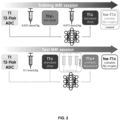
figure 2 represents an example of training pipeline and operation pipeline of a CNN in the method according to the invention; -
figure 3 represents a preferred architecture of CNN used in the method according to the invention; -
figure 4 illustrates an embodiment of the method according to the invention; -
figure 5 compares T1 c and T1 c+ contrast images obtained using the method according to the invention. - The present invention proposes a method for medical imaging, in particular for processing at least a candidate pre-contrast image and a candidate contrast image respectively depicting a body part prior to and during or after an injection of a first dose of contrast agent, a convolutional neural network (CNN).
- By pre-contrast image, or "plain" image, it is meant an image depicting a given body part (to be monitored) prior to an injection of contrast agent, for a person or an animal. By contrast image it is meant an image depicting said body part during or after the injection of contrast agent.
- In other words, if there is a temporal sequence of images, the first one is the pre-contrast image, and each of the following is a contrast image. Note that the contrast images may be images of a given phase (e.g. arterial, portal, delayed) or fully dynamic contrast enhanced (DCE).
- In the following description, we will take the preferred example of brain imaging, i.e. said body part is the brain.
- The (pre-contrast or contrast) images are either directly acquired, or derived from images directly acquired, by a medical imaging device of the scanner type.
- Said imaging with injection of contrast agent may be:
- CT (Computed Tomography) → the medical imaging device is an X-ray rotational scanner capable of tomographic reconstruction;
- MRI (Magnetic Resonance Imaging) → the medical imaging device is an MRI scanner;
- Mammography → the medical imaging device is an X-ray mammograph;
- Etc.
- The acquisition of a said image may involve the injection of a contrast agent such as gadolinium (GBCA) for MRI or appropriate x-ray contrast agents.
- Note that the "images" can be 2D objects (with two spatial dimensions) but also possibly 3D objects (with three spatial dimensions), i.e. volumes constituted of stacks of bidimensional images as "slices" according to a third spatial dimension - in other words, we have 2+1 spatial dimensions).
- Furthermore, we can have a plurality of pre-contrast images and/or candidate contrast images.
- In a preferred MRI embodiment, we have at least a T1-weighted pre-contrast image and a T1-weighted contrast image, in particular GRE (gradient echo) T1-weighted pre-contrast/contrast images, but we may further have:
- a T2-flair-weighted pre-contrast image (in particular TSE (turbospin echo)); and/or
- an ADC (apparent diffusion coefficient) pre-contrast map (automatically converted from EPI (echo-planar) DWI (diffusion weighted)),
- Note that a TSE (turbospin echo) T1-weighted contrast image can also be available, but it will not be presently used.
- The above-mentioned methods are implemented within an architecture such as illustrated in
Figure 1 , by means of a first and/orsecond server first server 1a is the training server (implementing a training method of the CNN) and thesecond server 1b is a processing server (implementing the processing method). It is fully possible that these two servers may be merged. - Each of these
servers extended network 2 such as the Internet for data exchange. Each one comprises data processing means 11a, 11b of processor type (in particular thedata processor 11a of thefirst server 1a have strong computing power, since learning is long and complex compared with ordinary use of the trained models), and optionally storage means 12a, 12b such as a computer memory e.g. a hard disk. Thesecond server 1b may be connected to one or moremedical imaging devices 10 as client equipment, for providing images to be processed, and receiving back parameters. - Note that it is supposed that the
imaging device 10 comprises an injector for performing the injection of contrast agent, said injector applying the injection parameters. - The
memory 12a of thefirst server 1a stores a training database i.e. a set of images referred to as training images (as opposed to so-called inputted images that precisely are sought to be processed). Each image of the database could be pre-contrast or contrast, and contrast images may be labelled in terms of a dose of contrast agent injected. Note that images corresponding the same injection (i.e. forming a temporal sequence) are associated into sequences. - In the following description, we will refer to a "low dose" and a "standard dose" of injected contrast agent. Both are predetermined. For convenience, a contrast image depicting a body part after an injection of the standard dose of contrast agent will be referred to as "standard dose contrast image", and a contrast image depicting a body part after an injection of the low dose of contrast agent will be referred to as "low dose contrast image".
- The standard dose, or "full-dose", is the recommended dose which is generally used for a medical examination. While side effects are possible, such dose is not particularly dangerous for the patient, and allows an image quality level sufficient for analysis/diagnostic purposes. In the case of GBCA, the standard dose is 0.1mmol/kg.
- The low dose, or "reduced dose", is a dose which is lower than the standard dose, and which causes less effects to the patient health. Said low dose may be any fraction of the standard dose and is preferably between 1/10 and 1/2 (between 10 and 50%) of the standard dose, preferably between 1/5 and 1/3 (between 20 and 33%) of the standard dose, preferably around 1/4 (25%). In the case of GBCA, the low dose is for instance 0.025mmol/kg (25% of 0.1 mmol/kg).
- Obviously, the low dose contrast image presents less contrast than the standard dose contrast image. And naively amplifying the contrast enhancement of a x% low-dose CE-MRI by a factor of 100/x results in poor image quality with widespread noise and ambiguous structures.
- The present CNN is trained to reconstruct, from at least a training pre-contrast image and a training contrast image respectively depicting a body part prior to and after an injection of said low dose of contrast agent, a training contrast image depicting said body part after an injection of said standard dose of contrast agent.
- Such CNNs are known to the skilled person, see for instance the application
WO2019074938 or the document Ammari S, Bône A, Balleyguier C, et al. Can Deep Learning Replace Gadolinium in Neuro-Oncology? Invest Radiol. 2021; Publish Ah(00):1-9. doi: 10.10971rli.00000000000008 , and allow to reduce the contrast agent dose: by applying the CNN to a candidate pre-contrast image and a candidate contrast image depicting a body part prior to and after an injection of only the low dose of contrast agent, a synthetic contrast image simulating said body part after an injection of the standard dose can be generated. - To rephrase, this CNN is able to predict the standard dose contrast image from the pre-contrast image and the low dose contrast image, and hence can be referred as "dose minimization CNN".
- Indeed, we can still have the quality of the standard dose contrast image while only injecting the low dose of contrast agent.
- Possible embodiments of this CNN and training methods will be described below.
- Preferably, said CNN use as inputs as least the T1-weighted pre-contrast/contrast images (and possibly also the T2-flair-weigthed pre-contrast image and/or the ADC pre-contrast map) and outputs another T1-weighted contrast image.
- The present method proposes a clever use of this dose minimization CNN to virtually increase the dose, by applying the dose reduction CNN on candidate contrast image depicting a body part after an injection of a "first dose" higher than low dose of contrast agent, possibly up to the standard dose (but not above). This leads to the generation of a synthetic contrast image simulating said body part after an injection of a "second dose" of contrast agent which is higher than the standard dose, i.e. a "super dose".
- Consequently, we are able to simulate the injection of a high dose that could be dangerous to the patient but allows a very high quality of image, while only injecting no more than the standard dose of contrast agent.
- In other words, the CNN is not used for the task it is actually trained for, which is very unusual, see the two exemplary pipelines of
figure 2 wherein the standard dose contrast image is directly used as the first dose contrast image. - In this figure, the T1-weighted pre-contrast image is referred to as "T1", the T2-flair-weighted pre-contrast image is referred to as "T2", the ADC pre-contrast map is referred to as "ADC", the low-dose T1-weighted contrast image is referred to as "T1c-", the standard-dose T1-weighted contrast image is referred to as "T1c", the "super dose" T1-weighted contrast image is referred to as "T1c+".
- Hypothesizing that the CNN primarily learned to amplify the difference of contrast between their pre-contrast and post-contrast inputs, replacing T1c- by T1c images at inference was expected to synthesize approximate quadruple-dose T1c+ images.
- For instance, assuming the low dose (Dc-) is x% of the standard dose (Dc):

- Because the same CNN is used, the first and second doses (D1, D2) can be written with respect to the same equation: D1=x/100*D2.
- And thus: Dc- < D1 ≤ Dc

- Consequently, in the embodiment wherein the low dose is 25% of the standard dose, the second dose can reach 400% of the standard dose if the standard dose is used as first dose (see
figure 2 ). In other words, if the standard dose is 0.1 mmol.kg, the second dose can reach 0.4 mmol/kg (virtual supplementary injection of 0.3 mmol/kg) hence the name "super dose". - Note that said first dose is advantageously at least 80% of the standard dose, preferably around 100% of the standard dose, meaning that the second dose is typically at least 160% of the standard dose (80%/50%), preferably at least 240% (80%/33%), and preferably between 320% and 400% of the standard dose (80%/25% and 100%/25%).
- The
figure 3 depicts an exemplary architecture of dose minimization CNN, of the U-Net type. Note that in the case of tridimensional pre-contrast/contrast images, the CNN is itself a tridimensional network as in the example offigure 2 , handling quadridimensional feature maps. - U-Net is a neural network of the encoder-decoder type: it comprises an encoder branch (or "contracting path") that maps the input (at least the pre-contrast image and the low dose image/first dose image) into a high-level representation and then a decoder branch (or "expanding path") generating the output image (the standard dose image/second dose image) from the high-level representation.
- U-Net further comprises skip (or "lateral") connections between the encoder branch and decoder branch.
- The encoder branch, acts as a backbone, and can be seen as a conventional feature extraction network that can be of many types, and in particular a conventional CNN, preferably a fully convolutional neural network (direct succession of blocks of convolution layers and non-linear layers such as ReLU (rectified linear unit), that in particular alternates residual and strided convolution blocks to downsample). The encoder branch extracts from the input image a plurality of initial feature maps representative of the input image at different scales. More precisely, the backbone consists of a plurality of successive convolution blocks, such that the first block produces a first initial feature map from the input, then the second block produces a second initial feature map from the first initial feature map, etc.
- It is conventionally understood for convolutional neural networks that the scale is smaller with each successive map (in other words the resolution decreases, the feature map becomes "smaller" and therefore less detailed), but of greater semantic depth, since increasingly high-level structures of the image have been captured. Specifically, initial feature maps have increasing numbers of channels as their spatial size decreases.
- In practice, a pooling layer is placed between two blocks to decrease the scale by a factor of 2 (typically 2x2x2 convolution with
stride 2 for down sampling in case of 3D images), and from one block to another the number of filters of the convolution layers used (generally 3×3×3 convolutions) is increased (and preferably doubled). - In the 5-level standard U-Net there is for example successive channel numbers of 32, 64, 128, 256 and 512, and successive map spatial sizes (for a 160×192×160 input image) of 160×192×160, 80×96×80, 40×48×40, 20×24×20, 10×12×10. We see that the input has already 4 channels - 3 pre-contrast images (T1, T2-flair ADC) and 1 contrast image (T1c- in the training and T1c in operation), while the output as a single channel (T1c in the training and T1c+ in operation).
- The feature maps obtained by the encoder branch are said to be initial because they will be reprocessed by the decoder branch. Indeed, as explained, "low-level" maps have a higher spatial resolution but a shallow semantic depth. The decoder branch aims to increase their semantic depth by incorporating the information from the "high-level" maps.
- Thus, said decoder branch of the CNN has the symmetrical architecture of the encoder branch, as it generates, from the initial feature maps, a plurality of enriched feature maps that are again representative of the input image at different scales, but they incorporate the information from the initial feature maps of smaller or equal scale while reducing the number of channels.
- In other words, the decoder branch also consists of a plurality of successive convolution blocks but in opposite order, such that the first block produces the first enriched feature map (from which the output image may be directly generated) from the second enriched feature map and the first initial feature map, after the second block produces the second enriched feature map from the third enriched feature map and the second initial feature map, etc. The decoder branch is also preferably a fully convolutional CNN (direct succession of blocks of convolution layers and non-linear layers such as ReLU, that in particular alternates residual and strided convolution blocks to upsample),
- In more details, each i-th enriched map has the scale of the corresponding i-th initial map (i.e. sensibly same spatial size) but incorporates the information of all j-th maps, for each j≥i. In practice, each i-th enriched map Di is generated according to the corresponding i-th initial map Ei and/or the next (i+1-th) enriched map, hence the "contracting and expansive" nature of the branches (i.e. "U" shaped): the initial maps are obtained in ascending order and then the enriched maps are obtained in descending order.
- Indeed, the maximum semantic level is obtained at the "smallest-scale" map, and from there each map is enriched on the way back down again with the information of the already enriched maps. The skip connections between the encoder branch and the decoder branch provide the decoder branch with the various initial maps.
- Typically, the generation of an enriched map based on the corresponding initial map and the smaller-scale enriched map comprises rescaling of the enriched map, typically doubling the scale (if there has been halving of scale in the encoder branch), i.e. up sampling of the enriched feature map with by a 2×2×2 convolution ("up-convolution") that halves the number of feature channels, then concatenation with the corresponding initial map Ei (cropped of necessary, both maps are now sensibly the same scale) to double again the number of channels, and from one block to another the number of filters of the convolution layers used (generally 3×3×3 convolutions) is again decreased (and preferably further halved).
- Downsampling and upsampling convolution blocks rely on 2×2×2 kernels, all other kernels are 3×3×3 at the exception of the final 1×1×1 convolution. All activation functions but the last sigmoid are 0.2-LeakyReLU.
- Note that the present invention is not limited to the specific U-Net architecture: could be suitable any CNN which is trained to reconstruct, from at least a training pre-contrast image and a training contrast image respectively depicting a body part prior to and after an injection of said low dose of contrast agent, a training contrast image depicting said body part after an injection of said standard dose of contrast agent.
- The training will be described below.
- As represented by
figure 4 , the present method starts with a step (a) of obtaining said pre-contrast image and contrast images to be processed (referred to as candidate pre-contrast and contrast images), in particular T1-weighted pre-contrast/contrast images, preferably from amedical imaging device 10 connected to thesecond server 1b. - The candidate pre-contrast image and candidate contrast image respectively depict a body part prior to and after an injection of a first dose of contrast agent, wherein said first dose is higher than a predetermined low dose, the predetermined reduced dose being lower than a predetermined standard dose.
- In the preferred MRI embodiment, step (a) comprises obtaining three candidate pre-contrast images (the candidate T1-weighted pre-contrast image, a candidate T2-flair-weighted pre-contrast image, and a candidate ADC pre-contrast map).
- Note that this step (a) may be implemented by the
data processor 11b of thesecond server 1b and/or by themedical imaging device 10. - In a main step (b), implemented by the
data processor 11b of thesecond server 1b, the CNN is applied to the candidate pre-contrast image and the candidate contrast image, so as to generate a synthetic contrast image simulating said body part after an injection of a second dose of contrast agent which is higher than the standard dose. - In the preferred MRI embodiment, the CNN is applied to the three candidate pre-contrast images and the candidate contrast image.
- The method advantageously comprises a previous step (a0) of training the CNN, implemented by the
data processor 11a of thefirst server 1a. - By training, it is meant the determination of the optimal values of parameters and weights or the CNN. Note that alternatively the CNN may be directly taken "off the shelf" with preset values of parameters and weights.
- Said training method can be performed according to the prior art, and any suitable training protocol known to a skilled person may be used.
- It is here to be understood that the CNN is trained to perform its original "dose minimization" task, i.e. to reconstruct, from at least a training pre-contrast image and a training contrast image respectively depicting a body part prior to and after an injection of said low dose of contrast agent, a training contrast image depicting said body part after an injection of said standard dose of contrast agent.
- Indeed, it is impossible to directly train a CNN to generate, from at least a training pre-contrast image and a training contrast image respectively depicting a body part prior to and after an injection of a first dose of contrast agent higher than the lower dose, the synthetic contrast image simulating said body part after an injection of a second dose of contrast agent which is higher than the standard dose: such training would require having training images after an injection of the second dose, thereby making the total injected dosage amount greater than the dosage amount suggested by recent clinical guidelines
- By contrast, to learn the "dose minimization" task, the training base has just to comprise a plurality of sequences of at least one training pre-contrast image (as explained there could be possibly three pre-contrast images), an "initial" training contrast image and a "final" training contrast image respectively depicting a body part prior to, after an injection of said low dose of contrast agent, and after an injection of said standard dose of contrast agent.
- The CNN is trained to predict the final training contrast image (as ground truth) from the training pre-contrast image(s) and the initial contrast image of the same sequence.
- In more details, for generating a training example, we perform the normal protocol for acquiring a contrast image depicting a body part after an injection of said standard dose of contrast agent, but the administration of the standard dose is simply split in two successive injections (1) the low dose and (2) the difference between the low dose and the standard dose, for example 0.025mmol/kg and 0.1-0.025=0.075mmol/kg of GBCA as explained, and an additional contrast image is acquired in between as the "initial" contrast image, this image actually depicting the body part after an injection of said low dose of contrast agent. Note that two-injection MRI protocols have been recently included in consensus guidelines for glioma imaging, the first injection playing the role of preload bolus for a possible later perfusion sequence. Consequently, it is harmless.
- To sum up, for a patient:
- the pre-contrast image(s) is(are) acquired;
- the low dose is injected and a first contrast image is acquired;
- the difference between the standard dose and the low dose is injected (so that the patient receives in total the standard dose), and a second contrast image is acquired;
- the sequence of the pre-contrast image(s), the two successive contrast images is labeled as a training example.
-
Figure 5 displays the T2-Flair, T1c, T1c+ and tse-T1c images for four example MRI exams from the test sample, either with brain metastases or glioma. We can see that the T1c+ images (the synthetic super dose contrast images) present an excellent quality and are easier to read. - Table 1 reports average image quality (IQ) scores for the T1c, T1c+ and tse-T1c images from 79 exams of a test sample. Grades are expressed on a 4-point Likert scale ranging from 1 (poor) to 4 (excellent). Standard deviations are given between parentheses. Differences across readers and post-contrast MRI images are compared using two-tailed t-tests, and p-values are reported. Best metrics are emphasized using a bold font when the 5% significance threshold is met)
- This table shows the synthetized T1c+ images were preferred by two readers (neuroradiologists with respectively 10 and 11 years of experience) for their general image quality, when no quality difference was found between T1c and tse-T1c images. On average between readers, T1c and tse-T1c were graded 2.7/4 (average to good), when T1c+ were graded 3.4/4 (good to excellent).
Table 1 IQ ±std (grades on a 4-point scale) T1c vs. T1c+ T1c vs. tse-T1c T1c+ vs. tse-T1C T1c T1c+ tse-T1c Reader Average 2.7 (±0.5) 3.4 (±0.3) 2.7 (±0.6) P<.001∗ P=.62 P<.001∗ Reader 12.8 (±0.5) 2.9 (±0.4) 2.7 (±0.6) P=.02∗ P=.36 P=.007∗ Reader 22.6 (±0.7) 3.9 (±0.4) 2.7 (±0.8) P<.001∗ P=.14 P<.001∗ R1 vs. R2 P=.007∗ P<.001∗ P=.89 - Table 2 reports the average contrast-to-noise ratio (CNR), lesion-to-brain ratio (LBR), and contrast enhancement percentage (CEP) performance metrics for the T1c, T1c+ and tse-T1c images from 52 exams of the test sample with at least one reference lesion. Standard deviations are given between parentheses. Differences across readers and post-contrast MRI images are compared using two-tailed t-tests, and p-values are reported. Best metrics are emphasized using a bold font when the 5% significance threshold is met.
- With an average CNR of 44.5, LBR of 1.66 and CEP of 112.4%, the synthetized T1c+ images outperform both T1c and tse-T1c images for all the considered metrics.
Table 2 T1c T1c+ tse-T1c T1c vs. T1c+ T1c vs. tse-T1c T1c+ vs. tse-T1C CNR 9.1 (±5.8) 44.5 (±25.7) 16.8 (±9.7) P<.001∗ P<.001∗ P<.001∗ LBR 1.31 (±0.18) 1.66 (±0.27) 1.44 (±0.24) P<.001∗ P<.001∗ P<.001∗ CEP 85.6 (±35.5) 112.4 (±36.5) 92.2 (±38.1) P<.001∗ P=.0.11 P<.001∗ - Finally, the average lesion detection performance reached when reading the T1c images, and when also jointly reading its post-processed T1c+ counterpart, was compared. In both reading scenarios, the pre-contrast T2-Flair image was available to readers as well. A read with access to T2-Flair, T1c and tse-T1c images defined 187 reference lesions with a median long axis length of 9.2mm (inter-quartile range of 10.7mm). Three nested evaluation configurations were considered, depending on the minimum included lesion size: 10mm, 5mm or 0mm - all lesions being considered in this case.
- The access to T1c+ images increased the lesion detection sensitivity (SE) for both readers in all evaluation configurations. On average across readers, the overall SE was 75% when T1c+ images were available (59% with T1c images only, P<.001*), 85% for lesions larger than 5mm (70% with T1c, P<.001*), and 96% for lesions larger than 10mm (88% with T1c, P=.008*). No difference was found in terms of FDR (false detection rates), which remained below 0.19/exam across readers, reading scenario, and evaluation configurations. No difference was found between readers for neither SE nor FDR.
- PPV remained higher than 90% in all configurations. On average across readers, PPV quantitative differences were 3% at maximum between the two reading scenarios. F1 was higher than 90% across readers and readings for lesions larger than 10mm, higher than 80% for lesions larger than 5mm, and higher 70% when all lesions were included. On average across readers, F1 was higher when T1c+ was available to readers by a margin that varied between 4% and 11% according to considered range of lesion sizes.
- In a second and a third aspect, the invention provides a computer program product comprising code instructions to execute a method (particularly on the
data processor second server second server
Claims (11)
- A method for medical imaging, the method being characterized in that it comprises the implementation, by a data processor (11b) of a second server (1b), of steps of:(a) Obtaining at least a candidate pre-contrast image and a candidate contrast image respectively depicting a body part prior to and after an injection of a first dose of contrast agent, wherein said first dose is higher than a predetermined low dose, the predetermined reduced dose being lower than a predetermined standard dose; a convolutional neural network, (CNN), being trained to reconstruct, from at least a training pre-contrast image and a training contrast image respectively depicting a body part prior to and after an injection of said low dose of contrast agent, a training contrast image depicting said body part after an injection of said standard dose of contrast agent;(b) Applying the CNN to the candidate pre-contrast image and the candidate contrast image, so as to generate a synthetic contrast image simulating said body part after an injection of a second dose of contrast agent which is higher than the standard dose.
- A method according to claim 1, wherein said low dose is between 10 and 50% of the standard dose, preferably around 25%.
- The method according to one of claims 1 and 2, wherein said first dose is at least 50% of the standard dose, preferably around 100% of the standard dose.
- The method according to claims 2 and 3 in combination, wherein said low dose is between 1/5 and 1/3 of the standard dose, preferably around 25%; and said first dose is at least 80% of the standard dose, preferably around 100% of the standard dose; so that said second dose is at least 240% of the standard dose, preferably between 320% and 400% of the standard dose.
- The method according to one of claims 1 to 4, wherein the candidate pre-contrast image and the candidate contrast image are acquired by a medical imaging device (10) connected to the second server (1b), in particular an MRI scanner.
- The method according to one of claims 1 to 5, wherein said at least one pre-contrast image and contrast image are T1-weighted images.
- The method according to claim 6, wherein step (a) comprises obtaining three candidate pre-contrast images, including the T1-weighted pre-contrast image, a T2-flair-weighted pre-contrast image, and an ADC pre-contrast map; the CNN being trained to reconstruct, from a training T1-weighted pre-contrast image, a training T2-flair-weighted pre-contrast image, and a training ADC pre-contrast map and a training T1-weighted contrast image respectively depicting a body part prior to and after an injection of said low dose of contrast agent, a training T1-weighted contrast image depicting said body part after an injection of said standard dose of contrast agent; step (b) comprising applying the CNN to the three candidate pre-contrast image and the candidate contrast image, so as to generate a synthetic T1-weighted contrast image simulating said body part after an injection of a second dose of contrast agent which is higher than the standard dose.
- The method according to one of claims 1 to 7, wherein said CNN comprises an encoder branch and then a decoder branch, with skip connections between the encoder branch and decoder branch.
- The method according to one of claims 1 to 8, comprising a previous step of training, by a data processor (11a) of a first server (1a), said CNN from a base of sequences of a training pre-contrast image, a first training contrast image and a second training image respectively depicting a body part prior to, after an injection of said low dose of contrast agent, and after an injection of said standard dose of contrast agent.
- Computer program product comprising code instructions to execute a method according to one of claims 1 to 9 for medical imaging, when said program is executed on a computer.
- A computer-readable medium, on which is stored a computer program product comprising code instructions for executing a method according to any one of claims 1 to 9 for medical imaging.
Priority Applications (13)
| Application Number | Priority Date | Filing Date | Title |
|---|---|---|---|
| EP21306909.9A EP4202855A1 (en) | 2021-12-22 | 2021-12-22 | Method for processing at least a pre-contrast image and a contrast image respectively depicting a body part prior to and after an injection of a dose of contrast agent |
| KR1020247021499A KR20240128851A (en) | 2021-12-22 | 2022-12-20 | A method of processing pre-contrast and post-contrast images, each depicting a body part before and after injection of at least the first dose of contrast agent. |
| CA3242588A CA3242588A1 (en) | 2021-12-22 | 2022-12-20 | Method for processing at least a pre-contrast image and a contrast image respectively depicting a body part prior to and after an injection of a first dose of contrast agent |
| PCT/EP2022/086849 WO2023118044A1 (en) | 2021-12-22 | 2022-12-20 | Method for processing at least a pre-contrast image and a contrast image respectively depicting a body part prior to and after an injection of a first dose of contrast agent |
| EP22836260.4A EP4452070A1 (en) | 2021-12-22 | 2022-12-20 | Method for processing at least a pre-contrast image and a contrast image respectively depicting a body part prior to and after an injection of a first dose of contrast agent |
| JP2024536520A JP2024546287A (en) | 2021-12-22 | 2022-12-20 | Method for processing pre-contrast and contrast images representative of a body part before and after injection of at least a first dose of contrast agent, respectively - Patents.com |
| AU2022420664A AU2022420664A1 (en) | 2021-12-22 | 2022-12-20 | Method for processing at least a pre-contrast image and a contrast image respectively depicting a body part prior to and after an injection of a first dose of contrast agent |
| IL313722A IL313722A (en) | 2021-12-22 | 2022-12-20 | Method for processing at least a pre-contrast image and a contrast image respectively depicting a body part prior to and after an injection of a first dose of contrast agent |
| CN202280084525.0A CN118488804A (en) | 2021-12-22 | 2022-12-20 | Method for processing at least a pre-contrast image and a contrast image depicting a body part before and after injection of a first dose of contrast agent, respectively |
| MX2024007905A MX2024007905A (en) | 2021-12-22 | 2022-12-20 | Method for processing at least a pre-contrast image and a contrast image respectively depicting a body part prior to and after an injection of a first dose of contrast agent. |
| US18/723,009 US20250049405A1 (en) | 2021-12-22 | 2022-12-20 | Method for processing at least a pre-contrast image and a contrast image respectively depicting a body part prior to and after an injection of a first dose of contrast agent |
| CL2024001872A CL2024001872A1 (en) | 2021-12-22 | 2024-06-19 | Method of processing at least one precontrast image and one contrast image respectively representing a body part before and after an injection of a first dose of contrast agent. |
| CONC2024/0008944A CO2024008944A2 (en) | 2021-12-22 | 2024-07-05 | Method of processing at least one precontrast image and one contrast image respectively representing a body part before and after an injection of a first dose of contrast agent |
Applications Claiming Priority (1)
| Application Number | Priority Date | Filing Date | Title |
|---|---|---|---|
| EP21306909.9A EP4202855A1 (en) | 2021-12-22 | 2021-12-22 | Method for processing at least a pre-contrast image and a contrast image respectively depicting a body part prior to and after an injection of a dose of contrast agent |
Publications (1)
| Publication Number | Publication Date |
|---|---|
| EP4202855A1 true EP4202855A1 (en) | 2023-06-28 |
Family
ID=80123042
Family Applications (2)
| Application Number | Title | Priority Date | Filing Date |
|---|---|---|---|
| EP21306909.9A Withdrawn EP4202855A1 (en) | 2021-12-22 | 2021-12-22 | Method for processing at least a pre-contrast image and a contrast image respectively depicting a body part prior to and after an injection of a dose of contrast agent |
| EP22836260.4A Pending EP4452070A1 (en) | 2021-12-22 | 2022-12-20 | Method for processing at least a pre-contrast image and a contrast image respectively depicting a body part prior to and after an injection of a first dose of contrast agent |
Family Applications After (1)
| Application Number | Title | Priority Date | Filing Date |
|---|---|---|---|
| EP22836260.4A Pending EP4452070A1 (en) | 2021-12-22 | 2022-12-20 | Method for processing at least a pre-contrast image and a contrast image respectively depicting a body part prior to and after an injection of a first dose of contrast agent |
Country Status (12)
| Country | Link |
|---|---|
| US (1) | US20250049405A1 (en) |
| EP (2) | EP4202855A1 (en) |
| JP (1) | JP2024546287A (en) |
| KR (1) | KR20240128851A (en) |
| CN (1) | CN118488804A (en) |
| AU (1) | AU2022420664A1 (en) |
| CA (1) | CA3242588A1 (en) |
| CL (1) | CL2024001872A1 (en) |
| CO (1) | CO2024008944A2 (en) |
| IL (1) | IL313722A (en) |
| MX (1) | MX2024007905A (en) |
| WO (1) | WO2023118044A1 (en) |
Cited By (1)
| Publication number | Priority date | Publication date | Assignee | Title |
|---|---|---|---|---|
| WO2025072730A3 (en) * | 2023-09-28 | 2025-05-08 | Angiowave Imaging, Inc. | System and method for angiographic dose reduction using machine learning with a concordance metric |
Citations (2)
| Publication number | Priority date | Publication date | Assignee | Title |
|---|---|---|---|---|
| WO2019074938A1 (en) | 2017-10-09 | 2019-04-18 | The Board Of Trustees Of The Leland Stanford Junior University | Contrast dose reduction for medical imaging using deep learning |
| WO2021061710A1 (en) * | 2019-09-25 | 2021-04-01 | Subtle Medical, Inc. | Systems and methods for improving low dose volumetric contrast-enhanced mri |
Family Cites Families (3)
| Publication number | Priority date | Publication date | Assignee | Title |
|---|---|---|---|---|
| US10580131B2 (en) * | 2017-02-23 | 2020-03-03 | Zebra Medical Vision Ltd. | Convolutional neural network for segmentation of medical anatomical images |
| US12277687B2 (en) * | 2020-04-30 | 2025-04-15 | Arizona Board Of Regents On Behalf Of Arizona State University | Systems, methods, and apparatuses for the use of transferable visual words for AI models through self-supervised learning in the absence of manual labeling for the processing of medical imaging |
| EP4016106A1 (en) * | 2020-12-18 | 2022-06-22 | Guerbet | Methods for training at least a prediction model for medical imaging, or for processing at least a pre-contrast image depicting a body part prior to an injection of contrast agent using said prediction model |
-
2021
- 2021-12-22 EP EP21306909.9A patent/EP4202855A1/en not_active Withdrawn
-
2022
- 2022-12-20 IL IL313722A patent/IL313722A/en unknown
- 2022-12-20 AU AU2022420664A patent/AU2022420664A1/en active Pending
- 2022-12-20 MX MX2024007905A patent/MX2024007905A/en unknown
- 2022-12-20 JP JP2024536520A patent/JP2024546287A/en active Pending
- 2022-12-20 US US18/723,009 patent/US20250049405A1/en active Pending
- 2022-12-20 KR KR1020247021499A patent/KR20240128851A/en active Pending
- 2022-12-20 WO PCT/EP2022/086849 patent/WO2023118044A1/en not_active Ceased
- 2022-12-20 EP EP22836260.4A patent/EP4452070A1/en active Pending
- 2022-12-20 CA CA3242588A patent/CA3242588A1/en active Pending
- 2022-12-20 CN CN202280084525.0A patent/CN118488804A/en active Pending
-
2024
- 2024-06-19 CL CL2024001872A patent/CL2024001872A1/en unknown
- 2024-07-05 CO CONC2024/0008944A patent/CO2024008944A2/en unknown
Patent Citations (2)
| Publication number | Priority date | Publication date | Assignee | Title |
|---|---|---|---|---|
| WO2019074938A1 (en) | 2017-10-09 | 2019-04-18 | The Board Of Trustees Of The Leland Stanford Junior University | Contrast dose reduction for medical imaging using deep learning |
| WO2021061710A1 (en) * | 2019-09-25 | 2021-04-01 | Subtle Medical, Inc. | Systems and methods for improving low dose volumetric contrast-enhanced mri |
Non-Patent Citations (3)
| Title |
|---|
| AMMARI SBONE ABALLEYGUIER C ET AL.: "Can Deep Learning Replace Gadolinium in Neuro-Oncology?", INVEST RADIOL, 2021 |
| BONE ALEXANDRE ET AL: "Contrast-Enhanced Brain MRI Synthesis With Deep Learning: Key Input Modalities and Asymptotic Performance", 2021 IEEE 18TH INTERNATIONAL SYMPOSIUM ON BIOMEDICAL IMAGING (ISBI), IEEE, 13 April 2021 (2021-04-13), pages 1159 - 1163, XP033917809, DOI: 10.1109/ISBI48211.2021.9434029 * |
| CUPITT PHILIP: "Patenting Artificial Intelligence at the European Patent Office", 11 April 2019 (2019-04-11), pages 1 - 6, XP055928134, Retrieved from the Internet <URL:https://www.marks-clerk.com/insights/patenting-artificial-intelligence-at-the-european-patent-office/> [retrieved on 20220606] * |
Cited By (1)
| Publication number | Priority date | Publication date | Assignee | Title |
|---|---|---|---|---|
| WO2025072730A3 (en) * | 2023-09-28 | 2025-05-08 | Angiowave Imaging, Inc. | System and method for angiographic dose reduction using machine learning with a concordance metric |
Also Published As
| Publication number | Publication date |
|---|---|
| MX2024007905A (en) | 2024-09-18 |
| US20250049405A1 (en) | 2025-02-13 |
| WO2023118044A1 (en) | 2023-06-29 |
| EP4452070A1 (en) | 2024-10-30 |
| AU2022420664A1 (en) | 2024-06-27 |
| CN118488804A (en) | 2024-08-13 |
| JP2024546287A (en) | 2024-12-19 |
| CO2024008944A2 (en) | 2024-12-09 |
| CA3242588A1 (en) | 2023-06-29 |
| CL2024001872A1 (en) | 2024-12-20 |
| IL313722A (en) | 2024-08-01 |
| KR20240128851A (en) | 2024-08-27 |
Similar Documents
| Publication | Publication Date | Title |
|---|---|---|
| US11844636B2 (en) | Dose reduction for medical imaging using deep convolutional neural networks | |
| Kim et al. | Improving arterial spin labeling by using deep learning | |
| EP4264308B1 (en) | Methods for training a cnn and for processing an inputted perfusion sequence using said cnn | |
| US20240407663A1 (en) | Synthetic contrast-enhanced mr images | |
| Winder et al. | Automatic arterial input function selection in CT and MR perfusion datasets using deep convolutional neural networks | |
| Jun et al. | Parallel imaging in time‐of‐flight magnetic resonance angiography using deep multistream convolutional neural networks | |
| Lin et al. | Reducing scan time of paediatric 99mTc-DMSA SPECT via deep learning | |
| Wu et al. | Image-based motion artifact reduction on liver dynamic contrast enhanced MRI | |
| US20250049405A1 (en) | Method for processing at least a pre-contrast image and a contrast image respectively depicting a body part prior to and after an injection of a first dose of contrast agent | |
| Schreiter et al. | Virtual dynamic contrast enhanced breast MRI using 2D U-Net architectures | |
| Ahmadzade et al. | AI-assisted post contrast brain MRI: eighty percent reduction in contrast dose | |
| Zeng et al. | Basis and current state of computed tomography perfusion imaging: a review | |
| EP4224420B1 (en) | A computer-implemented method for determining scar segmentation | |
| Garehdaghi et al. | Positron emission tomography image enhancement using magnetic resonance images and U-net structure | |
| Pandey et al. | Multiresolution imaging using golden angle stack‐of‐stars and compressed sensing for dynamic MR urography | |
| Bai et al. | Dual-domain unsupervised network for removing motion artifact related to Gadoxetic acid-enhanced MRI | |
| Liu et al. | Feasibility of the application of deep learning-reconstructed ultra-fast respiratory-triggered T2-weighted imaging at 3 T in liver imaging | |
| Yang et al. | Quiescent frame, contrast-enhanced coronary magnetic resonance angiography reconstructed using limited number of physiologic frames from 5D free-running acquisitions | |
| US20240377492A1 (en) | Apparatus and method for generating a perfusion image, and method for training an artificial neural network therefor | |
| Osman et al. | MR pulmonary angiography: Can it be used as an alternative for CT angiography in diagnosis of major pulmonary thrombosis? | |
| Ohlmeyer et al. | Virtual Dynamic Contrast Enhanced Breast MRI Using 2D U-Net Architectures | |
| Wang et al. | DSC-MRI derived relative CBV maps synthesized from IVIM-MRI data: Application in glioma IDH mutation status identification | |
| Havla et al. | Classification of arterial and venous cerebral vasculature based on wavelet postprocessing of CT perfusion data | |
| Yu | Facteurs Pronostiques en IRM chez les Patients Présentant un Accident Vasculaire Cérébral Ischémique Aigu |
Legal Events
| Date | Code | Title | Description |
|---|---|---|---|
| PUAI | Public reference made under article 153(3) epc to a published international application that has entered the european phase |
Free format text: ORIGINAL CODE: 0009012 |
|
| STAA | Information on the status of an ep patent application or granted ep patent |
Free format text: STATUS: REQUEST FOR EXAMINATION WAS MADE |
|
| 17P | Request for examination filed |
Effective date: 20211222 |
|
| AK | Designated contracting states |
Kind code of ref document: A1 Designated state(s): AL AT BE BG CH CY CZ DE DK EE ES FI FR GB GR HR HU IE IS IT LI LT LU LV MC MK MT NL NO PL PT RO RS SE SI SK SM TR |
|
| STAA | Information on the status of an ep patent application or granted ep patent |
Free format text: STATUS: THE APPLICATION IS DEEMED TO BE WITHDRAWN |
|
| 18D | Application deemed to be withdrawn |
Effective date: 20240103 |