EP4190917A1 - Methods for monitoring efficacy of allogeneic mesenchymal stem cell therapy in a subject - Google Patents
Methods for monitoring efficacy of allogeneic mesenchymal stem cell therapy in a subject Download PDFInfo
- Publication number
- EP4190917A1 EP4190917A1 EP22208663.9A EP22208663A EP4190917A1 EP 4190917 A1 EP4190917 A1 EP 4190917A1 EP 22208663 A EP22208663 A EP 22208663A EP 4190917 A1 EP4190917 A1 EP 4190917A1
- Authority
- EP
- European Patent Office
- Prior art keywords
- mesenchymal stem
- stem cells
- subject
- patients
- allogeneic
- Prior art date
- Legal status (The legal status is an assumption and is not a legal conclusion. Google has not performed a legal analysis and makes no representation as to the accuracy of the status listed.)
- Pending
Links
- 238000000034 method Methods 0.000 title claims abstract description 28
- 230000000735 allogeneic effect Effects 0.000 title claims description 47
- 238000011133 mesenchymal stem cell therapy Methods 0.000 title description 8
- 238000012544 monitoring process Methods 0.000 title description 7
- 206010048554 Endothelial dysfunction Diseases 0.000 claims abstract description 32
- 230000008694 endothelial dysfunction Effects 0.000 claims abstract description 32
- 210000002901 mesenchymal stem cell Anatomy 0.000 claims description 98
- 108010073929 Vascular Endothelial Growth Factor A Proteins 0.000 claims description 66
- 102000005789 Vascular Endothelial Growth Factors Human genes 0.000 claims description 66
- 108010019530 Vascular Endothelial Growth Factors Proteins 0.000 claims description 66
- 210000004027 cell Anatomy 0.000 claims description 25
- 206010019280 Heart failures Diseases 0.000 claims description 19
- 210000000130 stem cell Anatomy 0.000 claims description 14
- 238000002347 injection Methods 0.000 claims description 13
- 239000007924 injection Substances 0.000 claims description 13
- 230000003511 endothelial effect Effects 0.000 claims description 11
- 208000024172 Cardiovascular disease Diseases 0.000 claims description 10
- 230000001404 mediated effect Effects 0.000 claims description 7
- 230000024883 vasodilation Effects 0.000 claims description 6
- 230000007423 decrease Effects 0.000 claims description 5
- 201000010046 Dilated cardiomyopathy Diseases 0.000 claims description 4
- 206010048858 Ischaemic cardiomyopathy Diseases 0.000 claims description 4
- 201000011304 dilated cardiomyopathy 1A Diseases 0.000 claims description 3
- 239000000463 material Substances 0.000 abstract description 2
- 238000002560 therapeutic procedure Methods 0.000 description 16
- 206010012812 Diffuse cutaneous mastocytosis Diseases 0.000 description 8
- 230000003247 decreasing effect Effects 0.000 description 8
- 230000001965 increasing effect Effects 0.000 description 8
- 210000002966 serum Anatomy 0.000 description 8
- 230000008859 change Effects 0.000 description 7
- 230000006872 improvement Effects 0.000 description 7
- 208000037265 diseases, disorders, signs and symptoms Diseases 0.000 description 6
- 230000002526 effect on cardiovascular system Effects 0.000 description 6
- 230000008753 endothelial function Effects 0.000 description 6
- 230000000302 ischemic effect Effects 0.000 description 6
- 108010074051 C-Reactive Protein Proteins 0.000 description 5
- 102100032752 C-reactive protein Human genes 0.000 description 5
- 210000002302 brachial artery Anatomy 0.000 description 5
- 230000002596 correlated effect Effects 0.000 description 5
- 102100037362 Fibronectin Human genes 0.000 description 4
- 108010067306 Fibronectins Proteins 0.000 description 4
- 208000035475 disorder Diseases 0.000 description 4
- 230000000694 effects Effects 0.000 description 4
- 210000002216 heart Anatomy 0.000 description 4
- 210000004369 blood Anatomy 0.000 description 3
- 239000008280 blood Substances 0.000 description 3
- 230000001332 colony forming effect Effects 0.000 description 3
- 239000002243 precursor Substances 0.000 description 3
- 238000009168 stem cell therapy Methods 0.000 description 3
- 238000009580 stem-cell therapy Methods 0.000 description 3
- 238000012360 testing method Methods 0.000 description 3
- 230000001225 therapeutic effect Effects 0.000 description 3
- 230000006442 vascular tone Effects 0.000 description 3
- 206010056370 Congestive cardiomyopathy Diseases 0.000 description 2
- 230000009471 action Effects 0.000 description 2
- 210000001185 bone marrow Anatomy 0.000 description 2
- 230000000747 cardiac effect Effects 0.000 description 2
- 230000005757 colony formation Effects 0.000 description 2
- 230000001447 compensatory effect Effects 0.000 description 2
- 230000004069 differentiation Effects 0.000 description 2
- 230000003292 diminished effect Effects 0.000 description 2
- 201000010099 disease Diseases 0.000 description 2
- 210000002889 endothelial cell Anatomy 0.000 description 2
- 230000036541 health Effects 0.000 description 2
- 230000001771 impaired effect Effects 0.000 description 2
- 238000005259 measurement Methods 0.000 description 2
- 230000007246 mechanism Effects 0.000 description 2
- 230000004044 response Effects 0.000 description 2
- 230000004936 stimulating effect Effects 0.000 description 2
- 201000001320 Atherosclerosis Diseases 0.000 description 1
- 206010007559 Cardiac failure congestive Diseases 0.000 description 1
- 208000031229 Cardiomyopathies Diseases 0.000 description 1
- 208000017667 Chronic Disease Diseases 0.000 description 1
- 102000004127 Cytokines Human genes 0.000 description 1
- 108090000695 Cytokines Proteins 0.000 description 1
- 206010061818 Disease progression Diseases 0.000 description 1
- 238000002965 ELISA Methods 0.000 description 1
- 229940121710 HMGCoA reductase inhibitor Drugs 0.000 description 1
- 206010020772 Hypertension Diseases 0.000 description 1
- 201000008450 Intracranial aneurysm Diseases 0.000 description 1
- 206010029113 Neovascularisation Diseases 0.000 description 1
- 101710091104 Probable cinnamyl alcohol dehydrogenase 1 Proteins 0.000 description 1
- 230000005856 abnormality Effects 0.000 description 1
- 230000004913 activation Effects 0.000 description 1
- 230000001464 adherent effect Effects 0.000 description 1
- 210000004504 adult stem cell Anatomy 0.000 description 1
- 230000002411 adverse Effects 0.000 description 1
- 230000032683 aging Effects 0.000 description 1
- 230000033115 angiogenesis Effects 0.000 description 1
- 239000002870 angiogenesis inducing agent Substances 0.000 description 1
- 230000000702 anti-platelet effect Effects 0.000 description 1
- 239000003146 anticoagulant agent Substances 0.000 description 1
- 230000003416 augmentation Effects 0.000 description 1
- 239000002876 beta blocker Substances 0.000 description 1
- 229940097320 beta blocking agent Drugs 0.000 description 1
- 230000004071 biological effect Effects 0.000 description 1
- 230000033228 biological regulation Effects 0.000 description 1
- 230000015572 biosynthetic process Effects 0.000 description 1
- 230000022131 cell cycle Effects 0.000 description 1
- HVYWMOMLDIMFJA-DPAQBDIFSA-N cholesterol Natural products C1C=C2C[C@@H](O)CC[C@]2(C)[C@@H]2[C@@H]1[C@@H]1CC[C@H]([C@H](C)CCCC(C)C)[C@@]1(C)CC2 HVYWMOMLDIMFJA-DPAQBDIFSA-N 0.000 description 1
- 235000012000 cholesterol Nutrition 0.000 description 1
- 230000000052 comparative effect Effects 0.000 description 1
- 238000012258 culturing Methods 0.000 description 1
- 230000006378 damage Effects 0.000 description 1
- 230000000994 depressogenic effect Effects 0.000 description 1
- 206010012601 diabetes mellitus Diseases 0.000 description 1
- 230000003205 diastolic effect Effects 0.000 description 1
- 230000005750 disease progression Effects 0.000 description 1
- 239000002934 diuretic Substances 0.000 description 1
- 229940030606 diuretics Drugs 0.000 description 1
- 229940079593 drug Drugs 0.000 description 1
- 239000003814 drug Substances 0.000 description 1
- 230000004064 dysfunction Effects 0.000 description 1
- 210000003038 endothelium Anatomy 0.000 description 1
- 210000003989 endothelium vascular Anatomy 0.000 description 1
- 238000005516 engineering process Methods 0.000 description 1
- 230000002708 enhancing effect Effects 0.000 description 1
- 230000002349 favourable effect Effects 0.000 description 1
- 239000012997 ficoll-paque Substances 0.000 description 1
- 230000006870 function Effects 0.000 description 1
- 239000003102 growth factor Substances 0.000 description 1
- 208000019622 heart disease Diseases 0.000 description 1
- 230000004217 heart function Effects 0.000 description 1
- 239000002471 hydroxymethylglutaryl coenzyme A reductase inhibitor Substances 0.000 description 1
- 239000004816 latex Substances 0.000 description 1
- 229920000126 latex Polymers 0.000 description 1
- 210000004072 lung Anatomy 0.000 description 1
- 230000010534 mechanism of action Effects 0.000 description 1
- 238000002483 medication Methods 0.000 description 1
- 108020004999 messenger RNA Proteins 0.000 description 1
- 229940021182 non-steroidal anti-inflammatory drug Drugs 0.000 description 1
- 238000001543 one-way ANOVA Methods 0.000 description 1
- 230000003076 paracrine Effects 0.000 description 1
- 239000002245 particle Substances 0.000 description 1
- 230000007310 pathophysiology Effects 0.000 description 1
- 210000005259 peripheral blood Anatomy 0.000 description 1
- 239000011886 peripheral blood Substances 0.000 description 1
- 230000036581 peripheral resistance Effects 0.000 description 1
- 210000002381 plasma Anatomy 0.000 description 1
- 230000003389 potentiating effect Effects 0.000 description 1
- 230000035755 proliferation Effects 0.000 description 1
- 238000012552 review Methods 0.000 description 1
- 230000003248 secreting effect Effects 0.000 description 1
- 230000035945 sensitivity Effects 0.000 description 1
- 230000000391 smoking effect Effects 0.000 description 1
- 238000007619 statistical method Methods 0.000 description 1
- 210000000603 stem cell niche Anatomy 0.000 description 1
- 230000000638 stimulation Effects 0.000 description 1
- 230000035882 stress Effects 0.000 description 1
- 230000004083 survival effect Effects 0.000 description 1
- 230000035488 systolic blood pressure Effects 0.000 description 1
- 230000008685 targeting Effects 0.000 description 1
- 238000002604 ultrasonography Methods 0.000 description 1
- 230000004218 vascular function Effects 0.000 description 1
- 210000005166 vasculature Anatomy 0.000 description 1
- 230000000304 vasodilatating effect Effects 0.000 description 1
Images


Classifications
-
- A—HUMAN NECESSITIES
- A61—MEDICAL OR VETERINARY SCIENCE; HYGIENE
- A61K—PREPARATIONS FOR MEDICAL, DENTAL OR TOILETRY PURPOSES
- A61K35/00—Medicinal preparations containing materials or reaction products thereof with undetermined constitution
- A61K35/12—Materials from mammals; Compositions comprising non-specified tissues or cells; Compositions comprising non-embryonic stem cells; Genetically modified cells
- A61K35/28—Bone marrow; Haematopoietic stem cells; Mesenchymal stem cells of any origin, e.g. adipose-derived stem cells
-
- A—HUMAN NECESSITIES
- A61—MEDICAL OR VETERINARY SCIENCE; HYGIENE
- A61P—SPECIFIC THERAPEUTIC ACTIVITY OF CHEMICAL COMPOUNDS OR MEDICINAL PREPARATIONS
- A61P9/00—Drugs for disorders of the cardiovascular system
-
- A—HUMAN NECESSITIES
- A61—MEDICAL OR VETERINARY SCIENCE; HYGIENE
- A61K—PREPARATIONS FOR MEDICAL, DENTAL OR TOILETRY PURPOSES
- A61K9/00—Medicinal preparations characterised by special physical form
- A61K9/0012—Galenical forms characterised by the site of application
- A61K9/0019—Injectable compositions; Intramuscular, intravenous, arterial, subcutaneous administration; Compositions to be administered through the skin in an invasive manner
-
- A—HUMAN NECESSITIES
- A61—MEDICAL OR VETERINARY SCIENCE; HYGIENE
- A61P—SPECIFIC THERAPEUTIC ACTIVITY OF CHEMICAL COMPOUNDS OR MEDICINAL PREPARATIONS
- A61P9/00—Drugs for disorders of the cardiovascular system
- A61P9/04—Inotropic agents, i.e. stimulants of cardiac contraction; Drugs for heart failure
-
- A—HUMAN NECESSITIES
- A61—MEDICAL OR VETERINARY SCIENCE; HYGIENE
- A61P—SPECIFIC THERAPEUTIC ACTIVITY OF CHEMICAL COMPOUNDS OR MEDICINAL PREPARATIONS
- A61P9/00—Drugs for disorders of the cardiovascular system
- A61P9/08—Vasodilators for multiple indications
-
- A—HUMAN NECESSITIES
- A61—MEDICAL OR VETERINARY SCIENCE; HYGIENE
- A61P—SPECIFIC THERAPEUTIC ACTIVITY OF CHEMICAL COMPOUNDS OR MEDICINAL PREPARATIONS
- A61P9/00—Drugs for disorders of the cardiovascular system
- A61P9/10—Drugs for disorders of the cardiovascular system for treating ischaemic or atherosclerotic diseases, e.g. antianginal drugs, coronary vasodilators, drugs for myocardial infarction, retinopathy, cerebrovascula insufficiency, renal arteriosclerosis
-
- C—CHEMISTRY; METALLURGY
- C12—BIOCHEMISTRY; BEER; SPIRITS; WINE; VINEGAR; MICROBIOLOGY; ENZYMOLOGY; MUTATION OR GENETIC ENGINEERING
- C12Q—MEASURING OR TESTING PROCESSES INVOLVING ENZYMES, NUCLEIC ACIDS OR MICROORGANISMS; COMPOSITIONS OR TEST PAPERS THEREFOR; PROCESSES OF PREPARING SUCH COMPOSITIONS; CONDITION-RESPONSIVE CONTROL IN MICROBIOLOGICAL OR ENZYMOLOGICAL PROCESSES
- C12Q1/00—Measuring or testing processes involving enzymes, nucleic acids or microorganisms; Compositions therefor; Processes of preparing such compositions
- C12Q1/68—Measuring or testing processes involving enzymes, nucleic acids or microorganisms; Compositions therefor; Processes of preparing such compositions involving nucleic acids
- C12Q1/6876—Nucleic acid products used in the analysis of nucleic acids, e.g. primers or probes
- C12Q1/6883—Nucleic acid products used in the analysis of nucleic acids, e.g. primers or probes for diseases caused by alterations of genetic material
-
- G—PHYSICS
- G01—MEASURING; TESTING
- G01N—INVESTIGATING OR ANALYSING MATERIALS BY DETERMINING THEIR CHEMICAL OR PHYSICAL PROPERTIES
- G01N33/00—Investigating or analysing materials by specific methods not covered by groups G01N1/00 - G01N31/00
- G01N33/48—Biological material, e.g. blood, urine; Haemocytometers
- G01N33/50—Chemical analysis of biological material, e.g. blood, urine; Testing involving biospecific ligand binding methods; Immunological testing
- G01N33/5005—Chemical analysis of biological material, e.g. blood, urine; Testing involving biospecific ligand binding methods; Immunological testing involving human or animal cells
-
- G—PHYSICS
- G01—MEASURING; TESTING
- G01N—INVESTIGATING OR ANALYSING MATERIALS BY DETERMINING THEIR CHEMICAL OR PHYSICAL PROPERTIES
- G01N33/00—Investigating or analysing materials by specific methods not covered by groups G01N1/00 - G01N31/00
- G01N33/48—Biological material, e.g. blood, urine; Haemocytometers
- G01N33/50—Chemical analysis of biological material, e.g. blood, urine; Testing involving biospecific ligand binding methods; Immunological testing
- G01N33/74—Chemical analysis of biological material, e.g. blood, urine; Testing involving biospecific ligand binding methods; Immunological testing involving hormones or other non-cytokine intercellular protein regulatory factors such as growth factors, including receptors to hormones and growth factors
-
- C—CHEMISTRY; METALLURGY
- C12—BIOCHEMISTRY; BEER; SPIRITS; WINE; VINEGAR; MICROBIOLOGY; ENZYMOLOGY; MUTATION OR GENETIC ENGINEERING
- C12Q—MEASURING OR TESTING PROCESSES INVOLVING ENZYMES, NUCLEIC ACIDS OR MICROORGANISMS; COMPOSITIONS OR TEST PAPERS THEREFOR; PROCESSES OF PREPARING SUCH COMPOSITIONS; CONDITION-RESPONSIVE CONTROL IN MICROBIOLOGICAL OR ENZYMOLOGICAL PROCESSES
- C12Q2600/00—Oligonucleotides characterized by their use
- C12Q2600/158—Expression markers
-
- G—PHYSICS
- G01—MEASURING; TESTING
- G01N—INVESTIGATING OR ANALYSING MATERIALS BY DETERMINING THEIR CHEMICAL OR PHYSICAL PROPERTIES
- G01N2800/00—Detection or diagnosis of diseases
- G01N2800/32—Cardiovascular disorders
- G01N2800/324—Coronary artery diseases, e.g. angina pectoris, myocardial infarction
-
- G—PHYSICS
- G01—MEASURING; TESTING
- G01N—INVESTIGATING OR ANALYSING MATERIALS BY DETERMINING THEIR CHEMICAL OR PHYSICAL PROPERTIES
- G01N2800/00—Detection or diagnosis of diseases
- G01N2800/52—Predicting or monitoring the response to treatment, e.g. for selection of therapy based on assay results in personalised medicine; Prognosis
Definitions
- the present disclosure is directed to materials and methods for treating endothelial dysfunction in a human subject and for monitoring efficacy of mesenchymal stem cell therapy in a subject suffering from a cardiovascular disorder.
- Heart failure remains a leading cause of morbidity and mortality in the United States 1 .
- the failing circulation is characterized not only by depressed cardiac function, but also by endothelial dysfunction 2 .
- Endothelial dysfunction produces increased systemic vascular resistance, which augments stress on the failing heart, and contributes to HF symptomology 3,4 .
- Endothelial dysfunction is also a crucial component of the pathophysiology of numerous cardiovascular (CV) disorders and manifests in patients with CV risk factors such as atherosclerosis, hypertension, and diabetes mellitus 5,6 .
- CV cardiovascular
- EPCs endothelial progenitor cells
- a method of monitoring efficacy of a therapy for endothelial dysfunction comprises determining levels of Vascular Endothelial Growth Factor (VEGF) and endothelial progenitor cells (EPCs) in a first sample from the subject before administration of a therapy for endothelial dysfunction; and determining levels of VEGF and EPCs in a second sample from the subject after administration of the therapy for endothelial dysfunction; wherein a decreased level of VEGF and an increased level of EPCs in the second sample compared to levels of VEGF and EPCs in the first sample is indicative of effective treatment of the endothelial dysfunction.
- the therapy for endothelial dysfunction comprises mesenchymal stem cell therapy.
- the mesenchymal stem cell therapy comprises the administration of allogeneic mesenchymal stem cells to the subject.
- the invention provides a method of monitoring efficacy of a therapy for endothelial dysfunction comprising determining levels of Vascular Endothelial Growth Factor (VEGF) and endothelial progenitor cells (EPCs) in a sample from the subject after administration of the therapy for endothelial dysfunction, and comparing the levels of VEGF and EPC to a predetermined criterion, e.g., levels of VEGF and EPC from healthy subjects.
- VEGF Vascular Endothelial Growth Factor
- EPCs endothelial progenitor cells
- a decreased level of VEGF (such that the level of VEGF in the sample is commensurate with a level of VEGF in healthy subjects) and an increased level of EPCs (such that the level of EPCs is commensurate with a level of EPCs in healthy subjects) in the sample compared to the predetermined criterion is indicative of effective treatment of the endothelial dysfunction.
- Any therapy for endothelial cell dysfunction is contemplated herein; in one aspect, the therapy is a cell-based therapy, such as administration of stem cells (e.g., mesenchymal stem cells).
- VEGF Vascular Endothelial Growth Factor
- the steps of determining the level of VEGF and/or EPCs in the sample is performed by any means, such as those known in the art and described below.
- the determining the level of VEGF in a sample entails quantifying the amount of VEGF protein in the sample in some aspects of the invention, although quantifying the amount of VEGF DNA or mRNA also is contemplated.
- the method comprises isolating EPCs from the sample, culturing the EPCs for at least 24 hours, and determining the number of colony forming units (CFUs).
- the sample is a blood sample or serum sample.
- the disclosure further provides a method of identifying endothelial dysfunction in a subject comprising determining a level of Vascular Endothelial Growth Factor (VEGF) in a sample from the subject; and determining a level of endothelial progenitor cells (EPCs) in the sample from the subject, wherein an elevated level of VEGF in the sample and a decreased level of EPC in the sample is indicative of endothelial dysfunction in the subject
- VEGF Vascular Endothelial Growth Factor
- EPCs endothelial progenitor cells
- a method of treating endothelial dysfunction in a subject in need thereof comprising administering to a subject allogeneic mesenchymal stem cells (MSCs) in an amount effect to decrease circulating levels of VEGF and increase levels of EPCs in the subject, thereby treating the endothelial dysfunction.
- MSCs mesenchymal stem cells
- the subject is suffering from a cardiovascular disorder such as, for example, heart failure, idiopathic dilated cardiomyopathy and ischemic cardiomyopathy.
- the method improves vascular tone.
- the mesenchymal stem cells are administered according to any suitable method, including those known in the art. In some embodiments, the cells are administered locally. In some embodiments, the cells are administered by transendocardial injection.
- the mesenchymal stem cells are optionally administered in an amount ranging from about 20 million to about 100 million cells.
- the mesenchymal stem cells are administered in an amount ranging from about 20 million to about 30 million cells, or about 20 millions to about 40 million cells, or about 20 million to about 50 million cells, or about 20 million to about 60 million cells, or about 20 million to about 70 million cells, or about 20 million to about 80 million cells, or about 20 million to about 90 million cells, or about 30 million to about 50 million cells, or about 30 million to about 70 million, or about 30 million to about 90 million cells, or about 50 million to about 100 million cells.
- about 20 million, or about 25 million, or about 30 million, or about 35 million, or about 40 million, or about 45 million, or about 50 million, or about 55 million, or about 50 million, or about 55 million, or about 60 million, or about 65 million, or about 70 million, or about 75 million, or about 80 million, or about 85 million, or about 90 million, or about 95 million, or about 100 million mesenchymal stem cells are administered to the subject.
- administration of the mesenchymal stem cells to the subject results in an increase in flow-mediated vasodilation (FMD) in the subject of at least 3%.
- FMD flow-mediated vasodilation
- the subject is a human subject.
- the present disclosure is based, at least in part, on the discovery that increased circulating levels of Vascular Endothelial Growth Factor (VEGF), optionally in combination with decreased levels of endothelial progenitor cells (EPCs), is indicative of endothelial dysfunction in a subject, and reduced levels of VEGF and increased levels of EPCs are indicative of effectiveness of therapy for endothelial dysfunction, vascular tone, and cardiovascular disease.
- VEGF Vascular Endothelial Growth Factor
- EPCs endothelial progenitor cells
- MSCs are adult stem cells that are prototypically found in bone marrow and have the capacity to differentiate into multiple cell types. They stimulate the proliferation and differentiation of endogenous precursor cells and play a crucial role in maintaining stem cell niches 27 . In addition, MSCs secrete paracrine factors which participate in angiogenesis, cardiomyogenesis, neovascularization, stimulation of other endogenous stem cells, and regulation of the immune system 28,29 . While MSCs are known to stimulate cardiac precursor cells and cell cycle activity in the heart, their role in stimulating other endogenous precursor populations has heretofore been unknown. The data provided herein establish that MSCs stimulate endogenous EPC activation, increasing the number and quality of functional EPCs. These findings suggest that augmentation of EPCs may represent a novel mechanism of action by which MSCs exert favorable biological effects.
- Autologous MSCs were derived from the patient's bone marrow (iliac crest aspiration) 4-6 weeks before cardiac catheterization.
- EPC-CFUs Peripheral blood samples were obtained from patients before and 3 months after MSC injection. EPCs were isolated from samples using Ficoll-Paque and 5 million cells were seeded on 6-well Fibronectin-coated dishes (BD biosciences) in CFU-Hill medium (stem cell technologies, cat#05900)17. The non-adherent cells were collected 48 hours later and 1 million cells were seeded on 24-well Fibronectin-coated dishes. On day 5, EPC-CFUs were counted in 5 wells and the average was obtained.
- Flow-Mediated Vasodilation (FMD): Brachial artery diameter measurements and FMD% were performed in the morning, after an overnight fast. The subjects' right arm was immobilized in an extended position, and the brachial artery was scanned via ultrasound 5-10 cm above the antecubital fossa 17,18 . A brachial cuff was then inflated to a supra-systolic pressure (40 to 50 mmHg above systolic pressure) for 5 minutes. Subsequently, the cuff was deflated and the brachial artery diameter was recorded for 3 minutes.
- a supra-systolic pressure 40 to 50 mmHg above systolic pressure
- VEGF Serum vascular endothelium growth factor
- C-reactive Protein was evaluated using a nephelometric method in which blood plasma samples were incubated with latex particles coated with CRP monoclonal antibodies. The analytical sensitivity was 0.2mg/L and results were reported in mg/L.
- EPC-CFUs were also examined for morphology. EPCs from HF patients had disorganized and incomplete colony formation ( Figure 2C and E ), resulting in clusters that failed to form functional colonies. Three months after allogeneic MSC treatment, patient colonies were organized and healthy in appearance ( Figure 2D and F ). These findings suggest that transendocardial MSC therapy stimulates EPC bioactivity in patients with HF of both ischemic and non-ischemic etiology.
- This Example demonstrates a potent and clinically relevant efficacy outcome of transendocardial therapy with MSCs in patients with advanced HF.
- Allogeneic MSCs restored flow mediated brachial artery dilatation, EPC bioactivity, and VEGF levels towards normal.
- As abnormalities in the vascular function of patients with CV disease is shown to be highly predictive of adverse outcomes and disease progression, targeting endothelial function is a significant therapeutic strategy.
- the data also reveals VEGF as a novel indicator for successful treatment of endothelial dysfunction, restoration of vascular tone, and alleviation of complications of cardiovascular disease.
- Subjects demonstrating improved FMD displayed reduced circulating VEGF levels, as well as higher levels of EPC-CFUs, compared to heart failure subjects demonstrating no improvement in FMD.
Landscapes
- Health & Medical Sciences (AREA)
- Life Sciences & Earth Sciences (AREA)
- Chemical & Material Sciences (AREA)
- Engineering & Computer Science (AREA)
- Organic Chemistry (AREA)
- General Health & Medical Sciences (AREA)
- Immunology (AREA)
- Proteomics, Peptides & Aminoacids (AREA)
- Bioinformatics & Cheminformatics (AREA)
- Molecular Biology (AREA)
- Analytical Chemistry (AREA)
- Medicinal Chemistry (AREA)
- Zoology (AREA)
- Biotechnology (AREA)
- Genetics & Genomics (AREA)
- Wood Science & Technology (AREA)
- Pharmacology & Pharmacy (AREA)
- Veterinary Medicine (AREA)
- Public Health (AREA)
- Animal Behavior & Ethology (AREA)
- Biomedical Technology (AREA)
- Hematology (AREA)
- Pathology (AREA)
- Urology & Nephrology (AREA)
- Microbiology (AREA)
- Physics & Mathematics (AREA)
- Biochemistry (AREA)
- Cardiology (AREA)
- Cell Biology (AREA)
- Nuclear Medicine, Radiotherapy & Molecular Imaging (AREA)
- Heart & Thoracic Surgery (AREA)
- General Chemical & Material Sciences (AREA)
- Chemical Kinetics & Catalysis (AREA)
- General Engineering & Computer Science (AREA)
- Biophysics (AREA)
- Developmental Biology & Embryology (AREA)
- Epidemiology (AREA)
- General Physics & Mathematics (AREA)
- Food Science & Technology (AREA)
- Virology (AREA)
Abstract
Description
- This invention was made with U.S. government support under grant number R01 HL 110737-01 awarded by the National Institutes of Health, Heart, Lung and Blood Institute. The U.S. government has certain rights in the invention.
- The present disclosure is directed to materials and methods for treating endothelial dysfunction in a human subject and for monitoring efficacy of mesenchymal stem cell therapy in a subject suffering from a cardiovascular disorder.
- Heart failure (HF) remains a leading cause of morbidity and mortality in the United States1. The failing circulation is characterized not only by depressed cardiac function, but also by endothelial dysfunction2. Endothelial dysfunction produces increased systemic vascular resistance, which augments stress on the failing heart, and contributes to HF symptomology3,4. Endothelial dysfunction is also a crucial component of the pathophysiology of numerous cardiovascular (CV) disorders and manifests in patients with CV risk factors such as atherosclerosis, hypertension, and diabetes mellitus5,6. Moreover, endothelial progenitor cells (EPCs) regulate the health of the vasculature by incorporating into the endothelium, replacing injured endothelial cells, and secreting angiogenic factors that activate mature endothelial cells7,8. Notably, HF patients have decreased circulating EPC levels and bioactivity .
- A method of monitoring efficacy of a therapy for endothelial dysfunction is provided. In one aspect, the method comprises determining levels of Vascular Endothelial Growth Factor (VEGF) and endothelial progenitor cells (EPCs) in a first sample from the subject before administration of a therapy for endothelial dysfunction; and determining levels of VEGF and EPCs in a second sample from the subject after administration of the therapy for endothelial dysfunction; wherein a decreased level of VEGF and an increased level of EPCs in the second sample compared to levels of VEGF and EPCs in the first sample is indicative of effective treatment of the endothelial dysfunction. In some embodiments, the therapy for endothelial dysfunction comprises mesenchymal stem cell therapy. In some embodiments, the mesenchymal stem cell therapy comprises the administration of allogeneic mesenchymal stem cells to the subject.
- Alternatively, the invention provides a method of monitoring efficacy of a therapy for endothelial dysfunction comprising determining levels of Vascular Endothelial Growth Factor (VEGF) and endothelial progenitor cells (EPCs) in a sample from the subject after administration of the therapy for endothelial dysfunction, and comparing the levels of VEGF and EPC to a predetermined criterion, e.g., levels of VEGF and EPC from healthy subjects. A decreased level of VEGF (such that the level of VEGF in the sample is commensurate with a level of VEGF in healthy subjects) and an increased level of EPCs (such that the level of EPCs is commensurate with a level of EPCs in healthy subjects) in the sample compared to the predetermined criterion is indicative of effective treatment of the endothelial dysfunction. Any therapy for endothelial cell dysfunction is contemplated herein; in one aspect, the therapy is a cell-based therapy, such as administration of stem cells (e.g., mesenchymal stem cells).
- Also described herein is a method of monitoring efficacy of a mesenchymal stem cell therapy in a subject suffering from a cardiovascular disorder comprising determining a level of Vascular Endothelial Growth Factor (VEGF) in a sample from the subject after administration of the stem cell therapy, and comparing the level of VEGF to a predetermined criterion, e.g., level of VEGF from healthy subjects. A decreased level of VEGF in the sample such that it is commensurate with a level of VEGF in healthy subjects is indicative of effective stem cell therapy.
- The steps of determining the level of VEGF and/or EPCs in the sample is performed by any means, such as those known in the art and described below. For example, the determining the level of VEGF in a sample entails quantifying the amount of VEGF protein in the sample in some aspects of the invention, although quantifying the amount of VEGF DNA or mRNA also is contemplated. In some embodiments, the method comprises isolating EPCs from the sample, culturing the EPCs for at least 24 hours, and determining the number of colony forming units (CFUs).
- In some embodiments, the sample is a blood sample or serum sample.
- The disclosure further provides a method of identifying endothelial dysfunction in a subject comprising determining a level of Vascular Endothelial Growth Factor (VEGF) in a sample from the subject; and determining a level of endothelial progenitor cells (EPCs) in the sample from the subject, wherein an elevated level of VEGF in the sample and a decreased level of EPC in the sample is indicative of endothelial dysfunction in the subject
- In another aspect, described herein is a method of treating endothelial dysfunction in a subject in need thereof comprising administering to a subject allogeneic mesenchymal stem cells (MSCs) in an amount effect to decrease circulating levels of VEGF and increase levels of EPCs in the subject, thereby treating the endothelial dysfunction. In some embodiments, the subject is suffering from a cardiovascular disorder such as, for example, heart failure, idiopathic dilated cardiomyopathy and ischemic cardiomyopathy. In various embodiments, the method improves vascular tone.
- The mesenchymal stem cells are administered according to any suitable method, including those known in the art. In some embodiments, the cells are administered locally. In some embodiments, the cells are administered by transendocardial injection.
- The mesenchymal stem cells are optionally administered in an amount ranging from about 20 million to about 100 million cells. In some embodiments, the mesenchymal stem cells are administered in an amount ranging from about 20 million to about 30 million cells, or about 20 millions to about 40 million cells, or about 20 million to about 50 million cells, or about 20 million to about 60 million cells, or about 20 million to about 70 million cells, or about 20 million to about 80 million cells, or about 20 million to about 90 million cells, or about 30 million to about 50 million cells, or about 30 million to about 70 million, or about 30 million to about 90 million cells, or about 50 million to about 100 million cells. In some embodiments, about 20 million, or about 25 million, or about 30 million, or about 35 million, or about 40 million, or about 45 million, or about 50 million, or about 55 million, or about 50 million, or about 55 million, or about 60 million, or about 65 million, or about 70 million, or about 75 million, or about 80 million, or about 85 million, or about 90 million, or about 95 million, or about 100 million mesenchymal stem cells are administered to the subject.
- In some embodiments, administration of the mesenchymal stem cells to the subject results in an increase in flow-mediated vasodilation (FMD) in the subject of at least 3%.
- In any of the methods described herein, the subject is a human subject.
-
-
Figure 1 . Endothelial function in patients with heart failure, including dilated and ischemic cardiomyopathy.Figure 1A shows that patients (n=22) in the study had impaired EPC-CFUs compared to healthy controls (n=10, ****P<0.001).Figure 1B shows that patients (n=22) in the study had reduced FMD% compared to healthy controls (n=10, **P=0.0017). -
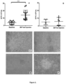
Figure 2 . Endothelial colony forming units in heart failure patients treated with either allogeneic or autologous mesenchymal stem cells (MSCs).Figure 2A shows that patients treated with allogeneic MSCs had a significant improvement in EPC-CFUs 3 months post treatment (n=15, ****P<0.001).Figure 2B shows that patients treated with autologous MSCs had no change in EPC-CFUs post treatment (n=7).Figures 2C and 2E show representative EPC-CFUs plated on fibronectin for 5 days before MSC administration (magnification 20x).Figures 2D and 2F shows representative EPC-CFYs plated on fibronectin for 5 days after MSC administration (magnification 20x). -
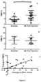
Figure 3 . Flow mediated vasodilation (FMD) measurements before and after mesenchymal stem cell (MSC) treatment.Figure 3A shows that patients treated with allogeneic MSCs had an increase in FMD% 3 months post injection (n=15, ***P<0.001).Figure 3B shows that patients treated with autologous MSCs had no significant difference in FMD% 3 months post injection (n=7, P=NS).Figure 3C shows that correlation between the absolute change in FMD% and the absolute change in EPF-CFUs from baseline to 3 months post MSC injection in all patients (P<0.001, R=0.684). -
Figure 4 . Serum VEGF concentration in patients and controls.Figure 4A demonstrates that patients (n=14) had a higher level of circulating VEGF compared to controls (n=9) at baseline (P=0.0009).Figure 4B demonstrates that patients who received allogeneic MSCs (n=9) had a decrease in VEGF serum levels post injection (Δ-547.5±350.8 pg/mL), while patients who received autologous MSCs (n=5) had an increase in serum VEGF post injection (Δ814.1±875.8), and there was a difference between groups (**P=0.0012).Figure 4C shows that there is a correlation between EPC-CFUs and serum VEGF in patients at both baseline and 3 months post MSC treatment (R=-0.421, P=0.026).Figure 4D shows that the change in EPC-CFUs from baseline to 3 months post treatment strongly correlated with the change in VEGF (R=-0.863, P<0.001). - The present disclosure is based, at least in part, on the discovery that increased circulating levels of Vascular Endothelial Growth Factor (VEGF), optionally in combination with decreased levels of endothelial progenitor cells (EPCs), is indicative of endothelial dysfunction in a subject, and reduced levels of VEGF and increased levels of EPCs are indicative of effectiveness of therapy for endothelial dysfunction, vascular tone, and cardiovascular disease. Data provided herein demonstrated that the administration of allogeneic mesenchymal stem cells (MSCs) to subjects suffering from heart failure stimulated EPC bioactivity and restored flow-mediated vasodilation towards normal. The ability of allogeneic MSCs greatly exceeded that of autologous MSCs in restoring endothelial function, enhancing EPC CFU formation, and suppressing VEGF levels. Together these findings offer major new clinical insights into the bioactivity of MSCs, suggest a novel therapeutic principle for disorders characterized by endothelial dysfunction, and provide useful diagnostic markers for detecting and measuring successful treatment.
- MSCs are adult stem cells that are prototypically found in bone marrow and have the capacity to differentiate into multiple cell types. They stimulate the proliferation and differentiation of endogenous precursor cells and play a crucial role in maintaining stem cell niches27. In addition, MSCs secrete paracrine factors which participate in angiogenesis, cardiomyogenesis, neovascularization, stimulation of other endogenous stem cells, and regulation of the immune system28,29. While MSCs are known to stimulate cardiac precursor cells and cell cycle activity in the heart, their role in stimulating other endogenous precursor populations has heretofore been unknown. The data provided herein establish that MSCs stimulate endogenous EPC activation, increasing the number and quality of functional EPCs. These findings suggest that augmentation of EPCs may represent a novel mechanism of action by which MSCs exert favorable biological effects.
- Over the last decade, there has been an emerging interest in the use of MSCs in CV disorders30. Clinical trials have demonstrated a major safety profile for MSC administration, and suggested efficacy in patients with HF30-32; however, underlying mechanism(s) of action continue to be vigorously debated. The instant finding that allogeneic MSC injections in patients with both ischemic and non-ischemic HF results in an improvement in endothelial function specifically by restoring EPC function, FMD, and reducing VEGF levels towards normal offers a major new insight into the mechanisms of action of MSCs. In the study population, increased serum VEGF correlated with diminished EPC-CFUs, consistent with the idea that VEGF plays a compensatory role, a finding also reported in patients with cerebral aneurysm26. The findings provided herein establish that allogeneic MSCs can be employed to stimulate EPC bioactivity, improve arterial physiologic vasodilatory responses, and decrease unfavorable cytokine mobilization in patients with CV disease and other disorders associated with endothelial dysfunction.
- The data provided herein demonstrate that allogeneic MSCs restored endothelial function in patients to a degree greatly exceeding that of autologous MSCs. In the study described below, all allogeneic stem cell donors were healthy, young donors between the ages of 20 and 35. Patients receiving their own stem cells not only had underlying chronic diseases, but were also older (between the ages of 45 and 75). Autologous MSCs aging may impair the survival, differentiation, and ability to recruit EPCs to areas of damage, ultimately reducing their therapeutic efficacy38,39.
- Study Population: The HF patients were enrolled in POSETDON-DCM (NCT01392625), "A Phase I/II, Randomized Pilot Study of the Comparative Safety and Efficacy of Transendocardial Injection of Autologous Mesenchymal Stem Cells Versus Allogeneic Mesenchymal Stem Cells in Patients with Nonischemic Dilated Cardiomyopathy" and in TRIDENT (NCT02013674), "The TRansendocardial Stem Cell Injection Delivery Effects on Neomyogenesis Study". In PODEIDON-DCM, patients were randomized to receive by transendocardial delivery either 100 million autologous or allogeneic MSCs. Autologous MSCs were derived from the patient's bone marrow (iliac crest aspiration) 4-6 weeks before cardiac catheterization. In the TRIDENT study, ICM patients were randomized to receive either 20 or 100 million allogeneic MSCs transendocardially. Allogeneic MSCs were manufactured by the University of Miami Cell Manufacturing Program. Healthy subjects (n=10) were enrolled ranging in ages from 22-58 years and both genders. All subjects provided written informed consent and the study was approved by the University of Miami Institutional Review Board.
- Endothelial Colony Forming Units (EPC-CFUs): Peripheral blood samples were obtained from patients before and 3 months after MSC injection. EPCs were isolated from samples using Ficoll-Paque and 5 million cells were seeded on 6-well Fibronectin-coated dishes (BD biosciences) in CFU-Hill medium (stem cell technologies, cat#05900)17. The non-adherent cells were collected 48 hours later and 1 million cells were seeded on 24-well Fibronectin-coated dishes. On
day 5, EPC-CFUs were counted in 5 wells and the average was obtained. - Flow-Mediated Vasodilation (FMD): Brachial artery diameter measurements and FMD% were performed in the morning, after an overnight fast. The subjects' right arm was immobilized in an extended position, and the brachial artery was scanned via ultrasound 5-10 cm above the antecubital fossa17,18. A brachial cuff was then inflated to a supra-systolic pressure (40 to 50 mmHg above systolic pressure) for 5 minutes. Subsequently, the cuff was deflated and the brachial artery diameter was recorded for 3 minutes.
- VEGF ELISA: Serum vascular endothelium growth factor (VEGF) levels (Invitrogen #KHG0111) were measured in DCM patients at baseline and 6 months after allogeneic (n=6) or autologous (n=5) MSC treatment. In ICM patients (n=4), VEGF was measured at baseline and 3 months after allogeneic MSC treatment. DCM and ICM patients who received allogeneic MSCs were combined and compared to patients who received autologous. Lastly, VEGF was measured in controls (n=9).
- C-reactive Protein: C-reactive protein (CRP) was evaluated using a nephelometric method in which blood plasma samples were incubated with latex particles coated with CRP monoclonal antibodies. The analytical sensitivity was 0.2mg/L and results were reported in mg/L.
- Statistical Analysis: To assess the difference between autologous and allogeneic groups, an unpaired, two-tailed T-test was used. To measure the difference before and after treatment in each group, both a paired, two-tailed T-test and a one-way ANOVA was utilized. Correlations were measured using Pearson correlation, assuming a Gaussian distribution. Data are presented as mean and standard deviation of the mean. To determine the difference between HUVEC treatment groups, an unpaired, two-tailed T-test was used.
- Baseline Characteristics: A total of 22 patients were analyzed for this study. Fifteen patients received allogeneic MSCs and 7 patients received autologous MSCs. Baseline characteristics of the study subjects are summarized in Table 1. Age and sex were evenly distributed.
Table 1. Summary of patients (n=22) and healthy controls (n=10). Patients are broken down by etiology and cell treatment: DCM patients receiving allogeneic MSCs (n=9), DCM patients receiving autologous MSCs (n=7) and ICM patients receiving allogeneic MSCs (n=6).. Characteristic DCM Allogeneic (N=9, 27%) DCM Autologous (N=7, 21 %) ICM Allogeneic (N=6, 18%) Healthy Controls (N=10, 33%) Age - yr. Median 60 55 71 44 Range 45-70 48-73 65-75 22-58 Male sex- no. 8 (89%) 6 (86%) 6 (100%) 7 (70%) Race/Ethnicity White 8 (89%) 3 (43%) 6 (100%) 7(70%) White/Hispanic 0 (0%) 4(57%) 0 (0%) 3 (30%) Black 1 (11%) 0 (0%) 0 (0%) 0 (0%) History of CVD HF 2 (22%) 0 (0%) 1 (17%) 0 (0%) CAD 1 (11%) 0 (0%) 6 (100%) 0 (0%) CHF 0 1 (14%) 4 (67%) 0 (0%) History of Smoking 4(44%) 4(57%) 4 (67%) 0 (0%) Blood pressure-mmHg (systolic/diastolic) Median 126/77 108/65 113/70 118/67 Range 108- 99-142/59-84 85-134/53- 111-130/59- Cholesterol Median 202 175 147 N/A Range 129-282 112-207 129-178 N/A C-Reactive Protein (mg/mL) Median 0.3 0.3 0.15 N/A Range 0.2-0.4 0.2-0.5 0.1-0.6 N/A Medications ASA/NSAIDS 5 (56%) 4 (57%) 5 (83%) 0 (0%) Beta-Blockers 7 (78%) 7 (100%) 6 (100%) 0 (0%) ACE 6 (67%) 7 (100%) 6 (100%) 1 (10%) Diuretics 8 (89%) 6 (86%) 4 (67%) 0 (0%) Statins 3 (33%) 3 (43%) 6 (100%) 1 (10%) Antiplatelet 4 (44%) 2 (29%) 3 (50%) 0 (0%) Other 9 (100%) 7 (100%) 6 (100%) 0 (0%) - EPC-CFUs and FMD in Heart Failure Patients and Healthy Subjects: Patients with ischemic (n=15) as well as non-ischemic (n=6) cardiomyopathy had endothelial dysfunction at baseline, characterized by a reduced ability to form EPC-CFUs and an impaired FMD response (
Figure 1 ). Specifically, patients had decreased EPC-CFU counts compared to healthy controls (4±3 vs. 25±16, respectively, P<0.001) as well as diminished FMD% (5.6±3.2 vs. 9.2±4, respectively, P=0.0017). - EPC-CFUs in Heart Failure Patients Treated with MSCs: Patients were assessed for EPC-CFUs before and 3 months after MSC treatment. Patients who received allogeneic MSCs had a significant improvement in the number of EPC-CFUs post-treatment (Δ10±5, P<0.001,
Figure 2A ). On the other hand, patients who received autologous MSCs showed no improvement (Δ1±3, P=NS,Figure 2B ). Moreover, we compared allogeneic MSC treatment versus autologous MSC treatment, and allogeneic MSCs were superior in stimulating EPC colony formation (P=0.02). - EPC-CFUs were also examined for morphology. EPCs from HF patients had disorganized and incomplete colony formation (
Figure 2C and E ), resulting in clusters that failed to form functional colonies. Three months after allogeneic MSC treatment, patient colonies were organized and healthy in appearance (Figure 2D and F ). These findings suggest that transendocardial MSC therapy stimulates EPC bioactivity in patients with HF of both ischemic and non-ischemic etiology. - FMD in Heart Failure Patients Treated with MSCs: All patients were evaluated using brachial artery FMD before and 3 months after MSC injection. Patients who received allogeneic MSCs had a dramatic improvement in FMD% (Δ3.7±3%, P=0.001,
Figure 3A ). In contrast, patients who received autologous MSCs had no improvement and the majority of patients worsened 3 months post-treatment (Δ-0.46±3%,Figure 3B ). - The difference between treatment with autologous MSCs and allogeneic MSCs was analyzed. There was a striking difference between the two cell types (P=0.005), suggesting that autologous MSCs do not restore endothelial function in this patient population. We also assessed the correlation between EPC-CFUs and FMD% in all patients and found a highly significant correlation between ΔFMD% and ΔEPC-CFUs (P<0.001, R=0.684,
Figure 3C ). - Assessment of VEGF in Patients Receiving Autologous and Allogeneic MSCs: Circulating VEGF levels in patients and healthy control serum was determined. At baseline, patients had profoundly elevated circulating VEGF compared to controls (1130.3±803.3 vs. 2.0±5.9 pg/mL, P<0.001,
Figure 4A ). Allogeneic MSCs reduced VEGF levels (-547.5±350.8 pg/mL, P=0.002), while patients who received autologous MSCs had an increase in VEGF (814.1±875.8 pg/mL,Figure 4B ). Furthermore, there was a significant difference between patients who received allogeneic MSCs versus patients who received autologous MSCs (P=0.0012,Figure 4B ). - VEGF levels correlated with EPC-CFUs (P=0.026, R=-0.421,
Figure 4C ). Even more striking, there was a significant correlation between the change in VEGF and the change in EPC-CFUs from baseline to three months post treatment (R=0.863, P<0.001, Figure 4G). Notably, high levels of VEGF correlated with low levels of EPCs, evidenced by the autologous group. Conversely, lower levels of VEGF correlated with high levels of EPC-CFUs, illustrated by the allogeneic group. Taken together, these data demonstrate MSCs stimulate EPC mobilization and suppress compensatory elevations in circulating VEGF concentrations. - This Example demonstrates a potent and clinically relevant efficacy outcome of transendocardial therapy with MSCs in patients with advanced HF. Allogeneic MSCs restored flow mediated brachial artery dilatation, EPC bioactivity, and VEGF levels towards normal. As abnormalities in the vascular function of patients with CV disease is shown to be highly predictive of adverse outcomes and disease progression, targeting endothelial function is a significant therapeutic strategy. The data also reveals VEGF as a novel indicator for successful treatment of endothelial dysfunction, restoration of vascular tone, and alleviation of complications of cardiovascular disease. Subjects demonstrating improved FMD displayed reduced circulating VEGF levels, as well as higher levels of EPC-CFUs, compared to heart failure subjects demonstrating no improvement in FMD.
-
- 1. Go AS, Mozaffarian D, Roger VL, et al. Heart Disease and Stroke Statistics-2014 Update: A Report From the American Heart Association. Circulation 2014;129:e28-e292.
- 2. Blum A. Heart failure--new insights. The Israel Medical Association journal: IMAJ 2009;11:105-11.
- 3. Marti CN, Gheorghiade M, Kalogeropoulos AP, Georgiopoulou VV, Quyyumi AA, Butler J. Endothelial Dysfunction, Arterial Stiffness, and Heart Failure. Journal of the American College of Cardiology 2012;60:1455-69.
- 4. Santiago R, Antoni B-G. Vascular dysfunction in idiopathic dilated cardiomyopathy. Nature Reviews Cardiology 2009;6:590-8.
- 5. Schulman IH, Zhou MS, Raij L. Interaction between nitric oxide and angiotensin II in the endothelium: role in atherosclerosis and hypertension. Journal of hypertension Supplement: official journal of the International Society of Hypertension 2006;24:S45-50.
- 6. Polovina MM, Potpara TS. Endothelial dysfunction in metabolic and vascular disorders. Postgraduate medicine 2014;126:38-53.
- 7. Zampetaki A, Kirton JP, Xu Q. Vascular repair by endothelial progenitor cells. Cardiovascular Research 2008;78:413-21.
- 8. Rehman J, Li J, Orschell CM, March KL. Peripheral Blood "Endothelial Progenitor Cells" Are Derived From Monocyte/Macrophages and Secrete Angiogenic Growth Factors. Circulation 2003;107:1164-9.
- 9. Schulman IH, Hare JM. Key developments in stem cell therapy in cardiology. Regen Med 2012;7:17-24.
- 10. Karantalis V, Balkan W, Schulman IH, Hatzistergos KE, Hare JM. Cell-based therapy for prevention and reversal of myocardial remodeling. Am J Physiol Heart Circ Physiol 2012;303:H256-70.
- 11. Williams AR, Hare JM. Mesenchymal stem cells: biology, pathophysiology, translational findings, and therapeutic implications for cardiac disease. Circulation research 2011;109:923-40.
- 12. Suncion VY, Schulman IH, Hare JM. Concise review: the role of clinical trials in deciphering mechanisms of action of cardiac cell-based therapy. Stem Cells Translational Medicine 2012;1:29-35.
- 13. Hatzistergos KE, Quevedo H, Oskouei BN, et al. Bone marrow mesenchymal stem cells stimulate cardiac stem cell proliferation and differentiation. Circulation research 2010;107:913-22.
- 14. Chen L, Tredget EE, Wu PY, Wu Y. Paracrine factors of mesenchymal stem cells recruit macrophages and endothelial lineage cells and enhance wound healing. PloS one 2008;3:e1886.
- 15. Werner N, Kosiol S, Schiegl T, et al. Circulating endothelial progenitor cells and cardiovascular outcomes. N Engl J Med 2005;353:999-1007.
- 16. Shantsila E, Watson T, Lip GY. Endothelial progenitor cells in cardiovascular disorders. J Am Coll Cardiol 2007;49:741-52.
- 17. Hill JM, Zalos G, Halcox JPJ, et al. Circulating Endothelial Progenitor Cells, Vascular Function, and Cardiovascular Risk. New England Journal of Medicine 2003;348:593-600.
- 18. Corretti MC, Anderson TJ, Benjamin EJ, et al. Guidelines for the ultrasound assessment of endothelial-dependent flow-mediated vasodilation of the brachial artery: A report of the International Brachial Artery Reactivity Task Force. Journal of the American College of Cardiology 2002;39:257-65.
- 19. Werner N, Wassmann S, Ahlers P, et al. Endothelial progenitor cells correlate with endothelial function in patients with coronary artery disease. Basic research in cardiology 2007;102:565-71.
- 20. Shantsila E, Watson T, Lip GYH. Endothelial Progenitor Cells in Cardiovascular Disorders. Journal of the American College of Cardiology 2007;49:741-52.
- 21. Schmidt-Lucke C, Rössig L, Fichtlscherer S, et al. Reduced Number of Circulating Endothelial Progenitor Cells Predicts Future Cardiovascular Events: Proof of Concept for the Clinical Importance of Endogenous Vascular Repair. Circulation 2005;111:2981-7.
- 22. Chin BSP, Chung NAY, Gibbs CR, Blann AD, Lip GYH. Vascular endothelial growth factor and soluble P-selectin in acute and chronic congestive heart failure. The American Journal of Cardiology 2002;90:1258-60.
- 23. Tsai WC, Li YH, Huang YY, Lin CC, Chao TH, Chen JH. Plasma vascular endothelial growth factor as a marker for early vascular damage in hypertension. Clinical Science 2005;109:39-43.
- 24. Seko Y, Fukuda S, Nagai R. Serum levels of endostatin, vascular endothelial growth factor (VEGF) and hepatocyte growth factor (HGF) in patients with acute myocardial infarction undergoing early reperfusion therapy. Clinical Science 2004;106:4.
- 25. Eleuteri E, Di Stefano A, Tarro Genta F, et al. Stepwise increase of angiopoietin-2 serum levels is related to haemodynamic and functional impairment in stable chronic heart failure. European Journal of Cardiovascular Prevention & Rehabilitation 2011;18:607-14.
- 26. Wei H, Mao Q, Liu L, et al. Changes and function of circulating endothelial progenitor cells in patients with cerebral aneurysm. Journal of neuroscience research 2011;89:1822-8.
- 27. Williams AR, Hare JM. Mesenchymal Stem Cells: Biology, Pathophysiology, Translational Findings, and Therapeutic Implications for Cardiac Disease. Circulation Research 2011;109:923-40.
- 28. Gnecchi M, Zhang Z, Ni A, Dzau VJ. Paracrine Mechanisms in Adult Stem Cell Signaling and Therapy. Circulation Research 2008;103:1204-19.
- 29. Caplan AI, Dennis JE. Mesenchymal stem cells as trophic mediators. Journal of Cellular Biochemistry 2006;98:1076-84.
- 30. Telukuntla KS, Suncion VY, Schulman IH, Hare JM. The Advancing Field of Cell-Based Therapy: Insights and Lessons From Clinical Trials. Journal of the American Heart Association 2013;2.
- 31. Hare JM, Traverse JH, Henry TD, et al. A randomized, double-blind, placebo-controlled, dose-escalation study of intravenous adult human mesenchymal stem cells (prochymal) after acute myocardial infarction. J Am Coll Cardiol 2009;54:2277-86.
- 32. Hare JM, Fishman JE, Gerstenblith G, et al. Comparison of allogeneic vs autologous bone marrow-derived mesenchymal stem cells delivered by transendocardial injection in patients with ischemic cardiomyopathy: The poseidon randomized trial. JAMA 2012;308:2369-79.
- 33. Vasa M, Fichtlscherer S, Aicher A, et al. Number and Migratory Activity of Circulating Endothelial Progenitor Cells Inversely Correlate With Risk Factors for Coronary Artery Disease. Circulation Research 2001;89:e1-e7.
- 34. Franz Alber H, Dulak J, Frick M, et al. Atorvastatin decreases vascular endothelial growth factor in patients with coronary artery disease. Journal of the American College of Cardiology 2002;39:1951-5.
- 35. Yu KR, Kang KS. Aging-related genes in mesenchymal stem cells: a mini-review. Gerontology 2013;59:557-63.
- 36. Bustos ML, Huleihel L, Kapetanaki MG, et al. Aging Mesenchymal Stem Cells Fail to Protect Because of Impaired Migration and Antiinflammatory Response. American Journal of Respiratory and Critical Care Medicine 2014;189:787-98.
- 37. Efimenko A, Dzhoyashvili N, Kalinina N, et al. Adipose-Derived Mesenchymal Stromal Cells From Aged Patients With Coronary Artery Disease Keep Mesenchymal Stromal Cell Properties but Exhibit Characteristics of Aging and Have Impaired Angiogenic Potential. Stem Cells Translational Medicine 2013.
- 38. Asumda FZ. Age-associated changes in the ecological niche: implications for mesenchymal stem cell aging. Stem cell research & therapy 2013;4:47.
- 39. Chang EI, Loh SA, Ceradini DJ, et al. Age Decreases Endothelial Progenitor Cell Recruitment Through Decreases in Hypoxia-Inducible Factor 1α Stabilization During Ischemia. Circulation 2007;116:2818-29.
- The invention further relates to the following embodiments:
- Embodiment 1. A method of monitoring efficacy of a therapy for endothelial dysfunction in a subject comprising
- determining levels of Vascular Endothelial Growth Factor (VEGF) and endothelial progenitor cells (EPCs) in a first sample from the subject before administration of a therapy for endothelial dysfunction; and
- determining levels of VEGF and EPCs in a second sample from the subject after administration of a therapy for endothelial dysfunction;
- wherein a decreased level of VEGF in the sample and an increased level of EPCs in the second sample compared to the levels of VEGF and EPC in the first sample is indicative of effective treatment of the endothelial dysfunction.
- Embodiment 2. The method of embodiment 1, wherein the therapy for endothelial dysfunction comprises mesenchymal stem cell therapy.
- Embodiment 3. The method of embodiment 2, wherein the mesenchymal stem cell therapy comprises the administration of allogeneic mesenchymal stem cells to the subject.
- Embodiment 4. A method of monitoring efficacy of mesenchymal stem cell therapy in a subject suffering from a cardiovascular disorder comprising determining a level of Vascular Endothelial Growth Factor (VEGF) in a sample from the subject after administration of the stem cell therapy, and comparing the level of VEGF to a predetermined criterion.
-
Embodiment 5. A method of treating endothelial dysfunction in a subject in need thereof comprising administering to a subject allogeneic mesenchymal stem cells in an amount effect to decrease circulating levels of Vascular Endothelial Growth Factor and increase levels of endothelial progenitor cells in the subject, thereby treating the endothelial dysfunction. - Embodiment 6. The method of
embodiment 5, wherein the subject is suffering from a cardiovascular disorder. - Embodiment 7. The method of embodiment 6, wherein the cardiovascular disorder is selected from the group consisting of heart failure, idiopathic dilated cardiomyopathy and ischemic cardiomyopathy.
- Embodiment 8. The method of
embodiment 5, wherein the mesenchymal stem cells are administered locally. - Embodiment 9. The method of embodiment 8, wherein the mesenchymal stem cells are administered by transendocardial injection.
-
Embodiment 10. The method ofembodiment 5, wherein the mesenchymal stem cells are administered in an amount ranging from about 20 million to about 100 million cells. - Embodiment 11. The method of
embodiment 5, wherein administration of the mesenchymal stem cells results in an increase in flow-mediated vasodilation (FMD) in the subject of at least 3%.
Claims (7)
- Allogeneic mesenchymal stem cells for use in a method of treating endothelial dysfunction in a subject in need thereof comprising administering to a subject the allogeneic mesenchymal stem cells in an amount effective to decrease circulating levels of Vascular Endothelial Growth Factor and increase levels of endothelial progenitor cells in the subject, thereby treating the endothelial dysfunction.
- The allogeneic mesenchymal stem cells for the use of claim 1, wherein the subject is suffering from a cardiovascular disorder.
- The allogeneic mesenchymal stem cells for the use of claim 2, wherein the cardiovascular disorder is selected from the group consisting of heart failure, idiopathic dilated cardiomyopathy and ischemic cardiomyopathy.
- The allogeneic mesenchymal stem cells for the use of claim 1, wherein the mesenchymal stem cells are administered locally.
- The allogeneic mesenchymal stem cells for the use of claim 4, wherein the mesenchymal stem cells are administered by transendocardial injection.
- The allogeneic mesenchymal stem cells for the use of claim 1, wherein the mesenchymal stem cells are administered in an amount ranging from about 20 million to about 100 million cells.
- The allogeneic mesenchymal stem cells for the use of claim 1, wherein administration of the mesenchymal stem cells results in an increase in flow-mediated vasodilation (FMD) in the subject of at least 3%.
Applications Claiming Priority (3)
| Application Number | Priority Date | Filing Date | Title |
|---|---|---|---|
| US201462080984P | 2014-11-17 | 2014-11-17 | |
| PCT/US2015/060624 WO2016081309A1 (en) | 2014-11-17 | 2015-11-13 | Materials and methods for treating endothelial dysfunction and methods for monitoring efficacy of therapy in a subject |
| EP15861319.0A EP3221697B1 (en) | 2014-11-17 | 2015-11-13 | Methods for monitoring efficacy of allogeneic mesenchymal stem cell therapy in a subject |
Related Parent Applications (1)
| Application Number | Title | Priority Date | Filing Date |
|---|---|---|---|
| EP15861319.0A Division EP3221697B1 (en) | 2014-11-17 | 2015-11-13 | Methods for monitoring efficacy of allogeneic mesenchymal stem cell therapy in a subject |
Publications (1)
| Publication Number | Publication Date |
|---|---|
| EP4190917A1 true EP4190917A1 (en) | 2023-06-07 |
Family
ID=56014411
Family Applications (2)
| Application Number | Title | Priority Date | Filing Date |
|---|---|---|---|
| EP22208663.9A Pending EP4190917A1 (en) | 2014-11-17 | 2015-11-13 | Methods for monitoring efficacy of allogeneic mesenchymal stem cell therapy in a subject |
| EP15861319.0A Active EP3221697B1 (en) | 2014-11-17 | 2015-11-13 | Methods for monitoring efficacy of allogeneic mesenchymal stem cell therapy in a subject |
Family Applications After (1)
| Application Number | Title | Priority Date | Filing Date |
|---|---|---|---|
| EP15861319.0A Active EP3221697B1 (en) | 2014-11-17 | 2015-11-13 | Methods for monitoring efficacy of allogeneic mesenchymal stem cell therapy in a subject |
Country Status (11)
| Country | Link |
|---|---|
| US (1) | US20170360837A1 (en) |
| EP (2) | EP4190917A1 (en) |
| JP (1) | JP2018503802A (en) |
| KR (1) | KR102626288B1 (en) |
| AU (1) | AU2015350250A1 (en) |
| BR (1) | BR112017010313B1 (en) |
| CA (1) | CA2968222A1 (en) |
| ES (1) | ES2935903T3 (en) |
| MA (1) | MA40996A (en) |
| WO (1) | WO2016081309A1 (en) |
| ZA (1) | ZA201704110B (en) |
Citations (1)
| Publication number | Priority date | Publication date | Assignee | Title |
|---|---|---|---|---|
| US7370108B1 (en) | 2000-07-14 | 2008-05-06 | Entrust, Inc. | Method for determining behavior of an information system to control its ability to deliver a user's quality of service |
Family Cites Families (2)
| Publication number | Priority date | Publication date | Assignee | Title |
|---|---|---|---|---|
| US8617538B2 (en) * | 2009-09-28 | 2013-12-31 | Board Of Regents Of The University Of Texas System | Mesodermal-like cell population for treating ischemia in mammals |
| PT2611906T (en) * | 2010-08-31 | 2024-04-16 | Gallant Pet Inc | Systemic, allogenic stem cell therapies for treatment of diseases in animals |
-
2015
- 2015-11-12 MA MA040996A patent/MA40996A/en unknown
- 2015-11-13 BR BR112017010313-3A patent/BR112017010313B1/en active IP Right Grant
- 2015-11-13 JP JP2017526979A patent/JP2018503802A/en active Pending
- 2015-11-13 ES ES15861319T patent/ES2935903T3/en active Active
- 2015-11-13 EP EP22208663.9A patent/EP4190917A1/en active Pending
- 2015-11-13 US US15/527,270 patent/US20170360837A1/en not_active Abandoned
- 2015-11-13 EP EP15861319.0A patent/EP3221697B1/en active Active
- 2015-11-13 KR KR1020177016542A patent/KR102626288B1/en active Active
- 2015-11-13 WO PCT/US2015/060624 patent/WO2016081309A1/en not_active Ceased
- 2015-11-13 AU AU2015350250A patent/AU2015350250A1/en not_active Abandoned
- 2015-11-13 CA CA2968222A patent/CA2968222A1/en not_active Abandoned
-
2017
- 2017-06-15 ZA ZA2017/04110A patent/ZA201704110B/en unknown
Patent Citations (1)
| Publication number | Priority date | Publication date | Assignee | Title |
|---|---|---|---|---|
| US7370108B1 (en) | 2000-07-14 | 2008-05-06 | Entrust, Inc. | Method for determining behavior of an information system to control its ability to deliver a user's quality of service |
Non-Patent Citations (47)
| Title |
|---|
| ALYNE FELIX ET AL: "Remodeling of the thoracic aorta after bone marrow cell transplantation", INTERNATIONAL JOURNAL OF CLINICAL AND EXPERIMENTAL PATHOLOGY, vol. 7, no. 9, 15 August 2014 (2014-08-15), US, pages 5527 - 5537, XP055444572, ISSN: 1936-2625 * |
| ASUMDA FZ: "Age-associated changes in the ecological niche: implications for mesenchymal stem cell aging", STEM CELL RESEARCH & THERAPY, vol. 4, 2013, pages 47 |
| BLUM A.: "Heart failure--new insights.", IMAJ, vol. 11, 2009, pages 105 - 11 |
| BUSTOS MLHULEIHEL LKAPETANAKI MG ET AL.: "Aging Mesenchymal Stem Cells Fail to Protect Because of Impaired Migration and Antiinflammatory Response", AMERICAN JOURNAL OF RESPIRATORY AND CRITICAL CARE MEDICINE, vol. 189, 2014, pages 787 - 98 |
| CAPLAN AIDENNIS JE: "Mesenchymal stem cells as trophic mediators", JOURNAL OF CELLULAR BIOCHEMISTRY, vol. 98, 2006, pages 1076 - 84, XP009110967, DOI: 10.1002/jcb.20886 |
| CHANG EILOH SACERADINI DJ ET AL.: "Age Decreases Endothelial Progenitor Cell Recruitment Through Decreases in Hypoxia-Inducible Factor la Stabilization During Ischemia", CIRCULATION, vol. 116, 2007, pages 2818 - 29, XP007907889, DOI: 10.1161/CIRCULATIONAHA.107.715847 |
| CHIN BSPCHUNG NAYGIBBS CRBLANN ADLIP GYH: "Vascular endothelial growth factor and soluble P-selectin in acute and chronic congestive heart failure", THE AMERICAN JOURNAL OF CARDIOLOGY, vol. 90, 2002, pages 1258 - 60 |
| CORRETTI MCANDERSON TJBENJAMIN EJ ET AL.: "Guidelines for the ultrasound assessment of endothelial-dependent flow-mediated vasodilation of the brachial artery: A report of the International Brachial Artery Reactivity Task Force.", JOURNAL OF THE AMERICAN COLLEGE OF CARDIOLOGY, vol. 39, 2002, pages 257 - 65, XP002591839 |
| EFIMENKO ADZHOYASHVILI NKALININA N ET AL.: "Adipose-Derived Mesenchymal Stromal Cells From Aged Patients With Coronary Artery Disease Keep Mesenchymal Stromal Cell Properties but Exhibit Characteristics of Aging and Have Impaired Angiogenic Potential", STEM CELLS TRANSLATIONAL MEDICINE, 2013 |
| ELEUTERI EDI STEFANO ATARRO GENTA F ET AL.: "Stepwise increase of angiopoietin-2 serum levels is related to haemodynamic and functional impairment in stable chronic heart failure", EUROPEAN JOURNAL OF CARDIOVASCULAR PREVENTION & REHABILITATION, vol. 18, 2011, pages 607 - 14 |
| FRANZ ALBER HDULAK JFRICK M ET AL.: "Atorvastatin decreases vascular endothelial growth factor in patients with coronary artery disease", JOURNAL OF THE AMERICAN COLLEGE OF CARDIOLOGY, vol. 39, 2002, pages 1951 - 5 |
| GNECCHI MZHANG ZNI ADZAU VJ: "Paracrine Mechanisms in Adult Stem Cell Signaling and Therapy", CIRCULATION RESEARCH, vol. 103, 2008, pages 1204 - 19, XP055861964, DOI: 10.1161/CIRCRESAHA.108.176826 |
| GO ASMOZAFFARIAN DROGER VL ET AL.: "Heart Disease and Stroke Statistics-2014 Update: A Report From the American Heart Association", CIRCULATION, vol. 129, 2014, pages e28 - e292 |
| HARE JMFISHMAN JEGERSTENBLITH G ET AL.: "Comparison of allogeneic vs autologous bone marrow-derived mesenchymal stem cells delivered by transendocardial injection in patients with ischemic cardiomyopathy: The poseidon randomized trial", JAMA, vol. 308, 2012, pages 2369 - 79 |
| HARE JMTRAVERSE JHHENRY TD ET AL.: "A randomized, double-blind, placebo-controlled, dose-escalation study of intravenous adult human mesenchymal stem cells (prochymal) after acute myocardial infarction", J AM COLL CARDIOL, vol. 54, 2009, pages 2277 - 86, XP029649197, DOI: 10.1016/j.jacc.2009.06.055 |
| HATZISTERGOS KEQUEVEDO HOSKOUEI BN ET AL.: "Bone marrow mesenchymal stem cells stimulate cardiac stem cell proliferation and differentiation", CIRCULATION RESEARCH, vol. 107, 2010, pages 913 - 22, XP055255062, DOI: 10.1161/CIRCRESAHA.110.222703 |
| HILL JMZALOS GHALCOX JPJ ET AL.: "Circulating Endothelial Progenitor Cells, Vascular Function, and Cardiovascular Risk", NEW ENGLAND JOURNAL OF MEDICINE, vol. 348, 2003, pages 593 - 600, XP002980439, DOI: 10.1056/NEJMoa022287 |
| ICHIM THOMAS E ET AL: "Placental mesenchymal and cord blood stem cell therapy for dilated cardiomyopathy", REPRODUCTIVE BIOMEDICINE ONLINE,, vol. 16, no. 6, 1 June 2008 (2008-06-01), pages 898 - 905, XP009180675, ISSN: 1472-6491, DOI: 10.1016/S1472-6483(10)60159-9 * |
| IMANISHI Y ET AL: "Allogenic mesenchymal stem cell transplantation has a therapeutic effect in acute myocardial infarction in rats", JOURNAL OF MOLECULAR AND CELLULAR CARDIOLOGY, ACADEMIC PRESS, GB, vol. 44, no. 4, 1 April 2008 (2008-04-01), pages 662 - 671, XP022640584, ISSN: 0022-2828, [retrieved on 20071113], DOI: 10.1016/J.YJMCC.2007.11.001 * |
| JOSHUA M. HARE ET AL: "Comparison of Allogeneic vs Autologous Bone Marrow-Derived Mesenchymal Stem Cells Delivered by Transendocardial Injection in Patients With Ischemic Cardiomyopathy", JAMA THE JOURNAL OF THE AMERICAN MEDICAL ASSOCIATION, vol. 308, no. 22, 12 December 2012 (2012-12-12), US, pages 2369, XP055254332, ISSN: 0098-7484, DOI: 10.1001/jama.2012.25321 * |
| KARANTALIS VBALKAN WSCHULMAN IHHATZISTERGOS KEHARE JM: "Cell-based therapy for prevention and reversal of myocardial remodeling.", AM J PHYSIOL HEART CIRC PHYSIOL, vol. 303, 2012, pages H256 - 70 |
| MARTI CNGHEORGHIADE MKALOGEROPOULOS APGEORGIOPOULOU VVQUYYUMI AABUTLER J.: "Endothelial Dysfunction, Arterial Stiffness, and Heart Failure", JOURNAL OF THE AMERICAN COLLEGE OF CARDIOLOGY, vol. 60, 2012, pages 1455 - 69 |
| MOON HYUNG-HO ET AL: "MSC-based VEGF gene therapy in rat myocardial infarction model using facial amphipathic bile acid-conjugated polyethyleneimine", BIOMATERIALS, ELSEVIER, AMSTERDAM, NL, vol. 35, no. 5, 23 November 2013 (2013-11-23), pages 1744 - 1754, XP028801323, ISSN: 0142-9612, DOI: 10.1016/J.BIOMATERIALS.2013.11.019 * |
| NARITA TAKUYA ET AL: "Bone marrow-derived mesenchymal stem cells for the treatment of heart failure", HEART FAILURE REVIEWS, KLUWER ACADEMIC PUBLISHERS, DORDRECHT, NL, vol. 20, no. 1, 27 May 2014 (2014-05-27), pages 53 - 68, XP035417931, ISSN: 1382-4147, [retrieved on 20140527], DOI: 10.1007/S10741-014-9435-X * |
| NI NATHAN C ET AL: "The Promise and Challenges of Cardiac Stem Cell Therapy", SEMINARS IN THORACIC AND CARDIOVASCULAR SURGERY, vol. 26, no. 1 (Spring-2014), 1 June 2014 (2014-06-01), pages 44 - 52, XP028873800, ISSN: 1043-0679, DOI: 10.1053/J.SEMTCVS.2014.03.001 * |
| POLOVINA MMPOTPARA TS: "Endothelial dysfunction in metabolic and vascular disorders", POSTGRADUATE MEDICINE, vol. 126, 2014, pages 38 - 53 |
| PREMER COURTNEY ET AL: "Intracardiac Mesenchymal Stem Cells Restore Endothelial Function in Heart Failure by Stimulating the Release of Endothelial Progenitor Cells", CIRCULATION, AMERICAN HEART ASSOCIATION, US, vol. 130, no. Suppl. 2, 25 November 2014 (2014-11-25), pages 18801, XP009503727, ISSN: 0009-7322 * |
| PSALTIS P J ET AL: "Reparative Effects of Allogeneic Mesenchymal Precursor Cells Delivered Transendocardially in Experimental Nonischemic Cardiomyopathy", JACC: CARDIOVASCULAR INTERVENTIONS, ELSEVIER, AMSTERDAM, NL, vol. 3, no. 9, 1 September 2010 (2010-09-01), pages 974 - 983, XP027290326, ISSN: 1936-8798, [retrieved on 20100901] * |
| RASTOGI S ET AL: "Effects of intravenous injections of hypoxia-conditioned bone marrow-derived stem cell medium in dogs with heart failure", EUROPEAN JOURNAL OF HEART FAILURE SUPPLEMENTS, ELSEVIER, vol. 7, 1 June 2008 (2008-06-01), pages 114, XP022761798, ISSN: 1567-4215, [retrieved on 20080601], DOI: 10.1016/S1567-4215(08)60316-1 * |
| REHMAN JLI JORSCHELL CMMARCH KL: "Peripheral Blood ''Endothelial Progenitor Cells'' Are Derived From Monocyte/Macrophages and Secrete Angiogenic Growth Factors", CIRCULATION, vol. 107, 2003, pages 1164 - 9, XP008022340, DOI: 10.1161/01.CIR.0000058702.69484.A0 |
| SANTIAGO RANTONI B-G: "Vascular dysfunction in idiopathic dilated cardiomyopathy", NATURE REVIEWS CARDIOLOGY, vol. 6, 2009, pages 590 - 8 |
| SCHMIDT-LUCKE CROSSIG LFICHTLSCHERER S ET AL.: "Reduced Number of Circulating Endothelial Progenitor Cells Predicts Future Cardiovascular Events: Proof of Concept for the Clinical Importance of Endogenous Vascular Repair", CIRCULATION, vol. 111, 2005, pages 2981 - 7, XP002531336, DOI: 10.1161/CIRCULATIONAHA.104.504340 |
| SCHULMAN IHHARE JM: "Key developments in stem cell therapy in cardiology", REGEN MED, vol. 7, 2012, pages 17 - 24 |
| SCHULMAN IHZHOU MSRAIJ L: "Interaction between nitric oxide and angiotensin II in the endothelium: role in atherosclerosis and hypertension", JOURNAL OF HYPERTENSION SUPPLEMENT: OFFICIAL JOURNAL OF THE INTERNATIONAL SOCIETY OF HYPERTENSION, vol. 24, 2006, pages 45 - 50 |
| SEKO YFUKUDA SNAGAI R: "Serum levels of endostatin, vascular endothelial growth factor (VEGF) and hepatocyte growth factor (HGF) in patients with acute myocardial infarction undergoing early reperfusion therapy", CLINICAL SCIENCE, vol. 106, 2004, pages 4 |
| SHANTSILA EWATSON TLIP GY: "Endothelial progenitor cells in cardiovascular disorders", J AM COLL CARDIOL, vol. 49, 2007, pages 741 - 52, XP005891079, DOI: 10.1016/j.jacc.2006.09.050 |
| SHANTSILA EWATSON TLIP GYH: "Endothelial Progenitor Cells in Cardiovascular Disorders.", JOURNAL OF THE AMERICAN COLLEGE OF CARDIOLOGY, vol. 49, 2007, pages 741 - 52, XP005891079, DOI: 10.1016/j.jacc.2006.09.050 |
| SUNCION VYSCHULMAN IHHARE JM: "Concise review: the role of clinical trials in deciphering mechanisms of action of cardiac cell-based therapy", STEM CELLS TRANSLATIONAL MEDICINE, vol. 1, 2012, pages 29 - 35 |
| TELUKUNTLA KSSUNCION VYSCHULMAN IHHARE JM: "The Advancing Field of Cell-Based Therapy: Insights and Lessons From Clinical Trials.", JOURNAL OF THE AMERICAN HEART ASSOCIATION, vol. 2, 2013 |
| TSAI WCLI YHHUANG YYLIN CCCHAO THCHEN JH: "Plasma vascular endothelial growth factor as a marker for early vascular damage in hypertension", CLINICAL SCIENCE, vol. 109, 2005, pages 39 - 43 |
| VASA MFICHTLSCHERER SAICHER A ET AL.: "Number and Migratory Activity of Circulating Endothelial Progenitor Cells Inversely Correlate With Risk Factors for Coronary Artery Disease", CIRCULATION RESEARCH, vol. 89, 2001, pages 1 - 7 |
| WEI HMAO QLIU L ET AL.: "Changes and function of circulating endothelial progenitor cells in patients with cerebral aneurysm", JOURNAL OF NEUROSCIENCE RESEARCH, vol. 89, 2011, pages 1822 - 8 |
| WERNER NKOSIOL SSCHIEGL T ET AL.: "Circulating endothelial progenitor cells and cardiovascular outcomes", N ENGL J MED, vol. 353, 2005, pages 999 - 1007 |
| WERNER NWASSMANN SAHLERS P ET AL.: "Endothelial progenitor cells correlate with endothelial function in patients with coronary artery disease", BASIC RESEARCH IN CARDIOLOGY, vol. 102, 2007, pages 565 - 71, XP019562813, DOI: 10.1007/s00395-007-0680-1 |
| WILLIAMS ARHARE JM: "Mesenchymal stem cells: biology, pathophysiology, translational findings, and therapeutic implications for cardiac disease", CIRCULATION RESEARCH, vol. 109, 2011, pages 923 - 40, XP055054593, DOI: 10.1161/CIRCRESAHA.111.243147 |
| YU KRKANG KS: "Aging-related genes in mesenchymal stem cells: a mini-review", GERONTOLOGY, vol. 59, 2013, pages 557 - 63 |
| ZAMPETAKI AKIRTON JPXU Q: "Vascular repair by endothelial progenitor cells", CARDIOVASCULAR RESEARCH, vol. 78, 2008, pages 413 - 21 |
Also Published As
| Publication number | Publication date |
|---|---|
| BR112017010313A2 (en) | 2017-12-26 |
| KR102626288B1 (en) | 2024-01-16 |
| AU2015350250A1 (en) | 2017-06-29 |
| MA40996A (en) | 2017-09-26 |
| WO2016081309A1 (en) | 2016-05-26 |
| ZA201704110B (en) | 2018-05-30 |
| EP3221697A4 (en) | 2018-04-11 |
| US20170360837A1 (en) | 2017-12-21 |
| KR20170083624A (en) | 2017-07-18 |
| EP3221697B1 (en) | 2022-11-23 |
| BR112017010313B1 (en) | 2024-01-23 |
| ES2935903T3 (en) | 2023-03-13 |
| JP2018503802A (en) | 2018-02-08 |
| EP3221697A1 (en) | 2017-09-27 |
| CA2968222A1 (en) | 2016-05-26 |
Similar Documents
| Publication | Publication Date | Title |
|---|---|---|
| Premer et al. | Allogeneic mesenchymal stem cells restore endothelial function in heart failure by stimulating endothelial progenitor cells | |
| Chio et al. | Microglial activation as a compelling target for treating acute traumatic brain injury | |
| EP3029061B1 (en) | Mesenchymal stem cells, compositions, and methods for treatment of cardiac tissue damage | |
| Philippou et al. | Cytokines in muscle damage | |
| Shabbir et al. | Activation of host tissue trophic factors through JAK-STAT3 signaling: a mechanism of mesenchymal stem cell-mediated cardiac repair | |
| Wan et al. | Regulation of endothelial progenitor cell functions during hyperglycemia: new therapeutic targets in diabetic wound healing | |
| Bhasin et al. | Paracrine mechanisms of intravenous bone marrow-derived mononuclear stem cells in chronic ischemic stroke | |
| Zhang et al. | ADSCs stimulated by VEGF-C alleviate intestinal inflammation via dual mechanisms of enhancing lymphatic drainage by a VEGF-C/VEGFR-3-dependent mechanism and inhibiting the NF-κ B pathway by the secretome | |
| CN118845838A (en) | Secretome of human oral mucosal stem cells | |
| Ollewagen et al. | Rheumatoid cachexia: the underappreciated role of myoblast, macrophage and fibroblast interplay in the skeletal muscle niche | |
| Deng et al. | Effects of GM-CSF on the stem cells mobilization and plasma C-reactive protein levels in patients with acute myocardial infarction | |
| Mikirova et al. | Circulating endothelial progenitor cells: a new approach to anti-aging medicine? | |
| Rasmussen et al. | Both acute and prolonged administration of EPO reduce cerebral and systemic vascular conductance in humans | |
| Ge et al. | Differential deposition of fibronectin by asthmatic bronchial epithelial cells | |
| She et al. | Hyperglycemia suppresses cardiac stem cell homing to peri-infarcted myocardium via regulation of ERK1/2 and p38 MAPK activities | |
| Wang et al. | Human Adipose Tissue Lysate‐Based Hydrogel for Lasting Immunomodulation to Effectively Improve Spinal Cord Injury Repair | |
| Allegra et al. | Endothelial progenitor cells: pathogenetic role and therapeutic perspectives | |
| EP3221697B1 (en) | Methods for monitoring efficacy of allogeneic mesenchymal stem cell therapy in a subject | |
| Garabedian et al. | Langerhans cell histiocytosis: a cytokine/chemokine-mediated disorder? | |
| US10172898B2 (en) | Stem cell/endothelial progenitor cell mobilization by nutraceutical formulations | |
| Delgaudine et al. | Effects of granulocyte–colony-stimulating factor on progenitor cell mobilization and heart perfusion and function in normal mice | |
| Premer et al. | ÔØ Å ÒÙ× Ö ÔØ | |
| KR20240159829A (en) | Compositions and methods for modulating the immune system | |
| Hong et al. | Brain natriuretic peptide and C-type natriuretic peptide are differently regulated by age but similarly elevated in coronary artery disease | |
| Khan et al. | Allogeneic Mesenchymal Stem Cells Restore Endothelial Function in Heart Failure by Stimulating Endothelial Progenitor Cells |
Legal Events
| Date | Code | Title | Description |
|---|---|---|---|
| PUAI | Public reference made under article 153(3) epc to a published international application that has entered the european phase |
Free format text: ORIGINAL CODE: 0009012 |
|
| STAA | Information on the status of an ep patent application or granted ep patent |
Free format text: STATUS: THE APPLICATION HAS BEEN PUBLISHED |
|
| AC | Divisional application: reference to earlier application |
Ref document number: 3221697 Country of ref document: EP Kind code of ref document: P |
|
| AK | Designated contracting states |
Kind code of ref document: A1 Designated state(s): AL AT BE BG CH CY CZ DE DK EE ES FI FR GB GR HR HU IE IS IT LI LT LU LV MC MK MT NL NO PL PT RO RS SE SI SK SM TR |
|
| STAA | Information on the status of an ep patent application or granted ep patent |
Free format text: STATUS: REQUEST FOR EXAMINATION WAS MADE |
|
| 17P | Request for examination filed |
Effective date: 20231207 |
|
| RAV | Requested validation state of the european patent: fee paid |
Extension state: MD Effective date: 20231207 Extension state: MA Effective date: 20231207 |
|
| RAX | Requested extension states of the european patent have changed |
Extension state: ME Payment date: 20231207 Extension state: BA Payment date: 20231207 |
|
| RBV | Designated contracting states (corrected) |
Designated state(s): AL AT BE BG CH CY CZ DE DK EE ES FI FR GB GR HR HU IE IS IT LI LT LU LV MC MK MT NL NO PL PT RO RS SE SI SK SM TR |
|
| REG | Reference to a national code |
Ref country code: HK Ref legal event code: DE Ref document number: 40094976 Country of ref document: HK |
|
| STAA | Information on the status of an ep patent application or granted ep patent |
Free format text: STATUS: EXAMINATION IS IN PROGRESS |
|
| 17Q | First examination report despatched |
Effective date: 20240617 |
|
| 17Q | First examination report despatched |
Effective date: 20240626 |