EP4190330A1 - Use of 2,3,5-substituted thiophene compound for preventing, alleviation, or treating mastocytosis - Google Patents
Use of 2,3,5-substituted thiophene compound for preventing, alleviation, or treating mastocytosis Download PDFInfo
- Publication number
- EP4190330A1 EP4190330A1 EP21849180.1A EP21849180A EP4190330A1 EP 4190330 A1 EP4190330 A1 EP 4190330A1 EP 21849180 A EP21849180 A EP 21849180A EP 4190330 A1 EP4190330 A1 EP 4190330A1
- Authority
- EP
- European Patent Office
- Prior art keywords
- mastocytosis
- kit
- phi
- substitution
- cell line
- Prior art date
- Legal status (The legal status is an assumption and is not a legal conclusion. Google has not performed a legal analysis and makes no representation as to the accuracy of the status listed.)
- Granted
Links
Images




Classifications
-
- A—HUMAN NECESSITIES
- A61—MEDICAL OR VETERINARY SCIENCE; HYGIENE
- A61K—PREPARATIONS FOR MEDICAL, DENTAL OR TOILETRY PURPOSES
- A61K31/00—Medicinal preparations containing organic active ingredients
- A61K31/33—Heterocyclic compounds
- A61K31/395—Heterocyclic compounds having nitrogen as a ring hetero atom, e.g. guanethidine or rifamycins
- A61K31/435—Heterocyclic compounds having nitrogen as a ring hetero atom, e.g. guanethidine or rifamycins having six-membered rings with one nitrogen as the only ring hetero atom
- A61K31/44—Non condensed pyridines; Hydrogenated derivatives thereof
- A61K31/445—Non condensed piperidines, e.g. piperocaine
- A61K31/4523—Non condensed piperidines, e.g. piperocaine containing further heterocyclic ring systems
- A61K31/4535—Non condensed piperidines, e.g. piperocaine containing further heterocyclic ring systems containing a heterocyclic ring having sulfur as a ring hetero atom, e.g. pizotifen
-
- A—HUMAN NECESSITIES
- A61—MEDICAL OR VETERINARY SCIENCE; HYGIENE
- A61P—SPECIFIC THERAPEUTIC ACTIVITY OF CHEMICAL COMPOUNDS OR MEDICINAL PREPARATIONS
- A61P3/00—Drugs for disorders of the metabolism
- A61P3/04—Anorexiants; Antiobesity agents
-
- A—HUMAN NECESSITIES
- A23—FOODS OR FOODSTUFFS; TREATMENT THEREOF, NOT COVERED BY OTHER CLASSES
- A23L—FOODS, FOODSTUFFS OR NON-ALCOHOLIC BEVERAGES, NOT OTHERWISE PROVIDED FOR; PREPARATION OR TREATMENT THEREOF
- A23L33/00—Modifying nutritive qualities of foods; Dietetic products; Preparation or treatment thereof
- A23L33/10—Modifying nutritive qualities of foods; Dietetic products; Preparation or treatment thereof using additives
-
- A—HUMAN NECESSITIES
- A61—MEDICAL OR VETERINARY SCIENCE; HYGIENE
- A61P—SPECIFIC THERAPEUTIC ACTIVITY OF CHEMICAL COMPOUNDS OR MEDICINAL PREPARATIONS
- A61P29/00—Non-central analgesic, antipyretic or antiinflammatory agents, e.g. antirheumatic agents; Non-steroidal antiinflammatory drugs [NSAID]
-
- A—HUMAN NECESSITIES
- A61—MEDICAL OR VETERINARY SCIENCE; HYGIENE
- A61P—SPECIFIC THERAPEUTIC ACTIVITY OF CHEMICAL COMPOUNDS OR MEDICINAL PREPARATIONS
- A61P35/00—Antineoplastic agents
- A61P35/02—Antineoplastic agents specific for leukemia
-
- A—HUMAN NECESSITIES
- A23—FOODS OR FOODSTUFFS; TREATMENT THEREOF, NOT COVERED BY OTHER CLASSES
- A23V—INDEXING SCHEME RELATING TO FOODS, FOODSTUFFS OR NON-ALCOHOLIC BEVERAGES AND LACTIC OR PROPIONIC ACID BACTERIA USED IN FOODSTUFFS OR FOOD PREPARATION
- A23V2002/00—Food compositions, function of food ingredients or processes for food or foodstuffs
-
- A—HUMAN NECESSITIES
- A23—FOODS OR FOODSTUFFS; TREATMENT THEREOF, NOT COVERED BY OTHER CLASSES
- A23V—INDEXING SCHEME RELATING TO FOODS, FOODSTUFFS OR NON-ALCOHOLIC BEVERAGES AND LACTIC OR PROPIONIC ACID BACTERIA USED IN FOODSTUFFS OR FOOD PREPARATION
- A23V2200/00—Function of food ingredients
- A23V2200/30—Foods, ingredients or supplements having a functional effect on health
- A23V2200/332—Promoters of weight control and weight loss
-
- A—HUMAN NECESSITIES
- A23—FOODS OR FOODSTUFFS; TREATMENT THEREOF, NOT COVERED BY OTHER CLASSES
- A23V—INDEXING SCHEME RELATING TO FOODS, FOODSTUFFS OR NON-ALCOHOLIC BEVERAGES AND LACTIC OR PROPIONIC ACID BACTERIA USED IN FOODSTUFFS OR FOOD PREPARATION
- A23V2250/00—Food ingredients
- A23V2250/30—Other Organic compounds
Definitions
- the present invention relates to the use of a 2,3,5-substituted thiophene compound for the prevention, amelioration or treatment of mastocytosis.
- Mastocytosis is a rare disease in which mast cells are abnormally accumulated in internal organs such as skin, bone marrow, liver, spleen and lymph nodes.
- a study in the UK reported that the incidence of systemic mastocytosis is 2 in 300,000 per year.
- mast cells are immune cells that are a major cause of allergy. On the surface of mast cells, there are surface factors to which IgE-type antibodies may bind. Unlike other antibodies that detach relatively immediately after binding to a substrate, IgE hardly detach after binding to mast cells. In this case, when a substance (i.e., an allergen) that binds to IgE enters the human body and binds to IgE, mast cells having IgE bound thereto are activated. These activated mast cells induce an allergic reaction by secreting the neurotransmitter histamine to the outside.
- a substance i.e., an allergen
- mastocytosis In patients with mastocytosis that begins in adulthood, mastocytosis affects not only the skin but also internal organs, whereas if the disease begins in childhood, distinct symptoms thereof appear on the skin, but very mild symptoms may appear in body organs or the disease may not affect organs. If symptoms of this disease appear in the bone marrow or internal organs of the body, the disease is called "systemic mastocytosis". In most patients, the disease tends to progress slowly, but in some patients diagnosed with mastocytosis, hematologic diseases such as myelodysplastic disorder or myeloproliferative disorder are also found. The progression and prognosis of mastocytosis is determined in relation to these hematologic diseases. More severe mastocytosis and mast cell leukemia occur in very few patients. Mastocytosis occurs in equal proportions in males and females, and the childhood-onset form of mastocytosis mainly begins around 2 years of age.
- mast cell growth factor (c-kit) receptors In the case of adult-onset mastocytosis, excessive activity of mast cell growth factor (c-kit) receptors due to genetic mutations causes abnormal mast cells. In addition, these mutations are considered responsible for the accumulation of mast cells in specific tissues. Symptoms are caused by substances such as histamine, heparin, and prostaglandin D2, which are mediators released from mast cells. Histamine is a natural chemical substance and is released during a normal allergic reaction. This causes itching, wheezing, vasodilation, and excessive secretion of stomach acid. It has been reported that, when a mutation in the KIT gene gains a function, systemic mastocytosis occurs.
- KIT is a tyrosine kinase receptor involved in hematopoiesis, mast cell development and function, gametogenesis and melanogenesis.
- the KIT gene is located on the long arm of chromosome 4 (4q12), and encodes a class III receptor tyrosine kinase, KIT.
- KIT D816V is the most prevalent KIT mutation in mastocytosis and occurs in more than 90% of the cases that fulfill the World Health Organization diagnostic criteria for systemic mastocytosis. However, its exact pathogenesis is still unknown.
- TK inhibitor imatinib (STI571), which is widely used in clinical hematology, has recently been found to counteract the growth of neoplastic mast cells exhibiting wild type (wt) KIT or the rarely occurring F522C-mutated variant of KIT ( Akin C, Brockow K, D'Ambrosio C, et al. Effects of tyrosine kinase inhibitor STI571 on human mast cells bearing wild-type or mutated forms of c-kit. Exp Hematol 2003; 31:686-692 .; Ma Y, Zeng S, Metcalfe DD, et al.
- the c-KIT mutation causing human mastocytosis is resistant to STI571 and other KIT kinase inhibitors; kinases with enzymatic site mutations show different inhibitor sensitivity profiles than wild-type kinases and those with regulatory type mutations.
- Juxtamembrane mutant V560GKit is more sensitive to Imatinib (STI571) compared with wild-type c-kit whereas the kinase domain mutant D816VKit is resistant.
- the present inventors have conducted studies to develop a novel substance that may be used for the treatment of mastocytosis, thereby completing the present invention.
- An object of the present invention is to provide a pharmaceutical composition for preventing or treating mastocytosis containing a compound represented by the following Formula 1 or a pharmaceutically acceptable salt thereof:
- Another object of the present invention is to provide a food composition for preventing or ameliorating mastocytosis containing a compound represented by the following Formula 1 or a pharmaceutically acceptable salt thereof:
- Still another object of the present invention is to provide a method for treating mastocytosis comprising a step of administering, to a mastocytosis patient, a pharmaceutical composition containing a compound represented by the following Formula 1 or a pharmaceutically acceptable salt thereof as an active ingredient:
- Yet another object of the present invention is to provide the use of a compound represented by the following Formula 1 or a pharmaceutically acceptable salt thereof for preventing or treating mastocytosis:
- Still yet another object of the present invention is to provide the use of a compound represented by the following Formula 1 or a pharmaceutically acceptable salt thereof for preparing a pharmaceutical composition for preventing or treating mastocytosis:
- One aspect of the present invention provides a pharmaceutical composition for preventing or treating mastocytosis containing a compound represented by the following Formula 1 or a pharmaceutically acceptable salt thereof:
- Another aspect of the present invention provides a food composition for preventing or ameliorating mastocytosis containing a compound represented by the following Formula 1 or a pharmaceutically acceptable salt thereof:
- Still another aspect of the present invention provides a method for treating mastocytosis comprising a step of administering, to a mastocytosis patient, a pharmaceutical composition containing a compound represented by the following Formula 1 or a pharmaceutically acceptable salt thereof as an active ingredient:
- Yet another aspect of the present invention provides the use of a compound represented by the following Formula 1 or a pharmaceutically acceptable salt thereof for preventing or treating mastocytosis:
- Still yet another aspect of the present invention provides the use of a compound represented by the following Formula 1 or a pharmaceutically acceptable salt thereof for preparing a pharmaceutical composition for preventing or treating mastocytosis:
- composition containing a 2,3,5-substituted thiophene compound according to one embodiment of the present invention has excellent inhibitory activity against the growth of mastocytosis cells and against an inflammatory response caused by mastocytosis, and thus may be effectively used for the prevention, amelioration or treatment of mastocytosis.
- One aspect of the present invention provides a pharmaceutical composition for preventing or treating mastocytosis containing a compound represented by the following Formula 1 or a pharmaceutically acceptable salt thereof:
- the mastocytosis may be any one disease selected from the group consisting of mast cell leukemia, systemic mastocytosis, and invasive systemic mastocytosis.
- the mastocytosis may be caused by substitution of valine or histidine for aspartic acid at amino acid position 816 of c-kit protein.
- the mastocytosis may be caused by at least one substitution selected from the group consisting of substitution of valine for glycine at amino acid position 560 of c-kit protein, substitution of proline for alanine at amino acid position 829 of c-kit protein, substitution of proline for leucine at amino acid position 576 of c-kit protein, substitution of aspartic acid for valine at amino acid position 559 of c-kit protein, substitution of isoleucine for threonine at amino acid position 670 of c-kit protein, and substitution of alanine for valine at amino acid position 654 of c-kit protein
- Another aspect of the present invention provides a food composition for preventing or ameliorating mastocytosis containing a compound represented by the following Formula 1 or a pharmaceutically acceptable salt thereof:
- Still another aspect of the present invention provides a method for treating mastocytosis comprising a step of administering, to a mastocytosis patient, a pharmaceutical composition containing a compound represented by the following Formula 1 or a pharmaceutically acceptable salt thereof as an active ingredient:
- Yet another aspect of the present invention provides the use of a compound represented by the following Formula 1 or a pharmaceutically acceptable salt thereof for preventing or treating mastocytosis:
- Still yet another aspect of the present invention provides the use of a compound represented by the following Formula 1 or a pharmaceutically acceptable salt thereof for preparing a pharmaceutical composition for preventing or treating mastocytosis:
- PKI-101 The inhibitory activity of the compound represented by Formula 1 (hereinafter referred to as "PHI-101") against a c-kit mutant cell line was measured.
- a Ba/F3 cell line with a c-kit D816V mutation ( FIG. 1 ) was added to each well of a 96-well plate at a concentration of 10,000 cells/100 ⁇ l. After 4 hours, 100 ul of the Ba/F3 cell line was treated with 0.5 ⁇ l of each of control compounds and PHI-101 (10-point 1:3 serial dilutions) to a final concentration of up to 50 ⁇ M, and cultured at 37°C under 5% CO 2 for 72 hours.
- the control compounds imatinib and sunitinib which are c-kit inhibitors were used. After culture, the cells were counted using a Celltiter glo assay kit (Promega), and the 50% growth inhibition values (GI 50 , ⁇ M) of the control compounds and PHI-101 were measured.
- the c-kit D816V mutant Ba/F3 cell line was dispensed at a concentration of 1 ⁇ 10 6 cells/ml, treated with each of the control compounds and PHI-101 at concentrations of 0.1, 1 and 10 ⁇ M, and then cultured for 2 hours.
- lysates of the cells were obtained using lysis buffer (50 mM Tris-HCl pH 7.5, 1% NP40, 1 mM EDTA, 150 mM NaCl, 5 mM, Na 3 VO 4 and 2.5 mM NaF, and a protease inhibitor cocktail).
- Equal amounts of the cell lysates were electrophoresed using 8% SDS-PAGE gel at 100 V for 1 hour and 30 minutes, and the separated protein was electrically transferred to nitrocellulose membranes.
- the membranes were blocked with 5% skim milk at room temperature for 30 minutes, and allowed to react with primary antibodies (diluted 1:5,000), including p-c-kit tyrosine 719 (Y719), p-STAT3 tyrosine 705 (Y705) and p-ERK1/2 threonine 202/tyrosine 204 (T202/Y204) at 4°C for 16 to 20 hours.
- primary antibodies diluted 1:5,000
- primary antibodies diluted 1:5,000
- primary antibodies diluted 1:5,000
- primary antibodies diluted 1:5,000
- primary antibodies diluted 1:5,000
- anti- ⁇ actin diluted at a ratio of 1:10,000 was used as a primary antibody.
- the membranes were washed 3 times with TBST (Tris-buffered saline, 0.1% Tween 20) for 5 minutes each time, and then incubated with an HRP-conjugated anti-rabbit secondary antibody (diluted at a ratio of 1:10,000) at room temperature for 1 hour.
- TBST Tris-buffered saline, 0.1% Tween 20
- HRP-conjugated anti-rabbit secondary antibody diluted at a ratio of 1:10,000
- PHI-101 showed higher inhibitory activity against p-c-kit (Y719) phosphorylation than the control compounds, and showed downstream signaling (p-STAT3, p-ERK1/2) inhibitory activity similar to those of the control compounds ( FIG. 2 ).
- the c-kit D816V mutant Ba/F3 cell line was treated with PHI-101, and then the apoptotic effect of PHI-101 was analyzed.
- the c-kit D816V mutant Ba/F3 cell line was adjusted to a concentration of 1 ⁇ 10 6 cells/ml and treated with each of PHI-101 at concentrations of 0.1, 1 and 10 ⁇ M and the control compound sunitinib at concentrations of 1 and 10 ⁇ M for 24 hours. Thereafter, the medium was removed and the cells were washed once with 1 ml PBS. The PBS was removed by centrifugation at 1,000 rpm for 5 minutes. The cells were dissolved in 100 ⁇ l of PBS containing Ca 2+ and Mg 2+ and allowed to react with 5 ⁇ l of Annexin V for 20 minutes.
- the c-kit D816V mutant Ba/F3 cell line was treated with 1 ⁇ M of PHI-101, and then the SubG1/GO-1 population was analyzed by cell cycle FACS assay.
- the c-kit D816V mutant Ba/F3 cell line was adjusted to a concentration of 1 ⁇ 10 6 cells/ml and treated with each of PHI-101 and the control compound sunitinib at a concentration of 1 ⁇ M for 24 hours. Thereafter, the medium was removed, and the c-kit D816V mutant Ba/F3 cell line was fixed in 70% ethanol at 4°C for 24 hours, and then incubated in a PBS buffer solution containing 40 pg/ml of propidium iodide (PI) and 100 pg/ml of RNase for 30 minutes to completely stain the total DNA. Cell cycle analysis of the stained cells was performed using a flow cytometer (FACS-C6, BD). The proportion of cells in G0/G1 phase, S phase and G2/M phase was measured, and based on the measurement results, the SubG1/GO-1 population was determined.
- PI propidium iodide
- the inhibitory activity of PHI-101 against a mastocytosis cell line was measured.
- the GI 50 ( ⁇ M) values of control compounds and PHI-101 were measured in the same manner as in Example 1-1, and as the control compounds, imatinib, dasatinib, sunitinib and ponatinib were used.
- control compounds and PHI-101 against c-kit phosphorylation and downstream signaling were measured, and as the control compounds, imatinib and sunitinib were used.
- inhibitory activities against c-kit phosphorylation and downstream signaling were measured in the same manner as in Example 1-2, except that the HMC-1.2 cell line was treated with each of control compounds at concentrations of 0.1 and 1 ⁇ M and PHI-101 at concentrations of 0.01, 0.1 and 1 ⁇ M, and p-c-kit, p-STAT5, p-AKT and p-ERK each diluted at 1:3000, were used as primary antibodies.
- a mastocytosis cell line was treated with PHI-101, and the levels of cleaved PARP and cleaved caspase-3, which are apoptosis markers, were analyzed.
- imatinib was used as a control compound.
- the apoptotic effect of PHI-101 against the HMC-1.2 cell line was analyzed in the same manner as in Example 1-3, except that the HMC-1.2 cell line was treated with each of the control compounds at a concentration of 1 ⁇ M and PHI-101 at concentrations of 0.1 and 1 ⁇ M.
- a mastocytosis cell line was treated with PHI-101, and the levels of cleaved PARP and cleaved caspase-3, which are apoptosis markers, were analyzed.
- As control compounds imatinib and sunitinib were used.
- the apoptotic effect of PHI-101 against the HMC-1.2 cell line was analyzed in the same manner as in Example 1-2, except that the HMC-1.2 cell line was treated with each of the control compounds at a concentration of 1 ⁇ M and PHI-101 at concentrations of 0.1 and 1 ⁇ M, and that cleaved PARP and cleaved caspase-3, each diluted at 1:5,000, were used as primary antibodies.
- mice 6-8-week-old NOD SCID female mice (18 to 22g) (GemPharmatech Co., Ltd.) were prepared. Mice were acclimatized for 7 days before use in the experiment, and mice with abnormal health were excluded from the experiment. Mice were bred in a cage (300 ⁇ 180 ⁇ 150 mm) at a 22 ⁇ 3°C and a relative humidity of 40% to 70% with a 12-hr light/12-hr dark cycle. Mice were allowed access to sterilized dry granular food (Beijing Keaoxieli Feed Co., Ltd., Beijing, China) and water ad libitum for the entire experimental period except for the period specified in the protocol.
- dry granular food Beijing Keaoxieli Feed Co., Ltd., Beijing, China
- HMC-1.2 cells suspension-cultured in a 75T flask 50 ml of HMC-1.2 cells suspension-cultured in a 75T flask were placed in a conical tube and centrifuged at 1,200 rpm for 2 minutes, centrifuged with DPBS (Dulbecco's Phosphate-Buffered Saline, Welgene) at 1,200 rpm for 2 minutes, washed, and then suspended in RPMI1640 (Welgene) without antibiotics and fetal bovine serum (FBS). Then, the cells were stained with trypan blue, counted with a hemocytometer, and then HMC-1.2 cells (1 ⁇ 10 6 ) in a mixture of 0.1 ml L-15 and Matrigel (BD, cat. No. 356234) (1:1) were subcutaneously injected into the right flanks of mice anesthetized by intraperitoneal administration of Avertin (250 mg/kg), thus inducing tumors.
- DPBS Dynabecco
- mice were assigned to each group so that the average tumor volume at each group and treatment time was the same between the groups. Then, each of imatinib at a dose of 90 mg/kg, sunitinib at doses of 20 and 40 mg/kg, and PHI-101 at doses of 7, 20, 40 and 80 mg/kg was administered orally to the mice once daily for 3 weeks.
- imatinib at a dose of 90 mg/kg
- sunitinib at doses of 20 and 40 mg/kg
- PHI-101 at doses of 7, 20, 40 and 80 mg/kg was administered orally to the mice once daily for 3 weeks.
- each compound was dissolved in 5% 1-methyl-2-pyrrolidone and then completely mixed in 15% Kolliphor, 30% PEG E400 HS15 and 50% 0.05M citric acid.
- mice were sacrificed 3 weeks after the start of oral administration of each drug and the tumor weight was measured using a digital scale.
- one-way ANOVA was used to test the significance of each group.
- mice were sacrificed, the body weight and spleen weight were measured using a digital scale, and then the spleen/body weight ratio was calculated.
- a kinase-tagged T7 phage strain was prepared from an E. coli host derived from a BL21 strain. E. coli was grown in log-phase, infected with T7 phage, and then incubated with shaking at 32°C until lysis. The lysates were centrifuged and filtered to remove cell debris. Thereafter, the kinase produced in HEK-293 cells was labeled with DNA for qPCR detection. Streptavidin-coated magnetic beads were treated with biotinylated small molecule ligand for 30 minutes at room temperature to produce affinity resins for kinase assay.
- the liganded beads were blocked with excess biotin and washed with blocking buffer (SeaBlock(Pierce), 1% BSA, 0.05% Tween 20, 1 mM DTT) to remove unbound ligand and to reduce nonspecific binding. Binding reactions were induced by combining kinase, ligand affinity beads and PHI-101 in 1x binding buffer (20% SeaBlock, 0.17x PBS, 0.05% Tween 20, 6 mM DTT). PHI-101 was prepared as a 111X stock in 100% DMSO. The binding constant (Kd) was determined using an 11-point 3-fold compound dilution series with three DMSO control points.
- Bind binding constant
- All compounds for Kd measurements were dispensed by acoustic transfer (non-contact dispensing) in 100% DMSO. The compounds were then diluted directly into the assays such that the final concentration of DMSO was 0.9%. All reactions were performed in polypropylene 384-well plates at a final volume of 0.02 ml. The assay plates were incubated at room temperature with shaking for 1 hour and the affinity beads were washed with wash buffer (lx PBS, 0.05% Tween 20). The beads were then re-suspended in elution buffer (lx PBS, 0.05% Tween 20, 0.5 ⁇ M non-biotinylated affinity ligand) and incubated at room temperature with shaking for 30 minutes. The kinase concentration in the eluates was measured by qPCR.
- PHI-101 can be particularly effectively used for the treatment of mastocytosis having A829P, D816H, D816V, L576P or V559D mutation in c-kit protein, mastocytosis having both V559D and T670I mutations in c-kit protein, and mastocytosis having both V559D and V654A mutations in c-kit protein.
Landscapes
- Health & Medical Sciences (AREA)
- Life Sciences & Earth Sciences (AREA)
- Chemical & Material Sciences (AREA)
- Veterinary Medicine (AREA)
- Medicinal Chemistry (AREA)
- Pharmacology & Pharmacy (AREA)
- Animal Behavior & Ethology (AREA)
- General Health & Medical Sciences (AREA)
- Public Health (AREA)
- Nuclear Medicine, Radiotherapy & Molecular Imaging (AREA)
- Engineering & Computer Science (AREA)
- Chemical Kinetics & Catalysis (AREA)
- General Chemical & Material Sciences (AREA)
- Organic Chemistry (AREA)
- Hematology (AREA)
- Epidemiology (AREA)
- Child & Adolescent Psychology (AREA)
- Bioinformatics & Cheminformatics (AREA)
- Diabetes (AREA)
- Obesity (AREA)
- Mycology (AREA)
- Nutrition Science (AREA)
- Food Science & Technology (AREA)
- Polymers & Plastics (AREA)
- Rheumatology (AREA)
- Pain & Pain Management (AREA)
- Oncology (AREA)
- Pharmaceuticals Containing Other Organic And Inorganic Compounds (AREA)
- Coloring Foods And Improving Nutritive Qualities (AREA)
Abstract
Description
- The present invention relates to the use of a 2,3,5-substituted thiophene compound for the prevention, amelioration or treatment of mastocytosis.
- Mastocytosis is a rare disease in which mast cells are abnormally accumulated in internal organs such as skin, bone marrow, liver, spleen and lymph nodes. A study in the UK reported that the incidence of systemic mastocytosis is 2 in 300,000 per year.
- Mast cells are immune cells that are a major cause of allergy. On the surface of mast cells, there are surface factors to which IgE-type antibodies may bind. Unlike other antibodies that detach relatively immediately after binding to a substrate, IgE hardly detach after binding to mast cells. In this case, when a substance (i.e., an allergen) that binds to IgE enters the human body and binds to IgE, mast cells having IgE bound thereto are activated. These activated mast cells induce an allergic reaction by secreting the neurotransmitter histamine to the outside. In patients with mastocytosis that begins in adulthood, mastocytosis affects not only the skin but also internal organs, whereas if the disease begins in childhood, distinct symptoms thereof appear on the skin, but very mild symptoms may appear in body organs or the disease may not affect organs. If symptoms of this disease appear in the bone marrow or internal organs of the body, the disease is called "systemic mastocytosis". In most patients, the disease tends to progress slowly, but in some patients diagnosed with mastocytosis, hematologic diseases such as myelodysplastic disorder or myeloproliferative disorder are also found. The progression and prognosis of mastocytosis is determined in relation to these hematologic diseases. More severe mastocytosis and mast cell leukemia occur in very few patients. Mastocytosis occurs in equal proportions in males and females, and the childhood-onset form of mastocytosis mainly begins around 2 years of age.
- In the case of adult-onset mastocytosis, excessive activity of mast cell growth factor (c-kit) receptors due to genetic mutations causes abnormal mast cells. In addition, these mutations are considered responsible for the accumulation of mast cells in specific tissues. Symptoms are caused by substances such as histamine, heparin, and prostaglandin D2, which are mediators released from mast cells. Histamine is a natural chemical substance and is released during a normal allergic reaction. This causes itching, wheezing, vasodilation, and excessive secretion of stomach acid. It has been reported that, when a mutation in the KIT gene gains a function, systemic mastocytosis occurs. KIT is a tyrosine kinase receptor involved in hematopoiesis, mast cell development and function, gametogenesis and melanogenesis. The KIT gene is located on the long arm of chromosome 4 (4q12), and encodes a class III receptor tyrosine kinase, KIT. KIT D816V is the most prevalent KIT mutation in mastocytosis and occurs in more than 90% of the cases that fulfill the World Health Organization diagnostic criteria for systemic mastocytosis. However, its exact pathogenesis is still unknown.
- The TK inhibitor imatinib (STI571), which is widely used in clinical hematology, has recently been found to counteract the growth of neoplastic mast cells exhibiting wild type (wt) KIT or the rarely occurring F522C-mutated variant of KIT (Akin C, Brockow K, D'Ambrosio C, et al. Effects of tyrosine kinase inhibitor STI571 on human mast cells bearing wild-type or mutated forms of c-kit. Exp Hematol 2003; 31:686-692.; Ma Y, Zeng S, Metcalfe DD, et al. The c-KIT mutation causing human mastocytosis is resistant to STI571 and other KIT kinase inhibitors; kinases with enzymatic site mutations show different inhibitor sensitivity profiles than wild-type kinases and those with regulatory type mutations. Blood 2002; 99:1741-1744.; Frost MJ, Ferrao PT, Hughes TP, Ashman LK. Juxtamembrane mutant V560GKit is more sensitive to Imatinib (STI571) compared with wild-type c-kit whereas the kinase domain mutant D816VKit is resistant. MoI Cancer Ther. 2002; 1 : 1115- 1124.; Akin C, Fumo G, Yavuz AS, Lipsky PE, Neckers L, Metcalfe DD. A novel form of mastocytosis associated with a transmembrane c-kit mutation and response to imatinib. Blood. 2004; 103:3222-3225). However, imatinib failed to inhibit the growth of neoplastic mast cells with the c-KIT mutation D816V. Therefore, there is a need to develop a novel TK inhibitor that inhibits the growth of neoplastic mast cells in systemic mastocytosis by inhibiting KIT D816V.
- Accordingly, the present inventors have conducted studies to develop a novel substance that may be used for the treatment of mastocytosis, thereby completing the present invention.
- An object of the present invention is to provide a pharmaceutical composition for preventing or treating mastocytosis containing a compound represented by the following Formula 1 or a pharmaceutically acceptable salt thereof:

- Another object of the present invention is to provide a food composition for preventing or ameliorating mastocytosis containing a compound represented by the following Formula 1 or a pharmaceutically acceptable salt thereof:

- Still another object of the present invention is to provide a method for treating mastocytosis comprising a step of administering, to a mastocytosis patient, a pharmaceutical composition containing a compound represented by the following
Formula 1 or a pharmaceutically acceptable salt thereof as an active ingredient:

- Yet another object of the present invention is to provide the use of a compound represented by the following Formula 1 or a pharmaceutically acceptable salt thereof for preventing or treating mastocytosis:

- Still yet another object of the present invention is to provide the use of a compound represented by the following Formula 1 or a pharmaceutically acceptable salt thereof for preparing a pharmaceutical composition for preventing or treating mastocytosis:

- One aspect of the present invention provides a pharmaceutical composition for preventing or treating mastocytosis containing a compound represented by the following Formula 1 or a pharmaceutically acceptable salt thereof:

- Another aspect of the present invention provides a food composition for preventing or ameliorating mastocytosis containing a compound represented by the following Formula 1 or a pharmaceutically acceptable salt thereof:

- Still another aspect of the present invention provides a method for treating mastocytosis comprising a step of administering, to a mastocytosis patient, a pharmaceutical composition containing a compound represented by the following
Formula 1 or a pharmaceutically acceptable salt thereof as an active ingredient:

- Yet another aspect of the present invention provides the use of a compound represented by the following Formula 1 or a pharmaceutically acceptable salt thereof for preventing or treating mastocytosis:

- Still yet another aspect of the present invention provides the use of a compound represented by the following Formula 1 or a pharmaceutically acceptable salt thereof for preparing a pharmaceutical composition for preventing or treating mastocytosis:

- The composition containing a 2,3,5-substituted thiophene compound according to one embodiment of the present invention has excellent inhibitory activity against the growth of mastocytosis cells and against an inflammatory response caused by mastocytosis, and thus may be effectively used for the prevention, amelioration or treatment of mastocytosis.
-
-
FIG. 1 shows a mutation of aspartic acid (D)-to-valine (V) substitution at amino acid position 816 in the amino acid sequence of c-kit protein. -
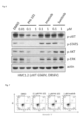
FIG. 2 depicts photographs showing the inhibitory activities of PHI-101 (A) according to an embodiment of the present invention, sunitinib (B) and dasatinib (C) against signaling in the c-kit D816V Ba/F3 cell line at various treatment concentrations. -
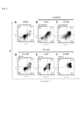
FIG. 3 depicts graphs showing the results of Annexin V-PI FACS analysis of the c-kit D816V Ba/F3 cell line treated with various concentrations of dimethyl sulfoxide (DMSO) (A) as a negative control, sunitinib (B) and (C), and PHI-101 (D), (E) and (F) according to one embodiment of the present invention. -
FIG. 4 depicts graphs showing the results of cell cycle FACS analysis of the c-kit D816V Ba/F3 cell line treated with dimethyl sulfoxide (DMSO) (A) as a negative control, sunitinib (B), and PHI-101 (C) according to one embodiment of the present invention. -
FIG. 5 is a graph showing the population at each cell cycle phase of the c-kit D816V Ba/F3 cell line treated with dimethyl sulfoxide (DMSO) (A) as a negative control, sunitinib (B), and PHI-101 (C) according to one embodiment of the present invention. -
FIG. 6 is a photograph showing the inhibitory activities of PHI-101 according to one embodiment of the present invention, imatinib and sunitinib against signaling in the HMC-1.2 cell line at various treatment concentrations. -
FIG. 7 depicts graphs showing the results of Annexin V-PI FACS analysis performed to evaluate the apoptotic effects of PHI-101 according to one embodiment of the present invention and imatinib against the HMC-1.2 cell line at various treatment concentrations. -
FIG. 8 is a photograph showing the results of Western blot analysis performed to evaluate the apoptotic effects of PHI-101 according to one embodiment of the present invention, imatinib and sunitinib against the HMC-1.2 cell line at various treatment concentrations. -
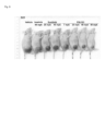
FIG. 9 is a photograph comparing the status of mastocytosis mouse models onday 21 after oral administration of each of PHI-101 according to one embodiment of the present invention, imatinib and sunitinib. -
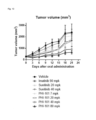
FIG. 10 is a graph comparing the tumor growth inhibitory activities of PHI-101 according to one embodiment of the present invention, imatinib and sunitinib in mastocytosis mouse models after oral administration. -
FIG. 11 depict graphs showing tumor volumes in mastocytosis mouse models on day 0 (A) and day 21 (B) after oral administration of each of PHI-101 according to one embodiment of the present invention, imatinib and sunitinib. -
FIG. 12 depicts graphs showing tumor volumes (A) and tumor weights (B) (percentages relative to a control) in mastocytosis mouse models on day 21 (B) after oral administration of each of PHI-101 according to one embodiment of the present invention, imatinib and sunitinib. -
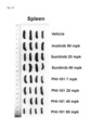
FIG. 13 is a photograph showing the spleens in mastocytosis mouse models on day 21 (B) after oral administration of each of PHI-101 according to one embodiment of the present invention, imatinib and sunitinib. -
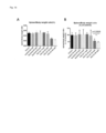
FIG. 14 depicts graphs showing spleen/body weight ratio (A) and spleen/body weight ratio (B) (percentages relative to a control) in mastocytosis mouse models on day 21 (B) after oral administration of each of PHI-101 according to one embodiment of the present invention, imatinib and sunitinib. - One aspect of the present invention provides a pharmaceutical composition for preventing or treating mastocytosis containing a compound represented by the following
Formula 1 or a pharmaceutically acceptable salt thereof:
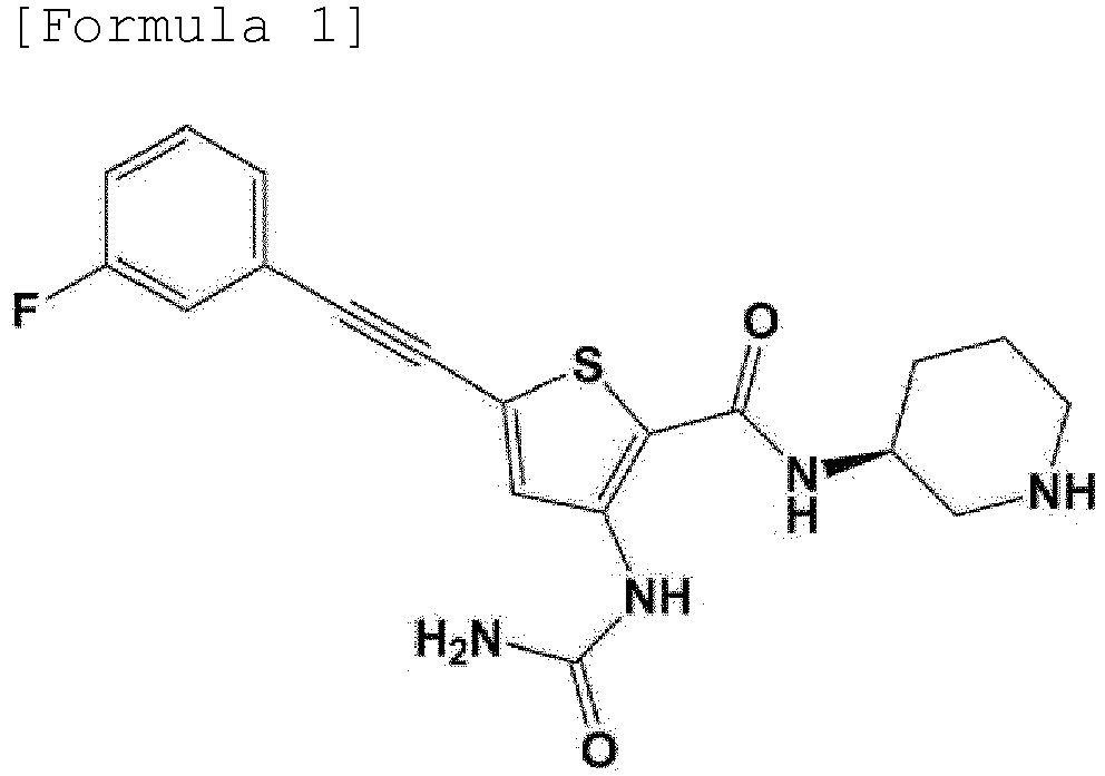
- In one embodiment of the present invention, the mastocytosis may be any one disease selected from the group consisting of mast cell leukemia, systemic mastocytosis, and invasive systemic mastocytosis.
- In one embodiment of the present invention, the mastocytosis may be caused by substitution of valine or histidine for aspartic acid at amino acid position 816 of c-kit protein.
- In one embodiment of the present invention, the mastocytosis may be caused by at least one substitution selected from the group consisting of substitution of valine for glycine at
amino acid position 560 of c-kit protein, substitution of proline for alanine at amino acid position 829 of c-kit protein, substitution of proline for leucine at amino acid position 576 of c-kit protein, substitution of aspartic acid for valine at amino acid position 559 of c-kit protein, substitution of isoleucine for threonine atamino acid position 670 of c-kit protein, and substitution of alanine for valine at amino acid position 654 of c-kit protein - Another aspect of the present invention provides a food composition for preventing or ameliorating mastocytosis containing a compound represented by the following
Formula 1 or a pharmaceutically acceptable salt thereof:

- Still another aspect of the present invention provides a method for treating mastocytosis comprising a step of administering, to a mastocytosis patient, a pharmaceutical composition containing a compound represented by the following
Formula 1 or a pharmaceutically acceptable salt thereof as an active ingredient:

- Yet another aspect of the present invention provides the use of a compound represented by the following
Formula 1 or a pharmaceutically acceptable salt thereof for preventing or treating mastocytosis:

- Still yet another aspect of the present invention provides the use of a compound represented by the following
Formula 1 or a pharmaceutically acceptable salt thereof for preparing a pharmaceutical composition for preventing or treating mastocytosis:

- Hereinafter, the present invention will be described in detail with reference to one or more examples. However, these examples are for illustrating the present invention, and the scope of the present disclosure is not limited by these examples.
- The inhibitory activity of the compound represented by Formula 1 (hereinafter referred to as "PHI-101") against a c-kit mutant cell line was measured.


- Specifically, 100 µl of a Ba/F3 cell line with a c-kit D816V mutation (
FIG. 1 ) was added to each well of a 96-well plate at a concentration of 10,000 cells/100 µl. After 4 hours, 100 ul of the Ba/F3 cell line was treated with 0.5 µl of each of control compounds and PHI-101 (10-point 1:3 serial dilutions) to a final concentration of up to 50 µM, and cultured at 37°C under 5% CO2 for 72 hours. As the control compounds, imatinib and sunitinib which are c-kit inhibitors were used. After culture, the cells were counted using a Celltiter glo assay kit (Promega), and the 50% growth inhibition values (GI50, µM) of the control compounds and PHI-101 were measured. - As a result, it was confirmed that PHI-101 exhibited higher inhibitory activity against the c-kit D816V Ba/F3 mutant cell line than the control compounds, and exhibited higher selectivity for the c-kit D816V Ba/F3 mutant cell line than for the parental Ba/F3 cell line (Table 1).
[Table 1] GI50 (µM) PHI-101 Imatinib Sunitinib cKIT D816V Ba/F3 0.912 11.48 2.647 Parental Ba/F3 5.010 12.73 13.32 - The inhibitory activities of control compounds and PHI-101 against c-kit phosphorylation and downstream signaling (p-STAT3, p-ERK1/2) in the c-kit D816V mutant Ba/F3 cell line were measured, and as the control compounds, sunitinib and dasatinib were used.
- Specifically, the c-kit D816V mutant Ba/F3 cell line was dispensed at a concentration of 1 × 10⁶ cells/ml, treated with each of the control compounds and PHI-101 at concentrations of 0.1, 1 and 10 µM, and then cultured for 2 hours. Next, lysates of the cells were obtained using lysis buffer (50 mM Tris-HCl pH 7.5, 1% NP40, 1 mM EDTA, 150 mM NaCl, 5 mM, Na3VO4 and 2.5 mM NaF, and a protease inhibitor cocktail). Equal amounts of the cell lysates were electrophoresed using 8% SDS-PAGE gel at 100 V for 1 hour and 30 minutes, and the separated protein was electrically transferred to nitrocellulose membranes. The membranes were blocked with 5% skim milk at room temperature for 30 minutes, and allowed to react with primary antibodies (diluted 1:5,000), including p-c-kit tyrosine 719 (Y719), p-STAT3 tyrosine 705 (Y705) and p-ERK1/2 threonine 202/tyrosine 204 (T202/Y204) at 4°C for 16 to 20 hours. As a control for comparing the level of expression, anti-β actin (diluted at a ratio of 1:10,000) was used as a primary antibody. Thereafter, the membranes were washed 3 times with TBST (Tris-buffered saline, 0.1% Tween 20) for 5 minutes each time, and then incubated with an HRP-conjugated anti-rabbit secondary antibody (diluted at a ratio of 1:10,000) at room temperature for 1 hour. Next, the membranes were washed three times with TBST for 5 minutes each time and treated with a chemiluminescent substrate reagent, and the expression level of the protein was measured by detecting luminescence with an X-ray film in the dark.
- As a result, it was confirmed that PHI-101 showed higher inhibitory activity against p-c-kit (Y719) phosphorylation than the control compounds, and showed downstream signaling (p-STAT3, p-ERK1/2) inhibitory activity similar to those of the control compounds (
FIG. 2 ). - The c-kit D816V mutant Ba/F3 cell line was treated with PHI-101, and then the apoptotic effect of PHI-101 was analyzed.
- Specifically, the c-kit D816V mutant Ba/F3 cell line was adjusted to a concentration of 1 × 10⁶ cells/ml and treated with each of PHI-101 at concentrations of 0.1, 1 and 10 µM and the control compound sunitinib at concentrations of 1 and 10 µM for 24 hours. Thereafter, the medium was removed and the cells were washed once with 1 ml PBS. The PBS was removed by centrifugation at 1,000 rpm for 5 minutes. The cells were dissolved in 100 µl of PBS containing Ca2+ and Mg2+ and allowed to react with 5 µl of Annexin V for 20 minutes. After completion of the reaction, 400 µl of PBS containing Ca2+ and Mg2+ was added, and 5 µl of propidium iodide (PI) was added, and then the degree of apoptotic cell death was analyzed by FACS (fluorescence activated cell sorter) assay using a flow cytometer (FACS-C6, BD).
- As a result, it was confirmed that PHI-101 strongly induced apoptosis even at a concentration of 0.1 µM, and increased apoptosis in a concentration-dependent manner (
FIG. 3 ) . - The c-kit D816V mutant Ba/F3 cell line was treated with 1 µM of PHI-101, and then the SubG1/GO-1 population was analyzed by cell cycle FACS assay.
- Specifically, the c-kit D816V mutant Ba/F3 cell line was adjusted to a concentration of 1 × 10⁶ cells/ml and treated with each of PHI-101 and the control compound sunitinib at a concentration of 1 µM for 24 hours. Thereafter, the medium was removed, and the c-kit D816V mutant Ba/F3 cell line was fixed in 70% ethanol at 4°C for 24 hours, and then incubated in a PBS buffer solution containing 40 pg/ml of propidium iodide (PI) and 100 pg/ml of RNase for 30 minutes to completely stain the total DNA. Cell cycle analysis of the stained cells was performed using a flow cytometer (FACS-C6, BD). The proportion of cells in G0/G1 phase, S phase and G2/M phase was measured, and based on the measurement results, the SubG1/GO-1 population was determined.
- As a result, it was confirmed that the SubG1/GO-1 population was increased by treatment with PHI-101, indicating that the cell cycle progression of the c-kit D816V mutant Ba/F3 cell line treated with PHI-101 was inhibited compared to that of the cell line treated with sunitinib (
FIGS. 4 and 5 ). - The inhibitory activity of PHI-101 against a mastocytosis cell line was measured.
- Specifically, for the HMC-1.2 cell line (imatinib-resistant cell line, having c-kit G560V/D816V mutation), the GI50 (µM) values of control compounds and PHI-101 were measured in the same manner as in Example 1-1, and as the control compounds, imatinib, dasatinib, sunitinib and ponatinib were used.
- As a result, it was confirmed that PHI-101 exhibited significant inhibitory activity against the HMC-1.2 cell line compared to the control compounds (Table 2).
[Table 2] GI50 (µM) PHI-101 Imatinib Dasatinib Sunitinib Ponatinib HMC-1.2 0.033 17.140 0.312 1.299 0.194 - The inhibitory activities of control compounds and PHI-101 against c-kit phosphorylation and downstream signaling (p-STAT5, p-AKT, p-ERK) in a mastocytosis cell line were measured, and as the control compounds, imatinib and sunitinib were used.
- Specifically, inhibitory activities against c-kit phosphorylation and downstream signaling were measured in the same manner as in Example 1-2, except that the HMC-1.2 cell line was treated with each of control compounds at concentrations of 0.1 and 1 µM and PHI-101 at concentrations of 0.01, 0.1 and 1 µM, and p-c-kit, p-STAT5, p-AKT and p-ERK each diluted at 1:3000, were used as primary antibodies.
- As a result, it was confirmed that PHI-101 decreased c-kit phosphorylation and downstream signaling in a concentration-dependent manner, exhibited significantly high inhibitory activity against downstream signaling compared to the control compounds, and strongly inhibited downstream signaling even at a concentration of 0.1 µM (
FIG. 6 ). - A mastocytosis cell line was treated with PHI-101, and the levels of cleaved PARP and cleaved caspase-3, which are apoptosis markers, were analyzed. As a control compound, imatinib was used.
- Specifically, the apoptotic effect of PHI-101 against the HMC-1.2 cell line was analyzed in the same manner as in Example 1-3, except that the HMC-1.2 cell line was treated with each of the control compounds at a concentration of 1 µM and PHI-101 at concentrations of 0.1 and 1 µM.
- As a result, it was confirmed that PHI-101 strongly induced apoptosis even at a concentration of 1 µM, indicating that PHI-101 had an apoptotic effect against the mastocytosis cell line (
FIG. 7 ). - A mastocytosis cell line was treated with PHI-101, and the levels of cleaved PARP and cleaved caspase-3, which are apoptosis markers, were analyzed. As control compounds, imatinib and sunitinib were used.
- Specifically, the apoptotic effect of PHI-101 against the HMC-1.2 cell line was analyzed in the same manner as in Example 1-2, except that the HMC-1.2 cell line was treated with each of the control compounds at a concentration of 1 µM and PHI-101 at concentrations of 0.1 and 1 µM, and that cleaved PARP and cleaved caspase-3, each diluted at 1:5,000, were used as primary antibodies.
- As a result, it was confirmed that the levels of cleaved PARP and cleaved caspase-3 in the HMC-1.2 cell line treated with PHI-101 were higher than those in the HMC-1.2 cell lines treated with the control compounds, indicating that PHI-101 had an apoptotic effect against the mastocytosis cell line (
FIG. 8 ). - As mice, 6-8-week-old NOD SCID female mice (18 to 22g) (GemPharmatech Co., Ltd.) were prepared. Mice were acclimatized for 7 days before use in the experiment, and mice with abnormal health were excluded from the experiment. Mice were bred in a cage (300 × 180 × 150 mm) at a 22±3°C and a relative humidity of 40% to 70% with a 12-hr light/12-hr dark cycle. Mice were allowed access to sterilized dry granular food (Beijing Keaoxieli Feed Co., Ltd., Beijing, China) and water ad libitum for the entire experimental period except for the period specified in the protocol.
- 50 ml of HMC-1.2 cells suspension-cultured in a 75T flask were placed in a conical tube and centrifuged at 1,200 rpm for 2 minutes, centrifuged with DPBS (Dulbecco's Phosphate-Buffered Saline, Welgene) at 1,200 rpm for 2 minutes, washed, and then suspended in RPMI1640 (Welgene) without antibiotics and fetal bovine serum (FBS). Then, the cells were stained with trypan blue, counted with a hemocytometer, and then HMC-1.2 cells (1 × 10⁶) in a mixture of 0.1 ml L-15 and Matrigel (BD, cat. No. 356234) (1:1) were subcutaneously injected into the right flanks of mice anesthetized by intraperitoneal administration of Avertin (250 mg/kg), thus inducing tumors.
- Thereafter, when the average tumor volume reached 150 to 200 mm3, 4 mice were assigned to each group so that the average tumor volume at each group and treatment time was the same between the groups. Then, each of imatinib at a dose of 90 mg/kg, sunitinib at doses of 20 and 40 mg/kg, and PHI-101 at doses of 7, 20, 40 and 80 mg/kg was administered orally to the mice once daily for 3 weeks. For oral administration, each compound was dissolved in 5% 1-methyl-2-pyrrolidone and then completely mixed in 15% Kolliphor, 30% PEG E400 HS15 and 50% 0.05M citric acid.
- In the mastocytosis mouse model with the HMC-1.2 cell line, whether mastocytosis was inhibited by administration of PHI-101 was examined.
- Specifically, while the presence or absence of abnormalities in the mastocytosis mouse model of Example 3-1 was monitored daily by checking the skin condition, bleeding and stool status, whether the body weight changed was checked by measuring the mouse body weight at intervals of 3 days after the start of oral administration of each drug. In addition, the width and length of the tumor were measured using a caliper at 3-day intervals, and the tumor volume was calculated using the following equation.

(V: tumor volume, W: tumor width, and L: tumor length) - In addition, the mice were sacrificed 3 weeks after the start of oral administration of each drug and the tumor weight was measured using a digital scale. For each data, one-way ANOVA was used to test the significance of each group.
- As a result, it was confirmed that the growth of mastocytosis cells was inhibited by the administration of PHI-101 in a concentration-dependent manner, and that the growth of mastocytosis cells in the group treated with PHI-101 at a concentration of 40 mg/kg or more was significantly inhibited compared to that in the group treated with each control compound (
FIGS. 9 to 12 ).[Table 3] Dose (mpk, mg/kg) Tumor growth inhibition (TGI)(%) Timor volume Tumor weight Vehicle - 0 0 Imatinib 90 11.3 7.8 Sunitinib 20 37.3 49.5 40 46.2 47.9 PHI-101 7 30.2 32.7 20 43.6 51.6 40 68.4 68.3 80 78.5 82.7 - Meanwhile, no abnormal symptoms were found in the mastocytosis mouse model during the experiment, and it was confirmed that there was no administration group that showed non-specific weight loss (<10%), and PHI-101 caused no weight loss even at a high dose of 80 mg/kg.
- In the mastocytosis mouse model with the HMC-1.2 cell line, whether an inflammatory response caused by mastocytosis was inhibited by the administration of PHI-101 was examined.
- Specifically, 3 weeks after administering each drug to the mastocytosis mouse model of Example 3-1, the mice were sacrificed, the body weight and spleen weight were measured using a digital scale, and then the spleen/body weight ratio was calculated.
- As a result, it was confirmed that the administration of PHI-101 decreased the spleen/body weight ratio in a concentration-dependent manner compared to that in the control group (
FIGS. 13 and14 and Table 4), indicating that the inflammation response caused by mastocytosis was also effectively inhibited by the administration of PHI-101.[Table 4] Dose (mpk, mg/kg) Spleen/body weight ratio % of control Inhibition (%) Vehicle - 100.0 0 Imatinib 90 97.7 2.3 Sunitinib 20 104.3 -4.3 40 102.7 -2.7 PHI-101 7 105.0 -5 20 95.0 5 40 55.7 44.3 80 46.3 53.7 - A kinase-tagged T7 phage strain was prepared from an E. coli host derived from a BL21 strain. E. coli was grown in log-phase, infected with T7 phage, and then incubated with shaking at 32°C until lysis. The lysates were centrifuged and filtered to remove cell debris. Thereafter, the kinase produced in HEK-293 cells was labeled with DNA for qPCR detection. Streptavidin-coated magnetic beads were treated with biotinylated small molecule ligand for 30 minutes at room temperature to produce affinity resins for kinase assay. The liganded beads were blocked with excess biotin and washed with blocking buffer (SeaBlock(Pierce), 1% BSA, 0.05
% Tween % Tween % Tween 20, 0.5 µM non-biotinylated affinity ligand) and incubated at room temperature with shaking for 30 minutes. The kinase concentration in the eluates was measured by qPCR. - 11-point 3-fold serial dilutions of each test compound were prepared in 100% DMSO at 100x final test concentration and then diluted to 1x in the assay (final DMSO concentration=1%). Most Kd values were determined using a compound top concentration = 30,000 nM. If the initial Kd determined was <0.5 nM (lowest concentration tested), the measurement was repeated with a serial dilution starting at a lower top concentration. A K d value reported as 40,000nM indicates that the K d was determined to be >30,000nM.
- The Kd value of c-kit protein was calculated with a standard dose-response curve (Equation 1) using the Hill equation.

- The Hill Slope was set to -1. Curves were fitted using a non-linear least square fit with the Levenberg-Marquardt algorithm.
- The inhibitory effect of PHI-101 against the binding activity of various c-kit protein mutations was analyzed by the method described in Examples 4-1 to 4-3.
- As a result, it was confirmed that PHI-101 inhibited the binding activity of seven mutant c-kit proteins (Table 5) .
[Table 5] KIT mutation Kd (nM) KIT (A829P) 1.3 KIT (D816H) 4.8 KIT (D816V) 2.1 KIT (L576P) 39 KIT (V559D) 16 KIT (V559D, T670I) 27 KIT (V559D, V654A) 26 - From the above results, it was confirmed that PHI-101 can be particularly effectively used for the treatment of mastocytosis having A829P, D816H, D816V, L576P or V559D mutation in c-kit protein, mastocytosis having both V559D and T670I mutations in c-kit protein, and mastocytosis having both V559D and V654A mutations in c-kit protein.
- So far, the present invention has been described with reference to the embodiments. Those of ordinary skill in the art to which the present invention pertains will appreciate that the present invention may be embodied in modified forms without departing from the essential characteristics of the present invention. Therefore, the disclosed embodiments should be considered from an illustrative point of view, not from a restrictive point of view. The scope of the present invention is defined by the claims rather than the foregoing description, and all differences within the scope equivalent thereto should be construed as being included in the present invention.
Claims (8)
- A pharmaceutical composition for preventing or treating mastocytosis containing a compound represented by the following Formula 1 or a pharmaceutically acceptable salt thereof:

- The pharmaceutical composition of claim 1, wherein the mastocytosis is any one disease selected from the group consisting of mast cell leukemia, systemic mastocytosis, and invasive systemic mastocytosis.
- The pharmaceutical composition of claim 1, wherein the mastocytosis is caused by substitution of valine or histidine for aspartic acid at amino acid position 816 of c-kit protein.
- The pharmaceutical composition of claim 1, wherein the mastocytosis is caused by at least one substitution selected from the group consisting of:substitution of valine for glycine at amino acid position 560 of c-kit protein;substitution of proline for alanine at amino acid position 829 of c-kit protein;substitution of proline for leucine at amino acid position 576 of c-kit protein;substitution of aspartic acid for valine at amino acid position 559 of c-kit protein;substitution of isoleucine for threonine at amino acid position 670 of c-kit protein; andsubstitution of alanine for valine at amino acid position 654 of c-kit protein
- A food composition for preventing or ameliorating mastocytosis containing a compound represented by the following Formula 1 or a pharmaceutically acceptable salt thereof:


- A method for treating mastocytosis comprising a step of administering, to a mastocytosis patient, a pharmaceutical composition containing a compound represented by the following Formula 1 or a pharmaceutically acceptable salt thereof as an active ingredient:

- Use of a compound represented by the following Formula 1 or a pharmaceutically acceptable salt thereof for preventing or treating mastocytosis:


- Use of a compound represented by the following Formula 1 or a pharmaceutically acceptable salt thereof for preparing a pharmaceutical composition for preventing or treating mastocytosis:

Applications Claiming Priority (2)
| Application Number | Priority Date | Filing Date | Title |
|---|---|---|---|
| KR1020200095988A KR102535842B1 (en) | 2020-07-31 | 2020-07-31 | Use of 2,3,5-Substituted Thiophene Compound for Prevention, Improvement or Treatment of Mastocytosis |
| PCT/KR2021/010001 WO2022025716A1 (en) | 2020-07-31 | 2021-07-30 | Use of 2,3,5-substituted thiophene compound for preventing, alleviation, or treating mastocytosis |
Publications (3)
| Publication Number | Publication Date |
|---|---|
| EP4190330A1 true EP4190330A1 (en) | 2023-06-07 |
| EP4190330A4 EP4190330A4 (en) | 2024-08-14 |
| EP4190330B1 EP4190330B1 (en) | 2025-09-10 |
Family
ID=80035886
Family Applications (1)
| Application Number | Title | Priority Date | Filing Date |
|---|---|---|---|
| EP21849180.1A Active EP4190330B1 (en) | 2020-07-31 | 2021-07-30 | Use of 2,3,5-substituted thiophene compound for preventing, alleviation, or treating mastocytosis |
Country Status (5)
| Country | Link |
|---|---|
| US (1) | US20230263792A1 (en) |
| EP (1) | EP4190330B1 (en) |
| JP (1) | JP7511742B2 (en) |
| KR (1) | KR102535842B1 (en) |
| WO (1) | WO2022025716A1 (en) |
Families Citing this family (1)
| Publication number | Priority date | Publication date | Assignee | Title |
|---|---|---|---|---|
| JP7407298B2 (en) * | 2020-03-04 | 2023-12-28 | ファロス・アイバイオ・カンパニー・リミテッド | Use of 2,3,5-substituted thiophene compounds to prevent, improve or treat ovarian cancer |
Family Cites Families (6)
| Publication number | Priority date | Publication date | Assignee | Title |
|---|---|---|---|---|
| WO2005066163A2 (en) * | 2004-01-05 | 2005-07-21 | Astrazeneca Ab | Thiophene derivatives as chk 1 inihibitors |
| WO2006034075A1 (en) * | 2004-09-17 | 2006-03-30 | Osi Pharmaceuticals, Inc. | (hydrazido) (amino) thiophene compounds |
| CN103638028A (en) | 2005-05-02 | 2014-03-19 | 诺瓦提斯公司 | Use of pyrimidylamimobenzamide derivatives for the treatment of systematic mastocytosis |
| JP2009515978A (en) * | 2005-11-14 | 2009-04-16 | バイエル ヘルスケア エルエルシー | Treatment of cancer with acquired resistance to KIT inhibitors |
| CN101687854A (en) * | 2007-05-04 | 2010-03-31 | Irm责任有限公司 | Compounds and compositions as C-KIT and PDGFR kinase inhibitors |
| CN108884066B (en) * | 2016-02-16 | 2021-08-24 | 韩国科学技术研究院 | 2,3,5-Substituted Thiophene Compounds as Protein Kinase Inhibitors |
-
2020
- 2020-07-31 KR KR1020200095988A patent/KR102535842B1/en active Active
-
2021
- 2021-07-30 US US18/006,584 patent/US20230263792A1/en active Pending
- 2021-07-30 JP JP2023504836A patent/JP7511742B2/en active Active
- 2021-07-30 EP EP21849180.1A patent/EP4190330B1/en active Active
- 2021-07-30 WO PCT/KR2021/010001 patent/WO2022025716A1/en not_active Ceased
Also Published As
| Publication number | Publication date |
|---|---|
| JP2023535217A (en) | 2023-08-16 |
| KR102535842B1 (en) | 2023-05-23 |
| KR20220015673A (en) | 2022-02-08 |
| JP7511742B2 (en) | 2024-07-05 |
| WO2022025716A1 (en) | 2022-02-03 |
| EP4190330B1 (en) | 2025-09-10 |
| EP4190330A4 (en) | 2024-08-14 |
| US20230263792A1 (en) | 2023-08-24 |
Similar Documents
| Publication | Publication Date | Title |
|---|---|---|
| CN107787373B (en) | Use of farnesyl transferase inhibitors for the treatment of cancer | |
| US20190192522A1 (en) | Inhibitors of the fibroblast growth factor receptor 4 in combination with cyclin-dependent kinase inhibitors | |
| US20110306622A1 (en) | Methods of treating hematological disorders with quinazolinone compounds in selected subjects | |
| US20120128665A1 (en) | Preselection of subjects for therapeutic treatment based on hypoxic status | |
| WO2003002109A2 (en) | Use of tyrosine kinase inhibitors for treating autoimmune diseases | |
| AU2018331267A1 (en) | Improved treatment of atopic dermatitis with tradipitant | |
| US7678805B2 (en) | Use of tyrosine kinase inhibitors for treating inflammatory bowel diseases (IBD) | |
| AU2007299764B2 (en) | Method of optimizing the treatment of Philadelphia-positive leukemia with Abl tyrosine kinase inhibitors | |
| EP4190330B1 (en) | Use of 2,3,5-substituted thiophene compound for preventing, alleviation, or treating mastocytosis | |
| JP2013505446A (en) | Biomarkers for IAP inhibitor compounds | |
| KR101928618B1 (en) | Therapeutic effect prediction method for colorectal cancer patient in whom expression of tk1 protein has increased | |
| EP4582087A1 (en) | Pharmaceutical composition for treating lymphoma | |
| JP7511741B2 (en) | Use of 2,3,5-substituted thiophene compounds for the prevention, amelioration or treatment of gastrointestinal stromal tumors | |
| JP3725800B2 (en) | Method for screening compound for possibility of inhibiting tumor formation and pharmaceutical composition containing the compound | |
| JP7827310B2 (en) | Pharmaceutical composition for treating lymphoma | |
| TW202410904A (en) | Pharmaceutical compositions for treating lymphoma containing BCV, a pharmaceutically acceptable salt thereof, or a solvate thereof | |
| US20220143006A1 (en) | Methods of treating cancer with farnesyltransferase inhibitors | |
| CN119925601A (en) | Application of the tetrahydrobiopterin pathway as a target in resistance to QDPR-deficient tumors | |
| Kelly et al. | Aetiological Subtypes of Transient Ischemic Attack and Ischaemic Stroke in CKD: A Population-Based Study: PO2153 | |
| JP2005516993A (en) | Treatment of neuroblastoma | |
| WO2017022634A1 (en) | Method and kit for evaluating prognosis, distant recurrence risk and invasion of glioma, and pharmaceutical composition for treating glioma | |
| WO2024048555A1 (en) | Combination drug | |
| WO2019113269A1 (en) | Methods of treating cancer patients with farnesyltransferase inhibitors | |
| AU2002329528A1 (en) | Use of tyrosine kinase inhibitors for treating autoimmune diseases |
Legal Events
| Date | Code | Title | Description |
|---|---|---|---|
| STAA | Information on the status of an ep patent application or granted ep patent |
Free format text: STATUS: THE INTERNATIONAL PUBLICATION HAS BEEN MADE |
|
| PUAI | Public reference made under article 153(3) epc to a published international application that has entered the european phase |
Free format text: ORIGINAL CODE: 0009012 |
|
| STAA | Information on the status of an ep patent application or granted ep patent |
Free format text: STATUS: REQUEST FOR EXAMINATION WAS MADE |
|
| 17P | Request for examination filed |
Effective date: 20230215 |
|
| AK | Designated contracting states |
Kind code of ref document: A1 Designated state(s): AL AT BE BG CH CY CZ DE DK EE ES FI FR GB GR HR HU IE IS IT LI LT LU LV MC MK MT NL NO PL PT RO RS SE SI SK SM TR |
|
| DAV | Request for validation of the european patent (deleted) | ||
| DAX | Request for extension of the european patent (deleted) | ||
| A4 | Supplementary search report drawn up and despatched |
Effective date: 20240712 |
|
| RIC1 | Information provided on ipc code assigned before grant |
Ipc: A23L 33/10 20160101ALI20240708BHEP Ipc: A61P 3/04 20060101ALI20240708BHEP Ipc: A61K 31/4535 20060101AFI20240708BHEP |
|
| GRAP | Despatch of communication of intention to grant a patent |
Free format text: ORIGINAL CODE: EPIDOSNIGR1 |
|
| STAA | Information on the status of an ep patent application or granted ep patent |
Free format text: STATUS: GRANT OF PATENT IS INTENDED |
|
| INTG | Intention to grant announced |
Effective date: 20250416 |
|
| GRAS | Grant fee paid |
Free format text: ORIGINAL CODE: EPIDOSNIGR3 |
|
| GRAA | (expected) grant |
Free format text: ORIGINAL CODE: 0009210 |
|
| STAA | Information on the status of an ep patent application or granted ep patent |
Free format text: STATUS: THE PATENT HAS BEEN GRANTED |
|
| AK | Designated contracting states |
Kind code of ref document: B1 Designated state(s): AL AT BE BG CH CY CZ DE DK EE ES FI FR GB GR HR HU IE IS IT LI LT LU LV MC MK MT NL NO PL PT RO RS SE SI SK SM TR |
|
| REG | Reference to a national code |
Ref country code: GB Ref legal event code: FG4D |
|
| REG | Reference to a national code |
Ref country code: CH Ref legal event code: EP |
|
| REG | Reference to a national code |
Ref country code: DE Ref legal event code: R096 Ref document number: 602021038490 Country of ref document: DE |
|
| REG | Reference to a national code |
Ref country code: IE Ref legal event code: FG4D |
|
| REG | Reference to a national code |
Ref country code: CH Ref legal event code: R17 Free format text: ST27 STATUS EVENT CODE: U-0-0-R10-R17 (AS PROVIDED BY THE NATIONAL OFFICE) Effective date: 20251212 |
|
| PG25 | Lapsed in a contracting state [announced via postgrant information from national office to epo] |
Ref country code: NO Free format text: LAPSE BECAUSE OF FAILURE TO SUBMIT A TRANSLATION OF THE DESCRIPTION OR TO PAY THE FEE WITHIN THE PRESCRIBED TIME-LIMIT Effective date: 20251210 |
|
| REG | Reference to a national code |
Ref country code: LT Ref legal event code: MG9D |
|
| PG25 | Lapsed in a contracting state [announced via postgrant information from national office to epo] |
Ref country code: FI Free format text: LAPSE BECAUSE OF FAILURE TO SUBMIT A TRANSLATION OF THE DESCRIPTION OR TO PAY THE FEE WITHIN THE PRESCRIBED TIME-LIMIT Effective date: 20250910 |
|
| REG | Reference to a national code |
Ref country code: NL Ref legal event code: MP Effective date: 20250910 |
|
| PG25 | Lapsed in a contracting state [announced via postgrant information from national office to epo] |
Ref country code: HR Free format text: LAPSE BECAUSE OF FAILURE TO SUBMIT A TRANSLATION OF THE DESCRIPTION OR TO PAY THE FEE WITHIN THE PRESCRIBED TIME-LIMIT Effective date: 20250910 |
|
| PG25 | Lapsed in a contracting state [announced via postgrant information from national office to epo] |
Ref country code: GR Free format text: LAPSE BECAUSE OF FAILURE TO SUBMIT A TRANSLATION OF THE DESCRIPTION OR TO PAY THE FEE WITHIN THE PRESCRIBED TIME-LIMIT Effective date: 20251211 |
|
| PG25 | Lapsed in a contracting state [announced via postgrant information from national office to epo] |
Ref country code: SE Free format text: LAPSE BECAUSE OF FAILURE TO SUBMIT A TRANSLATION OF THE DESCRIPTION OR TO PAY THE FEE WITHIN THE PRESCRIBED TIME-LIMIT Effective date: 20250910 |
|
| PG25 | Lapsed in a contracting state [announced via postgrant information from national office to epo] |
Ref country code: LV Free format text: LAPSE BECAUSE OF FAILURE TO SUBMIT A TRANSLATION OF THE DESCRIPTION OR TO PAY THE FEE WITHIN THE PRESCRIBED TIME-LIMIT Effective date: 20250910 |
|
| PG25 | Lapsed in a contracting state [announced via postgrant information from national office to epo] |
Ref country code: PL Free format text: LAPSE BECAUSE OF FAILURE TO SUBMIT A TRANSLATION OF THE DESCRIPTION OR TO PAY THE FEE WITHIN THE PRESCRIBED TIME-LIMIT Effective date: 20250910 Ref country code: BG Free format text: LAPSE BECAUSE OF FAILURE TO SUBMIT A TRANSLATION OF THE DESCRIPTION OR TO PAY THE FEE WITHIN THE PRESCRIBED TIME-LIMIT Effective date: 20250910 |
|
| PG25 | Lapsed in a contracting state [announced via postgrant information from national office to epo] |
Ref country code: RS Free format text: LAPSE BECAUSE OF FAILURE TO SUBMIT A TRANSLATION OF THE DESCRIPTION OR TO PAY THE FEE WITHIN THE PRESCRIBED TIME-LIMIT Effective date: 20251210 |
|
| PG25 | Lapsed in a contracting state [announced via postgrant information from national office to epo] |
Ref country code: ES Free format text: LAPSE BECAUSE OF FAILURE TO SUBMIT A TRANSLATION OF THE DESCRIPTION OR TO PAY THE FEE WITHIN THE PRESCRIBED TIME-LIMIT Effective date: 20250910 |
|
| REG | Reference to a national code |
Ref country code: AT Ref legal event code: MK05 Ref document number: 1835167 Country of ref document: AT Kind code of ref document: T Effective date: 20250910 |
|
| PG25 | Lapsed in a contracting state [announced via postgrant information from national office to epo] |
Ref country code: NL Free format text: LAPSE BECAUSE OF FAILURE TO SUBMIT A TRANSLATION OF THE DESCRIPTION OR TO PAY THE FEE WITHIN THE PRESCRIBED TIME-LIMIT Effective date: 20250910 |
|
| PG25 | Lapsed in a contracting state [announced via postgrant information from national office to epo] |
Ref country code: SM Free format text: LAPSE BECAUSE OF FAILURE TO SUBMIT A TRANSLATION OF THE DESCRIPTION OR TO PAY THE FEE WITHIN THE PRESCRIBED TIME-LIMIT Effective date: 20250910 |
|
| PG25 | Lapsed in a contracting state [announced via postgrant information from national office to epo] |
Ref country code: AT Free format text: LAPSE BECAUSE OF FAILURE TO SUBMIT A TRANSLATION OF THE DESCRIPTION OR TO PAY THE FEE WITHIN THE PRESCRIBED TIME-LIMIT Effective date: 20250910 |
|
| PG25 | Lapsed in a contracting state [announced via postgrant information from national office to epo] |
Ref country code: IT Free format text: LAPSE BECAUSE OF FAILURE TO SUBMIT A TRANSLATION OF THE DESCRIPTION OR TO PAY THE FEE WITHIN THE PRESCRIBED TIME-LIMIT Effective date: 20250910 |
|
| PG25 | Lapsed in a contracting state [announced via postgrant information from national office to epo] |
Ref country code: IS Free format text: LAPSE BECAUSE OF FAILURE TO SUBMIT A TRANSLATION OF THE DESCRIPTION OR TO PAY THE FEE WITHIN THE PRESCRIBED TIME-LIMIT Effective date: 20260110 |
|
| PG25 | Lapsed in a contracting state [announced via postgrant information from national office to epo] |
Ref country code: PT Free format text: LAPSE BECAUSE OF FAILURE TO SUBMIT A TRANSLATION OF THE DESCRIPTION OR TO PAY THE FEE WITHIN THE PRESCRIBED TIME-LIMIT Effective date: 20260112 Ref country code: CZ Free format text: LAPSE BECAUSE OF FAILURE TO SUBMIT A TRANSLATION OF THE DESCRIPTION OR TO PAY THE FEE WITHIN THE PRESCRIBED TIME-LIMIT Effective date: 20250910 |
|
| PG25 | Lapsed in a contracting state [announced via postgrant information from national office to epo] |
Ref country code: EE Free format text: LAPSE BECAUSE OF FAILURE TO SUBMIT A TRANSLATION OF THE DESCRIPTION OR TO PAY THE FEE WITHIN THE PRESCRIBED TIME-LIMIT Effective date: 20250910 Ref country code: SK Free format text: LAPSE BECAUSE OF FAILURE TO SUBMIT A TRANSLATION OF THE DESCRIPTION OR TO PAY THE FEE WITHIN THE PRESCRIBED TIME-LIMIT Effective date: 20250910 |