EP4186927B1 - Methods and compositions for modifying macrophage polarization into pro-inflammatory cells to treat cancer - Google Patents
Methods and compositions for modifying macrophage polarization into pro-inflammatory cells to treat cancer Download PDFInfo
- Publication number
- EP4186927B1 EP4186927B1 EP23151596.6A EP23151596A EP4186927B1 EP 4186927 B1 EP4186927 B1 EP 4186927B1 EP 23151596 A EP23151596 A EP 23151596A EP 4186927 B1 EP4186927 B1 EP 4186927B1
- Authority
- EP
- European Patent Office
- Prior art keywords
- sirpa
- macrophages
- cells
- cancer
- tumor
- Prior art date
- Legal status (The legal status is an assumption and is not a legal conclusion. Google has not performed a legal analysis and makes no representation as to the accuracy of the status listed.)
- Active
Links
Images


Classifications
-
- C—CHEMISTRY; METALLURGY
- C07—ORGANIC CHEMISTRY
- C07K—PEPTIDES
- C07K16/00—Immunoglobulins [IG], e.g. monoclonal or polyclonal antibodies
- C07K16/18—Immunoglobulins [IG], e.g. monoclonal or polyclonal antibodies against material from animals or humans
- C07K16/28—Immunoglobulins [IG], e.g. monoclonal or polyclonal antibodies against material from animals or humans against receptors, cell surface antigens or cell surface determinants
- C07K16/2896—Immunoglobulins [IG], e.g. monoclonal or polyclonal antibodies against material from animals or humans against receptors, cell surface antigens or cell surface determinants against molecules with a "CD"-designation, not provided for elsewhere
-
- A—HUMAN NECESSITIES
- A61—MEDICAL OR VETERINARY SCIENCE; HYGIENE
- A61P—SPECIFIC THERAPEUTIC ACTIVITY OF CHEMICAL COMPOUNDS OR MEDICINAL PREPARATIONS
- A61P35/00—Antineoplastic agents
-
- C—CHEMISTRY; METALLURGY
- C07—ORGANIC CHEMISTRY
- C07K—PEPTIDES
- C07K16/00—Immunoglobulins [IG], e.g. monoclonal or polyclonal antibodies
- C07K16/18—Immunoglobulins [IG], e.g. monoclonal or polyclonal antibodies against material from animals or humans
- C07K16/28—Immunoglobulins [IG], e.g. monoclonal or polyclonal antibodies against material from animals or humans against receptors, cell surface antigens or cell surface determinants
- C07K16/2803—Immunoglobulins [IG], e.g. monoclonal or polyclonal antibodies against material from animals or humans against receptors, cell surface antigens or cell surface determinants against the immunoglobulin superfamily
- C07K16/2827—Immunoglobulins [IG], e.g. monoclonal or polyclonal antibodies against material from animals or humans against receptors, cell surface antigens or cell surface determinants against the immunoglobulin superfamily against B7 molecules, e.g. CD80, CD86
-
- C—CHEMISTRY; METALLURGY
- C07—ORGANIC CHEMISTRY
- C07K—PEPTIDES
- C07K16/00—Immunoglobulins [IG], e.g. monoclonal or polyclonal antibodies
- C07K16/18—Immunoglobulins [IG], e.g. monoclonal or polyclonal antibodies against material from animals or humans
- C07K16/28—Immunoglobulins [IG], e.g. monoclonal or polyclonal antibodies against material from animals or humans against receptors, cell surface antigens or cell surface determinants
- C07K16/2878—Immunoglobulins [IG], e.g. monoclonal or polyclonal antibodies against material from animals or humans against receptors, cell surface antigens or cell surface determinants against the NGF-receptor/TNF-receptor superfamily, e.g. CD27, CD30, CD40, CD95
-
- C—CHEMISTRY; METALLURGY
- C12—BIOCHEMISTRY; BEER; SPIRITS; WINE; VINEGAR; MICROBIOLOGY; ENZYMOLOGY; MUTATION OR GENETIC ENGINEERING
- C12N—MICROORGANISMS OR ENZYMES; COMPOSITIONS THEREOF; PROPAGATING, PRESERVING, OR MAINTAINING MICROORGANISMS; MUTATION OR GENETIC ENGINEERING; CULTURE MEDIA
- C12N15/00—Mutation or genetic engineering; DNA or RNA concerning genetic engineering, vectors, e.g. plasmids, or their isolation, preparation or purification; Use of hosts therefor
- C12N15/09—Recombinant DNA-technology
- C12N15/11—DNA or RNA fragments; Modified forms thereof; Non-coding nucleic acids having a biological activity
- C12N15/113—Non-coding nucleic acids modulating the expression of genes, e.g. antisense oligonucleotides; Antisense DNA or RNA; Triplex- forming oligonucleotides; Catalytic nucleic acids, e.g. ribozymes; Nucleic acids used in co-suppression or gene silencing
- C12N15/1138—Non-coding nucleic acids modulating the expression of genes, e.g. antisense oligonucleotides; Antisense DNA or RNA; Triplex- forming oligonucleotides; Catalytic nucleic acids, e.g. ribozymes; Nucleic acids used in co-suppression or gene silencing against receptors or cell surface proteins
-
- C—CHEMISTRY; METALLURGY
- C12—BIOCHEMISTRY; BEER; SPIRITS; WINE; VINEGAR; MICROBIOLOGY; ENZYMOLOGY; MUTATION OR GENETIC ENGINEERING
- C12N—MICROORGANISMS OR ENZYMES; COMPOSITIONS THEREOF; PROPAGATING, PRESERVING, OR MAINTAINING MICROORGANISMS; MUTATION OR GENETIC ENGINEERING; CULTURE MEDIA
- C12N5/00—Undifferentiated human, animal or plant cells, e.g. cell lines; Tissues; Cultivation or maintenance thereof; Culture media therefor
- C12N5/06—Animal cells or tissues; Human cells or tissues
- C12N5/0602—Vertebrate cells
- C12N5/0634—Cells from the blood or the immune system
- C12N5/0645—Macrophages, e.g. Kuepfer cells in the liver; Monocytes
-
- A—HUMAN NECESSITIES
- A61—MEDICAL OR VETERINARY SCIENCE; HYGIENE
- A61K—PREPARATIONS FOR MEDICAL, DENTAL OR TOILETRY PURPOSES
- A61K39/00—Medicinal preparations containing antigens or antibodies
- A61K2039/505—Medicinal preparations containing antigens or antibodies comprising antibodies
-
- A—HUMAN NECESSITIES
- A61—MEDICAL OR VETERINARY SCIENCE; HYGIENE
- A61K—PREPARATIONS FOR MEDICAL, DENTAL OR TOILETRY PURPOSES
- A61K39/00—Medicinal preparations containing antigens or antibodies
- A61K2039/505—Medicinal preparations containing antigens or antibodies comprising antibodies
- A61K2039/507—Comprising a combination of two or more separate antibodies
-
- C—CHEMISTRY; METALLURGY
- C07—ORGANIC CHEMISTRY
- C07K—PEPTIDES
- C07K2317/00—Immunoglobulins specific features
- C07K2317/70—Immunoglobulins specific features characterized by effect upon binding to a cell or to an antigen
- C07K2317/76—Antagonist effect on antigen, e.g. neutralization or inhibition of binding
-
- C—CHEMISTRY; METALLURGY
- C12—BIOCHEMISTRY; BEER; SPIRITS; WINE; VINEGAR; MICROBIOLOGY; ENZYMOLOGY; MUTATION OR GENETIC ENGINEERING
- C12N—MICROORGANISMS OR ENZYMES; COMPOSITIONS THEREOF; PROPAGATING, PRESERVING, OR MAINTAINING MICROORGANISMS; MUTATION OR GENETIC ENGINEERING; CULTURE MEDIA
- C12N2310/00—Structure or type of the nucleic acid
- C12N2310/10—Type of nucleic acid
- C12N2310/14—Type of nucleic acid interfering nucleic acids [NA]
-
- C—CHEMISTRY; METALLURGY
- C12—BIOCHEMISTRY; BEER; SPIRITS; WINE; VINEGAR; MICROBIOLOGY; ENZYMOLOGY; MUTATION OR GENETIC ENGINEERING
- C12N—MICROORGANISMS OR ENZYMES; COMPOSITIONS THEREOF; PROPAGATING, PRESERVING, OR MAINTAINING MICROORGANISMS; MUTATION OR GENETIC ENGINEERING; CULTURE MEDIA
- C12N2320/00—Applications; Uses
- C12N2320/30—Special therapeutic applications
- C12N2320/31—Combination therapy
Definitions
- the present invention pertains to the field of immunotherapy. More specifically, the present invention provides an anti-SIRPa compound selected from the group consisting of an antibody or fragment(s) thereof, wherein the anti-SIRPa compound binds specifically to the extracellular domain of SIRPa and blocks SIRPa pathway, combined with a second therapeutic agent selected from the group consisting of a blocking anti-PDL1 antibody and a blocking anti-PD 1 antibody, for use in the treatment of a solid cancer.
- New generation immunotherapeutic molecules revolutionizing cancer treatments, block suppressor mechanisms of these cells, allowing the effector T cells (Teff) to exercise their action.
- This is the concept called "Inhibit inhibitors”.
- An anti-CTLA-4 antibody (Yervoy ® ) has been the first molecule for treating metastatic forms of malignant melanoma prolonging the mean survival of patients of 6 to 10 months, with a quarter of patients still alive after 2 years. Unfortunately, these results, as spectacular as they are, still do not cure most patients.
- the present invention relies on an "inhibit inhibitors" approach and provides a new method useful in immunotherapy. More specifically, the present invention pertains to a method for modifying macrophage polarization in order to induce a pro-inflammatory environment.
- the method consists in the use of an anti-SIRPa compound able to inhibit the polarization of anti-inflammatory M2-type macrophages and/or favors pro-inflammatory M1-type macrophages, for inhibiting the anti-inflammatory signal provided by M2-type macrophages and favoring the pro-inflammatory signal provided by M1-type macrophages.
- This approach allows to reestablish an inflammatory environment favorable to the action of the T effector cells, in particular in eliminating the cancer cells.
- Macrophages are cells that have the highest plasticity of the hematopoietic system. They are involved both in innate immunity (phagocytosis capacity) and in adaptive immunity (cell polarization), but also in ontogeny, in homeostasis and in tissue repair (Mantovani et al., 2013; Wynn et al., 2013). Macrophages are present in all tissues. They have a large phenotypic and functional diversity. During ontogeny, these cells also exhibit a diversity of origins which persists into adulthood. In the tissues, monocytes-macrophages respond to environmental stimuli (product from microbial infection, damaged cells, activated lymphocytes) and acquire distinct phenotypes. For a long time, these cells have been classified according to their function in a binary manner in connection with the inflammatory condition.
- cytokines are also identified as inducing M2 type polarization such as IL33, which induces overexpression of Arg1 (arginase 1), CCL24 or CCL17 playing a role in inflammation.
- IL21 and more commonly CSF1 are major players in the polarization of macrophages. Macrophages may also acquire the status of "M2-like", sharing common characteristics of M2.
- stimuli such as immune complexes associated with LPS, IL-1, glucocorticoids, TGF beta, Wnt5a and IL10 result in a functional phenotype of type "M2-like".
- M1, M2 and M2-like macrophages represent only the extremes on a continuum of functional states that must be integrated in an environmental complex system.
- M1 macrophages present IL12 high , IL23 high and IL10 low phenotype and produce molecular effectors such as reactive oxygen species (ROS) and intermediates of Nitric Oxide (NO) and inflammatory cytokines (IL1b, TNF- ⁇ , IL-6).
- ROS reactive oxygen species
- NO Nitric Oxide
- IL1b inflammatory cytokines
- IL6 inflammatory cytokines
- M1 macrophages participate in Th1 responses, play a role in resistance against intracellular parasites and are key effectors in the elimination of tumor cells.
- M2 macrophages have an IL12 low , IL-23 low and IL10 high phenotype with a variability in the production of inflammatory cytokines according to stimuli present in the environment.
- M2 cells display on their surface a strong expression of scavenger, mannose and galactose-type receptors.
- the metabolism of arginine is changed to an ornithine and polyamines metabolism.
- M2 macrophages are generally associated to a Th2 type response, to a parasite clearance, to a decrease of inflammation, to tissue repair promotion, angiogenesis, tumor growth and immune regulation.
- M1 and M2 also have distinct expression profiles of chemokines.
- M1 macrophages express CXCL9 and CXCL10 chemokines which are known for attracting Th1, while M2 macrophages express CCL17, CCL22 and CCL24.
- Chemokines such as CCL2 and CXCL4 can also polarize macrophages to an M2-like phenotype.
- macrophages Depending on their polarization state, macrophages have different characteristics in terms of iron, folate and glucose metabolisms.
- M1 express large amounts of proteins involved in iron storage, such as Ferritin, while they express only weakly Ferroportin, involved in the iron exportation to the extracellular medium.
- M2 macrophages express low levels of Ferritin but high levels of Ferroportin. This difference can result in functional outcomes, such as a bacteriostatic effect of M1 (protection against infection) and an effect promoting tissue repair by M2 macrophages, which also promote the tumor growth, as observed in some studies.
- the management of iron by macrophages according to their polarity is an important element underlining the importance of controlling the polarization of macrophages according to the condition of an individual.
- macrophages face an oxygen gradient in tissues under normal or pathological conditions. Macrophages or monocytes adapt to this gradient by modifying their glycolytic metabolism.
- the HIF1 and 2 are transcriptional factors leaders of these changes, including expression of chemokines or chemokine receptor CXCR4 or CXCL12 and VEGF (an angiogenic factor). Macrophages are involved in the tissue response to hypoxic conditions.
- polyamines in the cell environment appears to be a type 2 macrophage polarization factor.
- the present invention aims to modulate the polarization of macrophages in order to inhibit the anti-inflammatory M2-type macrophages and/ or to favor the pro-inflammatory M1-type macrophages.
- tumor implements escape mechanisms in 3 stages: elimination - equilibrium - escape.
- the first phase is to eliminate immune mechanisms recognizing tumor cells.
- an equilibrium is set up between tumor cells and immunity: between killing and survive. This equilibrium can persist during years without the tumor progressing.
- tumor cells are subjected to genetic instability and eventually escape, inducing proper immune response or inhibiting the anti-tumor immune response by inducing said suppressor mechanisms, i.e., blocking the anti-tumor response.
- the cells are then recognized as normal.
- the adaptive immune response is suppressed / blocked by activating a number of pathways leading to the inhibition of differentiation and activation of dendritic cells (via the presence in tumor microenvironment factors such as IL10 and VEGF).
- tumor microenvironment factors such as IL10 and VEGF.
- Treg regulatory T cells
- tumor suppressor cells such as MDSC (myeloid derived suppressor cells) and TAM (Tumor Associated Macrophages or M2), affects tumor development of bad prognosis by the secretion of cytokines, growth factors, enzymes degrading the extracellular matrix and proteases (Cornelissen et al., 2012).
- Immunotherapy is safe but in some cancer has a moderate efficacy partly due to the presence of immunosuppressive cells in peripheral blood, lymphoid organs and within the tumour environment that hamper immunotherapeutic treatments.
- Several strategies have been performed or are currently tested to improve the efficacy of immunotherapy by acting on suppressive cells such as MDSCs, Tregs and TAMs, which are increased in most cancer patients. It is becoming increasingly clear that these populations contribute to the impaired antitumour responses frequently observed in cancer patients.
- the present invention aims to modify the M1/M2 balance of macrophage population to favor the M1-type macrophages, in order to provide an immune environment propitious to immunotherapy.
- Macrophages are found in large numbers in tumors. Originally it was thought that this cell population was involved in an anti-tumor response but many experimental and clinical studies have shown that macrophages are involved in the tumor initiation and progression as well as in the metastatic process. During the tumor process, macrophages secrete pro-inflammatory cytokines such as IFNy, TNF- ⁇ and IL6, attracting other immune cells creating chronic inflammation and causing the initiation and tumor progression.
- tumor macrophages adopt an immunosuppressive cellular profile and are less active, allowing tumor growth and transition to malignancy. TAMs are responsible for migration, extravasation and invasion of tumor cells (metastasis) and are involved in tumor angiogenesis (Qian and Pollard, 2010; DeNardo et al., 2010; Hanahan and Coussens, 2012).
- MHCII MHC class II
- VCAM VCAM
- CD11c MHC class II
- TAM's function was previously based on anti-tumor M1 macrophage type (iNOS inducible) and M2 pro-tumoral ARG-positive macrophage type.
- the present invention pertains to the inhibition of TAM in order to decrease or prevent the tumoral process, including the metastatic process.
- SIRPa Signal regulatory protein alpha
- CD172a or SHPS-1 and herein noted “SIRPa” Signal regulatory protein alpha
- SIRPa was first identified as a membrane protein mainly present on macrophages and myeloid cells that was associated with the Src homology region 2 (SH2) domain - containing phosphatases - SHP-1 and SHP-2.
- SIRPa is the prototypic member of the SIRP paired receptor family of closely related SIRP proteins. Engagement of SIRPa by CD47 provides a downregulatory signal that inhibits host cell phagocytosis, and CD47 therefore functions as a "don't-eat-me" signal.
- SIRPa is expressed on monocytes, most subpopulations of tissue macrophages, granulocytes, subsets of dendritic cells (DCs) in tissues, some bone marrow progenitor cells, and to varying levels on neurons, with a notably high expression in synapse-rich areas of the brain, such as the granular layer of the cerebellum and the hippocampus (Seiffert et al, 1994; Adams et al, 1998; Milling et al, 2010).
- DCs dendritic cells
- the SIRPa interaction with CD47 is largely described and provides a downregulatory signal that inhibits host cell phagocytosis (see review Barclay et al, Annu. Rev. Immunol., 2014). Both CD47 and SIRPa also engage in other interactions. Investigators have suggested that the lung surfactant proteins SP-A and SP-D control inflammatory responses in the lung through interactions with SIRPa (Janssen et al., 2008).
- CD47-SIRPa interactions One of the best characterized physiological functions of CD47-SIRPa interactions is their role in the homeostasis of hematopoietic cells, in particular red blood cells and platelets. Because CD47 acts as a don't-eat-me signal and, as such, is an important determinant of host cell phagocytosis by macrophages, the potential contribution of CD47-SIRPa interactions in cancer cell clearance has been intensely investigated in recent years.
- SIRPa/CD47 pathway is nowadays also subject to different pharmaceutical developments, all directed towards enhancement of macrophages phagocytosis.
- cancer cells carry aberrant cargo such as unfamiliar proteins or normal proteins at abnormal levels, yet these cells frequently subvert innate immune control mechanisms by concurrently over-expressing immunoregulatory molecules.
- CD47 Barclay and Van den Berg, 2014
- CD47 has interactions with several different ligands such as SIRPa.
- Blockade of the CD47/SIRPa pathway by enhancing antibody-dependent phagocytosis by macrophages, has been described to synergize with depleting therapeutic anticancer antibodies (Weiskopf et al., 2013) such as Trastuzumab (anti-Her2), Cetuximab (anti-EGFR), Rituximab (anti-CD20) and Alemtuzumab (anti-CD52).
- SIRPa has been described to be implicated into the phagocytic function of myeloid cells, the antigen presentation and cytokine secretion of dendritic cells and trafficking of mature granulocytes.
- the function of SIRPa on macrophage polarization and their potent suppressive function during tumorigenesis have never been described.
- WO2010/130053 disclosed a method for treating hematological cancer comprising modulating the interaction between human SIRPa and CD47.
- This document showed that the blockade of SIRPa-CD47 induces the activation of the innate immune system via the phagocytosis pathway.
- myeloid cells were used and the transplant was rejected when animals were treated with an antagonist of CD47. This result suggests an increase of phagocytosis upon treatment with anti-CD47, but not a modification in the polarization state of the macrophages nor a modification into a pro-inflammatory function of the macrophages.
- a method for inhibiting cell functioning for use in anti-inflammatory and anti-tumor therapies was described in WO0066159 .
- This method comprises administering a drug comprising a substance that specifically recognizes the extracellular domain of SIRPa and that inhibits the functioning of pathologic myeloid cells.
- the inventors of this patent consider that an anti-SIRPa antibody specific to the extracellular domain has the property to block the inflammation and inhibit macrophage phagocytosis (referred to as the functioning of pathologic myeloid cells in the text). This effect is in total contradiction with the results disclosed below and do not suggest any effect of an anti-SIRPa antibody on the macrophage polarization nor on pro-inflammatory function.
- a CD47-Fc and a CD47-extended fusobody molecules that bind to SIRPa were studied and claimed in the patent application number WO2012/172521 . These molecules have been claimed to be able to inhibit immune complex-stimulated cell cytokines release (e.g., release of IL-6, IL10, IL12p70, IL23, IL8 and TNF- ⁇ ) from peripheral blood monocytes, DCs and/or monocytes-derived DCs stimulated with Pansorbin or soluble CD40L and IFNy. The activity of these molecules is completely different from the activity of anti-SIRPa antibodies disclosed in the present specification.
- WO2013/056352 describes the use of an antibody anti-human SIRPa (called 29AM4-5, and corresponding to SIRP-29 as named by the inventors of the present invention) in a model of SIRPa-positive AML, i.e. xenotransplantation into immunodeficient mice of primary human AML cells expressing human SIRPa to which bind said antibody anti-human SIRPa.
- the approach consists in the use of an anti-human SIRPa antibody to act on tumor cells expressing human SIRPa. The treatment is thus directly directed toward the tumor.
- WO 2015/138600 describes anti-human SIRPa antibodies that bind to human SIRPa and block the interaction with CD47 expressed on a target cell with SIRPa expressed on a phagocytic cell. This document does not suggest nor show any evidence of the effect of an anti-SIRPa on macrophage polarization nor on the increase of the pro-inflammatory function of macrophages.
- the inventors named in WO 2015/138600 published an abstract at the 56th ASH annual meeting ( Weiskopf et al, ASH 2014 ). In this abstract, which is posterior to the filing of WO 2015/138600 , they mentioned that anti-human SIRPa antibodies blocking the interaction between CD47 and SIRPa were not sufficient to induce human macrophage phagocytosis.
- these publications suggest that the mechanism of action of an anti-human SIRPa in vivo is different from that of an anti-human CD47, for which the in vivo phagocytic efficacy was largely described in the literature.
- the inventors provide a new insight in the use of anti-SIRPa compounds since the modulation of the immune environment is a major achievement in the treatment of many diseases, especially in cancer.
- One advantage of the invention is that the use of an anti-SIPRa compound will induce less side effects than the use of an anti-CD47 compound.
- CD47 is expressed by a large range of cells, and not only by tumor cells and interacts with several ligands.
- the expression of SIRPa being more limited, the effect of the therapy will be more targeted on the tumor microenvironment.
- the use of an anti-SIRPa compound will be less toxic and less deleterious than the use of an anti-CD47 compound.
- the therapy being directed toward macrophages and not tumor cells, no selection pressure will be exercised on tumor cells allowing to prevent tumor escape and the development of tumor resistance to the treatment.
- the present invention hence pertains to an anti-SIRPa compound selected from the group consisting of an antibody or a fragment thereof, wherein the anti-SIRPa compound binds specifically to the extracellular domain of SIRPa and blocks SIRPa pathway, combined with a second therapeutic agent selected from the group consisting of an immune checkpoint blocker selected from the group of a blocking anti-PDL1 antibody and a blocking anti-PD1 antibody, for use in the treatment of a solid cancer
- said anti-SIRPa compound can be selected from the group consisting of an anti-SIRPa antagonist antibody, and fragments thereof.
- Anti-SIRPa compounds in particular anti-SIRPa specific antibodies, can thus be used in the treatments of cancers.
- the present disclosure concerns an anti-SIRPa compound able to inhibit the polarization of anti-inflammatory M2-type macrophages and/or favors pro-inflammatory M1-type macrophage for use in the treatment of cancer, with the exception of SIRPa-positive acute myeloid leukemia and/or SIRPa-positive non acute myeloid leukemia and/or SIRPa-positive non-Hodgkin leukemia or SIRPa-positive hematologic cancers.
- the therapeutical approach aims to target SIRPa on the macrophage in order to modulate their polarization and to recreate an immune context detrimental to the tumor development and survival.
- the success on the anti-tumor treatment is based on an indirect pathway, and does not require that the tumor cells are sensitive to the anti-SIRPa compound.
- the present invention concerns the use of an anti-SIRPa compound as defined herein, i.e. able to inhibit the polarization of anti-inflammatory M2-type macrophages and/or favors pro-inflammatory M1-type macrophage, in the treatment of cancer, wherein said compound is administered to a patient presenting a SIRPa-negative tumor.
- Another aspect of the present disclosure is a method for ex vivo obtaining pro-inflammatory M1-type macrophages by incubating macrophages with an anti-SIRPa compound as defined herein.
- a method of following-up a treatment by an anti-SIRPa compound as defined herein, to assess its efficacy by measuring the presence of pro-inflammatory M1-type macrophages and/or measuring the presence of anti-inflammatory M2-type macrophages in a sample from an individual treated by said compound, is also part of the present disclosure.
- the term "polarization" is used herein to designate the phenotypic features and the functional features of the macrophages.
- the phenotype can be defined through the surface markers expressed by the macrophages.
- the functionality can be defined for example based on the nature and the quantity of chemokines and/or cytokines expressed, in particular secreted, by the macrophages. Indeed, the macrophages present different phenotypic and functional features depending of their state, either pro-inflammatory M1-type macrophage or anti-inflammatory M2-type macrophage.
- M2-type macrophages can be characterized by the expression of surface markers such as CD206, CD11b, PD-L1 and CD200R and then secretion of cytokines such as CCL17.
- M1-type macrophages can be defined by the expression of surface markers such as CD86 and CCR7 and the secretion of cytokines such as IL-6, TNF- ⁇ and IL12p40.
- anti-SIPRa compounds allow to modulate the polarization of macrophages population by inhibiting the M2-type macrophages and/or favoring the M1-type macrophages.
- activation usually used to mean the perturbation of macrophages with exogenous agents. Macrophages change their polarization states in response to growth-factors (CSF-1 and GM-CSF) and external stimuli such as microbes, microbial product and nucleotides derivatives, antibody-Fc receptor stimulation, glucocorticoids, phagocytosis.
- CSF-1 and GM-CSF growth-factors
- GM-CSF growth-factors
- external stimuli such as microbes, microbial product and nucleotides derivatives, antibody-Fc receptor stimulation, glucocorticoids, phagocytosis.
- An anti-SIRPa compound can be a compound specifically binding to the extracellular domain of the signal regulatory protein alpha (SIRPa).
- An anti-SIRPa compound can also be a SIRPa-blocking compound.
- an anti-SIRPa compound is an antagonist peptide or an anti-SIRPa antibody, in particular an anti-SIRPa antagonist antibody.
- an antibody refers to polyclonal antibody, monoclonal antibody or recombinant antibody.
- the monoclonal antibodies of the present invention include recombinant antibodies for example, chimeric, CDR graft and humanized antibodies, but also antigen-binding moiety.
- the term "antigen-binding moiety" of an antibody refers to one or more fragments of an antibody of the invention, said fragment(s) still having the binding affinities as defined above.
- examples of binding fragments include Fab fragment, F(ab')2 fragment, Fv fragment, and single chain Fv (ScFv). Other forms of single chain antibodies such as “diabodies” are likewise included here.
- an antagonist peptide refers to a peptide able to inhibit the interaction of SIRPa with one of its ligands, especially with CD47, or to a peptide able to prevent or decrease the SIRPa signaling pathway.
- An anti-SIRPa compound can also be a compound able to inhibit the expression of the SIRPa protein such as an antisens oligonucleotide, or an interfering RNA such as a shRNA or a siRNA.
- siRNA able to modulate the polarization of the macrophages are provided in the experimental part. Further, a skilled person in the art knows how to identify such compound.
- an anti-SIRPa compound able to inhibit the polarization of anti-inflammatory M2-type macrophages and/or favors pro-inflammatory M1-type macrophage can be identified by applying one of the protocols described in the experimental part below.
- an anti-SIRPa X compound can be performed as follows: M0 macrophages are obtained by culturing monocytes 5 to 6 days with M-CSF (100 ⁇ g/mL), then cells are cultured in vitro during 2 days with recombinant hIL4 (20ng/mL) in the presence or in the absence of said compound X.
- M-CSF 100 ⁇ g/mL
- anti-SIRPa mAb with a known activity can be used.
- Any compound not targeting the extracellular domain of SIRPa can be used as a negative control (e.g., an anti-SIRPb antibody or a CD47-Fc).
- the secretion of different cytokines, for example IL-6, TNF- ⁇ and IL12 and chemokine CCL17 can be measured by ELISA.
- the effect of an anti-SIRPa compound can be evaluated in comparison with the effect of the particular antibody SE7C2 (Santa Cruz sc-23863), used at a concentration of 10 ⁇ g/mL.
- SE7C2 Santa Cruz sc-23863
- the compound X is classified as an "anti-SIRPa compound” if it the results of the testing show that it is as efficient as SE7C2, or more efficient than SE7C2.
- the comparison can be done with any anti-SIRPa compound of reference, as a positive control.
- cancer means all types of cancers.
- the cancers can be solid or non-solid cancers.
- Non limitative examples of cancers are carcinomas or adenocarcinomas such as breast, prostate, ovary, liver, lung, bladder, pancreas or colon cancer, sarcomas, lymphomas, melanomas, leukemias, germ cell cancers and blastomas.
- treat refers to any reduction or amelioration of the progression, severity, and/or duration of cancer, particularly a solid tumor; for example in a liver cancer, reduction of one or more symptoms thereof that results from the administration of one or more therapies.
- a “therapeutic agent” designates any active force or substance capable of producing an effect. Therapeutic agents thus include radiations, surgery, probiotics as well as any kind of drug.
- a "drug blocking an immune checkpoint”, or “immune checkpoint blocker” or “immune checkpoint blockade drug” or “immune checkpoint inhibitor” designates any drug, molecule or composition which blocks an immune checkpoint.
- it encompasses an anti-CTLA-4 antibody, an anti-PD-1 antibody, an anti-PD-L1 antibody and an anti-SIRPa antibody.
- two or more immune checkpoint inhibitors can be combined to target both adaptive or innate immune cells. This approach is particularly interesting in the treatment of cancer.
- an anti-SIRPa compound can be combined with an anti-PD-L1 compound. Such combinations can be used simultaneously separately or sequentially, particularly, in the treatment of cancer.
- immune checkpoint stimulator designates any drug, molecule or composition which activates an immune checkpoint.
- Such molecules or drugs that stimulate costimulatory molecules, resulting in target cell activation are for example anti-CD 137 antibodies.
- a first aspect of the present invention is an anti-SIRPa compound selected from the group consisting of an antibody or a fragment thereof, wherein the anti-SIRPa compound binds specifically to the extracellular domain of SIRPa and blocks SIRPa pathway, combined with a second therapeutic agent selected from the group consisting of an immune checkpoint blocker selected from the group of a blocking anti-PDL1 antibody and a blocking anti-PD1 antibody for use in the treatment of a solid cancer.
- Using an anti-SIRPa compound modifies macrophage polarization, in particular inhibits the polarization of anti-inflammatory M2-type macrophages and/or favors pro-inflammatory M1-type macrophage, in an individual (e.g., a human) in need thereof.
- M1 macrophage activation
- M2 macrophage activation
- M1 and M2 subtypes remain relevant to describe the extremes on a continuum of macrophage states, M1 being the most pro-inflammatory state and M2 being more associated with a decrease of inflammation.
- modifying macrophage polarization it is herein meant that the compound modifies the balance between the different subtypes of macrophages in the treated individual, at least at the phenotypic and/or functional level.
- the treatment of an individual with an anti-SIRPa compound leads to a modification in the profile of surface markers expressed by the individual's macrophage (including a decrease of CD206, CD11b, PD-L1 and/or CD200R expression and an increase of CD86 and CCR7 expression) and the profile of chemokines and cytokines expressed and/or secreted by the individual's macrophages (including a decrease of CCL-17 expression and an increase of IL6, TNF- ⁇ and IL12p40 expression).
- the modification of macrophage polarization by the anti-SIRPa compound includes an inhibition of M2-type macrophage polarization and/or an increase of pro-inflammatory M1-type macrophage polarization.
- it can include an inhibition of M2 phenotypic polarization of macrophages, leading to a decrease of the proportion of macrophages overexpressing cellular markers such as CD206, CD11b, PD-L1 and/or CD200R and/or producing cytokines such as CCL17.
- the anti-SIRPa compound can induce the emergence of more macrophages exhibiting a M1 phenotypic polarization and an increase of the proportion of macrophages overexpressing cellular markers such as CD86 and CCR7 and/or producing cytokines such as IL-6, IL-12p40 and/or TNF- ⁇ .
- the anti-SIRPa compound can thus modulate the macrophage polarization at both phenotypic level (expression of cellular surface markers) and functional level (production of chemokines and cytokines).
- the compound used for modifying macrophage polarization is an anti-SIRPa compound.
- An exemplary test to determine if a given compound is an anti-SIRPa compound in the sense of the present specification is described above.
- anti-SIRPa antagonist antibodies and fragments thereof especially anti-SIRPa antibodies such as those used in the experiments described below, any other antibody selected amongst the many anti-SIRPa commercially available antibodies or any other (new) anti-SIRPa antagonist antibody, or fragments of such antibodies.
- Modifying the polarization of macrophages to favor M1 pro-inflammatory cells can be useful in a number of pathologies or situations. As described above, this modification is particularly useful in the context of cancers, to restore an anti-tumor activity of macrophages and/or prevent the pro-tumoral activity of M2-type macrophages. Indeed, immune responses due to an excess of M2-type macrophage polarization also occur in infectious diseases, vaccination, trauma and chronic inflammatory diseases.
- Macrophages are supposed to interact with stem cells or progenitor cells to control repair and remodeling functions.
- Cells from macrophages lineage present some neuroprotective effect.
- Mesenchymal cells (MSC) are targeted to promote tissue repair and immunoregulation. Injection of MSC was associated with a benefit for the recovery of functions of the spinal cord injury such as axonal preservation and reduced scare formation.
- the neuroprotective effect was attributed to a polarization shift from M2 to M1 macrophages by MSC (Nakajima et al., 2012), indicating that any molecules enabling this shift, such as an anti-SIRPa compound, could have a neuroprotective effect.
- Macrophages are also involved in some iron deficiencies such as hemochromatosis, where the iron homeostasis is clearly impaired.
- Polycythemia vera or essential polycythemia rubra is a myeloproliferative disorder characterized by polycythemia (significant increase in the number of red blood cells) and an increase in the total cell volume. Red cells subsequently pass into the blood and could progress into a myeloproliferative syndrome.
- Patient treated with an anti-CD47 present anemia indicating that the blockade of CD47 is not as safe as expected; targeting SIRPa through an anti-SIRPa compound in the sense of the present invention could avoid this side effect.
- the present disclosure thus describes the use of an anti-SIRPa compound, for modifying macrophage polarization to favor M1 pro-inflammatory macrophages in an individual suffering from a cancer.
- the anti-SIRPa compound is used to treat an individual who has a cancer selected from the group consisting of lung cancers, ovary cancers, liver cancers, bladder cancers, brain cancers, breast cancers, colon cancers, thymomas, gliomas and melanomas.
- the anti-SIRPa compound is used in the treatment of any cancer with the exception of SIRPa-positive acute myeloid leukemia (AML).
- AML acute myeloid leukemia
- the treatment of tumor cells involved in AML, which express SIRPa is thus directed toward the tumor cells, i.e. through a direct targeting of the tumor.
- This therapeutical strategy is thus different from the indirect approach proposed in the present invention.
- the anti-SIRPa compound is used in the treatment of any cancer with the exception of SIRPa-positive acute myeloid leukemia and SIRPa-positive non acute myeloid leukemia.
- the anti-SIRPa compound is used in the treatment of any cancer with the exception of SIRPa-positive non-Hodgkin lymphoma or non-Hodgkin lymphoma. In a further particular embodiment, the anti-SIRPa compound is used in the treatment of any cancer with the exception of SIRPa-positive acute myeloid leukemia and/or SIRPa-positive non acute myeloid leukemia and/or non-Hodgkin lymphoma or hematologic cancers.
- the anti-SIRPa compound is used in the treatment of any cancer with the exception of i) SIRPa-positive acute myeloid leukemia or acute myeloid leukemia, and/or ii) SIRPa-positive non acute myeloid leukemia or non acute myeloid leukemia, and/or iii) SIRPa-positive non-Hodgkin lymphoma or non-Hodgkin lymphoma, or iv) SIRP-a positive hematologic cancers or hematologic cancers.
- the anti-SIRPa compound is used in the treatment of cancer with SIRPa-negative tumor cells, as described thereafter in the description.
- Modulation macrophage polarization is a very attractive approach to treat cancer especially in a combined therapy of cancer.
- Antonia et al. (Antonia et al., 2014) defined immuno-oncology combination that could be interesting for cancer treatment using checkpoint inhibitor approaches.
- Immuno-oncology is an evolving treatment modality that includes immunotherapies designed to harness the patient's own immune system.
- an anti-SIRPa compound in the sense of the present disclosure, can be combined with some other potential strategies for overcoming tumor immune evasion mechanisms with agents in clinical development or already on the market (see table 1 from (Antonia et al., 2014)):
- an anti-SIRPa compound can also be combined with a microbiome-modulating strategy to improve the anti-cancer immune response.
- the present invention is thus an anti-SIRPa compound in combination with a second therapeutic agent, to treat a cancer patient as defined in the claims.
- Such combinations can be used simultaneously separately or sequentially, particularly, in the treatment of cancer.
- the second therapeutic agent is selected from the group consisting of a blocking anti-PDL1 antibody and a blocking anti-PD1 antibody. As exemplified in the experimental part below, these combinations produce synergistic effects.
- the anti-SIRPa mAb is combined with an anti-PD-L1 mAb in the treatment of melanoma.
- Another aspect of the present disclosure which is not part of the present invention is a method for ex vivo obtaining pro-inflammatory M1-type macrophages, comprising a step of incubating macrophages with an anti-SIRPa compound as described above.
- This method can be useful, for example, in cell therapy, especially for cancer patients.
- the present text also describes a method which is not part of the present invention for treating a cancer patient, comprising a step of obtaining macrophages from said patient, followed by a step of modifying their polarization to favor M1-associated pro-inflammatory functions through incubation with an anti-SIRPa compound, and a step of re-administering the obtained pro-inflammatory cells to the patient.
- additional steps such as expanding the cells, selecting the cells which exhibit a M1-type phenotype and/or counter-selecting those which exhibit a M2-type phenotype
- additional steps can be introduced in such a method.
- the present disclosure which is not part of the present invention pertains to a method for in vivo determining the efficacy of a treatment by an anti-SIRPa compound as defined above, comprising measuring the presence of pro-inflammatory M1-type macrophages and/or measuring the presence of M2-type macrophages in a sample from an individual treated by said compound.
- the presence of pro-inflammatory macrophages can be measured, for example, by measuring the levels of IL6, TNF- ⁇ and/or IL12p40 secreted by the macrophages present in the sample.
- the presence of M2-type macrophages can be measured, for example, by measuring the expression of CD206, CD11b, PD-L1 and/or CD200R on the surface of macrophages.
- This therapeutic approach could not have been envisaged before, as prior art taught that the treatment of cancer relied on the inhibition of the SIRPa-CD47 interaction at tumor cell level (especially by inducing phagocytosis).
- the inventors of the present invention demonstrated that, on the contrary, anti-SIRPa mAb do not induce phagocytosis and act on a distinct pathway.
- anti-SIPRa compounds target the M2-type macrophages and allow to repolarize this population of macrophages into M1-type macrophages.
- the proposed invention responds to a "new clinical situation" not described before.
- the anti-SIRPa compound of the invention allows to treat cancer by indirect approach, targeting the innate immune system, in particular by inhibiting the inhibition of inflammation in tumor environment, and more specifically by inhibiting the anti-inflammatory M2-type macrophages.
- an object of the invention consists in an anti-SIRPa compound for use in the treatment of cancer as defined in the claims, wherein the said compound is administered to a patient presenting a SIRPa-negative tumor.
- a "SIRPa-negative tumor” corresponds to a tumor containing a SIRPa-negative cellular population.
- this term encompasses both a tumor consisting in SIRPa-negative cells and a tumor which may contain a mixed population of SIRPa-positive tumor cells and SIRPa-negative tumor cells.
- the present invention also concerns said compound for use in treating a solid cancer wherein the tumor of the patient comprises a mixed population of tumor cells containing both SIRPa-positive and SIRPa-negative cells.
- the present disclosure also provides a method for treating cancer comprising the administration of an anti-SIRPa compound able to inhibit the polarization of anti-inflammatory M2-type macrophage and/or to favor pro-inflammatory M1-type macrophage, to a patient in need thereof.
- the present invention concerns a combination product comprising:
- the present disclosure that is not part of the invention also pertains to a method for in vivo determining if an individual is likely to be a good responder to a treatment by an anti-SIRPa compound in the sense of the present invention, comprising measuring the presence of M2-type macrophages in a sample from said individual, for example by measuring the expression of CD206, CD11b, PD-L1 and/or CD200R on the surface of macrophages present in the sample.
- the sample used either to assess whether an individual is likely to be a good responder to a treatment with an anti-SIRPa compound or to determine the efficacy of such a treatment can be a blood sample, a tissue sample, a sample from a tumor or a sample of synovial liquid.
- PBMC peripheral blood Mononuclear cells
- SIRPa mAbs SE7C2 (Santa Cruz sc-23863) or clone p84 (Merck Millipore); Sirp ⁇ / ⁇ mAbs: SE5A5 (BioLegend BLE323802); anti-CD47 : B6H12 (eBioscience 14-0479-82) or CC2C6 (BioLegend BLE323102); CD47-Fc : SinoBiological 12283-H02H; anti-CD137 antibody (clone 3H3 produced in-house) and anti-PD-L1 antibody (10F-9G2 from BioXCell)
- M1/M2 differentiation in the same well was realised by culturing monocytes in 24-well plates at 4.10 5 cells/well in complete RPMI with GM-CSF (10ng/mL - CellGenix) for 3 days and then with GM-CSF and M-CSF (2ng/ml and 10ng/mL, respectively - R&D systems) for 3 supplementary days (Haegel et al., 2013) .
- Antibodies were added at day 0 and day 3. At day 6, cells were harvested and were analysed by Flow Cytometry using antibodies from BD Bioscience.
- In vitro mouse macrophages differentiation was performed by culturing cells from the bone marrow in 24-well plates at 0,5.10 6 cells/mL in complete RPMI for 4 days supplemented by murine M-CSF (100 ⁇ g/mL - Peprotech) to induce M0 macrophages.
- M2 polarization is induced by murine IL-4 (20ng/mL - Peprotech) to generate M2 anti-inflammatory macrophages for 24 hours.
- Analysis of phenotype changes was performed by flow cytometry staining using antibodies from BD Bioscience.
- iNOS expression was revealed by the SmartFlare TM technology (Prigodich et al., 2012). Briefly, monocytes or macrophages were collected and incubated with the iNOS probe (1nM final) 2h at 4°C, washed and analysed on a LSR II (BD).
- siRNA coding for endogenous SIRPa were transfected into macrophages (M0-macrophages pre-polarized by M-CSF). Three sequences of siRNA (ThermoFisher Scientific, ref: 112328, 112327 and 109944) were chosen and pooled to downregulate SIRPa expression. In a 24-well plate, 90 pmol of siRNA-SIRPa (3*30pmol of each siRNA-SIRPa) were diluted in 100 ⁇ l Opti-MEM Medium without serum and mixed gently. Then 1 ⁇ l/well of lipofectamine RNAiMAX (ThermoFisher Scientific, ref 13778-150) was mixed and incubated for 20 min. at room temperature.
- M2 were plated at 100 000 cells/ml in 500 ⁇ l of complete growth medium with IL4 (20ng/ml) +/- 100 ⁇ l of the siRNA-lipofectamine complexes. Null-siRNA was used as a control of transfection. Cells were incubated 48 hours at 37°C, 5%CO 2 .
- Cytokines and chemokines released in the supernatant were analyzed by Elisa using materials of BD Bioscience and R&D systems (references below), according to the manufacturer's instructions. Supernatants were diluted at 1/200. Name References Species Providers CCL17/TARC Dy364 Human R&D systems IL-6 555220 BD IL12p40 55171 BD CCL-2 DY279 R&D systems TNF- ⁇ 555212 BD
- PK136 anti-NK1.1 (mouse mAb IgG2a) and SF1-1.1 anti-H2Kd (mouse mAb IgG1) were used as isotypic control antibodies.
- Human M1 pro-inflammatory macrophages were generated as previously described and stained with a fluorescent dye.
- the CD47-expressing Raji cells were stained with another fluorescent dye and incubated with M1 stained macrophages during 2h at 37°C.
- Cells were fixed with paraformaldehyde and analysis of phagocytosis was evaluated by flow cytometry by gating on M1 fluorescent macrophages and analysis of target (Raji) cells fluorescence into M1 macrophages.
- mice received 2.5 ⁇ 10 6 Hepa1.6 mouse hepatoma cells in 100 ⁇ L through the portal vein, as previously described (Gauttier et al., 2014).
- mice were injected intraperitoneally with 100 ⁇ g of rat anti-4-1BB mAb (clone 3H3 produced in house), or with 300 ⁇ g of anti-mouse SIRPa monoclonal antibody (clone P84 from Merck Millipore) or both antibodies or an irrelevant control antibody (clone 3G8) 3 times/week for 4 weeks or with 200 ⁇ g of the anti-PD-L1 mAb (clone 10F-9G2 from BioXCell) or received both antibodies (anti-Sirpa + anti-PDL1) for 4 weeks.
- mice received subcutaneous injection of 2 ⁇ 10 6 B16-Ova mouse melanoma cells into the flank. Mice were treated i.p. from day 0 after tumor inoculation with either 300 ⁇ g of an irrelevant control antibody (clone 3G8) or anti-mouse SIRPa monoclonal antibody (clone P84) 3 times per week or with 200 ⁇ g of the anti-PD-L1 mAb (clone 10F-9G2 from BioXCell) twice a week or received both antibodies (anti-Sirpa and anti-PD-L1 antibodies) for 4 weeks. Some animals were sacrificed at two weeks after tumor inoculation to characterized tumor leukocytes infiltrates by flow cytometry. The overall survival was analyzed.
- an irrelevant control antibody clone 3G8
- anti-mouse SIRPa monoclonal antibody clone P84
- mice Eight-weeks-old Balb/c fenate mice were injected with 0,25.106 4T1 (mammary carcinoma) cells in the mammary gland in 50 ⁇ L. Mice were treated i.p. from day 4 after tumor injection with either 300 ⁇ g of an irrelevant control antibody (clone 3G8) or the anti-mouse SIRPa blocking antibody (clone P84) three times a week and during four weeks. Mice were euthanized six weeks after tumor inoculation. Tumor measurement was performed every 2-3 days and the tumor volume was determined according the calcul : length * width * Pi/6 (mm3).
- Example 1 In vitro study of the macrophage polarization (M1 and M2) and blocking SIRPa pathway
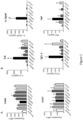
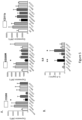
- Figure 1A shows that antibodies directed against SIRP molecules or CD47 do not prevent M1 macrophage polarization induced by GM-CSF + M-CSF because the expressions of the M1 cell surface markers (CD86 and CCR7) are not modified compared to control conditions.
- the over-expression of CD206, CD200R, CD11b and PD-L1 was significantly inhibited with selective anti-SIRP alpha mAb ( Figure 2A and Figure 11 ) but not with control antibodies (anti-SIRPa/b or Sirpb), CD47-Fc recombinant protein or anti-CD47 mAbs (clones B6H12 and CC2C6).
- Figure 11 shows that blockade of SIRPa by a monoclonal antibody prevents the acquisition of the M2 macrophage phenotype induced by IL-4, indeed the expression of M2 markers (CD206, CD11b, and PD-L1) did not raise whereas it was observed in the isotype control condition. This prevention of anti-inflammatory status of macrophage was not observed when the cognate ligand of SIRPa (CD47) was blocked with a monoclonal antibody.
- cytokines and chemokines secretion showed that anti-SIRPa mAb prevented CCL-17 secretion (a hallmark chemokine of M2 secretion) while they increased secretion of pro-inflammatory cytokines (IL-6, IL12p40, TNF- ⁇ ) and chemokine (CCL-2) secreted by M1 macrophages ( Figure 1B and 2B ).
- Anti-SIRPa thus seems to play a role on the prevention of M2 polarization and on the pro-inflammatory function of the macrophages.
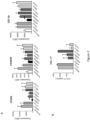
- SIRPa The role of SIRPa on M2 polarization was then studied at a transcriptional level with SIRPa inhibition, using a cocktail of siRNAs.
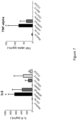
- the results, presented in Figure 6A confirmed that inhibiting SIRPa inhibits M2 polarization of the cells (CD206 and CD200R expression).
- Maximal/terminal M1 polarization is considered achieved using M-CSF + IFNg + LPS, which results in all hallmarks of M1 macrophage, in particular iNOS expression.
- Figure 4 shows the expression of iNOS in M1 polarized cells treated or not with different blocking antibodies.
- blocking SIRPa with an antibody increased the expression of iNOS.
- anti-SIRP ⁇ or anti-CD47 mAb did not induce any modification in the iNOS expression profile.
- blocking SIRPa modifies macrophage function in a pro-inflammatory state such as M1 type.
- the M1 surface markers tested were not modified compared to the controls.
- M1/M2 macrophages The phenotype and function of polarized M1/M2 macrophages has been, to some extent, reversed in vitro or in vivo due to the plasticity of these cells (Sica and Mantovani, 2012).
- Results presented Figure 7 show that the anti-SIRPa mAb is able to repolarize M2 macrophages into M1 pro-inflammatory macrophages, since IL6 and TNF- ⁇ cytokines were induced (hallmark cytokines of M1 macrophages). This effect was not observed when cells were treated with an anti-Sirp ⁇ nor with CD47-Fc nor anti-CD47 antibodies. As explained above, cell surface markers were not modified.
- results of Figure 12 show that two different anti-SIRPa mAb allow to induce the polarization of macrophage in favor of M2-type macrophages, both at phenotypic level with expression of surface markers CD200R and CD80 (A) and at functional level with secretion of IL-6 (B). Further, results of Figure 14 show that the selective blockade of SIRPa but not CD47 prevents M2 polarization (B) and did not affect M1 polarization (A).
- SIRPa is important for the M2 polarization of the monocyte and blocking this pathway is a good opportunity to produce macrophages with pro-inflammatory profile to treat pathologies in need thereof such as cancer or for infectious diseases, in which macrophages are blocked in an M2 state.
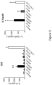
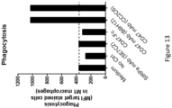
- Anti-SIRPa antibody does not increase the human macrophage phagocytosis on tumor cells expressing CD47
- Figure 8 represents the overall survival rate of animals inoculated with hepatocarcinoma and treated with an anti-CD137, an anti-SIRPa or both during 4 weeks. 20% of the animals treated with anti-Sirpa monotherapy survived more than 25 days and 25% of animals treated with anti-CD137 monotherapy survived more than 30 days. However, 100% of the animals receiving the combo anti-Sirp+anti-CD137 were still alive after 80 days, compared to the other conditions, showing a synergistic effect of the 2 molecules.
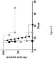
- Figure 9 represents the overall survival rate of animals inoculated with hepatocarcinoma and treated with an anti-PD-L1, an anti-SIRPa or both during 4 weeks.
- the results showed a very interesting surviving rate when animals were treated with both molecules, compared to each molecule alone (20% of alived animals after 20 days with anti-sirpa compare to 12% of alived animals with anti-PD-L1).
- This result indicates a synergistic effect of the anti-SIRPa antibody with the anti-PD-L1 antibody in a cancer model.
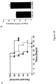
- Figure 10 A represents the overall survival rate of animals inoculated with melanoma cells and treated with an anti-PD-L1, an anti-SIRPa or both during 4 weeks. Compared to the treatment with each molecule alone, the combination showed a better efficacy.
- Figure 10 B shows the macrophage infiltrate in animals treated with anti-SIRPa, confirming an increase in macrophage number into the tumor.
- SIRPa is an interesting target for cancer treatment, especially when combined with other immunotherapies, and suggest that Sirp is a new checkpoint inhibitor that is important to block in the aim to restore a pro-inflammatory tumor environment.
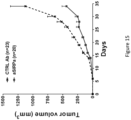
- Figure 15 shows that monotherapy with anti-SIRPa mAb (clone P84) inhibit tumor development in a syngeneic and orthotopic triple-negative breast model in mice. From 2 weeks post-tumor inoculation until the end of the experiment, anti-SIRPa treated mice have a significant reduction in tumor volume.
- SIRPa signal regulatory protein alpha
- SIRPa signal regulatory protein alpha
- Antibody therapy targeting the CD47 protein is effective in a model of aggressive metastatic leiomyosarcoma. Proc. Natl. Acad. Sci. U. S. A. 109, 6656-6661 .
- CD47 is an adverse prognostic factor and therapeutic antibody target on human acute myeloid leukemia stem cells.
- Human signal-regulatory protein is expressed on normal, but not on subsets of leukemic myeloid cells and mediates cellular adhesion involving its counterreceptor CD47. Blood 94, 3633-3643 .
- mice to humans From mice to humans: developments in cancer immunoediting. J. Clin. Invest. 125, 3338-3346 .
- the CD47-signal regulatory protein alpha (SIRPa) interaction is a therapeutic target for human solid tumors. Proc. Natl. Acad. Sci. U. S. A. 109, 6662-6667 .
Landscapes
- Health & Medical Sciences (AREA)
- Chemical & Material Sciences (AREA)
- Organic Chemistry (AREA)
- Immunology (AREA)
- Life Sciences & Earth Sciences (AREA)
- Genetics & Genomics (AREA)
- Engineering & Computer Science (AREA)
- General Health & Medical Sciences (AREA)
- Biomedical Technology (AREA)
- Biochemistry (AREA)
- Molecular Biology (AREA)
- Medicinal Chemistry (AREA)
- Biophysics (AREA)
- Biotechnology (AREA)
- Wood Science & Technology (AREA)
- Bioinformatics & Cheminformatics (AREA)
- Zoology (AREA)
- Proteomics, Peptides & Aminoacids (AREA)
- General Engineering & Computer Science (AREA)
- Microbiology (AREA)
- Public Health (AREA)
- Cell Biology (AREA)
- Nuclear Medicine, Radiotherapy & Molecular Imaging (AREA)
- Pharmacology & Pharmacy (AREA)
- Animal Behavior & Ethology (AREA)
- Chemical Kinetics & Catalysis (AREA)
- Veterinary Medicine (AREA)
- Hematology (AREA)
- Gastroenterology & Hepatology (AREA)
- General Chemical & Material Sciences (AREA)
- Physics & Mathematics (AREA)
- Plant Pathology (AREA)
- Medicines That Contain Protein Lipid Enzymes And Other Medicines (AREA)
- Medicines Containing Antibodies Or Antigens For Use As Internal Diagnostic Agents (AREA)
- Peptides Or Proteins (AREA)
- Measurement Of Optical Distance (AREA)
- Optical Radar Systems And Details Thereof (AREA)
- Medicines Containing Material From Animals Or Micro-Organisms (AREA)
Description
- The present invention pertains to the field of immunotherapy. More specifically, the present invention provides an anti-SIRPa compound selected from the group consisting of an antibody or fragment(s) thereof, wherein the anti-SIRPa compound binds specifically to the extracellular domain of SIRPa and blocks SIRPa pathway, combined with a second therapeutic agent selected from the group consisting of a blocking anti-PDL1 antibody and a blocking anti-PD 1 antibody, for use in the treatment of a solid cancer.
- Cancers resulting from uncontrolled cell proliferation form a group of varied diseases. Surgery and radiation therapy do not treat all cancers, especially metastatic stages. More effective treatments are supposed to reach every organ of the patient: it is the case with modern chemotherapy that can induce the death of tumor cells. However, the cytotoxic effect of the drugs remains a major obstacle to chemotherapy.
- The rise of molecular biology and genetics has allowed the understanding of the mechanisms leading to cancer cell development and of "targeted therapies". These treatments, combined with chemotherapy, specifically attack tumor cells, sparing healthy cells. However, although these therapies significantly lengthen life expectancy of patients, none has yet resulted in healing. Today, it is well established that human tumor cells may be resistant to treatment and escape to the surveillance by the immune system. Combination therapies are thus essential to cure a patient, but with the drawback of multiplying the problems of side effects.
- Initial research conducted in the field of cancer immunotherapy aimed at "boosting" the effector cells of the immune system, making them more aggressive towards tumors. This strategy has in fact proved somewhat successful.
- New generation immunotherapeutic molecules, revolutionizing cancer treatments, block suppressor mechanisms of these cells, allowing the effector T cells (Teff) to exercise their action. This is the concept called "Inhibit inhibitors". An anti-CTLA-4 antibody (Yervoy®) has been the first molecule for treating metastatic forms of malignant melanoma prolonging the mean survival of patients of 6 to 10 months, with a quarter of patients still alive after 2 years. Unfortunately, these results, as spectacular as they are, still do not cure most patients.
- The present invention relies on an "inhibit inhibitors" approach and provides a new method useful in immunotherapy. More specifically, the present invention pertains to a method for modifying macrophage polarization in order to induce a pro-inflammatory environment. The method consists in the use of an anti-SIRPa compound able to inhibit the polarization of anti-inflammatory M2-type macrophages and/or favors pro-inflammatory M1-type macrophages, for inhibiting the anti-inflammatory signal provided by M2-type macrophages and favoring the pro-inflammatory signal provided by M1-type macrophages. This approach allows to reestablish an inflammatory environment favorable to the action of the T effector cells, in particular in eliminating the cancer cells.
- Macrophages are cells that have the highest plasticity of the hematopoietic system. They are involved both in innate immunity (phagocytosis capacity) and in adaptive immunity (cell polarization), but also in ontogeny, in homeostasis and in tissue repair (Mantovani et al., 2013; Wynn et al., 2013). Macrophages are present in all tissues. They have a large phenotypic and functional diversity. During ontogeny, these cells also exhibit a diversity of origins which persists into adulthood. In the tissues, monocytes-macrophages respond to environmental stimuli (product from microbial infection, damaged cells, activated lymphocytes) and acquire distinct phenotypes. For a long time, these cells have been classified according to their function in a binary manner in connection with the inflammatory condition.
- Depending on stimuli monocyte-macrophage received, they reprogram their transcriptome, resulting in distinct functional and phenotypic spectra. Macrophages are categorized simplistically into 2 sub-populations or states of polarization (or activation): classical activation phenotype M1 and the alternative activation phenotype M2 (Gabrilovich et al., 2012). The M1 classification is associated in vitro with the use of IFNg factor alone or in combination with microbial factors such as LPS or inflammatory cytokines such as TNF-α and GM-CSF. The polarization M2 is rather associated with the IL4 or IL13 (Stein et al., 1992). Other cytokines are also identified as inducing M2 type polarization such as IL33, which induces overexpression of Arg1 (arginase 1), CCL24 or CCL17 playing a role in inflammation. The IL21 and more commonly CSF1 are major players in the polarization of macrophages. Macrophages may also acquire the status of "M2-like", sharing common characteristics of M2. In fact, a large number of stimuli such as immune complexes associated with LPS, IL-1, glucocorticoids, TGF beta, Wnt5a and IL10 result in a functional phenotype of type "M2-like".
- Similarly, in vivo studies have shown the existence of M1, M2 and M2-like macrophages. These subtypes represent only the extremes on a continuum of functional states that must be integrated in an environmental complex system.
- Generally, M1 macrophages present IL12high, IL23high and IL10low phenotype and produce molecular effectors such as reactive oxygen species (ROS) and intermediates of Nitric Oxide (NO) and inflammatory cytokines (IL1b, TNF-α, IL-6). M1 macrophages participate in Th1 responses, play a role in resistance against intracellular parasites and are key effectors in the elimination of tumor cells. In contrast, M2 macrophages have an IL12low, IL-23low and IL10high phenotype with a variability in the production of inflammatory cytokines according to stimuli present in the environment. M2 cells display on their surface a strong expression of scavenger, mannose and galactose-type receptors. The metabolism of arginine is changed to an ornithine and polyamines metabolism. M2 macrophages are generally associated to a Th2 type response, to a parasite clearance, to a decrease of inflammation, to tissue repair promotion, angiogenesis, tumor growth and immune regulation.
- M1 and M2 also have distinct expression profiles of chemokines. M1 macrophages express CXCL9 and CXCL10 chemokines which are known for attracting Th1, while M2 macrophages express CCL17, CCL22 and CCL24. Chemokines such as CCL2 and CXCL4 can also polarize macrophages to an M2-like phenotype.
- Depending on their polarization state, macrophages have different characteristics in terms of iron, folate and glucose metabolisms. For example, M1 express large amounts of proteins involved in iron storage, such as Ferritin, while they express only weakly Ferroportin, involved in the iron exportation to the extracellular medium. In contrast, M2 macrophages express low levels of Ferritin but high levels of Ferroportin. This difference can result in functional outcomes, such as a bacteriostatic effect of M1 (protection against infection) and an effect promoting tissue repair by M2 macrophages, which also promote the tumor growth, as observed in some studies. The management of iron by macrophages according to their polarity is an important element underlining the importance of controlling the polarization of macrophages according to the condition of an individual.
- Similarly, macrophages face an oxygen gradient in tissues under normal or pathological conditions. Macrophages or monocytes adapt to this gradient by modifying their glycolytic metabolism. The HIF1 and 2 are transcriptional factors leaders of these changes, including expression of chemokines or chemokine receptor CXCR4 or CXCL12 and VEGF (an angiogenic factor). Macrophages are involved in the tissue response to hypoxic conditions.
- The presence of polyamines in the cell environment appears to be a
type 2 macrophage polarization factor. - The present invention aims to modulate the polarization of macrophages in order to inhibit the anti-inflammatory M2-type macrophages and/ or to favor the pro-inflammatory M1-type macrophages.
- In the tumor environment, different defense cells exist. This is a priori a paradox because their presence should mean that they are attacking the tumor. In reality, many of these immune cells are maintained in an inactive stage and are rendered inoperative by the presence of regulatory cells. Instead of fighting the tumor, these regulatory cells facilitate tumor development by helping to overcome barriers, allowing it to spread and form secondary tumors, i.e., metastases.
- The cellular and molecular effectors of inflammation are important actors in the tumor microenvironment. Indeed, since the 90s, this interrelationship between inflammation and tumor is the subject of numerous studies (Mittal et al., 2014; Teng et al., 2015). To escape the immune system, tumor implements escape mechanisms in 3 stages: elimination - equilibrium - escape. The first phase is to eliminate immune mechanisms recognizing tumor cells. Then an equilibrium is set up between tumor cells and immunity: between killing and survive. This equilibrium can persist during years without the tumor progressing. During this period, tumor cells are subjected to genetic instability and eventually escape, inducing proper immune response or inhibiting the anti-tumor immune response by inducing said suppressor mechanisms, i.e., blocking the anti-tumor response. The cells are then recognized as normal.
- It is in this latter suppressor mechanism that inflammatory cells and molecules play an important role. The adaptive immune response is suppressed / blocked by activating a number of pathways leading to the inhibition of differentiation and activation of dendritic cells (via the presence in tumor microenvironment factors such as IL10 and VEGF). There is also an increase of the regulatory T cells (Treg) in peripheral blood and lymph nodes inhibiting innate and adaptive responses. The presence of tumor suppressor cells in the microenvironment, such as MDSC (myeloid derived suppressor cells) and TAM (Tumor Associated Macrophages or M2), affects tumor development of bad prognosis by the secretion of cytokines, growth factors, enzymes degrading the extracellular matrix and proteases (Cornelissen et al., 2012).
- Immunotherapy is safe but in some cancer has a moderate efficacy partly due to the presence of immunosuppressive cells in peripheral blood, lymphoid organs and within the tumour environment that hamper immunotherapeutic treatments. Several strategies have been performed or are currently tested to improve the efficacy of immunotherapy by acting on suppressive cells such as MDSCs, Tregs and TAMs, which are increased in most cancer patients. It is becoming increasingly clear that these populations contribute to the impaired antitumour responses frequently observed in cancer patients.
- Therefore, combating immunosuppression through modulation of these cell types is an important key to increase the efficacy of immunotherapy and should lead to a better prognosis for cancer patients. The present invention aims to modify the M1/M2 balance of macrophage population to favor the M1-type macrophages, in order to provide an immune environment propitious to immunotherapy.
- Macrophages are found in large numbers in tumors. Originally it was thought that this cell population was involved in an anti-tumor response but many experimental and clinical studies have shown that macrophages are involved in the tumor initiation and progression as well as in the metastatic process. During the tumor process, macrophages secrete pro-inflammatory cytokines such as IFNy, TNF-α and IL6, attracting other immune cells creating chronic inflammation and causing the initiation and tumor progression. However, once the tumor installed, tumor macrophages (TAMs) adopt an immunosuppressive cellular profile and are less active, allowing tumor growth and transition to malignancy. TAMs are responsible for migration, extravasation and invasion of tumor cells (metastasis) and are involved in tumor angiogenesis (Qian and Pollard, 2010; DeNardo et al., 2010; Hanahan and Coussens, 2012).
- Monocytes Ly6C+(CD14+)/CD11bhigh arriving at the tumor undergo phenotypic changes such as a decrease of the Ly6C and CD11b markers and expression of MHC class II (MHCII), VCAM and CD11c. However, differentiation and distribution of TAMs depend on the tumor's anatomical localization and on its stage of development. The definition TAM's function was previously based on anti-tumor M1 macrophage type (iNOS inducible) and M2 pro-tumoral ARG-positive macrophage type. This simplistic dichotomy must be seen in a context of great plasticity of TAM in a cytokines and chemokines environment favoring their suppressive function and allowing the recruitment of Treg involved in tolerance to tumor cells (reviewed in Ugel et al., 2015; Wynn et al., 2013).
- The present invention pertains to the inhibition of TAM in order to decrease or prevent the tumoral process, including the metastatic process.
- Signal regulatory protein alpha, also termed CD172a or SHPS-1 and herein noted "SIRPa", was first identified as a membrane protein mainly present on macrophages and myeloid cells that was associated with the Src homology region 2 (SH2) domain - containing phosphatases - SHP-1 and SHP-2. SIRPa is the prototypic member of the SIRP paired receptor family of closely related SIRP proteins. Engagement of SIRPa by CD47 provides a downregulatory signal that inhibits host cell phagocytosis, and CD47 therefore functions as a "don't-eat-me" signal.
- SIRPa is expressed on monocytes, most subpopulations of tissue macrophages, granulocytes, subsets of dendritic cells (DCs) in tissues, some bone marrow progenitor cells, and to varying levels on neurons, with a notably high expression in synapse-rich areas of the brain, such as the granular layer of the cerebellum and the hippocampus (Seiffert et al, 1994; Adams et al, 1998; Milling et al, 2010).
- The SIRPa interaction with CD47 is largely described and provides a downregulatory signal that inhibits host cell phagocytosis (see review Barclay et al, Annu. Rev. Immunol., 2014). Both CD47 and SIRPa also engage in other interactions. Investigators have suggested that the lung surfactant proteins SP-A and SP-D control inflammatory responses in the lung through interactions with SIRPa (Janssen et al., 2008).
- One of the best characterized physiological functions of CD47-SIRPa interactions is their role in the homeostasis of hematopoietic cells, in particular red blood cells and platelets. Because CD47 acts as a don't-eat-me signal and, as such, is an important determinant of host cell phagocytosis by macrophages, the potential contribution of CD47-SIRPa interactions in cancer cell clearance has been intensely investigated in recent years.
- The SIRPa/CD47 pathway is nowadays also subject to different pharmaceutical developments, all directed towards enhancement of macrophages phagocytosis. In fact, like infected cells, cancer cells carry aberrant cargo such as unfamiliar proteins or normal proteins at abnormal levels, yet these cells frequently subvert innate immune control mechanisms by concurrently over-expressing immunoregulatory molecules. It is becoming increasingly clear that one such mechanism involves CD47 (Barclay and Van den Berg, 2014), a protein of "self" expressed by normal cells. CD47 has interactions with several different ligands such as SIRPa. This specific interaction is known to lead to a "don't eat me" signal to phagocytic macrophages, which then leave target cells unaffected (Oldenborg et al., 2000) Over-expression of CD47 by cancer cells renders them resistant to macrophages, even when the cancer cells are coated with therapeutic antibodies (Zhao et al., 2011), and correlates with poor clinical outcomes in numerous solid and hematological cancers (Majeti et al., 2009; Willingham et al., 2012). In experimental models, in particular human tumor-xenograft models in immunodeficient mice, blockade of the CD47/SIRPa pathway was very effective to promote tumor elimination by macrophages and to decrease cancer cell dissemination and metastasis formation (Chao et al., 2011; Edris et al., 2012; Uluçkan et al., 2009; Wang et al., 2013). In these studies, TAM function or phenotype has not been studied. Blockade of the CD47/SIRPa pathway, by enhancing antibody-dependent phagocytosis by macrophages, has been described to synergize with depleting therapeutic anticancer antibodies (Weiskopf et al., 2013) such as Trastuzumab (anti-Her2), Cetuximab (anti-EGFR), Rituximab (anti-CD20) and Alemtuzumab (anti-CD52).
- From the above, it appears that SIRPa has been described to be implicated into the phagocytic function of myeloid cells, the antigen presentation and cytokine secretion of dendritic cells and trafficking of mature granulocytes. However, the function of SIRPa on macrophage polarization and their potent suppressive function during tumorigenesis have never been described.
-
WO2010/130053 disclosed a method for treating hematological cancer comprising modulating the interaction between human SIRPa and CD47. This document showed that the blockade of SIRPa-CD47 induces the activation of the innate immune system via the phagocytosis pathway. In the transplantation model of human leukemia described in this patent specification, myeloid cells were used and the transplant was rejected when animals were treated with an antagonist of CD47. This result suggests an increase of phagocytosis upon treatment with anti-CD47, but not a modification in the polarization state of the macrophages nor a modification into a pro-inflammatory function of the macrophages. - A method for inhibiting cell functioning for use in anti-inflammatory and anti-tumor therapies was described in
WO0066159 - A CD47-Fc and a CD47-extended fusobody molecules that bind to SIRPa were studied and claimed in the patent application number
WO2012/172521 . These molecules have been claimed to be able to inhibit immune complex-stimulated cell cytokines release (e.g., release of IL-6, IL10, IL12p70, IL23, IL8 and TNF-α) from peripheral blood monocytes, DCs and/or monocytes-derived DCs stimulated with Pansorbin or soluble CD40L and IFNy. The activity of these molecules is completely different from the activity of anti-SIRPa antibodies disclosed in the present specification. -
WO2013/056352 describes the use of an antibody anti-human SIRPa (called 29AM4-5, and corresponding to SIRP-29 as named by the inventors of the present invention) in a model of SIRPa-positive AML, i.e. xenotransplantation into immunodeficient mice of primary human AML cells expressing human SIRPa to which bind said antibody anti-human SIRPa. Thus, the approach consists in the use of an anti-human SIRPa antibody to act on tumor cells expressing human SIRPa. The treatment is thus directly directed toward the tumor. - Alblas J. et al (2005, Molecular and Cellular Biology) observed that an anti-SIRPa antibody (ED9) is not able to induce to the production of inflammatory cytokines, especially TNF-α and IL-6 (
Figure 1A ). However, the inventors of the present invention demonstrated, on the contrary, that this particular antibody does allow to repolarize M2 anti-inflammatory macrophages into M1 pro-inflammatory macrophages (Figure 14B of the experiment part). The difference of result could be explained by the fact the authors of Alblas et al. used a macrophage cell line (rat NR8383 cell line) (which may no longer express SIRPa), whereas the inventors used a fresh preparation of rat macrophage derived from bone marrow in the course of the experiment. -
WO 2015/138600 describes anti-human SIRPa antibodies that bind to human SIRPa and block the interaction with CD47 expressed on a target cell with SIRPa expressed on a phagocytic cell. This document does not suggest nor show any evidence of the effect of an anti-SIRPa on macrophage polarization nor on the increase of the pro-inflammatory function of macrophages. Interestingly, the inventors named inWO 2015/138600 published an abstract at the 56th ASH annual meeting (Weiskopf et al, ASH 2014). In this abstract, which is posterior to the filing ofWO 2015/138600 , they mentioned that anti-human SIRPa antibodies blocking the interaction between CD47 and SIRPa were not sufficient to induce human macrophage phagocytosis. Altogether, these publications suggest that the mechanism of action of an anti-human SIRPa in vivo is different from that of an anti-human CD47, for which the in vivo phagocytic efficacy was largely described in the literature. - From the above, it appears that various blocking SIRPa-CD47 interaction strategies targeting CD47 or SIRPa with an antibody or a fusion protein show distinct results and different efficiencies, indicating distinct roles for each of the targets. Indeed, an antibody directed against the CD47 antigen blocks the interaction of CD47 with all its ligands. The use of a SIRPa-Fc protein that binds to the cells through its Fc and blocks the endogenous CD47 pathway and prevents its activation is not able to block the activity of endogenous SIRPa, contrary to an anti-SIRPa antibody. The direct role of the SIRPa pathway in modulating the immune system has been so far undervalued compared to the CD47 pathway. None of the prior art studies suggests nor describes any function of the SIRPa pathway in the polarization of macrophages (either at a phenotype level or at a functional level) that plays a crucial role in the tumor escape mechanisms.
- In this context, the inventors provide a new insight in the use of anti-SIRPa compounds since the modulation of the immune environment is a major achievement in the treatment of many diseases, especially in cancer.
- The invention is set out in the appended set of claims.
- As described in the experimental part below, the inventors have now identified SIRPa as a new checkpoint inhibitor and demonstrated its role in macrophage polarization. They indeed showed that an anti-SIRPa compound as defined herein, induces a pro-inflammatory function of macrophages associated to type 1 macrophages (M1 pro-inflammatory = M (IFNg)) and inhibits the suppressive activity of M2-type macrophages in the tumor, since the pro-inflammatory profile of macrophages is obtained at the expense of
type 2 macrophages (M2 type high phagocytic activity = M (IL4)). This effect is obtained by targeting SIRPa but not CD47, which is involved in phagocytosis function. One advantage of the invention is that the use of an anti-SIPRa compound will induce less side effects than the use of an anti-CD47 compound. Indeed, CD47 is expressed by a large range of cells, and not only by tumor cells and interacts with several ligands. The expression of SIRPa being more limited, the effect of the therapy will be more targeted on the tumor microenvironment. Thus, the use of an anti-SIRPa compound will be less toxic and less deleterious than the use of an anti-CD47 compound. Further, the therapy being directed toward macrophages and not tumor cells, no selection pressure will be exercised on tumor cells allowing to prevent tumor escape and the development of tumor resistance to the treatment. - The present invention hence pertains to an anti-SIRPa compound selected from the group consisting of an antibody or a fragment thereof, wherein the anti-SIRPa compound binds specifically to the extracellular domain of SIRPa and blocks SIRPa pathway, combined with a second therapeutic agent selected from the group consisting of an immune checkpoint blocker selected from the group of a blocking anti-PDL1 antibody and a blocking anti-PD1 antibody, for use in the treatment of a solid cancer
- In a particular embodiment, said anti-SIRPa compound can be selected from the group consisting of an anti-SIRPa antagonist antibody, and fragments thereof.
- Anti-SIRPa compounds, in particular anti-SIRPa specific antibodies, can thus be used in the treatments of cancers.
- In a particular embodiment, the present disclosure concerns an anti-SIRPa compound able to inhibit the polarization of anti-inflammatory M2-type macrophages and/or favors pro-inflammatory M1-type macrophage for use in the treatment of cancer, with the exception of SIRPa-positive acute myeloid leukemia and/or SIRPa-positive non acute myeloid leukemia and/or SIRPa-positive non-Hodgkin leukemia or SIRPa-positive hematologic cancers.
- An important aspect of the invention is that the therapeutical approach aims to target SIRPa on the macrophage in order to modulate their polarization and to recreate an immune context detrimental to the tumor development and survival. Basically, the success on the anti-tumor treatment is based on an indirect pathway, and does not require that the tumor cells are sensitive to the anti-SIRPa compound. Accordingly, in a particular embodiment, the present invention concerns the use of an anti-SIRPa compound as defined herein, i.e. able to inhibit the polarization of anti-inflammatory M2-type macrophages and/or favors pro-inflammatory M1-type macrophage, in the treatment of cancer, wherein said compound is administered to a patient presenting a SIRPa-negative tumor.
- Combinations of anti-SIRPa compounds as defined herein with other therapeutic agents, as defined in the claims, are part of the invention, since a synergistic effect was demonstrated by the inventors.
- Another aspect of the present disclosure is a method for ex vivo obtaining pro-inflammatory M1-type macrophages by incubating macrophages with an anti-SIRPa compound as defined herein.
- The present disclosure also pertains to a method for selecting patients likely to benefit from a treatment by an anti-SIRPa compound as defined herein, by measuring the presence of M2-type macrophages in a sample from the patient.
- A method of following-up a treatment by an anti-SIRPa compound as defined herein, to assess its efficacy by measuring the presence of pro-inflammatory M1-type macrophages and/or measuring the presence of anti-inflammatory M2-type macrophages in a sample from an individual treated by said compound, is also part of the present disclosure.
-
-
Figure 1 : Monocyte type 1 polarization with GM-CSF + M-CSF protocol: Effect of SIRPa blockade: Human Monocytes were cultivated with growth factors M-CSF and GM-CSF to induce Macrophages and treated or not with Ctrl Ab or anti-Sirp antibodies (anti-a or ab or b isotype), or a CD47-Fc protein, or some anti-CD47 antibodies (B6H12 or CC2C6). Then type 1 macrophage phenotype and function were analysed by FACS and ELISA. A. represents cell surface markers analysis: CD86 and CCR7. B. represents secreted cytokine/chemokine: IL6, IL12p40, CCL-2 and TNF-α. IL6, p12p40 and CCL-2 are significantly increased when cells are treated with an anti-SIRPa antibody. -
Figure 2 :Monocyte Type 2 polarization with GM-CSF + M-CSF protocol : Effect of SIRPa blockade: Human Monocytes were cultivated with growth factors M-CSF and GM-CSF to induce macrophages and treated or not with Ctrl Ab or anti-Sirp antibodies (anti-a or ab or b isotype), or a CD47-Fc protein, or some anti-CD47 antibodies (B6H12 or CC2C6). ThenType 2 macrophage phenotype and functional analysis were performed by FACS and ELISA. A. Shows marker expression at cell surface : CD206, CD200R, CD11b and B. Shows secreted Chemokine : CCL-17 -
Figure 3 : Monocyte type 1 polarization with M-CSF and IFNgamma protocol: Effect of SIRPa blockade : Human monocytes were cultivated 5-6 days with M-CSF and then 2 days with IFNgamma with or without Ctrl Ab or anti-Sirp antibodies (anti-a or ab or b Sirp isotype), or a CD47-Fc protein, or some anti-CD47 antibodies (B6H12 or CC2C6). M1 secreted cytokines were analysed : IL6 and IL12p40. Statistical analysis were performed -
Figure 4 : Monocyte type 1 polarization with M-CSG + IFNgamma and LPS protocol: Effect of SIRPa blockade on iNOS expression: Human monocytes were cultivated 5-6 days with M-CSF and then 2 days with IFNgamma+LPS with or without antibodies directed against : SIRPa: SE7C2 (A.) or SIRPa/b: SE5A5 (B.) or CD47: B6H12 (C.), iNOS expression was then analysed by FACS. Ctrl Ab is represented by dotted line and monocytes before polarization by empty grey line on the three panels -
Figure 5 :Monocyte type 2 polarization with M-CSF + IL4 protocol: Effect of SIRPa blockade with antibody : Human monocytes were cultivated 5-6 days with M-CSF and then with IL4 with or without Ctrl Ab or anti-Sirp antibodies (anti-sirp a or ab or b), or a CD47-Fc fusion protein, or some anti-CD47 antibodies (B6H12 or CC2C6). A. Represents expression of cell surface markers: CD206, CD200R and CD11b and B. Represents the release of IL6 cytokine in the supernatant -
Figure 6 :Monocyte type 2 polarization with M-CSF + IL4 protocol: Effect of SIRPa blockade at a mRNA level with a siRNA Knock down assay: Human monocytes were cultivated 5-6 days with M-CSF and then 2days with IL4. Cells were transfected with null-siRNA or SIRPa siRNA : A. represents the expression of the cell surface markers : CD206 and CD200R; B. represents the IL6 cytokine release in the supernatant. The anti-SIRPa SiRNA are provided by ThermoFisher scientific. -
Figure 7 : Effect of SIRPa blockade on M2 repolarization after M-CSF+IL4 treatment: Human monocyte were cultivated 5-6 days with M-CSF and then 2 days with IL4. M2 macrophages were then treated with or without Ctrl Ab or anti-Sirp antibodies (anti-sirp a or ab or b), or a CD47-Fc fusion protein, or some anti-CD47 antibodies (B6H12 or CC2C6). The Cytokines IL6 and TNF-α releases were then measured in the supernatant. -
Figure 8 : Effect of a combined anti-SIRPa + anti-CD137 treatment on an in vivo model of Hepatocarcinoma: One week after tumor inoculation, animals were treated 3 times/week for 4 weeks with 3G8 isotype control antibody (Iso ctrl: black square: n=33), or an anti-SIRPa antibody (clone p84 :grey square; n= 33) or an antiCD137 antibody (4-1BB mAb: black triangle; n=8) or a combined treatment (Anti-SIRPa+anti-CD137: grey diamond; n=8). The overall survival rate was then analyzed. -
Figure 9 : Effect of a combined anti-SIRPa + anti-PD-L1 treatment on an in vivo model of Hepatocarcinoma: One week after tumor inoculation, animals were treated 3 times/week for 4 weeks, animals were treated with 3G8 isotype control antibody (Iso ctrl: black square: n=5), or an anti-SIRPa antibody (clone p84: grey square; n= 5) or an anti-PD-L1 antibody (10F-9G2 mAb: black triangle; n=8) or a combined treatment (anti-SIRPa+anti-PD-L1: grey diamond; n=5). The overall survival rate was then analyzed. -
Figure 10 : Effect of a combined anti-SIRPa + anti-PD-L1 treatment on an in vivo model of melanoma: In the same time as tumor inoculation, animals were treated with isotype control antibody (Iso ctrl: star: n=5), or an anti-SIRPa antibody (clone p84 : square; n= 5) or an anti-PD-L1 antibody (10F-9G2 mAb: triangle; n=8) or a combined treatment (anti-SIRPa+anti-PD-L1: circle; n=5).A. The overall survival rate was then analyzed. B. Some animals were sacrificed 2 weeks after first inoculation to analyze macrophage infiltrate using CMH class II/CD11b markers. -
Figure 11 : Effect of anti-SIRPA antibody on mouse macrophage polarization: Mouse M2 macrophage polarization phenotype is inhibited by anti-SIRPa mAb but not by anti-CD47 mAb as shown by the quantification of the expression of the M2 markers CD206, CD11b and PD-L1. In this experiment, anti-SIRPa mAb corresponds to p84 clone and anti-CD47 mAb corresponds to MIAP310 clone. -
Figure 12 : Effect of anti-SIPRa antibody on human M2 macrophage polarization: Human M2 macrophage polarization phenotype is inhibited by anti-SIRPa mAbs but not with anti-CD47 mAb. A. By detection of M2 surface markers (CD200R, CD80). B. At functional level by evaluation of cytokine secretion (IL-6). In this experiment, anti-SIRPa mAbs correspond to SE7C2 or SIRP29 clones and anti-CD47 mAb corresponds to B6H12 clone. -
Figure 13 : Effect of anti-SIPRa antibody on human M1 phagocytosis: Human macrophage phagocytosis of CD47+ tumor cells (Raji) is increased by CD47 targeting agents, but not with SIRPa targeting agents. Analysis of phagocytosis was evaluated by flow cytometry by gating on M1 fluorescent macrophages and analysis of target (Raji) cells fluorescence into M1 macrophages. In this experiment, the CD47 targeting agents correspond to anti-CD47 mAbs B6H12 or CC2C6 clones and the SIRPa targeting agents correspond to anti-SIRPa clone SE7C2 or CD47-Fc protein. -
Figure 14 : Effect of anti-SIRPA antibody on rat M1/M2 polarization: A. Rat M2 macrophages become pro-inflammatory and secrete inflammatory cytokines (IL-6 and TNF-α) in presence of anti-SIRPa mAb, but not anti-CD47 mAb. B. M1 rat macrophages polarization is not modified by anti-SIRPa or anti-CD47 mAbs. Anti-SIRPa mAb used in this experiment corresponds to ED9 clone and anti-CD47 mAb corresponds to OX101 clone. -
Figure 15 : Effect of anti-SIRPa antibody on a mouse model of mammary tumor. Monotherapy with anti-SIRPa mAb inhibit tumor growth in the 4T1 mammary cancer model in mice. Anti-SIPRa mAb used in this experiment corresponds to clone p84. - Throughout the present text, the following definitions are used:
- The term "polarization" is used herein to designate the phenotypic features and the functional features of the macrophages. The phenotype can be defined through the surface markers expressed by the macrophages. The functionality, can be defined for example based on the nature and the quantity of chemokines and/or cytokines expressed, in particular secreted, by the macrophages. Indeed, the macrophages present different phenotypic and functional features depending of their state, either pro-inflammatory M1-type macrophage or anti-inflammatory M2-type macrophage. M2-type macrophages can be characterized by the expression of surface markers such as CD206, CD11b, PD-L1 and CD200R and then secretion of cytokines such as CCL17. M1-type macrophages can be defined by the expression of surface markers such as CD86 and CCR7 and the secretion of cytokines such as IL-6, TNF-α and IL12p40. In the context of the invention, anti-SIPRa compounds allow to modulate the polarization of macrophages population by inhibiting the M2-type macrophages and/or favoring the M1-type macrophages.
- It encompasses the meaning of the term "activation" usually used to mean the perturbation of macrophages with exogenous agents. Macrophages change their polarization states in response to growth-factors (CSF-1 and GM-CSF) and external stimuli such as microbes, microbial product and nucleotides derivatives, antibody-Fc receptor stimulation, glucocorticoids, phagocytosis.
- An "anti-SIRPa compound", as used herein refers to a compound that specifically binds to the extracellular domain of SIRPa, or a nucleic acid encoding such compound, as well as to a compound able to inhibit the expression of the SIRPa protein. Such compound is able to inhibit the polarization of anti-inflammatory M2-type macrophages and/or favors pro-inflammatory M1-type macrophage.
- An anti-SIRPa compound can be a compound specifically binding to the extracellular domain of the signal regulatory protein alpha (SIRPa). An anti-SIRPa compound can also be a SIRPa-blocking compound. Preferably, an anti-SIRPa compound is an antagonist peptide or an anti-SIRPa antibody, in particular an anti-SIRPa antagonist antibody.
- As used herein, an antibody refers to polyclonal antibody, monoclonal antibody or recombinant antibody. The monoclonal antibodies of the present invention include recombinant antibodies for example, chimeric, CDR graft and humanized antibodies, but also antigen-binding moiety. As used herein, the term "antigen-binding moiety" of an antibody (or simply "antibody moiety") refers to one or more fragments of an antibody of the invention, said fragment(s) still having the binding affinities as defined above. In accordance with the term "antigen-binding moiety" of an antibody, examples of binding fragments include Fab fragment, F(ab')2 fragment, Fv fragment, and single chain Fv (ScFv). Other forms of single chain antibodies such as "diabodies" are likewise included here.
- As used herein, an antagonist peptide refers to a peptide able to inhibit the interaction of SIRPa with one of its ligands, especially with CD47, or to a peptide able to prevent or decrease the SIRPa signaling pathway.
- An anti-SIRPa compound can also be a compound able to inhibit the expression of the SIRPa protein such as an antisens oligonucleotide, or an interfering RNA such as a shRNA or a siRNA. Examples of siRNA able to modulate the polarization of the macrophages are provided in the experimental part. Further, a skilled person in the art knows how to identify such compound.
- In a particular embodiment, an anti-SIRPa compound able to inhibit the polarization of anti-inflammatory M2-type macrophages and/or favors pro-inflammatory M1-type macrophage can be identified by applying one of the protocols described in the experimental part below.
- For example, the identification of an anti-SIRPa X compound can be performed as follows:
M0 macrophages are obtained by culturingmonocytes 5 to 6 days with M-CSF (100µg/mL), then cells are cultured in vitro during 2 days with recombinant hIL4 (20ng/mL) in the presence or in the absence of said compound X. As a positive control of anti-SIRPa activity, anti-SIRPa mAb with a known activity can be used. Any compound not targeting the extracellular domain of SIRPa can be used as a negative control (e.g., an anti-SIRPb antibody or a CD47-Fc). The secretion of different cytokines, for example IL-6, TNF-α and IL12 and chemokine CCL17 can be measured by ELISA. - The compound X can be classified as an "anti-SIRPa compound" if (i) it inhibits hallmarks of M2 macrophage features, in particular the overexpression of surface markers such as CD206, CD11b, PD-L1 and/or CD200R, and the secretion of specific cytokines such as CCL17 and/or (ii) it increases hallmarks of M1 macrophage features, in particular the expression of surface markers such as CD86 and CCR7 and the secretion of cytokines such as IL-6, TNF-α, IL12-p40 and/or IL12, as efficiently or more efficiently than the positive control mAb.
- In a particular embodiment, the effect of an anti-SIRPa compound can be evaluated in comparison with the effect of the particular antibody SE7C2 (Santa Cruz sc-23863), used at a concentration of 10 µg/mL. The compound X is classified as an "anti-SIRPa compound" if it the results of the testing show that it is as efficient as SE7C2, or more efficient than SE7C2.
- Based on this protocol, the comparison can be done with any anti-SIRPa compound of reference, as a positive control.
- As used herein, "cancer" means all types of cancers. In particular, the cancers can be solid or non-solid cancers. Non limitative examples of cancers are carcinomas or adenocarcinomas such as breast, prostate, ovary, liver, lung, bladder, pancreas or colon cancer, sarcomas, lymphomas, melanomas, leukemias, germ cell cancers and blastomas.
- As used herein, the terms "treat", "treatment" and "treating" refer to any reduction or amelioration of the progression, severity, and/or duration of cancer, particularly a solid tumor; for example in a liver cancer, reduction of one or more symptoms thereof that results from the administration of one or more therapies.
- As used herein, a "therapeutic agent" designates any active force or substance capable of producing an effect. Therapeutic agents thus include radiations, surgery, probiotics as well as any kind of drug.
- In the present text, a "drug blocking an immune checkpoint", or "immune checkpoint blocker" or "immune checkpoint blockade drug" or "immune checkpoint inhibitor" designates any drug, molecule or composition which blocks an immune checkpoint. In particular, it encompasses an anti-CTLA-4 antibody, an anti-PD-1 antibody, an anti-PD-L1 antibody and an anti-SIRPa antibody. Further, two or more immune checkpoint inhibitors can be combined to target both adaptive or innate immune cells. This approach is particularly interesting in the treatment of cancer. With this aim, an anti-SIRPa compound can be combined with an anti-PD-L1 compound. Such combinations can be used simultaneously separately or sequentially, particularly, in the treatment of cancer.
- On the contrary "immune checkpoint stimulator" designates any drug, molecule or composition which activates an immune checkpoint. Such molecules or drugs that stimulate costimulatory molecules, resulting in target cell activation are for example anti-CD 137 antibodies.
- Other definitions will be specified below, when necessary.
- A first aspect of the present invention is an anti-SIRPa compound selected from the group consisting of an antibody or a fragment thereof, wherein the anti-SIRPa compound binds specifically to the extracellular domain of SIRPa and blocks SIRPa pathway, combined with a second therapeutic agent selected from the group consisting of an immune checkpoint blocker selected from the group of a blocking anti-PDL1 antibody and a blocking anti-PD1 antibody for use in the treatment of a solid cancer. Using an anti-SIRPa compound modifies macrophage polarization, in particular inhibits the polarization of anti-inflammatory M2-type macrophages and/or favors pro-inflammatory M1-type macrophage, in an individual (e.g., a human) in need thereof. As described above, macrophages mature in tissues and are activated in a dynamic response to combinations of stimuli to acquire specialized functional phenotypes that, in certain cases, are detrimental to the individual. As for the lymphocyte system, a dichotomy has been proposed for macrophage activation: classic (M1) vs. alternative (M2). Although several intermediate functional states have been observed, M1 and M2 subtypes remain relevant to describe the extremes on a continuum of macrophage states, M1 being the most pro-inflammatory state and M2 being more associated with a decrease of inflammation.
- By "modifying macrophage polarization", it is herein meant that the compound modifies the balance between the different subtypes of macrophages in the treated individual, at least at the phenotypic and/or functional level. Hence, according to the present invention, the treatment of an individual with an anti-SIRPa compound leads to a modification in the profile of surface markers expressed by the individual's macrophage (including a decrease of CD206, CD11b, PD-L1 and/or CD200R expression and an increase of CD86 and CCR7 expression) and the profile of chemokines and cytokines expressed and/or secreted by the individual's macrophages (including a decrease of CCL-17 expression and an increase of IL6, TNF-α and IL12p40 expression).
- According to a particular embodiment of the present invention, the modification of macrophage polarization by the anti-SIRPa compound includes an inhibition of M2-type macrophage polarization and/or an increase of pro-inflammatory M1-type macrophage polarization. In particular, it can include an inhibition of M2 phenotypic polarization of macrophages, leading to a decrease of the proportion of macrophages overexpressing cellular markers such as CD206, CD11b, PD-L1 and/or CD200R and/or producing cytokines such as CCL17. Concomitantly, the anti-SIRPa compound can induce the emergence of more macrophages exhibiting a M1 phenotypic polarization and an increase of the proportion of macrophages overexpressing cellular markers such as CD86 and CCR7 and/or producing cytokines such as IL-6, IL-12p40 and/or TNF-α. The anti-SIRPa compound can thus modulate the macrophage polarization at both phenotypic level (expression of cellular surface markers) and functional level (production of chemokines and cytokines).
- According to the present invention, the compound used for modifying macrophage polarization is an anti-SIRPa compound. An exemplary test to determine if a given compound is an anti-SIRPa compound in the sense of the present specification is described above.
- Among the compounds which can be used according to the present invention, one can cite anti-SIRPa antagonist antibodies and fragments thereof, especially anti-SIRPa antibodies such as those used in the experiments described below, any other antibody selected amongst the many anti-SIRPa commercially available antibodies or any other (new) anti-SIRPa antagonist antibody, or fragments of such antibodies.
- Modifying the polarization of macrophages to favor M1 pro-inflammatory cells can be useful in a number of pathologies or situations. As described above, this modification is particularly useful in the context of cancers, to restore an anti-tumor activity of macrophages and/or prevent the pro-tumoral activity of M2-type macrophages. Indeed, immune responses due to an excess of M2-type macrophage polarization also occur in infectious diseases, vaccination, trauma and chronic inflammatory diseases.
- Macrophages are supposed to interact with stem cells or progenitor cells to control repair and remodeling functions. Cells from macrophages lineage present some neuroprotective effect. Mesenchymal cells (MSC) are targeted to promote tissue repair and immunoregulation. Injection of MSC was associated with a benefit for the recovery of functions of the spinal cord injury such as axonal preservation and reduced scare formation. The neuroprotective effect was attributed to a polarization shift from M2 to M1 macrophages by MSC (Nakajima et al., 2012), indicating that any molecules enabling this shift, such as an anti-SIRPa compound, could have a neuroprotective effect.
- Macrophages are also involved in some iron deficiencies such as hemochromatosis, where the iron homeostasis is clearly impaired. Polycythemia vera or essential polycythemia rubra is a myeloproliferative disorder characterized by polycythemia (significant increase in the number of red blood cells) and an increase in the total cell volume. Red cells subsequently pass into the blood and could progress into a myeloproliferative syndrome. Patient treated with an anti-CD47 present anemia indicating that the blockade of CD47 is not as safe as expected; targeting SIRPa through an anti-SIRPa compound in the sense of the present invention could avoid this side effect.
- The present disclosure thus describes the use of an anti-SIRPa compound, for modifying macrophage polarization to favor M1 pro-inflammatory macrophages in an individual suffering from a cancer.
- According to a particular embodiment, the anti-SIRPa compound is used to treat an individual who has a cancer selected from the group consisting of lung cancers, ovary cancers, liver cancers, bladder cancers, brain cancers, breast cancers, colon cancers, thymomas, gliomas and melanomas.
- In a particular embodiment, the anti-SIRPa compound is used in the treatment of any cancer with the exception of SIRPa-positive acute myeloid leukemia (AML). Indeed, the treatment of tumor cells involved in AML, which express SIRPa is thus directed toward the tumor cells, i.e. through a direct targeting of the tumor. This therapeutical strategy is thus different from the indirect approach proposed in the present invention. In a more particular embodiment, the anti-SIRPa compound is used in the treatment of any cancer with the exception of SIRPa-positive acute myeloid leukemia and SIRPa-positive non acute myeloid leukemia. In another particular embodiment, the anti-SIRPa compound is used in the treatment of any cancer with the exception of SIRPa-positive non-Hodgkin lymphoma or non-Hodgkin lymphoma. In a further particular embodiment, the anti-SIRPa compound is used in the treatment of any cancer with the exception of SIRPa-positive acute myeloid leukemia and/or SIRPa-positive non acute myeloid leukemia and/or non-Hodgkin lymphoma or hematologic cancers. In a further embodiment, the anti-SIRPa compound is used in the treatment of any cancer with the exception of i) SIRPa-positive acute myeloid leukemia or acute myeloid leukemia, and/or ii) SIRPa-positive non acute myeloid leukemia or non acute myeloid leukemia, and/or iii) SIRPa-positive non-Hodgkin lymphoma or non-Hodgkin lymphoma, or iv) SIRP-a positive hematologic cancers or hematologic cancers. In a further embodiment, the anti-SIRPa compound is used in the treatment of cancer with SIRPa-negative tumor cells, as described thereafter in the description.
- Modulation macrophage polarization is a very attractive approach to treat cancer especially in a combined therapy of cancer. Antonia et al. (Antonia et al., 2014) defined immuno-oncology combination that could be interesting for cancer treatment using checkpoint inhibitor approaches. Immuno-oncology is an evolving treatment modality that includes immunotherapies designed to harness the patient's own immune system.
- In this context, an anti-SIRPa compound, in the sense of the present disclosure, can be combined with some other potential strategies for overcoming tumor immune evasion mechanisms with agents in clinical development or already on the market (see table 1 from (Antonia et al., 2014)):
- 1- Reversing the inhibition of adaptive immunity (blocking T-cell checkpoint pathways), for example by using an anti-CTLA4, an anti-PD1 or an PD-L1 molecule;
- 2- Switching on adaptive immunity (promoting T-cell costimulatory receptor signaling using agonist antibodies), for example by using an anti-CD137 molecule;
- 3- Improving the function of innate immune cells;
- 4- Activating the immune system (potentiating immune-cell effector function), for example through vaccine-based strategies.
- Recently, Zitvogel et al. highlighted the importance of the intestinal microbiome for optimal therapeutic immunomodulation (Viaud et al., 2014;
WO 2015/075688 ). In the frame of the present disclosure an anti-SIRPa compound can also be combined with a microbiome-modulating strategy to improve the anti-cancer immune response. - The present invention is thus an anti-SIRPa compound in combination with a second therapeutic agent, to treat a cancer patient as defined in the claims. Such combinations can be used simultaneously separately or sequentially, particularly, in the treatment of cancer.
- According to the present invention, the second therapeutic agent is selected from the group consisting of a blocking anti-PDL1 antibody and a blocking anti-PD1 antibody. As exemplified in the experimental part below, these combinations produce synergistic effects. In another aspect of the invention, the anti-SIRPa mAb is combined with an anti-PD-L1 mAb in the treatment of melanoma.
- Another aspect of the present disclosure which is not part of the present invention is a method for ex vivo obtaining pro-inflammatory M1-type macrophages, comprising a step of incubating macrophages with an anti-SIRPa compound as described above. This method can be useful, for example, in cell therapy, especially for cancer patients. The present text also describes a method which is not part of the present invention for treating a cancer patient, comprising a step of obtaining macrophages from said patient, followed by a step of modifying their polarization to favor M1-associated pro-inflammatory functions through incubation with an anti-SIRPa compound, and a step of re-administering the obtained pro-inflammatory cells to the patient. Of course, additional steps (such as expanding the cells, selecting the cells which exhibit a M1-type phenotype and/or counter-selecting those which exhibit a M2-type phenotype) can be introduced in such a method.
- According to another of its aspects, the present disclosure which is not part of the present invention pertains to a method for in vivo determining the efficacy of a treatment by an anti-SIRPa compound as defined above, comprising measuring the presence of pro-inflammatory M1-type macrophages and/or measuring the presence of M2-type macrophages in a sample from an individual treated by said compound. When performing the method, the presence of pro-inflammatory macrophages can be measured, for example, by measuring the levels of IL6, TNF-α and/or IL12p40 secreted by the macrophages present in the sample. The presence of M2-type macrophages can be measured, for example, by measuring the expression of CD206, CD11b, PD-L1 and/or CD200R on the surface of macrophages.
- In order to avoid an unnecessary treatment with an anti-SIRPa compound, it is of major importance to correctly identify patients who are likely to benefit from such a treatment, i.e., patients for whom this treatment will be efficient. Patients exhibiting high levels of M2-type macrophages, especially cancer patients having a high quantity of tumor-infiltrating M2-type macrophages, are those who are the most likely to respond to a treatment with an anti-SIRPa according to the present invention.
- It is one aspect of the invention to be able to treat patients presenting a SIRPa-negative tumor, i.e. to propose the use of an anti-SIRPa compound, in combination, to patients wherein the tumor cells do not express SIRPa. This therapeutic approach could not have been envisaged before, as prior art taught that the treatment of cancer relied on the inhibition of the SIRPa-CD47 interaction at tumor cell level (especially by inducing phagocytosis). The inventors of the present invention demonstrated that, on the contrary, anti-SIRPa mAb do not induce phagocytosis and act on a distinct pathway. They demonstrated that anti-SIPRa compounds target the M2-type macrophages and allow to repolarize this population of macrophages into M1-type macrophages. Thus, the proposed invention responds to a "new clinical situation" not described before. To go further in the difference between the claimed invention and the prior art teaching, the anti-SIRPa compound of the invention allows to treat cancer by indirect approach, targeting the innate immune system, in particular by inhibiting the inhibition of inflammation in tumor environment, and more specifically by inhibiting the anti-inflammatory M2-type macrophages.
- Accordingly, an object of the invention consists in an anti-SIRPa compound for use in the treatment of cancer as defined in the claims, wherein the said compound is administered to a patient presenting a SIRPa-negative tumor. As used herein, a "SIRPa-negative tumor" corresponds to a tumor containing a SIRPa-negative cellular population. However, taking into account the heterogenous nature of a tumor, this term encompasses both a tumor consisting in SIRPa-negative cells and a tumor which may contain a mixed population of SIRPa-positive tumor cells and SIRPa-negative tumor cells. In a particular embodiment, the present invention also concerns said compound for use in treating a solid cancer wherein the tumor of the patient comprises a mixed population of tumor cells containing both SIRPa-positive and SIRPa-negative cells.
- The present disclosure also provides a method for treating cancer comprising the administration of an anti-SIRPa compound able to inhibit the polarization of anti-inflammatory M2-type macrophage and/or to favor pro-inflammatory M1-type macrophage, to a patient in need thereof.
- The present invention concerns a combination product comprising:
- at least one anti-SIRPa compound, said anti-SIRPa compound being selected from the group of an anti-SIRPa antagonist antibody or fragment thereof, and
- a second therapeutic agent selected from the group consisting of a blocking anti-PDL1 antibody and a blocking anti-PD1 antibody;
- The advantageous embodiments are as defined above.
- The present disclosure that is not part of the invention also pertains to a method for in vivo determining if an individual is likely to be a good responder to a treatment by an anti-SIRPa compound in the sense of the present invention, comprising measuring the presence of M2-type macrophages in a sample from said individual, for example by measuring the expression of CD206, CD11b, PD-L1 and/or CD200R on the surface of macrophages present in the sample.
- When performing the above methods, the sample used either to assess whether an individual is likely to be a good responder to a treatment with an anti-SIRPa compound or to determine the efficacy of such a treatment can be a blood sample, a tissue sample, a sample from a tumor or a sample of synovial liquid.
- Throughout the experimental part disclosed below, the terms used are in accordance with the scientific community working on macrophages (Murray et al., 2014).
- The experimental results have been obtained with the materials and methods which follow:
- PBMC (peripheral blood Mononuclear cells) were isolated and purified using centrifugal counterflow elutriation (Clinical Transfer Facility CICBT0503, Dr. M. Gregoire, Nantes) by the protocol used and described by Coulais et al. (Coulais et al., 2012).
- All the blocking molecules tested in the experiments were used at 10 µg/mL.
- SIRPa mAbs: SE7C2 (Santa Cruz sc-23863) or clone p84 (Merck Millipore); Sirp α /β mAbs: SE5A5 (BioLegend BLE323802); anti-CD47 : B6H12 (eBioscience 14-0479-82) or CC2C6 (BioLegend BLE323102); CD47-Fc : SinoBiological 12283-H02H; anti-CD137 antibody (clone 3H3 produced in-house) and anti-PD-L1 antibody (10F-9G2 from BioXCell)
- Conventional differentiation (Zajac et al., 2013) was performed by culturing monocytes in 24-well plates at 4.105 cells/well in complete RPMI for 5 to 6 days in M-CSF (100µg/mL - R&D systems) to induce M0 macrophages. M2 polarisation is induced by rhIL-4 (20ng/mL-CellGenix) to generate M2 anti-inflammatory macrophages or by rhIFNg +/- LPS (20ng/mL - R&D systems ; 100ng/mL - Sigma-Aldrich, respectively) to generate M1 pro-inflammatory macrophages for 2 days, then cells were harvested and were analysed by Flow Cytometry using antibodies from BD Bioscience.
- M1/M2 differentiation in the same well was realised by culturing monocytes in 24-well plates at 4.105 cells/well in complete RPMI with GM-CSF (10ng/mL - CellGenix) for 3 days and then with GM-CSF and M-CSF (2ng/ml and 10ng/mL, respectively - R&D systems) for 3 supplementary days (Haegel et al., 2013) . Antibodies were added at
day 0 and day 3. Atday 6, cells were harvested and were analysed by Flow Cytometry using antibodies from BD Bioscience. - Secreted cytokines were titrated by ELISA (BD Bioscience and R&D systems).
- In vitro mouse macrophages differentiation was performed by culturing cells from the bone marrow in 24-well plates at 0,5.106 cells/mL in complete RPMI for 4 days supplemented by murine M-CSF (100µg/mL - Peprotech) to induce M0 macrophages. M2 polarization is induced by murine IL-4 (20ng/mL - Peprotech) to generate M2 anti-inflammatory macrophages for 24 hours. Analysis of phenotype changes was performed by flow cytometry staining using antibodies from BD Bioscience.
- Expression markers of macrophages were analysed by flow cytometry using as the fluorochromes referred to in the table below:
Name Fluorochrome Reference Isotype Species Providers Dilution CD11b Pacific blue 558123 IgG1 Mouse BD 1/100 CD197 (CCR7) Pe-Cy7 557648 IgG2a BD HLA-DR APC-Cy7 335831 IgG2a Mouse BD CD86 PECy5 555659 IgG1 Mouse BD CD200R A647 MCA2282A647 IgG1 Mouse AbD Serotec CD206 FITC 551135 IgG1 Mouse BD - iNOS expression was revealed by the SmartFlare™ technology (Prigodich et al., 2012). Briefly, monocytes or macrophages were collected and incubated with the iNOS probe (1nM final) 2h at 4°C, washed and analysed on a LSR II (BD).
- siRNA coding for endogenous SIRPa were transfected into macrophages (M0-macrophages pre-polarized by M-CSF). Three sequences of siRNA (ThermoFisher Scientific, ref: 112328, 112327 and 109944) were chosen and pooled to downregulate SIRPa expression. In a 24-well plate, 90 pmol of siRNA-SIRPa (3*30pmol of each siRNA-SIRPa) were diluted in 100µl Opti-MEM Medium without serum and mixed gently. Then 1µl/well of lipofectamine RNAiMAX (ThermoFisher Scientific, ref 13778-150) was mixed and incubated for 20 min. at room temperature. M2 were plated at 100 000 cells/ml in 500µl of complete growth medium with IL4 (20ng/ml) +/- 100µl of the siRNA-lipofectamine complexes. Null-siRNA was used as a control of transfection. Cells were incubated 48 hours at 37°C, 5%CO2.
- Cytokines and chemokines released in the supernatant were analyzed by Elisa using materials of BD Bioscience and R&D systems (references below), according to the manufacturer's instructions. Supernatants were diluted at 1/200.
Name References Species Providers CCL17/TARC Dy364 Human R&D systems IL-6 555220 BD IL12p40 55171 BD CCL-2 DY279 R&D systems TNF-α 555212 BD - PK136 anti-NK1.1 (mouse mAb IgG2a) and SF1-1.1 anti-H2Kd (mouse mAb IgG1) were used as isotypic control antibodies.
- To assert the ability of anti-SIRPa antibody to induce phagocytosis, Human M1 pro-inflammatory macrophages were generated as previously described and stained with a fluorescent dye. The CD47-expressing Raji cells were stained with another fluorescent dye and incubated with M1 stained macrophages during 2h at 37°C. Cells were fixed with paraformaldehyde and analysis of phagocytosis was evaluated by flow cytometry by gating on M1 fluorescent macrophages and analysis of target (Raji) cells fluorescence into M1 macrophages.
- Eight-weeks-old C57Bl/6J male mice received 2.5 × 10⁶Hepa1.6 mouse hepatoma cells in 100 µL through the portal vein, as previously described (Gauttier et al., 2014). Four and eight days after tumour inoculation, mice were injected intraperitoneally with 100 µg of rat anti-4-1BB mAb (clone 3H3 produced in house), or with 300µg of anti-mouse SIRPa monoclonal antibody (clone P84 from Merck Millipore) or both antibodies or an irrelevant control antibody (clone 3G8) 3 times/week for 4 weeks or with 200 µg of the anti-PD-L1 mAb (clone 10F-9G2 from BioXCell) or received both antibodies (anti-Sirpa + anti-PDL1) for 4 weeks.
- Eight-weeks-old C57Bl/6J male mice received subcutaneous injection of 2 × 10⁶ B16-Ova mouse melanoma cells into the flank. Mice were treated i.p. from
day 0 after tumor inoculation with either 300µg of an irrelevant control antibody (clone 3G8) or anti-mouse SIRPa monoclonal antibody (clone P84) 3 times per week or with 200 µg of the anti-PD-L1 mAb (clone 10F-9G2 from BioXCell) twice a week or received both antibodies (anti-Sirpa and anti-PD-L1 antibodies) for 4 weeks. Some animals were sacrificed at two weeks after tumor inoculation to characterized tumor leukocytes infiltrates by flow cytometry. The overall survival was analyzed. - Eight-weeks-old Balb/c femelle mice were injected with 0,25.106 4T1 (mammary carcinoma) cells in the mammary gland in 50µL. Mice were treated i.p. from day 4 after tumor injection with either 300µg of an irrelevant control antibody (clone 3G8) or the anti-mouse SIRPa blocking antibody (clone P84) three times a week and during four weeks. Mice were euthanized six weeks after tumor inoculation. Tumor measurement was performed every 2-3 days and the tumor volume was determined according the calcul : length * width * Pi/6 (mm3).
-
Figure 1A shows that antibodies directed against SIRP molecules or CD47 do not prevent M1 macrophage polarization induced by GM-CSF + M-CSF because the expressions of the M1 cell surface markers (CD86 and CCR7) are not modified compared to control conditions. In contrast, the over-expression of CD206, CD200R, CD11b and PD-L1 (hallmarks of M2 macrophage phenotype) was significantly inhibited with selective anti-SIRP alpha mAb (Figure 2A andFigure 11 ) but not with control antibodies (anti-SIRPa/b or Sirpb), CD47-Fc recombinant protein or anti-CD47 mAbs (clones B6H12 and CC2C6). In particular,Figure 11 shows that blockade of SIRPa by a monoclonal antibody prevents the acquisition of the M2 macrophage phenotype induced by IL-4, indeed the expression of M2 markers (CD206, CD11b, and PD-L1) did not raise whereas it was observed in the isotype control condition. This prevention of anti-inflammatory status of macrophage was not observed when the cognate ligand of SIRPa (CD47) was blocked with a monoclonal antibody. - Measurement of cytokines and chemokines secretion showed that anti-SIRPa mAb prevented CCL-17 secretion (a hallmark chemokine of M2 secretion) while they increased secretion of pro-inflammatory cytokines (IL-6, IL12p40, TNF-α) and chemokine (CCL-2) secreted by M1 macrophages (
Figure 1B and2B ). Anti-SIRPa thus seems to play a role on the prevention of M2 polarization and on the pro-inflammatory function of the macrophages. The selective inhibition of M2 macrophage polarization by only anti-SIRPa mAb was confirmed when monocytes were exclusively and terminally differentiated in M2 (not M1) macrophages with M-CSF + IL-4 protocol (Figure 5A .), while M1 polarization was not modified when monocytes were treated with M-CSF+IFN y (data not shown). Again, only SIRPa mAb (not others anti-SIRP molecules mAb or anti-CD47 mAbs) prevented over-expression of surface markers typical of M2 (CD206 and CD200R), while in same time they increased IL-6 pro-inflammatory cytokine secretion (Figure 5 B) . The role of SIRPa on M2 polarization was then studied at a transcriptional level with SIRPa inhibition, using a cocktail of siRNAs. The results, presented inFigure 6A , confirmed that inhibiting SIRPa inhibits M2 polarization of the cells (CD206 and CD200R expression). - Taken together, these results demonstrate that the inhibition of the M2-type macrophage can be obtained by inhibiting the SIPRa-CD47 interaction using anti-SIRPa antibody, but not using anti-CD47 antibody. Further, the inhibition of the M2 phenotype is observed both at phenotypic level (inhibition of the expression of M2-specific surface markers) and at functional level (inhibition of M2-specific cytokine secretion).
- The inventors observed that selective anti-SIRPa mAb increases inflammatory cytokines secretion (IL6, IL12p40, CCL2 and TNFα) under M1+M2 polarization with GM-CSF + M-CSF (
Figure1B ). This property was confirmed in a conventional M1 (only) macrophage polarization assay induced by M-CSF + IFNg +/- LPS (Figure 3 ), showing an increase of IL6 and IL12p40 in the supernatant. Maximal/terminal M1 polarization is considered achieved using M-CSF + IFNg + LPS, which results in all hallmarks of M1 macrophage, in particular iNOS expression.Figure 4 shows the expression of iNOS in M1 polarized cells treated or not with different blocking antibodies. As shown in this figure, blocking SIRPa with an antibody increased the expression of iNOS. However, anti-SIRPβ or anti-CD47 mAb did not induce any modification in the iNOS expression profile. These results confirmed that blocking SIRPa modifies macrophage function in a pro-inflammatory state such as M1 type. However, the M1 surface markers tested were not modified compared to the controls. - The phenotype and function of polarized M1/M2 macrophages has been, to some extent, reversed in vitro or in vivo due to the plasticity of these cells (Sica and Mantovani, 2012). The inventors addressed the question of the repolarization of the M2 type into a pro-inflammatory M1 type by blocking SIRPa. To do so, monocytes were cultivated with M-CSF + IL-4 inducing M2 terminal polarization. Then M2 cells were treated with different blocking antibodies. Results presented
Figure 7 show that the anti-SIRPa mAb is able to repolarize M2 macrophages into M1 pro-inflammatory macrophages, since IL6 and TNF-α cytokines were induced (hallmark cytokines of M1 macrophages). This effect was not observed when cells were treated with an anti-Sirpβ nor with CD47-Fc nor anti-CD47 antibodies. As explained above, cell surface markers were not modified. Similarly, results ofFigure 12 show that two different anti-SIRPa mAb allow to induce the polarization of macrophage in favor of M2-type macrophages, both at phenotypic level with expression of surface markers CD200R and CD80 (A) and at functional level with secretion of IL-6 (B). Further, results ofFigure 14 show that the selective blockade of SIRPa but not CD47 prevents M2 polarization (B) and did not affect M1 polarization (A). Indeed, neither the addition of the specific monoclonal antibody of rat SIRPa, nor the anti-CD47 monoclonal antibody during polarization of pro-inflammatory M1-type macrophages affects the secretion of pro-inflammatory cytokines (IL-6 & TNF-a). On the contrary, the specific blockade of SIRPa (but not with anti-CD47) during M2 polarization of rat macrophages switches their cytokine profile towards an inflammatory profile (like M1 macrophages) with the secretion of IL-6 and TNF-α. - These results showed that SIRPa is important for the M2 polarization of the monocyte and blocking this pathway is a good opportunity to produce macrophages with pro-inflammatory profile to treat pathologies in need thereof such as cancer or for infectious diseases, in which macrophages are blocked in an M2 state.
- Taken together, all these results demonstrate that all anti-SIRPa antibodies tested in these experiments (whether directed against the human, mouse or rat SIRPa) are able to modulate the macrophage polarization by inhibiting the M2-type macrophage phenotype. On the contrary, anti-CD47 antibodies has no effect on macrophage polarization.
- The ability of an anti-SIRPa antibody (clone SE7C2) to induce phagocytosis was assayed on human macrophage.
Figure 13 shows that two different anti-CD47 mAbs increase the phagocytosis of CD47+ tumor cells (Raji) by M1 macrophages as described in literature. In contrast, the selective blockade of SIRPa by the anti-SIRPa mAb or a recombinant CD47-Fc fusion protein, do not affect increase phagocytic activity of M1 macrophages. These results suggest that phagocytosis of CD47+ tumor cells are not induce by SIRPa/CD47 interaction but rather by an ADCP (Antibody-dependent cellular phagocytosis) mechanism. Further, these results confirm that anti-SIRPa compounds cannot induce phagocytosis of tumor cells. -
Figure 8 represents the overall survival rate of animals inoculated with hepatocarcinoma and treated with an anti-CD137, an anti-SIRPa or both during 4 weeks. 20% of the animals treated with anti-Sirpa monotherapy survived more than 25 days and 25% of animals treated with anti-CD137 monotherapy survived more than 30 days. However, 100% of the animals receiving the combo anti-Sirp+anti-CD137 were still alive after 80 days, compared to the other conditions, showing a synergistic effect of the 2 molecules. -
Figure 9 represents the overall survival rate of animals inoculated with hepatocarcinoma and treated with an anti-PD-L1, an anti-SIRPa or both during 4 weeks. The results showed a very interesting surviving rate when animals were treated with both molecules, compared to each molecule alone (20% of alived animals after 20 days with anti-sirpa compare to 12% of alived animals with anti-PD-L1). This result indicates a synergistic effect of the anti-SIRPa antibody with the anti-PD-L1 antibody in a cancer model. -
Figure 10 A represents the overall survival rate of animals inoculated with melanoma cells and treated with an anti-PD-L1, an anti-SIRPa or both during 4 weeks. Compared to the treatment with each molecule alone, the combination showed a better efficacy.Figure 10 B shows the macrophage infiltrate in animals treated with anti-SIRPa, confirming an increase in macrophage number into the tumor. - The in vivo experiments on 2 different cancer models showed that SIRPa is an interesting target for cancer treatment, especially when combined with other immunotherapies, and suggest that Sirp is a new checkpoint inhibitor that is important to block in the aim to restore a pro-inflammatory tumor environment.
-
Figure 15 shows that monotherapy with anti-SIRPa mAb (clone P84) inhibit tumor development in a syngeneic and orthotopic triple-negative breast model in mice. From 2 weeks post-tumor inoculation until the end of the experiment, anti-SIRPa treated mice have a significant reduction in tumor volume. - Adams, S., van der Laan, L.J., Vernon-Wilson, E., Renardel de Lavalette, C., Döpp, E.A., Dijkstra, C.D., Simmons, D.L., and van den Berg, T.K. (1998). Signal-regulatory protein is selectively expressed by myeloid and neuronal cells. J. Immunol. Baltim. Md 1950 161, 1853-1859.
- Antonia, S.J., Larkin, J., and Ascierto, P.A. (2014). Immuno-oncology combinations: a review of clinical experience and future prospects. Clin. Cancer Res. Off. J. Am. Assoc. Cancer Res. 20, 6258-6268.
- Barclay, A.N., and Van den Berg, T.K. (2014a). The interaction between signal regulatory protein alpha (SIRPa) and CD47: structure, function, and therapeutic target. Annu. Rev. Immunol. 32, 25-50.
- Barclay, A.N., and Van den Berg, T.K. (2014b). The interaction between signal regulatory protein alpha (SIRPa) and CD47: structure, function, and therapeutic target. Annu. Rev. Immunol. 32, 25-50.
- Chao, M.P., Tang, C., Pachynski, R.K., Chin, R., Majeti, R., and Weissman, I.L. (2011). Extranodal dissemination of non-Hodgkin lymphoma requires CD47 and is inhibited by anti-CD47 antibody therapy. Blood 118, 4890-4901.
- Cornelissen, R., Heuvers, M.E., Maat, A.P., Hendriks, R.W., Hoogsteden, H.C., Aerts, J.G.J.V., Hegmans, J.P.J.J., Cornelissen, R., Heuvers, M.E., Maat, A.P., et al. (2012). New Roads Open Up for Implementing Immunotherapy in Mesothelioma, New Roads Open Up for Implementing Immunotherapy in Mesothelioma. J. Immunol. Res. J. Immunol. Res. 2012, 2012, e927240.
- Coulais, D., Panteme, C., Fonteneau, J.-F., and Grégoire, M. (2012). Purification of circulating plasmacytoid dendritic cells using counterflow centrifugal elutriation and immunomagnetic beads. Cytotherapy 14, 887-896.
- DeNardo, D.G., Andreu, P., and Coussens, L.M. (2010). Interactions between lymphocytes and myeloid cells regulate pro- versus anti-tumor immunity. Cancer Metastasis Rev. 29, 309-316.
- Edris, B., Weiskopf, K., Volkmer, A.K., Volkmer, J.-P., Willingham, S.B., Contreras-Trujillo, H., Liu, J., Majeti, R., West, R.B., Fletcher, J.A., et al. (2012). Antibody therapy targeting the CD47 protein is effective in a model of aggressive metastatic leiomyosarcoma. Proc. Natl. Acad. Sci. U. S. A. 109, 6656-6661.
- Gabrilovich, D.I., Ostrand-Rosenberg, S., and Bronte, V. (2012). Coordinated regulation of myeloid cells by tumours. Nat. Rev. Immunol. 12, 253-268.
- Haegel, H., Thioudellet, C., Hallet, R., Geist, M., Menguy, T., Le Pogam, F., Marchand, J.-B., Toh, M.-L., Duong, V., Calcei, A., et al. (2013). A unique anti-CD115 monoclonal antibody which inhibits osteolysis and skews human monocyte differentiation from M2-polarized macrophages toward dendritic cells. .
- Hanahan, D., and Coussens, L.M. (2012). Accessories to the Crime: Functions of Cells Recruited to the Tumor Microenvironment. Cancer Cell 21, 309-322.
- Janssen, W.J., McPhillips, K.A., Dickinson, M.G., Linderman, D.J., Morimoto, K., Xiao, Y.Q., Oldham, K.M., Vandivier, R.W., Henson, P.M., and Gardai, S.J. (2008). Surfactant proteins A and D suppress alveolar macrophage phagocytosis via interaction with SIRP alpha. Am. J. Respir. Crit. Care Med. 178, 158-167.
- Majeti, R., Chao, M.P., Alizadeh, A.A., Pang, W.W., Jaiswal, S., Gibbs, K.D.J., van Rooijen, N., and Weissman, I.L. (2009). CD47 is an adverse prognostic factor and therapeutic antibody target on human acute myeloid leukemia stem cells. Cell 138, 286-299.
- Mantovani, A., Biswas, S.K., Galdiero, M.R., Sica, A., and Locati, M. (2013). Macrophage plasticity and polarization in tissue repair and remodelling. J. Pathol. 229, 176-185.
- McIlroy, D., Barteau, B., Cany, J., Richard, P., Gourden, C., Conchon, S., and Pitard, B. (2009). DNA/amphiphilic block copolymer nanospheres promote low-dose DNA vaccination. Mol. Ther. J. Am. Soc. Gene Ther. 17, 1473-1481.
- Milling, S., Yrlid, U., Cerovic, V., and MacPherson, G. (2010). Subsets of migrating intestinal dendritic cells. Immunol. Rev. 234, 259-267.
- Mittal, D., Gubin, M.M., Schreiber, R.D., and Smyth, M.J. (2014). New insights into cancer immunoediting and its three component phases--elimination, equilibrium and escape. Curr. Opin. Immunol. 27, 16-25.
- Murray, P.J., Allen, J.E., Biswas, S.K., Fisher, E.A., Gilroy, D.W., Goerdt, S., Gordon, S., Hamilton, J.A., Ivashkiv, L.B., Lawrence, T., et al. (2014). Macrophage Activation and Polarization: Nomenclature and Experimental Guidelines. Immunity 41, 14-20.
- Nakajima, H., Uchida, K., Guerrero, A.R., Watanabe, S., Sugita, D., Takeura, N., Yoshida, A., Long, G., Wright, K.T., Johnson, W.E.B., et al. (2012). Transplantation of mesenchymal stem cells promotes an alternative pathway of macrophage activation and functional recovery after spinal cord injury. J. Neurotrauma 29, 1614-1625.
- Oldenborg, P.A., Zheleznyak, A., Fang, Y.F., Lagenaur, C.F., Gresham, H.D., and Lindberg, F.P. (2000). Role of CD47 as a marker of self on red blood cells. Science 288, 2051-2054.
- Prigodich, A.E., Randeria, P.S., Briley, W.E., Kim, N.J., Daniel, W.L., Giljohann, D.A., and Mirkin, C.A. (2012). Multiplexed nanoflares: mRNA detection in live cells. Anal. Chem. 84, 2062-2066.
- Qian, B.-Z., and Pollard, J.W. (2010). Macrophage Diversity Enhances Tumor Progression and Metastasis. Cell 141, 39-51.
- Seiffert, M., Cant, C., Chen, Z., Rappold, I., Brugger, W., Kanz, L., Brown, E.J., Ullrich, A., and Bühring, H.J. (1999). Human signal-regulatory protein is expressed on normal, but not on subsets of leukemic myeloid cells and mediates cellular adhesion involving its counterreceptor CD47. Blood 94, 3633-3643.
- Sica, A., and Mantovani, A. (2012). Macrophage plasticity and polarization: in vivo veritas. J. Clin. Invest. 122, 787-795.
- Stein, M., Keshav, S., Harris, N., and Gordon, S. (1992). Interleukin 4 potently enhances murine macrophage mannose receptor activity: a marker of alternative immunologic macrophage activation. J. Exp. Med. 176, 287-292.
- Teng, M.W.L., Galon, J., Fridman, W.-H., and Smyth, MJ. (2015). From mice to humans: developments in cancer immunoediting. J. Clin. Invest. 125, 3338-3346.
- Ugel, S., Sanctis, F.D., Mandruzzato, S., and Bronte, V. (2015). Tumor-induced myeloid deviation: when myeloid-derived suppressor cells meet tumor-associated macrophages. J. Clin. Invest. 125, 3365-3376.
- Uluçkan, O., Becker, S.N., Deng, H., Zou, W., Prior, J.L., Piwnica-Worms, D., Frazier, W.A., and Weilbaecher, K.N. (2009). CD47 regulates bone mass and tumor metastasis to bone. Cancer Res. 69, 3196-3204.
- Viaud, S et al. (2014) Harnessing the intestinal microbiome for optimal therapeutic immunomodulation. Cancer Res. 2014 .
- Wang, Y, Xu, Z., Guo, S., Zhang, L., Sharma, A., Robertson, G.P., and Huang, L. (2013). Intravenous delivery of siRNA targeting CD47 effectively inhibits melanoma tumor growth and lung metastasis. Mol. Ther. J. Am. Soc. Gene Ther. 21, 1919-1929.
- Weiskopf, K., Ring, A.M., Ho, C.C.M., Volkmer, J.-P., Levin, A.M., Volkmer, A.K., Ozkan, E., Femhoff, N.B., van de Rijn, M., Weissman, I.L., et al. (2013). Engineered SIRPa variants as immunotherapeutic adjuvants to anticancer antibodies. Science 341, 88-91.
- Willingham, S.B., Volkmer, J.-P., Gentles, A.J., Sahoo, D., Dalerba, P., Mitra, S.S., Wang, J., Contreras-Trujillo, H., Martin, R., Cohen, J.D., et al. (2012). The CD47-signal regulatory protein alpha (SIRPa) interaction is a therapeutic target for human solid tumors. Proc. Natl. Acad. Sci. U. S. A. 109, 6662-6667.
- Wynn, T.A., Chawla, A., and Pollard, J.W. (2013). Macrophage biology in development, homeostasis and disease. Nature 496, 445-455.
- Zhao, X.W., van Beek, E.M., Schomagel, K., Van der Maaden, H., Van Houdt, M., Otten, M.A., Finetti, P., Van Egmond, M., Matozaki, T., Kraal, G., et al. (2011). CD47-signal regulatory protein-α (SIRPa) interactions form a barrier for antibody-mediated tumor cell destruction. Proc. Natl. Acad. Sci. U. S. A. 108, 18342-18347.
Claims (3)
- An anti-SIRPa compound selected from the group consisting of an antibody or fragment(s) thereof, wherein the anti-SIRPa compound binds specifically to the extracellular domain of SIRPa and blocks SIRPa pathway, combined witha second therapeutic agent selected from the group consisting of a blocking anti-PDL1 antibody and a blocking anti-PD1 antibody,for use in the treatment of a solid cancer.
- The anti-SIRPa compound of claim 1, for use according to claim 1, wherein the cancer is a lung cancer, an ovary cancer, a liver cancer, a bladder cancer, a brain cancer, a breast cancer, a colon cancer, a thymoma, a glioma, or a melanoma.
- The anti-SIRPa compound of claim 1 or 2, for use according to claim 1 or 2, wherein said cancer is selected from the group consisting of melanoma and colon cancer.
Priority Applications (1)
| Application Number | Priority Date | Filing Date | Title |
|---|---|---|---|
| EP25171501.7A EP4592391A3 (en) | 2015-10-21 | 2016-10-21 | Methods and compositions for modifying macrophage polarization into pro-inflammatory cells to treat cancer |
Applications Claiming Priority (3)
| Application Number | Priority Date | Filing Date | Title |
|---|---|---|---|
| EP15190918 | 2015-10-21 | ||
| PCT/EP2016/075466 WO2017068164A1 (en) | 2015-10-21 | 2016-10-21 | Methods and compositions for modifying macrophage polarization into pro-inflammatory cells to treat cancer |
| EP16785163.3A EP3365370A1 (en) | 2015-10-21 | 2016-10-21 | Methods and compositions for modifying macrophage polarization into pro-inflammatory cells to treat cancer |
Related Parent Applications (1)
| Application Number | Title | Priority Date | Filing Date |
|---|---|---|---|
| EP16785163.3A Division EP3365370A1 (en) | 2015-10-21 | 2016-10-21 | Methods and compositions for modifying macrophage polarization into pro-inflammatory cells to treat cancer |
Related Child Applications (1)
| Application Number | Title | Priority Date | Filing Date |
|---|---|---|---|
| EP25171501.7A Division EP4592391A3 (en) | 2015-10-21 | 2016-10-21 | Methods and compositions for modifying macrophage polarization into pro-inflammatory cells to treat cancer |
Publications (2)
| Publication Number | Publication Date |
|---|---|
| EP4186927A1 EP4186927A1 (en) | 2023-05-31 |
| EP4186927B1 true EP4186927B1 (en) | 2025-04-23 |
Family
ID=54540804
Family Applications (3)
| Application Number | Title | Priority Date | Filing Date |
|---|---|---|---|
| EP23151596.6A Active EP4186927B1 (en) | 2015-10-21 | 2016-10-21 | Methods and compositions for modifying macrophage polarization into pro-inflammatory cells to treat cancer |
| EP25171501.7A Pending EP4592391A3 (en) | 2015-10-21 | 2016-10-21 | Methods and compositions for modifying macrophage polarization into pro-inflammatory cells to treat cancer |
| EP16785163.3A Withdrawn EP3365370A1 (en) | 2015-10-21 | 2016-10-21 | Methods and compositions for modifying macrophage polarization into pro-inflammatory cells to treat cancer |
Family Applications After (2)
| Application Number | Title | Priority Date | Filing Date |
|---|---|---|---|
| EP25171501.7A Pending EP4592391A3 (en) | 2015-10-21 | 2016-10-21 | Methods and compositions for modifying macrophage polarization into pro-inflammatory cells to treat cancer |
| EP16785163.3A Withdrawn EP3365370A1 (en) | 2015-10-21 | 2016-10-21 | Methods and compositions for modifying macrophage polarization into pro-inflammatory cells to treat cancer |
Country Status (8)
| Country | Link |
|---|---|
| US (3) | US20180312600A1 (en) |
| EP (3) | EP4186927B1 (en) |
| JP (3) | JP7078533B2 (en) |
| DK (1) | DK4186927T3 (en) |
| ES (1) | ES3032613T3 (en) |
| HU (1) | HUE071893T2 (en) |
| PL (1) | PL4186927T3 (en) |
| WO (1) | WO2017068164A1 (en) |
Families Citing this family (33)
| Publication number | Priority date | Publication date | Assignee | Title |
|---|---|---|---|---|
| EP4186927B1 (en) * | 2015-10-21 | 2025-04-23 | Ose Immunotherapeutics | Methods and compositions for modifying macrophage polarization into pro-inflammatory cells to treat cancer |
| KR20220163516A (en) | 2016-01-11 | 2022-12-09 | 포티 세븐, 인코포레이티드 | Humanized, mouse or chimeric anti-cd47 monoclonal antibodies |
| EP3405499A4 (en) * | 2016-01-21 | 2020-03-18 | The Board of Trustees of the Leland Stanford Junior University | TREATING CANCER WITH COMBINATIONS OF IMMUNE REGULATORS |
| SMT202400438T1 (en) | 2016-04-14 | 2024-11-15 | Ose Immunotherapeutics | New anti-sirpa antibodies and their therapeutic applications |
| JOP20190009A1 (en) | 2016-09-21 | 2019-01-27 | Alx Oncology Inc | Antibodies against signal-regulatory protein alpha and methods of use |
| CA2999058C (en) | 2016-10-20 | 2024-03-12 | I-Mab | Novel cd47 monoclonal antibodies and uses thereof |
| JP7173971B2 (en) | 2016-12-09 | 2022-11-16 | アレクトル エルエルシー | ANTI-SIRP-α ANTIBODY AND METHOD OF USE THEREOF |
| HUE067040T2 (en) * | 2017-05-16 | 2024-09-28 | Byondis Bv | Anti-sirpalpha antibodies |
| CN111448210B (en) * | 2017-07-26 | 2024-05-14 | 四十七公司 | Anti-SIRP-α antibodies and related methods |
| JP7395471B2 (en) * | 2017-10-13 | 2023-12-11 | オーセ イミュノセラピューティクス | Modified anti-SIRPa antibody and its use |
| WO2019089653A1 (en) * | 2017-10-30 | 2019-05-09 | Apellis Pharmaceuticals, Inc. | Treatment of disorders |
| US20210000920A1 (en) * | 2017-12-22 | 2021-01-07 | Atossa Therapeutics, Inc. | Intraductal methods of treatment of breast disorders |
| SI3752190T1 (en) | 2018-02-12 | 2022-11-30 | Forty Seven, Inc. | Anti-cancer regimen using anti-cd47 and anti-cd20 antibodies |
| CN112105646A (en) | 2018-03-13 | 2020-12-18 | Ose免疫疗法 | Use of anti-human SIRPa v1 antibody and method for producing anti-SIRPa v1 antibody |
| US11292850B2 (en) | 2018-03-21 | 2022-04-05 | ALX Oncology Inc. | Antibodies against signal-regulatory protein α and methods of use |
| TW202448946A (en) | 2018-05-25 | 2024-12-16 | 美商阿列克特有限責任公司 | Anti-sirpa antibodies and methods of use thereof |
| AU2019302152B2 (en) * | 2018-07-10 | 2025-07-10 | Daiichi Sankyo Company, Limited | Anti-sirpalpha antibody |
| PE20211491A1 (en) | 2018-09-27 | 2021-08-11 | Celgene Corp | PROTEINS OF FIXING TO SIRPa AND METHODS OF USE OF THEM |
| US11591390B2 (en) | 2018-09-27 | 2023-02-28 | Celgene Corporation | SIRP-α binding proteins and methods of use thereof |
| CA3118966A1 (en) | 2018-11-15 | 2020-05-22 | Byondis B.V. | Humanized anti-sirpa antibodies |
| MA56119A (en) | 2019-06-07 | 2022-04-13 | Alx Oncology Inc | METHODS AND REAGENTS FOR REDUCING CD47-BINDING DRUG INTERFERENCE IN SEROLOGICAL ASSAYS |
| JP2022538284A (en) * | 2019-06-27 | 2022-09-01 | ザ ジョージ ワシントン ユニバーシティ, ア コングレッショナリー チャータード ノット-フォー-プロフィット コーポレイション | HDAC6-activated macrophages, compositions and uses thereof |
| JP2022540768A (en) * | 2019-06-27 | 2022-09-20 | ヴェルソー セラピューティクス, インコーポレイテッド | Anti-CD53 compositions and methods for modulating myeloid cell inflammatory phenotype and uses thereof |
| KR20220042131A (en) * | 2019-06-27 | 2022-04-04 | 베르조 테라퓨틱스, 아이엔씨. | Anti-LRRC25 compositions and methods for modulating myeloid cell inflammatory phenotype, and uses thereof |
| BR112022008817A2 (en) | 2019-11-27 | 2022-07-26 | Alx Oncology Inc | COMBINED THERAPIES FOR CANCER TREATMENT |
| WO2021127556A1 (en) * | 2019-12-20 | 2021-06-24 | Board Of Regents, The University Of Texas System | Methods for treating cancer comprising low dose radiation |
| JP2023515706A (en) * | 2020-04-24 | 2023-04-13 | ジョージア ステイト ユニヴァーシティー リサーチ ファウンデーション インコーポレイテッド | SIRPα-deficient macrophages for treating cancer |
| KR20230018475A (en) | 2020-06-01 | 2023-02-07 | 알렉소 온콜로지 인크. | Combination therapy including hypomethylating agents for cancer treatment |
| EP4256336A1 (en) | 2020-12-06 | 2023-10-11 | ALX Oncology Inc. | Multimers for reducing the interference of drugs that bind cd47 in serological assays |
| JP2024520902A (en) | 2021-05-13 | 2024-05-27 | エーエルエックス オンコロジー インコーポレイテッド | Combination Therapies for Treating Cancer |
| CN116472351A (en) | 2021-08-17 | 2023-07-21 | 杭州九源基因工程有限公司 | Monoclonal antibody targeting SIRPα and use thereof |
| EP4532548A1 (en) | 2022-06-01 | 2025-04-09 | ALX Oncology Inc. | Combination therapies for treating urothelial carcinoma |
| CN115887620B (en) * | 2022-11-08 | 2025-02-28 | 中山大学·深圳 | Application of CSF1-CD47 in the preparation of early transplantation immune rejection inhibitors |
Citations (4)
| Publication number | Priority date | Publication date | Assignee | Title |
|---|---|---|---|---|
| WO2000066159A1 (en) | 1999-04-28 | 2000-11-09 | Faculteit Der Geneeskunde Van De Vrije Universiteit | Method for inhibiting cell functioning for use in anti-inflammatory and anti-tumour therapies |
| WO2009131453A1 (en) | 2008-04-23 | 2009-10-29 | Stichting Sanquin Bloedvoorziening | Compositions and methods to enhance the immune system. |
| WO2015138600A2 (en) | 2014-03-11 | 2015-09-17 | The Board Of Trustees Of The Leland Stanford Junior University | Anti sirp-alpha antibodies and bi-specific macrophage enhancing antibodies |
| WO2016063233A1 (en) | 2014-10-24 | 2016-04-28 | Effimune | Method and compositions for inducing differentiation of myeloid derived suppressor cell to treat cancer and infectious diseases |
Family Cites Families (7)
| Publication number | Priority date | Publication date | Assignee | Title |
|---|---|---|---|---|
| US10907209B2 (en) | 2009-05-15 | 2021-02-02 | University Health Network | Compositions and methods for treating hematological cancers targeting the SIRPα CD47 interaction |
| US20140193408A1 (en) | 2011-06-16 | 2014-07-10 | Novartis Ag | Soluble proteins for use as therapeutics |
| US20140242095A1 (en) | 2011-10-19 | 2014-08-28 | University Health Network | Antibodies and antibody fragments targeting sirp-alpha and their use in treating hematologic cancers |
| US10570204B2 (en) * | 2013-09-26 | 2020-02-25 | The Medical College Of Wisconsin, Inc. | Methods for treating hematologic cancers |
| EP2876167A1 (en) | 2013-11-21 | 2015-05-27 | Institut Gustave Roussy | Microbiota composition, as a marker of responsiveness to chemotherapy, and use of microbial modulators (pre-,pro- or synbiotics) for improving the efficacy of a cancer treatment |
| US20170165364A1 (en) * | 2014-02-21 | 2017-06-15 | Idac Theranostics, Inc. | Therapeutic agent for solid cancer |
| EP4186927B1 (en) * | 2015-10-21 | 2025-04-23 | Ose Immunotherapeutics | Methods and compositions for modifying macrophage polarization into pro-inflammatory cells to treat cancer |
-
2016
- 2016-10-21 EP EP23151596.6A patent/EP4186927B1/en active Active
- 2016-10-21 PL PL23151596.6T patent/PL4186927T3/en unknown
- 2016-10-21 WO PCT/EP2016/075466 patent/WO2017068164A1/en not_active Ceased
- 2016-10-21 EP EP25171501.7A patent/EP4592391A3/en active Pending
- 2016-10-21 US US15/769,689 patent/US20180312600A1/en not_active Abandoned
- 2016-10-21 ES ES23151596T patent/ES3032613T3/en active Active
- 2016-10-21 DK DK23151596.6T patent/DK4186927T3/en active
- 2016-10-21 EP EP16785163.3A patent/EP3365370A1/en not_active Withdrawn
- 2016-10-21 HU HUE23151596A patent/HUE071893T2/en unknown
- 2016-10-21 JP JP2018521040A patent/JP7078533B2/en active Active
-
2022
- 2022-01-20 JP JP2022007399A patent/JP2022040378A/en active Pending
- 2022-05-18 US US17/747,798 patent/US20220298259A1/en not_active Abandoned
-
2024
- 2024-04-24 US US18/644,720 patent/US20240279352A1/en active Pending
- 2024-10-25 JP JP2024188397A patent/JP2025010617A/en active Pending
Patent Citations (5)
| Publication number | Priority date | Publication date | Assignee | Title |
|---|---|---|---|---|
| WO2000066159A1 (en) | 1999-04-28 | 2000-11-09 | Faculteit Der Geneeskunde Van De Vrije Universiteit | Method for inhibiting cell functioning for use in anti-inflammatory and anti-tumour therapies |
| WO2009131453A1 (en) | 2008-04-23 | 2009-10-29 | Stichting Sanquin Bloedvoorziening | Compositions and methods to enhance the immune system. |
| WO2015138600A2 (en) | 2014-03-11 | 2015-09-17 | The Board Of Trustees Of The Leland Stanford Junior University | Anti sirp-alpha antibodies and bi-specific macrophage enhancing antibodies |
| WO2016063233A1 (en) | 2014-10-24 | 2016-04-28 | Effimune | Method and compositions for inducing differentiation of myeloid derived suppressor cell to treat cancer and infectious diseases |
| EP3209691B1 (en) | 2014-10-24 | 2020-07-15 | OSE Immunotherapeutics | Compositions for inducing differentiation of myeloid derived suppressor cell to treat cancer and infectious diseases |
Non-Patent Citations (38)
| Title |
|---|
| ALBLAS JACQUELINE ET AL: "Signal regulatory protein alpha ligation induces macrophage nitric oxide production through JAK/STAT- and phosphatidylinositol 3-kinase/Rac1/NAPDH oxidase/H2O2-dependent pathwayst", MOLECULAR AND CELLULAR BIOLOGY, AMERICAN SOCIETY FOR PHARMACOLOGY AND EXPERIMENTAL THERAPEUTICS, US, vol. 25, no. 16, 1 August 2005 (2005-08-01), US , pages 7181 - 7192, XP002769490, ISSN: 0270-7306, DOI: 10.1128/MCB.25.16.7181-7192.2005 |
| ANONYMOUS: "OSE Immunotherapeutics To Present The Preclinical Data Of Its New Checkpoint Inhibitor Effi-DEM On SIRPa/CD47 Strategic Pathway At The 2016 Immune Checkpoint Inhibitors Conference", BIOSPACE, 4 October 2016 (2016-10-04), XP093360952, Retrieved from the Internet <URL:https://www.biospace.com/ose-immunotherapeutics-to-present-the-preclinical-data-of-its-new-checkpoint-inhibitor-effi-dem-on-sirp-a-cd47-strategic-pathway-at-the-2016-b-immune> |
| ANONYMOUS: "OSE Pharma and Effimune announce the launch of a non-interventional study in hepatocellular carcinoma under a private-public collaborative program", EFFIMUNE; OSE PHARMA, 19 May 2016 (2016-05-19), XP093360954, Retrieved from the Internet <URL:https://www.atlanpolebiotherapies.com/wp-content/uploads/2016/05/Effimune-mai2016-en.pdf> |
| ANTONIO SICA, ALBERTO MANTOVANI: "Macrophage plasticity and polarization: in vivo veritas", JOURNAL OF CLINICAL INVESTIGATION, vol. 122, no. 3, 1 March 2012 (2012-03-01), pages 787 - 795, XP055111479, ISSN: 00219738, DOI: 10.1172/JCI59643 |
| BORCH TROELS H ET AL: "Reorienting the immune system in the treatment of cancer by using anti-PD-1 and anti-PD-L1 antibodies", DRUG DISCOVERY TODAY, vol. 20, no. 9, 1 September 2015 (2015-09-01), pages 1127 - 1134, XP002791774, DOI: 10.1016/j.drudis.2015.07.003 |
| BOYERINAS B. ET AL.: "Antibody-dependent cellular cytotoxicity activity of a novel anti-PD-L1 antibody avelumab (MSB0010718C) on human tumor cells.", CANCER IMMUNOL., vol. 3, no. 10, 1 October 2015 (2015-10-01), pages 1148 - 1157, XP002776034, DOI: 10.1158/2326-6066.CIR-15-0059 |
| CARIAD CHESTER, AURELIEN MARABELLE, ROCH HOUOT, HOLBROOK E KOHRT: "Dual antibody therapy to harness the innate anti-tumor immune response to enhance antibody targeting of tumors", CURRENT OPINION IN IMMUNOLOGY, ELSEVIER, OXFORD., GB, vol. 33, 1 April 2015 (2015-04-01), GB , pages 1 - 8, XP055414087, ISSN: 0952-7915, DOI: 10.1016/j.coi.2014.12.010 |
| D18 - RND SYSTEMS RAT SIRP ALPHA ANTIBODY |
| D19 - NOVUSBIO SIRP ALPHA ANTIBODY |
| D28 - EP3209691; Summons to oral proceedings 1st instance examination |
| D3 - KIPP WEISKOPF: "Direct SIRPa Blockage Augments Macrophage Responses to Therapeutic Anticancer Antibodies", AMERICAN SOCIETY OF HAEMATOLOGY , vol. 124, no. 21, 6 December 2014 (2014-12-06), pages 2729, XP086741988, DOI: 10.1182/blood.V124.21.2729.2729 |
| D30 - IPER for WO 2017/068164 |
| D35 - Reply to R161/162 Comm. for EP3365370 |
| DAHAN RONY ET AL: "Fc[gamma]Rs Modulate the Anti-tumor Activity of Antibodies Targeting the PD-1/PD-L1 Axis.", CANCER CELL, vol. 28, no. 3, 14 September 2015 (2015-09-14), pages 285 - 295, XP002766294, ISSN: 1878-3686 |
| DE VRIES HELGA E , JEROME J A HENDRIKS, HENK HONING, CHANTAL RENARDEL DE LAVALETTE, SUSANNE M A VAN DER POL, ERIK HOOIJBERG, CHRI: "Signal-Regulatory Protein -CD47 Interactions Are Required for the Transmigration of Monocytes Across Cerebral Endothelium1", THE JOURNAL OF IMMUNOLOGY, 1 June 2002 (2002-06-01), pages 1 - 9, XP093298333, Retrieved from the Internet <URL:https://pubmed.ncbi.nlm.nih.gov/12023387/> DOI: 10.4049/jimmunol.168.11.5832 |
| DREW M. PARDOLL: "The blockade of immune checkpoints in cancer immunotherapy", NATURE REVIEWS CANCER, NATURE PUB. GROUP, LONDON, vol. 12, no. 4, 1 April 2012 (2012-04-01), London , pages 252 - 264, XP055415943, ISSN: 1474-175X, DOI: 10.1038/nrc3239 |
| DREW M. PARDOLL: "The blockade of immune checkpoints in cancer immunotherapy", NATURE REVIEWS CANCER, vol. 12, no. 4, 1 April 2012 (2012-04-01), London, pages 252 - 264, XP055415943, ISSN: 1474-175X, DOI: 10.1038/nrc3239 * |
| GAUTTIER VANESSA, PENGAM SABRINA, DURAND JUSTINE, BITEAU KEVIN, MARY CAROLINE, MORELLO AURORE, NéEL MéLANIE, PORTO GEORG: "Selective SIRPα blockade reverses tumor T cell exclusion and overcomes cancer immunotherapy resistance", THE JOURNAL OF CLINICAL INVESTIGATION, B M J GROUP, vol. 130, no. 11, 2 November 2020 (2020-11-02), pages 6109 - 6123, XP055832928, DOI: 10.1172/JCI135528 |
| JONATHAN T. SOCKOLOSKY, MICHAEL DOUGAN, JESSICA R. INGRAM, CHIA CHI M. HO, MONIQUE J. KAUKE, STEVEN C. ALMO, HIDDE L. PLOEGH, K. C: "Durable antitumor responses to CD47 blockade require adaptive immune stimulation", PROCEEDINGS OF THE NATIONAL ACADEMY OF SCIENCES (PNAS), NATIONAL ACADEMY OF SCIENCES, vol. 113, no. 19, 10 May 2016 (2016-05-10), pages E2646 - E2654, XP055437347, ISSN: 0027-8424, DOI: 10.1073/pnas.1604268113 |
| KATHLEEN M. MAHONEY, RENNERT PAUL D., FREEMAN GORDON J.: "Combination cancer immunotherapy and new immunomodulatory targets", NATURE REVIEWS DRUG DISCOVERY, NATURE PUBLISHING GROUP, GB, vol. 14, no. 8, 2 August 2015 (2015-08-02), GB , pages 561 - 584, XP055240362, ISSN: 1474-1776, DOI: 10.1038/nrd4591 |
| KATSUTO TAKENAKA, TATIANA K PRASOLAVA, JEAN C Y WANG, STEVEN M MORTIN-TOTH, SAM KHALOUEI, OLGA I GAN, JOHN E DICK & JAYNE S DANSKA: "Polymorphism in Sira modulates engraftment of human hematopoietic stem cells.", NATURE IMMULOGY, NATURE PUBLISHING GROUP US, NEW YORK, vol. 8, no. 12, 1 December 2007 (2007-12-01), New York , pages 1313 - 1323, XP008133576, ISSN: 1529-2908, DOI: 10.1038/ni1527 |
| M. SEIFFERT: "Signal-regulatory protein alpha (SIRPalpha) but not SIRPbeta is involved in T-cell activation, binds to CD47 with high affinity, and is expressed on immature CD34+CD38- hematopoietic cells", BLOOD, vol. 97, no. 9, 1 May 2001 (2001-05-01), pages 2741 - 2749, XP055056672, ISSN: 00064971, DOI: 10.1182/blood.V97.9.2741 |
| MAHBAN IRANDOUST, ALVAREZ ZARATE JULIAN, HUBEEK ISABELLE, VAN BEEK ELLEN, SCHORNAGEL KARIN, BROEKHUIZEN AART J. F., AKYUZ MERCAN, : "Engagement of SIRPα Inhibits Growth and Induces Programmed Cell Death in Acute Myeloid Leukemia Cells", PLOS ONE, PUBLIC LIBRARY OF SCIENCE, vol. 8, no. 1, pages e52143, XP055260753, DOI: 10.1371/journal.pone.0052143 |
| MATLUNG HANKE L., SZILAGYI KATKA, KUIJPERS TACO W., VAN LIER RENE A.W., VAN DEN BERG TIMO K.: "Immune checkpoint blockade: Which switches to hit and how much?", IMMUNOLOGY LETTERS, ELSEVIER BV, NL, vol. 180, 1 December 2016 (2016-12-01), NL , pages 73 - 74, XP093360945, ISSN: 0165-2478, DOI: 10.1016/j.imlet.2016.08.014 |
| MCCRACKEN M.N., CHA A.C. & WEISSMAN I.L.: "Molecular pathways: Activating T cells after caner cell phagocytosis from blockade of CD47 "Don't Eat Me" signals.", CLIN CANCER RES, vol. 21, no. 16, 26 June 2015 (2015-06-26), pages 3597 - 3601, XP002779226 |
| MICHAEL H KERSHAW, MARK J. SMYTH: "Making Macrophages Eat Cancer", SCIENCE, AMERICAN ASSOCIATION FOR THE ADVANCEMENT OF SCIENCE, US, vol. 341, no. 6141, 5 July 2013 (2013-07-05), US , pages 41 - 41, XP055223936, ISSN: 0036-8075, DOI: 10.1126/science.1241716 |
| MURRAY P J, ALLEN J E, BISWAS S K, FISHER E A, GILROY D W, GOERDT S, GORDON S, HAMILTON J A, IVASHKIV L B, LAWRENCE T, LOCATI M, M: "Macrophage Activation and Polarization: Nomenclature and Experimental Guidelines", IMMUNITY, vol. 41, no. 1, 17 July 2014 (2014-07-17), pages 14 - 20, XP002776702, DOI: 10.1016/j.immuni.2014.06.008 |
| POIRIER N., S. PENGAM, N. DILEK, U. CLAIRE, M. BERNARD, V. DAGUIN, G. VANESSA, B. GILLES, V. BERNARD: "Control of immune tolerance by the SIRPα CD47 pathway and myeloid-derived suppressor cells", EUROPEAN JOURNAL OF CANCER, vol. 61, no. 1, 1 January 2016 (2016-01-01), pages S216, XP093360925 |
| PRICE JORDAN V., VANCE RUSSELL E.: "The Macrophage Paradox", IMMUNITY, CELL PRESS, AMSTERDAM, NL, vol. 41, no. 5, 1 November 2014 (2014-11-01), AMSTERDAM, NL , pages 685 - 693, XP093360949, ISSN: 1074-7613, DOI: 10.1016/j.immuni.2014.10.015 |
| ROBERT H VONDERHEIDE: "CD47 blockade as another immune checkpoint therapy for cancer", NATURE MEDICINE, NATURE PUBLISHING GROUP US, NEW YORK, vol. 21, no. 10, 1 October 2015 (2015-10-01), New York, pages 1122 - 1123, XP055559428, ISSN: 1078-8956, DOI: 10.1038/nm.3965 |
| SAKAMOTO MARIKO, MURATA YOJI, TANAKA DAISUKE, KAKUCHI YUKA, OKAMOTO TAKESHI, HAZAMA DAISUKE, SAITO YASUYUKI, KOTANI TAKENORI, OHNI: "Anticancer efficacy of monotherapy with antibodies to SIRPα/SIRPβ1 mediated by induction of antitumorigenic macrophages", PROCEEDINGS OF THE NATIONAL ACADEMY OF SCIENCES (PNAS), NATIONAL ACADEMY OF SCIENCES, vol. 119, no. 1, 5 January 2022 (2022-01-05), XP093360943, ISSN: 0027-8424, DOI: 10.1073/pnas.2109923118 |
| SEIFFERT M, ET AL.: "HUMAN SIGNAL-REGULATORY PROTEIN IS EXPRESSED ON NORMAL, BUT NOT ON SUBSETS OF LEUKEMIC MYELOID CELLS AND MEDIATES CELLULAR ADHESION INVOLVING ITS COUNTERRECEPTOR CD47", BLOOD, AMSTERDAM, NL, vol. 94, no. 11, 1 December 1999 (1999-12-01), AMSTERDAM, NL, pages 3633 - 3643, XP000973562 |
| TOPALIAN S L, DRAKE C G, PARDOLL D M: "Targeting the PD-1/B7-H1(PD-L1) pathway to activate anti-tumor immunity", CURRENT OPINION IN IMMUNOLOGY, ELSEVIER, OXFORD., GB, vol. 24, no. 2, 1 April 2012 (2012-04-01), GB , pages 207 - 212, XP002714810, ISSN: 0952-7915, DOI: 10.1016/j.coi.2011.12.009 |
| WEINTRAUB KAREN: "Releasing the brakes", NATURE, vol. 504, no. 19, 26 December 2013 (2013-12-26), pages S6 - S8, XP093360939 |
| WONG ANDREA SUT, MORTIN-TOTH STEVEN, SUNG MICHAEL, CANTY ANGELO J, GULBAN OMID, GREAVES DAVID R, DANSKA JAYNE S: "Polymorphism in the Innate Immune Receptor SIRPα Controls CD47 Binding and Autoimmunity in the Nonobese Diabetic Mouse", THE JOURNAL OF IMMUNOLOGY, AMERICAN ASSOCIATION OF IMMUNOLOGISTS, UNITED STATES, vol. 193, no. 10, 15 November 2014 (2014-11-15), United States, pages 4833 - 4844, XP093360927, ISSN: 0022-1767, DOI: 10.4049/jimmunol.1401984 |
| X. W. ZHAO, E. M. VAN BEEK, K. SCHORNAGEL, H. VAN DER MAADEN, M. VAN HOUDT, M. A. OTTEN, P. FINETTI, M. VAN EGMOND, T. MATOZAKI, G: "CD47-signal regulatory protein- (SIRP ) interactions form a barrier for antibody-mediated tumor cell destruction", PROCEEDINGS OF THE NATIONAL ACADEMY OF SCIENCES, NATIONAL ACADEMY OF SCIENCES, vol. 108, no. 45, 8 November 2011 (2011-11-08), pages 18342 - 18347, XP055055636, ISSN: 00278424, DOI: 10.1073/pnas.1106550108 |
| XI ZHAO: "Targeting CD47-SIRPα interactions for potentiating therapeutic antibody-mediated tumor cell destruction by phagocytes (ACADEMISCH PROEFSCHRIFT ter verkrijging van de graad van doctor aan de Universiteit van Amsterdam) ", 1 January 2014 (2014-01-01), XP055449192, Retrieved from the Internet <URL:https://pure.uva.nl/ws/files/2034194/142705_thesis.pdf> |
| XIAOJUAN LIU, YANG PU, KYLE CRON, LIUFU DENG, JUSTIN KLINE, WILLIAM A FRAZIER, HAIRONG XU, HUA PENG, YANG-XIN FU, MENG MICHELLE XU: "CD47 blockade triggers T cell–mediated destruction of immunogenic tumors", NATURE MEDICINE, NATURE PUBLISHING GROUP US, NEW YORK, vol. 21, no. 10, 31 October 2015 (2015-10-31), New York, pages 1209 - 1215, XP055478072, ISSN: 1078-8956, DOI: 10.1038/nm.3931 |
Also Published As
| Publication number | Publication date |
|---|---|
| WO2017068164A1 (en) | 2017-04-27 |
| US20220298259A1 (en) | 2022-09-22 |
| ES3032613T3 (en) | 2025-07-22 |
| EP4592391A3 (en) | 2025-10-08 |
| JP2025010617A (en) | 2025-01-22 |
| PL4186927T3 (en) | 2025-08-04 |
| EP3365370A1 (en) | 2018-08-29 |
| JP7078533B2 (en) | 2022-05-31 |
| JP2018531274A (en) | 2018-10-25 |
| JP2022040378A (en) | 2022-03-10 |
| US20240279352A1 (en) | 2024-08-22 |
| DK4186927T3 (en) | 2025-06-10 |
| US20180312600A1 (en) | 2018-11-01 |
| EP4186927A1 (en) | 2023-05-31 |
| EP4592391A2 (en) | 2025-07-30 |
| HUE071893T2 (en) | 2025-09-28 |
Similar Documents
| Publication | Publication Date | Title |
|---|---|---|
| US20240279352A1 (en) | Methods and compositions for modifying macrophage polarization into pro-inflammatory cells to treat cancer | |
| JP2018531274A6 (en) | Methods and compositions for treating cancer by altering macrophage polarization to pro-inflammatory cells | |
| US10501537B2 (en) | Methods for treating cancer | |
| Fu et al. | Combination foretinib and anti-PD-1 antibody immunotherapy for colorectal carcinoma | |
| KR20190082192A (en) | Use of PD-1 / PD-L1 inhibitors and / or CTLA-4 inhibitors with multiple cytokine-containing biologics for cancer treatment | |
| US20230088070A1 (en) | Use of il-1beta binding antibodies | |
| Tong et al. | Concomitant type I IFN and M-CSF signaling reprograms monocyte differentiation and drives pro-tumoral arginase production | |
| WO2013187983A1 (en) | Methods an compositions for treating or diagnosing melanoma | |
| US20220025036A1 (en) | Use of il-1beta binding antibodies | |
| Edelmann et al. | Tumor vessel normalization via PFKFB3 inhibition alleviates hypoxia and increases tumor necrosis in rectal cancer upon radiotherapy | |
| JP2025179057A (en) | Selection of patients for combination therapy | |
| HK40088829B (en) | Methods and compositions for modifying macrophage polarization into pro-inflammatory cells to treat cancer | |
| HK40088829A (en) | Methods and compositions for modifying macrophage polarization into pro-inflammatory cells to treat cancer | |
| HK40123807A (en) | Methods and compositions for modifying macrophage polarization into pro-inflammatory cells to treat cancer | |
| Liu et al. | Prospects and applications of NK therapy in the treatment of gliomas | |
| Kudo-Saito et al. | Targeting myeloid villains in the treatment with immune checkpoint inhibitors in gastrointestinal cancer | |
| US20230406949A1 (en) | Combinatorial immunotherapeutic methods and compositions for pancreatic ductal adenocarcinoma treatment | |
| US20240307497A1 (en) | Adiponectin alone or in combination with extracorporeal photopheresis (ecp) for immune related adverse events of immune checkpoint inhibitors | |
| US20200132691A1 (en) | Detection and targeting of tumor-promoting neutrophils | |
| CN112074276A (en) | Inositol-based immunotherapy | |
| Bapat | Targeting the janus kinase pathway in tumor associated macrophages in breast cancer | |
| Liu | Hypoxia Activated Prodrug and Anti-Angiogenic Therapy Cooperate to Treat Pancreatic Cancer But Elicit Immune Suppressive G-Mdsc Infiltration | |
| Indino | IMPAIRMENT OF TOLL-LIKE RECEPTOR 9 AGONIST ANTI-TUMOR ACTIVITY BY ANTI-PD-1 ANTIBODY: ROLE OF MACROPHAGES | |
| Balestrieri | Identification of Galectin-1 as a Marker for Claudin-Low Breast Cancer and Implications for Tumor Progression | |
| Yee | Cytokine-Activated γδ T Cells in Adoptive Immunotherapy |
Legal Events
| Date | Code | Title | Description |
|---|---|---|---|
| PUAI | Public reference made under article 153(3) epc to a published international application that has entered the european phase |
Free format text: ORIGINAL CODE: 0009012 |
|
| STAA | Information on the status of an ep patent application or granted ep patent |
Free format text: STATUS: THE APPLICATION HAS BEEN PUBLISHED |
|
| AC | Divisional application: reference to earlier application |
Ref document number: 3365370 Country of ref document: EP Kind code of ref document: P |
|
| AK | Designated contracting states |
Kind code of ref document: A1 Designated state(s): AL AT BE BG CH CY CZ DE DK EE ES FI FR GB GR HR HU IE IS IT LI LT LU LV MC MK MT NL NO PL PT RO RS SE SI SK SM TR |
|
| P01 | Opt-out of the competence of the unified patent court (upc) registered |
Effective date: 20230804 |
|
| REG | Reference to a national code |
Ref country code: HK Ref legal event code: DE Ref document number: 40088829 Country of ref document: HK |
|
| STAA | Information on the status of an ep patent application or granted ep patent |
Free format text: STATUS: REQUEST FOR EXAMINATION WAS MADE |
|
| 17P | Request for examination filed |
Effective date: 20231130 |
|
| RBV | Designated contracting states (corrected) |
Designated state(s): AL AT BE BG CH CY CZ DE DK EE ES FI FR GB GR HR HU IE IS IT LI LT LU LV MC MK MT NL NO PL PT RO RS SE SI SK SM TR |
|
| STAA | Information on the status of an ep patent application or granted ep patent |
Free format text: STATUS: EXAMINATION IS IN PROGRESS |
|
| 17Q | First examination report despatched |
Effective date: 20240227 |
|
| GRAP | Despatch of communication of intention to grant a patent |
Free format text: ORIGINAL CODE: EPIDOSNIGR1 |
|
| STAA | Information on the status of an ep patent application or granted ep patent |
Free format text: STATUS: GRANT OF PATENT IS INTENDED |
|
| INTG | Intention to grant announced |
Effective date: 20241115 |
|
| GRAS | Grant fee paid |
Free format text: ORIGINAL CODE: EPIDOSNIGR3 |
|
| GRAA | (expected) grant |
Free format text: ORIGINAL CODE: 0009210 |
|
| STAA | Information on the status of an ep patent application or granted ep patent |
Free format text: STATUS: THE PATENT HAS BEEN GRANTED |
|
| AC | Divisional application: reference to earlier application |
Ref document number: 3365370 Country of ref document: EP Kind code of ref document: P |
|
| AK | Designated contracting states |
Kind code of ref document: B1 Designated state(s): AL AT BE BG CH CY CZ DE DK EE ES FI FR GB GR HR HU IE IS IT LI LT LU LV MC MK MT NL NO PL PT RO RS SE SI SK SM TR |
|
| REG | Reference to a national code |
Ref country code: GB Ref legal event code: FG4D |
|
| REG | Reference to a national code |
Ref country code: CH Ref legal event code: EP |
|
| REG | Reference to a national code |
Ref country code: DE Ref legal event code: R096 Ref document number: 602016092050 Country of ref document: DE |
|
| REG | Reference to a national code |
Ref country code: IE Ref legal event code: FG4D |
|
| REG | Reference to a national code |
Ref country code: DK Ref legal event code: T3 Effective date: 20250603 |
|
| REG | Reference to a national code |
Ref country code: SE Ref legal event code: TRGR |
|
| REG | Reference to a national code |
Ref country code: NL Ref legal event code: FP |
|
| REG | Reference to a national code |
Ref country code: ES Ref legal event code: FG2A Ref document number: 3032613 Country of ref document: ES Kind code of ref document: T3 Effective date: 20250722 |
|
| REG | Reference to a national code |
Ref country code: SK Ref legal event code: T3 Ref document number: E 46711 Country of ref document: SK |
|
| REG | Reference to a national code |
Ref country code: HU Ref legal event code: AG4A Ref document number: E071893 Country of ref document: HU |
|
| PG25 | Lapsed in a contracting state [announced via postgrant information from national office to epo] |
Ref country code: PT Free format text: LAPSE BECAUSE OF FAILURE TO SUBMIT A TRANSLATION OF THE DESCRIPTION OR TO PAY THE FEE WITHIN THE PRESCRIBED TIME-LIMIT Effective date: 20250825 Ref country code: FI Free format text: LAPSE BECAUSE OF FAILURE TO SUBMIT A TRANSLATION OF THE DESCRIPTION OR TO PAY THE FEE WITHIN THE PRESCRIBED TIME-LIMIT Effective date: 20250423 |
|
| PGFP | Annual fee paid to national office [announced via postgrant information from national office to epo] |
Ref country code: DK Payment date: 20250930 Year of fee payment: 10 |
|
| REG | Reference to a national code |
Ref country code: LT Ref legal event code: MG9D |
|
| PG25 | Lapsed in a contracting state [announced via postgrant information from national office to epo] |
Ref country code: GR Free format text: LAPSE BECAUSE OF FAILURE TO SUBMIT A TRANSLATION OF THE DESCRIPTION OR TO PAY THE FEE WITHIN THE PRESCRIBED TIME-LIMIT Effective date: 20250724 Ref country code: NO Free format text: LAPSE BECAUSE OF FAILURE TO SUBMIT A TRANSLATION OF THE DESCRIPTION OR TO PAY THE FEE WITHIN THE PRESCRIBED TIME-LIMIT Effective date: 20250723 |
|
| PGFP | Annual fee paid to national office [announced via postgrant information from national office to epo] |
Ref country code: NL Payment date: 20250926 Year of fee payment: 10 Ref country code: PL Payment date: 20250922 Year of fee payment: 10 |
|
| PG25 | Lapsed in a contracting state [announced via postgrant information from national office to epo] |
Ref country code: BG Free format text: LAPSE BECAUSE OF FAILURE TO SUBMIT A TRANSLATION OF THE DESCRIPTION OR TO PAY THE FEE WITHIN THE PRESCRIBED TIME-LIMIT Effective date: 20250423 |
|
| PG25 | Lapsed in a contracting state [announced via postgrant information from national office to epo] |
Ref country code: HR Free format text: LAPSE BECAUSE OF FAILURE TO SUBMIT A TRANSLATION OF THE DESCRIPTION OR TO PAY THE FEE WITHIN THE PRESCRIBED TIME-LIMIT Effective date: 20250423 |
|
| PGFP | Annual fee paid to national office [announced via postgrant information from national office to epo] |
Ref country code: FR Payment date: 20250922 Year of fee payment: 10 |
|
| PG25 | Lapsed in a contracting state [announced via postgrant information from national office to epo] |
Ref country code: RS Free format text: LAPSE BECAUSE OF FAILURE TO SUBMIT A TRANSLATION OF THE DESCRIPTION OR TO PAY THE FEE WITHIN THE PRESCRIBED TIME-LIMIT Effective date: 20250723 |
|
| PGFP | Annual fee paid to national office [announced via postgrant information from national office to epo] |
Ref country code: SK Payment date: 20250925 Year of fee payment: 10 |
|
| PG25 | Lapsed in a contracting state [announced via postgrant information from national office to epo] |
Ref country code: IS Free format text: LAPSE BECAUSE OF FAILURE TO SUBMIT A TRANSLATION OF THE DESCRIPTION OR TO PAY THE FEE WITHIN THE PRESCRIBED TIME-LIMIT Effective date: 20250823 |
|
| PG25 | Lapsed in a contracting state [announced via postgrant information from national office to epo] |
Ref country code: LV Free format text: LAPSE BECAUSE OF FAILURE TO SUBMIT A TRANSLATION OF THE DESCRIPTION OR TO PAY THE FEE WITHIN THE PRESCRIBED TIME-LIMIT Effective date: 20250423 |
|
| REG | Reference to a national code |
Ref country code: CH Ref legal event code: U11 Free format text: ST27 STATUS EVENT CODE: U-0-0-U10-U11 (AS PROVIDED BY THE NATIONAL OFFICE) Effective date: 20251101 |
|
| PGFP | Annual fee paid to national office [announced via postgrant information from national office to epo] |
Ref country code: HU Payment date: 20250923 Year of fee payment: 10 |
|
| PGFP | Annual fee paid to national office [announced via postgrant information from national office to epo] |
Ref country code: DE Payment date: 20251015 Year of fee payment: 10 |
|
| PGFP | Annual fee paid to national office [announced via postgrant information from national office to epo] |
Ref country code: GB Payment date: 20251023 Year of fee payment: 10 |
|
| PG25 | Lapsed in a contracting state [announced via postgrant information from national office to epo] |
Ref country code: SM Free format text: LAPSE BECAUSE OF FAILURE TO SUBMIT A TRANSLATION OF THE DESCRIPTION OR TO PAY THE FEE WITHIN THE PRESCRIBED TIME-LIMIT Effective date: 20250423 |
|
| PGFP | Annual fee paid to national office [announced via postgrant information from national office to epo] |
Ref country code: AT Payment date: 20250922 Year of fee payment: 10 |
|
| PGFP | Annual fee paid to national office [announced via postgrant information from national office to epo] |
Ref country code: IT Payment date: 20251007 Year of fee payment: 10 |
|
| PGFP | Annual fee paid to national office [announced via postgrant information from national office to epo] |
Ref country code: TR Payment date: 20251015 Year of fee payment: 10 Ref country code: BE Payment date: 20251020 Year of fee payment: 10 |
|
| REG | Reference to a national code |
Ref country code: DE Ref legal event code: R026 Ref document number: 602016092050 Country of ref document: DE |
|
| PGFP | Annual fee paid to national office [announced via postgrant information from national office to epo] |
Ref country code: CH Payment date: 20251101 Year of fee payment: 10 Ref country code: SE Payment date: 20251029 Year of fee payment: 10 |
|
| PGFP | Annual fee paid to national office [announced via postgrant information from national office to epo] |
Ref country code: CZ Payment date: 20251020 Year of fee payment: 10 |
|
| PLBI | Opposition filed |
Free format text: ORIGINAL CODE: 0009260 |
|
| PG25 | Lapsed in a contracting state [announced via postgrant information from national office to epo] |
Ref country code: EE Free format text: LAPSE BECAUSE OF FAILURE TO SUBMIT A TRANSLATION OF THE DESCRIPTION OR TO PAY THE FEE WITHIN THE PRESCRIBED TIME-LIMIT Effective date: 20250423 |
|
| REG | Reference to a national code |
Ref country code: CH Ref legal event code: L10 Free format text: ST27 STATUS EVENT CODE: U-0-0-L10-L00 (AS PROVIDED BY THE NATIONAL OFFICE) Effective date: 20260128 |
|
| PGFP | Annual fee paid to national office [announced via postgrant information from national office to epo] |
Ref country code: ES Payment date: 20251110 Year of fee payment: 10 |
|
| PLBI | Opposition filed |
Free format text: ORIGINAL CODE: 0009260 |
|
| PLAX | Notice of opposition and request to file observation + time limit sent |
Free format text: ORIGINAL CODE: EPIDOSNOBS2 |
|
| REG | Reference to a national code |
Ref country code: CH Ref legal event code: L10 Free format text: ST27 STATUS EVENT CODE: U-0-0-L10-L00 (AS PROVIDED BY THE NATIONAL OFFICE) Effective date: 20260204 |
|
| 26 | Opposition filed |
Opponent name: STRAWMAN LIMITED Effective date: 20260116 |
|
| 26 | Opposition filed |
Opponent name: BYONDIS B.V. Effective date: 20260122 |
|
| PG25 | Lapsed in a contracting state [announced via postgrant information from national office to epo] |
Ref country code: RO Free format text: LAPSE BECAUSE OF FAILURE TO SUBMIT A TRANSLATION OF THE DESCRIPTION OR TO PAY THE FEE WITHIN THE PRESCRIBED TIME-LIMIT Effective date: 20250423 |