EP4170373A1 - Computer-implemented method for determining a dynamic contrast-enhanced magnetic resonance data set, magnetic resonance imaging device, computer program and electronically readable storage medium - Google Patents
Computer-implemented method for determining a dynamic contrast-enhanced magnetic resonance data set, magnetic resonance imaging device, computer program and electronically readable storage medium Download PDFInfo
- Publication number
- EP4170373A1 EP4170373A1 EP21203544.8A EP21203544A EP4170373A1 EP 4170373 A1 EP4170373 A1 EP 4170373A1 EP 21203544 A EP21203544 A EP 21203544A EP 4170373 A1 EP4170373 A1 EP 4170373A1
- Authority
- EP
- European Patent Office
- Prior art keywords
- magnetic resonance
- space
- peripheral
- acquisition
- reconstruction
- Prior art date
- Legal status (The legal status is an assumption and is not a legal conclusion. Google has not performed a legal analysis and makes no representation as to the accuracy of the status listed.)
- Granted
Links
Images


Classifications
-
- A—HUMAN NECESSITIES
- A61—MEDICAL OR VETERINARY SCIENCE; HYGIENE
- A61B—DIAGNOSIS; SURGERY; IDENTIFICATION
- A61B5/00—Measuring for diagnostic purposes; Identification of persons
- A61B5/05—Detecting, measuring or recording for diagnosis by means of electric currents or magnetic fields; Measuring using microwaves or radio waves
- A61B5/055—Detecting, measuring or recording for diagnosis by means of electric currents or magnetic fields; Measuring using microwaves or radio waves involving electronic [EMR] or nuclear [NMR] magnetic resonance, e.g. magnetic resonance imaging
-
- G—PHYSICS
- G01—MEASURING; TESTING
- G01R—MEASURING ELECTRIC VARIABLES; MEASURING MAGNETIC VARIABLES
- G01R33/00—Arrangements or instruments for measuring magnetic variables
- G01R33/20—Arrangements or instruments for measuring magnetic variables involving magnetic resonance
- G01R33/44—Arrangements or instruments for measuring magnetic variables involving magnetic resonance using nuclear magnetic resonance [NMR]
- G01R33/48—NMR imaging systems
- G01R33/54—Signal processing systems, e.g. using pulse sequences ; Generation or control of pulse sequences; Operator console
- G01R33/56—Image enhancement or correction, e.g. subtraction or averaging techniques, e.g. improvement of signal-to-noise ratio and resolution
- G01R33/5601—Image enhancement or correction, e.g. subtraction or averaging techniques, e.g. improvement of signal-to-noise ratio and resolution involving use of a contrast agent for contrast manipulation, e.g. a paramagnetic, super-paramagnetic, ferromagnetic or hyperpolarised contrast agent
-
- G—PHYSICS
- G01—MEASURING; TESTING
- G01R—MEASURING ELECTRIC VARIABLES; MEASURING MAGNETIC VARIABLES
- G01R33/00—Arrangements or instruments for measuring magnetic variables
- G01R33/20—Arrangements or instruments for measuring magnetic variables involving magnetic resonance
- G01R33/44—Arrangements or instruments for measuring magnetic variables involving magnetic resonance using nuclear magnetic resonance [NMR]
- G01R33/48—NMR imaging systems
- G01R33/54—Signal processing systems, e.g. using pulse sequences ; Generation or control of pulse sequences; Operator console
- G01R33/56—Image enhancement or correction, e.g. subtraction or averaging techniques, e.g. improvement of signal-to-noise ratio and resolution
- G01R33/561—Image enhancement or correction, e.g. subtraction or averaging techniques, e.g. improvement of signal-to-noise ratio and resolution by reduction of the scanning time, i.e. fast acquiring systems, e.g. using echo-planar pulse sequences
- G01R33/5611—Parallel magnetic resonance imaging, e.g. sensitivity encoding [SENSE], simultaneous acquisition of spatial harmonics [SMASH], unaliasing by Fourier encoding of the overlaps using the temporal dimension [UNFOLD], k-t-broad-use linear acquisition speed-up technique [k-t-BLAST], k-t-SENSE
-
- G—PHYSICS
- G01—MEASURING; TESTING
- G01R—MEASURING ELECTRIC VARIABLES; MEASURING MAGNETIC VARIABLES
- G01R33/00—Arrangements or instruments for measuring magnetic variables
- G01R33/20—Arrangements or instruments for measuring magnetic variables involving magnetic resonance
- G01R33/44—Arrangements or instruments for measuring magnetic variables involving magnetic resonance using nuclear magnetic resonance [NMR]
- G01R33/48—NMR imaging systems
- G01R33/54—Signal processing systems, e.g. using pulse sequences ; Generation or control of pulse sequences; Operator console
- G01R33/56—Image enhancement or correction, e.g. subtraction or averaging techniques, e.g. improvement of signal-to-noise ratio and resolution
- G01R33/561—Image enhancement or correction, e.g. subtraction or averaging techniques, e.g. improvement of signal-to-noise ratio and resolution by reduction of the scanning time, i.e. fast acquiring systems, e.g. using echo-planar pulse sequences
- G01R33/5619—Image enhancement or correction, e.g. subtraction or averaging techniques, e.g. improvement of signal-to-noise ratio and resolution by reduction of the scanning time, i.e. fast acquiring systems, e.g. using echo-planar pulse sequences by temporal sharing of data, e.g. keyhole, block regional interpolation scheme for k-Space [BRISK]
-
- G—PHYSICS
- G01—MEASURING; TESTING
- G01R—MEASURING ELECTRIC VARIABLES; MEASURING MAGNETIC VARIABLES
- G01R33/00—Arrangements or instruments for measuring magnetic variables
- G01R33/20—Arrangements or instruments for measuring magnetic variables involving magnetic resonance
- G01R33/44—Arrangements or instruments for measuring magnetic variables involving magnetic resonance using nuclear magnetic resonance [NMR]
- G01R33/48—NMR imaging systems
- G01R33/4818—MR characterised by data acquisition along a specific k-space trajectory or by the temporal order of k-space coverage, e.g. centric or segmented coverage of k-space
-
- G—PHYSICS
- G01—MEASURING; TESTING
- G01R—MEASURING ELECTRIC VARIABLES; MEASURING MAGNETIC VARIABLES
- G01R33/00—Arrangements or instruments for measuring magnetic variables
- G01R33/20—Arrangements or instruments for measuring magnetic variables involving magnetic resonance
- G01R33/44—Arrangements or instruments for measuring magnetic variables involving magnetic resonance using nuclear magnetic resonance [NMR]
- G01R33/48—NMR imaging systems
- G01R33/54—Signal processing systems, e.g. using pulse sequences ; Generation or control of pulse sequences; Operator console
- G01R33/56—Image enhancement or correction, e.g. subtraction or averaging techniques, e.g. improvement of signal-to-noise ratio and resolution
- G01R33/565—Correction of image distortions, e.g. due to magnetic field inhomogeneities
- G01R33/56518—Correction of image distortions, e.g. due to magnetic field inhomogeneities due to eddy currents, e.g. caused by switching of the gradient magnetic field
-
- G—PHYSICS
- G01—MEASURING; TESTING
- G01R—MEASURING ELECTRIC VARIABLES; MEASURING MAGNETIC VARIABLES
- G01R33/00—Arrangements or instruments for measuring magnetic variables
- G01R33/20—Arrangements or instruments for measuring magnetic variables involving magnetic resonance
- G01R33/44—Arrangements or instruments for measuring magnetic variables involving magnetic resonance using nuclear magnetic resonance [NMR]
- G01R33/48—NMR imaging systems
- G01R33/54—Signal processing systems, e.g. using pulse sequences ; Generation or control of pulse sequences; Operator console
- G01R33/56—Image enhancement or correction, e.g. subtraction or averaging techniques, e.g. improvement of signal-to-noise ratio and resolution
- G01R33/565—Correction of image distortions, e.g. due to magnetic field inhomogeneities
- G01R33/56572—Correction of image distortions, e.g. due to magnetic field inhomogeneities caused by a distortion of a gradient magnetic field, e.g. non-linearity of a gradient magnetic field
Definitions
- the invention concerns a computer-implemented method for determining a dynamic contrast-enhanced magnetic resonance data set of an imaging region of a patient, wherein the k-space to be acquired is divided into a central region comprising the k-space center and a peripheral region surrounding the central region, and wherein the peripheral region comprises a partitioning number of peripheral k-space portions, comprising the steps of
- the invention further concerns a magnetic resonance imaging device, a computer program and an electronically readable storage medium.
- Magnetic resonance imaging has become an established modality in medical imaging.
- DCE-MRI dynamic contrast-enhanced MRI
- Examples include imaging of the renal arteries or the carotid arteries.
- three-dimensional acquisitions are performed, which should be of a high spatial and temporal resolution. The acquisition time is limited and governed by the evolution of the contrast agent along the vessels. Hence, a compromise between temporal and spatial resolution has to be found.
- a known technique for DCE-MRI is based on the so-called TWIST sampling scheme (time-resolved angiography with stochastic trajectories, see the article by Ting Song et al., "Optimal k-Space Sampling for Dynamic Contrast-Enhanced MRI with an Application to MR Renography", Magnetic Resonance in Medicine 61:1242-1248 (2009 )).
- the k-space to be acquired is divided into a central region and a peripheral region, which, again, is divided into several (n, partitioning number) peripheral k-space portions.
- these peripheral k-space portions in concrete embodiments subsets of k-space lines in the peripheral region, are usually each spread over the peripheral region.
- An order of the peripheral k-space portions is defined.
- pairs of the central region (A) and one of the peripheral k-space portions (B1...Bn) are acquired, the latter chosen according to the pre-determined order.
- the acquisition order may be (A, B1), (A, B2), (A, B3), (A, B1), (A, B2), ...
- Cartesian k-space trajectories are employed.
- a sliding reconstruction window is used in order to reconstruct several magnetic resonance images for different time frames, wherein the sliding reconstruction window covers n consecutively acquired pairs, that is, magnetic resonance signals for the whole k-space to be acquired.
- consecutive magnetic resonance images are not disjunct on the magnetic resonance signals (k-space data) level.
- the temporal resolution is considerably increased compared to simply acquiring consecutive images.
- undersampling and parallel imaging techniques may be combined with TWIST imaging.
- CAIPIRINHA improves reconstruction quality for multislice acquisitions by modulating the phase of the simultaneously excited slices. This modification incurs interslice shifts in the phase encoding direction between aliasing image slices, thereby increasing the variation in the coil sensitivity profiles across the slices to improve slice dealiasing.
- the application of interslice shifts to three-dimensional imaging forms the basis of 2D-CAIPIRINHA, wherein the encoding strategy in the phase encoding direction and the slice select direction is modified to shift the spatial aliasing pattern to reduce aliasing and better exploit the coil sensitivity variation.
- the degree of noise enhancement may be quantified by the so-called g-factor (geometry factor). High degrees of undersampling along the Cartesian k-space trajectory lead to large g-factors, signifying considerable noise enhancement.
- the k-space trajectories for each acquisition step are chosen to allow controlled aliasing in all three spatial dimensions including the readout direction.
- the invention is based on a technique dividing the k-space to be acquired into a central region and a peripheral region, wherein the peripheral region is further subdivided into peripheral k-space portions to be acquired alternatingly with the central portion in a pre-determined order, such that, by using a sliding reconstruction window, for each time step, the oldest acquired magnetic resonance signals of the central region and a certain peripheral k-space portion are replaced by the more recently acquired magnetic resonance signals of the central region and the certain peripheral k-space portion.
- a sequence unit in a control device may control the acquisition components of a magnetic resonance device to acquire magnetic resonance signals.
- certain k-space trajectories implemented by gradient coils of a gradient system of the magnetic resonance device, are used to acquire the respective magnetic resonance signals.
- the invention proposes to use a 3D k-space trajectory which allows controlled aliasing in all three dimensions and hence can use the whole coil sensitivity information. That is, in particular, a three-dimensional sensitivity distribution of the reception coils is used as the sensitivity information during reconstruction.
- a TWIST sampling scheme is used to stochastically define the peripheral k-space portions. It is referred to the above-cited article by Ting Song et al.
- the division of the k-space may be performed in a plane perpendicular to the readout direction, that is, in a plane spanned by the two phase encoding directions (ky, kz).
- the regions and portions would then comprise k-space lines in the readout direction (kx), while in the current invention, three-dimensional sub-trajectories replacing the conventional k-space lines and allowing to exploit the full three-dimensional sensitivity information are used.
- the k-space trajectory is defined by a wave-encoded 3D gradient echo magnetic resonance sequence using sinusoidal gradient pulses with a quarter cycle phase shift in the phase encoding directions.
- wave-encoding has already been proposed for so called wave-CAIPI approaches and can easily be transferred to and employed in the method of the invention, since it only requires adding additional gradient pulses and does not affect phase encoding.
- the sinusoidal gradient wave oscillations lead to corkscrew-shaped readout trajectories in 3D k-space (instead of k-space lines).
- the additional wave-encoding leads to wide voxel spreading of aliased image voxels in all three spatial dimensions, including the readout direction kx (often also called frequency encoding direction), which is not used for acceleration in Cartesian imaging.
- the additional phase deposition results in voxel-spreading along the readout direction which varies linearly as a function of the spatial y and z position.
- a well distributed aliasing pattern is created, allowing use of the whole three-dimensional sensitivity distribution in reconstruction. Hence, undersampling artifacts can be more efficiently corrected.
- the resulting g-factor is lower, such that reconstructed magnetic resonance images of the magnetic resonance data set show lower noise enhancement compared to the conventional Cartesian technique.
- the wave-CAIPI approach and its effect on the g-factor is, for example, described in
- an accelerated setup is proposed for DCE-MRI, using a k-space trajectory allowing to take coil sensitivity information from all three dimensions into account, in particular a wave-encoded 3D k-space trajectory.
- 3D k-space trajectories inherently yield higher signal-to-noise-ratio than 2D techniques, which may be traded for an improved spatial and/or temporal resolution.
- the 3D k-space trajectory provides increased potential for acceleration with parallel imaging or compressed sensing, due to the possibility to exploit the full three-dimensional coil sensitivity information, that is, the three-dimensional sensitivity distribution of the reception coils.
- Wave-encoding leads to lower noise enhancement in highly undersampled magnetic resonance data sets.
- the proposed technique bears potential to significantly increase temporal and/or spatial resolution in DCE-MRI.
- a low rank plus sparse (L+S) reconstruction technique is used as the reconstruction technique, wherein the dynamic magnetic resonance data as a space-time matrix is assumed as a linear superposition of a highly spatially and temporarily correlated image background matrix and a sparse dynamic information matrix.
- the L+S image reconstruction technique has proven very effective in dynamic MRI applications.
- the L+S model takes spatial correlation in 3D images into account, but also temporal correlations in dynamic image series, which facilitates reconstruction of highly undersampled magnetic resonance data sets.
- the L+S compressed sensing model is promising, since the magnetic resonance image series typically shows only little variation in signal intensities, such that the image series could be efficiently represented by a low-rank background (L-matrix) and a temporally varying sparse component (S-matrix).
- L-matrix low-rank background
- S-matrix temporally varying sparse component
- L+S compressed sensing reconstruction techniques are used instead of, for example, GRAPPA and/or SMS, lower noise in undersampled images results, providing the possibility for higher acceleration without compromising image quality.
- the different time steps in TWIST imaging that is the individual magnetic resonance images, differ only marginally and are therefore highly correlated in the temporal dimension. These correlations can be leveraged to achieve reconstructions of high quality.
- the reconstruction if further aided by the incoherent appearance of aliasing caused by the wave-encoding, providing a synergistic effect.
- the dynamic compressed sensing L+S model i.e., including temporal regularization
- the dynamic compressed sensing L+S model is efficient in the reconstruction of dynamic DCE-MRI data, as, for example, also described in an article by R. Otazo et al., "Low-rank plus sparse matrix decomposition for accelerated dynamic MRI with separation of background and dynamic components", Magnetic Resonance in Medicine 73:3 (2015 ).
- the L+S model the inherent temporal correlations of the individual phases, which are defined by the sliding reconstruction window, are exploited to arrive at high quality image reconstructions.
- image reconstruction is further facilitated by the wave-encoding technique, since wave-encoding leads to incoherent aliasing in all dimensions, which is beneficial for general compressed sensing reconstructions.
- wave-encoding technique and the L+S compressed sensing model, higher acquisition acceleration is possible, leading to increased temporal resolutions in DCE-MRI.
- TWIST offers two independent ways to improve the temporal resolution, that is by lowering pA (reducing the size of the central region) and by lowering pB (sampling the peripheral region more sparsely. Both approaches have certain tradeoffs: lowering pA will decrease the amount of low spatial frequency information, while lowering pB will decrease the fidelity of small features and fine image textures.
- the gradient system transfer function may be used to correct for inaccuracies during reconstruction.
- the challenges regarding the reconstruction of non-Cartesian k-space can be efficiently overcome by using the Gradient System Transfer Function (GSTF) to correct for gradient inaccuracies. Since gradient errors may lead to severe image artifacts, these challenges regarding the reconstruction of non-Cartesian k-space can be efficiently overcome by using the so-called gradient system transfer function (GSTF) to correct for gradient inaccuracies and hence further increase image quality and/or the option for increasing resolution.
- GSTF Gradient System Transfer Function
- GSTF-based correction approaches are, for example, described in the already cited articles by J. R. J. Richter regarding wave-CAIPI, and in
- the invention further concerns a magnetic resonance imaging device, comprising a control device configured to perform a method according to the invention. All comments and remarks regarding the method according to the invention analogously apply to the magnetic resonance imaging device according to the invention, such that the same advantages can be achieved.
- the control device may comprise at least one processor and at least one storage means.
- the control device may further, to acquire magnetic resonance signals, comprise a sequence unit, in particular controlling operation of a gradient system and at least one radio frequency system, in particular comprising the plurality of reception coils.
- the control device may further comprise a reconstruction unit for reconstructing the magnetic resonance images from the magnetic resonance signals acquired using the sequence unit, which is configured to use a k-space trajectory allowing controlled aliasing in all three spatial dimensions including the readout direction, in particular a wave-encoded 3D gradient echo trajectory.
- a computer program according to the invention can be directly loaded into a control device of a magnetic resonance imaging device and comprises program means to perform the steps of a method according to the invention when the computer program is executed on the control device.
- the computer program may be stored on an electronically readable storage medium according to the invention, which thus comprises control information comprising a computer program according to the invention such that when the electronically readable storage medium is used in a control device of a magnetic resonance device, the control device performs the steps of a method according to the invention.
- the electronically readable storage medium is preferably a non-transitional medium, for example a CD-ROM.
- Fig. 1 an embodiment of a method for operating a magnetic resonance device for determining a dynamic contrast-enhanced magnetic resonance dataset, comprising a time series of magnetic resonance images, is described.
- angiography of the renal or carotid arteries can be carried out.
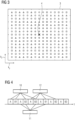
- a step S1 the k-space 1 to be acquired is divided into two regions A, B, as shown in Fig. 2 , for example dependent on a chosen proportion of the central region A with respect to the k-space 1, in this case definable by the radius 2, also see the parameter pA as described above.
- the central region A comprises the k-space center.
- the outer or peripheral region B surrounds the region A.
- Fig. 3 shows trajectory starting points in the phase encoding plane 3 (k y and k z -directions).
- the k-space center 4 is accordingly marked, as are the starting points belonging to the central region A as filled circles.
- the peripheral region B is further divided into a pre-determined partitioning number of peripheral k-space portions B1 ... Bn.
- the starting points of the peripheral region B may be assigned according to sorting criteria, for example increasing radial distance as a primary sort key and increasing azimuthal angle, which is measured counterclockwise from the k y -direction, as secondary sort key.
- the Bi have at least approximately equal size.
- n of three exemplarily a partitioning number n of three is illustrated, wherein the squares belong to B1, the unfilled circles to B2 and the triangles to B3.
- a step S2 magnetic resonance signals are acquired by parallel imaging using multiple reception coils, for which a three-dimensional sensitivity distribution is known as sensitivity information.
- the sampling scheme of the k-space 1 is to scan pairs of the central region A and a peripheral k-space portion Bi in a pre-determined order, for example (A, B1), (A, B2), (A, B3), (A, B1), (A, B2), ... in the example provided above, as also shown in Fig. 4 .
- Each pair thus has two acquisition steps, one for the central region A and one for the respective peripheral k-space portion Bi.
- step S2 does not use Cartesian trajectories, that is, k-space lines as sub-trajectories associated with each of the points in Fig. 2 , but three-dimensional corkscrew trajectories.
- Cartesian trajectories that is, k-space lines as sub-trajectories associated with each of the points in Fig. 2 , but three-dimensional corkscrew trajectories.
- step S2 does not use Cartesian trajectories, that is, k-space lines as sub-trajectories associated with each of the points in Fig. 2 , but three-dimensional corkscrew trajectories.
- These are implemented using a wave-encoded 3D gradient echo sequence as exemplarily shown in the pulse diagram of Fig. 5 .
- the usual readout gradient pulses 5 and phase encoding pulses 6 remain unchanged regarding conventional sequences relating to a k-space line, however, additional sinusoidal gradient pulses 7 with a quarter cycle
- the additional wave-encoding leads to wide voxel-spreading of aliased image voxels in all three spatial dimensions. Therefore, the removal of undersampling artifacts during reconstruction is improved, the resulting g-factor is lower and therefore, reconstructed magnetic resonance images exhibit lower noise enhancement.
- 2D-CAIPIRINHA undersampling can be employed.
- a time series of magnetic resonance images, each associated with a time step (TWIST phase), is reconstructed as the magnetic resonance data set.
- a sliding reconstruction window is used. For example, if a first magnetic resonance image 10 is reconstructed from the first shown acquired magnetic resonance signals of B1, B2, and the pair A, B3, for the magnetic resonance image 11 next in time, the older acquisitions of A and B1 are removed and replaced by the magnetic resonance signals of A and B1 acquired next in time. For the next time step, old acquisitions of A and B2 are replaced with the next later acquired magnetic resonance signals of A and B2 for the magnetic resonance image 12, and so on.
- a complete sampling of the k-space 1 may be performed at the beginning and/or the end of the acquisition process, such that an initial and/or final reconstruction can be performed, and, for the first n pairs, magnetic resonance signals of the initial complete sampling are successively replaced until the sliding reconstruction window can be employed.
- step S3 The actual reconstruction in step S3 is performed using a reconstruction technique. Since aliasing is controlled in all three dimensions, the full three-dimensional coil sensitivity distribution is used as the sensitivity information during reconstruction, increasing the amount of total information used.
- an L+S reconstruction technique is combined with the wave-encoded 3D gradient echo trajectory.
- a compressed sensing model with temporal regularization is particularly advantageous in this case.
- Consecutive magnetic resonance images 10, 11, 12 differ only marginally and therefore are highly correlated in the temporal dimension. These correlations are exploited to yield high quality reconstructions using the L+S compressed sensing model.
- the controlled aliasing in three dimensions for the case of wave-encoded sampling lead to higher reconstruction quality.
- the combination of the wave-encoding technique and the L+S reconstruction technique in particular allows higher acceleration and hence potential for significant progress in temporal resolution, while maintaining high spatial resolution.
- Fig. 6 is a principle drawing of a magnetic resonance imaging device 13 according to the invention.
- the magnetic resonance imaging device 13 comprises a main magnet unit 14 having a cylindrical bore 15 into which a patient may be introduced using a patient table 16.
- a radio frequency coil arrangement of a radio frequency system and a gradient coil arrangement of a gradient system may be provided.
- the magnetic resonance signals are acquired using a local head coil 17 comprising a plurality of reception coils 18 whose three-dimensional coil sensitivity distribution is known as sensitivity information to be used in reconstruction step S3, as explained above.
- the operation of the magnetic resonance imaging device 13 is controlled by a control device 19, which comprises a sequence unit 20 for controlling the acquisition of magnetic resonance signals, in particular also according to step S2, a planning unit 21, where step S1 can be executed, and a reconstruction unit 22 for reconstructing magnetic resonance images 10, 11, 12 from the acquired magnetic resonance signals, in particular also according to step S3 as explained above.
- the control device 19 further comprises a storage means 23, where, for example, the sensitivity information, acquisition parameters like the division of the k-space 1, magnetic resonance images 10, 11, 12 and the like may be stored.
Landscapes
- Physics & Mathematics (AREA)
- Health & Medical Sciences (AREA)
- Nuclear Medicine, Radiotherapy & Molecular Imaging (AREA)
- High Energy & Nuclear Physics (AREA)
- General Health & Medical Sciences (AREA)
- Radiology & Medical Imaging (AREA)
- Engineering & Computer Science (AREA)
- Condensed Matter Physics & Semiconductors (AREA)
- General Physics & Mathematics (AREA)
- Signal Processing (AREA)
- Life Sciences & Earth Sciences (AREA)
- Biophysics (AREA)
- Pathology (AREA)
- Biomedical Technology (AREA)
- Heart & Thoracic Surgery (AREA)
- Medical Informatics (AREA)
- Molecular Biology (AREA)
- Surgery (AREA)
- Animal Behavior & Ethology (AREA)
- Public Health (AREA)
- Veterinary Medicine (AREA)
- Magnetic Resonance Imaging Apparatus (AREA)
Abstract
Description
- The invention concerns a computer-implemented method for determining a dynamic contrast-enhanced magnetic resonance data set of an imaging region of a patient, wherein the k-space to be acquired is divided into a central region comprising the k-space center and a peripheral region surrounding the central region, and wherein the peripheral region comprises a partitioning number of peripheral k-space portions, comprising the steps of
- using a plurality of reception coils to acquire magnetic resonance signals using parallel imaging and a k-space acquisition scheme, in which alternatingly the central region and one of the peripheral k-space portions, the peripheral k-space portion being chosen according to a pre-determined order, are imaged in acquisitions steps of a pair, such that, after the partition number of such pairs, the whole k-space to be acquired has been imaged and a sliding reconstruction window can be applied to reconstruct an additional magnetic resonance image after each acquisition of such a pair, wherein during acquisition undersampling in at least two k-space directions is performed,
- reconstructing a time series of magnetic resonance images forming the magnetic resonance data set from the magnetic resonance signals and sensitivity information regarding the plurality of reception coils by using the sliding reconstruction window and a reconstruction technique for undersampled magnetic resonance data.
- The invention further concerns a magnetic resonance imaging device, a computer program and an electronically readable storage medium.
- Magnetic resonance imaging (MRI) has become an established modality in medical imaging. For angiography in various body regions, dynamic contrast-enhanced MRI (DCE-MRI) may be used. Examples include imaging of the renal arteries or the carotid arteries. To accurately assess vessel constitution and blood flow, in particular regarding irregularities, three-dimensional acquisitions are performed, which should be of a high spatial and temporal resolution. The acquisition time is limited and governed by the evolution of the contrast agent along the vessels. Hence, a compromise between temporal and spatial resolution has to be found.
- A known technique for DCE-MRI is based on the so-called TWIST sampling scheme (time-resolved angiography with stochastic trajectories, see the article by Ting Song et al., "Optimal k-Space Sampling for Dynamic Contrast-Enhanced MRI with an Application to MR Renography", Magnetic Resonance in Medicine 61:1242-1248 (2009)). Here, the k-space to be acquired is divided into a central region and a peripheral region, which, again, is divided into several (n, partitioning number) peripheral k-space portions. In the TWIST technique, these peripheral k-space portions, in concrete embodiments subsets of k-space lines in the peripheral region, are usually each spread over the peripheral region. An order of the peripheral k-space portions is defined. During acquisition, pairs of the central region (A) and one of the peripheral k-space portions (B1...Bn) are acquired, the latter chosen according to the pre-determined order. For example, for partitioning number n=3, the acquisition order may be (A, B1), (A, B2), (A, B3), (A, B1), (A, B2), ... Hence, after each n acquisitions, the k-space has been fully sampled. In each acquisition step for a pair, that is for A and the Bi, i=1...n, Cartesian k-space trajectories are employed.
- During reconstruction, a sliding reconstruction window is used in order to reconstruct several magnetic resonance images for different time frames, wherein the sliding reconstruction window covers n consecutively acquired pairs, that is, magnetic resonance signals for the whole k-space to be acquired. Hence, consecutive magnetic resonance images are not disjunct on the magnetic resonance signals (k-space data) level. However, the temporal resolution is considerably increased compared to simply acquiring consecutive images.
- To further increase the temporal resolution, undersampling and parallel imaging techniques may be combined with TWIST imaging.
- Generally, undersampling may result in aliasing artefacts in the spatial domain. Hence, dynamic imaging and reconstruction approaches have been proposed using parallel imaging techniques, in particular to control aliasing. For example, GRAPPA (see M. A. Griswold et al, "Generalized autocalibrating partially parallel acquisitions (GRAPPA)", Magnetic Resonance in Medicine 47(6):1202-1210 (2002)) or controlled aliasing in parallel imaging results in higher acceleration (CAIPIRINHA, see F. A. Breuer et al, "Controlled Aliasing in Parallel Imaging Results in Higher Acceleration (CAIPIRINHA) for Multi-Slice Imaging", Magnetic Resonance in Medicine 53:684-691 (2005)) may be used. For example, CAIPIRINHA improves reconstruction quality for multislice acquisitions by modulating the phase of the simultaneously excited slices. This modification incurs interslice shifts in the phase encoding direction between aliasing image slices, thereby increasing the variation in the coil sensitivity profiles across the slices to improve slice dealiasing. The application of interslice shifts to three-dimensional imaging forms the basis of 2D-CAIPIRINHA, wherein the encoding strategy in the phase encoding direction and the slice select direction is modified to shift the spatial aliasing pattern to reduce aliasing and better exploit the coil sensitivity variation.
- However, parallel imaging techniques inherently increase the noise in the reconstructed magnetic resonance images. The degree of noise enhancement may be quantified by the so-called g-factor (geometry factor). High degrees of undersampling along the Cartesian k-space trajectory lead to large g-factors, signifying considerable noise enhancement.
- Regarding reconstruction techniques, in an article by Ricardo Otazo et al., "Low-rank plus sparse matrix decomposition for accelerated dynamic MRI with separation of background and dynamic components", Magnetic Resonance in Medicine 73 (2015), pages 1125-1136, a new model for undersampled dynamic magnetic resonance data was proposed. This approach assumes that the dynamic MRI data, represented as a space-time matrix M, is a linear superposition of a low-rank matrix L and a sparse matrix S, wherein L represents highly spatially and temporally correlated elements, such as the image background, while S captures dynamic information that is sparse in an appropriate transform domain. Hence, this reconstruction technique is also known as L+S technique.
- It is an object of the current invention to, in particular significantly, increase image quality and/or temporal and/or spatial resolution in DCE-MRI.
- This object is achieved by providing a method according to claim 1, a magnetic resonance device according to
claim 7, a computer program according to claim 8 and an electronically readable storage medium according to claim 9. - In a method as initially described, according to the invention, the k-space trajectories for each acquisition step are chosen to allow controlled aliasing in all three spatial dimensions including the readout direction.
- The invention is based on a technique dividing the k-space to be acquired into a central region and a peripheral region, wherein the peripheral region is further subdivided into peripheral k-space portions to be acquired alternatingly with the central portion in a pre-determined order, such that, by using a sliding reconstruction window, for each time step, the oldest acquired magnetic resonance signals of the central region and a certain peripheral k-space portion are replaced by the more recently acquired magnetic resonance signals of the central region and the certain peripheral k-space portion. A sequence unit in a control device may control the acquisition components of a magnetic resonance device to acquire magnetic resonance signals. In all the acquisition steps for the central region and the peripheral k-space portions, certain k-space trajectories, implemented by gradient coils of a gradient system of the magnetic resonance device, are used to acquire the respective magnetic resonance signals. Here, the invention proposes to use a 3D k-space trajectory which allows controlled aliasing in all three dimensions and hence can use the whole coil sensitivity information. That is, in particular, a three-dimensional sensitivity distribution of the reception coils is used as the sensitivity information during reconstruction.
- Preferably, a TWIST sampling scheme is used to stochastically define the peripheral k-space portions. It is referred to the above-cited article by Ting Song et al. In particular, in preferred embodiments, the division of the k-space may be performed in a plane perpendicular to the readout direction, that is, in a plane spanned by the two phase encoding directions (ky, kz). In a conventional approach, the regions and portions would then comprise k-space lines in the readout direction (kx), while in the current invention, three-dimensional sub-trajectories replacing the conventional k-space lines and allowing to exploit the full three-dimensional sensitivity information are used.
- In especially preferred embodiments, for each acquisition step, the k-space trajectory is defined by a wave-encoded 3D gradient echo magnetic resonance sequence using sinusoidal gradient pulses with a quarter cycle phase shift in the phase encoding directions. Such wave-encoding has already been proposed for so called wave-CAIPI approaches and can easily be transferred to and employed in the method of the invention, since it only requires adding additional gradient pulses and does not affect phase encoding. The sinusoidal gradient wave oscillations lead to corkscrew-shaped readout trajectories in 3D k-space (instead of k-space lines). The additional wave-encoding leads to wide voxel spreading of aliased image voxels in all three spatial dimensions, including the readout direction kx (often also called frequency encoding direction), which is not used for acceleration in Cartesian imaging. In the image domain, the additional phase deposition results in voxel-spreading along the readout direction which varies linearly as a function of the spatial y and z position. A well distributed aliasing pattern is created, allowing use of the whole three-dimensional sensitivity distribution in reconstruction. Hence, undersampling artifacts can be more efficiently corrected. The resulting g-factor is lower, such that reconstructed magnetic resonance images of the magnetic resonance data set show lower noise enhancement compared to the conventional Cartesian technique. The wave-CAIPI approach and its effect on the g-factor is, for example, described in
- B. Bilgic et al, "Wave-CAIPI for Highly Accelerated 3D Imaging", Magnetic Resonance in Medicine 73:6 (2015),
- J. A. J. Richter et al, "Accelerated aortic 4D flow MRI with wave-CAIPI", Magnetic Resonance in Medicine 85:2595-2607 (2021),
- J. A. J. Richter et al, "Free-breathing self-gated 4D lung MRI using wave-CAIPI", Magnetic Resonance in Medicine 84: 3223-3233 (2020), and
- D. Polak et al, "Highly-accelerated volumetric brain examination using optimized wave-CAIPI encoding", Magnetic Resonance in Medicine 50(3):961-974 (2019).
- In summary, an accelerated setup is proposed for DCE-MRI, using a k-space trajectory allowing to take coil sensitivity information from all three dimensions into account, in particular a wave-encoded 3D k-space trajectory. 3D k-space trajectories inherently yield higher signal-to-noise-ratio than 2D techniques, which may be traded for an improved spatial and/or temporal resolution. The 3D k-space trajectory provides increased potential for acceleration with parallel imaging or compressed sensing, due to the possibility to exploit the full three-dimensional coil sensitivity information, that is, the three-dimensional sensitivity distribution of the reception coils. Wave-encoding leads to lower noise enhancement in highly undersampled magnetic resonance data sets. Hence, the proposed technique bears potential to significantly increase temporal and/or spatial resolution in DCE-MRI.
- In an especially advantageous embodiment of the current invention, a low rank plus sparse (L+S) reconstruction technique is used as the reconstruction technique, wherein the dynamic magnetic resonance data as a space-time matrix is assumed as a linear superposition of a highly spatially and temporarily correlated image background matrix and a sparse dynamic information matrix. The L+S image reconstruction technique has proven very effective in dynamic MRI applications. The L+S model takes spatial correlation in 3D images into account, but also temporal correlations in dynamic image series, which facilitates reconstruction of highly undersampled magnetic resonance data sets. In particular in DCE-MRI, the L+S compressed sensing model is promising, since the magnetic resonance image series typically shows only little variation in signal intensities, such that the image series could be efficiently represented by a low-rank background (L-matrix) and a temporally varying sparse component (S-matrix). If L+S compressed sensing reconstruction techniques are used instead of, for example, GRAPPA and/or SMS, lower noise in undersampled images results, providing the possibility for higher acceleration without compromising image quality. For example, the different time steps in TWIST imaging, that is the individual magnetic resonance images, differ only marginally and are therefore highly correlated in the temporal dimension. These correlations can be leveraged to achieve reconstructions of high quality. Generally, the reconstruction if further aided by the incoherent appearance of aliasing caused by the wave-encoding, providing a synergistic effect.
- That is, while conventional image reconstruction processes each time step, in particular each TWIST phase, individually, the dynamic compressed sensing L+S model, i.e., including temporal regularization, is efficient in the reconstruction of dynamic DCE-MRI data, as, for example, also described in an article by R. Otazo et al., "Low-rank plus sparse matrix decomposition for accelerated dynamic MRI with separation of background and dynamic components", Magnetic Resonance in Medicine 73:3 (2015). By employing the L+S model, the inherent temporal correlations of the individual phases, which are defined by the sliding reconstruction window, are exploited to arrive at high quality image reconstructions. In the inventive approach, image reconstruction is further facilitated by the wave-encoding technique, since wave-encoding leads to incoherent aliasing in all dimensions, which is beneficial for general compressed sensing reconstructions. Combining the wave-encoding technique and the L+S compressed sensing model, higher acquisition acceleration is possible, leading to increased temporal resolutions in DCE-MRI.
- Regarding undersampling, as known from the TWIST sampling scheme and approach, two sampling parameters may be defined and optimized accordingly, namely pA, which specifies the proportion of the central region A regarding the k-space to be acquired, and pB, which determines the density of sampling of the peripheral region B. Hence, TWIST offers two independent ways to improve the temporal resolution, that is by lowering pA (reducing the size of the central region) and by lowering pB (sampling the peripheral region more sparsely. Both approaches have certain tradeoffs: lowering pA will decrease the amount of low spatial frequency information, while lowering pB will decrease the fidelity of small features and fine image textures.
- In preferred embodiments, the gradient system transfer function may be used to correct for inaccuracies during reconstruction. The challenges regarding the reconstruction of non-Cartesian k-space can be efficiently overcome by using the Gradient System Transfer Function (GSTF) to correct for gradient inaccuracies. Since gradient errors may lead to severe image artifacts, these challenges regarding the reconstruction of non-Cartesian k-space can be efficiently overcome by using the so-called gradient system transfer function (GSTF) to correct for gradient inaccuracies and hence further increase image quality and/or the option for increasing resolution. GSTF-based correction approaches are, for example, described in the already cited articles by J. R. J. Richter regarding wave-CAIPI, and in
- M. Stich et al, "Field camera versus phantom-based measurement of the gradient system transfer function (GSTF) with dwell time compensation", Magnetic Resonance Imaging 71:125-131 (2020) and
- P. Eirich et al., "Cardiac real-time MRI using a preemphasized spiral acquisition based on the gradient system transfer function", Magnetic Resonance in Medicine 85 (5) :2747-2760 (2021).
- The invention further concerns a magnetic resonance imaging device, comprising a control device configured to perform a method according to the invention. All comments and remarks regarding the method according to the invention analogously apply to the magnetic resonance imaging device according to the invention, such that the same advantages can be achieved.
- The control device may comprise at least one processor and at least one storage means. As principally known in the state of the art, the control device may further, to acquire magnetic resonance signals, comprise a sequence unit, in particular controlling operation of a gradient system and at least one radio frequency system, in particular comprising the plurality of reception coils. The control device may further comprise a reconstruction unit for reconstructing the magnetic resonance images from the magnetic resonance signals acquired using the sequence unit, which is configured to use a k-space trajectory allowing controlled aliasing in all three spatial dimensions including the readout direction, in particular a wave-encoded 3D gradient echo trajectory.
- A computer program according to the invention can be directly loaded into a control device of a magnetic resonance imaging device and comprises program means to perform the steps of a method according to the invention when the computer program is executed on the control device. The computer program may be stored on an electronically readable storage medium according to the invention, which thus comprises control information comprising a computer program according to the invention such that when the electronically readable storage medium is used in a control device of a magnetic resonance device, the control device performs the steps of a method according to the invention. The electronically readable storage medium is preferably a non-transitional medium, for example a CD-ROM.
- Other objects and features of the present invention will become apparent from the following detailed description considered in conjunction with the accompanying drawings. The drawings, however, are only principle sketches designed solely for the purpose of illustration and do not limit the invention. The drawings show:
- Fig. 1
- a flow chart of a method according to the invention,
- Fig. 2
- a drawing explaining the definition of the central region and the peripheral region,
- Fig. 3
- a possible definition of peripheral k-space portions,
- Fig. 4
- a TWIST sampling scheme with a sliding reconstruction window,
- Fig. 5
- a pulse sequence diagram for an acquisition step employing wave-encoding, and
- Fig. 6
- a magnetic resonance imaging device according to the invention.
- In
Fig. 1 , an embodiment of a method for operating a magnetic resonance device for determining a dynamic contrast-enhanced magnetic resonance dataset, comprising a time series of magnetic resonance images, is described. For example, angiography of the renal or carotid arteries can be carried out. - In a step S1, the k-space 1 to be acquired is divided into two regions A, B, as shown in
Fig. 2 , for example dependent on a chosen proportion of the central region A with respect to the k-space 1, in this case definable by theradius 2, also see the parameter pA as described above. The central region A comprises the k-space center. The outer or peripheral region B surrounds the region A. -
Fig. 3 shows trajectory starting points in the phase encoding plane 3 (ky and kz-directions). The k-space center 4 is accordingly marked, as are the starting points belonging to the central region A as filled circles. In the TWIST sampling scheme, the peripheral region B is further divided into a pre-determined partitioning number of peripheral k-space portions B1 ... Bn. Here, in an embodiment, the starting points of the peripheral region B may be assigned according to sorting criteria, for example increasing radial distance as a primary sort key and increasing azimuthal angle, which is measured counterclockwise from the ky-direction, as secondary sort key. The points on the resulting sorted list are then sequentially assigned to one of n disjoint and nonoverlapping subsets forming the peripheral k-space portions Bi, i = 1 ,2 ..., n. The Bi have at least approximately equal size. - In
Fig. 3 , exemplarily a partitioning number n of three is illustrated, wherein the squares belong to B1, the unfilled circles to B2 and the triangles to B3. - In a step S2, magnetic resonance signals are acquired by parallel imaging using multiple reception coils, for which a three-dimensional sensitivity distribution is known as sensitivity information. The sampling scheme of the k-space 1 is to scan pairs of the central region A and a peripheral k-space portion Bi in a pre-determined order, for example (A, B1), (A, B2), (A, B3), (A, B1), (A, B2), ... in the example provided above, as also shown in
Fig. 4 . Each pair thus has two acquisition steps, one for the central region A and one for the respective peripheral k-space portion Bi. - However, step S2 does not use Cartesian trajectories, that is, k-space lines as sub-trajectories associated with each of the points in
Fig. 2 , but three-dimensional corkscrew trajectories. These are implemented using a wave-encoded 3D gradient echo sequence as exemplarily shown in the pulse diagram ofFig. 5 . As can be seen, the usualreadout gradient pulses 5 andphase encoding pulses 6 remain unchanged regarding conventional sequences relating to a k-space line, however, additionalsinusoidal gradient pulses 7 with a quarter cycle phase shift were added in the phase encoding directions (Gy/Gz). Also indicated inFig. 5 are the radio frequency pulse 8 and the readout time window 9. - The additional wave-encoding leads to wide voxel-spreading of aliased image voxels in all three spatial dimensions. Therefore, the removal of undersampling artifacts during reconstruction is improved, the resulting g-factor is lower and therefore, reconstructed magnetic resonance images exhibit lower noise enhancement. For example, 2D-CAIPIRINHA undersampling can be employed.
- In a step S3 of
Fig. 1 , a time series of magnetic resonance images, each associated with a time step (TWIST phase), is reconstructed as the magnetic resonance data set. As shown inFig. 4 , a sliding reconstruction window is used. For example, if a firstmagnetic resonance image 10 is reconstructed from the first shown acquired magnetic resonance signals of B1, B2, and the pair A, B3, for themagnetic resonance image 11 next in time, the older acquisitions of A and B1 are removed and replaced by the magnetic resonance signals of A and B1 acquired next in time. For the next time step, old acquisitions of A and B2 are replaced with the next later acquired magnetic resonance signals of A and B2 for themagnetic resonance image 12, and so on. - It is noted that, in some embodiments, a complete sampling of the k-space 1 (A and all Bi) may be performed at the beginning and/or the end of the acquisition process, such that an initial and/or final reconstruction can be performed, and, for the first n pairs, magnetic resonance signals of the initial complete sampling are successively replaced until the sliding reconstruction window can be employed.
- The actual reconstruction in step S3 is performed using a reconstruction technique. Since aliasing is controlled in all three dimensions, the full three-dimensional coil sensitivity distribution is used as the sensitivity information during reconstruction, increasing the amount of total information used.
- In this embodiment, an L+S reconstruction technique is combined with the wave-encoded 3D gradient echo trajectory. As already explained, such a compressed sensing model with temporal regularization is particularly advantageous in this case. Consecutive
magnetic resonance images -
Fig. 6 is a principle drawing of a magneticresonance imaging device 13 according to the invention. The magneticresonance imaging device 13 comprises amain magnet unit 14 having acylindrical bore 15 into which a patient may be introduced using a patient table 16. Surrounding thebore 15, a radio frequency coil arrangement of a radio frequency system and a gradient coil arrangement of a gradient system (not shown) may be provided. In this case, the magnetic resonance signals are acquired using alocal head coil 17 comprising a plurality of reception coils 18 whose three-dimensional coil sensitivity distribution is known as sensitivity information to be used in reconstruction step S3, as explained above. - The operation of the magnetic
resonance imaging device 13 is controlled by acontrol device 19, which comprises asequence unit 20 for controlling the acquisition of magnetic resonance signals, in particular also according to step S2, aplanning unit 21, where step S1 can be executed, and areconstruction unit 22 for reconstructingmagnetic resonance images control device 19 further comprises a storage means 23, where, for example, the sensitivity information, acquisition parameters like the division of the k-space 1,magnetic resonance images - Although the present invention has been described in detail with reference to the preferred embodiment, the present invention is not limited by the disclosed examples from which the skilled person is able to derive other variations without departing from the scope of the invention.
Claims (9)
- Computer-implemented method for determining a dynamic contrast-enhanced magnetic resonance data set of an imaging region of a patient, wherein the k-space (1) to be acquired is divided into a central region (A) comprising the k-space center (4) and a peripheral region (B) surrounding the central region (A), and wherein the peripheral region (B) comprises a partitioning number of peripheral k-space portions (Bi), comprising the steps of- using a plurality of reception coils (18) to acquire magnetic resonance signals using parallel imaging and a k-space acquisition scheme, in which alternatingly the central region (A) and one of the peripheral k-space portions (Bi), the peripheral k-space portion (Bi) being chosen according to a pre-determined order, are imaged in acquisition steps of a pair, such that, after the partition number of such pairs, the whole k-space (1) to be acquired has been imaged and a sliding reconstruction window can be applied to reconstruct an additional magnetic resonance image (10, 11, 12) after each acquisition of such a pair, wherein during acquisition undersampling in at least two k-space directions is performed,- reconstructing a time series of magnetic resonance images (10, 11, 12) forming the magnetic resonance data set from the magnetic resonance signals and sensitivity information regarding the plurality of reception coils (18) by using the sliding reconstruction window and a reconstruction technique for undersampled magnetic resonance data,characterized in that the k-space trajectories for each acquisition step are chosen to allow controlled aliasing in all three spatial dimensions including the readout direction.
- Method according to claim 1, characterized in that a TWIST sampling scheme is used to stochastically define the peripheral k-space portions (Bi).
- Method according to claim 1 or 2, characterized in that, for each acquisition step, the k-space trajectory is defined by a wave-encoded 3D gradient echo magnetic resonance sequence using sinusoidal gradient pulses (7) with a quarter cycle phase shift in the phase encoding directions.
- Method according to one of the preceding claims, characterized in that a low rank plus sparse reconstruction technique is used as the reconstruction technique, wherein the dynamic magnetic resonance data as a space-time matrix is assumed as a linear superposition of a highly spatially and temporally correlated image background matrix and a sparse dynamic information matrix.
- Method according to one of the preceding claims, characterized in that a three-dimensional sensitivity distribution of the reception coils (18) is used as the sensitivity information.
- Method according to one of the preceding claims, characterized in that the gradient system transfer function is used to correct for inaccuracies during reconstruction.
- Magnetic resonance imaging device (13), comprising a control device (19) configured to perform a method according to one of the preceding claims.
- Computer program, which performs the steps of a method according to one of the preceding claims when the computer program is executed on a control device (19) of a magnetic resonance imaging device (13).
- Electronically readable storage medium, on which a computer program according to claim 8 is stored.
Priority Applications (2)
| Application Number | Priority Date | Filing Date | Title |
|---|---|---|---|
| EP21203544.8A EP4170373B1 (en) | 2021-10-19 | 2021-10-19 | Computer-implemented method for determining a dynamic contrast-enhanced magnetic resonance data set, magnetic resonance imaging device, computer program and electronically readable storage medium |
| US17/967,191 US12140654B2 (en) | 2021-10-19 | 2022-10-17 | Computer-implemented method for determining a dynamic contrast-enhanced magnetic resonance data set, magnetic resonance imaging device, computer program and electronically readable storage medium |
Applications Claiming Priority (1)
| Application Number | Priority Date | Filing Date | Title |
|---|---|---|---|
| EP21203544.8A EP4170373B1 (en) | 2021-10-19 | 2021-10-19 | Computer-implemented method for determining a dynamic contrast-enhanced magnetic resonance data set, magnetic resonance imaging device, computer program and electronically readable storage medium |
Publications (3)
| Publication Number | Publication Date |
|---|---|
| EP4170373A1 true EP4170373A1 (en) | 2023-04-26 |
| EP4170373B1 EP4170373B1 (en) | 2025-06-18 |
| EP4170373C0 EP4170373C0 (en) | 2025-06-18 |
Family
ID=78676265
Family Applications (1)
| Application Number | Title | Priority Date | Filing Date |
|---|---|---|---|
| EP21203544.8A Active EP4170373B1 (en) | 2021-10-19 | 2021-10-19 | Computer-implemented method for determining a dynamic contrast-enhanced magnetic resonance data set, magnetic resonance imaging device, computer program and electronically readable storage medium |
Country Status (2)
| Country | Link |
|---|---|
| US (1) | US12140654B2 (en) |
| EP (1) | EP4170373B1 (en) |
Family Cites Families (2)
| Publication number | Priority date | Publication date | Assignee | Title |
|---|---|---|---|---|
| KR20180067442A (en) * | 2016-12-12 | 2018-06-20 | 지멘스 헬스케어 게엠베하 | Accelerated magnetic resonance imaging |
| EP4152031B1 (en) * | 2021-09-17 | 2024-11-20 | Siemens Healthineers AG | Method for determining a functional magnetic resonance data set, magnetic resonance imaging device, computer program and electronically readable storage medium |
-
2021
- 2021-10-19 EP EP21203544.8A patent/EP4170373B1/en active Active
-
2022
- 2022-10-17 US US17/967,191 patent/US12140654B2/en active Active
Non-Patent Citations (17)
| Title |
|---|
| B. BILGIC ET AL.: "Wave-CAIPI for Highly Accelerated 3D Imaging", MAGNETIC RESONANCE IN MEDICINE, vol. 73, 2015, pages 6 |
| BERKIN BILGIC ET AL: "Wave-CAIPI for highly accelerated 3D imaging : Wave-CAIPI for Highly Accelerated 3D Imaging", MAGNETIC RESONANCE IN MEDICINE, vol. 73, no. 6, 1 July 2014 (2014-07-01), US, pages 2152 - 2162, XP055645454, ISSN: 0740-3194, DOI: 10.1002/mrm.25347 * |
| D. POLAK ET AL.: "Highly-accelerated volumetric brain examination using optimized wave-CAIPI encoding", MAGNETIC RESONANCE IN MEDICINE, vol. 50, no. 3, 2019, pages 961 - 974 |
| F. A. BREUER ET AL.: "Controlled Aliasing in Parallel Imaging Results in Higher Acceleration (CAIPIRINHA) for Multi-Slice Imaging", MAGNETIC RESONANCE IN MEDICINE, vol. 53, 2005, pages 684 - 691, XP055070789, DOI: 10.1002/mrm.20401 |
| HAMILTON JESSE ET AL: "Recent advances in parallel imaging for MRI", PROGRESS IN NUCLEAR MAGNETIC RESONANCE SPECTROSCOPY, vol. 101, 2 May 2017 (2017-05-02), pages 71 - 95, XP085170513, ISSN: 0079-6565, DOI: 10.1016/J.PNMRS.2017.04.002 * |
| J. A. J. RICHTER ET AL.: "Accelerated aortic 4D flow MRI with wave-CAIPI", MAGNETIC RESONANCE IN MEDICINE, vol. 85, 2021, pages 2595 - 2607 |
| J. A. J. RICHTER ET AL.: "Free-breathing self-gated 4D lung MRI using wave-CAIPI", MAGNETIC RESONANCE IN MEDICINE, vol. 84, 2020, pages 3223 - 3233 |
| M. A. GRISWOLDGENERALIZED AUTOCALIBRAT-ING PARTIALLY PARALLEL ACQUISITIONS (GRAPPA ET AL., MAGNETIC RESONANCE IN MEDICINE, vol. 47, no. 6, 2002, pages 1202 - 1210 |
| M. STICH ET AL.: "Field camera versus phantom-based measurement of the gradient system transfer function (GSTF) with dwell time compensation", MAGNETIC RESONANCE IMAGING, vol. 71, 2020, pages 125 - 131, XP086202724, DOI: 10.1016/j.mri.2020.06.005 |
| MICHAELY HENRIK J. ET AL: "CAIPIRINHA-Dixon-TWIST (CDT)-Volume-Interpolated Breath-Hold Examination (VIBE)", INVESTIGATIVE RADIOLOGY, vol. 48, no. 8, 1 August 2013 (2013-08-01), pages 590 - 597, XP055900691 * |
| OTHMAN AHMED E. ET AL: "Feasibility of CAIPIRINHA-Dixon-TWIST-VIBE for dynamic contrast-enhanced MRI of the prostate", EUROPEAN JOURNAL OF RADIOLOGY., vol. 84, no. 11, 1 November 2015 (2015-11-01), NL, pages 2110 - 2116, XP055900912, ISSN: 0720-048X, DOI: 10.1016/j.ejrad.2015.08.013 * |
| P. EIRICH ET AL.: "Cardiac real-time MRI using a pre-emphasized spiral acquisition based on the gradient system transfer function", MAGNETIC RESONANCE IN MEDICINE, vol. 85, no. 5, 2021, pages 2747 - 2760 |
| RICARDO OTAZO ET AL.: "Low-rank plus sparse matrix decomposition for accelerated dynamic MRI with separation of background and dynamic components", MAGNETIC RESONANCE IN MEDICINE, vol. 73, 2015, pages 1125 - 1136, XP055712852, DOI: 10.1002/mrm.25240 |
| RICARDO OTAZO ET AL: "Low-rank plus sparse matrix decomposition for accelerated dynamic MRI with separation of background and dynamic components : L+S Reconstruction", MAGNETIC RESONANCE IN MEDICINE, vol. 73, no. 3, 23 April 2014 (2014-04-23), US, pages 1125 - 1136, XP055712852, ISSN: 0740-3194, DOI: 10.1002/mrm.25240 * |
| RICHTER JULIAN A. J. ET AL: "Accelerated aortic 4D flow MRI with wave-CAIPI", MAGNETIC RESONANCE IN MEDICINE, vol. 85, no. 5, 24 November 2020 (2020-11-24), US, pages 2595 - 2607, XP055900733, ISSN: 0740-3194, Retrieved from the Internet <URL:https://onlinelibrary.wiley.com/doi/full-xml/10.1002/mrm.28605> DOI: 10.1002/mrm.28605 * |
| SONG TING ET AL: "Optimal k -space sampling for dynamic contrast-enhanced MRI with an application to MR renography", MAGNETIC RESONANCE IN MEDICINE, vol. 61, no. 5, 19 February 2009 (2009-02-19), US, pages 1242 - 1248, XP055900748, ISSN: 0740-3194, DOI: 10.1002/mrm.21901 * |
| TING SONG ET AL.: "Optimal k-Space Sampling for Dynamic Contrast-Enhanced MRI with an Application to MR Renography", MAGNETIC RESONANCE IN MEDICINE, vol. 61, 2009, pages 1242 - 1248 |
Also Published As
| Publication number | Publication date |
|---|---|
| EP4170373B1 (en) | 2025-06-18 |
| US20230117041A1 (en) | 2023-04-20 |
| US12140654B2 (en) | 2024-11-12 |
| EP4170373C0 (en) | 2025-06-18 |
Similar Documents
| Publication | Publication Date | Title |
|---|---|---|
| US11181598B2 (en) | Multi-contrast MRI image reconstruction using machine learning | |
| US8981776B2 (en) | Method for magnetic resonance imaging with controlled aliasing | |
| CN102870000B (en) | Method for simultaneous multi-slice magnetic resonance imaging using single and multiple channel receiver coils | |
| US11002815B2 (en) | System and method for reducing artifacts in echo planar magnetic resonance imaging | |
| US8427153B2 (en) | Method for motion correction in magnetic resonance imaging using radio frequency coil arrays | |
| US9317917B2 (en) | Method, reconstruction device, and magnetic resonance apparatus for reconstructing magnetic resonance raw data | |
| US9709650B2 (en) | Method for calibration-free locally low-rank encouraging reconstruction of magnetic resonance images | |
| US12027254B2 (en) | Multi-contrast MRI image reconstruction using machine learning | |
| US10203394B2 (en) | Metal resistant MR imaging | |
| JP2020523075A (en) | MR imaging using variable contrast stack-of-star acquisition | |
| CN114895228B (en) | Magnetic resonance imaging method, device, equipment and medium based on multi-segment simultaneous excitation | |
| US9684047B2 (en) | Method and system for rapid MRI acquisition using tailored signal excitation modules (RATE) | |
| CN107209238A (en) | Parallel many section MR imagings with the suppression to sideband artefact | |
| US9476957B2 (en) | Method and apparatus for accelerating magnetic resonance imaging | |
| CN115639510A (en) | Magnetic resonance imaging method, spectroscopic imaging method, apparatus, device, and storage medium | |
| US11543482B2 (en) | Magnetic resonance imaging using motion-compensated image reconstruction | |
| US10401455B2 (en) | Method and magnetic resonance apparatus for avoidance of artifacts in the acquisition of magnetic resonance measurement data | |
| US11009576B2 (en) | Method for magnetic resonance imaging using slice quadratic phase for spatiotemporal encoding | |
| US11474178B2 (en) | Method for generating a magnetic resonance image | |
| US10818047B2 (en) | Iterative reconstruction of quantitative magnetic resonance images | |
| EP4170373B1 (en) | Computer-implemented method for determining a dynamic contrast-enhanced magnetic resonance data set, magnetic resonance imaging device, computer program and electronically readable storage medium | |
| CN116953582A (en) | Multiple coil sensitivity maps and SENSE reconstruction for a magnetic resonance imaging apparatus | |
| US11874353B2 (en) | Multi-shot echo planar imaging using reordered segments and recursive radio frequency pulse design giving matched slice profiles across segments | |
| Lin et al. | High temporal resolution retrospective motion correction with radial parallel imaging | |
| US20250093449A1 (en) | Method for generating a series of magnetic resonance images with cross-frame iterative reconstruction |
Legal Events
| Date | Code | Title | Description |
|---|---|---|---|
| PUAI | Public reference made under article 153(3) epc to a published international application that has entered the european phase |
Free format text: ORIGINAL CODE: 0009012 |
|
| STAA | Information on the status of an ep patent application or granted ep patent |
Free format text: STATUS: THE APPLICATION HAS BEEN PUBLISHED |
|
| AK | Designated contracting states |
Kind code of ref document: A1 Designated state(s): AL AT BE BG CH CY CZ DE DK EE ES FI FR GB GR HR HU IE IS IT LI LT LU LV MC MK MT NL NO PL PT RO RS SE SI SK SM TR |
|
| STAA | Information on the status of an ep patent application or granted ep patent |
Free format text: STATUS: REQUEST FOR EXAMINATION WAS MADE |
|
| 17P | Request for examination filed |
Effective date: 20231023 |
|
| RBV | Designated contracting states (corrected) |
Designated state(s): AL AT BE BG CH CY CZ DE DK EE ES FI FR GB GR HR HU IE IS IT LI LT LU LV MC MK MT NL NO PL PT RO RS SE SI SK SM TR |
|
| RAP1 | Party data changed (applicant data changed or rights of an application transferred) |
Owner name: SIEMENS HEALTHINEERS AG |
|
| GRAP | Despatch of communication of intention to grant a patent |
Free format text: ORIGINAL CODE: EPIDOSNIGR1 |
|
| STAA | Information on the status of an ep patent application or granted ep patent |
Free format text: STATUS: GRANT OF PATENT IS INTENDED |
|
| INTG | Intention to grant announced |
Effective date: 20250221 |
|
| GRAS | Grant fee paid |
Free format text: ORIGINAL CODE: EPIDOSNIGR3 |
|
| GRAA | (expected) grant |
Free format text: ORIGINAL CODE: 0009210 |
|
| STAA | Information on the status of an ep patent application or granted ep patent |
Free format text: STATUS: THE PATENT HAS BEEN GRANTED |
|
| AK | Designated contracting states |
Kind code of ref document: B1 Designated state(s): AL AT BE BG CH CY CZ DE DK EE ES FI FR GB GR HR HU IE IS IT LI LT LU LV MC MK MT NL NO PL PT RO RS SE SI SK SM TR |
|
| REG | Reference to a national code |
Ref country code: GB Ref legal event code: FG4D |
|
| REG | Reference to a national code |
Ref country code: CH Ref legal event code: EP |
|
| REG | Reference to a national code |
Ref country code: DE Ref legal event code: R096 Ref document number: 602021032381 Country of ref document: DE |
|
| REG | Reference to a national code |
Ref country code: CH Ref legal event code: EP |
|
| REG | Reference to a national code |
Ref country code: IE Ref legal event code: FG4D |
|
| U01 | Request for unitary effect filed |
Effective date: 20250702 |
|
| U07 | Unitary effect registered |
Designated state(s): AT BE BG DE DK EE FI FR IT LT LU LV MT NL PT RO SE SI Effective date: 20250710 |
|
| PG25 | Lapsed in a contracting state [announced via postgrant information from national office to epo] |
Ref country code: GR Free format text: LAPSE BECAUSE OF FAILURE TO SUBMIT A TRANSLATION OF THE DESCRIPTION OR TO PAY THE FEE WITHIN THE PRESCRIBED TIME-LIMIT Effective date: 20250919 Ref country code: NO Free format text: LAPSE BECAUSE OF FAILURE TO SUBMIT A TRANSLATION OF THE DESCRIPTION OR TO PAY THE FEE WITHIN THE PRESCRIBED TIME-LIMIT Effective date: 20250918 |
|
| PG25 | Lapsed in a contracting state [announced via postgrant information from national office to epo] |
Ref country code: HR Free format text: LAPSE BECAUSE OF FAILURE TO SUBMIT A TRANSLATION OF THE DESCRIPTION OR TO PAY THE FEE WITHIN THE PRESCRIBED TIME-LIMIT Effective date: 20250618 |
|
| PG25 | Lapsed in a contracting state [announced via postgrant information from national office to epo] |
Ref country code: RS Free format text: LAPSE BECAUSE OF FAILURE TO SUBMIT A TRANSLATION OF THE DESCRIPTION OR TO PAY THE FEE WITHIN THE PRESCRIBED TIME-LIMIT Effective date: 20250918 |
|
| U20 | Renewal fee for the european patent with unitary effect paid |
Year of fee payment: 5 Effective date: 20251020 |
|
| PG25 | Lapsed in a contracting state [announced via postgrant information from national office to epo] |
Ref country code: IS Free format text: LAPSE BECAUSE OF FAILURE TO SUBMIT A TRANSLATION OF THE DESCRIPTION OR TO PAY THE FEE WITHIN THE PRESCRIBED TIME-LIMIT Effective date: 20251018 |
|
| PGFP | Annual fee paid to national office [announced via postgrant information from national office to epo] |
Ref country code: GB Payment date: 20251111 Year of fee payment: 5 |
|
| PG25 | Lapsed in a contracting state [announced via postgrant information from national office to epo] |
Ref country code: SM Free format text: LAPSE BECAUSE OF FAILURE TO SUBMIT A TRANSLATION OF THE DESCRIPTION OR TO PAY THE FEE WITHIN THE PRESCRIBED TIME-LIMIT Effective date: 20250618 |
|
| PG25 | Lapsed in a contracting state [announced via postgrant information from national office to epo] |
Ref country code: CZ Free format text: LAPSE BECAUSE OF FAILURE TO SUBMIT A TRANSLATION OF THE DESCRIPTION OR TO PAY THE FEE WITHIN THE PRESCRIBED TIME-LIMIT Effective date: 20250618 |
|
| PG25 | Lapsed in a contracting state [announced via postgrant information from national office to epo] |
Ref country code: PL Free format text: LAPSE BECAUSE OF FAILURE TO SUBMIT A TRANSLATION OF THE DESCRIPTION OR TO PAY THE FEE WITHIN THE PRESCRIBED TIME-LIMIT Effective date: 20250618 |
|
| PG25 | Lapsed in a contracting state [announced via postgrant information from national office to epo] |
Ref country code: SK Free format text: LAPSE BECAUSE OF FAILURE TO SUBMIT A TRANSLATION OF THE DESCRIPTION OR TO PAY THE FEE WITHIN THE PRESCRIBED TIME-LIMIT Effective date: 20250618 |
|
| PG25 | Lapsed in a contracting state [announced via postgrant information from national office to epo] |
Ref country code: ES Free format text: LAPSE BECAUSE OF FAILURE TO SUBMIT A TRANSLATION OF THE DESCRIPTION OR TO PAY THE FEE WITHIN THE PRESCRIBED TIME-LIMIT Effective date: 20250618 |
|
| PLBE | No opposition filed within time limit |
Free format text: ORIGINAL CODE: 0009261 |
|
| STAA | Information on the status of an ep patent application or granted ep patent |
Free format text: STATUS: NO OPPOSITION FILED WITHIN TIME LIMIT |
|
| REG | Reference to a national code |
Ref country code: CH Ref legal event code: L10 Free format text: ST27 STATUS EVENT CODE: U-0-0-L10-L00 (AS PROVIDED BY THE NATIONAL OFFICE) Effective date: 20260430 |