EP4163304A1 - Droplets and method for producing same - Google Patents
Droplets and method for producing same Download PDFInfo
- Publication number
- EP4163304A1 EP4163304A1 EP21822923.5A EP21822923A EP4163304A1 EP 4163304 A1 EP4163304 A1 EP 4163304A1 EP 21822923 A EP21822923 A EP 21822923A EP 4163304 A1 EP4163304 A1 EP 4163304A1
- Authority
- EP
- European Patent Office
- Prior art keywords
- protein
- droplet
- domain
- thioredoxin
- amino acid
- Prior art date
- Legal status (The legal status is an assumption and is not a legal conclusion. Google has not performed a legal analysis and makes no representation as to the accuracy of the status listed.)
- Pending
Links
Images








Classifications
-
- C—CHEMISTRY; METALLURGY
- C07—ORGANIC CHEMISTRY
- C07K—PEPTIDES
- C07K14/00—Peptides having more than 20 amino acids; Gastrins; Somatostatins; Melanotropins; Derivatives thereof
- C07K14/435—Peptides having more than 20 amino acids; Gastrins; Somatostatins; Melanotropins; Derivatives thereof from animals; from humans
- C07K14/575—Hormones
-
- C—CHEMISTRY; METALLURGY
- C12—BIOCHEMISTRY; BEER; SPIRITS; WINE; VINEGAR; MICROBIOLOGY; ENZYMOLOGY; MUTATION OR GENETIC ENGINEERING
- C12N—MICROORGANISMS OR ENZYMES; COMPOSITIONS THEREOF; PROPAGATING, PRESERVING, OR MAINTAINING MICROORGANISMS; MUTATION OR GENETIC ENGINEERING; CULTURE MEDIA
- C12N9/00—Enzymes; Proenzymes; Compositions thereof; Processes for preparing, activating, inhibiting, separating or purifying enzymes
- C12N9/90—Isomerases (5.)
-
- C—CHEMISTRY; METALLURGY
- C07—ORGANIC CHEMISTRY
- C07K—PEPTIDES
- C07K1/00—General methods for the preparation of peptides, i.e. processes for the organic chemical preparation of peptides or proteins of any length
- C07K1/10—General methods for the preparation of peptides, i.e. processes for the organic chemical preparation of peptides or proteins of any length using coupling agents
-
- C—CHEMISTRY; METALLURGY
- C12—BIOCHEMISTRY; BEER; SPIRITS; WINE; VINEGAR; MICROBIOLOGY; ENZYMOLOGY; MUTATION OR GENETIC ENGINEERING
- C12N—MICROORGANISMS OR ENZYMES; COMPOSITIONS THEREOF; PROPAGATING, PRESERVING, OR MAINTAINING MICROORGANISMS; MUTATION OR GENETIC ENGINEERING; CULTURE MEDIA
- C12N15/00—Mutation or genetic engineering; DNA or RNA concerning genetic engineering, vectors, e.g. plasmids, or their isolation, preparation or purification; Use of hosts therefor
- C12N15/09—Recombinant DNA-technology
- C12N15/11—DNA or RNA fragments; Modified forms thereof; Non-coding nucleic acids having a biological activity
- C12N15/62—DNA sequences coding for fusion proteins
-
- C—CHEMISTRY; METALLURGY
- C12—BIOCHEMISTRY; BEER; SPIRITS; WINE; VINEGAR; MICROBIOLOGY; ENZYMOLOGY; MUTATION OR GENETIC ENGINEERING
- C12Y—ENZYMES
- C12Y503/00—Intramolecular oxidoreductases (5.3)
- C12Y503/04—Intramolecular oxidoreductases (5.3) transposing S-S bonds (5.3.4)
- C12Y503/04001—Protein disulfide-isomerase (5.3.4.1), i.e. disufide bond-forming enzyme
-
- C—CHEMISTRY; METALLURGY
- C07—ORGANIC CHEMISTRY
- C07K—PEPTIDES
- C07K2319/00—Fusion polypeptide
Definitions
- a droplet is beginning to be known as a multi-stage reaction field for a sequential enzymatic reaction or the like, and in terms of its constituent factors, is known to be formed through addition of a second messenger by RNA, DNA, ATP, or the like to a natively unstructured protein called an intrinsically disordered protein.
- FUS which is a typical example capable of forming a droplet, is known to be such that a low complexity domain (LC domain) thereof, in which a structureless sequence appears repeatedly, forms a core of the droplet.
- a chaperone is known as a factor for controlling formation/dissociation of the droplet, and is also reported to be contained in the droplet.
- the term "chaperone” collectively refers to a group of proteins that control aggregation of proteins. However, there is no example in which a protein having functions as a chaperone and an enzyme forms a droplet by itself.
- a thioredoxin is known as a domain of a protein having imparted thereto two or more functions of a chaperone and an enzyme.
- the "thioredoxin” is a low-molecular-weight redox protein, and is known to play an important role in various life reactions by promoting reduction/cleavage of a disulfide bond formed by a cysteine residue of another protein.
- an object of the present invention is to develop a technology for preparing such droplet.
- a droplet can be produced by using a protein containing one or more thioredoxin-like domains.
- the inventors of the present invention have investigated factors for forming a droplet, a preferred structure of the protein containing a thioredoxin-like domain, and other conditions. Thus, the inventors have completed the present invention.
- the present invention provides the following items:
- the droplet can be prepared using a protein that has not been known heretofore to form a droplet.
- its constituent proteins interact at such a distance as to be able to be brought into contact with each other while having fluidity, and hence, for example, when a substance serving as a reaction raw material is added, the substance can enter between the constituent proteins of the droplet to be concentrated in the vicinity of the thioredoxins of the constituent proteins.
- the droplet of the present invention is useful for, for example, use as a reaction field for the enzymatic reaction or disaggregation reaction of thioredoxins or the like, or the storage of a defective protein.
- a protein having thioredoxin activity forms a droplet by itself, and hence the present invention is extremely useful for an enzymatic reaction or a disaggregation reaction.
- protein is used in a meaning including a polypeptide in which amino acids are polymerized via peptide bonds.
- protein encompasses both of a protein modified with a glycan or the like and an unmodified protein, unless otherwise stated. The same applies to a protein that is not specified to be a protein.
- the present invention provides a droplet including an assembly of a protein, wherein the protein is a PDI family protein and/or an artificial protein, and contains a thioredoxin-like domain.
- the “droplet” refers to an approximately spherical assembly in which a protein is liquid-liquid phase-separated, and in which the assembly of the protein has reversibility unlike an aggregation(aggregate) of an indefinite shape due to thermal denaturation or the like.
- the protein may optionally form the droplet in combination with a non-protein substance, such as a nucleic acid (e.g., DNA or RNA) or a crowder (e.g., ficol or glycerol).
- a non-protein substance such as a nucleic acid (e.g., DNA or RNA) or a crowder (e.g., ficol or glycerol).
- droplet formation also occurs through a temperature change or a pH change.
- the "assembly" means that a protein accumulates.
- the formation of the droplet occurs through: droplet formation through assembly of molecules at a molecular level; and droplet growth at a microscopically observable level through fusion of the droplets.
- the assembly is an accumulation having reversibility of a protein unlike, for example, an aggregation of an indefinite shape of a protein due to thermal denaturation, chemical denaturation, or the like. Accordingly, the formation of the droplet through assembly of a protein and the breakdown of the droplet through dissociation of the protein for forming the droplet can be controlled by adjusting conditions in a surrounding environment (e.g., the concentration of a factor for promoting assembly, a temperature, a pH, and the excluded volume effect of a crowder).
- a surrounding environment e.g., the concentration of a factor for promoting assembly, a temperature, a pH, and the excluded volume effect of a crowder.
- proteins typically have fluidity unlike an aggregation, though interacting at such a distance as to be able to be brought into contact with each other. Accordingly, a dispersion, a droplet, and an aggregation of a protein are sometimes likened to three states of matter (gas, liquid, and solid). Specifically, a state in which a protein is dispersed in an aqueous solution is likened to a gas, a droplet, whose constituent components such as a protein have fluidity, is likened to a liquid, and an aggregation, which has no fluidity because of a strong interaction between constituent components, is likened to a solid.
- the "droplet" which is the subject matter of the present invention, is intended to be a mass formed by a protein that has reversibly assembled, not a particle of a liquid, such as water or an organic solvent, or a particle obtained by wrapping a liquid in a membrane such as a lipid bilayer. Accordingly, the droplet, which is a reversible assembly of a protein, is beginning to be reported as a multi-stage reaction field for an enzyme-substrate reaction or an efficient chemical reaction field, and finds use as a super-enhancer.
- examples of the protein having a thioredoxin-like domain include a protein disulfide isomerase (PDI) family protein and an artificial protein.
- PDI protein disulfide isomerase
- examples of the PDI family protein include PDI, ERp46, ERp72, and P5. Of those, P5 or the like is preferred.
- examples of the artificial protein include variants of the PDI family protein.
- the protein having a thioredoxin-like domain exists in all organisms, and examples thereof include those derived from: mammals, such as a human, a monkey, a mouse, a rat, a rabbit, a cat, a dog, a pig, a bovine, a horse, and a sheep; birds such as a chicken; and microorganisms such as yeast.
- the protein having a thioredoxin-like domain preferably has a chaperone function.
- the "chaperone function" is a function of assisting appropriate folding of a protein, and the use of the protein having a chaperone function can be expected to suppress irreversible aggregation of the protein and to promote reversible assembly, and hence is preferred.
- hitherto there has been no report that a droplet can be formed with a protein having both of enzyme activity and a chaperone function, and hence the present invention is also useful in such respect.
- the protein preferably has one or more thioredoxin-like domains.
- the lower limit of the number of thioredoxin-like domains per protein molecule is not limited, but is preferably 1 or more, more preferably 2 or more.
- the upper limit of the number of thioredoxin-like domains per protein molecule is also not limited, but is preferably 7 or less, more preferably 6 or less, still more preferably 5 or less, still more preferably 4 or less.
- the range of the number of thioredoxin-like domains per protein molecule is also not limited, but
- the "thioredoxin-like domain” plays an important role in various life reactions such as a redox reaction existing in all organisms.
- the molecular weight of the thioredoxin-like domain is, for example, from about 10 kDa to about 15 kDa, typically about 13 KDa.
- a thioredoxin-like domain having a redox-active CxxC motif can directly catalyze a redox reaction of a target molecule via the motif, and a thioredoxin-like domain having no redox-active CxxC motif is involved in substrate recognition or the like.
- Activity of reducing/cleaving a disulfide bond may be measured by, for example, a method described in Non-patent Literature 7.
- Examples of the thioredoxin-like domain having activity of reducing/cleaving a disulfide bond include an a0 domain and an "a" domain in P5, which is a PDI family protein.
- the thioredoxin-like domain having activity of reducing/cleaving a disulfide bond preferably has the amino acid sequence of CGHC as shown in Fig. 20 .
- Examples of the amino acid sequence of the a0 domain include: the amino acid sequence of the a0 domain of any one of various organisms shown in (the amino acid sequences of) Fig.
- amino acid sequence of the "a" domain examples include: the amino acid sequence of the "a" domain of any one of the various organisms shown in (the amino acid sequences of) Fig. 20 ; and an amino acid sequence having one or more (e.g., 1 to 5, 1 to 3, or 1 or 2) amino acids added, substituted, or deleted in the above-mentioned amino acid sequence.
- the protein for forming the droplet has, as the thioredoxin-like domain, a domain formed of the above-mentioned amino acid sequence having one or more amino acids added, substituted, or deleted in the amino acid sequence of the a0 domain shown in Fig. 20 , the position at which the amino acid is added, substituted, or deleted is preferably a position other than amino acids conserved in a plurality of biological species (e.g., other than amino acids conserved in 4 or more kinds, 5 or more kinds, 6 or more kinds, or all 7 kinds of the 7 kinds shown in Fig. 20 )).
- the protein for forming the droplet has, as the thioredoxin-like domain, a domain formed of the above-mentioned amino acid sequence having one or more amino acids added, substituted, or deleted in the amino acid sequence of the "a" domain shown in Fig. 20
- the position at which the amino acid is added, substituted, or deleted is preferably a position other than amino acids conserved in a plurality of biological species (e.g., other than amino acids conserved in 4 or more kinds, 5 or more kinds, 6 or more kinds, or all 7 kinds of the 7 kinds shown in Fig. 20 )).
- thioredoxin-like domain having no activity of reducing/cleaving a disulfide bond is a "b" domain in P5.
- amino acid sequence of the "b" domain include: the amino acid sequence of the "b” domain of any one of the various organisms shown in (the amino acid sequences of) Fig. 20 ; and an amino acid sequence having one or more (e.g., 1 to 5, 1 to 3, or 1 or 2) amino acids added, substituted, or deleted in the above-mentioned amino acid sequence.
- the protein for forming the droplet has, as the thioredoxin-like domain, a domain formed of the above-mentioned amino acid sequence having one or more amino acids added, substituted, or deleted in the amino acid sequence of the "b" domain shown in Fig. 20 , the position at which the amino acid is added, substituted, or deleted is preferably a position other than amino acids conserved in a plurality of biological species (e.g., other than amino acids conserved in 4 or more kinds, 5 or more kinds, 6 or more kinds, or all 7 kinds of the 7 kinds shown in Fig. 20 )).
- the protein having a thioredoxin-like domain preferably has a region to which a factor for promoting assembly binds (assembly-promoting factor-binding region).
- a factor for promoting assembly binds include a calcium ion and other metal ions. Of those, a calcium ion or the like is preferred.
- the "assembly-promoting factor-binding region” means a region containing a sequence of E and D, and allowing the assembly-promoting factor to bind thereto.
- the assembly-promoting factor-binding region may be present in a region other than the thioredoxin-like domain or the like in the protein of the present invention, may be included in part of the thioredoxin-like domain or the like, or may satisfy both of the foregoing.
- the assembly-promoting factor-binding region is sometimes referred to as "calcium-binding region".
- the calcium-binding region may form a domain like an EF-hand domain.
- An example of the calcium-binding region is a calcium-binding region in P5.
- Examples of the amino acid sequence of the calcium-binding region in P5 include calcium-binding region portions in the amino acid sequences shown in Fig. 20 , more specifically the amino acid sequences of
- the assembly-promoting factor-binding region (e.g., the calcium-binding region) may be included in part of any one of the thioredoxin-like domains, but the protein preferably has the assembly-promoting factor-binding region separate from the thioredoxin-like domains.
- the protein for forming the droplet preferably has: two thioredoxin-like domains each having activity of reducing/cleaving a disulfide bond; one thioredoxin-like domain having no activity of reducing/cleaving a disulfide bond; and one assembly-promoting factor-binding region.
- the protein preferably has a linker sequence, for example, between a thioredoxin-like domain and another thioredoxin-like domain, between an assembly-promoting factor-binding region and another assembly-promoting factor-binding region, or between a thioredoxin-like domain and an assembly-promoting factor-binding region.
- the linker preferably contains at least one kind of amino acid residue selected from the group consisting of: serine; glycine; proline; glutamic acid; glutamine; aspartic acid; asparagine; arginine; lysine; and tyrosine.
- the linker sequence when the protein having a thioredoxin-like domain has a linker sequence, the linker sequence preferably has a certain length. By having a certain length, the linker sequence is predicted to lower the solubility of the thioredoxin-like domain to suitably contribute to droplet formation.
- At least one linker sequence e.g., a linker sequence between the a0 domain and the "a" domain or a linker sequence between a "b" domain and the calcium-binding region to be described later
- the upper limit of the length of each linker sequence is not particularly limited, but for example, is preferably 50 amino acids or less.
- a signal sequence for endoplasmic reticulum localization may be fused to a protein having the above-mentioned domains.
- the signal sequence include amino acid sequences shown as signal sequences in Fig. 20 and an amino acid sequence having one or more (e.g., 1 to 5, 1 to 3, or 1 or 2) amino acids added, substituted, or deleted in any one of the above-mentioned amino acid sequences.
- the protein When P5 or a variant thereof is used as the protein for forming the droplet, the protein preferably contains all of the a0 domain, the "a" domain, and the "b” domain.
- examples of the amino acid sequence of such protein include: an amino acid sequence having the amino acid sequences of the a0 domain, "a" domain, "b” domain, and calcium-binding region of any one of the various organisms shown in Fig.
- amino acid sequence also include: an amino acid sequence further having the linker sequence between the a0 domain and the "a” domain, the linker sequence between the "a” domain and the “b” domain, and the linker sequence between the "b” domain and the calcium-binding region; and an amino acid sequence also having the signal sequence in addition to the above-mentioned linker sequences); and an amino acid sequence having an amino acid sequence having one or more (e.g., 1 to 5, 1 to 3, or 1 or 2) amino acids added, substituted, or deleted in the above-mentioned amino acid sequence.
- an amino acid is added, substituted, or deleted from the above-mentioned standard amino acids, an example of the position thereof is the above-mentioned position.
- examples of the protein for forming the droplet include: a protein having the amino acid sequence of P5 of any one of the various organisms shown in Fig. 20 ; and a protein having an amino acid sequence having one or more (e.g., 1 to 5, 1 to 3, or 1 or 2) amino acids added, substituted, or deleted in the above-mentioned amino acid sequence.
- the protein having a thioredoxin-like domain may be a monomer, but preferably forms a dimer or a higher multimer (dimer, trimer, tetramer, or higher form).
- proteins forming a dimer or a higher multimer are sometimes collectively referred to as "multimers".
- the protein having a thioredoxin-like domain may be a mixture of two or more kinds selected from those monomers and multimers.
- the protein for forming the droplet have an unstructured region, and have such a structure that at least one of the N- or C-terminus of the unstructured region is the thioredoxin-binding region.
- the protein for forming the droplet of the present invention preferably has a structure having all of the above-mentioned three features.
- the droplet has an approximately spherical form.
- the droplet preferably has an average particle diameter of 50 nm or more.
- the upper limit of the average particle diameter of the droplets is not limited, but for example, is preferably 20 um or less.
- the average particle diameter may be measured using a dynamic light scattering apparatus, and more specifically, may be measured by a method described in Examples to be described later.
- the droplet may be obtained in a state of being dispersed in an aqueous or organic liquid. Accordingly, the present invention also provides a dispersion liquid of a droplet.
- the dispersion liquid of a droplet of the present invention typically shows gel-like properties.
- the upper limit of the pH of the dispersion liquid of a droplet of the present invention is not particularly limited, but is preferably 10 or less, more preferably 9 or less, still more preferably 7.4 or less.
- the range of the pH is not particularly limited, but is, for example, preferably from 2 to 10, more preferably from 5 to 9, still more preferably from 6 to 7.4.
- the present invention provides a method of producing a droplet including a step of assembling a protein in the presence of an assembly-promoting factor, the protein being a PDI family protein and/or an artificial protein, and containing a thioredoxin-like domain.
- the assembly-promoting factor, the protein containing a thioredoxin-like domain, and the like are as described above.
- the method of the present invention includes the step of assembling a protein in the presence of an assembly-promoting factor.
- the step is performed by mixing the assembly-promoting factor and the protein.
- the step is generally performed in an aqueous or organic liquid, preferably an aqueous liquid.
- the aqueous liquid include water, ethanol, methanol, and trifluoroethanol.
- the organic liquid include formamide and ethylene glycol. Those liquids may be used alone or as a mixture thereof. For example, there may be used a mixture of water with at least one kind selected from the group consisting of: ethanol; methanol; and trifluoroethanol.
- the blending ratio (v/v) of the at least one kind of hydrophilic liquid selected from the group consisting of: ethanol; methanol; and trifluoroethanol may be set to, for example, 15% or less, preferably 13% or less, more preferably 10% or less with respect to the volume of the mixed liquid of water and the hydrophilic liquid.
- the blending ratio of the hydrophilic liquid may be set to, for example, 1% or more, preferably 3% or more, more preferably 5% or more with respect to the volume of the mixed liquid of water and the hydrophilic liquid.
- the component other than the protein having a thioredoxin-like domain and the assembly-promoting factor examples include a pH adjusting agent, such as oxidized glutathione or reduced glutathione, and a buffering agent such as HEPES. Those components may be used alone or in combination thereof.
- the usage amount of the protein having a thioredoxin-like domain is not particularly limited, but for example, when the step is performed in a liquid, is preferably 5 ⁇ M or more, more preferably 20 ⁇ M or more.
- the usage amount of the assembly-promoting factor is also not particularly limited, but when the step is performed in a liquid, is preferably 500 ⁇ M or more, more preferably 4 mM or more.
- the upper limit of a pH is not particularly limited, but is preferably 10 or less, more preferably 9 or less, still more preferably 7.4 or less.
- the range of the pH is not particularly limited, but is, for example, preferably from 2 to 10, more preferably from 5 to 9, still more preferably from 6 to 7.4.
- a temperature when the step is performed is also not particularly limited, but is, for example, preferably from 0°C to 70°C, more preferably from 0°C to 20°C.
- a period of time for which the step is performed is also not particularly limited, but is, for example, preferably 1 minute or more, more preferably from 10 minutes to 60 minutes.
- a step of collecting the resultant droplet may be further performed.
- a method known per se e.g., filtration, centrifugation, or chromatography
- filtration, centrifugation, or chromatography may be appropriately used.
- the base sequence of recombinant P5 having added thereto a His ⁇ 6 tag was inserted between the restriction enzyme NdeI-BamHI sites of a pET15b vector for Escherichia coli expression (Novagen) to produce a plasmid.
- Competent cells BL21(DE3) were transformed with the P5 plasmid, and then cultured in LA agar medium (LB agar medium+50 mg/L ampicillin) overnight at 37°C.
- LA agar medium LB agar medium+50 mg/L ampicillin
- a single colony was picked up from the agar medium and inoculated in 100 mL of LA medium (100 mL of LB medium+5 mg of ampicillin), and then subjected to shaking culture overnight at 37°C.
- the bacterial cells were lysed with a buffer (50 mM Tris/HCl, pH 8.1, 0.3 M NaCl, 1 mM phenylmethylsulfonyl fluoride), and then disrupted with a homogenizer.
- the disrupted solution was centrifuged (12,000 ⁇ g, 20 min, 4°C), the supernatant was further ultracentrifuged (38,000 rpm, 30 min, 4°C), and the supernatant was collected.
- Ni-NTA Agarose (Qiagen) that had been equilibrated with a buffer (50 mM Tris/HCl, pH 8.1, 0.3 M NaCl) was added to the supernatant, and the mixture was stirred for 1 hour at 4°C. After 1 hour, the resultant was applied to an open column, washed twice with 40 mL of a buffer (50 mM Tris/HCl, pH 8.1, 0.3 M NaCl, 20 mM imidazole), and eluted 5 times with 10 mL of a buffer (50 mM Tris/HCl, pH 8.1, 0.3 M NaCl, 200 mM imidazole).
- a buffer 50 mM Tris/HCl, pH 8.1, 0.3 M NaCl
- the eluted sample was recognized by 10% SDS-PAGE, and then the eluted fraction of P5 was concentrated with Amicon Ultra (MWCO, 10,000: Millipore) to 500 ⁇ L.
- the concentrated sample was purified by being subjected to strong anion-exchange chromatography (MonoQ 10/100GL; GE Healthcare) utilizing an AKTA chromatography system (GE Helthcare). At that time, buffer A (50 mM Tris/HCl, pH 8.1, 1 mM EDTA) and buffer B (50 mM Tris/HCl, pH 8.1, 0.5 M NaCl, 1 mM EDTA) were used.
- the eluted sample was recognized by 10% SDS-PAGE, and then the eluted fraction of P5 was concentrated with Amicon Ultra (MWCO, 10,000: Millipore) to 250 ⁇ L. Further, the concentrate was purified by gel filtration chromatography (Superdex 200 10/300GL; GE Healthcare). At that time, a buffer (50 mM Tris/HCl, pH 8.1, 0.3 M NaCl, 1 mM EDTA) was used. The eluted sample was recognized by 10% SDS-PAGE, and then the eluted fraction of P5 was concentrated with Amicon Ultra (MWCO, 10,000: Millipore) to 500 ⁇ L, and the concentration was determined by a BCA method. Thus, a high-purity purified product was obtained ( Fig. 1 ).
- P5 as the high-purity purified product was titrated with calcium, and it was found with a dynamic light scattering (DLS) apparatus capable of evaluating a particle diameter that P5 associated in accordance with calcium titration ( Fig. 1 ).
- a P5 protein solution was set to 50 mM HEPES at a pH of 7.2 and a protein concentration of 50 ⁇ M.
- Titration with EDTA serving as a chelating agent for calcium converts a calcium-dependent P5 associated state into P5 in a non-associated state ( Fig. 2 ). According to the above-mentioned results, P5 can reversibly assemble in a calcium-dependent manner.
- the calcium-dependent associated bodies of P5 of Test Example 1 described above were subjected to phase-contrast observation with a confocal laser scanning biological microscope (FV1000, Olympus) fitted with an objective lens UCPLFLN 20 ⁇ PH (NA0.7).
- the calcium-dependent P5 associated bodies were observed as nearly truly spherical particles.
- a situation in which the associated bodies fused together along with the passage of time was also observed, showing that the associated bodies grew ( Fig. 3 ) .
- FRAP Fluorescence recovery after photobleaching
- a sample in which P5 having a fluorescent protein (mCherry) fused thereto (mCherry-P5) was allowed to coexist was prepared (50 mM HEPES, pH 7.5, 50 ⁇ M P5, 4 ⁇ M mCherry-P5), and observed in the same manner as in Test Example 2 ( Fig. 4 ).
- mCherry-P5 was excited with a laser at 559 nm, and phase-contrast and fluorescence images were acquired. As shown in the upper part of Fig.
- a refractive index was measured using a tomocube (http://www.tomocube.com/).
- Associated bodies of P5 were prepared in the same manner as in Test Example 1 except that 50 ⁇ M P5 and 4 mM CaCl 2 were added under the conditions of 50 mM HEPES at a pH of 7.2, and measurement was started within 10 minutes after the addition of CaCl 2 .
- Fig. 7 shows images immediately after the start of the measurement (left part of Fig. 7 ), about 12 seconds after the start of the measurement (center of Fig. 7 ), and about 14 seconds thereafter (right part of Fig. 7 ). As a result, an image capturing the moment droplets are about to fuse together is shown at the center of Fig. 7 .
- droplet surfaces are relatively rough, meaning high fluidity ( Fig. 7 ).
- RI refractive index
- ERp46, ERp72, ERp57, and ERp44 which are enzymes/chaperones localized in the endoplasmic reticulum, are easily concentrated inside the droplets, but PDI hardly enters the inside of the droplets.
- mCherry-ERp57 and GFP-ERp72 at least two kinds of enzymes/chaperones can be concentrated inside the droplets.
- glutathione labeled with ATTO 532 can also be concentrated inside the droplets ( Fig. 8 ).
- the droplets can concentrate other enzymes, and are expected to be applied to an industry involving using enzymes.
- P5 was titrated with calcium, and that P5 associated in accordance with calcium titration was evaluated in terms of the pH dependence of P5 droplet formation with a dynamic light scattering (DLS) apparatus capable of evaluating a particle diameter ( Fig. 9 ).
- a P5 protein solution was set to 50 mM HEPES at a pH of 6.7, 7.0, 7.4, 7.6, 7.8, or 8.0, and a protein concentration of 50 ⁇ M. As shown in Fig. 9 , a pH of 7.4 or less is preferred for droplet formation.
- P5 was titrated with calcium, and that P5 associated in accordance with calcium titration was evaluated in terms of the temperature dependence of P5 droplet formation with a dynamic light scattering (DLS) apparatus capable of evaluating a particle diameter ( Fig. 10 ).
- a P5 protein solution was set to 50 mM HEPES at a pH of 7.5 and a protein concentration of 50 ⁇ M, and calcium titration was performed in the same manner as in Test Example 1 except for being performed at 10°C, 20°C, or 30°C. As shown in Fig. 10 , a lower temperature is preferred for droplet formation.
- each mutant P5 illustrated in Fig. 11 was produced and evaluated for its functional site with a dynamic light scattering (DLS) apparatus capable of evaluating a particle diameter ( Fig. 11 ).
- DLS dynamic light scattering
- Calcium titration was performed in the same manner as in Test Example 1 except that P5 and mutant protein solutions were each used as 50 mM HEPES at a pH of 7.2 and a protein concentration of 50 ⁇ M. As shown in Fig. 11 , full-length P5 is preferred for droplet formation.
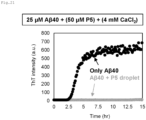
- Amyloid fibril formation of 25 ⁇ M A ⁇ 40 (amyloid beta 1-40, Peptide Institute, Inc.) was detected using a fluorescence plate reader having a stirring function (SH-9000Lab, Corona Electric). Specifically, A ⁇ 40 was added at 25 ⁇ M to an aqueous solution (pH 7.2) containing 2.5 ⁇ M thioflavin T and 50 mM HEPES.
- Thioflavin T is a fluorescent molecule capable of detecting fibrils, and is capable of recognizing the progress of amyloid fibrils as shown in Fig. 21 ("only A ⁇ 40" in Fig. 21 ).
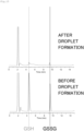
- Test Example 12 In order to extract structural information on P5 to which calcium did not bind, the full-length structure of P5 was determined by a small angle X-ray scattering method and X-ray crystallography ( Fig. 12 ). The determination of the full-length structure of P5 was performed in accordance with a method described in Non-patent Literature 8. In order to prepare non-droplet P5, a solution of P5 obtained by the method described in Test Example 1 was subjected to gel filtration chromatography (Superdex 200 10/300GL; GE Healthcare) with a buffer (20 mM phosphate, pH 8.0, 150 mM NaCl, 5% glycerol) immediately before measurement.
- Gel filtration chromatography Superdex 200 10/300GL; GE Healthcare
- the eluted sample was concentrated to a concentration of 3 mg/mL using Amicon Ultra (MWCO, 10,000: Millipore).
- a small angle X-ray scattering (SAXS) experiment of non-droplet P5 was performed at SPring-8 BL45XU under the conditions of a detector PILATUS 3X 2M (DECTRIS), an X-ray wavelength of 1.0 ⁇ , an X-ray exposure time of 1 second ⁇ 20, a camera length of 2.0 m, and a temperature of 20.2°C, and each scattering pattern was obtained.
- a radius of gyration was calculated through the Guinier approximation of the scattering pattern, and a distance distribution function was obtained with GNOM software.
- the molecular structure of non-droplet P5 was constructed with GAJOE software. As a result, it was shown that flexibility was rich between thioredoxin-like domains, and that a dimer was formed.
- the dimer-formation region exhibits a leucine zipper motif according to the crystal structure, and the substitution of this site with alanine can produce a P5 mutant for forming a monomer ( Fig. 13 ).
- the monomer P5 mutant also has an ability to form a droplet, though the ability to form a droplet is lower than that of the wild type ( Fig. 14 ).
- Test Example 14 In order to verify folding catalysis in P5 droplets, Bovine Pancreatic Trypsin Inhibitor (BPTI) was adopted as a substrate. P5 not forming a droplet catalyzes the oxidative folding of BPTI (left part of Fig. 16 ).
- BPTI Bovine Pancreatic Trypsin Inhibitor
- the P5 droplets serve to concentrate and store a denatured substrate.
- 20 mM N-ethylmaleimide was added to the reaction liquid to stop the reaction, and detection was performed with a BPTI antibody.
- electrophoretic bands conceivably of a dimer and a trimer of BPTI were observed.
- Such reaction occurred conceivably due to the special situation in which glutathione was concentrated in the droplets of a protein having a thioredoxin-like domain.
- Test Example 15 In order to examine the influences of organic solvents on P5 droplets, 5% v/v of methanol, ethanol, or trifluoroethanol was added to the P5 droplets obtained in Test Example 2, and the mixture was observed with a phase-contrast microscope. When an organic solvent is added at about 5% in v/v, the P5 droplets keep their shapes ( Fig. 17 ). Although the droplets were produced using P5 in this Test Example, in view of the property by which a GFP-PDI family member added after the formation of the droplet P5 can be included thereinside, it is conceived that a PDI family protein and/or an artificial protein other than P5, the protein having a thioredoxin-like domain, can also produce a droplet.
- Test Example 16 In order to examine the incorporation of a protein into P5 droplets, a mixed solution (molar ratio 1:5) of a PDI family protein having the fluorescent protein GFP fused thereto and P5, which was obtained in the same manner as in Test Example 1, was prepared and caused to form droplets by adding calcium, and then the resultant was centrifuged at 15,000 ⁇ g for 15 minutes to be separated into a condensed layer (corresponding to the inside of the droplets) and a dilute layer (corresponding to the outside of the droplets).
- Droplets such as an enzyme droplet can be expected to be applied in biopharmaceuticals, the beauty industry, industrial enzymes, and the food industry (e.g., the alcoholic beverage industry). More specifically, for example, droplet technologies can be expected to: be used in the quality control of a disulfide-containing protein preparation, such as insulin or an immunoglobulin, the restoration of a disulfide bond (e.g., hair repair), and the concentration and delivery of a redox molecule GSH; be used in the treatment of Parkinson's disease, diabetes, or the like; and be applied to a neurodegenerative disease through chaperone concentration.
- the novel droplet provided by the present invention can be applied in the above-mentioned fields, and hence is industrially extremely useful.
Landscapes
- Chemical & Material Sciences (AREA)
- Health & Medical Sciences (AREA)
- Life Sciences & Earth Sciences (AREA)
- Organic Chemistry (AREA)
- Genetics & Genomics (AREA)
- Engineering & Computer Science (AREA)
- Zoology (AREA)
- General Health & Medical Sciences (AREA)
- Bioinformatics & Cheminformatics (AREA)
- Biochemistry (AREA)
- Wood Science & Technology (AREA)
- Molecular Biology (AREA)
- General Engineering & Computer Science (AREA)
- Biomedical Technology (AREA)
- Medicinal Chemistry (AREA)
- Biotechnology (AREA)
- Biophysics (AREA)
- Microbiology (AREA)
- Proteomics, Peptides & Aminoacids (AREA)
- Analytical Chemistry (AREA)
- Physics & Mathematics (AREA)
- Plant Pathology (AREA)
- Endocrinology (AREA)
- Toxicology (AREA)
- Gastroenterology & Hepatology (AREA)
- Peptides Or Proteins (AREA)
- Enzymes And Modification Thereof (AREA)
Abstract
Description
- This application claims priority from
Japanese Patent Application No. 2020-100517, filed on June 9, 2020 - A droplet is beginning to be known as a multi-stage reaction field for a sequential enzymatic reaction or the like, and in terms of its constituent factors, is known to be formed through addition of a second messenger by RNA, DNA, ATP, or the like to a natively unstructured protein called an intrinsically disordered protein. FUS, which is a typical example capable of forming a droplet, is known to be such that a low complexity domain (LC domain) thereof, in which a structureless sequence appears repeatedly, forms a core of the droplet. A chaperone is known as a factor for controlling formation/dissociation of the droplet, and is also reported to be contained in the droplet. The term "chaperone" collectively refers to a group of proteins that control aggregation of proteins. However, there is no example in which a protein having functions as a chaperone and an enzyme forms a droplet by itself. A thioredoxin is known as a domain of a protein having imparted thereto two or more functions of a chaperone and an enzyme. The "thioredoxin" is a low-molecular-weight redox protein, and is known to play an important role in various life reactions by promoting reduction/cleavage of a disulfide bond formed by a cysteine residue of another protein.
-
- NPL 1: Science, 360(6391), 922-927 (2018)
- NPL 2: Proc. Natl. Acad. Sci. USA, 108(11), 4334-4339 (2011)
- NPL 3: Nat. Rev. Mol. Cell Biol., 18(5), 285-298 (2017)
- NPL 4: Cell, 149(4), 753-767 (2012)
- NPL 5: Cell, 149(4), 768-779 (2012)
- NPL 6: Int. J. Biol. Macromol., 10(PtA), 10-18 (2018)
- NPL 7: J. Biol. Chem.; 289(39): 27004-18 (2014)
- NPL 8: Structure, 29, 1-14 (2021)
- As it has not been known heretofore that a protein having chaperone and enzyme functions forms a droplet by itself, an object of the present invention is to develop a technology for preparing such droplet.
- Under such circumstances, the inventors of the present invention have made extensive investigations, and as a result, have found that a droplet can be produced by using a protein containing one or more thioredoxin-like domains. On the basis of such novel finding, the inventors of the present invention have investigated factors for forming a droplet, a preferred structure of the protein containing a thioredoxin-like domain, and other conditions. Thus, the inventors have completed the present invention.
- Thus, the present invention provides the following items:
-
Item 1. A droplet including an assembly of a protein, wherein the protein is a PDI family protein and/or an artificial protein, and contains a thioredoxin-like domain. -
Item 2. The droplet according toItem 1, wherein the protein has a calcium-binding region. -
Item 3. The droplet according toItem - wherein the protein has two or more thioredoxin-like domains,
- wherein at least two of the two or more thioredoxin-like domains are bound to each other via a linker, and
- wherein the linker contains at least one kind of amino acid residue selected from the group consisting of: serine; glycine; proline; glutamic acid; glutamine; aspartic acid; asparagine; arginine; lysine; and tyrosine.
-
Item 4. The droplet according to any one ofItems 1 to 3, wherein the protein is a dimer or a higher multimer. -
Item 5. The droplet according to any one ofItems 2 to 4, wherein the PDI family protein is P5. -
Item 6. A method of producing a droplet including a step of assembling a protein in the presence of a calcium ion,
the protein being a PDI family protein and/or an artificial protein, and containing a thioredoxin-like domain, the protein further having a calcium-binding region. - Item 7. The method according to
Item 6,- wherein at least two of the two or more thioredoxin-like domains are bound to each other via a linker, and
- wherein the linker contains at least one kind of amino acid residue selected from the group consisting of: serine; glycine; proline; glutamic acid; glutamine; aspartic acid; asparagine; arginine; lysine; and tyrosine.
-
Item 8. The method according toItem 6 or 7, wherein the PDI family protein is a dimer or a higher multimer. -
Item 9. The method according to any one ofItems 6 to 8, wherein the PDI family protein is P5. - According to the present invention, the droplet can be prepared using a protein that has not been known heretofore to form a droplet. As described later, typically, in a droplet, its constituent proteins interact at such a distance as to be able to be brought into contact with each other while having fluidity, and hence, for example, when a substance serving as a reaction raw material is added, the substance can enter between the constituent proteins of the droplet to be concentrated in the vicinity of the thioredoxins of the constituent proteins. Accordingly, the droplet of the present invention is useful for, for example, use as a reaction field for the enzymatic reaction or disaggregation reaction of thioredoxins or the like, or the storage of a defective protein. Hitherto, there has been no example in which a protein having thioredoxin activity forms a droplet by itself, and hence the present invention is extremely useful for an enzymatic reaction or a disaggregation reaction.
-
- The upper left part of
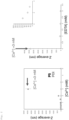
Fig. 1 shows SDS-PAGE results of a purified protein, and shows that the protein is a high-purity purified product. The right part is an illustration of the configuration of thioredoxin-like domains of a PDI family used and SS exchange active sites thereof. The lower left part shows a change in particle diameter by DLS. Among the PDI family, P5 associates in a calcium-dependent manner. - The left part of
Fig. 2 is a graph obtained by scaling up the long axis of the particle diameter ofFig. 1 to 5 ,000 nm. P5 forms associated bodies of about 4,500 nm in the presence of 3 mM calcium. The right part ofFig. 2 shows the results of titration of the associated bodies produced in the left part with EDTA. The P5 associated bodies return to non-associated bodies along with the titration, meaning the presence of reversibility. -
Fig. 3 shows with a phase-contrast microscope that the associated bodies of P5 are spherical. Along with a time change, the spherical associated bodies fuse together, and the spherical associated bodies grow larger. - The images of
Fig. 4 show the results of overlaying of fluorescence microscope images and phase-contrast microscope images. When an intense laser is applied, spherical assemblies that have undergone fluorescence bleaching once undergo fluorescence recovery along with a time change. The lower part ofFig. 4 shows the results of analysis of fluorescence recovery, indicating that a half time is about 19 seconds. This FRAP experiment supports that the spherical associated bodies are droplets. -
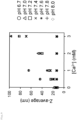
Fig. 5 shows the concentrations of supernatant fractions obtained by mixing calcium and P5 at various concentrations and then centrifuging the mixtures. -
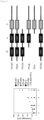
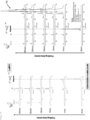
Fig. 6 shows the determination of the calcium-binding region of P5 by ITC measurement. Various mutants of P5 are illustrated in the upper part ofFig. 6 , and the results of an ITC experiment of each mutant and calcium are shown in the lower part ofFig. 6 . An upward heat quantity represents a heat quantity due to binding, and an unstructured region positioned at the C-terminus was found to be the calcium-binding region. -
Fig. 7 shows real-time observation of droplets using a fluorescence microscope capable of measuring a refractive index. An image capturing the moment droplets are about to fuse together is shown at the center. In addition, droplet surfaces are relatively rough, meaning high fluidity. -
Fig. 8 shows that various enzymes/chaperones and glutathione can be concentrated inside droplets.Fig. 8 supports the state of being droplets with a refractive index (RI), and indicates localization with various enzymes/chaperones and glutathione that are fluorescently labeled. It is shown that ERp46, ERp72, ERp57, and ERp44, which are enzymes/chaperones localized in the endoplasmic reticulum, are easily concentrated inside the droplets, but PDI hardly enters the inside of the droplets. In addition, it is shown that, through use of mCherry-ERp57 and GFP-ERp72, at least two kinds of enzymes/chaperones can be concentrated inside the droplets. In addition, glutathione labeled with ATTO 532 can also be concentrated inside the droplets. -
Fig. 9 shows pH dependence in droplet formation. A pH of 7.4 or less is preferred for droplet formation. -
Fig. 10 shows temperature dependence in droplet formation. A lower temperature is preferred for droplet formation. -
Fig. 11 shows the importance of domains of P5 in droplet formation. Full-length P5 is preferred for droplet formation. -
Fig. 12 shows a novel structure of P5. Flexibility between thioredoxin-like domains is relatively high. In addition, a dimer is formed in a solution. - The upper part of
Fig. 13 reveals from a crystal structure that dimer formation of P5 uses as a driving force a leucine zipper motif positioned in the 4th α-helix in an a0 domain at the beginning. Accordingly, the substitution of this site with alanine gives a monomer in a solution (lower part ofFig. 13 ). -
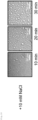
Fig. 14 shows with a phase-contrast microscope that associated bodies of a P5 monomer can form droplets as with a wild type. -
Fig. 15 shows the concentration of glutathione by P5 droplets. Reversed-phase HPLC was used for the separation of glutathione. It is found that, after droplet formation, nearly all glutathione disappeared. -
Fig. 16 shows substrate oxidative folding catalysis by P5 droplets. Reversed-phase HPLC was used for the folding state of a substrate. It is found that, after droplet formation, nearly all of a reduced and denatured substrate disappeared immediately after an oxidative folding reaction. -
Fig. 17 shows examples of application of organic solvents to P5 droplets. Various organic solvents were added in v/v, and observation was performed with a phase-contrast microscope. -
Fig. 18 shows the effect of salt on P5 droplets. Observation was performed with a phase-contrast microscope. Salt influences the size of droplet formation as compared to the salt-free conditions ofFig. 3 , and hence is a control factor for the size of droplet formation. -
Fig. 19 shows the degree of incorporation into the inside of droplets. Various PDI family members fluorescently labeled with GFP were produced, and the degrees of their incorporation were evaluated. The results reveal that PDI is hardly incorporated and ERp46 is easily incorporated. -
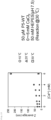
Fig. 20 shows the amino acid sequences of P5 of various organisms. -
Fig. 21 shows that droplets suppress amyloid fibril formation. The vertical axis represents fluorescence intensity, the experiment uses thioflavin T for specifically detecting amyloid fibrils, and it is shown that the addition of droplets suppressed amyloid fibrils. - In the present invention, the term "protein" is used in a meaning including a polypeptide in which amino acids are polymerized via peptide bonds. In addition, herein, the term "protein" encompasses both of a protein modified with a glycan or the like and an unmodified protein, unless otherwise stated. The same applies to a protein that is not specified to be a protein.
- The present invention provides a droplet including an assembly of a protein, wherein the protein is a PDI family protein and/or an artificial protein, and contains a thioredoxin-like domain.
- The "droplet" refers to an approximately spherical assembly in which a protein is liquid-liquid phase-separated, and in which the assembly of the protein has reversibility unlike an aggregation(aggregate) of an indefinite shape due to thermal denaturation or the like. When the protein assembles to form the droplet, the protein may optionally form the droplet in combination with a non-protein substance, such as a nucleic acid (e.g., DNA or RNA) or a crowder (e.g., ficol or glycerol). In addition, droplet formation also occurs through a temperature change or a pH change.
- In the present invention, the "assembly" means that a protein accumulates. In a typical embodiment, the formation of the droplet occurs through: droplet formation through assembly of molecules at a molecular level; and droplet growth at a microscopically observable level through fusion of the droplets.
- In addition, as described above, the assembly is an accumulation having reversibility of a protein unlike, for example, an aggregation of an indefinite shape of a protein due to thermal denaturation, chemical denaturation, or the like. Accordingly, the formation of the droplet through assembly of a protein and the breakdown of the droplet through dissociation of the protein for forming the droplet can be controlled by adjusting conditions in a surrounding environment (e.g., the concentration of a factor for promoting assembly, a temperature, a pH, and the excluded volume effect of a crowder).
- In addition, typically, in a state of forming a droplet, proteins have fluidity unlike an aggregation, though interacting at such a distance as to be able to be brought into contact with each other. Accordingly, a dispersion, a droplet, and an aggregation of a protein are sometimes likened to three states of matter (gas, liquid, and solid). Specifically, a state in which a protein is dispersed in an aqueous solution is likened to a gas, a droplet, whose constituent components such as a protein have fluidity, is likened to a liquid, and an aggregation, which has no fluidity because of a strong interaction between constituent components, is likened to a solid. In addition, like water and oil undergo liquid-liquid phase separation(LLPS) instead of mixing with each other, biological phase separation refers to a situation in which droplet formation occurs via a protein. As described above, the "droplet", which is the subject matter of the present invention, is intended to be a mass formed by a protein that has reversibly assembled, not a particle of a liquid, such as water or an organic solvent, or a particle obtained by wrapping a liquid in a membrane such as a lipid bilayer. Accordingly, the droplet, which is a reversible assembly of a protein, is beginning to be reported as a multi-stage reaction field for an enzyme-substrate reaction or an efficient chemical reaction field, and finds use as a super-enhancer.
- In the present invention, examples of the protein having a thioredoxin-like domain include a protein disulfide isomerase (PDI) family protein and an artificial protein. Those proteins may be used alone or in combination thereof. Examples of the PDI family protein include PDI, ERp46, ERp72, and P5. Of those, P5 or the like is preferred. Examples of the artificial protein include variants of the PDI family protein. The protein having a thioredoxin-like domain exists in all organisms, and examples thereof include those derived from: mammals, such as a human, a monkey, a mouse, a rat, a rabbit, a cat, a dog, a pig, a bovine, a horse, and a sheep; birds such as a chicken; and microorganisms such as yeast.
- The protein having a thioredoxin-like domain preferably has a chaperone function. The "chaperone function" is a function of assisting appropriate folding of a protein, and the use of the protein having a chaperone function can be expected to suppress irreversible aggregation of the protein and to promote reversible assembly, and hence is preferred. In addition, hitherto, there has been no report that a droplet can be formed with a protein having both of enzyme activity and a chaperone function, and hence the present invention is also useful in such respect.
- In a typical embodiment, the protein preferably has one or more thioredoxin-like domains. The lower limit of the number of thioredoxin-like domains per protein molecule is not limited, but is preferably 1 or more, more preferably 2 or more. The upper limit of the number of thioredoxin-like domains per protein molecule is also not limited, but is preferably 7 or less, more preferably 6 or less, still more preferably 5 or less, still more preferably 4 or less. The range of the number of thioredoxin-like domains per protein molecule is also not limited, but
- In the present invention, the "thioredoxin-like domain" plays an important role in various life reactions such as a redox reaction existing in all organisms. In the present invention, the molecular weight of the thioredoxin-like domain is, for example, from about 10 kDa to about 15 kDa, typically about 13 KDa. A thioredoxin-like domain having a redox-active CxxC motif can directly catalyze a redox reaction of a target molecule via the motif, and a thioredoxin-like domain having no redox-active CxxC motif is involved in substrate recognition or the like. Activity of reducing/cleaving a disulfide bond may be measured by, for example, a method described in Non-patent Literature 7.
- Examples of the thioredoxin-like domain having activity of reducing/cleaving a disulfide bond include an a0 domain and an "a" domain in P5, which is a PDI family protein. In addition, the thioredoxin-like domain having activity of reducing/cleaving a disulfide bond preferably has the amino acid sequence of CGHC as shown in
Fig. 20 . Examples of the amino acid sequence of the a0 domain include: the amino acid sequence of the a0 domain of any one of various organisms shown in (the amino acid sequences of)Fig. 20 ; and an amino acid sequence having one or more (e.g., 1 to 5, 1 to 3, or 1 or 2) amino acids added, substituted, or deleted in the above-mentioned amino acid sequence. Examples of the amino acid sequence of the "a" domain include: the amino acid sequence of the "a" domain of any one of the various organisms shown in (the amino acid sequences of)Fig. 20 ; and an amino acid sequence having one or more (e.g., 1 to 5, 1 to 3, or 1 or 2) amino acids added, substituted, or deleted in the above-mentioned amino acid sequence. When the protein for forming the droplet has, as the thioredoxin-like domain, a domain formed of the above-mentioned amino acid sequence having one or more amino acids added, substituted, or deleted in the amino acid sequence of the a0 domain shown inFig. 20 , the position at which the amino acid is added, substituted, or deleted is preferably a position other than amino acids conserved in a plurality of biological species (e.g., other than amino acids conserved in 4 or more kinds, 5 or more kinds, 6 or more kinds, or all 7 kinds of the 7 kinds shown inFig. 20 )). When the protein for forming the droplet has, as the thioredoxin-like domain, a domain formed of the above-mentioned amino acid sequence having one or more amino acids added, substituted, or deleted in the amino acid sequence of the "a" domain shown inFig. 20 , the position at which the amino acid is added, substituted, or deleted is preferably a position other than amino acids conserved in a plurality of biological species (e.g., other than amino acids conserved in 4 or more kinds, 5 or more kinds, 6 or more kinds, or all 7 kinds of the 7 kinds shown inFig. 20 )). - An example of the thioredoxin-like domain having no activity of reducing/cleaving a disulfide bond is a "b" domain in P5. Examples of the amino acid sequence of the "b" domain include: the amino acid sequence of the "b" domain of any one of the various organisms shown in (the amino acid sequences of)
Fig. 20 ; and an amino acid sequence having one or more (e.g., 1 to 5, 1 to 3, or 1 or 2) amino acids added, substituted, or deleted in the above-mentioned amino acid sequence. When the protein for forming the droplet has, as the thioredoxin-like domain, a domain formed of the above-mentioned amino acid sequence having one or more amino acids added, substituted, or deleted in the amino acid sequence of the "b" domain shown inFig. 20 , the position at which the amino acid is added, substituted, or deleted is preferably a position other than amino acids conserved in a plurality of biological species (e.g., other than amino acids conserved in 4 or more kinds, 5 or more kinds, 6 or more kinds, or all 7 kinds of the 7 kinds shown inFig. 20 )). - In addition, in the present invention, the protein having a thioredoxin-like domain preferably has a region to which a factor for promoting assembly binds (assembly-promoting factor-binding region). Examples of the assembly-promoting factor include a calcium ion and other metal ions. Of those, a calcium ion or the like is preferred. Herein, the "assembly-promoting factor-binding region" means a region containing a sequence of E and D, and allowing the assembly-promoting factor to bind thereto. The assembly-promoting factor-binding region may be present in a region other than the thioredoxin-like domain or the like in the protein of the present invention, may be included in part of the thioredoxin-like domain or the like, or may satisfy both of the foregoing.
- When the assembly-promoting factor is a calcium ion, the assembly-promoting factor-binding region is sometimes referred to as "calcium-binding region". The calcium-binding region may form a domain like an EF-hand domain. An example of the calcium-binding region is a calcium-binding region in P5. Examples of the amino acid sequence of the calcium-binding region in P5 include calcium-binding region portions in the amino acid sequences shown in
Fig. 20 , more specifically the amino acid sequences of - PTIVEREPWDGRDGELPVEDDIDLSDVELDDLGKDEL (SEQ ID NO: 1),
- PTITPREPWDGKDGELPVEDDIDLSDVELDDLEKDEL (SEQ ID NO: 2),
- PKIHAVEPWDGKDGELPVEDDIDLSDVDLDDIWDKDEL (SEQ ID NO: 3),
- PTISTREPWDGKDGELPVEDDIDLSDVELDDLEKDEL (SEQ ID NO: 4),
- PAISVRDPWDGQDGVLPVEDDIDLSDVELDDLEKDEL (SEQ ID NO: 5),
- PKIHTVEAWDGKDGVLPVEDDIDLSDVDLDDLDKDEL (SEQ ID NO: 6), and
- PKINTVQAWDGKDGELPMEDDIDLSDVDLDDLEKDEL (SEQ ID NO: 7),
- In the present invention, the assembly-promoting factor-binding region (e.g., the calcium-binding region) may be included in part of any one of the thioredoxin-like domains, but the protein preferably has the assembly-promoting factor-binding region separate from the thioredoxin-like domains. In the present invention, the protein for forming the droplet preferably has: two thioredoxin-like domains each having activity of reducing/cleaving a disulfide bond; one thioredoxin-like domain having no activity of reducing/cleaving a disulfide bond; and one assembly-promoting factor-binding region.
- In addition, in the present invention, the protein preferably has a linker sequence, for example, between a thioredoxin-like domain and another thioredoxin-like domain, between an assembly-promoting factor-binding region and another assembly-promoting factor-binding region, or between a thioredoxin-like domain and an assembly-promoting factor-binding region. In addition, the linker preferably contains at least one kind of amino acid residue selected from the group consisting of: serine; glycine; proline; glutamic acid; glutamine; aspartic acid; asparagine; arginine; lysine; and tyrosine. Any such amino acid residue is preferred because the amino acid residue hardly causes steric hindrance, and hence domains bound via the linker can each move flexibly to easily assume an arrangement appropriate for droplet formation. In addition, in the present invention, when the protein having a thioredoxin-like domain has a linker sequence, the linker sequence preferably has a certain length. By having a certain length, the linker sequence is predicted to lower the solubility of the thioredoxin-like domain to suitably contribute to droplet formation. Accordingly, when the protein having a thioredoxin-like domain has a linker sequence, at least one linker sequence (e.g., a linker sequence between the a0 domain and the "a" domain or a linker sequence between a "b" domain and the calcium-binding region to be described later) has preferably 5 or more amino acids, more preferably 25 or more amino acids. In addition, the upper limit of the length of each linker sequence is not particularly limited, but for example, is preferably 50 amino acids or less.
- In addition, as the protein for forming the droplet, a signal sequence for endoplasmic reticulum localization may be fused to a protein having the above-mentioned domains. Examples of the signal sequence include amino acid sequences shown as signal sequences in
Fig. 20 and an amino acid sequence having one or more (e.g., 1 to 5, 1 to 3, or 1 or 2) amino acids added, substituted, or deleted in any one of the above-mentioned amino acid sequences. - When P5 or a variant thereof is used as the protein for forming the droplet, the protein preferably contains all of the a0 domain, the "a" domain, and the "b" domain. In addition, examples of the amino acid sequence of such protein include: an amino acid sequence having the amino acid sequences of the a0 domain, "a" domain, "b" domain, and calcium-binding region of any one of the various organisms shown in
Fig. 20 (preferred examples of such amino acid sequence also include: an amino acid sequence further having the linker sequence between the a0 domain and the "a" domain, the linker sequence between the "a" domain and the "b" domain, and the linker sequence between the "b" domain and the calcium-binding region; and an amino acid sequence also having the signal sequence in addition to the above-mentioned linker sequences); and an amino acid sequence having an amino acid sequence having one or more (e.g., 1 to 5, 1 to 3, or 1 or 2) amino acids added, substituted, or deleted in the above-mentioned amino acid sequence. When an amino acid is added, substituted, or deleted from the above-mentioned standard amino acids, an example of the position thereof is the above-mentioned position. In addition, examples of the protein for forming the droplet include: a protein having the amino acid sequence of P5 of any one of the various organisms shown inFig. 20 ; and a protein having an amino acid sequence having one or more (e.g., 1 to 5, 1 to 3, or 1 or 2) amino acids added, substituted, or deleted in the above-mentioned amino acid sequence. - In addition, in the present invention, the protein having a thioredoxin-like domain may be a monomer, but preferably forms a dimer or a higher multimer (dimer, trimer, tetramer, or higher form). Herein, proteins forming a dimer or a higher multimer are sometimes collectively referred to as "multimers". In the present invention, the protein having a thioredoxin-like domain may be a mixture of two or more kinds selected from those monomers and multimers.
- In addition, in the present invention, it is preferred that the protein for forming the droplet have an unstructured region, and have such a structure that at least one of the N- or C-terminus of the unstructured region is the thioredoxin-binding region. In addition, in the present invention, it is preferred that one calcium-binding site be present per monomer of the protein. It is particularly preferred that calcium bind to the unstructured region. The protein for forming the droplet of the present invention preferably has a structure having all of the above-mentioned three features.
- In a typical embodiment of the present invention, the droplet has an approximately spherical form. In addition, in one embodiment of the present invention, the droplet preferably has an average particle diameter of 50 nm or more. In the present invention, partially because the droplets may be fused together, the upper limit of the average particle diameter of the droplets is not limited, but for example, is preferably 20 um or less. In such embodiment, the average particle diameter may be measured using a dynamic light scattering apparatus, and more specifically, may be measured by a method described in Examples to be described later.
- In the present invention, the droplet may be obtained in a state of being dispersed in an aqueous or organic liquid. Accordingly, the present invention also provides a dispersion liquid of a droplet. The dispersion liquid of a droplet of the present invention typically shows gel-like properties. The upper limit of the pH of the dispersion liquid of a droplet of the present invention is not particularly limited, but is preferably 10 or less, more preferably 9 or less, still more preferably 7.4 or less. In the dispersion liquid of the droplet of the present invention, the range of the pH is not particularly limited, but is, for example, preferably from 2 to 10, more preferably from 5 to 9, still more preferably from 6 to 7.4.
- The present invention provides a method of producing a droplet including a step of assembling a protein in the presence of an assembly-promoting factor, the protein being a PDI family protein and/or an artificial protein, and containing a thioredoxin-like domain. The assembly-promoting factor, the protein containing a thioredoxin-like domain, and the like are as described above.
- The method of the present invention includes the step of assembling a protein in the presence of an assembly-promoting factor. In a typical embodiment, the step is performed by mixing the assembly-promoting factor and the protein. In addition, the step is generally performed in an aqueous or organic liquid, preferably an aqueous liquid. Examples of the aqueous liquid include water, ethanol, methanol, and trifluoroethanol. Examples of the organic liquid include formamide and ethylene glycol. Those liquids may be used alone or as a mixture thereof. For example, there may be used a mixture of water with at least one kind selected from the group consisting of: ethanol; methanol; and trifluoroethanol. In such embodiment, the blending ratio (v/v) of the at least one kind of hydrophilic liquid selected from the group consisting of: ethanol; methanol; and trifluoroethanol may be set to, for example, 15% or less, preferably 13% or less, more preferably 10% or less with respect to the volume of the mixed liquid of water and the hydrophilic liquid. In such embodiment, the blending ratio of the hydrophilic liquid may be set to, for example, 1% or more, preferably 3% or more, more preferably 5% or more with respect to the volume of the mixed liquid of water and the hydrophilic liquid. When the step is performed in a liquid, a component other than the protein having a thioredoxin-like domain and the assembly-promoting factor may be added to the liquid. Examples of the component other than the protein having a thioredoxin-like domain and the assembly-promoting factor include a pH adjusting agent, such as oxidized glutathione or reduced glutathione, and a buffering agent such as HEPES. Those components may be used alone or in combination thereof.
- The usage amount of the protein having a thioredoxin-like domain is not particularly limited, but for example, when the step is performed in a liquid, is preferably 5 µM or more, more preferably 20 µM or more. The usage amount of the assembly-promoting factor is also not particularly limited, but when the step is performed in a liquid, is preferably 500 µM or more, more preferably 4 mM or more.
- In the step, the upper limit of a pH is not particularly limited, but is preferably 10 or less, more preferably 9 or less, still more preferably 7.4 or less. In the step, the range of the pH is not particularly limited, but is, for example, preferably from 2 to 10, more preferably from 5 to 9, still more preferably from 6 to 7.4. A temperature when the step is performed is also not particularly limited, but is, for example, preferably from 0°C to 70°C, more preferably from 0°C to 20°C. A period of time for which the step is performed is also not particularly limited, but is, for example, preferably 1 minute or more, more preferably from 10 minutes to 60 minutes. When the step is performed, the protein having a thioredoxin-like domain assembles through the action of the assembly-promoting factor to form a droplet.
- In the method of the present invention, a step of collecting the resultant droplet may be further performed. A method known per se (e.g., filtration, centrifugation, or chromatography) may be appropriately used.
- The base sequence of recombinant P5 having added thereto a His×6 tag was inserted between the restriction enzyme NdeI-BamHI sites of a pET15b vector for Escherichia coli expression (Novagen) to produce a plasmid. Competent cells BL21(DE3) were transformed with the P5 plasmid, and then cultured in LA agar medium (LB agar medium+50 mg/L ampicillin) overnight at 37°C. A single colony was picked up from the agar medium and inoculated in 100 mL of LA medium (100 mL of LB medium+5 mg of ampicillin), and then subjected to shaking culture overnight at 37°C. 2 L of LB medium supplemented with 100 mg of ampicillin and 20 mL of the pre-culture broth was subjected to shaking culture for 4 hours at 37°C. After 4 hours (OD=about 0.5), isopropyl β-D-1-thiogalactopyranoside (IPTG) was added so as to achieve a final concentration of 0.5 mM, followed by further shaking culture for 4 hours at 37°C. After 4 hours, centrifugation was performed (5,000× g, 10 minutes, 4°C) to collect the bacterial cells. The bacterial cells were lysed with a buffer (50 mM Tris/HCl, pH 8.1, 0.3 M NaCl, 1 mM phenylmethylsulfonyl fluoride), and then disrupted with a homogenizer. The disrupted solution was centrifuged (12,000× g, 20 min, 4°C), the supernatant was further ultracentrifuged (38,000 rpm, 30 min, 4°C), and the supernatant was collected. Ni-NTA Agarose (Qiagen) that had been equilibrated with a buffer (50 mM Tris/HCl, pH 8.1, 0.3 M NaCl) was added to the supernatant, and the mixture was stirred for 1 hour at 4°C. After 1 hour, the resultant was applied to an open column, washed twice with 40 mL of a buffer (50 mM Tris/HCl, pH 8.1, 0.3 M NaCl, 20 mM imidazole), and eluted 5 times with 10 mL of a buffer (50 mM Tris/HCl, pH 8.1, 0.3 M NaCl, 200 mM imidazole). The eluted sample was recognized by 10% SDS-PAGE, and then the eluted fraction of P5 was concentrated with Amicon Ultra (MWCO, 10,000: Millipore) to 500 µL. The concentrated sample was purified by being subjected to strong anion-exchange chromatography (
MonoQ 10/100GL; GE Healthcare) utilizing an AKTA chromatography system (GE Helthcare). At that time, buffer A (50 mM Tris/HCl, pH 8.1, 1 mM EDTA) and buffer B (50 mM Tris/HCl, pH 8.1, 0.5 M NaCl, 1 mM EDTA) were used. The eluted sample was recognized by 10% SDS-PAGE, and then the eluted fraction of P5 was concentrated with Amicon Ultra (MWCO, 10,000: Millipore) to 250 µL. Further, the concentrate was purified by gel filtration chromatography (Superdex 200 10/300GL; GE Healthcare). At that time, a buffer (50 mM Tris/HCl, pH 8.1, 0.3 M NaCl, 1 mM EDTA) was used. The eluted sample was recognized by 10% SDS-PAGE, and then the eluted fraction of P5 was concentrated with Amicon Ultra (MWCO, 10,000: Millipore) to 500 µL, and the concentration was determined by a BCA method. Thus, a high-purity purified product was obtained (Fig. 1 ). - P5 as the high-purity purified product was titrated with calcium, and it was found with a dynamic light scattering (DLS) apparatus capable of evaluating a particle diameter that P5 associated in accordance with calcium titration (
Fig. 1 ). A P5 protein solution was set to 50 mM HEPES at a pH of 7.2 and a protein concentration of 50 µM. Titration with EDTA serving as a chelating agent for calcium converts a calcium-dependent P5 associated state into P5 in a non-associated state (Fig. 2 ). According to the above-mentioned results, P5 can reversibly assemble in a calcium-dependent manner. - The calcium-dependent associated bodies of P5 of Test Example 1 described above were subjected to phase-contrast observation with a confocal laser scanning biological microscope (FV1000, Olympus) fitted with an objective lens UCPLFLN 20×PH (NA0.7). The calcium-dependent P5 associated bodies were observed as nearly truly spherical particles. A situation in which the associated bodies fused together along with the passage of time was also observed, showing that the associated bodies grew (
Fig. 3 ) . - Unlike an ordinary aggregation, a droplet has fluidity, and hence a substance moves in and out of the droplet. Fluorescence recovery after photobleaching (FRAP) is considered to be a general method for examining substance characteristics and associated molecular transport dynamics for droplet-like associated bodies (Bracha 2019 Nat Biotech). In view of this, in order to verify whether the calcium-dependent P5 associated bodies were droplets, a FRAP experiment was performed. At this time, in addition to the same composition as in Test Example 1 described above, a sample in which P5 having a fluorescent protein (mCherry) fused thereto (mCherry-P5) was allowed to coexist was prepared (50 mM HEPES, pH 7.5, 50 µM P5, 4 µM mCherry-P5), and observed in the same manner as in Test Example 2 (
Fig. 4 ). mCherry-P5 was excited with a laser at 559 nm, and phase-contrast and fluorescence images were acquired. As shown in the upper part ofFig. 4 , the fluorescence of mCherry-P5 was recognized in the P5 associated bodies observed in the phase-contrast images, showing that mCherry-P5 was incorporated into the P5 associated bodies. After that, the associated bodies were photobleached, and as a result, it was recognized that the fluorescence of the associated bodies was recovered along with a time change (lower part ofFig. 4 . Measurement was performed 3 times, and a similar result was obtained every time. The lower left part ofFig. 4 shows a graph for one of those times. Statistical analysis results are shown in the lower right part ofFig. 4 ). The results show that mCherry-P5 bleached inside the associated bodies was discharged and intact mCherry-P5 newly flowed in from the outside, supporting that the calcium-dependent P5 associated bodies are droplets having fluidity. - In order to verify optimal conditions for droplet formation, a search was made for the conditions of calcium and P5 concentrations. A solution of non-droplet P5 (5 µM to 100 µM P5) obtained by the method described in Test Example 1 and a calcium solution were mixed at various concentrations, and the mixture was left to stand still for 10 minutes. After that, the droplet was centrifuged at 15,000 g and 4°C for 30 minutes to be fractionated into a precipitate, and the concentration of a supernatant fraction was calibrated. As a result, it was shown that, for P5 to form a droplet, a critical concentration was 20 µM, and calcium was appropriately 4 mM or more (
Fig. 5 ). - In order to identify a calcium-binding region, various mutants of P5 were produced and subjected to interaction analysis with an isothermal titration calorimeter ITC. In order to identify the calcium-binding site of P5, various mutants of P5 were produced as illustrated in
Fig. 6 , and an ITC experiment was performed under the conditions of 50 mM HEPES at a pH of 7.5. As a result, it was found that calcium bound to an unstructured region positioned at the C-terminus (Fig. 6 ). - In order to perform real-time observation of droplet formation, a refractive index was measured using a tomocube (http://www.tomocube.com/). Associated bodies of P5 were prepared in the same manner as in Test Example 1 except that 50 µM P5 and 4 mM CaCl2 were added under the conditions of 50 mM HEPES at a pH of 7.2, and measurement was started within 10 minutes after the addition of CaCl2.
Fig. 7 shows images immediately after the start of the measurement (left part ofFig. 7 ), about 12 seconds after the start of the measurement (center ofFig. 7 ), and about 14 seconds thereafter (right part ofFig. 7 ). As a result, an image capturing the moment droplets are about to fuse together is shown at the center ofFig. 7 . In addition, droplet surfaces are relatively rough, meaning high fluidity (Fig. 7 ). - In order to verify whether various enzymes/chaperones and glutathione were able to be concentrated inside droplets, evaluation was performed through a refractive index with a tomocube and fluorescence observation. Associated bodies of P5 were prepared in the same manner as in Test Example 1 except for adding 50 µM P5 and 4 mM CaCl2 under the conditions of 50 mM HEPES at a pH of 7.2, and measurement was performed within 10 minutes after the addition of CaCl2. In addition, GFP-fused various enzymes/chaperones and fluorescently labeled glutathione were added at 5 pM.
Fig. 8 supports the state of being droplets with a refractive index (RI), and indicates localization with various enzymes/chaperones and glutathione that are fluorescently labeled. It is shown that ERp46, ERp72, ERp57, and ERp44, which are enzymes/chaperones localized in the endoplasmic reticulum, are easily concentrated inside the droplets, but PDI hardly enters the inside of the droplets. In addition, it is shown that, through use of mCherry-ERp57 and GFP-ERp72, at least two kinds of enzymes/chaperones can be concentrated inside the droplets. In addition, glutathione labeled with ATTO 532 can also be concentrated inside the droplets (Fig. 8 ). As shown inFig. 8 , only PDI tends not to be incorporated into the inside of the droplets, but the other PDI family members can be concentrated. Accordingly, the droplets can concentrate other enzymes, and are expected to be applied to an industry involving using enzymes. - P5 was titrated with calcium, and that P5 associated in accordance with calcium titration was evaluated in terms of the pH dependence of P5 droplet formation with a dynamic light scattering (DLS) apparatus capable of evaluating a particle diameter (
Fig. 9 ). A P5 protein solution was set to 50 mM HEPES at a pH of 6.7, 7.0, 7.4, 7.6, 7.8, or 8.0, and a protein concentration of 50 µM. As shown inFig. 9 , a pH of 7.4 or less is preferred for droplet formation. - P5 was titrated with calcium, and that P5 associated in accordance with calcium titration was evaluated in terms of the temperature dependence of P5 droplet formation with a dynamic light scattering (DLS) apparatus capable of evaluating a particle diameter (
Fig. 10 ). A P5 protein solution was set to 50 mM HEPES at a pH of 7.5 and a protein concentration of 50 µM, and calcium titration was performed in the same manner as in Test Example 1 except for being performed at 10°C, 20°C, or 30°C. As shown inFig. 10 , a lower temperature is preferred for droplet formation. - In order to identify an inner region of P5 required for titration formation, each mutant P5 illustrated in
Fig. 11 was produced and evaluated for its functional site with a dynamic light scattering (DLS) apparatus capable of evaluating a particle diameter (Fig. 11 ). Calcium titration was performed in the same manner as in Test Example 1 except that P5 and mutant protein solutions were each used as 50 mM HEPES at a pH of 7.2 and a protein concentration of 50 µM. As shown inFig. 11 , full-length P5 is preferred for droplet formation. - Amyloid fibril formation of 25 µM Aβ40 (amyloid beta 1-40, Peptide Institute, Inc.) was detected using a fluorescence plate reader having a stirring function (SH-9000Lab, Corona Electric). Specifically, Aβ40 was added at 25 µM to an aqueous solution (pH 7.2) containing 2.5 µM thioflavin T and 50 mM HEPES. Thioflavin T is a fluorescent molecule capable of detecting fibrils, and is capable of recognizing the progress of amyloid fibrils as shown in
Fig. 21 ("only Aβ40" inFig. 21 ). Meanwhile, when Aβ40 was added at 25 µM to an aqueous solution (pH 7.2) containing 2.5 µM thioflavin T and 50 mM HEPES and having P5 added thereto at 50 µM and Ca2+ added thereto at 4 mM, amyloid fibril formation was completely suppressed ("only Aβ40+P5 droplet" inFig. 21 ). P5 forms a droplet under this condition, and hence it is found that the droplet functions as a chaperone that suppresses amyloid fibril formation. - Test Example 12. In order to extract structural information on P5 to which calcium did not bind, the full-length structure of P5 was determined by a small angle X-ray scattering method and X-ray crystallography (
Fig. 12 ). The determination of the full-length structure of P5 was performed in accordance with a method described inNon-patent Literature 8. In order to prepare non-droplet P5, a solution of P5 obtained by the method described in Test Example 1 was subjected to gel filtration chromatography (Superdex 200 10/300GL; GE Healthcare) with a buffer (20 mM phosphate, pH 8.0, 150 mM NaCl, 5% glycerol) immediately before measurement. The eluted sample was concentrated to a concentration of 3 mg/mL using Amicon Ultra (MWCO, 10,000: Millipore). A small angle X-ray scattering (SAXS) experiment of non-droplet P5 was performed at SPring-8 BL45XU under the conditions of a detector PILATUS 3X 2M (DECTRIS), an X-ray wavelength of 1.0 Å, an X-ray exposure time of 1 second×20, a camera length of 2.0 m, and a temperature of 20.2°C, and each scattering pattern was obtained. A radius of gyration was calculated through the Guinier approximation of the scattering pattern, and a distance distribution function was obtained with GNOM software. On the basis of those data, the molecular structure of non-droplet P5 was constructed with GAJOE software. As a result, it was shown that flexibility was rich between thioredoxin-like domains, and that a dimer was formed. The dimer-formation region exhibits a leucine zipper motif according to the crystal structure, and the substitution of this site with alanine can produce a P5 mutant for forming a monomer (Fig. 13 ). The monomer P5 mutant also has an ability to form a droplet, though the ability to form a droplet is lower than that of the wild type (Fig. 14 ). - Test Example 13. In order to verify whether the P5 droplet was capable of concentrating low-molecular glutathione, a 500 µM solution of non-droplet P5 (final concentration: 50 µM P5) obtained by the method described in Test Example 1 was mixed with 50 mM HEPES at a pH of 7.5, 4 mM Ca, and [GSH]/[GSSG]=[1 mM]/[0.2 mM] at a ratio 1:9 (molar ratio) to achieve a final concentration of P5 of 50 µM, and the mixture was left to stand still for 30 minutes at 10°C and centrifuged at 900 g for 15 minutes at 4°C, followed by the detection of the glutathione concentration of the supernatant. As a result, as compared to before the formation of droplets, nearly all glutathione disappeared and was incorporated into the droplets, indicating that glutathione was concentrated in P5 (
Fig. 15 ). - Test Example 14. In order to verify folding catalysis in P5 droplets, Bovine Pancreatic Trypsin Inhibitor (BPTI) was adopted as a substrate. P5 not forming a droplet catalyzes the oxidative folding of BPTI (left part of
Fig. 16 ). Meanwhile, under the condition that droplets were formed by mixing 500 µM non-droplet P5 with 50 mM HEPES at a pH of 7.5, 50 µM P5, 4 mM Ca, and [GSH]/[GSSG]=[1 mM]/[0.2 mM] at a ratio of 1:9 to achieve a final concentration of P5 of 50 µM, and leaving the mixture to stand still for 10 minutes at 37°C to form droplets, followed by the addition of the substrate BPTI at 30 µM, it is found that the oxidative folding of BPTI did not progress in 1 minute, and BPTI was incorporated into the droplets (right part ofFig. 16 ). This means that the P5 droplets serve to concentrate and store a denatured substrate. In addition, 20 mM N-ethylmaleimide was added to the reaction liquid to stop the reaction, and detection was performed with a BPTI antibody. As a result, electrophoretic bands conceivably of a dimer and a trimer of BPTI were observed. Such reaction occurred conceivably due to the special situation in which glutathione was concentrated in the droplets of a protein having a thioredoxin-like domain. - Test Example 15. In order to examine the influences of organic solvents on P5 droplets, 5% v/v of methanol, ethanol, or trifluoroethanol was added to the P5 droplets obtained in Test Example 2, and the mixture was observed with a phase-contrast microscope. When an organic solvent is added at about 5% in v/v, the P5 droplets keep their shapes (
Fig. 17 ). Although the droplets were produced using P5 in this Test Example, in view of the property by which a GFP-PDI family member added after the formation of the droplet P5 can be included thereinside, it is conceived that a PDI family protein and/or an artificial protein other than P5, the protein having a thioredoxin-like domain, can also produce a droplet. - Test Example 16. In order to examine the incorporation of a protein into P5 droplets, a mixed solution (molar ratio 1:5) of a PDI family protein having the fluorescent protein GFP fused thereto and P5, which was obtained in the same manner as in Test Example 1, was prepared and caused to form droplets by adding calcium, and then the resultant was centrifuged at 15,000× g for 15 minutes to be separated into a condensed layer (corresponding to the inside of the droplets) and a dilute layer (corresponding to the outside of the droplets). An absorbance at 488 nm before droplet formation, and the absorbance at 488 nm of the dilute layer after droplet formation/centrifugal separation were measured to calculate the ratio of the GFP-fused protein incorporated into the inside of the droplets. The results are shown in
Fig. 18 andFig. 19 . The results of the test revealed that about 20% of the GFP-fused PDI family protein was incorporated into the P5 droplets. - Droplets such as an enzyme droplet (droplet formed by an enzymatic protein) can be expected to be applied in biopharmaceuticals, the beauty industry, industrial enzymes, and the food industry (e.g., the alcoholic beverage industry). More specifically, for example, droplet technologies can be expected to: be used in the quality control of a disulfide-containing protein preparation, such as insulin or an immunoglobulin, the restoration of a disulfide bond (e.g., hair repair), and the concentration and delivery of a redox molecule GSH; be used in the treatment of Parkinson's disease, diabetes, or the like; and be applied to a neurodegenerative disease through chaperone concentration. The novel droplet provided by the present invention can be applied in the above-mentioned fields, and hence is industrially extremely useful.
Claims (9)
- A droplet comprising an assembly of a protein, wherein the protein is a PDI family protein and/or an artificial protein, and contains a thioredoxin-like domain.
- The droplet according to claim 1, wherein the protein has a calcium-binding region.
- The droplet according to claim 1 or 2,wherein the protein has two or more thioredoxin-like domains,wherein at least two of the two or more thioredoxin-like domains are bound to each other via a linker, andwherein the linker contains at least one kind of amino acid residue selected from the group consisting of: serine; glycine; proline; glutamic acid; glutamine; aspartic acid; asparagine; arginine; lysine; and tyrosine.
- The droplet according to any one of claims 1 to 3, wherein the protein is a dimer or a higher multimer.
- The droplet according to any one of claims 2 to 4, wherein the PDI family protein is P5.
- A method of producing a droplet comprising a step of assembling a protein in the presence of a calcium ion,
the protein being a PDI family protein and/or an artificial protein, and containing a thioredoxin-like domain, the protein further having a calcium-binding region. - The method according to claim 6,wherein at least two of the two or more thioredoxin-like domains are bound to each other via a linker, andwherein the linker contains at least one kind of amino acid residue selected from the group consisting of: serine; glycine; proline; glutamic acid; glutamine; aspartic acid; asparagine; arginine; lysine; and tyrosine.
- The method according to claim 6 or 7, wherein the PDI family protein is a dimer or a higher multimer.
- The method according to any one of claims 6 to 8, wherein the PDI family protein is P5.
Applications Claiming Priority (2)
| Application Number | Priority Date | Filing Date | Title |
|---|---|---|---|
| JP2020100517 | 2020-06-09 | ||
| PCT/JP2021/021437 WO2021251306A1 (en) | 2020-06-09 | 2021-06-04 | Droplets and method for producing same |
Publications (2)
| Publication Number | Publication Date |
|---|---|
| EP4163304A1 true EP4163304A1 (en) | 2023-04-12 |
| EP4163304A4 EP4163304A4 (en) | 2023-11-15 |
Family
ID=78846266
Family Applications (1)
| Application Number | Title | Priority Date | Filing Date |
|---|---|---|---|
| EP21822923.5A Pending EP4163304A4 (en) | 2020-06-09 | 2021-06-04 | Droplets and method for producing same |
Country Status (5)
| Country | Link |
|---|---|
| US (1) | US20230183671A1 (en) |
| EP (1) | EP4163304A4 (en) |
| JP (1) | JP7194403B2 (en) |
| CN (1) | CN115812083A (en) |
| WO (1) | WO2021251306A1 (en) |
Family Cites Families (2)
| Publication number | Priority date | Publication date | Assignee | Title |
|---|---|---|---|---|
| US6852834B2 (en) * | 2000-03-20 | 2005-02-08 | Ashutosh Chilkoti | Fusion peptides isolatable by phase transition |
| GB2540763A (en) * | 2015-07-24 | 2017-02-01 | Evocatal Gmbh | Catalytically active protein aggregates and methods for producing the same |
-
2021
- 2021-06-04 JP JP2022502960A patent/JP7194403B2/en active Active
- 2021-06-04 CN CN202180035809.6A patent/CN115812083A/en active Pending
- 2021-06-04 EP EP21822923.5A patent/EP4163304A4/en active Pending
- 2021-06-04 US US17/926,032 patent/US20230183671A1/en active Pending
- 2021-06-04 WO PCT/JP2021/021437 patent/WO2021251306A1/en not_active Ceased
Also Published As
| Publication number | Publication date |
|---|---|
| JPWO2021251306A1 (en) | 2021-12-16 |
| US20230183671A1 (en) | 2023-06-15 |
| JP7194403B2 (en) | 2022-12-22 |
| CN115812083A (en) | 2023-03-17 |
| WO2021251306A1 (en) | 2021-12-16 |
| EP4163304A4 (en) | 2023-11-15 |
Similar Documents
| Publication | Publication Date | Title |
|---|---|---|
| Vincentelli et al. | High‐throughput automated refolding screening of inclusion bodies | |
| Gentle et al. | Direct production of proteins with N-terminal cysteine for site-specific conjugation | |
| Reed et al. | Redox sensitive protein droplets from recombinant oleosin | |
| EP3487870B1 (en) | Monomeric streptavidin mutants, methods of using the same and processes of manufacturing proteins | |
| EP3828200A1 (en) | Cyclic single-chain antibody | |
| Kallipolitou et al. | Unusual properties of the fungal conventional kinesin neck domain from Neurospora crassa | |
| US20250059578A1 (en) | Sequences and methods for production of recombinant biological molecules in vesicles | |
| KR20150027743A (en) | Process for purifying recombinant plasmodium falciparum circumsporozoite protein | |
| Zhang et al. | The four-helix bundle of the neuronal target membrane SNARE complex is neither disordered in the middle nor uncoiled at the C-terminal region | |
| Ikenoue et al. | De Novo Peptides That Induce the Liquid–Liquid Phase Separation of α‑Synuclein | |
| Ishitsuka et al. | Evaluation of genetically encoded chemical tags as orthogonal fluorophore labeling tools for single-molecule FRET applications | |
| Tsirigotaki et al. | Analysis of translocation-competent secretory proteins by HDX-MS | |
| US20230183671A1 (en) | Droplets and method for producing same | |
| Shimada et al. | Functional expression and characterization of a bacterial light-harvesting membrane protein in Escherichia coli and cell-free synthesis systems | |
| de Marco | Molecular and chemical chaperones for improving the yields of soluble recombinant proteins | |
| US20230296616A1 (en) | Arginine fluorescent probe, preparation method therefor and application thereof | |
| Agree et al. | Folding and stability of the C-terminal half of apolipoprotein AI examined with a Cys-specific fluorescence probe | |
| Tych et al. | Using single-molecule optical tweezers to study the conformational cycle of the Hsp90 molecular chaperone | |
| Tripathi et al. | Myosin-specific adaptations of in vitro fluorescence microscopy-based motility assays | |
| Sengupta et al. | Expression and purification of single cysteine-containing mutant variants of the mouse prion protein by oxidative refolding | |
| Bilokapic et al. | Idiosyncratic helix-turn-helix motif in Methanosarcina barkeri seryl-tRNA synthetase has a critical architectural role | |
| KR101451229B1 (en) | Method for protein labeling method using unnatural amino acid and protein labeling composition thereof | |
| Gehling et al. | Redox-dependent synaptic clustering of gephyrin | |
| Odagwe | Production of the complex of ATP-specific succinyl-CoA synthetase and erythroid-specific aminolevulinate acid synthase by co-expression in Escherichia coli | |
| KR20190100220A (en) | Endocytosis Enhancers for Drug Delivery Systems |
Legal Events
| Date | Code | Title | Description |
|---|---|---|---|
| STAA | Information on the status of an ep patent application or granted ep patent |
Free format text: STATUS: THE INTERNATIONAL PUBLICATION HAS BEEN MADE |
|
| PUAI | Public reference made under article 153(3) epc to a published international application that has entered the european phase |
Free format text: ORIGINAL CODE: 0009012 |
|
| STAA | Information on the status of an ep patent application or granted ep patent |
Free format text: STATUS: REQUEST FOR EXAMINATION WAS MADE |
|
| 17P | Request for examination filed |
Effective date: 20221116 |
|
| AK | Designated contracting states |
Kind code of ref document: A1 Designated state(s): AL AT BE BG CH CY CZ DE DK EE ES FI FR GB GR HR HU IE IS IT LI LT LU LV MC MK MT NL NO PL PT RO RS SE SI SK SM TR |
|
| DAV | Request for validation of the european patent (deleted) | ||
| DAX | Request for extension of the european patent (deleted) | ||
| A4 | Supplementary search report drawn up and despatched |
Effective date: 20231017 |
|
| RIC1 | Information provided on ipc code assigned before grant |
Ipc: C07K 14/575 20060101ALI20231011BHEP Ipc: C12N 9/90 20060101ALI20231011BHEP Ipc: C12N 15/62 20060101ALI20231011BHEP Ipc: C12N 15/61 20060101ALI20231011BHEP Ipc: C07K 19/00 20060101AFI20231011BHEP |