EP4140391A1 - Eye state assessment method and electronic device - Google Patents
Eye state assessment method and electronic device Download PDFInfo
- Publication number
- EP4140391A1 EP4140391A1 EP21208958.5A EP21208958A EP4140391A1 EP 4140391 A1 EP4140391 A1 EP 4140391A1 EP 21208958 A EP21208958 A EP 21208958A EP 4140391 A1 EP4140391 A1 EP 4140391A1
- Authority
- EP
- European Patent Office
- Prior art keywords
- optic
- eye
- assessment result
- assessment
- determining
- Prior art date
- Legal status (The legal status is an assumption and is not a legal conclusion. Google has not performed a legal analysis and makes no representation as to the accuracy of the status listed.)
- Granted
Links
Images


Classifications
-
- G—PHYSICS
- G06—COMPUTING OR CALCULATING; COUNTING
- G06T—IMAGE DATA PROCESSING OR GENERATION, IN GENERAL
- G06T7/00—Image analysis
- G06T7/0002—Inspection of images, e.g. flaw detection
- G06T7/0012—Biomedical image inspection
-
- G—PHYSICS
- G16—INFORMATION AND COMMUNICATION TECHNOLOGY [ICT] SPECIALLY ADAPTED FOR SPECIFIC APPLICATION FIELDS
- G16H—HEALTHCARE INFORMATICS, i.e. INFORMATION AND COMMUNICATION TECHNOLOGY [ICT] SPECIALLY ADAPTED FOR THE HANDLING OR PROCESSING OF MEDICAL OR HEALTHCARE DATA
- G16H50/00—ICT specially adapted for medical diagnosis, medical simulation or medical data mining; ICT specially adapted for detecting, monitoring or modelling epidemics or pandemics
- G16H50/20—ICT specially adapted for medical diagnosis, medical simulation or medical data mining; ICT specially adapted for detecting, monitoring or modelling epidemics or pandemics for computer-aided diagnosis, e.g. based on medical expert systems
-
- A—HUMAN NECESSITIES
- A61—MEDICAL OR VETERINARY SCIENCE; HYGIENE
- A61B—DIAGNOSIS; SURGERY; IDENTIFICATION
- A61B3/00—Apparatus for testing the eyes; Instruments for examining the eyes
- A61B3/0016—Operational features thereof
- A61B3/0025—Operational features thereof characterised by electronic signal processing, e.g. eye models
-
- A—HUMAN NECESSITIES
- A61—MEDICAL OR VETERINARY SCIENCE; HYGIENE
- A61B—DIAGNOSIS; SURGERY; IDENTIFICATION
- A61B3/00—Apparatus for testing the eyes; Instruments for examining the eyes
- A61B3/10—Objective types, i.e. instruments for examining the eyes independent of the patients' perceptions or reactions
- A61B3/12—Objective types, i.e. instruments for examining the eyes independent of the patients' perceptions or reactions for looking at the eye fundus, e.g. ophthalmoscopes
-
- A—HUMAN NECESSITIES
- A61—MEDICAL OR VETERINARY SCIENCE; HYGIENE
- A61B—DIAGNOSIS; SURGERY; IDENTIFICATION
- A61B3/00—Apparatus for testing the eyes; Instruments for examining the eyes
- A61B3/10—Objective types, i.e. instruments for examining the eyes independent of the patients' perceptions or reactions
- A61B3/14—Arrangements specially adapted for eye photography
-
- A—HUMAN NECESSITIES
- A61—MEDICAL OR VETERINARY SCIENCE; HYGIENE
- A61B—DIAGNOSIS; SURGERY; IDENTIFICATION
- A61B5/00—Measuring for diagnostic purposes; Identification of persons
- A61B5/72—Signal processing specially adapted for physiological signals or for diagnostic purposes
- A61B5/7271—Specific aspects of physiological measurement analysis
- A61B5/7275—Determining trends in physiological measurement data; Predicting development of a medical condition based on physiological measurements, e.g. determining a risk factor
-
- G—PHYSICS
- G06—COMPUTING OR CALCULATING; COUNTING
- G06V—IMAGE OR VIDEO RECOGNITION OR UNDERSTANDING
- G06V10/00—Arrangements for image or video recognition or understanding
- G06V10/70—Arrangements for image or video recognition or understanding using pattern recognition or machine learning
- G06V10/77—Processing image or video features in feature spaces; using data integration or data reduction, e.g. principal component analysis [PCA] or independent component analysis [ICA] or self-organising maps [SOM]; Blind source separation
- G06V10/80—Fusion, i.e. combining data from various sources at the sensor level, preprocessing level, feature extraction level or classification level
- G06V10/809—Fusion, i.e. combining data from various sources at the sensor level, preprocessing level, feature extraction level or classification level of classification results, e.g. where the classifiers operate on the same input data
-
- G—PHYSICS
- G06—COMPUTING OR CALCULATING; COUNTING
- G06V—IMAGE OR VIDEO RECOGNITION OR UNDERSTANDING
- G06V10/00—Arrangements for image or video recognition or understanding
- G06V10/70—Arrangements for image or video recognition or understanding using pattern recognition or machine learning
- G06V10/82—Arrangements for image or video recognition or understanding using pattern recognition or machine learning using neural networks
-
- G—PHYSICS
- G06—COMPUTING OR CALCULATING; COUNTING
- G06V—IMAGE OR VIDEO RECOGNITION OR UNDERSTANDING
- G06V40/00—Recognition of biometric, human-related or animal-related patterns in image or video data
- G06V40/10—Human or animal bodies, e.g. vehicle occupants or pedestrians; Body parts, e.g. hands
- G06V40/18—Eye characteristics, e.g. of the iris
-
- G—PHYSICS
- G06—COMPUTING OR CALCULATING; COUNTING
- G06V—IMAGE OR VIDEO RECOGNITION OR UNDERSTANDING
- G06V40/00—Recognition of biometric, human-related or animal-related patterns in image or video data
- G06V40/10—Human or animal bodies, e.g. vehicle occupants or pedestrians; Body parts, e.g. hands
- G06V40/18—Eye characteristics, e.g. of the iris
- G06V40/193—Preprocessing; Feature extraction
-
- G—PHYSICS
- G16—INFORMATION AND COMMUNICATION TECHNOLOGY [ICT] SPECIALLY ADAPTED FOR SPECIFIC APPLICATION FIELDS
- G16H—HEALTHCARE INFORMATICS, i.e. INFORMATION AND COMMUNICATION TECHNOLOGY [ICT] SPECIALLY ADAPTED FOR THE HANDLING OR PROCESSING OF MEDICAL OR HEALTHCARE DATA
- G16H30/00—ICT specially adapted for the handling or processing of medical images
- G16H30/40—ICT specially adapted for the handling or processing of medical images for processing medical images, e.g. editing
-
- G—PHYSICS
- G16—INFORMATION AND COMMUNICATION TECHNOLOGY [ICT] SPECIALLY ADAPTED FOR SPECIFIC APPLICATION FIELDS
- G16H—HEALTHCARE INFORMATICS, i.e. INFORMATION AND COMMUNICATION TECHNOLOGY [ICT] SPECIALLY ADAPTED FOR THE HANDLING OR PROCESSING OF MEDICAL OR HEALTHCARE DATA
- G16H50/00—ICT specially adapted for medical diagnosis, medical simulation or medical data mining; ICT specially adapted for detecting, monitoring or modelling epidemics or pandemics
- G16H50/30—ICT specially adapted for medical diagnosis, medical simulation or medical data mining; ICT specially adapted for detecting, monitoring or modelling epidemics or pandemics for calculating health indices; for individual health risk assessment
-
- G—PHYSICS
- G06—COMPUTING OR CALCULATING; COUNTING
- G06T—IMAGE DATA PROCESSING OR GENERATION, IN GENERAL
- G06T2207/00—Indexing scheme for image analysis or image enhancement
- G06T2207/10—Image acquisition modality
- G06T2207/10024—Color image
-
- G—PHYSICS
- G06—COMPUTING OR CALCULATING; COUNTING
- G06T—IMAGE DATA PROCESSING OR GENERATION, IN GENERAL
- G06T2207/00—Indexing scheme for image analysis or image enhancement
- G06T2207/20—Special algorithmic details
- G06T2207/20081—Training; Learning
-
- G—PHYSICS
- G06—COMPUTING OR CALCULATING; COUNTING
- G06T—IMAGE DATA PROCESSING OR GENERATION, IN GENERAL
- G06T2207/00—Indexing scheme for image analysis or image enhancement
- G06T2207/20—Special algorithmic details
- G06T2207/20084—Artificial neural networks [ANN]
-
- G—PHYSICS
- G06—COMPUTING OR CALCULATING; COUNTING
- G06T—IMAGE DATA PROCESSING OR GENERATION, IN GENERAL
- G06T2207/00—Indexing scheme for image analysis or image enhancement
- G06T2207/30—Subject of image; Context of image processing
- G06T2207/30004—Biomedical image processing
- G06T2207/30041—Eye; Retina; Ophthalmic
Definitions
- the disclosure relates to a method and an electronic device for assessing the state of the human body, and more particularly to a method and an electronic device for assessing the eye state.
- glaucoma is the second leading cause of blindness in Taiwan and the United States.
- OCT optical coherence tomography
- RNFL retinal nerve fiber layer
- CDR cup-to-disc ratio
- RNFL defect of optic disc to optic cup can be obtained from the fundus photography to determine the degree of glaucoma.
- the disclosure provides an eye state assessment method and an electronic device which can be used to solve the above technical problems.
- the disclosure provides an eye state assessment method for an electronic device, including: obtaining a first fundus photography of an eye, in which the first fundus photography includes an optic disc image area; obtaining the optic disc image area from the first fundus photography and generating multiple optic cup-to-disc ratio assessment results by multiple first models based on the optic disc image area; obtaining a first assessment result of the eye based on the optic cup-to-disc ratio assessment results; performing multiple data augmentation operations on the first fundus photography to generate multiple second fundus veins respectively corresponding to the data augmentation operations; generating multiple retinal nerve fiber layer defect assessment results by multiple second models based on the second fundus veins; obtaining a second assessment result of the eye based on the multiple retinal nerve fiber layer defect assessment results; and obtaining an optic nerve assessment result of the eye based on the first assessment result and the second assessment result.
- the disclosure provides an electronic device, including a storage circuit and a processor.
- the storage circuit stores a program code.
- the processor is coupled to the storage circuit and accesses the program code to execute: obtaining a first fundus photography of an eye, in which the first fundus photography includes an optic disc image area; obtaining the optic disc image area from the first fundus photography and generating multiple optic cup-to-disc ratio assessment results area by multiple first models based on the optic disc image; obtaining a first assessment result of the eye based on the optic cup-to-disc ratio assessment results; performing multiple data augmentation operations on the first fundus photography to generate multiple second fundus veins respectively corresponding to the data augmentation operations; generating multiple retinal nerve fiber layer defect assessment results by multiple second models based on the second fundusticians; obtaining a second assessment result of the eye based on the retinal nerve fiber layer defect assessment results; and obtaining an optic nerve assessment result of the eye based on the first assessment result and the second assessment result.
- an electronic device 100 may be, for example, a variety of computer devices and/or smart devices, but the disclosure may not be limited thereto.
- the electronic device 100 may include a storage circuit 102 and a processor 104.
- the storage circuit 102 may be, for example, any type of fixed or removable random access memory (RAM), read-only memory (ROM), flash memory, hard disk, or other similar devices or a combination thereof, which may be configured to record multiple codes or modules.
- the processor 104 is coupled to the storage circuit 102, and may be a general-purpose processor, a special-purpose processor, a traditional processor, a digital signal processor, multiple microprocessors, one or more microprocessors combined with the digital signal processor cores, controllers, microcontrollers, application specific integrated circuits (ASIC), field programmable gate array (FPGA), any other types of integrated circuits, a state machine, or an advanced reduced instruction set computer (Advanced RISC Machine, ARM) and similar products.
- ASIC application specific integrated circuits
- FPGA field programmable gate array
- the processor 104 may access the modules and program codes recorded in the storage circuit 102 so as to implement the eye state assessment method proposed in the disclosure.
- the details of the method are as follows.
- FIG. 2 is a flowchart of an eye state assessment method according to an embodiment of the disclosure.
- the method of this embodiment may be executed by the electronic device 100 of Figure 1 , and the details of each step in Figure 2 will be explained below with the components shown in Figure 1 .
- the processor 104 may obtain a first fundus photography of an eye A.
- the eye A is, for example, one of the eyes of a certain patient
- the first fundus photography is, for example, a color fundus photography obtained by medical personnel operating an instrument for taking a fundus photography of the eye A, but the disclosure may not be limited thereto.
- the processor 104 may first crop the fundus photography with black borders after obtaining the fundus photography so as to generate a fundus photography without black borders as the first fundus photography, but the disclosure is not limited thereto. In order to make the concept of the disclosure easier to understand, the following will be supplemented with Figure 3 for further explanation.
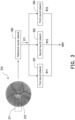
- FIG 3 is a schematic diagram of obtaining a first assessment result of the eye according to an embodiment of the disclosure.
- the processor 104 obtains a first fundus photography 310 shown in Figure 3 in step S210, in which the first fundus photography 310 may include an optic disc image area 311 and an optic cup image area 312, which respectively correspond to the optic disc and the optic cup in the eye A.
- the processor 104 may obtain the optic disc image area 311 from the first fundus photography 310, and generate multiple CDR assessment results by the multiple first models based on the optic disc image area 311.
- the processor 104 may input the first fundus photography 310 into a pre-trained reference neural network 320, in which the reference neural network 320 may identify and output the optic disc image area 311 in response to the first fundus photography 310.
- the reference neural network 320 may first be trained based on training images marked with a specific region of interest (region of interest, ROI), so as to learn the characteristics of these ROIs through the training images. Therefore, in this embodiment, in order for the reference neural network 320 to have the ability to recognize the optic disc image area in any fundus photography, the reference neural network 320 may first learn based on multiple training images (i.e. fundus nibs) marked with optic disc image areas so as to learn the characteristics of the optic disc image areas in the fundus veins, but the disclosure is not limited thereto.
- the processor 104 may provide the optic disc image area 311 output by the reference neural network 320 to the multiple first models so as to generate corresponding CDR assessment results.
- the CDR assessment result of each first model may be that a ratio between an optic cup axial length and an optic disc axial length in the eye A (i.e. CDR) is normal or abnormal, but the disclosure may not be limited thereto.
- the multiple first models under consideration include a first neural network 331, a second neural network 332, and a third neural network 333 shown, but the disclosure is not limited thereto.
- the first neural network 331 may output a CDR assessment result R11 (for example, normal or abnormal) in response to the optic disc image area 311.
- a CDR assessment result R11 for example, normal or abnormal
- the first neural network 331 may first learn based on multiple optic disc image areas having normal CDR so as to learn the characteristics of the optic disc image areas with normal CDR. Moreover, the first neural network 331 may also learn based on multiple optic disc image areas having abnormal CDRs so as to learn the characteristics of the optic disc image areas with abnormal CDRs, but the disclosure is not limited thereto.
- the processor 104 may input the optic disc image area 311 to the second neural network 332, and the second neural network 332 may output the first CDR of the eye A in response to the optic disc image area 311.
- the second neural network 332 may first learn based on multiple optic disc image areas marked with corresponding CDRs so as to learn the characteristics of the optic disc image areas corresponding to various CDRs, but the disclosure is not limited thereto.
- the processor 104 may determine whether the first CDR is higher than a preset threshold (for example, 0.7). In one embodiment, in response to determining that the first CDR is higher than the preset threshold, the processor 104 may determine that a CDR assessment result R12 of the second neural network 332 is abnormal. On the other hand, in response to determining that the first CDR is not higher than the preset threshold, the processor 104 may determine that the CDR assessment result R12 of the second neural network 332 is normal, but the disclosure is not limited thereto.
- a preset threshold for example, 0.7
- the processor 104 may input the optic disc image area 311 to the third neural network 333, and the third neural network 333 may output the optic cup axial length and the optic disc axial length of the eye A in response to the optic disc image area 311.
- the third neural network 333 may first learn based on multiple optic disc image areas marked with corresponding the optic cup axial lengths and the optic disc axial lengths, so as to learn the characteristics of the optic disc image area corresponding to a variety of the optic cup axial lengths and the optic disc axial lengths, but the disclosure it not limited thereto.
- the processor 104 may obtain the second CDR of the eye A accordingly.
- the processor 104 may divide the optic cup axial length by the optic disc axial length so as to obtain the second CDR, but the disclosure is not limited thereto.
- the processor 104 may determine whether the second CDR is higher than the preset threshold (for example, 0.7). In one embodiment, in response to determining that the second CDR is higher than the preset threshold, the processor 104 may determine that a CDR assessment result R13 of the third neural network 333 is abnormal. On the other hand, in response to determining that the second CDR is not higher than the preset threshold, the processor 104 may determine that the CDR assessment result R13 of the third neural network 333 is normal, but the disclosure is not limited thereto.
- the preset threshold for example, 0.7
- the processor 104 may obtain a first assessment result RR1 of the eye A based on the multiple CDR assessment results R11 to R13.
- the processor 104 identifies multiple first results indicating normality in the CDR assessment results R11 to R13, and identifies multiple second results indicating abnormality in the CDR assessment results R11 to R13. Then, in response to determining that the first results are more than the second results, the processor 104 may determine that the first assessment result RR1 indicates normality (that is, the CDR of the eye A is normal). On the other hand, in response to determining that the first results are less than the second results, the processor 104 may determine that the first assessment result RR1 indicates abnormality (that is, the CDR of the eye A is abnormal).
- the processor 104 may make a majority decision based on the CDR assessment results R11 to R13. If there are more first results indicating normality in the CDR assessment results R11 to R13, the processor 104 may determine that the first assessment result RR1 indicates normality; if there are more second results indicating abnormality in the CDR assessment results R11 to R13, the processor 104 may determine that the first assessment result RR1 indicates abnormality, but the disclosure may not be limited thereto.
- step S240 the processor 104 may perform multiple data augmentation operations on the first fundus photography 310 so as to generate multiple second fundus veins respectively corresponding to the multiple data augmentation operations.
- the processor 104 may perform multiple data augmentation operations on the first fundus photography 310 so as to generate multiple second fundus veins respectively corresponding to the multiple data augmentation operations.
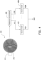
- the processor 104 may perform M data augmentation operations (M is a positive integer) on the first fundus photography 310 so as to generate M second fundus penetrates 411 to 41M.
- M data augmentation operations may include rotating, shifting, flipping, zooming, and stretching the first fundus photography 310, but the disclosure is not limited thereto.
- the processor 104 may input the second fundusticians 411 to 41M to second models 421 to 42M, respectively, so as to output corresponding confidence values of prediction by the second models 421 to 42M as RNFL defect assessment results R21 to R2M.
- the confidence value of prediction corresponding to each of the second models 421 to 42M i.e. the RNFL defect assessment results R21 to R2M
- each of the second models 421 to 42M may first learn based on multiple (data-augmented) fundus penetrates marked with the corresponding abnormalities of RNFLs so as to learn the characteristics of the fundus photography corresponding to the abnormality of a variety of RNFLs, but the disclosure is not limited thereto.
- the processor 104 may obtain a second assessment result RR2 of the eye A based on the multiple RNFL defect assessment results R21 to R2M.
- the processor 104 may obtain a squared difference between the confidence value of prediction corresponding to each of the second models 421 to 42M and a reference base value (for example, 0.5), and identify a candidate model in the second models 421 to 42M accordingly.
- the squared difference corresponding to the candidate model may be the largest among the second models 421 to 42M.
- the processor 104 may determine whether the confidence value of prediction corresponding to the candidate model is higher than the reference base value. If so, the processor 104 may determine that the second assessment result RR2 of the eye A indicates abnormality; if not, the processor 104 may determine that the second assessment result RR2 of the eye A indicates normality.
- the processor 104 may obtain an optic nerve assessment result of the eye A based on the first assessment result RR1 and the second assessment result RR2.
- the optic nerve assessment result of eye A belongs to a first risk degree.
- the optic nerve assessment result of eye A belongs to a second risk degree, in which the second risk degree is greater than the first risk degree.
- the optic nerve assessment result of eye A belongs to a third risk degree, in which the third risk degree is greater than the second risk degree.
- the optic nerve assessment result of eye A belongs to a fourth risk degree, in which the fourth risk degree is greater than the third risk degree.
- the first, second, third, and fourth risk degrees may be understood as the probability of the eye A suffering from glaucoma.
- the optic nerve assessment result of the eye A belongs to the first risk degree, it means that the probability of the eye A suffering from glaucoma is low.
- the optic nerve assessment result of eye A belongs to the fourth risk degree, it means that the probability of the eye A suffering from glaucoma is higher, but the disclosure is not limited thereto.
- the disclosure may present the obtained optic nerve assessment result to the doctor or other relevant personnel as auxiliary information for diagnosing the eye A, but the disclosure is not limited thereto.
- first, second, third, and fourth risk degrees may be roughly understood as normal, low-risk, medium-risk, and high-risk, which may be organized as shown in Table 1 below, but the disclosure is not limited thereto.
- Second assessment result RR2 Optic nerve assessment results normal normal normal abnormal normal low risk normal abnormal medium risk abnormal abnormal high risk
- multiple first models having different recognition mechanisms may individually generate corresponding CDR assessment results based on the optic disc image area, and then a majority decision is made to determine whether the first assessment result indicates normality or abnormality.
- multiple different data augmentations may be performed on the first fundus photography so as to obtain multiple second fundusticians, and then a corresponding RNFL defect result based on one of the second fundusticians may be generated by each second model, and a second assessment result indicating that the RNFL of the eye is normal or abnormal is obtained accordingly.
- comprehensive consideration of the first and second assessment results can be taken so as to obtain the optic nerve assessment result of the eye.
- the disclosure can provide a reference for a doctor when assessing the patient's eye state, thereby helping the doctor to offer an appropriate assessment result to the patient's eye.
- the doctor can make an appropriate assessment of whether or not the patient has glaucoma based on the optic nerve assessment result provided by the embodiment of the disclosure, thereby improving the quality of diagnosis.
Landscapes
- Engineering & Computer Science (AREA)
- Health & Medical Sciences (AREA)
- Life Sciences & Earth Sciences (AREA)
- General Health & Medical Sciences (AREA)
- Medical Informatics (AREA)
- Physics & Mathematics (AREA)
- Public Health (AREA)
- Biomedical Technology (AREA)
- Theoretical Computer Science (AREA)
- Ophthalmology & Optometry (AREA)
- Computer Vision & Pattern Recognition (AREA)
- Animal Behavior & Ethology (AREA)
- Biophysics (AREA)
- Heart & Thoracic Surgery (AREA)
- Surgery (AREA)
- Veterinary Medicine (AREA)
- Molecular Biology (AREA)
- General Physics & Mathematics (AREA)
- Multimedia (AREA)
- Databases & Information Systems (AREA)
- Signal Processing (AREA)
- Radiology & Medical Imaging (AREA)
- Nuclear Medicine, Radiotherapy & Molecular Imaging (AREA)
- Artificial Intelligence (AREA)
- Pathology (AREA)
- Epidemiology (AREA)
- Primary Health Care (AREA)
- Evolutionary Computation (AREA)
- Quality & Reliability (AREA)
- Data Mining & Analysis (AREA)
- Software Systems (AREA)
- Computing Systems (AREA)
- Human Computer Interaction (AREA)
- Physiology (AREA)
- Psychiatry (AREA)
- Eye Examination Apparatus (AREA)
- Testing, Inspecting, Measuring Of Stereoscopic Televisions And Televisions (AREA)
Abstract
Description
- The disclosure relates to a method and an electronic device for assessing the state of the human body, and more particularly to a method and an electronic device for assessing the eye state.
- According to statistics, glaucoma is the second leading cause of blindness in Taiwan and the United States. Generally, glaucoma is diagnosed using optical coherence tomography (OCT) to calculate the thickness of retinal nerve fiber layer (RNFL), but most people do not intentionally take OCT images.
- Compared with the high cost and difficulty of obtaining OCT, color fundus photography is relatively easy to obtain. Many symptoms of eye diseases can be observed in fundus photographies. The cup-to-disc ratio (CDR) and RNFL defect of optic disc to optic cup can be obtained from the fundus photography to determine the degree of glaucoma.
- However, since the degree of glaucoma may vary from doctor to doctor based on fundus photographies, the design of a mechanism to assist the doctor in making the determination is an important issue for those skilled in the art.
- In view of the above, the disclosure provides an eye state assessment method and an electronic device which can be used to solve the above technical problems.
- The disclosure provides an eye state assessment method for an electronic device, including: obtaining a first fundus photography of an eye, in which the first fundus photography includes an optic disc image area; obtaining the optic disc image area from the first fundus photography and generating multiple optic cup-to-disc ratio assessment results by multiple first models based on the optic disc image area; obtaining a first assessment result of the eye based on the optic cup-to-disc ratio assessment results; performing multiple data augmentation operations on the first fundus photography to generate multiple second fundus photographies respectively corresponding to the data augmentation operations; generating multiple retinal nerve fiber layer defect assessment results by multiple second models based on the second fundus photographies; obtaining a second assessment result of the eye based on the multiple retinal nerve fiber layer defect assessment results; and obtaining an optic nerve assessment result of the eye based on the first assessment result and the second assessment result.
- The disclosure provides an electronic device, including a storage circuit and a processor. The storage circuit stores a program code. The processor is coupled to the storage circuit and accesses the program code to execute: obtaining a first fundus photography of an eye, in which the first fundus photography includes an optic disc image area; obtaining the optic disc image area from the first fundus photography and generating multiple optic cup-to-disc ratio assessment results area by multiple first models based on the optic disc image; obtaining a first assessment result of the eye based on the optic cup-to-disc ratio assessment results; performing multiple data augmentation operations on the first fundus photography to generate multiple second fundus photographies respectively corresponding to the data augmentation operations; generating multiple retinal nerve fiber layer defect assessment results by multiple second models based on the second fundus photographies; obtaining a second assessment result of the eye based on the retinal nerve fiber layer defect assessment results; and obtaining an optic nerve assessment result of the eye based on the first assessment result and the second assessment result.
- The accompanying drawings are included to provide a further understanding of the disclosure, and are incorporated in and constitute a part of this specification. The drawings illustrate embodiments of the disclosure and, together with the description, serve to explain the principles of the disclosure.
-
Figure 1 is a schematic diagram of an electronic device according to an embodiment of the disclosure. -
Figure 2 is a flowchart of an eye state assessment method according to an embodiment of the disclosure. -
Figure 3 is a schematic diagram of obtaining a first assessment result of the eye according to an embodiment of the disclosure. -
Figure 4 is a schematic diagram of obtaining a second assessment result of the eye according toFigure 3 . - Reference will now be made in detail to the exemplary embodiments of the disclosure, examples of which are illustrated in the accompanying drawings. Wherever possible, the same reference numbers are used in the drawings and the description to refer to the same or like parts.
- Referring to
Figure 1 , which is a schematic diagram of an electronic device according to an embodiment of the disclosure. In different embodiments, anelectronic device 100 may be, for example, a variety of computer devices and/or smart devices, but the disclosure may not be limited thereto. - As shown in
Figure 1 , theelectronic device 100 may include astorage circuit 102 and aprocessor 104. Thestorage circuit 102 may be, for example, any type of fixed or removable random access memory (RAM), read-only memory (ROM), flash memory, hard disk, or other similar devices or a combination thereof, which may be configured to record multiple codes or modules. - The
processor 104 is coupled to thestorage circuit 102, and may be a general-purpose processor, a special-purpose processor, a traditional processor, a digital signal processor, multiple microprocessors, one or more microprocessors combined with the digital signal processor cores, controllers, microcontrollers, application specific integrated circuits (ASIC), field programmable gate array (FPGA), any other types of integrated circuits, a state machine, or an advanced reduced instruction set computer (Advanced RISC Machine, ARM) and similar products. - In one embodiment of the disclosure, the
processor 104 may access the modules and program codes recorded in thestorage circuit 102 so as to implement the eye state assessment method proposed in the disclosure. The details of the method are as follows. - Referring to
Figure 2 , which is a flowchart of an eye state assessment method according to an embodiment of the disclosure. The method of this embodiment may be executed by theelectronic device 100 ofFigure 1 , and the details of each step inFigure 2 will be explained below with the components shown inFigure 1 . - First, in step S210, the
processor 104 may obtain a first fundus photography of an eye A. In one embodiment, the eye A is, for example, one of the eyes of a certain patient, and the first fundus photography is, for example, a color fundus photography obtained by medical personnel operating an instrument for taking a fundus photography of the eye A, but the disclosure may not be limited thereto. - In one embodiment, since the generally captured fundus photography may include black borders, the
processor 104 may first crop the fundus photography with black borders after obtaining the fundus photography so as to generate a fundus photography without black borders as the first fundus photography, but the disclosure is not limited thereto. In order to make the concept of the disclosure easier to understand, the following will be supplemented withFigure 3 for further explanation. - Referring to
Figure 3 , which is a schematic diagram of obtaining a first assessment result of the eye according to an embodiment of the disclosure. InFigure 3 , it is assumed that theprocessor 104 obtains afirst fundus photography 310 shown inFigure 3 in step S210, in which thefirst fundus photography 310 may include an opticdisc image area 311 and an opticcup image area 312, which respectively correspond to the optic disc and the optic cup in the eye A. - After obtaining the
first fundus photography 310, in step S220, theprocessor 104 may obtain the opticdisc image area 311 from thefirst fundus photography 310, and generate multiple CDR assessment results by the multiple first models based on the opticdisc image area 311. - In one embodiment, the
processor 104 may input thefirst fundus photography 310 into a pre-trained referenceneural network 320, in which the referenceneural network 320 may identify and output the opticdisc image area 311 in response to thefirst fundus photography 310. - In one embodiment, before the reference
neural network 320 is configured to identify the opticdisc image area 311 in thefirst fundus photography 310, the referenceneural network 320 may first be trained based on training images marked with a specific region of interest (region of interest, ROI), so as to learn the characteristics of these ROIs through the training images. Therefore, in this embodiment, in order for the referenceneural network 320 to have the ability to recognize the optic disc image area in any fundus photography, the referenceneural network 320 may first learn based on multiple training images (i.e. fundus photographies) marked with optic disc image areas so as to learn the characteristics of the optic disc image areas in the fundus photographies, but the disclosure is not limited thereto. - After that, the
processor 104 may provide the opticdisc image area 311 output by the referenceneural network 320 to the multiple first models so as to generate corresponding CDR assessment results. In one embodiment of the disclosure, the CDR assessment result of each first model may be that a ratio between an optic cup axial length and an optic disc axial length in the eye A (i.e. CDR) is normal or abnormal, but the disclosure may not be limited thereto. - In the scenario of
Figure 3 , it is assumed that the multiple first models under consideration (the number of which is, for example, N, and N is an odd number) include a firstneural network 331, a secondneural network 332, and a thirdneural network 333 shown, but the disclosure is not limited thereto. - In one embodiment, the first
neural network 331 may output a CDR assessment result R11 (for example, normal or abnormal) in response to the opticdisc image area 311. - In one embodiment, in order for the first
neural network 331 to have the ability to identify whether the corresponding CDR is normal in response to any optic disc image area, the firstneural network 331 may first learn based on multiple optic disc image areas having normal CDR so as to learn the characteristics of the optic disc image areas with normal CDR. Moreover, the firstneural network 331 may also learn based on multiple optic disc image areas having abnormal CDRs so as to learn the characteristics of the optic disc image areas with abnormal CDRs, but the disclosure is not limited thereto. - In one embodiment, the
processor 104 may input the opticdisc image area 311 to the secondneural network 332, and the secondneural network 332 may output the first CDR of the eye A in response to the opticdisc image area 311. - In one embodiment, in order for the second
neural network 332 to have the ability to identify the CDR in response to any optic disc image area, the secondneural network 332 may first learn based on multiple optic disc image areas marked with corresponding CDRs so as to learn the characteristics of the optic disc image areas corresponding to various CDRs, but the disclosure is not limited thereto. - After obtaining the first CDR, the
processor 104 may determine whether the first CDR is higher than a preset threshold (for example, 0.7). In one embodiment, in response to determining that the first CDR is higher than the preset threshold, theprocessor 104 may determine that a CDR assessment result R12 of the secondneural network 332 is abnormal. On the other hand, in response to determining that the first CDR is not higher than the preset threshold, theprocessor 104 may determine that the CDR assessment result R12 of the secondneural network 332 is normal, but the disclosure is not limited thereto. - In one embodiment, the
processor 104 may input the opticdisc image area 311 to the thirdneural network 333, and the thirdneural network 333 may output the optic cup axial length and the optic disc axial length of the eye A in response to the opticdisc image area 311. - In one embodiment, in order for the third
neural network 333 to have the ability to identify the optic cup axial length and the optic disc axial length in response to any optic disc image area, the thirdneural network 333 may first learn based on multiple optic disc image areas marked with corresponding the optic cup axial lengths and the optic disc axial lengths, so as to learn the characteristics of the optic disc image area corresponding to a variety of the optic cup axial lengths and the optic disc axial lengths, but the disclosure it not limited thereto. - After obtaining the optic cup axial length and the optic disc axial length corresponding to the optic
disc image area 311, theprocessor 104 may obtain the second CDR of the eye A accordingly. For example, theprocessor 104 may divide the optic cup axial length by the optic disc axial length so as to obtain the second CDR, but the disclosure is not limited thereto. - Then, the
processor 104 may determine whether the second CDR is higher than the preset threshold (for example, 0.7). In one embodiment, in response to determining that the second CDR is higher than the preset threshold, theprocessor 104 may determine that a CDR assessment result R13 of the thirdneural network 333 is abnormal. On the other hand, in response to determining that the second CDR is not higher than the preset threshold, theprocessor 104 may determine that the CDR assessment result R13 of the thirdneural network 333 is normal, but the disclosure is not limited thereto. - After obtaining the CDR assessment results R11 to R13, in step S230, the
processor 104 may obtain a first assessment result RR1 of the eye A based on the multiple CDR assessment results R11 to R13. - In one embodiment, the
processor 104 identifies multiple first results indicating normality in the CDR assessment results R11 to R13, and identifies multiple second results indicating abnormality in the CDR assessment results R11 to R13. Then, in response to determining that the first results are more than the second results, theprocessor 104 may determine that the first assessment result RR1 indicates normality (that is, the CDR of the eye A is normal). On the other hand, in response to determining that the first results are less than the second results, theprocessor 104 may determine that the first assessment result RR1 indicates abnormality (that is, the CDR of the eye A is abnormal). - In short, the
processor 104 may make a majority decision based on the CDR assessment results R11 to R13. If there are more first results indicating normality in the CDR assessment results R11 to R13, theprocessor 104 may determine that the first assessment result RR1 indicates normality; if there are more second results indicating abnormality in the CDR assessment results R11 to R13, theprocessor 104 may determine that the first assessment result RR1 indicates abnormality, but the disclosure may not be limited thereto. - Further, in step S240, the
processor 104 may perform multiple data augmentation operations on thefirst fundus photography 310 so as to generate multiple second fundus photographies respectively corresponding to the multiple data augmentation operations. In order to make the concept of the disclosure easier to understand, the following will be supplemented withFigure 3 for further explanation. - Referring to
Figure 4 , which is a schematic diagram of obtaining a second assessment result of the eye according toFigure 3 . InFigure 4 , theprocessor 104 may perform M data augmentation operations (M is a positive integer) on thefirst fundus photography 310 so as to generate Msecond fundus photographies 411 to 41M. The M data augmentation operations may include rotating, shifting, flipping, zooming, and stretching thefirst fundus photography 310, but the disclosure is not limited thereto. - After that, in
step 250, theprocessor 104 may input thesecond fundus photographies 411 to 41M tosecond models 421 to 42M, respectively, so as to output corresponding confidence values of prediction by thesecond models 421 to 42M as RNFL defect assessment results R21 to R2M. In one embodiment, the confidence value of prediction corresponding to each of thesecond models 421 to 42M (i.e. the RNFL defect assessment results R21 to R2M) may indicate the degree of abnormality of the RNFL of the eye A. - In one embodiment, in order for each of the
second models 421 to 42M to have the ability to identify the degree of abnormality of the corresponding RNFL in response to the fundus photography in which data has been augmented, each of thesecond models 421 to 42M may first learn based on multiple (data-augmented) fundus photographies marked with the corresponding abnormalities of RNFLs so as to learn the characteristics of the fundus photography corresponding to the abnormality of a variety of RNFLs, but the disclosure is not limited thereto. - After obtaining the confidence value of prediction of each of the
second models 421 to 42M (that is, the RNFL defect assessment results R21 to R2M), in step S260, theprocessor 104 may obtain a second assessment result RR2 of the eye A based on the multiple RNFL defect assessment results R21 to R2M. - In one embodiment, the
processor 104 may obtain a squared difference between the confidence value of prediction corresponding to each of thesecond models 421 to 42M and a reference base value (for example, 0.5), and identify a candidate model in thesecond models 421 to 42M accordingly. In one embodiment, the squared difference corresponding to the candidate model may be the largest among thesecond models 421 to 42M. - After that, the
processor 104 may determine whether the confidence value of prediction corresponding to the candidate model is higher than the reference base value. If so, theprocessor 104 may determine that the second assessment result RR2 of the eye A indicates abnormality; if not, theprocessor 104 may determine that the second assessment result RR2 of the eye A indicates normality. - After obtaining the first assessment result RR1 (which may indicate normal or abnormal) and the second assessment result RR2 (which may indicate normal or abnormal) of the eye A, in step S270, the
processor 104 may obtain an optic nerve assessment result of the eye A based on the first assessment result RR1 and the second assessment result RR2. - In one embodiment, in response to determining that the first assessment result RR1 indicates normality and the second assessment result RR2 indicates normality, it is determined that the optic nerve assessment result of eye A belongs to a first risk degree. In response to determining that the first assessment result RR1 indicates abnormality and the second assessment result RR2 indicates normality, it is determined that the optic nerve assessment result of eye A belongs to a second risk degree, in which the second risk degree is greater than the first risk degree. In response to determining that the first assessment result RR1 indicates normality and the second assessment result RR2 indicates abnormality, it is determined that the optic nerve assessment result of eye A belongs to a third risk degree, in which the third risk degree is greater than the second risk degree. In response to determining that the first assessment result RR1 indicates abnormality and the second assessment result RR2 indicates abnormality, it is determined that the optic nerve assessment result of eye A belongs to a fourth risk degree, in which the fourth risk degree is greater than the third risk degree.
- In one embodiment, the first, second, third, and fourth risk degrees may be understood as the probability of the eye A suffering from glaucoma. For example, when the optic nerve assessment result of the eye A belongs to the first risk degree, it means that the probability of the eye A suffering from glaucoma is low. On the other hand, when the optic nerve assessment result of eye A belongs to the fourth risk degree, it means that the probability of the eye A suffering from glaucoma is higher, but the disclosure is not limited thereto. In this way, the disclosure may present the obtained optic nerve assessment result to the doctor or other relevant personnel as auxiliary information for diagnosing the eye A, but the disclosure is not limited thereto.
- In one embodiment, the first, second, third, and fourth risk degrees may be roughly understood as normal, low-risk, medium-risk, and high-risk, which may be organized as shown in Table 1 below, but the disclosure is not limited thereto.
First assessment result RR1 Second assessment result RR2 Optic nerve assessment results normal normal normal abnormal normal low risk normal abnormal medium risk abnormal abnormal high risk - In summary, according to the disclosure, after obtaining the optic disc image area, multiple first models having different recognition mechanisms may individually generate corresponding CDR assessment results based on the optic disc image area, and then a majority decision is made to determine whether the first assessment result indicates normality or abnormality. Further, according to disclosure, multiple different data augmentations may be performed on the first fundus photography so as to obtain multiple second fundus photographies, and then a corresponding RNFL defect result based on one of the second fundus photographies may be generated by each second model, and a second assessment result indicating that the RNFL of the eye is normal or abnormal is obtained accordingly. After that, according to the disclosure, comprehensive consideration of the first and second assessment results can be taken so as to obtain the optic nerve assessment result of the eye. In this way, the disclosure can provide a reference for a doctor when assessing the patient's eye state, thereby helping the doctor to offer an appropriate assessment result to the patient's eye. For example, the doctor can make an appropriate assessment of whether or not the patient has glaucoma based on the optic nerve assessment result provided by the embodiment of the disclosure, thereby improving the quality of diagnosis.
- It will be apparent to those skilled in the art that various modifications and variations may be made to the structure of the disclosure without departing from the scope or spirit of the disclosure. In view of the foregoing, it is intended that the disclosure cover modifications and variations of this disclosure provided they fall within the scope of the following claims and their equivalents.
Claims (12)
- An eye state assessment method for an electronic device (100), comprising:obtaining a first fundus photography (310) of an eye (A), wherein the first fundus photography (310) comprises an optic disc image area (311);obtaining the optic disc image area (311) from the first fundus photography (310) and generating a plurality of optic cup-to-disc ratio assessment results by a plurality of first models based on the optic disc image area (311);obtaining a first assessment result (RR1) of the eye (A) based on the optic cup-to-disc ratio assessment results;performing a plurality of data augmentation operations on the first fundus photography (310) to generate a plurality of second fundus photographies (411-41M) respectively corresponding to the data augmentation operations;generating a plurality of retinal nerve fiber layer defect assessment results by a plurality of second models (421-42M) based on the second fundus photographies (411-41M);obtaining a second assessment result (RR2) of the eye (A) based on the retinal nerve fiber layer defect assessment results; andobtaining an optic nerve assessment result of the eye (A) based on the first assessment result (RR1) and the second assessment result (RR2).
- The method according to claim 1, wherein the first fundus photography (310) comprises a color fundus photography of the eye (A), and the color fundus photography does not have black borders.
- The method according to claim 1, wherein a step of obtaining the optic disc image area (311) from the first fundus photography (310) comprises:
inputting the first fundus photography (310) to a reference neural network (320), wherein the reference neural network (320) recognizes the optic disc image area (311) in the first fundus photography (310) in response to the first fundus photography (310), and outputs the optic disc image area (311). - The method according to claim 1, wherein the first models comprise N neural networks (331-333), and each of the optic cup-to-disc ratio assessment results indicates whether an optic cup-to-disc ratio of the eye (A) is normal or abnormal, wherein N is an odd number.
- The method according to claim 4, wherein a step of obtaining the first assessment result (RR1) of the eye (A) based on the optic cup-to-disc ratio assessment results comprises:identifying a plurality of first results indicating normality from the optic cup-to-disc ratio assessment results, and identifying a plurality of second results indicating abnormality from the optic cup-to-disc ratio assessment results;determining that the first assessment result (RR1) indicates normality in response to determining that the first results are more than the second results; anddetermining that the first assessment result (RR1) indicates abnormality in response to determining that the first results are less than the second results.
- The method according to claim 1, wherein the first models comprise a first neural network (331), and the method comprises:
inputting the optic disc image area (311) to the first neural network (331), wherein the first neural network (331) outputs one of the optic cup-to-disc ratio assessment results in response to the optic disc image area (311). - The method according to claim 1, wherein the first models comprise a second neural network (332), and the method comprises:inputting the optic disc image area (311) into the second neural network (332), wherein the second neural network (332) outputs a first optic cup-to-disc ratio of the eye (A) in response to the optic disc image area (311);determining that one of the optic cup-to-disc ratio assessment results corresponding to the second neural network (332) indicates abnormality in response to determining that the first optic cup-to-disc ratio is higher than a preset threshold; anddetermining that one of the optic cup-to-disc ratio assessment results corresponding to the second neural network (332) indicates normality in response to determining that the first optic cup-to-disc ratio is not higher than the preset threshold.
- The method according to claim 1, wherein the optic disc image area (311) comprises an optic cup image area (312), the first models comprise a third neural network (333), and the method comprises:inputting the optic disc image area (311) into the third neural network (333), wherein the third neural network (333) outputs an optic cup axial length of the eye (A) in response to the optic cup image area (312), and outputs an optic disc axial length of the eye (A) in response to the optic disc image area (311);obtaining a second optic cup-to-disc ratio of the eye (A) based on the optic cup axial length and the optic disc axial length;determining that one of the optic cup-to-disc ratio assessment results corresponding to the third neural network (333) indicates abnormality in response to determining that the second optic cup-to-disc ratio is higher than a preset threshold; anddetermining that one of the optic cup-to-disc ratio assessment results corresponding to the third neural network (333) indicates normality in response to determining that the second optic cup-to-disc ratio is not higher than the preset threshold.
- The method according to claim 1, wherein a step of generating the retinal nerve fiber layer defect assessment results by the second models (421-42M) based on the second fundus photographies (411-41M) comprises:inputting one of the second fundus photographies (411-41M) into one of the second models (421-42M), wherein the one of the second models (421-42M) outputs a confidence value of prediction as one of the retinal nerve fiber layer defect assessment results in response to the one of the second fundus photographies (411-41M), wherein the confidence value of prediction indicates a degree of abnormality of the eye (A).
- The method according to claim 9, wherein a step of obtaining the second assessment result (RR2) of the eye (A) based on the retinal nerve fiber layer defect assessment results comprises:obtaining a square difference between the confidence value of prediction corresponding to each of the second models (421-42M) and a reference base value, and identifying a candidate model in the second models (421-42M) accordingly, wherein the square difference corresponding to the candidate model is the largest among the second modes;determining that the second assessment result (RR2) of the eye (A) indicates abnormality in response to determining that the confidence value of prediction corresponding to the candidate model is higher than the reference base value; anddetermining that the second assessment result (RR2) of the eye (A) indicates normality in response to determining that the confidence value of prediction corresponding to the candidate model is not higher than the reference base value.
- The method described in claim 1, wherein the first assessment result (RR1) indicates normality or abnormality, the second assessment result (RR2) indicates normality or abnormality, and a step of obtaining an optic nerve assessment result of the eye (A) based on the first assessment result (RR1) and the second assessment result (RR2) comprises:determining that the optic nerve assessment result of the eye (A) belongs to a first risk degree in response to determining that the first assessment result (RR1) indicates normality and the second assessment result (RR2) indicates normality;determining that the optic nerve assessment result of the eye (A) belongs to a second risk degree in response to determining that the first assessment result (RR1) indicates abnormality and the second assessment result (RR2) indicates normality, wherein the second risk degree is greater than the first risk degree;determining that the optic nerve assessment result of the eye (A) belongs to a third risk degree in response to determining that the first assessment result (RR1) indicates normality and the second assessment result (RR2) indicates abnormality, wherein the third risk degree is greater than the second risk degree; anddetermining that the optic nerve assessment result of the eye (A) belongs to a fourth risk degree in response to determining that the first assessment result (RR1) indicates abnormality and the second assessment result (RR2) indicates abnormality, wherein the fourth risk degree is greater than the third risk degree.
- An electronic device (100), comprising:a storage circuit (102), storing a program code;a processor (104), coupled to the storage circuit (102) and accessing the program code to execute:obtaining a first fundus photography (310) of an eye (A), wherein the first fundus photography (310) comprises an optic disc image area (311);obtaining the optic disc image area (311) from the first fundus photography (310) and generating a plurality of optic cup-to-disc ratio assessment results by a plurality of first models based on the optic disc image area (311);obtaining a first assessment result (RR1) of the eye (A) based on the optic cup-to-disc ratio assessment results;performing a plurality of data augmentation operations on the first fundus photography (310) to generate a plurality of second fundus photographies (411-41M) respectively corresponding to the data augmentation operations;generating a plurality of retinal nerve fiber layer defect assessment results by a plurality of second models (421-42M) based on the second fundus photographies (411-41M);obtaining a second assessment result (RR2) of the eye (A) based on the retinal nerve fiber layer defect assessment results; andobtaining an optic nerve assessment result of the eye (A) based on the first assessment result (RR1) and the second assessment result (RR2).
Applications Claiming Priority (1)
| Application Number | Priority Date | Filing Date | Title |
|---|---|---|---|
| TW110131705A TWI784688B (en) | 2021-08-26 | 2021-08-26 | Eye state assessment method and electronic device |
Publications (2)
| Publication Number | Publication Date |
|---|---|
| EP4140391A1 true EP4140391A1 (en) | 2023-03-01 |
| EP4140391B1 EP4140391B1 (en) | 2024-02-28 |
Family
ID=78695561
Family Applications (1)
| Application Number | Title | Priority Date | Filing Date |
|---|---|---|---|
| EP21208958.5A Active EP4140391B1 (en) | 2021-08-26 | 2021-11-18 | Eye state assessment method and electronic device |
Country Status (3)
| Country | Link |
|---|---|
| US (1) | US12106472B2 (en) |
| EP (1) | EP4140391B1 (en) |
| TW (1) | TWI784688B (en) |
Cited By (2)
| Publication number | Priority date | Publication date | Assignee | Title |
|---|---|---|---|---|
| CN117084622A (en) * | 2023-10-16 | 2023-11-21 | 罗麦(北京)营养食品研究有限公司 | Eye-based health analysis method, system, equipment and storage medium |
| TWI906180B (en) * | 2025-05-15 | 2025-11-21 | 亞東學校財團法人亞東科技大學 | Eye detecting system and detecting method |
Families Citing this family (1)
| Publication number | Priority date | Publication date | Assignee | Title |
|---|---|---|---|---|
| US12646175B2 (en) * | 2023-01-28 | 2026-06-02 | Sankar Foundation | Multivariable artificial intelligence (AI) based monitoring system for early detection of glaucoma |
Citations (2)
| Publication number | Priority date | Publication date | Assignee | Title |
|---|---|---|---|---|
| US20160100753A1 (en) * | 2009-11-16 | 2016-04-14 | Jiang Liu | Obtaining data for automatic glaucoma screening, and screening and diagnostic techniques and systems using the data |
| US20200401841A1 (en) * | 2019-06-24 | 2020-12-24 | Samsung Sds Co., Ltd. | Apparatus for diagnosing glaucoma |
Family Cites Families (12)
| Publication number | Priority date | Publication date | Assignee | Title |
|---|---|---|---|---|
| US11839430B2 (en) * | 2008-03-27 | 2023-12-12 | Doheny Eye Institute | Optical coherence tomography-based ophthalmic testing methods, devices and systems |
| WO2016032397A1 (en) * | 2014-08-25 | 2016-03-03 | Agency For Science, Technology And Research (A*Star) | Methods and systems for assessing retinal images, and obtaining information from retinal images |
| US10210955B1 (en) * | 2017-08-16 | 2019-02-19 | Arsh Banerjee | Methods for diagnosing zika virus |
| WO2019083129A1 (en) * | 2017-10-27 | 2019-05-02 | 주식회사 뷰노 | Method for supporting reading of fundus image of subject, and device using same |
| CN110930446B (en) * | 2018-08-31 | 2024-03-19 | 福州依影健康科技有限公司 | A pre-processing method and storage device for quantitative analysis of fundus images |
| CN109411086A (en) * | 2018-12-26 | 2019-03-01 | 深圳硅基智能科技有限公司 | The screening system of eye fundus image lesion |
| CN110211087B (en) * | 2019-01-28 | 2023-06-30 | 南通大学 | Shareable semi-automatic diabetic retinopathy labeling method |
| CN112602114B (en) * | 2019-08-01 | 2024-07-23 | 京东方科技集团股份有限公司 | Image processing method and device, neural network and training method, storage medium |
| CN110555845B (en) * | 2019-09-27 | 2024-09-17 | 北京鹰瞳远见信息科技有限公司 | Fundus OCT image recognition method and device |
| GB202000926D0 (en) * | 2020-01-22 | 2020-03-04 | Ucl Business Ltd | Method of diagnosis |
| TWI746287B (en) * | 2020-11-26 | 2021-11-11 | 宏碁股份有限公司 | Data storage system and data storage method |
| US11610306B2 (en) * | 2020-12-16 | 2023-03-21 | Industrial Technology Research Institute | Medical image analysis method and device |
-
2021
- 2021-08-26 TW TW110131705A patent/TWI784688B/en active
- 2021-10-21 US US17/506,703 patent/US12106472B2/en active Active
- 2021-11-18 EP EP21208958.5A patent/EP4140391B1/en active Active
Patent Citations (2)
| Publication number | Priority date | Publication date | Assignee | Title |
|---|---|---|---|---|
| US20160100753A1 (en) * | 2009-11-16 | 2016-04-14 | Jiang Liu | Obtaining data for automatic glaucoma screening, and screening and diagnostic techniques and systems using the data |
| US20200401841A1 (en) * | 2019-06-24 | 2020-12-24 | Samsung Sds Co., Ltd. | Apparatus for diagnosing glaucoma |
Non-Patent Citations (3)
| Title |
|---|
| KO YU-CHIEH ET AL: "Deep learning assisted detection of glaucomatous optic neuropathy and potential designs for a generalizable model", PLOS ONE, 14 May 2020 (2020-05-14), San Francisco, pages 1 - 15, XP055915872, Retrieved from the Internet <URL:https://www.ncbi.nlm.nih.gov/pmc/articles/PMC7224540/pdf/pone.0233079.pdf> [retrieved on 20220426], DOI: 10.1371/journal.pone.0233079 * |
| MOSHKOV NIKITA ET AL: "Test-time augmentation for deep learning-based cell segmentation on microscopy images", SCIENTIFIC REPORTS, vol. 10, 5068, 19 March 2020 (2020-03-19), XP055923179, Retrieved from the Internet <URL:https://www.nature.com/articles/s41598-020-61808-3.pdf> [retrieved on 20220519], DOI: 10.1038/s41598-020-61808-3 * |
| SHINDE RUTUJA: "Glaucoma detection in retinal fundus images using U-Net and supervised machine learning algorithms", INTELLIGENCE-BASED MEDICINE, vol. 5, 14 July 2021 (2021-07-14), pages 100038, XP055915873, ISSN: 2666-5212, DOI: 10.1016/j.ibmed.2021.100038 * |
Cited By (3)
| Publication number | Priority date | Publication date | Assignee | Title |
|---|---|---|---|---|
| CN117084622A (en) * | 2023-10-16 | 2023-11-21 | 罗麦(北京)营养食品研究有限公司 | Eye-based health analysis method, system, equipment and storage medium |
| CN117084622B (en) * | 2023-10-16 | 2024-02-02 | 罗麦(北京)营养食品研究有限公司 | Eye-based health analysis method, system, equipment and storage medium |
| TWI906180B (en) * | 2025-05-15 | 2025-11-21 | 亞東學校財團法人亞東科技大學 | Eye detecting system and detecting method |
Also Published As
| Publication number | Publication date |
|---|---|
| EP4140391B1 (en) | 2024-02-28 |
| US12106472B2 (en) | 2024-10-01 |
| TWI784688B (en) | 2022-11-21 |
| US20230066166A1 (en) | 2023-03-02 |
| TW202309924A (en) | 2023-03-01 |
Similar Documents
| Publication | Publication Date | Title |
|---|---|---|
| EP4140391B1 (en) | Eye state assessment method and electronic device | |
| Singh et al. | Deep learning system applicability for rapid glaucoma prediction from fundus images across various data sets | |
| Khaing et al. | ChoroidNET: a dense dilated U-Net model for choroid layer and vessel segmentation in optical coherence tomography images | |
| CN110163844B (en) | Fundus focus detection method, fundus focus detection device, fundus focus detection computer device and fundus focus storage medium | |
| El-Ateif et al. | Eye diseases diagnosis using deep learning and multimodal medical eye imaging | |
| CN116563932A (en) | Eye image recognition method and related equipment based on multi-task learning | |
| CN110619332A (en) | Data processing method, device and equipment based on visual field inspection report | |
| US12573032B2 (en) | System and methods of predicting Parkinson's disease based on retinal images using machine learning | |
| Singh et al. | A pre-trained VGG16 model for cataract eye disease prediction | |
| Zhou et al. | A multi-class fundus disease classification system based on an adaptive scale discriminator and hybrid loss | |
| CN113052012B (en) | Eye disease image identification method and system based on improved D-S evidence | |
| El-Hoseny et al. | Optimized deep learning approach for efficient diabetic retinopathy classification combining VGG16-CNN | |
| Muchori et al. | Diabetic macular edema detection based on non-mydriatic retinal image | |
| US20250014751A1 (en) | Disease diagnosis method using trained model, and system carrying out same | |
| CN115778314B (en) | Eye condition assessment methods and electronic devices | |
| Vijaya et al. | Machine learning in automatic diabetic retinopathy detection and classification systems: a survey and comparison of methods. | |
| US20260044956A1 (en) | A method and system for staging diabetic kidney disease using deep learning | |
| Mandiga et al. | Retinal blindness detection due to diabetes using MobileNetV2 and SVM | |
| Wang et al. | Two-stage cross-domain ocular disease recognition with data augmentation | |
| US11744457B2 (en) | Method for measuring anomalies of refraction using a reflection image of pupil in visible light | |
| US20220165421A1 (en) | Machine learning model based method and analysis system for performing covid-19 testing according to eye image captured by smartphone | |
| KR102919505B1 (en) | Personalized examination recommendation and diagnosis assistance method and system using ai analysis of retinal photography | |
| Shourie et al. | OCTNet: leveraging VGG16 for automated classification of retina health from OCT scans | |
| CN118279702B (en) | Tear overflow detection model construction method, tear overflow detection method, device and equipment | |
| Vaish et al. | Improving classification accuracy for diabetic retinopathy with deep learning |
Legal Events
| Date | Code | Title | Description |
|---|---|---|---|
| PUAI | Public reference made under article 153(3) epc to a published international application that has entered the european phase |
Free format text: ORIGINAL CODE: 0009012 |
|
| STAA | Information on the status of an ep patent application or granted ep patent |
Free format text: STATUS: REQUEST FOR EXAMINATION WAS MADE |
|
| 17P | Request for examination filed |
Effective date: 20220824 |
|
| AK | Designated contracting states |
Kind code of ref document: A1 Designated state(s): AL AT BE BG CH CY CZ DE DK EE ES FI FR GB GR HR HU IE IS IT LI LT LU LV MC MK MT NL NO PL PT RO RS SE SI SK SM TR |
|
| GRAP | Despatch of communication of intention to grant a patent |
Free format text: ORIGINAL CODE: EPIDOSNIGR1 |
|
| STAA | Information on the status of an ep patent application or granted ep patent |
Free format text: STATUS: GRANT OF PATENT IS INTENDED |
|
| RIC1 | Information provided on ipc code assigned before grant |
Ipc: G16H 50/30 20180101ALI20230830BHEP Ipc: G16H 30/40 20180101ALI20230830BHEP Ipc: G06V 10/80 20220101ALI20230830BHEP Ipc: A61B 3/00 20060101ALI20230830BHEP Ipc: G06V 10/82 20220101ALI20230830BHEP Ipc: G16H 50/20 20180101ALI20230830BHEP Ipc: G06V 40/18 20220101ALI20230830BHEP Ipc: G06T 7/62 20170101ALI20230830BHEP Ipc: G06T 7/00 20170101ALI20230830BHEP Ipc: A61B 3/12 20060101AFI20230830BHEP |
|
| INTG | Intention to grant announced |
Effective date: 20230919 |
|
| GRAS | Grant fee paid |
Free format text: ORIGINAL CODE: EPIDOSNIGR3 |
|
| GRAA | (expected) grant |
Free format text: ORIGINAL CODE: 0009210 |
|
| STAA | Information on the status of an ep patent application or granted ep patent |
Free format text: STATUS: THE PATENT HAS BEEN GRANTED |
|
| AK | Designated contracting states |
Kind code of ref document: B1 Designated state(s): AL AT BE BG CH CY CZ DE DK EE ES FI FR GB GR HR HU IE IS IT LI LT LU LV MC MK MT NL NO PL PT RO RS SE SI SK SM TR |
|
| REG | Reference to a national code |
Ref country code: GB Ref legal event code: FG4D |
|
| REG | Reference to a national code |
Ref country code: CH Ref legal event code: EP |
|
| REG | Reference to a national code |
Ref country code: DE Ref legal event code: R096 Ref document number: 602021009807 Country of ref document: DE |
|
| REG | Reference to a national code |
Ref country code: IE Ref legal event code: FG4D |
|
| REG | Reference to a national code |
Ref country code: LT Ref legal event code: MG9D |
|
| PG25 | Lapsed in a contracting state [announced via postgrant information from national office to epo] |
Ref country code: IS Free format text: LAPSE BECAUSE OF FAILURE TO SUBMIT A TRANSLATION OF THE DESCRIPTION OR TO PAY THE FEE WITHIN THE PRESCRIBED TIME-LIMIT Effective date: 20240628 |
|
| REG | Reference to a national code |
Ref country code: NL Ref legal event code: MP Effective date: 20240228 |
|
| PG25 | Lapsed in a contracting state [announced via postgrant information from national office to epo] |
Ref country code: LT Free format text: LAPSE BECAUSE OF FAILURE TO SUBMIT A TRANSLATION OF THE DESCRIPTION OR TO PAY THE FEE WITHIN THE PRESCRIBED TIME-LIMIT Effective date: 20240228 |
|
| PG25 | Lapsed in a contracting state [announced via postgrant information from national office to epo] |
Ref country code: GR Free format text: LAPSE BECAUSE OF FAILURE TO SUBMIT A TRANSLATION OF THE DESCRIPTION OR TO PAY THE FEE WITHIN THE PRESCRIBED TIME-LIMIT Effective date: 20240529 |
|
| PG25 | Lapsed in a contracting state [announced via postgrant information from national office to epo] |
Ref country code: HR Free format text: LAPSE BECAUSE OF FAILURE TO SUBMIT A TRANSLATION OF THE DESCRIPTION OR TO PAY THE FEE WITHIN THE PRESCRIBED TIME-LIMIT Effective date: 20240228 Ref country code: NL Free format text: LAPSE BECAUSE OF FAILURE TO SUBMIT A TRANSLATION OF THE DESCRIPTION OR TO PAY THE FEE WITHIN THE PRESCRIBED TIME-LIMIT Effective date: 20240228 Ref country code: RS Free format text: LAPSE BECAUSE OF FAILURE TO SUBMIT A TRANSLATION OF THE DESCRIPTION OR TO PAY THE FEE WITHIN THE PRESCRIBED TIME-LIMIT Effective date: 20240528 |
|
| PG25 | Lapsed in a contracting state [announced via postgrant information from national office to epo] |
Ref country code: ES Free format text: LAPSE BECAUSE OF FAILURE TO SUBMIT A TRANSLATION OF THE DESCRIPTION OR TO PAY THE FEE WITHIN THE PRESCRIBED TIME-LIMIT Effective date: 20240228 |
|
| PG25 | Lapsed in a contracting state [announced via postgrant information from national office to epo] |
Ref country code: RS Free format text: LAPSE BECAUSE OF FAILURE TO SUBMIT A TRANSLATION OF THE DESCRIPTION OR TO PAY THE FEE WITHIN THE PRESCRIBED TIME-LIMIT Effective date: 20240528 Ref country code: NO Free format text: LAPSE BECAUSE OF FAILURE TO SUBMIT A TRANSLATION OF THE DESCRIPTION OR TO PAY THE FEE WITHIN THE PRESCRIBED TIME-LIMIT Effective date: 20240528 Ref country code: NL Free format text: LAPSE BECAUSE OF FAILURE TO SUBMIT A TRANSLATION OF THE DESCRIPTION OR TO PAY THE FEE WITHIN THE PRESCRIBED TIME-LIMIT Effective date: 20240228 Ref country code: LT Free format text: LAPSE BECAUSE OF FAILURE TO SUBMIT A TRANSLATION OF THE DESCRIPTION OR TO PAY THE FEE WITHIN THE PRESCRIBED TIME-LIMIT Effective date: 20240228 Ref country code: IS Free format text: LAPSE BECAUSE OF FAILURE TO SUBMIT A TRANSLATION OF THE DESCRIPTION OR TO PAY THE FEE WITHIN THE PRESCRIBED TIME-LIMIT Effective date: 20240628 Ref country code: HR Free format text: LAPSE BECAUSE OF FAILURE TO SUBMIT A TRANSLATION OF THE DESCRIPTION OR TO PAY THE FEE WITHIN THE PRESCRIBED TIME-LIMIT Effective date: 20240228 Ref country code: GR Free format text: LAPSE BECAUSE OF FAILURE TO SUBMIT A TRANSLATION OF THE DESCRIPTION OR TO PAY THE FEE WITHIN THE PRESCRIBED TIME-LIMIT Effective date: 20240529 Ref country code: FI Free format text: LAPSE BECAUSE OF FAILURE TO SUBMIT A TRANSLATION OF THE DESCRIPTION OR TO PAY THE FEE WITHIN THE PRESCRIBED TIME-LIMIT Effective date: 20240228 Ref country code: ES Free format text: LAPSE BECAUSE OF FAILURE TO SUBMIT A TRANSLATION OF THE DESCRIPTION OR TO PAY THE FEE WITHIN THE PRESCRIBED TIME-LIMIT Effective date: 20240228 Ref country code: BG Free format text: LAPSE BECAUSE OF FAILURE TO SUBMIT A TRANSLATION OF THE DESCRIPTION OR TO PAY THE FEE WITHIN THE PRESCRIBED TIME-LIMIT Effective date: 20240228 |
|
| PG25 | Lapsed in a contracting state [announced via postgrant information from national office to epo] |
Ref country code: PT Free format text: LAPSE BECAUSE OF FAILURE TO SUBMIT A TRANSLATION OF THE DESCRIPTION OR TO PAY THE FEE WITHIN THE PRESCRIBED TIME-LIMIT Effective date: 20240628 Ref country code: PL Free format text: LAPSE BECAUSE OF FAILURE TO SUBMIT A TRANSLATION OF THE DESCRIPTION OR TO PAY THE FEE WITHIN THE PRESCRIBED TIME-LIMIT Effective date: 20240228 |
|
| REG | Reference to a national code |
Ref country code: AT Ref legal event code: MK05 Ref document number: 1660408 Country of ref document: AT Kind code of ref document: T Effective date: 20240228 |
|
| PG25 | Lapsed in a contracting state [announced via postgrant information from national office to epo] |
Ref country code: SE Free format text: LAPSE BECAUSE OF FAILURE TO SUBMIT A TRANSLATION OF THE DESCRIPTION OR TO PAY THE FEE WITHIN THE PRESCRIBED TIME-LIMIT Effective date: 20240228 Ref country code: PT Free format text: LAPSE BECAUSE OF FAILURE TO SUBMIT A TRANSLATION OF THE DESCRIPTION OR TO PAY THE FEE WITHIN THE PRESCRIBED TIME-LIMIT Effective date: 20240628 Ref country code: PL Free format text: LAPSE BECAUSE OF FAILURE TO SUBMIT A TRANSLATION OF THE DESCRIPTION OR TO PAY THE FEE WITHIN THE PRESCRIBED TIME-LIMIT Effective date: 20240228 Ref country code: LV Free format text: LAPSE BECAUSE OF FAILURE TO SUBMIT A TRANSLATION OF THE DESCRIPTION OR TO PAY THE FEE WITHIN THE PRESCRIBED TIME-LIMIT Effective date: 20240228 |
|
| PG25 | Lapsed in a contracting state [announced via postgrant information from national office to epo] |
Ref country code: DK Free format text: LAPSE BECAUSE OF FAILURE TO SUBMIT A TRANSLATION OF THE DESCRIPTION OR TO PAY THE FEE WITHIN THE PRESCRIBED TIME-LIMIT Effective date: 20240228 |
|
| PG25 | Lapsed in a contracting state [announced via postgrant information from national office to epo] |
Ref country code: SM Free format text: LAPSE BECAUSE OF FAILURE TO SUBMIT A TRANSLATION OF THE DESCRIPTION OR TO PAY THE FEE WITHIN THE PRESCRIBED TIME-LIMIT Effective date: 20240228 |
|
| PG25 | Lapsed in a contracting state [announced via postgrant information from national office to epo] |
Ref country code: CZ Free format text: LAPSE BECAUSE OF FAILURE TO SUBMIT A TRANSLATION OF THE DESCRIPTION OR TO PAY THE FEE WITHIN THE PRESCRIBED TIME-LIMIT Effective date: 20240228 Ref country code: EE Free format text: LAPSE BECAUSE OF FAILURE TO SUBMIT A TRANSLATION OF THE DESCRIPTION OR TO PAY THE FEE WITHIN THE PRESCRIBED TIME-LIMIT Effective date: 20240228 |
|
| PG25 | Lapsed in a contracting state [announced via postgrant information from national office to epo] |
Ref country code: AT Free format text: LAPSE BECAUSE OF FAILURE TO SUBMIT A TRANSLATION OF THE DESCRIPTION OR TO PAY THE FEE WITHIN THE PRESCRIBED TIME-LIMIT Effective date: 20240228 |
|
| PG25 | Lapsed in a contracting state [announced via postgrant information from national office to epo] |
Ref country code: SK Free format text: LAPSE BECAUSE OF FAILURE TO SUBMIT A TRANSLATION OF THE DESCRIPTION OR TO PAY THE FEE WITHIN THE PRESCRIBED TIME-LIMIT Effective date: 20240228 |
|
| PG25 | Lapsed in a contracting state [announced via postgrant information from national office to epo] |
Ref country code: SM Free format text: LAPSE BECAUSE OF FAILURE TO SUBMIT A TRANSLATION OF THE DESCRIPTION OR TO PAY THE FEE WITHIN THE PRESCRIBED TIME-LIMIT Effective date: 20240228 Ref country code: SK Free format text: LAPSE BECAUSE OF FAILURE TO SUBMIT A TRANSLATION OF THE DESCRIPTION OR TO PAY THE FEE WITHIN THE PRESCRIBED TIME-LIMIT Effective date: 20240228 Ref country code: RO Free format text: LAPSE BECAUSE OF FAILURE TO SUBMIT A TRANSLATION OF THE DESCRIPTION OR TO PAY THE FEE WITHIN THE PRESCRIBED TIME-LIMIT Effective date: 20240228 Ref country code: EE Free format text: LAPSE BECAUSE OF FAILURE TO SUBMIT A TRANSLATION OF THE DESCRIPTION OR TO PAY THE FEE WITHIN THE PRESCRIBED TIME-LIMIT Effective date: 20240228 Ref country code: DK Free format text: LAPSE BECAUSE OF FAILURE TO SUBMIT A TRANSLATION OF THE DESCRIPTION OR TO PAY THE FEE WITHIN THE PRESCRIBED TIME-LIMIT Effective date: 20240228 Ref country code: CZ Free format text: LAPSE BECAUSE OF FAILURE TO SUBMIT A TRANSLATION OF THE DESCRIPTION OR TO PAY THE FEE WITHIN THE PRESCRIBED TIME-LIMIT Effective date: 20240228 Ref country code: AT Free format text: LAPSE BECAUSE OF FAILURE TO SUBMIT A TRANSLATION OF THE DESCRIPTION OR TO PAY THE FEE WITHIN THE PRESCRIBED TIME-LIMIT Effective date: 20240228 |
|
| REG | Reference to a national code |
Ref country code: DE Ref legal event code: R097 Ref document number: 602021009807 Country of ref document: DE |
|
| PG25 | Lapsed in a contracting state [announced via postgrant information from national office to epo] |
Ref country code: IT Free format text: LAPSE BECAUSE OF FAILURE TO SUBMIT A TRANSLATION OF THE DESCRIPTION OR TO PAY THE FEE WITHIN THE PRESCRIBED TIME-LIMIT Effective date: 20240228 |
|
| PG25 | Lapsed in a contracting state [announced via postgrant information from national office to epo] |
Ref country code: IT Free format text: LAPSE BECAUSE OF FAILURE TO SUBMIT A TRANSLATION OF THE DESCRIPTION OR TO PAY THE FEE WITHIN THE PRESCRIBED TIME-LIMIT Effective date: 20240228 |
|
| PLBE | No opposition filed within time limit |
Free format text: ORIGINAL CODE: 0009261 |
|
| STAA | Information on the status of an ep patent application or granted ep patent |
Free format text: STATUS: NO OPPOSITION FILED WITHIN TIME LIMIT |
|
| 26N | No opposition filed |
Effective date: 20241129 |
|
| PG25 | Lapsed in a contracting state [announced via postgrant information from national office to epo] |
Ref country code: SI Free format text: LAPSE BECAUSE OF FAILURE TO SUBMIT A TRANSLATION OF THE DESCRIPTION OR TO PAY THE FEE WITHIN THE PRESCRIBED TIME-LIMIT Effective date: 20240228 |
|
| REG | Reference to a national code |
Ref country code: CH Ref legal event code: PL |
|
| PG25 | Lapsed in a contracting state [announced via postgrant information from national office to epo] |
Ref country code: MC Free format text: LAPSE BECAUSE OF FAILURE TO SUBMIT A TRANSLATION OF THE DESCRIPTION OR TO PAY THE FEE WITHIN THE PRESCRIBED TIME-LIMIT Effective date: 20240228 |
|
| PG25 | Lapsed in a contracting state [announced via postgrant information from national office to epo] |
Ref country code: LU Free format text: LAPSE BECAUSE OF NON-PAYMENT OF DUE FEES Effective date: 20241118 |
|
| REG | Reference to a national code |
Ref country code: CH Ref legal event code: PL |
|
| PG25 | Lapsed in a contracting state [announced via postgrant information from national office to epo] |
Ref country code: CH Free format text: LAPSE BECAUSE OF NON-PAYMENT OF DUE FEES Effective date: 20241130 |
|
| REG | Reference to a national code |
Ref country code: BE Ref legal event code: MM Effective date: 20241130 |
|
| PG25 | Lapsed in a contracting state [announced via postgrant information from national office to epo] |
Ref country code: BE Free format text: LAPSE BECAUSE OF NON-PAYMENT OF DUE FEES Effective date: 20241130 |
|
| PGFP | Annual fee paid to national office [announced via postgrant information from national office to epo] |
Ref country code: GB Payment date: 20250925 Year of fee payment: 5 |
|
| PGFP | Annual fee paid to national office [announced via postgrant information from national office to epo] |
Ref country code: FR Payment date: 20250930 Year of fee payment: 5 |
|
| PG25 | Lapsed in a contracting state [announced via postgrant information from national office to epo] |
Ref country code: IE Free format text: LAPSE BECAUSE OF NON-PAYMENT OF DUE FEES Effective date: 20241118 |
|
| PGFP | Annual fee paid to national office [announced via postgrant information from national office to epo] |
Ref country code: DE Payment date: 20250930 Year of fee payment: 5 |
|
| PG25 | Lapsed in a contracting state [announced via postgrant information from national office to epo] |
Ref country code: HU Free format text: LAPSE BECAUSE OF FAILURE TO SUBMIT A TRANSLATION OF THE DESCRIPTION OR TO PAY THE FEE WITHIN THE PRESCRIBED TIME-LIMIT; INVALID AB INITIO Effective date: 20211118 |
|
| PG25 | Lapsed in a contracting state [announced via postgrant information from national office to epo] |
Ref country code: CY Free format text: LAPSE BECAUSE OF FAILURE TO SUBMIT A TRANSLATION OF THE DESCRIPTION OR TO PAY THE FEE WITHIN THE PRESCRIBED TIME-LIMIT; INVALID AB INITIO Effective date: 20211118 |
|
| PG25 | Lapsed in a contracting state [announced via postgrant information from national office to epo] |
Ref country code: TR Free format text: LAPSE BECAUSE OF FAILURE TO SUBMIT A TRANSLATION OF THE DESCRIPTION OR TO PAY THE FEE WITHIN THE PRESCRIBED TIME-LIMIT Effective date: 20240228 |