EP4116324A1 - Adenovirus immunoassay method and adenovirus immunoassay instrument - Google Patents
Adenovirus immunoassay method and adenovirus immunoassay instrument Download PDFInfo
- Publication number
- EP4116324A1 EP4116324A1 EP21789366.8A EP21789366A EP4116324A1 EP 4116324 A1 EP4116324 A1 EP 4116324A1 EP 21789366 A EP21789366 A EP 21789366A EP 4116324 A1 EP4116324 A1 EP 4116324A1
- Authority
- EP
- European Patent Office
- Prior art keywords
- adenovirus
- amino acid
- acid sequence
- sequence containing
- antigen
- Prior art date
- Legal status (The legal status is an assumption and is not a legal conclusion. Google has not performed a legal analysis and makes no representation as to the accuracy of the status listed.)
- Granted
Links
Images
Classifications
-
- C—CHEMISTRY; METALLURGY
- C07—ORGANIC CHEMISTRY
- C07K—PEPTIDES
- C07K16/00—Immunoglobulins [IG], e.g. monoclonal or polyclonal antibodies
- C07K16/18—Immunoglobulins [IG], e.g. monoclonal or polyclonal antibodies against material from animals or humans
- C07K16/28—Immunoglobulins [IG], e.g. monoclonal or polyclonal antibodies against material from animals or humans against receptors, cell surface antigens or cell surface determinants
- C07K16/2803—Immunoglobulins [IG], e.g. monoclonal or polyclonal antibodies against material from animals or humans against receptors, cell surface antigens or cell surface determinants against the immunoglobulin superfamily
-
- C—CHEMISTRY; METALLURGY
- C07—ORGANIC CHEMISTRY
- C07K—PEPTIDES
- C07K16/00—Immunoglobulins [IG], e.g. monoclonal or polyclonal antibodies
- C07K16/08—Immunoglobulins [IG], e.g. monoclonal or polyclonal antibodies against material from viruses
- C07K16/081—DNA viruses
-
- G—PHYSICS
- G01—MEASURING; TESTING
- G01N—INVESTIGATING OR ANALYSING MATERIALS BY DETERMINING THEIR CHEMICAL OR PHYSICAL PROPERTIES
- G01N33/00—Investigating or analysing materials by specific methods not covered by groups G01N1/00 - G01N31/00
- G01N33/48—Biological material, e.g. blood, urine; Haemocytometers
- G01N33/50—Chemical analysis of biological material, e.g. blood, urine; Testing involving biospecific ligand binding methods; Immunological testing
- G01N33/53—Immunoassay; Biospecific binding assay; Materials therefor
- G01N33/569—Immunoassay; Biospecific binding assay; Materials therefor for microorganisms, e.g. protozoa, bacteria, viruses
- G01N33/56983—Viruses
-
- C—CHEMISTRY; METALLURGY
- C07—ORGANIC CHEMISTRY
- C07K—PEPTIDES
- C07K2317/00—Immunoglobulins specific features
- C07K2317/30—Immunoglobulins specific features characterized by aspects of specificity or valency
- C07K2317/33—Crossreactivity, e.g. for species or epitope, or lack of said crossreactivity
-
- G—PHYSICS
- G01—MEASURING; TESTING
- G01N—INVESTIGATING OR ANALYSING MATERIALS BY DETERMINING THEIR CHEMICAL OR PHYSICAL PROPERTIES
- G01N2333/00—Assays involving biological materials from specific organisms or of a specific nature
- G01N2333/005—Assays involving biological materials from specific organisms or of a specific nature from viruses
- G01N2333/01—DNA viruses
- G01N2333/075—Adenoviridae
-
- G—PHYSICS
- G01—MEASURING; TESTING
- G01N—INVESTIGATING OR ANALYSING MATERIALS BY DETERMINING THEIR CHEMICAL OR PHYSICAL PROPERTIES
- G01N2470/00—Immunochemical assays or immunoassays characterised by the reaction format or reaction type
- G01N2470/04—Sandwich assay format
-
- Y—GENERAL TAGGING OF NEW TECHNOLOGICAL DEVELOPMENTS; GENERAL TAGGING OF CROSS-SECTIONAL TECHNOLOGIES SPANNING OVER SEVERAL SECTIONS OF THE IPC; TECHNICAL SUBJECTS COVERED BY FORMER USPC CROSS-REFERENCE ART COLLECTIONS [XRACs] AND DIGESTS
- Y02—TECHNOLOGIES OR APPLICATIONS FOR MITIGATION OR ADAPTATION AGAINST CLIMATE CHANGE
- Y02A—TECHNOLOGIES FOR ADAPTATION TO CLIMATE CHANGE
- Y02A50/00—TECHNOLOGIES FOR ADAPTATION TO CLIMATE CHANGE in human health protection, e.g. against extreme weather
- Y02A50/30—Against vector-borne diseases, e.g. mosquito-borne, fly-borne, tick-borne or waterborne diseases whose impact is exacerbated by climate change
Definitions
- the present invention relates to an immunoassay for adenovirus and an immunoassay device therefor, and to an anti-adenovirus antibody for the immunoassay and the device.
- Adenovirus is known as a pathogen of the following: a respiratory disease such as acute febrile pharyngitis, pharyngoconjunctivitis, acute airway inflammation, or viral pneumonia; an eye disease such as acute follicular conjunctivitis or epidemic keratoconjunctivitis; a gastrointestinal disease such as transmissible gastroenteritis; a urologic disease such as urethritis; or the like.
- adenovirus is classified into seven species A to G, and there are more than 80 types of adenovirus.
- Non-patent Document 1 When humans are infected with adenovirus, the humans can exhibit various clinical symptoms, and do not often exhibit a specific pathological condition, and thus, it is difficult to prove adenovirus infection from a clinical symptom. In addition, adenovirus is very infectious, and preventing herd infection is considered to involve proving viral infection early.
- Examples of methods developed to detect adenovirus rapidly and simply include an immunochromatography to be performed with an anti-adenovirus antibody and a method to be performed with EIA.
- the positive ratio is 60% or less, and there is a demand for a rapid higher-sensitivity diagnostic method or an anti-adenovirus monoclonal antibody usable for such a method.
- Non-Patent Document 2 The infectious disease surveillance of National Institute of Infectious Diseases provides the information that there are patients who have been found to have pharyngoconjunctival fever, transmissible gastroenteritis, or epidemic keratoconjunctivitis as an adenovirus-associated disease. It is known that acute respiratory disease and pharyngoconjunctival fever are caused by the adenovirus species B, C, and E, that transmissible gastroenteritis is caused by the adenovirus species A, F, and G, and that epidemic keratoconjunctivitis is caused by the adenovirus species B, D, and E (Non-Patent Document 2).
- the species B adenovirus type 3 and the species E adenovirus type 4 are the most common etiology of epidemic keratoconjunctivitis and pharyngoconjunctival fever, and the species D adenovirus type 8, type 19, and type 37 are also the causes of a serious outbreak of epidemic keratoconjunctivitis in some countries, particularly in East Asia and Southeast Asia.
- Adenovirus type 8, type 19, and type 37 are well known as the epidemiologic causes of nosocomial infection. Nosocomial infection induced by adenovirus has recently posed a notable social issue in public hygiene and an economical and ethical issue in hospitals.
- An object of the present invention is to provide: a monoclonal antibody that makes it possible that adenovirus contained in a test specimen is detected and measured rapidly, simply, and with high-sensitivity; and an immunoassay for adenovirus and an immunoassay device therefor, for both of which the monoclonal antibody is used.
- the present invention is as follows.
- the present invention can provide: a monoclonal antibody that makes it possible that adenovirus contained in a test specimen is detected and measured rapidly, simply, and with high-sensitivity; and an immunoassay for adenovirus and an immunoassay device therefor, for both of which the monoclonal antibody is used.
- a monoclonal antibody or an antigen-binding fragment thereof according to the present invention is specifically bound to adenovirus, and includes heavy chains CDR1 to CDR3 as shown in the following (a) to (c) and a light chain CDR1 as shown in the following (d):
- a monoclonal antibody or an antigen-binding fragment thereof according to the present invention has a basic structure composed of a heavy chain and a light chain, and the heavy chain and the light chain have the respective variable regions that can specifically bind to an antigen.
- V H refers to the variable region of the heavy chain
- V L refers to the variable region of the light chain.
- the variable region of the heavy chain and that of the light chain each contain amino acid sequences of complementarity-determining regions (CDR), that is, CDR1, CDR2, and CDR3, and a framework region (FR).
- CDR complementarity-determining regions
- FR framework region
- the variable region contains three or four FRs (for example, FR1, FR2, FR3, and optionally FR4) together with the three CDRs.
- Examples of a monoclonal antibody of the present invention include quadruplechain antibodies (for example, having two light chains and two heavy chains), recombinant antibodies, or modified antibodies (for example, chimeric antibodies, humanized antibodies, human antibodies, CDR transplanted antibodies, primatized antibodies, deimmunized antibodies, synhumanized (synhumanized) antibodies, half antibodies, and bispecific antibodies).
- the class of the monoclonal antibody is not limited to IgG, and may also be IgM or IgY.
- an antigen-binding fragment of a monoclonal antibody is a fragment that is an antigen-binding site alone separated from the monoclonal antibody.
- a fragment include fragments having specific antigen-binding capacity, such as Fab, Fab', F(ab')2, and single-chain antibodies (scFv) prepared by known methods.
- a preparation method known to those skilled in the art can be used.
- a preparation method include: a method in which an antibody is digested with protease (for example, pepsin, papain, or the like) by a conventional method, and then purified by a known method of separating/purifying protein; a preparation method based on genetic recombination; and the like.
- protease for example, pepsin, papain, or the like
- the monoclonal antibody or the antigen-binding fragment thereof according to the present invention the following antibody 1 or antibody 2 or an antigen-binding fragment thereof is preferable from the viewpoint of the ability to detect adenovirus with higher sensitivity.
- a monoclonal antibody or an antigen-binding fragment thereof which includes heavy chains CDR1 to CDR3 as shown in the following (a-1) to (c-1) and light chains CDR1 to CDR3 as shown in the following (d-1) to (f-1):
- a monoclonal antibody or an antigen-binding fragment thereof which includes heavy chains CDR1 to CDR3 as shown in the following (a-2) to (c-2) and light chains CDR1 to CDR3 as shown in the following (d-2) to (f-2):
- Examples of a monoclonal antibody or an antigen-binding fragment thereof according to the present invention include such an antibody or a fragment thereof that has a sequence identity of 60% or more, 65% or more, 70% or more, 75% or more, 80% or more, 85% or more, 90% or more, 91% or more, 92% or more, 93% or more, 94% or more, 95% or more, 96% or more, 97% or more, 98% or more, or 99% or more to each of the amino acid sequences contained in the heavy chains CDR1 to CDR3 and/or the light chains CDR1 to CDR3 of each of the antibody 1 and the antibody 2.
- the monoclonal antibody of the present invention may be obtained by immunizing an animal with a complex or an extract containing adenovirus as an antigen of interest or with the adenovirus or a partial peptide thereof by a known immunological method, and then preparing a hybridoma using cells of the immunized animal.
- the length of the peptide used for the immunization is not limited.
- a peptide of not less than 5 amino acids, more preferably not less than 10 amino acids may be used to provide the immunogen.
- the immunogen can be obtained from a culture liquid, or can be obtained by incorporating DNA encoding an arbitrary adenovirus antigen into a plasmid vector, and introducing the resulting vector to a host cell, followed by allowing expression of the adenovirus.
- an arbitrary adenovirus antigen or the partial peptide thereof to be used as the immunogen can be expressed as a fusion protein with a protein exemplified below, and the expressed fusion protein can be used as the immunogen after purification or without purification.
- the preparation of the fusion protein can be carried out using, for example, Glutathion S-transferase (GST), maltose-binding protein (MBP), thioredoxin (TRX), Nus-tag, S-tag, HSV-tag, FRAG tag, polyhistidine tag, or the like which is commonly used as a "protein expression/purification tag" by those skilled in the art.
- GST Glutathion S-transferase
- MBP maltose-binding protein
- TRX thioredoxin
- Nus-tag S-tag
- HSV-tag HSV-tag
- FRAG tag polyhistidine tag
- polyhistidine tag polyhistidine tag
- the preparation of the monoclonal antibody from the immunized animal can be easily carried out by the well-known method of Kohler et al. ( Kohler et al., Nature, vol. 256, p. 495-497 (1975 )). That is, antibody-producing cells such as spleen cells or lymphocytes are recovered from the immunized animal, and the recovered cells are fused with mouse myeloma cells by a conventional method to prepare hybridomas. The resulting hybridomas are cloned by the limiting dilution method or the like. Thereafter, a monoclonal antibody that undergoes antigen-antibody reaction with the antigen used for the immunization of the animal is selected from the monoclonal antibodies produced by the cloned hybridomas.
- Purification of the monoclonal antibody from ascites or a culture supernatant may be carried out by a known immunoglobulin purification method.
- the method include fractionation by salting out using ammonium sulfate or sodium sulfate, PEG fractionation, ethanol fractionation, DEAE ion-exchange chromatography, and gel filtration.
- affinity chromatography using a carrier to which any of protein A, protein G, and protein L is bound may be used for the purification.
- a "monoclonal antibody” means a “monoclonal antibody or an antigen-binding fragment thereof', unless otherwise obvious from the context.
- adenovirus is detected by performing an immunoassay of adenovirus using antigen-antibody reaction between the above-mentioned monoclonal antibody and adenovirus in a sample. That a monoclonal antibody undergoes antigen-antibody reaction with adenovirus means that a monoclonal antibody reacts specifically with adenovirus.
- the term "specific" means that, in a liquid system containing a mixture of antigen proteins and the monoclonal antibody, the antibody does not cause antigen-antibody reaction with components other than the antigen proteins at a detectable level, or, even in cases where the antibody causes a certain binding reaction or association reaction with such a component, the reaction is evidently weaker than the antigen-antibody reaction between the antibody and the antigen proteins.
- immunoassays examples include any method well known to those skilled in the art, such as a competition method, condensation method, Western blotting, immunostaining method, sandwich method, and the like.
- a sandwich method is preferable as an immunoassay in the present invention.
- the sandwich method per se is well known in the field of immunoassays, and can be carried out by, for example, immunochromatography or ELISA.
- These sandwich methods per se are well known, and the method of the present invention can be carried out in the same manner as a well-known sandwich method except that the above-mentioned specific monoclonal antibody is used.
- two kinds of antibodies an immobilized antibody immobilized in a solid phase and a labeled antibody
- a method of the present invention at least any one of these two kinds of antibodies is the above-mentioned monoclonal antibody of the present invention.
- the immobilized antibody immobilized in a solid phase the amount of the antibody that can be immobilized per unit area is limited.
- a monoclonal antibody according to the present invention is preferably used at least for the immobilized antibody.
- the monoclonal antibody of a single kind can be used as a solid-phased antibody and a labelled antibody to perform a sandwich method.
- any solid phase may be used as the solid phase on which the antibody is to be immobilized, as long as the antibody can be immobilized thereon by a known technique.
- the solid phase may be arbitrarily selected from known solid phases such as porous thin films (membranes) having a capillary action; particles, test tubes, and resin plates.
- known solid phases such as porous thin films (membranes) having a capillary action; particles, test tubes, and resin plates.
- the substance for labeling the antibody include enzymes, radioisotopes, fluorescent substances, luminescent substances, colored particles, and colloidal particles.
- lateral-flow immunoassay methods using a membrane are preferred from the viewpoint of enabling simple and rapid clinical tests.
- adenovirus is quantified or semi-quantified using the monoclonal antibody in the present invention
- quantification or semi-quantification involves “measurement” inevitably, and thus, is included in the “measurement” in the present invention. That is, in the present invention, “measurement” in an immunoassay includes any of quantification, semi-quantification, and detection.
- Adenovirus was allowed to infect mammalian cells sensitive thereto. The resulting cells were cultured for some days. Then, the culture liquid of the adenovirus-infected cells were inactivated by ultraviolet irradiation and made ready for use.
- mice were immunized with the adenovirus-inactivated antigen prepared in 1, and kept for a certain period. From each mouse, the spleen was removed, and fusion with mouse myeloma cells (P3 ⁇ 63) was carried out by the method of Kohler et al. ( Kohler et al., Nature, vol. 256, p. 495-497 (1975 )) to obtain a plurality of hybridoma cell lines that produce anti-adenovirus antibodies.
- the obtained cell line was intraperitoneally administered to pristane-treated BALB/c mice. About two weeks later, antibody-containing ascites was collected. From the ascites obtained, IgG was purified by affinity chromatography using a protein A column, to obtain a plurality of purified anti-adenovirus monoclonal antibodies.
- two antibodies an antibody 1 and an antibody 2 were used, which were selected from a plurality of the resulting anti-adenovirus monoclonal antibodies, considering reactivity and specificity.
- the anti-adenovirus antibody (antibody 2) prepared in Example 1 was diluted with a buffer.
- the resulting liquid and an anti-mouse IgG antibody were made ready for use.
- the anti-adenovirus antibody and the anti-mouse IgG antibody were linearly applied respectively to the sample pad side and the absorber side of a nitrocellulose membrane lined with a PET film. Then, the nitrocellulose membrane was dried sufficiently under warm air to obtain a membrane having an anti-adenovirus antibody immobilized thereon.
- the anti-adenovirus antibody (antibody 1) prepared in Example 1 was covalently bound to colored polystyrene particles, and the colored polystyrene particles were suspended in a suspension. Then, colored polystyrene particles to which the anti-adenovirus antibodies were bound and which were dispersed sufficiently by ultrasonication were obtained. In the present description, the particles obtained here are referred to as "anti-adenovirus antibody-immobilized particles".
- a predetermined amount of the anti-adenovirus antibody-immobilized particles prepared in 2 were applied to a glass-fiber non-woven fabric, and the non-woven fabric was then dried sufficiently under warm air.
- the pad obtained here is referred to as a "labeled antibody pad”.
- the anti-adenovirus antibody-immobilized membrane prepared in 1 and the labelled antibody pads prepared in 2 and 3 were laminated with other members (backing sheet, absorption zone, and sample pad), and the resulting laminate was cut into a piece with a width of 5 mm, to provide an adenovirus test device.
- a culture liquid of adenovirus-infected cells of each type was diluted with a buffer to prepare a two-fold dilution series of each type of adenovirus.
- the adenovirus diluted liquid prepared was added to a sample suspension (10 mM Tris (pH 8.0), 1 w/v% polyoxyethylene octylphenyl ether, 3 w/v% arginine, and 3 w/v% BSA), and 50 ⁇ L of the resulting mixture was added dropwise to the adenovirus test device produced in 4. Then, the test device was left to stand for 5 minutes.
- the test result was evaluated as + (positive). In cases where coloring could be visually observed only at the position where the anti-mouse IgG antibody was applied and where coloring could not be visually observed at the position where the anti-adenovirus antibody was applied, the test result was evaluated as - (negative). In cases where coloring could not be visually observed at the position where the anti-mouse IgG antibody was applied, the test result was evaluated as invalid.
- adenovirus test device produced in 4 50 ⁇ L of a sample suspension containing virus that induces respiratory infection was added dropwise, and the test device was left to stand for 5 minutes.
- the test result was evaluated as +. In cases where coloring could be visually observed only at the position where the anti-mouse IgG antibody was applied and where coloring could not be visually observed at the position where the anti-adenovirus antibody was applied, the test result was evaluated as -. In cases where coloring could not be visually observed at the position where the anti-mouse IgG antibody was applied, the test result was evaluated as invalid.
- the lowest detection sensitivity was compared between the adenovirus test device (the present device) prepared in 4 and a commercially available adenovirus kit.
- Culture liquids of adenovirus-infected cells were each diluted with a buffer to prepare two-fold dilution series.
- 50 ⁇ L of a sample suspension containing an adenovirus diluted liquid was added dropwise, and the present device was left to stand for 5 minutes and evaluated.
- the commercially available kit was used with a prescribed amount of adenovirus diluted liquid as a sample, and the test result was evaluated by performing a test in accordance with the document attached to each kit.
- the antigen recognition site of the anti-adenovirus monoclonal antibody obtained in Example 1 was verified by Western blotting and LC-MS/MS.
- Adenovirus was allowed to infect A549 cells, and cultured. On culture day 7, the adenovirus-infected cells were recovered and disrupted by ultrasonication. Cell residues were removed from the disrupted-cell liquid by centrifugation to obtain a concentrated-adenovirus liquid.
- a two-fold dilution series of the concentrated-adenovirus liquid obtained in 1 was prepared, and supplemented with reagents having the respective final concentrations: 62.5 mM Tris-HCl (pH 6.5), 10 w/v% glycerol, 2.3 w/v% SDS, and 0.05% BPB (dye).
- the resulting mixture was subjected to a conventional method SDS-PAGE without being thermally denatured.
- a two-fold dilution series of the concentrated-adenovirus liquid obtained in 1 was prepared, and supplemented with reagents having the respective final concentrations: 62.5 mM Tris-HCl (pH 6.5), 10 w/v% glycerol, 2.3 w/v% SDS, 0.05% BPB (dye), and 5% 2-mercaptoethanol. After being thermally denatured at 95°C for 1 minute, the resulting mixture was subjected to a conventional method SDS-PAGE.
- the gels electrophoresed in 2 and 3 were transferred to a PVDF membrane. After blocking of the membrane using skim milk, the membrane was sufficiently washed with PBS-Tween. The membrane was then allowed to react with the anti-adenovirus antibody whose concentration was adjusted to 3.8 ⁇ g/mL using PBS-Tween, at room temperature for 1 hour. After sufficiently washing the membrane with PBS-Tween, the membrane was allowed to react with a 3000-fold diluted HRPlabeled anti-mouse antibody at room temperature for 1 hour. After sufficiently washing the membrane with PBS-Tween, signals were detected using a chemiluminescence detection reagent.
- Example 1 two antibodies (the antibody 1 and the antibody 2) produced in Example 1 reacted strongly with the approximately 200 kD protein (hexon trimer) contained in the sample obtained in 2 (without reduction treatment) (the left diagram). It is considered that the reduction treatment caused the hexon trimer to become a monomer, and it was recognized that the sample obtained in 3 (with reduction treatment) reacted very weakly with the approximately 100 kD protein (hexon monomer) (right diagram).
- the main protein contained in the sample obtained in 2 was found at approximately 200 kD (the left diagram), and that contained in the sample obtained in 3 (with reduction treatment) was found at 100 to 150 kD (the right diagram).
- Each stained region denoted by a rectangle in the diagram was cut out, then hydrolyzed with trypsin, and analyzed by LC-MS/MS to give an amino acid sequence.
- the peptide fragment obtained was analyzed using Mascot (Ver. 2.5) (from Matrix Science Inc.) and Scaffold (from Proteome Software, Inc.), resulting in revealing that both of the stained regions contained a hexon protein of adenovirus as a main constituent.
- Fig. 1 and Fig. 2 have revealed that both of the two antibodies obtained in Example 1 reacted more strongly with a protein of a hexon trimer than with a protein of a hexon monomer, and reacted weakly with a monomeric hexon protein.
- the hybridoma cell lines produced in Example 1 to produce the anti-adenovirus antibodies 1 and 2 were sent to Perseus Proteomics Inc., which was requested to make a base sequence analysis of the variable regions of the antibodies.
- RNAs were extracted from the hybridoma cell lines.
- the amplified products of the variable regions of the anti-adenovirus antibodies were obtained by PCR, and base sequences were obtained from the amplified products.
Landscapes
- Health & Medical Sciences (AREA)
- Life Sciences & Earth Sciences (AREA)
- Chemical & Material Sciences (AREA)
- Immunology (AREA)
- Engineering & Computer Science (AREA)
- Molecular Biology (AREA)
- Virology (AREA)
- General Health & Medical Sciences (AREA)
- Biochemistry (AREA)
- Medicinal Chemistry (AREA)
- Urology & Nephrology (AREA)
- Hematology (AREA)
- Organic Chemistry (AREA)
- Biomedical Technology (AREA)
- Tropical Medicine & Parasitology (AREA)
- Food Science & Technology (AREA)
- Biotechnology (AREA)
- Cell Biology (AREA)
- Proteomics, Peptides & Aminoacids (AREA)
- Microbiology (AREA)
- Biophysics (AREA)
- Genetics & Genomics (AREA)
- Physics & Mathematics (AREA)
- Analytical Chemistry (AREA)
- General Physics & Mathematics (AREA)
- Pathology (AREA)
- Peptides Or Proteins (AREA)
- Preparation Of Compounds By Using Micro-Organisms (AREA)
- Measuring Or Testing Involving Enzymes Or Micro-Organisms (AREA)
Abstract
Description
- The present invention relates to an immunoassay for adenovirus and an immunoassay device therefor, and to an anti-adenovirus antibody for the immunoassay and the device.
- Adenovirus is known as a pathogen of the following: a respiratory disease such as acute febrile pharyngitis, pharyngoconjunctivitis, acute airway inflammation, or viral pneumonia; an eye disease such as acute follicular conjunctivitis or epidemic keratoconjunctivitis; a gastrointestinal disease such as transmissible gastroenteritis; a urologic disease such as urethritis; or the like. At present, adenovirus is classified into seven species A to G, and there are more than 80 types of adenovirus. It has been reported that the types up to type 51 are serotypes, and that the types from type 52 and above are genotypes based on the determination of the entire base sequence (Non-patent Document 1). When humans are infected with adenovirus, the humans can exhibit various clinical symptoms, and do not often exhibit a specific pathological condition, and thus, it is difficult to prove adenovirus infection from a clinical symptom. In addition, adenovirus is very infectious, and preventing herd infection is considered to involve proving viral infection early.
- Examples of methods developed to detect adenovirus rapidly and simply include an immunochromatography to be performed with an anti-adenovirus antibody and a method to be performed with EIA. However, in the ophthalmologic field where a specimen can be collected only in a small amount, the positive ratio is 60% or less, and there is a demand for a rapid higher-sensitivity diagnostic method or an anti-adenovirus monoclonal antibody usable for such a method.
- The infectious disease surveillance of National Institute of Infectious Diseases provides the information that there are patients who have been found to have pharyngoconjunctival fever, transmissible gastroenteritis, or epidemic keratoconjunctivitis as an adenovirus-associated disease. It is known that acute respiratory disease and pharyngoconjunctival fever are caused by the adenovirus species B, C, and E, that transmissible gastroenteritis is caused by the adenovirus species A, F, and G, and that epidemic keratoconjunctivitis is caused by the adenovirus species B, D, and E (Non-Patent Document 2). The species B adenovirus type 3 and the species E adenovirus type 4 are the most common etiology of epidemic keratoconjunctivitis and pharyngoconjunctival fever, and the species D adenovirus type 8, type 19, and type 37 are also the causes of a serious outbreak of epidemic keratoconjunctivitis in some countries, particularly in East Asia and Southeast Asia. Adenovirus type 8, type 19, and type 37 are well known as the epidemiologic causes of nosocomial infection. Nosocomial infection induced by adenovirus has recently posed a notable social issue in public hygiene and an economical and ethical issue in hospitals.
- Up to now, a plurality of monoclonal antibodies that react with adenovirus have been produced and reported. For example, disclosed is a method in which adenovirus is detected using a monoclonal antibody that reacts with a specific subtype of adenovirus (
Patent Documents 1 and 2). -
- [Non-patent Document 1] Seto D, et al., J Virol 85: 5701-5702, 2011
- [Non-patent Document 2] IASR Vol. 38, p.133-135: July 2017
- [Patent Document 1]
JP 2000-290298 A - [Patent Document 2]
JP 2000-290299 A - However, none of the monoclonal antibodies for adenovirus that are currently produced have sufficient detection sensitivity for adenovirus, and there is a demand for a monoclonal antibody that reacts with higher sensitivity.
- An object of the present invention is to provide: a monoclonal antibody that makes it possible that adenovirus contained in a test specimen is detected and measured rapidly, simply, and with high-sensitivity; and an immunoassay for adenovirus and an immunoassay device therefor, for both of which the monoclonal antibody is used.
- As a result of intensive study on the above-mentioned problems, the present inventors have discovered a specific monoclonal antibody that reacts with adenovirus with high sensitivity, and have thereby completed the present invention.
- That is, the present invention is as follows.
- [1] A monoclonal antibody or an antigen-binding fragment thereof, including heavy chains CDR1 to CDR3 as shown in the following (a) to (c) and a light chain CDR1 as shown in the following (d):
- (a) a heavy chain CDR1 composed of an amino acid sequence containing NY;
- (b) a heavy chain CDR2 composed of an amino acid sequence containing SN;
- (c) a heavy chain CDR3 composed of an amino acid sequence containing SYY and DY; and
- (d) a light chain CDR1 composed of an amino acid sequence containing NG.
- [2] The monoclonal antibody or the antigen-binding fragment thereof according to [1], including heavy chains CDR1 to CDR3 as shown in the following (a-1) to (c-1) and light chains CDR1 to CDR3 as shown in the following (d-1) to (f-1):
- (a-1) a heavy chain CDR1 composed of an amino acid sequence containing NNYYWN;
- (b-1) a heavy chain CDR2 composed of an amino acid sequence containing YIKYDGSNNNNPSLKN;
- (c-1) a heavy chain CDR3 composed of an amino acid sequence containing RASYYWDYFDV;
- (d-1) a light chain CDR1 composed of an amino acid sequence containing KANEDIYNGLA;
- (e-1) a light chain CDR2 composed of an amino acid sequence containing GATSLEA; and
- (f-1) a light chain CDR3 composed of an amino acid sequence containing QQYWSTPLT.
- [3] The monoclonal antibody or the antigen-binding fragment thereof according to [1], including heavy chains CDR1 to CDR3 as shown in the following (a-2) to (c-2) and light chains CDR1 to CDR3 as shown in the following (d-2) to (f-2):
- (a-2) a heavy chain CDR1 composed of an amino acid sequence containing NYWIH;
- (b-2) a heavy chain CDR2 composed of an amino acid sequence containing EIDPTNGRSNYNEKFKT;
- (c-2) a heavy chain CDR3 composed of an amino acid sequence containing RSYYGSTYDYGLDY;
- (d-2) a light chain CDR1 composed of an amino acid sequence containing RS SKSLLHLNGNTYLY;
- (e-2) a light chain CDR2 composed of an amino acid sequence containing RMSNLAS; and
- (f-2) a light chain CDR3 composed of an amino acid sequence containing VQHLEYPYT.
- [4] The monoclonal antibody or the antigen-binding fragment thereof according to any one of [1] to [3], having an identity of at least 60% or more with each of the amino acid sequences contained in the heavy chains CDR1 to CDR3 and/or the light chains CDR1 to CDR3.
- [5] An immunoassay for adenovirus, including performing the immunoassay of adenovirus by using antigen-antibody reaction between the monoclonal antibody or the antigen-binding fragment thereof according to any one of [1] to [4] and adenovirus in a sample.
- [6] The immunoassay according to [5], wherein the immunoassay is a sandwich method, and the monoclonal antibody or the antigen-binding fragment thereof is used as at least any one of a label or a solid phase.
- [7] An immunoassay device for adenovirus, including the monoclonal antibody or the antigen-binding fragment thereof according to any one of [1] to [4].
- The present invention can provide: a monoclonal antibody that makes it possible that adenovirus contained in a test specimen is detected and measured rapidly, simply, and with high-sensitivity; and an immunoassay for adenovirus and an immunoassay device therefor, for both of which the monoclonal antibody is used.
-
-
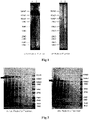
Fig. 1 is a diagram illustrating the results of Western blotting performed in Example 3. -
Fig. 2 is a diagram illustrating the results obtained by staining, with CBB, the gel electrophoresed in Example 3. - Below, embodiments of the present invention will be described in detail.
- A monoclonal antibody or an antigen-binding fragment thereof according to the present invention is specifically bound to adenovirus, and includes heavy chains CDR1 to CDR3 as shown in the following (a) to (c) and a light chain CDR1 as shown in the following (d):
- (a) a heavy chain CDR1 composed of an amino acid sequence containing NY;
- (b) a heavy chain CDR2 composed of an amino acid sequence containing SN;
- (c) a heavy chain CDR3 composed of an amino acid sequence containing SYY and DY; and
- (d) a light chain CDR1 composed of an amino acid sequence containing NG.
- A monoclonal antibody or an antigen-binding fragment thereof according to the present invention has a basic structure composed of a heavy chain and a light chain, and the heavy chain and the light chain have the respective variable regions that can specifically bind to an antigen. VH refers to the variable region of the heavy chain, and VL refers to the variable region of the light chain. The variable region of the heavy chain and that of the light chain each contain amino acid sequences of complementarity-determining regions (CDR), that is, CDR1, CDR2, and CDR3, and a framework region (FR). For example, the variable region contains three or four FRs (for example, FR1, FR2, FR3, and optionally FR4) together with the three CDRs.
- Examples of a monoclonal antibody of the present invention include quadruplechain antibodies (for example, having two light chains and two heavy chains), recombinant antibodies, or modified antibodies (for example, chimeric antibodies, humanized antibodies, human antibodies, CDR transplanted antibodies, primatized antibodies, deimmunized antibodies, synhumanized (synhumanized) antibodies, half antibodies, and bispecific antibodies). The class of the monoclonal antibody is not limited to IgG, and may also be IgM or IgY.
- In the present invention, an antigen-binding fragment of a monoclonal antibody is a fragment that is an antigen-binding site alone separated from the monoclonal antibody. Examples of such a fragment include fragments having specific antigen-binding capacity, such as Fab, Fab', F(ab')2, and single-chain antibodies (scFv) prepared by known methods.
- To produce such an antigen-binding fragment, a preparation method known to those skilled in the art can be used. Examples of such a method include: a method in which an antibody is digested with protease (for example, pepsin, papain, or the like) by a conventional method, and then purified by a known method of separating/purifying protein; a preparation method based on genetic recombination; and the like.
- As the monoclonal antibody or the antigen-binding fragment thereof according to the present invention, the following
antibody 1 orantibody 2 or an antigen-binding fragment thereof is preferable from the viewpoint of the ability to detect adenovirus with higher sensitivity. - A monoclonal antibody or an antigen-binding fragment thereof, which includes heavy chains CDR1 to CDR3 as shown in the following (a-1) to (c-1) and light chains CDR1 to CDR3 as shown in the following (d-1) to (f-1):
- (a-1) a heavy chain CDR1 composed of an amino acid sequence containing NNYYWN;
- (b-1) a heavy chain CDR2 composed of an amino acid sequence containing YIKYDGSNNNNPSLKN;
- (c-1) a heavy chain CDR3 composed of an amino acid sequence containing RASYYWDYFDV;
- (d-1) a light chain CDR1 composed of an amino acid sequence containing KANEDIYNGLA;
- (e-1) a light chain CDR2 composed of an amino acid sequence containing GATSLEA; and
- (f-1) a light chain CDR3 composed of an amino acid sequence containing QQYWSTPLT.
- A monoclonal antibody or an antigen-binding fragment thereof, which includes heavy chains CDR1 to CDR3 as shown in the following (a-2) to (c-2) and light chains CDR1 to CDR3 as shown in the following (d-2) to (f-2):
- (a-2) a heavy chain CDR1 composed of an amino acid sequence containing NYWIH;
- (b-2) a heavy chain CDR2 composed of an amino acid sequence containing EIDPTNGRSNYNEKFKT;
- (c-2) a heavy chain CDR3 composed of an amino acid sequence containing RSYYGSTYDYGLDY;
- (d-2) a light chain CDR1 composed of an amino acid sequence containing RS SKSLLHLNGNTYLY;
- (e-2) a light chain CDR2 composed of an amino acid sequence containing RMSNLAS; and
- (f-2) a light chain CDR3 composed of an amino acid sequence containing VQHLEYPYT.
- Examples of a monoclonal antibody or an antigen-binding fragment thereof according to the present invention include such an antibody or a fragment thereof that has a sequence identity of 60% or more, 65% or more, 70% or more, 75% or more, 80% or more, 85% or more, 90% or more, 91% or more, 92% or more, 93% or more, 94% or more, 95% or more, 96% or more, 97% or more, 98% or more, or 99% or more to each of the amino acid sequences contained in the heavy chains CDR1 to CDR3 and/or the light chains CDR1 to CDR3 of each of the
antibody 1 and theantibody 2. - The monoclonal antibody of the present invention may be obtained by immunizing an animal with a complex or an extract containing adenovirus as an antigen of interest or with the adenovirus or a partial peptide thereof by a known immunological method, and then preparing a hybridoma using cells of the immunized animal. The length of the peptide used for the immunization is not limited. Preferably, a peptide of not less than 5 amino acids, more preferably not less than 10 amino acids, may be used to provide the immunogen.
- The immunogen can be obtained from a culture liquid, or can be obtained by incorporating DNA encoding an arbitrary adenovirus antigen into a plasmid vector, and introducing the resulting vector to a host cell, followed by allowing expression of the adenovirus. Alternatively, such an arbitrary adenovirus antigen or the partial peptide thereof to be used as the immunogen can be expressed as a fusion protein with a protein exemplified below, and the expressed fusion protein can be used as the immunogen after purification or without purification. The preparation of the fusion protein can be carried out using, for example, Glutathion S-transferase (GST), maltose-binding protein (MBP), thioredoxin (TRX), Nus-tag, S-tag, HSV-tag, FRAG tag, polyhistidine tag, or the like which is commonly used as a "protein expression/purification tag" by those skilled in the art. Preferably, the fusion protein with such a protein is cleaved into the portion of the arbitrary adenovirus antigen or the partial peptide thereof, and the tag portion, using a digestive enzyme, and subjected to separation/purification before use as the immunogen.
- The preparation of the monoclonal antibody from the immunized animal can be easily carried out by the well-known method of Kohler et al. (Kohler et al., Nature, vol. 256, p. 495-497 (1975)). That is, antibody-producing cells such as spleen cells or lymphocytes are recovered from the immunized animal, and the recovered cells are fused with mouse myeloma cells by a conventional method to prepare hybridomas. The resulting hybridomas are cloned by the limiting dilution method or the like. Thereafter, a monoclonal antibody that undergoes antigen-antibody reaction with the antigen used for the immunization of the animal is selected from the monoclonal antibodies produced by the cloned hybridomas.
- Purification of the monoclonal antibody from ascites or a culture supernatant may be carried out by a known immunoglobulin purification method. Examples of the method include fractionation by salting out using ammonium sulfate or sodium sulfate, PEG fractionation, ethanol fractionation, DEAE ion-exchange chromatography, and gel filtration. Depending on the species of the immunized animal and the class of the monoclonal antibody, affinity chromatography using a carrier to which any of protein A, protein G, and protein L is bound may be used for the purification.
- In the present invention, using the above-mentioned monoclonal antibody or an antigen-binding fragment thereof makes it possible to detect adenovirus with very high sensitivity. In the following description preceding the Examples section, a "monoclonal antibody" means a "monoclonal antibody or an antigen-binding fragment thereof', unless otherwise obvious from the context.
- In the present invention, adenovirus is detected by performing an immunoassay of adenovirus using antigen-antibody reaction between the above-mentioned monoclonal antibody and adenovirus in a sample. That a monoclonal antibody undergoes antigen-antibody reaction with adenovirus means that a monoclonal antibody reacts specifically with adenovirus. The term "specific" means that, in a liquid system containing a mixture of antigen proteins and the monoclonal antibody, the antibody does not cause antigen-antibody reaction with components other than the antigen proteins at a detectable level, or, even in cases where the antibody causes a certain binding reaction or association reaction with such a component, the reaction is evidently weaker than the antigen-antibody reaction between the antibody and the antigen proteins.
- In the present invention, examples of immunoassays that can be used include any method well known to those skilled in the art, such as a competition method, condensation method, Western blotting, immunostaining method, sandwich method, and the like.
- A sandwich method is preferable as an immunoassay in the present invention. The sandwich method per se is well known in the field of immunoassays, and can be carried out by, for example, immunochromatography or ELISA. These sandwich methods per se are well known, and the method of the present invention can be carried out in the same manner as a well-known sandwich method except that the above-mentioned specific monoclonal antibody is used.
- In a sandwich method, two kinds of antibodies (an immobilized antibody immobilized in a solid phase and a labeled antibody) that recognize an antigen are used. In a method of the present invention, at least any one of these two kinds of antibodies is the above-mentioned monoclonal antibody of the present invention. In the case of the immobilized antibody immobilized in a solid phase, the amount of the antibody that can be immobilized per unit area is limited. To better achieve that object of the present invention which is to enhance the sensitivity, a monoclonal antibody according to the present invention is preferably used at least for the immobilized antibody. In cases where at least two antigens to be recognized by the monoclonal antibody are present in a single molecule or a single complex, the monoclonal antibody of a single kind can be used as a solid-phased antibody and a labelled antibody to perform a sandwich method.
- In the immunoassay based on the detection principle of a sandwich method, any solid phase may be used as the solid phase on which the antibody is to be immobilized, as long as the antibody can be immobilized thereon by a known technique. The solid phase may be arbitrarily selected from known solid phases such as porous thin films (membranes) having a capillary action; particles, test tubes, and resin plates. Examples of the substance for labeling the antibody include enzymes, radioisotopes, fluorescent substances, luminescent substances, colored particles, and colloidal particles. Among the above-mentioned immunoassay methods using various materials, lateral-flow immunoassay methods using a membrane are preferred from the viewpoint of enabling simple and rapid clinical tests.
- In cases where adenovirus is quantified or semi-quantified using the monoclonal antibody in the present invention, such quantification or semi-quantification involves "measurement" inevitably, and thus, is included in the "measurement" in the present invention. That is, in the present invention, "measurement" in an immunoassay includes any of quantification, semi-quantification, and detection.
- The present invention is described below by way of Examples. However, the present invention is not limited to the following Examples.
- Adenovirus was allowed to infect mammalian cells sensitive thereto. The resulting cells were cultured for some days. Then, the culture liquid of the adenovirus-infected cells were inactivated by ultraviolet irradiation and made ready for use.
- BALB/c mice were immunized with the adenovirus-inactivated antigen prepared in 1, and kept for a certain period. From each mouse, the spleen was removed, and fusion with mouse myeloma cells (P3×63) was carried out by the method of Kohler et al. (Kohler et al., Nature, vol. 256, p. 495-497 (1975)) to obtain a plurality of hybridoma cell lines that produce anti-adenovirus antibodies.
- The obtained cell line was intraperitoneally administered to pristane-treated BALB/c mice. About two weeks later, antibody-containing ascites was collected. From the ascites obtained, IgG was purified by affinity chromatography using a protein A column, to obtain a plurality of purified anti-adenovirus monoclonal antibodies.
- In the below-mentioned Examples, two antibodies, an
antibody 1 and anantibody 2, were used, which were selected from a plurality of the resulting anti-adenovirus monoclonal antibodies, considering reactivity and specificity. - The anti-adenovirus antibody (antibody 2) prepared in Example 1 was diluted with a buffer. The resulting liquid and an anti-mouse IgG antibody were made ready for use. The anti-adenovirus antibody and the anti-mouse IgG antibody were linearly applied respectively to the sample pad side and the absorber side of a nitrocellulose membrane lined with a PET film. Then, the nitrocellulose membrane was dried sufficiently under warm air to obtain a membrane having an anti-adenovirus antibody immobilized thereon.
- The anti-adenovirus antibody (antibody 1) prepared in Example 1 was covalently bound to colored polystyrene particles, and the colored polystyrene particles were suspended in a suspension. Then, colored polystyrene particles to which the anti-adenovirus antibodies were bound and which were dispersed sufficiently by ultrasonication were obtained. In the present description, the particles obtained here are referred to as "anti-adenovirus antibody-immobilized particles".
- A predetermined amount of the anti-adenovirus antibody-immobilized particles prepared in 2 were applied to a glass-fiber non-woven fabric, and the non-woven fabric was then dried sufficiently under warm air. In the present description, the pad obtained here is referred to as a "labeled antibody pad".
- The anti-adenovirus antibody-immobilized membrane prepared in 1 and the labelled antibody pads prepared in 2 and 3 were laminated with other members (backing sheet, absorption zone, and sample pad), and the resulting laminate was cut into a piece with a width of 5 mm, to provide an adenovirus test device.
- A culture liquid of adenovirus-infected cells of each type was diluted with a buffer to prepare a two-fold dilution series of each type of adenovirus.
- The adenovirus diluted liquid prepared was added to a sample suspension (10 mM Tris (pH 8.0), 1 w/v% polyoxyethylene octylphenyl ether, 3 w/v% arginine, and 3 w/v% BSA), and 50 µL of the resulting mixture was added dropwise to the adenovirus test device produced in 4. Then, the test device was left to stand for 5 minutes.
- In cases where coloring could be visually observed at both the position where the anti-mouse IgG antibody was applied and the position where the anti-adenovirus antibody was applied, the test result was evaluated as + (positive). In cases where coloring could be visually observed only at the position where the anti-mouse IgG antibody was applied and where coloring could not be visually observed at the position where the anti-adenovirus antibody was applied, the test result was evaluated as - (negative). In cases where coloring could not be visually observed at the position where the anti-mouse IgG antibody was applied, the test result was evaluated as invalid.
- The lowest adenovirus concentration at which the test result was evaluated as positive was regarded as the lowest detection sensitivity. The results are shown in Table 1.
[Table 1] Adenovirus Lowest Detection Sensitivity Type Species TCID50/Test copies/Test 1 C 1.95×10² 2.50×10³ 2 C 1.95×10² 6.25×10³ 3 B 2.79×10² 1.25×10⁴ 5 C 7.85×101 6.25×10³ 6 C 5.58×10² 3.13×10³ 7 B 1.74×10² 1.25×10⁴ 8 D 1.95×101 1.25×10³ 11 B 2.79×10² 5.00×10⁴ 19 D 2.44×101 1.25×10⁴ 31 A 4.48×10² 1.25×10⁵ 37 D 3.14×101 2.50×10³ - As shown in Table 1, it was possible to confirm that the immunoassay device used with the anti-adenovirus antibody of the present invention reacted with many types of adenovirus belonging to the species A to D.
- In addition it was possible to confirm that the device reacted with each of the subtypes: type 53, type 54, type 56, type 64, type 79, type 81, and type 85.
- To the adenovirus test device produced in 4, 50 µL of a sample suspension containing virus that induces respiratory infection was added dropwise, and the test device was left to stand for 5 minutes.
- In cases where coloring could be visually observed at both the position where the anti-mouse IgG antibody was applied and the position where the anti-adenovirus antibody was applied, the test result was evaluated as +. In cases where coloring could be visually observed only at the position where the anti-mouse IgG antibody was applied and where coloring could not be visually observed at the position where the anti-adenovirus antibody was applied, the test result was evaluated as -. In cases where coloring could not be visually observed at the position where the anti-mouse IgG antibody was applied, the test result was evaluated as invalid.
- The results are shown in Table 2.
[Table 2] Virus Measurement Results Coxsackievirus type A9 - Coxsackievirus type B4 - Coxsackievirus type B5 - Coxsackievirus type B6 - Echo virus type 2 - Echo virus type 3 - Echo virus type 4 - Echo virus type 6 - Echo virus type 9 - Echo virus type 11 - Echo virus type 30 - Herpes simplex virus type 1 - Human Metapneumovirus type A - Human Metapneumovirus type B - Influenza virus A/New Caledonia/20/99 (H1N1) - Influenza virus A/Beijing/262/95 (H1N1) - Influenza virus A/New York/55/2004 (H3N2) - Influenza virus A/Hiroshima/52/2005 (H3N2) - Influenza virus B/Shanghai/361/2002 (Yamagata) - Influenza virus B/Malaysia/2506/2004 (Victoria) - Measles virus - Mumps virus - Parainfluenza virus type 1 - Parainfluenza virus type 2 - Parainfluenza virus type 3 - Parainfluenza virus type 4 - RS virus Long strain (type A) - RS virus CH-18 strain (type B) - - As shown in Table 2, it was possible to confirm that the adenovirus immunoassay device used with the anti-adenovirus antibody of the present invention reacted specifically with adenovirus, because the immunoassay device reacted with adenovirus, but exhibited no crossreactivity with the etiological virus of any other respiratory infection.
- The lowest detection sensitivity was compared between the adenovirus test device (the present device) prepared in 4 and a commercially available adenovirus kit.
- Culture liquids of adenovirus-infected cells were each diluted with a buffer to prepare two-fold dilution series. To the present device, 50 µL of a sample suspension containing an adenovirus diluted liquid was added dropwise, and the present device was left to stand for 5 minutes and evaluated. The commercially available kit was used with a prescribed amount of adenovirus diluted liquid as a sample, and the test result was evaluated by performing a test in accordance with the document attached to each kit.
- Assuming that the largest adenovirus dilution ratio at which the test result was evaluated as positive with the present device is "1", the largest dilution ratio at which the test result was evaluated as positive with a commercially available adenovirus kit was regarded as relative sensitivity, and is shown in Table 3.
[Table 3] Kit Relative Sensitivity of Each Type (Species)* Type 1 (C) Type 2 (C) Type 3 (B) Type 4 (E) Typ e 11 (B) Typ e 37 (D) Type 54 (D) Present Kit 1 1 1 1 1 1 1 Kit A 1 1/2 1 1 1 1 1 Kit B 1/2 1/4 1/2 1/2 1/2 1/2 1/4 Kit C 1/8 1/8 1/8 1/20 1/20 NT NT Kit D 1 1/2 1/2 1/2 1/2 1/2 1/2 Kit E 1/16 1/32 1/100 1/80 1/400 NT NT Kit F 1/2 1/4 1/2 1/2 1/2 1/2 1/4 Kit G 1/4 1/8 1/8 1/8 1/4 1/4 1/4 Kit H 1/8 1/4 1/4 1/4 1/4 1/4 1/4 NT: Not tested - As shown in Table 3, it was possible to confirm that the immunoassay device used with the anti-adenovirus antibody of the present invention had the highest reactivity with
adenovirus type 2, and also had the highest reactivity with another type of adenovirus in the same manner as the kit A did. - The antigen recognition site of the anti-adenovirus monoclonal antibody obtained in Example 1 was verified by Western blotting and LC-MS/MS.
- Adenovirus was allowed to infect A549 cells, and cultured. On culture day 7, the adenovirus-infected cells were recovered and disrupted by ultrasonication. Cell residues were removed from the disrupted-cell liquid by centrifugation to obtain a concentrated-adenovirus liquid.
- A two-fold dilution series of the concentrated-adenovirus liquid obtained in 1 was prepared, and supplemented with reagents having the respective final concentrations: 62.5 mM Tris-HCl (pH 6.5), 10 w/v% glycerol, 2.3 w/v% SDS, and 0.05% BPB (dye). The resulting mixture was subjected to a conventional method SDS-PAGE without being thermally denatured.
- A two-fold dilution series of the concentrated-adenovirus liquid obtained in 1 was prepared, and supplemented with reagents having the respective final concentrations: 62.5 mM Tris-HCl (pH 6.5), 10 w/v% glycerol, 2.3 w/v% SDS, 0.05% BPB (dye), and 5% 2-mercaptoethanol. After being thermally denatured at 95°C for 1 minute, the resulting mixture was subjected to a conventional method SDS-PAGE.
- The gels electrophoresed in 2 and 3 were transferred to a PVDF membrane. After blocking of the membrane using skim milk, the membrane was sufficiently washed with PBS-Tween. The membrane was then allowed to react with the anti-adenovirus antibody whose concentration was adjusted to 3.8 µg/mL using PBS-Tween, at room temperature for 1 hour. After sufficiently washing the membrane with PBS-Tween, the membrane was allowed to react with a 3000-fold diluted HRPlabeled anti-mouse antibody at room temperature for 1 hour. After sufficiently washing the membrane with PBS-Tween, signals were detected using a chemiluminescence detection reagent.
- The results of Western Blotting are shown in
Fig. 1 . - A shown in
Fig. 1 , two antibodies (theantibody 1 and the antibody 2) produced in Example 1 reacted strongly with the approximately 200 kD protein (hexon trimer) contained in the sample obtained in 2 (without reduction treatment) (the left diagram). It is considered that the reduction treatment caused the hexon trimer to become a monomer, and it was recognized that the sample obtained in 3 (with reduction treatment) reacted very weakly with the approximately 100 kD protein (hexon monomer) (right diagram). - The results obtained by staining, with CBB, the gels electrophoresed in 2 and 3 are shown in
Fig. 2 . - As shown by the arrow in
Fig. 2 , the main protein contained in the sample obtained in 2 (without reduction treatment) was found at approximately 200 kD (the left diagram), and that contained in the sample obtained in 3 (with reduction treatment) was found at 100 to 150 kD (the right diagram). Each stained region denoted by a rectangle in the diagram was cut out, then hydrolyzed with trypsin, and analyzed by LC-MS/MS to give an amino acid sequence. The peptide fragment obtained was analyzed using Mascot (Ver. 2.5) (from Matrix Science Inc.) and Scaffold (from Proteome Software, Inc.), resulting in revealing that both of the stained regions contained a hexon protein of adenovirus as a main constituent. -
Fig. 1 and Fig. 2 have revealed that both of the two antibodies obtained in Example 1 reacted more strongly with a protein of a hexon trimer than with a protein of a hexon monomer, and reacted weakly with a monomeric hexon protein. - The hybridoma cell lines produced in Example 1 to produce the
anti-adenovirus antibodies - From the base sequence information obtained in 1, the amino acid sequences of the variable regions were predicted using a Kabat numbering system. The results are shown in Table 4.
[Table 4] Antibody 1Heavy Chain CDR1 Sequence NNYYWN SEQ ID NO: 1 CDR2 Sequence YIKYDGSNNNNPSLKN SEQ ID NO: 2 CDR3 Sequence RASYYWDYFDV SEQ ID NO: 3 Light Chain CDR1 Sequence KANEDIYNGLA SEQ ID NO: 4 CDR2 Sequence GATSLEA SEQ ID NO: 5 CDR3 Sequence QQYWSTPLT SEQ ID NO: 6 Antibody 2Heavy Chain CDR1 Sequence NYWIH SEQ ID NO: 7 CDR2 Sequence EIDPTNGRSNYNEKFKT SEQ ID NO: 8 CDR3 Sequence RSYYGSTYDYGLDY SEQ ID NO: 9 Light Chain CDR1 Sequence RSSKSLLHLNGNTYLY SEQ ID NO: 10 CDR2 Sequence RMSNLAS SEQ ID NO: 11 CDR3 Sequence VQHLEYPYT SEQ ID NO: 12 - From the result of Table 4, it is understood that the sequences common between the
antibody 1 and theantibody 2 exist as follows: NY in the heavy chain CDR1, SN in the heavy chain CDR2, SYY and DY in the heavy chain CDR3, and NG in the light chain CDR1. Accordingly, a monoclonal antibody or an antigen-binding fragment thereof according to the present invention which has these common sequences makes it possible to detect adenovirus with much higher sensitivity than a conventional known anti-adenovirus antibody.
Sequence Listing
Claims (7)
- A monoclonal antibody or an antigen-binding fragment thereof, comprising heavy chains CDR1 to CDR3 as shown in the following (a) to (c) and a light chain CDR1 as shown in the following (d):(a) a heavy chain CDR1 composed of an amino acid sequence containing NY;(b) a heavy chain CDR2 composed of an amino acid sequence containing SN;(c) a heavy chain CDR3 composed of an amino acid sequence containing SYY and DY; and(d) a light chain CDR1 composed of an amino acid sequence containing NG.
- The monoclonal antibody or the antigen-binding fragment thereof according to claim 1, comprising heavy chains CDR1 to CDR3 as shown in the following (a) to (c) and light chains CDR1 to CDR3 as shown in the following (d) to (f):(a-1) a heavy chain CDR1 composed of an amino acid sequence containing NNYYWN;(b-1) a heavy chain CDR2 composed of an amino acid sequence containing YIKYDGSNNNNPSLKN;(c-1) a heavy chain CDR3 composed of an amino acid sequence containing RASYYWDYFDV;(d-1) a light chain CDR1 composed of an amino acid sequence containing KANEDIYNGLA;(e-1) a light chain CDR2 composed of an amino acid sequence containing GATSLEA; and(f-1) a light chain CDR3 composed of an amino acid sequence containing QQYWSTPLT.
- The monoclonal antibody or the antigen-binding fragment thereof according to claim 1, comprising heavy chains CDR1 to CDR3 as shown in the following (a) to (c) and light chains CDR1 to CDR3 as shown in the following (d) to (f):(a-2) a heavy chain CDR1 composed of an amino acid sequence containing NYWIH;(b-2) a heavy chain CDR2 composed of an amino acid sequence containing EIDPTNGRSNYNEKFKT;(c-2) a heavy chain CDR3 composed of an amino acid sequence containing RSYYGSTYDYGLDY;(d-2) a light chain CDR1 composed of an amino acid sequence containing RS SKSLLHLNGNTYLY;(e-2) a light chain CDR2 composed of an amino acid sequence containing RMSNLAS; and(f-2) a light chain CDR3 composed of an amino acid sequence containing VQHLEYPYT.
- The monoclonal antibody or the antigen-binding fragment thereof according to any one of claims 1 to 3, having an identity of at least 60% or more with each of the amino acid sequences contained in the heavy chains CDR1 to CDR3 and/or the light chains CDR1 to CDR3.
- An immunoassay for adenovirus, comprising performing the immunoassay of adenovirus by using antigen-antibody reaction between the monoclonal antibody or the antigen-binding fragment thereof according to any one of claims 1 to 4 and adenovirus in a sample.
- The immunoassay according to claim 5, wherein the immunoassay is a sandwich method, and the monoclonal antibody or the antigen-binding fragment thereof is used as at least any one of a label or a solid phase.
- An immunoassay device for adenovirus, comprising the monoclonal antibody or the antigen-binding fragment thereof according to any one of claims 1 to 4.
Applications Claiming Priority (2)
| Application Number | Priority Date | Filing Date | Title |
|---|---|---|---|
| JP2020073239A JP7511377B2 (en) | 2020-04-16 | 2020-04-16 | Method and instrument for immunoassay of adenovirus |
| PCT/JP2021/015541 WO2021210633A1 (en) | 2020-04-16 | 2021-04-15 | Adenovirus immunoassay method and adenovirus immunoassay instrument |
Publications (3)
| Publication Number | Publication Date |
|---|---|
| EP4116324A1 true EP4116324A1 (en) | 2023-01-11 |
| EP4116324A4 EP4116324A4 (en) | 2023-09-06 |
| EP4116324B1 EP4116324B1 (en) | 2026-02-11 |
Family
ID=78085199
Family Applications (1)
| Application Number | Title | Priority Date | Filing Date |
|---|---|---|---|
| EP21789366.8A Active EP4116324B1 (en) | 2020-04-16 | 2021-04-15 | Adenovirus immunoassay method and adenovirus immunoassay instrument |
Country Status (8)
| Country | Link |
|---|---|
| US (1) | US12650429B2 (en) |
| EP (1) | EP4116324B1 (en) |
| JP (2) | JP7511377B2 (en) |
| KR (1) | KR20220167286A (en) |
| CN (1) | CN115362258A (en) |
| ES (1) | ES3066179T3 (en) |
| TW (1) | TWI861390B (en) |
| WO (1) | WO2021210633A1 (en) |
Families Citing this family (2)
| Publication number | Priority date | Publication date | Assignee | Title |
|---|---|---|---|---|
| CN118240067B (en) * | 2022-06-17 | 2025-04-01 | 东莞市朋志生物科技有限公司 | Anti-adenovirus antibodies, reagents and kits for detecting adenovirus |
| CN116789807B (en) * | 2023-06-14 | 2024-01-16 | 珠海重链生物科技有限公司 | Anti-adenovirus monoclonal antibody and application thereof |
Family Cites Families (8)
| Publication number | Priority date | Publication date | Assignee | Title |
|---|---|---|---|---|
| US4487829A (en) | 1982-03-23 | 1984-12-11 | Massachusetts Institute Of Technology | Production and use of monoclonal antibodies against adenoviruses |
| JP2000290298A (en) | 1999-04-06 | 2000-10-17 | Toagosei Co Ltd | Monoclonal antibody which reacts with adenovirus and detection of adenovirus therewith |
| JP2000290299A (en) | 1999-04-07 | 2000-10-17 | Toagosei Co Ltd | Monoclonal antibody and detection of adenovirus therewith |
| JP4554472B2 (en) * | 2005-08-26 | 2010-09-29 | 国立大学法人鳥取大学 | Parvovirus antigen detection kit |
| JP5435844B2 (en) | 2007-02-13 | 2014-03-05 | 株式会社ビーエル | Immunodetection method for influenza virus H5 subtype |
| CA2679676A1 (en) | 2008-09-05 | 2010-03-05 | Wei-Gang Hu | Monoclonal antibodies for neutralizing anthrax toxin |
| AU2014298373B2 (en) | 2013-08-01 | 2019-12-05 | Ludwig Institute For Cancer Research Ltd | Anti-GARP protein and uses thereof |
| JP6182027B2 (en) | 2013-09-10 | 2017-08-16 | デンカ生研株式会社 | Method for measuring influenza B virus |
-
2020
- 2020-04-16 JP JP2020073239A patent/JP7511377B2/en active Active
-
2021
- 2021-04-15 CN CN202180028577.1A patent/CN115362258A/en active Pending
- 2021-04-15 TW TW110113505A patent/TWI861390B/en active
- 2021-04-15 EP EP21789366.8A patent/EP4116324B1/en active Active
- 2021-04-15 US US17/918,926 patent/US12650429B2/en active Active
- 2021-04-15 WO PCT/JP2021/015541 patent/WO2021210633A1/en not_active Ceased
- 2021-04-15 KR KR1020227035461A patent/KR20220167286A/en active Pending
- 2021-04-15 ES ES21789366T patent/ES3066179T3/en active Active
-
2022
- 2022-12-28 JP JP2022211401A patent/JP7455947B2/en active Active
Non-Patent Citations (1)
| Title |
|---|
| QIU HONGLING ET AL: "Epitope mapping and cross-reactivity analysis of the monoclonal antibodies against hexon protein of human adenovirus type 3", VIRUS RESEARCH, vol. 146, no. 1-2, 1 December 2009 (2009-12-01), NL, pages 58 - 65, XP055857567, ISSN: 0168-1702, DOI: 10.1016/j.virusres.2009.08.011 * |
Also Published As
| Publication number | Publication date |
|---|---|
| JP7511377B2 (en) | 2024-07-05 |
| KR20220167286A (en) | 2022-12-20 |
| US12650429B2 (en) | 2026-06-09 |
| EP4116324A4 (en) | 2023-09-06 |
| US20240053338A1 (en) | 2024-02-15 |
| ES3066179T3 (en) | 2026-05-11 |
| JP2023052731A (en) | 2023-04-12 |
| WO2021210633A1 (en) | 2021-10-21 |
| EP4116324B1 (en) | 2026-02-11 |
| JP2021168619A (en) | 2021-10-28 |
| CN115362258A (en) | 2022-11-18 |
| TWI861390B (en) | 2024-11-11 |
| JP7455947B2 (en) | 2024-03-26 |
| TW202146898A (en) | 2021-12-16 |
Similar Documents
| Publication | Publication Date | Title |
|---|---|---|
| TW202200603A (en) | Epitope of antibody against structural protein of sars-cov-2, antibody that reacts with the epitope, method for detecting sars-cov-2 using the antibody, kit for detecting sars-cov-2 containing the antibody, method for detecting anti-sars-cov-2 antibody containing polypeptide of the epitope, kit for detecting anti-sars-cov-2 antibody containing polypeptide of the epitope, vaccine for sars-cov-2 containing polypeptide of the epitope, and therapeutic agent for sars-cov-2 infectious disease containing the antibody | |
| JP7804731B2 (en) | Adenovirus immunoassay method and immunoassay device | |
| CN105683756A (en) | Method for measuring influenza b virus | |
| JP7455947B2 (en) | Adenovirus immunoassay method and immunoassay device | |
| JP2021075496A (en) | Antibody recognizing anti-rs virus, and immunoassay method and immunoassay instrument using the antibody | |
| EP4116321A1 (en) | Adenovirus immunization measurement method and immunization measurement device | |
| EP4116322A1 (en) | Adenovirus immunoassay method and immunoassay instrument | |
| JP2023181275A (en) | Mycoplasma pneumoniae immunoassay method and immunoassay device | |
| KR20250135899A (en) | Method for detecting SARS-CoV-2 and kit therefor |
Legal Events
| Date | Code | Title | Description |
|---|---|---|---|
| STAA | Information on the status of an ep patent application or granted ep patent |
Free format text: STATUS: THE INTERNATIONAL PUBLICATION HAS BEEN MADE |
|
| PUAI | Public reference made under article 153(3) epc to a published international application that has entered the european phase |
Free format text: ORIGINAL CODE: 0009012 |
|
| STAA | Information on the status of an ep patent application or granted ep patent |
Free format text: STATUS: REQUEST FOR EXAMINATION WAS MADE |
|
| 17P | Request for examination filed |
Effective date: 20221005 |
|
| AK | Designated contracting states |
Kind code of ref document: A1 Designated state(s): AL AT BE BG CH CY CZ DE DK EE ES FI FR GB GR HR HU IE IS IT LI LT LU LV MC MK MT NL NO PL PT RO RS SE SI SK SM TR |
|
| DAV | Request for validation of the european patent (deleted) | ||
| DAX | Request for extension of the european patent (deleted) | ||
| A4 | Supplementary search report drawn up and despatched |
Effective date: 20230804 |
|
| RIC1 | Information provided on ipc code assigned before grant |
Ipc: C12P 21/08 20060101ALI20230731BHEP Ipc: G01N 33/569 20060101ALI20230731BHEP Ipc: C12N 15/13 20060101ALI20230731BHEP Ipc: C07K 16/46 20060101ALI20230731BHEP Ipc: C07K 16/08 20060101AFI20230731BHEP |
|
| STAA | Information on the status of an ep patent application or granted ep patent |
Free format text: STATUS: EXAMINATION IS IN PROGRESS |
|
| 17Q | First examination report despatched |
Effective date: 20240417 |
|
| GRAP | Despatch of communication of intention to grant a patent |
Free format text: ORIGINAL CODE: EPIDOSNIGR1 |
|
| STAA | Information on the status of an ep patent application or granted ep patent |
Free format text: STATUS: GRANT OF PATENT IS INTENDED |
|
| INTG | Intention to grant announced |
Effective date: 20250904 |
|
| GRAS | Grant fee paid |
Free format text: ORIGINAL CODE: EPIDOSNIGR3 |
|
| GRAA | (expected) grant |
Free format text: ORIGINAL CODE: 0009210 |
|
| STAA | Information on the status of an ep patent application or granted ep patent |
Free format text: STATUS: THE PATENT HAS BEEN GRANTED |
|
| AK | Designated contracting states |
Kind code of ref document: B1 Designated state(s): AL AT BE BG CH CY CZ DE DK EE ES FI FR GB GR HR HU IE IS IT LI LT LU LV MC MK MT NL NO PL PT RO RS SE SI SK SM TR |
|
| P01 | Opt-out of the competence of the unified patent court (upc) registered |
Free format text: CASE NUMBER: UPC_APP_0000316_4116324/2026 Effective date: 20260106 |
|
| REG | Reference to a national code |
Ref country code: CH Ref legal event code: F10 Free format text: ST27 STATUS EVENT CODE: U-0-0-F10-F00 (AS PROVIDED BY THE NATIONAL OFFICE) Effective date: 20260211 Ref country code: GB Ref legal event code: FG4D |
|
| REG | Reference to a national code |
Ref country code: DE Ref legal event code: R096 Ref document number: 602021047791 Country of ref document: DE |
|
| REG | Reference to a national code |
Ref country code: CH Ref legal event code: R17 Free format text: ST27 STATUS EVENT CODE: U-0-0-R10-R17 (AS PROVIDED BY THE NATIONAL OFFICE) Effective date: 20260309 |
|
| REG | Reference to a national code |
Ref country code: IE Ref legal event code: FG4D |
|
| PGFP | Annual fee paid to national office [announced via postgrant information from national office to epo] |
Ref country code: GB Payment date: 20260331 Year of fee payment: 6 |
|
| PGFP | Annual fee paid to national office [announced via postgrant information from national office to epo] |
Ref country code: NL Payment date: 20260319 Year of fee payment: 6 |
|
| PGFP | Annual fee paid to national office [announced via postgrant information from national office to epo] |
Ref country code: FR Payment date: 20260319 Year of fee payment: 6 |
|
| REG | Reference to a national code |
Ref country code: NL Ref legal event code: FP |
|
| REG | Reference to a national code |
Ref country code: CH Ref legal event code: U11 Free format text: ST27 STATUS EVENT CODE: U-0-0-U10-U11 (AS PROVIDED BY THE NATIONAL OFFICE) Effective date: 20260501 |
|
| REG | Reference to a national code |
Ref country code: ES Ref legal event code: FG2A Ref document number: 3066179 Country of ref document: ES Kind code of ref document: T3 Effective date: 20260511 |
|
| PG25 | Lapsed in a contracting state [announced via postgrant information from national office to epo] |
Ref country code: RS Free format text: LAPSE BECAUSE OF FAILURE TO SUBMIT A TRANSLATION OF THE DESCRIPTION OR TO PAY THE FEE WITHIN THE PRESCRIBED TIME-LIMIT Effective date: 20260511 |
|
| PGFP | Annual fee paid to national office [announced via postgrant information from national office to epo] |
Ref country code: ES Payment date: 20260501 Year of fee payment: 6 |
|
| PG25 | Lapsed in a contracting state [announced via postgrant information from national office to epo] |
Ref country code: PL Free format text: LAPSE BECAUSE OF FAILURE TO SUBMIT A TRANSLATION OF THE DESCRIPTION OR TO PAY THE FEE WITHIN THE PRESCRIBED TIME-LIMIT Effective date: 20260211 |
|
| PG25 | Lapsed in a contracting state [announced via postgrant information from national office to epo] |
Ref country code: GR Free format text: LAPSE BECAUSE OF FAILURE TO SUBMIT A TRANSLATION OF THE DESCRIPTION OR TO PAY THE FEE WITHIN THE PRESCRIBED TIME-LIMIT Effective date: 20260512 |
|
| PGFP | Annual fee paid to national office [announced via postgrant information from national office to epo] |
Ref country code: IT Payment date: 20260428 Year of fee payment: 6 |
|
| REG | Reference to a national code |
Ref country code: LT Ref legal event code: MG9D |
|
| PG25 | Lapsed in a contracting state [announced via postgrant information from national office to epo] |
Ref country code: LT Free format text: LAPSE BECAUSE OF FAILURE TO SUBMIT A TRANSLATION OF THE DESCRIPTION OR TO PAY THE FEE WITHIN THE PRESCRIBED TIME-LIMIT Effective date: 20260211 |
|
| PGFP | Annual fee paid to national office [announced via postgrant information from national office to epo] |
Ref country code: DE Payment date: 20260317 Year of fee payment: 6 |
|
| PG25 | Lapsed in a contracting state [announced via postgrant information from national office to epo] |
Ref country code: NO Free format text: LAPSE BECAUSE OF FAILURE TO SUBMIT A TRANSLATION OF THE DESCRIPTION OR TO PAY THE FEE WITHIN THE PRESCRIBED TIME-LIMIT Effective date: 20260511 |
|
| PG25 | Lapsed in a contracting state [announced via postgrant information from national office to epo] |
Ref country code: FI Free format text: LAPSE BECAUSE OF FAILURE TO SUBMIT A TRANSLATION OF THE DESCRIPTION OR TO PAY THE FEE WITHIN THE PRESCRIBED TIME-LIMIT Effective date: 20260211 Ref country code: AT Free format text: LAPSE BECAUSE OF FAILURE TO SUBMIT A TRANSLATION OF THE DESCRIPTION OR TO PAY THE FEE WITHIN THE PRESCRIBED TIME-LIMIT Effective date: 20260211 Ref country code: SE Free format text: LAPSE BECAUSE OF FAILURE TO SUBMIT A TRANSLATION OF THE DESCRIPTION OR TO PAY THE FEE WITHIN THE PRESCRIBED TIME-LIMIT Effective date: 20260211 |
|
| REG | Reference to a national code |
Ref country code: AT Ref legal event code: MK05 Ref document number: 1885590 Country of ref document: AT Kind code of ref document: T Effective date: 20260211 |
|
| PGFP | Annual fee paid to national office [announced via postgrant information from national office to epo] |
Ref country code: CH Payment date: 20260501 Year of fee payment: 6 |
|
| PG25 | Lapsed in a contracting state [announced via postgrant information from national office to epo] |
Ref country code: HR Free format text: LAPSE BECAUSE OF FAILURE TO SUBMIT A TRANSLATION OF THE DESCRIPTION OR TO PAY THE FEE WITHIN THE PRESCRIBED TIME-LIMIT Effective date: 20260211 |
|
| PG25 | Lapsed in a contracting state [announced via postgrant information from national office to epo] |
Ref country code: IS Free format text: LAPSE BECAUSE OF FAILURE TO SUBMIT A TRANSLATION OF THE DESCRIPTION OR TO PAY THE FEE WITHIN THE PRESCRIBED TIME-LIMIT Effective date: 20260611 Ref country code: PT Free format text: LAPSE BECAUSE OF FAILURE TO SUBMIT A TRANSLATION OF THE DESCRIPTION OR TO PAY THE FEE WITHIN THE PRESCRIBED TIME-LIMIT Effective date: 20260611 |
|
| PG25 | Lapsed in a contracting state [announced via postgrant information from national office to epo] |
Ref country code: LV Free format text: LAPSE BECAUSE OF FAILURE TO SUBMIT A TRANSLATION OF THE DESCRIPTION OR TO PAY THE FEE WITHIN THE PRESCRIBED TIME-LIMIT Effective date: 20260211 |
|
| PG25 | Lapsed in a contracting state [announced via postgrant information from national office to epo] |
Ref country code: BG Free format text: LAPSE BECAUSE OF FAILURE TO SUBMIT A TRANSLATION OF THE DESCRIPTION OR TO PAY THE FEE WITHIN THE PRESCRIBED TIME-LIMIT Effective date: 20260211 |