EP4095756A1 - Method and system for medical image interpretation - Google Patents
Method and system for medical image interpretation Download PDFInfo
- Publication number
- EP4095756A1 EP4095756A1 EP21184540.9A EP21184540A EP4095756A1 EP 4095756 A1 EP4095756 A1 EP 4095756A1 EP 21184540 A EP21184540 A EP 21184540A EP 4095756 A1 EP4095756 A1 EP 4095756A1
- Authority
- EP
- European Patent Office
- Prior art keywords
- symptom
- neural network
- network model
- convolutional neural
- medical image
- Prior art date
- Legal status (The legal status is an assumption and is not a legal conclusion. Google has not performed a legal analysis and makes no representation as to the accuracy of the status listed.)
- Granted
Links
Images


Classifications
-
- G—PHYSICS
- G06—COMPUTING OR CALCULATING; COUNTING
- G06T—IMAGE DATA PROCESSING OR GENERATION, IN GENERAL
- G06T7/00—Image analysis
- G06T7/0002—Inspection of images, e.g. flaw detection
- G06T7/0012—Biomedical image inspection
-
- G—PHYSICS
- G06—COMPUTING OR CALCULATING; COUNTING
- G06F—ELECTRIC DIGITAL DATA PROCESSING
- G06F18/00—Pattern recognition
- G06F18/20—Analysing
- G06F18/24—Classification techniques
- G06F18/241—Classification techniques relating to the classification model, e.g. parametric or non-parametric approaches
-
- G—PHYSICS
- G06—COMPUTING OR CALCULATING; COUNTING
- G06N—COMPUTING ARRANGEMENTS BASED ON SPECIFIC COMPUTATIONAL MODELS
- G06N3/00—Computing arrangements based on biological models
- G06N3/02—Neural networks
- G06N3/04—Architecture, e.g. interconnection topology
- G06N3/045—Combinations of networks
-
- G—PHYSICS
- G06—COMPUTING OR CALCULATING; COUNTING
- G06N—COMPUTING ARRANGEMENTS BASED ON SPECIFIC COMPUTATIONAL MODELS
- G06N3/00—Computing arrangements based on biological models
- G06N3/02—Neural networks
- G06N3/04—Architecture, e.g. interconnection topology
- G06N3/0464—Convolutional networks [CNN, ConvNet]
-
- G—PHYSICS
- G06—COMPUTING OR CALCULATING; COUNTING
- G06N—COMPUTING ARRANGEMENTS BASED ON SPECIFIC COMPUTATIONAL MODELS
- G06N3/00—Computing arrangements based on biological models
- G06N3/02—Neural networks
- G06N3/08—Learning methods
-
- G—PHYSICS
- G06—COMPUTING OR CALCULATING; COUNTING
- G06N—COMPUTING ARRANGEMENTS BASED ON SPECIFIC COMPUTATIONAL MODELS
- G06N3/00—Computing arrangements based on biological models
- G06N3/02—Neural networks
- G06N3/08—Learning methods
- G06N3/09—Supervised learning
-
- G—PHYSICS
- G06—COMPUTING OR CALCULATING; COUNTING
- G06V—IMAGE OR VIDEO RECOGNITION OR UNDERSTANDING
- G06V10/00—Arrangements for image or video recognition or understanding
- G06V10/40—Extraction of image or video features
- G06V10/44—Local feature extraction by analysis of parts of the pattern, e.g. by detecting edges, contours, loops, corners, strokes or intersections; Connectivity analysis, e.g. of connected components
- G06V10/443—Local feature extraction by analysis of parts of the pattern, e.g. by detecting edges, contours, loops, corners, strokes or intersections; Connectivity analysis, e.g. of connected components by matching or filtering
- G06V10/449—Biologically inspired filters, e.g. difference of Gaussians [DoG] or Gabor filters
- G06V10/451—Biologically inspired filters, e.g. difference of Gaussians [DoG] or Gabor filters with interaction between the filter responses, e.g. cortical complex cells
- G06V10/454—Integrating the filters into a hierarchical structure, e.g. convolutional neural networks [CNN]
-
- G—PHYSICS
- G06—COMPUTING OR CALCULATING; COUNTING
- G06V—IMAGE OR VIDEO RECOGNITION OR UNDERSTANDING
- G06V10/00—Arrangements for image or video recognition or understanding
- G06V10/70—Arrangements for image or video recognition or understanding using pattern recognition or machine learning
- G06V10/82—Arrangements for image or video recognition or understanding using pattern recognition or machine learning using neural networks
-
- G—PHYSICS
- G06—COMPUTING OR CALCULATING; COUNTING
- G06V—IMAGE OR VIDEO RECOGNITION OR UNDERSTANDING
- G06V40/00—Recognition of biometric, human-related or animal-related patterns in image or video data
- G06V40/10—Human or animal bodies, e.g. vehicle occupants or pedestrians; Body parts, e.g. hands
- G06V40/18—Eye characteristics, e.g. of the iris
- G06V40/193—Preprocessing; Feature extraction
-
- G—PHYSICS
- G06—COMPUTING OR CALCULATING; COUNTING
- G06V—IMAGE OR VIDEO RECOGNITION OR UNDERSTANDING
- G06V40/00—Recognition of biometric, human-related or animal-related patterns in image or video data
- G06V40/10—Human or animal bodies, e.g. vehicle occupants or pedestrians; Body parts, e.g. hands
- G06V40/18—Eye characteristics, e.g. of the iris
- G06V40/197—Matching; Classification
-
- G—PHYSICS
- G16—INFORMATION AND COMMUNICATION TECHNOLOGY [ICT] SPECIALLY ADAPTED FOR SPECIFIC APPLICATION FIELDS
- G16H—HEALTHCARE INFORMATICS, i.e. INFORMATION AND COMMUNICATION TECHNOLOGY [ICT] SPECIALLY ADAPTED FOR THE HANDLING OR PROCESSING OF MEDICAL OR HEALTHCARE DATA
- G16H30/00—ICT specially adapted for the handling or processing of medical images
- G16H30/20—ICT specially adapted for the handling or processing of medical images for handling medical images, e.g. DICOM, HL7 or PACS
-
- G—PHYSICS
- G16—INFORMATION AND COMMUNICATION TECHNOLOGY [ICT] SPECIALLY ADAPTED FOR SPECIFIC APPLICATION FIELDS
- G16H—HEALTHCARE INFORMATICS, i.e. INFORMATION AND COMMUNICATION TECHNOLOGY [ICT] SPECIALLY ADAPTED FOR THE HANDLING OR PROCESSING OF MEDICAL OR HEALTHCARE DATA
- G16H30/00—ICT specially adapted for the handling or processing of medical images
- G16H30/40—ICT specially adapted for the handling or processing of medical images for processing medical images, e.g. editing
-
- G—PHYSICS
- G16—INFORMATION AND COMMUNICATION TECHNOLOGY [ICT] SPECIALLY ADAPTED FOR SPECIFIC APPLICATION FIELDS
- G16H—HEALTHCARE INFORMATICS, i.e. INFORMATION AND COMMUNICATION TECHNOLOGY [ICT] SPECIALLY ADAPTED FOR THE HANDLING OR PROCESSING OF MEDICAL OR HEALTHCARE DATA
- G16H50/00—ICT specially adapted for medical diagnosis, medical simulation or medical data mining; ICT specially adapted for detecting, monitoring or modelling epidemics or pandemics
- G16H50/20—ICT specially adapted for medical diagnosis, medical simulation or medical data mining; ICT specially adapted for detecting, monitoring or modelling epidemics or pandemics for computer-aided diagnosis, e.g. based on medical expert systems
-
- G—PHYSICS
- G06—COMPUTING OR CALCULATING; COUNTING
- G06N—COMPUTING ARRANGEMENTS BASED ON SPECIFIC COMPUTATIONAL MODELS
- G06N3/00—Computing arrangements based on biological models
- G06N3/02—Neural networks
- G06N3/08—Learning methods
- G06N3/084—Backpropagation, e.g. using gradient descent
-
- G—PHYSICS
- G06—COMPUTING OR CALCULATING; COUNTING
- G06T—IMAGE DATA PROCESSING OR GENERATION, IN GENERAL
- G06T2207/00—Indexing scheme for image analysis or image enhancement
- G06T2207/20—Special algorithmic details
- G06T2207/20081—Training; Learning
-
- G—PHYSICS
- G06—COMPUTING OR CALCULATING; COUNTING
- G06T—IMAGE DATA PROCESSING OR GENERATION, IN GENERAL
- G06T2207/00—Indexing scheme for image analysis or image enhancement
- G06T2207/20—Special algorithmic details
- G06T2207/20084—Artificial neural networks [ANN]
-
- G—PHYSICS
- G06—COMPUTING OR CALCULATING; COUNTING
- G06T—IMAGE DATA PROCESSING OR GENERATION, IN GENERAL
- G06T2207/00—Indexing scheme for image analysis or image enhancement
- G06T2207/30—Subject of image; Context of image processing
- G06T2207/30004—Biomedical image processing
-
- G—PHYSICS
- G06—COMPUTING OR CALCULATING; COUNTING
- G06V—IMAGE OR VIDEO RECOGNITION OR UNDERSTANDING
- G06V2201/00—Indexing scheme relating to image or video recognition or understanding
- G06V2201/03—Recognition of patterns in medical or anatomical images
Definitions
- the disclosure relates to medical image interpretation, and more particularly to a method and a system for medical image interpretation based on deep learning.
- the disclosure provides a method and a system for medical image interpretation, where a convolutional neural network model may provide both symptom interpretation information and disease interpretation information, making the method and the system closer to actual requirements of medical personnel.
- An embodiment of the disclosure provides a method for medical image interpretation, adapted for a computer device, and including the following steps.
- a medical image is provided to a convolutional neural network model, where the convolutional neural network model includes a feature extraction part, a first classifier, and N second classifiers.
- N feature maps are generated by using a last layer of the feature extraction part of the convolutional neural network model.
- N symptom interpretation results of N symptoms of a disease are obtained based on the N feature maps through the N second classifiers.
- a disease interpretation result of the disease is obtained based on the N feature maps through the first classifier.
- An embodiment of the disclosure provides a system for medical image interpretation, including a storage device and a processor.
- the processor is connected to the storage device and is configured to perform the following steps.
- a medical image is provided to a convolutional neural network model, where the convolutional neural network model includes a feature extraction part, a first classifier, and N second classifiers.
- N feature maps are generated by using a last layer of the feature extraction part of the convolutional neural network model.
- N symptom interpretation results of N symptoms of a disease are obtained based on the N feature maps through the N second classifiers.
- a disease interpretation result of the disease is obtained based on the N feature maps through the first classifier.
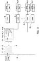
- FIG. 1 is a schematic diagram of a system for medical image interpretation according to an embodiment of the disclosure. However, it is only for ease of description, and is not intended to limit the disclosure.
- a system 10 for medical image interpretation includes a storage device 110 and a processor 120.
- the system 10 for medical image interpretation may be implemented as a notebook computer, a desktop computer, a tablet computer, an industrial computer, a server, or other types of computer devices, and the disclosure is not limited thereto.
- the storage device 110 is used to store images, instructions, program codes, software modules, or other data.
- the storage device 110 may include a volatile storage circuit and a non-volatile storage circuit.
- the volatile storage circuit is used to store data in a volatile manner.
- the volatile storage circuit may include a random access memory (RAM) or a similar volatile storage medium.
- the non-volatile storage circuit is used to store data in a non-volatile manner.
- the non-volatile storage circuit may include a read only memory (ROM), a solid state disk (SSD), and/or a traditional hard disk drive (HDD) or a similar non-volatile storage medium.
- ROM read only memory
- SSD solid state disk
- HDD traditional hard disk drive
- the processor 120 is coupled to the storage device 110 to control the entire or part of operation of the system 10 for medical image interpretation.
- the processor 120 is, for example, a central processing unit (CPU) or other programmable general-purpose or special-purpose microprocessor, a digital signal processor (DSP), a programmable controller, an application specific integrated circuit (ASIC), a programmable logic device (PLD), a graphics processing unit (GPU), or other similar device or a combination of these devices.
- the processor 120 may execute the program codes, software modules, instructions, and the like recorded in the storage device 110 to implement the method for medical image interpretation in the embodiments of the disclosure.
- FIG. 2 is a flowchart of a method for medical image interpretation according to an embodiment of the disclosure.
- the method of this embodiment is adapted for the system 10 for medical image interpretation in the above embodiment. Detailed steps of this embodiment are described as follows with reference to elements of the system 10 for medical image interpretation.
- a convolutional neural network model may assist medical personnel in interpreting a medical image; that is, the convolutional neural network model may assess presence or severity of a disease of a patient based on the medical image.
- medical personnel determines the presence and severity of a certain disease of a patient according to multiple symptoms in the medical image.
- the convolutional neural network model not only provides a disease interpretation result but also assesses the presence or severity of N symptoms of a certain disease in the medical image. Taking retinopathy as an example for the disease, the symptoms mentioned above may be, for example, hemorrhage or angiogenesis.
- the processor 120 provides the medical image to the convolutional neural network model.
- the medical image may include an X-ray image, an ultrasound image, a computed tomography (CT) image, a magnetic resonance imaging (MRI) image, a pathological slice image, or other captured image of the patient for medical diagnosis.
- CT computed tomography
- MRI magnetic resonance imaging
- the medical image may be a fundus image captured through a funduscope.
- the convolutional neural network model is constructed in advance based on a training dataset for deep learning, and model parameters of the convolutional neural network model (such as weight information, the number of convolutional kernels, and the number of network layers) have been determined by prior training and may be recorded in the storage device 110.
- the processor 120 may input the medical image to the convolutional neural network model, and the convolutional neural network model may output a disease interpretation result of the patient in response to the medical image.
- the disease interpretation result includes disease classification information of a disease.
- the convolutional neural network model may assess severity of a certain disease as grade 0 (not sick), grade 1, grade 2, or grade 3 based on the medical image.
- the convolutional neural network model may further output N symptom interpretation results of N symptoms of a disease according to the medical image.
- the N symptom interpretation results respectively include symptom presence information or symptom classification information of one of the N symptoms. For example, assuming that a certain disease has N symptoms, the convolutional neural network model may assess the severity of these symptoms as grade 0 (not existing), grade 1, grade 2, or grade 3 based on the medical image.
- the symptom presence information serves as an interpretation result of whether the symptom exists.
- the processor 120 generates N feature maps by using the last layer of a feature extraction part of the convolutional neural network model.
- the convolutional neural network model may include the feature extraction part and a first classifier.
- the feature extraction part of the convolutional neural network model is used to perform convolutional computation processing to the medical image for extracting features of the medical image and thereby generating the feature maps (or called feature vectors).
- these feature maps are fed into the first classifier, so that the first classifier outputs the disease interpretation result of a certain disease.
- the feature extraction part may include at least one convolutional layer. In an embodiment, the feature extraction part may include at least one convolutional layer and at least one pooling layer. In an embodiment, the feature extraction part of the convolutional neural network model includes multiple layers sequentially connected in series. These layers may include at least one convolutional layer and at least one pooling layer. Each layer in the feature extraction part of the convolutional neural network model may receive and input the medical image or the feature maps generated by the previous layer for executing relative computation processing to generate and output the feature maps. The convolutional layer in the feature extraction part may generate the feature maps by performing convolutional computation according to one or more convolutional kernels.
- the number of the feature maps output by each convolutional layer depends on the number of the convolutional kernels used by the convolutional layer.
- the convolutional kernels may slide on the feature maps by a fixed step length. Whenever the convolutional kernels are shifted, each weight included in the convolutional kernels is multiplied by and then added with all feature values of an overlapping area on the feature maps.
- the pooling layer may downsample the feature maps to obtain lower-dimension feature maps.
- the number of the feature maps output by the last layer of the feature extraction part of the convolutional neural network model is consistent with the number of the symptoms.
- the last layer of the feature extraction part of the convolutional neural network model is a convolutional layer or a pooling layer. That is, assuming that a certain disease has N types of the symptoms observable in the medical image, the number of the feature maps output by the last layer of the feature extraction part is N as well, and N is an integer greater than 0.
- the number of the convolutional kernels used in the last convolutional layer in the feature extraction part of the convolutional neural network model depends on the number of the symptoms.
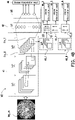
- FIG. 3 is a schematic diagram of a convolutional neural network model according to an embodiment of the disclosure.
- a convolutional neural network model M1 may include a feature extraction part 31 and a classifier 32 (i.e. the first classifier).
- the feature extraction part 31 includes multiple layers L_1 to L_x connected in series with each other, and the last layer L_x in the feature extraction part 31 may be a convolutional layer or a pooling layer.
- the last layer L_x of the feature extraction part 31 outputs N feature maps Fm_1 to Fm_N.
- the processor 120 obtains N symptom interpretation results of N symptoms of a disease based on the N feature maps through N second classifiers.
- the convolutional neural network model further includes the N second classifiers.
- the N second classifiers respectively output one of the N symptom interpretation results according to one of the N feature maps.
- the processor 120 may input the N feature maps to the corresponding second classifiers in a one-to-one manner so that the second classifiers respectively output one of the N symptom interpretation results in response to receiving one of the N feature maps.
- each second classifier may identify the symptoms and the severity thereof in the medical image according to a corresponding one of the feature maps, thereby outputting the corresponding symptom interpretation result respectively.
- the convolutional neural network model M1 may further include N classifiers 33_1 to 33_N (i.e. the second classifiers).
- the classifier 33_1 outputs a symptom interpretation result 34_1 related to a symptom of a disease according to the feature map Fm_1.
- the classifier 33_2 outputs a symptom interpretation result 34_2 related to another symptom of the disease according to the feature map Fm_2.
- the N classifiers 33_1 to 33_N of the convolutional neural network model M1 may output N symptom interpretation results 34_1 ⁇ 34_N related to N symptoms.
- the processor 120 obtains a disease interpretation result of the disease based on the N feature maps through the first classifier.
- the processor 120 may feed the N feature maps into the first classifier.
- the first classifier may determine the presence and/or severity of the disease in the patient according to the N feature maps, thereby outputting a disease interpretation result 36.
- the classifier 32 i.e. the first classifier
- classification functions of the first classifier and the N second classifiers may be implemented by at least one fully connected layer, respectively. That is, the first classifier includes at least one fully connected layer, and each of the N second classifiers includes at least one fully connected layer.
- the processor 120 may generate a one-dimensional vector according to the N feature maps and feed the one-dimensional vector into the at least one fully connected layer in the first classifier for classification, thereby obtaining the disease interpretation result.
- the processor 120 may respectively generate a one-dimensional vector according to one of the N feature maps and feed the one-dimensional vector into the at least one fully connected layer in a corresponding one of the second classifiers for classification, thereby obtaining one of the N symptom interpretation results.
- the processor 120 may synchronously train the feature extraction part, the first classifier, and the N second classifiers in the convolutional neural network model based on an overall loss function.
- the processor 120 may adjust weight information in the convolutional neural network model by backpropagation from back to front according to loss values output by the overall loss function, such as the weight information in the convolutional kernels and the weight information of the fully connected layers.
- the convolutional neural network model performs backpropagation training based on the overall loss function.
- This overall loss function includes a weighted sum of (N+1) loss values of (N+1) loss functions, and N loss functions among the (N+1) loss functions respectively correspond to the N second classifiers. The remaining one among the (N+1) loss functions corresponds to the first classifier.
- These loss functions are, for example, mean square error (MSE) loss functions, mean absolute error (MAE) loss functions, Softmax loss functions, or the like.
- MSE mean square error
- MAE mean absolute error
- Softmax loss functions Softmax loss functions
- the convolutional neural network model may generate a disease interpretation result and N symptom interpretation results according to current weight information.
- the processor 120 may obtain an overall loss value and update the weight information in the convolutional neural network model by backpropagation.
- the processor 120 inputs the disease interpretation result and the corresponding real label information to the loss function corresponding to the first classifier to obtain a loss value.
- the processor 120 further inputs each symptom interpretation result and the corresponding real label information to a loss function corresponding to the second classifiers to obtain multiple loss values respectively related to multiple symptoms.
- the processor 120 may obtain the overall loss value.
- the convolutional neural network model in the embodiments of the disclosure may be optimized towards the disease interpretation result, and this optimization direction is generated according to reasonable features of the symptoms. That is, the convolutional neural network model makes decisions based on specific symptom conditions in the medical image to avoid unexpected deviations existing in the training medical image and reducing reliability of the convolutional neural network model.
- the processor 120 may obtain the disease interpretation result of a certain disease and the symptom interpretation results of multiple symptoms by inputting the medical image to the convolutional neural network model.
- the disease interpretation result and the symptom interpretation results may be displayed on a display device or be printed on paper for medical personnel's reference to provide auxiliary diagnosis information that better meets medical personnel's requirements.
- the processor 120 may input one of the N feature maps to one of the N second classifiers so that the one of the N second classifiers outputs one of the N symptom interpretation results. That is, the N feature maps may be directly input to the corresponding second classifiers respectively, so that the N second classifiers may respectively generate the N symptom interpretation results according to the N feature maps.
- the convolutional neural network model may further include a symptom feature extraction part, and the symptom feature extraction part includes at least one convolutional layer.
- the processor 120 may input one of the N feature maps to the symptom feature extraction part to obtain at least one symptom feature map, and may input the at least one symptom feature map to one of the N second classifiers so that the one of the N second classifiers outputs one of the N symptom interpretation results. That is, the processor 120 may respectively perform convolutional computation processing to the N feature maps for feature extraction and then input feature extraction results to the corresponding second classifiers.
- the processor 120 may input a medical image Img_m to a convolutional neural network model M2.
- the convolutional neural network model M2 includes a feature extraction part 41 and a classifier 42.
- the feature extraction part 41 may include three convolutional layers Lc1 to Lc3. It should be noted that the convolutional layer Lc3, which is the last layer, generates eight feature maps Fm1_1 to Fm1_8 by using eight convolutional kernels. The eight feature maps Fm1_1 to Fm1_8 are fed into the classifier 42.
- the classifier 42 may include fully connected layers Lf1 and Lf2.
- the fully connected layer Lf1 may be referred to as a flat layer, and the fully connected layer Lf2 may be referred to as an output layer.
- the fully connected layer Lf2 may output a disease interpretation result 46.
- the disease interpretation result 46 may include disease classification information of a disease, such as the presence or severity of the disease of a patient.
- the convolutional neural network model M2 further includes eight classifiers 43_1 to 43_8.
- the eight feature maps Fm1_1 to Fm1_8 may be respectively fed into the classifiers 43_1 to 43_8.
- the classifiers 43_1 to 43_8 may respectively include two fully connected layers (for example, the fully connected layers Lf3 and Lf4 of the classifier 43_1).
- the classifiers 43_1 to 43_8 may respectively generate symptom interpretation results 44_1 to 44_8 according to their corresponding feature maps Fm1_1 to Fm1_8. Based on this, the processor 120 may generate the disease interpretation result 46 and the symptom interpretation results 44_1 to 44_8 for medical personnel's reference by using the convolutional neural network model M2.
- a convolutional neural network model M3 similar to the convolutional neural network model M2 in FIG. 4A , may include the feature extraction part 41, the classifier 42, and the eight classifiers 43_1 to 43_8.
- the convolutional neural network model M3 may further include symptom feature extraction parts 48_1 to 48_8 respectively connected with the classifiers 43_1 to 43_8.
- the symptom feature extraction parts 48_1 to 48_8 may respectively include a convolutional layer (for example, a convolutional layer Lc4 of the symptom feature extraction part 48_1).
- the symptom feature extraction parts 48_1 to 48_8 may respectively perform feature extraction to their corresponding feature maps Fm1_1 to Fm1_8 to generate multiple symptom feature maps. These symptom feature maps may be respectively input to the corresponding classifiers 43_1 to 43_8 so that the classifiers 43_1 to 43_8 may generate the symptom interpretation results 44_1 to 44_8 according to the corresponding symptom feature maps, respectively.
- FIG. 4A and FIG. 4B are only used to describe the disclosure, and are not intended to limit the disclosure.
- the number of the fully connected layers in the classifiers may be designed according to actual requirements, and the number of the convolutional layers in the feature extraction parts and the symptom feature extraction parts may also be designed according to actual requirements.
- the convolutional neural network model may not only provide the disease interpretation result of a disease but also provide the symptom interpretation results of multiple symptoms of the disease. Therefore, the medical personnel may clearly understand that the convolutional neural network model makes decision information about the disease based on the presence of these symptoms in the medical image and thereby improves the reliability of using the convolutional neural network model for medical image interpretation.
Landscapes
- Engineering & Computer Science (AREA)
- Health & Medical Sciences (AREA)
- Theoretical Computer Science (AREA)
- Physics & Mathematics (AREA)
- General Health & Medical Sciences (AREA)
- General Physics & Mathematics (AREA)
- Biomedical Technology (AREA)
- Medical Informatics (AREA)
- Data Mining & Analysis (AREA)
- Evolutionary Computation (AREA)
- Artificial Intelligence (AREA)
- Public Health (AREA)
- Life Sciences & Earth Sciences (AREA)
- Computer Vision & Pattern Recognition (AREA)
- General Engineering & Computer Science (AREA)
- Software Systems (AREA)
- Computing Systems (AREA)
- Molecular Biology (AREA)
- Epidemiology (AREA)
- Primary Health Care (AREA)
- Mathematical Physics (AREA)
- Computational Linguistics (AREA)
- Biophysics (AREA)
- Multimedia (AREA)
- Nuclear Medicine, Radiotherapy & Molecular Imaging (AREA)
- Radiology & Medical Imaging (AREA)
- Databases & Information Systems (AREA)
- Pathology (AREA)
- Ophthalmology & Optometry (AREA)
- Human Computer Interaction (AREA)
- Evolutionary Biology (AREA)
- Bioinformatics & Computational Biology (AREA)
- Bioinformatics & Cheminformatics (AREA)
- Quality & Reliability (AREA)
- Biodiversity & Conservation Biology (AREA)
- Measuring And Recording Apparatus For Diagnosis (AREA)
- Medical Treatment And Welfare Office Work (AREA)
Abstract
Description
- The disclosure relates to medical image interpretation, and more particularly to a method and a system for medical image interpretation based on deep learning.
- As deep learning becomes well-developed, artificial intelligence is increasingly used to assist medical image interpretation. However, artificial intelligence models having high accuracy are often relatively complicated, with their decision-making logic or determination basis too difficult to understand. In other words, the artificial intelligence models are unable to explain reasons behind their decisions and actions to human beings, which may affect reliability of the artificial intelligence models. In particular, in the field of medical diagnosis with assistance of artificial intelligence models, decision-making rationality and transparency are of utmost importance.
- In this regard, a series of explainable AI (as known as XAI) methods have been proposed to be the focus of research. These methods try to interpret how artificial intelligence models make decisions. At present, most of the explainable AI methods may only present importance and location of features in input data, and are unable to further explain the features, so it cannot provide any valuable explanation for medical personnel. More specifically, even if the explainable AI method may present relevant information about the important features in the input data, medical personnel still cannot determine whether the features that the artificial intelligence model focuses on is the symptoms of a specific disease. Therefore, the information cannot serve as the basis for interpretation confidence improvement or further explanation.
- The disclosure provides a method and a system for medical image interpretation, where a convolutional neural network model may provide both symptom interpretation information and disease interpretation information, making the method and the system closer to actual requirements of medical personnel.
- An embodiment of the disclosure provides a method for medical image interpretation, adapted for a computer device, and including the following steps. A medical image is provided to a convolutional neural network model, where the convolutional neural network model includes a feature extraction part, a first classifier, and N second classifiers. N feature maps are generated by using a last layer of the feature extraction part of the convolutional neural network model. N symptom interpretation results of N symptoms of a disease are obtained based on the N feature maps through the N second classifiers. A disease interpretation result of the disease is obtained based on the N feature maps through the first classifier.
- An embodiment of the disclosure provides a system for medical image interpretation, including a storage device and a processor. The processor is connected to the storage device and is configured to perform the following steps. A medical image is provided to a convolutional neural network model, where the convolutional neural network model includes a feature extraction part, a first classifier, and N second classifiers. N feature maps are generated by using a last layer of the feature extraction part of the convolutional neural network model. N symptom interpretation results of N symptoms of a disease are obtained based on the N feature maps through the N second classifiers. A disease interpretation result of the disease is obtained based on the N feature maps through the first classifier.
- In order to make the above-mentioned features and advantages of the present invention more comprehensible, the following specific embodiments are described in detail in conjunction with the accompanying drawings.
-
-
FIG. 1 is a schematic diagram of a system for medical image interpretation according to an embodiment of the disclosure. -
FIG. 2 is a flowchart of a method for medical image interpretation according to an embodiment of the disclosure. -
FIG. 3 is a schematic diagram of a convolutional neural network model according to an embodiment of the disclosure. -
FIG. 4A andFIG. 4B are schematic diagrams of convolutional neural network models according to embodiments of the disclosure. - Part of the embodiments of the disclosure will be described in detail below with accompanying drawings. For reference numerals used in the following description, the same reference numerals appearing in different drawings will be regarded as the same or similar elements. These embodiments are only a part of the disclosure and do not disclose all possible implementations of the disclosure. More precisely, these embodiments only serve as examples of the system and the method as recited in the claims of the disclosure.
-
FIG. 1 is a schematic diagram of a system for medical image interpretation according to an embodiment of the disclosure. However, it is only for ease of description, and is not intended to limit the disclosure. With reference toFIG. 1 , asystem 10 for medical image interpretation includes astorage device 110 and aprocessor 120. In some embodiments, thesystem 10 for medical image interpretation may be implemented as a notebook computer, a desktop computer, a tablet computer, an industrial computer, a server, or other types of computer devices, and the disclosure is not limited thereto. - The
storage device 110 is used to store images, instructions, program codes, software modules, or other data. Thestorage device 110 may include a volatile storage circuit and a non-volatile storage circuit. The volatile storage circuit is used to store data in a volatile manner. For example, the volatile storage circuit may include a random access memory (RAM) or a similar volatile storage medium. The non-volatile storage circuit is used to store data in a non-volatile manner. For example, the non-volatile storage circuit may include a read only memory (ROM), a solid state disk (SSD), and/or a traditional hard disk drive (HDD) or a similar non-volatile storage medium. - The
processor 120 is coupled to thestorage device 110 to control the entire or part of operation of thesystem 10 for medical image interpretation. Theprocessor 120 is, for example, a central processing unit (CPU) or other programmable general-purpose or special-purpose microprocessor, a digital signal processor (DSP), a programmable controller, an application specific integrated circuit (ASIC), a programmable logic device (PLD), a graphics processing unit (GPU), or other similar device or a combination of these devices. Theprocessor 120 may execute the program codes, software modules, instructions, and the like recorded in thestorage device 110 to implement the method for medical image interpretation in the embodiments of the disclosure. -
FIG. 2 is a flowchart of a method for medical image interpretation according to an embodiment of the disclosure. With reference toFIG. 2 , the method of this embodiment is adapted for thesystem 10 for medical image interpretation in the above embodiment. Detailed steps of this embodiment are described as follows with reference to elements of thesystem 10 for medical image interpretation. - In the embodiments of the disclosure, a convolutional neural network model may assist medical personnel in interpreting a medical image; that is, the convolutional neural network model may assess presence or severity of a disease of a patient based on the medical image. Generally speaking, when using a medical image for disease diagnosis, medical personnel determines the presence and severity of a certain disease of a patient according to multiple symptoms in the medical image. Based on this, in the embodiments of the disclosure, the convolutional neural network model not only provides a disease interpretation result but also assesses the presence or severity of N symptoms of a certain disease in the medical image. Taking retinopathy as an example for the disease, the symptoms mentioned above may be, for example, hemorrhage or angiogenesis.
- In step S201, the
processor 120 provides the medical image to the convolutional neural network model. The medical image may include an X-ray image, an ultrasound image, a computed tomography (CT) image, a magnetic resonance imaging (MRI) image, a pathological slice image, or other captured image of the patient for medical diagnosis. For example, the medical image may be a fundus image captured through a funduscope. - In an embodiment, the convolutional neural network model is constructed in advance based on a training dataset for deep learning, and model parameters of the convolutional neural network model (such as weight information, the number of convolutional kernels, and the number of network layers) have been determined by prior training and may be recorded in the
storage device 110. Theprocessor 120 may input the medical image to the convolutional neural network model, and the convolutional neural network model may output a disease interpretation result of the patient in response to the medical image. The disease interpretation result includes disease classification information of a disease. For example, the convolutional neural network model may assess severity of a certain disease as grade 0 (not sick), grade 1, grade 2, or grade 3 based on the medical image. In addition, in an embodiment, the convolutional neural network model may further output N symptom interpretation results of N symptoms of a disease according to the medical image. The N symptom interpretation results respectively include symptom presence information or symptom classification information of one of the N symptoms. For example, assuming that a certain disease has N symptoms, the convolutional neural network model may assess the severity of these symptoms as grade 0 (not existing), grade 1, grade 2, or grade 3 based on the medical image. The symptom presence information serves as an interpretation result of whether the symptom exists. - In step S202, the
processor 120 generates N feature maps by using the last layer of a feature extraction part of the convolutional neural network model. In detail, in an embodiment, the convolutional neural network model may include the feature extraction part and a first classifier. The feature extraction part of the convolutional neural network model is used to perform convolutional computation processing to the medical image for extracting features of the medical image and thereby generating the feature maps (or called feature vectors). In following, these feature maps are fed into the first classifier, so that the first classifier outputs the disease interpretation result of a certain disease. - In an embodiment, the feature extraction part may include at least one convolutional layer. In an embodiment, the feature extraction part may include at least one convolutional layer and at least one pooling layer. In an embodiment, the feature extraction part of the convolutional neural network model includes multiple layers sequentially connected in series. These layers may include at least one convolutional layer and at least one pooling layer. Each layer in the feature extraction part of the convolutional neural network model may receive and input the medical image or the feature maps generated by the previous layer for executing relative computation processing to generate and output the feature maps. The convolutional layer in the feature extraction part may generate the feature maps by performing convolutional computation according to one or more convolutional kernels. The number of the feature maps output by each convolutional layer depends on the number of the convolutional kernels used by the convolutional layer. The convolutional kernels may slide on the feature maps by a fixed step length. Whenever the convolutional kernels are shifted, each weight included in the convolutional kernels is multiplied by and then added with all feature values of an overlapping area on the feature maps. On the other hand, the pooling layer may downsample the feature maps to obtain lower-dimension feature maps.
- It should be noted that in the embodiments of the disclosure, the number of the feature maps output by the last layer of the feature extraction part of the convolutional neural network model is consistent with the number of the symptoms. In an embodiment, the last layer of the feature extraction part of the convolutional neural network model is a convolutional layer or a pooling layer. That is, assuming that a certain disease has N types of the symptoms observable in the medical image, the number of the feature maps output by the last layer of the feature extraction part is N as well, and N is an integer greater than 0. In light of this, in the embodiments of the disclosure, the number of the convolutional kernels used in the last convolutional layer in the feature extraction part of the convolutional neural network model depends on the number of the symptoms.
- For example,
FIG. 3 is a schematic diagram of a convolutional neural network model according to an embodiment of the disclosure. With reference toFIG. 3 , a convolutional neural network model M1 may include afeature extraction part 31 and a classifier 32 (i.e. the first classifier). Thefeature extraction part 31 includes multiple layers L_1 to L_x connected in series with each other, and the last layer L_x in thefeature extraction part 31 may be a convolutional layer or a pooling layer. The last layer L_x of thefeature extraction part 31 outputs N feature maps Fm_1 to Fm_N. - Next, in step S203, the
processor 120 obtains N symptom interpretation results of N symptoms of a disease based on the N feature maps through N second classifiers. In more detail, in addition to the feature extraction part and the first classifier, the convolutional neural network model further includes the N second classifiers. Here, the N second classifiers respectively output one of the N symptom interpretation results according to one of the N feature maps. In an embodiment, theprocessor 120 may input the N feature maps to the corresponding second classifiers in a one-to-one manner so that the second classifiers respectively output one of the N symptom interpretation results in response to receiving one of the N feature maps. In other words, each second classifier may identify the symptoms and the severity thereof in the medical image according to a corresponding one of the feature maps, thereby outputting the corresponding symptom interpretation result respectively. - For example, with reference to
FIG. 3 again, the convolutional neural network model M1 may further include N classifiers 33_1 to 33_N (i.e. the second classifiers). The classifier 33_1 outputs a symptom interpretation result 34_1 related to a symptom of a disease according to the feature map Fm_1. The classifier 33_2 outputs a symptom interpretation result 34_2 related to another symptom of the disease according to the feature map Fm_2. In the same way, the N classifiers 33_1 to 33_N of the convolutional neural network model M1 may output N symptom interpretation results 34_1∼34_N related to N symptoms. - In addition, in step S204, the
processor 120 obtains a disease interpretation result of the disease based on the N feature maps through the first classifier. In an embodiment, theprocessor 120 may feed the N feature maps into the first classifier. The first classifier may determine the presence and/or severity of the disease in the patient according to the N feature maps, thereby outputting adisease interpretation result 36. For example, with reference toFIG. 3 again, the classifier 32 (i.e. the first classifier) may receive the N feature maps Fm_1 to Fm_N to output thedisease interpretation result 36. - In an embodiment, classification functions of the first classifier and the N second classifiers may be implemented by at least one fully connected layer, respectively. That is, the first classifier includes at least one fully connected layer, and each of the N second classifiers includes at least one fully connected layer. In detail, the
processor 120 may generate a one-dimensional vector according to the N feature maps and feed the one-dimensional vector into the at least one fully connected layer in the first classifier for classification, thereby obtaining the disease interpretation result. In addition, theprocessor 120 may respectively generate a one-dimensional vector according to one of the N feature maps and feed the one-dimensional vector into the at least one fully connected layer in a corresponding one of the second classifiers for classification, thereby obtaining one of the N symptom interpretation results. - In addition, in one embodiment, before actually using the trained convolutional neural network model, it is necessary to train the convolutional neural network model with training data, and these training data include a large number of labeled training medical images. In an embodiment, during a training phase of the convolutional neural network model, the
processor 120 may synchronously train the feature extraction part, the first classifier, and the N second classifiers in the convolutional neural network model based on an overall loss function. Theprocessor 120 may adjust weight information in the convolutional neural network model by backpropagation from back to front according to loss values output by the overall loss function, such as the weight information in the convolutional kernels and the weight information of the fully connected layers. - In an embodiment, the convolutional neural network model performs backpropagation training based on the overall loss function. This overall loss function includes a weighted sum of (N+1) loss values of (N+1) loss functions, and N loss functions among the (N+1) loss functions respectively correspond to the N second classifiers. The remaining one among the (N+1) loss functions corresponds to the first classifier. These loss functions are, for example, mean square error (MSE) loss functions, mean absolute error (MAE) loss functions, Softmax loss functions, or the like. In detail, each symptom classification operation and disease classification operation uses the loss functions independent from each other, and an overall loss value generated by the overall loss function is the weighted sum of the (N+1) loss values of the (N+1) loss functions independent from each other.
- For example, when the
processor 120 inputs a certain labeled training medical image to the convolutional neural network model, the convolutional neural network model may generate a disease interpretation result and N symptom interpretation results according to current weight information. By inputting the above disease interpretation result, the N symptom interpretation results, and real label information to the overall loss function, theprocessor 120 may obtain an overall loss value and update the weight information in the convolutional neural network model by backpropagation. Theprocessor 120 inputs the disease interpretation result and the corresponding real label information to the loss function corresponding to the first classifier to obtain a loss value. In addition, theprocessor 120 further inputs each symptom interpretation result and the corresponding real label information to a loss function corresponding to the second classifiers to obtain multiple loss values respectively related to multiple symptoms. In following, by weighting and calculating the above (N+1) loss values, theprocessor 120 may obtain the overall loss value. - Based on the architecture of the convolutional neural network model and the design of the loss functions, the convolutional neural network model in the embodiments of the disclosure may be optimized towards the disease interpretation result, and this optimization direction is generated according to reasonable features of the symptoms. That is, the convolutional neural network model makes decisions based on specific symptom conditions in the medical image to avoid unexpected deviations existing in the training medical image and reducing reliability of the convolutional neural network model.
- Based on the foregoing description, in the embodiments of the disclosure, the
processor 120 may obtain the disease interpretation result of a certain disease and the symptom interpretation results of multiple symptoms by inputting the medical image to the convolutional neural network model. The disease interpretation result and the symptom interpretation results may be displayed on a display device or be printed on paper for medical personnel's reference to provide auxiliary diagnosis information that better meets medical personnel's requirements. - In addition, in an embodiment, the
processor 120 may input one of the N feature maps to one of the N second classifiers so that the one of the N second classifiers outputs one of the N symptom interpretation results. That is, the N feature maps may be directly input to the corresponding second classifiers respectively, so that the N second classifiers may respectively generate the N symptom interpretation results according to the N feature maps. Alternatively, in an embodiment, the convolutional neural network model may further include a symptom feature extraction part, and the symptom feature extraction part includes at least one convolutional layer. Theprocessor 120 may input one of the N feature maps to the symptom feature extraction part to obtain at least one symptom feature map, and may input the at least one symptom feature map to one of the N second classifiers so that the one of the N second classifiers outputs one of the N symptom interpretation results. That is, theprocessor 120 may respectively perform convolutional computation processing to the N feature maps for feature extraction and then input feature extraction results to the corresponding second classifiers. - For convenience of description regarding the concept of the disclosure, a further description is provided below with reference to
FIG. 4A andFIG. 4B .FIG. 4A andFIG. 4B are schematic diagrams of the convolutional neural network models according to the embodiments of the disclosure. As exemplified inFIG. 4A andFIG. 4B , it is assumed that the number of symptoms is eight, which means N = 8. - With reference to
FIG. 4A , theprocessor 120 may input a medical image Img_m to a convolutional neural network model M2. The convolutional neural network model M2 includes afeature extraction part 41 and aclassifier 42. Thefeature extraction part 41 may include three convolutional layers Lc1 to Lc3. It should be noted that the convolutional layer Lc3, which is the last layer, generates eight feature maps Fm1_1 to Fm1_8 by using eight convolutional kernels. The eight feature maps Fm1_1 to Fm1_8 are fed into theclassifier 42. Theclassifier 42 may include fully connected layers Lf1 and Lf2. The fully connected layer Lf1 may be referred to as a flat layer, and the fully connected layer Lf2 may be referred to as an output layer. The fully connected layer Lf2 may output adisease interpretation result 46. Thedisease interpretation result 46 may include disease classification information of a disease, such as the presence or severity of the disease of a patient. In addition, since the number of the symptoms is eight, the convolutional neural network model M2 further includes eight classifiers 43_1 to 43_8. The eight feature maps Fm1_1 to Fm1_8 may be respectively fed into the classifiers 43_1 to 43_8. The classifiers 43_1 to 43_8 may respectively include two fully connected layers (for example, the fully connected layers Lf3 and Lf4 of the classifier 43_1). The classifiers 43_1 to 43_8 may respectively generate symptom interpretation results 44_1 to 44_8 according to their corresponding feature maps Fm1_1 to Fm1_8. Based on this, theprocessor 120 may generate thedisease interpretation result 46 and the symptom interpretation results 44_1 to 44_8 for medical personnel's reference by using the convolutional neural network model M2. - On the other hand, with reference to
FIG. 4B , a convolutional neural network model M3, similar to the convolutional neural network model M2 inFIG. 4A , may include thefeature extraction part 41, theclassifier 42, and the eight classifiers 43_1 to 43_8. Different from the example inFIG. 4A , the convolutional neural network model M3 may further include symptom feature extraction parts 48_1 to 48_8 respectively connected with the classifiers 43_1 to 43_8. The symptom feature extraction parts 48_1 to 48_8 may respectively include a convolutional layer (for example, a convolutional layer Lc4 of the symptom feature extraction part 48_1). The symptom feature extraction parts 48_1 to 48_8 may respectively perform feature extraction to their corresponding feature maps Fm1_1 to Fm1_8 to generate multiple symptom feature maps. These symptom feature maps may be respectively input to the corresponding classifiers 43_1 to 43_8 so that the classifiers 43_1 to 43_8 may generate the symptom interpretation results 44_1 to 44_8 according to the corresponding symptom feature maps, respectively. - However, the examples in
FIG. 4A andFIG. 4B are only used to describe the disclosure, and are not intended to limit the disclosure. The number of the fully connected layers in the classifiers may be designed according to actual requirements, and the number of the convolutional layers in the feature extraction parts and the symptom feature extraction parts may also be designed according to actual requirements. - In summary, in the embodiments of the disclosure, when medical personnel use the convolutional neural network model to assist medical image interpretation, the convolutional neural network model may not only provide the disease interpretation result of a disease but also provide the symptom interpretation results of multiple symptoms of the disease. Therefore, the medical personnel may clearly understand that the convolutional neural network model makes decision information about the disease based on the presence of these symptoms in the medical image and thereby improves the reliability of using the convolutional neural network model for medical image interpretation.
Claims (18)
- A method for medical image interpretation, adapted for a computer device, the method comprising:providing a medical image (Img_m) to a convolutional neural network model (M1, M2, M3), wherein the convolutional neural network model (M1, M2, M3) comprises a feature extraction part (31, 41), a first classifier (32), and N second classifiers (33_1 to 33_N);generating N feature maps (Fm_1 to Fm_N) by using the feature extraction part (31, 41) of the convolutional neural network model (M1, M2, M3);obtaining N symptom interpretation results (34_1 to 34_N, 44_1 to 44_8) of N symptoms of a disease based on the N feature maps (Fm_1 to Fm_N) through the N second classifiers (33_1 to 33_N); andobtaining a disease interpretation result (36, 46) of the disease based on the N feature maps (Fm_1 to Fm_N) through the first classifier (32).
- The method for medical image interpretation according to claim 1, wherein the N feature maps (Fm_1 to Fm_N) are generated by using a last layer (L_x) of the feature extraction part (31, 41) of the convolutional neural network model (M1, M2, M3), and the last layer (L_x) is a convolutional layer (Lc1 to Lc4) or a pooling layer.
- The method for medical image interpretation according to any of the claims 1 or 2, wherein the feature extraction part (31, 41) of the convolutional neural network model (M1, M2, M3) comprises a plurality of layers (L_1 to L_x) sequentially connected in series, and the feature extraction part (31, 41) of the convolutional neural network model (M1, M2, M3) comprises at least one convolutional layer (Lc1 to Lc4) and preferably wherein the feature extraction part (31, 41) of the convolutional neural network model (M1, M2, M3) further comprises at least one pooling layer.
- The method for medical image interpretation according to any of the claims 1 to 3, wherein the first classifier (32) comprises at least one fully connected layer (Lf1, Lf2, Lf3, Lf4), and each of the N second classifiers (33_1 to 33_N) comprises at least one fully connected layer (Lf1, Lf2, Lf3, Lf4).
- The method for medical image interpretation according to any of the claims 1 to 4, wherein the N symptom interpretation results (34_1 to 34_N, 44_1 to 44_8) respectively comprise symptom presence information or symptom classification information of one of the N symptoms.
- The method for medical image interpretation according to any of the claims 1 to 5, wherein the disease interpretation result (36, 46) comprises disease classification information of the disease.
- The method for medical image interpretation according to any of the claims 1 to 6, further comprising:
training the convolutional neural network model (M1, M2, M3), wherein the convolutional neural network model (M1, M2, M3) is trained based on an overall loss function, the overall loss function comprises a weighted sum of (N+1) loss values of (N+1) loss functions, N loss functions among the (N+1) loss functions correspond to the N second classifiers (33_1 to 33_N) respectively, and a remaining one among the (N+1) loss functions corresponds to the first classifier (32). - The method for medical image interpretation according to any of the claims 1 to 7, wherein a step of obtaining N symptom interpretation results (34_1 to 34_N, 44_1 to 44_8) of the N symptoms of the disease based on the N feature maps (Fm_1 to Fm_N) through the N second classifiers (33_1 to 33_N) comprises:
inputting one of the N feature maps (Fm_1 to Fm_N) to one of the N second classifiers (33_1 to 33_N) so that the one of the N second classifiers (33_1 to 33_N) outputs one of the N symptom interpretation results (34_1 to 34_N, 44_1 to 44_8). - The method for medical image interpretation according to any of the claims 1 to 8, wherein the convolutional neural network model (M1, M2, M3) further comprises a symptom feature extraction part (48_1 to 48_8), the symptom feature extraction part (48_1 to 48_8) comprises at least one convolutional layer (Lc1 to Lc4), and a step of obtaining N symptom interpretation results (34_1 to 34_N, 44_1 to 44_8) of the N symptoms of the disease based on the N feature maps (Fm_1 to Fm_N) through the N second classifiers (33_1 to 33_N) comprises:inputting one of the N feature maps (Fm_1 to Fm_N) to the symptom feature extraction part (48_1 to 48_8) for obtaining at least one symptom feature map; andinputting the at least one symptom feature map to one of the N second classifiers (33_1 to 33_N) so that the one of the N second classifiers (33_1 to 33_N) outputs one of the N symptom interpretation results (34_1 to 34_N, 44_1 to 44_8).
- A system (10) for medical image interpretation, comprising:a storage device (110); anda processor (120), coupled to the storage device (110) and configured to:provide a medical image (Img_m) to a convolutional neural network model (M1, M2, M3), wherein the convolutional neural network model (M1, M2, M3) comprises a feature extraction part (31, 41), a first classifier (32), and N second classifiers (33_1 to 33_N);generate N feature maps (Fm_1 to Fm_N) by using the feature extraction part (31, 41) of the convolutional neural network model (M1, M2, M3);obtain N symptom interpretation results (34_1 to 34_N, 44_1 to 44_8) of N symptoms of a disease based on the N feature maps (Fm_1 to Fm_N) through the N second classifiers (33_1 to 33_N); andobtain a disease interpretation result (36, 46) of the disease based on the N feature maps (Fm_1 to Fm_N) through the first classifier (32).
- The system (10) for medical image interpretation according to claim 10, wherein the N feature maps (Fm_1 to Fm_N) are generated by using a last layer (L_x) of the feature extraction part (31, 41) of the convolutional neural network model (M1, M2, M3), and the last layer (L_x) is a convolutional layer (Lc1 to Lc4) or a pooling layer.
- The system (10) for medical image interpretation according to claim 10 or 11, wherein the feature extraction part (31, 41) of the convolutional neural network model (M1, M2, M3) comprises a plurality of layers (L_1 to L_x) sequentially connected in series, and the feature extraction part (31, 41) of the convolutional neural network model (M1, M2, M3) comprises at least one convolutional layer (Lc1 to Lc4),
and preferably, wherein the feature extraction part (31, 41) of the convolutional neural network model (M1, M2, M3) further comprises at least one pooling layer. - The system (10) for medical image interpretation according to any of the claims 10 to 12, wherein the first classifier (32) comprises at least one fully connected layer (Lf1, Lf2, Lf3, Lf4), and each of the N second classifiers (33_1 to 33_N) comprises at least one fully connected layer (Lf1, Lf2, Lf3, Lf4).
- The system (10) for medical image interpretation according to any of the claims 10 to 13, wherein the N symptom interpretation results (34_1 to 34_N, 44_1 to 44_8) respectively comprise symptom presence information or symptom classification information of one of the N symptoms.
- The system (10) for medical image interpretation according to any of the claims 10 to 14, wherein the disease interpretation result (36, 46) comprises disease classification information of the disease.
- The system (10) for medical image interpretation according to any of the claims 10 to 15, wherein the processor (120) is further configured to:
train the convolutional neural network model (M1, M2, M3), wherein the convolutional neural network model (M1, M2, M3) is trained based on an overall loss function, the overall loss function comprises a weighted sum of (N+1) loss values of (N+1) loss functions, N loss functions among the (N+1) loss functions correspond to the N second classifiers (33_1 to 33_N) respectively, and a remaining one among the (N+1) loss functions corresponds to the first classifier (32). - The system (10) for medical image interpretation according to any of the claims 10 to 16, wherein the processor (120) is further configured to:
input one of the N feature maps (Fm_1 to Fm_N) to one of the N second classifiers (33_1 to 33_N) so that the one of the N second classifiers (33_1 to 33_N) outputs one of the N symptom interpretation results (34_1 to 34_N, 44_1 to 44_8). - The system (10) for medical image interpretation according to any of the claims 10 to 17, wherein the convolutional neural network model (M1, M2, M3) further comprises a symptom feature extraction part (48_1 to 48_8), the symptom feature extraction part (48_1 to 48_8) comprises at least one convolutional layer (Lc1 to Lc4), and the processor (120) is further configured to:input one of the N feature maps (Fm_1 to Fm_N) to the symptom feature extraction part (48_1 to 48_8) for obtaining at least one symptom feature map; andinput the at least one symptom feature map to one of the N second classifiers (33_1 to 33_N) so that the one of the N second classifiers (33_1 to 33_N) outputs one of the N symptom interpretation results (34_1 to 34_N, 44_1 to 44_8).
Applications Claiming Priority (1)
| Application Number | Priority Date | Filing Date | Title |
|---|---|---|---|
| TW110119273A TWI832057B (en) | 2021-05-27 | 2021-05-27 | Method and system for interpreting medical image |
Publications (2)
| Publication Number | Publication Date |
|---|---|
| EP4095756A1 true EP4095756A1 (en) | 2022-11-30 |
| EP4095756B1 EP4095756B1 (en) | 2024-03-27 |
Family
ID=76845102
Family Applications (1)
| Application Number | Title | Priority Date | Filing Date |
|---|---|---|---|
| EP21184540.9A Active EP4095756B1 (en) | 2021-05-27 | 2021-07-08 | Method and system for medical image interpretation |
Country Status (3)
| Country | Link |
|---|---|
| US (1) | US11961231B2 (en) |
| EP (1) | EP4095756B1 (en) |
| TW (1) | TWI832057B (en) |
Families Citing this family (2)
| Publication number | Priority date | Publication date | Assignee | Title |
|---|---|---|---|---|
| WO2023135586A1 (en) * | 2022-01-12 | 2023-07-20 | Vijendra Venkatesh | A system for detection and classification of cardiac diseases using custom deep neural network techniques |
| EP4310778B1 (en) * | 2022-07-18 | 2025-02-26 | Tata Consultancy Services Limited | Method and system for semi-supervised domain adaptation based universal lesion detection |
Citations (1)
| Publication number | Priority date | Publication date | Assignee | Title |
|---|---|---|---|---|
| WO2019232027A1 (en) * | 2018-05-29 | 2019-12-05 | The General Hospital Corporation | System and method for analyzing medical images to detect/classify medical conditions using machine-learning and case radiology atlas |
Family Cites Families (4)
| Publication number | Priority date | Publication date | Assignee | Title |
|---|---|---|---|---|
| CN106934798B (en) * | 2017-02-20 | 2020-08-21 | 苏州体素信息科技有限公司 | Diabetic retinopathy classification and classification method based on deep learning |
| WO2019054045A1 (en) * | 2017-09-15 | 2019-03-21 | 富士フイルム株式会社 | Medical image processing device, medical image processing method, and medical image processing program |
| TWI673683B (en) | 2018-03-28 | 2019-10-01 | National Yunlin University Of Science And Technology | System and method for identification of symptom image |
| JP7129870B2 (en) * | 2018-10-01 | 2022-09-02 | 富士フイルム株式会社 | Learning device, method, and program for discriminator for discriminating disease area, discriminator for discriminating disease area, and device and program for discriminating disease region |
-
2021
- 2021-05-27 TW TW110119273A patent/TWI832057B/en active
- 2021-06-25 US US17/357,994 patent/US11961231B2/en active Active
- 2021-07-08 EP EP21184540.9A patent/EP4095756B1/en active Active
Patent Citations (1)
| Publication number | Priority date | Publication date | Assignee | Title |
|---|---|---|---|---|
| WO2019232027A1 (en) * | 2018-05-29 | 2019-12-05 | The General Hospital Corporation | System and method for analyzing medical images to detect/classify medical conditions using machine-learning and case radiology atlas |
Also Published As
| Publication number | Publication date |
|---|---|
| EP4095756B1 (en) | 2024-03-27 |
| US20220383490A1 (en) | 2022-12-01 |
| TW202247092A (en) | 2022-12-01 |
| TWI832057B (en) | 2024-02-11 |
| US11961231B2 (en) | 2024-04-16 |
Similar Documents
| Publication | Publication Date | Title |
|---|---|---|
| US10853449B1 (en) | Report formatting for automated or assisted analysis of medical imaging data and medical diagnosis | |
| JP7154322B2 (en) | Medical image processing method and apparatus, electronic equipment and storage medium | |
| US10496884B1 (en) | Transformation of textbook information | |
| US10692602B1 (en) | Structuring free text medical reports with forced taxonomies | |
| US11195273B2 (en) | Disease detection from weakly annotated volumetric medical images using convolutional long short-term memory | |
| US9892361B2 (en) | Method and system for cross-domain synthesis of medical images using contextual deep network | |
| US12009104B2 (en) | Medical image processing apparatus, medical image processing method, machine learning system, and program | |
| Ullah et al. | Explainable artificial intelligence approach in combating real-time surveillance of COVID19 pandemic from CT scan and X-ray images using ensemble model | |
| Shi et al. | Survey on neural networks used for medical image processing | |
| US11417424B2 (en) | Disease detection from weakly annotated volumetric medical images using convolutional long short-term memory and multiple instance learning | |
| CN111369564B (en) | Image processing method, model training method and model training device | |
| Yadav et al. | TE-CapsNet: time efficient capsule network for automatic disease classification from medical images | |
| EP4095756B1 (en) | Method and system for medical image interpretation | |
| Perumal Sankar et al. | An effective content based medical image retrieval by using ABC based artificial neural network (ANN) | |
| Peng et al. | DGFAU-Net: Global feature attention upsampling network for medical image segmentation | |
| Zhang et al. | A spine segmentation method under an arbitrary field of view based on 3d swin transformer | |
| CN117409021A (en) | Ultrasound image segmentation system and segmentation method based on multiple attention guidance | |
| Annapoorani et al. | Evolving medical image classification: a three-tiered framework combining MSPLnet and IRNet-VGG19 | |
| CN114565623A (en) | Pulmonary vessel segmentation method, device, storage medium and electronic equipment | |
| Patra et al. | Multimodal continual learning with sonographer eye-tracking in fetal ultrasound | |
| Shen et al. | DSKCA-UNet: Dynamic selective kernel channel attention for medical image segmentation | |
| US20200090810A1 (en) | Medical information processing apparatus, method and system | |
| Zhang et al. | Recognition and standardization of cardiac MRI orientation via multi-tasking learning and deep neural networks | |
| Wu et al. | Asa-coronet: Adaptive self-attention network for covid-19 automated diagnosis using chest x-ray images | |
| KR102780066B1 (en) | Method for providing information of rib fracture and a device using the same |
Legal Events
| Date | Code | Title | Description |
|---|---|---|---|
| PUAI | Public reference made under article 153(3) epc to a published international application that has entered the european phase |
Free format text: ORIGINAL CODE: 0009012 |
|
| STAA | Information on the status of an ep patent application or granted ep patent |
Free format text: STATUS: REQUEST FOR EXAMINATION WAS MADE |
|
| 17P | Request for examination filed |
Effective date: 20220822 |
|
| AK | Designated contracting states |
Kind code of ref document: A1 Designated state(s): AL AT BE BG CH CY CZ DE DK EE ES FI FR GB GR HR HU IE IS IT LI LT LU LV MC MK MT NL NO PL PT RO RS SE SI SK SM TR |
|
| STAA | Information on the status of an ep patent application or granted ep patent |
Free format text: STATUS: EXAMINATION IS IN PROGRESS |
|
| 17Q | First examination report despatched |
Effective date: 20230920 |
|
| REG | Reference to a national code |
Ref country code: DE Ref legal event code: R079 Free format text: PREVIOUS MAIN CLASS: G06N0003040000 Ipc: G06N0003045000 Ref country code: DE Ref legal event code: R079 Ref document number: 602021010844 Country of ref document: DE Free format text: PREVIOUS MAIN CLASS: G06N0003040000 Ipc: G06N0003045000 |
|
| GRAP | Despatch of communication of intention to grant a patent |
Free format text: ORIGINAL CODE: EPIDOSNIGR1 |
|
| STAA | Information on the status of an ep patent application or granted ep patent |
Free format text: STATUS: GRANT OF PATENT IS INTENDED |
|
| RIC1 | Information provided on ipc code assigned before grant |
Ipc: G06N 3/084 20230101ALN20231115BHEP Ipc: G16H 50/20 20180101ALI20231115BHEP Ipc: G16H 30/40 20180101ALI20231115BHEP Ipc: G06V 40/18 20220101ALI20231115BHEP Ipc: G06V 10/82 20220101ALI20231115BHEP Ipc: G06V 10/44 20220101ALI20231115BHEP Ipc: G06N 3/09 20230101ALI20231115BHEP Ipc: G06N 3/0464 20230101ALI20231115BHEP Ipc: G06N 3/045 20230101AFI20231115BHEP |
|
| INTG | Intention to grant announced |
Effective date: 20231129 |
|
| GRAS | Grant fee paid |
Free format text: ORIGINAL CODE: EPIDOSNIGR3 |
|
| GRAA | (expected) grant |
Free format text: ORIGINAL CODE: 0009210 |
|
| STAA | Information on the status of an ep patent application or granted ep patent |
Free format text: STATUS: THE PATENT HAS BEEN GRANTED |
|
| AK | Designated contracting states |
Kind code of ref document: B1 Designated state(s): AL AT BE BG CH CY CZ DE DK EE ES FI FR GB GR HR HU IE IS IT LI LT LU LV MC MK MT NL NO PL PT RO RS SE SI SK SM TR |
|
| REG | Reference to a national code |
Ref country code: GB Ref legal event code: FG4D |
|
| REG | Reference to a national code |
Ref country code: CH Ref legal event code: EP |
|
| REG | Reference to a national code |
Ref country code: DE Ref legal event code: R096 Ref document number: 602021010844 Country of ref document: DE |
|
| REG | Reference to a national code |
Ref country code: IE Ref legal event code: FG4D |
|
| PG25 | Lapsed in a contracting state [announced via postgrant information from national office to epo] |
Ref country code: LT Free format text: LAPSE BECAUSE OF FAILURE TO SUBMIT A TRANSLATION OF THE DESCRIPTION OR TO PAY THE FEE WITHIN THE PRESCRIBED TIME-LIMIT Effective date: 20240327 |
|
| REG | Reference to a national code |
Ref country code: LT Ref legal event code: MG9D |
|
| PG25 | Lapsed in a contracting state [announced via postgrant information from national office to epo] |
Ref country code: GR Free format text: LAPSE BECAUSE OF FAILURE TO SUBMIT A TRANSLATION OF THE DESCRIPTION OR TO PAY THE FEE WITHIN THE PRESCRIBED TIME-LIMIT Effective date: 20240628 |
|
| PG25 | Lapsed in a contracting state [announced via postgrant information from national office to epo] |
Ref country code: HR Free format text: LAPSE BECAUSE OF FAILURE TO SUBMIT A TRANSLATION OF THE DESCRIPTION OR TO PAY THE FEE WITHIN THE PRESCRIBED TIME-LIMIT Effective date: 20240327 Ref country code: RS Free format text: LAPSE BECAUSE OF FAILURE TO SUBMIT A TRANSLATION OF THE DESCRIPTION OR TO PAY THE FEE WITHIN THE PRESCRIBED TIME-LIMIT Effective date: 20240627 |
|
| PG25 | Lapsed in a contracting state [announced via postgrant information from national office to epo] |
Ref country code: RS Free format text: LAPSE BECAUSE OF FAILURE TO SUBMIT A TRANSLATION OF THE DESCRIPTION OR TO PAY THE FEE WITHIN THE PRESCRIBED TIME-LIMIT Effective date: 20240627 Ref country code: NO Free format text: LAPSE BECAUSE OF FAILURE TO SUBMIT A TRANSLATION OF THE DESCRIPTION OR TO PAY THE FEE WITHIN THE PRESCRIBED TIME-LIMIT Effective date: 20240627 Ref country code: LT Free format text: LAPSE BECAUSE OF FAILURE TO SUBMIT A TRANSLATION OF THE DESCRIPTION OR TO PAY THE FEE WITHIN THE PRESCRIBED TIME-LIMIT Effective date: 20240327 Ref country code: HR Free format text: LAPSE BECAUSE OF FAILURE TO SUBMIT A TRANSLATION OF THE DESCRIPTION OR TO PAY THE FEE WITHIN THE PRESCRIBED TIME-LIMIT Effective date: 20240327 Ref country code: GR Free format text: LAPSE BECAUSE OF FAILURE TO SUBMIT A TRANSLATION OF THE DESCRIPTION OR TO PAY THE FEE WITHIN THE PRESCRIBED TIME-LIMIT Effective date: 20240628 Ref country code: FI Free format text: LAPSE BECAUSE OF FAILURE TO SUBMIT A TRANSLATION OF THE DESCRIPTION OR TO PAY THE FEE WITHIN THE PRESCRIBED TIME-LIMIT Effective date: 20240327 Ref country code: BG Free format text: LAPSE BECAUSE OF FAILURE TO SUBMIT A TRANSLATION OF THE DESCRIPTION OR TO PAY THE FEE WITHIN THE PRESCRIBED TIME-LIMIT Effective date: 20240327 |
|
| PGFP | Annual fee paid to national office [announced via postgrant information from national office to epo] |
Ref country code: FR Payment date: 20240621 Year of fee payment: 4 |
|
| REG | Reference to a national code |
Ref country code: NL Ref legal event code: MP Effective date: 20240327 |
|
| PG25 | Lapsed in a contracting state [announced via postgrant information from national office to epo] |
Ref country code: SE Free format text: LAPSE BECAUSE OF FAILURE TO SUBMIT A TRANSLATION OF THE DESCRIPTION OR TO PAY THE FEE WITHIN THE PRESCRIBED TIME-LIMIT Effective date: 20240327 Ref country code: LV Free format text: LAPSE BECAUSE OF FAILURE TO SUBMIT A TRANSLATION OF THE DESCRIPTION OR TO PAY THE FEE WITHIN THE PRESCRIBED TIME-LIMIT Effective date: 20240327 |
|
| PG25 | Lapsed in a contracting state [announced via postgrant information from national office to epo] |
Ref country code: NL Free format text: LAPSE BECAUSE OF FAILURE TO SUBMIT A TRANSLATION OF THE DESCRIPTION OR TO PAY THE FEE WITHIN THE PRESCRIBED TIME-LIMIT Effective date: 20240327 |
|
| REG | Reference to a national code |
Ref country code: AT Ref legal event code: MK05 Ref document number: 1670618 Country of ref document: AT Kind code of ref document: T Effective date: 20240327 |
|
| PG25 | Lapsed in a contracting state [announced via postgrant information from national office to epo] |
Ref country code: NL Free format text: LAPSE BECAUSE OF FAILURE TO SUBMIT A TRANSLATION OF THE DESCRIPTION OR TO PAY THE FEE WITHIN THE PRESCRIBED TIME-LIMIT Effective date: 20240327 |
|
| PG25 | Lapsed in a contracting state [announced via postgrant information from national office to epo] |
Ref country code: IS Free format text: LAPSE BECAUSE OF FAILURE TO SUBMIT A TRANSLATION OF THE DESCRIPTION OR TO PAY THE FEE WITHIN THE PRESCRIBED TIME-LIMIT Effective date: 20240727 |
|
| PGFP | Annual fee paid to national office [announced via postgrant information from national office to epo] |
Ref country code: DE Payment date: 20240625 Year of fee payment: 4 |
|
| PG25 | Lapsed in a contracting state [announced via postgrant information from national office to epo] |
Ref country code: SM Free format text: LAPSE BECAUSE OF FAILURE TO SUBMIT A TRANSLATION OF THE DESCRIPTION OR TO PAY THE FEE WITHIN THE PRESCRIBED TIME-LIMIT Effective date: 20240327 Ref country code: PT Free format text: LAPSE BECAUSE OF FAILURE TO SUBMIT A TRANSLATION OF THE DESCRIPTION OR TO PAY THE FEE WITHIN THE PRESCRIBED TIME-LIMIT Effective date: 20240729 |
|
| PG25 | Lapsed in a contracting state [announced via postgrant information from national office to epo] |
Ref country code: ES Free format text: LAPSE BECAUSE OF FAILURE TO SUBMIT A TRANSLATION OF THE DESCRIPTION OR TO PAY THE FEE WITHIN THE PRESCRIBED TIME-LIMIT Effective date: 20240327 |
|
| PG25 | Lapsed in a contracting state [announced via postgrant information from national office to epo] |
Ref country code: EE Free format text: LAPSE BECAUSE OF FAILURE TO SUBMIT A TRANSLATION OF THE DESCRIPTION OR TO PAY THE FEE WITHIN THE PRESCRIBED TIME-LIMIT Effective date: 20240327 Ref country code: CZ Free format text: LAPSE BECAUSE OF FAILURE TO SUBMIT A TRANSLATION OF THE DESCRIPTION OR TO PAY THE FEE WITHIN THE PRESCRIBED TIME-LIMIT Effective date: 20240327 |
|
| PG25 | Lapsed in a contracting state [announced via postgrant information from national office to epo] |
Ref country code: AT Free format text: LAPSE BECAUSE OF FAILURE TO SUBMIT A TRANSLATION OF THE DESCRIPTION OR TO PAY THE FEE WITHIN THE PRESCRIBED TIME-LIMIT Effective date: 20240327 |
|
| PG25 | Lapsed in a contracting state [announced via postgrant information from national office to epo] |
Ref country code: PL Free format text: LAPSE BECAUSE OF FAILURE TO SUBMIT A TRANSLATION OF THE DESCRIPTION OR TO PAY THE FEE WITHIN THE PRESCRIBED TIME-LIMIT Effective date: 20240327 |
|
| PG25 | Lapsed in a contracting state [announced via postgrant information from national office to epo] |
Ref country code: SK Free format text: LAPSE BECAUSE OF FAILURE TO SUBMIT A TRANSLATION OF THE DESCRIPTION OR TO PAY THE FEE WITHIN THE PRESCRIBED TIME-LIMIT Effective date: 20240327 |
|
| PG25 | Lapsed in a contracting state [announced via postgrant information from national office to epo] |
Ref country code: SM Free format text: LAPSE BECAUSE OF FAILURE TO SUBMIT A TRANSLATION OF THE DESCRIPTION OR TO PAY THE FEE WITHIN THE PRESCRIBED TIME-LIMIT Effective date: 20240327 Ref country code: SK Free format text: LAPSE BECAUSE OF FAILURE TO SUBMIT A TRANSLATION OF THE DESCRIPTION OR TO PAY THE FEE WITHIN THE PRESCRIBED TIME-LIMIT Effective date: 20240327 Ref country code: RO Free format text: LAPSE BECAUSE OF FAILURE TO SUBMIT A TRANSLATION OF THE DESCRIPTION OR TO PAY THE FEE WITHIN THE PRESCRIBED TIME-LIMIT Effective date: 20240327 Ref country code: PT Free format text: LAPSE BECAUSE OF FAILURE TO SUBMIT A TRANSLATION OF THE DESCRIPTION OR TO PAY THE FEE WITHIN THE PRESCRIBED TIME-LIMIT Effective date: 20240729 Ref country code: PL Free format text: LAPSE BECAUSE OF FAILURE TO SUBMIT A TRANSLATION OF THE DESCRIPTION OR TO PAY THE FEE WITHIN THE PRESCRIBED TIME-LIMIT Effective date: 20240327 Ref country code: IS Free format text: LAPSE BECAUSE OF FAILURE TO SUBMIT A TRANSLATION OF THE DESCRIPTION OR TO PAY THE FEE WITHIN THE PRESCRIBED TIME-LIMIT Effective date: 20240727 Ref country code: ES Free format text: LAPSE BECAUSE OF FAILURE TO SUBMIT A TRANSLATION OF THE DESCRIPTION OR TO PAY THE FEE WITHIN THE PRESCRIBED TIME-LIMIT Effective date: 20240327 Ref country code: EE Free format text: LAPSE BECAUSE OF FAILURE TO SUBMIT A TRANSLATION OF THE DESCRIPTION OR TO PAY THE FEE WITHIN THE PRESCRIBED TIME-LIMIT Effective date: 20240327 Ref country code: CZ Free format text: LAPSE BECAUSE OF FAILURE TO SUBMIT A TRANSLATION OF THE DESCRIPTION OR TO PAY THE FEE WITHIN THE PRESCRIBED TIME-LIMIT Effective date: 20240327 Ref country code: AT Free format text: LAPSE BECAUSE OF FAILURE TO SUBMIT A TRANSLATION OF THE DESCRIPTION OR TO PAY THE FEE WITHIN THE PRESCRIBED TIME-LIMIT Effective date: 20240327 |
|
| PG25 | Lapsed in a contracting state [announced via postgrant information from national office to epo] |
Ref country code: IT Free format text: LAPSE BECAUSE OF FAILURE TO SUBMIT A TRANSLATION OF THE DESCRIPTION OR TO PAY THE FEE WITHIN THE PRESCRIBED TIME-LIMIT Effective date: 20240327 |
|
| PG25 | Lapsed in a contracting state [announced via postgrant information from national office to epo] |
Ref country code: IT Free format text: LAPSE BECAUSE OF FAILURE TO SUBMIT A TRANSLATION OF THE DESCRIPTION OR TO PAY THE FEE WITHIN THE PRESCRIBED TIME-LIMIT Effective date: 20240327 |
|
| REG | Reference to a national code |
Ref country code: DE Ref legal event code: R097 Ref document number: 602021010844 Country of ref document: DE |
|
| PG25 | Lapsed in a contracting state [announced via postgrant information from national office to epo] |
Ref country code: DK Free format text: LAPSE BECAUSE OF FAILURE TO SUBMIT A TRANSLATION OF THE DESCRIPTION OR TO PAY THE FEE WITHIN THE PRESCRIBED TIME-LIMIT Effective date: 20240327 |
|
| PG25 | Lapsed in a contracting state [announced via postgrant information from national office to epo] |
Ref country code: DK Free format text: LAPSE BECAUSE OF FAILURE TO SUBMIT A TRANSLATION OF THE DESCRIPTION OR TO PAY THE FEE WITHIN THE PRESCRIBED TIME-LIMIT Effective date: 20240327 |
|
| PG25 | Lapsed in a contracting state [announced via postgrant information from national office to epo] |
Ref country code: MC Free format text: LAPSE BECAUSE OF FAILURE TO SUBMIT A TRANSLATION OF THE DESCRIPTION OR TO PAY THE FEE WITHIN THE PRESCRIBED TIME-LIMIT Effective date: 20240327 |
|
| PLBE | No opposition filed within time limit |
Free format text: ORIGINAL CODE: 0009261 |
|
| STAA | Information on the status of an ep patent application or granted ep patent |
Free format text: STATUS: NO OPPOSITION FILED WITHIN TIME LIMIT |
|
| REG | Reference to a national code |
Ref country code: CH Ref legal event code: PL |
|
| 26N | No opposition filed |
Effective date: 20250103 |
|
| PG25 | Lapsed in a contracting state [announced via postgrant information from national office to epo] |
Ref country code: LU Free format text: LAPSE BECAUSE OF NON-PAYMENT OF DUE FEES Effective date: 20240708 |
|
| PG25 | Lapsed in a contracting state [announced via postgrant information from national office to epo] |
Ref country code: LU Free format text: LAPSE BECAUSE OF NON-PAYMENT OF DUE FEES Effective date: 20240708 |
|
| PG25 | Lapsed in a contracting state [announced via postgrant information from national office to epo] |
Ref country code: SI Free format text: LAPSE BECAUSE OF FAILURE TO SUBMIT A TRANSLATION OF THE DESCRIPTION OR TO PAY THE FEE WITHIN THE PRESCRIBED TIME-LIMIT Effective date: 20240327 Ref country code: CH Free format text: LAPSE BECAUSE OF NON-PAYMENT OF DUE FEES Effective date: 20240731 Ref country code: BE Free format text: LAPSE BECAUSE OF NON-PAYMENT OF DUE FEES Effective date: 20240731 |
|
| REG | Reference to a national code |
Ref country code: BE Ref legal event code: MM Effective date: 20240731 |
|
| PG25 | Lapsed in a contracting state [announced via postgrant information from national office to epo] |
Ref country code: IE Free format text: LAPSE BECAUSE OF NON-PAYMENT OF DUE FEES Effective date: 20240708 |
|
| PG25 | Lapsed in a contracting state [announced via postgrant information from national office to epo] |
Ref country code: CY Free format text: LAPSE BECAUSE OF FAILURE TO SUBMIT A TRANSLATION OF THE DESCRIPTION OR TO PAY THE FEE WITHIN THE PRESCRIBED TIME-LIMIT; INVALID AB INITIO Effective date: 20210708 |
|
| REG | Reference to a national code |
Ref country code: DE Ref legal event code: R119 Ref document number: 602021010844 Country of ref document: DE |
|
| PG25 | Lapsed in a contracting state [announced via postgrant information from national office to epo] |
Ref country code: HU Free format text: LAPSE BECAUSE OF FAILURE TO SUBMIT A TRANSLATION OF THE DESCRIPTION OR TO PAY THE FEE WITHIN THE PRESCRIBED TIME-LIMIT; INVALID AB INITIO Effective date: 20210708 |
|
| GBPC | Gb: european patent ceased through non-payment of renewal fee |
Effective date: 20250708 |
|
| PG25 | Lapsed in a contracting state [announced via postgrant information from national office to epo] |
Ref country code: GB Free format text: LAPSE BECAUSE OF NON-PAYMENT OF DUE FEES Effective date: 20250708 |
|
| PG25 | Lapsed in a contracting state [announced via postgrant information from national office to epo] |
Ref country code: DE Free format text: LAPSE BECAUSE OF NON-PAYMENT OF DUE FEES Effective date: 20260203 |
|
| PG25 | Lapsed in a contracting state [announced via postgrant information from national office to epo] |
Ref country code: FR Free format text: LAPSE BECAUSE OF NON-PAYMENT OF DUE FEES Effective date: 20250731 |
|
| PG25 | Lapsed in a contracting state [announced via postgrant information from national office to epo] |
Ref country code: TR Free format text: LAPSE BECAUSE OF FAILURE TO SUBMIT A TRANSLATION OF THE DESCRIPTION OR TO PAY THE FEE WITHIN THE PRESCRIBED TIME-LIMIT Effective date: 20240327 |