EP4088649B1 - Classification method and classification device for classifying level of amd - Google Patents
Classification method and classification device for classifying level of amd Download PDFInfo
- Publication number
- EP4088649B1 EP4088649B1 EP21198357.2A EP21198357A EP4088649B1 EP 4088649 B1 EP4088649 B1 EP 4088649B1 EP 21198357 A EP21198357 A EP 21198357A EP 4088649 B1 EP4088649 B1 EP 4088649B1
- Authority
- EP
- European Patent Office
- Prior art keywords
- fundus image
- classification
- edge
- predetermined area
- model
- Prior art date
- Legal status (The legal status is an assumption and is not a legal conclusion. Google has not performed a legal analysis and makes no representation as to the accuracy of the status listed.)
- Active
Links
Images


Classifications
-
- G—PHYSICS
- G06—COMPUTING OR CALCULATING; COUNTING
- G06T—IMAGE DATA PROCESSING OR GENERATION, IN GENERAL
- G06T7/00—Image analysis
- G06T7/0002—Inspection of images, e.g. flaw detection
- G06T7/0012—Biomedical image inspection
-
- G—PHYSICS
- G06—COMPUTING OR CALCULATING; COUNTING
- G06V—IMAGE OR VIDEO RECOGNITION OR UNDERSTANDING
- G06V10/00—Arrangements for image or video recognition or understanding
- G06V10/70—Arrangements for image or video recognition or understanding using pattern recognition or machine learning
- G06V10/82—Arrangements for image or video recognition or understanding using pattern recognition or machine learning using neural networks
-
- A—HUMAN NECESSITIES
- A61—MEDICAL OR VETERINARY SCIENCE; HYGIENE
- A61B—DIAGNOSIS; SURGERY; IDENTIFICATION
- A61B3/00—Apparatus for testing the eyes; Instruments for examining the eyes
- A61B3/10—Objective types, i.e. instruments for examining the eyes independent of the patients' perceptions or reactions
- A61B3/12—Objective types, i.e. instruments for examining the eyes independent of the patients' perceptions or reactions for looking at the eye fundus, e.g. ophthalmoscopes
-
- A—HUMAN NECESSITIES
- A61—MEDICAL OR VETERINARY SCIENCE; HYGIENE
- A61B—DIAGNOSIS; SURGERY; IDENTIFICATION
- A61B5/00—Measuring for diagnostic purposes; Identification of persons
- A61B5/72—Signal processing specially adapted for physiological signals or for diagnostic purposes
- A61B5/7235—Details of waveform analysis
- A61B5/7264—Classification of physiological signals or data, e.g. using neural networks, statistical classifiers, expert systems or fuzzy systems
- A61B5/7267—Classification of physiological signals or data, e.g. using neural networks, statistical classifiers, expert systems or fuzzy systems involving training the classification device
-
- G—PHYSICS
- G06—COMPUTING OR CALCULATING; COUNTING
- G06N—COMPUTING ARRANGEMENTS BASED ON SPECIFIC COMPUTATIONAL MODELS
- G06N20/00—Machine learning
-
- G—PHYSICS
- G06—COMPUTING OR CALCULATING; COUNTING
- G06N—COMPUTING ARRANGEMENTS BASED ON SPECIFIC COMPUTATIONAL MODELS
- G06N3/00—Computing arrangements based on biological models
- G06N3/02—Neural networks
- G06N3/08—Learning methods
-
- G—PHYSICS
- G06—COMPUTING OR CALCULATING; COUNTING
- G06V—IMAGE OR VIDEO RECOGNITION OR UNDERSTANDING
- G06V10/00—Arrangements for image or video recognition or understanding
- G06V10/40—Extraction of image or video features
- G06V10/44—Local feature extraction by analysis of parts of the pattern, e.g. by detecting edges, contours, loops, corners, strokes or intersections; Connectivity analysis, e.g. of connected components
- G06V10/443—Local feature extraction by analysis of parts of the pattern, e.g. by detecting edges, contours, loops, corners, strokes or intersections; Connectivity analysis, e.g. of connected components by matching or filtering
- G06V10/449—Biologically inspired filters, e.g. difference of Gaussians [DoG] or Gabor filters
- G06V10/451—Biologically inspired filters, e.g. difference of Gaussians [DoG] or Gabor filters with interaction between the filter responses, e.g. cortical complex cells
- G06V10/454—Integrating the filters into a hierarchical structure, e.g. convolutional neural networks [CNN]
-
- G—PHYSICS
- G06—COMPUTING OR CALCULATING; COUNTING
- G06V—IMAGE OR VIDEO RECOGNITION OR UNDERSTANDING
- G06V40/00—Recognition of biometric, human-related or animal-related patterns in image or video data
- G06V40/10—Human or animal bodies, e.g. vehicle occupants or pedestrians; Body parts, e.g. hands
- G06V40/18—Eye characteristics, e.g. of the iris
-
- G—PHYSICS
- G06—COMPUTING OR CALCULATING; COUNTING
- G06V—IMAGE OR VIDEO RECOGNITION OR UNDERSTANDING
- G06V40/00—Recognition of biometric, human-related or animal-related patterns in image or video data
- G06V40/10—Human or animal bodies, e.g. vehicle occupants or pedestrians; Body parts, e.g. hands
- G06V40/18—Eye characteristics, e.g. of the iris
- G06V40/193—Preprocessing; Feature extraction
-
- G—PHYSICS
- G06—COMPUTING OR CALCULATING; COUNTING
- G06V—IMAGE OR VIDEO RECOGNITION OR UNDERSTANDING
- G06V40/00—Recognition of biometric, human-related or animal-related patterns in image or video data
- G06V40/10—Human or animal bodies, e.g. vehicle occupants or pedestrians; Body parts, e.g. hands
- G06V40/18—Eye characteristics, e.g. of the iris
- G06V40/197—Matching; Classification
-
- G—PHYSICS
- G16—INFORMATION AND COMMUNICATION TECHNOLOGY [ICT] SPECIALLY ADAPTED FOR SPECIFIC APPLICATION FIELDS
- G16H—HEALTHCARE INFORMATICS, i.e. INFORMATION AND COMMUNICATION TECHNOLOGY [ICT] SPECIALLY ADAPTED FOR THE HANDLING OR PROCESSING OF MEDICAL OR HEALTHCARE DATA
- G16H30/00—ICT specially adapted for the handling or processing of medical images
- G16H30/40—ICT specially adapted for the handling or processing of medical images for processing medical images, e.g. editing
-
- G—PHYSICS
- G16—INFORMATION AND COMMUNICATION TECHNOLOGY [ICT] SPECIALLY ADAPTED FOR SPECIFIC APPLICATION FIELDS
- G16H—HEALTHCARE INFORMATICS, i.e. INFORMATION AND COMMUNICATION TECHNOLOGY [ICT] SPECIALLY ADAPTED FOR THE HANDLING OR PROCESSING OF MEDICAL OR HEALTHCARE DATA
- G16H50/00—ICT specially adapted for medical diagnosis, medical simulation or medical data mining; ICT specially adapted for detecting, monitoring or modelling epidemics or pandemics
- G16H50/20—ICT specially adapted for medical diagnosis, medical simulation or medical data mining; ICT specially adapted for detecting, monitoring or modelling epidemics or pandemics for computer-aided diagnosis, e.g. based on medical expert systems
-
- G—PHYSICS
- G06—COMPUTING OR CALCULATING; COUNTING
- G06T—IMAGE DATA PROCESSING OR GENERATION, IN GENERAL
- G06T2207/00—Indexing scheme for image analysis or image enhancement
- G06T2207/20—Special algorithmic details
- G06T2207/20081—Training; Learning
-
- G—PHYSICS
- G06—COMPUTING OR CALCULATING; COUNTING
- G06T—IMAGE DATA PROCESSING OR GENERATION, IN GENERAL
- G06T2207/00—Indexing scheme for image analysis or image enhancement
- G06T2207/20—Special algorithmic details
- G06T2207/20084—Artificial neural networks [ANN]
-
- G—PHYSICS
- G06—COMPUTING OR CALCULATING; COUNTING
- G06T—IMAGE DATA PROCESSING OR GENERATION, IN GENERAL
- G06T2207/00—Indexing scheme for image analysis or image enhancement
- G06T2207/30—Subject of image; Context of image processing
- G06T2207/30004—Biomedical image processing
- G06T2207/30041—Eye; Retina; Ophthalmic
-
- G—PHYSICS
- G06—COMPUTING OR CALCULATING; COUNTING
- G06V—IMAGE OR VIDEO RECOGNITION OR UNDERSTANDING
- G06V2201/00—Indexing scheme relating to image or video recognition or understanding
- G06V2201/03—Recognition of patterns in medical or anatomical images
Definitions
- the disclosure relates to a classification method and a classification device for classifying a level of age-related macular degeneration.
- input data of the artificial intelligence model includes mostly original fundus images or pre-processed fundus images.
- a doctor does not base a diagnosis on an entire eyeball area in a fundus image, but on a macula in the fundus image.
- the artificial intelligence model and the doctor have different determination criteria. Therefore, the determination result of the artificial intelligence model may be inaccurate.
- Diabetic retinopathy which causes blindness due to the physiological changes in the retina of human eye, which occurs due to the progression of diabetes.
- Diabetic retinopathy images differ from normal fundus images by lesions such as: microaneurysm, hemorrhages, exudates, cotton wool spots and variations in blood vessels etc. Appearance of these features on the retina leads to vision loss. The sharp vision is affected severely when the features appear on the macula as it contains higher concentration of cones.
- macula and fovea are detected based on the localization.
- the detection of these feature is essential for automatic grading of macular edema or degeneration.
- the severity of the macular degeneration can be predicted.
- the method is tested on DRIVE, Aria and DIARETDB1 databases.
- the method successfully detects the macula and fovea for all the images.
- the accuracy of the proposed method of macula detection is found to be 100% in normal images.
- the method is also applied on images with lesions. Here the overlapped region of the macula and lesions are detected to find the severity of macular degeneration.
- the relative odds of a positive screen test in AMD cases were 89.74 [95% CI: 3.05-2641.59] times more likely than a negative screen test in non-AMD cases.
- the positive likelihood ratio was 8.22 [95% CI: 1.52-44.48] and the negative likelihood ratio was 0.09 [95% CI: 0.02-0.52].
- the included studies show promising results for the diagnostic accuracy of the machine learning classifiers for AMD and its implementation in clinical settings.
- the disclosure provides a classification method and a classification device for classifying a level of age-related macular degeneration, in which the level of AMD can be classified according to a reasonable area in a fundus image.
- a classification device for classifying a level of age-related macular degeneration includes a processor, a storage medium, and a transceiver.
- the storage medium stores an object detection model, a first classification model and a second classification model.
- the processor is coupled to the storage medium and the transceiver.
- the processor is configured to: obtain a fundus image through the transceiver, detect a macula in the fundus image detected by the object detection model to generate a bounding box of the macula, calculate an intersection over union between a predetermined area and the bounding box in the fundus image, generate a classification of the fundus image by inputting an image of the macula in the bounding box into the first classification model in response to the intersection over union being greater than a threshold; and generate the classification of the fundus image by inputting the fundus image into the second classification model in response to the intersection over union being less than or equal to the threshold.
- a center point of the predetermined area is located at a geometric center of the fundus image.
- the fundus image and the predetermined area are rectangles.
- a first edge of the predetermined area is distanced from a first boundary of the fundus image by a first distance
- a second edge of the predetermined area is distanced from a second boundary of the fundus image by the first distance.
- the second edge is an opposite edge of the first edge
- the second boundary is an opposite edge of the first boundary.
- the predetermined area is a rectangle.
- the processor is further configured to obtain an eyeball area in the fundus image, and generate a length of a long edge and a length of a short edge of the rectangle according to a diameter of the eyeball area.
- the classification indicates correspondence of the fundus image to one of a first stage, a second stage, a third stage, and a fourth stage of the age-related macular degeneration.
- the first classification model and the second classification model have a same convolutional neural network architecture.
- a classification method for classifying a level of age-related macular degeneration includes the following.
- An object detection model, a first classification model and a second classification model are pre-stored.
- a fundus image is obtained.
- a macula in the fundus image is detected by the object detection model to generate a bounding box of the macula.
- An intersection over union between a predetermined area and the bounding box in the fundus image is calculated.
- a classification of the fundus image is generated by inputting an image of the macula in the bounding box into the first classification model in response to the intersection over union being greater than a threshold.
- the classification of the fundus image is generated by inputting the fundus image into the second classification model in response to the intersection over union being less than or equal to the threshold.
- the classification device of the disclosure may classify the level of the AMD according to the same determination criteria with the doctor when a position of the macula in the fundus image is reasonable. If the position of the macula in the fundus image is not reasonable, the classification device may classify the level of the AMD according to the entire fundus image.
- FIG. 1 is a schematic diagram showing a classification device 100 for classifying a level of AMD according to an embodiment of the disclosure.
- the classification device 100 may include a processor 110, a storage medium 120, and a transceiver 130.
- the classification device 100 may be configured to determine the level of the AMD corresponding to a fundus image.
- the classification device 100 may classify the fundus image input into the classification device 100 into a first stage, a second stage, a third stage, or a fourth stage of the AMD.
- the processor 110 is, for example, the central processing unit (CPU), or other programmable general-purpose or special-purpose micro control unit (MCU), microprocessor, digital signal processor (DSP), programmable controller, application specific integrated circuit (ASIC), graphics processing unit (GPU), image signal processor (ISP), image processing unit (IPU), arithmetic logic unit (ALU), complex programmable logic device (CPLD), field programmable gate array (FPGA), or other similar elements or a combination of the above elements.
- the processor 110 may be coupled to the storage medium 120 and the transceiver 130, and may access and execute multiple modules and various applications stored in the storage medium 120.
- the storage medium 120 is, for example, any form of fixed or mobile random access memory (RAM), read-only memory (ROM), flash memory, hard disk drive (HDD), solid state drive (SSD), or similar elements or a combination of the above elements.
- the storage medium 120 is configured to store multiple modules or various applications that may be executed by the processor 110.
- the storage medium 120 may store multiple models, such as a first classification model 121, a second classification model 122, and an object detection model 123.
- the object detection model 123 is configured to detect a macula in the fundus image and generate a bounding box corresponding to the macula in the fundus image.
- the first classification model 121 is configured to classify the fundus image according to an image in the bounding box. In other words, the first classification model 121 classifies the fundus image according to a portion of the fundus image.
- the second classification model 122 is configured to classify the fundus image according to the entire fundus image.
- the transceiver 130 transmits and receives signals in a wireless or wired manner.
- the transceiver 130 may also perform operations such as low noise amplification, impedance matching, frequency mixing, frequency up- or down-conversion, filtering, amplification, and the like.
- FIG. 2 is a schematic diagram showing a fundus image 200 according to an embodiment of the disclosure.
- the fundus image 200 may include an eyeball area 210 and a macula 220.
- the processor 110 may extract the eyeball area 210 from the fundus image 200 according to the Hough transform.
- the processor 110 may input the fundus image 200 into the object detection model 123.
- the object detection model 123 is, for example, a machine learning model.
- the object detection model 123 detects the macula in the fundus image 200 to generate the bounding box in the fundus image 200, as shown in FIG. 3.
- FIG. 3 is a schematic diagram showing a predetermined area 300 and a bounding box 400 according to an embodiment of the disclosure.
- the processor 110 calculates an intersection over union (IOU) between the predetermined area 300 and the bounding box 400 in the fundus image 200. If the intersection over union between the predetermined area 300 and the bounding box 400 is greater than a threshold, the processor 110 generates a classification of the fundus image 200 according to the first classification model 121. Specifically, the processor 110 inputs an image in the bounding box 400 (i.e., an image of the macula 220) into the first classification model 121. The first classification model 121 generates the classification of the fundus image 200 according to the image in the bounding box 400. The processor 110 may output the classification of the fundus image 200 through the transceiver 130 for the user's reference. The user may determine whether the fundus image 200 corresponds to the first stage, the second stage, the third stage, or the fourth stage of the AMD according to the classification output by the transceiver 130.
- IOU intersection over union
- the processor 110 If the intersection over union between the predetermined area 300 and the bounding box 400 is less than or equal to the threshold, the processor 110 generates the classification of the fundus image 200 according to the second classification model 122. Specifically, the processor 110 inputs the fundus image 200 (i.e., an image of the entire eyeball) into the second classification model 122. The second classification model 122 generates the classification of the fundus image 200 according to the fundus image 200. The processor 110 may output the classification of the fundus image 200 through the transceiver 130 for the user's reference. The user may determine whether the fundus image 200 corresponds to the first stage, the second stage, the third stage, or the fourth stage of the AMD according to the classification output by the transceiver 130.
- the first classification model 121 or the second classification model 122 is, for example, a machine learning model.
- the first classification model 121 and the second classification model 122 may have the same convolutional neural network architecture.
- the convolutional neural network in the first classification model 121 and the convolutional neural network in the second classification model 122 may have different weights.
- the fundus image 200 and the predetermined area 300 may be rectangles.
- the processor 110 may determine a position of the predetermined area 300 according to the geometric center of the fundus image 200.
- a center point 310 of the predetermined area 300 may be located at the geometric center of the fundus image 200.
- the fundus image 200 may have a first boundary 21, a second boundary 22, a third boundary 23, and a fourth boundary 24.
- the first boundary 21 and the second boundary 22 may be the short edges of the rectangle, and the third boundary 23 and the fourth boundary 24 may be the long edges of the rectangle.
- the second boundary 22 may be an opposite edge of the first boundary 21, and the fourth boundary 24 may be an opposite edge of the third boundary 23.
- the predetermined area 300 may have a first edge 31, a second edge 32, a third edge 33, and a fourth edge 34.
- the first edge 31 and the second edge 32 may be the short edges of the rectangle, and the third edge 33 and the fourth edge 34 may be the long edges of the rectangle.
- the second edge 32 may be an opposite edge of the first edge 31, and the fourth edge 34 may be an opposite edge of the third edge 33.
- the processor 110 may determine the predetermined area 300 according to the boundaries of the fundus image 200. Specifically, assuming that the predetermined area 300 is a rectangle, the storage medium 120 may pre-store a first distance D1 and a second distance D2. The processor 110 may determine that the first edge 31 of the predetermined area 300 is distanced from the first boundary 21 of the fundus image 200 by the first distance D1, and the second edge 32 of the predetermined area 300 is distanced from the second boundary 22 of the fundus image 200 by the first distance D1.
- the processor 110 may determine that the third edge 33 of the predetermined area 300 is distanced from the third boundary 23 of the fundus image 200 by the second distance D2, and the fourth edge 34 of the predetermined area 300 is distanced from the fourth boundary 24 of the fundus image 200 by the second distance D2.
- the processor 110 may determine the predetermined area 300 according to the diameter of the eyeball area 210. Assuming that the predetermined area 300 is a rectangle, the processor 110 may calculate a length of the long edge and a length of the short edge of the rectangle (i.e., the predetermined area 300) according to the diameter of the eyeball area 210. For example, the processor 110 may multiply the diameter of the eyeball area 210 by 0.9 to calculate the length of the long edge (i.e., the length of the third edge 33 or the fourth edge 34) of the predetermined area 300. The processor 110 may multiply the diameter of the eyeball area 210 by 0.8 to calculate the length of the short edge (i.e., the length of the first edge 31 or the second edge 32) of the predetermined area 300.
- the processor 110 may multiply the diameter of the eyeball area 210 by 0.8 to calculate the length of the short edge (i.e., the length of the first edge 31 or the second edge 32) of the predetermined area 300.
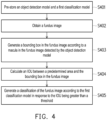
- FIG. 4 is a flowchart of a classification method for classifying a level of AMD.
- an object detection model and a first classification model are pre-stored.
- a fundus image is obtained.
- a bounding box is generated in the fundus image according to a macula in the fundus image detected by the object detection model.
- an intersection over union between a predetermined area and the bounding box in the fundus image is calculated.
- a classification of the fundus image is generated according to the first classification model in response to the intersection over union being greater than a threshold.
- the classification device of the disclosure may store the first classification model and the second classification model.
- the first classification model determines the level of the AMD according to the bounding box in the fundus image
- the second classification model determines the level of the AMD according to the entire fundus image. If the object detection model determines that the macula is present in the predetermined area in the fundus image, it means that the position of the macula in the fundus image is reasonable. Accordingly, the classification device may classify the fundus image according to the first classification model. If the object detection model determines that the macula is not present in the predetermined area in the fundus image, it means that the position of the macula in the fundus image is not reasonable. Accordingly, the classification device may classify the fundus image according to the second classification model. In other words, if the position of the macula in the fundus image is reasonable, the classification device may classify the level of the AMD according to the same criteria with the doctor.
Landscapes
- Engineering & Computer Science (AREA)
- Health & Medical Sciences (AREA)
- General Health & Medical Sciences (AREA)
- Physics & Mathematics (AREA)
- Medical Informatics (AREA)
- Theoretical Computer Science (AREA)
- General Physics & Mathematics (AREA)
- Life Sciences & Earth Sciences (AREA)
- Public Health (AREA)
- Biomedical Technology (AREA)
- Artificial Intelligence (AREA)
- Computer Vision & Pattern Recognition (AREA)
- Evolutionary Computation (AREA)
- Multimedia (AREA)
- Epidemiology (AREA)
- Molecular Biology (AREA)
- Primary Health Care (AREA)
- Ophthalmology & Optometry (AREA)
- Radiology & Medical Imaging (AREA)
- Nuclear Medicine, Radiotherapy & Molecular Imaging (AREA)
- Data Mining & Analysis (AREA)
- Software Systems (AREA)
- Pathology (AREA)
- Biophysics (AREA)
- Mathematical Physics (AREA)
- Databases & Information Systems (AREA)
- Human Computer Interaction (AREA)
- Computing Systems (AREA)
- Animal Behavior & Ethology (AREA)
- Heart & Thoracic Surgery (AREA)
- Veterinary Medicine (AREA)
- Surgery (AREA)
- Quality & Reliability (AREA)
- General Engineering & Computer Science (AREA)
- Signal Processing (AREA)
- Fuzzy Systems (AREA)
- Physiology (AREA)
- Psychiatry (AREA)
- Biodiversity & Conservation Biology (AREA)
- Computational Linguistics (AREA)
Description
- The disclosure relates to a classification method and a classification device for classifying a level of age-related macular degeneration.
- When determining a level of age-related macular degeneration (AMD) with an artificial intelligence (AI) model, input data of the artificial intelligence model includes mostly original fundus images or pre-processed fundus images. However, when determining severity level of the AMD, a doctor does not base a diagnosis on an entire eyeball area in a fundus image, but on a macula in the fundus image. In other words, the artificial intelligence model and the doctor have different determination criteria. Therefore, the determination result of the artificial intelligence model may be inaccurate.
- The article "Automatic grading of macular degeneration from color fundus images" by Jyoti Prakash Medhi et al, published on October 30, 2012 in the conference documents, pages 511-514 at the "2012 World Congress on Information and Communication Technologies" relates to the problem of diabetic retinopathy which causes blindness due to the physiological changes in the retina of human eye, which occurs due to the progression of diabetes. Diabetic retinopathy images differ from normal fundus images by lesions such as: microaneurysm, hemorrhages, exudates, cotton wool spots and variations in blood vessels etc. Appearance of these features on the retina leads to vision loss. The sharp vision is affected severely when the features appear on the macula as it contains higher concentration of cones. In this paper macula and fovea (macula center) are detected based on the localization. The detection of these feature is essential for automatic grading of macular edema or degeneration. Depending on the number of lesions on the macula the severity of the macular degeneration can be predicted. The method is tested on DRIVE, Aria and DIARETDB1 databases. The method successfully detects the macula and fovea for all the images. The accuracy of the proposed method of macula detection is found to be 100% in normal images. The method is also applied on images with lesions. Here the overlapped region of the macula and lesions are detected to find the severity of macular degeneration.
- The article "Eye diagnostic accuracy of current machine learning classifiers for age-related macular degeneration: a systematic review and meta-analysis" by Ronald Cheung et al, published via the Internet website nature.com/eye on May 6, 2021 relates to studies addressing the problem to systematically review and meta-analyze the diagnostic accuracy of current machine learning classifiers for age-related macular degeneration (AMD). Artificial intelligence diagnostic algorithms can automatically detect and diagnose AMD through training data from large sets of fundus or OCT images. The use of AI algorithms is a powerful tool, and it is a method of obtaining a cost-effective, simple, and fast diagnosis of AMD. The study MEDLINE, EMBASE, CINAHL, and ProQuest Dissertations and Theses were searched systematically and thoroughly. Conferences held through Association for Research in Vision and Ophthalmology, American Academy of Ophthalmology, and Canadian Society of Ophthalmology were searched. Studies were screened using Covidence software and data on sensitivity, specificity and area under curve were extracted from the included studies. STATA 15.0 was used to conduct the meta-analysis. The search strategy identified 307 records from online databases and 174 records from gray literature. Total of 13 records, 64,798 subjects (and 612,429 images), were used for the quantitative analysis. The pooled estimate for sensitivity was 0.918 [95% CI: 0.678, 0.98] and specificity was 0.888 [95% CI: 0.578, 0.98] for AMD screening using machine learning classifiers. The relative odds of a positive screen test in AMD cases were 89.74 [95% CI: 3.05-2641.59] times more likely than a negative screen test in non-AMD cases. The positive likelihood ratio was 8.22 [95% CI: 1.52-44.48] and the negative likelihood ratio was 0.09 [95% CI: 0.02-0.52]. The included studies show promising results for the diagnostic accuracy of the machine learning classifiers for AMD and its implementation in clinical settings.
- The disclosure provides a classification method and a classification device for classifying a level of age-related macular degeneration, in which the level of AMD can be classified according to a reasonable area in a fundus image.
- In the disclosure, a classification device for classifying a level of age-related macular degeneration includes a processor, a storage medium, and a transceiver. The storage medium stores an object detection model, a first classification model and a second classification model. The processor is coupled to the storage medium and the transceiver.
- The processor is configured to: obtain a fundus image through the transceiver, detect a macula in the fundus image detected by the object detection model to generate a bounding box of the macula, calculate an intersection over union between a predetermined area and the bounding box in the fundus image, generate a classification of the fundus image by inputting an image of the macula in the bounding box into the first classification model in response to the intersection over union being greater than a threshold; and generate the classification of the fundus image by inputting the fundus image into the second classification model in response to the intersection over union being less than or equal to the threshold.
- In an embodiment of the disclosure, a center point of the predetermined area is located at a geometric center of the fundus image.
- In an embodiment of the disclosure, the fundus image and the predetermined area are rectangles. A first edge of the predetermined area is distanced from a first boundary of the fundus image by a first distance, and a second edge of the predetermined area is distanced from a second boundary of the fundus image by the first distance. The second edge is an opposite edge of the first edge, and the second boundary is an opposite edge of the first boundary.
- In an embodiment of the disclosure, the predetermined area is a rectangle. The processor is further configured to obtain an eyeball area in the fundus image, and generate a length of a long edge and a length of a short edge of the rectangle according to a diameter of the eyeball area.
- In an embodiment of the disclosure, the classification indicates correspondence of the fundus image to one of a first stage, a second stage, a third stage, and a fourth stage of the age-related macular degeneration.
- In an embodiment of the disclosure, the first classification model and the second classification model have a same convolutional neural network architecture.
- In the disclosure, a classification method for classifying a level of age-related macular degeneration includes the following. An object detection model, a first classification model and a second classification model are pre-stored. A fundus image is obtained. A macula in the fundus image is detected by the object detection model to generate a bounding box of the macula. An intersection over union between a predetermined area and the bounding box in the fundus image is calculated. A classification of the fundus image is generated by inputting an image of the macula in the bounding box into the first classification model in response to the intersection over union being greater than a threshold. And the classification of the fundus image is generated by inputting the fundus image into the second classification model in response to the intersection over union being less than or equal to the threshold.
- Based on the foregoing, the classification device of the disclosure may classify the level of the AMD according to the same determination criteria with the doctor when a position of the macula in the fundus image is reasonable. If the position of the macula in the fundus image is not reasonable, the classification device may classify the level of the AMD according to the entire fundus image.
- To make the aforementioned more comprehensible, several embodiments accompanied with drawings are described in detail as follows. The invention is set out in the appended set of claims.
- The accompanying drawings are included to provide a further understanding of the disclosure, and are incorporated in and constitute a part of this specification. The drawings illustrate exemplary embodiments of the disclosure and, together with the description, serve to explain the principles of the disclosure.
-
FIG. 1 is a schematic diagram showing a classification device for classifying a level of AMD according to an embodiment of the disclosure. -
FIG. 2 is a schematic diagram showing a fundus image according to an embodiment of the disclosure. -
FIG. 3 is a schematic diagram showing a predetermined area and a bounding box according to an embodiment of the disclosure. -
FIG. 4 is a flowchart of a classification method for classifying a level of AMD. - To make the content of the disclosure more comprehensible, embodiments will be described below as examples for reliably implementing the disclosure accordingly. In addition, wherever possible, elements/members/steps labeled with the same reference numerals in the drawings and embodiments refer to the same or similar parts.
-
FIG. 1 is a schematic diagram showing aclassification device 100 for classifying a level of AMD according to an embodiment of the disclosure. Theclassification device 100 may include aprocessor 110, astorage medium 120, and atransceiver 130. Theclassification device 100 may be configured to determine the level of the AMD corresponding to a fundus image. Theclassification device 100 may classify the fundus image input into theclassification device 100 into a first stage, a second stage, a third stage, or a fourth stage of the AMD. - The
processor 110 is, for example, the central processing unit (CPU), or other programmable general-purpose or special-purpose micro control unit (MCU), microprocessor, digital signal processor (DSP), programmable controller, application specific integrated circuit (ASIC), graphics processing unit (GPU), image signal processor (ISP), image processing unit (IPU), arithmetic logic unit (ALU), complex programmable logic device (CPLD), field programmable gate array (FPGA), or other similar elements or a combination of the above elements. Theprocessor 110 may be coupled to thestorage medium 120 and thetransceiver 130, and may access and execute multiple modules and various applications stored in thestorage medium 120. - The
storage medium 120 is, for example, any form of fixed or mobile random access memory (RAM), read-only memory (ROM), flash memory, hard disk drive (HDD), solid state drive (SSD), or similar elements or a combination of the above elements. Thestorage medium 120 is configured to store multiple modules or various applications that may be executed by theprocessor 110. In this embodiment, thestorage medium 120 may store multiple models, such as afirst classification model 121, asecond classification model 122, and anobject detection model 123. - The
object detection model 123 is configured to detect a macula in the fundus image and generate a bounding box corresponding to the macula in the fundus image. Thefirst classification model 121 is configured to classify the fundus image according to an image in the bounding box. In other words, thefirst classification model 121 classifies the fundus image according to a portion of the fundus image. Thesecond classification model 122 is configured to classify the fundus image according to the entire fundus image. - The
transceiver 130 transmits and receives signals in a wireless or wired manner. Thetransceiver 130 may also perform operations such as low noise amplification, impedance matching, frequency mixing, frequency up- or down-conversion, filtering, amplification, and the like. - The
processor 110 obtains the fundus image through thetransceiver 130.FIG. 2 is a schematic diagram showing afundus image 200 according to an embodiment of the disclosure. Thefundus image 200 may include aneyeball area 210 and amacula 220. In an embodiment, theprocessor 110 may extract theeyeball area 210 from thefundus image 200 according to the Hough transform. After thefundus image 200 is obtained, theprocessor 110 may input thefundus image 200 into theobject detection model 123. Theobject detection model 123 is, for example, a machine learning model. Theobject detection model 123 detects the macula in thefundus image 200 to generate the bounding box in thefundus image 200, as shown inFIG. 3. FIG. 3 is a schematic diagram showing apredetermined area 300 and abounding box 400 according to an embodiment of the disclosure. - After the
bounding box 400 is generated, theprocessor 110 calculates an intersection over union (IOU) between thepredetermined area 300 and thebounding box 400 in thefundus image 200. If the intersection over union between thepredetermined area 300 and thebounding box 400 is greater than a threshold, theprocessor 110 generates a classification of thefundus image 200 according to thefirst classification model 121. Specifically, theprocessor 110 inputs an image in the bounding box 400 (i.e., an image of the macula 220) into thefirst classification model 121. Thefirst classification model 121 generates the classification of thefundus image 200 according to the image in thebounding box 400. Theprocessor 110 may output the classification of thefundus image 200 through thetransceiver 130 for the user's reference. The user may determine whether thefundus image 200 corresponds to the first stage, the second stage, the third stage, or the fourth stage of the AMD according to the classification output by thetransceiver 130. - If the intersection over union between the
predetermined area 300 and thebounding box 400 is less than or equal to the threshold, theprocessor 110 generates the classification of thefundus image 200 according to thesecond classification model 122. Specifically, theprocessor 110 inputs the fundus image 200 (i.e., an image of the entire eyeball) into thesecond classification model 122. Thesecond classification model 122 generates the classification of thefundus image 200 according to thefundus image 200. Theprocessor 110 may output the classification of thefundus image 200 through thetransceiver 130 for the user's reference. The user may determine whether thefundus image 200 corresponds to the first stage, the second stage, the third stage, or the fourth stage of the AMD according to the classification output by thetransceiver 130. - The
first classification model 121 or thesecond classification model 122 is, for example, a machine learning model. In an embodiment, thefirst classification model 121 and thesecond classification model 122 may have the same convolutional neural network architecture. However, since thefirst classification model 121 and thesecond classification model 122 are trained according to different training data sets or hyperparameters, the convolutional neural network in thefirst classification model 121 and the convolutional neural network in thesecond classification model 122 may have different weights. - The
fundus image 200 and thepredetermined area 300 may be rectangles. Theprocessor 110 may determine a position of thepredetermined area 300 according to the geometric center of thefundus image 200. Acenter point 310 of thepredetermined area 300 may be located at the geometric center of thefundus image 200. Thefundus image 200 may have afirst boundary 21, asecond boundary 22, a third boundary 23, and afourth boundary 24. Thefirst boundary 21 and thesecond boundary 22 may be the short edges of the rectangle, and the third boundary 23 and thefourth boundary 24 may be the long edges of the rectangle. Thesecond boundary 22 may be an opposite edge of thefirst boundary 21, and thefourth boundary 24 may be an opposite edge of the third boundary 23. On the other hand, thepredetermined area 300 may have afirst edge 31, asecond edge 32, athird edge 33, and afourth edge 34. Thefirst edge 31 and thesecond edge 32 may be the short edges of the rectangle, and thethird edge 33 and thefourth edge 34 may be the long edges of the rectangle. Thesecond edge 32 may be an opposite edge of thefirst edge 31, and thefourth edge 34 may be an opposite edge of thethird edge 33. - In an embodiment, the
processor 110 may determine thepredetermined area 300 according to the boundaries of thefundus image 200. Specifically, assuming that thepredetermined area 300 is a rectangle, thestorage medium 120 may pre-store a first distance D1 and a second distance D2. Theprocessor 110 may determine that thefirst edge 31 of thepredetermined area 300 is distanced from thefirst boundary 21 of thefundus image 200 by the first distance D1, and thesecond edge 32 of thepredetermined area 300 is distanced from thesecond boundary 22 of thefundus image 200 by the first distance D1. On the other hand, theprocessor 110 may determine that thethird edge 33 of thepredetermined area 300 is distanced from the third boundary 23 of thefundus image 200 by the second distance D2, and thefourth edge 34 of thepredetermined area 300 is distanced from thefourth boundary 24 of thefundus image 200 by the second distance D2. - In an embodiment, the
processor 110 may determine thepredetermined area 300 according to the diameter of theeyeball area 210. Assuming that thepredetermined area 300 is a rectangle, theprocessor 110 may calculate a length of the long edge and a length of the short edge of the rectangle (i.e., the predetermined area 300) according to the diameter of theeyeball area 210. For example, theprocessor 110 may multiply the diameter of theeyeball area 210 by 0.9 to calculate the length of the long edge (i.e., the length of thethird edge 33 or the fourth edge 34) of thepredetermined area 300. Theprocessor 110 may multiply the diameter of theeyeball area 210 by 0.8 to calculate the length of the short edge (i.e., the length of thefirst edge 31 or the second edge 32) of thepredetermined area 300. -
FIG. 4 is a flowchart of a classification method for classifying a level of AMD. In step S401, an object detection model and a first classification model are pre-stored. In step S402, a fundus image is obtained. In step S403, a bounding box is generated in the fundus image according to a macula in the fundus image detected by the object detection model. In step S404, an intersection over union between a predetermined area and the bounding box in the fundus image is calculated. In step S405, a classification of the fundus image is generated according to the first classification model in response to the intersection over union being greater than a threshold. - In summary of the foregoing, the classification device of the disclosure may store the first classification model and the second classification model. The first classification model determines the level of the AMD according to the bounding box in the fundus image, and the second classification model determines the level of the AMD according to the entire fundus image. If the object detection model determines that the macula is present in the predetermined area in the fundus image, it means that the position of the macula in the fundus image is reasonable. Accordingly, the classification device may classify the fundus image according to the first classification model. If the object detection model determines that the macula is not present in the predetermined area in the fundus image, it means that the position of the macula in the fundus image is not reasonable. Accordingly, the classification device may classify the fundus image according to the second classification model. In other words, if the position of the macula in the fundus image is reasonable, the classification device may classify the level of the AMD according to the same criteria with the doctor.
Claims (7)
- A classification device (100) for classifying a level of age-related macular degeneration, the classification device (100) comprising:a transceiver (130);a storage medium (120), storing an object detection model (123), a first classification model (121) and a second classification model (122); anda processor (110), coupled to the storage medium (120) and the transceiver (130), wherein the processor (110) is configured to:obtain a fundus image (200) through the transceiver (130);detect a macula (220) in the fundus image (200) detected by the object detection model (123) to generate a bounding box (400) of the macula (220);calculate an intersection over union between a predetermined area (300) and the bounding box (400) in the fundus image (200);generate a classification of the fundus image (200) by inputting an image of the macula (220) in the bounding box (400) into the first classification model (121) in response to the intersection over union being greater than a threshold; andgenerate the classification of the fundus image (200) by inputting the fundus image (200) into the second classification model (122) in response to the intersection over union being less than or equal to the threshold.
- The classification device (100) described in claim 1, wherein a center point (310) of the predetermined area (300) is located at a geometric center of the fundus image (200).
- The classification device (100) described in claim 2, wherein the fundus image (200) and the predetermined area (300) are rectangles, a first edge (31) of the predetermined area (300) is distanced from a first boundary (21) of the fundus image (200) by a first distance (D1), and a second edge (32) of the predetermined area (300) is distanced from a second boundary (22) of the fundus image (200) by the first distance (D1), wherein the second edge (32) is an opposite edge of the first edge (31), and the second boundary (22) is an opposite edge of the first boundary (21).
- The classification device (100) described in any one of claims 1 to 3, wherein the predetermined area (300) is a rectangle, and the processor (110) is further configured to:obtain an eyeball area (210) in the fundus image (200); andgenerate a length of a long edge and a length of a short edge of the rectangle according to a diameter of the eyeball area (210).
- The classification device (100) described in any one of claims 1 to 4, wherein the classification indicates correspondence of the fundus image (200) to one of a first stage, a second stage, a third stage, and a fourth stage of the age-related macular degeneration.
- The classification device (100) described in any one of claims 1 to 5, wherein the first classification model (121) and the second classification model (122) have a same convolutional neural network architecture.
- A classification method for classifying a level of age-related macular degeneration, the classification method comprising:pre-storing an object detection model (123), a first classification model (121), and a second classification model (122);obtaining a fundus image (200);detecting a macula (220) in the fundus image (200) by the object detection model (123) to generate a bounding box (400) of the macula (220);calculating an intersection over union between a predetermined area (300) and the bounding box (400) in the fundus image (200);generating a classification of the fundus image (200) by inputting an image of the macula (220) in the bounding box (400) into the first classification model (121) in response to the intersection over union being greater than a threshold; andgenerating the classification of the fundus image (200) by inputting the fundus image (200) into the second classification model (122) in response to the intersection over union being less than or equal to the threshold.
Applications Claiming Priority (1)
| Application Number | Priority Date | Filing Date | Title |
|---|---|---|---|
| TW110117544A TWI817121B (en) | 2021-05-14 | 2021-05-14 | Classification method and classification device for classifying level of amd |
Publications (2)
| Publication Number | Publication Date |
|---|---|
| EP4088649A1 EP4088649A1 (en) | 2022-11-16 |
| EP4088649B1 true EP4088649B1 (en) | 2024-07-17 |
Family
ID=77910611
Family Applications (1)
| Application Number | Title | Priority Date | Filing Date |
|---|---|---|---|
| EP21198357.2A Active EP4088649B1 (en) | 2021-05-14 | 2021-09-22 | Classification method and classification device for classifying level of amd |
Country Status (4)
| Country | Link |
|---|---|
| US (1) | US12361539B2 (en) |
| EP (1) | EP4088649B1 (en) |
| CN (1) | CN115345816B (en) |
| TW (1) | TWI817121B (en) |
Families Citing this family (1)
| Publication number | Priority date | Publication date | Assignee | Title |
|---|---|---|---|---|
| JP2023552377A (en) * | 2020-12-03 | 2023-12-15 | ジェネンテック, インコーポレイテッド | Multimodal prediction of geographic atrophy growth rate |
Family Cites Families (18)
| Publication number | Priority date | Publication date | Assignee | Title |
|---|---|---|---|---|
| US20140126787A1 (en) * | 2012-11-08 | 2014-05-08 | Lasarow Healthcare Technologies Limited | Device and method for generating melanoma risk assessments |
| US9775506B2 (en) | 2012-11-08 | 2017-10-03 | The Johns Hopkins University | System and method for detecting and classifying severity of retinal disease |
| WO2017031099A1 (en) * | 2015-08-20 | 2017-02-23 | Ohio University | Devices and methods for classifying diabetic and macular degeneration |
| JP6594170B2 (en) * | 2015-11-12 | 2019-10-23 | キヤノン株式会社 | Image processing apparatus, image processing method, image projection system, and program |
| US12008748B2 (en) * | 2018-05-31 | 2024-06-11 | Vuno, Inc. | Method for classifying fundus image of subject and device using same |
| CN109377474B (en) * | 2018-09-17 | 2021-06-15 | 苏州大学 | A Macular Localization Method Based on Improved Faster R-CNN |
| US20200193156A1 (en) * | 2018-12-12 | 2020-06-18 | Tesseract Health, Inc. | Biometric identification techniques |
| AU2019441740B2 (en) * | 2019-04-18 | 2025-12-18 | Tracery Ophthalmics Inc. | Detection, prediction, and classification for ocular disease |
| US11195271B2 (en) * | 2019-05-20 | 2021-12-07 | Macuject Pty Ltd. | Confidence-based method and system for analyzing images of a retina |
| CN110378211A (en) * | 2019-06-11 | 2019-10-25 | 广州海昇计算机科技有限公司 | Based on object detection method, system, device and the storage medium under the scene of classroom |
| EP4023143A4 (en) * | 2019-08-30 | 2023-08-30 | Canon Kabushiki Kaisha | INFORMATION PROCESSING DEVICE, INFORMATION PROCESSING METHOD, INFORMATION PROCESSING SYSTEM AND PROGRAM |
| CN110570421B (en) * | 2019-09-18 | 2022-03-22 | 北京鹰瞳科技发展股份有限公司 | Multitask fundus image classification method and apparatus |
| CN111028230A (en) * | 2019-12-24 | 2020-04-17 | 贵州大学 | A detection algorithm for optic disc and macular location in fundus images based on YOLO-V3 |
| WO2021175478A1 (en) * | 2020-03-03 | 2021-09-10 | Vito Nv | A system and method for classifying images of retina of eyes of subjects |
| US12299872B2 (en) * | 2020-06-16 | 2025-05-13 | Welch Allyn, Inc. | Detecting and tracking macular degeneration |
| CN112508864B (en) * | 2020-11-20 | 2022-08-02 | 昆明理工大学 | Retinal vessel image segmentation method based on improved UNet + |
| CN112598028B (en) * | 2020-12-10 | 2022-06-07 | 上海鹰瞳医疗科技有限公司 | Fundus image registration model training method, fundus image registration method and device |
| US11610306B2 (en) * | 2020-12-16 | 2023-03-21 | Industrial Technology Research Institute | Medical image analysis method and device |
-
2021
- 2021-05-14 TW TW110117544A patent/TWI817121B/en active
- 2021-08-31 US US17/462,020 patent/US12361539B2/en active Active
- 2021-09-22 EP EP21198357.2A patent/EP4088649B1/en active Active
- 2021-09-23 CN CN202111114182.1A patent/CN115345816B/en active Active
Also Published As
| Publication number | Publication date |
|---|---|
| TW202244842A (en) | 2022-11-16 |
| CN115345816B (en) | 2025-10-21 |
| TWI817121B (en) | 2023-10-01 |
| CN115345816A (en) | 2022-11-15 |
| US20220366559A1 (en) | 2022-11-17 |
| EP4088649A1 (en) | 2022-11-16 |
| US12361539B2 (en) | 2025-07-15 |
Similar Documents
| Publication | Publication Date | Title |
|---|---|---|
| Prasad et al. | Multiple eye disease detection using Deep Neural Network | |
| Antal et al. | An ensemble-based system for automatic screening of diabetic retinopathy | |
| JP6907563B2 (en) | Image processing device and image processing program | |
| EP3192051B1 (en) | Automated analysis of angiographic images | |
| KR102313143B1 (en) | Diabetic retinopathy detection and severity classification apparatus Based on Deep Learning and method thereof | |
| JP6878923B2 (en) | Image processing equipment, image processing system, and image processing program | |
| Jebaseeli et al. | Segmentation of retinal blood vessels from ophthalmologic diabetic retinopathy images | |
| WO2018143180A1 (en) | Image processing device and image processing program | |
| CN111862020B (en) | Method and device for predicting physiological age of anterior ocular segment, server and storage medium | |
| WO2017101529A1 (en) | Electrocardio lead intelligent selection method and system | |
| Oliveira et al. | Improved automated screening of diabetic retinopathy | |
| Ding et al. | Automatic detection of subretinal fluid and sub-retinal pigment epithelium fluid in optical coherence tomography images | |
| CN110619332B (en) | Data processing method, device and equipment based on visual field inspection report | |
| KR20210124024A (en) | Method, apparatus and computer program for calculating quality score of fundus image data using artificial intelligence | |
| CN117612241B (en) | Early warning method for teenager myopia and related equipment | |
| Gothane et al. | Diabetic retinopathy detection using deep learning | |
| Fadzil et al. | Analysis of foveal avascular zone in colour fundus images for grading of diabetic retinopathy severity | |
| Balakrishnan et al. | A hybrid PSO-DEFS based feature selection for the identification of diabetic retinopathy | |
| Hani et al. | Gaussian Bayes classifier for medical diagnosis and grading: application to diabetic retinopathy | |
| CN115206494B (en) | Film reading system and film reading method based on fundus image classification | |
| EP4088649A1 (en) | Classification method and classification device for classifying level of amd | |
| Navaneethan et al. | Automated Diabetic Retinopathy Detection and Multi-Stage Classification Using Convolutional Neural Networks | |
| Hameed et al. | Detection of eye diseases using ML | |
| Leontidis et al. | Exploiting the retinal vascular geometry in identifying the progression to diabetic retinopathy using penalized logistic regression and random forests | |
| CN115131592B (en) | Fundus image classification reading system and reading method |
Legal Events
| Date | Code | Title | Description |
|---|---|---|---|
| PUAI | Public reference made under article 153(3) epc to a published international application that has entered the european phase |
Free format text: ORIGINAL CODE: 0009012 |
|
| STAA | Information on the status of an ep patent application or granted ep patent |
Free format text: STATUS: REQUEST FOR EXAMINATION WAS MADE |
|
| 17P | Request for examination filed |
Effective date: 20220822 |
|
| AK | Designated contracting states |
Kind code of ref document: A1 Designated state(s): AL AT BE BG CH CY CZ DE DK EE ES FI FR GB GR HR HU IE IS IT LI LT LU LV MC MK MT NL NO PL PT RO RS SE SI SK SM TR |
|
| REG | Reference to a national code |
Ref country code: DE Free format text: PREVIOUS MAIN CLASS: A61B0003120000 Ref country code: DE Ref legal event code: R079 Ref document number: 602021015730 Country of ref document: DE Free format text: PREVIOUS MAIN CLASS: A61B0003120000 Ipc: G06V0040180000 |
|
| GRAP | Despatch of communication of intention to grant a patent |
Free format text: ORIGINAL CODE: EPIDOSNIGR1 |
|
| STAA | Information on the status of an ep patent application or granted ep patent |
Free format text: STATUS: GRANT OF PATENT IS INTENDED |
|
| RIC1 | Information provided on ipc code assigned before grant |
Ipc: G16H 50/20 20180101ALI20240226BHEP Ipc: G16H 30/40 20180101ALI20240226BHEP Ipc: A61B 3/12 20060101ALI20240226BHEP Ipc: G06V 10/82 20220101ALI20240226BHEP Ipc: G06V 10/44 20220101ALI20240226BHEP Ipc: G06V 40/18 20220101AFI20240226BHEP |
|
| INTG | Intention to grant announced |
Effective date: 20240313 |
|
| GRAS | Grant fee paid |
Free format text: ORIGINAL CODE: EPIDOSNIGR3 |
|
| GRAA | (expected) grant |
Free format text: ORIGINAL CODE: 0009210 |
|
| STAA | Information on the status of an ep patent application or granted ep patent |
Free format text: STATUS: THE PATENT HAS BEEN GRANTED |
|
| AK | Designated contracting states |
Kind code of ref document: B1 Designated state(s): AL AT BE BG CH CY CZ DE DK EE ES FI FR GB GR HR HU IE IS IT LI LT LU LV MC MK MT NL NO PL PT RO RS SE SI SK SM TR |
|
| REG | Reference to a national code |
Ref country code: CH Ref legal event code: EP |
|
| REG | Reference to a national code |
Ref country code: DE Ref legal event code: R096 Ref document number: 602021015730 Country of ref document: DE |
|
| REG | Reference to a national code |
Ref country code: IE Ref legal event code: FG4D |
|
| REG | Reference to a national code |
Ref country code: LT Ref legal event code: MG9D |
|
| REG | Reference to a national code |
Ref country code: NL Ref legal event code: MP Effective date: 20240717 |
|
| PG25 | Lapsed in a contracting state [announced via postgrant information from national office to epo] |
Ref country code: PT Free format text: LAPSE BECAUSE OF FAILURE TO SUBMIT A TRANSLATION OF THE DESCRIPTION OR TO PAY THE FEE WITHIN THE PRESCRIBED TIME-LIMIT Effective date: 20241118 |
|
| REG | Reference to a national code |
Ref country code: AT Ref legal event code: MK05 Ref document number: 1704890 Country of ref document: AT Kind code of ref document: T Effective date: 20240717 |
|
| PG25 | Lapsed in a contracting state [announced via postgrant information from national office to epo] |
Ref country code: NL Free format text: LAPSE BECAUSE OF FAILURE TO SUBMIT A TRANSLATION OF THE DESCRIPTION OR TO PAY THE FEE WITHIN THE PRESCRIBED TIME-LIMIT Effective date: 20240717 |
|
| PG25 | Lapsed in a contracting state [announced via postgrant information from national office to epo] |
Ref country code: PT Free format text: LAPSE BECAUSE OF FAILURE TO SUBMIT A TRANSLATION OF THE DESCRIPTION OR TO PAY THE FEE WITHIN THE PRESCRIBED TIME-LIMIT Effective date: 20241118 Ref country code: NL Free format text: LAPSE BECAUSE OF FAILURE TO SUBMIT A TRANSLATION OF THE DESCRIPTION OR TO PAY THE FEE WITHIN THE PRESCRIBED TIME-LIMIT Effective date: 20240717 |
|
| PG25 | Lapsed in a contracting state [announced via postgrant information from national office to epo] |
Ref country code: NO Free format text: LAPSE BECAUSE OF FAILURE TO SUBMIT A TRANSLATION OF THE DESCRIPTION OR TO PAY THE FEE WITHIN THE PRESCRIBED TIME-LIMIT Effective date: 20241017 |
|
| PG25 | Lapsed in a contracting state [announced via postgrant information from national office to epo] |
Ref country code: FI Free format text: LAPSE BECAUSE OF FAILURE TO SUBMIT A TRANSLATION OF THE DESCRIPTION OR TO PAY THE FEE WITHIN THE PRESCRIBED TIME-LIMIT Effective date: 20240717 Ref country code: PL Free format text: LAPSE BECAUSE OF FAILURE TO SUBMIT A TRANSLATION OF THE DESCRIPTION OR TO PAY THE FEE WITHIN THE PRESCRIBED TIME-LIMIT Effective date: 20240717 Ref country code: GR Free format text: LAPSE BECAUSE OF FAILURE TO SUBMIT A TRANSLATION OF THE DESCRIPTION OR TO PAY THE FEE WITHIN THE PRESCRIBED TIME-LIMIT Effective date: 20241018 |
|
| PG25 | Lapsed in a contracting state [announced via postgrant information from national office to epo] |
Ref country code: BG Free format text: LAPSE BECAUSE OF FAILURE TO SUBMIT A TRANSLATION OF THE DESCRIPTION OR TO PAY THE FEE WITHIN THE PRESCRIBED TIME-LIMIT Effective date: 20240717 |
|
| PG25 | Lapsed in a contracting state [announced via postgrant information from national office to epo] |
Ref country code: LV Free format text: LAPSE BECAUSE OF FAILURE TO SUBMIT A TRANSLATION OF THE DESCRIPTION OR TO PAY THE FEE WITHIN THE PRESCRIBED TIME-LIMIT Effective date: 20240717 |
|
| PG25 | Lapsed in a contracting state [announced via postgrant information from national office to epo] |
Ref country code: IS Free format text: LAPSE BECAUSE OF FAILURE TO SUBMIT A TRANSLATION OF THE DESCRIPTION OR TO PAY THE FEE WITHIN THE PRESCRIBED TIME-LIMIT Effective date: 20241117 Ref country code: AT Free format text: LAPSE BECAUSE OF FAILURE TO SUBMIT A TRANSLATION OF THE DESCRIPTION OR TO PAY THE FEE WITHIN THE PRESCRIBED TIME-LIMIT Effective date: 20240717 |
|
| PG25 | Lapsed in a contracting state [announced via postgrant information from national office to epo] |
Ref country code: HR Free format text: LAPSE BECAUSE OF FAILURE TO SUBMIT A TRANSLATION OF THE DESCRIPTION OR TO PAY THE FEE WITHIN THE PRESCRIBED TIME-LIMIT Effective date: 20240717 |
|
| PG25 | Lapsed in a contracting state [announced via postgrant information from national office to epo] |
Ref country code: ES Free format text: LAPSE BECAUSE OF FAILURE TO SUBMIT A TRANSLATION OF THE DESCRIPTION OR TO PAY THE FEE WITHIN THE PRESCRIBED TIME-LIMIT Effective date: 20240717 Ref country code: RS Free format text: LAPSE BECAUSE OF FAILURE TO SUBMIT A TRANSLATION OF THE DESCRIPTION OR TO PAY THE FEE WITHIN THE PRESCRIBED TIME-LIMIT Effective date: 20241017 |
|
| PG25 | Lapsed in a contracting state [announced via postgrant information from national office to epo] |
Ref country code: RS Free format text: LAPSE BECAUSE OF FAILURE TO SUBMIT A TRANSLATION OF THE DESCRIPTION OR TO PAY THE FEE WITHIN THE PRESCRIBED TIME-LIMIT Effective date: 20241017 Ref country code: PL Free format text: LAPSE BECAUSE OF FAILURE TO SUBMIT A TRANSLATION OF THE DESCRIPTION OR TO PAY THE FEE WITHIN THE PRESCRIBED TIME-LIMIT Effective date: 20240717 Ref country code: NO Free format text: LAPSE BECAUSE OF FAILURE TO SUBMIT A TRANSLATION OF THE DESCRIPTION OR TO PAY THE FEE WITHIN THE PRESCRIBED TIME-LIMIT Effective date: 20241017 Ref country code: LV Free format text: LAPSE BECAUSE OF FAILURE TO SUBMIT A TRANSLATION OF THE DESCRIPTION OR TO PAY THE FEE WITHIN THE PRESCRIBED TIME-LIMIT Effective date: 20240717 Ref country code: IS Free format text: LAPSE BECAUSE OF FAILURE TO SUBMIT A TRANSLATION OF THE DESCRIPTION OR TO PAY THE FEE WITHIN THE PRESCRIBED TIME-LIMIT Effective date: 20241117 Ref country code: HR Free format text: LAPSE BECAUSE OF FAILURE TO SUBMIT A TRANSLATION OF THE DESCRIPTION OR TO PAY THE FEE WITHIN THE PRESCRIBED TIME-LIMIT Effective date: 20240717 Ref country code: GR Free format text: LAPSE BECAUSE OF FAILURE TO SUBMIT A TRANSLATION OF THE DESCRIPTION OR TO PAY THE FEE WITHIN THE PRESCRIBED TIME-LIMIT Effective date: 20241018 Ref country code: FI Free format text: LAPSE BECAUSE OF FAILURE TO SUBMIT A TRANSLATION OF THE DESCRIPTION OR TO PAY THE FEE WITHIN THE PRESCRIBED TIME-LIMIT Effective date: 20240717 Ref country code: ES Free format text: LAPSE BECAUSE OF FAILURE TO SUBMIT A TRANSLATION OF THE DESCRIPTION OR TO PAY THE FEE WITHIN THE PRESCRIBED TIME-LIMIT Effective date: 20240717 Ref country code: BG Free format text: LAPSE BECAUSE OF FAILURE TO SUBMIT A TRANSLATION OF THE DESCRIPTION OR TO PAY THE FEE WITHIN THE PRESCRIBED TIME-LIMIT Effective date: 20240717 Ref country code: AT Free format text: LAPSE BECAUSE OF FAILURE TO SUBMIT A TRANSLATION OF THE DESCRIPTION OR TO PAY THE FEE WITHIN THE PRESCRIBED TIME-LIMIT Effective date: 20240717 |
|
| PG25 | Lapsed in a contracting state [announced via postgrant information from national office to epo] |
Ref country code: DK Free format text: LAPSE BECAUSE OF FAILURE TO SUBMIT A TRANSLATION OF THE DESCRIPTION OR TO PAY THE FEE WITHIN THE PRESCRIBED TIME-LIMIT Effective date: 20240717 Ref country code: SM Free format text: LAPSE BECAUSE OF FAILURE TO SUBMIT A TRANSLATION OF THE DESCRIPTION OR TO PAY THE FEE WITHIN THE PRESCRIBED TIME-LIMIT Effective date: 20240717 Ref country code: RO Free format text: LAPSE BECAUSE OF FAILURE TO SUBMIT A TRANSLATION OF THE DESCRIPTION OR TO PAY THE FEE WITHIN THE PRESCRIBED TIME-LIMIT Effective date: 20240717 |
|
| REG | Reference to a national code |
Ref country code: DE Ref legal event code: R097 Ref document number: 602021015730 Country of ref document: DE |
|
| PG25 | Lapsed in a contracting state [announced via postgrant information from national office to epo] |
Ref country code: MC Free format text: LAPSE BECAUSE OF FAILURE TO SUBMIT A TRANSLATION OF THE DESCRIPTION OR TO PAY THE FEE WITHIN THE PRESCRIBED TIME-LIMIT Effective date: 20240717 Ref country code: EE Free format text: LAPSE BECAUSE OF FAILURE TO SUBMIT A TRANSLATION OF THE DESCRIPTION OR TO PAY THE FEE WITHIN THE PRESCRIBED TIME-LIMIT Effective date: 20240717 |
|
| PG25 | Lapsed in a contracting state [announced via postgrant information from national office to epo] |
Ref country code: CZ Free format text: LAPSE BECAUSE OF FAILURE TO SUBMIT A TRANSLATION OF THE DESCRIPTION OR TO PAY THE FEE WITHIN THE PRESCRIBED TIME-LIMIT Effective date: 20240717 |
|
| PG25 | Lapsed in a contracting state [announced via postgrant information from national office to epo] |
Ref country code: SK Free format text: LAPSE BECAUSE OF FAILURE TO SUBMIT A TRANSLATION OF THE DESCRIPTION OR TO PAY THE FEE WITHIN THE PRESCRIBED TIME-LIMIT Effective date: 20240717 |
|
| REG | Reference to a national code |
Ref country code: CH Ref legal event code: PL |
|
| PG25 | Lapsed in a contracting state [announced via postgrant information from national office to epo] |
Ref country code: LU Free format text: LAPSE BECAUSE OF NON-PAYMENT OF DUE FEES Effective date: 20240922 |
|
| PLBE | No opposition filed within time limit |
Free format text: ORIGINAL CODE: 0009261 |
|
| STAA | Information on the status of an ep patent application or granted ep patent |
Free format text: STATUS: NO OPPOSITION FILED WITHIN TIME LIMIT |
|
| 26N | No opposition filed |
Effective date: 20250422 |
|
| REG | Reference to a national code |
Ref country code: BE Ref legal event code: MM Effective date: 20240930 |
|
| PG25 | Lapsed in a contracting state [announced via postgrant information from national office to epo] |
Ref country code: BE Free format text: LAPSE BECAUSE OF NON-PAYMENT OF DUE FEES Effective date: 20240930 |
|
| PG25 | Lapsed in a contracting state [announced via postgrant information from national office to epo] |
Ref country code: CH Free format text: LAPSE BECAUSE OF NON-PAYMENT OF DUE FEES Effective date: 20240930 |
|
| PG25 | Lapsed in a contracting state [announced via postgrant information from national office to epo] |
Ref country code: IE Free format text: LAPSE BECAUSE OF NON-PAYMENT OF DUE FEES Effective date: 20240922 |
|
| PG25 | Lapsed in a contracting state [announced via postgrant information from national office to epo] |
Ref country code: SE Free format text: LAPSE BECAUSE OF FAILURE TO SUBMIT A TRANSLATION OF THE DESCRIPTION OR TO PAY THE FEE WITHIN THE PRESCRIBED TIME-LIMIT Effective date: 20240717 |
|
| PGFP | Annual fee paid to national office [announced via postgrant information from national office to epo] |
Ref country code: DE Payment date: 20250730 Year of fee payment: 5 |
|
| PGFP | Annual fee paid to national office [announced via postgrant information from national office to epo] |
Ref country code: GB Payment date: 20250731 Year of fee payment: 5 |
|
| PGFP | Annual fee paid to national office [announced via postgrant information from national office to epo] |
Ref country code: FR Payment date: 20250808 Year of fee payment: 5 |
|
| PG25 | Lapsed in a contracting state [announced via postgrant information from national office to epo] |
Ref country code: IT Free format text: LAPSE BECAUSE OF FAILURE TO SUBMIT A TRANSLATION OF THE DESCRIPTION OR TO PAY THE FEE WITHIN THE PRESCRIBED TIME-LIMIT Effective date: 20240717 Ref country code: CY Free format text: LAPSE BECAUSE OF FAILURE TO SUBMIT A TRANSLATION OF THE DESCRIPTION OR TO PAY THE FEE WITHIN THE PRESCRIBED TIME-LIMIT; INVALID AB INITIO Effective date: 20210922 |
|
| PG25 | Lapsed in a contracting state [announced via postgrant information from national office to epo] |
Ref country code: HU Free format text: LAPSE BECAUSE OF FAILURE TO SUBMIT A TRANSLATION OF THE DESCRIPTION OR TO PAY THE FEE WITHIN THE PRESCRIBED TIME-LIMIT; INVALID AB INITIO Effective date: 20210922 |