EP4086606A2 - Module for freezing and storage of frozen tissue - Google Patents
Module for freezing and storage of frozen tissue Download PDFInfo
- Publication number
- EP4086606A2 EP4086606A2 EP22169969.7A EP22169969A EP4086606A2 EP 4086606 A2 EP4086606 A2 EP 4086606A2 EP 22169969 A EP22169969 A EP 22169969A EP 4086606 A2 EP4086606 A2 EP 4086606A2
- Authority
- EP
- European Patent Office
- Prior art keywords
- base
- tissue specimen
- mold
- lid
- frozen tissue
- Prior art date
- Legal status (The legal status is an assumption and is not a legal conclusion. Google has not performed a legal analysis and makes no representation as to the accuracy of the status listed.)
- Granted
Links
Images


Classifications
-
- G—PHYSICS
- G01—MEASURING; TESTING
- G01N—INVESTIGATING OR ANALYSING MATERIALS BY DETERMINING THEIR CHEMICAL OR PHYSICAL PROPERTIES
- G01N1/00—Sampling; Preparing specimens for investigation
- G01N1/28—Preparing specimens for investigation including physical details of (bio-)chemical methods covered elsewhere, e.g. G01N33/50, C12Q
- G01N1/36—Embedding or analogous mounting of samples
-
- B—PERFORMING OPERATIONS; TRANSPORTING
- B01—PHYSICAL OR CHEMICAL PROCESSES OR APPARATUS IN GENERAL
- B01L—CHEMICAL OR PHYSICAL LABORATORY APPARATUS FOR GENERAL USE
- B01L3/00—Containers or dishes for laboratory use, e.g. laboratory glassware; Droppers
- B01L3/50—Containers for the purpose of retaining a material to be analysed, e.g. test tubes
- B01L3/508—Rigid containers without fluid transport within
-
- G—PHYSICS
- G01—MEASURING; TESTING
- G01N—INVESTIGATING OR ANALYSING MATERIALS BY DETERMINING THEIR CHEMICAL OR PHYSICAL PROPERTIES
- G01N1/00—Sampling; Preparing specimens for investigation
- G01N1/28—Preparing specimens for investigation including physical details of (bio-)chemical methods covered elsewhere, e.g. G01N33/50, C12Q
- G01N1/42—Low-temperature sample treatment, e.g. cryofixation
-
- B—PERFORMING OPERATIONS; TRANSPORTING
- B01—PHYSICAL OR CHEMICAL PROCESSES OR APPARATUS IN GENERAL
- B01L—CHEMICAL OR PHYSICAL LABORATORY APPARATUS FOR GENERAL USE
- B01L2300/00—Additional constructional details
- B01L2300/04—Closures and closing means
- B01L2300/041—Connecting closures to device or container
-
- B—PERFORMING OPERATIONS; TRANSPORTING
- B01—PHYSICAL OR CHEMICAL PROCESSES OR APPARATUS IN GENERAL
- B01L—CHEMICAL OR PHYSICAL LABORATORY APPARATUS FOR GENERAL USE
- B01L2300/00—Additional constructional details
- B01L2300/06—Auxiliary integrated devices, integrated components
- B01L2300/0609—Holders integrated in container to position an object
-
- G—PHYSICS
- G01—MEASURING; TESTING
- G01N—INVESTIGATING OR ANALYSING MATERIALS BY DETERMINING THEIR CHEMICAL OR PHYSICAL PROPERTIES
- G01N1/00—Sampling; Preparing specimens for investigation
- G01N1/28—Preparing specimens for investigation including physical details of (bio-)chemical methods covered elsewhere, e.g. G01N33/50, C12Q
- G01N1/30—Staining; Impregnating ; Fixation; Dehydration; Multistep processes for preparing samples of tissue, cell or nucleic acid material and the like for analysis
- G01N1/31—Apparatus therefor
- G01N2001/315—Basket-type carriers for tissues
-
- G—PHYSICS
- G01—MEASURING; TESTING
- G01N—INVESTIGATING OR ANALYSING MATERIALS BY DETERMINING THEIR CHEMICAL OR PHYSICAL PROPERTIES
- G01N1/00—Sampling; Preparing specimens for investigation
- G01N1/28—Preparing specimens for investigation including physical details of (bio-)chemical methods covered elsewhere, e.g. G01N33/50, C12Q
- G01N1/36—Embedding or analogous mounting of samples
- G01N2001/366—Moulds; Demoulding
Definitions
- Tissue that is surgically removed from patients or subjects is routinely frozen for both diagnostic and research needs.
- Tissue obtained from frozen sections for histopathologic evaluation under the microscope is routinely frozen in a freezing compound, the most commonly used is OCT (Optimal Cutting Temperature - a mixture of polyproplyglycols, sucrose and water).
- OCT Optimal Cutting Temperature - a mixture of polyproplyglycols, sucrose and water
- Tissue obtained for research, or destined for a biobank is routinely frozen in the same matrix. This matrix has the benefit of protecting the tissue, as well as functioning as an embedding matrix for the preparation of frozen sections on a cryotome.
- a frozen tissue specimen storage kit comprising:
- a method for preparing a frozen tissue specimen block comprising:
- a method for microtome sectioning a frozen tissue specimen block comprising:
- tissue samples particularly biological tissue samples that are prepared for cryosectioning, archiving as frozen tissue, or subsequently fixed and processed as surgical specimens.
- the handling, labeling and storage of OCT-embedded frozen tissue is complicated, with ad hoc solutions applied, and no standardization.
- the vexing issue well documented in the literature, is the temperature fluctuation that occurs in frozen tissue handling, resulting in specimen degradation.
- specimen labeling A second, but equally problematic issue is specimen labeling.
- No current solution allows for a durable label to be affixed to the OCT embedded specimen.
- an insulated storage unit with integrated labeling, that addresses these problems.
- a tissue specimen is placed in a mold and a freezing matrix material (e.g., OCT) is applied to the tissue sample via introduction of the freezing matrix material into the mold.
- the freezing matrix material may fully or partially encompass the tissue sample.
- the resulting matrix material-embedded tissue is secured or placed onto a base (optionally via a Slotted U-shaped clamp).
- the mold/base/tissue sample is then frozen (along with optional Slotted U-shaped clamp).
- the base and a cooperating lid are snapped shut, and the entirety of the base/tissue sample/lid unit is then frozen.
- Freezing may be accomplished by a variety of methods including placing the unit in a freezer, placing the unit in freezing bath (e.g., isopentane, liquid nitrogen, dry ice (with or without an ethanol slurry), or placing the unit on a frozen surface.
- the freezing process secures the base to the frozen block of freezing matrix material and tissue.
- the mold can be removed (in the embodiments with the lid the lid can also be removed).
- the base is coupled to a chuck, meaning that the chuck is mounted with the frozen tissue available for immediate cryosectioning. After cryosectioning, the lid is replaced and the specimen is placed in an appropriate freezer. Alternatively the unit can be immediately archived for future use.
- the lid is placed onto the base, and the resulting unit is immersed in a fixative, such as neutral buffered formalin or other solutions at room temperature.
- a fixative such as neutral buffered formalin or other solutions at room temperature.
- the freezing matrix material will dissolve, the tissue specimen is retained in the orientation that it was cryosectioned. This alignment facilitates the production of well-aligned permanent section for evaluation by a pathologist for diagnosis confirmation after rendering a preliminary frozen section diagnosis.
- a mold is provided for embedding a tissue specimen in a matrix material.
- the matrix material may be any material suitable for freezing while maintaining the integrity of the tissue specimen.
- Illustrative matrix materials include water, saline, honey, sucrose and other sugar solutions, including those that contain formaldehyde, polyethylene glycol solutions.
- Freezing media include compounds of: saline solutions; sugar solution, most commonly sucrose and trehalose; polyethylene glycols; cellulose agarose; and latex.
- the matrix material may contain other chemicals, including fixatives, such as ethanol, formaldehyde, acids (acetic and picric for examples) and other chemical solutions that undergo a phase transition from gel(viscous liquid) to a solid under change of temperature (typically cooling, however heating (hydrogel)
- fixatives such as ethanol, formaldehyde, acids (acetic and picric for examples)
- other chemical solutions that undergo a phase transition from gel(viscous liquid) to a solid under change of temperature (typically cooling, however heating (hydrogel)
- Particular matrix materials are optimal cutting temperature compound (OCT, e.g., Tissue Tek TM available from Sakura Finetek, Jung Tissue Freezing Medium, or Leica Microsystems Surgipath FSC 22 Frozen Section Compound).
- OCT optimal cutting temperature compound
- the mold may be any mold suitable for holding a tissue specimen and the matrix material.
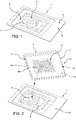
- An example of a mold 1 is shown in FIG. 1 .
- the mold 1 includes a cavity 4 into which
- some molds are round. Typical round molds have a 10-20 mm diameter, and a 5-10 mm depth. Typical rectilinear molds are 10-30 mm on sides, and 5-10 mm depth.
- a liquid matrix material 3 is introduced into the mold cavity 4 to partially or fully encompass the tissue specimen 2.
- the mold may include a flange 5, or a similar element, for handling and positioning the mold as required.
- the mold is made from a flexible material such as, for example, clear plastic.
- a base then is aligned and placed on the mold that contains the tissue specimen and the matrix material to form a mold/base construct.
- An example of a planar base 6 is shown in FIG. 2 .
- the base 6 includes a first surface 7 and an opposing second surface 8.
- the base 6 also defines an outer peripheral edge 9.
- the base 6 further includes protuberances 10 extending from the second surface 8.
- the protuberances 10 may be in the form of an array of individual small bumps or small projections.
- the protuberances array may be centered on the second surface 8. The protuberances assist in holding the tissue specimen in place.
- the base 6 further includes a ridge 11 at the peripheral edge 9 on three sides of the base.
- the ridge 11 could be provided on two sides or four sides of the base.
- the ridge 11 could be a matching circular shape.
- the ridge 11 is low in height (e.g., 0.5 to 2 mm) and can be 1.5-2.5 mm in width and does not constitute laterally extending walls.
- the mold 1 fits into the base 6 as shown in FIG. 2 such that the peripheral edges of the mold flange 5 contact the ridge 11 on multiple sides (e.g., three sides) of the base 6.
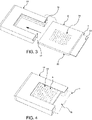
- the ridge 11 retains the mold 1 forming a mold/base construct 20 as demonstrated in FIG. 3 .
- the second surface 8 of the base 6 contacts the mold 1 so that the second surface 8 covers the mold cavity 4.
- the base protuberances 10 are aligned with and contact a layer of matrix material disposed on top of the tissue specimen 2.
- the edge 12 of the base 6 that does not include a ridge receives an optional extension 13 of the mold flange 5.
- An opening 19 is present in the ridge 11 opposite the edge 12 of the base 6. The opening 19 provides a means for un-snapping the base from other components as described in more detail below.
- the base 6 also includes a plurality of holes 16 extending from the first surface 7 to the second surface 8. The holes 16 permit discharge of excess freezing matrix material when the assembly is placed in a slotted U-shaped clamp as described below. In certain embodiments, the base does not include any holes.
- the ridge 11 is present on all four sides of the base. In this embodiment, the mold fits inside the ridges 11.
- the base also include an area 42 for labeling (shown in FIGS. 7 and 11 ).
- a label identifying the specific specimen may be affixed to the facing-out first surface 7.
- Illustrative labeling includes mechanical engraving or inscribing, writing in pencil or solvent resistant pen or printer, affixing an adhesive label (including a barcode), or affixing or embedding an RFID tag. Since the base travels with the specimen during processing, the label ensures that chain-of-custody for the specimen is maintained.
- a slotted U-shaped clamp 14 is used for securing the mold to the base during freezing.
- the slotted U-shaped clamp 14 includes a slot 15 and an open end 17.
- the base 6 and mold 1 i.e., the mold/base construct 20
- the base 6 and mold 1 are together slid into the open end 17 of the slotted U-shaped clamp 14 via inserting the base 6 and mold 1 together into the slot 15 provided in the slotted U-shaped clamp 14 and the base 6 and mold 1.
- the slot 15 may be offset from the vertical center of the U-shaped clamp 14 resulting in relatively flat mold/base construct 20.
- the slot 15 may be 2 mm from one surface of the U-shaped clamp 14 and 4.5 mm from the opposing surface of the U-shaped clamp 14.
- the mold extension 13 extends beyond the open end 17 of the slotted U-shaped clamp 14 to provide a means for holding the mold and base while inserting and removing the mold and base.
- the base has a sufficient length to enclose mold extension 13.
- the slotted U-shaped clamp can accept the mold extension into the clamp to provide more surface area for clamping.
- the slotted U-shaped clamp 14 holding the mold/base construct 20 see FIG.
- the temperature depends upon the specific matrix material.
- the freezing temperature may range, for example, from 0 to - 196°C, more particularly 0 to -4 °C.
- the freezing, base protuberances 10 and/or base holes 16 are sufficient for securing the matrix material and tissue specimen onto the second surface 8 of the base.
- the mold is removed from the base.
- the mold is a smooth material that is sufficiently flexible, even at low temperatures, and it is snapped, peeled, or pulled off.
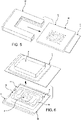
- An example is shown in FIG. 6 .
- the mold 1 can be removed by grasping the mold flange extension 13 and separating the mold 1 from the base 6.
- the tissue specimen block 33 extends from the second surface 8 of the base 6.
- the tissue specimen block 33 is centered on the second surface 8 of the base 6.
- the tissue specimen block 33 also is supported in a standalone position on the second surface 8. In other words, the tissue block 33 is not surrounded by, or retained by, any external walls.
- the base holding the tissue specimen block may be coupled with a lid to form a base/lid construct 21 (see FIGS. 8A , 8B and 8C ).
- the lid and base are snapped together.
- Base/lid construct 21 is for storage of the frozen tissue block in a controlled environment.
- the base holding the tissue specimen block may be attached to a chuck that is, in turn, attached to a cryostat as described in more detail below.
- the tissue specimen may then be cryosectioned prior to coupling of the lid.
- FIG. 7 An example of a lid 22 is shown in FIG. 7 .
- the lid 22 has a first side 23 and an opposing second side 24.
- the first side 23 defines a continuous planar surface.
- the second side 24 defines a recessed portion 25 configured to cover the tissue specimen block.
- the recessed portion 25 includes a bottom 26 and laterally extending walls 27. In FIGS. 8B and 8C the frozen tissue specimen block 33 does not contact the bottom 26 or walls 27 of the recessed portion 25.
- the lid 22 also includes an outer rim 28 configured to engage with the outer peripheral edge 9 of the base 6.
- the outer rim 28 and the peripheral edge are snap-fit together such that the lid 22 and the base 6 are can be coupled and de-coupled from each other.
- the outer rim 28 does not form part of the recessed portion 25 due to presence of a shelf 29 disposed between the outer rim 28 and the walls 27 of the recessed portion 25.
- the outer rim 28 extends upwards from the shelf 29.
- the peripheral dimension of the outer rim 28 is such that the peripheral edges 9 of the base 6 fit within the outer rim 28.
- the shelf 29 defines a slot 30 located contiguous to the outer rim 28 and that engages with the ridge 11 of the base 6.
- the outer rim 28 is present on only three peripheral sides of the lid 22 that coincide with the three peripheral sides of the base 6 at which the ridge 11 is also present.
- the lid 22 is removably coupled to the base 6 via a snap-fit constructed between (a) the outer rim 28 of the lid 22 and the peripheral edge 9 of the base 6 and (b) slot 30 of the lid 22 and the ridge 11 of the base 6.
- One side 31 of the lid 22 does not include the outer rim 28.
- An opening 32 is present in the outer rim 28 opposite the side 31 of the lid 22.
- the location of lid outer rim opening 32 coincides with the location of the base ridge opening 19. Openings 32 and 19 cooperate to provide an opening for insertion of a fingernail or a mechanical lever for un-snapping the base 6 from the lid 22.
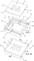
- FIGS. 11 and 12 Another embodiment of a lid and a base is shown in FIGS. 11 and 12 .
- the base includes a single, centered hole 40 that permits exiting of excess freezing matrix material.
- the lid includes a plurality of holes 41 extending through the recessed portion bottom 26. The holes 41 allow access for a liquid fixative into the base/lid construct holding a tissue specimen, for example, when the construct is immersed in the liquid fixative.
- the final assembly shown in FIGS. 8A , 8B and 8C stores the frozen tissue specimen block 33 between the base 6 and the lid 22.
- the final assembly also may have an outside surface area for labeling, for example, written, barcode (printed), adhesive label, or an integrated RFID tag.
- the closed assembly protects the tissue specimen from deformation and thermal isolations and shock, within the freezer (small changes in temp), during handling, inventory and management (moving from freezers), as well as provides insulation when in transfer.
- the fact that the lid can be removed while the base remains in a freezing compartment or on a frozen plate/surface, and the base affixed and cut, without melting/and embedding means the tissue does not have to have a freeze-thaw cycle to cut new sections after storage.
- the assembly is modular so that it will fit in existing storage units commonly used in research and clinical settings, with the label readily presentable.
- the dimensions of the final assembly are sufficiently large to hold a whole body organ (e.g., primate brain, kidney, etc.) cross-section.
- the dimensions of the final assembly may be 2 to 160 mm in length, 2 to 160 mm in width, and 2 to 50 mm in thickness.
- the dimensions may be 2.8 x 4.0 x 6.0 mm.
- the dimensions may be up to 40 x 60 x 15 mm.
- the assembly is affordable so that it can be a single use object.
- the modular device disclosed herein is designed to work with any tissue holders on cryostats or freezing mold models.
- the lid and base may be made from any suitable material.
- the material should be temperature-stable (i.e., insulative), resist solvents such as xylene or alcohols, and resist fixatives such as neutral buffered formalin.
- ABS Acrylonitrile butadiene styrene
- polyamide e.g., nylon
- the base that holds the frozen tissue specimen may be attached to a chuck that is, in turn, attached to a cryostat.
- a chuck 34 is provided with an arm 35 for mounting the chuck onto a cryostat.
- the arm 35 may be in the form of a cylinder or shaft that extends from a first surface 36 of the chuck 34.
- the chuck 34 includes a second surface 37 opposing the first surface 36.
- the base 6 may be attached to the second surface 37 of the chuck 34.
- the second surface 37 may define a peripheral raised rim 38 that is dimensioned to receive the base 6.
- the first surface 7 of the base contacts the second surface 37 of the chuck 34.
- the raised rim may include at least one opening 39 into which a fingernail or mechanical lever can be inserted for removing the base from the chuck.
- the lid 22 is removed from the base 6 thus making the frozen tissue specimen available for cryosectioning.
- a thin layer of freezing medium is usually applied to the bottom of the well of the cryomold.
- the tissue is then placed in the mold.
- the freezing matrix material is then added around the tissue until the mold is full, being careful not to displace the tissue when filling the mold.
- the open side of the mold is up as to not spill the freezing matrix material.
- the cryomold can now be removed and discarded.
- the articles and methods disclosed herein will facilitate specimen handling where the frozen tissue goes from a cryostat to a tissue processing cassette without manipulation. There is an incentive for this, as it will preserve the "cut face" of the tissue to allow better matching of the frozen sections to the permanent sections, a critical and complicated aspect of quality assurance in surgical pathology.
Landscapes
- Health & Medical Sciences (AREA)
- Chemical & Material Sciences (AREA)
- Analytical Chemistry (AREA)
- General Health & Medical Sciences (AREA)
- Biochemistry (AREA)
- Physics & Mathematics (AREA)
- Life Sciences & Earth Sciences (AREA)
- General Physics & Mathematics (AREA)
- Immunology (AREA)
- Pathology (AREA)
- Clinical Laboratory Science (AREA)
- Chemical Kinetics & Catalysis (AREA)
- Hematology (AREA)
- Sampling And Sample Adjustment (AREA)
Abstract
Description
- This application claims the benefit of
U.S. Provisional Application No. 62/403,585, filed October 3, 2016 - Tissue that is surgically removed from patients or subjects is routinely frozen for both diagnostic and research needs. There is a lack of standardization in freezing protocols, partially driven by utility. Tissue obtained from frozen sections for histopathologic evaluation under the microscope is routinely frozen in a freezing compound, the most commonly used is OCT (Optimal Cutting Temperature - a mixture of polyproplyglycols, sucrose and water). Tissue obtained for research, or destined for a biobank is routinely frozen in the same matrix. This matrix has the benefit of protecting the tissue, as well as functioning as an embedding matrix for the preparation of frozen sections on a cryotome.
- Currently, there is no routine method of storage of OCT embedded tissue after freezing. Small molds may help position and orient the tissue, but a protective enclosure that can be labeled and does not require the tissue to be partially thawed for use (cryosection, TMA production, other sampling) does not exist.
- Disclosed herein in one embodiment is an article comprising:
- a planar base having a first surface and an opposing second surface and an outer peripheral edge, wherein the second surface includes an area comprising a plurality of protuberances and configured to attach and maintain orientation of a frozen tissue specimen block; and
- a removable lid having a first side and a second side, wherein the first side defines a continuous planar surface and the second side defines a recessed portion configured to cover a frozen tissue specimen block and an outer rim configured to engage with the outer peripheral edge of the base, wherein the outer rim does not form part of the recessed portion,
- wherein the area of the base and the recessed portion of the lid are aligned.
- Also disclosed herein is a frozen tissue specimen storage kit comprising:
- a base having a first surface and an opposing second surface and an outer peripheral edge, wherein the second surface includes an area comprising a plurality of protuberances and configured to attach and maintain orientation of a frozen tissue specimen block;
- a removable lid having a (i) first side and a second side, wherein the first side defines a continuous planar surface and the second side defines a recessed portion configured to cover a frozen tissue specimen block and (ii) an outer rim configured to engage with the outer peripheral edge of the base;
- a slotted U-shaped clamp configured to receive the base and a tissue specimen mold that is aligned with the area of the base; and
- a chuck configured for mounting at least the base onto a cryostat.
- Further disclosed herein is a method for preparing a frozen tissue specimen block comprising:
- placing a tissue specimen and matrix material into a mold having an opening;
- contacting the mold with a base having a first surface and an opposing second surface and an outer peripheral edge, wherein the second surface includes a holding area comprising a plurality of protuberances, such that the mold opening aligns with the holding area of the base;
- subjecting the base/mold/tissue specimen to a temperature sufficient to freeze the matrix material resulting in a frozen tissue specimen block disposed on the holding area of the base;
- coupling a lid to the base, wherein the lid has a (i) first side and a second side, wherein the first side defines a continuous planar surface and the second side defines a recessed portion configured to cover the frozen tissue specimen block, and (ii) an outer rim engaged with the outer peripheral edge of the base.
- Additionally disclosed is a method for microtome sectioning a frozen tissue specimen block comprising:
- providing a frozen tissue specimen block stored inside a holder, the holder comprising (i) a base having a first surface and an opposing second surface and an outer peripheral edge, wherein the second surface includes a frozen tissue specimen attachment area comprising a plurality of protuberances, and (ii) a lid having a recessed portion configured to cover the frozen tissue specimen block;
- mounting a chuck onto a cryostat, wherein the chuck has a peripheral rim;
- coupling the base of the holder to the chuck via engaging the peripheral edge of the base with the peripheral rim of the chuck;
- removing the lid from the base to present a frozen tissue specimen block extending from the second surface of the base; and
- microtome sectioning the frozen tissue specimen block.
- The foregoing will become more apparent from the following detailed description, which proceeds with reference to the accompanying figures.
-
-
FIG. 1 is a perspective view of a mold holding a tissue specimen. -
FIG. 2 is a perspective view of a mold and a unit base. -
FIG. 3 is a perspective view of a mold/base construct for inserting into a slotted U-shaped clamp. -
FIG. 4 is a perspective view of the mold/base construct inserted into the slotted U-shaped clamp. -
FIG. 5 is a perspective view of a mold/base construct removed from the slotted U-shaped clamp. -
FIG. 6 is a perspective view of the mold removed from the base. -
FIG. 7 is a perspective view of the base and lid. -
FIG. 8A is a perspective view of the base/lid assembly. -
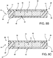
FIGS. 8B and 8C are cross-section views of different embodiments of the base/lid assembly view in the direction shown inFIG. 8A . -
FIG. 9 is a perspective view of a chuck mount that is retaining the base that holds a frozen tissue specimen block. -
FIG. 10 is a side view of a chuck mount that is retaining the base that holds a frozen tissue specimen block. -
FIG. 11 is a perspective view of another embodiment of the base and lid. -
FIG. 12 is a perspective view of another embodiment of the base. - Disclosed herein are devices and methods for freezing and storing tissue samples, particularly biological tissue samples that are prepared for cryosectioning, archiving as frozen tissue, or subsequently fixed and processed as surgical specimens. Currently, the handling, labeling and storage of OCT-embedded frozen tissue is complicated, with ad hoc solutions applied, and no standardization. The vexing issue, well documented in the literature, is the temperature fluctuation that occurs in frozen tissue handling, resulting in specimen degradation. A second, but equally problematic issue is specimen labeling. No current solution allows for a durable label to be affixed to the OCT embedded specimen. Disclosed herein is an insulated storage unit, with integrated labeling, that addresses these problems.
- In general, a tissue specimen is placed in a mold and a freezing matrix material (e.g., OCT) is applied to the tissue sample via introduction of the freezing matrix material into the mold. The freezing matrix material may fully or partially encompass the tissue sample. The resulting matrix material-embedded tissue is secured or placed onto a base (optionally via a Slotted U-shaped clamp). The mold/base/tissue sample is then frozen (along with optional Slotted U-shaped clamp). In certain embodiments, the base and a cooperating lid are snapped shut, and the entirety of the base/tissue sample/lid unit is then frozen. Freezing may be accomplished by a variety of methods including placing the unit in a freezer, placing the unit in freezing bath (e.g., isopentane, liquid nitrogen, dry ice (with or without an ethanol slurry), or placing the unit on a frozen surface. The freezing process secures the base to the frozen block of freezing matrix material and tissue. After freezing, the mold can be removed (in the embodiments with the lid the lid can also be removed). The base is coupled to a chuck, meaning that the chuck is mounted with the frozen tissue available for immediate cryosectioning. After cryosectioning, the lid is replaced and the specimen is placed in an appropriate freezer. Alternatively the unit can be immediately archived for future use.
- In a further embodiment, after cryosectioning the lid is placed onto the base, and the resulting unit is immersed in a fixative, such as neutral buffered formalin or other solutions at room temperature. Although the freezing matrix material will dissolve, the tissue specimen is retained in the orientation that it was cryosectioned. This alignment facilitates the production of well-aligned permanent section for evaluation by a pathologist for diagnosis confirmation after rendering a preliminary frozen section diagnosis.
- A mold is provided for embedding a tissue specimen in a matrix material. The matrix material may be any material suitable for freezing while maintaining the integrity of the tissue specimen. Illustrative matrix materials include water, saline, honey, sucrose and other sugar solutions, including those that contain formaldehyde, polyethylene glycol solutions. Freezing media include compounds of: saline solutions; sugar solution, most commonly sucrose and trehalose; polyethylene glycols; cellulose agarose; and latex. The matrix material may contain other chemicals, including fixatives, such as ethanol, formaldehyde, acids (acetic and picric for examples) and other chemical solutions that undergo a phase transition from gel(viscous liquid) to a solid under change of temperature (typically cooling, however heating (hydrogel) Particular matrix materials are optimal cutting temperature compound (OCT, e.g., Tissue Tek™ available from Sakura Finetek, Jung Tissue Freezing Medium, or Leica
Microsystems Surgipath FSC 22 Frozen Section Compound). The mold may be any mold suitable for holding a tissue specimen and the matrix material. An example of amold 1 is shown inFIG. 1 . Themold 1 includes acavity 4 into which atissue specimen 2 is placed. The mold can have differing depths and shapes. For example, some molds are round. Typical round molds have a 10-20 mm diameter, and a 5-10 mm depth. Typical rectilinear molds are 10-30 mm on sides, and 5-10 mm depth. Aliquid matrix material 3 is introduced into themold cavity 4 to partially or fully encompass thetissue specimen 2. The mold may include aflange 5, or a similar element, for handling and positioning the mold as required. In certain embodiments, the mold is made from a flexible material such as, for example, clear plastic. - A base then is aligned and placed on the mold that contains the tissue specimen and the matrix material to form a mold/base construct. An example of a
planar base 6 is shown inFIG. 2 . Thebase 6 includes afirst surface 7 and an opposingsecond surface 8. Thebase 6 also defines an outerperipheral edge 9. Thebase 6 further includesprotuberances 10 extending from thesecond surface 8. Theprotuberances 10 may be in the form of an array of individual small bumps or small projections. The protuberances array may be centered on thesecond surface 8. The protuberances assist in holding the tissue specimen in place. - The
base 6 further includes aridge 11 at theperipheral edge 9 on three sides of the base. In certain embodiments, theridge 11 could be provided on two sides or four sides of the base. In certain embodiments, if a circular mold is used theridge 11 could be a matching circular shape. Theridge 11 is low in height (e.g., 0.5 to 2 mm) and can be 1.5-2.5 mm in width and does not constitute laterally extending walls. Themold 1 fits into thebase 6 as shown inFIG. 2 such that the peripheral edges of themold flange 5 contact theridge 11 on multiple sides (e.g., three sides) of thebase 6. Thus, theridge 11 retains themold 1 forming a mold/base construct 20 as demonstrated inFIG. 3 . Thesecond surface 8 of thebase 6 contacts themold 1 so that thesecond surface 8 covers themold cavity 4. In particular, thebase protuberances 10 are aligned with and contact a layer of matrix material disposed on top of thetissue specimen 2. Theedge 12 of thebase 6 that does not include a ridge receives anoptional extension 13 of themold flange 5. Anopening 19 is present in theridge 11 opposite theedge 12 of thebase 6. Theopening 19 provides a means for un-snapping the base from other components as described in more detail below. Thebase 6 also includes a plurality ofholes 16 extending from thefirst surface 7 to thesecond surface 8. Theholes 16 permit discharge of excess freezing matrix material when the assembly is placed in a slotted U-shaped clamp as described below. In certain embodiments, the base does not include any holes. - In another embodiment of the
base 6, theridge 11 is present on all four sides of the base. In this embodiment, the mold fits inside theridges 11. - The base also include an
area 42 for labeling (shown inFIGS. 7 and11 ). For example, a label identifying the specific specimen may be affixed to the facing-outfirst surface 7. Illustrative labeling includes mechanical engraving or inscribing, writing in pencil or solvent resistant pen or printer, affixing an adhesive label (including a barcode), or affixing or embedding an RFID tag. Since the base travels with the specimen during processing, the label ensures that chain-of-custody for the specimen is maintained. - In certain embodiments as shown in
FIGS. 3-5 a slottedU-shaped clamp 14 is used for securing the mold to the base during freezing. The slottedU-shaped clamp 14 includes aslot 15 and anopen end 17. Thebase 6 and mold 1 (i.e., the mold/base construct 20) are together slid into theopen end 17 of the slottedU-shaped clamp 14 via inserting thebase 6 andmold 1 together into theslot 15 provided in the slottedU-shaped clamp 14 and thebase 6 andmold 1. In certain embodiments, theslot 15 may be offset from the vertical center of theU-shaped clamp 14 resulting in relatively flat mold/base construct 20. For example, theslot 15 may be 2 mm from one surface of theU-shaped clamp 14 and 4.5 mm from the opposing surface of theU-shaped clamp 14. In certain embodiments, themold extension 13 extends beyond theopen end 17 of the slottedU-shaped clamp 14 to provide a means for holding the mold and base while inserting and removing the mold and base. In other embodiments, the base has a sufficient length to enclosemold extension 13. In certain embodiments, the slotted U-shaped clamp can accept the mold extension into the clamp to provide more surface area for clamping. The slottedU-shaped clamp 14 holding the mold/base construct 20 (seeFIG. 4 ) then is subjected to a temperature sufficient for freezing the matrix material and tissue specimen resulting in converting the tissue specimen block into a frozen tissue specimen block 33 (block 33 is shown inFIGS. 6 ,8B and 8C ). The temperature depends upon the specific matrix material. The freezing temperature may range, for example, from 0 to - 196°C, more particularly 0 to -4 °C. The freezing,base protuberances 10 and/or base holes 16 are sufficient for securing the matrix material and tissue specimen onto thesecond surface 8 of the base. After formation of the frozentissue specimen block 18, the mold/base construct 20 is removed from the slottedU-shaped clamp 14 as shown inFIG. 5 . - After formation of the frozen tissue specimen block, the mold is removed from the base. The mold is a smooth material that is sufficiently flexible, even at low temperatures, and it is snapped, peeled, or pulled off. An example is shown in
FIG. 6 . Themold 1 can be removed by grasping themold flange extension 13 and separating themold 1 from thebase 6. After removal of the mold, thetissue specimen block 33 extends from thesecond surface 8 of thebase 6. In certain embodiments, thetissue specimen block 33 is centered on thesecond surface 8 of thebase 6. Thetissue specimen block 33 also is supported in a standalone position on thesecond surface 8. In other words, thetissue block 33 is not surrounded by, or retained by, any external walls. - The base holding the tissue specimen block may be coupled with a lid to form a base/lid construct 21 (see
FIGS. 8A ,8B and 8C ). In certain embodiments, the lid and base are snapped together. Base/lid construct 21 is for storage of the frozen tissue block in a controlled environment. - In certain embodiments, the base holding the tissue specimen block may be attached to a chuck that is, in turn, attached to a cryostat as described in more detail below. The tissue specimen may then be cryosectioned prior to coupling of the lid.
- An example of a
lid 22 is shown inFIG. 7 . Thelid 22 has afirst side 23 and an opposingsecond side 24. Thefirst side 23 defines a continuous planar surface. Thesecond side 24 defines a recessedportion 25 configured to cover the tissue specimen block. The recessedportion 25 includes a bottom 26 and laterally extendingwalls 27. InFIGS. 8B and 8C the frozentissue specimen block 33 does not contact the bottom 26 orwalls 27 of the recessedportion 25. - The
lid 22 also includes anouter rim 28 configured to engage with the outerperipheral edge 9 of thebase 6. In certain embodiments, theouter rim 28 and the peripheral edge are snap-fit together such that thelid 22 and thebase 6 are can be coupled and de-coupled from each other. Theouter rim 28 does not form part of the recessedportion 25 due to presence of ashelf 29 disposed between theouter rim 28 and thewalls 27 of the recessedportion 25. In certain embodiments, theouter rim 28 extends upwards from theshelf 29. The peripheral dimension of theouter rim 28 is such that theperipheral edges 9 of thebase 6 fit within theouter rim 28. In certain embodiments, theshelf 29 defines aslot 30 located contiguous to theouter rim 28 and that engages with theridge 11 of thebase 6. In certain embodiments theouter rim 28 is present on only three peripheral sides of thelid 22 that coincide with the three peripheral sides of thebase 6 at which theridge 11 is also present. Thus, thelid 22 is removably coupled to thebase 6 via a snap-fit constructed between (a) theouter rim 28 of thelid 22 and theperipheral edge 9 of thebase 6 and (b)slot 30 of thelid 22 and theridge 11 of thebase 6. - One
side 31 of thelid 22 does not include theouter rim 28. Anopening 32 is present in theouter rim 28 opposite theside 31 of thelid 22. The location of lid outer rim opening 32 coincides with the location of thebase ridge opening 19.Openings base 6 from thelid 22. - Another embodiment of a lid and a base is shown in
FIGS. 11 and 12 . In this embodiment, the base includes a single,centered hole 40 that permits exiting of excess freezing matrix material. The lid includes a plurality ofholes 41 extending through the recessedportion bottom 26. Theholes 41 allow access for a liquid fixative into the base/lid construct holding a tissue specimen, for example, when the construct is immersed in the liquid fixative. - The final assembly shown in
FIGS. 8A ,8B and 8C stores the frozentissue specimen block 33 between thebase 6 and thelid 22. The final assembly also may have an outside surface area for labeling, for example, written, barcode (printed), adhesive label, or an integrated RFID tag. The closed assembly protects the tissue specimen from deformation and thermal isolations and shock, within the freezer (small changes in temp), during handling, inventory and management (moving from freezers), as well as provides insulation when in transfer. The fact that the lid can be removed while the base remains in a freezing compartment or on a frozen plate/surface, and the base affixed and cut, without melting/and embedding means the tissue does not have to have a freeze-thaw cycle to cut new sections after storage. The assembly is modular so that it will fit in existing storage units commonly used in research and clinical settings, with the label readily presentable. In certain embodiments, the dimensions of the final assembly are sufficiently large to hold a whole body organ (e.g., primate brain, kidney, etc.) cross-section. In certain embodiments, the dimensions of the final assembly may be 2 to 160 mm in length, 2 to 160 mm in width, and 2 to 50 mm in thickness. In certain embodiments, the dimensions may be 2.8 x 4.0 x 6.0 mm. In certain embodiments, the dimensions may be up to 40 x 60 x 15 mm. In certain embodiments, the assembly is affordable so that it can be a single use object. The modular device disclosed herein is designed to work with any tissue holders on cryostats or freezing mold models. - The lid and base may be made from any suitable material. The material should be temperature-stable (i.e., insulative), resist solvents such as xylene or alcohols, and resist fixatives such as neutral buffered formalin. Acrylonitrile butadiene styrene (ABS) and polyamide (e.g., nylon) are illustrative plastics for making the device.
- In certain embodiments the base that holds the frozen tissue specimen may be attached to a chuck that is, in turn, attached to a cryostat. For example, as shown in
FIGS. 9 and 10 , achuck 34 is provided with anarm 35 for mounting the chuck onto a cryostat. Thearm 35 may be in the form of a cylinder or shaft that extends from afirst surface 36 of thechuck 34. Thechuck 34 includes asecond surface 37 opposing thefirst surface 36. Thebase 6 may be attached to thesecond surface 37 of thechuck 34. For instance, thesecond surface 37 may define a peripheral raisedrim 38 that is dimensioned to receive thebase 6. Thefirst surface 7 of the base contacts thesecond surface 37 of thechuck 34. In certain embodiments, the raised rim may include at least oneopening 39 into which a fingernail or mechanical lever can be inserted for removing the base from the chuck. Thelid 22 is removed from thebase 6 thus making the frozen tissue specimen available for cryosectioning. - An illustrative process is described below:
A thin layer of freezing medium is usually applied to the bottom of the well of the cryomold. The tissue is then placed in the mold. The freezing matrix material is then added around the tissue until the mold is full, being careful not to displace the tissue when filling the mold. The open side of the mold is up as to not spill the freezing matrix material. Place the base structured side down, the edges of the base align it to the freezing mold. At this point wipe off extraneous freezing media. Slide the mold and base into the slotted U-shaped clamp and freeze. After freezing is complete any excess freeing media should be scraped off. Once frozen the rough surface of the base will secure the frozen block of freezing medium and tissue to the base. The cryomold can now be removed and discarded. - The articles and methods disclosed herein will facilitate specimen handling where the frozen tissue goes from a cryostat to a tissue processing cassette without manipulation. There is an incentive for this, as it will preserve the "cut face" of the tissue to allow better matching of the frozen sections to the permanent sections, a critical and complicated aspect of quality assurance in surgical pathology.
- In view of the many possible embodiments to which the principles of the disclosed devices and methods may be applied, it should be recognized that the illustrated embodiments are only preferred examples of the invention and should not be taken as limiting the scope of the invention.
- The invention is further illustrated by the following numbered clauses:
- 1. An article comprising:
- a planar base having a first surface and an opposing second surface and an outer peripheral edge, wherein the second surface includes an area comprising a plurality of protuberances and configured to attach and maintain orientation of a frozen tissue specimen block; and
- a removable lid having a first side and a second side, wherein the first side defines a continuous planar surface and the second side defines a recessed portion configured to cover a frozen tissue specimen block and an outer rim configured to engage with the outer peripheral edge of the base, wherein the outer rim does not form part of the recessed portion,
- wherein the area of the base and the recessed portion of the lid are aligned.
- 2. The article of
clause 1, wherein the article stores a frozen tissue specimen block between the base and the lid. - 3. The article of
clause - 4. The article of any one of
clauses 1 to 3, wherein the base has four sides and the second surface of the base comprises a ridge on only three sides of the base configured to receive a tissue specimen mold. - 5. The article of
clause 1, wherein the planar base does not include any extending walls at the peripheral edge. - 6. The article of
clause 4, wherein the second side of the lid further defines a slot contiguous with the outer rim and the slot is configured to engage with the ridge of the base. - 7. The article of any one of
clauses 1 to 6, wherein the base and the lid are configured for snap fitting together. - 8. A frozen tissue specimen storage kit comprising:
- a base having a first surface and an opposing second surface and an outer peripheral edge, wherein the second surface includes an area comprising a plurality of protuberances and configured to attach and maintain orientation of a frozen tissue specimen block;
- a removable lid having a (i) first side and a second side, wherein the first side defines a continuous planar surface and the second side defines a recessed portion configured to cover a frozen tissue specimen block and (ii) an outer rim configured to engage with the outer peripheral edge of the base;
- a slotted U-shaped clamp configured to receive the base and a tissue specimen mold that is aligned with the area of the base; and
- a chuck configured for mounting at least the base onto a cryostat.
- 9. The kit of
claim 8, wherein the lid has four sides and the outer rim of the lid comprises a ridge on only three sides of the lid. - 10. The kit of
clause - 11. The kit of any of
clauses 8 to 10, wherein the chuck defines a peripheral raised rim that is configured to receive the base. - 12. A method for preparing a frozen tissue specimen block comprising:
- placing a tissue specimen and matrix material into a mold having an opening;
- contacting the mold with a base having a first surface and an opposing second surface and an outer peripheral edge, wherein the second surface includes a holding area comprising a plurality of protuberances, such that the mold opening aligns with the holding area of the base;
- subjecting the base/mold/tissue specimen to a temperature sufficient to freeze the matrix material resulting in a frozen tissue specimen block disposed on the holding area of the base; and
- coupling a lid to the base, wherein the lid has a (i) first side and a second side, wherein the first side defines a continuous planar surface and the second side defines a recessed portion configured to cover the frozen tissue specimen block, and (ii) an outer rim engaged with the outer peripheral edge of the base.
- 13. The method of
clause 12, wherein the lid is snapped onto the base. - 14. The method of
clause - 15. A method for microtome sectioning a frozen tissue specimen block comprising:
- providing a frozen tissue specimen block stored inside a holder, the holder comprising (i) a base having a first surface and an opposing second surface and an outer peripheral edge, wherein the second surface includes a frozen tissue specimen attachment area comprising a plurality of protuberances, and (ii) a lid having a recessed portion configured to cover the frozen tissue specimen block;
- mounting a chuck onto a cryostat, wherein the chuck has a peripheral rim;
- coupling the base of the holder to the chuck via engaging the peripheral edge of the base with the peripheral rim of the chuck;
- removing the lid from the base to present a frozen tissue specimen block extending from the second surface of the base; and
- microtome sectioning the frozen tissue specimen block
Claims (16)
- A frozen tissue specimen storage kit comprising:a base having a first surface and an opposing second surface and an outer peripheral edge, wherein the second surface includes an area comprising a plurality of protuberances and configured to attach and maintain orientation of a frozen tissue specimen block;a removable lid having a (i) first side and a second side, wherein the first side defines a continuous planar surface and the second side defines a recessed portion configured to cover a frozen tissue specimen block and (ii) an outer rim configured to engage with the outer peripheral edge of the base;a slotted U-shaped clamp configured to receive the base and a tissue specimen mold that is aligned with the area of the base; anda chuck configured for mounting at least the base onto a cryostat.
- The kit of claim 1, wherein the lid has four sides and the outer rim of the lid comprises a ridge on only three sides of the lid.
- The kit of claim 1 or claim 2, wherein the base has four sides and the second surface of the base comprises a ridge on only three sides of the base configured to receive a tissue specimen mold.
- The kit of any of claims 1 to 3, wherein the slotted U-shaped clamp includes a slot and an open end and the slotted U-shaped clamp is configured to secure a tissue specimen mold to the base when the base and mold and are inserted together into the slot.
- The kit of claim 4, wherein the tissue specimen mold comprises a mold extension which extends beyond the open end of the slotted U-shaped clamp when the tissue specimen mold is inserted into the slot of the U-shaped clamp to provide a means for holding the mold and base while inserting and removing the mold and base.
- The kit of claim any of claims 1 to 5, wherein the chuck is provided with an arm configured for mounting the chuck to a cryostat.
- The kit of claim 6, wherein the arm is in the form of a cylinder or shaft that extends from a first surface of the chuck.
- The kit of any of claims 1 to 7, wherein the chuck defines a peripheral raised rim that is configured to receive the base.
- The kit of any of claims 1 to 8, wherein base and/or lid further comprises a label.
- The kit according to claim 9, wherein the label is an engraved, inscribed, written, printed, adhesive, barcode or an RFID label.
- A method for preparing a frozen tissue specimen block comprising:placing a tissue specimen and matrix material into a mold having an opening;contacting the mold with a base having a first surface and an opposing second surface and an outer peripheral edge, wherein the second surface includes a holding area comprising a plurality of protuberances, such that the mold opening aligns with the holding area of the base;subjecting the base/mold/tissue specimen to a temperature sufficient to freeze the matrix material resulting in a frozen tissue specimen block disposed on the holding area of the base; andcoupling a lid to the base, wherein the lid has a (i) first side and a second side, wherein the first side defines a continuous planar surface and the second side defines a recessed portion configured to cover the frozen tissue specimen block, and (ii) an outer rim engaged with the outer peripheral edge of the base.
- The method of claim 11, wherein the lid is snapped onto the base.
- The method of claim 11, wherein a slotted U-shaped clamp is used for securing the mold to the base during freezing.
- The method of any of claims 11 to 13, further comprising inserting the base/lid/frozen tissue specimen block assembly into a storage unit.
- The method of any of claims 11 to 14, further comprising labelling an outside surface of the base and/or lid, optionally wherein the labelling comprises mechanical engraving or inscribing, writing, printing, affixing an adhesive label (including a barcode), or affixing or embedding an RFID tag.
- A method for microtome sectioning a frozen tissue specimen block comprising:providing a frozen tissue specimen block stored inside a holder, the holder comprising (i) a base having a first surface and an opposing second surface and an outer peripheral edge, wherein the second surface includes a frozen tissue specimen attachment area comprising a plurality of protuberances, and (ii) a lid having a recessed portion configured to cover the frozen tissue specimen block;mounting a chuck onto a cryostat, wherein the chuck has a peripheral rim;coupling the base of the holder to the chuck via engaging the peripheral edge of the base with the peripheral rim of the chuck;removing the lid from the base to present a frozen tissue specimen block extending from the second surface of the base; andmicrotome sectioning the frozen tissue specimen block.
Applications Claiming Priority (3)
| Application Number | Priority Date | Filing Date | Title |
|---|---|---|---|
| US201662403585P | 2016-10-03 | 2016-10-03 | |
| EP17785103.7A EP3519795B1 (en) | 2016-10-03 | 2017-09-29 | Module for freezing and storage of frozen tissue |
| PCT/US2017/054531 WO2018067407A1 (en) | 2016-10-03 | 2017-09-29 | Module for freezing and storage of frozen tissue |
Related Parent Applications (1)
| Application Number | Title | Priority Date | Filing Date |
|---|---|---|---|
| EP17785103.7A Division EP3519795B1 (en) | 2016-10-03 | 2017-09-29 | Module for freezing and storage of frozen tissue |
Publications (3)
| Publication Number | Publication Date |
|---|---|
| EP4086606A2 true EP4086606A2 (en) | 2022-11-09 |
| EP4086606A3 EP4086606A3 (en) | 2023-02-08 |
| EP4086606B1 EP4086606B1 (en) | 2025-02-12 |
Family
ID=60120162
Family Applications (2)
| Application Number | Title | Priority Date | Filing Date |
|---|---|---|---|
| EP17785103.7A Active EP3519795B1 (en) | 2016-10-03 | 2017-09-29 | Module for freezing and storage of frozen tissue |
| EP22169969.7A Active EP4086606B1 (en) | 2016-10-03 | 2017-09-29 | Frozen tissue specimen storage kit |
Family Applications Before (1)
| Application Number | Title | Priority Date | Filing Date |
|---|---|---|---|
| EP17785103.7A Active EP3519795B1 (en) | 2016-10-03 | 2017-09-29 | Module for freezing and storage of frozen tissue |
Country Status (3)
| Country | Link |
|---|---|
| US (1) | US11802821B2 (en) |
| EP (2) | EP3519795B1 (en) |
| WO (1) | WO2018067407A1 (en) |
Families Citing this family (4)
| Publication number | Priority date | Publication date | Assignee | Title |
|---|---|---|---|---|
| CN113340643A (en) * | 2021-06-07 | 2021-09-03 | 宁波市临床病理诊断中心 | Frozen tissue sample holder and using method thereof |
| CN117677439A (en) * | 2021-06-18 | 2024-03-08 | 莱卡生物系统努斯洛赫有限责任公司 | Cassette and tissue embedding method using the cassette |
| EP4374153A4 (en) * | 2021-11-10 | 2025-03-05 | Leica Biosystems Nussloch GmbH | CASSETTE AND COATING METHOD USING SAME |
| CN114002039A (en) | 2021-11-25 | 2022-02-01 | 丁伟 | Frozen sample holder with identification ring |
Family Cites Families (8)
| Publication number | Priority date | Publication date | Assignee | Title |
|---|---|---|---|---|
| US3982862A (en) | 1975-04-03 | 1976-09-28 | Pickett John E P | Two-part composite device for histologic tissue processing and embedding |
| US5358692A (en) | 1993-09-02 | 1994-10-25 | Reynolds Douglas W | Tissue cassette holder |
| US20040262318A1 (en) * | 2000-12-08 | 2004-12-30 | Ardais Corporation | Container, method and system for cryptopreserved material |
| US7172558B2 (en) | 2003-05-05 | 2007-02-06 | Duke University | Device for containing and analyzing surgically excised tissue and related methods |
| JP4758672B2 (en) * | 2005-04-21 | 2011-08-31 | 村角工業株式会社 | Embedding tray for preparation of histopathological specimen |
| CA2621386C (en) | 2005-09-08 | 2016-05-24 | London Health Sciences Centre Research Inc. | An embedding method and apparatus for the preparation of frozen section tissue |
| EP2316010B1 (en) * | 2008-08-27 | 2022-03-16 | The University of Miami | Tissue cassette and method for preparing tissue samples |
| GB201120626D0 (en) * | 2011-11-30 | 2012-01-11 | Ge Healthcare Uk Ltd | Biological sample storage apparatus and method |
-
2017
- 2017-09-29 EP EP17785103.7A patent/EP3519795B1/en active Active
- 2017-09-29 WO PCT/US2017/054531 patent/WO2018067407A1/en not_active Ceased
- 2017-09-29 EP EP22169969.7A patent/EP4086606B1/en active Active
- 2017-09-29 US US16/338,860 patent/US11802821B2/en active Active
Also Published As
| Publication number | Publication date |
|---|---|
| US11802821B2 (en) | 2023-10-31 |
| EP3519795A1 (en) | 2019-08-07 |
| US20210262906A1 (en) | 2021-08-26 |
| EP4086606A3 (en) | 2023-02-08 |
| EP4086606B1 (en) | 2025-02-12 |
| EP3519795B1 (en) | 2022-04-27 |
| WO2018067407A1 (en) | 2018-04-12 |
Similar Documents
| Publication | Publication Date | Title |
|---|---|---|
| JP4792586B2 (en) | Biological tissue fixation / embedding / slicing cassette and operation method thereof | |
| EP4086606B1 (en) | Frozen tissue specimen storage kit | |
| JP6487320B2 (en) | Tissue sample container and method | |
| JP5254218B2 (en) | Apparatus and method for preparing and storing microscopic specimens for analysis | |
| JP5124652B2 (en) | Tissue stabilization container system for molecular and pathological diagnosis | |
| EP1340062B1 (en) | Cryostorage method and device | |
| US12337322B2 (en) | Cassette assembly and processing method | |
| JP7583479B2 (en) | Sample holder with wireless transponder for attachment to sample collection body | |
| GB2518439A (en) | Cassette | |
| JP2025172873A (en) | Cryopreservation device with integrated tracking device chamber | |
| EP4058197B1 (en) | Rfid enabled specimen holder | |
| EP1558391B1 (en) | Low-temperature storage of suspension samples in suspended sample chambers | |
| US11714033B2 (en) | Tissue embedding cassette with shield | |
| Golubeva et al. | Collection and preparation of rodent tissue samples for histopathological and molecular studies in carcinogenesis | |
| CA3243434C (en) | Cryopreservation device with integrated tracking device chamber | |
| US20240138821A1 (en) | Slotted specimen holder, wireless transponder loading cartridge, wireless transponder dispenser and methods |
Legal Events
| Date | Code | Title | Description |
|---|---|---|---|
| PUAI | Public reference made under article 153(3) epc to a published international application that has entered the european phase |
Free format text: ORIGINAL CODE: 0009012 |
|
| STAA | Information on the status of an ep patent application or granted ep patent |
Free format text: STATUS: THE APPLICATION HAS BEEN PUBLISHED |
|
| AC | Divisional application: reference to earlier application |
Ref document number: 3519795 Country of ref document: EP Kind code of ref document: P |
|
| AK | Designated contracting states |
Kind code of ref document: A2 Designated state(s): AL AT BE BG CH CY CZ DE DK EE ES FI FR GB GR HR HU IE IS IT LI LT LU LV MC MK MT NL NO PL PT RO RS SE SI SK SM TR |
|
| PUAL | Search report despatched |
Free format text: ORIGINAL CODE: 0009013 |
|
| AK | Designated contracting states |
Kind code of ref document: A3 Designated state(s): AL AT BE BG CH CY CZ DE DK EE ES FI FR GB GR HR HU IE IS IT LI LT LU LV MC MK MT NL NO PL PT RO RS SE SI SK SM TR |
|
| RIC1 | Information provided on ipc code assigned before grant |
Ipc: G01N 1/31 19950101ALN20230105BHEP Ipc: B01L 3/00 19680901ALI20230105BHEP Ipc: G01N 1/42 19950101ALI20230105BHEP Ipc: G01N 1/36 19950101AFI20230105BHEP |
|
| STAA | Information on the status of an ep patent application or granted ep patent |
Free format text: STATUS: REQUEST FOR EXAMINATION WAS MADE |
|
| 17P | Request for examination filed |
Effective date: 20230808 |
|
| RBV | Designated contracting states (corrected) |
Designated state(s): AL AT BE BG CH CY CZ DE DK EE ES FI FR GB GR HR HU IE IS IT LI LT LU LV MC MK MT NL NO PL PT RO RS SE SI SK SM TR |
|
| GRAP | Despatch of communication of intention to grant a patent |
Free format text: ORIGINAL CODE: EPIDOSNIGR1 |
|
| STAA | Information on the status of an ep patent application or granted ep patent |
Free format text: STATUS: GRANT OF PATENT IS INTENDED |
|
| RIC1 | Information provided on ipc code assigned before grant |
Ipc: G01N 1/31 20060101ALN20240402BHEP Ipc: B01L 3/00 20060101ALI20240402BHEP Ipc: G01N 1/42 20060101ALI20240402BHEP Ipc: G01N 1/36 20060101AFI20240402BHEP |
|
| INTG | Intention to grant announced |
Effective date: 20240416 |
|
| GRAJ | Information related to disapproval of communication of intention to grant by the applicant or resumption of examination proceedings by the epo deleted |
Free format text: ORIGINAL CODE: EPIDOSDIGR1 |
|
| STAA | Information on the status of an ep patent application or granted ep patent |
Free format text: STATUS: REQUEST FOR EXAMINATION WAS MADE |
|
| GRAP | Despatch of communication of intention to grant a patent |
Free format text: ORIGINAL CODE: EPIDOSNIGR1 |
|
| STAA | Information on the status of an ep patent application or granted ep patent |
Free format text: STATUS: GRANT OF PATENT IS INTENDED |
|
| INTC | Intention to grant announced (deleted) | ||
| RAP3 | Party data changed (applicant data changed or rights of an application transferred) |
Owner name: OSHETSKI, MICHAEL Owner name: THE U.S.A. AS REPRESENTED BY THE SECRETARY,DEPARTMENT OF HEALTH AND HUMAN SERVICES |
|
| RIC1 | Information provided on ipc code assigned before grant |
Ipc: G01N 1/31 20060101ALN20240821BHEP Ipc: B01L 3/00 20060101ALI20240821BHEP Ipc: G01N 1/42 20060101ALI20240821BHEP Ipc: G01N 1/36 20060101AFI20240821BHEP |
|
| INTG | Intention to grant announced |
Effective date: 20240829 |
|
| GRAS | Grant fee paid |
Free format text: ORIGINAL CODE: EPIDOSNIGR3 |
|
| GRAA | (expected) grant |
Free format text: ORIGINAL CODE: 0009210 |
|
| STAA | Information on the status of an ep patent application or granted ep patent |
Free format text: STATUS: THE PATENT HAS BEEN GRANTED |
|
| AC | Divisional application: reference to earlier application |
Ref document number: 3519795 Country of ref document: EP Kind code of ref document: P |
|
| AK | Designated contracting states |
Kind code of ref document: B1 Designated state(s): AL AT BE BG CH CY CZ DE DK EE ES FI FR GB GR HR HU IE IS IT LI LT LU LV MC MK MT NL NO PL PT RO RS SE SI SK SM TR |
|
| REG | Reference to a national code |
Ref country code: GB Ref legal event code: FG4D |
|
| REG | Reference to a national code |
Ref country code: CH Ref legal event code: EP |
|
| REG | Reference to a national code |
Ref country code: DE Ref legal event code: R096 Ref document number: 602017087801 Country of ref document: DE |
|
| P01 | Opt-out of the competence of the unified patent court (upc) registered |
Free format text: CASE NUMBER: APP_5244/2025 Effective date: 20250131 |
|
| REG | Reference to a national code |
Ref country code: IE Ref legal event code: FG4D |
|
| REG | Reference to a national code |
Ref country code: NL Ref legal event code: MP Effective date: 20250212 |
|
| PG25 | Lapsed in a contracting state [announced via postgrant information from national office to epo] |
Ref country code: RS Free format text: LAPSE BECAUSE OF FAILURE TO SUBMIT A TRANSLATION OF THE DESCRIPTION OR TO PAY THE FEE WITHIN THE PRESCRIBED TIME-LIMIT Effective date: 20250512 |
|
| PG25 | Lapsed in a contracting state [announced via postgrant information from national office to epo] |
Ref country code: FI Free format text: LAPSE BECAUSE OF FAILURE TO SUBMIT A TRANSLATION OF THE DESCRIPTION OR TO PAY THE FEE WITHIN THE PRESCRIBED TIME-LIMIT Effective date: 20250212 |
|
| PG25 | Lapsed in a contracting state [announced via postgrant information from national office to epo] |
Ref country code: PL Free format text: LAPSE BECAUSE OF FAILURE TO SUBMIT A TRANSLATION OF THE DESCRIPTION OR TO PAY THE FEE WITHIN THE PRESCRIBED TIME-LIMIT Effective date: 20250212 |
|
| PG25 | Lapsed in a contracting state [announced via postgrant information from national office to epo] |
Ref country code: ES Free format text: LAPSE BECAUSE OF FAILURE TO SUBMIT A TRANSLATION OF THE DESCRIPTION OR TO PAY THE FEE WITHIN THE PRESCRIBED TIME-LIMIT Effective date: 20250212 |
|
| REG | Reference to a national code |
Ref country code: LT Ref legal event code: MG9D |
|
| PG25 | Lapsed in a contracting state [announced via postgrant information from national office to epo] |
Ref country code: IS Free format text: LAPSE BECAUSE OF FAILURE TO SUBMIT A TRANSLATION OF THE DESCRIPTION OR TO PAY THE FEE WITHIN THE PRESCRIBED TIME-LIMIT Effective date: 20250612 Ref country code: NO Free format text: LAPSE BECAUSE OF FAILURE TO SUBMIT A TRANSLATION OF THE DESCRIPTION OR TO PAY THE FEE WITHIN THE PRESCRIBED TIME-LIMIT Effective date: 20250512 |
|
| PG25 | Lapsed in a contracting state [announced via postgrant information from national office to epo] |
Ref country code: NL Free format text: LAPSE BECAUSE OF FAILURE TO SUBMIT A TRANSLATION OF THE DESCRIPTION OR TO PAY THE FEE WITHIN THE PRESCRIBED TIME-LIMIT Effective date: 20250212 |
|
| PG25 | Lapsed in a contracting state [announced via postgrant information from national office to epo] |
Ref country code: HR Free format text: LAPSE BECAUSE OF FAILURE TO SUBMIT A TRANSLATION OF THE DESCRIPTION OR TO PAY THE FEE WITHIN THE PRESCRIBED TIME-LIMIT Effective date: 20250212 |
|
| PG25 | Lapsed in a contracting state [announced via postgrant information from national office to epo] |
Ref country code: PT Free format text: LAPSE BECAUSE OF FAILURE TO SUBMIT A TRANSLATION OF THE DESCRIPTION OR TO PAY THE FEE WITHIN THE PRESCRIBED TIME-LIMIT Effective date: 20250612 Ref country code: LV Free format text: LAPSE BECAUSE OF FAILURE TO SUBMIT A TRANSLATION OF THE DESCRIPTION OR TO PAY THE FEE WITHIN THE PRESCRIBED TIME-LIMIT Effective date: 20250212 |
|
| PG25 | Lapsed in a contracting state [announced via postgrant information from national office to epo] |
Ref country code: BG Free format text: LAPSE BECAUSE OF FAILURE TO SUBMIT A TRANSLATION OF THE DESCRIPTION OR TO PAY THE FEE WITHIN THE PRESCRIBED TIME-LIMIT Effective date: 20250212 Ref country code: GR Free format text: LAPSE BECAUSE OF FAILURE TO SUBMIT A TRANSLATION OF THE DESCRIPTION OR TO PAY THE FEE WITHIN THE PRESCRIBED TIME-LIMIT Effective date: 20250513 |
|
| REG | Reference to a national code |
Ref country code: AT Ref legal event code: MK05 Ref document number: 1766435 Country of ref document: AT Kind code of ref document: T Effective date: 20250212 |
|
| PG25 | Lapsed in a contracting state [announced via postgrant information from national office to epo] |
Ref country code: SE Free format text: LAPSE BECAUSE OF FAILURE TO SUBMIT A TRANSLATION OF THE DESCRIPTION OR TO PAY THE FEE WITHIN THE PRESCRIBED TIME-LIMIT Effective date: 20250212 |
|
| PG25 | Lapsed in a contracting state [announced via postgrant information from national office to epo] |
Ref country code: SM Free format text: LAPSE BECAUSE OF FAILURE TO SUBMIT A TRANSLATION OF THE DESCRIPTION OR TO PAY THE FEE WITHIN THE PRESCRIBED TIME-LIMIT Effective date: 20250212 |
|
| PG25 | Lapsed in a contracting state [announced via postgrant information from national office to epo] |
Ref country code: DK Free format text: LAPSE BECAUSE OF FAILURE TO SUBMIT A TRANSLATION OF THE DESCRIPTION OR TO PAY THE FEE WITHIN THE PRESCRIBED TIME-LIMIT Effective date: 20250212 |
|
| PGFP | Annual fee paid to national office [announced via postgrant information from national office to epo] |
Ref country code: DE Payment date: 20250929 Year of fee payment: 9 |
|
| PG25 | Lapsed in a contracting state [announced via postgrant information from national office to epo] |
Ref country code: IT Free format text: LAPSE BECAUSE OF FAILURE TO SUBMIT A TRANSLATION OF THE DESCRIPTION OR TO PAY THE FEE WITHIN THE PRESCRIBED TIME-LIMIT Effective date: 20250212 |
|
| PGFP | Annual fee paid to national office [announced via postgrant information from national office to epo] |
Ref country code: GB Payment date: 20250929 Year of fee payment: 9 |
|
| PG25 | Lapsed in a contracting state [announced via postgrant information from national office to epo] |
Ref country code: AT Free format text: LAPSE BECAUSE OF FAILURE TO SUBMIT A TRANSLATION OF THE DESCRIPTION OR TO PAY THE FEE WITHIN THE PRESCRIBED TIME-LIMIT Effective date: 20250212 |
|
| PGFP | Annual fee paid to national office [announced via postgrant information from national office to epo] |
Ref country code: FR Payment date: 20250925 Year of fee payment: 9 |
|
| PG25 | Lapsed in a contracting state [announced via postgrant information from national office to epo] |
Ref country code: EE Free format text: LAPSE BECAUSE OF FAILURE TO SUBMIT A TRANSLATION OF THE DESCRIPTION OR TO PAY THE FEE WITHIN THE PRESCRIBED TIME-LIMIT Effective date: 20250212 Ref country code: CZ Free format text: LAPSE BECAUSE OF FAILURE TO SUBMIT A TRANSLATION OF THE DESCRIPTION OR TO PAY THE FEE WITHIN THE PRESCRIBED TIME-LIMIT Effective date: 20250212 |
|
| PG25 | Lapsed in a contracting state [announced via postgrant information from national office to epo] |
Ref country code: RO Free format text: LAPSE BECAUSE OF FAILURE TO SUBMIT A TRANSLATION OF THE DESCRIPTION OR TO PAY THE FEE WITHIN THE PRESCRIBED TIME-LIMIT Effective date: 20250212 |
|
| PG25 | Lapsed in a contracting state [announced via postgrant information from national office to epo] |
Ref country code: SK Free format text: LAPSE BECAUSE OF FAILURE TO SUBMIT A TRANSLATION OF THE DESCRIPTION OR TO PAY THE FEE WITHIN THE PRESCRIBED TIME-LIMIT Effective date: 20250212 |
|
| REG | Reference to a national code |
Ref country code: DE Ref legal event code: R097 Ref document number: 602017087801 Country of ref document: DE |
|
| PLBE | No opposition filed within time limit |
Free format text: ORIGINAL CODE: 0009261 |
|
| STAA | Information on the status of an ep patent application or granted ep patent |
Free format text: STATUS: NO OPPOSITION FILED WITHIN TIME LIMIT |
|
| REG | Reference to a national code |
Ref country code: CH Ref legal event code: L10 Free format text: ST27 STATUS EVENT CODE: U-0-0-L10-L00 (AS PROVIDED BY THE NATIONAL OFFICE) Effective date: 20251224 |
|
| 26N | No opposition filed |
Effective date: 20251113 |
|
| REG | Reference to a national code |
Ref country code: CH Ref legal event code: H13 Free format text: ST27 STATUS EVENT CODE: U-0-0-H10-H13 (AS PROVIDED BY THE NATIONAL OFFICE) Effective date: 20260504 |
|
| PG25 | Lapsed in a contracting state [announced via postgrant information from national office to epo] |
Ref country code: LU Free format text: LAPSE BECAUSE OF NON-PAYMENT OF DUE FEES Effective date: 20250929 |
|
| REG | Reference to a national code |
Ref country code: BE Ref legal event code: MM Effective date: 20250930 |
|
| PG25 | Lapsed in a contracting state [announced via postgrant information from national office to epo] |
Ref country code: LT Free format text: LAPSE BECAUSE OF FAILURE TO SUBMIT A TRANSLATION OF THE DESCRIPTION OR TO PAY THE FEE WITHIN THE PRESCRIBED TIME-LIMIT Effective date: 20250212 |
|
| PG25 | Lapsed in a contracting state [announced via postgrant information from national office to epo] |
Ref country code: MC Free format text: LAPSE BECAUSE OF FAILURE TO SUBMIT A TRANSLATION OF THE DESCRIPTION OR TO PAY THE FEE WITHIN THE PRESCRIBED TIME-LIMIT Effective date: 20250212 |
|
| PG25 | Lapsed in a contracting state [announced via postgrant information from national office to epo] |
Ref country code: CH Free format text: LAPSE BECAUSE OF NON-PAYMENT OF DUE FEES Effective date: 20250930 Ref country code: BE Free format text: LAPSE BECAUSE OF NON-PAYMENT OF DUE FEES Effective date: 20250930 Ref country code: IE Free format text: LAPSE BECAUSE OF NON-PAYMENT OF DUE FEES Effective date: 20250929 |