EP4071165A1 - A new non-aggregative splicing isoform of human tau is decreased in alzheimer s disease - Google Patents
A new non-aggregative splicing isoform of human tau is decreased in alzheimer s disease Download PDFInfo
- Publication number
- EP4071165A1 EP4071165A1 EP21382283.6A EP21382283A EP4071165A1 EP 4071165 A1 EP4071165 A1 EP 4071165A1 EP 21382283 A EP21382283 A EP 21382283A EP 4071165 A1 EP4071165 A1 EP 4071165A1
- Authority
- EP
- European Patent Office
- Prior art keywords
- tau
- peptide
- protein
- seq
- isoform
- Prior art date
- Legal status (The legal status is an assumption and is not a legal conclusion. Google has not performed a legal analysis and makes no representation as to the accuracy of the status listed.)
- Withdrawn
Links
- 108010029485 Protein Isoforms Proteins 0.000 title description 101
- 102000001708 Protein Isoforms Human genes 0.000 title description 101
- 101000891579 Homo sapiens Microtubule-associated protein tau Proteins 0.000 title description 39
- 208000037265 diseases, disorders, signs and symptoms Diseases 0.000 title description 25
- 201000010099 disease Diseases 0.000 title description 23
- 102000057063 human MAPT Human genes 0.000 title description 8
- 230000003247 decreasing effect Effects 0.000 title description 5
- 108090000765 processed proteins & peptides Proteins 0.000 claims abstract description 163
- 239000000203 mixture Substances 0.000 claims abstract description 35
- 125000003275 alpha amino acid group Chemical group 0.000 claims abstract description 33
- 230000002776 aggregation Effects 0.000 claims abstract description 26
- 238000004220 aggregation Methods 0.000 claims abstract description 26
- 108010026424 tau Proteins Proteins 0.000 claims description 189
- 102000013498 tau Proteins Human genes 0.000 claims description 188
- 108090000623 proteins and genes Proteins 0.000 claims description 112
- 102000004169 proteins and genes Human genes 0.000 claims description 101
- 150000001413 amino acids Chemical class 0.000 claims description 55
- 238000000034 method Methods 0.000 claims description 54
- 208000024827 Alzheimer disease Diseases 0.000 claims description 44
- 239000013598 vector Substances 0.000 claims description 26
- 230000002401 inhibitory effect Effects 0.000 claims description 25
- 239000012634 fragment Substances 0.000 claims description 13
- 238000000338 in vitro Methods 0.000 claims description 12
- 230000015572 biosynthetic process Effects 0.000 claims description 11
- 108091033319 polynucleotide Proteins 0.000 claims description 10
- 102000040430 polynucleotide Human genes 0.000 claims description 10
- 239000002157 polynucleotide Substances 0.000 claims description 10
- 238000011161 development Methods 0.000 claims description 9
- 239000008194 pharmaceutical composition Substances 0.000 claims description 9
- 208000034799 Tauopathies Diseases 0.000 claims description 8
- 239000003795 chemical substances by application Substances 0.000 claims description 7
- 239000000546 pharmaceutical excipient Substances 0.000 claims description 7
- 239000000090 biomarker Substances 0.000 claims description 5
- 239000003755 preservative agent Substances 0.000 claims description 5
- 206010002022 amyloidosis Diseases 0.000 claims description 4
- 210000004962 mammalian cell Anatomy 0.000 claims description 4
- 238000004393 prognosis Methods 0.000 claims description 4
- 230000000087 stabilizing effect Effects 0.000 claims description 4
- 239000003599 detergent Substances 0.000 claims description 3
- 239000003937 drug carrier Substances 0.000 claims description 3
- 239000000017 hydrogel Substances 0.000 claims description 3
- 239000003002 pH adjusting agent Substances 0.000 claims description 3
- 230000002335 preservative effect Effects 0.000 claims description 3
- 208000031124 Dementia Alzheimer type Diseases 0.000 claims description 2
- 210000005260 human cell Anatomy 0.000 claims description 2
- 230000007505 plaque formation Effects 0.000 claims description 2
- 239000004034 viscosity adjusting agent Substances 0.000 claims description 2
- 239000003112 inhibitor Substances 0.000 abstract description 72
- 102000013455 Amyloid beta-Peptides Human genes 0.000 abstract description 8
- 108010090849 Amyloid beta-Peptides Proteins 0.000 abstract description 8
- 231100000419 toxicity Toxicity 0.000 abstract description 3
- 230000001988 toxicity Effects 0.000 abstract description 3
- 235000018102 proteins Nutrition 0.000 description 74
- 210000004027 cell Anatomy 0.000 description 60
- YAEMHJKFIIIULI-UHFFFAOYSA-N n-(4-methoxybenzyl)-n'-(5-nitro-1,3-thiazol-2-yl)urea Chemical compound C1=CC(OC)=CC=C1CNC(=O)NC1=NC=C([N+]([O-])=O)S1 YAEMHJKFIIIULI-UHFFFAOYSA-N 0.000 description 59
- 229940024606 amino acid Drugs 0.000 description 54
- 235000001014 amino acid Nutrition 0.000 description 53
- 210000004556 brain Anatomy 0.000 description 47
- 108091032973 (ribonucleotides)n+m Proteins 0.000 description 42
- 230000014509 gene expression Effects 0.000 description 34
- 102100040243 Microtubule-associated protein tau Human genes 0.000 description 31
- 238000011529 RT qPCR Methods 0.000 description 31
- 241000894007 species Species 0.000 description 25
- 238000001262 western blot Methods 0.000 description 23
- 102000029749 Microtubule Human genes 0.000 description 22
- 108091022875 Microtubule Proteins 0.000 description 22
- 210000004688 microtubule Anatomy 0.000 description 22
- 102100029666 Serine/arginine-rich splicing factor 2 Human genes 0.000 description 20
- 108091034117 Oligonucleotide Proteins 0.000 description 19
- 230000027455 binding Effects 0.000 description 19
- 101000587430 Homo sapiens Serine/arginine-rich splicing factor 2 Proteins 0.000 description 18
- 230000014759 maintenance of location Effects 0.000 description 17
- FAPWRFPIFSIZLT-UHFFFAOYSA-M Sodium chloride Chemical compound [Na+].[Cl-] FAPWRFPIFSIZLT-UHFFFAOYSA-M 0.000 description 16
- 206010012289 Dementia Diseases 0.000 description 15
- 230000026731 phosphorylation Effects 0.000 description 14
- 238000006366 phosphorylation reaction Methods 0.000 description 14
- 102000004196 processed proteins & peptides Human genes 0.000 description 14
- 238000011002 quantification Methods 0.000 description 14
- 150000001875 compounds Chemical class 0.000 description 13
- 102000001267 GSK3 Human genes 0.000 description 11
- 108060006662 GSK3 Proteins 0.000 description 11
- 239000006228 supernatant Substances 0.000 description 11
- 102000030431 Asparaginyl endopeptidase Human genes 0.000 description 10
- 108010051109 Cell-Penetrating Peptides Proteins 0.000 description 10
- 102000020313 Cell-Penetrating Peptides Human genes 0.000 description 10
- 101000746263 Conus leopardus Conotoxin Lp5.1 Proteins 0.000 description 10
- TWRXJAOTZQYOKJ-UHFFFAOYSA-L Magnesium chloride Chemical compound [Mg+2].[Cl-].[Cl-] TWRXJAOTZQYOKJ-UHFFFAOYSA-L 0.000 description 10
- 238000004458 analytical method Methods 0.000 description 10
- 108010055066 asparaginylendopeptidase Proteins 0.000 description 10
- 230000001086 cytosolic effect Effects 0.000 description 10
- 239000013604 expression vector Substances 0.000 description 10
- 239000008188 pellet Substances 0.000 description 10
- 238000003753 real-time PCR Methods 0.000 description 10
- PEDCQBHIVMGVHV-UHFFFAOYSA-N Glycerine Chemical compound OCC(O)CO PEDCQBHIVMGVHV-UHFFFAOYSA-N 0.000 description 9
- 230000001594 aberrant effect Effects 0.000 description 9
- 239000000872 buffer Substances 0.000 description 9
- 230000008878 coupling Effects 0.000 description 9
- 238000010168 coupling process Methods 0.000 description 9
- 238000005859 coupling reaction Methods 0.000 description 9
- 238000001514 detection method Methods 0.000 description 9
- 239000000284 extract Substances 0.000 description 9
- 230000000971 hippocampal effect Effects 0.000 description 9
- NFVJNJQRWPQVOA-UHFFFAOYSA-N n-[2-chloro-5-(trifluoromethyl)phenyl]-2-[3-(4-ethyl-5-ethylsulfanyl-1,2,4-triazol-3-yl)piperidin-1-yl]acetamide Chemical compound CCN1C(SCC)=NN=C1C1CN(CC(=O)NC=2C(=CC=C(C=2)C(F)(F)F)Cl)CCC1 NFVJNJQRWPQVOA-UHFFFAOYSA-N 0.000 description 9
- 108020004414 DNA Proteins 0.000 description 8
- 108700024394 Exon Proteins 0.000 description 8
- 230000008499 blood brain barrier function Effects 0.000 description 8
- 210000001218 blood-brain barrier Anatomy 0.000 description 8
- 230000000694 effects Effects 0.000 description 8
- 238000005516 engineering process Methods 0.000 description 8
- 230000006870 function Effects 0.000 description 8
- 108020004999 messenger RNA Proteins 0.000 description 8
- 239000011780 sodium chloride Substances 0.000 description 8
- 239000000243 solution Substances 0.000 description 8
- 239000003826 tablet Substances 0.000 description 8
- 108020004705 Codon Proteins 0.000 description 7
- LFQSCWFLJHTTHZ-UHFFFAOYSA-N Ethanol Chemical compound CCO LFQSCWFLJHTTHZ-UHFFFAOYSA-N 0.000 description 7
- 102100031181 Glyceraldehyde-3-phosphate dehydrogenase Human genes 0.000 description 7
- 108010039259 RNA Splicing Factors Proteins 0.000 description 7
- 102000015097 RNA Splicing Factors Human genes 0.000 description 7
- 108020004445 glyceraldehyde-3-phosphate dehydrogenase Proteins 0.000 description 7
- 210000001320 hippocampus Anatomy 0.000 description 7
- 239000007788 liquid Substances 0.000 description 7
- 230000001404 mediated effect Effects 0.000 description 7
- 238000002360 preparation method Methods 0.000 description 7
- 239000000523 sample Substances 0.000 description 7
- 238000013519 translation Methods 0.000 description 7
- 102000001049 Amyloid Human genes 0.000 description 6
- 108010094108 Amyloid Proteins 0.000 description 6
- 241000894006 Bacteria Species 0.000 description 6
- 239000002775 capsule Substances 0.000 description 6
- 238000010367 cloning Methods 0.000 description 6
- 230000018109 developmental process Effects 0.000 description 6
- 239000002552 dosage form Substances 0.000 description 6
- 230000005764 inhibitory process Effects 0.000 description 6
- 231100000252 nontoxic Toxicity 0.000 description 6
- 230000003000 nontoxic effect Effects 0.000 description 6
- 238000001543 one-way ANOVA Methods 0.000 description 6
- YBYRMVIVWMBXKQ-UHFFFAOYSA-N phenylmethanesulfonyl fluoride Chemical compound FS(=O)(=O)CC1=CC=CC=C1 YBYRMVIVWMBXKQ-UHFFFAOYSA-N 0.000 description 6
- 238000003757 reverse transcription PCR Methods 0.000 description 6
- 231100000331 toxic Toxicity 0.000 description 6
- 230000002588 toxic effect Effects 0.000 description 6
- 108091093088 Amplicon Proteins 0.000 description 5
- 238000003559 RNA-seq method Methods 0.000 description 5
- JLCPHMBAVCMARE-UHFFFAOYSA-N [3-[[3-[[3-[[3-[[3-[[3-[[3-[[3-[[3-[[3-[[3-[[5-(2-amino-6-oxo-1H-purin-9-yl)-3-[[3-[[3-[[3-[[3-[[3-[[5-(2-amino-6-oxo-1H-purin-9-yl)-3-[[5-(2-amino-6-oxo-1H-purin-9-yl)-3-hydroxyoxolan-2-yl]methoxy-hydroxyphosphoryl]oxyoxolan-2-yl]methoxy-hydroxyphosphoryl]oxy-5-(5-methyl-2,4-dioxopyrimidin-1-yl)oxolan-2-yl]methoxy-hydroxyphosphoryl]oxy-5-(6-aminopurin-9-yl)oxolan-2-yl]methoxy-hydroxyphosphoryl]oxy-5-(6-aminopurin-9-yl)oxolan-2-yl]methoxy-hydroxyphosphoryl]oxy-5-(6-aminopurin-9-yl)oxolan-2-yl]methoxy-hydroxyphosphoryl]oxy-5-(6-aminopurin-9-yl)oxolan-2-yl]methoxy-hydroxyphosphoryl]oxyoxolan-2-yl]methoxy-hydroxyphosphoryl]oxy-5-(5-methyl-2,4-dioxopyrimidin-1-yl)oxolan-2-yl]methoxy-hydroxyphosphoryl]oxy-5-(4-amino-2-oxopyrimidin-1-yl)oxolan-2-yl]methoxy-hydroxyphosphoryl]oxy-5-(5-methyl-2,4-dioxopyrimidin-1-yl)oxolan-2-yl]methoxy-hydroxyphosphoryl]oxy-5-(5-methyl-2,4-dioxopyrimidin-1-yl)oxolan-2-yl]methoxy-hydroxyphosphoryl]oxy-5-(6-aminopurin-9-yl)oxolan-2-yl]methoxy-hydroxyphosphoryl]oxy-5-(6-aminopurin-9-yl)oxolan-2-yl]methoxy-hydroxyphosphoryl]oxy-5-(4-amino-2-oxopyrimidin-1-yl)oxolan-2-yl]methoxy-hydroxyphosphoryl]oxy-5-(4-amino-2-oxopyrimidin-1-yl)oxolan-2-yl]methoxy-hydroxyphosphoryl]oxy-5-(4-amino-2-oxopyrimidin-1-yl)oxolan-2-yl]methoxy-hydroxyphosphoryl]oxy-5-(6-aminopurin-9-yl)oxolan-2-yl]methoxy-hydroxyphosphoryl]oxy-5-(4-amino-2-oxopyrimidin-1-yl)oxolan-2-yl]methyl [5-(6-aminopurin-9-yl)-2-(hydroxymethyl)oxolan-3-yl] hydrogen phosphate Polymers Cc1cn(C2CC(OP(O)(=O)OCC3OC(CC3OP(O)(=O)OCC3OC(CC3O)n3cnc4c3nc(N)[nH]c4=O)n3cnc4c3nc(N)[nH]c4=O)C(COP(O)(=O)OC3CC(OC3COP(O)(=O)OC3CC(OC3COP(O)(=O)OC3CC(OC3COP(O)(=O)OC3CC(OC3COP(O)(=O)OC3CC(OC3COP(O)(=O)OC3CC(OC3COP(O)(=O)OC3CC(OC3COP(O)(=O)OC3CC(OC3COP(O)(=O)OC3CC(OC3COP(O)(=O)OC3CC(OC3COP(O)(=O)OC3CC(OC3COP(O)(=O)OC3CC(OC3COP(O)(=O)OC3CC(OC3COP(O)(=O)OC3CC(OC3COP(O)(=O)OC3CC(OC3COP(O)(=O)OC3CC(OC3COP(O)(=O)OC3CC(OC3CO)n3cnc4c(N)ncnc34)n3ccc(N)nc3=O)n3cnc4c(N)ncnc34)n3ccc(N)nc3=O)n3ccc(N)nc3=O)n3ccc(N)nc3=O)n3cnc4c(N)ncnc34)n3cnc4c(N)ncnc34)n3cc(C)c(=O)[nH]c3=O)n3cc(C)c(=O)[nH]c3=O)n3ccc(N)nc3=O)n3cc(C)c(=O)[nH]c3=O)n3cnc4c3nc(N)[nH]c4=O)n3cnc4c(N)ncnc34)n3cnc4c(N)ncnc34)n3cnc4c(N)ncnc34)n3cnc4c(N)ncnc34)O2)c(=O)[nH]c1=O JLCPHMBAVCMARE-UHFFFAOYSA-N 0.000 description 5
- 238000010171 animal model Methods 0.000 description 5
- 238000003556 assay Methods 0.000 description 5
- 239000012472 biological sample Substances 0.000 description 5
- 210000004899 c-terminal region Anatomy 0.000 description 5
- 238000003776 cleavage reaction Methods 0.000 description 5
- 239000002299 complementary DNA Substances 0.000 description 5
- 230000001965 increasing effect Effects 0.000 description 5
- 238000011534 incubation Methods 0.000 description 5
- 238000003780 insertion Methods 0.000 description 5
- 230000037431 insertion Effects 0.000 description 5
- 229910001629 magnesium chloride Inorganic materials 0.000 description 5
- 239000000463 material Substances 0.000 description 5
- 230000007246 mechanism Effects 0.000 description 5
- 230000007017 scission Effects 0.000 description 5
- 239000000126 substance Substances 0.000 description 5
- 238000002560 therapeutic procedure Methods 0.000 description 5
- -1 troches Substances 0.000 description 5
- XLYOFNOQVPJJNP-UHFFFAOYSA-N water Substances O XLYOFNOQVPJJNP-UHFFFAOYSA-N 0.000 description 5
- 238000011537 Coomassie blue staining Methods 0.000 description 4
- 206010011878 Deafness Diseases 0.000 description 4
- 241000282412 Homo Species 0.000 description 4
- 101001071233 Homo sapiens PHD finger protein 1 Proteins 0.000 description 4
- 101150070547 MAPT gene Proteins 0.000 description 4
- 208000016285 Movement disease Diseases 0.000 description 4
- 241000699666 Mus <mouse, genus> Species 0.000 description 4
- 102100036879 PHD finger protein 1 Human genes 0.000 description 4
- 239000013504 Triton X-100 Substances 0.000 description 4
- 229920004890 Triton X-100 Polymers 0.000 description 4
- 102000004243 Tubulin Human genes 0.000 description 4
- 108090000704 Tubulin Proteins 0.000 description 4
- 230000008033 biological extinction Effects 0.000 description 4
- 239000006185 dispersion Substances 0.000 description 4
- 238000002474 experimental method Methods 0.000 description 4
- 239000011536 extraction buffer Substances 0.000 description 4
- 210000005153 frontal cortex Anatomy 0.000 description 4
- 230000002209 hydrophobic effect Effects 0.000 description 4
- 238000001114 immunoprecipitation Methods 0.000 description 4
- 239000002105 nanoparticle Substances 0.000 description 4
- 230000004770 neurodegeneration Effects 0.000 description 4
- 102000039446 nucleic acids Human genes 0.000 description 4
- 108020004707 nucleic acids Proteins 0.000 description 4
- 150000007523 nucleic acids Chemical class 0.000 description 4
- 230000008569 process Effects 0.000 description 4
- 230000002441 reversible effect Effects 0.000 description 4
- 150000003384 small molecules Chemical class 0.000 description 4
- 230000001225 therapeutic effect Effects 0.000 description 4
- 125000000430 tryptophan group Chemical group [H]N([H])C(C(=O)O*)C([H])([H])C1=C([H])N([H])C2=C([H])C([H])=C([H])C([H])=C12 0.000 description 4
- DCXYFEDJOCDNAF-UHFFFAOYSA-N Asparagine Natural products OC(=O)C(N)CC(N)=O DCXYFEDJOCDNAF-UHFFFAOYSA-N 0.000 description 3
- 102000005593 Endopeptidases Human genes 0.000 description 3
- 108010059378 Endopeptidases Proteins 0.000 description 3
- 240000006570 Euonymus japonicus Species 0.000 description 3
- 235000016796 Euonymus japonicus Nutrition 0.000 description 3
- 102000019058 Glycogen Synthase Kinase 3 beta Human genes 0.000 description 3
- 108010051975 Glycogen Synthase Kinase 3 beta Proteins 0.000 description 3
- HTTJABKRGRZYRN-UHFFFAOYSA-N Heparin Chemical compound OC1C(NC(=O)C)C(O)OC(COS(O)(=O)=O)C1OC1C(OS(O)(=O)=O)C(O)C(OC2C(C(OS(O)(=O)=O)C(OC3C(C(O)C(O)C(O3)C(O)=O)OS(O)(=O)=O)C(CO)O2)NS(O)(=O)=O)C(C(O)=O)O1 HTTJABKRGRZYRN-UHFFFAOYSA-N 0.000 description 3
- 108091092195 Intron Proteins 0.000 description 3
- DCXYFEDJOCDNAF-REOHCLBHSA-N L-asparagine Chemical compound OC(=O)[C@@H](N)CC(N)=O DCXYFEDJOCDNAF-REOHCLBHSA-N 0.000 description 3
- 108091027974 Mature messenger RNA Proteins 0.000 description 3
- 206010029260 Neuroblastoma Diseases 0.000 description 3
- DNIAPMSPPWPWGF-UHFFFAOYSA-N Propylene glycol Chemical compound CC(O)CO DNIAPMSPPWPWGF-UHFFFAOYSA-N 0.000 description 3
- DBMJMQXJHONAFJ-UHFFFAOYSA-M Sodium laurylsulphate Chemical compound [Na+].CCCCCCCCCCCCOS([O-])(=O)=O DBMJMQXJHONAFJ-UHFFFAOYSA-M 0.000 description 3
- CZMRCDWAGMRECN-UGDNZRGBSA-N Sucrose Chemical compound O[C@H]1[C@H](O)[C@@H](CO)O[C@@]1(CO)O[C@@H]1[C@H](O)[C@@H](O)[C@H](O)[C@@H](CO)O1 CZMRCDWAGMRECN-UGDNZRGBSA-N 0.000 description 3
- 229930006000 Sucrose Natural products 0.000 description 3
- 229960001230 asparagine Drugs 0.000 description 3
- 235000009582 asparagine Nutrition 0.000 description 3
- 239000000969 carrier Substances 0.000 description 3
- 238000005119 centrifugation Methods 0.000 description 3
- 230000008859 change Effects 0.000 description 3
- 239000003153 chemical reaction reagent Substances 0.000 description 3
- 238000007796 conventional method Methods 0.000 description 3
- 210000004748 cultured cell Anatomy 0.000 description 3
- 238000003745 diagnosis Methods 0.000 description 3
- 230000003292 diminished effect Effects 0.000 description 3
- 239000003623 enhancer Substances 0.000 description 3
- 229960002897 heparin Drugs 0.000 description 3
- 229920000669 heparin Polymers 0.000 description 3
- 238000009396 hybridization Methods 0.000 description 3
- 238000001727 in vivo Methods 0.000 description 3
- 239000004615 ingredient Substances 0.000 description 3
- 238000002347 injection Methods 0.000 description 3
- 239000007924 injection Substances 0.000 description 3
- 239000012139 lysis buffer Substances 0.000 description 3
- 238000004519 manufacturing process Methods 0.000 description 3
- 238000011201 multiple comparisons test Methods 0.000 description 3
- 230000002018 overexpression Effects 0.000 description 3
- 230000001575 pathological effect Effects 0.000 description 3
- 230000007170 pathology Effects 0.000 description 3
- 239000006187 pill Substances 0.000 description 3
- 230000008488 polyadenylation Effects 0.000 description 3
- 229920001223 polyethylene glycol Polymers 0.000 description 3
- 239000000843 powder Substances 0.000 description 3
- 238000000746 purification Methods 0.000 description 3
- 238000003762 quantitative reverse transcription PCR Methods 0.000 description 3
- 230000002829 reductive effect Effects 0.000 description 3
- 230000001105 regulatory effect Effects 0.000 description 3
- 235000019333 sodium laurylsulphate Nutrition 0.000 description 3
- 239000007787 solid Substances 0.000 description 3
- 239000005720 sucrose Substances 0.000 description 3
- 239000004094 surface-active agent Substances 0.000 description 3
- 238000003786 synthesis reaction Methods 0.000 description 3
- 239000003981 vehicle Substances 0.000 description 3
- KDCGOANMDULRCW-UHFFFAOYSA-N 7H-purine Chemical compound N1=CNC2=NC=NC2=C1 KDCGOANMDULRCW-UHFFFAOYSA-N 0.000 description 2
- 241000167854 Bourreria succulenta Species 0.000 description 2
- 229920002261 Corn starch Polymers 0.000 description 2
- 150000008574 D-amino acids Chemical class 0.000 description 2
- 102000016911 Deoxyribonucleases Human genes 0.000 description 2
- 108010053770 Deoxyribonucleases Proteins 0.000 description 2
- 238000001061 Dunnett's test Methods 0.000 description 2
- KCXVZYZYPLLWCC-UHFFFAOYSA-N EDTA Chemical compound OC(=O)CN(CC(O)=O)CCN(CC(O)=O)CC(O)=O KCXVZYZYPLLWCC-UHFFFAOYSA-N 0.000 description 2
- 238000002965 ELISA Methods 0.000 description 2
- LYCAIKOWRPUZTN-UHFFFAOYSA-N Ethylene glycol Chemical compound OCCO LYCAIKOWRPUZTN-UHFFFAOYSA-N 0.000 description 2
- 229930091371 Fructose Natural products 0.000 description 2
- RFSUNEUAIZKAJO-ARQDHWQXSA-N Fructose Chemical compound OC[C@H]1O[C@](O)(CO)[C@@H](O)[C@@H]1O RFSUNEUAIZKAJO-ARQDHWQXSA-N 0.000 description 2
- 239000005715 Fructose Substances 0.000 description 2
- 108010010803 Gelatin Proteins 0.000 description 2
- 101000587442 Homo sapiens Serine/arginine-rich splicing factor 6 Proteins 0.000 description 2
- 208000023105 Huntington disease Diseases 0.000 description 2
- 238000012313 Kruskal-Wallis test Methods 0.000 description 2
- 150000008575 L-amino acids Chemical class 0.000 description 2
- CSNNHWWHGAXBCP-UHFFFAOYSA-L Magnesium sulfate Chemical compound [Mg+2].[O-][S+2]([O-])([O-])[O-] CSNNHWWHGAXBCP-UHFFFAOYSA-L 0.000 description 2
- 241001465754 Metazoa Species 0.000 description 2
- 102000009664 Microtubule-Associated Proteins Human genes 0.000 description 2
- 108010020004 Microtubule-Associated Proteins Proteins 0.000 description 2
- 241000699670 Mus sp. Species 0.000 description 2
- 239000000020 Nitrocellulose Substances 0.000 description 2
- 108091028043 Nucleic acid sequence Proteins 0.000 description 2
- 240000004808 Saccharomyces cerevisiae Species 0.000 description 2
- 101710123513 Serine/arginine-rich splicing factor 2 Proteins 0.000 description 2
- 102100029710 Serine/arginine-rich splicing factor 6 Human genes 0.000 description 2
- VYPSYNLAJGMNEJ-UHFFFAOYSA-N Silicium dioxide Chemical compound O=[Si]=O VYPSYNLAJGMNEJ-UHFFFAOYSA-N 0.000 description 2
- 230000002159 abnormal effect Effects 0.000 description 2
- 238000002835 absorbance Methods 0.000 description 2
- 230000004913 activation Effects 0.000 description 2
- 239000011543 agarose gel Substances 0.000 description 2
- 125000000539 amino acid group Chemical group 0.000 description 2
- AVKUERGKIZMTKX-NJBDSQKTSA-N ampicillin Chemical compound C1([C@@H](N)C(=O)N[C@H]2[C@H]3SC([C@@H](N3C2=O)C(O)=O)(C)C)=CC=CC=C1 AVKUERGKIZMTKX-NJBDSQKTSA-N 0.000 description 2
- 229960000723 ampicillin Drugs 0.000 description 2
- 239000004599 antimicrobial Substances 0.000 description 2
- 239000011324 bead Substances 0.000 description 2
- 230000033228 biological regulation Effects 0.000 description 2
- 210000005013 brain tissue Anatomy 0.000 description 2
- 238000010805 cDNA synthesis kit Methods 0.000 description 2
- 125000003178 carboxy group Chemical group [H]OC(*)=O 0.000 description 2
- 238000004113 cell culture Methods 0.000 description 2
- 235000019693 cherries Nutrition 0.000 description 2
- 239000008120 corn starch Substances 0.000 description 2
- 108091092330 cytoplasmic RNA Proteins 0.000 description 2
- 230000003013 cytotoxicity Effects 0.000 description 2
- 231100000135 cytotoxicity Toxicity 0.000 description 2
- 230000007423 decrease Effects 0.000 description 2
- 208000035475 disorder Diseases 0.000 description 2
- 239000003814 drug Substances 0.000 description 2
- 239000007938 effervescent tablet Substances 0.000 description 2
- 238000001493 electron microscopy Methods 0.000 description 2
- DEFVIWRASFVYLL-UHFFFAOYSA-N ethylene glycol bis(2-aminoethyl)tetraacetic acid Chemical compound OC(=O)CN(CC(O)=O)CCOCCOCCN(CC(O)=O)CC(O)=O DEFVIWRASFVYLL-UHFFFAOYSA-N 0.000 description 2
- 239000000835 fiber Substances 0.000 description 2
- 238000007667 floating Methods 0.000 description 2
- 239000007850 fluorescent dye Substances 0.000 description 2
- 235000003599 food sweetener Nutrition 0.000 description 2
- 230000002496 gastric effect Effects 0.000 description 2
- 239000008273 gelatin Substances 0.000 description 2
- 229920000159 gelatin Polymers 0.000 description 2
- 235000019322 gelatine Nutrition 0.000 description 2
- 235000011852 gelatine desserts Nutrition 0.000 description 2
- 230000002068 genetic effect Effects 0.000 description 2
- KWIUHFFTVRNATP-UHFFFAOYSA-N glycine betaine Chemical compound C[N+](C)(C)CC([O-])=O KWIUHFFTVRNATP-UHFFFAOYSA-N 0.000 description 2
- 239000001257 hydrogen Substances 0.000 description 2
- 229910052739 hydrogen Inorganic materials 0.000 description 2
- 239000012133 immunoprecipitate Substances 0.000 description 2
- 238000000126 in silico method Methods 0.000 description 2
- 230000001939 inductive effect Effects 0.000 description 2
- 238000001802 infusion Methods 0.000 description 2
- 230000003993 interaction Effects 0.000 description 2
- 239000002502 liposome Substances 0.000 description 2
- 239000006166 lysate Substances 0.000 description 2
- HQKMJHAJHXVSDF-UHFFFAOYSA-L magnesium stearate Chemical compound [Mg+2].CCCCCCCCCCCCCCCCCC([O-])=O.CCCCCCCCCCCCCCCCCC([O-])=O HQKMJHAJHXVSDF-UHFFFAOYSA-L 0.000 description 2
- 238000002595 magnetic resonance imaging Methods 0.000 description 2
- 239000012528 membrane Substances 0.000 description 2
- 230000004048 modification Effects 0.000 description 2
- 238000012986 modification Methods 0.000 description 2
- 230000035772 mutation Effects 0.000 description 2
- 208000015122 neurodegenerative disease Diseases 0.000 description 2
- 229920001220 nitrocellulos Polymers 0.000 description 2
- 239000006191 orally-disintegrating tablet Substances 0.000 description 2
- 230000000144 pharmacologic effect Effects 0.000 description 2
- 239000013612 plasmid Substances 0.000 description 2
- 239000013600 plasmid vector Substances 0.000 description 2
- 210000002442 prefrontal cortex Anatomy 0.000 description 2
- 238000002731 protein assay Methods 0.000 description 2
- 230000002285 radioactive effect Effects 0.000 description 2
- 238000003259 recombinant expression Methods 0.000 description 2
- 150000003839 salts Chemical class 0.000 description 2
- 229940016590 sarkosyl Drugs 0.000 description 2
- 108700004121 sarkosyl Proteins 0.000 description 2
- KSAVQLQVUXSOCR-UHFFFAOYSA-M sodium lauroyl sarcosinate Chemical compound [Na+].CCCCCCCCCCCC(=O)N(C)CC([O-])=O KSAVQLQVUXSOCR-UHFFFAOYSA-M 0.000 description 2
- 239000002904 solvent Substances 0.000 description 2
- 229910001220 stainless steel Inorganic materials 0.000 description 2
- 239000010935 stainless steel Substances 0.000 description 2
- 238000003860 storage Methods 0.000 description 2
- UCSJYZPVAKXKNQ-HZYVHMACSA-N streptomycin Chemical compound CN[C@H]1[C@H](O)[C@@H](O)[C@H](CO)O[C@H]1O[C@@H]1[C@](C=O)(O)[C@H](C)O[C@H]1O[C@@H]1[C@@H](NC(N)=N)[C@H](O)[C@@H](NC(N)=N)[C@H](O)[C@H]1O UCSJYZPVAKXKNQ-HZYVHMACSA-N 0.000 description 2
- 239000006190 sub-lingual tablet Substances 0.000 description 2
- 239000000725 suspension Substances 0.000 description 2
- 239000003765 sweetening agent Substances 0.000 description 2
- 208000024891 symptom Diseases 0.000 description 2
- 208000011580 syndromic disease Diseases 0.000 description 2
- 238000001308 synthesis method Methods 0.000 description 2
- 239000006188 syrup Substances 0.000 description 2
- 235000020357 syrup Nutrition 0.000 description 2
- 238000012360 testing method Methods 0.000 description 2
- 238000001890 transfection Methods 0.000 description 2
- URAYPUMNDPQOKB-UHFFFAOYSA-N triacetin Chemical compound CC(=O)OCC(OC(C)=O)COC(C)=O URAYPUMNDPQOKB-UHFFFAOYSA-N 0.000 description 2
- 235000015112 vegetable and seed oil Nutrition 0.000 description 2
- 239000008158 vegetable oil Substances 0.000 description 2
- 230000003612 virological effect Effects 0.000 description 2
- 102000040650 (ribonucleotides)n+m Human genes 0.000 description 1
- MQLACMBJVPINKE-UHFFFAOYSA-N 10-[(3-hydroxy-4-methoxyphenyl)methylidene]anthracen-9-one Chemical compound C1=C(O)C(OC)=CC=C1C=C1C2=CC=CC=C2C(=O)C2=CC=CC=C21 MQLACMBJVPINKE-UHFFFAOYSA-N 0.000 description 1
- BHNQPLPANNDEGL-UHFFFAOYSA-N 2-(4-octylphenoxy)ethanol Chemical compound CCCCCCCCC1=CC=C(OCCO)C=C1 BHNQPLPANNDEGL-UHFFFAOYSA-N 0.000 description 1
- QKNYBSVHEMOAJP-UHFFFAOYSA-N 2-amino-2-(hydroxymethyl)propane-1,3-diol;hydron;chloride Chemical compound Cl.OCC(N)(CO)CO QKNYBSVHEMOAJP-UHFFFAOYSA-N 0.000 description 1
- FHCSBLWRGCOVPT-UHFFFAOYSA-N AZD2858 Chemical compound C1CN(C)CCN1S(=O)(=O)C1=CC=C(C=2N=C(C(N)=NC=2)C(=O)NC=2C=NC=CC=2)C=C1 FHCSBLWRGCOVPT-UHFFFAOYSA-N 0.000 description 1
- 102000007469 Actins Human genes 0.000 description 1
- 108010085238 Actins Proteins 0.000 description 1
- 241001198707 Aglia tau Species 0.000 description 1
- GUBGYTABKSRVRQ-XLOQQCSPSA-N Alpha-Lactose Chemical compound O[C@@H]1[C@@H](O)[C@@H](O)[C@@H](CO)O[C@H]1O[C@@H]1[C@@H](CO)O[C@H](O)[C@H](O)[C@H]1O GUBGYTABKSRVRQ-XLOQQCSPSA-N 0.000 description 1
- 239000004475 Arginine Substances 0.000 description 1
- 108010011485 Aspartame Proteins 0.000 description 1
- 241000416162 Astragalus gummifer Species 0.000 description 1
- 108091003079 Bovine Serum Albumin Proteins 0.000 description 1
- 238000011746 C57BL/6J (JAX™ mouse strain) Methods 0.000 description 1
- 241000282472 Canis lupus familiaris Species 0.000 description 1
- 108091026890 Coding region Proteins 0.000 description 1
- LEVWYRKDKASIDU-QWWZWVQMSA-N D-cystine Chemical compound OC(=O)[C@H](N)CSSC[C@@H](N)C(O)=O LEVWYRKDKASIDU-QWWZWVQMSA-N 0.000 description 1
- 102000016928 DNA-directed DNA polymerase Human genes 0.000 description 1
- 108010014303 DNA-directed DNA polymerase Proteins 0.000 description 1
- 241000702421 Dependoparvovirus Species 0.000 description 1
- 235000019739 Dicalciumphosphate Nutrition 0.000 description 1
- 206010061818 Disease progression Diseases 0.000 description 1
- 239000006144 Dulbecco’s modified Eagle's medium Substances 0.000 description 1
- 102000004190 Enzymes Human genes 0.000 description 1
- 108090000790 Enzymes Proteins 0.000 description 1
- 241000588724 Escherichia coli Species 0.000 description 1
- 241000282326 Felis catus Species 0.000 description 1
- 201000011240 Frontotemporal dementia Diseases 0.000 description 1
- 102000002254 Glycogen Synthase Kinase 3 Human genes 0.000 description 1
- 108010014905 Glycogen Synthase Kinase 3 Proteins 0.000 description 1
- 101001066129 Homo sapiens Glyceraldehyde-3-phosphate dehydrogenase Proteins 0.000 description 1
- 108010001336 Horseradish Peroxidase Proteins 0.000 description 1
- OUYCCCASQSFEME-QMMMGPOBSA-N L-tyrosine Chemical compound OC(=O)[C@@H](N)CC1=CC=C(O)C=C1 OUYCCCASQSFEME-QMMMGPOBSA-N 0.000 description 1
- GUBGYTABKSRVRQ-QKKXKWKRSA-N Lactose Natural products OC[C@H]1O[C@@H](O[C@H]2[C@H](O)[C@@H](O)C(O)O[C@@H]2CO)[C@H](O)[C@@H](O)[C@H]1O GUBGYTABKSRVRQ-QKKXKWKRSA-N 0.000 description 1
- 235000010643 Leucaena leucocephala Nutrition 0.000 description 1
- 240000007472 Leucaena leucocephala Species 0.000 description 1
- KDXKERNSBIXSRK-UHFFFAOYSA-N Lysine Natural products NCCCCC(N)C(O)=O KDXKERNSBIXSRK-UHFFFAOYSA-N 0.000 description 1
- 239000004472 Lysine Substances 0.000 description 1
- 241000124008 Mammalia Species 0.000 description 1
- 244000246386 Mentha pulegium Species 0.000 description 1
- 235000016257 Mentha pulegium Nutrition 0.000 description 1
- 235000004357 Mentha x piperita Nutrition 0.000 description 1
- 229920000168 Microcrystalline cellulose Polymers 0.000 description 1
- 229940119336 Microtubule stabilizer Drugs 0.000 description 1
- 241000238367 Mya arenaria Species 0.000 description 1
- 229910020700 Na3VO4 Inorganic materials 0.000 description 1
- 108020003217 Nuclear RNA Proteins 0.000 description 1
- 102000043141 Nuclear RNA Human genes 0.000 description 1
- 241000283977 Oryctolagus Species 0.000 description 1
- 208000018737 Parkinson disease Diseases 0.000 description 1
- 229930182555 Penicillin Natural products 0.000 description 1
- JGSARLDLIJGVTE-MBNYWOFBSA-N Penicillin G Chemical compound N([C@H]1[C@H]2SC([C@@H](N2C1=O)C(O)=O)(C)C)C(=O)CC1=CC=CC=C1 JGSARLDLIJGVTE-MBNYWOFBSA-N 0.000 description 1
- 108010043958 Peptoids Proteins 0.000 description 1
- 239000002202 Polyethylene glycol Substances 0.000 description 1
- 108010026552 Proteome Proteins 0.000 description 1
- 238000002123 RNA extraction Methods 0.000 description 1
- 238000010240 RT-PCR analysis Methods 0.000 description 1
- 102100028023 Saitohin Human genes 0.000 description 1
- 101710136038 Saitohin Proteins 0.000 description 1
- 229920001800 Shellac Polymers 0.000 description 1
- 238000000692 Student's t-test Methods 0.000 description 1
- 229920001615 Tragacanth Polymers 0.000 description 1
- 239000007983 Tris buffer Substances 0.000 description 1
- 241000251539 Vertebrata <Metazoa> Species 0.000 description 1
- 238000011481 absorbance measurement Methods 0.000 description 1
- 239000002250 absorbent Substances 0.000 description 1
- 230000002745 absorbent Effects 0.000 description 1
- 239000004480 active ingredient Substances 0.000 description 1
- 239000002671 adjuvant Substances 0.000 description 1
- 239000000443 aerosol Substances 0.000 description 1
- 230000032683 aging Effects 0.000 description 1
- 150000001298 alcohols Chemical class 0.000 description 1
- 239000000783 alginic acid Substances 0.000 description 1
- 235000010443 alginic acid Nutrition 0.000 description 1
- 229920000615 alginic acid Polymers 0.000 description 1
- 229960001126 alginic acid Drugs 0.000 description 1
- 150000004781 alginic acids Chemical class 0.000 description 1
- PNEYBMLMFCGWSK-UHFFFAOYSA-N aluminium oxide Inorganic materials [O-2].[O-2].[O-2].[Al+3].[Al+3] PNEYBMLMFCGWSK-UHFFFAOYSA-N 0.000 description 1
- BFNBIHQBYMNNAN-UHFFFAOYSA-N ammonium sulfate Chemical compound N.N.OS(O)(=O)=O BFNBIHQBYMNNAN-UHFFFAOYSA-N 0.000 description 1
- 229910052921 ammonium sulfate Inorganic materials 0.000 description 1
- 235000011130 ammonium sulphate Nutrition 0.000 description 1
- DZHSAHHDTRWUTF-SIQRNXPUSA-N amyloid-beta polypeptide 42 Chemical compound C([C@@H](C(=O)N[C@@H](C)C(=O)N[C@@H](CCC(O)=O)C(=O)N[C@@H](CC(O)=O)C(=O)N[C@H](C(=O)NCC(=O)N[C@@H](CO)C(=O)N[C@@H](CC(N)=O)C(=O)N[C@@H](CCCCN)C(=O)NCC(=O)N[C@@H](C)C(=O)N[C@H](C(=O)N[C@@H]([C@@H](C)CC)C(=O)NCC(=O)N[C@@H](CC(C)C)C(=O)N[C@@H](CCSC)C(=O)N[C@@H](C(C)C)C(=O)NCC(=O)NCC(=O)N[C@@H](C(C)C)C(=O)N[C@@H](C(C)C)C(=O)N[C@@H]([C@@H](C)CC)C(=O)N[C@@H](C)C(O)=O)[C@@H](C)CC)C(C)C)NC(=O)[C@H](CC=1C=CC=CC=1)NC(=O)[C@@H](NC(=O)[C@H](CC(C)C)NC(=O)[C@H](CCCCN)NC(=O)[C@H](CCC(N)=O)NC(=O)[C@H](CC=1N=CNC=1)NC(=O)[C@H](CC=1N=CNC=1)NC(=O)[C@@H](NC(=O)[C@H](CCC(O)=O)NC(=O)[C@H](CC=1C=CC(O)=CC=1)NC(=O)CNC(=O)[C@H](CO)NC(=O)[C@H](CC(O)=O)NC(=O)[C@H](CC=1N=CNC=1)NC(=O)[C@H](CCCNC(N)=N)NC(=O)[C@H](CC=1C=CC=CC=1)NC(=O)[C@H](CCC(O)=O)NC(=O)[C@H](C)NC(=O)[C@@H](N)CC(O)=O)C(C)C)C(C)C)C1=CC=CC=C1 DZHSAHHDTRWUTF-SIQRNXPUSA-N 0.000 description 1
- 239000007864 aqueous solution Substances 0.000 description 1
- ODKSFYDXXFIFQN-UHFFFAOYSA-N arginine Natural products OC(=O)C(N)CCCNC(N)=N ODKSFYDXXFIFQN-UHFFFAOYSA-N 0.000 description 1
- 125000000637 arginyl group Chemical group N[C@@H](CCCNC(N)=N)C(=O)* 0.000 description 1
- IAOZJIPTCAWIRG-QWRGUYRKSA-N aspartame Chemical compound OC(=O)C[C@H](N)C(=O)N[C@H](C(=O)OC)CC1=CC=CC=C1 IAOZJIPTCAWIRG-QWRGUYRKSA-N 0.000 description 1
- 239000000605 aspartame Substances 0.000 description 1
- 229960003438 aspartame Drugs 0.000 description 1
- 235000010357 aspartame Nutrition 0.000 description 1
- CKLJMWTZIZZHCS-REOHCLBHSA-L aspartate group Chemical group N[C@@H](CC(=O)[O-])C(=O)[O-] CKLJMWTZIZZHCS-REOHCLBHSA-L 0.000 description 1
- 230000001580 bacterial effect Effects 0.000 description 1
- 230000009286 beneficial effect Effects 0.000 description 1
- 150000001576 beta-amino acids Chemical class 0.000 description 1
- 229960003237 betaine Drugs 0.000 description 1
- 239000011230 binding agent Substances 0.000 description 1
- 230000031018 biological processes and functions Effects 0.000 description 1
- OWMVSZAMULFTJU-UHFFFAOYSA-N bis-tris Chemical compound OCCN(CCO)C(CO)(CO)CO OWMVSZAMULFTJU-UHFFFAOYSA-N 0.000 description 1
- 239000006189 buccal tablet Substances 0.000 description 1
- 239000006172 buffering agent Substances 0.000 description 1
- 239000001506 calcium phosphate Substances 0.000 description 1
- 210000000234 capsid Anatomy 0.000 description 1
- 206010061592 cardiac fibrillation Diseases 0.000 description 1
- 210000000170 cell membrane Anatomy 0.000 description 1
- 230000005754 cellular signaling Effects 0.000 description 1
- 230000004700 cellular uptake Effects 0.000 description 1
- 210000003169 central nervous system Anatomy 0.000 description 1
- 239000007958 cherry flavor Substances 0.000 description 1
- 210000000349 chromosome Anatomy 0.000 description 1
- 239000004927 clay Substances 0.000 description 1
- 238000000576 coating method Methods 0.000 description 1
- 230000006999 cognitive decline Effects 0.000 description 1
- 208000010877 cognitive disease Diseases 0.000 description 1
- 238000011109 contamination Methods 0.000 description 1
- 238000007334 copolymerization reaction Methods 0.000 description 1
- 238000012937 correction Methods 0.000 description 1
- 230000001054 cortical effect Effects 0.000 description 1
- 238000002425 crystallisation Methods 0.000 description 1
- 230000008025 crystallization Effects 0.000 description 1
- 235000018417 cysteine Nutrition 0.000 description 1
- 125000000151 cysteine group Chemical group N[C@@H](CS)C(=O)* 0.000 description 1
- 229960003067 cystine Drugs 0.000 description 1
- 230000009089 cytolysis Effects 0.000 description 1
- 238000007405 data analysis Methods 0.000 description 1
- 239000003405 delayed action preparation Substances 0.000 description 1
- 238000000326 densiometry Methods 0.000 description 1
- 238000002405 diagnostic procedure Methods 0.000 description 1
- 238000010586 diagram Methods 0.000 description 1
- NEFBYIFKOOEVPA-UHFFFAOYSA-K dicalcium phosphate Chemical compound [Ca+2].[Ca+2].[O-]P([O-])([O-])=O NEFBYIFKOOEVPA-UHFFFAOYSA-K 0.000 description 1
- 229940038472 dicalcium phosphate Drugs 0.000 description 1
- 229910000390 dicalcium phosphate Inorganic materials 0.000 description 1
- 235000005911 diet Nutrition 0.000 description 1
- 230000037213 diet Effects 0.000 description 1
- 238000009792 diffusion process Methods 0.000 description 1
- 229940042399 direct acting antivirals protease inhibitors Drugs 0.000 description 1
- 230000005750 disease progression Effects 0.000 description 1
- 239000002612 dispersion medium Substances 0.000 description 1
- 238000009826 distribution Methods 0.000 description 1
- 239000000975 dye Substances 0.000 description 1
- 238000000635 electron micrograph Methods 0.000 description 1
- 238000001962 electrophoresis Methods 0.000 description 1
- 238000004520 electroporation Methods 0.000 description 1
- 238000004993 emission spectroscopy Methods 0.000 description 1
- 239000003797 essential amino acid Substances 0.000 description 1
- 235000020776 essential amino acid Nutrition 0.000 description 1
- 230000007717 exclusion Effects 0.000 description 1
- 238000000605 extraction Methods 0.000 description 1
- 238000013213 extrapolation Methods 0.000 description 1
- 239000012091 fetal bovine serum Substances 0.000 description 1
- 230000002600 fibrillogenic effect Effects 0.000 description 1
- 239000000796 flavoring agent Substances 0.000 description 1
- 239000012530 fluid Substances 0.000 description 1
- 102000034287 fluorescent proteins Human genes 0.000 description 1
- 108091006047 fluorescent proteins Proteins 0.000 description 1
- 235000013305 food Nutrition 0.000 description 1
- 235000013355 food flavoring agent Nutrition 0.000 description 1
- 239000003205 fragrance Substances 0.000 description 1
- 238000004108 freeze drying Methods 0.000 description 1
- 239000000499 gel Substances 0.000 description 1
- 239000007903 gelatin capsule Substances 0.000 description 1
- WHUUTDBJXJRKMK-VKHMYHEASA-L glutamate group Chemical group N[C@@H](CCC(=O)[O-])C(=O)[O-] WHUUTDBJXJRKMK-VKHMYHEASA-L 0.000 description 1
- ZDXPYRJPNDTMRX-UHFFFAOYSA-N glutamine Natural products OC(=O)C(N)CCC(N)=O ZDXPYRJPNDTMRX-UHFFFAOYSA-N 0.000 description 1
- 125000005908 glyceryl ester group Chemical group 0.000 description 1
- 239000001087 glyceryl triacetate Substances 0.000 description 1
- 235000013773 glyceryl triacetate Nutrition 0.000 description 1
- 150000002334 glycols Chemical class 0.000 description 1
- 230000012010 growth Effects 0.000 description 1
- 238000003306 harvesting Methods 0.000 description 1
- 238000000265 homogenisation Methods 0.000 description 1
- 235000001050 hortel pimenta Nutrition 0.000 description 1
- 102000047486 human GAPDH Human genes 0.000 description 1
- WGCNASOHLSPBMP-UHFFFAOYSA-N hydroxyacetaldehyde Natural products OCC=O WGCNASOHLSPBMP-UHFFFAOYSA-N 0.000 description 1
- 239000003701 inert diluent Substances 0.000 description 1
- 238000007918 intramuscular administration Methods 0.000 description 1
- 238000001990 intravenous administration Methods 0.000 description 1
- BPHPUYQFMNQIOC-NXRLNHOXSA-N isopropyl beta-D-thiogalactopyranoside Chemical compound CC(C)S[C@@H]1O[C@H](CO)[C@H](O)[C@H](O)[C@H]1O BPHPUYQFMNQIOC-NXRLNHOXSA-N 0.000 description 1
- 239000008101 lactose Substances 0.000 description 1
- XMGQYMWWDOXHJM-UHFFFAOYSA-N limonene Chemical compound CC(=C)C1CCC(C)=CC1 XMGQYMWWDOXHJM-UHFFFAOYSA-N 0.000 description 1
- 239000012160 loading buffer Substances 0.000 description 1
- 238000011068 loading method Methods 0.000 description 1
- 239000007937 lozenge Substances 0.000 description 1
- 239000000314 lubricant Substances 0.000 description 1
- 125000003588 lysine group Chemical group [H]N([H])C([H])([H])C([H])([H])C([H])([H])C([H])([H])C([H])(N([H])[H])C(*)=O 0.000 description 1
- 235000019359 magnesium stearate Nutrition 0.000 description 1
- 229910052943 magnesium sulfate Inorganic materials 0.000 description 1
- 238000012423 maintenance Methods 0.000 description 1
- 238000013507 mapping Methods 0.000 description 1
- 238000005259 measurement Methods 0.000 description 1
- 239000002609 medium Substances 0.000 description 1
- 230000002503 metabolic effect Effects 0.000 description 1
- 229910052751 metal Inorganic materials 0.000 description 1
- 239000002184 metal Substances 0.000 description 1
- 125000002496 methyl group Chemical group [H]C([H])([H])* 0.000 description 1
- 235000010270 methyl p-hydroxybenzoate Nutrition 0.000 description 1
- OSWPMRLSEDHDFF-UHFFFAOYSA-N methyl salicylate Chemical compound COC(=O)C1=CC=CC=C1O OSWPMRLSEDHDFF-UHFFFAOYSA-N 0.000 description 1
- 244000005700 microbiome Species 0.000 description 1
- 235000019813 microcrystalline cellulose Nutrition 0.000 description 1
- 239000008108 microcrystalline cellulose Substances 0.000 description 1
- 229940016286 microcrystalline cellulose Drugs 0.000 description 1
- 238000001000 micrograph Methods 0.000 description 1
- 239000011859 microparticle Substances 0.000 description 1
- 230000029115 microtubule polymerization Effects 0.000 description 1
- 230000003278 mimic effect Effects 0.000 description 1
- 238000012544 monitoring process Methods 0.000 description 1
- 210000004897 n-terminal region Anatomy 0.000 description 1
- 230000012106 negative regulation of microtubule depolymerization Effects 0.000 description 1
- 210000002241 neurite Anatomy 0.000 description 1
- 210000002682 neurofibrillary tangle Anatomy 0.000 description 1
- 238000010899 nucleation Methods 0.000 description 1
- 239000002773 nucleotide Substances 0.000 description 1
- 125000003729 nucleotide group Chemical group 0.000 description 1
- 239000003921 oil Substances 0.000 description 1
- 235000019198 oils Nutrition 0.000 description 1
- 230000003287 optical effect Effects 0.000 description 1
- 239000007968 orange flavor Substances 0.000 description 1
- 230000003647 oxidation Effects 0.000 description 1
- 238000007254 oxidation reaction Methods 0.000 description 1
- 238000012856 packing Methods 0.000 description 1
- 230000036961 partial effect Effects 0.000 description 1
- 239000002245 particle Substances 0.000 description 1
- 229940049954 penicillin Drugs 0.000 description 1
- 239000000137 peptide hydrolase inhibitor Substances 0.000 description 1
- 239000000816 peptidomimetic Substances 0.000 description 1
- 230000035699 permeability Effects 0.000 description 1
- 230000003285 pharmacodynamic effect Effects 0.000 description 1
- 239000008363 phosphate buffer Substances 0.000 description 1
- 230000004962 physiological condition Effects 0.000 description 1
- 229920000642 polymer Polymers 0.000 description 1
- 238000003752 polymerase chain reaction Methods 0.000 description 1
- 238000006116 polymerization reaction Methods 0.000 description 1
- 229920005862 polyol Polymers 0.000 description 1
- 150000003077 polyols Chemical class 0.000 description 1
- 229920001184 polypeptide Polymers 0.000 description 1
- 230000001323 posttranslational effect Effects 0.000 description 1
- 229920001592 potato starch Polymers 0.000 description 1
- 239000002244 precipitate Substances 0.000 description 1
- 230000002265 prevention Effects 0.000 description 1
- 238000012545 processing Methods 0.000 description 1
- 239000000047 product Substances 0.000 description 1
- 230000009465 prokaryotic expression Effects 0.000 description 1
- 230000002062 proliferating effect Effects 0.000 description 1
- 230000001737 promoting effect Effects 0.000 description 1
- 230000000069 prophylactic effect Effects 0.000 description 1
- 235000010232 propyl p-hydroxybenzoate Nutrition 0.000 description 1
- 230000001681 protective effect Effects 0.000 description 1
- 230000004845 protein aggregation Effects 0.000 description 1
- 230000004853 protein function Effects 0.000 description 1
- 230000004844 protein turnover Effects 0.000 description 1
- 230000017854 proteolysis Effects 0.000 description 1
- 230000006337 proteolytic cleavage Effects 0.000 description 1
- 230000009711 regulatory function Effects 0.000 description 1
- 230000000630 rising effect Effects 0.000 description 1
- 238000012163 sequencing technique Methods 0.000 description 1
- ZLGIYFNHBLSMPS-ATJNOEHPSA-N shellac Chemical compound OCCCCCC(O)C(O)CCCCCCCC(O)=O.C1C23[C@H](C(O)=O)CCC2[C@](C)(CO)[C@@H]1C(C(O)=O)=C[C@@H]3O ZLGIYFNHBLSMPS-ATJNOEHPSA-N 0.000 description 1
- 239000004208 shellac Substances 0.000 description 1
- 229940113147 shellac Drugs 0.000 description 1
- 235000013874 shellac Nutrition 0.000 description 1
- 239000000377 silicon dioxide Substances 0.000 description 1
- 238000002415 sodium dodecyl sulfate polyacrylamide gel electrophoresis Methods 0.000 description 1
- 239000007892 solid unit dosage form Substances 0.000 description 1
- 238000000527 sonication Methods 0.000 description 1
- 238000010186 staining Methods 0.000 description 1
- 238000007619 statistical method Methods 0.000 description 1
- 230000001954 sterilising effect Effects 0.000 description 1
- 238000004659 sterilization and disinfection Methods 0.000 description 1
- 229960005322 streptomycin Drugs 0.000 description 1
- 238000007920 subcutaneous administration Methods 0.000 description 1
- 230000008093 supporting effect Effects 0.000 description 1
- 239000000454 talc Substances 0.000 description 1
- 229910052623 talc Inorganic materials 0.000 description 1
- 229940124597 therapeutic agent Drugs 0.000 description 1
- 238000011287 therapeutic dose Methods 0.000 description 1
- 210000001519 tissue Anatomy 0.000 description 1
- 230000000699 topical effect Effects 0.000 description 1
- 238000013518 transcription Methods 0.000 description 1
- 230000035897 transcription Effects 0.000 description 1
- 229960002622 triacetin Drugs 0.000 description 1
- LENZDBCJOHFCAS-UHFFFAOYSA-N tris Chemical compound OCC(N)(CO)CO LENZDBCJOHFCAS-UHFFFAOYSA-N 0.000 description 1
- IHIXIJGXTJIKRB-UHFFFAOYSA-N trisodium vanadate Chemical compound [Na+].[Na+].[Na+].[O-][V]([O-])([O-])=O IHIXIJGXTJIKRB-UHFFFAOYSA-N 0.000 description 1
- GPRLSGONYQIRFK-MNYXATJNSA-N triton Chemical compound [3H+] GPRLSGONYQIRFK-MNYXATJNSA-N 0.000 description 1
- 230000007306 turnover Effects 0.000 description 1
- OUYCCCASQSFEME-UHFFFAOYSA-N tyrosine Natural products OC(=O)C(N)CC1=CC=C(O)C=C1 OUYCCCASQSFEME-UHFFFAOYSA-N 0.000 description 1
- 125000001493 tyrosinyl group Chemical group [H]OC1=C([H])C([H])=C(C([H])=C1[H])C([H])([H])C([H])(N([H])[H])C(*)=O 0.000 description 1
- 238000005199 ultracentrifugation Methods 0.000 description 1
- 238000001291 vacuum drying Methods 0.000 description 1
- 238000010200 validation analysis Methods 0.000 description 1
- 238000003260 vortexing Methods 0.000 description 1
- 235000012431 wafers Nutrition 0.000 description 1
- 239000001993 wax Substances 0.000 description 1
- 239000000080 wetting agent Substances 0.000 description 1
- 239000009637 wintergreen oil Substances 0.000 description 1
- DGVVWUTYPXICAM-UHFFFAOYSA-N β‐Mercaptoethanol Chemical compound OCCS DGVVWUTYPXICAM-UHFFFAOYSA-N 0.000 description 1
Images














Classifications
-
- C—CHEMISTRY; METALLURGY
- C07—ORGANIC CHEMISTRY
- C07K—PEPTIDES
- C07K14/00—Peptides having more than 20 amino acids; Gastrins; Somatostatins; Melanotropins; Derivatives thereof
- C07K14/435—Peptides having more than 20 amino acids; Gastrins; Somatostatins; Melanotropins; Derivatives thereof from animals; from humans
- C07K14/46—Peptides having more than 20 amino acids; Gastrins; Somatostatins; Melanotropins; Derivatives thereof from animals; from humans from vertebrates
- C07K14/47—Peptides having more than 20 amino acids; Gastrins; Somatostatins; Melanotropins; Derivatives thereof from animals; from humans from vertebrates from mammals
- C07K14/4701—Peptides having more than 20 amino acids; Gastrins; Somatostatins; Melanotropins; Derivatives thereof from animals; from humans from vertebrates from mammals not used
- C07K14/4711—Alzheimer's disease; Amyloid plaque core protein
-
- A—HUMAN NECESSITIES
- A61—MEDICAL OR VETERINARY SCIENCE; HYGIENE
- A61P—SPECIFIC THERAPEUTIC ACTIVITY OF CHEMICAL COMPOUNDS OR MEDICINAL PREPARATIONS
- A61P25/00—Drugs for disorders of the nervous system
-
- A—HUMAN NECESSITIES
- A61—MEDICAL OR VETERINARY SCIENCE; HYGIENE
- A61P—SPECIFIC THERAPEUTIC ACTIVITY OF CHEMICAL COMPOUNDS OR MEDICINAL PREPARATIONS
- A61P25/00—Drugs for disorders of the nervous system
- A61P25/28—Drugs for disorders of the nervous system for treating neurodegenerative disorders of the central nervous system, e.g. nootropic agents, cognition enhancers, drugs for treating Alzheimer's disease or other forms of dementia
-
- G—PHYSICS
- G01—MEASURING; TESTING
- G01N—INVESTIGATING OR ANALYSING MATERIALS BY DETERMINING THEIR CHEMICAL OR PHYSICAL PROPERTIES
- G01N33/00—Investigating or analysing materials by specific methods not covered by groups G01N1/00 - G01N31/00
- G01N33/48—Biological material, e.g. blood, urine; Haemocytometers
- G01N33/50—Chemical analysis of biological material, e.g. blood, urine; Testing involving biospecific ligand binding methods; Immunological testing
- G01N33/68—Chemical analysis of biological material, e.g. blood, urine; Testing involving biospecific ligand binding methods; Immunological testing involving proteins, peptides or amino acids
- G01N33/6893—Chemical analysis of biological material, e.g. blood, urine; Testing involving biospecific ligand binding methods; Immunological testing involving proteins, peptides or amino acids related to diseases not provided for elsewhere
- G01N33/6896—Neurological disorders, e.g. Alzheimer's disease
-
- G—PHYSICS
- G01—MEASURING; TESTING
- G01N—INVESTIGATING OR ANALYSING MATERIALS BY DETERMINING THEIR CHEMICAL OR PHYSICAL PROPERTIES
- G01N2333/00—Assays involving biological materials from specific organisms or of a specific nature
- G01N2333/435—Assays involving biological materials from specific organisms or of a specific nature from animals; from humans
- G01N2333/46—Assays involving biological materials from specific organisms or of a specific nature from animals; from humans from vertebrates
- G01N2333/47—Assays involving proteins of known structure or function as defined in the subgroups
- G01N2333/4701—Details
- G01N2333/4709—Amyloid plaque core protein
-
- G—PHYSICS
- G01—MEASURING; TESTING
- G01N—INVESTIGATING OR ANALYSING MATERIALS BY DETERMINING THEIR CHEMICAL OR PHYSICAL PROPERTIES
- G01N2800/00—Detection or diagnosis of diseases
- G01N2800/28—Neurological disorders
- G01N2800/2814—Dementia; Cognitive disorders
- G01N2800/2821—Alzheimer
Definitions
- the invention relates to compositions and methods useful in inhibiting aggregation of Tau and amyloid b proteins.
- Tau is a microtubule-associated protein (MAP) that acts as a neurite microtubule stabilizer [9, 61]. Tau overexpression, especially in its modified forms (i.e. phosphorylated, aggregated or truncated) may result in a toxic gain of function [5].
- Human Tau protein isoforms are expressed from a unique gene ( MAPT ) located at chromosome 17 [30, 42] and has at least 16 exons [2, 3]. Exons 1, 4, 5, 7, 9, 11, 12 and 13 are constitutive while the others are subjected to alternative splicing [2, 60]. Importantly, over 90% of human genes allegedly undergo alternative splicing [59], although recent evidence point out that this may not be the main mechanism to explain proteome complexity [55].
- Truncated Tau proteins lead to a toxic gain of function, promoting abnormal microtubule assembly and inducing aggregation, being a key feature of Alzheimer's disease, especially in the sporadic form [43, 65].
- Tau truncation can take place at N-terminal or C-terminal regions [6, 22, 41, 44, 46, 48, 52, 63, 64]. Recently, a truncated toxic Tau fragment raised upon asparagine endopeptidase (AEP) cleavage, has been reported in mouse and elderly and Alzheimer's disease human brains [63]. This toxic fragment contains residues 1-368 of the human Tau molecule and the last residue (asparagine 368) coincides with the end of exon 12.
- AEP asparagine endopeptidase
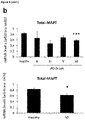
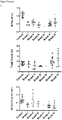
- the resulting Tau truncated isoform is reduced in the later stages of Alzheimer's disease patients' brain, in contrast to increasing total Tau levels.
- this new Tau isoform is able to bind to microtubules and is less prone to aggregate, suggesting a beneficial role in the pathology.
- a first aspect of the invention refers to an isolated tau-isoform protein or peptide, or a fragment thereof, comprising at the C-terminus domain amino acid sequence WVGCCPW (SEQ ID NO 1) or amino acid sequence GVGWVG, wherein the C-terminus domain is understood as the 30, 25, 20 or preferably 18 amino acids sited at the end of the amino acid chain of the protein or peptide.
- the said protein or peptide consists essentially of or consists of amino acid sequence WVGCCPW (SEQ ID NO 1) or of amino acid sequence GVGWVG.
- the said protein or peptide comprises at the C-terminus domain amino acid sequence KKVKGVGWVGCCPWVYGH (SEQ ID NO 2), wherein preferably the C-terminus domain is understood as the 30, 25, 20 or preferably 18 amino acids sited at the end of the amino acid chain of the protein or peptide.
- the said protein or peptide consists essentially of or consists of amino acid sequence KKVKGVGWVGCCPWVYGH (SEQ ID NO 2).
- the said protein or peptide comprises, consists essentially of or consists of peptide sequence:
- the said protein or peptide comprises, consists essentially of or consists of peptide sequence:
- a second aspect of the invention refers to a composition comprising one or a plurality of tau-isoform protein/s or peptide/s as defined in the first aspect of the invention or as defined in any of the preferred embodiments of the first aspect.
- a third aspect of the invention refers to a composition
- a composition comprising a polynucleotide encoding at least one tau-isoform protein or peptide as defined in the first aspect of the invention or as defined in any of the preferred embodiments of the first aspect.
- the polynucleotide is disposed within a vector selected for its ability to express the protein or peptide in a mammalian cell, preferably in a human cell.
- a fourth aspect of the invention refers to a composition as defined in any of the second or third aspects of the invention, for use in a method of inhibiting the formation of Ab aggregates or tau aggregates in a subject in need thereof.
- a fifth aspect of the invention refers to a composition as defined in any of the second or third aspects of the invention, for use in a method of inhibiting the development or progression of Ab plaque formation in a subject in need thereof.
- a sixth aspect of the invention refers to a composition as defined in any of the second or third aspects of the invention, for use in a method of inhibiting the development or progression of a tauopathy in a subject in need thereof.
- a seventh aspect of the invention refers to a composition as defined in any of the second or third aspects of the invention, for use in a method of inhibiting the development or progression of Alzheimer's disease in a subject in need thereof.
- An eighth aspect of the invention refers to a composition comprising an effective amount of the protein as defined in the second aspect or the polynucleotide as defined in the third aspect, for use in a method of treatment of a subject having an amyloid disease characterized by aggregation of Tau protein.
- a ninth aspect refers to a pharmaceutical composition
- a pharmaceutical composition comprising a composition as defined in the second or third aspect of the invention, and a pharmaceutically acceptable carrier including a peptide stabilizing excipient.
- the peptide stabilizing excipient comprises a preservative, a tonicity adjusting agent, a detergent, a hydrogel, a viscosity adjusting agent, or a pH adjusting agent.
- a tenth aspect refers to an in vitro use of the isolated tau-isoform protein or peptide as defined in the first aspect of the invention or as defined in any of the preferred embodiments of the first aspect, as a biomarker for the prognosis of an amyloid disease characterized by aggregation of Tau protein in a human subject.
- An eleventh aspect of the invention refers to an in vitro use of the isolated tau-isoform protein or peptide as defined in the first aspect of the invention or as defined in any of the preferred embodiments of the first aspect, as a biomarker for the prognosis of Alzheimer's disease in a human subject.
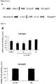
- MAPT gene has been proven to be genuinely subjected to alternative splicing [1, 55].
- MAPT contains a cryptic exonic sequence codifying for the protein saitohin nested in the Tau gene within intron 9 [14].
- the removal of RNA sequences in Tau RNA is very usual, but the translation of intronic sequences could also take place.
- Tau alternative splicing is also regulated by several features such as development, aging, disease or brain region [8, 54, 56, 58]. Tau isoforms lacking exons 2, 3 and 10, can be mainly found at early developmental stages [2, 56] and alternative splicing of exon 10 has been related to some tauopathies [10, 50].
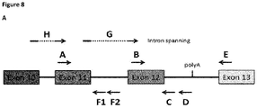
- this isoform is human-specific, since other species' Tau orthologues do not contain the polyadenylation canonical site that allows intron retention to give rise to a truncated protein. This is especially noteworthy considering that Alzheimer's disease is a human pathology that is not well reproduced in animal models.
- intron 12 retention A possible mechanism explaining intron 12 retention is that trans-acting splicing factors could be modified, for example by phosphorylation, and that the modified factor could change its function.
- modification of intron splicing has been previously reported for SR-proteins [40] and, specifically, for SRSF2 (SC35) which, when phosphorylated by GSK3 kinase, favors the expression of Tau 3R (lacking exon 10) [29]. Additionally, it has been described that the absence of SRSF2 favors exon 10 inclusion increasing Tau 4R species [12].
- intron splicing events have been previously reported for SRSF2 and other member of the SR-proteins family [40].
- the novel Tau fragment described in this invention is somehow similar to the previously reported fragment generated by cleavage of AEP [63].
- the isoforms truncated by intron retention exhibit a potentially protective behavior, with decreased aggregation capacity but conservation of microtubule assembly capacity.
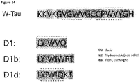
- This novel Tau isoforms contain a unique 18-aminoacid extra peptide, including the sequence GVGWVG, ( Figure 14 ).
- GVGWVG sequence GVGWVG
- W-Tau lacks the 12 amino acids after the fourth repeat, present in exon 13, which are found in the core of Tau filaments isolated from the brain of patients suffering some tauopathies, including Alzheimer's disease [62]. It is also worth noting that the specific W-Tau 18 amino acid sequence contains two adjacent cysteines (KKVKGVGWVGCCPWVYGH), whose oxidation to cystine can mediate the formation of a strained eight-member ring [47], a conformation that would substantially differ of that of other Tau isoforms and that has been proposed to be able to affect protein function and conformation stability [33, 39].
- peptide-based inhibitors that reduce or prevent amyloid beta (Ab) and/or tau aggregation and toxicity, preferably of already-aggregated species, and methods of using these peptide-based inhibitors. It is noted, that this invention is especially suitable for treating diseases such as Alzheimer's disease (AD) characterized by plaques of amyloid beta (Ab) and neurofibrillary tangles of tau. Ab aggregation is thought to occur at early stages of the disease, and ultimately gives way to the formation of tau tangles which track with cognitive decline.
- AD Alzheimer's disease
- Ab amyloid beta
- Embodiments of the invention include, for example, a composition of matter comprising at least one peptide inhibitor (from hereinafter “peptide inhibitor/s of the invention") that reduces or prevents amyloid beta (Ab) and/or tau aggregation and toxicity, preferably of already-aggregated species, wherein the at least one peptide inhibitor comprises or consists of an amino acid sequence WVGCCPW (SEQ ID NO 1), KKVKGVGWVGCCPWVYGH (SEQ ID NO 2), GVGWVG (SEQ ID NO 3), GWVGCCPW (SEQ ID NO 4), VGWVGCCPW (SEQ ID NO 5), GVGWVGCCPW (SEQ ID NO 6), KGVGWVGCCPW (SEQ ID NO 7), VKGVGWVGCCPW (SEQ ID NO 8), WVGCCPWV (SEQ ID NO 9), GWVGCCPWV (SEQ ID NO 10), WVGCCPWVY (SEQ ID NO 11), WVGCCPWV
- the peptide inhibitor/s of the invention consists of or are comprised in a tau-isoform protein or peptide, or in a fragment thereof, wherein preferably the peptide inhibitor/s of the invention comprised or consists of peptide sequence:
- composition comprises a plurality of peptide inhibitor/s of the invention.
- the peptide inhibitor sequence is coupled to a plurality of heterologous amino acids, for example a linker amino acid sequence and/or a cell penetrating peptide (CPP) amino acid sequence, this specific embodiment shall be explained in more detail below.
- CPP cell penetrating peptide
- the peptide inhibitor/s of the invention consists of or is comprised in a tau-isoform protein or peptide, or in a fragment thereof, and the peptide inhibitor of the invention is preferably located at the C-terminus domain or at the C-terminus motif or sequence (also known as the carboxyl-terminus, carboxy-terminus, C-terminal tail, C-terminal end, or COOH-terminus) of the tau-isoform protein or peptide, or of the fragment thereof.
- C-terminus is understood as the end of an amino acid chain (protein or polypeptide), terminated by a free carboxyl group (-COOH); therefore, preferably the peptide inhibitor/s of the invention is comprised within the last 30 amino acids of the amino acid chain, preferably within the last 25 amino acids of the amino acid chain, preferably within the last 20 amino acids of the amino acid chain, more preferably within the last 18 amino acids of the amino acid chain.
- compositions of matter comprising a polynucleotide encoding at least one of the peptide inhibitor/s of the invention.
- these polynucleotides are disposed within a vector selected for its ability to express the peptide inhibitor in a mammalian cell.
- the composition comprises a vector disposed within a mammalian cell.
- the peptide inhibitor/s of the invention or a protein or peptide comprising the same is typically less than 400 amino acids in length, less than 300 amino acids in length, less than 250 amino acids in length, less than 200 amino acids in length, less than 150 amino acids in length, less than 100 amino acids in length, less than 50 amino acids in length, less than 25 amino acids in length, less than 20 amino acids in length, more preferably less or equal to 18 amino acids in length.
- the peptide inhibitor/s of the invention or a protein or peptide containing the same may be coupled to heterologous amino acids such as a cell penetrating peptide (CPP) amino acid sequence, typically one less than 30 amino acids in length.
- CPP cell penetrating peptide
- the peptide is coupled to the heterologous amino acids by a peptide linker comprising 1-7 amino acids.
- the heterologous amino acids forms a polycationic structure.
- the heterologous amino acids forms an amphipathic structure.
- Embodiments of the invention include peptides wherein the peptide comprises at least one D amino acid (e.g. a peptide comprising all D-amino acids).
- Embodiments of the invention can further compare metabolic stability and efficacy of L- and D form peptide inhibitors.
- Embodiments of the invention include coupling of at least one of the peptide inhibitor/s of the invention by chemical or biological (genetic) means to proteins or peptides to carry the peptide inhibitor across the blood-brain-barrier (BBB), as a therapy for neurodegeneration or movement disorder (it is noted that such "coupled” peptides shall also be understood as forming part of the definition of "peptide inhibitor/s of the invention”).
- embodiments of the invention include coupling of any of the peptide inhibitor/s of the invention to a cell penetrating peptide and then coupling the resulting peptide by chemical or biological (genetic) means to proteins or peptides to carry the inhibitor across the blood-brain-barrier (BBB), as a therapy for neurodegeneration or movement disorder.
- embodiments of the invention also include coupling of any of the peptide inhibitor/s of the invention to small molecules that aid in the ability of the peptide inhibitors to cross the BBB and/or cell membranes.
- embodiments of the invention also include coupling of any of the peptide inhibitor/s of the invention to enzymes or small molecules (e.g.
- Embodiments of the invention also include coupling of any of the peptide inhibitor/s of the invention to small molecules that aid in the ability of the peptide inhibitors to degrade amyloid aggregates.
- Embodiments of the invention also include coupling of any of the peptide inhibitor/s of the invention by chemical means to a nano-particle capable of crossing the BBB and possibly also entering into cells, for example for use as a therapy for neurodegenation or movement disorder.
- Embodiments of the invention also include coupling of any of the peptide inhibitor/s of the invention by chemical means to a metal-containing nano-particle capable of crossing the BBB to create a diagnostic/biomarker for MRI or PET diagnosis of neurodegenerative disease or movement disorders (e.g. Parkinson's disease).
- Embodiments of the invention also include insertion of any of the peptide inhibitor/s of the invention into the CDRs of an antibody to produce inhibitors of greater potency and/or specificity.
- the antibody can be a full antibody, and Fab domain, or a single-chain antibody. Coupling of the resulting antibody to a nano-particle provides embodiments of the invention useful for therapy or diagnosis as noted above.
- a peptide inhibitor/s of the invention can be synthesized (e.g., chemically or by recombinant expression in a suitable host cell) by any of a variety of art-recognized methods.
- a practitioner can, for example, using conventional techniques, generate nucleic acid (e.g., DNA) encoding the peptide and insert it into an expression vector, in which the sequence is under the control of an expression control sequence such as a promoter or an enhancer, which can then direct the synthesis of the peptide.
- Suitable expression vectors e.g., plasmid vectors, viral, including phage, vectors, artificial vectors, yeast vectors, eukaryotic vectors, etc.
- capping inhibitor peptides are loaded into adeno-associated virus (AAV) capsids and hydrogel-based polymers to mediate delivery across the blood brain barrier
- Another aspect of the invention is a method for reactivating or restoring a biological or biochemical activity (function) of Tau and/or Ab which results from aberrant conformation of the Tau and/or Ab proteins.
- the method comprises contacting the Tau and/or Ab protein molecule having an aberrant conformation with an effective amount of the peptide inhibitor/s of the invention.
- the lost biological or biochemical activity of the Tau and/or Ab molecule is reactivated or restored.
- Another aspect of the invention is a method for inhibiting or preventing a loss of a biological or biochemical activity (function), of a Tau and/or Ab protein which results from aberrant conformation of the Tau and/or Ab protein.
- the method comprises contacting the Tau and/or Ab protein molecule having an aberrant conformation with an effective amount of the peptide inhibitor/s of the invention.
- the loss of activity of the Tau and/or Ab molecule is inhibited or prevented.
- Another aspect of the invention is a method for treating a subject having a disease or condition which is mediated by loss of function of Tau and/or Ab, such as a pathological syndrome in which Tau and/or Ab has an abnormal conformation (e.g. is aggregated or misfolded). That is, the pathological syndrome is associated with Tau and/or Ab having an aberrant conformation.
- the method comprises administering to the subject an effective amount of one or more peptides of the invention.
- a cocktail of two of more of the peptide inhibitor/s of the invention is used.
- Yet another embodiment of the invention is a method of observing the presence or absence of Tau amyloid fibrils and/or Ab in a biological sample comprising combining a biological sample with the peptide inhibitor/s of the invention that binds to Tau or Ab, allowing the peptide to bind to Tau amyloid fibrils and/or Ab that may be present in the biological sample, and then monitoring this combination for the presence of complexes formed between Tau amyloid fibrils and/or Ab and the peptide; wherein the presence of said complexes show the presence of Tau amyloid fibrils and/or Ab in the biological sample.
- the presence of complexes formed between Tau amyloid fibrils and/or Ab and the peptide is monitored using a detectable label that is coupled to the peptide (e.g. a heterologous peptide tag).
- a detectable label that is coupled to the peptide (e.g. a heterologous peptide tag).
- the method is performed on a biological sample obtained from an individual suspected of suffering from a tauopathy.
- Such embodiments of the invention can be used, for example, in diagnostic methods designed to observe the presence or status of Alzheimer's disease, for example to detect disease beginnings before clinical symptoms, and to follow the effectiveness (or lack of effectiveness), of a therapeutic treatment.
- Active variants of the inhibitory peptides described above are also included in the present invention.
- An "active variant” is a variant which retains at least one of the properties of the peptide inhibitor/s of the invention (e.g., the ability to inhibit, reduce or prevent Ab or Tau aggregation (i.e. fibrillation and/or tau cytotoxicity, wherein aggregation is understood as the formation of aggregates including fibers or fibrils)).
- Fibrilization refers to the formation of fiber or fibrils, such as amyloid fibrils.
- Suitable active variants include peptidomimetic compounds (any compound containing non-peptidic structural elements that is capable of mimicking the biochemical and/or biological action(s) of a natural mimicked peptide), including, for example, those designed to mimic the structure and/or binding activity (such as, for example, hydrogen bonds and hydrophobic packing interactions) of the peptides according to the methods disclosed herein).
- peptidomimetic compounds any compound containing non-peptidic structural elements that is capable of mimicking the biochemical and/or biological action(s) of a natural mimicked peptide
- binding activity such as, for example, hydrogen bonds and hydrophobic packing interactions
- active variants of the peptide inhibitor/s of the invention are shortened by 1-3 (e.g., 1, 2 or 3) amino acids at either the N-terminus, the C-terminus, or both of the starting inhibitory peptide.
- the active variants are lengthened (extended) by 1, 2, 3 or 4 amino acids at the C-terminal end of the starting inhibitory peptide.
- amino acids other than the ones noted above are substituted. These amino acids can help protect the peptide inhibitors against proteolysis or otherwise stabilize the peptides, and/or contribute to desirable pharmacodynamic properties in other ways.
- the non-natural amino acids allow an inhibitor to bind more tightly to the target because the side chains optimize hydrogen bonding and/or apolar interactions with it.
- non-natural amino acids offer the opportunity of introducing detectable markers, such as strongly fluorescent markers which can be used, e.g., to measure values such as inhibition constants.
- detectable markers such as strongly fluorescent markers which can be used, e.g., to measure values such as inhibition constants.
- peptide mimetics such as, e.g., peptoids, beta amino acids, N-ethylated amino acids, and small molecule mimetics.
- non-natural amino acids are substituted for amino acids in the sequence. More than 100 non-natural amino acids are commercially available and well know to the skilled person.
- one or more (e.g. 1, 2, 3, 4, 5, 6, 7, 8, 9 or 10) of the L-amino acids are substituted with a D amino acid.
- one or more (e.g. 1, 2, 3, 4, 5, 6, 7, 8, 9 or 10) N-methylated residues are included in the peptide.
- the peptide inhibitor/s of the invention can comprise, e.g., L-amino acids, D- amino acids, other non-natural amino acids, or combinations thereof.
- Active variants include molecules comprising various tags at the N-terminus or the C-terminus of the peptide (e.g. tags comprising a stretch of heterologous amino acids).
- the peptide inhibitor/s of the invention can comprise as tags at its N-terminus and/or at its C-terminus: 1, 2, 3, 4, 5, 6, 7, 8, 9 or 10 or more Lysine residues; 1, 2, 3, 4, 5, 6, 7, 8, 9 or 10 or more Arginine residues; 1, 2, 3, 4, 5, 6, 7, 8, 9 or 10 or more Glutamate residues; 1, 2, 3, 4, 5, 6, 7, 8, 9 or 10 or more Aspartate residues; combinations of these amino acid residues; or other polar tags that will be evident to a skilled worker.
- Other active variants include mutations of the Tau and/or Ab sequence which increase affinity of the inhibitory peptides for Tau and/or Ab.
- the peptide inhibitor/s of the invention is isolated or purified, using conventional techniques such as the methods described herein.
- isolated is meant separated from components with which it is normally associated, e.g., components present after the peptide is synthesized.
- An isolated peptide can be a cleavage product of a protein which contains the peptide sequence.
- a “purified” inhibitory peptide can be, e.g., greater than 90%, 95%, 98% or 99% pure.
- the peptide is fused to any of a variety of cell penetrating peptides (CPPs).
- CPPs typically have an amino acid composition that either contains a high relative abundance of positively charged amino acids such as lysine or arginine or has sequences that contain an alternating pattern of polar/charged amino acids and non- polar, hydrophobic amino acids. These two types of structures are referred to as polycationic or amphipathic, respectively.
- a third class of CPP's are the hydrophobic peptides, containing only apolar residues, with low net charge or have hydrophobic amino acid groups that are crucial for cellular uptake.
- Typical CPP's that can be fused to an inhibitory peptide of the invention are known such as, for example in WO 2018/005867 , the contents of which are incorporated herein by reference.
- the peptide inhibitor/s of the invention can be synthesized (e.g., chemically or by recombinant expression in a suitable host cell) by any of a variety of art- recognized methods.
- a practitioner can, for example, using conventional techniques, generate nucleic acid (e.g., DNA) encoding the peptide and insert it into an expression vector, in which the sequence is under the control of an expression control sequence such as a promoter or an enhancer, which can then direct the synthesis of the peptide.
- Suitable expression vectors e.g., plasmid vectors, viral, including phage, vectors, artificial vectors, yeast vectors, eukaryotic vectors, etc.
- compositions comprising one or more of the peptide inhibitor/s of the invention and a pharmaceutically acceptable carrier.
- the components of the pharmaceutical composition can be detectably labeled, e.g. with a radioactive or fluorescent label, or with a label, for example one that is suitable for detection by positron emission spectroscopy (PET) or magnetic resonance imaging (MRI).
- PET positron emission spectroscopy
- MRI magnetic resonance imaging
- peptides of the invention can be coupled to a detectable label selected from the group consisting of a radioactive label, a radio- opaque label, a fluorescent dye, a fluorescent protein, a colorimetric label, and the like.
- the inhibitory peptide is present in an effective amount for the desired purpose.
- the compositions may contain preservatives and/or antimicrobial agents as well as pharmaceutically acceptable auxiliary substances as required to approximate physiological conditions, such as pH adjusting and buffering agents, tonicity adjusting agents, wetting agents, detergents and the like.
- “Pharmaceutically acceptable” means that which is useful in preparing a pharmaceutical composition that is generally safe, non-toxic, and neither biologically nor otherwise undesirable and includes that which is acceptable for veterinary as well as human pharmaceutical use.
- “pharmaceutically acceptable salts” of a compound means salts that are pharmaceutically acceptable, as defined herein, and that possess the desired pharmacological activity of the parent compound.
- polynucleotide encoding the peptide inhibitor/s of the invention.
- the polynucleotide is operably linked to a regulatory control sequence (e.g., a promoter or an enhancer) to facilitate production of the encoded protein following introduction (e.g. by transfection) into a suitable cell.
- a regulatory control sequence e.g., a promoter or an enhancer
- Other embodiments include a cell comprising the expression vector; and a method of making the peptide inhibitor/s of the invention comprising cultivating the cell and harvesting the peptide thus generated.
- Another aspect of the invention is a kit for carrying out any of the methods described herein.
- One aspect of the invention is a method for reducing or inhibiting Tau and/or Ab aggregation, comprising contacting Tau and/or Ab proteins with an effective amount of one or more of the inhibitory peptides of the invention.
- Such a method can be carried out in solution or in a cell (e.g. cells in culture or in a subject).
- Another aspect of the invention is a method for treating a subject having a disease or condition which is mediated by the presence of fibrillated Tau (sometimes referred to herein as a Tauopathy or a Tau-mediated disease or condition), comprising administering to the subject an effective amount of an inhibitory peptide or pharmaceutical composition of the invention.
- diseases or conditions are, e.g., Alzheimer's disease.
- Another aspect of the invention is a method to prevent the onset of such diseases or conditions (e.g., Alzheimer's disease), or to treat a subject in the early stages of such diseases or conditions, or that is developing such a disease or condition, in order to prevent or inhibit development of the condition or disease.
- an "effective amount" of the peptide inhibitor/s of the invention is an amount that can elicit a measurable amount of a desired outcome, e.g. inhibition of Tau and/or Ab aggregation or cytotoxicity; for a diagnostic assay, an amount that can detect a target of interest, such as an Tau and/or Ab aggregate; or in a method of treatment, an amount that can reduce or ameliorate, by a measurable amount, a symptom of the disease or condition that is being treated.
- a “subject” can be any subject (patient) having aggregated (fibrillated) Tau and/or Ab molecules associated with a condition or disease which can be treated by a method of the present invention.
- the subject has Alzheimer's disease.
- Typical subjects include vertebrates, such as mammals, including laboratory animals, dogs, cats, non-human primates and humans.
- the peptide inhibitor/s of the invention can be formulated as pharmaceutical compositions in a variety of forms adapted to the chosen route of administration, for example, orally, nasally, intraperitoneally, or parenterally, by intravenous, intramuscular, topical or subcutaneous routes, or by injection into tissue.
- Suitable oral forms for administering the inhibitors include lozenges, troches, tablets, capsules, effervescent tablets, orally disintegrating tablets, floating tablets designed to increase gastric retention times, buccal patches, and sublingual tablets.
- the peptide inhibitor/s of the invention can be systemically administered, e.g., orally, in combination with a pharmaceutically acceptable vehicle such as an inert diluent or an assimilable edible carrier, or by inhalation or insufflation. They can be enclosed in coated or uncoated hard or soft shell gelatin capsules, can be compressed into tablets, or can be incorporated directly with the food of the patient's diet.
- a pharmaceutically acceptable vehicle such as an inert diluent or an assimilable edible carrier, or by inhalation or insufflation.
- the compounds can be combined with one or more excipients and used in the form of ingestible tablets, buccal tablets, troches, capsules, elixirs, suspensions, syrups, wafers, and the like.
- compositions suitable for administration to humans are meant to include, but is not limited to, those ingredients described in Remington: The Science and Practice of Pharmacy, Lippincott Williams & Wilkins, 21st ed. (2006 ) (hereinafter Remington's).
- the inhibitors can be combined with a fine inert powdered carrier and inhaled by the subject or insufflated.
- Such compositions and preparations should contain at least 0.1% compounds.
- the percentage of the compositions and preparations may, of course, be varied and may conveniently be between about 2% to about 60% of the weight of a given unit dosage form.
- the tablets, troches, pills, capsules, and the like may also contain the following: binders such as gum tragacanth, acacia, corn starch or gelatin; excipients such as dicalcium phosphate; a disintegrating agent such as corn starch, potato starch, alginic acid and the like; a lubricant such as magnesium stearate; and a sweetening agent such as sucrose, fructose, lactose or aspartame or a flavoring agent such as peppermint, oil of wintergreen, or cherry flavoring can be added.
- a liquid carrier such as a vegetable oil or a polyethylene glycol.
- a syrup or elixir may contain the active compound, sucrose or fructose as a sweetening agent, methyl and propylparabens as preservatives, a dye and flavoring such as cherry or orange flavor.
- Various other materials can be present as coatings or to otherwise modify the physical form of the solid unit dosage form. For instance, tablets, pills, or capsules can be coated with gelatin, wax, shellac or sugar and the like.
- any material used in preparing any unit dosage form should be pharmaceutically acceptable and substantially non-toxic in the amounts employed.
- the peptide inhibitor/s of the invention can be incorporated into sustained-release preparations and devices.
- the inhibitors can be incorporated into time release capsules, time release tablets, and time release pills.
- the composition is administered using a dosage form selected from the group consisting of effervescent tablets, orally disintegrating tablets, floating tablets designed to increase gastric retention times, buccal patches, and sublingual tablets.
- the peptide inhibitor/s of the invention may also be administered intravenously or intraperitoneally by infusion or injection.
- Solutions of the inhibitors can be prepared in water, optionally mixed with a nontoxic surfactant.
- Dispersions can also be prepared in glycerol, liquid polyethylene glycols, triacetin, and mixtures thereof and in oils. Under ordinary conditions of storage and use, these preparations can contain a preservative to prevent the growth of microorganisms.
- the pharmaceutical dosage forms suitable for injection or infusion can include sterile aqueous solutions or dispersions or sterile powders comprising the compounds which are adapted for the extemporaneous preparation of sterile injectable or infusible solutions or dispersions, optionally encapsulated in liposomes.
- the liquid carrier or vehicle can be a solvent or liquid dispersion medium comprising, for example, water, ethanol, a polyol (for example, glycerol, propylene glycol, liquid polyethylene glycols, and the like), vegetable oils, nontoxic glyceryl esters, and suitable mixtures thereof.
- the proper fluidity can be maintained, for example, by the formation of liposomes, by the maintenance of the required particle size in the case of dispersions or by the use of surfactants.
- Sterile injectable solutions are prepared by incorporating the compounds in the required amount in the appropriate solvent with various of the other ingredients enumerated above, as required, followed by filter sterilization.
- the preferred methods of preparation are vacuum drying and freeze drying techniques, which yield a powder of the active ingredient plus any additional desired ingredient present in the previously sterile-filtered solutions.
- Useful solid carriers include finely divided solids such as talc, clay, microcrystalline cellulose, silica, alumina and the like. Other solid carriers include conventional nontoxic polymeric nanoparticles or microparticles.
- Useful liquid carriers include water, alcohols or glycols or water/alcohol/glycol blends, in which the compounds can be dissolved or dispersed at effective levels, optionally with the aid of non-toxic surfactants. Adjuvants such as fragrances and additional antimicrobial agents can be added to optimize the properties for a given use.
- the resultant liquid compositions can be applied from absorbent pads, used to impregnate bandages and other dressings, or sprayed onto the affected area using pump-type or aerosol sprayers.
- Useful dosages of the peptide inhibitor/s of the invention or pharmaceutical compositions of the invention can be determined by comparing their in vitro activity, and in vivo activity in animal models. Methods for the extrapolation of effective dosages in mice, and other animals, to humans are known to the art.
- Effective dosages and routes of administration of agents of the invention are conventional.
- the exact amount (effective dose) of the agent will vary from subject to subject, depending on, for example, the species, age, weight and general or clinical condition of the subject, the severity or mechanism of any disorder being treated, the particular agent or vehicle used, the method and scheduling of administration, and the like.
- a therapeutically effective dose can be determined empirically, by conventional procedures known to those of skill in the art. See, e.g., The Pharmacological Basis of Therapeutics, Goodman and Gilman, eds., Macmillan Publishing Co., New York .
- an, effective dose can be estimated initially either in cell culture assays or in suitable animal models. The animal model may also be used to determine the appropriate concentration ranges and routes of administration. Such information can then be used to determine useful doses and routes for administration in humans.
- a therapeutic dose can also be selected by analogy to dosages for comparable therapeutic agents.
- Treatment may involve daily or multi-daily doses of compound(s) over a period of a few days to months, or even years.
- the transcript including the translation of intron 12 up to the first stop codon has been named TIR-MAPT ( Truncated by Intron Retention MAPT), while the protein generated from such transcript has been termed W-Tau, due to the appearance of two characteristic tryptophan residues (W) in this isoform, an amino acid that does not appear elsewhere within the Tau molecule.
- ET-Tau Endopeptidase Truncated Tau
- HEK293T (CRL-11268, ATCC), SK-N-MC (HTB-10, ATCC) and SH-SY5Y (CRL-2266, ATCC) cells were cultured in DMEM or MEM supplemented with 10% fetal bovine serum, 2 mM glutamine, non-essential amino acids, 10 U/ml penicillin and 10 ⁇ g/ml streptomycin, at 37o C and 5% CO 2 .
- TIR-MAPT transcript was obtained from an SH-SH5Y RNA extract, using specific oligos (TauNt and TauD, see table 1 ) and was cloned into pBlueScript-SK+ (212205, Agilent Technologies) thanks to a TA-cloning strategy [37].
- specific oligos that include the appropriate restriction sites A1 and TIR-T-BglII and ET-T-BglII, see table 1
- TIR-MAPT and ET-MAPT were subcloned into a eukaryotic expression vector pSG5 (216201, Agilent technologies).
- Tau isoforms were cloned into a prokaryotic expression vector pRK172 using specific oligos (TAU-PRK172 fw, TIR-T-pRKpWPI rv and ET-T-pRKpWPI rv; see table 1 ). After cloning, all vectors were sequenced using the described oligos (table 1) . Table 1 Detailed information of the oligonucleotides used as primers for semi-quantitative, quantitative RT-PCR and cloning.
- RNA-seq raw data from 363 samples of 3 brain regions (frontal cortex, dorsolateral prefrontal cortex and hippocampus) of 180 human brain healthy donors were retrieved from the Genotype-Tissue Expression (GTEx) project [36] (table 2). SRA files were converted to FASTQ files and reads were re-mapped to human genome GRCh38 using STAR version 2.5.2a [18]. Gene expression quantification was performed with RSEM version 1.3.1 [35]. Table 2. Number of healthy brain samples and donors per brain region within GTEx that were RNA-seq analyzed. Venn diagram shows number of individuals for each sample region. Brain Region Number of samples Duplicated samples Triplicated samples Number of donors Frontal cortex 134 3 2 127 Dorsolateral prefrontal cortex 120 1 4 115 Hippocampus 109 3 1 100
- the annotation file was retrieved from GENCODE (gencode.v23.annotation.gtf) and was modified in order to include a new MAPT- related gene ( TIR-MAPT ) whose genome coordinates are chr17:45894382-46018851, which includes part of the intron 12 (chr17: 46018731-46018851) as the 3' end of the gene in the region mapping the oligonucleotide sequence TauD (Table 2 and Fig. 1 ).
- Gene expression levels were obtained from RSEM as Transcripts Per Kilobase Million (TPM) values. Expression levels of MAPT and TIR-MAPT genes were analyzed per brain region. For those donors with more than one sample in the same brain region, analysis was done only in the sample with highest MAPT expression.
- pRK172 vectors encoding different Tau isoforms were transformed in BL21 E. coli competent cells by electroporation, from which Tau was purified, as described elsewhere [49]. These bacteria were cultured in LB medium with 100 ng/ml of ampicillin at 37o C overnight. Bacterial suspensions were transferred to 1L of LB with 100 ng/ml of ampicillin and further incubated at 37o C, up until optical density readings at 600 nm ranged between 0.6 and 0.8. At this point, 0.4 mM of IPTG was added to each sample so as to trigger transcription, and incubated once again at 37oC for 2 hours.
- the samples were centrifuged at 2,950 x g at 4o C for 20 minutes and pellets were resuspended in Tau buffer, which consists of buffer A (0.1M MES pH 6.4, 0.5 mM MgCl 2 and 2mM EGT) supplemented with 1 mM PMSF; 0.5 NaCl and 5 mM ⁇ -mercaptoethanol. Resuspended pellets were sonicated on ice at 24 microns peak to peak (mpp) 5 times for 1 minute, waiting 10 seconds between repetitions.
- buffer A 0.1M MES pH 6.4, 0.5 mM MgCl 2 and 2mM EGT
- samples were centrifuged at 13,850 x g at 4o C for 10 minutes and the resultant supernatants were subjected to 5 minutes at 100o C and 5 minutes on ice before centrifuging one more time at 13,850 x g at 4o C for 30 minutes.
- Supernatants were kept and Tris 1M was added until pH reaches a level of 11, in order to get rid of residual DNA.
- Ammonium sulfate 50% was added to the samples and they were incubated agitating at 4o C for at least 1h before centrifuging again at 13,850 x g at 4o C for 1h.
- Pellets containing purified Tau were resuspended in buffer A and the purification process was checked by SDS-PAGE and Coomassie blue staining in order to check if any other protein bands appeared upon Coomassie blue staining.
- Protein concentration for each Tau isoform was firstly determined by Coomassie Blue staining comparison with known amounts of protein and compared to results obtained by ELISA (Abcam, ab273617). ELISA results yielded higher variability, especially for 3R Tau isoforms. Consequently, concentrations were calculated by using Coomassie Blue staining data adjusted by using absorbance measurements corrected by individually-calculated extinction coefficients ( ⁇ ), based on the specific amino acid sequence of each isoform [24] (Table 3) . Namely, we measured Tau concentration as indicated by Riversidel and collaborators [34]: absorbance at 280 nm (A 280 ) was measured for each Tau isoform and concentration was determined as the ratio between A280 and ⁇ .
- Guidel [34] which has been typically used for all CNS human Tau isoforms [21], but are not valid for W-Tau isoforms that contain 2 extra tryptophan residues nor for ET-Tau isoforms, which lack 1 tyrosine that appear on the rest of the isoforms.
- RNA integrity numbers were calculated using the Agilent 2100 Bioanalyzer system (Agilent Technologies), and only RNAs with RIN > 5 were used for RT-qPCR.
- PCR Semiquantitative PCR was performed using 1 ⁇ l of cDNA (0.5 ng/ ⁇ l) supplemented with 2.5 mM MgCl 2 , 0.2 mM each dNTP, 1.3 M betaine, 0.5 mM of each primer (described in table 1 ) and 0.025 U/ml of GoTaq ® Flexi DNA Polymerase (M829, Promega), in the following conditions: 95o C for 2 min, and 35 cycles (PCR1-8) or 30 cycles (PCR9-13) of 95o C for 45 s, 58.6o C for 45 s and 72o C for 45 s, followed by a final extension of 10 min at 72o C. PCR combinations are described in table 4. Table 4.
- PCR number Forward primer Reverse primer Amplicon length 1 TauB TauC 188 2 TauB TauD 232 3 TauG TauC 204 4 TauG TauD 248 5 TauA TauC 255 6 TauA TauD 299 7 TauH TauC 280 8 TauH TauD 324 9 TauA TauE 256 10 TauB TauE 200 11 TauG TauE 216 12 TauH TauE 281 13 GAPDH Fw GAPDH Rv 231
- RNA was retrotranscribed with the Transcriptor First Strand cDNA Synthesis Kit (04379012001, Roche) using 20 ng/ ⁇ l RNA with oligo(dT)18 primer. Quantitative PCR was performed in a LightCycler480 (Roche) in the following conditions: 50o C for 2 min, 95o C for 10 min, and 40 cycles of 95o C for 15 s and 60o C for 1 min. Specific intron-spanning oligonucleotides against MAPT or TIR-MAPT transcript were designed (MAPT-E11-E13-fw/rv and TIR-T-fw/rv, respectively; see table 1 ) . Gene expression was normalized to GAPDH expression using TaqMan primer human GAPDH (Hs02758991_g1, Applied Biosystems). For every RT-qPCR experiment, only samples with RIN above 5 were used.
- Frozen brain tissue was homogenized using a TissueLyser (Retsch MM300, Qiagen, Hilden, Germany) (30 Hz, 5 min) with 5-mm stainless steel beads (69989, Qiagen) in total extraction buffer: 50 mM Tris-HCI pH 7.5, 300 mM NaCl, 0.5% SDS (sodium dodecyl sulphate) and 1% Triton X-100 ( Figures 2d , 2e and Fig. 13b ) . The same buffer was also used for cells ( Figures 2b , 2c , 3b , 4a , 5b and Fig. 11b and Fig. 12a -b ) .
- a strong lysis buffer 50 mM Tris-HCI pH 7.6, 400 mM NaCl, 1 mM EDTA, 1mM EGTA, 1% SDS
- the homogenates are incubated for 15 min at 95o C, centrifuged (16,100 x g, 10 min) and the supernatant was considered the brain extract.
- Protein concentration was measured using the DC protein assay kit (500-0111, Bio-Rad). From each sample, equal amount of total protein was resolved on a 10% Bis-Tris gel and transferred to nitrocellulose membranes.
- Blots are probed with the corresponding primary antibodies (see below), followed by horseradish peroxidase-conjugated anti-mouse or anti-rabbit antibody (Dako, Glostrup, Denmark). Protein expression was quantified by measuring the ECL signal per lane with ImageJ software (http://rsbweb.nih.gov/ij/).
- Tau 12 (Millipore, MAB2241; amino acids 6-18, diluted 1/1000), Tau 5 (Calbiochem, D00139295, amino acids 210-241, diluted 1/1000), Tau antibody (NOVUSBIO, NB100-82247, epitope around amino acid 231, diluted 1/500) and Tau 7.51 ([44], amino acids 315-376, diluted 1/100) were used as antibodies recognizing all Tau isoforms; Tau 1 (Chemicon, MAB3420, diluted 1/1000) recognizing dephosphorylated residues Ser195, 198, 199 and 202 and AT8 (phospho-Ser202/Thr205; Innogenetics, MN1020, diluted 1/100), AT180 (phospho-Thr231; Innogenetics, MN1040, diluted 1/100), Tau 404 (phospho-Ser404; Life Technologies, 44758G, diluted 1/1000), Tau 396 (phospho-S
- W-Tau peptide A peptide composed of the W-Tau unique sequence (KKVKGVGWVGCCPWVYGH) (W-Tau peptide) was synthesised and a polyclonal IgG antibody against that peptide was obtained from Abyntek (Bizkaia, Spain), using New Zealand rabbit as host strain. This antibody was named W-Tau antibody and its specificity was tested (diluted 1/1000) to ensure it reacts exclusively with TIR-Tau but not with other Tau isoforms ( Fig. 2a -c).
- Immunoprecipitation was performed with W-Tau and total Tau (NOVUSBIO, NB100-82247) antibodies using SantaCruz Immunoprecipitation Kit (SC-2003) using 500 ⁇ g of whole cell extracts and following provider's guidelines.
- HEK293T cells were transfected with pSG5 plasmids (2 ⁇ g/p60) encoding different Tau isoforms using Lipofectamine and Plus reagents following instructions of the supplier (#18324 and #11514, respectively, Life Technologies). 48 h after transfection, cells were collected, pelleted and homogenized in 200 ⁇ l of total extraction buffer. Protein concentrations were measured by using the DC protein assay kit (500-0111, Bio-Rad) and samples containing equivalent amounts of protein were analyzed by Western blot, as previously described, using nitrocellulose membranes. Immunodetection was performed with antibodies Tau 7.51 and Tau 5 for total Tau, Tau 1 for dephosphorylated Tau and AT8, AT180, Tau 404, Tau 396 and PHF1 for phosphorylated Tau.
- HEK293T cells were transfected with pSG5 plasmids (2 ⁇ g/p60) encoding different Tau isoforms using Lipofectamine and Plus reagents (Life Technologies). After 48 h, cells were collected, pelleted and homogenized in 150 ⁇ l of lysis buffer (50 mM Tris-HCI (pH 7,4), 150 mM NaCl, 20 mM NaF, 1 mM Na 3 VO 4 , 0.5 mM MgSO 4 ; supplemented with protease inhibitors cocktail, 04693159001 Roche) at 4oC. Homogenates were centrifuged at 27,000 x g for 20 min at 4oC.
- lysis buffer 50 mM Tris-HCI (pH 7,4), 150 mM NaCl, 20 mM NaF, 1 mM Na 3 VO 4 , 0.5 mM MgSO 4 ; supplemented with protease inhibitors cocktail, 04693159001 Roche
- Pellets were discarded and 1% sarkosyl was added to the supernatants, being incubated for 1.5 h agitating at room temperature. Upon incubation, samples were centrifuged at 150,000 x g during 45 minutes at 4o C. Supernatants were kept as sarkosyl-soluble fraction and pellets were resuspended in 100 ⁇ l of a mix of total extraction buffer and loading buffer (1:1) and considered sarkosyl-insoluble fraction.
- Results were further confirmed by carrying out a similar protocol to measure Triton X-100 solubility. Namely, pelleted cells were homogenized in 500 ⁇ l of lysis buffer (1% Triton X-100, 50 mM Tris-HCI pH 7, 100 mM NaCl and 1mM EDTA) and incubated 20 minutes at 4o C. The samples were centrifuged at 18,000 x g for 5 minutes and the supernatants were kept as soluble fraction. Pellets were resuspended in total extraction buffer (see point 11 of methods) and considered Triton-insoluble fraction.
- lysis buffer 1% Triton X-100, 50 mM Tris-HCI pH 7, 100 mM NaCl and 1mM EDTA
- Tau aggregates were grown for the different Tau isoforms by vapor diffusion in hanging drops in the standard way used for protein crystallizations [15].
- the different Tau isoforms were purified from bacteria as described above and their concentration was estimated by measuring absorbance at 280 nm and taking into account individually-calculated extinction coefficients for each isoform (table 3). Equivalent quantities of each isoform were added to the corresponding volume of buffer A (0.1M MES pH 6.4, 0.5 mM MgCl 2 and 2 mM EGT) plus 50 mM NaCl in the presence of heparin, reaching a concentration of 1 mg/ml.
- the reservoir contained 0.2 M NaCl in buffer A.
- the brains of twelve 2-month-old C57BL/6J mice were homogenized with a potter in isotonic buffer (0.32 M sucrose, 1 mM EGTA, 1 mM MgCl 2 , 10 mM phosphate buffer pH 7 and 1mM PMSF) at 4oC.
- isotonic buffer (0.32 M sucrose, 1 mM EGTA, 1 mM MgCl 2 , 10 mM phosphate buffer pH 7 and 1mM PMSF
- Supernatant was collected after 40 min ultracentrifugation at 100,000 x g at 4oC in an Optima L-100 XP Ultracentrifuge (Beckman Coulter).
- SK-N-MC human neuroblastoma cells (ATCC ® HTB-10 TM ) were treated with GSK3 inhibitors SB216763 (25 ⁇ M, GlaxoSmithKline compounds[13]) and AR-14418 (10 ⁇ M, AstraZeneca compound[7]) or A ⁇ (5 ⁇ g/ml, Neosystem Laboratoire, France) for 24 h and whole cell extracts were used for Western blot analysis, probing the blots with W-Tau antibody. From those cells treated with AR-14418 and A ⁇ , RNA-enriched cytoplasmic fraction was also retrieved and TIR-MAPT RNA levels were assessed by quantitative RT-PCR as previously described.
- Exonic and intronic human MAPT sequences were retrieved from ENSEMBL (ENSG00000186868).
- Exon 12- exon 13 and exon 12 - intron 12 sequences (Exon 13 - exon 14 and exon 13 - intron 13, according to ENSEMBL nomenclature of Tau exons/introns) were analyzed using ESEFinder 3.0 (http://exon.cshl.edu/ESE/) [53].
- ESEFinder settings were adjusted to show binding sites with score 4.0 or higher.
- Quantitative data represented as mean ⁇ SD or SEM, were compared between groups using the two-tailed Student's t-test. For multiple comparisons, one-way ANOVA and Dunnett's test were performed to compare each isoform and the correspondent full length isoform. When the distribution of the data was not Gaussian a nonparametric Kruskal-Wallis test was used. The differences are given with their corresponding statistical significance or p value, which is the probability that the difference occurred merely by chance under the null hypothesis (* p ⁇ 0.05; ** p ⁇ 0.01; *** p ⁇ 0.001; **** p ⁇ 0.0001; N.S. not significant). The method used for each experiment is specified in the corresponding figure legend.
- human intron 12 includes a canonical stop codon sequence followed by a polyadenylation canonical site, so intron 12-containing species would be translated as a truncated protein similar to the truncated isoform mediated by AEP (ET-Tau), with 18 more amino acids, which we have named W-Tau ( Fig. 1a ).
- this isoform would be human-specific, since this polyadenylation site cannot be found in other species, such as mice.
- intron-spanning primers matching exon 10 to detect RNA species containing intron 12 never detected these as mature cytoplasmic polyA positive RNA ( Fig. 8a -b and Table 4, PCR 7 and 8).
- Exon 10 is spliced-out in Tau isoforms with 3 repeats (Tau 3R) and it has been previously described that proliferative SH-SY5Y cells mainly express these Tau isoforms [57], so detection of RNA species with partial inclusion of exon 10 was not expected a priori.
- Tau 3R Tau isoforms with 3 repeats
- TIR-MAPT polyA positive mRNA was found in both hippocampus and frontal cortex in human samples ( Fig. 1e ).
- TIR-MAPT expression levels in human brain were more than 100-fold the expression found in SH-SY5Y, suggesting a higher relevance of this RNA species in human brains than in cultured cells.
- RNA-seq data from 363 samples from 3 different brain regions of 180 healthy donors were used to confirm the existence of TIR-MAPT transcripts (Table 2).
- TIR-MAPT gene mature mRNA containing the first part of intron 12 up until the canonical stop site was expressed in the majority of samples ( Fig. 1f ) supporting our previous findings in a big cohort of human samples.
- Gene expression of MAPT mature mRNA was compared with TIR-MAPT ( Fig. 1g ). Even though TIR-MAPT is expressed at lower levels, the great number of samples expressing this intron-retaining species suggests a relevant function in human brain.
- RNA species containing intron 12 some of the reads would correspond to a putative Tau protein without inserts, and with 3 tandem repeats (T30), but substituting the 3'-terminal of the RNA for the sequence of intron 12 until the first stop codon; what we have named TIR-T30.
- the putative translation of this RNA species into a protein has been named W-T30, due to the presence of two tryptophan residues (W) in the sequence of intron 12 translation ( Fig. 9 ). This RNA species would be equivalent to that found in SH-SY5Y cells.
- W-T30 could be specifically immunoprecipitated with both W-Tau (Abyntek) or Total Tau (NOVUSBIO, NB100-82247) antibodies ( Fig 2a ). and then that W-Tau antibody only precipitates W-Tau without any background on control untreated cells ( Fig 2b ). In both cases, the immunoprecipitated protein was confirmed to be Tau by detecting it by Western blot with Tau 7.51 antibody.
- eukaryotic vector pSG5 to include exon 10 corresponding to the fourth tandem repeat of the microtubule binding domain and the two Tau inserts (thus obtaining W-T42).
- exon 10 corresponding to the fourth tandem repeat of the microtubule binding domain and the two Tau inserts (thus obtaining W-T42).
- asparagine endopeptidase truncated Tau corresponding isoforms were also cloned into pSG5 expression vector (ET: Endopeptidase truncated; ET-T42 and ET-T30); mimicking the cut of endopeptidase by including a stop codon after the one that encodes asparagine 368.
- the pSG5 vectors encoding the different isoforms were employed to induce overexpression of these isoforms, in order to test W-Tau properties and compare them to those of full-length isoforms (T42 and T30) and those of asparagine endopeptidase truncated isoforms (ET42 and ET30) ( Fig. 3a ).
- T42 and T30 full-length isoforms
- ET42 and ET30 asparagine endopeptidase truncated isoforms
- Fig. 3a we studied the phosphorylation of W-Tau isoform, comparing it with the phosphorylation found in other Tau isoforms, expressed in HEK293T cells. We expected differential detections according to the epitopes that remain despite truncation and those that are lost because of it ( Fig. 3a ).
- ESEFinder 3.0 indicates the presence of a high-scored SRSF6 splicing binding site in the canonical MAPT sequence, close to the beginning of Exon 13 ( Fig. 12 ). This site is not present in the TIR-MAPT sequence . However, in its place, ESEFinder 3.0 indicates the appearance of a high-scored SRSF2 (also known as SC-35) splicing binding site in intron 12, in contrast to MAPT sequence splicing out intron 12 ( Fig. 12 ).
- SRSF2 also known as SC-35
- TIR-MAPT RNA Expression of TIR-MAPT RNA in the brain of AD patients.
- TIR-MAPT mRNA levels were studied in a cohort of hippocampal brain samples from non-demented individuals and AD patients classified according to their Braak stage (see Table 5).
- the study of absolute levels of TIR-MAPT mRNA revealed that it slightly decreases in the first stages of the disease (Braak II and III) but increases in the advanced stages (Braak V and VI), although no significant differences were found ( Fig. 6a ).
- Total MAPT mRNA levels were similarly studied, finding in this case decreased expression in AD patients with respect to healthy individuals, more significantly in Braak stage VI ( Fig. 6b ). This data correlates with what we previously describe in the analysis of RNAseq dataset ( Fig. 1g ).
Landscapes
- Health & Medical Sciences (AREA)
- Life Sciences & Earth Sciences (AREA)
- Engineering & Computer Science (AREA)
- Biomedical Technology (AREA)
- Chemical & Material Sciences (AREA)
- Neurology (AREA)
- Neurosurgery (AREA)
- General Health & Medical Sciences (AREA)
- Medicinal Chemistry (AREA)
- Organic Chemistry (AREA)
- Molecular Biology (AREA)
- Bioinformatics & Cheminformatics (AREA)
- Pharmacology & Pharmacy (AREA)
- Nuclear Medicine, Radiotherapy & Molecular Imaging (AREA)
- Animal Behavior & Ethology (AREA)
- General Chemical & Material Sciences (AREA)
- Public Health (AREA)
- Veterinary Medicine (AREA)
- Proteomics, Peptides & Aminoacids (AREA)
- Biochemistry (AREA)
- Chemical Kinetics & Catalysis (AREA)
- Hematology (AREA)
- Immunology (AREA)
- Urology & Nephrology (AREA)
- Cell Biology (AREA)
- Microbiology (AREA)
- Food Science & Technology (AREA)
- Physics & Mathematics (AREA)
- Analytical Chemistry (AREA)
- Biotechnology (AREA)
- General Physics & Mathematics (AREA)
- Pathology (AREA)
- Hospice & Palliative Care (AREA)
- Psychiatry (AREA)
- Toxicology (AREA)
- Zoology (AREA)
- Gastroenterology & Hepatology (AREA)
- Biophysics (AREA)
- Genetics & Genomics (AREA)
- Peptides Or Proteins (AREA)
Abstract
The present invention refers to a composition of matter comprising at least one peptide inhibitor (from hereinafter "peptide inhibitor/s of the invention") that reduces or prevents amyloid beta (Ab) and/or tau aggregation and toxicity, preferably of already-aggregated species, wherein the at least one peptide inhibitor comprises or consists of an amino acid sequence WVGCCPW (SEQ ID NO 1), KKVKGVGWVGCCPWVYGH (SEQ ID NO 2), GVGWVG (SEQ ID NO 3), GWVGCCPW (SEQ ID NO 4), VGWVGCCPW (SEQ ID NO 5), GVGWVGCCPW (SEQ ID NO 6), KGVGWVGCCPW (SEQ ID NO 7), VKGVGWVGCCPW (SEQ ID NO 8), WVGCCPWV (SEQ ID NO 9), GWVGCCPWV (SEQ ID NO 10), WVGCCPWVY (SEQ ID NO 11), WVGCCPWVYG (SEQ ID NO 12) or WVGCCPWVYGH (SEQ ID NO: 13) or any combination thereof.
Description
- The invention relates to compositions and methods useful in inhibiting aggregation of Tau and amyloid b proteins.
- Tau is a microtubule-associated protein (MAP) that acts as a neurite microtubule stabilizer [9, 61]. Tau overexpression, especially in its modified forms (i.e. phosphorylated, aggregated or truncated) may result in a toxic gain of function [5]. Human Tau protein isoforms are expressed from a unique gene (MAPT) located at chromosome 17 [30, 42] and has at least 16 exons [2, 3].
Exons exon 10 of MAPT cause some tauopathies, including FTDP-17 [16, 25, 32], and may be associated with others such as Huntington's disease, whose patients display a four-repeat tauopathy with nuclear rods [19]. - Truncated Tau proteins lead to a toxic gain of function, promoting abnormal microtubule assembly and inducing aggregation, being a key feature of Alzheimer's disease, especially in the sporadic form [43, 65].
- Tau truncation can take place at N-terminal or C-terminal regions [6, 22, 41, 44, 46, 48, 52, 63, 64]. Recently, a truncated toxic Tau fragment raised upon asparagine endopeptidase (AEP) cleavage, has been reported in mouse and elderly and Alzheimer's disease human brains [63]. This toxic fragment contains residues 1-368 of the human Tau molecule and the last residue (asparagine 368) coincides with the end of
exon 12. - In the present invention, we present evidence of a new, human-specific truncated form of Tau similar to Tau 1-368 generated by
intron 12 retention in human neuroblastoma cell lines and brain. An analogous mechanism of truncation has been described for a toxic huntingtin fragment [52]. This intron-retaining RNA species would generate a truncated Tau protein similar to the one resulting from AEP cleavage, but followed by 18 extra amino acids, which dramatically reduces its ability to aggregate. - Strikingly, the resulting Tau truncated isoform is reduced in the later stages of Alzheimer's disease patients' brain, in contrast to increasing total Tau levels. Despite the similarity of its sequence to that of the AEP truncated isoform, this new Tau isoform is able to bind to microtubules and is less prone to aggregate, suggesting a beneficial role in the pathology.
-
-
Figure 1 TIR-MAPT RNA expression. a. Schematic representation of the MAPT gene and MAPT and TIR-MAPT mRNAs generated from it. A, C, D and E represent the hybridization sites of the primers designed for semi-quantitative PCR employed for the PCRs in b (Table 1). The fragment ofintron 12 that would remain upon retention is represented with colored stripes. b. Representative images of agarose gels showing of semi-quantitative PCR results using total or cytoplasmic-enriched RNA of SH-SY5Y cells. Results showed the existence of RNA species fromexon 11 tointron 12 whereintron 11 was spliced out (PCR 5 and 6). Controls of the addition (RT+) or no addition of retrotranscriptase (RT-) were included. PCR 9 show MAPT in which bothintron -
Figure 2 W-Tau antibody validation and protein expression in human brain. a. Immunoprecipitation assay of HEK239T cells overexpressing W-T30 using W-Tau (Abyntek) or total Tau (NOVUSBIO, NB100-82247) antibodies and Western blot detection of immunoprecipitated Tau using 7.51 antibody. b. Immunoprecipitation of the same cells and control untransfected cells using W-Tau antibody and detection of immunoprecipitated Tau using total Tau (Tau 7.51) antibody. c. HEK293T cells were transfected with eukaryotic expression vectors empty or encoding different MAPT isoforms (T42, T30, W-T42, W-T30, ET-42 and ET-T30). Representative Western Blot for Tau 7.51 and W-Tau antibodies proving W-Tau specificity. d. Western blot detection of W-Tau in frontal lateral cortex and hippocampus of one human subject that show bands at 50 kDa (W-T42), 38 KDa (W-T30), and 31 KDa (W-Tau truncated). e. Representative Western blot of frontal lateral cortex and hippocampus samples of the same 3 human subjects (A, B and C) showing the presence of W-Tau and total-Tau (Tau5). -
Figure 3 W-Tau phosphorylation pattern, a. Schematic representation of different Tau protein isoforms: Full length isoform with 4 repeats (R) and two insertions (N) (T42), full length isoform with 3 repeats and no insertions (T30), the truncated by intron retention isoforms W-Tau with 4 repeats, two insertions and an extra peptide (W-T42) or with 3 repeats, no insertions and the extra peptide (W-T30); and the correspondent asparagine endopeptidase-truncated isoforms (ET-T42 and ET-T30). Representation of the antibodies recognizing the Tau molecule at their corresponding epitopes: Antibodies recognize all isoforms of Tau (Tau12 on amino acids 6-18;Tau 5 on amino acids 210-241 and Tau 7.51 on amino acids 315-376) or specific against dephosphorylated Tau in residues Ser195, 198, 199 and 202 (Tau1), and phospho-Tau in residues Ser202/Thr205 (AT8), Thr231 (AT180), Ser404 (Tau404), Ser396 (Tau396) and Ser396/Ser404 (PHF1). W-Tau antibody recognizes the unique peptide present on W-Tau isoforms. b. Samples from HEK293T cells transfected with the different isoforms were probed with different antibodies for phosphorylated and non-phosphorylated Tau. c. Quantification of the data of different Tau epitopes with respect to total Tau measured with Tau 7.51 antibody, showing mean and SEM (n=4). One-way ANOVA and Dunnett's test were performed to compare each isoform and T42 full-length level of phosphorylation. -
Figure 4 W-Tau aggregation capacity. a. Representative Western blot of the presence of Tau in 1% sarkosyl-soluble and insoluble cell fractions of HEK293T cell overexpressing different Tau isoforms (T42, T30, W-T42, W-T30, ET-42 and ET-T30) detected withTau 5 antibody b-c. Quantification of the signal obtained fromTau 5 showing sarkosyl-soluble vs insoluble fractions (b) or soluble/insoluble ratio (c). Graphs show means and SEM (N = 3). One-way ANOVA and Dunnett's multiple comparisons test were performed to compare each isoform with the correspondent full length isoform, * p ≤ 0.05. d. Representative electron microscopy images of Tau aggregates obtained upon in vitro incubation from partially purified T42, W-T42 and ET-42 extracts from HEK293T cells in the presence of heparin to prompt aggregates formation. Scale bars show 500 nm wide. e. Western blot showing soluble (supernatant) and aggregated (pellet) protein from the incubation in d, upon centrifugation to separate both fractions. Quantification shows the ratio between soluble and aggregated protein. -
Figure 5 Modulation of TIR-MAPT and W-Tau by GSK3. a. Detection of mature cytoplasmic RNA levels of TIR-MAPT, total MAPT and the ratio TIR-MAPT/total-MAPT in SK-N-MC human neuroblastoma cell line in the absence or presence of 5 µg/ml amyloid-β and the GSK-3 inhibitor AR-014418 (10 µM) for 24h. Graphs show mean and SEM. *** p ≤ 0.001; **** p ≤ 0.0001. b. Western blot analysis of the protein levels of W-Tau (W-Tau antibody) in SK-N-MC cells treated with SB216763 (25 µM), Aβ (5 µl) or AR-14418 (10 µM). Right panel shows the quantification of the signal of complete lanes obtained with W-Tau antibody. Graph show mean and SEM. One-way ANOVA for multiple comparisons followed by a Kruskal-Wallis test was performed to compare treated and untreated cells. *: p ≤ 0.05. c. Schematic representation of the modulation of TIR-MAPT by splicing factor SC35, regulated by GSK3-mediated phosphorylation. Blue arrows represent activation; red, truncated arrows represent inhibition -
Figure 6 TIR-MAPT RNA expression in the brains of AD patients. a. Measurement of TIR-MAPT, b. total-MAPT and c. TIR-MAPT/total-MAPT ratio of RNA levels by RT-qPCR of non-demented (n=8) and AD hippocampal samples (n=16) classified according to their Braak stage (Braak II n = 3, Braak III n = 2, Braak V n = 7 and Braak VI n = 4), and healthy vs. AD patients. Graphs show means and SE (*: p ≤ 0.05; *** p ≤ 0.001, †: p = 0,078) -
Figure 7 Tau protein determination in AD patients' brain. a. Immunodetection of the presence of W-Tau isoforms using W-Tau antibody in two different frontal lateral cortex brain extracts derived from AD patients' brains (Braak V and VI, respectively). The immunoprecipitate was characterized by Westernblotting. Left panel shows the blot developed using W-Tau antibody; right panel using Tau 7.51 antibody. b. Western blot analysis of the levels of W-Tau and total Tau in frontal lateral cortical samples of non-demented (n=9) and AD patients classified according to their Braak stage (Braak I = 3; Braak II n=6, Braak III n=3, Braak IV n=1, and Braak V n=10, Braak VI n=8). c. Quantification of W-Tau and total Tau protein levels as well as W-Tau/total-Tau ratio of each group. One-way ANOVA and Dunnett's multiple comparisons test were performed and statistical significance of each group with respect to non-demented control individuals was given (* p ≤ 0.05; ** p ≤ 0.01; *** p ≤ 0.001; **** p ≤ 0.0001). A. U.: Arbitrary Units -
Figure 8 Semiquantitative and quantitative determination of W-Tau in SH- SY5Y. a. Schematic representation of the hybridization sites of the primers (detailed in Supplementary Table 2) designed for semi-quantitative PCR in MAPT sequence. b. Representative images of agarose gels showing semi-quantitative PCR results using total or cytoplasmic-enriched mRNA of SH-SY5Y cells. Results showed the existence of RNA species fromexon 12 to intron 12 (PCR 1 and 2) and fromexon 11 tointron 12 whereintron 11 was spliced out (PCR 3 and 4). On the contrary, RNA species fromexon 10 tointron 12 whereintrons intron 12 was spliced out were found in all fractions (PCR PCR 13 amplifying GAPDH transcript was used as control. Detailed information of all semi-quantitative PCR combinations and amplicon sizes is provided in Supplementary Table 5. c. Quantitative RT-PCR analysis of the same analysis shown in b. -
Figure 9 Comparison of predicted translation of MAPT canonical transcript and intron 12-retaining MAPT transcript. a. Tau protein sequence for the isoform containing 3 tandem repeats without inserts, compared to the predicted translation of intron 12-retaining MAPT RNA sequences with 3 tandem repeats and without inserts. The highlighted fragments correspond to the exons and intron depicted inFig. 1a . The difference between both sequences is marked in bold letters. b. Nucleotide cDNA sequence of TIR-MAPT and corresponding amino acid sequence of W-Tau with two inserts and 4 repeats (W-T42). The part of the sequence highlighted in orange indicates the TIR-MAPT or W-Tau specific sequence corresponding to intron 12 retention up to the stop codon TAA. -
Figure 10 Microtubule binding capacity of W-Tau. a. Electronic microscopy images comparing microtubule polymerization in the absence or presence of W-T42 isoform and corresponding quantification of microtubules per field. Seven fields were analyzed for control lacking W-T42 and 13 fields were analyzed for samples containing W-T42. Graph show mean and SEM (**** p ≤ 0.0001). b. Representative Western blot images of T42, W-T42 and ET-T42 purified from bacteria, as were used for microtubule-binding experiments. c. Western blot analysis of the microtubule-bound Tau after copolymerization of different human Tau isoforms with mouse brain microtubules detected with an anti-human Tau antibody (HT7). Quantification of Tau/tubulin ratio of each Tau isoform with respect to the full-length isoform (T42). Graph show means and SEM of three independent experiments for 4R isoforms. -
Figure 11 W-Tau aggregation capacity. a. Representative Western blot of the presence of Tau in 1% Triton X-100-soluble and insoluble cell fractions of HEK293T cells overexpressing different Tau isoforms (T42, T30, W-T42, W-T30, ET-42 and ET-T30), detected with 7.51 antibody. b. Representative Western blot of the presence of W-Tau in 1% sarkosyl-soluble and insoluble cell fractions of HEK293T cell overexpressing different Tau isoforms (T42,T30, W-T42, W-T30, ET-42 and ET-T30) detected with W-Tau antibody. c. Quantification of the signal obtained with W-Tau antibody (N=2). -
Figure 12 Study of potential splicing factors involved in the exclusion or inclusion ofintron 12. Literature searching and in silico study using ESEFinder 3.0 (http://exon.cshl.edu/ESE/) of the exon 12-exon 13 or exon 12-intron 12 boundary of human MAPT a. Sequence of exon 12-exon 13 boundary in MAPT RNA. Splicing factor binding site predicted by ESEFinder for SRSF6 is indicated. b. Sequence of exon 12-intron 12 human MAPT RNA, including the coding sequence expressing the extra peptide present in W-Tau. The exon 12-intron 12 junction is similar to the low-affinity SRSF2 binding sequence reported by Masaki et al, 2019 (AGGTAAAG), and ESEFinder predicts another SRSF2 binding site (GGATGCTG). c. Schematic representation of the splicing factors binding sites in full-length MAPT species versus TIR-MAPT species. -
Figure 13 . W-Tau expression in human brain a. Quantification of W-Tau 38 KDa (W-T30) and 52 KDa band (W-T42) of the western blot shown inFig. 7b of frontal lateral cortex samples of non-demented subjects (n=9) and AD patients classified according to their Braak stage (Braak I = 3; Braak II n=6, Braak III n=3, Braak IV n=1, and Braak V n=10, Braak VI n=8). b. Representative Western blot analysis of W-Tau and total-Tau (Tau5) expression in hippocampal brain samples of non-demented (n=9) and AD patients classified according to their Braak stage (Braak II n=3, Braak III n=3, Braak V n=4 and Braak VI n=4). c. Quantification of each W-Tau band 31 KDa (W-Tau truncated), 38 KDa (W-T30) or 52 KDa (W-T42) in non-demented (control) individuals, early-mild AD (Braak II-III) or severe AD (Braak V-VI). d. Similar quantification of the expression of W-Tau bands with respect to total-Tau expression. One-way ANOVA and Dunnett's multiple comparisons test were performed and statistical significance of each group with respect to non-demented control individuals was given (* p ≤ 0.05; ** p ≤ 0.01; *** p ≤ 0.001; **** p ≤ 0.0001). A.U.: Arbitrary Units. -
Figure 14 Comparison of the amino acid sequence of the W-Tau peptide with those of different Tau-Aβ cross-seeding inhibitors. These inhibitors are described in Griner et al [26]. - A first aspect of the invention refers to an isolated tau-isoform protein or peptide, or a fragment thereof, comprising at the C-terminus domain amino acid sequence WVGCCPW (SEQ ID NO 1) or amino acid sequence GVGWVG, wherein the C-terminus domain is understood as the 30, 25, 20 or preferably 18 amino acids sited at the end of the amino acid chain of the protein or peptide.
- In a preferred embodiment of the first aspect of the invention, the said protein or peptide consists essentially of or consists of amino acid sequence WVGCCPW (SEQ ID NO 1) or of amino acid sequence GVGWVG.
- In another preferred embodiment of the first aspect of the invention, the said protein or peptide comprises at the C-terminus domain amino acid sequence KKVKGVGWVGCCPWVYGH (SEQ ID NO 2), wherein preferably the C-terminus domain is understood as the 30, 25, 20 or preferably 18 amino acids sited at the end of the amino acid chain of the protein or peptide.
- In another preferred embodiment of the first aspect of the invention, the said protein or peptide consists essentially of or consists of amino acid sequence KKVKGVGWVGCCPWVYGH (SEQ ID NO 2).
- In another preferred embodiment of the first aspect of the invention, the said protein or peptide comprises, consists essentially of or consists of peptide sequence:

- In another preferred embodiment of the first aspect of the invention, the said protein or peptide comprises, consists essentially of or consists of peptide sequence:

- A second aspect of the invention refers to a composition comprising one or a plurality of tau-isoform protein/s or peptide/s as defined in the first aspect of the invention or as defined in any of the preferred embodiments of the first aspect.
- A third aspect of the invention refers to a composition comprising a polynucleotide encoding at least one tau-isoform protein or peptide as defined in the first aspect of the invention or as defined in any of the preferred embodiments of the first aspect. Preferably, the polynucleotide is disposed within a vector selected for its ability to express the protein or peptide in a mammalian cell, preferably in a human cell.
- A fourth aspect of the invention refers to a composition as defined in any of the second or third aspects of the invention, for use in a method of inhibiting the formation of Ab aggregates or tau aggregates in a subject in need thereof.
- A fifth aspect of the invention refers to a composition as defined in any of the second or third aspects of the invention, for use in a method of inhibiting the development or progression of Ab plaque formation in a subject in need thereof.
- A sixth aspect of the invention refers to a composition as defined in any of the second or third aspects of the invention, for use in a method of inhibiting the development or progression of a tauopathy in a subject in need thereof.
- A seventh aspect of the invention refers to a composition as defined in any of the second or third aspects of the invention, for use in a method of inhibiting the development or progression of Alzheimer's disease in a subject in need thereof.
- An eighth aspect of the invention refers to a composition comprising an effective amount of the protein as defined in the second aspect or the polynucleotide as defined in the third aspect, for use in a method of treatment of a subject having an amyloid disease characterized by aggregation of Tau protein.
- A ninth aspect refers to a pharmaceutical composition comprising a composition as defined in the second or third aspect of the invention, and a pharmaceutically acceptable carrier including a peptide stabilizing excipient. Preferably, the peptide stabilizing excipient comprises a preservative, a tonicity adjusting agent, a detergent, a hydrogel, a viscosity adjusting agent, or a pH adjusting agent.
- A tenth aspect refers to an in vitro use of the isolated tau-isoform protein or peptide as defined in the first aspect of the invention or as defined in any of the preferred embodiments of the first aspect, as a biomarker for the prognosis of an amyloid disease characterized by aggregation of Tau protein in a human subject.
- An eleventh aspect of the invention refers to an in vitro use of the isolated tau-isoform protein or peptide as defined in the first aspect of the invention or as defined in any of the preferred embodiments of the first aspect, as a biomarker for the prognosis of Alzheimer's disease in a human subject.
- In the description of embodiments, reference may be made to the accompanying figures which form a part hereof, and in which is shown by way of illustration a specific embodiment in which the invention may be practiced. It is to be understood that other embodiments may be utilized, and structural changes may be made without departing from the scope of the present invention. Many of the techniques and procedures described or referenced herein are well understood and commonly employed by those skilled in the art. Unless otherwise defined, all terms of art, notations and other scientific terms or terminology used herein are intended to have the meanings commonly understood by those of skill in the art to which this invention pertains. In some cases, terms with commonly understood meanings are defined herein for clarity and/or for ready reference, and the inclusion of such definitions herein should not necessarily be construed to represent a substantial difference over what is generally understood in the art.
- Although recent evidence point out that a large proportion of predicted alternative transcripts might not be translated into proteins [55], MAPT gene has been proven to be genuinely subjected to alternative splicing [1, 55]. Also, MAPT contains a cryptic exonic sequence codifying for the protein saitohin nested in the Tau gene within intron 9 [14]. Thus, the removal of RNA sequences in Tau RNA is very usual, but the translation of intronic sequences could also take place. Curiously, it has been suggested that the region of "big" Tau corresponding to exon 4a arose, evolutionary, from an intron from another protein [20].
- Tau alternative splicing is also regulated by several features such as development, aging, disease or brain region [8, 54, 56, 58]. Tau
isoforms lacking exons exon 10 has been related to some tauopathies [10, 50]. - Recently, a toxic fragment containing residues 1-368 has been reported [63]. Since residue 368 coincides with the end of
exon 12, in this invention we have studied whether a failure in the removal ofintron 12 could give rise to a novel Tau isoform. We found thatintron 12 retention can take place, in low proportions in non-demented human control as well as AD patients' brains. The failure ofintron 12 removal results in the appearance of a Tau isoform containing 18 additional residues afterexon 12. Strikingly, this new isoform is significantly reduced in AD, especially in the advanced stages, while total Tau is accumulated. It is worth emphasizing that this isoform is human-specific, since other species' Tau orthologues do not contain the polyadenylation canonical site that allows intron retention to give rise to a truncated protein. This is especially noteworthy considering that Alzheimer's disease is a human pathology that is not well reproduced in animal models. - A possible
mechanism explaining intron 12 retention is that trans-acting splicing factors could be modified, for example by phosphorylation, and that the modified factor could change its function. Moreover, modification of intron splicing has been previously reported for SR-proteins [40] and, specifically, for SRSF2 (SC35) which, when phosphorylated by GSK3 kinase, favors the expression ofTau 3R (lacking exon 10) [29]. Additionally, it has been described that the absence of SRSF2 favorsexon 10inclusion increasing Tau 4R species [12]. Notably, intron splicing events have been previously reported for SRSF2 and other member of the SR-proteins family [40]. In this invention, we found that GSK3 inhibition also induces the retention ofintron 12, presumably by the inhibition of SRSF2 function, inducing the expression of TIR-MAPT mRNA and protein, while total Tau levels remain unmodified. SRSF2 could play a role in this process, since there are two open recognition sites for this factor in TIR-MAPT sequence, one of them located at the exon 12 -intron 12 junction [11, 38]. Of note, W-Tau levels did not change in amyloid-treated cultured cells, even though amyloid beta peptide has been reported to stimulate GSK3 [12, 28]. This may be due to GSK3 being constitutively active and, consequently, amyloid-mediated activation does not imply further SRSF2 phosphorylation when comparison to untreated cells. - The novel Tau fragment described in this invention is somehow similar to the previously reported fragment generated by cleavage of AEP [63]. However, contrary to what was expected, the isoforms truncated by intron retention exhibit a potentially protective behavior, with decreased aggregation capacity but conservation of microtubule assembly capacity. This novel Tau isoforms contain a unique 18-aminoacid extra peptide, including the sequence GVGWVG, (
Figure 14 ). Thus, we suggest that the decreased aggregation capacity of W-Tau could be related to the presence of this extra peptide. Moreover, W-Tau lacks the 12 amino acids after the fourth repeat, present inexon 13, which are found in the core of Tau filaments isolated from the brain of patients suffering some tauopathies, including Alzheimer's disease [62]. It is also worth noting that the specific W-Tau 18 amino acid sequence contains two adjacent cysteines (KKVKGVGWVGCCPWVYGH), whose oxidation to cystine can mediate the formation of a strained eight-member ring [47], a conformation that would substantially differ of that of other Tau isoforms and that has been proposed to be able to affect protein function and conformation stability [33, 39]. - As for the relevance of this novel truncated isoform in AD's development and progression, similar TIR-MAPT mRNA levels were found in non-demented control subjects and AD patients' samples. However, on a protein level, we have detected higher levels of W-Tau protein in healthy individuals compared to AD patients. Discordant RNA and protein levels for Tau in AD have been previously described [23]. We hypothesize that, in this case, it may be related to a relatively faster turnover for W-Tau isoforms, maybe linked to a different conformation rising from its different sequence. These data also showed a clearly less aggregative pattern on W-Tau isoforms when compared to total Tau (
Fig. 7 ), suggesting that this is a less aggregation-prone Tau isoform indeed. - Finally, therapeutic correction of aberrant splicing that could take place in Alzheimer's disease has been suggested for some nuclear RNAs [4, 31]. Given the potential role of this new Tau truncated isoform on a possible AD therapy, we will look for future therapeutic strategies based on an increase of this less aggregation-prone Tau isoform or the prevention of its decrease.
- Therefore, herein we report peptide-based inhibitors that reduce or prevent amyloid beta (Ab) and/or tau aggregation and toxicity, preferably of already-aggregated species, and methods of using these peptide-based inhibitors. It is noted, that this invention is especially suitable for treating diseases such as Alzheimer's disease (AD) characterized by plaques of amyloid beta (Ab) and neurofibrillary tangles of tau. Ab aggregation is thought to occur at early stages of the disease, and ultimately gives way to the formation of tau tangles which track with cognitive decline.
- Embodiments of the invention include, for example, a composition of matter comprising at least one peptide inhibitor (from hereinafter "peptide inhibitor/s of the invention") that reduces or prevents amyloid beta (Ab) and/or tau aggregation and toxicity, preferably of already-aggregated species, wherein the at least one peptide inhibitor comprises or consists of an amino acid sequence WVGCCPW (SEQ ID NO 1), KKVKGVGWVGCCPWVYGH (SEQ ID NO 2), GVGWVG (SEQ ID NO 3), GWVGCCPW (SEQ ID NO 4), VGWVGCCPW (SEQ ID NO 5), GVGWVGCCPW (SEQ ID NO 6), KGVGWVGCCPW (SEQ ID NO 7), VKGVGWVGCCPW (SEQ ID NO 8), WVGCCPWV (SEQ ID NO 9), GWVGCCPWV (SEQ ID NO 10), WVGCCPWVY (SEQ ID NO 11), WVGCCPWVYG (SEQ ID NO 12) or WVGCCPWVYGH (SEQ ID NO: 13) or any combination thereof; Preferably, wherein the at least one peptide inhibitor comprises or consists of amino acid sequence WVGCCPW (SEQ ID NO 1), KKVKGVGWVGCCPWVYGH (SEQ ID NO 2), or GVGWVG (SEQ ID NO 3); more preferably, wherein the at least one peptide inhibitor comprises or consists of amino acid sequence KKVKGVGWVGCCPWVYGH (SEQ ID NO 2). Preferably, the peptide inhibitor/s of the invention consists of or are comprised in a tau-isoform protein or peptide, or in a fragment thereof, wherein preferably the peptide inhibitor/s of the invention comprised or consists of peptide sequence:
- MAEPRQEFEVMEDHAGTYGLGDRKDQGGYTMHQDQEGDTDAGLKAEEAGIGDTPSLEDEAAGHVTQ ARMVSKSKDGTGSDDKKAKGADGKTKIATPRGAAPPGQKGQANATRIPAKTPPAPKTPPSSGEPPKSGD RSGYSSPGSPGTPGSRSRTPSLPTPPTREPKKVAVVRTPPKSPSSAKSRLQTAPVPMPDLKNVKSKIGSTEN LKHQPGGGKVQIVYKPVDLSKVTSKCGSLGNIHHKPGGGQVEVKSEKLDFKDRVQSKIGSLDNITHVPGG GNKKVKGVGWVGCCPWVYGH (SEQ ID NO 14). Preferably,
- Preferably, the peptide inhibitor/s of the invention comprised or consist of peptide sequence:

- Optionally the composition comprises a plurality of peptide inhibitor/s of the invention. In certain embodiments of the invention, the peptide inhibitor sequence is coupled to a plurality of heterologous amino acids, for example a linker amino acid sequence and/or a cell penetrating peptide (CPP) amino acid sequence, this specific embodiment shall be explained in more detail below.
- In certain embodiments of the invention, it is noted that the peptide inhibitor/s of the invention consists of or is comprised in a tau-isoform protein or peptide, or in a fragment thereof, and the peptide inhibitor of the invention is preferably located at the C-terminus domain or at the C-terminus motif or sequence (also known as the carboxyl-terminus, carboxy-terminus, C-terminal tail, C-terminal end, or COOH-terminus) of the tau-isoform protein or peptide, or of the fragment thereof. C-terminus is understood as the end of an amino acid chain (protein or polypeptide), terminated by a free carboxyl group (-COOH); therefore, preferably the peptide inhibitor/s of the invention is comprised within the last 30 amino acids of the amino acid chain, preferably within the last 25 amino acids of the amino acid chain, preferably within the last 20 amino acids of the amino acid chain, more preferably within the last 18 amino acids of the amino acid chain.
- Another embodiment of the invention is a composition of matter comprising a polynucleotide encoding at least one of the peptide inhibitor/s of the invention. Typically, these polynucleotides are disposed within a vector selected for its ability to express the peptide inhibitor in a mammalian cell. In certain embodiments of the invention, the composition comprises a vector disposed within a mammalian cell.
- In certain embodiments of the invention, the peptide inhibitor/s of the invention or a protein or peptide comprising the same, is typically less than 400 amino acids in length, less than 300 amino acids in length, less than 250 amino acids in length, less than 200 amino acids in length, less than 150 amino acids in length, less than 100 amino acids in length, less than 50 amino acids in length, less than 25 amino acids in length, less than 20 amino acids in length, more preferably less or equal to 18 amino acids in length.
- As noted above, in certain embodiments of the invention, the peptide inhibitor/s of the invention or a protein or peptide containing the same may be coupled to heterologous amino acids such as a cell penetrating peptide (CPP) amino acid sequence, typically one less than 30 amino acids in length. Optionally the peptide is coupled to the heterologous amino acids by a peptide linker comprising 1-7 amino acids. In some embodiments of the invention, the heterologous amino acids forms a polycationic structure. In other embodiments of the invention, the heterologous amino acids forms an amphipathic structure. Embodiments of the invention include peptides wherein the peptide comprises at least one D amino acid (e.g. a peptide comprising all D-amino acids). Embodiments of the invention can further compare metabolic stability and efficacy of L- and D form peptide inhibitors.
- Embodiments of the invention include coupling of at least one of the peptide inhibitor/s of the invention by chemical or biological (genetic) means to proteins or peptides to carry the peptide inhibitor across the blood-brain-barrier (BBB), as a therapy for neurodegeneration or movement disorder (it is noted that such "coupled" peptides shall also be understood as forming part of the definition of "peptide inhibitor/s of the invention"). For example, embodiments of the invention include coupling of any of the peptide inhibitor/s of the invention to a cell penetrating peptide and then coupling the resulting peptide by chemical or biological (genetic) means to proteins or peptides to carry the inhibitor across the blood-brain-barrier (BBB), as a therapy for neurodegeneration or movement disorder. Embodiments of the invention also include coupling of any of the peptide inhibitor/s of the invention to small molecules that aid in the ability of the peptide inhibitors to cross the BBB and/or cell membranes. Embodiments of the invention also include coupling of any of the peptide inhibitor/s of the invention to enzymes or small molecules (e.g. fluorophores) to aid in the diagnosis of pathology (ante and/or postmortum). Embodiments of the invention also include coupling of any of the peptide inhibitor/s of the invention to small molecules that aid in the ability of the peptide inhibitors to degrade amyloid aggregates.
- Embodiments of the invention also include coupling of any of the peptide inhibitor/s of the invention by chemical means to a nano-particle capable of crossing the BBB and possibly also entering into cells, for example for use as a therapy for neurodegenation or movement disorder. Embodiments of the invention also include coupling of any of the peptide inhibitor/s of the invention by chemical means to a metal-containing nano-particle capable of crossing the BBB to create a diagnostic/biomarker for MRI or PET diagnosis of neurodegenerative disease or movement disorders (e.g. Parkinson's disease).
- Embodiments of the invention also include insertion of any of the peptide inhibitor/s of the invention into the CDRs of an antibody to produce inhibitors of greater potency and/or specificity. The antibody can be a full antibody, and Fab domain, or a single-chain antibody. Coupling of the resulting antibody to a nano-particle provides embodiments of the invention useful for therapy or diagnosis as noted above.
- A peptide inhibitor/s of the invention can be synthesized (e.g., chemically or by recombinant expression in a suitable host cell) by any of a variety of art-recognized methods. In order to generate sufficient quantities of an inhibitory peptide for use in a method of the invention, a practitioner can, for example, using conventional techniques, generate nucleic acid (e.g., DNA) encoding the peptide and insert it into an expression vector, in which the sequence is under the control of an expression control sequence such as a promoter or an enhancer, which can then direct the synthesis of the peptide. For example, one can (a) synthesize the DNA de novo, with suitable linkers at the ends to clone it into the vector; (b) clone the entire DNA sequence into the vector; or (c) starting with overlapping oligonucleotides, join them by conventional PCR-based gene synthesis methods and insert the resulting DNA into the vector. Suitable expression vectors (e.g., plasmid vectors, viral, including phage, vectors, artificial vectors, yeast vectors, eukaryotic vectors, etc.) will be evident to skilled workers, as will methods for making the vectors, inserting sequences of interest, expressing the proteins encoded by the nucleic acid, and isolating or purifying the expressed proteins. In illustrative embodiments of the invention, capping inhibitor peptides are loaded into adeno-associated virus (AAV) capsids and hydrogel-based polymers to mediate delivery across the blood brain barrier
- One aspect of the invention is a method for reducing or inhibiting Tau aggregation, comprising contacting Tau amyloid protofilaments with an effective amount of at least one of the peptide inhibitor/s of the invention. Such a method can be carried out in vitro (in solution) or in vivo (e.g. cells in culture or in a subject). Another aspect of the invention is a method for reducing or inhibiting Ab aggregation, comprising contacting Ab proteins with an effective amount of one or more of the peptide inhibitor/s of the invention. Such a method can be carried out in vitro (in solution) or in vivo (e.g. cells in culture or in a subject).
- Another aspect of the invention is a method for reactivating or restoring a biological or biochemical activity (function) of Tau and/or Ab which results from aberrant conformation of the Tau and/or Ab proteins. The method comprises contacting the Tau and/or Ab protein molecule having an aberrant conformation with an effective amount of the peptide inhibitor/s of the invention. As a result of contacting the Tau and/or Ab protein having the aberrant conformation, the lost biological or biochemical activity of the Tau and/or Ab molecule is reactivated or restored.
- Another aspect of the invention is a method for inhibiting or preventing a loss of a biological or biochemical activity (function), of a Tau and/or Ab protein which results from aberrant conformation of the Tau and/or Ab protein. The method comprises contacting the Tau and/or Ab protein molecule having an aberrant conformation with an effective amount of the peptide inhibitor/s of the invention. As a result of contacting the Tau and/or Ab protein having the aberrant conformation, the loss of activity of the Tau and/or Ab molecule is inhibited or prevented.
- Another aspect of the invention is a method for treating a subject having a disease or condition which is mediated by loss of function of Tau and/or Ab, such as a pathological syndrome in which Tau and/or Ab has an abnormal conformation (e.g. is aggregated or misfolded). That is, the pathological syndrome is associated with Tau and/or Ab having an aberrant conformation. The method comprises administering to the subject an effective amount of one or more peptides of the invention. In some embodiments, a cocktail of two of more of the peptide inhibitor/s of the invention is used.
- Yet another embodiment of the invention is a method of observing the presence or absence of Tau amyloid fibrils and/or Ab in a biological sample comprising combining a biological sample with the peptide inhibitor/s of the invention that binds to Tau or Ab, allowing the peptide to bind to Tau amyloid fibrils and/or Ab that may be present in the biological sample, and then monitoring this combination for the presence of complexes formed between Tau amyloid fibrils and/or Ab and the peptide; wherein the presence of said complexes show the presence of Tau amyloid fibrils and/or Ab in the biological sample. Optionally in this method, the presence of complexes formed between Tau amyloid fibrils and/or Ab and the peptide is monitored using a detectable label that is coupled to the peptide (e.g. a heterologous peptide tag). Typically, the method is performed on a biological sample obtained from an individual suspected of suffering from a tauopathy. Such embodiments of the invention can be used, for example, in diagnostic methods designed to observe the presence or status of Alzheimer's disease, for example to detect disease beginnings before clinical symptoms, and to follow the effectiveness (or lack of effectiveness), of a therapeutic treatment.
- Active variants of the inhibitory peptides described above are also included in the present invention. An "active variant" is a variant which retains at least one of the properties of the peptide inhibitor/s of the invention (e.g., the ability to inhibit, reduce or prevent Ab or Tau aggregation (i.e. fibrillation and/or tau cytotoxicity, wherein aggregation is understood as the formation of aggregates including fibers or fibrils)). Fibrilization, as used herein, refers to the formation of fiber or fibrils, such as amyloid fibrils.
- Suitable active variants include peptidomimetic compounds (any compound containing non-peptidic structural elements that is capable of mimicking the biochemical and/or biological action(s) of a natural mimicked peptide), including, for example, those designed to mimic the structure and/or binding activity (such as, for example, hydrogen bonds and hydrophobic packing interactions) of the peptides according to the methods disclosed herein).
- In one embodiment, active variants of the peptide inhibitor/s of the invention are shortened by 1-3 (e.g., 1, 2 or 3) amino acids at either the N-terminus, the C-terminus, or both of the starting inhibitory peptide. In another embodiment, the active variants are lengthened (extended) by 1, 2, 3 or 4 amino acids at the C-terminal end of the starting inhibitory peptide. A variety of other types of active variants are included in embodiments of the invention. In some embodiments, amino acids other than the ones noted above are substituted. These amino acids can help protect the peptide inhibitors against proteolysis or otherwise stabilize the peptides, and/or contribute to desirable pharmacodynamic properties in other ways. In some embodiments, the non-natural amino acids allow an inhibitor to bind more tightly to the target because the side chains optimize hydrogen bonding and/or apolar interactions with it. In addition, non- natural amino acids offer the opportunity of introducing detectable markers, such as strongly fluorescent markers which can be used, e.g., to measure values such as inhibition constants. Also included are peptide mimetics, such as, e.g., peptoids, beta amino acids, N-ethylated amino acids, and small molecule mimetics.
- In one embodiment, non-natural amino acids are substituted for amino acids in the sequence. More than 100 non-natural amino acids are commercially available and well know to the skilled person.
- In another embodiment, one or more (e.g. 1, 2, 3, 4, 5, 6, 7, 8, 9 or 10) of the L-amino acids are substituted with a D amino acid. In another embodiment, one or more (e.g. 1, 2, 3, 4, 5, 6, 7, 8, 9 or 10) N-methylated residues are included in the peptide. The peptide inhibitor/s of the invention can comprise, e.g., L-amino acids, D- amino acids, other non-natural amino acids, or combinations thereof.
- Active variants include molecules comprising various tags at the N-terminus or the C-terminus of the peptide (e.g. tags comprising a stretch of heterologous amino acids). For example, the peptide inhibitor/s of the invention can comprise as tags at its N-terminus and/or at its C-terminus: 1, 2, 3, 4, 5, 6, 7, 8, 9 or 10 or more Lysine residues; 1, 2, 3, 4, 5, 6, 7, 8, 9 or 10 or more Arginine residues; 1, 2, 3, 4, 5, 6, 7, 8, 9 or 10 or more Glutamate residues; 1, 2, 3, 4, 5, 6, 7, 8, 9 or 10 or more Aspartate residues; combinations of these amino acid residues; or other polar tags that will be evident to a skilled worker. Other active variants include mutations of the Tau and/or Ab sequence which increase affinity of the inhibitory peptides for Tau and/or Ab.
- In one embodiment of the invention, the peptide inhibitor/s of the invention is isolated or purified, using conventional techniques such as the methods described herein. By "isolated" is meant separated from components with which it is normally associated, e.g., components present after the peptide is synthesized. An isolated peptide can be a cleavage product of a protein which contains the peptide sequence. A "purified" inhibitory peptide can be, e.g., greater than 90%, 95%, 98% or 99% pure.
- In one embodiment, to enhance the cell permeability of an inhibitory peptide of the invention, the peptide is fused to any of a variety of cell penetrating peptides (CPPs). CPPs typically have an amino acid composition that either contains a high relative abundance of positively charged amino acids such as lysine or arginine or has sequences that contain an alternating pattern of polar/charged amino acids and non- polar, hydrophobic amino acids. These two types of structures are referred to as polycationic or amphipathic, respectively. A third class of CPP's are the hydrophobic peptides, containing only apolar residues, with low net charge or have hydrophobic amino acid groups that are crucial for cellular uptake. Typical CPP's that can be fused to an inhibitory peptide of the invention are known such as, for example in
WO 2018/005867 , the contents of which are incorporated herein by reference. - Preferably, the peptide inhibitor/s of the invention can be synthesized (e.g., chemically or by recombinant expression in a suitable host cell) by any of a variety of art- recognized methods. In order to generate sufficient quantities of an inhibitory peptide for use in a method of the invention, a practitioner can, for example, using conventional techniques, generate nucleic acid (e.g., DNA) encoding the peptide and insert it into an expression vector, in which the sequence is under the control of an expression control sequence such as a promoter or an enhancer, which can then direct the synthesis of the peptide. For example, one can (a) synthesize the DNA de novo, with suitable linkers at the ends to clone it into the vector; (b) clone the entire DNA sequence into the vector; or (c) starting with overlapping oligonucleotides, join them by conventional PCR-based gene synthesis methods and insert the resulting DNA into the vector. Suitable expression vectors (e.g., plasmid vectors, viral, including phage, vectors, artificial vectors, yeast vectors, eukaryotic vectors, etc.) will be evident to skilled workers, as will methods for making the vectors, inserting sequences of interest, expressing the proteins encoded by the nucleic acid, and isolating or purifying the expressed proteins.
- Another aspect of the invention is a pharmaceutical composition comprising one or more of the peptide inhibitor/s of the invention and a pharmaceutically acceptable carrier. Optionally, the components of the pharmaceutical composition can be detectably labeled, e.g. with a radioactive or fluorescent label, or with a label, for example one that is suitable for detection by positron emission spectroscopy (PET) or magnetic resonance imaging (MRI). For example, peptides of the invention can be coupled to a detectable label selected from the group consisting of a radioactive label, a radio- opaque label, a fluorescent dye, a fluorescent protein, a colorimetric label, and the like. In some embodiments, the inhibitory peptide is present in an effective amount for the desired purpose. The compositions may contain preservatives and/or antimicrobial agents as well as pharmaceutically acceptable auxiliary substances as required to approximate physiological conditions, such as pH adjusting and buffering agents, tonicity adjusting agents, wetting agents, detergents and the like.
- "Pharmaceutically acceptable" means that which is useful in preparing a pharmaceutical composition that is generally safe, non-toxic, and neither biologically nor otherwise undesirable and includes that which is acceptable for veterinary as well as human pharmaceutical use. For example, "pharmaceutically acceptable salts" of a compound means salts that are pharmaceutically acceptable, as defined herein, and that possess the desired pharmacological activity of the parent compound.
- Another aspect of the invention is a polynucleotide encoding the peptide inhibitor/s of the invention. In embodiments of the invention, the polynucleotide is operably linked to a regulatory control sequence (e.g., a promoter or an enhancer) to facilitate production of the encoded protein following introduction (e.g. by transfection) into a suitable cell. Other embodiments include a cell comprising the expression vector; and a method of making the peptide inhibitor/s of the invention comprising cultivating the cell and harvesting the peptide thus generated.
- Another aspect of the invention is a kit for carrying out any of the methods described herein.
- One aspect of the invention is a method for reducing or inhibiting Tau and/or Ab aggregation, comprising contacting Tau and/or Ab proteins with an effective amount of one or more of the inhibitory peptides of the invention. Such a method can be carried out in solution or in a cell (e.g. cells in culture or in a subject).
- Another aspect of the invention is a method for treating a subject having a disease or condition which is mediated by the presence of fibrillated Tau (sometimes referred to herein as a Tauopathy or a Tau-mediated disease or condition), comprising administering to the subject an effective amount of an inhibitory peptide or pharmaceutical composition of the invention. Among such diseases or conditions are, e.g., Alzheimer's disease. Another aspect of the invention is a method to prevent the onset of such diseases or conditions (e.g., Alzheimer's disease), or to treat a subject in the early stages of such diseases or conditions, or that is developing such a disease or condition, in order to prevent or inhibit development of the condition or disease.
- An "effective amount" of the peptide inhibitor/s of the invention is an amount that can elicit a measurable amount of a desired outcome, e.g. inhibition of Tau and/or Ab aggregation or cytotoxicity; for a diagnostic assay, an amount that can detect a target of interest, such as an Tau and/or Ab aggregate; or in a method of treatment, an amount that can reduce or ameliorate, by a measurable amount, a symptom of the disease or condition that is being treated.
- A "subject" can be any subject (patient) having aggregated (fibrillated) Tau and/or Ab molecules associated with a condition or disease which can be treated by a method of the present invention. In one embodiment of the invention, the subject has Alzheimer's disease. Typical subjects include vertebrates, such as mammals, including laboratory animals, dogs, cats, non-human primates and humans.
- The peptide inhibitor/s of the invention can be formulated as pharmaceutical compositions in a variety of forms adapted to the chosen route of administration, for example, orally, nasally, intraperitoneally, or parenterally, by intravenous, intramuscular, topical or subcutaneous routes, or by injection into tissue.
- Suitable oral forms for administering the inhibitors include lozenges, troches, tablets, capsules, effervescent tablets, orally disintegrating tablets, floating tablets designed to increase gastric retention times, buccal patches, and sublingual tablets.
- The peptide inhibitor/s of the invention can be systemically administered, e.g., orally, in combination with a pharmaceutically acceptable vehicle such as an inert diluent or an assimilable edible carrier, or by inhalation or insufflation. They can be enclosed in coated or uncoated hard or soft shell gelatin capsules, can be compressed into tablets, or can be incorporated directly with the food of the patient's diet. For oral therapeutic administration, the compounds can be combined with one or more excipients and used in the form of ingestible tablets, buccal tablets, troches, capsules, elixirs, suspensions, syrups, wafers, and the like. For compositions suitable for administration to humans, the term "excipient" is meant to include, but is not limited to, those ingredients described in Remington: The Science and Practice of Pharmacy, Lippincott Williams & Wilkins, 21st ed. (2006) (hereinafter Remington's). The inhibitors can be combined with a fine inert powdered carrier and inhaled by the subject or insufflated. Such compositions and preparations should contain at least 0.1% compounds. The percentage of the compositions and preparations may, of course, be varied and may conveniently be between about 2% to about 60% of the weight of a given unit dosage form.
- The tablets, troches, pills, capsules, and the like may also contain the following: binders such as gum tragacanth, acacia, corn starch or gelatin; excipients such as dicalcium phosphate; a disintegrating agent such as corn starch, potato starch, alginic acid and the like; a lubricant such as magnesium stearate; and a sweetening agent such as sucrose, fructose, lactose or aspartame or a flavoring agent such as peppermint, oil of wintergreen, or cherry flavoring can be added. When the unit dosage form is a capsule, it may contain, in addition to materials of the above type, a liquid carrier, such as a vegetable oil or a polyethylene glycol. A syrup or elixir may contain the active compound, sucrose or fructose as a sweetening agent, methyl and propylparabens as preservatives, a dye and flavoring such as cherry or orange flavor. Various other materials can be present as coatings or to otherwise modify the physical form of the solid unit dosage form. For instance, tablets, pills, or capsules can be coated with gelatin, wax, shellac or sugar and the like. Of course, any material used in preparing any unit dosage form should be pharmaceutically acceptable and substantially non-toxic in the amounts employed.
- In addition, the peptide inhibitor/s of the invention can be incorporated into sustained-release preparations and devices. For example, the inhibitors can be incorporated into time release capsules, time release tablets, and time release pills. In some embodiments, the composition is administered using a dosage form selected from the group consisting of effervescent tablets, orally disintegrating tablets, floating tablets designed to increase gastric retention times, buccal patches, and sublingual tablets.
- The peptide inhibitor/s of the invention may also be administered intravenously or intraperitoneally by infusion or injection. Solutions of the inhibitors can be prepared in water, optionally mixed with a nontoxic surfactant. Dispersions can also be prepared in glycerol, liquid polyethylene glycols, triacetin, and mixtures thereof and in oils. Under ordinary conditions of storage and use, these preparations can contain a preservative to prevent the growth of microorganisms.
- The pharmaceutical dosage forms suitable for injection or infusion can include sterile aqueous solutions or dispersions or sterile powders comprising the compounds which are adapted for the extemporaneous preparation of sterile injectable or infusible solutions or dispersions, optionally encapsulated in liposomes. In all cases, the ultimate dosage form should be sterile, fluid and stable under the conditions of manufacture and storage. The liquid carrier or vehicle can be a solvent or liquid dispersion medium comprising, for example, water, ethanol, a polyol (for example, glycerol, propylene glycol, liquid polyethylene glycols, and the like), vegetable oils, nontoxic glyceryl esters, and suitable mixtures thereof. The proper fluidity can be maintained, for example, by the formation of liposomes, by the maintenance of the required particle size in the case of dispersions or by the use of surfactants.
- Sterile injectable solutions are prepared by incorporating the compounds in the required amount in the appropriate solvent with various of the other ingredients enumerated above, as required, followed by filter sterilization. In the case of sterile powders for the preparation of sterile injectable solutions, the preferred methods of preparation are vacuum drying and freeze drying techniques, which yield a powder of the active ingredient plus any additional desired ingredient present in the previously sterile-filtered solutions.
- Useful solid carriers include finely divided solids such as talc, clay, microcrystalline cellulose, silica, alumina and the like. Other solid carriers include conventional nontoxic polymeric nanoparticles or microparticles. Useful liquid carriers include water, alcohols or glycols or water/alcohol/glycol blends, in which the compounds can be dissolved or dispersed at effective levels, optionally with the aid of non-toxic surfactants. Adjuvants such as fragrances and additional antimicrobial agents can be added to optimize the properties for a given use. The resultant liquid compositions can be applied from absorbent pads, used to impregnate bandages and other dressings, or sprayed onto the affected area using pump-type or aerosol sprayers.
- Useful dosages of the peptide inhibitor/s of the invention or pharmaceutical compositions of the invention can be determined by comparing their in vitro activity, and in vivo activity in animal models. Methods for the extrapolation of effective dosages in mice, and other animals, to humans are known to the art.
- Effective dosages and routes of administration of agents of the invention are conventional. The exact amount (effective dose) of the agent will vary from subject to subject, depending on, for example, the species, age, weight and general or clinical condition of the subject, the severity or mechanism of any disorder being treated, the particular agent or vehicle used, the method and scheduling of administration, and the like. A therapeutically effective dose can be determined empirically, by conventional procedures known to those of skill in the art. See, e.g., The Pharmacological Basis of Therapeutics, Goodman and Gilman, eds., Macmillan Publishing Co., New York. For example, an, effective dose can be estimated initially either in cell culture assays or in suitable animal models. The animal model may also be used to determine the appropriate concentration ranges and routes of administration. Such information can then be used to determine useful doses and routes for administration in humans. A therapeutic dose can also be selected by analogy to dosages for comparable therapeutic agents.
- The particular mode of administration and the dosage regimen will be selected by the attending clinician, taking into account the particulars of the case (e.g., the subject, the disease, the disease state involved, and whether the treatment is prophylactic). Treatment may involve daily or multi-daily doses of compound(s) over a period of a few days to months, or even years.
- The Examples below provide illustrative methods and materials that can be used in the practice the various embodiments of the invention disclosed herein.
- Hippocampal and frontal lateral cortex brain samples from sporadic Alzheimer's disease patients and control subjects were kindly provided by Dr. A. Rabano from Banco de Tejidos (Fundación CIEN, Instituto de Salud Carlos III, Madrid, Spain). Based on quantitative pathological features, the Alzheimer's brain specimens were classified according to Braak stages I (n=3), II (n = 6), III (n = 3), IV (n=1), V (n = 10) and VI (n = 8), and non-demented control subjects (n = 9). Written informed consent premortem was obtained from all patients.
- Classical nomenclature [2] for MAPT gene exon/intron numbers has been used consistently throughout the present invention, although some of the databases (such as ENSEMBL: https://www.ensembl.org/index.html) employ a different nomenclature assigning different numbers to exons and introns (according to ENSEMBL, intron retention would take place in intron 13).
- As for the new Tau isoform described in this invention, the transcript including the translation of
intron 12 up to the first stop codon has been named TIR-MAPT (Truncated by Intron Retention MAPT), while the protein generated from such transcript has been termed W-Tau, due to the appearance of two characteristic tryptophan residues (W) in this isoform, an amino acid that does not appear elsewhere within the Tau molecule. - In addition, Tau truncated by asparagine endopeptidase, as previously described by Zhang et al [63] is referred to as ET-Tau (Endopeptidase Truncated Tau) throughout the text.
- HEK293T (CRL-11268, ATCC), SK-N-MC (HTB-10, ATCC) and SH-SY5Y (CRL-2266, ATCC) cells were cultured in DMEM or MEM supplemented with 10% fetal bovine serum, 2 mM glutamine, non-essential amino acids, 10 U/ml penicillin and 10 µg/ml streptomycin, at 37º C and 5% CO2.
- TIR-MAPT transcript was obtained from an SH-SH5Y RNA extract, using specific oligos (TauNt and TauD, see table 1) and was cloned into pBlueScript-SK+ (212205, Agilent Technologies) thanks to a TA-cloning strategy [37]. Using specific oligos that include the appropriate restriction sites (A1 and TIR-T-BglII and ET-T-BglII, see table 1) TIR-MAPT and ET-MAPT were subcloned into a eukaryotic expression vector pSG5 (216201, Agilent technologies). Similarly, Tau isoforms were cloned into a prokaryotic expression vector pRK172 using specific oligos (TAU-PRK172 fw, TIR-T-pRKpWPI rv and ET-T-pRKpWPI rv; see table 1). After cloning, all vectors were sequenced using the described oligos (table 1).
Table 1 Detailed information of the oligonucleotides used as primers for semi-quantitative, quantitative RT-PCR and cloning. Name Sequence Oligos for semiquantitative RT-PCR TauA ACCAGTTGACCTGAGCAAGG TauB GGTGGCCAGGTGGAAGTAAA TauC GACCAGCCTTTGTCCACTCA TauD AGCCAGTCAACACAGAAGCC TauE GACACCACTGGCGACTTGTA TauG CCATCATAAACCAGGAGGTGGC TauH GCGGCAGTGTGCAAATAGTC GAPDH Fw GAGAAGGCTGGGGCTCATTT GAPDH Rv AGTGATGGCATGGACTGTGG Oligos for quantitative RT-PCR TIR-T-fw CATAAACCAGGAGGTGGCCAG TIR-T-rv CACCCTACCCCCTTTACCTTTT MAPT-E11-E13-fw GTCGAAGATTGGGTCCCTGG MAPT-E11-E13-rv GACACCACTGGCGACTTGTA Oligos for cloning ET-T-Pacl GTTTAATTAATCAATTTCCTCCGCCAGGGACGTGGG TIR-T-Pacl AATTAATTAATGCCCATATACCCAAGGGCAGC A22 CAAGATCTCAATTTCCTCCGCCAGGGACGTGGG Tau-Nt ATGGCTGAGCCCCGCCAGGAG TIR-T-BglII GAGATCTTAATGCCCATATACCCAAGGGCAGC ET-T-Pacl GTTTAATTAATCAATTTCCTCCGCCAGGGACGTGGG TIR-T-Pacl AATTAATTAATGCCCATATACCCAAGGGCAGC Tau-Nt ATGGCTGAGCCCCGCCAGGAG TIR-T-BglII GAGATCTTAATGCCCATATACCCAAGGGCAGC TAU-PRK172 fw GCGGATCCATATGGCTGAGCCC TIR-T-pRKpWPI rv GCGAATTCTTAATGCCCATATACCCAAGGG ET-T-pRKpWPI rv GTTCTGAATTCTTAATTAATCAATTTCCTCCGCCAGG Extra oligos for sequencing A22 CAAGATCTCAATTTCCTCCGCCAGGGACGTGGG A1 GGCGAATTCGGATCCTATGGCTGAGCCCCGCCAGGAG A4 GCTGCTCCCCGCGGTGTG Tau-Ct ACCCTGCTTGGCCAGGGAGGC Tau R1 Rv CCGCTGTTGGAGTGCTCTTA Tau 447 Rv CGTTTTACCATCAGCCCCCT pSGTau-fw CTCACTATAGGGCGAATTCATG pSGTau-rv AGCGGAAGAGTCTAGAGTCG - RNA-seq raw data from 363 samples of 3 brain regions (frontal cortex, dorsolateral prefrontal cortex and hippocampus) of 180 human brain healthy donors were retrieved from the Genotype-Tissue Expression (GTEx) project [36] (table 2). SRA files were converted to FASTQ files and reads were re-mapped to human genome GRCh38 using STAR version 2.5.2a [18]. Gene expression quantification was performed with RSEM version 1.3.1 [35].
Table 2. Number of healthy brain samples and donors per brain region within GTEx that were RNA-seq analyzed. Venn diagram shows number of individuals for each sample region. Brain Region Number of samples Duplicated samples Triplicated samples Number of donors Frontal cortex 134 3 2 127 Dorsolateral prefrontal cortex 120 1 4 115 Hippocampus 109 3 1 100 - The annotation file was retrieved from GENCODE (gencode.v23.annotation.gtf) and was modified in order to include a new MAPT-related gene (TIR-MAPT) whose genome coordinates are chr17:45894382-46018851, which includes part of the intron 12 (chr17: 46018731-46018851) as the 3' end of the gene in the region mapping the oligonucleotide sequence TauD (Table 2 and
Fig. 1 ). Gene expression levels were obtained from RSEM as Transcripts Per Kilobase Million (TPM) values. Expression levels of MAPT and TIR-MAPT genes were analyzed per brain region. For those donors with more than one sample in the same brain region, analysis was done only in the sample with highest MAPT expression. - Upon cloning, pRK172 vectors encoding different Tau isoforms were transformed in BL21 E. coli competent cells by electroporation, from which Tau was purified, as described elsewhere [49]. These bacteria were cultured in LB medium with 100 ng/ml of ampicillin at 37º C overnight. Bacterial suspensions were transferred to 1L of LB with 100 ng/ml of ampicillin and further incubated at 37º C, up until optical density readings at 600 nm ranged between 0.6 and 0.8. At this point, 0.4 mM of IPTG was added to each sample so as to trigger transcription, and incubated once again at 37ºC for 2 hours. The samples were centrifuged at 2,950 x g at 4º C for 20 minutes and pellets were resuspended in Tau buffer, which consists of buffer A (0.1M MES pH 6.4, 0.5 mM MgCl2 and 2mM EGT) supplemented with 1 mM PMSF; 0.5 NaCl and 5 mM β-mercaptoethanol. Resuspended pellets were sonicated on ice at 24 microns peak to peak (mpp) 5 times for 1 minute, waiting 10 seconds between repetitions. Upon sonication, samples were centrifuged at 13,850 x g at 4º C for 10 minutes and the resultant supernatants were subjected to 5 minutes at 100º C and 5 minutes on ice before centrifuging one more time at 13,850 x g at 4º C for 30 minutes. Supernatants were kept and Tris 1M was added until pH reaches a level of 11, in order to get rid of residual DNA.
Ammonium sulfate 50% was added to the samples and they were incubated agitating at 4º C for at least 1h before centrifuging again at 13,850 x g at 4º C for 1h. Pellets containing purified Tau were resuspended in buffer A and the purification process was checked by SDS-PAGE and Coomassie blue staining in order to check if any other protein bands appeared upon Coomassie blue staining. - Protein concentration for each Tau isoform was firstly determined by Coomassie Blue staining comparison with known amounts of protein and compared to results obtained by ELISA (Abcam, ab273617). ELISA results yielded higher variability, especially for 3R Tau isoforms. Consequently, concentrations were calculated by using Coomassie Blue staining data adjusted by using absorbance measurements corrected by individually-calculated extinction coefficients (ε), based on the specific amino acid sequence of each isoform [24] (Table 3). Namely, we measured Tau concentration as indicated by Kundel and collaborators [34]: absorbance at 280 nm (A280) was measured for each Tau isoform and concentration was determined as the ratio between A280 and ε. Extinction coefficients can be estimated from the amino acid sequence of the protein [24], according to the equation: ε280 = number of tryptophan residues (W) x 5500 + tyrosine residues x 1490 [17]. Using this equation and the amino acid composition of each Tau isoform, the extinction coefficients were calculated for each one (Table 3). Calculated values forT42 and T30 coincide with those calculated by Kundel [34], which has been typically used for all CNS human Tau isoforms [21], but are not valid for W-Tau isoforms that contain 2 extra tryptophan residues nor for ET-Tau isoforms, which lack 1 tyrosine that appear on the rest of the isoforms.

- Either total RNA or cytosolic enriched fraction RNA was purified from cells using RNAeasy Mini Kit (74104, Qiagen) following the protocols described in Qiagen handbook. For brain tissue, previous homogenization using a TissueLyser (Retsch MM300, Qiagen, Hilden, Germany) (30 Hz, 5 min) with 5-mm stainless steel beads (69989, Qiagen) in 700 µl QIAzol Lysis Reagent (79306, Qiagen) was performed. RNA integrity numbers (RIN) were calculated using the Agilent 2100 Bioanalyzer system (Agilent Technologies), and only RNAs with RIN > 5 were used for RT-qPCR.
- RNA was purified using RNAeasy Mini Kit (74104, Qiagen) with the following modifications. Cells were pelleted and frozen at -80º C. Pellets were carefully resuspended in 175 µl of precooled (4º C) buffer: 50 mM TrisHCl pH 8,140 mM NaCl, 1.5 mM MgCl2, 0.5% (v/v) Nonidet P40 (1.06 g/ml) plus 1 mM DTT just before use and incubated on ice for 5 min. Lysates were centrifuged at 4º C for 2 min at 300 x g. Supernatants were kept as cytoplasmic enriched fraction. To each fraction, 600 µl of Buffer RLT were added. After vortexing, 430 µl of
ethanol 100% were added to the homogenized lysate. Samples were transferred to RNeasy spin columns, centrifuged for 15s at 9,000 x g and the flow-through was discarded. Samples were treated with 10 µl of DNase in 70 µl of buffer RDD (RNase-free DNase Set 79254, Qiagen) for 15 min at room temperature. The rest of the extraction was performed following the protocol and RNA was collected in 30 µl of RNAse free water twice. - Total RNA was purified using RNAeasy Mini Kit (74104, Qiagen) following provider's guidelines. Cytoplasmic RNA fractions were purified as previously described. Retrotranscription was performed using 40 ng/µl of RNA with the Transcriptor First Strand cDNA Synthesis Kit (04379012001, Roche) using oligo(dT)18 primer. Semiquantitative PCR was performed using 1 µl of cDNA (0.5 ng/µl) supplemented with 2.5 mM MgCl2, 0.2 mM each dNTP, 1.3 M betaine, 0.5 mM of each primer (described in table 1) and 0.025 U/ml of GoTaq® Flexi DNA Polymerase (M829, Promega), in the following conditions: 95º C for 2 min, and 35 cycles (PCR1-8) or 30 cycles (PCR9-13) of 95º C for 45 s, 58.6º C for 45 s and 72º C for 45 s, followed by a final extension of 10 min at 72º C. PCR combinations are described in table 4.
Table 4. Detailed information of the oligonucleotides used for each semi-quantitative RT-PCR and the length of the corresponding amplicons. PCR number Forward primer Reverse primer Amplicon length 1 TauB TauC 188 2 TauB TauD 232 3 TauG TauC 204 4 TauG TauD 248 5 TauA TauC 255 6 TauA TauD 299 7 TauH TauC 280 8 TauH TauD 324 9 TauA TauE 256 10 TauB TauE 200 11 TauG TauE 216 12 TauH TauE 281 13 GAPDH Fw GAPDH Rv 231 - RNA was retrotranscribed with the Transcriptor First Strand cDNA Synthesis Kit (04379012001, Roche) using 20 ng/µl RNA with oligo(dT)18 primer. Quantitative PCR was performed in a LightCycler480 (Roche) in the following conditions: 50º C for 2 min, 95º C for 10 min, and 40 cycles of 95º C for 15 s and 60º C for 1 min. Specific intron-spanning oligonucleotides against MAPT or TIR-MAPT transcript were designed (MAPT-E11-E13-fw/rv and TIR-T-fw/rv, respectively; see table 1). Gene expression was normalized to GAPDH expression using TaqMan primer human GAPDH (Hs02758991_g1, Applied Biosystems). For every RT-qPCR experiment, only samples with RIN above 5 were used.
- Frozen brain tissue was homogenized using a TissueLyser (Retsch MM300, Qiagen, Hilden, Germany) (30 Hz, 5 min) with 5-mm stainless steel beads (69989, Qiagen) in total extraction buffer: 50 mM Tris-HCI pH 7.5, 300 mM NaCl, 0.5% SDS (sodium dodecyl sulphate) and 1% Triton X-100 (
Figures 2d ,2e andFig. 13b ). The same buffer was also used for cells (Figures 2b ,2c ,3b ,4a ,5b andFig. 11b andFig. 12a -b). For human cortex brain extracts, a strong lysis buffer (50 mM Tris-HCI pH 7.6, 400 mM NaCl, 1 mM EDTA, 1mM EGTA, 1% SDS) was also used inFigure 7b . The homogenates are incubated for 15 min at 95º C, centrifuged (16,100 x g, 10 min) and the supernatant was considered the brain extract. Protein concentration was measured using the DC protein assay kit (500-0111, Bio-Rad). From each sample, equal amount of total protein was resolved on a 10% Bis-Tris gel and transferred to nitrocellulose membranes. Blots are probed with the corresponding primary antibodies (see below), followed by horseradish peroxidase-conjugated anti-mouse or anti-rabbit antibody (Dako, Glostrup, Denmark). Protein expression was quantified by measuring the ECL signal per lane with ImageJ software (http://rsbweb.nih.gov/ij/). - Different antibodies were used for the different essays, as indicated in each one. Altogether, Tau 12 (Millipore, MAB2241; amino acids 6-18, diluted 1/1000), Tau 5 (Calbiochem, D00139295, amino acids 210-241, diluted 1/1000), Tau antibody (NOVUSBIO, NB100-82247, epitope around amino acid 231, diluted 1/500) and Tau 7.51 ([44], amino acids 315-376, diluted 1/100) were used as antibodies recognizing all Tau isoforms; Tau 1 (Chemicon, MAB3420, diluted 1/1000) recognizing dephosphorylated residues Ser195, 198, 199 and 202 and AT8 (phospho-Ser202/Thr205; Innogenetics, MN1020, diluted 1/100), AT180 (phospho-Thr231; Innogenetics, MN1040, diluted 1/100), Tau 404 (phospho-Ser404; Life Technologies, 44758G, diluted 1/1000), Tau 396 (phospho-Ser396; Life Technologies, 44752G, diluted 1/1000) and PHF1 (phospho-Ser396/Ser404, kind gift of Peter Davies [26], diluted 1/100) as antibodies recognizing phosphorylated Tau isoforms. In addition, β-actin (SIGMA A5441, diluted 1/20000) or GAPDH (Cell signalling, 2118, diluted 1/1000 antibodies were used as loading control.
- A peptide composed of the W-Tau unique sequence (KKVKGVGWVGCCPWVYGH) (W-Tau peptide) was synthesised and a polyclonal IgG antibody against that peptide was obtained from Abyntek (Bizkaia, Spain), using New Zealand rabbit as host strain. This antibody was named W-Tau antibody and its specificity was tested (diluted 1/1000) to ensure it reacts exclusively with TIR-Tau but not with other Tau isoforms (
Fig. 2a -c). - Immunoprecipitation was performed with W-Tau and total Tau (NOVUSBIO, NB100-82247) antibodies using SantaCruz Immunoprecipitation Kit (SC-2003) using 500 µg of whole cell extracts and following provider's guidelines.
- HEK293T cells were transfected with pSG5 plasmids (2 µg/p60) encoding different Tau isoforms using Lipofectamine and Plus reagents following instructions of the supplier (#18324 and #11514, respectively, Life Technologies). 48 h after transfection, cells were collected, pelleted and homogenized in 200 µl of total extraction buffer. Protein concentrations were measured by using the DC protein assay kit (500-0111, Bio-Rad) and samples containing equivalent amounts of protein were analyzed by Western blot, as previously described, using nitrocellulose membranes. Immunodetection was performed with antibodies Tau 7.51 and
Tau 5 for total Tau,Tau 1 for dephosphorylated Tau and AT8, AT180,Tau 404,Tau 396 and PHF1 for phosphorylated Tau. - Once again, HEK293T cells were transfected with pSG5 plasmids (2 µg/p60) encoding different Tau isoforms using Lipofectamine and Plus reagents (Life Technologies). After 48 h, cells were collected, pelleted and homogenized in 150 µl of lysis buffer (50 mM Tris-HCI (pH 7,4), 150 mM NaCl, 20 mM NaF, 1 mM Na3VO4, 0.5 mM MgSO4; supplemented with protease inhibitors cocktail, 04693159001 Roche) at 4ºC. Homogenates were centrifuged at 27,000 x g for 20 min at 4ºC. Pellets were discarded and 1% sarkosyl was added to the supernatants, being incubated for 1.5 h agitating at room temperature. Upon incubation, samples were centrifuged at 150,000 x g during 45 minutes at 4º C. Supernatants were kept as sarkosyl-soluble fraction and pellets were resuspended in 100 µl of a mix of total extraction buffer and loading buffer (1:1) and considered sarkosyl-insoluble fraction.
- Results were further confirmed by carrying out a similar protocol to measure Triton X-100 solubility. Namely, pelleted cells were homogenized in 500 µl of lysis buffer (1% Triton X-100, 50 mM Tris-
HCI pH 7, 100 mM NaCl and 1mM EDTA) and incubated 20 minutes at 4º C. The samples were centrifuged at 18,000 x g for 5 minutes and the supernatants were kept as soluble fraction. Pellets were resuspended in total extraction buffer (seepoint 11 of methods) and considered Triton-insoluble fraction. - In both cases, equivalent volumes of each sample were then analyzed by Western blotting.
- Tau aggregates were grown for the different Tau isoforms by vapor diffusion in hanging drops in the standard way used for protein crystallizations [15]. The different Tau isoforms were purified from bacteria as described above and their concentration was estimated by measuring absorbance at 280 nm and taking into account individually-calculated extinction coefficients for each isoform (table 3). Equivalent quantities of each isoform were added to the corresponding volume of buffer A (0.1M MES pH 6.4, 0.5 mM MgCl2 and 2 mM EGT) plus 50 mM NaCl in the presence of heparin, reaching a concentration of 1 mg/ml. The reservoir contained 0.2 M NaCl in buffer A. Aggregates were obtained after incubation for 10 days at room temperature (see also [49]). An aliquot was kept for electron microscopy analysis, and the rest of the samples were centrifuged in Airfuga at 28 pounds per square inch (psi) for 30 minutes at room temperature to separate soluble and aggregated protein. The fractionated proteins were characterized by electrophoresis and Western blot.
- For microtubule preparation, the brains of twelve 2-month-old C57BL/6J mice were homogenized with a potter in isotonic buffer (0.32 M sucrose, 1 mM EGTA, 1 mM MgCl2, 10 mM phosphate buffer pH 7 and 1mM PMSF) at 4ºC. Supernatant was collected after 40 min ultracentrifugation at 100,000 x g at 4ºC in an Optima L-100 XP Ultracentrifuge (Beckman Coulter). 100 µl of this supernatant were incubated with equal quantity of protein for the different Tau isoforms, purified from bacteria as mentioned above during 30 minutes at 37ºC, in the presence of 30% glycerol, 1 mM PMSF and 1 mM GTP to promote tubulin polymerization. After that, samples were ultracentrifuged for 60 min at 100,000 x g at 25° C. Equivalent volumes of pellets containing microtubules and microtubule-bound Tau were analyzed by Western Blot, using
Tau 5 and tubulin antibodies and the ratio betweenTau 5 and tubulin signal was calculated for each isoform as their microtubule binding ratio, normalized to the corresponding full-length isoform. - SK-N-MC human neuroblastoma cells (ATCC® HTB-10™) were treated with GSK3 inhibitors SB216763 (25 µM, GlaxoSmithKline compounds[13]) and AR-14418 (10 µM, AstraZeneca compound[7]) or Aβ (5 µg/ml, Neosystem Laboratoire, Strasbourg, France) for 24 h and whole cell extracts were used for Western blot analysis, probing the blots with W-Tau antibody. From those cells treated with AR-14418 and Aβ, RNA-enriched cytoplasmic fraction was also retrieved and TIR-MAPT RNA levels were assessed by quantitative RT-PCR as previously described.
- Exonic and intronic human MAPT sequences were retrieved from ENSEMBL (ENSG00000186868). Exon 12-
exon 13 and exon 12 -intron 12 sequences (Exon 13 - exon 14 and exon 13 -intron 13, according to ENSEMBL nomenclature of Tau exons/introns) were analyzed using ESEFinder 3.0 (http://exon.cshl.edu/ESE/) [53]. ESEFinder settings were adjusted to show binding sites with score 4.0 or higher. The analysis was completed by a detailed bibliographic search comparing SRSF2 binding sites described in Masaki et al, 2019 and Cavaloc et al, 1999 [11, 38] with junctions MAPT Exon 12 -exon 13 and exon 12 -intron 12 sequences. - Quantitative data, represented as mean ± SD or SEM, were compared between groups using the two-tailed Student's t-test. For multiple comparisons, one-way ANOVA and Dunnett's test were performed to compare each isoform and the correspondent full length isoform. When the distribution of the data was not Gaussian a nonparametric Kruskal-Wallis test was used. The differences are given with their corresponding statistical significance or p value, which is the probability that the difference occurred merely by chance under the null hypothesis (* p≤0.05; ** p≤0.01; *** p ≤ 0.001; **** p ≤ 0.0001; N.S. not significant). The method used for each experiment is specified in the corresponding figure legend.
- Recently, the presence of new protein isoforms generated by intron retention in neurodegenerative diseases such as Alzheimer's disease [45] and Huntington's disease [52] has been described. This, together with evidence of an asparagine-endopeptidase truncated Tau isoform [63], whose cleavage point coincides with the end of
exon 12 led us to seek possible intron retention events within the intron betweenexons 12 and 13 (intron 12). Curiously,human intron 12 includes a canonical stop codon sequence followed by a polyadenylation canonical site, so intron 12-containing species would be translated as a truncated protein similar to the truncated isoform mediated by AEP (ET-Tau), with 18 more amino acids, which we have named W-Tau (Fig. 1a ). Importantly, this isoform would be human-specific, since this polyadenylation site cannot be found in other species, such as mice. We could detectintron 12 retaining RNA species in whichintron 11 was spliced-out in SH-SY5Y mature (polyA positive) total RNA and cytoplasmic enriched fraction (Fig. 1b ,Fig. 8 and Tables 1 and 4). This was observed by means of semi-quantitative PCR using an exon 11-matching forward oligo andintron 12 reverse oligos (Fig. 1a -b, Table 4,PCR 5 and 6). As expected, we could also detect the equivalent amplicon using an exon 12-matching reverse oligo in whichintron 12 was spliced out (Fig. 1a -b, Table 4, PCR 9). Additionally, we could observe similar results using anexon 12 forward oligo andintron 12 reverse oligos (Fig 8a -b, Table 4PCR 1 and 2). Genomic DNA contamination was ruled out using intron-spanning forward oligos (Fig. 8a -b and Table 4,PCR primers matching exon 10 to detect RNAspecies containing intron 12 never detected these as mature cytoplasmic polyA positive RNA (Fig. 8a -b and Table 4, PCR 7 and 8).Exon 10 is spliced-out in Tau isoforms with 3 repeats (Tau 3R) and it has been previously described that proliferative SH-SY5Y cells mainly express these Tau isoforms [57], so detection of RNA species with partial inclusion ofexon 10 was not expected a priori. As a control, mature polyA positive RNA species including only exons were found in our samples (Fig. 8a -b and Table 4, PCR 10-12), even detecting transcripts that include exon 10 (Tau 4R), although with decreased efficiency (Fig. 8a -b and table 4, PCR 12). These results were also validated by qPCR using the same oligos (Fig. 8c ) and different, qPCR-optimized ones (Fig. 1c -d, Table 1). Moreover, we were able to amplify whole-length TIR-MAPT cDNA from starting codon tointron 12 using specific primers (TauNt and TauD, Table 1) in SH-SY5Y cells. This cDNA would correspond to TIR-MAPT with 3 repeats and no inserts (TIR-T30). - More importantly, TIR-MAPT polyA positive mRNA was found in both hippocampus and frontal cortex in human samples (
Fig. 1e ). Remarkably, TIR-MAPT expression levels in human brain were more than 100-fold the expression found in SH-SY5Y, suggesting a higher relevance of this RNA species in human brains than in cultured cells. - Additionally, RNA-seq data from 363 samples from 3 different brain regions of 180 healthy donors were used to confirm the existence of TIR-MAPT transcripts (Table 2). TIR-MAPT gene mature mRNA containing the first part of
intron 12 up until the canonical stop site was expressed in the majority of samples (Fig. 1f ) supporting our previous findings in a big cohort of human samples. Gene expression of MAPT mature mRNA was compared with TIR-MAPT (Fig. 1g ). Even though TIR-MAPT is expressed at lower levels, the great number of samples expressing this intron-retaining species suggests a relevant function in human brain. Among the RNAspecies containing intron 12, some of the reads would correspond to a putative Tau protein without inserts, and with 3 tandem repeats (T30), but substituting the 3'-terminal of the RNA for the sequence ofintron 12 until the first stop codon; what we have named TIR-T30. The putative translation of this RNA species into a protein has been named W-T30, due to the presence of two tryptophan residues (W) in the sequence ofintron 12 translation (Fig. 9 ). This RNA species would be equivalent to that found in SH-SY5Y cells. - With the aim of determining whether this new Tau isoform is present in human brain as a protein, we obtained a specific antibody against the unique 18 amino acid sequence of W-Tau peptide (KKVKGVGWVGCCPWVYGH; W-Tau antibody), corresponding to the translation of
intron 12 until the canonical stop codon. To confirm the specificity of this antibody, we cloned TIR-MAPT cDNA obtained from SH-SY5Y (TIR-T30, which would correspond to W-T30 as a protein) into the expression vector pSG5 (216201, Agilent technologies) and used it to overexpress this isoform in HEK293T cells. We demonstrated that W-T30 could be specifically immunoprecipitated with both W-Tau (Abyntek) or Total Tau (NOVUSBIO, NB100-82247) antibodies (Fig 2a ). and then that W-Tau antibody only precipitates W-Tau without any background on control untreated cells (Fig 2b ). In both cases, the immunoprecipitated protein was confirmed to be Tau by detecting it by Western blot with Tau 7.51 antibody. - To further confirm this specificity and contrast it with otherTau isoforms, we further modified the eukaryotic vector pSG5 to include
exon 10 corresponding to the fourth tandem repeat of the microtubule binding domain and the two Tau inserts (thus obtaining W-T42). Additionally, asparagine endopeptidase truncated Tau corresponding isoforms were also cloned into pSG5 expression vector (ET: Endopeptidase truncated; ET-T42 and ET-T30); mimicking the cut of endopeptidase by including a stop codon after the one that encodes asparagine 368. We used this vector encoding W-Tau, ET-Tau and the full-length equivalents (T42 and T30) to overexpress these proteins on HEK293T cells, together with control cells transfected with the empty pSG5 vector. We observed that, while total Tau antibodies (such as Tau 7.51) recognize all these isoforms, W-Tau antibody only shows signal for W-Tau isoforms, without any background on other species (Fig. 2c ). - Once the specificity of the antibody was confirmed, we probed human hippocampal and frontal lateral cortex samples with it, confirming the existence of this novel Tau isoform as a protein in human brain (
Fig 2d -e). In the first image, we show an example of a human subject that exhibits a predominant band close to 52 KDa that may correspond to a 4R form of W-Tau (Fig 2d , 50 kDa band). Also, a smaller band at 38 KDa suggest the presence of W-T30 isoform in the hippocampal sample. Moreover, there is another band around 31 KDa that is recognized by W-Tau antibody, that might be explained as a proteolytic cleavage of the above bands, although further studies will be necessary to unravel the identity of this band. Then, we studied W-Tau expression in another 3 human subjects by comparing hippocampal and frontal lateral cortex samples (Fig. 2e ). We could observe a predominant expression of 52KDa 4R W-Tau bands, especially in frontal lateral cortex whose levels vary among individuals, and the 31 KDa band that may correspond to the truncated W-Tau form. All these results together indicate thatintron 12 retaining Tau isoforms are expressed in human brain, of which two of them show similar molecular weights to those of W-T42 and W-T30 in cells upon overexpression. - The pSG5 vectors encoding the different isoforms were employed to induce overexpression of these isoforms, in order to test W-Tau properties and compare them to those of full-length isoforms (T42 and T30) and those of asparagine endopeptidase truncated isoforms (ET42 and ET30) (
Fig. 3a ). Firstly, we studied the phosphorylation of W-Tau isoform, comparing it with the phosphorylation found in other Tau isoforms, expressed in HEK293T cells. We expected differential detections according to the epitopes that remain despite truncation and those that are lost because of it (Fig. 3a ). Indeed, phosphorylation of AT8 sites Ser202/Thr205 and AT180 (phospho-Thr231) still occurred in truncated isoforms (Fig. 3b -c). However, as expected, PHF1 phosphorylation sites Ser396/Ser404 were not observed in truncated isoforms because these sequences are not present in those species. Also, some of the modifiable residues remained in dephosphorylated state, as shown byTau 1 antibody label (dephospho- Ser195, 198, 199 and 202) (Fig 3b -c) and negative staining for phospho-Thr212 and phospho-Ser214 (AT100) (data not shown). - These results point out a similar post-translational processing of W-Tau when compared to other Tau isoforms, at least regarding its phosphorylation on the mentioned epitopes.
- Then, we analyzed the effect of W-Tau on the polymerization of mouse brain microtubules in vitro by means of electron microscopy in the presence or absence of W-Tau, showing a higher proportion of microtubules in the presence of W-Tau (W-T42) (
Fig. 10a ). - In addition, we analyzed the capacity of T42, W-T42 and ET-T42 purified from Tau-expressing bacteria (
Fig. 10b ) to bind to microtubules purified from mouse brain, separating microtubule-bound and free Tau by centrifugation and characterizing Tau enrichment with respect to tubulin in microtubule-bound fraction by Western blot withTau 5 antibody. As expected, all the tested isoforms were able to bind to microtubules (Fig. 10b -c), since all of them contain microtubule-binding repeats. Of note, W-T42 showed a high affinity to bind microtubules compared to other isoforms harboring 4 repeats and 2 inserts (Fig. 10c ). In accordance with previously published results [63], ET-T42 showed lower affinity to bind microtubules than full-length Tau. - We compared the aggregation tendency of different Tau isoforms when overexpressed in HEK293T cells, by quantification of Tau in Sarkosyl soluble versus insoluble protein fractions, with the insoluble fraction including Tau aggregates (
Fig. 4a ). With respect to full-length T42 aggregation capacity, we could observe that a similar soluble/aggregated protein ratio was found in full-length Tau isoforms. However, in truncated Tau isoforms, the ratio was different in W-Tau isoforms, with a higher proportion of soluble protein when compared to both full-length and ET-Tau isoforms, being significatively higher for W-T30 with respect to its correspondent full-length T-30 (Fig. 3b -c). These results were further confirmed by means of a similar assay that tested solubility with Triton X-100 (Fig. 11a ). A similar analysis could be performed with the signal obtained from W-Tau antibody (Fig. 11b -c). - In addition, we carried out the analysis of the formation of protein aggregates by in vitro incubation of purified T42, W-T42 and ET-T42 with heparin, observing a reduced proportion of Tau aggregates when W-T42 was tested, compared to the rest of the isoforms (
Fig. 4d ), as determined by densitometry of electrophoretically fractionated protein bands present upon centrifugation in supernatant (non-aggregated protein) versus those present in pellet (aggregated protein) (Fig. 4e ). These results support that W-Tau displays diminished aggregation tendency. - We performed an in silico study, along with a bibliographic search, of the potential splicing factors involved in the process. Results obtained with ESEFinder 3.0 indicate the presence of a high-scored SRSF6 splicing binding site in the canonical MAPT sequence, close to the beginning of Exon 13 (
Fig. 12 ). This site is not present in the TIR-MAPT sequence. However, in its place, ESEFinder 3.0 indicates the appearance of a high-scored SRSF2 (also known as SC-35) splicing binding site inintron 12, in contrast to MAPT sequence splicing out intron 12 (Fig. 12 ). We re-analyzed MAPT exon 12 -exon 13 and exon 12 -intron 12 junction sequences using SRSF2 previously described binding sites [11, 38]. Surprisingly, we found that exon 12 -intron 12 junction sequence, AGGTAAAG, is extremely similar to a specific SRSF2 binding site AGGTRAG (R for any Purine, A or G) previously described by Masaki et al [38]. Both analyses suggested that SRSF2 may bind to TIR-MAPT sequence at the exon 12-intron 12 junction and/or at the beginning ofintron 12 pointing to SRSF2 as a potential responsible for the regulation of this process. This evidence may be further supported by previous well-established evidence that SRSF2 is also involved inTau exon 10 splicing [29], pointing out a regulatory function of this splicing factor on Tau alternative splicing that may be linked to the intron retention described in this work. - Additionally, previous work of our group suggests that the inhibition of GSK3-mediated SRSF2 phosphorylation increases the inclusion of exon 10 [29]. Thus, we tested the effect of the inhibition of SRSF2 phosphorylation carried out by the constitutively active GSK3 kinase [29] on the retention of
MAPT intron 12. We detected increased TIR-MAPT cytoplasmic mature RNA levels in SK-N-MC cells in the presence of the GSK3β inhibitor AR-014418 [7], compared to both untreated and amyloid β treated cells (Fig. 5a ). When we analyzed W-Tau protein levels, we could observe that SB-216763, other GSK3β inhibitor also results in increased W-Tau levels, although these increase is more relevant for AR-014418 (Fig. 5b ). These preliminary data indicate that SRSF2 may be involved in this process via GSK3β regulation (Fig. 5c ). - Finally, it is worth noting that the presence of beta amyloid peptide, that could stimulate GSK3 [28], did not result in a change in the level of neither TIR-MAPT nor W-Tau (
Fig. 5a -b), probably because GSK3 is constitutively active and, therefore, amyloid treated cells behave as untreated control cells containing already SC35 in its phosphorylated form. However, as previously described, amyloid β did result in a significant increase of total MAPT RNA (Fig. 5a ). - We studied TIR-MAPT mRNA levels in a cohort of hippocampal brain samples from non-demented individuals and AD patients classified according to their Braak stage (see Table 5). The study of absolute levels of TIR-MAPT mRNA revealed that it slightly decreases in the first stages of the disease (Braak II and III) but increases in the advanced stages (Braak V and VI), although no significant differences were found (
Fig. 6a ). Total MAPT mRNA levels were similarly studied, finding in this case decreased expression in AD patients with respect to healthy individuals, more significantly in Braak stage VI (Fig. 6b ). This data correlates with what we previously describe in the analysis of RNAseq dataset (Fig. 1g ). Finally, when we studied TIR-MAPT/total-MAPT ratio we could observe a significant increase in Braak VI samples as well as a tendency towards an increased ratio in AD patients with respect to non-demented individuals (Fig. 6c ).Table 5. Information of brain samples from control non-demented subjects and AD patients classified according to their Braak stage. Braak stage Sex Age Hippo campal samples Cortex samples Control M 41 qPCR, WB WB Control F 58 qPCR, WB qPCR, WB Control F 78 qPCR WB Control M 43 qPCR, WB WB Control M 56 qPCR, WB WB Control F 49 qPCR, WB WB Control M 84 qPCR WB Control M 84 WB Control M 78 qPCR, WB WB Control M 14 WB I F 87 WB I M 77 WB I M 78 WB II M 87 qPCR WB II M 80 qPCR WB II F 85 qPCR WB II M 84 WB II M 103 WB II F 82 WB III M 85 qPCR, WB WB III M 85 qPCR, WB WB III F Not known WB III M 81 WB IV F 98 WB V F 87 WB V F 73 qPCR, WB WB I V F 88 qPCR, WB WB V F 82 qPCR, WB WB V M 87 qPCR, WB WB V M 81 qPCR WB V M 62 qPCR WB V F 89 qPCR WB V F 80 WB V F 84 WB VI M 68 WB VI M 76 qPCR, WB WB VI M 88 WB VI F 86 WB VI F 81 qPCR, WB WB VI M 92 qPCR WB VI F 84 qPCR, WB WB VI F 81 qPCR, WB WB - Finally, in accordance to what was found for control human brains (
Fig. 2c and d), we confirmed that W-Tau antibody was able to specifically immunoprecipitate W-Tau protein from human brain in extracts from AD patients (Braak stages V and VI) that could be identified as Tau again with a total-Tau antibody (Tau 7.51) (Fig. 7a ). - Once this was confirmed, we analyzed the levels of W-Tau protein in the cortex of healthy individuals and AD patients classified according to their Braak stage (
Fig. 7b ). We could observe that W-Tau is expressed in both healthy and AD individuals. The analysis of both W-Tau bands showed a strong diminished expression in all Braak stages with respect to non-demented individuals (Fig 7c ). Accordingly, the quantification of each band separately also yielded significant differences in all Braak stages with respect to non-demented individuals (Fig. 13a ). On the other hand, the expression of total-Tau seems to increase at later stages of the disease, but we could not detect significant differences among groups when individuals are separated by Braak stages in our samples (Fig. 7c ). However, the ratio of W-Tau with respect to total-Tau expression reveals an enrichment of W-Tau in advanced stages of the disease (Braak V and VI) (Fig. 7c ). - Additionally, we could also obtain hippocampal samples of some of these individuals, and we studied W-Tau expression (
Fig. 13b ). In such conditions, similar to what we previously observed inFig. 2d and e, we mainly detect 31 KDa (W-Tau truncated) and 50 KDa (W-T42) bands and, to a lesser extent, 38 KDa (W-t30) band. We could observe a trend towards diminished W-Tau expression in AD patients in the tree bands being significant in advanced stages of the disease for the 38 KDa (W-T30) band as well as in W-Tau/total Tau ratio (Fig. 13c and d) - These data together indicate an inverse correlation between W-Tau levels and disease progression that opposes total Tau levels behavior, strongly suggesting a relevant role of this new intron-retaining isoform in healthy individuals, whose loss would be related to the progression of the disease. Taken RNA and protein data together (
Fig. 6 andFig. 7 ) and considering the more marked presence of Tau aggregates in Total Tau but not in W-Tau, the changes on the amount of W-Tau in AD with respect to total Tau may be more related to differences on the protein turnover, rather than to their synthesis. -
- 1 Abascal F, Ezkurdia I, Rodriguez-Rivas J, Rodriguez JM, del Pozo A, Vázquez J, Valencia A, Tress ML (2015) Alternatively spliced homologous exons have ancient origins and are highly expressed at the protein level. PLoS computational biology 11:e1004325
- 2 Andreadis A (2005) Tau gene alternative splicing: expression patterns, regulation and modulation of function in normal brain and neurodegenerative diseases. Biochim Biophys Acta 1739: 91-103 Doi 10.1016/j.bbadis.2004.08.010
- 3 Andreadis A, Brown WM, Kosik KS (1992) Structure and novel exons of the human tau gene. Biochemistry 31: 10626-10633
- 4 Avale ME, Rodriguez-Martin T, Gallo JM (2013) Trans-splicing correction of tau isoform imbalance in a mouse model of tau mis-splicing. Hum Mol Genet 22: 2603-2611 Doi 10.1093/hmg/ddt108
- 5 Avila J, Lucas JJ, Perez M, Hernandez F (2004) Role of tau protein in both physiological and pathological conditions. Physiol Rev 84: 361-384 Doi 10.1152/physrev.00024.2003
- 6 Basurto-Islas G, Luna-Munoz J, Guillozet-Bongaarts AL, Binder LI, Mena R, Garcia-Sierra F (2008) Accumulation of aspartic acid421- and glutamic acid391-cleaved tau in neurofibrillary tangles correlates with progression in Alzheimer disease. J Neuropathol Exp Neurol 67: 470-483 Doi 10.1097/NEN.0b013e31817275c7
- 7 Bhat R, Xue Y, Berg S, Hellberg S, Ormö M, Nilsson Y, Radesäter AC, Jerning E, Markgren PO, Borgegård Tet al (2003) Structural insights and biological effects of glycogen synthase kinase 3-specific inhibitor AR-A014418. The Journal of biological chemistry 278: 45937-45945 Doi 10.1074/jbc.M306268200
- 8 Buée L, Bussière T, Buée-Scherrer V, Delacourte A, Hof PR (2000) Tau protein isoforms, phosphorylation and role in neurodegenerative disorders. Brain Research Reviews 33: 95-130
- 9 Caceres A, Kosik KS (1990) Inhibition of neurite polarity by tau antisense oligonucleotides in primary cerebellar neurons. Nature 343: 461-463 Doi 10.1038/343461a0
- 10 Caffrey TM, Joachim C, Paracchini S, Esiri MM, Wade-Martins R (2006) Haplotype-specific expression of
- 11 Cavaloc Y, BOURGEOIS CF, Kister L, Stévenin J (1999) The splicing factors 9G8 and SRp20 transactivate splicing through different and specific enhancers. Rna 5: 468-483
- 12 Chen K-L, Yuan R-Y, Hu C-J, Hsu CY (2010) Amyloid-β peptide alteration of tau exon-10 splicing via the GSK3p-SC35 pathway. Neurobiology of disease 40: 378-385
- 13 Coghlan MP, Culbert AA, Cross DA, Corcoran SL, Yates JW, Pearce NJ, Rausch OL, Murphy GJ, Carter PS, Cox LR (2000) Selective small molecule inhibitors of glycogen synthase kinase-3 modulate glycogen metabolism and gene transcription. Chemistry & biology 7: 793-803
- 14 Conrad C, Vianna C, Freeman M, Davies P (2002) A polymorphic gene nested within an intron of the tau gene: implications for Alzheimer's disease. Proc Natl Acad Sci U S A 99: 7751-7756 Doi 10.1073/pnas.112194599
- 15 Crowther R,
- 16 D'Souza I, Poorkaj P, Hong M, Nochlin D, Lee VM, Bird TD, Schellenberg GD (1999) Missense and silent tau gene mutations cause frontotemporal dementia with parkinsonism-
- 17 Daniel BM, Edelstein S (1991) Protein methods. John Wiley and sons, City
- 18 Dobin A, Davis CA, Schlesinger F, Drenkow J, Zaleski C, Jha S, Batut P, Chaisson M, Gingeras TR (2013) STAR: ultrafast universal RNA-seq aligner. Bioinformatics 29: 15-21
- 19 Fernandez-Nogales M, Cabrera JR, Santos-Galindo M, Hoozemans JJ, Ferrer I, Rozemuller AJ, Hernandez F, Avila J, Lucas JJ (2014) Huntington's disease is a four-repeat tauopathy with tau nuclear rods. Nat Med 20: 881-885 Doi 10.1038/nm.3617
- 20 Fischer I, Baas PW (2020) Resurrecting the Mysteries of Big Tau. Trends in Neurosciences; 43(7):493-504 https://doi.org/10.1016/j.tins.2020.04.007.
- 21 Furukawa Y, Kaneko K, Nukina N (2011) Tau protein assembles into isoform-and disulfide-dependent polymorphic fibrils with distinct structural properties. Journal of Biological Chemistry 286: 27236-27246
- 22 Gamblin TC, Chen F, Zambrano A, Abraha A, Lagalwar S, Guillozet AL, Lu M, Fu Y, Garcia-Sierra F, LaPointe N et al (2003) Caspase cleavage of tau: linking amyloid and neurofibrillary tangles in Alzheimer's disease. Proc Natl Acad Sci U S A 100: 10032-10037 Doi 10.1073/pnas.1630428100
- 23 García-Escudero V, Gargini R, Martin-Maestro P, Garcia E, García-Escudero R, Avila J (2017) Tau mRNA 3' UTR-to-CDS ratio is increased in Alzheimer disease. Neuroscience letters 655: 101-108
- 24 Gill SC, Von Hippel PH (1989) Calculation of protein extinction coefficients from amino acid sequence data. Analytical biochemistry 182: 319-326
- 25 Goedert M, Crowther RA, Spillantini MG (1998) Tau mutations cause frontotemporal dementias. Neuron 21: 955-958
- 26 Greenberg SG, Davies P (1990) A preparation of Alzheimer paired helical filaments that displays distinct tau proteins by polyacrylamide gel electrophoresis. Proceedings of the National Academy of Sciences 87: 5827-5831
- 27 Griner SL, Seidler P, Bowler J, Murray KA, Yang TP, Sahay S, Sawaya MR, Cascio D, Rodriguez JA, Philipp S (2019) Structure-based inhibitors of amyloid beta core suggest a common interface with tau. eLife 8:e46924. doi: 10.7554/eLife.46924.
- 28 Hernandez F, de Barreda EG, Fuster-Matanzo A, Lucas JJ, Avila J (2010) GSK3: a possible link between beta amyloid peptide and tau protein. Experimental neurology 223: 322-325
- 29 Hernandez F, Perez M, Lucas JJ, Mata AM, Bhat R, Avila J (2004) Glycogen synthase kinase-3 plays a crucial role in
- 30 Himmler A (1989) Structure of the bovine tau gene: alternatively spliced transcripts generate a protein family. Mol Cell Biol 9: 1389-1396
- 31 Hinrich AJ, Jodelka FM, Chang JL, Brutman D, Bruno AM, Briggs CA, James BD, Stutzmann GE, Bennett DA, Miller SAet al (2016) Therapeutic correction of ApoER2 splicing in Alzheimer's disease mice using antisense oligonucleotides. EMBO Mol Med: 8: 328-345. Doi 10.15252/emmm.201505846
- 32 Hutton M, Lendon CL, Rizzu P, Baker M, Froelich S, Houlden H, Pickering-Brown S, Chakraverty S, Isaacs A, Grover Aet al (1998) Association of missense and 5'-splice-site mutations in tau with the inherited dementia FTDP-17. Nature 393: 702-705 Doi 10.1038/31508
- 33 Kim BM, Schultz LW, Raines RT (1999) Variants of ribonuclease inhibitor that resist oxidation. Protein science 8: 430-434
- 34 Kundel F, Hong L, Falcon B, McEwan WA, Michaels TC, Meisl G, Esteras N, Abramov AY, Knowles TJ, Goedert M (2018) Measurement of tau filament fragmentation provides insights into prion-like spreading. ACS chemical neuroscience 9: 1276-1282
- 35 Li B, Dewey CN (2011) RSEM: accurate transcript quantification from RNA-Seq data with or without a reference genome. BMC bioinformatics 12: 323
- 36 Lonsdale J, Thomas J, Salvatore M, Phillips R, Lo E, Shad S, Hasz R, Walters G, Garcia F, Young N (2013) The genotype-tissue expression (GTEx) project. Nature genetics 45: 580-585
- 37 Marchuk D, Drumm M, Saulino A, Collins FS (1991) Construction of T-vectors, a rapid and general system for direct cloning of unmodified PCR products. Nucleic Acids Res 19: 1154 Doi 10.1093/nar/19.5.1154
- 38 Masaki S, Ikeda S, Hata A, Shiozawa Y, Kon A, Ogawa S, Suzuki K, Hakuno F, Takahashi S-I, Kataoka N (2019) Myelodysplastic syndrome-associated SRSF2 mutations cause splicing changes by altering binding motif sequences. Frontiers in Genetics 10: 338
- 39 Miller SM, Moore MJ, Massey V, Williams Jr CH, Distefano MD, Ballou DP, Walsh CT (1989) Evidence for the participation of Cys558 and Cys559 at the active site of mercuric reductase. Biochemistry 28: 1194-1205
- 40 Moon H, Cho S, Loh TJ, Jang HN, Liu Y, Choi N, Oh J, Ha J, Zhou J, Cho S (2017) SRSF2 directly inhibits intron splicing to suppresses cassette exon inclusion. BMB reports 50:423
- 41 Motoi Y, Sahara N, Kambe T, Hattori N (2010) Tau and neurodegenerative disorders. Biomol Concepts 1: 131-145 Doi 10.1515/bmc.2010.017
- 42 Neve RL, Harris P, Kosik KS, Kurnit DM, Donlon TA (1986) Identification of cDNA clones for the human microtubule-associated protein tau and chromosomal localization of the genes for tau and microtubule-associated
- 43 Novak M (1994) Truncated tau protein as a new marker for Alzheimer's disease. Acta virologica 38: 173-189
- 44 Novak M, Jakes R, Edwards PC, Milstein C, Wischik CM (1991) Difference between the tau protein of Alzheimer paired helical filament core and normal tau revealed by epitope analysis of monoclonal antibodies 423 and 7.51. Proc Natl Acad Sci U S A 88: 5837-5841
- 45 Ong C-T, Adusumalli S (2020) Increased intron retention is linked to Alzheimer's disease. Neural Regeneration Research 15: 259-260 Doi 10.4103/1673-5374.265549
- 46 Ozcelik S, Sprenger F, Skachokova Z, Fraser G, Abramowski D, Clavaguera F, Probst A, Frank S, Muller M, Staufenbiel Met al (2016) Co-expression of truncated and full-length tau induces severe neurotoxicity. Mol Psychiatry 21(12): 1790-1798. Doi 10.1038/mp.2015.228
- 47 Park C, Raines RT (2001) Adjacent cysteine residues as a redox switch. Protein Engineering 14: 939-942
- 48 Park SY, Ferreira A (2005) The generation of a 17 kDa neurotoxic fragment: an alternative mechanism by which tau mediates beta-amyloid-induced neurodegeneration. J Neurosci 25: 5365-5375 Doi 10.1523/JNEUROSCI.1125-05.2005
- 49 Perez M, Arrasate M, Montejo De Garcini E, Munoz V, Avila J (2001) In vitro assembly of tau protein: mapping the regions involved in filament formation. Biochemistry 40: 5983-5991
- 50 Polanco JC, Li C, Bodea L-G, Martinez-Marmol R, Meunier FA, Götz J (2018) Amyloid-β and tau complexity-towards improved biomarkers and targeted therapies. Nature Reviews Neurology 14: 22
- 51 Raj T, Li YI, Wong G, Humphrey J, Wang M, Ramdhani S, Wang Y-C, Ng B, Gupta I, Haroutunian V (2018) Integrative transcriptome analyses of the aging brain implicate altered splicing in Alzheimer's disease susceptibility. Nature genetics 50: 1584
- 52 Sathasivam K, Neueder A, Gipson TA, Landles C, Benjamin AC, Bondulich MK, Smith DL, Faull RL, Roos RA, Howland Det al (2013) Aberrant splicing of HTT generates the
- 53 Smith PJ, Zhang C, Wang J, Chew SL, Zhang MQ, Krainer AR (2006) An increased specificity score matrix for the prediction of SF2/ASF-specific exonic splicing enhancers. Human Molecular Genetics 15: 2490-2508 Doi 10.1093/hmg/ddl171
- 54 Trabzuni D, Wray S, Vandrovcova J, Ramasamy A, Walker R, Smith C, Luk C, Gibbs JR, Dillman A, Hernandez DG (2012) MAPT expression and splicing is differentially regulated by brain region: relation to genotype and implication for tauopathies. Human molecular genetics 21: 4094-4103
- 55 Tress ML, Abascal F, Valencia A (2017) Alternative splicing may not be the key to proteome complexity. Trends in biochemical sciences 42: 98-110
- 56 Tuerde D, Kimura T, Miyasaka T, Furusawa K, Shimozawa A, Hasegawa M, Ando K, Hisanaga S-i (2018) Isoform-independent and-dependent phosphorylation of microtubule-associated protein tau in mouse brain during postnatal development. Journal of Biological Chemistry 293: 1781-1793
- 57 Uberti D, Rizzini C, Spano PF, Memo M (1997) Characterization of tau proteins in human neuroblastoma SH-SY5Y cell line. Neurosci Lett 235: 149-153 Doi S0304394097007155
- 58 Valenca GT, Srivastava GP, Oliveira-Filho J, White CC, Yu L, Schneider JA, Buchman AS, Shulman JM, Bennett DA, De Jager PL (2016) The role of MAPT haplotype H2 and isoform 1N/4R in parkinsonism of older adults. PloS one 11: e0157452
- 59 Wang ET, Sandberg R, Luo S, Khrebtukova I, Zhang L, Mayr C, Kingsmore SF, Schroth GP, Burge CB (2008) Alternative isoform regulation in human tissue transcriptomes. Nature 456: 470
- 60 Wang Y, Mandelkow E (2016) Tau in physiology and pathology. Nat Rev Neurosci 17:5-21 Doi 10.1038/nrn.2015.1
- 61 Weingarten MD, Lockwood AH, Hwo SY, Kirschner MW (1975) A protein factor essential for microtubule assembly. Proc Natl Acad Sci U S A 72: 1858-1862
- 62 Zhang W, Tarutani A, Newell KL, Murzin AG, Matsubara T, Falcon B, Vidal R, Garringer HJ, Shi Y, Ikeuchi T (2020) Novel tau filament fold in corticobasal degeneration. Nature 580:283-287
- 63 Zhang Z, Song M, Liu X, Kang SS, Kwon IS, Duong DM, Seyfried NT, Hu WT, Liu Z, Wang JZet al (2014) Cleavage of tau by asparagine endopeptidase mediates the neurofibrillary pathology in Alzheimer's disease. Nat Med 20: 1254-1262 Doi 10.1038/nm.3700
- 64 Zhao Y, Tseng IC, Heyser CJ, Rockenstein E, Mante M, Adame A, Zheng Q, Huang T, Wang X, Arslan PEet al (2015) Appoptosin-Mediated Caspase Cleavage of Tau Contributes to Progressive Supranuclear Palsy Pathogenesis. Neuron 87: 963-975 Doi 10.1016/j.neuron.2015.08.020
- 65 Zilka N, Filipcik P, Koson P, Fialova L, Skrabana R, Zilkova M, Rolkova G, Kontsekova E, Novak M (2006) Truncated tau from sporadic Alzheimer's disease suffices to drive neurofibrillary degeneration in vivo. FEBS letters 580: 3582-3588
Claims (20)
- An isolated tau-isoform protein or peptide, or a fragment thereof, comprising at the C-terminus domain amino acid sequence WVGCCPW (SEQ ID NO 1) or amino acid sequence GVGWVG, wherein the C-terminus domain is understood as the 30 amino acids sited at the end of the amino acid chain of the protein or peptide.
- The isolated protein or peptide of claim 1, wherein said protein or peptide consists of amino acid sequence WVGCCPW (SEQ ID NO 1) or amino acid sequence GVGWVG.
- The tau-isoform protein or peptide of claim 1, wherein the protein or peptide comprises at the C-terminus domain amino acid sequence KKVKGVGWVGCCPWVYGH (SEQ ID NO 2), wherein preferably the C-terminus domain is understood as the 20 or 18 amino acids sited at the end of the amino acid chain of the protein or peptide.
- The tau-isoform protein or peptide of claim 1, wherein the protein or peptide consists of amino acid sequence KKVKGVGWVGCCPWVYGH (SEQ ID NO 2).
- The tau-isoform protein or peptide according to claim 3, wherein the protein or peptide comprises peptide sequence:

- The tau-isoform protein or peptide according to claim 3, wherein the protein or peptide consists of SEQ ID NO 14.
- The tau-isoform protein or peptide according to claim 2, wherein the tau-isoform comprises peptide sequence:

- The tau-isoform protein or peptide according to claim 7, wherein the tau-isoform consists of SEQ ID NO 15.
- A composition comprising one or a plurality of tau-isoform protein/s or peptide/s according to any of claims 1 to 8.
- A composition comprising a polynucleotide encoding at least one tau-isoform protein or peptide according to any of claims 1 to 8.
- The composition of claim 10, wherein the polynucleotide is disposed within a vector selected for its ability to express the protein or peptide in a mammalian cell, preferably in a human cell.
- A composition as defined in claim 9 or as defined in claim 10, for use in a method of inhibiting the formation of Ab aggregates or tau aggregates in a subject in need thereof.
- A composition as defined in claim 9 or as defined in claim 10, for use in a method of inhibiting the development or progression of Ab plaque formation in a subject in need thereof.
- A composition as defined in claim 9 or as defined in claim 10, for use in a method of inhibiting the development or progression of a tauopathy in a subject in need thereof.
- A composition as defined in claim 9 or as defined in claim 10, for use in a method of inhibiting the development or progression of Alzheimer's disease in a subject in need thereof.
- A composition comprising an effective amount of the protein as defined in claim 9 or the polynucleotide as defined in claim 10, for use in a method of treatment of a subject having an amyloid disease characterized by aggregation of Tau protein.
- A pharmaceutical composition comprising a composition as defined in claim 9 or as defined in claim 10, and a pharmaceutically acceptable carrier including a peptide stabilizing excipient.
- The pharmaceutical composition of claim 17, wherein the peptide stabilizing excipient comprises a preservative, a tonicity adjusting agent, a detergent, a hydrogel, a viscosity adjusting agent, or a pH adjusting agent.
- An in vitro use of the protein or peptide of any of claims 1 to 8, as a biomarker for the prognosis of an amyloid disease characterized by aggregation of Tau protein in a human subject.
- An in vitro use of the protein or peptide of any of claims 1 to 8, as a biomarker for the prognosis of Alzheimer's disease in a human subject.
Priority Applications (2)
| Application Number | Priority Date | Filing Date | Title |
|---|---|---|---|
| EP21382283.6A EP4071165A1 (en) | 2021-04-05 | 2021-04-05 | A new non-aggregative splicing isoform of human tau is decreased in alzheimer s disease |
| PCT/EP2022/059025 WO2022214494A2 (en) | 2021-04-05 | 2022-04-05 | A new non-aggregative splicing isoform of human tau is decreased in alzheimer's disease |
Applications Claiming Priority (1)
| Application Number | Priority Date | Filing Date | Title |
|---|---|---|---|
| EP21382283.6A EP4071165A1 (en) | 2021-04-05 | 2021-04-05 | A new non-aggregative splicing isoform of human tau is decreased in alzheimer s disease |
Publications (1)
| Publication Number | Publication Date |
|---|---|
| EP4071165A1 true EP4071165A1 (en) | 2022-10-12 |
Family
ID=75529942
Family Applications (1)
| Application Number | Title | Priority Date | Filing Date |
|---|---|---|---|
| EP21382283.6A Withdrawn EP4071165A1 (en) | 2021-04-05 | 2021-04-05 | A new non-aggregative splicing isoform of human tau is decreased in alzheimer s disease |
Country Status (2)
| Country | Link |
|---|---|
| EP (1) | EP4071165A1 (en) |
| WO (1) | WO2022214494A2 (en) |
Families Citing this family (1)
| Publication number | Priority date | Publication date | Assignee | Title |
|---|---|---|---|---|
| US20240352102A1 (en) * | 2023-01-27 | 2024-10-24 | The United States Government As Represented By The Department Of Veterans Affairs | Compositions for treating tauopathies and methods of use thereof |
Citations (1)
| Publication number | Priority date | Publication date | Assignee | Title |
|---|---|---|---|---|
| WO2018005867A2 (en) | 2016-06-29 | 2018-01-04 | The Regents Of The University Of California | Structure-based peptide inhibitors of alpha-synuclein aggregation |
-
2021
- 2021-04-05 EP EP21382283.6A patent/EP4071165A1/en not_active Withdrawn
-
2022
- 2022-04-05 WO PCT/EP2022/059025 patent/WO2022214494A2/en not_active Ceased
Patent Citations (1)
| Publication number | Priority date | Publication date | Assignee | Title |
|---|---|---|---|---|
| WO2018005867A2 (en) | 2016-06-29 | 2018-01-04 | The Regents Of The University Of California | Structure-based peptide inhibitors of alpha-synuclein aggregation |
Non-Patent Citations (67)
| Title |
|---|
| ABASCAL FEZKURDIA IRODRIGUEZ-RIVAS JRODRIGUEZ JMDEL POZO AVAZQUEZ JVALENCIA ATRESS ML: "Alternatively spliced homologous exons have ancient origins and are highly expressed at the protein level", PLOS COMPUTATIONAL BIOLOGY, vol. ll, 2015, pages el004325 |
| ANDREADIS A: "Tau gene alternative splicing: expression patterns, regulation and modulation of function in normal brain and neurodegenerative diseases", BIOCHIM BIOPHYS ACTA, vol. 1739, 2005, pages 91 - 103, XP025329093, DOI: 10.1016/j.bbadis.2004.08.010 |
| ANDREADIS ABROWN WMKOSIK KS: "Structure and novel exons of the human tau gene", BIOCHEMISTRY, vol. 31, 1992, pages 10626 - 10633, XP002942588, DOI: 10.1021/bi00158a027 |
| AVALE MERODRIGUEZ-MARTIN TGALLO JM: "Trans-splicing correction of tau isoform imbalance in a mouse model of tau mis-splicing", HUM MOL GENET, vol. 22, 2013, pages 2603 - 2611 |
| AVILA JLUCAS JJPEREZ MHERNANDEZ F: "Role of tau protein in both physiological and pathological conditions", PHYSIOL REV, vol. 84, 2004, pages 361 - 384 |
| BASURTO-ISLAS GLUNA-MUNOZ JGUILLOZET-BONGAARTS ALBINDER LIMENA RGARCIA-SIERRA F: "Accumulation of aspartic acid421- and glutamic acid391-cleaved tau in neurofibrillary tangles correlates with progression in Alzheimer disease", J NEUROPATHOL EXP NEUROL, vol. 67, 2008, pages 470 - 483, XP009181912, DOI: 10.1097/NEN.0b013e31817275c7 |
| BHAT RXUE YBERG SHELLBERG SORMO MNILSSON YRADESATER ACJERNING EMARKGREN POBORGEGARD TET: "Structural insights and biological effects of glycogen synthase kinase 3-specific inhibitor AR-A014418", THE JOURNAL OF BIOLOGICAL CHEMISTRY, vol. 278, 2003, pages 45937 - 45945, XP002999443, DOI: 10.1074/jbc.M306268200 |
| BUEE LBUSSIERE TBUEE-SCHERRER VDELACOURTE AHOF PR: "Tau protein isoforms, phosphorylation and role in neurodegenerative disorders", BRAIN RESEARCH REVIEWS, vol. 33, 2000, pages 95 - 130 |
| CACERES AKOSIK KS: "Inhibition of neurite polarity by tau antisense oligonucleotides in primary cerebellar neurons", NATURE, vol. 343, 1990, pages 461 - 463 |
| CAFFREY TMJOACHIM CPARACCHINI SESIRI MMWADE-MARTINS R: "Haplotype-specific expression of exon 10 at the human MAPT locus", HUMAN MOLECULAR GENETICS, vol. 15, 2006, pages 3529 - 3537 |
| CAVALOC YBOURGEOIS CFKISTER LSTEVENIN J: "The splicing factors 9G8 and SRp20 transactivate splicing through different and specific enhancers", RNA, vol. 5, 1999, pages 468 - 483 |
| CHEN K L ET AL: "Amyloid-beta peptide alteration of tau exon-10 splicing via the GSK3beta-SC35 pathway", NEUROBIOLOGY OF DISEASE, ELSEVIER, AMSTERDAM, NL, vol. 40, no. 2, 1 November 2010 (2010-11-01), pages 378 - 385, XP027273505, ISSN: 0969-9961, [retrieved on 20100706] * |
| CHEN K-LYUAN R-YHU C-JHSU CY: "Amyloid-β peptide alteration of tau exon-10 splicing via the GSK3p-SC35 pathway", NEUROBIOLOGY OF DISEASE, vol. 40, 2010, pages 378 - 385, XP027273505 |
| COGHLAN MPCULBERT AACROSS DACORCORAN SLYATES JWPEARCE NJRAUSCH OLMURPHY GJCARTER PSCOX LR: "Selective small molecule inhibitors of glycogen synthase kinase-3 modulate glycogen metabolism and gene transcription", CHEMISTRY & BIOLOGY, vol. 7, 2000, pages 793 - 803, XP001062809, DOI: 10.1016/S1074-5521(00)00025-9 |
| CONRAD CVIANNA CFREEMAN MDAVIES P: "A polymorphic gene nested within an intron of the tau gene: implications for Alzheimer's disease", PROC NATL ACAD SCI U S A, vol. 99, 2002, pages 7751 - 7756, XP002963906, DOI: 10.1073/pnas.112194599 |
| CROWTHER ROLESEN 0SMITH MJAKES RGOEDERT M: "Assembly of Alzheimer - like filaments from full - length tau protein", FEBS LETTERS, vol. 337, 1994, pages 135 - 138, XP025812235, DOI: 10.1016/0014-5793(94)80260-2 |
| DOBIN ADAVIS CASCHLESINGER FDRENKOW JZALESKI CJHA SBATUT PCHAISSON MGINGERAS TR: "STAR: ultrafast universal RNA-seq aligner", BIOINFORMATICS, vol. 29, 2013, pages 15 - 21, XP055500895, DOI: 10.1093/bioinformatics/bts635 |
| D'SOUZA 1POORKAJ PHONG MNOCHLIN DLEE VMBIRD TDSCHELLENBERG GD: "Missense and silent tau gene mutations cause frontotemporal dementia with parkinsonism-chromosome 17 type, by affecting multiple alternative RNA splicing regulatory elements", PROC NATL ACAD SCI U S A, vol. 96, 1999, pages 5598 - 5603, XP002144733, DOI: 10.1073/pnas.96.10.5598 |
| FERNANDEZ-NOGALES MCABRERA JRSANTOS-GALINDO MHOOZEMANS JJFERRER 1ROZEMULLER AJHERNANDEZ FAVILA JLUCAS JJ: "Huntington's disease is a four-repeat tauopathy with tau nuclear rods", NAT MED, vol. 20, 2014, pages 881 - 885 |
| FISCHER 1BAAS PW: "Resurrecting the Mysteries of Big Tau", TRENDS IN NEUROSCIENCES, vol. 43, no. 7, 2020, pages 493 - 504, XP086202928, DOI: 10.1016/j.tins.2020.04.007 |
| FURUKAWA YKANEKO KNUKINA N: "Tau protein assembles into isoform-and disulfide-dependent polymorphic fibrils with distinct structural properties", JOURNAL OF BIOLOGICAL CHEMISTRY, vol. 286, 2011, pages 27236 - 27246 |
| GAMBLIN TCCHEN FZAMBRANO AABRAHA ALAGALWAR SGUILLOZET ALLU MFU YGARCIA-SIERRA FLAPOINTE N ET AL.: "Caspase cleavage of tau: linking amyloid and neurofibrillary tangles in Alzheimer's disease", PROC NATL ACAD SCI U S A, vol. 100, 2003, pages 10032 - 10037, XP055053496, DOI: 10.1073/pnas.1630428100 |
| GARCFA-ESCUDERO VGARGINI RMARTIN-MAESTRO PGARCIA EGARCFA-ESCUDERO RAVILA J: "Tau mRNA 3' UTR-to-CDS ratio is increased in Alzheimer disease", NEUROSCIENCE LETTERS, vol. 655, 2017, pages 101 - 108, XP085145652, DOI: 10.1016/j.neulet.2017.07.007 |
| GILL SCVON HIPPEL PH: "Calculation of protein extinction coefficients from amino acid sequence data", ANALYTICAL BIOCHEMISTRY, vol. 182, 1989, pages 319 - 326, XP024820625, DOI: 10.1016/0003-2697(89)90602-7 |
| GOEDERT MCROWTHER RASPILLANTINI MG: "Tau mutations cause frontotemporal dementias", NEURON, vol. 21, 1998, pages 955 - 958, XP085044100, DOI: 10.1016/S0896-6273(00)80615-7 |
| GREENBERG SGDAVIES P: "A preparation of Alzheimer paired helical filaments that displays distinct tau proteins by polyacrylamide gel electrophoresis", PROCEEDINGS OF THE NATIONAL ACADEMY OF SCIENCES, vol. 87, 1990, pages 5827 - 5831 |
| GRINER SLSEIDLER PBOWLER JMURRAY KAYANG TPSAHAY SSAWAYA MRCASCIO DRODRIGUEZ JAPHILIPP S: "Structure-based inhibitors of amyloid beta core suggest a common interface with tau", ELIFE, vol. 8, 2019, pages e46924 |
| HERNANDEZ FDE BARREDA EGFUSTER-MATANZO ALUCAS JJAVILA J: "GSK3: a possible link between beta amyloid peptide and tau protein", EXPERIMENTAL NEUROLOGY, vol. 223, 2010, pages 322 - 325, XP027038371 |
| HERNANDEZ FPEREZ MLUCAS JJMATA AMBHAT RAVILA J: "Glycogen synthase kinase-3 plays a crucial role in tau exon 10 splicing and intranuclear distribution of SC35. Implications for Alzheimer's disease", THE JOURNAL OF BIOLOGICAL CHEMISTRY, vol. 279, 2004, pages 3801 - 3806 |
| HIMMLER A: "Structure of the bovine tau gene: alternatively spliced transcripts generate a protein family", MOL CELL BIOL, vol. 9, 1989, pages 1389 - 1396 |
| HINRICH AJJODELKA FMCHANG JLBRUTMAN DBRUNO AMBRIGGS CAJAMES BDSTUTZMANN GEBENNETT DAMILLER SA ET AL.: "Therapeutic correction of ApoER2 splicing in Alzheimer's disease mice using antisense oligonucleotides", EMBO MOL MED, vol. 8, 2016, pages 328 - 345 |
| HUTTON MLENDON CLRIZZU PBAKER MFROELICH SHOULDEN HPICKERING-BROWN SCHAKRAVERTY SISAACS AGROVER A ET AL.: "Association of missense and 5'-splice-site mutations in tau with the inherited dementia FTDP-17", NATURE, vol. 393, 1998, pages 702 - 705 |
| KIM BMSCHULTZ LWRAINES RT: "Variants of ribonuclease inhibitor that resist oxidation", PROTEIN SCIENCE, vol. 8, 1999, pages 430 - 434, XP002137820 |
| KUNDEL FHONG LFALCON BMCEWAN WAMICHAELS TCMEISL GESTERAS NABRAMOV AYKNOWLES TJGOEDERT M: "Measurement of tau filament fragmentation provides insights into prion-like spreading", ACS CHEMICAL NEUROSCIENCE, vol. 9, 2018, pages 1276 - 1282 |
| LI BDEWEY CN: "RSEM: accurate transcript quantification from RNA-Seq data with or without a reference genome", BMC BIOINFORMATICS, vol. 12, 2011, pages 323, XP021104619, DOI: 10.1186/1471-2105-12-323 |
| LONSDALE JTHOMAS JSALVATORE MPHILLIPS RLO ESHAD SHASZ RWALTERS GGARCIA FYOUNG N: "The genotype-tissue expression (GTEx) project", NATURE GENETICS, vol. 45, 2013, pages 580 - 585 |
| MARCHUK DDRUMM MSAULINO ACOLLINS FS: "Construction of T-vectors, a rapid and general system for direct cloning of unmodified PCR products", NUCLEIC ACIDS RES, vol. 19, 1991, pages 1154, XP000176912 |
| MASAKI SIKEDA SHATA ASHIOZAWA YKON AOGAWA SSUZUKI KHAKUNO FTAKAHASHI S-IKATAOKA N: "Myelodysplastic syndrome-associated SRSF2 mutations cause splicing changes by altering binding motif sequences", FRONTIERS IN GENETICS, vol. 10, 2019, pages 338 |
| MILLER SMMOORE MJMASSEY VWILLIAMS JR CHDISTEFANO MDBALLOU DPWALSH CT: "Evidence for the participation of Cys558 and Cys559 at the active site of mercuric reductase", BIOCHEMISTRY, vol. 28, 1989, pages 1194 - 1205 |
| MOON HCHO SLOH TJJANG HNLIU YCHOI NOH JHA JZHOU JCHO S: "SRSF2 directly inhibits intron splicing to suppresses cassette exon inclusion", BMB REPORTS, vol. 50, 2017, pages 423 |
| MOTOI YSAHARA NKAMBE THATTORI N: "Tau and neurodegenerative disorders", BIOMOL CONCEPTS, vol. 1, 2010, pages 131 - 145 |
| NEVE RLHARRIS PKOSIK KSKURNIT DMDONLON TA: "Identification of cDNA clones for the human microtubule-associated protein tau and chromosomal localization of the genes for tau and microtubule-associated protein 2", BRAIN RES, vol. 387, 1986, pages 271 - 280, XP024331349, DOI: 10.1016/0169-328X(86)90033-1 |
| NOVAK M: "Truncated tau protein as a new marker for Alzheimer's disease", ACTA VIROLOGICA, vol. 38, 1994, pages 173 - 189, XP008005766 |
| NOVAK MJAKES REDWARDS PCMILSTEIN CWISCHIK CM: "Difference between the tau protein of Alzheimer paired helical filament core and normal tau revealed by epitope analysis of monoclonal antibodies 423 and 7.51", PROC NATL ACAD SCI U S A, vol. 88, 1991, pages 5837 - 5841, XP000579872, DOI: 10.1073/pnas.88.13.5837 |
| ONG C-TADUSUMALLI S: "Increased intron retention is linked to Alzheimer's disease", NEURAL REGENERATION RESEARCH, vol. 15, 2020, pages 259 - 260 |
| OZCELIK SSPRENGER FSKACHOKOVA ZFRASER GABRAMOWSKI DCLAVAGUERA FPROBST AFRANK SMULLER MSTAUFENBIEL MET: "Co-expression of truncated and full-length tau induces severe neurotoxicity", MOL PSYCHIATRY, vol. 21, no. 12, 2016, pages 1790 - 1798 |
| PARK CRAINES RT: "Adjacent cysteine residues as a redox switch", PROTEIN ENGINEERING, vol. 14, 2001, pages 939 - 942 |
| PARK SYFERREIRA A: "The generation of a 17 kDa neurotoxic fragment: an alternative mechanism by which tau mediates beta-amyloid-induced neurodegeneration", J NEUROSCI, vol. 25, 2005, pages 5365 - 5375, XP008150389, DOI: 10.1523/JNEUROSCI.1125-05.2005 |
| PEREZ MARRASATE MMONTEJO DE GARCINI EMUNOZ VAVILA J: "In vitro assembly of tau protein: mapping the regions involved in filament formation", BIOCHEMISTRY, vol. 40, 2001, pages 5983 - 5991, XP002690620, DOI: 10.1021/bi002961w |
| POLANCO JCLI CBODEA L-GMARTINEZ-MARMOL RMEUNIER FAGOTZ J: "Amyloid-β and tau complexity-towards improved biomarkers and targeted therapies", NATURE REVIEWS NEUROLOGY, vol. 14, 2018, pages 22 |
| RAJ TLI VIWONG GHUMPHREY JWANG MRAMDHANI SWANG Y-CNG BGUPTA 1HAROUTUNIAN V: "Integrative transcriptome analyses of the aging brain implicate altered splicing in Alzheimer's disease susceptibility", NATURE GENETICS, vol. 50, 2018, pages 1584, XP036900763, DOI: 10.1038/s41588-018-0238-1 |
| SATHASIVAM K. ET AL: "Aberrant splicing of HTT generates the pathogenic exon 1 protein in Huntington disease", PROCEEDINGS OF THE NATIONAL ACADEMY OF SCIENCES, vol. 110, no. 6, 5 February 2013 (2013-02-05), pages 2366 - 2370, XP055867220, ISSN: 0027-8424, [retrieved on 20130205], DOI: 10.1073/pnas.1221891110 * |
| SATHASIVAM KNEUEDER AGIPSON TALANDLES CBENJAMIN ACBONDULICH MKSMITH DLFAULL RLROOS RAHOWLAND D ET AL.: "Aberrant splicing of HTT generates the pathogenic exon 1 protein in Huntington disease", PROC NATL ACAD SCI U S A, vol. 110, 2013, pages 2366 - 2370 |
| SMITH PJZHANG CWANG JCHEW SLZHANG MQKRAINER AR: "An increased specificity score matrix for the prediction of SF2/ASF-specific exonic splicing enhancers", HUMAN MOLECULAR GENETICS, vol. 15, 2006, pages 2490 - 2508 |
| TRABZUNI DWRAY SVANDROVCOVA JRAMASAMY AWALKER RSMITH CLUK CGIBBS JRDILLMAN AHERNANDEZ DG: "MAPT expression and splicing is differentially regulated by brain region: relation to genotype and implication for tauopathies", HUMAN MOLECULAR GENETICS, vol. 21, 2012, pages 4094 - 4103 |
| TRESS MLABASCAL FVALENCIA A: "Alternative splicing may not be the key to proteome complexity", TRENDS IN BIOCHEMICAL SCIENCES, vol. 42, 2017, pages 98 - 110, XP029894428, DOI: 10.1016/j.tibs.2016.08.008 |
| TUERDE DKIMURA TMIYASAKA TFURUSAWA KSHIMOZAWA AHASEGAWA MANDO KHISANAGA S-I: "Isoform-independent and-dependent phosphorylation of microtubule-associated protein tau in mouse brain during postnatal development", JOURNAL OF BIOLOGICAL CHEMISTRY, vol. 293, 2018, pages 1781 - 1793 |
| UBERTI DRIZZINI CSPANO PFMEMO M: "Characterization of tau proteins in human neuroblastoma SH-SY5Y cell line", NEUROSCI LETT, vol. 235, 1997, pages 149 - 153 |
| VALENCA GTSRIVASTAVA GPOLIVEIRA-FILHO JWHITE CCYU LSCHNEIDER JABUCHMAN ASSHULMAN JMBENNETT DADE JAGER PL: "The role of MAPT haplotype H2 and isoform 1N/4R in parkinsonism of older adults", PLOS ONE, vol. 11, 2016, pages e0157452 |
| WANG ETSANDBERG RLUO SKHREBTUKOVA 1ZHANG LMAYR CKINGSMORE SFSCHROTH GPBURGE CB: "Alternative isoform regulation in human tissue transcriptomes", NATURE, vol. 456, 2008, pages 470, XP055596760, DOI: 10.1038/nature07509 |
| WANG YMANDELKOW E: "Tau in physiology and pathology", NAT REV NEUROSCI, vol. 17, 2016, pages 5 - 21 |
| WEINGARTEN MDLOCKWOOD AHHWO SYKIRSCHNER MW: "A protein factor essential for microtubule assembly", PROC NATL ACAD SCI U S A, vol. 72, 1975, pages 1858 - 1862 |
| ZHANG WTARUTANI ANEWELL KLMURZIN AGMATSUBARA TFALCON BVIDAL RGARRINGER HJSHI YIKEUCHI T: "Novel tau filament fold in corticobasal degeneration", NATURE, vol. 580, 2020, pages 283 - 287, XP037087633, DOI: 10.1038/s41586-020-2043-0 |
| ZHANG ZHENTAO ET AL: "Cleavage of tau by asparagine endopeptidase mediates the neurofibrillary pathology in Alzheimer's disease", NATURE MEDICINE, NATURE PUBLISHING GROUP US, NEW YORK, vol. 20, no. 11, 19 October 2014 (2014-10-19), pages 1254 - 1262, XP037113522, ISSN: 1078-8956, [retrieved on 20141019], DOI: 10.1038/NM.3700 * |
| ZHANG ZSONG MLIU XKANG SSKWON ISDUONG DMSEYFRIED NTHU WTLIU ZWANG JZ ET AL.: "Cleavage of tau by asparagine endopeptidase mediates the neurofibrillary pathology in Alzheimer's disease", NAT MED, vol. 20, 2014, pages 1254 - 1262, XP037104182, DOI: 10.1038/nm.3700 |
| ZHAO YTSENG 1CHEYSER CJROCKENSTEIN EMANTE MADAME AZHENG QHUANG TWANG XARSLAN PE ET AL.: "Appoptosin-Mediated Caspase Cleavage of Tau Contributes to Progressive Supranuclear Palsy Pathogenesis", NEURON, vol. 87, 2015, pages 963 - 975, XP029264810, DOI: 10.1016/j.neuron.2015.08.020 |
| ZILKA NFILIPCIK PKOSON PFIALOVA LSKRABANA RZILKOVA MROLKOVA GKONTSEKOVA ENOVAK M: "Truncated tau from sporadic Alzheimer's disease suffices to drive neurofibrillary degeneration in vivo", FEBS LETTERS, vol. 580, 2006, pages 3582 - 3588, XP028030674, DOI: 10.1016/j.febslet.2006.05.029 |
Also Published As
| Publication number | Publication date |
|---|---|
| WO2022214494A3 (en) | 2022-11-17 |
| WO2022214494A2 (en) | 2022-10-13 |
Similar Documents
| Publication | Publication Date | Title |
|---|---|---|
| Chu et al. | Pathological changes of tau related to Alzheimer’s disease | |
| Wen et al. | Cdk5 is involved in NFT-like tauopathy induced by transient cerebral ischemia in female rats | |
| US11613753B2 (en) | Methods and pharmaceutical compositions for treating tubulin carboxypeptidases associated diseases | |
| US20190142860A1 (en) | Nucleic acid based tia-1 inhibitors | |
| WO2012054008A2 (en) | Methods and compositions comprising tau oligomers | |
| JP2022518729A (en) | Treatment of central nervous system diseases | |
| WO2022214494A2 (en) | A new non-aggregative splicing isoform of human tau is decreased in alzheimer's disease | |
| US20140093494A1 (en) | Alpha synuclein toxicity | |
| WO2016009225A2 (en) | Method for preventing or treating a protein aggregation disease | |
| CN103957937B (en) | Compound for treating Alzheimer disease | |
| US20220034913A1 (en) | Methods and compostions of detecting and treating neurodegenerative disorders | |
| KR20200122601A (en) | Composition for preventing or treating neurodegenerative disease and method for screening agents using CD9 | |
| Park et al. | Effects of Tau on the activity of triose phosphate isomerase (TPI) in brain cells | |
| WO2011011733A2 (en) | Factor replacement therapy | |
| US20220251171A1 (en) | Apolipoprotein e fragments | |
| KR20150146181A (en) | Pharmaceutical composition comprising ATG12 inhibitor for preventing or treating Alzheimer's disease | |
| RU2797200C1 (en) | Methods and pharmaceutical compositions for the treatment of diseases associated with tubulin carboxypeptidases (tcp) | |
| Taddei et al. | Cerebral soluble ubiquitin is increased in patients with Alzheimer's disease | |
| RU2812207C2 (en) | Methods and pharmaceutical compositions for treatment of diseases associated with tubulin carboxypeptidases (tcp) | |
| WO2007024097A1 (en) | PHARMACEUTICAL COMPOSITION, COMPOSITION FOR SCREENING THERAPEUTICS PREVENTING AND TREATING BETA AMYLOID ACCUMULATION IN BRAIN COMPRISING GCPn(GLUTAMATE CARBOXYPEPTIDASE Π) AS AN ACTIVE INGREDIENT AND METHOD FOR SCREENING USING THE SAME | |
| Kallweit | Contribution of G4 Structure to Alzheimer’s Disease and Tau Pathology | |
| Koh et al. | Reducing miR485-3p ameliorates Alzheimers disease pathology by regulation of amyloid beta and neuro-inflammation | |
| US20090285820A1 (en) | Methods of Treating Degenerative Disorders With Rac 1B Inhibitor | |
| US20120136045A1 (en) | Screening for compounds that modulate gpr3-mediated beta-arrestin signaling and amyloid beta peptide generation | |
| Rudan Njavro | Function and substrates of the Alzheimer´ s disease protease BACE1 |
Legal Events
| Date | Code | Title | Description |
|---|---|---|---|
| PUAI | Public reference made under article 153(3) epc to a published international application that has entered the european phase |
Free format text: ORIGINAL CODE: 0009012 |
|
| STAA | Information on the status of an ep patent application or granted ep patent |
Free format text: STATUS: THE APPLICATION HAS BEEN PUBLISHED |
|
| AK | Designated contracting states |
Kind code of ref document: A1 Designated state(s): AL AT BE BG CH CY CZ DE DK EE ES FI FR GB GR HR HU IE IS IT LI LT LU LV MC MK MT NL NO PL PT RO RS SE SI SK SM TR |
|
| STAA | Information on the status of an ep patent application or granted ep patent |
Free format text: STATUS: THE APPLICATION IS DEEMED TO BE WITHDRAWN |
|
| 18D | Application deemed to be withdrawn |
Effective date: 20230413 |