EP4071159A1 - Crisscross cooperative self-assembly - Google Patents
Crisscross cooperative self-assembly Download PDFInfo
- Publication number
- EP4071159A1 EP4071159A1 EP22160474.7A EP22160474A EP4071159A1 EP 4071159 A1 EP4071159 A1 EP 4071159A1 EP 22160474 A EP22160474 A EP 22160474A EP 4071159 A1 EP4071159 A1 EP 4071159A1
- Authority
- EP
- European Patent Office
- Prior art keywords
- queen
- cc6hb
- 42mer
- plug
- cyan
- Prior art date
- Legal status (The legal status is an assumption and is not a legal conclusion. Google has not performed a legal analysis and makes no representation as to the accuracy of the status listed.)
- Granted
Links
Images


Classifications
-
- C—CHEMISTRY; METALLURGY
- C12—BIOCHEMISTRY; BEER; SPIRITS; WINE; VINEGAR; MICROBIOLOGY; ENZYMOLOGY; MUTATION OR GENETIC ENGINEERING
- C12Q—MEASURING OR TESTING PROCESSES INVOLVING ENZYMES, NUCLEIC ACIDS OR MICROORGANISMS; COMPOSITIONS OR TEST PAPERS THEREFOR; PROCESSES OF PREPARING SUCH COMPOSITIONS; CONDITION-RESPONSIVE CONTROL IN MICROBIOLOGICAL OR ENZYMOLOGICAL PROCESSES
- C12Q1/00—Measuring or testing processes involving enzymes, nucleic acids or microorganisms; Compositions therefor; Processes of preparing such compositions
- C12Q1/68—Measuring or testing processes involving enzymes, nucleic acids or microorganisms; Compositions therefor; Processes of preparing such compositions involving nucleic acids
- C12Q1/6813—Hybridisation assays
- C12Q1/6834—Enzymatic or biochemical coupling of nucleic acids to a solid phase
- C12Q1/6837—Enzymatic or biochemical coupling of nucleic acids to a solid phase using probe arrays or probe chips
-
- B—PERFORMING OPERATIONS; TRANSPORTING
- B82—NANOTECHNOLOGY
- B82Y—SPECIFIC USES OR APPLICATIONS OF NANOSTRUCTURES; MEASUREMENT OR ANALYSIS OF NANOSTRUCTURES; MANUFACTURE OR TREATMENT OF NANOSTRUCTURES
- B82Y30/00—Nanotechnology for materials or surface science, e.g. nanocomposites
-
- C—CHEMISTRY; METALLURGY
- C07—ORGANIC CHEMISTRY
- C07H—SUGARS; DERIVATIVES THEREOF; NUCLEOSIDES; NUCLEOTIDES; NUCLEIC ACIDS
- C07H1/00—Processes for the preparation of sugar derivatives
-
- C—CHEMISTRY; METALLURGY
- C07—ORGANIC CHEMISTRY
- C07H—SUGARS; DERIVATIVES THEREOF; NUCLEOSIDES; NUCLEOTIDES; NUCLEIC ACIDS
- C07H21/00—Compounds containing two or more mononucleotide units having separate phosphate or polyphosphate groups linked by saccharide radicals of nucleoside groups, e.g. nucleic acids
-
- C—CHEMISTRY; METALLURGY
- C07—ORGANIC CHEMISTRY
- C07H—SUGARS; DERIVATIVES THEREOF; NUCLEOSIDES; NUCLEOTIDES; NUCLEIC ACIDS
- C07H21/00—Compounds containing two or more mononucleotide units having separate phosphate or polyphosphate groups linked by saccharide radicals of nucleoside groups, e.g. nucleic acids
- C07H21/04—Compounds containing two or more mononucleotide units having separate phosphate or polyphosphate groups linked by saccharide radicals of nucleoside groups, e.g. nucleic acids with deoxyribosyl as saccharide radical
-
- C—CHEMISTRY; METALLURGY
- C12—BIOCHEMISTRY; BEER; SPIRITS; WINE; VINEGAR; MICROBIOLOGY; ENZYMOLOGY; MUTATION OR GENETIC ENGINEERING
- C12Q—MEASURING OR TESTING PROCESSES INVOLVING ENZYMES, NUCLEIC ACIDS OR MICROORGANISMS; COMPOSITIONS OR TEST PAPERS THEREFOR; PROCESSES OF PREPARING SUCH COMPOSITIONS; CONDITION-RESPONSIVE CONTROL IN MICROBIOLOGICAL OR ENZYMOLOGICAL PROCESSES
- C12Q1/00—Measuring or testing processes involving enzymes, nucleic acids or microorganisms; Compositions therefor; Processes of preparing such compositions
- C12Q1/68—Measuring or testing processes involving enzymes, nucleic acids or microorganisms; Compositions therefor; Processes of preparing such compositions involving nucleic acids
- C12Q1/6811—Selection methods for production or design of target specific oligonucleotides or binding molecules
Definitions
- biomolecules assemble into hierarchical structures through intermolecular interactions.
- synthetic biology it is possible to rationally design biosynthetic building blocks, with hierarchical structures arising from built-in functionality at the molecular level controlling intermolecular interactions.
- Such biosynthetic self-assembling structures have useful applications in the field of nanotechnology, for example.
- nucleation and hierarchical assembly programmable self-assembly of molecular structures, such as nucleic acid (e.g., DNA) and/or protein nanostructures, microstructures, and macrostructures.
- This technology referred to herein as 'crisscross cooperative assembly' can be used to program and rapidly assemble structures that only originate from provided macromolecular 'seeds,' thus may be considered a 'zero-background' assembly method.
- Crisscross cooperative assembly uses molecular (e.g ., nucleic acid or protein) building blocks ( FIG. 1A ) that are programmed to self-assemble into crisscrossed layers ( FIG. 1B ).
- a building block in some embodiments, may be a rod-shaped structure assembled from programmable nucleic acid hybridization interactions.
- this crisscross cooperative assembly technology uses a 'seed' structure from which programmable nucleic acid self-assembly begins. This seed structure is formed through irreversible interactions between a nucleating structure ( FIG. 1A ; 'queen') and a subset of building blocks ( FIG.
- FIG. 1A 'drones'
- FIG. 1B Another set of building blocks ( FIG. 1A ; 'workers') are added to the pre-existing seed layer ( FIG. 1B ). Binding between a sufficient number of building blocks (drones) and a nucleating structure (queen) to form a seed can trigger the addition of many additional layers of building blocks (workers), with each layer rotated by some degree ( e.g., 90°) relative to adjacent layers (above and/or below).
- some degree e.g. 90°
- the nucleating structure and the building blocks are engineered to interact with (e.g., bind to) each other based on a set of kinetic/nucleation energy parameters, as follows.
- An initial subset of building blocks (drones) should bind strongly (irreversibly/stably) to and form an aligned layer along the nucleating structure (queen).
- the building blocks (drones) of the initial subset should not interact with (bind to) each other.
- building blocks (workers) of a subsequent subset should not interact with (bind to) each other.
- any building block (drone) from the initial subset should have only one weak (reversible) interaction with any other building block (worker) from another subset.
- a single building block (drone) from an initial subset may interact with more than one building block (worker) from a subsequent subset, and a single building block (worker) from a subsequent subset may interact with more than one building block (drone) from the initial subset or another subset ('workers' of another subset).
- a single building block e.g., DNA nanorod
- eight other building blocks e.g ., DNA nanorods
- the single building block binds to each of the eight building blocks only once to form two layers having a 'crisscross' pattern.
- compositions comprising (a) a nucleating nucleic acid nanostructure, (b) a first layer of parallel elongated nucleic acid nanostructures stably bound to the nucleating nanostructure of (a), and (c) a second layer of parallel elongated nucleic acid nanostructures stably bound to the elongated nanostructures of (b) and rotated at an angle relative to the parallel elongated nanostructures of (b), wherein a single elongated nanostructure of (b) binds to multiple elongated nanostructures of (c), each through a single cooperative binding site.
- a single elongated nanostructure of (c) binds to multiple elongated nanostructures of (b), each through a single cooperative binding site.
- compositions comprising: (a) nucleating nanostructures; (b) a first subset of elongated nanostructures, wherein less than 10% of the nanostructures of (b) bind to each other, and wherein the nanostructures of (b) irreversibly bind to a nucleating nanostructure of (a); and (c) a second subset of elongated nanostructures, wherein less than 10% of the nanostructures of (c) bind to each other, wherein, in the absence of a nucleating nanostructure, a nanostructure of (b) can reversibly binding to a nanostructure of (a) only at a single position on the nanostructure of (a), and wherein, in the absence of a nucleating nanostructure, a nanostructure of (a) can reversibly binding to a nanostructure of (b) only at a single position on the nanostructure of (b). See, e.g., FIGS. 1A-1B .
- crisscross nucleic acid nanostructures comprising a first nanorod comprising a first plug strand and a second plug strand; a second nanorod comprising a third plug strand and a fourth plug strand, wherein the second nanorod is parallel to the first nanorod; a third nanorod comprising a fifth plug strand complementary to and bound to the first plug strand and a sixth plug strand complementary to and bound to the second plug strand; a fourth nanorod comprising a seventh plug strand complementary to and bound to the third plug strand and an eighth plug strand complementary to and bound to the fourth plug strand, wherein the third nanorod is parallel to the fourth nanorod. See, e.g., FIG. 18 .
- a crisscross nanostructure is not limited to 4 nanorods and, in many embodiments, includes at least 4 (e.g., at least 5, 10, 15, 20, 25, 50, 100 or more) nanorods arranged in a crisscross pattern as described herein.
- a crisscross nucleic acid nanostructure comprises a first plurality of nanorods parallel to each other, and a second plurality of nanorods parallel to each other, wherein the nanorods of the first plurality are bound to and perpendicular to (or are non-parallel to) the nanorods of the second plurality. See, e.g., FIG. 18 .
- each nanorod is comprised of DNA.
- a nanorod may be comprised of a 6-helix DNA bundle (see, e.g., Douglas SM1, Chou JJ, Shih WM. DNA-nanotube-induced alignment of membrane proteins for NMR structure determination. Proc Natl Acad Sci U S A. 104, 6644-6648, 2007 , incorporated herein by reference).
- crisscross nucleic acid slats comprising: a first plurality of at least four nucleic acid strands parallel to each other, each strand of the first plurality having a length of 20-100 nucleotides (e.g., 20-30, 20-40 or 20-50 nucleotides); and a second plurality of at least four nucleic acid strands parallel to each, each strand of the second plurality having a length of 20-100 nucleotides (e.g., 20-30, 20-40 or 20-50 nucleotides), wherein the at least four nucleic acid strands of the first plurality are bound to and perpendicular to the at least four nucleic acid strands of the second plurality. See, e.g., FIGS. 21A-21B .
- crisscross nucleic acid slats comprising: a first plurality of at least four nucleic acid strands parallel to each other, each strand of the first plurality having a length of at least 21 nucleotides; and a second plurality of at least four nucleic acid strands parallel to each, each strand of the second plurality having a length of at least 21 nucleotides, wherein the at least four nucleic acid strands of the first plurality are bound to and perpendicular to the at least four nucleic acid strands of the second plurality. See, e.g., FIGS. 21A-21B .
- nucleic acid nanostructures comprising a nucleic acid scaffold strand folded (e.g., M13 or M13-derived) into repeating loop-like shapes (e.g., 5-15 loops, or 5, 6, 7, 8, 9 or 10 loops) secured by shorter nucleic acid staple strands, wherein the repeating loop structures are bound to at least one (e.g., at least 2, 3, 4,, 5, 10, 20, 25, 30, 35, 40, 45, 50, 60, 70, 80, 90, 100 or more) crisscross nucleic acid slat. See, e.g., FIGS. 22 , 24A , 25 and 27B .
- nucleic acid nanostructures comprising a nucleic acid scaffold strand folded into repeating loop-like shapes secured by at least two crisscross nucleic acid slats. See, e.g., FIGS. 27B and 28B .
- the present disclosure also provides, in some aspects, methods of producing a crisscross nucleic acid nanostructures, comprising: combining in a reaction mixture (a) a first nanorod comprising a first plug strand and a second plug strand, (b) a second nanorod comprising a third plug strand and a fourth plug strand, wherein the second nanorod is parallel to the first nanorod, (c) a third nanorod comprising a fifth plug strand complementary to and bound to the first plug strand and a sixth plug strand complementary to and bound to the second plug strand, and (d) a fourth nanorod comprising a seventh plug strand complementary to and bound to the third plug strand and an eighth plug strand complementary to and bound to the fourth plug strand, wherein the third nanorod is parallel to the fourth nanorod; and incubating the reaction mixture under conditions (e.g., nucleic acid hybridization conditions) that result in assembly of a crisscross nucleic acid nanostructure. See, e.g., FIG.
- a method comprises (a) combining in a reaction mixture (i) a sample comprising a biomolecule; (ii) a nucleic acid strand capable of self-assembling into a nanostructure that comprise vertically-stacked parallel strands; (iii) a plurality of oligonucleotides, shorter than the nucleic acid strand of (ii), wherein the oligonucleotides of (iii) bind to the strand of (ii) to assemble the vertically-stacked parallel strands; (iv) two crisscross nucleic acid slats, wherein the two slats bind to the strand of (ii), and wherein each of the slats is linked to a biomolecule binding partner that specifically binds to the biomolecule in the sample; (b) incubating the reaction mixture under conditions that permit binding of the biomolecule binding partners to
- the methods may comprise combining in a reaction mixture (e.g., with hybridization buffer) (a) a sample comprising a biomolecule and (b) a nucleic acid nanostructure comprising (i) a nucleic acid scaffold strand capable of folding into repeating loop-like shapes (e.g., 2-15 vertically-stacked loops) and (ii) two crisscross nucleic acid slats, wherein a biomolecule binding partner (e.g., an antibody) that specifically binds to the biomolecule is linked to each of the crisscross nucleic acid slats such that in the presence of the cognate biomolecule the biomolecule binding partner binds to the biomolecule and the nucleic acid nanostructure folds into repeating loop-like shapes. See, e.g., FIGS. 28A-28B .
- a biomolecule binding partner e.g., an antibody
- the methods further comprise combining the reaction mixture with a plurality (e.g., 2-50 or 2-100) of crisscross nucleic acid slats to form a three-dimensional barrel-like structure. See, e.g., FIG. 24B .
- nucleic acid nanostructures as described herein may be designed, for example, using the following publicly-available tool described by Douglas SM, Marblestone AH, Teerapittayanon S, Vazquez A, Church GM, Shih WM. Rapid prototyping of 3D DNA-origami shapes with caDNAno. Nucleic Acids Res. 37, 5001-5006, 2009 , incorporated herein by reference. See, also, Douglas et al. Nature, 459(7245): 414-418, 2009 , incorporated herein by reference.
- custom shape e.g., megadalton-scale
- DNA nanostructures may be produced using a long 'scaffold' strand to template the assembly of hundreds of oligonucleotide 'staple' strands into a planar antiparallel array of cross-linked helices.
- This 'scaffolded DNA origami' method has also been adapted to produce 3D shapes formed as pleated layers of double helices constrained to a honeycomb lattice.
- caDNAno an open-source software package with a graphical user interface, may be used to aid in the design of DNA sequences for folding 3D DNA (or other nucleic acid) nanostructures.
- the caDNAno software is available at cadnano.org, along with example designs and video tutorials demonstrating their construction.
- cytoskeletal filaments such as actin and microtubules. This is achieved by securing each additional subunit by weak interactions to 2-3 already attached subunits at the growing end of the filament. This means that if any two monomers bind to each other in solution, they will rapidly (e.g., within milliseconds) dissociate from each other, because the single interaction is so weak. It is only after four subunits come together simultaneously - a rare event - that a stable nucleus will be formed. Therefore, untriggered spontaneous nucleation will be rare. Conversely, nucleation can be triggered by providing a macromolecular "seed" that mimics a fully formed filament end.
- Rapid and nucleation-limited growth are very useful features for programmable self-assembly, however technological modification of natural filaments such as actin or microtubules has many current drawbacks: (1) there is a limited understanding of how to tune the interaction strength between subunits; (2) the level of cooperativity is relatively low (the weak interactions upon binding are spread only over 2-3 subunits), therefore the suppression of spontaneous nucleation is not as robust as it could be; and (3) growth is limited to one-dimension (filament formation).
- Rapid, reversible, zero-background, triggered nucleation and growth can have useful applications in nanotechnology and biotechnology, such as ultrasensitive detection, and templates for miniaturized materials.
- a crisscross cooperative assembly technology as provided herein is based on a concept that may apply to many self-assembling molecules, including nucleic acids and proteins. For simplicity and ease of understanding, however, reference herein primarily will address crisscross cooperative assembly in the context of nucleic acids, such as deoxyribonucleic acid (DNA).
- a crisscross cooperative assembly system uses three basic components: a nucleating nanostructure, an initial (first) subset of nanostructures programmed to bind to the nucleating nanostructure, and another (second) subset of nanostructures programmed to bind to the nanostructures of the initial.
- An example of a crisscross cooperative assembly is provided in FIGS.
- the final structure includes layers of aligned molecular rods, where each layer is rotated by some amount (e.g., 90 degrees) relative to the layer below and above. For example, one layer may be perpendicular to another adjacent (directly above or below) layer. In some embodiments, one layer is rotated 20, 30, 40, 50, 60, 70, 80 or 90 degrees relative to an adjacent layer (measured alone the length of a drone and/or worker nanorod, for example).
- Each intersection between rods on adjacent layers adds a small binding energy; any given rod intersects with a large number of rods below and above, and the net binding energy can be tuned ( e.g., by adjusting the design of the binding interface, for example, the number of base pairs, or by adjusting subunit concentration, temperature, or salt concentration) to be large enough to achieve stable (irreversible) or slightly favorable (reversible) attachment as desired.
- any spontaneous crossing between two rods in solution is short-lived, as the net energy is very low because there is only one interaction.
- a rod can be stably (or else slightly favorably (reversibly)) added to a pre-existing crisscross structure (many attachment points can immediately be realized), but a structure will not spontaneously assemble in the absence of a pre-existing one. There should be no growth unless a structural mimic of a pre-existing crisscross structure-a seed-is added to the solution.
- An example protocol for a crisscross cooperative assembly system is as follows: (1) Design constitutive building blocks (queen, drones and workers) using DNA CAD tools. See, e.g., Douglas SM, Marblestone AH, Teerapittayanon S, Vazquez A, Church GM, Shih WM. Rapid prototyping of 3D DNA-origami shapes with caDNAno. Nucleic Acids Res. 37, 5001-5006, 2009 , incorporated herein by reference in its entirety.
- Cooperative binding site sequences and number of sites on queens are tailored to modulate the activation energy of nucleation as required.
- (2) Construct and purify constitutive building blocks using techniques in DNA synthesis and DNA origami.
- (3) Mix drones and workers in solution, and add queens to initiate growth of higher order DNA structures.
- Nanostructures bind to each other through cooperative binding sites.
- a "cooperative binding site” is the location at which two nanostructures interact (hybridize/bind).
- a nucleating nanostructure may be programmed with multiple nucleotide base sequences, each of which is complementary to a nucleotide base sequence of one of the nanostructures of the initial subset of nanostructures.
- a cooperative binding site may include plug and socket sites that include plug and socket strands.
- a plug strand is a nucleic acid strand (single-stranded nucleic acid) attached to a nucleic acid nanostructure, such as a nanorod.
- a plug strand contains a nucleotide sequence that is complementary to (and this binds to) a nucleotide sequence within a cognate socket strand.
- a pair of plug and socket strands include nucleotide sequences that are complementary to each other such that the plug and socket strand bind (hybridize) to each other to anchor, for example, a drone to a queen or a worker to a drone (see, e.g., FIG. 17B ).
- a queen includes multiple plug strands that direct and anchor a drone that includes multiple complementary (cognate) socket strands.
- a drone may include multiple plug strands that direct and anchor a worker that includes multiple complementary socket strands.
- Cooperative binding sites may also be used to assemble nucleic acid (e.g., DNA) slats onto another nucleic acid scaffold structure in a similar manner.
- DNA slats may be appended to a nucleic acid scaffold (queen) to secure the two- or three-dimensional shape of the scaffold structure.
- DNA slats are used to secure (hold together) the barrel shape of a larger scaffold nanostructure. "Growth" of these slats along the scaffold through cooperative binding sites results in a barrel-like shape that may be visualized by microscopy, for example.
- Cooperative binding sites are arranged on a nucleating nanostructure in a spatial configuration that facilitates binding and alignment of the initial e.g.,. scaffold) nanostructures.
- the length of a cooperative binding site may vary, depending in part on the desired strength (strong v. weak) of the intended interaction between two molecules having complementary sites.
- a cooperative binding site has a length of 5-50 nucleotides.
- a cooperative binding site may have a length of 5-40, 5-30, 5-20, 5-10, 5-15, 10-50, 10-40, 10-30, 10-20, 30-50, 30-40, or 40-50 nucleotides.
- a cooperative binding site has a length of 5, 10, 15, 20, 25, 30, 35, 40, 45, or 50 nucleotides.
- a single plug strand and/or socket strand may have a length of 5-20 (e.g., 5, 6, 7, 8, 9, 10, 11, 12, 13, 14, 15, 16, 17, 18, 19 or 20) nucleotides, for example.
- the number of cooperative binding sites on a nanostructure may also vary.
- the number of cooperative binding sites on a nanostructure is 3-1000.
- the number of cooperative binding sites on a nanostructure may be 3-900, 3-800, 3-700, 3-600, 3-500, 3-400, 3-300, 3-200, or 3-100.
- the number of cooperative binding sites on a nanostructure is 3-10, 3-15, 3-20, 3-25, 3-30, 3-35, 3-40, 3-45 or 3-50.
- the number of cooperative binding sites on a nanostructure is 3-15, 3-20, 3-25, 3-30, 3-35, 3-40, 3-45 or 3-50.
- the number of cooperative binding sites on a nanostructure is 3, 4, 5, 6, 7, 8, 9, 10, 11, 12, 13, 14, 15, 16, 17, 18, 19, 20, 25, 30, 35, 40, 45, 50, 55, 60, 65, 70, 75, 80, 85, 90, 95 or 100.
- the distance between cooperative binding sites may also vary.
- the distance between two cooperative binding sites on the same nanostructure is 20-1000 angstroms.
- the distance between two cooperative binding sites on a nanostructures may be 20-900, 20-800, 20-700, 20-600, 20-500, 20-400, 20-300, 20-200, 20-100, 50-1000, 50-900, 50-800, 50-700, 50-600, 50-500, 50-400, 50-300, 50-200, or 50-100 angstroms.
- the distance between two cooperative binding sites on a nanostructures is 20, 25, 30, 35, 40, 45, 50, 55, 60, 65, 70, 75, 80, 85, 90, 95, 100, 125, 150, 175, 200, 250, 300, 350, 400, 450 or 500 angstroms.
- the distance between cooperative binding sites may be 5 to 100 nucleotides (or nucleotide base pairs (bp)). In some embodiments, the distance between plug strands (and/or between socket strands) is 5-20, 5-25, 5-50 or 5-100 nucleotides. In some embodiments, the distance between plug strands (and/or between socket strands) is 5, 10, 15, 20, 25, 30, 35, 40, 45, 50, 55, 60, 65, 70, 75, 80, 85, 90, 95 or 100 nucleotides.
- the distance between plug strands (and/or between socket strands) is 5, 6, 7, 8, 9, 10, 11, 12, 13, 14, 15, 16, 17, 18, 19, 20 or 21 nucleotides. In some embodiments, the distance between plug strands (and/or between socket strands) is 42 +/- 21 nucleotides. For example, the distance between plug strands (and/or between socket strands) may be 21, 42 or 63 nucleotides. In some embodiments, the distance between plug strands (and/or between socket strands) is 42 nucleotides.
- the distance between cooperative binding sites for example, the distance between plug strands (and/or between socket strands) may be 5 to 35 nanometers (nm). In some embodiments, the distance between plug strands (and/or between socket strands) is 5, 6, 7, 8, 9, 10, 11, 12, 13, 14, 15, 16, 17, 18, 19, 20, 21, 22, 23, 24, 25, 26, 27, 28, 29, 30, 31, 32, 33, 34, or 35 nm. In some embodiments, the distance between plug strands (and/or between socket strands) is 14 +/- 7 nm. For example, the distance between plug strands (and/or between socket strands) may be 7, 14 or 21 nm. In some embodiments, the distance between plug strands (and/or between socket strands) is 14 nucleotides.
- the distance between two cooperative binding sites on a nanostructure is evenly spaced, while in other embodiments, the distances may vary.
- the distance between a first cooperative binding site and a second cooperative binding site may be 30 angstroms, while the distance between the second cooperative binding site and a third may be 30 angstroms, 40 angstroms or 50 angstroms.
- Two or more nanostructures are considered “aligned” if they are oriented in the same direction relative to one another.
- the 5' ends (or 3' ends) of the nanostructures maybe facing the same direction along its y axis.
- the top layer of the structure shown in FIG. 3B shows aligned nanorods bound to a nucleating nanostructure.
- the nanorods, in this example, are perpendicular to the nucleating nanostructure.
- a nucleating nanostructure is required to initiate assembly of the first (initial) and second (and, thus, subsequent, e.g., third, fourth, fifth, etc. ) subsets of nanostructures, and binding of the nanostructures in the first subset to the nucleating structure is required to initiate assembly of the nanostructures of the second subset.
- a "nucleating nanostructure” is any nanostructure programmed with binding sites that interacts strongly (irreversibly) with binding sites on each member of drone nanostructures of the initial subset, and aligns them for recruitment of subsequent subsets of worker nanostructures.
- a nucleating nanostructure may have a two-dimensional or a three-dimensional shape, for example.
- Additional subsets of nanostructures may be added to the crisscross cooperative assembly system to propagate growth of the end structure (e.g ., nanostructure, microstructure or macrostructure).
- end structure e.g ., nanostructure, microstructure or macrostructure.
- third, fourth and fifth subsets of nanostructures may be added. Binding of the nanostructures of the second subset to the first subset is required to initiate assembly of the nanostructures of the third subset; binding of the nanostructures of the third subset to the second subset is required to initiate assembly of the nanostructures of the fourth subset; and so on.
- the user-defined end structure may be assembled in one dimension, two dimensions ( see, e.g., FIG. 3B ) or three-dimensions.
- Each subset of nanostructures should follow a specific set of binding energy parameters. More specifically, the initial subset of nanostructures (e.g ., nanorods) should bind strongly (irreversibly) to and form an aligned layer (where each nanostructure is oriented in the same direction relative to one another) along the nucleating nanostructure. The nanostructures ( e.g ., nanorods) of the initial subset should not interact with (bind to) each other. Likewise, nanostructures (e.g. , nanorods) of a subsequent subset should not interact with (bind to) each other. Further, in the absence of a nucleating structure, any nanostructure (e.g.
- nanorod from the initial subset should have only one weak (reversible) interaction with any other nanostructure (e.g. , nanorod) from a subsequent subset.
- a single nanostructure (e.g., nanorod) from an initial subset may interact with more than one nanostructure ( e.g., nanorod) from a subsequent subset
- a single nanostructure ( e.g ., nanorod) from a subsequent subset may interact with more than one nanostructure (e.g ., nanorod) from the initial subset.
- a single nanostructure (e.g ., nanorod) may bind to eight other nanostructure ( e. g., nanorod), although the single nanostructure ( e.g., nanorod) binds to each of the eight nanostructure ( e.g., nanorod) only once to form two layers having a 'crisscross' pattern.
- a “strong interaction” refers to binding that is engaged more than 50% (e.g., more than 60%, 70%, 80% or 90%) of the time that the binding nanostructures are in a reaction together (the dissociation constant is lower than the concentration of the species/nanostructures in excess).
- a “weak interaction” - refers to binding that is engaged less than 1% of the time that the binding nanostructures are in a reaction together (the dissociation constant is at least 100 times higher than the concentration of the species/nanostructures in excess).
- a nucleating nanostructure may bind to two or more other nanostructures.
- a nucleating nanostructure binds to 5-1000 nanostructures (e.g ., DNA nanorods).
- a nucleating nanostructure may bind to 3-900, 3-800, 3-700, 3-600, 3-500, 3-400, 3-300, 3-200, or 3-100 nanostructures.
- a nucleating nanostructure binds to 3-10, 3-15, 3-20, 3-25, 3-30, 3-35, 3-40, 3-45 or 3-50 nanostructures.
- a nucleating nanostructure binds to 10-15, 10-20, 10-25, 10-30, 10-35, 10-40, 10-45 or 10-50 nanostructures.
- a nucleating nanostructure binds to 3, 4, 5, 6, 7, 8, 9, 10, 11, 12, 13, 14, 15, 16, 17, 18, 19, 20, 25, 30, 35, 40, 45, 50, 55, 60, 65, 70, 75, 80, 85, 90, 95 or 100 nanostructures (e.g ., DNA nanorods).
- a single subset of nanostructures may comprise 3-900, 3-800, 3-700, 3-600, 3-500, 3-400, 3-300, 3-200, or 3-100 nanostructures.
- a single subset of nanostructures comprises 3-10, 3-15, 3-20, 3-25, 3-30, 3-35, 3-40, 3-45 or 3-50 nanostructures.
- a single subset of nanostructures comprises 10-15, 10-20, 10-25, 10-30, 10-35, 10-40, 10-45 or 10-50 nanostructures.
- a single subset of nanostructures comprises 3, 4, 5, 6, 7, 8, 9, 10, 11, 12, 13, 14, 15, 16, 17, 18, 19, 20, 25, 30, 35, 40, 45, 50, 55, 60, 65, 70, 75, 80, 85, 90, 95 or 100 nanostructures (e.g ., DNA nanorods).
- a “subset of nanostructures” refers to a specific group of nanostructures that are similar in size (have similar dimensions) and structure/shape and are programmed to bind to either the nucleating nanostructure (the initial subset) or to a pre-existing layer formed by alignment and binding of other nanostructures that have already aligned and bound to the nucleating structure or nanostructures of another pre-existing layer.
- Nanostructures within a defined subset are programmed not bind to each other. Thus, in some embodiments, less than 10% of the nanostructures of a subset bind to another nanostructure of the same subset. In some embodiments, less than 9%, less than 8%, less than 7%, less than 6%, less than 5%, less than 4%, less than 3%, less than 2%, less than 1%, less than 0.5%, less than 0.2%, or less than 0.1% of the nanostructures of a subset bind to another nanostructure of the same subset. In some embodiments, none of the nanostructures of a subset bind to another nanostructure of the same subset.
- nanostructures are aligned to form multiple layers, each layer rotated by some degree relative to adjacent layers (above and below).
- An example of two layers rotated relative to one another is shown in FIG. 1B .
- the top layer of aligned nanorods is rotated 90 degrees relative to the bottom layer of aligned nanorods.
- the degree of rotation between two adjacent layers may vary.
- one layer is rotated 10-90 degrees, 20-90 degrees, 30-90 degrees, 40-90 degrees, 50-90 degrees, 60-90 degrees, 70-90 degrees, or 80-90 degrees relative to an adjacent layer.
- one layer is rotated 10, 15, 20, 25, 30, 35, 40, 45, 50, 55, 60, 65, 70, 75, 80, 85, or 90 degrees relative to an adjacent layer.
- a “nucleic acid nanostructure,” including a “DNA nanostructure,” refers to a nanostructure (e.g., a structure that is between 0.1 nm and 1 ⁇ m ( e.g., 0.1 nm and 100 nm) in each spatial dimension, e.g., 1D, 2D or 3D) that is rationally designed to self-assemble (is programmed) into a pre-determined, defined shape that would not otherwise assemble in nature.
- nucleic acids to build nanostructures is enabled by strict nucleotide base pairing rules (e.g., A binds to T, G binds to C, A does not bind to G or C, T does not bind to G or C), which result in portions of strands with complementary base sequences binding together to form strong, rigid structures.
- This allows for the rational design of nucleotide base sequences that will selectively assemble (self-assemble) to form nanostructures.
- nucleic acid (e.g., DNA) nanostructures include, but are not limited to, DNA origami structures, in which a long scaffold strand (e.g. , at least 500 nucleotides in length) is folded by hundreds ( e.g. , 100, 200, 200, 400, 500 or more) of short ( e.g ., less than 200, less than 100 nucleotides in length) auxiliary strands into a complex shape ( Rothemund, P. W. K. Nature 440, 297-302 (2006 ); Douglas, S. M. et al. Nature 459, 414-418 (2009 ); Andersen, E. S. et al.
- DNA origami structures in which a long scaffold strand (e.g. , at least 500 nucleotides in length) is folded by hundreds ( e.g. , 100, 200, 200, 400, 500 or more) of short ( e.g ., less than 200, less than 100 nucleotides in length
- crisscross cooperative assembly building blocks may be one of a number of nucleic acid nanostructure shapes, including, but not limited to, rods/tubes, sheets, ribbons, lattices, cubes, spheres, polyhedral, or another two-dimensional or three-dimensional shape.
- a nanostructure has junction(s), branch(es), crossovers, and/or double-crossovers formed by nucleotide base pairing of two or more nucleic acid strands ( see, e.g., Mao, C. PLoS Biology, 2(12), 2036-2038, 2004 ).
- a nucleic acid nanostructure has a handle and barrel shape, similar to that depicted in FIG. 22 .
- each component is a separately folded DNA-origami structure.
- FIG. 3A shows an example of a DNA origami queen, drone and worker, whereby the drone and worker are of identical architecture (six helix bundle DNA nanotubes). Queen, drones and workers can then assemble in a cooperative manner to form higher order 1D, 2D and 3D structures ( FIG. 3B ). 3D structures are contemplated by merging 1D and 2D design principles.
- FIG. 22 depicts a 3D queen nanostructure assembling with 2D drone/worker slats to form a barrel shape.
- a nucleic acid (e.g., DNA) slat is a slat-shaped nanostructure that is composed of DNA.
- a slat may be an antiparallel-crossover single-stranded slat (AXSSS) comprising single strands that cross a partnering single strand only once.
- AXSSS antiparallel-crossover single-stranded slat
- Also provided herein are paranemic crossover slats that include a pair of strands that cross another pair of strands.
- single-stranded DNA can be used to achieve cooperative assembly of higher order structures.
- drones and workers are replaced with oligonucleotides of various lengths (depending on the proposed architecture) that can assemble onto a DNA-origami queen nucleation site (shown in FIG. 4 ) or onto a single stranded DNA catenane structure shown in FIG. 5 , FIG. 22 and FGIS. 24A-24B.
- the ring structures depicted in FIG. 5 are comprised of single-stranded DNA that has exposed binding sites for drone and worker oligonucleotides.
- the components are folded into a DNA origami barrel queen ( FIGS.
- the scaffold can be tiled with extended DNA slats (slats) capable of seeding further DNA slats, leading to growth of the structure.
- DNA slats work in two steps: first, folding the origami queen site (for example, mixing M13 scaffold and staple strands), and second, mixing the crude DNA origami queen reaction with DNA slats, leading to growth of the structure. Varying salt concentrations, temperatures, and DNA slat concentration can alter the binding energy of the various sub-components, leading to reversible or irreversible binding, for example.
- nucleic acid nanostructures do not contain coding sequences (sequences that code for a full length mRNA or protein), thus, nucleic acid nanostructures do not contain a promoter or other genetic elements that control gene/protein expression.
- An individual single-stranded nucleic acid e.g., DNA strand or RNA strand without secondary structure
- an individual double-stranded nucleic acid e.g. , without secondary structure
- double helices found in nature or produced synthetically or recombinantly e.g ., such as a plasmid or other expression vector
- Nanostructures in some embodiments, have a void volume, which is the combine volume of space between nucleic acids that form a nanostructures. It should be understood that "space” includes fluid-filled space. Thus, a nanostructure in solution, have a void volume of 25% may include 75% nucleic acids and 25% reaction buffer (filling the 25% void volume of the nanostructure).
- a nanostructure in solution may have a void volume of at least 10% (e.g., 10-90%, 10-80%, 10-70%, 10-60%, 10-50%, 10-40%, or 10-30%), at least 20% ( e.g., 20-90%, 20-80%, 20-70%, 20-60%, 20-50%, 20-40%, or 20-30%), at least 30%, ( e.g., 30-90%, 30-80%, 30-70%, 30-60%, 30-50%, or 30-40%), at least 40% ( e.g., 40-90%, 40-80%, 40-70%, 40-60%, or 40-50%), at least 50% ( e.g., 50-90%, 50-80%, 50-70%, or 50-60%), at least 60% ( e.g., 60-90%, 60-80%, or 60-70%), at least 70% ( e.g., 70-90% or 70-80%), or at least 80% ( e.g., 80
- a “nucleic acid nanorod,” including a “DNA nanorod” is a nucleic acid (e.g., DNA) nanostructure in the shape of a rod.
- a nanorod is a three-dimensional cylindrical shape having a length longer than its diameter. Examples of nanorods are depicted in FIGS. 1A-1B and FIGS. 3A-3B .
- a nucleic acid nanorod comprises six helix bundles. For example, six DNA double helices may be connected to each other at two crossover sites.
- DNA double helices with 10.5 nucleotide pairs per turn facilitate the programming of DNA double crossover molecules to form hexagonally symmetric arrangements when the crossover points are separated by seven or fourteen nucleotide pairs (see, e.g., Mathieu F. et al. Nano Lett. 5(4), 661-664 (2005 )).
- Other methods of assembling nucleic acid nanorods may be used (see, e.g., Feldkamp, U. et al. Angew. Chem. Int. Ed. 45(12), 1856-1876 (2006 ); Hariri A. et al. Nature Chemistry, 7, 295-300 (2015 )).
- a nanorod has a length of 10-100 nm, or 10-500 nm.
- a nanorod may have a length of 10-500 nm, 10-400 nm, 10-300 nm, 10-200 nm, 10-100 nm, 10-90 nm, 10-80 nm, 10-70 nm, 10-60 nm, 10-50 nm, 10-30 nm, or 10-20 nm.
- a nanorod has a length of 100-500 nm, 200-500 nm, or 300-500 nm.
- a nanorod has a length of 100 nm, 150 nm, 200 nm, 250 nm, 300 nm, 350 nm, 400 nm, 450 nm or 500 nm. In some embodiments, a nanorod has a length of 10, 15, 20, 25, 30, 35, 40, 45, 50, 55, 60, 65, 70, 75, 80, 85, 90, 95 or 100 nm. In some embodiments, the length of a nanorod (or other nanostructure) is longer than 100 nm ( e.g., 100-1000 nm), or shorter than 10 nm ( e.g., 1-10 nm).
- a nanorod (or other nanostructure) has a diameter of 5-90 nm.
- a nanorod may have a diameter of 5-80 nm, 5-70 nm, 5-60 nm, 5-50 nm, 5-30 nm, 5-20 or 5-10 nm.
- a nanorod has a diameter of 5, 10, 15, 20, 25, 30, 35, 40, 45, 50, 55, 60, 65, 70, 75, 80, 85 or 90 nm.
- the diameter of a nanorod is longer than 9 nm, or shorter than 5 nm.
- a nanorod (or other nanostructure) has a circumference of 15-300 nm (C ⁇ 3.14 x d).
- a nucleic acid nanostructure such as a nanorod, is considered “elongated,” if the length of the nanostructure is longer than its width/diameter ( e.g. , by at least 10%, 20%, 25%, 50%, 100%, or 200%).
- Nucleic acid nanostructures are typically nanometer-scale structures (e.g., having lengths of 1 to 1000 nanometers). In some embodiments, however, the term "nanostructure” herein may include micrometer-scale structures (e.g ., assembled from more than one nanometer-scale or micrometer-scale structure). In some embodiments, a nanostructure has a dimension ( e.g., length or width/diameter) of greater than 500 nm or greater than 1000 nm. In some embodiments, a nanostructure has a dimension of 1 micrometer to 2 micrometers.
- a nanostructure has a dimension of 10 to 500 nm, 10 to 450 nm, 10 to 400 nm, 10 to 350 nm, 10 to 300 nm, 10 to 250 nm, 10 to 200 nm, 10 to 150 nm, 10 to 100 nm, 10 to 50 nm, or 10 to 25 nm.
- the nanostructure has a dimension of 500 to 450 nm, 500 to 400 nm, 500 to 350 nm, 500 to 300 nm, 500 to 250 nm, 500 to 200 nm, 500 to 150 nm, 500 to 100 nm, 500 to 50 nm, or 500 to 25nm.
- the nanostructure has a dimension of 10, 25, 50, 75, 100, 150, 200, 250, 300, 350, 400, 450, or 500 nm.
- a nucleic acid nanostructure is considered to "self-assemble.”
- Self-assembly refers to the process by which molecules adopt a defined arrangement without guidance or management from an outside source.
- synthetic nucleic acid self-assembly as provided herein, the nucleotide base sequences that guide assembly of nucleic acids are artificially designed, and the corresponding nucleic acids are accordingly synthesized by an outside source, such as one of skill in the art (using, for example, standard nucleic acid synthesis techniques).
- nucleotide base sequences within a single nucleic acid strand or between two difference nucleic acid strands can selectively bind to each other in solution based on a strict set of nucleotide base pairing rules (e.g., A binds to T, G binds to C, A does not bind to G or C, T does not bind to G or C).
- Self-assembly may be intramolecular (folding) or intermolecular.
- nanostructures and, thus, nanostructures, microstructures and macrostructures assembled from smaller nanostructures are "rationally designed.”
- a nanostructure does not assemble in nature. Nucleic acid strands for use in crisscross cooperative assembly are 'programmed' such that among a specific population of strands, complementary nucleotide base sequences within the same strand or between two different strands bind selectively to each other to form a complex, user-defined structure, such as a rod/tube, ribbon, lattice, sheet, polyhedral, cube, sphere, or other two-dimensional or three-dimensional shape.
- a nanostructure may have a regular shape (sides that are all equal and interior angles that are all equal) or an irregular shape (sides and angles of any length and degree).
- Self-assembly of a nucleating nanostructure and subsets of nanostructures occurs, in some embodiments, in a 'one-pot' reaction, whereby all nucleic acid nanostructures of a crisscross cooperative assembly system are combined in a reaction buffer, and then the reaction buffer is incubated under conditions that result in self-assembly of all of the nucleic acid nanostructures.
- Conditions that result in self-assembly of nucleic acid nanostructures of a crisscross cooperative assembly reaction may vary depending on the size, shape, composition and number of nucleic acid nanostructures in a particular reaction. Such conditions may be determined by one of ordinary skill in the art, for example, one who rationally designs/programs the nanostructures to self-assemble.
- a crisscross cooperative assembly method may be performed at a variety of temperatures. In some embodiments, a crisscross cooperative assembly method is performed at room temperature ( ⁇ 25 °C) or 37 °C. A crisscross cooperative assembly method may be performed at a temperature lower than 25 °C or higher than 37 °C.
- the salt concentration of the reaction buffer in which a crisscross cooperative assembly reaction is performed may also vary.
- the reaction buffer comprises MgCl 2 salt at a concentration of 1 mM-10 mM (e.g., 1 mM, 2 mM, 3 mM, 4 mM, 5 mM, 6 mM, 7 mM, 8 mM, 9 mM or 10 mM).
- the reaction buffer comprises NaCl at a concentration of 100 mM-500 mM (e.g., 100 mM, 200 mM, 300 mM, 400 mM or 500 mM).
- a crisscross cooperative assembly method is performed under high-salt conditions.
- the reaction buffer comprises MgCl 2 salt at a concentration of at least 20 mM (e.g., 20-500 mM, or 20-200 mM).
- the reaction buffer comprises NaCl at a concentration of at least 1 M ( e.g., 1-2 M, 1-3 M, 1-4 M, or 1-5 M).
- the ratio of nucleating nanostructure to non-nucleating nanostructure is 1:10 - 1:10 12 (trillion).
- the ratio of nucleating nanostructure to non-nucleating nanostructure may be 1:10 - 1:1000, 1:10 - 1:500, 1:10 - 1:100, 1:10 - 1:75, 1:10 - 1:50, or 1:10 - 1:25.
- the ratio of nucleating nanostructure to non-nucleating nanostructure is 1:1000, 1:500, 1:100, 1:90, 1:80, 1:70, 1:60, 1:50, 1:40, 1:30, 1:20 or 1:10.
- a crisscross cooperative assembly reaction is incubated for 2-96 hours.
- a crisscross cooperative assembly reaction may be incubated for 2-24 hours, 2-30 hours, 2-36 hours, 2-42 hours, 2-48 hours, 2-54 hours, 2-60 hours, 2-66 hours, 2-72 hours, 2-78 hours, 2-84 hours, 2-90 hours, or 2-96 hours.
- a crisscross cooperative assembly reaction is incubated for 3, 6, 9, 12, 15, 18, 21, 24, 27, 30, 33, 36, 39, 42, 45, 48, 51, 54, 57, 60, 63, 66, 69, or 72 hours.
- a crisscross cooperative assembly reaction is incubated for 2, 4, 6, 8, 10, 12, 14, 16, 18, 20, 22, 24, 26, 28, 30, 32, 34, 36, 38, 40, 42, 44, 46, 48, 50, 52, 54, 56, 58, 60, 62, 64, 66, 68, 70 or 72 hours.
- the crisscross assembly products may be used as biosensors that are capable of detecting a selected biomolecule (analyte) using a variety of different mechanisms and the systems described herein.
- a biomolecule analyte
- the presence of a biomolecule can be used to trigger crisscross assembly, which can then be detected (visualized), indicating the presence of the biomolecule.
- the biomolecule may be detected using a ring system.
- the large DNA ring (“host ring"), single-stranded DNA, may be split to incorporate a biomolecule capture site (analyte test site) to bind macromolecules in biological samples.
- the DNA ring loops through and encloses a number of discrete, separate “guest” rings, which are single-stranded DNA and function as catenane queens, so that the guest rings are catenated on the host ring, similar to individual beads on a bracelet.
- the guest rings are independently formed from separate single-stranded nucleic acids (see, e.g., FIGS.
- the guest rings are formed from a long single nucleic acid strand assembled into multiple (e.g., vertically stacked) rings (see, e.g., FIGS. 27A and 27B ).
- the number of guest rings can be 2, 3, 4, or 5 or more.
- each guest ring (catenane queen) comprises binding sites for drone and worker oligonucleotides and is therefore capable of crisscross assembly.
- the plurality of catenated guest rings when in close proximity forms a catenane queen comprising binding sites (e.g., plug strands) for drone and worker nucleic acids and/or structures and is thus capable of crisscross assembly.
- the number of binding sites per guest ring can vary, and may be 3, 4, 5, 6, 7, 8, 9, 10, 11, 12, 13, 14, 15, 16, 17, 18, 19, 20, 25, 30, 35, 40, 45, 50, 55, 60, 65, 70, 75, 80, 85, 90, 95 or 100.
- a biomolecule test site, located near the biomolecule capture site may also formed.
- the presence of the biomolecule in some embodiments may be detected in mixtures, such as biological samples, as follows. First, a biological sample is mixed with a high concentration of the catenane queen, allowing macromolecules of interest bind the biomolecule capture site. Then, a chemical reaction is used to reversibly cleave the biomolecule capture site. Catenane queens not bound to the target biomolecule will fall apart more quickly compared to those held together by the target biomolecule. The remaining catenane queens in the test mixture are re-ligated at the biomolecule test site. Subsequently, drones and workers are added to the test mixture to amplify remaining intact queens using readily observable micrometer-scale DNA structures. This system is modular, and the biomolecule capture site may be customized to bind disease markers, including proteins or nucleic acid sequences.
- Ultraspecific biosensors can also be created by adding a biomolecule detection system to the multiple guest-ring (e.g., guest-loop) catenane systems with DNA slats, as depicted in FIGS. 27-28 .
- a barrel queen is used; however, other 3-dimensional shapes are also possible (e.g., sheets, blocks and dendrimers).
- An example of the production of a barrel queen (a rolled sheet) is described above.
- FIGS. 21A and 21B An example of a DNA slat is depicted in FIGS. 21A and 21B .
- a multiple guest-ring catenane system can be formed.
- an eight-loop system is formed in a one-pot reaction.
- the number of loops (rings) can be varied, depending on the design of the system, and may be 2, 3, 4, 5, 6, 7, 8, 9, 10, 11, 12, 13, 14, 15, 16, 17, 18, 19, or 20 loops. Additional loops may be used.
- the handles (elongated structures) and loops (rings) are all part of the same single-stranded DNA (e.g., M13 DNA); the handle structures are programmed to link together by specific staple strands (slats).
- the system is designed around the specific staple strands/slats; in the presence of biomolecule, they hold the structure together and growth can occur from the parallel loops when drones and workers are added ( FIG. 28B ). In the absence of biomolecule, the staple strands/slats release the structure, and no growth can occur as the queen falls apart and the binding sites are not close enough for nucleation and growth even in the presence of drones and workers ( FIG. 28A ).
- the presence of the structures can be detected using any one of the methods described above, or with any method known in the art.
- Switchable bridge is a link between functional groups that forms or breaks in the presence of a particular agent (e.g., reaction agent or dissociation agent).
- agent e.g., reaction agent or dissociation agent
- switchable bridges include bonds formed via a "click chemistry" reaction (e.g., a between an azide and an alkyne), protein-protein binding (e.g., one or more antibodies binding to a target protein/antigen), a disulfide bond (between two thiols).
- a biosensor comprising (i) a first DNA slat comprising a first functional group (e.g., an azide or alkyne), a first binding partner (e.g., an antibody, aptamer or nanobody), and a second functional group (e.g., a thiol or nucleic acid), and (ii) a second DNA slat comprising a third functional group (e.g., a thiol or nucleic acid), a second binding partner (e.g., an antibody, aptamer or nanobody), and a fourth functional group (e.g., an azide or alkyne), wherein the first and fourth functional groups react in the presence of a reaction agent to form a link (e.g., a covalent link), wherein the first and fourth binding partners bind specifically to a biomolecule of interest to form a link (e.g., non-covalent link), and wherein the second and third functional groups form
- a biosensor comprises a first DNA slat comprising an azide, an antibody, and a thiol group, and a second DNA slat comprising an alkyne, an antibody, and a thiol group, wherein antibody of (i) and the antibody of (ii) bind specifically to a biomolecule of interest.
- a "first biomolecule binding partner” and a “second biomolecule binding partner” are any molecules that bind to the same target biomolecule to form a switchable bridge linking DNA slats to each other (via a non-covalent link).
- the first and second biomolecule binding partners are proteins or peptides.
- the first and second biomolecule binding partners may be antibodies that bind to different epitopes of the same antigen.
- the first and second biomolecule binding partners are antibodies (e.g., monoclonal, polyclonal, human, humanized or chimeric).
- the first and second biomolecule binding partners are antibody fragments (e.g., Fab, F(ab')2, Fc, scFv, or vhh).
- the biomolecule binding partners may also be nanobodies or aptamers. Other protein-protein binding partners may be used.
- a "first functional group” and a “fourth functional group” are functional groups that react with each other to form a link (bond, such as a covalent bond or a non-covalent bond), which forms a switchable bridge linking the DNA slats to each other.
- this bridge is formed through a click chemistry (azide-alkyne cycloaddition) reaction (e.g., V. V. Rostovtsev, et al., Angew. Chem. Int. Ed., 2002, 41, 2596-2599 ; and F. Himo, et al. J. Am. Chem. Soc., 2005, 127, 210-216 , each of which is incorporated herein by reference).
- one of the first or fourth functional group is an azide, while the other of the first or fourth functional groups is an alkyne.
- the first functional group may be azide
- the fourth functional group may be trans -cyclooctene (TCO).
- TCO trans -cyclooctene
- Other click chemistry functional groups may be used.
- a “second functional group” and a “third functional group” are functional groups that react with each other to form a link (bond, such as a covalent bond or a non-covalent bond), which forms yet another switchable bridge linking the DNA slats to each other. This bridge breaks (dissociates) in the presence of a dissociation agent.
- a “dissociation agent” is an agent (e.g., chemical) that breaks the bond (e.g., covalent bond) between the second and third functional groups.
- the second and third functional groups are thiol groups that react with each other to form a disulfide bridge.
- the dissociation agent is dithiothreitol (DTT).
- the concentration of DTT is 50 mM-200 mM.
- the concentration of DTT may be 100 mM.
- Other functional groups may be used.
- the examples demonstrates assembly of a nucleating nanostructures (queen).
- the sharpest bands from a screen of nucleic acid self-assembly reactions were selected and subjected to a 2 minute incubation at 90 °C for denaturing and then an 18 hour ramp.
- a seesaw experiment was performed, whereby the temperature was varied between 65-60°C (A), 60-55°C (B), 65-55°C (C), and 60-50°C (D).
- the structures were purified using band excision of the gel, followed by 15 minutes at 16k x g FreezeNSqueeze and then stained with 2% UF following a 2 minute ddH 2 O post-wash. The queen folded well, and no noticeable difference was observed between conditions A through D on the seesaw experiment ( FIG. 9B ).
- the experiment was repeated with different nanostructures that function as 'drones'.
- the sharpest bands from a large screen were selected and subjected to 2 minute incubation at 90°C for denaturing and then an 18 hour ramp.
- the temperature was varied between 70-60°C (A), 65-55°C (B), 65-60°C (C), and 60-55°C (D).
- the structures were purified using band excision of the gel, followed by 15 minutes at 16k x g FreezeNSqueeze and then stained with 1% UF.
- the drone folded well, and there was no noticeable difference between conditions A through D on the seesaw experiment ( FIG. 10B ).
- FIG. 11 the assembly of a queen together with a drone was examined ( FIG. 11 ).
- the following conditions were tested (with a 1:1:1 ratio): queen - all sites closed and both drones; queen - site 0 exposed and both drones; queen - site 1 exposed and both drones; queen - site 0/1 exposed and both drones; and queen - all sites exposed and both drones.
- Assembly was achieved through a 72 hour incubation period at 25 °C, and the structures were purified by band excision of the gel, followed by 15 minutes at 16k x g FreezeNSqueeze followed by 2% UF staining.
- FIG. 12A A schematic of the nucleating nanostructure architecture is shown in FIG. 12A .
- the single strands each contain binding and linker regions, including a 5 bp binding region and 3 and 5 nucleotide linker (poly T) regions.
- An exemplary 5 bp, 2 nucleotide linker is shown below:
- FIGS. 12B-12G The workers stack on top of drones in layers ( FIGS. 12B-12G ).
- the queen was shown to fold in both 5C or 10C steps ( FIG. 12A ). Folding occurred after a 2 minute 90°C denaturing period and then an 18 hour ramp.
- the queen was incubated under a 50-40°C thermal ramp, with 6mM MgCl 2 .
- the resulting structure is shown in FIG. 13B .
- FIGS. 14A-14B show drone/workers at different concentrations successfully assembling with queens. The structures were purified with band excision, 15 minutes at 16k x g FreezeNSqueeze and 2% UF staining. Without the queens, there was no sign of assembly ( FIG. 14C ).
- duplex length was then increased to 8bp and linker regions of 2 nt (v0.1) and 3 nt (v0.2) were tested. Weaving was introduced into the structure and staple strands were added to constrain the end of the scaffold loops ( FIG. 15A ). As seen in FIG. 16B , there was no assembly without the queen in any of the groups and under any of the salt concentration conditions.
- This Example demonstrates assembly of 6-helix bundle DNA nanorod drones into a crisscross structure via nucleation by a gridiron queen ( FIGS. 19A-19D ).
- the gridiron queen has 16 cells, each to which may bind a drone using 5 cooperative plug-socket binding sites. Shown here is site-specific binding of two 440 nm long drones ( FIG. 19A ), one 250 nm long drone ( FIG. 19B ), and sixteen 250 nm long drones ( FIG. 19C ).
- This example shows binding of drones to the gridiron queen using a 10 bp plug-socket.
- FIG. 19D shows that the queen in the reaction is completely bound by drones, when stoichiometric excess of drones is present. Additionally, functionality of the plug-socket binding system ( FIG. 18 ) is shown with the TEM micrographs of these assemblies and kinetics data ( FIGS. 19A-D and FIG. 20 ).
- the gridiron queen and 6-helix bundle drones were conceptualized and then designed using the caDNAno design tool (see FIGS. 16A-16C (and data not shown) for the queen, FIGS. 17A-17B (and data not shown) for the drones).
- Staple sequences designed in caDNAno were ordered commercially and folded with M13 phage scaffold DNA over the following conditions: drones in 6mM MgCl 2 , (90 °C/2mins, 60-50 °C/18 hrs); queen in 8mM MgCl 2 , ( ⁇ (94 °C - 86°C) in 4 °C / 5 min steps ⁇ ; ⁇ (85°C -70°C) in 1 °C / 5 minute steps ⁇ ; ⁇ (70°C to 40°C) in 1 °C / 15 minutes steps ⁇ ; ⁇ (40°C to 25°C) in 1 °C / 10 minute steps ⁇ ).
- the scaffold for the gridiron queen was comprised of a 8634 base genome from M13 phage, and staple DNA sequences were determined by caDNAno. Binding sequences for drones were manually appended to the 3' ends of the staple DNA to bind drones in the desired orientation. The 250 nm and 440 nm 6hb drones were also designed in caDNAno.
- the scaffold DNA was comprised of either the 8064 base genome from M13 phage (for the 440 nm drone), or a custom 3825 base sequence derived from M13 phage (for the 250 nm drone). Staple DNA sequences were determined by caDNAno and purchased commercially.
- the folded structures were separated from excess folding staples using agarose gel electrophoresis and bands containing the structure of interest were purified from the agarose gel matrix.
- the purified structures were placed onto carbon grids, stained with 2% uranyl formate, and analyzed by TEM to validate assembly of the correct structure (see FIG. 16C for the queen and FIG. 17B for the drones).
- the purified sub-components were assembled into crisscross formation using the following conditions: 0.1 or 0.01 nM queen, 1 nM drones, 30 mM MgCl 2 , 45 mM Tris-borate, 1 mM EDTA, and 0.01% Tween-20; incubation at 50 °Celsius for 8-24 hours. Assembly reactions were analyzed using gel electrophoresis and TEM, as shown in FIGS. 19A-19D . Kinetics of binding between the drone and queen are shown in FIG. 20 .
- FIGS. 21A-21B A schematic of the base unit is shown in FIGS. 21A-21B .
- the 21 nucleotide (nt) long oligonucleotide per DNA slat shown in FIGS. 21A-21B allows the 4 by 4 DNA slats array to retains the correct 10.5 bases/turn.
- the length of the DNA slats can be expanded by repeats of 21 nt, for example, achieving larger structures.
- 21A shows an abstraction of the DNA slats architecture and a matrix with the number of base pairs (bp) per binding site at each position. The alternation of 6 bp and 5 bp is used to retain the correct helicity and approximately same binding energy per DNA slat.
- An exemplary DNA slat strand with 16 binding sites (84 nt) is shown below:
- FIG. 21B shows a 3D rendering from the front and cross-section of the DNA slats architecture.
- the strands weave over and under each other.
- the DNA slats can reliably tile the ssDNA overhangs (from the M13 scaffold) of different DNA-Origami queens.
- FIGS. 23A-24B both show examples of ssDNA scaffold being tiled upon and the addition of DNA slats.
- the flat DNA-Origami queen shown in FIG. 23A is folded through a 2 minute 90°C denaturing period following an 18-hour ramp from 55°C - 50°C with 6 mM MgCl 2 .
- FIG. 25 shows both flat and barrel queen first tiled (as shown in FIGS. 23A-23B and FIGS 24A-24B ) and with extended DNA slats seeding the next generation of DNA slats to bind and eventually grow into micron sized structures.
- the queen nucleating site determines the shape of the subsequently grown structure.
- FIGS. 26A and 26B show the flat queen assembled in two types of terminal extensions.
- FIGS. 26C-26E show the flat queen with one, two, and three domain extensions. Samples shown in FIGS.
- FIGS. 30A-30B show a staggered design of DNA slats that bind to the flat queen, growing a ribbon like sheet. The subsequent formation of ribbons is shown in FIG. 31 . Control reactions, without the flat queen, showed no assembly of the DNA slats after 18 hours of running the reaction. Samples shown in FIG. 31 were prepared using crude flat queen reaction ( ⁇ 9 nM), DNA slats (7500 nM/strand), and 14mM MgCl 2 at 50 ⁇ for -66 hours.
- FIGS. 27A-27C show that by folding the barrel queen, a multiple guest-ring catenane system can be achieved through the addition of DNA slats.
- two DNA slats are needed.
- a close up view of the DNA slats weaving and catenating the ssDNA M13 scaffold loops is shown in FIG. 27C .
- FIG. 27D By ligating the two DNA slats on one end, a single DNA slat is created that captures all eight loops.
- FIG. 27D A 3D rendering of the purple DNA slat capturing eight loops.
- FIGS. 28A-28B show that without a biomolecule present, the queen falls apart (open DNA slats) and no nucleation of DNA slat mediated growth can occur.
- the current disclosure contains, inter alia , the following items
Landscapes
- Chemical & Material Sciences (AREA)
- Organic Chemistry (AREA)
- Life Sciences & Earth Sciences (AREA)
- Engineering & Computer Science (AREA)
- Health & Medical Sciences (AREA)
- Molecular Biology (AREA)
- Biochemistry (AREA)
- Genetics & Genomics (AREA)
- General Health & Medical Sciences (AREA)
- Biotechnology (AREA)
- Zoology (AREA)
- Proteomics, Peptides & Aminoacids (AREA)
- Wood Science & Technology (AREA)
- Physics & Mathematics (AREA)
- Nanotechnology (AREA)
- Microbiology (AREA)
- Bioinformatics & Cheminformatics (AREA)
- Analytical Chemistry (AREA)
- Biophysics (AREA)
- General Engineering & Computer Science (AREA)
- Immunology (AREA)
- Materials Engineering (AREA)
- Condensed Matter Physics & Semiconductors (AREA)
- Composite Materials (AREA)
- General Physics & Mathematics (AREA)
- Crystallography & Structural Chemistry (AREA)
- Measuring Or Testing Involving Enzymes Or Micro-Organisms (AREA)
- Apparatus Associated With Microorganisms And Enzymes (AREA)
- Compositions Of Macromolecular Compounds (AREA)
- Medicinal Preparation (AREA)
Abstract
Description
- This application claims the benefit under 35 U.S.C. § 119(e) of
U.S. provisional application number 62/370,098, filed August 2, 2016 - This invention was made with government support under 1435964 awarded by the Office of Naval Research. The government has certain rights in the invention.
- In nature, biomolecules assemble into hierarchical structures through intermolecular interactions. In synthetic biology, it is possible to rationally design biosynthetic building blocks, with hierarchical structures arising from built-in functionality at the molecular level controlling intermolecular interactions. Such biosynthetic self-assembling structures have useful applications in the field of nanotechnology, for example.
- Provided herein, in some embodiments, is a technology (including, for example, methods, compositions and kits) for controlling nucleation and hierarchical assembly (programmable self-assembly) of molecular structures, such as nucleic acid (e.g., DNA) and/or protein nanostructures, microstructures, and macrostructures. This technology, referred to herein as 'crisscross cooperative assembly' can be used to program and rapidly assemble structures that only originate from provided macromolecular 'seeds,' thus may be considered a 'zero-background' assembly method. Through the design of cooperative binding sites on individual biomolecular subunits that require simultaneous engagement with a large number of other subunits to achieve stable attachment, the system imposes an intrinsically high energetic barrier against spontaneous nucleation of structures, even in the presence of high concentrations of each individual component. Nucleation can only be triggered by providing a macromolecular 'seed' that resembles a pre-existing structural interface (presents multiple weak binding sites for stable capture of the next subunit). Addition of a seed that can stably capture individual subunits effectively bypasses the activation energy barrier against spontaneous nucleation to drive higher-order assembly of a microscale structure. Components can be continually added to the structures such that their growth in one-dimension, two-dimensions or three-dimensions is potentially as large as for other polymerization or crystallization processes.
- Crisscross cooperative assembly, as provided herein, uses molecular (e.g., nucleic acid or protein) building blocks (
FIG. 1A ) that are programmed to self-assemble into crisscrossed layers (FIG. 1B ). A building block, in some embodiments, may be a rod-shaped structure assembled from programmable nucleic acid hybridization interactions. As indicated above, this crisscross cooperative assembly technology uses a 'seed' structure from which programmable nucleic acid self-assembly begins. This seed structure is formed through irreversible interactions between a nucleating structure (FIG. 1A ; 'queen') and a subset of building blocks (FIG. 1A ; 'drones') that are aligned to form an initial seed layer along the nucleating structure. In the presence of a seed structure, another set of building blocks (FIG. 1A ; 'workers') are added to the pre-existing seed layer (FIG. 1B ). Binding between a sufficient number of building blocks (drones) and a nucleating structure (queen) to form a seed can trigger the addition of many additional layers of building blocks (workers), with each layer rotated by some degree (e.g., 90°) relative to adjacent layers (above and/or below). - The nucleating structure and the building blocks are engineered to interact with (e.g., bind to) each other based on a set of kinetic/nucleation energy parameters, as follows. An initial subset of building blocks (drones) should bind strongly (irreversibly/stably) to and form an aligned layer along the nucleating structure (queen). The building blocks (drones) of the initial subset should not interact with (bind to) each other. Likewise, building blocks (workers) of a subsequent subset should not interact with (bind to) each other. Further, in the absence of a nucleating structure (queen), any building block (drone) from the initial subset should have only one weak (reversible) interaction with any other building block (worker) from another subset. In the presence of a nucleating structure (queen), a single building block (drone) from an initial subset may interact with more than one building block (worker) from a subsequent subset, and a single building block (worker) from a subsequent subset may interact with more than one building block (drone) from the initial subset or another subset ('workers' of another subset). For example, with reference to
FIG. 1B , a single building block (e.g., DNA nanorod) may bind to eight other building blocks (e.g., DNA nanorods), although the single building block binds to each of the eight building blocks only once to form two layers having a 'crisscross' pattern. - The single interaction between a building block (drone) from the initial subset and a building block from a subsequent subset (worker) should be weak enough such that there is an arbitrarily large entropy penalty against nucleation in the absence of a seed structure (a large number of individual workers would have to come together simultaneously). With these parameters, zero-background and minimal defects can be achieved, even at high concentrations of interacting building blocks, thereby enabling rapid nucleation and assembly of nucleic acid nanostructures.
- Thus, provided herein are compositions, comprising (a) a nucleating nucleic acid nanostructure, (b) a first layer of parallel elongated nucleic acid nanostructures stably bound to the nucleating nanostructure of (a), and (c) a second layer of parallel elongated nucleic acid nanostructures stably bound to the elongated nanostructures of (b) and rotated at an angle relative to the parallel elongated nanostructures of (b), wherein a single elongated nanostructure of (b) binds to multiple elongated nanostructures of (c), each through a single cooperative binding site. In some embodiments, a single elongated nanostructure of (c) binds to multiple elongated nanostructures of (b), each through a single cooperative binding site.
- Also provided herein, in some aspects, are compositions comprising: (a) nucleating nanostructures; (b) a first subset of elongated nanostructures, wherein less than 10% of the nanostructures of (b) bind to each other, and wherein the nanostructures of (b) irreversibly bind to a nucleating nanostructure of (a); and (c) a second subset of elongated nanostructures, wherein less than 10% of the nanostructures of (c) bind to each other, wherein, in the absence of a nucleating nanostructure, a nanostructure of (b) can reversibly binding to a nanostructure of (a) only at a single position on the nanostructure of (a), and wherein, in the absence of a nucleating nanostructure, a nanostructure of (a) can reversibly binding to a nanostructure of (b) only at a single position on the nanostructure of (b). See, e.g.,
FIGS. 1A-1B . - Also provided herein, in some embodiments, are crisscross nucleic acid nanostructures, comprising a first nanorod comprising a first plug strand and a second plug strand; a second nanorod comprising a third plug strand and a fourth plug strand, wherein the second nanorod is parallel to the first nanorod; a third nanorod comprising a fifth plug strand complementary to and bound to the first plug strand and a sixth plug strand complementary to and bound to the second plug strand; a fourth nanorod comprising a seventh plug strand complementary to and bound to the third plug strand and an eighth plug strand complementary to and bound to the fourth plug strand, wherein the third nanorod is parallel to the fourth nanorod. See, e.g.,
FIG. 18 . A crisscross nanostructure is not limited to 4 nanorods and, in many embodiments, includes at least 4 (e.g., at least 5, 10, 15, 20, 25, 50, 100 or more) nanorods arranged in a crisscross pattern as described herein. - Thus, in some embodiments, a crisscross nucleic acid nanostructure, comprises a first plurality of nanorods parallel to each other, and a second plurality of nanorods parallel to each other, wherein the nanorods of the first plurality are bound to and perpendicular to (or are non-parallel to) the nanorods of the second plurality. See, e.g.,
FIG. 18 . - In some embodiments, each nanorod is comprised of DNA. For example, a nanorod may be comprised of a 6-helix DNA bundle (see, e.g., Douglas SM1, Chou JJ, Shih WM. DNA-nanotube-induced alignment of membrane proteins for NMR structure determination. Proc Natl Acad Sci U S A. 104, 6644-6648, 2007, incorporated herein by reference).
- Also provided herein, in some aspects, are crisscross nucleic acid slats, comprising: a first plurality of at least four nucleic acid strands parallel to each other, each strand of the first plurality having a length of 20-100 nucleotides (e.g., 20-30, 20-40 or 20-50 nucleotides); and a second plurality of at least four nucleic acid strands parallel to each, each strand of the second plurality having a length of 20-100 nucleotides (e.g., 20-30, 20-40 or 20-50 nucleotides), wherein the at least four nucleic acid strands of the first plurality are bound to and perpendicular to the at least four nucleic acid strands of the second plurality. See, e.g.,
FIGS. 21A-21B . - Also provided herein, in some aspects are crisscross nucleic acid slats, comprising: a first plurality of at least four nucleic acid strands parallel to each other, each strand of the first plurality having a length of at least 21 nucleotides; and a second plurality of at least four nucleic acid strands parallel to each, each strand of the second plurality having a length of at least 21 nucleotides, wherein the at least four nucleic acid strands of the first plurality are bound to and perpendicular to the at least four nucleic acid strands of the second plurality. See, e.g.,
FIGS. 21A-21B . - Further provided herein, in some aspects, are nucleic acid nanostructures comprising a nucleic acid scaffold strand folded (e.g., M13 or M13-derived) into repeating loop-like shapes (e.g., 5-15 loops, or 5, 6, 7, 8, 9 or 10 loops) secured by shorter nucleic acid staple strands, wherein the repeating loop structures are bound to at least one (e.g., at least 2, 3, 4,, 5, 10, 20, 25, 30, 35, 40, 45, 50, 60, 70, 80, 90, 100 or more) crisscross nucleic acid slat. See, e.g.,
FIGS. 22 ,24A ,25 and27B . - Further still, provided herein, in some aspects, are nucleic acid nanostructures, comprising a nucleic acid scaffold strand folded into repeating loop-like shapes secured by at least two crisscross nucleic acid slats. See, e.g.,
FIGS. 27B and28B . - The present disclosure also provides, in some aspects, methods of producing a crisscross nucleic acid nanostructures, comprising: combining in a reaction mixture (a) a first nanorod comprising a first plug strand and a second plug strand, (b) a second nanorod comprising a third plug strand and a fourth plug strand, wherein the second nanorod is parallel to the first nanorod, (c) a third nanorod comprising a fifth plug strand complementary to and bound to the first plug strand and a sixth plug strand complementary to and bound to the second plug strand, and (d) a fourth nanorod comprising a seventh plug strand complementary to and bound to the third plug strand and an eighth plug strand complementary to and bound to the fourth plug strand, wherein the third nanorod is parallel to the fourth nanorod; and incubating the reaction mixture under conditions (e.g., nucleic acid hybridization conditions) that result in assembly of a crisscross nucleic acid nanostructure. See, e.g.,
FIG. 22 . - Biomolecule (analyte) detection methods are also provided herein, in some aspects. In some embodiments, a method, comprises (a) combining in a reaction mixture (i) a sample comprising a biomolecule; (ii) a nucleic acid strand capable of self-assembling into a nanostructure that comprise vertically-stacked parallel strands; (iii) a plurality of oligonucleotides, shorter than the nucleic acid strand of (ii), wherein the oligonucleotides of (iii) bind to the strand of (ii) to assemble the vertically-stacked parallel strands; (iv) two crisscross nucleic acid slats, wherein the two slats bind to the strand of (ii), and wherein each of the slats is linked to a biomolecule binding partner that specifically binds to the biomolecule in the sample; (b) incubating the reaction mixture under conditions that permit binding of the biomolecule binding partners to the biomolecule and assembly of the nanostructure into vertically-stacked parallel strands; (c) removing the plurality of oligonucleotides of (iii) from the reaction mixture of (b); (d) incubating the reaction mixture of (c) in the presence of a plurality of crisscross nucleic acid slats of claim 27, wherein the crisscross nucleic acid slats bind to the vertically-stacked parallel strands to form a three-dimensional barrel structure. In some embodiments, the methods further comprise imaging the three-dimensional barrel structure. See, e.g.,
FIGS. 28A-28B . - In some embodiments, the methods may comprise combining in a reaction mixture (e.g., with hybridization buffer) (a) a sample comprising a biomolecule and (b) a nucleic acid nanostructure comprising (i) a nucleic acid scaffold strand capable of folding into repeating loop-like shapes (e.g., 2-15 vertically-stacked loops) and (ii) two crisscross nucleic acid slats, wherein a biomolecule binding partner (e.g., an antibody) that specifically binds to the biomolecule is linked to each of the crisscross nucleic acid slats such that in the presence of the cognate biomolecule the biomolecule binding partner binds to the biomolecule and the nucleic acid nanostructure folds into repeating loop-like shapes. See, e.g.,
FIGS. 28A-28B . - In some embodiments, the methods further comprise combining the reaction mixture with a plurality (e.g., 2-50 or 2-100) of crisscross nucleic acid slats to form a three-dimensional barrel-like structure. See, e.g.,
FIG. 24B . - It should be understood that the nucleic acid nanostructures as described herein (e.g., nanorods, slats, barrels, etc.) and variants thereof, as provided herein, may be designed, for example, using the following publicly-available tool described by Douglas SM, Marblestone AH, Teerapittayanon S, Vazquez A, Church GM, Shih WM. Rapid prototyping of 3D DNA-origami shapes with caDNAno. Nucleic Acids Res. 37, 5001-5006, 2009, incorporated herein by reference. See, also, Douglas et al. Nature, 459(7245): 414-418, 2009, incorporated herein by reference. For example, and as described elsewhere herein, it is known in the art that custom shape (e.g., megadalton-scale) DNA nanostructures may be produced using a long 'scaffold' strand to template the assembly of hundreds of oligonucleotide 'staple' strands into a planar antiparallel array of cross-linked helices. This 'scaffolded DNA origami' method has also been adapted to produce 3D shapes formed as pleated layers of double helices constrained to a honeycomb lattice. caDNAno, an open-source software package with a graphical user interface, may be used to aid in the design of DNA sequences for folding 3D DNA (or other nucleic acid) nanostructures. The caDNAno software is available at cadnano.org, along with example designs and video tutorials demonstrating their construction.
-
-
FIGS. 1A-1B show an abstraction of an example of crisscross cooperative assembly system.FIG. 1A indicates the system without a nucleation site and no self-assembly.FIG. 1B indicates the system after the addition of the nucleation site and triggered spontaneous self-assembly. Growth direction is indicated by the grey arrows and shows a 1D growth in this example. Individual components are referred to as 'queen,' 'drone' and 'worker.' -
FIG. 2 shows a graph depicting the principle by which the nucleation site (queen) structure initiates higher-order structures with drones and workers by lowering the activation energy for assembly. -
FIGS. 3A-3C show examples of DNA-origami crisscross assembly.FIG. 3A shows queen (Q) and drone/worker (D/W) architecture shown in cross-section (caDNAno software downloaded from cadnano.org) and in 3D representation. Each individual cylinder represents a double stranded DNA helix.FIG. 3B is a representation of 1D and 2D growth with the crisscross DNA-origami cooperative assembly. 3D growth can be achieved by creating a design that merges 1D and 2D growth.FIG. 3C shows different pathways for 2D growth. -
FIG. 4 shows an example of single-stranded DNA crisscross cooperative assembly. Oligonucleotides comprising the workers and drones of the system (shown as cylinders) are nucleated by the addition of a cubic DNA-Origami queen structure with a nucleation site. Stepwise assembly is shown in illustrated magnification. -
FIG. 5 shows an example of catenane crisscross cooperative assembly queen (catenane queen), useful for ultrasensitive detection. Oligonucleotides comprising the workers and drones of the system (shown as cylinders) are nucleated by the addition of a single-stranded catenane queen structure with a nucleation site. Binding sites on the structure shown to the left of the illustrated magnification indicate the nucleation for the workers/drones. Each host ring has multiple binding sites, collectively functioning as a cooperative binding site. -
FIG. 6 shows a catenane queen fromFIG. 5 that has been modified to serve as a biosensor. The large DNA ring has been split to incorporate and biomolecule capture site to bind a biomolecule (e.g., macromolecule) in biological samples. The presence of the biomolecule, in some embodiments may be detected in mixtures as follows: (1) A biological sample is mixed with a high concentration of the catenane queen and a biomolecule of interest binds the biomolecule capture site. (2) A chemical reaction is used to reversibly cleave the biomolecule capture site. (3) Catenane queens not bound to the target biomolecule fall apart more quickly compared to those held together by the target biomolecule. (4) Remaining catenane queens in the test mixture are re-ligated at the biomolecule capture site. (5) Drones and workers are added to the test mixture to amplify remaining queens using readily observable micrometer-scale DNA structures. This system is modular, as the biomolecule capture site may be customized to bind disease markers, including proteins or nucleic acid sequences. -
FIG. 7 shows a CAD schematic of an example of base-pairing linkages between a 6 helix bundle worker and a 6 helix bundle drone to queen. A plug socket linkage design may also be used, as shown inFIGS. 18-20 . The following CAD tool was used to design the structures: Douglas SM, Marblestone AH, Teerapittayanon S, Vazquez A, Church GM, Shih WM. Rapid prototyping of 3D DNA-origami shapes with caDNAno. Nucleic Acids Res. 37, 5001-5006, 2009. -
FIGS. 8A-8D are schematics depicting different example 'seed' designs (a 6-helix bundle nanorod bound to a nucleating nanostructure) with different cooperative bind site configurations. Additional example 'seed' designs are shown inFIGS. 16A-16C . -
FIG. 9A-9B show results from a seesaw experiment with an example of a nucleating nanostructure (queen) folded at different temperatures (A: 65-60 °C; B: 60-55 °C; C: 65-55 °C; D: 60-50 °C) at a MgCl2 concentration of 6 mM. -
FIGS. 10A-10B show results from a seesaw experiment with an example of a nanostructure (drone) folded at different temperatures (A: 70-60 °C; B: 65-55 °C; C: 65-60 °C; D: 60-55 °C) at a MgCl2 concentration of 6 mM. -
FIG. 11 shows a schematic depicting the assembly of an example seed structure (queen + drones). Images of the structures are also shown. Nanostructure assembly may also be carried out as shown inFIGS. 19A-19D . -
FIGS. 12A-12G show a seed structure forming from the assembly of a single-stranded DNA and additional nanostructures (drones). -
FIGS. 13A-13B show results from seesaw experiments with a single-stranded nucleating nanostructure (queen) at various temperatures and steps. -
FIGS. 14A-14C show results demonstrating that nanostructures (workers) assemble in the presence of a nucleating nanostructure (queen) but not in the absence of a nucleating nanostructure. -
FIGS. 15A-15B show results an example of nanostructures not assembling in the absence of a nucleating nanostructure (queen). -
FIG. 16A shows a two-dimensional view of the gridiron queen that can bind 16 drones simultaneously in a horizontal (coordinate x inFIG. 16B ) across the queen. Staples necessary to fold the scaffold into the queen are shown, and the 3' ends of the staples may be appended with overhanging single-stranded sequences to bind drones. A three-dimensional view (FIG. 16B ) shows the queen with binding sites in each drone-docking cell. A transmission electron microscope (TEM) image of the queen is shown inFIG. 16C . Lateral dimensions of the structure are approximately 72 nm x 240 nm. -
FIGS. 17A-17B depict drone and worker subcomponents.FIG. 17A shows drone and worker subcomponents constructed from 6-helix bundle scaffolded DNA origami to form rods that are customizable in length. The 3' ends of staples contain overhanging single strand DNA sequences that act as plug binding handles to interact with other components. Similarly, the lowermost helix contains socket sequences (i.e. single strand DNA scaffold not complemented by a folding staple) that accept plug sequences from other components. The plugs and socket respectively are periodic and can be situated, for example, every 42 bp (~ 14 nm) along the length of the component.FIG. 17B shows TEM images of test drones folded from two different scaffold sequences. The drone in the top image is ~250 nm in length, versus the drone in the bottom image, which is ~440 nm in length. -
FIG. 18 shows a detailed view of the plug-socket binding system. The case shown in the upper panel shows the full set of 5 single-stranded plug sequences extending from the queen (the small arrow) with matching socket sites in a six-helix bundle drone. In the lower pane, the binding sequence is drawn as a series of 'X' to indicate that both the length and sequence of the plug and socket may be varied. The scaffold sequences are drawn in black. Note that this design (shown for a drone-queen assembly) is also used to bind drones to workers. The gridiron queen sequences, from top to bottom, correspond to SEQ ID NOs: 685 and 686. The 6 helix bundle (hb) drone sequences, from left to right, correspond to SEQ ID NOs: 687 and 688. -
FIGS. 19A-19D shows how the plug-socket binding system can be used to program drones to bind to desired sites on the queen.FIG. 19A shows two 440 nm drones placed in the middle two queen cells,FIG. 19B shows one 250 nm drone placed in the middle queen cell, andFIG. 19C shows 250 nm located in every cell of the queen. The desired design is shown to the left, versus a TEM image of the assembled structure to the right.FIG. 19D shows bulk analysis of the design fromFIG. 19A using agarose gel electrophoresis for one design using a 7 bp plug-socket, and another with a 10 bp plug-socket. -
FIG. 20 shows the extent of free queen remaining over time as it becomes bound to a single 440 nm drone. -
FIGS. 21A-21B show an example of a crisscross DNA slat-based architecture.FIG. 21A is an abstraction of the crisscross DNA slats motif (right). Light and dark strands weave and are complementary to each other at each junction. The length of each binding site is shown on the right. Each row and column amount to 21 base pairs (bp). The matrix shows the number of base pairs (bp) per binding site at each position of the abstraction and 3D rendering.FIG. 21B is a 3D rendering of the DNA slats. On the left, the top down view shows the weaving of each strand. A cross section (A-A) is shown on the right. -
FIG. 22 shows the steps to DNA slats assembly.Step 1 shows DNA-origami folding of an arbitrary DNA-origami queen (a barrel queen shown as an example).Step 2 is the mixing of the crude DNA-origami queen reaction (from step 1) with DNA slats at various salt concentrations, temperatures, and DNA slat concentration. -
FIG. 23A shows a flat DNA-origami queen without any DNA slats added.FIG. 23B shows a flat DNA-origami queen with the addition of DNA slats and the correct formation of a sheet, by tiling the ssDNA scaffold of the queen with DNA slats. DNA slat tiled region is indicated in light gray. Scale bars on images are 600 nm and onenlarged view 100 nm. -
FIG. 24A shows a barrel DNA-origami queen without any DNA slats added. Scale bar on image is 400 nm and on enlarged view 100nm.FIG. 24B shows a barrel DNA-origami queen with the addition of DNA slats and the correct formation of a barrel, by tiling the ssDNA scaffold of the queen with DNA slats. Scale bar indicates 50nm. DNA slat tiled region is indicated in light gray. -
FIG. 25 depicts a growth mechanism of DNA slats seeded on a queen structure. The DNA-origami queen is mixed with DNA slats to tile the ssDNA scaffold of the queen and then later extend and polymerize the growth of micron-sized structures solely through DNA slats (DNA slats may be joined end-to-end, within the same plane, through nucleotide base pairing of adjacent slats). The upper design shows a flat DNA-origami queen with the growth of three linear sheets in the horizontal direction. The lower design shows a barrel DNA-origami queen with tubular growth in the vertical direction. -
FIGS. 26A-26E show three extensions of the first generation of DNA slats binding to the flat DNA-origami queen.FIG. 26A shows first generation extensions tiled with a short second generation of DNA slats, resulting in three tooth-like extensions on the queen.FIG. 26B shows first generation extensions tiled with a long second generation of DNA slats, which are terminally tiled with short third generation DNA slats.FIGS. 26C-26E show first, second, and third generations of DNA slats which are complementary to one another, resulting in extensions of linear sheet structures.FIG. 26C contains one extended first generation,FIG. 26D contains two extended first generations, andFIG. 26E contains three extended first generations. Scale bars forFIGS. 26A-26C are 100 nm and forFIGS. 26D-26E , 20 0nm. -
FIGS. 27A-27E show formation of multi-host-ring catenane systems with DNA slats in a one-pot reaction. (FIG. 27A ) Formation of eight loops with M13 scaffold through staple strands. Staple ("brown") strands fold stable DNA-Origami base and DNA slats catenate the eight loops. (FIG. 27B ) 3D view of barrel queen additionally serving as multi-host-ring catenane system with high yield typical for DNA-Origami. (FIG. 27C ) Abstract and 3D view of DNA slats weaving through the ssDNA M13 scaffold loops on the barrel queen. Through ligation of one side a single DNA slat catenates all eight loops. (FIG. 27D ) 3D rendering of DNA slat weaving and catenating eight separate ssDNA loops. Top shows a tilted bottom view and bottom a side view. (FIG. 27E ) Former technique to achieve a maximum of four-host-ring catenane system with low yield. -
FIGS. 28A-28B show a barrel queen used for ultrasensitive detection.FIG. 28A shows that the biomolecule presence is connected to the DNA slats (black) holding the eight loops together. Without the biomolecule, the queen falls apart and no growth can occur, even with DNA slats present in solution.FIG. 28B shows that biomolecule presence in the reaction holds the DNA slats (black) together and provides the close proximity of ssDNA scaffold for the tube structure to nucleate and grow. -
FIG. 29 shows a schematic (upper panel) of a queen with six binding sites per slat and a transmission electron microscope (TEM) image (lower panel) of the queen. -
FIGS. 30A-30B show a flat DNA-origami queen nucleating a staggered DNA slats ribbon.FIG. 30A depicts a flat DNA-origami queen without the bottom right hand sheet shown inFIG. 23A .FIG. 30B is a schematic explaining how the DNA slats (moving in a diagonal direction) assemble on the ssDNA scaffold on the flat queen. -
FIG. 31 shows an example of a biomolecule sensing and proofreading mechanism on DNA-Origami barrel queen. Top: Biomolecule present. (1) Biomolecule binds to antibody bridge. (2) Medium gray strand is displaced via toehold-mediated strand displacement. (3) Light gray strand binds to dark gray strands, sealing bridge. Bottom: No biomolecule present. (1) No biomolecule binds to the antibody bridge. (2) Medium gray strand is displaced via toehold-mediated strand displacement, leading to no bridge being intact and the subsequent falling apart of the barrel queen (shown inFIG. 28A ). - Nature achieves rapid and nucleation-limited growth of cytoskeletal filaments such as actin and microtubules. This is achieved by securing each additional subunit by weak interactions to 2-3 already attached subunits at the growing end of the filament. This means that if any two monomers bind to each other in solution, they will rapidly (e.g., within milliseconds) dissociate from each other, because the single interaction is so weak. It is only after four subunits come together simultaneously - a rare event - that a stable nucleus will be formed. Therefore, untriggered spontaneous nucleation will be rare. Conversely, nucleation can be triggered by providing a macromolecular "seed" that mimics a fully formed filament end.
- Rapid and nucleation-limited growth are very useful features for programmable self-assembly, however technological modification of natural filaments such as actin or microtubules has many current drawbacks: (1) there is a limited understanding of how to tune the interaction strength between subunits; (2) the level of cooperativity is relatively low (the weak interactions upon binding are spread only over 2-3 subunits), therefore the suppression of spontaneous nucleation is not as robust as it could be; and (3) growth is limited to one-dimension (filament formation).
- Rapid, reversible, zero-background, triggered nucleation and growth, as provided herein, can have useful applications in nanotechnology and biotechnology, such as ultrasensitive detection, and templates for miniaturized materials.
- The crisscross cooperative assembly technology as provided herein is based on a concept that may apply to many self-assembling molecules, including nucleic acids and proteins. For simplicity and ease of understanding, however, reference herein primarily will address crisscross cooperative assembly in the context of nucleic acids, such as deoxyribonucleic acid (DNA). A crisscross cooperative assembly system uses three basic components: a nucleating nanostructure, an initial (first) subset of nanostructures programmed to bind to the nucleating nanostructure, and another (second) subset of nanostructures programmed to bind to the nanostructures of the initial. An example of a crisscross cooperative assembly is provided in
FIGS. 1A-1B , wherein the nucleating structure is referred to as a 'queen,' nanostructures of the first subset are referred to as 'drones,' and nanostructures of the second (and any subsequent) subset are referred to as 'workers.' The final structure, in this example, includes layers of aligned molecular rods, where each layer is rotated by some amount (e.g., 90 degrees) relative to the layer below and above. For example, one layer may be perpendicular to another adjacent (directly above or below) layer. In some embodiments, one layer is rotated 20, 30, 40, 50, 60, 70, 80 or 90 degrees relative to an adjacent layer (measured alone the length of a drone and/or worker nanorod, for example). Each intersection between rods on adjacent layers adds a small binding energy; any given rod intersects with a large number of rods below and above, and the net binding energy can be tuned (e.g., by adjusting the design of the binding interface, for example, the number of base pairs, or by adjusting subunit concentration, temperature, or salt concentration) to be large enough to achieve stable (irreversible) or slightly favorable (reversible) attachment as desired. Before assembly initiates, any spontaneous crossing between two rods in solution is short-lived, as the net energy is very low because there is only one interaction. Thus, a rod can be stably (or else slightly favorably (reversibly)) added to a pre-existing crisscross structure (many attachment points can immediately be realized), but a structure will not spontaneously assemble in the absence of a pre-existing one. There should be no growth unless a structural mimic of a pre-existing crisscross structure-a seed-is added to the solution. - An example protocol for a crisscross cooperative assembly system is as follows: (1) Design constitutive building blocks (queen, drones and workers) using DNA CAD tools. See, e.g., Douglas SM, Marblestone AH, Teerapittayanon S, Vazquez A, Church GM, Shih WM. Rapid prototyping of 3D DNA-origami shapes with caDNAno. Nucleic Acids Res. 37, 5001-5006, 2009, incorporated herein by reference in its entirety.
- Cooperative binding site sequences and number of sites on queens are tailored to modulate the activation energy of nucleation as required. (2) Construct and purify constitutive building blocks using techniques in DNA synthesis and DNA origami. (3) Mix drones and workers in solution, and add queens to initiate growth of higher order DNA structures.
- Nanostructures bind to each other through cooperative binding sites. A "cooperative binding site" is the location at which two nanostructures interact (hybridize/bind). For example, a nucleating nanostructure may be programmed with multiple nucleotide base sequences, each of which is complementary to a nucleotide base sequence of one of the nanostructures of the initial subset of nanostructures. A cooperative binding site may include plug and socket sites that include plug and socket strands. A plug strand is a nucleic acid strand (single-stranded nucleic acid) attached to a nucleic acid nanostructure, such as a nanorod. A plug strand contains a nucleotide sequence that is complementary to (and this binds to) a nucleotide sequence within a cognate socket strand. Thus, a pair of plug and socket strands include nucleotide sequences that are complementary to each other such that the plug and socket strand bind (hybridize) to each other to anchor, for example, a drone to a queen or a worker to a drone (see, e.g.,
FIG. 17B ). In some embodiments, a queen includes multiple plug strands that direct and anchor a drone that includes multiple complementary (cognate) socket strands. Likewise, a drone may include multiple plug strands that direct and anchor a worker that includes multiple complementary socket strands. - Cooperative binding sites, e.g., plug and socket strands, may also be used to assemble nucleic acid (e.g., DNA) slats onto another nucleic acid scaffold structure in a similar manner. For example, as shown in
FIG. 22 , DNA slats may be appended to a nucleic acid scaffold (queen) to secure the two- or three-dimensional shape of the scaffold structure. In the example, shown inFIG. 22 , DNA slats are used to secure (hold together) the barrel shape of a larger scaffold nanostructure. "Growth" of these slats along the scaffold through cooperative binding sites results in a barrel-like shape that may be visualized by microscopy, for example. - Cooperative binding sites (e.g., plug and socket sequences) are arranged on a nucleating nanostructure in a spatial configuration that facilitates binding and alignment of the initial e.g.,. scaffold) nanostructures. The length of a cooperative binding site may vary, depending in part on the desired strength (strong v. weak) of the intended interaction between two molecules having complementary sites. In some embodiments, a cooperative binding site has a length of 5-50 nucleotides. For example, a cooperative binding site may have a length of 5-40, 5-30, 5-20, 5-10, 5-15, 10-50, 10-40, 10-30, 10-20, 30-50, 30-40, or 40-50 nucleotides. In some embodiments, a cooperative binding site has a length of 5, 10, 15, 20, 25, 30, 35, 40, 45, or 50 nucleotides. A single plug strand and/or socket strand may have a length of 5-20 (e.g., 5, 6, 7, 8, 9, 10, 11, 12, 13, 14, 15, 16, 17, 18, 19 or 20) nucleotides, for example.
- The number of cooperative binding sites on a nanostructure may also vary. In some embodiments, the number of cooperative binding sites on a nanostructure is 3-1000. For example, the number of cooperative binding sites on a nanostructure may be 3-900, 3-800, 3-700, 3-600, 3-500, 3-400, 3-300, 3-200, or 3-100. In some embodiments, the number of cooperative binding sites on a nanostructure is 3-10, 3-15, 3-20, 3-25, 3-30, 3-35, 3-40, 3-45 or 3-50. In some embodiments, the number of cooperative binding sites on a nanostructure is 3-15, 3-20, 3-25, 3-30, 3-35, 3-40, 3-45 or 3-50. In some embodiments, the number of cooperative binding sites on a nanostructure is 3, 4, 5, 6, 7, 8, 9, 10, 11, 12, 13, 14, 15, 16, 17, 18, 19, 20, 25, 30, 35, 40, 45, 50, 55, 60, 65, 70, 75, 80, 85, 90, 95 or 100.
- The distance between cooperative binding sites may also vary. In some embodiments, the distance between two cooperative binding sites on the same nanostructure is 20-1000 angstroms. For example, the distance between two cooperative binding sites on a nanostructures may be 20-900, 20-800, 20-700, 20-600, 20-500, 20-400, 20-300, 20-200, 20-100, 50-1000, 50-900, 50-800, 50-700, 50-600, 50-500, 50-400, 50-300, 50-200, or 50-100 angstroms. In some embodiments, the distance between two cooperative binding sites on a nanostructures is 20, 25, 30, 35, 40, 45, 50, 55, 60, 65, 70, 75, 80, 85, 90, 95, 100, 125, 150, 175, 200, 250, 300, 350, 400, 450 or 500 angstroms.
- In some embodiments, the distance between cooperative binding sites, for example, the distance between plug strands (and/or between socket strands) may be 5 to 100 nucleotides (or nucleotide base pairs (bp)). In some embodiments, the distance between plug strands (and/or between socket strands) is 5-20, 5-25, 5-50 or 5-100 nucleotides. In some embodiments, the distance between plug strands (and/or between socket strands) is 5, 10, 15, 20, 25, 30, 35, 40, 45, 50, 55, 60, 65, 70, 75, 80, 85, 90, 95 or 100 nucleotides. In some embodiments, the distance between plug strands (and/or between socket strands) is 5, 6, 7, 8, 9, 10, 11, 12, 13, 14, 15, 16, 17, 18, 19, 20 or 21 nucleotides. In some embodiments, the distance between plug strands (and/or between socket strands) is 42 +/- 21 nucleotides. For example, the distance between plug strands (and/or between socket strands) may be 21, 42 or 63 nucleotides. In some embodiments, the distance between plug strands (and/or between socket strands) is 42 nucleotides.
- One nucleotide unit measures 0.33 nm. Thus, in some embodiments, the distance between cooperative binding sites, for example, the distance between plug strands (and/or between socket strands) may be 5 to 35 nanometers (nm). In some embodiments, the distance between plug strands (and/or between socket strands) is 5, 6, 7, 8, 9, 10, 11, 12, 13, 14, 15, 16, 17, 18, 19, 20, 21, 22, 23, 24, 25, 26, 27, 28, 29, 30, 31, 32, 33, 34, or 35 nm. In some embodiments, the distance between plug strands (and/or between socket strands) is 14 +/- 7 nm. For example, the distance between plug strands (and/or between socket strands) may be 7, 14 or 21 nm. In some embodiments, the distance between plug strands (and/or between socket strands) is 14 nucleotides.
- In some embodiments, the distance between two cooperative binding sites on a nanostructure is evenly spaced, while in other embodiments, the distances may vary. For example, the distance between a first cooperative binding site and a second cooperative binding site may be 30 angstroms, while the distance between the second cooperative binding site and a third may be 30 angstroms, 40 angstroms or 50 angstroms.
- Two or more nanostructures are considered "aligned" if they are oriented in the same direction relative to one another. For example, the 5' ends (or 3' ends) of the nanostructures maybe facing the same direction along its y axis. The top layer of the structure shown in
FIG. 3B shows aligned nanorods bound to a nucleating nanostructure. The nanorods, in this example, are perpendicular to the nucleating nanostructure. - A nucleating nanostructure is required to initiate assembly of the first (initial) and second (and, thus, subsequent, e.g., third, fourth, fifth, etc.) subsets of nanostructures, and binding of the nanostructures in the first subset to the nucleating structure is required to initiate assembly of the nanostructures of the second subset. A "nucleating nanostructure" is any nanostructure programmed with binding sites that interacts strongly (irreversibly) with binding sites on each member of drone nanostructures of the initial subset, and aligns them for recruitment of subsequent subsets of worker nanostructures. That is, the binding sites between a nucleating nanostructure and nanostructures of the initial subset should be strong enough that the initial nanostructures bind to and align along the nucleating nanostructures and do not dissociate from the nucleating nanostructure under reaction conditions (e.g., isothermal, physiological conditions). A nucleating nanostructure may have a two-dimensional or a three-dimensional shape, for example.
- Additional subsets of nanostructures may be added to the crisscross cooperative assembly system to propagate growth of the end structure (e.g., nanostructure, microstructure or macrostructure). For example, third, fourth and fifth subsets of nanostructures may be added. Binding of the nanostructures of the second subset to the first subset is required to initiate assembly of the nanostructures of the third subset; binding of the nanostructures of the third subset to the second subset is required to initiate assembly of the nanostructures of the fourth subset; and so on. The user-defined end structure may be assembled in one dimension, two dimensions (see, e.g.,
FIG. 3B ) or three-dimensions. - Each subset of nanostructures (e.g., nanorods) should follow a specific set of binding energy parameters. More specifically, the initial subset of nanostructures (e.g., nanorods) should bind strongly (irreversibly) to and form an aligned layer (where each nanostructure is oriented in the same direction relative to one another) along the nucleating nanostructure. The nanostructures (e.g., nanorods) of the initial subset should not interact with (bind to) each other. Likewise, nanostructures (e.g., nanorods) of a subsequent subset should not interact with (bind to) each other. Further, in the absence of a nucleating structure, any nanostructure (e.g., nanorod) from the initial subset should have only one weak (reversible) interaction with any other nanostructure (e.g., nanorod) from a subsequent subset. In the presence of a nucleating structure, a single nanostructure (e.g., nanorod) from an initial subset may interact with more than one nanostructure (e.g., nanorod) from a subsequent subset, and a single nanostructure (e.g., nanorod) from a subsequent subset may interact with more than one nanostructure (e.g., nanorod) from the initial subset. For example, with reference to
FIG. 1B , a single nanostructure (e.g., nanorod) may bind to eight other nanostructure (e.g., nanorod), although the single nanostructure (e.g., nanorod) binds to each of the eight nanostructure (e.g., nanorod) only once to form two layers having a 'crisscross' pattern. - A "strong interaction" refers to binding that is engaged more than 50% (e.g., more than 60%, 70%, 80% or 90%) of the time that the binding nanostructures are in a reaction together (the dissociation constant is lower than the concentration of the species/nanostructures in excess).
- A "weak interaction" - refers to binding that is engaged less than 1% of the time that the binding nanostructures are in a reaction together (the dissociation constant is at least 100 times higher than the concentration of the species/nanostructures in excess).
- A nucleating nanostructure may bind to two or more other nanostructures. In some embodiments, a nucleating nanostructure binds to 5-1000 nanostructures (e.g., DNA nanorods). For example, a nucleating nanostructure may bind to 3-900, 3-800, 3-700, 3-600, 3-500, 3-400, 3-300, 3-200, or 3-100 nanostructures. In some embodiments, a nucleating nanostructure binds to 3-10, 3-15, 3-20, 3-25, 3-30, 3-35, 3-40, 3-45 or 3-50 nanostructures. In some embodiments, a nucleating nanostructure binds to 10-15, 10-20, 10-25, 10-30, 10-35, 10-40, 10-45 or 10-50 nanostructures. In some embodiments, a nucleating nanostructure binds to 3, 4, 5, 6, 7, 8, 9, 10, 11, 12, 13, 14, 15, 16, 17, 18, 19, 20, 25, 30, 35, 40, 45, 50, 55, 60, 65, 70, 75, 80, 85, 90, 95 or 100 nanostructures (e.g., DNA nanorods).
- Thus, a single subset of nanostructures (nanostructures programmed to interact with a single nucleating nanostructure) may comprise 3-900, 3-800, 3-700, 3-600, 3-500, 3-400, 3-300, 3-200, or 3-100 nanostructures. In some embodiments, a single subset of nanostructures comprises 3-10, 3-15, 3-20, 3-25, 3-30, 3-35, 3-40, 3-45 or 3-50 nanostructures. In some embodiments, a single subset of nanostructures comprises 10-15, 10-20, 10-25, 10-30, 10-35, 10-40, 10-45 or 10-50 nanostructures. In some embodiments, a single subset of nanostructures comprises 3, 4, 5, 6, 7, 8, 9, 10, 11, 12, 13, 14, 15, 16, 17, 18, 19, 20, 25, 30, 35, 40, 45, 50, 55, 60, 65, 70, 75, 80, 85, 90, 95 or 100 nanostructures (e.g., DNA nanorods).
- A "subset of nanostructures" refers to a specific group of nanostructures that are similar in size (have similar dimensions) and structure/shape and are programmed to bind to either the nucleating nanostructure (the initial subset) or to a pre-existing layer formed by alignment and binding of other nanostructures that have already aligned and bound to the nucleating structure or nanostructures of another pre-existing layer.
- Nanostructures within a defined subset are programmed not bind to each other. Thus, in some embodiments, less than 10% of the nanostructures of a subset bind to another nanostructure of the same subset. In some embodiments, less than 9%, less than 8%, less than 7%, less than 6%, less than 5%, less than 4%, less than 3%, less than 2%, less than 1%, less than 0.5%, less than 0.2%, or less than 0.1% of the nanostructures of a subset bind to another nanostructure of the same subset. In some embodiments, none of the nanostructures of a subset bind to another nanostructure of the same subset.
- With crisscross cooperative assembly, nanostructures are aligned to form multiple layers, each layer rotated by some degree relative to adjacent layers (above and below). An example of two layers rotated relative to one another is shown in
FIG. 1B . The top layer of aligned nanorods is rotated 90 degrees relative to the bottom layer of aligned nanorods. The degree of rotation between two adjacent layers may vary. In some embodiments, one layer is rotated 10-90 degrees, 20-90 degrees, 30-90 degrees, 40-90 degrees, 50-90 degrees, 60-90 degrees, 70-90 degrees, or 80-90 degrees relative to an adjacent layer. In some embodiments, one layer is rotated 10, 15, 20, 25, 30, 35, 40, 45, 50, 55, 60, 65, 70, 75, 80, 85, or 90 degrees relative to an adjacent layer. - A "nucleic acid nanostructure," including a "DNA nanostructure," refers to a nanostructure (e.g., a structure that is between 0.1 nm and 1 µm (e.g., 0.1 nm and 100 nm) in each spatial dimension, e.g., 1D, 2D or 3D) that is rationally designed to self-assemble (is programmed) into a pre-determined, defined shape that would not otherwise assemble in nature. The use of nucleic acids to build nanostructures is enabled by strict nucleotide base pairing rules (e.g., A binds to T, G binds to C, A does not bind to G or C, T does not bind to G or C), which result in portions of strands with complementary base sequences binding together to form strong, rigid structures. This allows for the rational design of nucleotide base sequences that will selectively assemble (self-assemble) to form nanostructures.
- Examples of nucleic acid (e.g., DNA) nanostructures include, but are not limited to, DNA origami structures, in which a long scaffold strand (e.g., at least 500 nucleotides in length) is folded by hundreds (e.g., 100, 200, 200, 400, 500 or more) of short (e.g., less than 200, less than 100 nucleotides in length) auxiliary strands into a complex shape (Rothemund, P. W. K. Nature 440, 297-302 (2006); Douglas, S. M. et al. Nature 459, 414-418 (2009); Andersen, E. S. et al. Nature 459, 73-76 (2009); Dietz, H. et al. Science 325, 725-730 (2009); Han, D. et al. Science 332, 342-346 (2011); Liu, Wet al. Angew. Chem. Int. Ed. 50, 264-267 (2011); Zhao, Z. et al. Nano Lett. 11, 2997-3002 (2011); Woo, S. & Rothemund, P. Nat. Chem. 3, 620-627 (2011); Tørring, T. et al. Chem. Soc. Rev. 40, 5636-5646 (2011)). Other more modular strategies have also been used to assemble DNA tiles (Fu, T. J. & Seeman, N. C. ); Winfree, E. et al. Nature 394, 539-544 (1998); Yan, H. et al. Science 301, 1882-1884 (2003); Rothemund, P. W. K. et al. PLoS Biol. 2, e424 (2004); Park, S. H. et al. Angew. Chem. Int. Ed. 45, 735-739 (2006); Schulman, R. & Winfree, E. Proc. Natl Acad. Sci. USA 104, 15236-15241 (2007); He, Y. et al. Nature 452, 198-201 (2008); Yin, P. et al. Science 321, 824-826 (2008); Sharma, J. et al. Science 323, 112-116 (2009); Zheng, J. P. et al. Nature 461, 74-77 (2009); Lin, C. et al. )) or RNA tiles (Chworos, A. et al. Science 306, 2068-2072 (2004); Delebecque, C. J. et al. Science 333, 470-474 (2011)) into periodic (Winfree, E. et al., Nature 394, 539-544 (1998); Yan, H. et al. Science 301, 1882-1884 (2003); Chworos, A. et al. Science 306, 2068-2072 (2004); Delebecque, C. J. et al. Science 333, 470-474 (2011)) and algorithmic (Rothemund, P. W. K. et al. PLoS Biol. 2, e424 (2004)) two-dimensional lattices (Seeman, N. C. J. Theor. Biol. 99, 237-247 (1982); Park, S. H. et al. Angew. Chem. Int. Ed. 45, 735-739 (2006)), extended ribbons (Schulman, R. & Winfree, E. Proc. Natl Acad. Sci. USA 104, 15236-15241 (2007); Yin, P. et al. Science 321, 824-826 (2008)) and tubes (Yan, H. et al. Science 301, 1882-1884 (2003); Yin, P. et al. Science 321, 824-826 (2008); Sharma, J. et al. Science 323, 112-116 (2009)), three-dimensional crystals (Zheng, J. P. et al. Nature 461, 74-77 (2009)), polyhedral (He, Y. et al. Nature 452, 198-201 (2008)) and simple finite two-dimensional shapes (Chworos, A. et al. Science 306, 2068-2072 (2004); Park, S. H. et al. Angew. Chem. Int. Ed. 45, 735-739 (2006)).
- Thus, crisscross cooperative assembly building blocks (e.g., nucleating nanostructures and subsets of nanostructures) may be one of a number of nucleic acid nanostructure shapes, including, but not limited to, rods/tubes, sheets, ribbons, lattices, cubes, spheres, polyhedral, or another two-dimensional or three-dimensional shape. In some embodiments, a nanostructure has junction(s), branch(es), crossovers, and/or double-crossovers formed by nucleotide base pairing of two or more nucleic acid strands (see, e.g., Mao, C. PLoS Biology, 2(12), 2036-2038, 2004).
- In some embodiments, a nucleic acid nanostructure has a handle and barrel shape, similar to that depicted in
FIG. 22 . - The versatile and stable nature of DNA origami enables the construction of various individual architectures that can be designed in a particular way, to facilitate to cooperative assembly of larger structures. In one example, each component is a separately folded DNA-origami structure.
FIG. 3A shows an example of a DNA origami queen, drone and worker, whereby the drone and worker are of identical architecture (six helix bundle DNA nanotubes). Queen, drones and workers can then assemble in a cooperative manner to formhigher order 1D, 2D and 3D structures (FIG. 3B ). 3D structures are contemplated by merging 1D and 2D design principles. For example,FIG. 22 depicts a 3D queen nanostructure assembling with 2D drone/worker slats to form a barrel shape. - A nucleic acid (e.g., DNA) slat is a slat-shaped nanostructure that is composed of DNA. A slat may be an antiparallel-crossover single-stranded slat (AXSSS) comprising single strands that cross a partnering single strand only once. Also provided herein are paranemic crossover slats that include a pair of strands that cross another pair of strands.
- Similar to the larger scale DNA-origami crisscross cooperative assembly, single-stranded DNA can be used to achieve cooperative assembly of higher order structures. In order to achieve this, drones and workers are replaced with oligonucleotides of various lengths (depending on the proposed architecture) that can assemble onto a DNA-origami queen nucleation site (shown in
FIG. 4 ) or onto a single stranded DNA catenane structure shown inFIG. 5 ,FIG. 22 and FGIS. 24A-24B. The ring structures depicted inFIG. 5 are comprised of single-stranded DNA that has exposed binding sites for drone and worker oligonucleotides. In another example, the components are folded into a DNA origami barrel queen (FIGS. 24A-24B ). The scaffold can be tiled with extended DNA slats (slats) capable of seeding further DNA slats, leading to growth of the structure. Generally, the DNA slats work in two steps: first, folding the origami queen site (for example, mixing M13 scaffold and staple strands), and second, mixing the crude DNA origami queen reaction with DNA slats, leading to growth of the structure. Varying salt concentrations, temperatures, and DNA slat concentration can alter the binding energy of the various sub-components, leading to reversible or irreversible binding, for example. - Typically, nucleic acid nanostructures do not contain coding sequences (sequences that code for a full length mRNA or protein), thus, nucleic acid nanostructures do not contain a promoter or other genetic elements that control gene/protein expression. An individual single-stranded nucleic acid (e.g., DNA strand or RNA strand without secondary structure), or an individual double-stranded nucleic acid (e.g., without secondary structure), for example, double helices found in nature or produced synthetically or recombinantly (e.g., such as a plasmid or other expression vector), are specifically excluded from the definition of a nucleic acid nanostructure.
- Nanostructures, in some embodiments, have a void volume, which is the combine volume of space between nucleic acids that form a nanostructures. It should be understood that "space" includes fluid-filled space. Thus, a nanostructure in solution, have a void volume of 25% may include 75% nucleic acids and 25% reaction buffer (filling the 25% void volume of the nanostructure). In some embodiments, a nanostructure in solution, e.g., in reaction buffer, may have a void volume of at least 10% (e.g., 10-90%, 10-80%, 10-70%, 10-60%, 10-50%, 10-40%, or 10-30%), at least 20% (e.g., 20-90%, 20-80%, 20-70%, 20-60%, 20-50%, 20-40%, or 20-30%), at least 30%, (e.g., 30-90%, 30-80%, 30-70%, 30-60%, 30-50%, or 30-40%), at least 40% (e.g., 40-90%, 40-80%, 40-70%, 40-60%, or 40-50%), at least 50% (e.g., 50-90%, 50-80%, 50-70%, or 50-60%), at least 60% (e.g., 60-90%, 60-80%, or 60-70%), at least 70% (e.g., 70-90% or 70-80%), or at least 80% (e.g., 80-90%). In some embodiments, a nanostructure has a void volume of 10%, 15%, 20%, 25%, 30%, 35%, 40%, 45%, 50%, 55%, 60%, 65%, 70%, 75%, 80%, 85%, or 90%.
- A "nucleic acid nanorod," including a "DNA nanorod" is a nucleic acid (e.g., DNA) nanostructure in the shape of a rod. A nanorod is a three-dimensional cylindrical shape having a length longer than its diameter. Examples of nanorods are depicted in
FIGS. 1A-1B andFIGS. 3A-3B . In some embodiments, a nucleic acid nanorod comprises six helix bundles. For example, six DNA double helices may be connected to each other at two crossover sites. DNA double helices with 10.5 nucleotide pairs per turn facilitate the programming of DNA double crossover molecules to form hexagonally symmetric arrangements when the crossover points are separated by seven or fourteen nucleotide pairs (see, e.g., Mathieu F. et al. Nano Lett. 5(4), 661-664 (2005)). Other methods of assembling nucleic acid nanorods (also referred to as nanotubes) may be used (see, e.g., Feldkamp, U. et al. Angew. Chem. Int. Ed. 45(12), 1856-1876 (2006); Hariri A. et al. Nature Chemistry, 7, 295-300 (2015)). - The length and diameter of a nanorod (or other nanostructure) may vary. In some embodiments, a nanorod (or other nanostructure) has a length of 10-100 nm, or 10-500 nm. For example, a nanorod may have a length of 10-500 nm, 10-400 nm, 10-300 nm, 10-200 nm, 10-100 nm, 10-90 nm, 10-80 nm, 10-70 nm, 10-60 nm, 10-50 nm, 10-30 nm, or 10-20 nm. In some embodiments, a nanorod has a length of 100-500 nm, 200-500 nm, or 300-500 nm. In some embodiments, a nanorod has a length of 100 nm, 150 nm, 200 nm, 250 nm, 300 nm, 350 nm, 400 nm, 450 nm or 500 nm. In some embodiments, a nanorod has a length of 10, 15, 20, 25, 30, 35, 40, 45, 50, 55, 60, 65, 70, 75, 80, 85, 90, 95 or 100 nm. In some embodiments, the length of a nanorod (or other nanostructure) is longer than 100 nm (e.g., 100-1000 nm), or shorter than 10 nm (e.g., 1-10 nm). In some embodiments, a nanorod (or other nanostructure) has a diameter of 5-90 nm. For example, a nanorod may have a diameter of 5-80 nm, 5-70 nm, 5-60 nm, 5-50 nm, 5-30 nm, 5-20 or 5-10 nm. In some embodiments, a nanorod has a diameter of 5, 10, 15, 20, 25, 30, 35, 40, 45, 50, 55, 60, 65, 70, 75, 80, 85 or 90 nm. In some embodiments, the diameter of a nanorod is longer than 9 nm, or shorter than 5 nm. Thus, in some embodiments, a nanorod (or other nanostructure) has a circumference of 15-300 nm (C ≈ 3.14 x d).
- A nucleic acid nanostructure, such as a nanorod, is considered "elongated," if the length of the nanostructure is longer than its width/diameter (e.g., by at least 10%, 20%, 25%, 50%, 100%, or 200%).
- Nucleic acid nanostructures are typically nanometer-scale structures (e.g., having lengths of 1 to 1000 nanometers). In some embodiments, however, the term "nanostructure" herein may include micrometer-scale structures (e.g., assembled from more than one nanometer-scale or micrometer-scale structure). In some embodiments, a nanostructure has a dimension (e.g., length or width/diameter) of greater than 500 nm or greater than 1000 nm. In some embodiments, a nanostructure has a dimension of 1 micrometer to 2 micrometers. In some embodiments, a nanostructure has a dimension of 10 to 500 nm, 10 to 450 nm, 10 to 400 nm, 10 to 350 nm, 10 to 300 nm, 10 to 250 nm, 10 to 200 nm, 10 to 150 nm, 10 to 100 nm, 10 to 50 nm, or 10 to 25 nm. In some embodiments, the nanostructure has a dimension of 500 to 450 nm, 500 to 400 nm, 500 to 350 nm, 500 to 300 nm, 500 to 250 nm, 500 to 200 nm, 500 to 150 nm, 500 to 100 nm, 500 to 50 nm, or 500 to 25nm. In some embodiments, the nanostructure has a dimension of 10, 25, 50, 75, 100, 150, 200, 250, 300, 350, 400, 450, or 500 nm.
- A nucleic acid nanostructure is considered to "self-assemble." Bottom up, self-assembly refers to the process by which molecules adopt a defined arrangement without guidance or management from an outside source. Although, it should be understood that with synthetic nucleic acid self-assembly, as provided herein, the nucleotide base sequences that guide assembly of nucleic acids are artificially designed, and the corresponding nucleic acids are accordingly synthesized by an outside source, such as one of skill in the art (using, for example, standard nucleic acid synthesis techniques). That is, one of ordinary skill in the art can 'program' nucleotide base sequences within a single nucleic acid strand or between two difference nucleic acid strands to selectively bind to each other in solution based on a strict set of nucleotide base pairing rules (e.g., A binds to T, G binds to C, A does not bind to G or C, T does not bind to G or C). Self-assembly may be intramolecular (folding) or intermolecular.
- The nanostructures and, thus, nanostructures, microstructures and macrostructures assembled from smaller nanostructures, are "rationally designed." A nanostructure, as discussed above, does not assemble in nature. Nucleic acid strands for use in crisscross cooperative assembly are 'programmed' such that among a specific population of strands, complementary nucleotide base sequences within the same strand or between two different strands bind selectively to each other to form a complex, user-defined structure, such as a rod/tube, ribbon, lattice, sheet, polyhedral, cube, sphere, or other two-dimensional or three-dimensional shape. A nanostructure may have a regular shape (sides that are all equal and interior angles that are all equal) or an irregular shape (sides and angles of any length and degree).
- Self-assembly of a nucleating nanostructure and subsets of nanostructures occurs, in some embodiments, in a 'one-pot' reaction, whereby all nucleic acid nanostructures of a crisscross cooperative assembly system are combined in a reaction buffer, and then the reaction buffer is incubated under conditions that result in self-assembly of all of the nucleic acid nanostructures.
- Conditions that result in self-assembly of nucleic acid nanostructures of a crisscross cooperative assembly reaction may vary depending on the size, shape, composition and number of nucleic acid nanostructures in a particular reaction. Such conditions may be determined by one of ordinary skill in the art, for example, one who rationally designs/programs the nanostructures to self-assemble.
- A crisscross cooperative assembly method may be performed at a variety of temperatures. In some embodiments, a crisscross cooperative assembly method is performed at room temperature (~25 °C) or 37 °C. A crisscross cooperative assembly method may be performed at a temperature lower than 25 °C or higher than 37 °C.
- The salt concentration of the reaction buffer in which a crisscross cooperative assembly reaction is performed may also vary. In some embodiments, the reaction buffer comprises MgCl2 salt at a concentration of 1 mM-10 mM (e.g., 1 mM, 2 mM, 3 mM, 4 mM, 5 mM, 6 mM, 7 mM, 8 mM, 9 mM or 10 mM). In some embodiments, the reaction buffer comprises NaCl at a concentration of 100 mM-500 mM (e.g., 100 mM, 200 mM, 300 mM, 400 mM or 500 mM). In some embodiments, a crisscross cooperative assembly method is performed under high-salt conditions. Thus, in some embodiments, the reaction buffer comprises MgCl2 salt at a concentration of at least 20 mM (e.g., 20-500 mM, or 20-200 mM). In some embodiments, the reaction buffer comprises NaCl at a concentration of at least 1 M (e.g., 1-2 M, 1-3 M, 1-4 M, or 1-5 M).
- In any given reaction, the number of initial nanostructures (drones) exceeds the number of nucleating nanostructures (queens). Thus, in some embodiments, the ratio of nucleating nanostructure to non-nucleating nanostructure (e.g., a drone from an initial subset, or a worker from a subsequent subset) is 1:10 - 1:1012 (trillion). For example, the ratio of nucleating nanostructure to non-nucleating nanostructure may be 1:10 - 1:1000, 1:10 - 1:500, 1:10 - 1:100, 1:10 - 1:75, 1:10 - 1:50, or 1:10 - 1:25. In some embodiments, the ratio of nucleating nanostructure to non-nucleating nanostructure is 1:1000, 1:500, 1:100, 1:90, 1:80, 1:70, 1:60, 1:50, 1:40, 1:30, 1:20 or 1:10.
- In some embodiments, a crisscross cooperative assembly reaction is incubated for 2-96 hours. For example, a crisscross cooperative assembly reaction may be incubated for 2-24 hours, 2-30 hours, 2-36 hours, 2-42 hours, 2-48 hours, 2-54 hours, 2-60 hours, 2-66 hours, 2-72 hours, 2-78 hours, 2-84 hours, 2-90 hours, or 2-96 hours. In some embodiments, a crisscross cooperative assembly reaction is incubated for 3, 6, 9, 12, 15, 18, 21, 24, 27, 30, 33, 36, 39, 42, 45, 48, 51, 54, 57, 60, 63, 66, 69, or 72 hours. In some embodiments, a crisscross cooperative assembly reaction is incubated for 2, 4, 6, 8, 10, 12, 14, 16, 18, 20, 22, 24, 26, 28, 30, 32, 34, 36, 38, 40, 42, 44, 46, 48, 50, 52, 54, 56, 58, 60, 62, 64, 66, 68, 70 or 72 hours.
- In some embodiments, the crisscross assembly products may be used as biosensors that are capable of detecting a selected biomolecule (analyte) using a variety of different mechanisms and the systems described herein. For example, in such systems, the presence of a biomolecule can be used to trigger crisscross assembly, which can then be detected (visualized), indicating the presence of the biomolecule.
- The biomolecule may be detected using a ring system. As depicted in
FIG. 6 , the large DNA ring ("host ring"), single-stranded DNA, may be split to incorporate a biomolecule capture site (analyte test site) to bind macromolecules in biological samples. The DNA ring loops through and encloses a number of discrete, separate "guest" rings, which are single-stranded DNA and function as catenane queens, so that the guest rings are catenated on the host ring, similar to individual beads on a bracelet. In some embodiments, the guest rings are independently formed from separate single-stranded nucleic acids (see, e.g.,FIGS. 5 and6 ), while in other embodiments, the guest rings are formed from a long single nucleic acid strand assembled into multiple (e.g., vertically stacked) rings (see, e.g.,FIGS. 27A and 27B ). The number of guest rings can be 2, 3, 4, or 5 or more. In embodiments, each guest ring (catenane queen) comprises binding sites for drone and worker oligonucleotides and is therefore capable of crisscross assembly. In embodiments, the plurality of catenated guest rings when in close proximity forms a catenane queen comprising binding sites (e.g., plug strands) for drone and worker nucleic acids and/or structures and is thus capable of crisscross assembly. The number of binding sites per guest ring can vary, and may be 3, 4, 5, 6, 7, 8, 9, 10, 11, 12, 13, 14, 15, 16, 17, 18, 19, 20, 25, 30, 35, 40, 45, 50, 55, 60, 65, 70, 75, 80, 85, 90, 95 or 100. A biomolecule test site, located near the biomolecule capture site may also formed. - The presence of the biomolecule, in some embodiments may be detected in mixtures, such as biological samples, as follows. First, a biological sample is mixed with a high concentration of the catenane queen, allowing macromolecules of interest bind the biomolecule capture site. Then, a chemical reaction is used to reversibly cleave the biomolecule capture site. Catenane queens not bound to the target biomolecule will fall apart more quickly compared to those held together by the target biomolecule. The remaining catenane queens in the test mixture are re-ligated at the biomolecule test site. Subsequently, drones and workers are added to the test mixture to amplify remaining intact queens using readily observable micrometer-scale DNA structures. This system is modular, and the biomolecule capture site may be customized to bind disease markers, including proteins or nucleic acid sequences.
- Ultraspecific biosensors can also be created by adding a biomolecule detection system to the multiple guest-ring (e.g., guest-loop) catenane systems with DNA slats, as depicted in
FIGS. 27-28 . In this example, a barrel queen is used; however, other 3-dimensional shapes are also possible (e.g., sheets, blocks and dendrimers). An example of the production of a barrel queen (a rolled sheet) is described above. - An example of a DNA slat is depicted in
FIGS. 21A and 21B . - Using a scaffold for DNA origami, for example an M13 scaffold and staple strands, a multiple guest-ring catenane system can be formed. For example, in
FIG. 27 , an eight-loop system is formed in a one-pot reaction. The number of loops (rings) can be varied, depending on the design of the system, and may be 2, 3, 4, 5, 6, 7, 8, 9, 10, 11, 12, 13, 14, 15, 16, 17, 18, 19, or 20 loops. Additional loops may be used. Unlike the system described above, the handles (elongated structures) and loops (rings) are all part of the same single-stranded DNA (e.g., M13 DNA); the handle structures are programmed to link together by specific staple strands (slats). The system is designed around the specific staple strands/slats; in the presence of biomolecule, they hold the structure together and growth can occur from the parallel loops when drones and workers are added (FIG. 28B ). In the absence of biomolecule, the staple strands/slats release the structure, and no growth can occur as the queen falls apart and the binding sites are not close enough for nucleation and growth even in the presence of drones and workers (FIG. 28A ). The presence of the structures can be detected using any one of the methods described above, or with any method known in the art. - DNA slats or other nucleic acids of a biosensor may be modified with one or more switchable bridges. A "switchable bridge" is a link between functional groups that forms or breaks in the presence of a particular agent (e.g., reaction agent or dissociation agent). Examples of switchable bridges include bonds formed via a "click chemistry" reaction (e.g., a between an azide and an alkyne), protein-protein binding (e.g., one or more antibodies binding to a target protein/antigen), a disulfide bond (between two thiols).
- Thus, some aspects of the present disclosure provide a biosensor comprising (i) a first DNA slat comprising a first functional group (e.g., an azide or alkyne), a first binding partner (e.g., an antibody, aptamer or nanobody), and a second functional group (e.g., a thiol or nucleic acid), and (ii) a second DNA slat comprising a third functional group (e.g., a thiol or nucleic acid), a second binding partner (e.g., an antibody, aptamer or nanobody), and a fourth functional group (e.g., an azide or alkyne), wherein the first and fourth functional groups react in the presence of a reaction agent to form a link (e.g., a covalent link), wherein the first and fourth binding partners bind specifically to a biomolecule of interest to form a link (e.g., non-covalent link), and wherein the second and third functional groups form a link (e.g., a covalent link) that breaks in the presence of a dissociation agent.
- In some embodiments, a biosensor comprises a first DNA slat comprising an azide, an antibody, and a thiol group, and a second DNA slat comprising an alkyne, an antibody, and a thiol group, wherein antibody of (i) and the antibody of (ii) bind specifically to a biomolecule of interest.
- A "first biomolecule binding partner" and a "second biomolecule binding partner" are any molecules that bind to the same target biomolecule to form a switchable bridge linking DNA slats to each other (via a non-covalent link). In some embodiments, the first and second biomolecule binding partners are proteins or peptides. For example, the first and second biomolecule binding partners may be antibodies that bind to different epitopes of the same antigen. Thus, in some embodiments, the first and second biomolecule binding partners are antibodies (e.g., monoclonal, polyclonal, human, humanized or chimeric). In some embodiments, the first and second biomolecule binding partners are antibody fragments (e.g., Fab, F(ab')2, Fc, scFv, or vhh). The biomolecule binding partners may also be nanobodies or aptamers. Other protein-protein binding partners may be used.
- A "first functional group" and a "fourth functional group" are functional groups that react with each other to form a link (bond, such as a covalent bond or a non-covalent bond), which forms a switchable bridge linking the DNA slats to each other. In some embodiments, this bridge is formed through a click chemistry (azide-alkyne cycloaddition) reaction (e.g., V. V. Rostovtsev, et al., Angew. Chem. Int. Ed., 2002, 41, 2596-2599; and F. Himo, et al. J. Am. Chem. Soc., 2005, 127, 210-216, each of which is incorporated herein by reference). Thus, in some embodiments, one of the first or fourth functional group is an azide, while the other of the first or fourth functional groups is an alkyne. For example, the first functional group may be azide, and the fourth functional group may be trans-cyclooctene (TCO). Other click chemistry functional groups may be used.
- A "second functional group" and a "third functional group" are functional groups that react with each other to form a link (bond, such as a covalent bond or a non-covalent bond), which forms yet another switchable bridge linking the DNA slats to each other. This bridge breaks (dissociates) in the presence of a dissociation agent. A "dissociation agent" is an agent (e.g., chemical) that breaks the bond (e.g., covalent bond) between the second and third functional groups. In some embodiments, the second and third functional groups are thiol groups that react with each other to form a disulfide bridge. Thus, in some embodiments, the dissociation agent is dithiothreitol (DTT). In some embodiments, the concentration of DTT is 50 mM-200 mM. For example, the concentration of DTT may be 100 mM. Other functional groups may be used.
-
- 1. A composition, comprising:
- (a) nucleating nanostructures;
- (b) a first subset of elongated nanostructures, wherein less than 10% of the nanostructures of (b) bind to each other, and wherein the nanostructures of (b) irreversibly bind to a nucleating nanostructure of (a); and
- (c) a second subset of elongated nanostructures, wherein less than 10% of the nanostructures of (c) bind to each other,
- 2. The composition of
embodiment 1 further comprising (d) a third subset of elongated nanostructures, wherein less than 10% of the nanostructures of (d) bind to each other. - 3. The composition of
embodiment - 4. The composition of
embodiment 3, wherein the nanostructures of (c) are aligned in one direction and bound to nanostructures of the first layer to form a second layer, wherein first layer is rotated by 10 degrees to 170 degrees relative to the second layer. - 5. The composition of
embodiment 4, wherein the first layer is rotated by 90 degrees relative to the second layer. - 6. The composition of
embodiment - 7. The composition of
embodiment 6, wherein the first layer is rotated by 90 degrees relative to the second layer. - 8. The composition of any one of embodiments 1-7, wherein the nucleating nanostructures of (a), the nanostructures of (b), the nanostructures of (c), and/or the nanostructures of (d) are nucleic acid nanostructures.
- 9. The composition of
embodiment 8, wherein the nucleating nanostructures of (a), the nanostructures of (b), the nanostructures of (c), and/or the nanostructures of (d) are DNA nanostructures. - 10. The composition of
embodiment 8 or 9, wherein the nucleating nanostructures of (a), the nanostructures of (b), the nanostructures of (c), and/or the nanostructures of (d) comprise a long nucleic acid strand bound to multiple nucleic acid strands that are shorter than the long nucleic acid strand. - 11. The composition of
embodiment 8 or 9, wherein the nucleating nanostructures of (a), the nanostructures of (b), the nanostructures of (c), and/or the nanostructures of (d) comprise multiple nucleic acid strands, each having a length of less than 200 nm. - 12. The composition of any one of embodiments 1-6, wherein the nucleating nanostructures of (a), the nanostructures of (b), the nanostructures of (c), and/or the nanostructures of (d) are protein nanostructures.
- 13. The composition of any one of embodiments 1-12, wherein the nucleating nanostructures of (a), the nanostructures of (b), the nanostructures of (c), and/or the nanostructures of (d) are rod-shaped.
- 14. A method, comprising:
- combining in reaction buffer
- (a) nucleating nanostructures,
- (b) a first subset of elongated nanostructures, wherein less than 10% of the nanostructures of (b) bind to each other, and wherein the nanostructures of (b) irreversibly bind to a nucleating nanostructure of (a), and
- (c) a second subset of elongated nanostructures, wherein less than 10% of the nanostructures of (c) bind to each other,
- wherein, in the absence of a nucleating nanostructure, a nanostructure of (b) can reversibly binding to a nanostructure of (a) only at a single position on the nanostructure of (a), and
- wherein, in the absence of a nucleating nanostructure, a nanostructure of (a) can reversibly binding to a nanostructure of (b) only at a single position on the nanostructure of (b); and
- combining in reaction buffer
- 15. The method of embodiment 14, wherein the reaction buffer further comprises (d) a third subset of elongated nanostructures, wherein less than 10% of the nanostructures of (d) bind to each other.
- 16. The method of
embodiment 14 or 15, wherein the nucleating nanostructures of (a), the nanostructures of (b), the nanostructures of (c), and/or the nanostructures of (d) are nucleic acid nanostructures. - 17. The method of embodiment 16, wherein the nucleating nanostructures of (a), the nanostructures of (b), the nanostructures of (c), and/or the nanostructures of (d) are DNA nanostructures.
- 18. The method of
embodiment 16 or 17, wherein the nucleating nanostructures of (a), the nanostructures of (b), the nanostructures of (c), and/or the nanostructures of (d) comprise a long nucleic acid strand bound to multiple nucleic acid strands that are shorter than the long nucleic acid strand. - 19. The method of
embodiment 16 or 17, wherein the nucleating nanostructures of (a), the nanostructures of (b), the nanostructures of (c), and/or the nanostructures of (d) comprise multiple nucleic acid strands, each having a length of less than 200 nm. - 20. The method of any one of embodiments 14-16, wherein the nucleating nanostructures of (a), the nanostructures of (b), the nanostructures of (c), and/or the nanostructures of (d) are protein nanostructures.
- 21. The method of any one of embodiments 14-20, wherein the nucleating nanostructures of (a), the nanostructures of (b), the nanostructures of (c), and/or the nanostructures of (d) are rod-shaped.
- 22. A composition, comprising:
- (a) nucleating DNA nanostructures;
- (b) a first subset of elongated DNA nanorods, wherein less than 10% of the nanostructures of (b) bind to each other, and wherein the DNA nanorods of (b) irreversibly bind to a nucleating DNA nanostructure of (a); and
- (c) a second subset of elongated DNA nanorods, wherein less than 10% of the DNA nanorods of (c) bind to each other,
- The examples demonstrates assembly of a nucleating nanostructures (queen). The sharpest bands from a screen of nucleic acid self-assembly reactions were selected and subjected to a 2 minute incubation at 90 °C for denaturing and then an 18 hour ramp. The gel (
FIG. 9A ) was 2% agarose (10 µL ethidium bromide, c = 10 mg/mL), and run at 60V for 240 minutes in 0.5x TBE and 11 mM MgCl2. A seesaw experiment was performed, whereby the temperature was varied between 65-60°C (A), 60-55°C (B), 65-55°C (C), and 60-50°C (D). The structures were purified using band excision of the gel, followed by 15 minutes at 16k x g FreezeNSqueeze and then stained with 2% UF following a 2 minute ddH2O post-wash. The queen folded well, and no noticeable difference was observed between conditions A through D on the seesaw experiment (FIG. 9B ). - The experiment was repeated with different nanostructures that function as 'drones'. The sharpest bands from a large screen were selected and subjected to 2 minute incubation at 90°C for denaturing and then an 18 hour ramp. The gel (
FIG. 10A ) was 2% agarose (10 µL ethidium bromide, c = 10 mg/mL), and run at 60V for 240 minutes in 0.5x TBE and 11mM MgCl2. In this experiment, the temperature was varied between 70-60°C (A), 65-55°C (B), 65-60°C (C), and 60-55°C (D). The structures were purified using band excision of the gel, followed by 15 minutes at 16k x g FreezeNSqueeze and then stained with 1% UF. The drone folded well, and there was no noticeable difference between conditions A through D on the seesaw experiment (FIG. 10B ). - Next, the assembly of a queen together with a drone was examined (
FIG. 11 ). The following conditions were tested (with a 1:1:1 ratio): queen - all sites closed and both drones; queen -site 0 exposed and both drones; queen -site 1 exposed and both drones; queen -site 0/1 exposed and both drones; and queen - all sites exposed and both drones. Assembly was achieved through a 72 hour incubation period at 25 °C, and the structures were purified by band excision of the gel, followed by 15 minutes at 16k x g FreezeNSqueeze followed by 2% UF staining. The samples were run on a 2% agarose gel (10 µL ethidium bromide, c = 10 mg/mL), and run at 60V for 240 minutes in 0.5x TBE and 11mM MgCl2. Approximately 10-15 ng of each structure were observed. - A similar system was built using single-stranded DNA instead of 6 helix bundles. A schematic of the nucleating nanostructure architecture is shown in
FIG. 12A . The single strands each contain binding and linker regions, including a 5 bp binding region and 3 and 5 nucleotide linker (poly T) regions. An exemplary 5 bp, 2 nucleotide linker is shown below:
- The workers stack on top of drones in layers (
FIGS. 12B-12G ). The queen was shown to fold in both 5C or 10C steps (FIG. 12A ). Folding occurred after a 2 minute 90°C denaturing period and then an 18 hour ramp. The samples were run on a 2% agarose gel (10 µL ethidium bromide, c = 10 mg/mL), and run at 60V for 240 minutes in 0.5x TBE and 11mM MgCl2. The queen was incubated under a 50-40°C thermal ramp, with 6mM MgCl2. The resulting structure is shown inFIG. 13B . - Assembly without the queen and only the workers (both short and long linkers) was examined. In this example, workers did not assemble under conditions with high salt concentration (1M NaCl, up to 15 mM MgCl2), low temperature (4°C), and a high concentration of workers (3.125 µM). Other conditions, including 10-20 mM PEG and high salt and a high concentration of oligonucleotides were also tested. No assembly occurred.
FIGS. 14A-14B show drone/workers at different concentrations successfully assembling with queens. The structures were purified with band excision, 15 minutes at 16k x g FreezeNSqueeze and 2% UF staining. Without the queens, there was no sign of assembly (FIG. 14C ). - The duplex length was then increased to 8bp and linker regions of 2 nt (v0.1) and 3 nt (v0.2) were tested. Weaving was introduced into the structure and staple strands were added to constrain the end of the scaffold loops (
FIG. 15A ). As seen inFIG. 16B , there was no assembly without the queen in any of the groups and under any of the salt concentration conditions. - This Example demonstrates assembly of 6-helix bundle DNA nanorod drones into a crisscross structure via nucleation by a gridiron queen (
FIGS. 19A-19D ). The gridiron queen has 16 cells, each to which may bind a drone using 5 cooperative plug-socket binding sites. Shown here is site-specific binding of two 440 nm long drones (FIG. 19A ), one 250 nm long drone (FIG. 19B ), and sixteen 250 nm long drones (FIG. 19C ). This example shows binding of drones to the gridiron queen using a 10 bp plug-socket. The agarose gel image inFIG. 19D shows that the queen in the reaction is completely bound by drones, when stoichiometric excess of drones is present. Additionally, functionality of the plug-socket binding system (FIG. 18 ) is shown with the TEM micrographs of these assemblies and kinetics data (FIGS. 19A-D andFIG. 20 ). - The gridiron queen and 6-helix bundle drones were conceptualized and then designed using the caDNAno design tool (see
FIGS. 16A-16C (and data not shown) for the queen,FIGS. 17A-17B (and data not shown) for the drones). Staple sequences designed in caDNAno were ordered commercially and folded with M13 phage scaffold DNA over the following conditions: drones in 6mM MgCl2, (90 °C/2mins, 60-50 °C/18 hrs); queen in 8mM MgCl2, ({(94 °C - 86°C) in 4 °C / 5 min steps}; {(85°C -70°C) in 1 °C / 5 minute steps}; {(70°C to 40°C) in 1 °C / 15 minutes steps}; {(40°C to 25°C) in 1 °C / 10 minute steps}). The scaffold for the gridiron queen was comprised of a 8634 base genome from M13 phage, and staple DNA sequences were determined by caDNAno. Binding sequences for drones were manually appended to the 3' ends of the staple DNA to bind drones in the desired orientation. The 250 nm and 440 nm 6hb drones were also designed in caDNAno. The scaffold DNA was comprised of either the 8064 base genome from M13 phage (for the 440 nm drone), or a custom 3825 base sequence derived from M13 phage (for the 250 nm drone). Staple DNA sequences were determined by caDNAno and purchased commercially. Scaffold sections for the sockets and plug sequences were customized to determine the orientation and final location of sub-components in assembled structures. The 5' ends of a subset of the staples are truncated to free scaffold so that it could act as a socket to bind plugs. The 3' ends of another subset of staples were appended with plug DNA sequences so that they could interact with other worker subcomponents. - The folded structures were separated from excess folding staples using agarose gel electrophoresis and bands containing the structure of interest were purified from the agarose gel matrix. The purified structures were placed onto carbon grids, stained with 2% uranyl formate, and analyzed by TEM to validate assembly of the correct structure (see
FIG. 16C for the queen andFIG. 17B for the drones). - The purified sub-components were assembled into crisscross formation using the following conditions: 0.1 or 0.01 nM queen, 1 nM drones, 30 mM MgCl2, 45 mM Tris-borate, 1 mM EDTA, and 0.01% Tween-20; incubation at 50 °Celsius for 8-24 hours. Assembly reactions were analyzed using gel electrophoresis and TEM, as shown in
FIGS. 19A-19D . Kinetics of binding between the drone and queen are shown inFIG. 20 . - A similar system was built using single-stranded DNA (ssDNA) instead of 6 helix bundles, referred to as "crisscross DNA slats" (short: "DNA slats"). A schematic of the base unit is shown in
FIGS. 21A-21B . The 21 nucleotide (nt) long oligonucleotide per DNA slat shown inFIGS. 21A-21B allows the 4 by 4 DNA slats array to retains the correct 10.5 bases/turn. The length of the DNA slats can be expanded by repeats of 21 nt, for example, achieving larger structures.FIG. 21A shows an abstraction of the DNA slats architecture and a matrix with the number of base pairs (bp) per binding site at each position. The alternation of 6 bp and 5 bp is used to retain the correct helicity and approximately same binding energy per DNA slat. An exemplary DNA slat strand with 16 binding sites (84 nt) is shown below:
-
FIG. 21B shows a 3D rendering from the front and cross-section of the DNA slats architecture. The strands weave over and under each other. The DNA slats can reliably tile the ssDNA overhangs (from the M13 scaffold) of different DNA-Origami queens.FIGS. 23A-24B both show examples of ssDNA scaffold being tiled upon and the addition of DNA slats. The flat DNA-Origami queen shown inFIG. 23A is folded through a 2 minute 90°C denaturing period following an 18-hour ramp from 55°C - 50°C with 6 mM MgCl2. The barrel DNA-Origami queen shown inFIG. 24A is folded by a 15 minute 80°C denaturing period following an 18-hour ramp from 60°C - 25°C with 8 mM MgCl2. The assembly process of DNA slats with the queen is shown inFIG. 22 . Once the queen is folded the crude reaction queen is mixed with the DNA slats, assembly conditions can be tuned by varying the concentrations of DNA slats (100 nM - 1000 nM), salt (5 mM - 30 mM MgCl2 and 0 - 1 M NaCl), and the temperature of assembly (4 °( - 55 °C). By altering the assembly conditions such as MgCl2 and the DNA slat concentration the kinetics of the assembly process can be influenced. - This Example shows how extending the DNA slats to create more binding sites facilitates polymerization seeded on the queen.
FIG. 25 shows both flat and barrel queen first tiled (as shown inFIGS. 23A-23B and FIGS 24A-24B ) and with extended DNA slats seeding the next generation of DNA slats to bind and eventually grow into micron sized structures. The queen nucleating site determines the shape of the subsequently grown structure.FIGS. 26A and 26B show the flat queen assembled in two types of terminal extensions.FIGS. 26C-26E show the flat queen with one, two, and three domain extensions. Samples shown inFIGS. 26A-26E were prepared using crude flat queen reaction (~1 nM), DNA slats (1000 nM/strand), and 15mM MgCl2 at 50 □ for 2 hours.FIGS. 30A-30B show a staggered design of DNA slats that bind to the flat queen, growing a ribbon like sheet. The subsequent formation of ribbons is shown inFIG. 31 . Control reactions, without the flat queen, showed no assembly of the DNA slats after 18 hours of running the reaction. Samples shown inFIG. 31 were prepared using crude flat queen reaction (~9 nM), DNA slats (7500 nM/strand), and 14mM MgCl2 at 50 □ for -66 hours. - Through the use of the barrel DNA-Origami queen multiple guest-ring catenane systems can be produced in a one-pot reaction.
FIGS. 27A-27C show that by folding the barrel queen, a multiple guest-ring catenane system can be achieved through the addition of DNA slats. In order to catenate the loops two DNA slats are needed. A close up view of the DNA slats weaving and catenating the ssDNA M13 scaffold loops is shown inFIG. 27C . By ligating the two DNA slats on one end, a single DNA slat is created that captures all eight loops. A 3D rendering of the purple DNA slat capturing eight loops is shown inFIG. 27D ..FIG. 24 shows the addition of 64 slats, which can simply be reduced, depending on size of the size and number of guest rings. Using the barrel queen with the DNA slats achieves a high yield in a one-pot reaction. The barrel queen can subsequently be transformed into an ultrasensitive biosensor, by coupling a biomolecule detection system to the DNA slats (see, e.g.,FIG. 31 ). Through the integration of proofreading steps, the analyte presence can be transferred into the open or closed state of the purple DNA slat.FIGS. 28A-28B show that without a biomolecule present, the queen falls apart (open DNA slats) and no nucleation of DNA slat mediated growth can occur. The presence of a biomolecule, however, keeps the DNA slats intact and holds the queen structure together, which can then trigger the growth of micron sized tubes, for example, that can subsequently be detected using low-cost optical instruments.Table 1. Exemplary Gridiron Queen Staple Sequences Sequence Comment SEQ ID NO: 
20170216_cc6hb_v3_queen, c0, h21, p0, control, cyan, start 0[94], end 21[94], 42mer 1 
20170216_cc6hb_v3_queen, c0, h20, p1, control, cyan, start 1[95], end 20[95], 42mer 2 
20170216_cc6hb_v3_queen, c0, h19, p2, control, cyan, start 0[178], end 19[94], 42mer 3 
20170216_cc6hb_v3_queen, c0, h18, p3, control, cyan, start 1[179], end 18[95], 42mer 4 
20170216_cc6hb_v3_queen, c0, h17, p4, control, cyan, start 0[262], end 17[94], 42mer 5 
20170216_cc6hb_v3_queen, c1, h21, p4, control, cyan, start 1[53], end 21[136], 42mer 6 
20170216_cc6hb_v3_queen, c1, h20, p3, control, cyan, start 2[136], end 20[137], 42mer 7 
20170216_cc6hb_v3_queen, c1, h19, p2, control, cyan, start 1[137], end 19[136], 42mer 8 
20170216_cc6hb_v3_queen, c1, h18, p1, control, cyan, start 2[220], end 18[137], 42mer 9 
20170216_cc6hb_v3_queen, c1, h17, p0, control, cyan, start 1[221], end 17[136], 42mer 10 
20170216_cc6hb_v3_queen, c2, h21, p0, control, cyan, start 2[94], end 21[178], 42mer 11 
20170216_cc6hb_v3_queen, c2, h20, p1, control, cyan, start 3[95], end 20[179], 42mer 12 
20170216_cc6hb_v3_queen, c2, h19, p2, control, cyan, start 2[178], end 19[178], 42mer 13 
20170216_cc6hb_v3_queen, c2, h18, p3, control, cyan, start 3[179], end 18[179], 42mer 14 
20170216_cc6hb_v3_queen, c2, h17, p4, control, cyan, start 2[262], end 17[178], 42mer 15 
20170216_cc6hb_v3_queen, c3, h21, p4, control, cyan, start 3[53], end 21[220], 42mer 16 
20170216_cc6hb_v3_queen, c3, h20, p3, control, cyan, start 4[136], end 20[221], 42mer 17 
20170216_cc6hb_v3_queen, c3, h19, p2, control, cyan, start 3[137], end 19[220], 42mer 18 
20170216_cc6hb_v3_queen, c3, h18, p1, control, cyan, start 4[220], end 18[221], 42mer 19 
20170216_cc6hb_v3_queen, c3, h17, p0, control, cyan, start 3[221], end 17[220], 42mer 20 
20170216_cc6hb_v3_queen, c4, h21, p0, control, cyan, start 4[94], end 21[262], 42mer 21 
20170216_cc6hb_v3_queen, c4, h20, p1, control, cyan, start 5[95], end 20[263], 42mer 22 
20170216_cc6hb_v3_queen, c4, h19, p2, control, cyan, start 4[178], end 19[262], 42mer 23 
20170216_cc6hb_v3_queen, c4, h18, p3, control, cyan, start 5[179], end 18[263], 42mer 24 
20170216_cc6hb_v3_queen, c4, h17, p4, control, cyan, start 4[262], end 17[262], 42mer 25 
20170216_cc6hb_v3_queen, c5, h21, p4, control, cyan, start 5[53], end 21[304], 42mer 26 
20170216_cc6hb_v3_queen, c5, h20, p3, control, cyan, start 6[136], end 20[305], 42mer 27 
20170216_cc6hb_v3_queen, c5, h19, p2, control, cyan, start 5[137], end 19[304], 42mer 28 
20170216_cc6hb_v3_queen, c5, h18, p1, control, cyan, start 6[220], end 18[305], 42mer 29 
20170216_cc6hb_v3_queen, c5, h17, p0, control, cyan, start 5[221], end 17[304], 42mer 30 
20170216_cc6hb_v3_queen, c6, h21, p0, control, cyan, start 6[94], end 21[346], 42mer 31 
20170216_cc6hb_v3_queen, c6, h20, p1, control, cyan, start 7[95], end 20[347], 42mer 32 
20170216_cc6hb_v3_queen, c6, h19, p2, control, cyan, start 6[178], end 19[346], 42mer 33 
20170216_cc6hb_v3_queen, c6, h18, p3, control, cyan, start 7[179], end 18[347], 42mer 34 
20170216_cc6hb_v3_queen, c6, h17, p4, control, cyan, start 6[262], end 17[346], 42mer 35 
20170216_cc6hb_v3_queen, c7, h21, p4, control, cyan, start 7[53], end 21[388], 42mer 36 
20170216_cc6hb_v3_queen, c7, h20, p3, control, cyan, start 8[136], end 20[389], 42mer 37 
20170216_cc6hb_v3_queen, c7, h19, p2, control, cyan, start 7[137], end 19[388], 42mer 38 
20170216_cc6hb_v3_queen, c7, h18, p1, control, cyan, start 8[220], end 18[389], 42mer 39 
20170216_cc6hb_v3_queen, c7, h17, p0, control, cyan, start 7[221], end 17[388], 42mer 40 
20170216_cc6hb_v3_queen, c8, h21, p0, control, cyan, start 8[94], end 21[430], 42mer 41 
20170216_cc6hb_v3_queen, c8, h20, p1, control, cyan, start 9[95], end 20[431], 42mer 42 
20170216_cc6hb_v3_queen, c8, h19, p2, control, cyan, start 8[178], end 19[430], 42mer 43 
20170216_cc6hb_v3_queen, c8, h18, p3, control, cyan, start 9[179], end 18[431], 42mer 44 
20170216_cc6hb_v3_queen, c8, h17, p4, control, cyan, start 8[262], end 17[430], 42mer 45 
20170216_cc6hb_v3_queen, c9, h21, p4, control, cyan, start 9[53], end 21[472], 42mer 46 
20170216_cc6hb_v3_queen, c9, h20, p3, control, cyan, start 10[136], end 20[473], 42mer 47 
20170216_cc6hb_v3_queen, c9, h19, p2, control, cyan, start 9[137], end 19[472], 42mer 48 
20170216_cc6hb_v3_queen, c9, h18, p1, control, cyan, start 10[220], end 18[473], 42mer 49 
20170216_cc6hb_v3_queen, c9, h17, p0, control, cyan, start 9[221], end 17[472], 42mer 50 
20170216_cc6hb_v3_queen, c10, h21, p0, control, cyan, start 10[94], end 21[514], 42mer 51 
20170216_cc6hb_v3_queen, c10, h20, p1, control, cyan, start 11[95], end 20[515], 42mer 52 
20170216_cc6hb_v3_queen, c10, h19, p2, control, cyan, start 10[178], end 19[514], 42mer 53 
20170216_cc6hb_v3_queen, c10, h18, p3, control, cyan, start 11[179], end 18[515], 42mer 54 
20170216_cc6hb_v3_queen, c10, h17, p4, control, cyan, start 10[262], end 17[514], 42mer 55 
20170216_cc6hb_v3_queen, c11, h21, p4, control, cyan, start 11[53], end 21[556], 42mer 56 
20170216_cc6hb_v3_queen, c11, h20, p3, control, cyan, start 12[136], end 20[557], 42mer 57 
20170216_cc6hb_v3_queen, c11, h19, p2, control, cyan, start 11[137], end 19[556], 42mer 58 
20170216_cc6hb_v3_queen, c11, h18, p1, control, cyan, start 12[220], end 18[557], 42mer 59 
20170216_cc6hb_v3_queen, c11, h17, p0, control, cyan, start 11[221], end 17[556], 42mer 60 
20170216_cc6hb_v3_queen, c12, h21, p0, control, cyan, start 12[94], end 21[598], 42mer 61 
20170216_cc6hb_v3_queen, c12, h20, p1, control, cyan, start 13[95], end 20[599], 42mer 62 
20170216_cc6hb_v3_queen, c12, h19, p2, control, cyan, start 12[178], end 19[598], 42mer 63 
20170216_cc6hb_v3_queen, c12, h18, p3, control, cyan, start 13[179], end 18[599], 42mer 64 
20170216_cc6hb_v3_queen, c12, h17, p4, control, cyan, start 12[262], end 17[598], 42mer 65 
20170216_cc6hb_v3_queen, c13, h21, p4, control, cyan, start 13[53], end 21[640], 42mer 66 
20170216_cc6hb_v3_queen, c13, h20, p3, control, cyan, start 14[136], end 20[641], 42mer 67 
20170216_cc6hb_v3_queen, c13, h19, p2, control, cyan, start 13[137], end 19[640], 42mer 68 
20170216_cc6hb_v3_queen, c13, h18, p1, control, cyan, start 14[220], end 18[641], 42mer 69 
20170216_cc6hb_v3_queen, c13, h17, p0, control, cyan, start 13[221], end 17[640], 42mer 70 
20170216_cc6hb_v3_queen, c14, h21, p0, control, cyan, start 14[94], end 21[682], 42mer 71 
20170216_cc6hb_v3_queen, c14, h20, p1, control, cyan, start 15[95], end 20[683], 42mer 72 
20170216_cc6hb_v3_queen, c14, h19, p2, control, cyan, start 14[178], end 19[682], 42mer 73 
20170216_cc6hb_v3_queen, c14, h18, p3, control, cyan, start 15[179], end 18[683], 42mer 74 
20170216_cc6hb_v3_queen, c14, h17, p4, control, cyan, start 14[262], end 17[682], 42mer 75 
20170216_cc6hb_v3_queen, c15, h21, p4, control, cyan, start 15[53], end 21[724], 42mer 76 
20170216_cc6hb_v3_queen, c15, h20, p3, control, cyan, start 16[136], end 20[725], 42mer 77 
20170216_cc6hb_v3_queen, c15, h19, p2, control, cyan, start 15[137], end 19[724], 42mer 78 
20170216_cc6hb_v3_queen, c15, h18, p1, control, cyan, start 16[220], end 18[725], 42mer 79 
20170216_cc6hb_v3_queen, c15, h17, p0, control, cyan, start 15[221], end 17[724], 42mer 80 
20170216_cc6hb_v3_queen, c7, h21, p4, 7bp plug, cyan, start 7[53], end 21[388], 49mer 81 
20170216_cc6hb_v3_queen, c7, h20, p3, 7bp plug, cyan, start 8[136], end 20[389], 49mer 82 
20170216_cc6hb_v3_queen, c7, h19, p2, 7bp plug, cyan, start 7[137], end 19[388], 49mer 83 
20170216_cc6hb_v3_queen, c7, h18, p1, 7bp plug, cyan, start 8[220], end 18[389], 49mer 84 
20170216_cc6hb_v3_queen, c7, h17, p0, 7bp plug, cyan, start 7[221], end 17[388], 49mer 85 
20170216_cc6hb_v3_queen, c8, h21, p0, 7bp plug, cyan, start 8[94], end 21[430], 49mer 86 
20170216_cc6hb_v3_queen, c8, h20, p1, 7bp plug, cyan, start 9[95], end 20[431], 49mer 87 
20170216_cc6hb_v3_queen, c8, h19, p2, 7bp plug, cyan, start 8[178], end 19[430], 49mer 88 
20170216_cc6hb_v3_queen, c8, h18, p3, 7bp plug, cyan, start 9[179], end 18[431], 49mer 89 
20170216_cc6hb_v3_queen, c8, h17, p4, 7bp plug, cyan, start 8[262], end 17[430], 49mer 90 
20170216_cc6hb_v3_queen, c7, h21, p4, 10bp plug, cyan, start 7[53], end 21[388], 52mer 91 
20170216_cc6hb_v3_queen, c7, h20, p3, 10bp plug, cyan, start 8[136], end 20[389], 52mer 92 
20170216_cc6hb_v3_queen, c7, h19, p2, 10bp plug, cyan, start 7[137], end 19[388], 52mer 93 
20170216_cc6hb_v3_queen, c7, h18, p1, 10bp plug, cyan, start 8[220], end 18[389], 52mer 94 
20170216_cc6hb_v3_queen, c7, h17, p0, 10bp plug, cyan, start 7[221], end 17[388], 52mer 95 
20170216_cc6hb_v3_queen, c8, h21, p0, 10bp plug, cyan, start 8[94], end 21[430], 52mer 96 
20170216_cc6hb_v3_queen, c8, h20, p1, 10bp plug, cyan, start 9[95], end 20[431], 52mer 97 
20170216_cc6hb_v3_queen, c8, h19, p2, 10bp plug, cyan, start 8[178], end 19[430], 52mer 98 
20170216_cc6hb_v3_queen, c8, h18, p3, 10bp plug, cyan, start 9[179], end 18[431], 52mer 99 
20170216_cc6hb_v3_queen, c8, h17, p4, 10bp plug, cyan, start 8[262], end 17[430], 52mer 100 
20170216_cc6hb_v3_queen, edge, na, na, na, black, start 0[136], end 20[53], 42mer 101 
20170216_cc6hb_v3_queen, edge, na, na, na, black, start 0[220], end 18[53], 42mer 102 
20170216_cc6hb_v3_queen, edge, na, na, na, black, start 16[94], end 21[766], 42mer 103 
20170216_cc6hb_v3_queen, edge, na, na, na, black, start 16[178], end 19[766], 42mer 104 
20170216_cc6hb_v3_queen, edge, na, na, na, black, start 16[262], end 17[766], 42mer 105 
20170216_cc6hb_v3_queen, edge, na, na, na, black, start 17[53], end 0[221], 42mer 106 
20170216_cc6hb_v3_queen, edge, na, na, na, black, start 17[95], end 1[262], 42mer 107 
20170216_cc6hb_v3_queen, edge, na, na, na, black, start 17[179], end 3[262], 42mer 108 
20170216_cc6hb_v3_queen, edge, na, na, na, black, start 17[263], end 5[262], 42mer 109 
20170216_cc6hb_v3_queen, edge, na, na, na, black, start 17[347], end 7[262], 42mer 110 
20170216_cc6hb_v3_queen, edge, na, na, na, black, start 17[431], end 9[262], 42mer 111 
20170216_cc6hb_v3_queen, edge, na, na, na, black, start 17[515], end 11[262], 42mer 112 
20170216_cc6hb_v3_queen, edge, na, na, na, black, start 17[599], end 13[262], 42mer 113 
20170216_cc6hb_v3_queen, edge, na, na, na, black, start 17[683], end 15[262], 42mer 114 
20170216_cc6hb_v3_queen, edge, na, na, na, black, start 18[766], end 16[179], 42mer 115 
20170216_cc6hb_v3_queen, edge, na, na, na, black, start 19[53], end 0[137], 42mer 116 
20170216_cc6hb_v3_queen, edge, na, na, na, black, start 20[766], end 16[95], 42mer 117 
20170216_cc6hb_v3_queen, edge, na, na, na, black, start 21 [53], end 0[53], 42mer 118 
20170216_cc6hb_v3_queen, edge, na, na, na, black, start 21[137], end 2[53], 42mer 119 
20170216_cc6hb_v3_queen, edge, na, na, na, black, start 21[221], end 4[53], 42mer 120 
20170216_cc6hb_v3_queen, edge, na, na, na, black, start 21[305], end 6[53], 42mer 121 
20170216_cc6hb_v3_queen, edge, na, na, na, black, start 21[389], end 8[53], 42mer 122 
20170216_cc6hb_v3_queen, edge, na, na, na, black, start 21[473], end 10[53], 42mer 123 
20170216_cc6hb_v3_queen, edge, na, na, na, black, start 21 [557], end 12[53], 42mer 124 
20170216_cc6hb_v3_queen, edge, na, na, na, black, start 21[641], end 14[53], 42mer 125 
20170216_cc6hb_v3_queen, edge, na, na, na, black, start 21[725], end 16[53], 42mer 126 
20170216_cc6hb_v3_queen, reference sheet, na, na, na, puke green, start 22[79], end 24[72], 40mer 127 
20170216_cc6hb_v3_queen, reference sheet, na, na, na, puke green, start 22[111], end 24[104], 40mer 128 
20170216_cc6hb_v3_queen, reference sheet, na, na, na, puke green, start 22[143], end 24[136], 40mer 129 
20170216_cc6hb_v3_queen, reference sheet, na, na, na, puke green, start 22[175], end 23[175], 32mer 130 GTGAGACGGGCGTCTATCA 20170216_cc6hb_v3_queen, reference sheet, na, na, na, puke green, start 23[53], end 22[56], 19mer 131 GTGGTTTTTGTTTCCTGTGTGAAA 20170216_cc6hb_v3_queen, reference sheet, na, na, na, puke green, start 24[71], end 25[79], 24mer 132 
20170216_cc6hb_v3_queen, reference sheet, na, na, na, puke green, start 24[87], end 22[80], 40mer 133 GGGGAGAGATTCCACACAACATAC 20170216_cc6hb_v3_queen, reference sheet, na, na, na, puke green, start 24[103], end 25[111], 24mer 134 
20170216_cc6hb_v3_queen, reference sheet, na, na, na, puke green, start 24[119], end 22[112], 40mer 135 TGCCAGCTGTGTAAAGCCTGGGGT 20170216_cc6hb_v3_queen, reference sheet, na, na, na, puke green, start 24[135], end 25[143], 24mer 136 
20170216_cc6hb_v3_queen, reference sheet, na, na, na, puke green, start 24[151], end 22[144], 40mer 137 
20170216_cc6hb_v3_queen, reference sheet, na, na, na, puke green, start 24[175], end 25[175], 32mer 138 GTCATAGCTCTTTTCACCA 20170216_cc6hb_v3_queen, reference sheet, na, na, na, puke green, start 25[56], end 24[53], 19mer 139 TTGTTATCCGCTCACAGCGGTTTG 20170216_cc6hb_v3_queen, reference sheet, na, na, na, puke green, start 25[80], end 24[88], 24mer 140 GAGCCGGAAGCATAAAGCATTAAT 20170216_cc6hb_v3_queen, reference sheet, na, na, na, puke green, start 25[112], end 24[120], 24mer 141 GCCTAATGAGTGAGCTGCTTTCCA 20170216_cc6hb_v3_queen, reference sheet, na, na, na, puke green, start 25[144], end 24[152], 24mer 142 
20170216_cc6hb_v3_queen, cell not used, na, na, na, red, start 18[178], end 2[179], 42mer 143 
20170216_cc6hb_v3_queen, cell not used, na, na, na, red, start 17[137], end 2[221], 42mer 144 
20170216_cc6hb_v3_queen, cell not used, na, na, na, red, start 17[221], end 4[221], 42mer 145 
20170216_cc6hb_v3_queen, cell not used, na, na, na, red, start 17[305], end 6[221], 42mer 146 
20170216_cc6hb_v3_queen, cell not used, na, na, na, red, start 17[389], end 8[221], 42mer 147 
20170216_cc6hb_v3_queen, cell not used, na, na, na, red, start 17[473], end 10[221], 42mer 148 
20170216_cc6hb_v3_queen, cell not used, na, na, na, red, start 17[557], end 12[221], 42mer 149 
20170216_cc6hb_v3_queen, cell not used, na, na, na, red, start 17[641], end 14[221], 42mer 150 
20170216_cc6hb_v3_queen, cell not used, na, na, na, red, start 17[725], end 16[221], 42mer 151 
20170216_cc6hb_v3_queen, cell not used, na, na, na, red, start 18[94], end 0[179], 42mer 152 
20170216_cc6hb_v3_queen, cell not used, na, na, na, red, start 18[136], end 1[220], 42mer 153 
20170216_cc6hb_v3_queen, cell not used, na, na, na, red, start 18[220], end 3[220], 42mer 154 
20170216_cc6hb_v3_queen, cell not used, na, na, na, red, start 18[262], end 4[179], 42mer 155 
20170216_cc6hb_v3_queen, cell not used, na, na, na, red, start 18[304], end 5[220], 42mer 156 
20170216_cc6hb_v3_queen, cell not used, na, na, na, red, start 18[346], end 6[179], 42mer 157 
20170216_cc6hb_v3_queen, cell not used, na, na, na, red, start 18[388], end 7[220], 42mer 158 
20170216_cc6hb_v3_queen, cell not used, na, na, na, red, start 18[430], end 8[179], 42mer 159 
20170216_cc6hb_v3_queen, cell not used, na, na, na, red, start 18[472], end 9[220], 42mer 160 
20170216_cc6hb_v3_queen, cell not used, na, na, na, red, start 18[514], end 10[179], 42mer 161 
20170216_cc6hb_v3_queen, cell not used, na, na, na, red, start 18[556], end 11[220], 42mer 162 
20170216_cc6hb_v3_queen, cell not used, na, na, na, red, start 18[598], end 12[179], 42mer 163 
20170216_cc6hb_v3_queen, cell not used, na, na, na, red, start 18[640], end 13[220], 42mer 164 
20170216_cc6hb_v3_queen, cell not used, na, na, na, red, start 18[682], end 14[179], 42mer 165 
20170216_cc6hb_v3_queen, cell not used, na, na, na, red, start 18[724], end 15[220], 42mer 166 
20170216_cc6hb_v3_queen, cell not used, na, na, na, red, start 19[95], end 1[178], 42mer 167 
20170216_cc6hb_v3_queen, cell not used, na, na, na, red, start 19[137], end 2[137], 42mer 168 
20170216_cc6hb_v3_queen, cell not used, na, na, na, red, start 19[179], end 3[178], 42mer 169 
20170216_cc6hb_v3_queen, cell not used, na, na, na, red, start 19[221], end 4[137], 42mer 170 
20170216_cc6hb_v3_queen, cell not used, na, na, na, red, start 19[263], end 5[178], 42mer 171 
20170216_cc6hb_v3_queen, cell not used, na, na, na, red, start 19[305], end 6[137], 42mer 172 
20170216_cc6hb_v3_queen, cell not used, na, na, na, red, start 19[347], end 7[178], 42mer 173 
20170216_cc6hb_v3_queen, cell not used, na, na, na, red, start 19[389], end 8[137], 42mer 174 
20170216_cc6hb_v3_queen, cell not used, na, na, na, red, start 19[431], end 9[178], 42mer 175 
20170216_cc6hb_v3_queen, cell not used, na, na, na, red, start 19[473], end 10[137], 42mer 176 
20170216_cc6hb_v3_queen, cell not used, na, na, na, red, start 19[515], end 11[178], 42mer 177 
20170216_cc6hb_v3_queen, cell not used, na, na, na, red, start 19[557], end 12[137], 42mer 178 
20170216_cc6hb_v3_queen, cell not used, na, na, na, red, start 19[599], end 13[178], 42mer 179 
20170216_cc6hb_v3_queen, cell not used, na, na, na, red, start 19[641], end 14[137], 42mer 180 
20170216_cc6hb_v3_queen, cell not used, na, na, na, red, start 19[683], end 15[178], 42mer 181 
20170216_cc6hb_v3_queen, cell not used, na, na, na, red, start 19[725], end 16[137], 42mer 182 
20170216_cc6hb_v3_queen, cell not used, na, na, na, red, start 20[94], end 0[95], 42mer 183 
20170216_cc6hb_v3_queen, cell not used, na, na, na, red, start 20[136], end 1[136], 42mer 184 
20170216_cc6hb_v3_queen, cell not used, na, na, na, red, start 20[178], end 2[95], 42mer 185 
20170216_cc6hb_v3_queen, cell not used, na, na, na, red, start 20[220], end 3[136], 42mer 186 
20170216_cc6hb_v3_queen, cell not used, na, na, na, red, start 20[262], end 4[95], 42mer 187 
20170216_cc6hb_v3_queen, cell not used, na, na, na, red, start 20[304], end 5[136], 42mer 188 
20170216_cc6hb_v3_queen, cell not used, na, na, na, red, start 20[346], end 6[95], 42mer 189 
20170216_cc6hb_v3_queen, cell not used, na, na, na, red, start 20[388], end 7[136], 42mer 190 
20170216_cc6hb_v3_queen, cell not used, na, na, na, red, start 20[430], end 8[95], 42mer 191 
20170216_cc6hb_v3_queen, cell not used, na, na, na, red, start 20[472], end 9[136], 42mer 192 
20170216_cc6hb_v3_queen, cell not used, na, na, na, red, start 20[514], end 10[95], 42mer 193 
20170216_cc6hb_v3_queen, cell not used, na, na, na, red, start 20[556], end 11[136], 42mer 194 
20170216_cc6hb_v3_queen, cell not used, na, na, na, red, start 20[598], end 12[95], 42mer 195 
20170216_cc6hb_v3_queen, cell not used, na, na, na, red, start 20[640], end 13[136], 42mer 196 
20170216_cc6hb_v3_queen, cell not used, na, na, na, red, start 20[682], end 14[95], 42mer 197 
20170216_cc6hb_v3_queen, cell not used, na, na, na, red, start 20[724], end 15[136], 42mer 198 
20170216_cc6hb_v3_queen, cell not used, na, na, na, red, start 21 [95], end 1[94], 42mer 199 
20170216_cc6hb_v3_queen, cell not used, na, na, na, red, start 21[179], end 3[94], 42mer 200 
20170216_cc6hb_v3_queen, cell not used, na, na, na, red, start 21[263], end 5[94], 42mer 201 
20170216_cc6hb_v3_queen, cell not used, na, na, na, red, start 21[347], end 7[94], 42mer 202 
20170216_cc6hb_v3_queen, cell not used, na, na, na, red, start 21 [431], end 9[94], 42mer 203 
20170216_cc6hb_v3_queen, cell not used, na, na, na, red, start 21[515], end 11[94], 42mer 204 
20170216_cc6hb_v3_queen, cell not used, na, na, na, red, start 21[599], end 13[94], 42mer 205 
20170216_cc6hb_v3_queen, cell not used, na, na, na, red, start 21[683], end 15[94], 42mer 206 
20170407_cc6hb_v3-1_queen, c0, h21, p0, 7 bp plug, cyan, start 0[94], end 21[94], 49mer 207 
20170407_cc6hb_v3-1_queen, c0, h20, p1,7 bp plug, cyan, start 1[95], end 20[95], 49mer 208 
20170407_cc6hb_v3-1_queen, c0, h19, p2, 7 bp plug, cyan, start 0[178], end 19[94], 49mer 209 
20170407_cc6hb_v3-1_queen, c0, h18, p3, 7 bp plug, cyan, start 1[179], end 18[95], 49mer 210 
20170407_cc6hb_v3-1_queen, c0, h17, p4, 7 bp plug, cyan, start 0[262], end 17[94], 49mer 211 
20170407_cc6hb_v3-1_queen, c1, h21, p4, 7 bp plug, cyan, start 1[53], end 21[136], 49mer 212 
20170407_cc6hb_v3-1_queen, c1, h20, p3, 7 bp plug, cyan, start 2[136], end 20[137], 49mer 213 
20170407_cc6hb_v3-1_queen, c1, h19, p2, 7 bp plug, cyan, start 1[137], end 19[136], 49mer 214 
20170407_cc6hb_v3-1_queen, c1, h18, p1, 7 bp plug, cyan, start 2[220], end 18[137], 49mer 215 
20170407_cc6hb_v3-1_queen, c1, h17, p0, 7 bp plug, cyan, start 1[221], end 17[136], 49mer 216 
20170407_cc6hb_v3-1_queen, c2, h21, p0, 7 bp plug, cyan, start 2[94], end 21[178], 49mer 217 
20170407_cc6hb_v3-1_queen, c2, h20, p1, 7 bp plug, cyan, start 3[95], end 20[179], 49mer 218 
20170407_cc6hb_v3-1_queen, c2, h19, p2, 7 bp plug, cyan, start 2[178], end 19[178], 49mer 219 
20170407_cc6hb_v3-1_queen, c2, h18, p3, 7 bp plug, cyan, start 3[179], end 18[179], 49mer 220 
20170407_cc6hb_v3-1_queen, c2, h17, p4, 7 bp plug, cyan, start 2[262], end 17[178], 49mer 221 
20170407_cc6hb_v3-1_queen, c3, h21, p4, 7 bp plug, cyan, start 3[53], end 21[220], 49mer 222 
20170407_cc6hb_v3-1_queen, c3, h20, p3, 7 bp plug, cyan, start 4[136], end 20[221], 49mer 223 
20170407_cc6hb_v3-1_queen, c3, hl9, p2, 7 bp plug, cyan, start 3[137], end 19[220], 49mer 224 
20170407_cc6hb_v3-1_queen, c3, h18, p1, 7 bp plug, cyan, start 4[220], end 18[221], 49mer 225 
20170407_cc6hb_v3-1_queen, c3, h17, p0, 7 bp plug, cyan, start 3[221], end 17[220], 49mer 226 
20170407_cc6hb_v3-1_queen, c4, h21, p0, 7 bp plug, cyan, start 4[94], end 21[262], 49mer 227 
20170407_cc6hb_v3-1_queen, c4, h20, p1, 7 bp plug, cyan, start 5[95], end 20[263], 49mer 228 
20170407_cc6hb_v3-1_queen, c4, h19, p2, 7 bp plug, cyan, start 4[178], end 19[262], 49mer 229 
20170407_cc6hb_v3-1_queen, c4, h18, p3, 7 bp plug, cyan, start 5[179], end 18[263], 49mer 230 
20170407_cc6hb_v3-1_queen, c4, h17, p4, 7 bp plug, cyan, start 4[262], end 17[262], 49mer 231 
20170407_cc6hb_v3-1_queen, c5, h21, p4, 7 bp plug, cyan, start 5[53], end 21[304], 49mer 232 
20170407_cc6hb_v3-1_queen, c5, h20, p3, 7 bp plug, cyan, start 6[136], end 20[305], 49mer 233 
20170407_cc6hb_v3-1_queen, c5, h19, p2, 7 bp plug, cyan, start 5[137], end 19[304], 49mer 234 
20170407_cc6hb_v3-1_queen, c5, h18, p1, 7 bp plug, cyan, start 6[220], end 18[305], 49mer 235 
20170407_cc6hb_v3-1_queen, c5, h17, p0, 7 bp plug, cyan, start 5[221], end 17[304], 49mer 236 
20170407_cc6hb_v3-1_queen, c6, h21, p0, 7 bp plug, cyan, start 6[94], end 21[346], 49mer 237 
20170407_cc6hb_v3-1_queen, c6, h20, p1, 7 bp plug, cyan, start 7[95], end 20[347], 49mer 238 
20170407_cc6hb_v3-1_queen, c6, h19, p2, 7 bp plug, cyan, start 6[178], end 19[346], 49mer 239 
20170407_cc6hb_v3-1_queen, c6, h18, p3, 7 bp plug, cyan, start 7[179], end 18[347], 49mer 240 
20170407_cc6hb_v3-1_queen, c6, h17, p4, 7 bp plug, cyan, start 6[262], end 17[346], 49mer 241 
20170407_cc6hb_v3-1_queen, c9, h21, p4, 7 bp plug, cyan, start 9[53], end 21[472], 49mer 242 
20170407_cc6hb_v3-1_queen, c9, h20, p3, 7 bp plug, cyan, start 10[136], end 20[473], 49mer 243 
20170407_cc6hb_v3-1_queen, c9, h19, p2, 7 bp plug, cyan, start 9[137], end 19[472], 49mer 244 
20170407_cc6hb_v3-1_queen, c9, h18, p1, 7 bp plug, cyan, start 10[220], end 18[473], 49mer 245 
20170407_cc6hb_v3-1_queen, c9, h17, p0, 7 bp plug, cyan, start 9[221], end 17[472], 49mer 246 
20170407_cc6hb_v3-1_queen, c10, h21, p0, 7 bp plug, cyan, start 10[94], end 21[514], 49mer 247 
20170407_cc6hb_v3-1_queen, c10, h20, p1, 7 bp plug, cyan, start 11[95], end 20[515], 49mer 248 
20170407_cc6hb_v3-1_queen, c10, hl9, p2, 7 bp plug, cyan, start 10[178], end 19[514], 49mer 249 
20170407_cc6hb_v3-1_queen, c10, hl8, p3, 7 bp plug, cyan, start 11[179], end 18[515], 49mer 250 
20170407_cc6hb_v3-1_queen, c10, h17, p4, 7 bp plug, cyan, start 10[262], end 17[514], 49mer 251 
20170407_cc6hb_v3-1_queen, c11, h21, p4, 7 bp plug, cyan, start 11[53], end 21[556], 49mer 252 
20170407_cc6hb_v3-1_queen, e11, h20, p3, 7 bp plug, cyan, start 12[136], end 20[557], 49mer 253 
20170407_cc6hb_v3-1_queen, e11, h19, p2, 7 bp plug, cyan, start 11[137], end 19[556], 49mer 254 
20170407_cc6hb_v3-1_queen, c11, h18, p1, 7 bp plug, cyan, start 12[220], end 18[557], 49mer 255 
20170407_cc6hb_v3-1_queen, c11, h17, p0, 7 bp plug, cyan, start 11[221], end 17[556], 49mer 256 
20170407_cc6hb_v3-1_queen, c12, h21, p0, 7 bp plug, cyan, start 12[94], end 21[598], 49mer 257 
20170407_cc6hb_v3-1_queen, c12, h20, p1, 7 bp plug, cyan, start 13[95], end 20[599], 49mer 258 
20170407_cc6hb_v3-1_queen, c12, hl9, p2, 7 bp plug, cyan, start 12[178], end 19[598], 49mer 259 
20170407_cc6hb_v3-1_queen, c12, hl8, p3, 7 bp plug, cyan, start 13[179], end 18[599], 49mer 260 
20170407_cc6hb_v3-1_queen, c12, hl7, p4, 7 bp plug, cyan, start 12[262], end 17[598], 49mer 261 
20170407_cc6hb_v3-1_queen, c13, h21, p4, 7 bp plug, cyan, start 13[53], end 21[640], 49mer 262 
20170407_cc6hb_v3-1_queen, c13, h20, p3, 7 bp plug, cyan, start 14[136], end 20[641], 49mer 263 
20170407_cc6hb_v3-1_queen, c13, h19, p2, 7 bp plug, cyan, start 13[137], end 19[640], 49mer 264 
20170407_cc6hb_v3-1_queen, c13, h18, p1, 7 bp plug, cyan, start 14[220], end 18[641], 49mer 265 
20170407_cc6hb_v3-1_queen, c13, h17, p0, 7 bp plug, cyan, start 13[221], end 17[640], 49mer 266 
20170407_cc6hb_v3-1_queen, c14, h21, p0, 7 bp plug, cyan, start 14[94], end 21[682], 49mer 267 
20170407_cc6hb_v3-1_queen, c14, h20, p1, 7 bp plug, cyan, start 15[95], end 20[683], 49mer 268 
20170407_cc6hb_v3-1_queen, c14, hl9, p2, 7 bp plug, cyan, start 14[178], end 19[682], 49mer 269 
20170407_cc6hb_v3-1_queen, c14, hl8, p3, 7 bp plug, cyan, start 15[179], end 18[683], 49mer 270 
20170407_cc6hb_v3-1_queen, c14, hl7, p4, 7 bp plug, cyan, start 14[262], end 17[682], 49mer 271 
20170407_cc6hb_v3-1_queen, c15, h21, p4, 7 bp plug, cyan, start 15[53], end 21[724], 49mer 272 
20170407_cc6hb_v3-1_queen, c15, h20, p3, 7 bp plug, cyan, start 16[136], end 20[725], 49mer 273 
20170407_cc6hb_v3-1_queen, c15, h19, p2, 7 bp plug, cyan, start 15[137], end 19[724], 49mer 274 
20170407_cc6hb_v3-1_queen, c15, h18, p1, 7 bp plug, cyan, start 16[220], end 18[725], 49mer 275 
20170407_cc6hb_v3-1_queen, c15, h17, p0, 7 bp plug, cyan, start 15[221], end 17[724], 49mer 276 
20170407_cc6hb_v3-1_queen, c0, h21, p0, 10 bp plug, cyan, start 0[94], end 21[94], 52mer 277 
20170407_cc6hb_v3-1_queen, c0, h20, p1, 10 bp plug, cyan, start 1[95], end 20[95], 52mer 278 
20170407_cc6hb_v3-1_queen, c0, h19, p2, 10 bp plug, cyan, start 0[178], end 19[94], 52mer 279 
20170407_cc6hb_v3-1_queen, c0, h18, p3, 10 bp plug, cyan, start 1[179], end 18[95], 52mer 280 
20170407_cc6hb_v3-1_queen, c0, h17, p4, 10 bp plug, cyan, start 0[262], end 17[94], 52mer 281 
20170407_cc6hb_v3-1_queen, c1, h21, p4, 10 bp plug, cyan, start 1[53], end 21[136], 52mer 282 
20170407_cc6hb_v3-1_queen, c1, h20, p3, 10 bp plug, cyan, start 2[136], end 20[137], 52mer 283 
20170407_cc6hb_v3-1_queen, c1, h19, p2, 10 bp plug, cyan, start 1[137], end 19[136], 52mer 284 
20170407_cc6hb_v3-1_queen, c1, h18, p1, 10 bp plug, cyan, start 2[220], end 18[137], 52mer 285 
20170407_cc6hb_v3-1_queen, c1, h17, p0, 10 bp plug, cyan, start 1[221], end 17[136], 52mer 286 
20170407_cc6hb_v3-1_queen, c2, h21, p0, 10 bp plug, cyan, start 2[94], end 21[178], 52mer 287 
20170407_cc6hb_v3-1_queen, c2, h20, p1, 10 bp plug, cyan, start 3[95], end 20[179], 52mer 288 
20170407_cc6hb_v3-1_queen, c2, h19, p2, 10 bp plug, cyan, start 2[178], end 19[178], 52mer 289 
20170407_cc6hb_v3-1_queen, c2, h18, p3, 10 bp plug, cyan, start 3[179], end 18[179], 52mer 290 
20170407_cc6hb_v3-1_queen, c2, h17, p4, 10 bp plug, cyan, start 2[262], end 17[178], 52mer 291 
20170407_cc6hb_v3-1_queen, c3, h21, p4, 10 bp plug, cyan, start 3[53], end 21[220], 52mer 292 
20170407_cc6hb_v3-1_queen, c3, h20, p3, 10 bp plug, cyan, start 4[136], end 20[221], 52mer 293 
20170407_cc6hb_v3-1_queen, c3, h19, p2, 10 bp plug, cyan, start 3[137], end 19[220], 52mer 294 
20170407_cc6hb_v3-1_queen, c3, h18, p1, 10 bp plug, cyan, start 4[220], end 18[221], 52mer 295 
20170407_cc6hb_v3-1_queen, c3, h17, p0, 10 bp plug, cyan, start 3[221], end 17[220], 52mer 296 
20170407_cc6hb_v3-1_queen, c4, h21, p0, 10 bp plug, cyan, start 4[94], end 21[262], 52mer 297 
20170407_cc6hb_v3-1_queen, c4, h20, p1, 10 bp plug, cyan, start 5[95], end 20[263], 52mer 298 
20170407_cc6hb_v3-1_queen, c4, h19, p2, 10 bp plug, cyan, start 4[178], end 19[262], 52mer 299 
20170407_cc6hb_v3-1_queen, c4, h18, p3, 10 bp plug, cyan, start 5[179], end 18[263], 52mer 300 
20170407_cc6hb_v3-1_queen, c4, h17, p4, 10 bp plug, cyan, start 4[262], end 17[262], 52mer 301 
20170407_cc6hb_v3-1_queen, c5, h21, p4, 10 bp plug, cyan, start 5[53], end 21[304], 52mer 302 
20170407_cc6hb_v3-1_queen, c5, h20, p3, 10 bp plug, cyan, start 6[136], end 20[305], 52mer 303 
20170407_cc6hb_v3-1_queen, c5, h19, p2, 10 bp plug, cyan, start 5[137], end 19[304], 52mer 304 
20170407_cc6hb_v3-1_queen, c5, h18, p1, 10 bp plug, cyan, start 6[220], end 18[305], 52mer 305 
20170407_cc6hb_v3-1_queen, c5, h17, p0, 10 bp plug, cyan, start 5[221], end 17[304], 52mer 306 
20170407_cc6hb_v3-1_queen, c6, h21, p0, 10 bp plug, cyan, start 6[94], end 21[346], 52mer 307 
20170407_cc6hb_v3-1_queen, c6, h20, p1, 10 bp plug, cyan, start 7[95], end 20[347], 52mer 308 
20170407_cc6hb_v3-1_queen, c6, h19, p2, 10 bp plug, cyan, start 6[178], end 19[346], 52mer 309 
20170407_cc6hb_v3-1_queen, c6, h18, p3, 10 bp plug, cyan, start 7[179], end 18[347], 52mer 310 
20170407_cc6hb_v3-1_queen, c6, h17, p4, 10 bp plug, cyan, start 6[262], end 17[346], 52mer 311 
20170407_cc6hb_v3-1_queen, c9, h21, p4, 10 bp plug, cyan, start 9[53], end 21[472], 52mer 312 
20170407_cc6hb_v3-1_queen, c9, h20, p3, 10 bp plug, cyan, start 10[136], end 20[473], 52mer 313 
20170407_cc6hb_v3-1_queen, c9, h19, p2, 10 bp plug, cyan, start 9[137], end 19[472], 52mer 314 
20170407_cc6hb_v3-1_queen, c9, h18, p1, 10 bp plug, cyan, start 10[220], end 18[473], 52mer 315 
20170407_cc6hb_v3-1_queen, c9, h17, p0, 10 bp plug, cyan, start 9[221], end 17[472], 52mer 316 
20170407_cc6hb_v3-1_queen, c10, h21, p0, 10 bp plug, cyan, start 10[94], end 21[514], 52mer 317 
20170407_cc6hb_v3-1_queen, c10, h20, p1, 10 bp plug, cyan, start 11[95], end 20[515], 52mer 318 
20170407_cc6hb_v3-1_queen, c10, h19, p2, 10 bp plug, cyan, start 10[178], end 19[514], 52mer 319 
20170407_cc6hb_v3-1_queen, c10, h18, p3, 10 bp plug, cyan, start 11[179], end 18[515], 52mer 320 
20170407_cc6hb_v3-1_queen, c10, h17, p4, 10 bp plug, cyan, start 10[262], end 17[514], 52mer 321 
20170407_cc6hb_v3-1_queen, c11, h21, p4, 10 bp plug, cyan, start 11[53], end 21[556], 52mer 322 
20170407_cc6hb_v3-1_queen, c11, h20, p3, 10 bp plug, cyan, start 12[136], end 20[557], 52mer 323 
20170407_cc6hb_v3-1_queen, c11, h19, p2, 10 bp plug, cyan, start 11[137], end 19[556], 52mer 324 
20170407_cc6hb_v3-1_queen, c11, h18, p1, 10 bp plug, cyan, start 12[220], end 18[557], 52mer 325 
20170407_cc6hb_v3-1_queen, c11, h17, p0, 10 bp plug, cyan, start 11[221], end 17[556], 52mer 326 
20170407_cc6hb_v3-1_queen, c12, h21, p0, 10 bp plug, cyan, start 12[94], end 21[598], 52mer 327 
20170407_cc6hb_v3-1_queen, c12, h20, p1, 10 bp plug, cyan, start 13[95], end 20[599], 52mer 328 
20170407_cc6hb_v3-1_queen, c12, h19, p2, 10 bp plug, cyan, start 12[178], end 19[598], 52mer 329 
20170407_cc6hb_v3-1_queen, c12, h18, p3, 10 bp plug, cyan, start 13[179], end 18[599], 52mer 330 
20170407_cc6hb_v3-1_queen, c12, h17, p4, 10 bp plug, cyan, start 12[262], end 17[598], 52mer 331 
20170407_cc6hb_v3-1_queen, c13, h21, p4, 10 bp plug, cyan, start 13[53], end 21[640], 52mer 332 
20170407_cc6hb_v3-1_queen, c13, h20, p3, 10 bp plug, cyan, start 14[136], end 20[641], 52mer 333 
20170407_cc6hb_v3-1_queen, c13, h19, p2, 10 bp plug, cyan, start 13[137], end 19[640], 52mer 334 
20170407_cc6hb_v3-1_queen, c13, h18, p1, 10 bp plug, cyan, start 14[220], end 18[641], 52mer 335 
20170407_cc6hb_v3-1_queen, c13, h17, p0, 10 bp plug, cyan, start 13[221], end 17[640], 52mer 336 
20170407_cc6hb_v3-1_queen, c14, h21, p0, 10 bp plug, cyan, start 14[94], end 21[682], 52mer 337 
20170407_cc6hb_v3-1_queen, c14, h20, p1, 10 bp plug, cyan, start 15[95], end 20[683], 52mer 338 
20170407_cc6hb_v3-1_queen, c14, h19, p2, 10 bp plug, cyan, start 14[178], end 19[682], 52mer 339 
20170407_cc6hb_v3-1_queen, c14, h18, p3, 10 bp plug, cyan, start 15[179], end 18[683], 52mer 340 
20170407_cc6hb_v3-1_queen, c14, h17, p4, 10 bp plug, cyan, start 14[262], end 17[682], 52mer 341 
20170407_cc6hb_v3-1_queen, c15, h21, p4, 10 bp plug, cyan, start 15[53], end 21[724], 52mer 342 
20170407_cc6hb_v3-1_queen, c15, h20, p3, 10 bp plug, cyan, start 16[136], end 20[725], 52mer 343 
20170407_cc6hb_v3-1_queen, c15, h19, p2, 10 bp plug, cyan, start 15[137], end 19[724], 52mer 344 
20170407_cc6hb_v3-1_queen, c15, h18, p1, 10 bp plug, cyan, start 16[220], end 18[725], 52mer 345 
20170407_cc6hb_v3-1_queen, c15, h17, p0, 10 bp plug, cyan, start 15[221], end 17[724], 52mer 346 Table 2. Exemplary 250nm Six-helix Bundle Sequences Sequence Comment SEQ ID NO: 
20170608 cc6hb v4 - base 250nm 6hb, grey standard seq, start 0[41], end 4[21], 49mer 347 
20170608 cc6hb v4 - base 250nm 6hb, grey standard seq, start 0[83], end 4[70], 42mer 348 
20170608 cc6hb v4 - base 250nm 6hb, grey standard seq, start 0[125], end 4[112], 42mer 349 
20170608 cc6hb v4 - base 250nm 6hb, grey standard seq, start 0[167], end 4[154], 42mer 350 
20170608 cc6hb v4 - base 250nm 6hb, grey standard seq, start 0[209], end 4[196], 42mer 351 
20170608 cc6hb v4 - base 250nm 6hb, grey standard seq, start 0[251], end 4[238], 42mer 352 
20170608 cc6hb v4 - base 250nm 6hb, grey standard seq, start 0[293], end 4[280], 42mer 353 
20170608 cc6hb v4 - base 250nm 6hb, grey standard seq, start 0[335], end 4[322], 42mer 354 
20170608 cc6hb v4 - base 250nm 6hb, grey standard seq, start 0[377], end 4[364], 42mer 355 
20170608 cc6hb v4 - base 250nm 6hb, grey standard seq, start 0[419], end 4[406], 42mer 356 
20170608 cc6hb v4 - base 250nm 6hb, grey standard seq, start 0[461], end 4[448], 42mer 357 
20170608 cc6hb v4 - base 250nm 6hb, grey standard seq, start 0[503], end 4[490], 42mer 358 
20170608 cc6hb v4 - base 250nm 6hb, grey standard seq, start 0[545], end 4[532], 42mer 359 
20170608 cc6hb v4 - base 250nm 6hb, grey standard seq, start 0[587], end 4[574], 42mer 360 
20170608 cc6hb v4 - base 250nm 6hb, grey standard seq, start 0[629], end 4[616], 42mer 361 
20170608 cc6hb v4 - base 250nm 6hb, grey standard seq, start 0[671], end 4[658], 42mer 362 
20170608 cc6hb v4 - base 250nm 6hb, grey standard seq, start 0[713], end 4[700], 42mer 363 
20170608 cc6hb v4 - base 250nm 6hb, grey standard seq, start 0[755], end 4[742], 42mer 364 GAACAAACGGCGGATTGACAATAATTCG 20170608 cc6hb v4 - base 250nm 6hb, grey standard seq, start 1[7], end 1[34], 28mer 365 
20170608 cc6hb v4 - base 250nm 6hb, grey standard seq, start 1 [35], end 1 [76], 42mer 366 
20170608 cc6hb v4 - base 250nm 6hb, grey standard seq, start 1[77], end 1[118], 42mer 367 
20170608 cc6hb v4 - base 250nm 6hb, grey standard seq, start 1[119], end 1[160], 42mer 368 
20170608 cc6hb v4 - base 250nm 6hb, grey standard seq, start 1[161], end 1[202], 42mer 369 
20170608 cc6hb v4 - base 250nm 6hb, grey standard seq, start 1[203], end 1[244], 42mer 370 
20170608 cc6hb v4 - base 250nm 6hb, grey standard seq, start 1[245], end 1[286], 42mer 371 
20170608 cc6hb v4 - base 250nm 6hb, grey standard seq, start 1[287], end 1 [328], 42mer 372 
20170608 cc6hb v4 - base 250nm 6hb, grey standard seq, start 1[329], end 1[370], 42mer 373 
20170608 cc6hb v4 - base 250nm 6hb, grey standard seq, start 1[371], end 1[412], 42mer 374 
20170608 cc6hb v4 - base 250nm 6hb, grey standard seq, start 1[413], end 1[454], 42mer 375 
20170608 cc6hb v4 - base 250nm 6hb, grey standard seq, start 1[455], end 1[496], 42mer 376 
20170608 cc6hb v4 - base 250nm 6hb, grey standard seq, start 1[497], end 1[538], 42mer 377 
20170608 cc6hb v4 - base 250nm 6hb, grey standard seq, start 1 [539], end 1 [580], 42mer 378 
20170608 cc6hb v4 - base 250nm 6hb, grey standard seq, start 1 [581], end 1 [622], 42mer 379 
20170608 cc6hb v4 - base 250nm 6hb, grey standard seq, start 1[623], end 1[664], 42mer 380 
20170608 cc6hb v4 - base 250nm 6hb, grey standard seq, start 1[665], end 1[706], 42mer 381 
20170608 cc6hb v4 - base 250nm 6hb, grey standard seq, start 1[707], end 1[755], 49mer 382 
20170608 cc6hb v4 - base 250nm 6hb, grey standard seq, start 2[55], end 2[14], 42mer 383 
20170608 cc6hb v4 - base 250nm 6hb, grey standard seq, start 3[14], end 3[48], 35mer 384 
20170608 cc6hb v4 - base 250nm 6hb, grey standard seq, start 3[49], end 3[90], 42mer 385 
20170608 cc6hb v4 - base 250nm 6hb, grey standard seq, start 3[91], end 3[132], 42mer 386 
20170608 cc6hb v4 - base 250nm 6hb, grey standard seq, start 3[133], end 3[174], 42mer 387 
20170608 cc6hb v4 - base 250nm 6hb, grey standard seq, start 3[175], end 3[216], 42mer 388 
20170608 cc6hb v4 - base 250nm 6hb, grey standard seq, start 3[217], end 3[258], 42mer 389 
20170608 cc6hb v4 - base 250nm 6hb, grey standard seq, start 3[259], end 3[300], 42mer 390 
20170608 cc6hb v4 - base 250nm 6hb, grey standard seq, start 3[301], end 3[342], 42mer 391 
20170608 cc6hb v4 - base 250nm 6hb, grey standard seq, start 3[343], end 3[384], 42mer 392 
20170608 cc6hb v4 - base 250nm 6hb, grey standard seq, start 3[385], end 3[426], 42mer 393 
20170608 cc6hb v4 - base 250nm 6hb, grey standard seq, start 3[427], end 3[468], 42mer 394 
20170608 cc6hb v4 - base 250nm 6hb, grey standard seq, start 3[469], end 3[510], 42mer 395 
20170608 cc6hb v4 - base 250nm 6hb, grey standard seq, start 3[511], end 3[552], 42mer 396 
20170608 cc6hb v4 - base 250nm 6hb, grey standard seq, start 3[553], end 3[594], 42mer 397 
20170608 cc6hb v4 - base 250nm 6hb, grey standard seq, start 3[595], end 3[636], 42mer 398 
20170608 cc6hb v4 - base 250nm 6hb, grey standard seq, start 3[637], end 3[678], 42mer 399 
20170608 cc6hb v4 - base 250nm 6hb, grey standard seq, start 3[679], end 3[720], 42mer 400 
20170608 cc6hb v4 - base 250nm 6hb, grey standard seq, start 3[721], end 2[756], 49mer 401 
20170608 cc6hb v4 - base 250nm 6hb, grey standard seq, start 4[69], end 5[55], 42mer 402 
20170608 cc6hb v4 - base 250nm 6hb, grey standard seq, start 4[111], end 5[97], 42mer 403 
20170608 cc6hb v4 - base 250nm 6hb, grey standard seq, start 4[153], end 5[139], 42mer 404 
20170608 cc6hb v4 - base 250nm 6hb, grey standard seq, start 4[195], end 5[181], 42mer 405 
20170608 cc6hb v4 - base 250nm 6hb, grey standard seq, start 4[237], end 5[223], 42mer 406 
20170608 cc6hb v4 - base 250nm 6hb, grey standard seq, start 4[279], end 5[265], 42mer 407 
20170608 cc6hb v4 - base 250nm 6hb, grey standard seq, start 4[321], end 5[307], 42mer 408 
20170608 cc6hb v4 - base 250nm 6hb, grey standard seq, start 4[363], end 5[349], 42mer 409 
20170608 cc6hb v4 - base 250nm 6hb, grey standard seq, start 4[405], end 5[391], 42mer 410 
20170608 cc6hb v4 - base 250nm 6hb, grey standard seq, start 4[447], end 5[433], 42mer 411 
20170608 cc6hb v4 - base 250nm 6hb, grey standard seq, start 4[489], end 5[475], 42mer 412 
20170608 cc6hb v4 - base 250nm 6hb, grey standard seq, start 4[531], end 5[517], 42mer 413 
20170608 cc6hb v4 - base 250nm 6hb, grey standard seq, start 4[573], end 5[559], 42mer 414 
20170608 cc6hb v4 - base 250nm 6hb, grey standard seq, start 4[615], end 5[601], 42mer 415 
20170608 cc6hb v4 - base 250nm 6hb, grey standard seq, start 4[657], end 5[643], 42mer 416 
20170608 cc6hb v4 - base 250nm 6hb, grey standard seq, start 4[699], end 5[685], 42mer 417 
20170608 cc6hb v4 - base 250nm 6hb, grey standard seq, start 4[741], end 5[727], 42mer 418 CGCATTAGACGGGAAGAGCCT 20170608 cc6hb v4 - base 250nm 6hb, grey standard seq, start 4[769], end 5[762], 21mer 419 AAGACAATGTGAGCGAGTAACAACCCGT 20170608 cc6hb v4 - base 250nm 6hb, grey standard seq, start 5[21], end 0[7], 28mer 420 
20170608 cc6hb v4 - base 250nm 6hb, yellow standard seq, start 5[56], end 0[42], 42mer 421 
20170608 cc6hb v4 - base 250nm 6hb, yellow standard seq, start 5[98], end 0[84], 42mer 422 
20170608 cc6hb v4 - base 250nm 6hb, yellow standard seq, start 5[140], end 0[126], 42mer 423 
20170608 cc6hb v4 - base 250nm 6hb, yellow standard seq, start 5[182], end 0[168], 42mer 424 
20170608 cc6hb v4 - base 250nm 6hb, yellow standard seq, start 5[224], end 0[210], 42mer 425 
20170608 cc6hb v4 - base 250nm 6hb, yellow standard seq, start 5[266], end 0[252], 42mer 426 
20170608 cc6hb v4 - base 250nm 6hb, yellow standard seq, start 5[308], end 0[294], 42mer 427 
20170608 cc6hb v4 - base 250nm 6hb, yellow standard seq, start 5[350], end 0[336], 42mer 428 
20170608 cc6hb v4 - base 250nm 6hb, yellow standard seq, start 5[392], end 0[378], 42mer 429 
20170608 cc6hb v4 - base 250nm 6hb, yellow standard seq, start 5[434], end 0[420], 42mer 430 
20170608 cc6hb v4 - base 250nm 6hb, yellow standard seq, start 5[476], end 0[462], 42mer 431 
20170608 cc6hb v4 - base 250nm 6hb, yellow standard seq, start 5[518], end 0[504], 42mer 432 
20170608 cc6hb v4 - base 250nm 6hb, yellow standard seq, start 5[560], end 0[546], 42mer 433 
20170608 cc6hb v4 - base 250nm 6hb, yellow standard seq, start 5[602], end 0[588], 42mer 434 
20170608 cc6hb v4 - base 250nm 6hb, yellow standard seq, start 5[644], end 0[630], 42mer 435 
20170608 cc6hb v4 - base 250nm 6hb, yellow standard seq, start 5[686], end 0[672], 42mer 436 
20170608 cc6hb v4 - base 250nm 6hb, yellow standard seq, start 5[728], end 0[714], 42mer 437 
20170608 cc6hb v4 - base 250nm 6hb, magenta standard seq, start 2[97], end 2[56], 42mer 438 
20170608 cc6hb v4 - base 250nm 6hb, magenta standard seq, start 2[139], end 2[98], 42mer 439 
20170608 cc6hb v4 - base 250nm 6hb, magenta standard seq, start 2[181], end 2[140], 42mer 440 
20170608 cc6hb v4 - base 250nm 6hb, magenta standard seq, start 2[223], end 2[182], 42mer 441 
20170608 cc6hb v4 - base 250nm 6hb, magenta standard seq, start 2[265], end 2[224], 42mer 442 
20170608 cc6hb v4 - base 250nm 6hb, magenta standard seq, start 2[307], end 2[266], 42mer 443 
20170608 cc6hb v4 - base 250nm 6hb, magenta standard seq, start 2[349], end 2[308], 42mer 444 
20170608 cc6hb v4 - base 250nm 6hb, magenta standard seq, start 2[391], end 2[350], 42mer 445 
20170608 cc6hb v4 - base 250nm 6hb, magenta standard seq, start 2[433], end 2[392], 42mer 446 
20170608 cc6hb v4 - base 250nm 6hb, magenta standard seq, start 2[475], end 2[434], 42mer 447 
20170608 cc6hb v4 - base 250nm 6hb, magenta standard seq, start 2[517], end 2[476], 42mer 448 
20170608 cc6hb v4 - base 250nm 6hb, magenta standard seq, start 2[559], end 2[518], 42mer 449 
20170608 cc6hb v4 - base 250nm 6hb, magenta standard seq, start 2[601], end 2[560], 42mer 450 
20170608 cc6hb v4 - base 250nm 6hb, magenta standard seq, start 2[643], end 2[602], 42mer 451 
20170608 cc6hb v4 - base 250nm 6hb, magenta standard seq, start 2[685], end 2[644], 42mer 452 
20170608 cc6hb v4 - base 250nm 6hb, magenta standard seq, start 2[726], end 2[686], 41mer 453 AGAGAGATAACCCACAAGTAAGCAGATAG 20170608 cc6hb v4 - base 250nm 6hb, magenta standard seq, start 2[755], end 2[727], 29mer 454 
20170608, cc6hb v4 - 250nm 16 component square, default 6hb, miniscaf, node-0 455 
20170608, cc6hb v4 - 250nm 16 component square, default 6hb, miniscaf, node-1 456 
20170608, cc6hb v4 - 250nm 16 component square, default 6hb, miniscaf, node-2 457 
20170608, cc6hb v4 - 250nm 16 component square, default 6hb, miniscaf, node-3 458 
20170608, cc6hb v4 - 250nm 16 component square, default 6hb, miniscaf, node-4 459 
20170608, cc6hb v4 - 250nm 16 component square, default 6hb, miniscaf, node-5 460 
20170608, cc6hb v4 - 250nm 16 component square, default 6hb, miniscaf, node-6 461 
20170608, cc6hb v4 - 250nm 16 component square, default 6hb, miniscaf, node-7 462 
20170608, cc6hb v4 - 250nm 16 component square, default 6hb, miniscaf, node-8 463 
20170608, cc6hb v4 - 250nm 16 component square, default 6hb, miniscaf, node-9 464 
20170608, cc6hb v4 - 250nm 16 component square, default 6hb, miniscaf, node-10 465 
20170608, cc6hb v4 - 250nm 16 component square, default 6hb, miniscaf, node-11 466 
20170608, cc6hb v4 - 250nm 16 component square, default 6hb, miniscaf, node-12 467 
20170608, cc6hb v4 - 250nm 16 component square, default 6hb, miniscaf, node-13 468 
20170608, cc6hb v4 - 250nm 16 component square, default 6hb, miniscaf, node-14 469 
20170608, cc6hb v4 - 250nm 16 component square, default 6hb, miniscaf, node-15 470 
20170608, cc6hb v4 - 250nm 16 component square, default 6hb, miniscaf, node-16 471 
cc6hbv3_miniscaf_10s_n4 472 
cc6hbv3_miniscaf_10s_n5 473 
cc6hbv3_miniscaf_10s_n6 474 
cc6hbv3_miniscaf_10s_n7 475 
cc6hbv3_miniscaf_10s_n8 476 
cc6hbv3_yellow_term_10s_n4 477 
cc6hbv3_yellow_term_10s_n5 478 
cc6hbv3_yellow_term_10s_n6 479 
cc6hbv3_yellow_term_10s_n7 480 
cc6hbv3_yellow_term_10s_n8 481 Table 3. Exemplary 440 nm Six-helix Bundle Sequence Comment SEQ ID NO: 
6hb_440nm, start 0[76], end 5[62] 482 
6hb_440nm, start 0[118], end 5[104] 483 
6hb_440nm, start 0[160], end 5[146] 484 
6hb_440nm, start 0[202], end 5[188] 485 
6hb_440nm, start 0[244], end 5[230] 486 
6hb_440nm, start 0[286], end 5[272] 487 
6hb_440nm, start 0[328], end 5[314] 488 
6hb_440nm, start 0[370], end 5[356] 489 
6hb_440nm, start 0[412], end 5[398] 490 
6hb_440nm, start 0[454], end 5[440] 491 
6hb_440nm, start 0[496], end 5[482] 492 
6hb_440nm, start 0[538], end 5[524] 493 
6hb_440nm, start 0[580], end 5[566] 494 
6hb_440nm, start 0[622], end 5[608] 495 
6hb_440nm, start 0[664], end 5[650] 496 
6hb_440nm, start 0[706], end 5[692] 497 
6hb_440nm, start 0[748], end 5[734] 498 
6hb_440nm, start 0[790], end 5[776] 499 
6hb_440nm, start 0[832], end 5[818] 500 
6hb_440nm, start 0[874], end 5[860] 501 
6hb_440nm, start 0[916], end 5[902] 502 
6hb_440nm, start 0[958], end 5[944] 503 
6hb_440nm, start 0[1000], end 5[986] 504 
6hb_440nm, start 0[1042], end 5[1028] 505 
6hb_440nm, start 0[1084], end 5[1070] 506 
6hb_440nm, start 0[1126], end 5[1112] 507 
6hb_440nm, start 0[1168], end 5[1154] 508 
6hb_440nm, start 0[1210], end 5[1196] 509 
6hb_440nm, start 0[1252], end 5[1238] 510 
6hb_440nm, start 0[1294], end 5[1280] 511 
6hb_440nm, start 0[1336], end 5[1322] 512 
6hb_440nm, start 1[38], end 1[79] 513 
6hb_440nm, start 1[80], end 1[121] 514 
6hb_440nm, start 1[122], end 1[163] 515 
6hb_440nm, start 1 [164], end 1[205] 516 
6hb_440nm, start 1[206], end 1[247] 517 
6hb_440nm, start 1[248], end 1[289] 518 
6hb_440nm, start 1[290], end 1[331] 519 
6hb_440nm, start 1 [332], end 1[373] 520 
6hb_440nm, start 1[374], end 1[415] 521 
6hb_440nm, start 1[416], end 1[457] 522 
6hb_440nm, start 1[458], end 1[499] 523 
6hb_440nm, start 1[500], end 1[541] 524 
6hb_440nm, start 1[542], end 1[583] 525 
6hb_440nm, start 1 [584], end 1[625] 526 
6hb_440nm, start 1[626], end 1[667] 527 
6hb_440nm, start 1 [668], end 1[709] 528 
6hb_440nm, start 1 [710], end 1[751] 529 
6hb_440nm, start 1[752], end 1[793] 530 
6hb_440nm, start 1[794], end 1[835] 531 
6hb_440nm, start 1 [836], end 1[877] 532 
6hb_440nm, start 1 [878], end 1[919] 533 
6hb_440nm, start 1 [920], end 1 [961] 534 
6hb_440nm, start 1[962], end 1 [1003] 535 
6hb_440nm, start 1[1004], end 1 [1045] 536 
6hb_440nm, start 1[1046], end 1[1087] 537 
6hb_440nm, start 1[1088], end 1[1129] 538 
6hb_440nm, start 1[1130], end 1[1171] 539 
6hb_440nm, start 1[1172], end 1[1213] 540 
6hb_440nm, start 1[1214], end 1 [1255] 541 
6hb_440nm, start 1[1256], end 1[1297] 542 
6hb_440nm, start 1[1298], end 1[1343] 543 
6hb_440nm, start 1[1344], end 5[1364] 544 
6hb_440nm, start 2[62], end 3[45] 545 
6hb_440nm, start 2[104], end 2[63] 546 
6hb_440nm, start 2[146], end 2[105] 547 
6hb_440nm, start 2[188], end 2[147] 548 
6hb_440nm, start 2[230], end 2[189] 549 
6hb_440nm, start 2[272], end 2[231] 550 
6hb_440nm, start 2[314], end 2[273] 551 
6hb_440nm, start 2[356], end 2[315] 552 
6hb_440nm, start 2[398], end 2[357] 553 
6hb_440nm, start 2[440], end 2[399] 554 
6hb_440nm, start 2[482], end 2[441] 555 
6hb_440nm, start 2[524], end 2[483] 556 
6hb_440nm, start 2[566], end 2[525] 557 
6hb_440nm, start 2[608], end 2[567] 558 
6hb_440nm, start 2[650], end 2[609] 559 
6hb_440nm, start 2[692], end 2[651] 560 
6hb_440nm, start 2[734], end 2[693] 561 
6hb_440nm, start 2[776], end 2[735] 562 
6hb_440nm, start 2[818], end 2[777] 563 
6hb_440nm, start 2[860], end 2[819] 564 
6hb_440nm, start 2[902], end 2[861] 565 
6hb_440nm, start 2[944], end 2[903] 566 
6hb_440nm, start 2[986], end 2[945] 567 
6hb_440nm, start 2[1028], end 2[987] 568 
6hb_440nm, start 2[1070], end 2[1029] 569 
6hb_440nm, start 2[1112], end 2[1071] 570 
6hb_440nm, start 2[1154], end 2[1113] 571 
6hb_440nm, start 2[1196], end 2[1155] 572 
6hb_440nm, start 2[1238], end 2[1197] 573 
6hb_440nm, start 2[1280], end 2[1239] 574 
6hb_440nm, start 2[1322], end 2[1281] 575 
6hb_440nm, start 2[1364], end 2[1323] 576 
6hb_440nm, start 3[46], end 3[87] 577 
6hb_440nm, start 3[88], end 3[129] 578 
6hb_440nm, start 3[130], end 3[171] 579 
6hb_440nm, start 3[172], end 3[213] 580 
6hb_440nm, start 3[214], end 3[255] 581 
6hb_440nm, start 3[256], end 3[297] 582 
6hb_440nm, start 3[298], end 3[339] 583 
6hb_440nm, start 3[340], end 3[381] 584 
6hb_440nm, start 3[382], end 3[423] 585 
6hb_440nm, start 3[424], end 3[465] 586 
6hb_440nm, start 3[466], end 3[507] 587 
6hb_440nm, start 3[508], end 3[549] 588 
6hb_440nm, start 3[550], end 3[591] 589 
6hb_440nm, start 3[592], end 3[633] 590 
6hb_440nm, start 3[634], end 3[675] 591 
6hb_440nm, start 3[676], end 3[717] 592 
6hb_440nm, start 3[718], end 3[759] 593 
6hb_440nm, start 3[760], end 3[801] 594 
6hb_440nm, start 3[802], end 3[843] 595 
6hb_440nm, start 3[844], end 3[885] 596 
6hb_440nm, start 3[886], end 3[927] 597 
6hb_440nm, start 3[928], end 3[969] 598 
6hb_440nm, start 3[970], end 3[1011] 599 
6hb_440nm, start 3[1012], end 3[1053] 600 
6hb_440nm, start 3[1054], end 3[1095] 601 
6hb_440nm, start 3[1096], end 3[1137] 602 
6hb_440nm, start 3[1138], end 3[1179] 603 
6hb_440nm, start 3[1180], end 3[1221] 604 
6hb_440nm, start 3[1222], end 3[1263] 605 
6hb_440nm, start 3[1264], end 3[1305] 606 
6hb_440nm, start 3[1306], end 3[1347] 607 
6hb_440nm, start 3[1348], end 2[1365] 608 
6hb_440nm, start 4[48], end 0[35] 609 
6hb_440nm, start 4[90], end 0[77] 610 
6hb_440nm, start 4[132], end 0[119] 611 
6hb_440nm, start 4[174], end 0[161] 612 
6hb_440nm, start 4[216], end 0[203] 613 
6hb_440nm, start 4[258], end 0[245] 614 
6hb_440nm, start 4[300], end 0[287] 615 
6hb_440nm, start 4[342], end 0[329] 616 
6hb_440nm, start 4[384], end 0[371] 617 
6hb_440nm, start 4[426], end 0[413] 618 
6hb_440nm, start 4[468], end 0[455] 619 
6hb_440nm, start 4[510], end 0[497] 620 
6hb_440nm, start 4[552], end 0[539] 621 
6hb_440nm, start 4[594], end 0[581] 622 
6hb_440nm, start 4[636], end 0[623] 623 
6hb_440nm, start 4[678], end 0[665] 624 
6hb_440nm, start 4[720], end 0[707] 625 
6hb_440nm, start 4[762], end 0[749] 626 
6hb_440nm, start 4[804], end 0[791] 627 
6hb_440nm, start 4[846], end 0[833] 628 
6hb_440nm, start 4[888], end 0[875] 629 
6hb_440nm, start 4[930], end 0[917] 630 
6hb_440nm, start 4[972], end 0[959] 631 
6hb_440nm, start 4[1014], end 0[1001] 632 
6hb_440nm, start 4[1056], end 0[1043] 633 
6hb_440nm, start 4[1098], end 0[1085] 634 
6hb_440nm, start 4[1140], end 0[1127] 635 
6hb_440nm, start 4[1182], end 0[1169] 636 
6hb_440nm, start 4[1224], end 0[1211] 637 
6hb_440nm, start 4[1266], end 0[1253] 638 
6hb_440nm, start 4[1308], end 0[1295] 639 
6hb_440nm, start 4[1350], end 0[1337] 640 
6hb_440nm, start 5[63], end 4[49] 641 
6hb_440nm, start 5[105], end 4[91] 642 
6hb_440nm, start 5[147], end 4[133] 643 
6hb_440nm, start 5[189], end 4[175] 644 
6hb_440nm, start 5[231], end 4[217] 645 
6hb_440nm, start 5[273], end 4[259] 646 
6hb_440nm, start 5[315], end 4[301] 647 
6hb_440nm, start 5[357], end 4[343] 648 
6hb_440nm, start 5[399], end 4[385] 649 
6hb_440nm, start 5[441], end 4[427] 650 
6hb_440nm, start 5[483], end 4[469] 651 
6hb_440nm, start 5[525], end 4[511] 652 
6hb_440nm, start 5[567], end 4[553] 653 
6hb_440nm, start 5[609], end 4[595] 654 
6hb_440nm, start 5[651], end 4[637] 655 
6hb_440nm, start 5[693], end 4[679] 656 
6hb_440nm, start 5[735], end 4[721] 657 
6hb_440nm, start 5[777], end 4[763] 658 
6hb_440nm, start 5[819], end 4[805] 659 
6hb_440nm, start 5[861], end 4[847] 660 
6hb_440nm, start 5[903], end 4[889] 661 
6hb_440nm, start 5[945], end 4[931] 662 
6hb_440nm, start 5[987], end 4[973] 663 
6hb_440nm, start 5[1029], end 4[1015] 664 
6hb_440nm, start 5[1071], end 4[1057] 665 
6hb_440nm, start 5[1113], end 4[1099] 666 
6hb_440nm, start 5[1155], end 4[1141] 667 
6hb_440nm, start 5[1197], end 4[1183] 668 
6hb_440nm, start 5[1239], end 4[1225] 669 
6hb_440nm, start 5[1281], end 4[1267] 670 
6hb_440nm, start 5[1323], end 4[1309] 671 
6hb_440nm, start 5[1365], end 4[1351] 672 
6hb_440nm, 7bp socket end distal to queen, start 5[280], end 4[259] 673 
6hb_440nm, 7bp socket end distal to queen, start 5[322], end 4[301] 674 
6hb_440nm, 7bp socket end distal to queen, start 5[364], end 4[343] 675 
6hb_440nm, 7bp socket end distal to queen, start 5[406], end 4[385] 676 
6hb_440nm, 7bp socket end distal to queen, start 5[448], end 4[427] 677 
6hb_440nm, 10bp socket end distal to queen, start 5[283], end 4[259] 678 
6hb_440nm, 10bp socket end distal to queen, start 5[325], end 4[301] 679 
6hb_440nm, 10bp socket end distal to queen, start 5[367], end 4[343] 680 
6hb_440nm, 10bp socket end distal to queen, start 5[409], end 4[385] 681 
6hb_440nm, 10bp socket end distal to queen, start 5[451], end 4[427] 682 - All references, patents and patent applications disclosed herein are incorporated by reference with respect to the subject matter for which each is cited, which in some cases may encompass the entirety of the document.
- The indefinite articles "a" and "an," as used herein in the specification and in the claims, unless clearly indicated to the contrary, should be understood to mean "at least one."
- It should also be understood that, unless clearly indicated to the contrary, in any methods claimed herein that include more than one step or act, the order of the steps or acts of the method is not necessarily limited to the order in which the steps or acts of the method are recited.
- In the claims, as well as in the specification above, all transitional phrases such as "comprising," "including," "carrying," "having," "containing," "involving," "holding," "composed of," and the like are to be understood to be open-ended, i.e., to mean including but not limited to. Only the transitional phrases "consisting of" and "consisting essentially of" shall be closed or semi-closed transitional phrases, respectively, as set forth in the United States Patent Office Manual of Patent Examining Procedures, Section 2111.03.
- The current disclosure contains, inter alia, the following items
- 1. A composition, comprising:
- (a) a nucleating nucleic acid nanostructure;
- (b) a first layer of parallel elongated nucleic acid nanostructures stably bound to the nucleating nanostructure of (a); and
- (c) a second layer of parallel elongated nucleic acid nanostructures stably bound to the elongated nanostructures of (b) and rotated at an angle relative to the parallel elongated nanostructures of (b), wherein a single elongated nanostructure of (b) binds to multiple elongated nanostructures of (c), each through a single cooperative binding site.
- 2. The composition of
item 1 further comprising (d) a third layer of parallel elongated nanostructures stably bound to the elongated nanostructures of (c) and rotated at an angle relative to the parallel elongated nanostructures of (c). - 3. The composition of
item - 4. The composition of
item 3, wherein the second layer is rotated by 90 degrees relative to the first layer. - 5. The composition of any one of items 1-4, wherein the third layer is rotated by 10 degrees to 170 degrees relative to the second layer.
- 6. The composition of
item 5, wherein the third layer is rotated by 90 degrees. - 7. The composition of any one of items 1-6, wherein the third layer is parallel to the first layer.
- 8. The composition of any one of items 1-7, wherein the nucleic acid of (a) is DNA.
- 9. The composition of any one of items 1-8, wherein the nucleic acid of (b) and/or (c) is DNA.
- 10. The composition of
item 8 or 9, wherein the nanostructures of (a), (b) and/or (c) comprise a nucleic acid scaffold strand bound to multiple nucleic acid staple strands that are shorter than the nucleic acid scaffold strand. - 11. The composition of
item 10, wherein the length of the nucleic acid scaffold strand is longer than 1 kilobase, and each of the nucleic acid staple strands is shorter than 200 nucleotides. - 12. The composition of any one of items 8-11, wherein the nucleating nanostructure of (b) and/or (c) comprise DNA nanorods.
- 13. The composition of any one of items 8-12, wherein the nucleating nanostructure of (b) and/or (c) comprise DNA slats.
- 14. A method, comprising:
- combining in reaction buffer
- (a) a nucleating nucleic acid nanostructure;
- (b) a first subset of elongated nucleic acid nanostructures that stably bind to the nucleating nanostructure of (a); and
- (c) a second subset of elongated nucleic acid nanostructures that stably bind to the elongated nanostructures of (b); and
- incubating the reaction buffer comprising (a), (b) and (c) under conditions that result in (i) stable binding of the elongated nanostructures of (b) to the nucleating nanostructure of (a) to form a first layer of parallel elongated nanostructures, and (ii) binding of the elongated nanostructures of (c) to the elongated nanostructures of the first layer to form a second layer of parallel elongated nanostructures that is rotated at an angle relative to the first layer, wherein a single elongated nanostructure of (b) binds to multiple elongated nanostructures of (c), each through a single cooperative binding site.
- combining in reaction buffer
- 15. The method of item 14, wherein the second layer is rotated by 10 degrees to 170 degrees relative to the first layer.
- 16. The method of
item 15, wherein the second layer is rotated by 90 degrees relative to the first layer. - 17. The method of any one of items 14-16, wherein the nucleic acid of (a) is DNA.
- 18. The method of any one of items 14-17, wherein the nucleic acid of (b) and/or (c) is DNA.
- 19. The method of
item - 20. The method of
item 19, wherein the length of the nucleic acid scaffold strand is longer than 1 kilobase, and each of the nucleic acid staple strands is shorter than 200 nucleotides. - 21. The method of any one of items 17-20, wherein the nucleating nanostructure of (b) and/or (c) comprise DNA nanorods.
- 22. The method of any one of items 17-21 wherein the nucleating nanostructure of (b) and/or (c) comprise DNA slats.
- 23. A crisscross nucleic acid nanostructure, comprising:
- a first nanorod comprising a first plug strand and a second plug strand;
- a second nanorod comprising a third plug strand and a fourth plug strand, wherein the second nanorod is parallel to the first nanorod;
- a third nanorod comprising a fifth plug strand complementary to and bound to the first plug strand and a sixth plug strand complementary to and bound to the second plug strand;
- a fourth nanorod comprising a seventh plug strand complementary to and bound to the third plug strand and an eighth plug strand complementary to and bound to the fourth plug strand, wherein the third nanorod is parallel to the fourth nanorod.
- 24. A crisscross nucleic acid nanostructure, comprising:
- (a) a first plurality of nanorods aligned parallel to each other; and
- (b) a second plurality of nanorods aligned parallel to each other, wherein the nanorods of the first plurality are bound to and perpendicular to the nanorods of the second plurality, a single nanorod of (b) binds to multiple nanorods of (a), each through a single cooperative binding site, and single nanorod of (a) binds to multiple nanorods of (b), each through a single cooperative binding site.
- 25. The crisscross nucleic acid nanostructure of item 23 or 24, wherein each nanorod comprises DNA.
- 26. The crisscross nucleic acid nanostructure of
item 25, wherein each nanorod comprises a 6-helix DNA bundle. - 27. A crisscross nucleic acid slat, comprising:
- (a) a first plurality of at least four nucleic acid strands aligned parallel to each other, each strand of the first plurality having a length of 15-30 nucleotides; and
- (b) a second plurality of at least four nucleic acid strands aligned parallel to each, each strand of the second plurality having a length of 15-30 nucleotides, wherein the at least four nucleic acid strands of the first plurality are bound to and perpendicular to the at least four nucleic acid strands of the second plurality, a single nucleic acid strand of (b) binds to multiple nucleic acid strands of (a), each through a single cooperative binding site, and a single nucleic acid strand of (a) binds to multiple nucleic acid strands of (b), each through a single cooperative binding site.
- 28. A nucleic acid nanostructure, comprising a nucleic acid scaffold strand folded into multiple stacked parallel loops that are bound to at least one crisscross nucleic acid slat of item 27.
- 29. A nucleic acid nanostructure comprising a stack of parallel nucleic acid loops bound to a plurality of nucleic acid slats of item 27.
- 30. A method of producing a crisscross nucleic acid nanostructure, comprising:
- combining in a reaction mixture
- (a) a first nanorod comprising a first plug strand and a second plug strand,
- (b) a second nanorod comprising a third plug strand and a fourth plug strand, wherein the second nanorod is parallel to the first nanorod,
- (c) a third nanorod comprising a fifth plug strand complementary to and bound to the first plug strand and a sixth plug strand complementary to and bound to the second plug strand, and
- (d) a fourth nanorod comprising a seventh plug strand complementary to and bound to the third plug strand and an eighth plug strand complementary to and bound to the fourth plug strand, wherein the third nanorod is parallel to the fourth nanorod; and
- incubating the reaction mixture under conditions that result in assembly of a crisscross nucleic acid nanostructure.
- combining in a reaction mixture
- 31. A biomolecule detection method, comprising
- (a) combining in a reaction mixture
- (i) a sample comprising a biomolecule;
- (ii) a nucleic acid strand capable of self-assembling into a nanostructure that comprises stacked parallel strands;
- (iii) least two crisscross nucleic acid slats of item 27, wherein the two slats bind to the stacked parallel strands of (ii), and wherein at least two of the slats are linked to a biomolecule binding partner that specifically binds to the biomolecule in the sample;
- (b) incubating the reaction mixture under conditions that permit binding of the biomolecule binding partners to the biomolecule and assembly of the nanostructure into stacked parallel strands; and
- (c) incubating the reaction mixture of (b) in the presence of a plurality of crisscross nucleic acid slats of item 27, wherein the crisscross nucleic acid slats bind to the stacked parallel strands to form a stable three-dimensional nanostructure.
- (a) combining in a reaction mixture
- 32. The method of item 31, further comprising imaging the three-dimensional nanostructure.
- 33. A biomolecule detection method, comprising:
- combining in a reaction mixture
- (a) a sample comprising a biomolecule and
- (b) a nucleic acid nanostructure comprising (i) a nucleic acid scaffold strand and nucleic acid staple strands capable of assembling into multiple stacked parallel loops and (ii) two crisscross nucleic acid slats of item 27 that are programmed to bind to the loops of (i),
- wherein a biomolecule binding partner that specifically binds to the biomolecule is linked to each of the crisscross nucleic acid slats such that in the presence of the biomolecule the biomolecule binding partners bind to the biomolecule and the nucleic acid nanostructure folds into multiple stacked parallel loops; and
- incubating the reaction mixture to assemble multiple stacked parallel loops.
- combining in a reaction mixture
- 34. The method of item 33, further comprising combining the reaction mixture with a plurality of crisscross nucleic acid slats of item 27 to form a three-dimensional nanostructure.


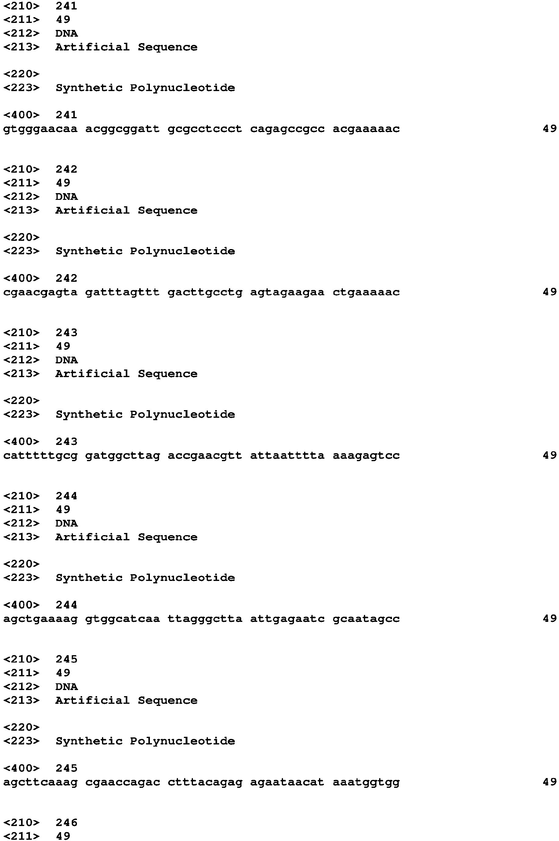


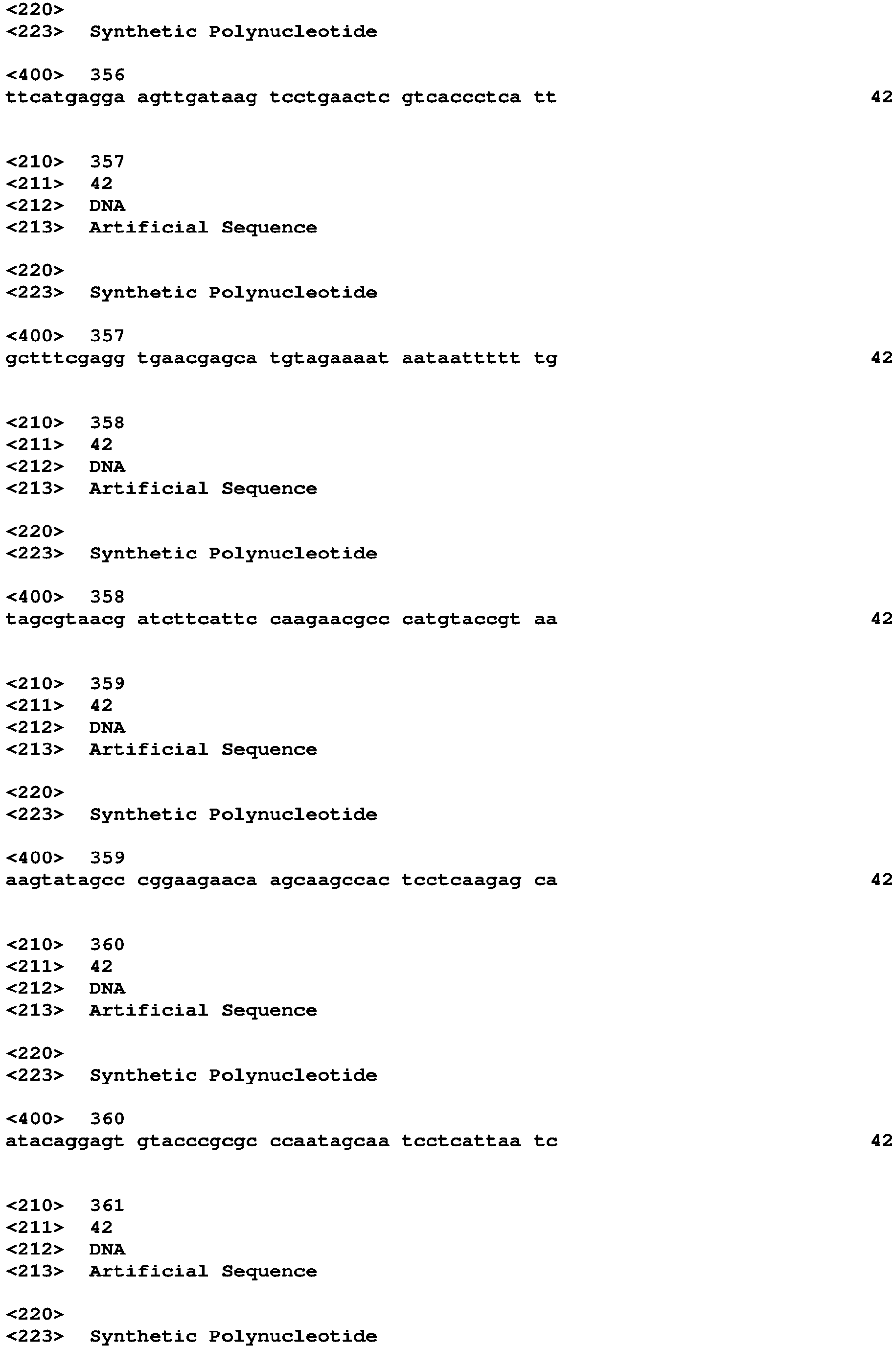
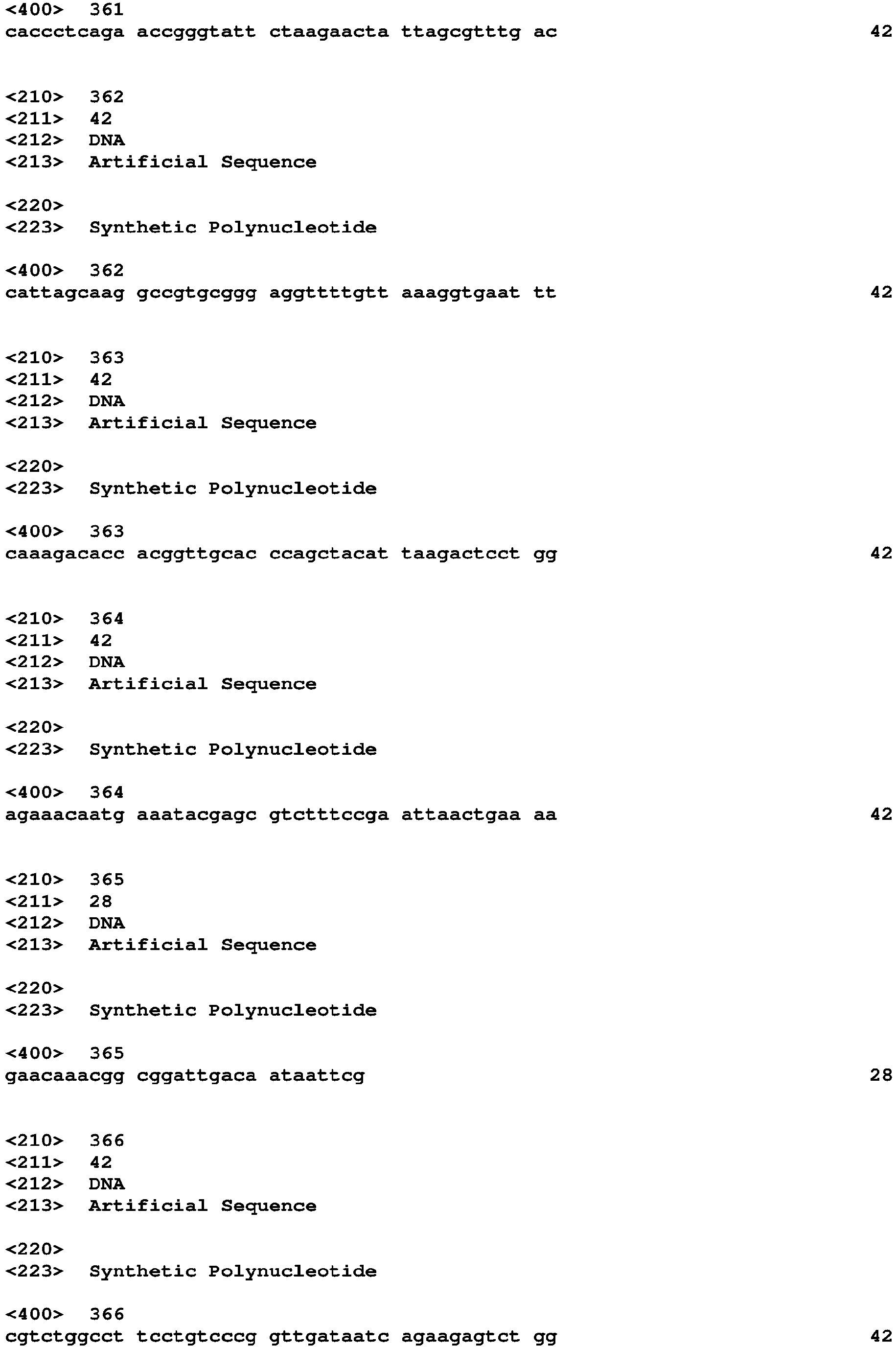


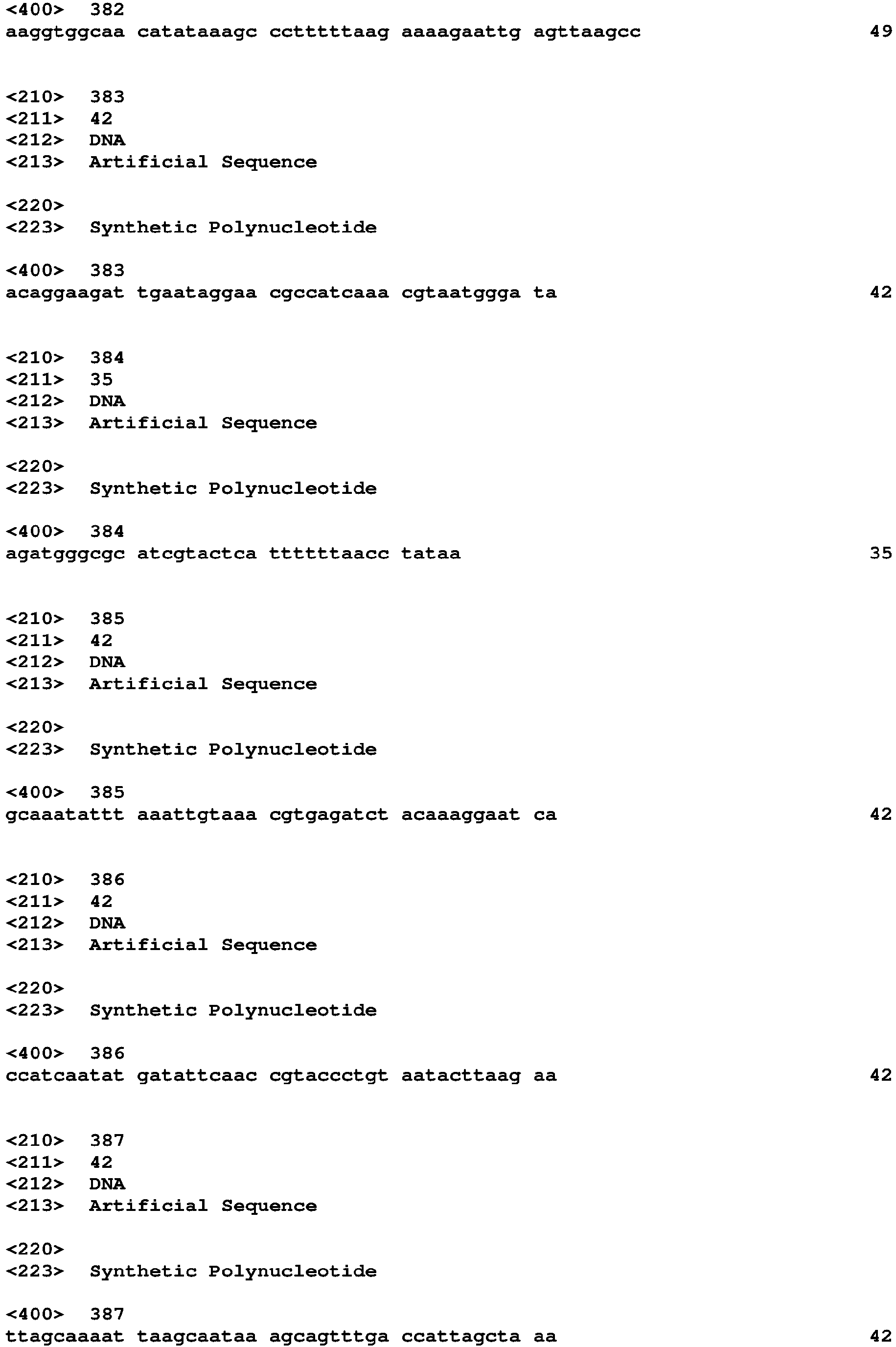






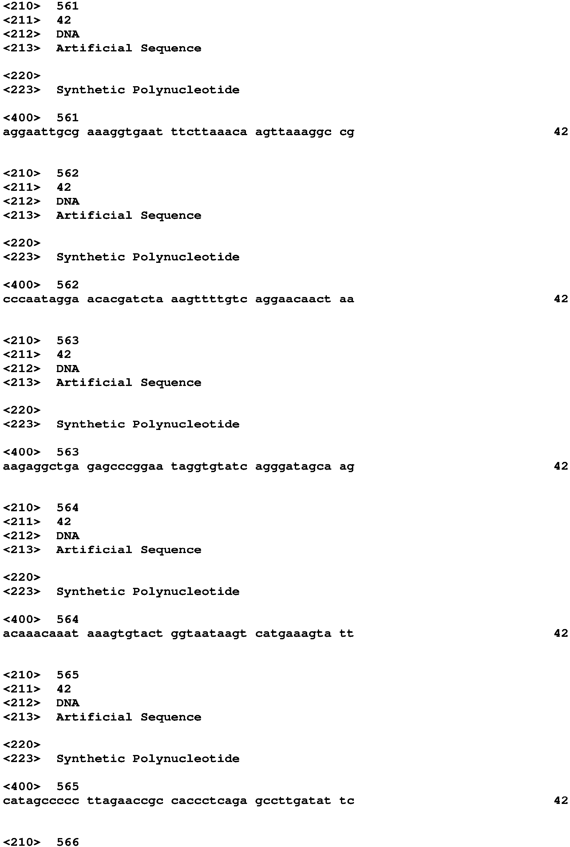


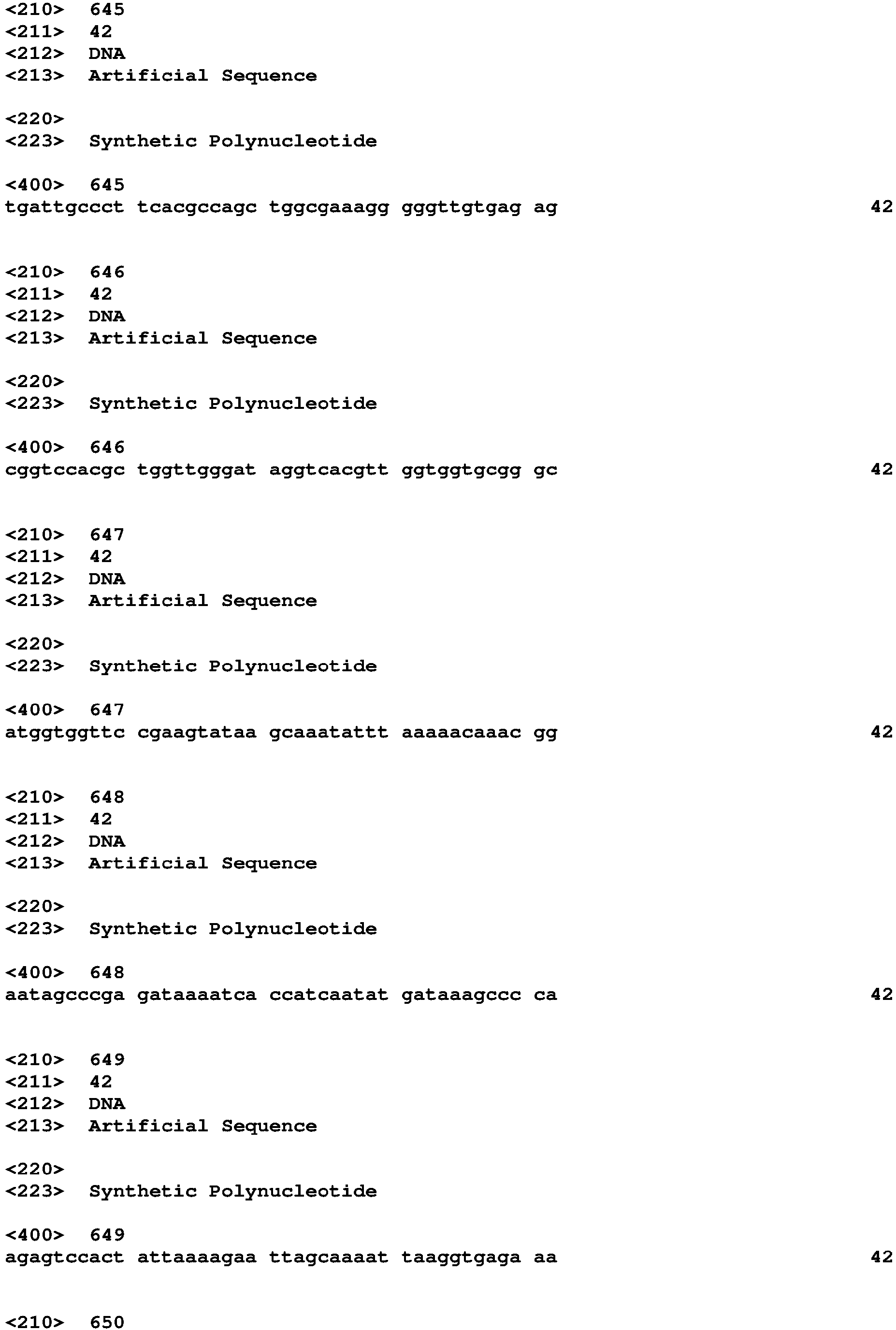


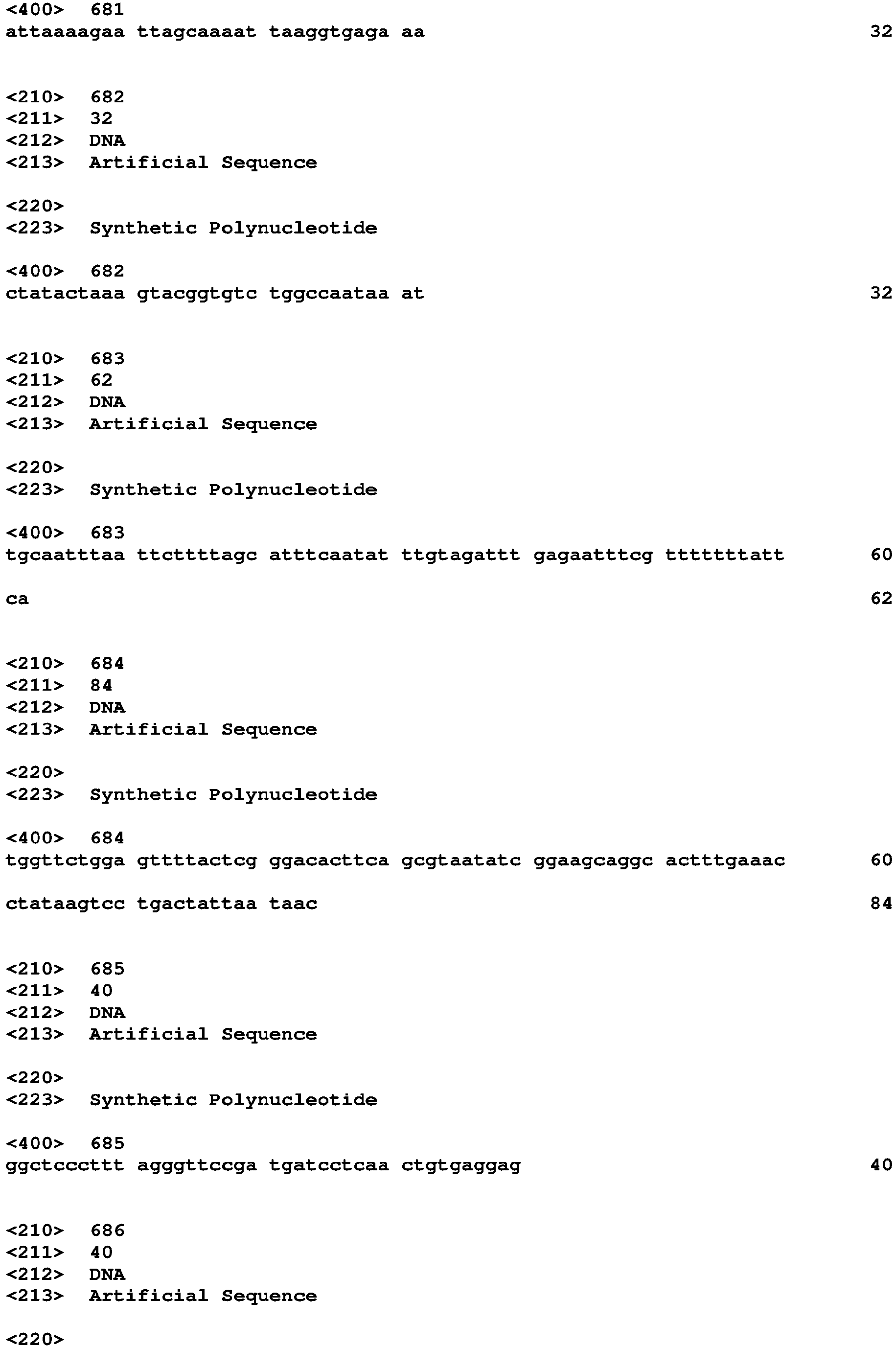

Claims (11)
- A crisscross nucleic acid nanostructure, comprising:(a) a first plurality of nanorods aligned parallel to each other; and(b) a second plurality of nanorods aligned parallel to each other, wherein the nanorods of the first plurality are bound to and perpendicular to the nanorods of the second plurality, a single nanorod of (b) binds to multiple nanorods of (a), each through a single cooperative binding site, and single nanorod of (a) binds to multiple nanorods of (b), each through a single cooperative binding site.
- The crisscross nucleic acid nanostructure of claim 1, wherein each nanorod comprises DNA.
- The crisscross nucleic acid nanostructure of claim 2, wherein each nanorod comprises a 6-helix DNA bundle.
- A method of producing a crisscross nucleic acid nanostructure, comprising:combining in a reaction mixture(a) a first nanorod comprising a first plug strand and a second plug strand,(b) a second nanorod comprising a third plug strand and a fourth plug strand, wherein the second nanorod is parallel to the first nanorod,(c) a third nanorod comprising a fifth plug strand complementary to and bound to the first plug strand and a sixth plug strand complementary to and bound to the second plug strand, and(d) a fourth nanorod comprising a seventh plug strand complementary to and bound to the third plug strand and an eighth plug strand complementary to and bound to the fourth plug strand, wherein the third nanorod is parallel to the fourth nanorod; andincubating the reaction mixture under conditions that result in assembly of a crisscross nucleic acid nanostructure.
- A crisscross nucleic acid slat, comprising:a first plurality of at least four nucleic acid strands parallel to each other, each strand of the first plurality having a length of at least 21 nucleotides; anda second plurality of at least four nucleic acid strands parallel to each, each strand of the second plurality having a length of at least 21 nucleotides, wherein the at least four nucleic acid strands of the first plurality are bound to and perpendicular to the at least four nucleic acid strands of the second plurality.
- A nanostructure comprising the crisscross nucleic acid slat of claim 5.
- The nanostructure of claim 6, wherein the nanostructure is two-dimensional.
- The nanostructure of claim 6, wherein the nanostructure is three-dimensional.
- A microstructure comprising the crisscross nucleic acid slat of claim 5.
- The microstructure of claim 9, wherein the microstructure is two-dimensional.
- The microstructure of claim 9, wherein the microstructure is three-dimensional.
Applications Claiming Priority (3)
| Application Number | Priority Date | Filing Date | Title |
|---|---|---|---|
| US201662370098P | 2016-08-02 | 2016-08-02 | |
| EP17837580.4A EP3494124B1 (en) | 2016-08-02 | 2017-08-02 | Crisscross cooperative self-assembly |
| PCT/US2017/045013 WO2018026880A2 (en) | 2016-08-02 | 2017-08-02 | Crisscross cooperative self-assembly |
Related Parent Applications (1)
| Application Number | Title | Priority Date | Filing Date |
|---|---|---|---|
| EP17837580.4A Division EP3494124B1 (en) | 2016-08-02 | 2017-08-02 | Crisscross cooperative self-assembly |
Publications (2)
| Publication Number | Publication Date |
|---|---|
| EP4071159A1 true EP4071159A1 (en) | 2022-10-12 |
| EP4071159B1 EP4071159B1 (en) | 2023-12-13 |
Family
ID=61073289
Family Applications (2)
| Application Number | Title | Priority Date | Filing Date |
|---|---|---|---|
| EP17837580.4A Active EP3494124B1 (en) | 2016-08-02 | 2017-08-02 | Crisscross cooperative self-assembly |
| EP22160474.7A Active EP4071159B1 (en) | 2016-08-02 | 2017-08-02 | Crisscross cooperative self-assembly |
Family Applications Before (1)
| Application Number | Title | Priority Date | Filing Date |
|---|---|---|---|
| EP17837580.4A Active EP3494124B1 (en) | 2016-08-02 | 2017-08-02 | Crisscross cooperative self-assembly |
Country Status (4)
| Country | Link |
|---|---|
| US (2) | US11254972B2 (en) |
| EP (2) | EP3494124B1 (en) |
| CN (2) | CN115948505B (en) |
| WO (1) | WO2018026880A2 (en) |
Families Citing this family (4)
| Publication number | Priority date | Publication date | Assignee | Title |
|---|---|---|---|---|
| WO2017096492A1 (en) * | 2015-12-11 | 2017-06-15 | Mcmaster University | Nucleic acid catenane with a linking duplex biosensor for detection of a microorganism target |
| US11414694B2 (en) | 2016-03-11 | 2022-08-16 | Children's Medical Center Corporation | Nucleic acid nanoswitch catenanes |
| EP3494124B1 (en) | 2016-08-02 | 2022-03-09 | President and Fellows of Harvard College | Crisscross cooperative self-assembly |
| US20250019750A1 (en) * | 2021-11-19 | 2025-01-16 | President And Fellows Of Harvard College | Enzyme-free isothermal exponential amplification |
Citations (1)
| Publication number | Priority date | Publication date | Assignee | Title |
|---|---|---|---|---|
| US20070117109A1 (en) * | 2005-06-14 | 2007-05-24 | California Institute Of Technology | Nanostructures, methods of making and using the same |
Family Cites Families (33)
| Publication number | Priority date | Publication date | Assignee | Title |
|---|---|---|---|---|
| US5591601A (en) | 1993-05-14 | 1997-01-07 | Ohio University Edison Animal Biotechnology Institute | DNA polymerase gene expression system utilizing an RNA polymerase co-delivered with the gene expression vector system |
| AU2656295A (en) | 1994-06-02 | 1996-01-04 | Chiron Corporation | Nucleic acid immunization using a virus-based infection/transfection system |
| BR0015484A (en) | 1999-11-13 | 2002-07-02 | Merck Patent Gmbh | High-order nucleic acid-based structures |
| JP2002114797A (en) | 2000-10-10 | 2002-04-16 | Atsushi Maruyama | Prepared material for forming triple-stranded nucleic acid |
| JP4061401B2 (en) | 2002-03-07 | 2008-03-19 | 国立大学法人九州大学 | DNA nanocage by self-organization of DNA, method for producing the same, and DNA nanotube and molecular carrier using the same |
| US7598363B2 (en) | 2004-06-10 | 2009-10-06 | New York University | Polygonal nanostructures of polynucleic acid multi-crossover molecules and assembly of lattices based on double crossover cohesion |
| US7964571B2 (en) | 2004-12-09 | 2011-06-21 | Egen, Inc. | Combination of immuno gene therapy and chemotherapy for treatment of cancer and hyperproliferative diseases |
| CA2670801A1 (en) | 2005-11-30 | 2007-06-07 | Intradigm Corporation | Compositions and methods of using sirna to knockdown gene expression and to improve solid organ and cell transplantation |
| JP2009213390A (en) | 2008-03-10 | 2009-09-24 | Nissan Motor Co Ltd | Nucleic acid complex |
| US20110243910A1 (en) | 2008-08-12 | 2011-10-06 | Japan Health Sciences Foundation | mammalian rna dependent rna polymerase |
| WO2010068432A1 (en) | 2008-11-25 | 2010-06-17 | Ecole Polytechnique Federale De Lausanne (Epfl) | Block copolymers and uses thereof |
| US20110033706A1 (en) | 2009-08-04 | 2011-02-10 | Yamuna Krishnan | Nanocapsules and methods for modular assembly |
| EP2491123B1 (en) | 2009-10-20 | 2018-04-18 | The Regents of The University of California | Single molecule nucleic acid nanoparticles |
| CA2800065A1 (en) | 2010-05-21 | 2011-11-24 | Peptimed, Inc. | Reagents and methods for treating cancer |
| US20130245102A1 (en) | 2010-10-12 | 2013-09-19 | Research Foundation Of The City University Of New York | Novel dna templates for small rna production in mammalian cells |
| AU2011319755B2 (en) | 2010-10-27 | 2017-02-23 | President And Fellows Of Harvard College | Compositions of toehold primer duplexes and methods of use |
| KR101094738B1 (en) | 2011-01-27 | 2011-12-16 | 성균관대학교산학협력단 | Nano Device Formation Method |
| WO2012151328A2 (en) | 2011-05-02 | 2012-11-08 | President And Fellows Of Harvard College | Spatial sequestration of dynamic nucleic acid circuits |
| SG10201605170VA (en) * | 2011-07-01 | 2016-08-30 | Harvard College | Method for forming nanoparticles having predetermined shapes |
| JP6430253B2 (en) | 2011-11-05 | 2018-11-28 | プレジデント アンド フェローズ オブ ハーバード カレッジ | Nucleic acid-based linkers for detecting and measuring interactions |
| US9371354B2 (en) | 2011-11-30 | 2016-06-21 | The Board Of Trustees Of The University Of Arkansas | DNA-linked nanoparticle building blocks for nanostructure assembly and methods of producing the same |
| CN103539065A (en) * | 2012-07-10 | 2014-01-29 | 中国科学院苏州纳米技术与纳米仿生研究所 | Method for constructing nanoparticle-nanorod composite structure and constructed composite structure |
| JP6596336B2 (en) * | 2012-07-24 | 2019-10-23 | プレジデント アンド フェローズ オブ ハーバード カレッジ | Self-assembly of nucleic acid nanostructures |
| CN103159170B (en) * | 2013-03-06 | 2014-06-18 | 中国科学院苏州纳米技术与纳米仿生研究所 | Three-dimensional nano-structure construction method |
| JP2017505104A (en) | 2013-11-08 | 2017-02-16 | デイナ ファーバー キャンサー インスティチュート,インコーポレイテッド | Nucleic acid nanostructures for in vivo drug delivery |
| WO2015130805A1 (en) * | 2014-02-26 | 2015-09-03 | Arizona Board Of Regents On Behalf Of Arizona State University | Dna gridiron compositions and methods |
| EP2940150A1 (en) | 2014-04-29 | 2015-11-04 | Baseclick GmbH | Self-assembly of DNA Origami: a new diganostic tool |
| WO2016144755A1 (en) | 2015-03-07 | 2016-09-15 | President And Fellows Of Harvard College | Single-stranded dna nanostructures |
| WO2017096492A1 (en) | 2015-12-11 | 2017-06-15 | Mcmaster University | Nucleic acid catenane with a linking duplex biosensor for detection of a microorganism target |
| US20190083522A1 (en) | 2016-03-10 | 2019-03-21 | President And Fellows Of Harvard College | Biosynthetic modules |
| US11414694B2 (en) | 2016-03-11 | 2022-08-16 | Children's Medical Center Corporation | Nucleic acid nanoswitch catenanes |
| EP3494124B1 (en) | 2016-08-02 | 2022-03-09 | President and Fellows of Harvard College | Crisscross cooperative self-assembly |
| US20250019750A1 (en) | 2021-11-19 | 2025-01-16 | President And Fellows Of Harvard College | Enzyme-free isothermal exponential amplification |
-
2017
- 2017-08-02 EP EP17837580.4A patent/EP3494124B1/en active Active
- 2017-08-02 US US16/322,787 patent/US11254972B2/en active Active
- 2017-08-02 WO PCT/US2017/045013 patent/WO2018026880A2/en not_active Ceased
- 2017-08-02 EP EP22160474.7A patent/EP4071159B1/en active Active
- 2017-08-02 CN CN202211182163.7A patent/CN115948505B/en active Active
- 2017-08-02 CN CN201780060902.6A patent/CN109790199B/en active Active
-
2022
- 2022-01-14 US US17/576,550 patent/US12351862B2/en active Active
Patent Citations (1)
| Publication number | Priority date | Publication date | Assignee | Title |
|---|---|---|---|---|
| US20070117109A1 (en) * | 2005-06-14 | 2007-05-24 | California Institute Of Technology | Nanostructures, methods of making and using the same |
Non-Patent Citations (30)
| Title |
|---|
| ANDERSEN, E. S. ET AL., NATURE, vol. 461, no. 7245, 2009, pages 414 - 418 |
| CHWOROS, A. ET AL., SCIENCE, vol. 306, 2004, pages 2068 - 2072 |
| D. HAN ET AL: "DNA Gridiron Nanostructures Based on Four-Arm Junctions", SCIENCE, vol. 339, no. 6126, 22 March 2013 (2013-03-22), US, pages 1412 - 1415, XP055675850, ISSN: 0036-8075, DOI: 10.1126/science.1232252 * |
| DELEBECQUE, C. J. ET AL., SCIENCE, vol. 333, 2011, pages 470 - 474 |
| DOUGLAS SM1CHOU JJSHIH WM: "DNA-nanotube-induced alignment of membrane proteins for NMR structure determination", PROC NATL ACAD SCI USA., vol. 104, 2007, pages 6644 - 6648, XP055031227, DOI: 10.1073/pnas.0700930104 |
| DOUGLAS SMMARBLESTONE AHTEERAPITTAYANON SVAZQUEZ ACHURCH GMSHIH WM: "Rapid prototyping of 3D DNA-origami shapes with caDNAno", NUCLEIC ACIDS RES, vol. 37, 2009, pages 5001 - 5006, XP002694878, DOI: 10.1093/nar/gkp436 |
| F. HIMO ET AL., J. AM. CHEM. SOC., vol. 127, 2005, pages 210 - 216 |
| FAN HONG ET AL: "3D Framework DNA Origami with Layered Crossovers", ANGEWANDTE CHEMIE, WILEY - V C H VERLAG GMBH & CO. KGAA, DE, vol. 128, no. 41, 15 September 2016 (2016-09-15), pages 13024 - 13027, XP071369427, ISSN: 0044-8249, DOI: 10.1002/ANGE.201607050 * |
| FELDKAMP, U. ET AL., ANGEW. CHEM. INT. ED., vol. 45, no. 12, 2006, pages 1856 - 1876 |
| FU, T. J.SEEMAN, N. C., BIOCHEMISTRY, vol. 32, 1993, pages 3211 - 3220 |
| HARIRI A. ET AL., NATURE CHEMISTRY, vol. 7, 2015, pages 295 - 300 |
| HE, Y. ET AL., NATURE, vol. 452, 2008, pages 198 - 201 |
| LIN, C. ET AL., CHEMPHYSCHEM, vol. 7, 2006, pages 1641 - 1647 |
| LIU, WET, ANGEW. CHEM. INT. ED., vol. 50, 2011, pages 264 - 267 |
| MAO, C., PLOS BIOLOGY, vol. 2, no. 12, 2004, pages 2036 - 2038 |
| MATHIEU F. ET AL., NANO LETT, vol. 5, no. 4, 2005, pages 661 - 664 |
| MATHIEU FREDERICK ET AL: "Six-helix bundles designed from DNA", NANO LETTERS, AMERICAN CHEMICAL SOCIETY, US, vol. 5, no. 4, 1 April 2005 (2005-04-01), pages 661 - 665, XP002488117, ISSN: 1530-6984, [retrieved on 20050302], DOI: 10.1021/NL050084F * |
| ROTHEMUND, P. W. K. ET AL., PLOS BIOL, vol. 2, 2004, pages e424 |
| ROTHEMUND, P. W. K., NATURE, vol. 440, 2006, pages 297 - 302 |
| SCHULMAN, R.WINFREE, E., PROC. NATL ACAD. SCI. USA, vol. 104, 2007, pages 15236 - 15241 |
| SEEMAN, N. C., J. THEOR. BIOL., vol. 99, 1982, pages 237 - 247 |
| SHARMA, J. ET AL., SCIENCE, vol. 323, 2009, pages 112 - 116 |
| SHAWN M DOUGLAS ET AL: "Self-assembly of DNA into nanoscale three-dimensional shapes", NATURE, MACMILLAN JOURNALS LTD, LONDON, vol. 459, no. 7245, 21 May 2009 (2009-05-21), pages 414 - 418, XP002690757, ISSN: 0028-0836, DOI: 10.1038/NATURE08016 * |
| TORRING, T. ET AL., CHEM. SOC. REV., vol. 40, 2011, pages 5636 - 5646 |
| V. V. ROSTOVTSEV ET AL., ANGEW. CHEM. INT. ED., vol. 41, 2002, pages 2596 - 2599 |
| WINFREE, E. ET AL., NATURE, vol. 394, 1998, pages 539 - 544 |
| WOO, S.ROTHEMUND, P., NAT. CHEM., vol. 3, 2011, pages 620 - 627 |
| YAN, H. ET AL., SCIENCE, vol. 301, 2003, pages 1882 - 1884 |
| YIN, P. ET AL., SCIENCE, vol. 321, 2008, pages 824 - 826 |
| ZHAO, Z. ET AL., NANO LETT, vol. 11, 2011, pages 2997 - 3002 |
Also Published As
| Publication number | Publication date |
|---|---|
| US12351862B2 (en) | 2025-07-08 |
| EP4071159B1 (en) | 2023-12-13 |
| US20190203277A1 (en) | 2019-07-04 |
| US20220403453A1 (en) | 2022-12-22 |
| CN109790199A (en) | 2019-05-21 |
| EP3494124A2 (en) | 2019-06-12 |
| CN109790199B (en) | 2022-10-18 |
| US11254972B2 (en) | 2022-02-22 |
| EP3494124B1 (en) | 2022-03-09 |
| EP3494124A4 (en) | 2020-08-26 |
| WO2018026880A3 (en) | 2018-03-22 |
| CN115948505A (en) | 2023-04-11 |
| WO2018026880A2 (en) | 2018-02-08 |
| CN115948505B (en) | 2025-07-25 |
Similar Documents
| Publication | Publication Date | Title |
|---|---|---|
| US12351862B2 (en) | Crisscross cooperative self-assembly | |
| Liu et al. | Branched kissing loops for the construction of diverse RNA homooligomeric nanostructures | |
| Rakonjac et al. | Filamentous bacteriophage: biology, phage display and nanotechnology applications | |
| Tamerler et al. | Molecular biomimetics: utilizing nature’s molecular ways in practical engineering | |
| Merzlyak et al. | Phage as templates for hybrid materials and mediators for nanomaterial synthesis | |
| US11168320B2 (en) | Structure assisted directed evolution of multivalent aptamers | |
| De Santis et al. | Peptide self-assembly for nanomaterials: the old new kid on the block | |
| AU2022202347A1 (en) | Libraries of Nucleic Acids and Methods for Making the Same | |
| Elrad et al. | Encapsulation of a polymer by an icosahedral virus | |
| Hernandez-Garcia et al. | Precise coating of a wide range of DNA templates by a protein polymer with a DNA binding domain | |
| JP6596336B2 (en) | Self-assembly of nucleic acid nanostructures | |
| Arai | Hierarchical design of artificial proteins and complexes toward synthetic structural biology | |
| Calcines-Cruz et al. | CRISPR-guided programmable self-assembly of artificial virus-like nucleocapsids | |
| US20250019750A1 (en) | Enzyme-free isothermal exponential amplification | |
| Aissaoui et al. | Modular imaging scaffold for single-particle electron microscopy | |
| JP2015171369A (en) | Method of mutagenesis | |
| CN107083389A (en) | A kind of new microenvironment large biological molecule common oscillator, its synthetic method and application | |
| US20180371455A1 (en) | Arrays of Intersecting Double Stranded Nucleic Acid Helices | |
| CN107075587A (en) | Method for self-assembly of multi-stranded nucleic acid modules into finite nucleic acid nanostructures | |
| Isbilir | Structural Characterization of Higher Order Complex Formation in Conjugative Transposition of an Antibiotic Resistance Carrying Mobile Genetic Element | |
| Marchi | Advancing DNA-based Nanotechnology Capabilities and Applications | |
| Pfeifer | Hierarchical assembly of DNA origami structures: theoretical aspects and biological applications | |
| Garmann et al. | Packaging of Single-Stranded RNA in Viruses and Virus-Like Particles | |
| Hoffmann | Filamentous Bacteriophage Proteins and Assembly | |
| WO2023238055A1 (en) | A nucleic acid origami modular device |
Legal Events
| Date | Code | Title | Description |
|---|---|---|---|
| PUAI | Public reference made under article 153(3) epc to a published international application that has entered the european phase |
Free format text: ORIGINAL CODE: 0009012 |
|
| STAA | Information on the status of an ep patent application or granted ep patent |
Free format text: STATUS: REQUEST FOR EXAMINATION WAS MADE |
|
| 17P | Request for examination filed |
Effective date: 20220307 |
|
| AC | Divisional application: reference to earlier application |
Ref document number: 3494124 Country of ref document: EP Kind code of ref document: P |
|
| AK | Designated contracting states |
Kind code of ref document: A1 Designated state(s): AL AT BE BG CH CY CZ DE DK EE ES FI FR GB GR HR HU IE IS IT LI LT LU LV MC MK MT NL NO PL PT RO RS SE SI SK SM TR |
|
| GRAP | Despatch of communication of intention to grant a patent |
Free format text: ORIGINAL CODE: EPIDOSNIGR1 |
|
| STAA | Information on the status of an ep patent application or granted ep patent |
Free format text: STATUS: GRANT OF PATENT IS INTENDED |
|
| INTG | Intention to grant announced |
Effective date: 20230628 |
|
| GRAS | Grant fee paid |
Free format text: ORIGINAL CODE: EPIDOSNIGR3 |
|
| GRAA | (expected) grant |
Free format text: ORIGINAL CODE: 0009210 |
|
| STAA | Information on the status of an ep patent application or granted ep patent |
Free format text: STATUS: THE PATENT HAS BEEN GRANTED |
|
| AC | Divisional application: reference to earlier application |
Ref document number: 3494124 Country of ref document: EP Kind code of ref document: P |
|
| AK | Designated contracting states |
Kind code of ref document: B1 Designated state(s): AL AT BE BG CH CY CZ DE DK EE ES FI FR GB GR HR HU IE IS IT LI LT LU LV MC MK MT NL NO PL PT RO RS SE SI SK SM TR |
|
| REG | Reference to a national code |
Ref country code: GB Ref legal event code: FG4D |
|
| REG | Reference to a national code |
Ref country code: CH Ref legal event code: EP |
|
| P01 | Opt-out of the competence of the unified patent court (upc) registered |
Effective date: 20231115 |
|
| REG | Reference to a national code |
Ref country code: DE Ref legal event code: R096 Ref document number: 602017077627 Country of ref document: DE |
|
| REG | Reference to a national code |
Ref country code: IE Ref legal event code: FG4D |
|
| PG25 | Lapsed in a contracting state [announced via postgrant information from national office to epo] |
Ref country code: GR Free format text: LAPSE BECAUSE OF FAILURE TO SUBMIT A TRANSLATION OF THE DESCRIPTION OR TO PAY THE FEE WITHIN THE PRESCRIBED TIME-LIMIT Effective date: 20240314 |
|
| REG | Reference to a national code |
Ref country code: LT Ref legal event code: MG9D |
|
| PG25 | Lapsed in a contracting state [announced via postgrant information from national office to epo] |
Ref country code: LT Free format text: LAPSE BECAUSE OF FAILURE TO SUBMIT A TRANSLATION OF THE DESCRIPTION OR TO PAY THE FEE WITHIN THE PRESCRIBED TIME-LIMIT Effective date: 20231213 |
|
| REG | Reference to a national code |
Ref country code: NL Ref legal event code: MP Effective date: 20231213 |
|
| PG25 | Lapsed in a contracting state [announced via postgrant information from national office to epo] |
Ref country code: ES Free format text: LAPSE BECAUSE OF FAILURE TO SUBMIT A TRANSLATION OF THE DESCRIPTION OR TO PAY THE FEE WITHIN THE PRESCRIBED TIME-LIMIT Effective date: 20231213 |
|
| PG25 | Lapsed in a contracting state [announced via postgrant information from national office to epo] |
Ref country code: LT Free format text: LAPSE BECAUSE OF FAILURE TO SUBMIT A TRANSLATION OF THE DESCRIPTION OR TO PAY THE FEE WITHIN THE PRESCRIBED TIME-LIMIT Effective date: 20231213 Ref country code: GR Free format text: LAPSE BECAUSE OF FAILURE TO SUBMIT A TRANSLATION OF THE DESCRIPTION OR TO PAY THE FEE WITHIN THE PRESCRIBED TIME-LIMIT Effective date: 20240314 Ref country code: ES Free format text: LAPSE BECAUSE OF FAILURE TO SUBMIT A TRANSLATION OF THE DESCRIPTION OR TO PAY THE FEE WITHIN THE PRESCRIBED TIME-LIMIT Effective date: 20231213 Ref country code: BG Free format text: LAPSE BECAUSE OF FAILURE TO SUBMIT A TRANSLATION OF THE DESCRIPTION OR TO PAY THE FEE WITHIN THE PRESCRIBED TIME-LIMIT Effective date: 20240313 |
|
| REG | Reference to a national code |
Ref country code: AT Ref legal event code: MK05 Ref document number: 1640375 Country of ref document: AT Kind code of ref document: T Effective date: 20231213 |
|
| PG25 | Lapsed in a contracting state [announced via postgrant information from national office to epo] |
Ref country code: NL Free format text: LAPSE BECAUSE OF FAILURE TO SUBMIT A TRANSLATION OF THE DESCRIPTION OR TO PAY THE FEE WITHIN THE PRESCRIBED TIME-LIMIT Effective date: 20231213 |
|
| PG25 | Lapsed in a contracting state [announced via postgrant information from national office to epo] |
Ref country code: SE Free format text: LAPSE BECAUSE OF FAILURE TO SUBMIT A TRANSLATION OF THE DESCRIPTION OR TO PAY THE FEE WITHIN THE PRESCRIBED TIME-LIMIT Effective date: 20231213 Ref country code: RS Free format text: LAPSE BECAUSE OF FAILURE TO SUBMIT A TRANSLATION OF THE DESCRIPTION OR TO PAY THE FEE WITHIN THE PRESCRIBED TIME-LIMIT Effective date: 20231213 Ref country code: NO Free format text: LAPSE BECAUSE OF FAILURE TO SUBMIT A TRANSLATION OF THE DESCRIPTION OR TO PAY THE FEE WITHIN THE PRESCRIBED TIME-LIMIT Effective date: 20240313 Ref country code: NL Free format text: LAPSE BECAUSE OF FAILURE TO SUBMIT A TRANSLATION OF THE DESCRIPTION OR TO PAY THE FEE WITHIN THE PRESCRIBED TIME-LIMIT Effective date: 20231213 Ref country code: LV Free format text: LAPSE BECAUSE OF FAILURE TO SUBMIT A TRANSLATION OF THE DESCRIPTION OR TO PAY THE FEE WITHIN THE PRESCRIBED TIME-LIMIT Effective date: 20231213 Ref country code: HR Free format text: LAPSE BECAUSE OF FAILURE TO SUBMIT A TRANSLATION OF THE DESCRIPTION OR TO PAY THE FEE WITHIN THE PRESCRIBED TIME-LIMIT Effective date: 20231213 |
|
| PG25 | Lapsed in a contracting state [announced via postgrant information from national office to epo] |
Ref country code: IS Free format text: LAPSE BECAUSE OF FAILURE TO SUBMIT A TRANSLATION OF THE DESCRIPTION OR TO PAY THE FEE WITHIN THE PRESCRIBED TIME-LIMIT Effective date: 20240413 |
|
| PG25 | Lapsed in a contracting state [announced via postgrant information from national office to epo] |
Ref country code: CZ Free format text: LAPSE BECAUSE OF FAILURE TO SUBMIT A TRANSLATION OF THE DESCRIPTION OR TO PAY THE FEE WITHIN THE PRESCRIBED TIME-LIMIT Effective date: 20231213 Ref country code: AT Free format text: LAPSE BECAUSE OF FAILURE TO SUBMIT A TRANSLATION OF THE DESCRIPTION OR TO PAY THE FEE WITHIN THE PRESCRIBED TIME-LIMIT Effective date: 20231213 |
|
| PG25 | Lapsed in a contracting state [announced via postgrant information from national office to epo] |
Ref country code: SK Free format text: LAPSE BECAUSE OF FAILURE TO SUBMIT A TRANSLATION OF THE DESCRIPTION OR TO PAY THE FEE WITHIN THE PRESCRIBED TIME-LIMIT Effective date: 20231213 |
|
| PG25 | Lapsed in a contracting state [announced via postgrant information from national office to epo] |
Ref country code: SM Free format text: LAPSE BECAUSE OF FAILURE TO SUBMIT A TRANSLATION OF THE DESCRIPTION OR TO PAY THE FEE WITHIN THE PRESCRIBED TIME-LIMIT Effective date: 20231213 Ref country code: SK Free format text: LAPSE BECAUSE OF FAILURE TO SUBMIT A TRANSLATION OF THE DESCRIPTION OR TO PAY THE FEE WITHIN THE PRESCRIBED TIME-LIMIT Effective date: 20231213 Ref country code: RO Free format text: LAPSE BECAUSE OF FAILURE TO SUBMIT A TRANSLATION OF THE DESCRIPTION OR TO PAY THE FEE WITHIN THE PRESCRIBED TIME-LIMIT Effective date: 20231213 Ref country code: IT Free format text: LAPSE BECAUSE OF FAILURE TO SUBMIT A TRANSLATION OF THE DESCRIPTION OR TO PAY THE FEE WITHIN THE PRESCRIBED TIME-LIMIT Effective date: 20231213 Ref country code: IS Free format text: LAPSE BECAUSE OF FAILURE TO SUBMIT A TRANSLATION OF THE DESCRIPTION OR TO PAY THE FEE WITHIN THE PRESCRIBED TIME-LIMIT Effective date: 20240413 Ref country code: EE Free format text: LAPSE BECAUSE OF FAILURE TO SUBMIT A TRANSLATION OF THE DESCRIPTION OR TO PAY THE FEE WITHIN THE PRESCRIBED TIME-LIMIT Effective date: 20231213 Ref country code: CZ Free format text: LAPSE BECAUSE OF FAILURE TO SUBMIT A TRANSLATION OF THE DESCRIPTION OR TO PAY THE FEE WITHIN THE PRESCRIBED TIME-LIMIT Effective date: 20231213 Ref country code: AT Free format text: LAPSE BECAUSE OF FAILURE TO SUBMIT A TRANSLATION OF THE DESCRIPTION OR TO PAY THE FEE WITHIN THE PRESCRIBED TIME-LIMIT Effective date: 20231213 |
|
| PG25 | Lapsed in a contracting state [announced via postgrant information from national office to epo] |
Ref country code: PT Free format text: LAPSE BECAUSE OF FAILURE TO SUBMIT A TRANSLATION OF THE DESCRIPTION OR TO PAY THE FEE WITHIN THE PRESCRIBED TIME-LIMIT Effective date: 20240415 Ref country code: PL Free format text: LAPSE BECAUSE OF FAILURE TO SUBMIT A TRANSLATION OF THE DESCRIPTION OR TO PAY THE FEE WITHIN THE PRESCRIBED TIME-LIMIT Effective date: 20231213 |
|
| PG25 | Lapsed in a contracting state [announced via postgrant information from national office to epo] |
Ref country code: PT Free format text: LAPSE BECAUSE OF FAILURE TO SUBMIT A TRANSLATION OF THE DESCRIPTION OR TO PAY THE FEE WITHIN THE PRESCRIBED TIME-LIMIT Effective date: 20240415 Ref country code: PL Free format text: LAPSE BECAUSE OF FAILURE TO SUBMIT A TRANSLATION OF THE DESCRIPTION OR TO PAY THE FEE WITHIN THE PRESCRIBED TIME-LIMIT Effective date: 20231213 |
|
| REG | Reference to a national code |
Ref country code: DE Ref legal event code: R097 Ref document number: 602017077627 Country of ref document: DE |
|
| PG25 | Lapsed in a contracting state [announced via postgrant information from national office to epo] |
Ref country code: DK Free format text: LAPSE BECAUSE OF FAILURE TO SUBMIT A TRANSLATION OF THE DESCRIPTION OR TO PAY THE FEE WITHIN THE PRESCRIBED TIME-LIMIT Effective date: 20231213 |
|
| PLBE | No opposition filed within time limit |
Free format text: ORIGINAL CODE: 0009261 |
|
| STAA | Information on the status of an ep patent application or granted ep patent |
Free format text: STATUS: NO OPPOSITION FILED WITHIN TIME LIMIT |
|
| PG25 | Lapsed in a contracting state [announced via postgrant information from national office to epo] |
Ref country code: SI Free format text: LAPSE BECAUSE OF FAILURE TO SUBMIT A TRANSLATION OF THE DESCRIPTION OR TO PAY THE FEE WITHIN THE PRESCRIBED TIME-LIMIT Effective date: 20231213 |
|
| PG25 | Lapsed in a contracting state [announced via postgrant information from national office to epo] |
Ref country code: SI Free format text: LAPSE BECAUSE OF FAILURE TO SUBMIT A TRANSLATION OF THE DESCRIPTION OR TO PAY THE FEE WITHIN THE PRESCRIBED TIME-LIMIT Effective date: 20231213 Ref country code: DK Free format text: LAPSE BECAUSE OF FAILURE TO SUBMIT A TRANSLATION OF THE DESCRIPTION OR TO PAY THE FEE WITHIN THE PRESCRIBED TIME-LIMIT Effective date: 20231213 |
|
| 26N | No opposition filed |
Effective date: 20240916 |
|
| REG | Reference to a national code |
Ref country code: CH Ref legal event code: PL |
|
| PG25 | Lapsed in a contracting state [announced via postgrant information from national office to epo] |
Ref country code: LU Free format text: LAPSE BECAUSE OF NON-PAYMENT OF DUE FEES Effective date: 20240802 |
|
| PG25 | Lapsed in a contracting state [announced via postgrant information from national office to epo] |
Ref country code: CH Free format text: LAPSE BECAUSE OF NON-PAYMENT OF DUE FEES Effective date: 20240831 Ref country code: MC Free format text: LAPSE BECAUSE OF FAILURE TO SUBMIT A TRANSLATION OF THE DESCRIPTION OR TO PAY THE FEE WITHIN THE PRESCRIBED TIME-LIMIT Effective date: 20231213 |
|
| REG | Reference to a national code |
Ref country code: BE Ref legal event code: MM Effective date: 20240831 |
|
| PG25 | Lapsed in a contracting state [announced via postgrant information from national office to epo] |
Ref country code: BE Free format text: LAPSE BECAUSE OF NON-PAYMENT OF DUE FEES Effective date: 20240831 |
|
| PG25 | Lapsed in a contracting state [announced via postgrant information from national office to epo] |
Ref country code: IE Free format text: LAPSE BECAUSE OF NON-PAYMENT OF DUE FEES Effective date: 20240802 |
|
| PG25 | Lapsed in a contracting state [announced via postgrant information from national office to epo] |
Ref country code: FI Free format text: LAPSE BECAUSE OF FAILURE TO SUBMIT A TRANSLATION OF THE DESCRIPTION OR TO PAY THE FEE WITHIN THE PRESCRIBED TIME-LIMIT Effective date: 20231213 |
|
| PGFP | Annual fee paid to national office [announced via postgrant information from national office to epo] |
Ref country code: DE Payment date: 20250827 Year of fee payment: 9 |
|
| PGFP | Annual fee paid to national office [announced via postgrant information from national office to epo] |
Ref country code: GB Payment date: 20250827 Year of fee payment: 9 |
|
| PGFP | Annual fee paid to national office [announced via postgrant information from national office to epo] |
Ref country code: FR Payment date: 20250825 Year of fee payment: 9 |
|
| PG25 | Lapsed in a contracting state [announced via postgrant information from national office to epo] |
Ref country code: CY Free format text: LAPSE BECAUSE OF FAILURE TO SUBMIT A TRANSLATION OF THE DESCRIPTION OR TO PAY THE FEE WITHIN THE PRESCRIBED TIME-LIMIT; INVALID AB INITIO Effective date: 20170802 |
|
| PG25 | Lapsed in a contracting state [announced via postgrant information from national office to epo] |
Ref country code: HU Free format text: LAPSE BECAUSE OF FAILURE TO SUBMIT A TRANSLATION OF THE DESCRIPTION OR TO PAY THE FEE WITHIN THE PRESCRIBED TIME-LIMIT; INVALID AB INITIO Effective date: 20170802 |