EP4066869A1 - Fibrocartilage preparation method using tensile stimulation - Google Patents
Fibrocartilage preparation method using tensile stimulation Download PDFInfo
- Publication number
- EP4066869A1 EP4066869A1 EP21770811.4A EP21770811A EP4066869A1 EP 4066869 A1 EP4066869 A1 EP 4066869A1 EP 21770811 A EP21770811 A EP 21770811A EP 4066869 A1 EP4066869 A1 EP 4066869A1
- Authority
- EP
- European Patent Office
- Prior art keywords
- tissue
- tensile
- cartilage
- fibrocartilage
- stimulus
- Prior art date
- Legal status (The legal status is an assumption and is not a legal conclusion. Google has not performed a legal analysis and makes no representation as to the accuracy of the status listed.)
- Granted
Links
Images




Classifications
-
- A—HUMAN NECESSITIES
- A61—MEDICAL OR VETERINARY SCIENCE; HYGIENE
- A61L—METHODS OR APPARATUS FOR STERILISING MATERIALS OR OBJECTS IN GENERAL; DISINFECTION, STERILISATION OR DEODORISATION OF AIR; CHEMICAL ASPECTS OF BANDAGES, DRESSINGS, ABSORBENT PADS OR SURGICAL ARTICLES; MATERIALS FOR BANDAGES, DRESSINGS, ABSORBENT PADS OR SURGICAL ARTICLES
- A61L27/00—Materials for grafts or prostheses or for coating grafts or prostheses
- A61L27/36—Materials for grafts or prostheses or for coating grafts or prostheses containing ingredients of undetermined constitution or reaction products thereof, e.g. transplant tissue, natural bone, extracellular matrix
- A61L27/38—Materials for grafts or prostheses or for coating grafts or prostheses containing ingredients of undetermined constitution or reaction products thereof, e.g. transplant tissue, natural bone, extracellular matrix containing added animal cells
- A61L27/3804—Materials for grafts or prostheses or for coating grafts or prostheses containing ingredients of undetermined constitution or reaction products thereof, e.g. transplant tissue, natural bone, extracellular matrix containing added animal cells characterised by specific cells or progenitors thereof, e.g. fibroblasts, connective tissue cells, kidney cells
- A61L27/3817—Cartilage-forming cells, e.g. pre-chondrocytes
-
- A—HUMAN NECESSITIES
- A61—MEDICAL OR VETERINARY SCIENCE; HYGIENE
- A61L—METHODS OR APPARATUS FOR STERILISING MATERIALS OR OBJECTS IN GENERAL; DISINFECTION, STERILISATION OR DEODORISATION OF AIR; CHEMICAL ASPECTS OF BANDAGES, DRESSINGS, ABSORBENT PADS OR SURGICAL ARTICLES; MATERIALS FOR BANDAGES, DRESSINGS, ABSORBENT PADS OR SURGICAL ARTICLES
- A61L27/00—Materials for grafts or prostheses or for coating grafts or prostheses
- A61L27/36—Materials for grafts or prostheses or for coating grafts or prostheses containing ingredients of undetermined constitution or reaction products thereof, e.g. transplant tissue, natural bone, extracellular matrix
- A61L27/38—Materials for grafts or prostheses or for coating grafts or prostheses containing ingredients of undetermined constitution or reaction products thereof, e.g. transplant tissue, natural bone, extracellular matrix containing added animal cells
- A61L27/3804—Materials for grafts or prostheses or for coating grafts or prostheses containing ingredients of undetermined constitution or reaction products thereof, e.g. transplant tissue, natural bone, extracellular matrix containing added animal cells characterised by specific cells or progenitors thereof, e.g. fibroblasts, connective tissue cells, kidney cells
- A61L27/3834—Cells able to produce different cell types, e.g. hematopoietic stem cells, mesenchymal stem cells, marrow stromal cells, embryonic stem cells
-
- A—HUMAN NECESSITIES
- A61—MEDICAL OR VETERINARY SCIENCE; HYGIENE
- A61L—METHODS OR APPARATUS FOR STERILISING MATERIALS OR OBJECTS IN GENERAL; DISINFECTION, STERILISATION OR DEODORISATION OF AIR; CHEMICAL ASPECTS OF BANDAGES, DRESSINGS, ABSORBENT PADS OR SURGICAL ARTICLES; MATERIALS FOR BANDAGES, DRESSINGS, ABSORBENT PADS OR SURGICAL ARTICLES
- A61L27/00—Materials for grafts or prostheses or for coating grafts or prostheses
- A61L27/36—Materials for grafts or prostheses or for coating grafts or prostheses containing ingredients of undetermined constitution or reaction products thereof, e.g. transplant tissue, natural bone, extracellular matrix
- A61L27/38—Materials for grafts or prostheses or for coating grafts or prostheses containing ingredients of undetermined constitution or reaction products thereof, e.g. transplant tissue, natural bone, extracellular matrix containing added animal cells
- A61L27/3839—Materials for grafts or prostheses or for coating grafts or prostheses containing ingredients of undetermined constitution or reaction products thereof, e.g. transplant tissue, natural bone, extracellular matrix containing added animal cells characterised by the site of application in the body
- A61L27/3843—Connective tissue
- A61L27/3852—Cartilage, e.g. meniscus
-
- A—HUMAN NECESSITIES
- A61—MEDICAL OR VETERINARY SCIENCE; HYGIENE
- A61L—METHODS OR APPARATUS FOR STERILISING MATERIALS OR OBJECTS IN GENERAL; DISINFECTION, STERILISATION OR DEODORISATION OF AIR; CHEMICAL ASPECTS OF BANDAGES, DRESSINGS, ABSORBENT PADS OR SURGICAL ARTICLES; MATERIALS FOR BANDAGES, DRESSINGS, ABSORBENT PADS OR SURGICAL ARTICLES
- A61L27/00—Materials for grafts or prostheses or for coating grafts or prostheses
- A61L27/36—Materials for grafts or prostheses or for coating grafts or prostheses containing ingredients of undetermined constitution or reaction products thereof, e.g. transplant tissue, natural bone, extracellular matrix
- A61L27/38—Materials for grafts or prostheses or for coating grafts or prostheses containing ingredients of undetermined constitution or reaction products thereof, e.g. transplant tissue, natural bone, extracellular matrix containing added animal cells
- A61L27/3839—Materials for grafts or prostheses or for coating grafts or prostheses containing ingredients of undetermined constitution or reaction products thereof, e.g. transplant tissue, natural bone, extracellular matrix containing added animal cells characterised by the site of application in the body
- A61L27/3843—Connective tissue
- A61L27/3852—Cartilage, e.g. meniscus
- A61L27/3856—Intervertebral discs
-
- A—HUMAN NECESSITIES
- A61—MEDICAL OR VETERINARY SCIENCE; HYGIENE
- A61L—METHODS OR APPARATUS FOR STERILISING MATERIALS OR OBJECTS IN GENERAL; DISINFECTION, STERILISATION OR DEODORISATION OF AIR; CHEMICAL ASPECTS OF BANDAGES, DRESSINGS, ABSORBENT PADS OR SURGICAL ARTICLES; MATERIALS FOR BANDAGES, DRESSINGS, ABSORBENT PADS OR SURGICAL ARTICLES
- A61L27/00—Materials for grafts or prostheses or for coating grafts or prostheses
- A61L27/36—Materials for grafts or prostheses or for coating grafts or prostheses containing ingredients of undetermined constitution or reaction products thereof, e.g. transplant tissue, natural bone, extracellular matrix
- A61L27/38—Materials for grafts or prostheses or for coating grafts or prostheses containing ingredients of undetermined constitution or reaction products thereof, e.g. transplant tissue, natural bone, extracellular matrix containing added animal cells
- A61L27/3895—Materials for grafts or prostheses or for coating grafts or prostheses containing ingredients of undetermined constitution or reaction products thereof, e.g. transplant tissue, natural bone, extracellular matrix containing added animal cells using specific culture conditions, e.g. stimulating differentiation of stem cells, pulsatile flow conditions
-
- C—CHEMISTRY; METALLURGY
- C12—BIOCHEMISTRY; BEER; SPIRITS; WINE; VINEGAR; MICROBIOLOGY; ENZYMOLOGY; MUTATION OR GENETIC ENGINEERING
- C12N—MICROORGANISMS OR ENZYMES; COMPOSITIONS THEREOF; PROPAGATING, PRESERVING, OR MAINTAINING MICROORGANISMS; MUTATION OR GENETIC ENGINEERING; CULTURE MEDIA
- C12N13/00—Treatment of microorganisms or enzymes with electrical or wave energy, e.g. magnetism, sonic waves
-
- C—CHEMISTRY; METALLURGY
- C12—BIOCHEMISTRY; BEER; SPIRITS; WINE; VINEGAR; MICROBIOLOGY; ENZYMOLOGY; MUTATION OR GENETIC ENGINEERING
- C12N—MICROORGANISMS OR ENZYMES; COMPOSITIONS THEREOF; PROPAGATING, PRESERVING, OR MAINTAINING MICROORGANISMS; MUTATION OR GENETIC ENGINEERING; CULTURE MEDIA
- C12N5/00—Undifferentiated human, animal or plant cells, e.g. cell lines; Tissues; Cultivation or maintenance thereof; Culture media therefor
- C12N5/06—Animal cells or tissues; Human cells or tissues
- C12N5/0602—Vertebrate cells
- C12N5/0652—Cells of skeletal and connective tissues; Mesenchyme
- C12N5/0655—Chondrocytes; Cartilage
-
- C—CHEMISTRY; METALLURGY
- C12—BIOCHEMISTRY; BEER; SPIRITS; WINE; VINEGAR; MICROBIOLOGY; ENZYMOLOGY; MUTATION OR GENETIC ENGINEERING
- C12N—MICROORGANISMS OR ENZYMES; COMPOSITIONS THEREOF; PROPAGATING, PRESERVING, OR MAINTAINING MICROORGANISMS; MUTATION OR GENETIC ENGINEERING; CULTURE MEDIA
- C12N5/00—Undifferentiated human, animal or plant cells, e.g. cell lines; Tissues; Cultivation or maintenance thereof; Culture media therefor
- C12N5/06—Animal cells or tissues; Human cells or tissues
- C12N5/0697—Artificial constructs associating cells of different lineages, e.g. tissue equivalents
-
- A—HUMAN NECESSITIES
- A61—MEDICAL OR VETERINARY SCIENCE; HYGIENE
- A61L—METHODS OR APPARATUS FOR STERILISING MATERIALS OR OBJECTS IN GENERAL; DISINFECTION, STERILISATION OR DEODORISATION OF AIR; CHEMICAL ASPECTS OF BANDAGES, DRESSINGS, ABSORBENT PADS OR SURGICAL ARTICLES; MATERIALS FOR BANDAGES, DRESSINGS, ABSORBENT PADS OR SURGICAL ARTICLES
- A61L2430/00—Materials or treatment for tissue regeneration
- A61L2430/06—Materials or treatment for tissue regeneration for cartilage reconstruction, e.g. meniscus
-
- A—HUMAN NECESSITIES
- A61—MEDICAL OR VETERINARY SCIENCE; HYGIENE
- A61L—METHODS OR APPARATUS FOR STERILISING MATERIALS OR OBJECTS IN GENERAL; DISINFECTION, STERILISATION OR DEODORISATION OF AIR; CHEMICAL ASPECTS OF BANDAGES, DRESSINGS, ABSORBENT PADS OR SURGICAL ARTICLES; MATERIALS FOR BANDAGES, DRESSINGS, ABSORBENT PADS OR SURGICAL ARTICLES
- A61L2430/00—Materials or treatment for tissue regeneration
- A61L2430/40—Preparation and treatment of biological tissue for implantation, e.g. decellularisation, cross-linking
-
- C—CHEMISTRY; METALLURGY
- C12—BIOCHEMISTRY; BEER; SPIRITS; WINE; VINEGAR; MICROBIOLOGY; ENZYMOLOGY; MUTATION OR GENETIC ENGINEERING
- C12M—APPARATUS FOR ENZYMOLOGY OR MICROBIOLOGY; APPARATUS FOR CULTURING MICROORGANISMS FOR PRODUCING BIOMASS, FOR GROWING CELLS OR FOR OBTAINING FERMENTATION OR METABOLIC PRODUCTS, i.e. BIOREACTORS OR FERMENTERS
- C12M21/00—Bioreactors or fermenters specially adapted for specific uses
- C12M21/08—Bioreactors or fermenters specially adapted for specific uses for producing artificial tissue or for ex-vivo cultivation of tissue
-
- C—CHEMISTRY; METALLURGY
- C12—BIOCHEMISTRY; BEER; SPIRITS; WINE; VINEGAR; MICROBIOLOGY; ENZYMOLOGY; MUTATION OR GENETIC ENGINEERING
- C12M—APPARATUS FOR ENZYMOLOGY OR MICROBIOLOGY; APPARATUS FOR CULTURING MICROORGANISMS FOR PRODUCING BIOMASS, FOR GROWING CELLS OR FOR OBTAINING FERMENTATION OR METABOLIC PRODUCTS, i.e. BIOREACTORS OR FERMENTERS
- C12M35/00—Means for application of stress for stimulating the growth of microorganisms or the generation of fermentation or metabolic products; Means for electroporation or cell fusion
- C12M35/04—Mechanical means, e.g. sonic waves, stretching forces, pressure or shear stimuli
-
- C—CHEMISTRY; METALLURGY
- C12—BIOCHEMISTRY; BEER; SPIRITS; WINE; VINEGAR; MICROBIOLOGY; ENZYMOLOGY; MUTATION OR GENETIC ENGINEERING
- C12N—MICROORGANISMS OR ENZYMES; COMPOSITIONS THEREOF; PROPAGATING, PRESERVING, OR MAINTAINING MICROORGANISMS; MUTATION OR GENETIC ENGINEERING; CULTURE MEDIA
- C12N2500/00—Specific components of cell culture medium
- C12N2500/05—Inorganic components
- C12N2500/10—Metals; Metal chelators
- C12N2500/20—Transition metals
- C12N2500/24—Iron; Fe chelators; Transferrin
- C12N2500/25—Insulin-transferrin; Insulin-transferrin-selenium
-
- C—CHEMISTRY; METALLURGY
- C12—BIOCHEMISTRY; BEER; SPIRITS; WINE; VINEGAR; MICROBIOLOGY; ENZYMOLOGY; MUTATION OR GENETIC ENGINEERING
- C12N—MICROORGANISMS OR ENZYMES; COMPOSITIONS THEREOF; PROPAGATING, PRESERVING, OR MAINTAINING MICROORGANISMS; MUTATION OR GENETIC ENGINEERING; CULTURE MEDIA
- C12N2500/00—Specific components of cell culture medium
- C12N2500/30—Organic components
- C12N2500/38—Vitamins
-
- C—CHEMISTRY; METALLURGY
- C12—BIOCHEMISTRY; BEER; SPIRITS; WINE; VINEGAR; MICROBIOLOGY; ENZYMOLOGY; MUTATION OR GENETIC ENGINEERING
- C12N—MICROORGANISMS OR ENZYMES; COMPOSITIONS THEREOF; PROPAGATING, PRESERVING, OR MAINTAINING MICROORGANISMS; MUTATION OR GENETIC ENGINEERING; CULTURE MEDIA
- C12N2501/00—Active agents used in cell culture processes, e.g. differentation
- C12N2501/10—Growth factors
- C12N2501/115—Basic fibroblast growth factor (bFGF, FGF-2)
-
- C—CHEMISTRY; METALLURGY
- C12—BIOCHEMISTRY; BEER; SPIRITS; WINE; VINEGAR; MICROBIOLOGY; ENZYMOLOGY; MUTATION OR GENETIC ENGINEERING
- C12N—MICROORGANISMS OR ENZYMES; COMPOSITIONS THEREOF; PROPAGATING, PRESERVING, OR MAINTAINING MICROORGANISMS; MUTATION OR GENETIC ENGINEERING; CULTURE MEDIA
- C12N2537/00—Supports and/or coatings for cell culture characterised by physical or chemical treatment
Definitions
- the present invention relates to a fibrocartilage preparation method and fibrocartilage prepared by the method.
- Degenerative arthritis is a disease in which local degenerative changes appear while articular cartilage wears away, and also refers to osteoarthritis or osteoarthrosis.
- Degenerative arthritis is one type of chronic arthritis, and is a disease in which a degenerative change occurs in the articular cartilage subjected to weight-bearing, resulting in overgrowth of bone on the joint surface, and is often seen in middle-aged adults.
- chondrocytes which make up the constituent components of cartilage, lose their elasticity while deteriorating in function due to aging, and over time, the surface of cartilage becomes rough and various types of materials flow into the articular joint cavity surrounded by the joint membrane, so that inflammation occurs.
- Clinically, recurrent pain, joint stiffness, a progressive joint movement disorder in the joint, and the like appear.
- drug treatment analgesics, steroids, non-steroidal anti-inflammatory agents, and the like
- chondroprotective agents hyaluronic acid, glucosamine, chondroitin and the like
- surgical treatment arthroscopic surgery, wedge high tibial osteotomy, joint replacement, and the like
- drug treatment has only an effect of non-specifically alleviating pain or an inflammatory response itself, and chondroprotective agents only serve to temporarily protect joints by simply supplying nutrients to chondrocytes or alleviating impact.
- meniscus cartilage which secures stability by fixing the shape and position according to joint movement between knee joint cartilage
- partial resection is performed to control the pain.
- partial or total-resection may ameliorate acute pain by primarily eliminating mechanical symptoms.
- the progression of arthritis cannot be prevented since the surgical procedure increases the load on the articular cartilage in the long term and causes degenerative changes. If the meniscus tear was sutured instead of resection, although the indications are very limited, a considerable failure rate has been reported.
- a cartilage treatment method using a tissue engineering technique is emerging to overcome the limitations of existing treatment methods for degenerative arthritis, and accordingly, there is a need for research and development on tissue engineering preparations in which a high degree of similarity to living body tissue is implemented by culturing transplantable tissue in vitro.
- the technical problem to be solved by the present invention is to provide a fibrocartilage preparation method.
- Another technical problem to be solved by the present invention is to provide fibrocartilage prepared by the method.
- An aspect of the present invention relates to a method for preparing fibrocartilage, the method including: culturing one or more cells selected from the group consisting of synovial cells, chondrocytes and mesenchymal stem cells under a tensile stimulus.
- the synovial cells refer to a group of cells present in the synovium, and may be used interchangeably with the same meaning as synovium-derived cells.
- the synovium is a tissue closest to the articular cartilage, and it has been reported that cells are recruited from the synovium to repair cartilage damage (Hunziker EB, Rosenberg LC, Repair of partial-thickness defects in articular cartilage: cell recruitment from the synovial membrane. The Journal of Bone and Joint Surgery. 1996 May;78(5):721-33 ), and furthermore, the synovium is the only tissue capable of producing hyaline cartilage in malignant conditions such as chondromatosis. This suggests that synovial cells may be utilized for the restoration of cartilage tissue.
- chondrocytes are cells that differentiate from mesenchymal cells to form cartilage tissue, and include articular chondrocytes, costal chondrocytes, meniscal chondrocytes, and the like. It is known that the chondrocytes perform a function of synthesizing and secreting a cartilaginous matrix in the cartilage, and these cells gather to form fibrocartilage.
- mesenchymal stem cells are a type of adult stem cells having adhesive properties, and are pluripotent stem cells capable of differentiating into chondrocytes, osteocytes, adipocytes, myocytes, and the like.
- Mesenchymal stem cells may be utilized for fibrocartilage formation using the ability of mesenchymal stem cells with abundant regenerative power to differentiate into chondrocytes.
- fibrocartilage is classified according to its nature and shape, is a cartilage in which white fibrous tissue and cartilaginous tissue are mixed at various ratios, and refers to a site where ligaments or tendons attach to joints.
- the fibrocartilage is located in various forms such as the vertebral body, the pubic bone, the intervertebral disc between the knees, the meniscus cartilage, and the interpubic disc, is a hard fiber composed of collagen, is resistant to compression, but is well torn and arranged in a compactly tangled state, and is present in connection with hyaline cartilage or connective tissue.
- the intervertebral disc is a fibrocartilage present between two vertebral bodies, and there are has 23 intervertebral discs in humans.
- the intervertebral disc is elastic and acts as a cushion that imparts flexibility to the spine and absorbs impact.
- the fibrocartilage of the intervertebral disc may be crushed and protrude, and such a disease is called a hernia of intervertebral disc (often referred to as a disc).
- meniscus cartilage is a wedge-shaped cartilage that acts as a shock absorber between the femur and the tibia. Such meniscus cartilage serves to protect joints.
- the interpubic disc refers to a disc composed of fibrocartilage connecting between pubic bones in the pubic symphysis. Pubic bones are bones present in the hip joint, and the interpubic disc fibrocartilage is firmly bonded such that the two pubic bones are located at the midline.
- the suppression of cartilage formation was caused by the promotion of nuclear translocation of YAP protein by tensile stimulation, and a method capable of improving the ability to form a cartilaginous matrix by suppressing the nuclear translocation of YAP protein while maintaining the ability to form a fibrous matrix was developed by adjusting the degree and duration of a tensile stimulus to perform experiments under various culture conditions.
- the tensile stimulus may be applied for 3 to 10 days, more specifically for 7 to 10 days.
- the present invention it was confirmed that by applying tensile stimulus, fibrocartilage was formed and the form was maintained, and furthermore, it was confirmed that the tendency of YAP protein to localize in the nucleus could be suppressed by adjusting the tensile stimulus duration, so that fibrocartilage may be effectively formed and prepared without a special support such as a scaffold.
- the tensile stimulus may be applied with a force of 5% to 20%, more specifically, 5% to 15%, and most specifically, 10% to 15%.
- chondrocytes after chondrocytes were cultured under the tensile stimulation, a step of removing the tensile stimulus and culturing the chondrocytes for 10 to 28 days may be additionally included. More specifically, chondrocytes may be cultured in a cartilage differentiation medium for 14 to 21 days.
- FIGS. 12 and 13 it was confirmed that when chondrocytes were cultured in a state where a tensile stimulus of 5 to 20% was applied, the thickness was increased as tissue was formed.
- a tensile stimulus of 5 to 10% was applied, the thickness of tissue was increased, and consistent tissue alignment was more apparent throughout the tissue, and in the case of a tensile stimulus of 15% or more, a high level of tissue alignment was exhibited along with an increase in tissue formation in consideration of an increase in the length of the entire tissue ( FIG. 14 ).
- the method may reduce the suppression of chondrocyte differentiation. That is, the ability to form a cartilaginous matrix may be improved.
- the present invention may solve a problem of chondrocyte differentiation suppression caused by tensile stimulation in the related art, and may form a fibrocartilage tissue with excellent ability to form a fibrous matrix and a cartilaginous matrix.
- the fibrocartilage tissue may be one or more selected from the group consisting of the intervertebral disc, the interpubic disc and the meniscus cartilage, but is not limited thereto.
- the chondrocytes may be any one selected from the group consisting of mesenchymal stem cells, articular chondrocytes, costal chondrocytes, and meniscal chondrocytes, but are not limited thereto, and any chondrocytes that can be utilized for the formation of fibrocartilage tissue may be included.
- Another aspect of the present invention relates to fibrocartilage prepared by the method.
- the fibrocartilage may be selected from the group consisting of the intervertebral disc, the interpubic disc and the meniscus cartilage, but is not limited thereto.
- a fibrocartilage tissue prepared by the method has both excellent tissue alignment and excellent ability to form a cartilaginous matrix ( FIGS. 8 to 13 ).
- the fibrocartilage preparation method of the present invention can form fibrocartilage tissue having high bio-similarity through in vitro culture, the fibrocartilage tissue can be utilized for tissue regeneration and transplantation for fibrocartilage reconstruction.
- Example 1 Confirmation of tissue alignment and whether a cartilage matrix formation by application of tensile stimulus
- chondrocytes obtained from meniscus cartilage which is fibrocartilage
- a growth medium alpha-MEM including 2 mM L-glutamine, 10 -4 M ascorbic acid, 10 -8 M dexamethasone, 1 ng/ml rhFGF-2, and 5% fetal bovine serum
- DMEM cartilage differentiation medium
- the fixed cells cultured in the medium were fixed using 4% paraformaldehyde, the fixed cells were embedded in paraffin were cut into sections, and then de-paraffinized using xylene. Thereafter, nuclear staining was performed for 5 minutes using Weigert's hematoxylin, and the cells were washed with distilled water for 10 minutes. After 10 minutes, the cells were rinsed using a 70% alcohol solution containing 1% HCl and then stained with 0.02% aqueous fast green for 5 minutes. After being stained with fast green, the cells were washed with 1% acetic acid and stained using 0.1% aqueous safranin O for 5 minutes. After staining with safranin O, the sample was immersed in alcohol 2 to 3 times, and then put into xylene for mounting.
- immunofluorescence (IF) method was performed on the treatment group cultured in 1-1 above.
- Sox-9 which is a cartilage differentiation marker
- the Sox-9 expression level was measured in the same manner as in 1-2.
- cartilage tissue obtained after the treatment with verteporfin was embedded in paraffin, sectioned to a thickness of 5 to 7 ⁇ m, dried, and then subjected to hematoxylin & eosin (H&E) staining. The stained tissue was observed under a high resolution optical microscope.
- H&E hematoxylin & eosin
- Example 2 Search for tensile application conditions under which tissue anisotropy is maintained
- chondrocytes cultured in the form of a sheet were rolled up longitudinally to form a cell cable, and a tensile stimulus of 10% was applied for 1 to 10 days using the static tension chamber designed in FIG. 7 , and it was confirmed whether tissue alignment was maintained after 1, 3, 7, and 10 days.
- the arrangement form of an actin cytoskeleton using phalloidin staining was confirmed in order to carefully evaluate the degree of tissue alignment maintenance, and the arrangement form of the extracellular matrix was confirmed by fluorescently staining type 1 collagen in the same manner as in 1-1 above.
- the duration of tensile stimulus application needs to exceed at least 3 days in order to maintain tissue alignment at the initial stage (5 to 7 days) of the cartilaginous matrix formation, while a tensile stimulus needs to be applied within a maximum of 10 days in order to prevent the irreversible nuclear localization tendency of YAP protein.
- Example 2 Based on the experimental results of Example 2, a cartilage tissue to be formed while being cultured using the cartilage differentiation medium for 3 weeks was confirmed in a state where a tensile stimulus of 5 to 20% was applied. The duration and conditions of culturing while applying a tensile stimulus of 5 to 20% are as illustrated in FIG. 11 .
- the thickness of the tissue formed for 3 weeks was observed with the naked eye, and as a result, it was confirmed that the growth of the tissue was increased over time throughout the entire tensile stimulus range of 5 to 20% ( FIGS. 12 and 13 ). In addition, it was confirmed that when a tensile stimulus of 5 to 10% was applied, the thickness of tissue was increased, and consistent tissue alignment was more apparent throughout the tissue, and in the case of a tensile stimulus of 15% or more, a high level of tissue alignment was exhibited along with an increase in tissue formation in consideration of an increase in the length of the entire tissue ( FIG. 14 ).
- Example 2 Based on the experimental results of Example 2, it was confirmed whether fibrocartilage could be formed after a tensile stimulus of 10% was applied for 7 days.
- fibrocartilage tissue was formed by culturing a treatment group in which a tensile stimulus of 10% was applied to chondrocytes for 7 days in a cartilage differentiation medium for 3 weeks. Thereafter, it was observed under a microscope whether tissue formation occurred.
- cartilage tissue was formed when the cells were cultured for 2 to 3 weeks by applying a tensile stimulus of 10% for 7 days, and then releasing tension ( FIG. 15 ).
- the degree of tissue alignment maintenance after cartilage differentiation was evaluated by investigating an angle exhibited by the nuclei, and thereafter, it was confirmed under a microscope whether alignment was maintained.
Landscapes
- Health & Medical Sciences (AREA)
- Life Sciences & Earth Sciences (AREA)
- Engineering & Computer Science (AREA)
- Biomedical Technology (AREA)
- Chemical & Material Sciences (AREA)
- Zoology (AREA)
- Cell Biology (AREA)
- General Health & Medical Sciences (AREA)
- Wood Science & Technology (AREA)
- Bioinformatics & Cheminformatics (AREA)
- Organic Chemistry (AREA)
- Genetics & Genomics (AREA)
- Veterinary Medicine (AREA)
- Transplantation (AREA)
- Epidemiology (AREA)
- Animal Behavior & Ethology (AREA)
- Chemical Kinetics & Catalysis (AREA)
- Public Health (AREA)
- Botany (AREA)
- Medicinal Chemistry (AREA)
- Dermatology (AREA)
- Oral & Maxillofacial Surgery (AREA)
- Biotechnology (AREA)
- Rheumatology (AREA)
- Biochemistry (AREA)
- Microbiology (AREA)
- General Engineering & Computer Science (AREA)
- Urology & Nephrology (AREA)
- Vascular Medicine (AREA)
- Developmental Biology & Embryology (AREA)
- Hematology (AREA)
- Materials For Medical Uses (AREA)
Abstract
Description
- The present invention relates to a fibrocartilage preparation method and fibrocartilage prepared by the method.
- Degenerative arthritis is a disease in which local degenerative changes appear while articular cartilage wears away, and also refers to osteoarthritis or osteoarthrosis. Degenerative arthritis is one type of chronic arthritis, and is a disease in which a degenerative change occurs in the articular cartilage subjected to weight-bearing, resulting in overgrowth of bone on the joint surface, and is often seen in middle-aged adults. In this disease, first, chondrocytes, which make up the constituent components of cartilage, lose their elasticity while deteriorating in function due to aging, and over time, the surface of cartilage becomes rough and various types of materials flow into the articular joint cavity surrounded by the joint membrane, so that inflammation occurs. Clinically, recurrent pain, joint stiffness, a progressive joint movement disorder in the joint, and the like appear.
- The cause of degenerative changes in articular cartilage has not yet been elucidated, but it is known that an absolute decrease in the number of chondrocytes and an imbalance between cartilage matrix synthesis and degradation occurring in chondrocytes are one of the causes. Therefore, degenerative changes in articular cartilage reduce cartilage strength and cushioning ability due to the decomposition of the cartilage matrix.
- As treatment for degenerative arthritis currently used clinically, drug treatment (analgesics, steroids, non-steroidal anti-inflammatory agents, and the like), chondroprotective agents (hyaluronic acid, glucosamine, chondroitin and the like), or is by a surgical treatment (arthroscopic surgery, wedge high tibial osteotomy, joint replacement, and the like). However, drug treatment has only an effect of non-specifically alleviating pain or an inflammatory response itself, and chondroprotective agents only serve to temporarily protect joints by simply supplying nutrients to chondrocytes or alleviating impact. In meniscus cartilage, which secures stability by fixing the shape and position according to joint movement between knee joint cartilage, a tear may occur due to partial damage, which causes pain, and in such cases, partial resection is performed to control the pain. In the case of a surgical procedure such as meniscus cartilage resection, partial or total-resection may ameliorate acute pain by primarily eliminating mechanical symptoms. However, there is a limitation in a sense that the progression of arthritis cannot be prevented since the surgical procedure increases the load on the articular cartilage in the long term and causes degenerative changes. If the meniscus tear was sutured instead of resection, although the indications are very limited, a considerable failure rate has been reported.
- A cartilage treatment method using a tissue engineering technique is emerging to overcome the limitations of existing treatment methods for degenerative arthritis, and accordingly, there is a need for research and development on tissue engineering preparations in which a high degree of similarity to living body tissue is implemented by culturing transplantable tissue in vitro.
- The technical problem to be solved by the present invention is to provide a fibrocartilage preparation method.
- Another technical problem to be solved by the present invention is to provide fibrocartilage prepared by the method.
- An aspect of the present invention relates to a method for preparing fibrocartilage, the method including: culturing one or more cells selected from the group consisting of synovial cells, chondrocytes and mesenchymal stem cells under a tensile stimulus.
- As used herein, the synovial cells refer to a group of cells present in the synovium, and may be used interchangeably with the same meaning as synovium-derived cells. The synovium is a tissue closest to the articular cartilage, and it has been reported that cells are recruited from the synovium to repair cartilage damage (Hunziker EB, Rosenberg LC, Repair of partial-thickness defects in articular cartilage: cell recruitment from the synovial membrane. The Journal of Bone and Joint Surgery. 1996 May;78(5):721-33), and furthermore, the synovium is the only tissue capable of producing hyaline cartilage in malignant conditions such as chondromatosis. This suggests that synovial cells may be utilized for the restoration of cartilage tissue.
- In the present invention, chondrocytes are cells that differentiate from mesenchymal cells to form cartilage tissue, and include articular chondrocytes, costal chondrocytes, meniscal chondrocytes, and the like. It is known that the chondrocytes perform a function of synthesizing and secreting a cartilaginous matrix in the cartilage, and these cells gather to form fibrocartilage.
- In the present invention, mesenchymal stem cells (MSC) are a type of adult stem cells having adhesive properties, and are pluripotent stem cells capable of differentiating into chondrocytes, osteocytes, adipocytes, myocytes, and the like. Mesenchymal stem cells may be utilized for fibrocartilage formation using the ability of mesenchymal stem cells with abundant regenerative power to differentiate into chondrocytes.
- In the present invention, fibrocartilage is classified according to its nature and shape, is a cartilage in which white fibrous tissue and cartilaginous tissue are mixed at various ratios, and refers to a site where ligaments or tendons attach to joints. The fibrocartilage is located in various forms such as the vertebral body, the pubic bone, the intervertebral disc between the knees, the meniscus cartilage, and the interpubic disc, is a hard fiber composed of collagen, is resistant to compression, but is well torn and arranged in a compactly tangled state, and is present in connection with hyaline cartilage or connective tissue.
- In the present invention, the intervertebral disc is a fibrocartilage present between two vertebral bodies, and there are has 23 intervertebral discs in humans. The intervertebral disc is elastic and acts as a cushion that imparts flexibility to the spine and absorbs impact. When an excessive impact is applied or elasticity is lost due to aging, the fibrocartilage of the intervertebral disc may be crushed and protrude, and such a disease is called a hernia of intervertebral disc (often referred to as a disc).
- In the present invention, as a cartilage commonly present in the knee joint, meniscus cartilage is a wedge-shaped cartilage that acts as a shock absorber between the femur and the tibia. Such meniscus cartilage serves to protect joints.
- As used herein, the interpubic disc refers to a disc composed of fibrocartilage connecting between pubic bones in the pubic symphysis. Pubic bones are bones present in the hip joint, and the interpubic disc fibrocartilage is firmly bonded such that the two pubic bones are located at the midline.
- Although scaffolds, synthetic polymers, biological matrices and the like have been used to form the fibrocartilage tissue in the related art, a method for forming a fibrocartilage tissue structure using only cells without a support has not been reported. This is because all the histological characteristics of a high expression level of
type 1 collagen, uniaxial anisotropic tissue alignment, and cartilage-specific matrix expression should be implemented in order to expect that a cartilage tissue formed by in vitro culture has the same functionality as the cartilage tissue in the body. - In particular, when a tensile stimulus is applied, there is a problem in that it is difficult to form fibrocartilage tissue as the formation of cartilaginous matrix is suppressed.
- In an exemplary embodiment of the present invention, it was confirmed that the nuclear localization tendency of YAP protein was increased and the expression level of Sox-9, which is a chondrocyte differentiation marker, was remarkably decreased after application of the tensile stimulus (
FIGS. 2 and3 ). Further, in an exemplary embodiment of the present invention, as a result of treatment with a material which suppresses the nuclear translocation of YAP protein, it was confirmed that the expression level of Sox-9 was again increased (FIG. 4 ). - From the aforementioned results, it was confirmed that when the tensile stimulus was applied, the nuclear translocation of YAP protein was promoted, and accordingly, the differentiation ability of chondrocytes was suppressed, and thus, the ability to form a cartilaginous matrix deteriorated.
- Accordingly, in the present invention, it was confirmed that the suppression of cartilage formation was caused by the promotion of nuclear translocation of YAP protein by tensile stimulation, and a method capable of improving the ability to form a cartilaginous matrix by suppressing the nuclear translocation of YAP protein while maintaining the ability to form a fibrous matrix was developed by adjusting the degree and duration of a tensile stimulus to perform experiments under various culture conditions.
- Specifically, the tensile stimulus may be applied for 3 to 10 days, more specifically for 7 to 10 days.
- In an exemplary embodiment of the present invention, it was confirmed that when a tensile stimulus was applied for 3 days or more, the form of cartilage was formed, and alignment was maintained even though the tensile stimulus was released, and it was confirmed that when the tensile stimulus was applied for 7 days or more, the form and uniaxial anisotropic tissue alignment were maintained for 5 days or more even though the tensile stimulus was released (
FIGS. 8 and9 ). - In addition, it was confirmed that it is effective to apply a tensile stimulus within 10 days in order to prevent the irreversible nuclear localization tendency of YAP protein (
FIG. 10 ). - That is, in the present invention, it was confirmed that by applying tensile stimulus, fibrocartilage was formed and the form was maintained, and furthermore, it was confirmed that the tendency of YAP protein to localize in the nucleus could be suppressed by adjusting the tensile stimulus duration, so that fibrocartilage may be effectively formed and prepared without a special support such as a scaffold. Specifically, the tensile stimulus may be applied with a force of 5% to 20%, more specifically, 5% to 15%, and most specifically, 10% to 15%.
- Further, specifically, after chondrocytes were cultured under the tensile stimulation, a step of removing the tensile stimulus and culturing the chondrocytes for 10 to 28 days may be additionally included. More specifically, chondrocytes may be cultured in a cartilage differentiation medium for 14 to 21 days.
- In an exemplary embodiment of the present invention, it was confirmed that when chondrocytes were cultured in a state where a tensile stimulus of 5 to 20% was applied, the thickness was increased as tissue was formed (
FIGS. 12 and13 ). In addition, it was confirmed that when a tensile stimulus of 5 to 10% was applied, the thickness of tissue was increased, and consistent tissue alignment was more apparent throughout the tissue, and in the case of a tensile stimulus of 15% or more, a high level of tissue alignment was exhibited along with an increase in tissue formation in consideration of an increase in the length of the entire tissue (FIG. 14 ). - In an exemplary embodiment of the present invention, as a result of culturing chondrocytes in a cartilage differentiation medium for 3 weeks in a state where the tensile stimulus was applied and then released, it was confirmed that fibrocartilage was formed. In contrast, it was confirmed that when the tensile stimulus was continuously applied for 3 weeks, fibrocartilage was not formed (
FIG. 15 ). - The above results suggest that when the tensile stimulus is sustained for a certain period of time, the nuclear translocation of YAP protein appears continuously, the ability to form a cartilaginous matrix is suppressed, and it may be difficult to form fibrocartilage, but when chondrocytes are cultured by temporarily applying the stimulation for about 5 to 10 days, and then releasing the stimulation, the nuclear translocation of YAP protein can be prevented while maintaining tissue alignment.
- The method may reduce the suppression of chondrocyte differentiation. That is, the ability to form a cartilaginous matrix may be improved.
- In an exemplary embodiment of the present invention, as a result of applying a tensile stimulus, and then releasing the tensile stimulus and culturing chondrocytes in a chondrocyte differentiation medium, it was confirmed that a high level of tissue alignment was maintained (
FIG. 16 ), and it was confirmed that both the synthesis amount oftype 1 collagen and the synthesis amount oftype 2 collagen were increased, and thus both a fibrous matrix and a cartilaginous matrix could be formed (FIG. 17 ). - According to the above results, by adjusting the degree and duration of a tensile stimulus to culture chondrocytes, the present invention may solve a problem of chondrocyte differentiation suppression caused by tensile stimulation in the related art, and may form a fibrocartilage tissue with excellent ability to form a fibrous matrix and a cartilaginous matrix.
- Specifically, the fibrocartilage tissue may be one or more selected from the group consisting of the intervertebral disc, the interpubic disc and the meniscus cartilage, but is not limited thereto.
- Furthermore, specifically, the chondrocytes may be any one selected from the group consisting of mesenchymal stem cells, articular chondrocytes, costal chondrocytes, and meniscal chondrocytes, but are not limited thereto, and any chondrocytes that can be utilized for the formation of fibrocartilage tissue may be included.
- Another aspect of the present invention relates to fibrocartilage prepared by the method.
- The description of the fibrocartilage and the fibrocartilage preparation method is the same as described above.
- The fibrocartilage may be selected from the group consisting of the intervertebral disc, the interpubic disc and the meniscus cartilage, but is not limited thereto.
- In an exemplary embodiment of the present invention, it was confirmed that a fibrocartilage tissue prepared by the method has both excellent tissue alignment and excellent ability to form a cartilaginous matrix (
FIGS. 8 to 13 ). - Since the fibrocartilage preparation method of the present invention can form fibrocartilage tissue having high bio-similarity through in vitro culture, the fibrocartilage tissue can be utilized for tissue regeneration and transplantation for fibrocartilage reconstruction.
- The effect of the present invention is not limited to the aforementioned effects, and it should be understood to include all possible effects deduced from the configuration of the invention described in the detailed description or the claims of the present invention.
-
-
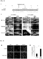
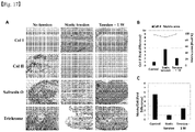
FIG. 1 shows the characteristics of the tissues formed by culturing a treatment group to which a tensile stimulus is applied and a control to which the tensile stimulus was not applied. -
FIG. 2 shows the results of confirming the nuclear localization tendency of YAP protein after application of a tensile stimulus. -
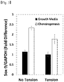
FIG. 3 shows the results of confirming the expression level of Sox-9 after application of a tensile stimulus. -
FIG. 4 shows the results of confirming whether the expression of Sox-9 is increased by the treatment with an inhibitor of nuclear translocation of YAP protein. -
FIG. 5 shows the results of confirming the ability to form a cartilage matrix when a group to which a tensile stimulus is applied is treated with an inhibitor of the nuclear translocation of YAP protein. -
FIG. 6 shows the results of confirming whether YAP protein translocates into the cytoplasm according to surface hardness. -
FIG. 7 shows a static tension chamber structure. -
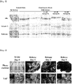
FIG. 8 shows the results of confirming whether tissue alignment is maintained after a tensile stimulus is applied and then the tension is released. -
FIG. 9 shows the results of fluorescent staining of the actin cytoskeleton andtype 1 collagen after a tensile stimulus is applied and then the tension is released. -
FIG. 10 shows the results of confirming the nuclear localization tendency of YAP protein after a tensile stimulus is applied and then the tension is released. -
FIG. 11 is a schematic view illustrating the duration and conditions of culturing while applying a tensile stimulus of 5% to 20%. -
FIG. 12 shows the results of confirming the change in tissue thickness for each culture week by comparing macroscopic photographs of tissues cultured by applying tensile stimuli of 5%, 10%, 15%, and 20%. -
FIG. 13 shows the results of confirming the change in tissue thickness with time under each condition for application of a tensile stimulus. -
FIG. 14 shows the results of performing safranin O, trichrome, and H&E staining to confirm the matrix characteristics of the tissues obtained 3 weeks after the induction of cartilage differentiation. -
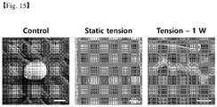
FIG. 15 shows the results of confirming whether fibrocartilage can be formed when a tensile stimulus is temporarily (7 days) applied, continuously applied, or not applied. -
FIG. 16 shows the results of confirming the degree of tissue alignment maintenance when a tensile stimulus is temporarily (7 days) applied, continuously applied, or not applied. -
FIG. 17 shows the results of confirming whether a fibrous matrix and a cartilaginous matrix are formed when a tensile stimulus is temporarily (7 days) applied, continuously applied, or not applied. - Hereinafter, the present invention will be described in detail through the Examples. However, the following Examples are only for exemplifying the present invention, and the present invention is not limited by the following Examples.
- After chondrocytes obtained from meniscus cartilage, which is fibrocartilage, were cultured in the form of a sheet, the characteristics of a tissue formed by sequentially culturing a treatment group to which a static tension of 10% was applied and a control to which the tension was not applied in a growth medium (alpha-MEM including 2 mM L-glutamine, 10-4 M ascorbic acid, 10-8 M dexamethasone, 1 ng/ml rhFGF-2, and 5% fetal bovine serum) and a cartilage differentiation medium (DMEM including 1 M proline, 0.3 mM ascorbic acid, 10-7 M dexamethansone, 10 ng/ml TGF-3, and 1x IT+3) were confirmed.
- First, in order to confirm the arrangement and synthesis amount of
type 1 collagen, immunostaining using an antibody againsttype 1 collagen (Chemicon, product number AB755P) was performed, and trichrome staining was performed with reference to the methods described in L G Luna literature (Histopathologic Methods and Color Atlas of Special Stains and Tissue Artifacts, Johnson Printers, Downers Grove, ILP151-152 c 1992). - Further, after the cells cultured in the medium were fixed using 4% paraformaldehyde, the fixed cells were embedded in paraffin were cut into sections, and then de-paraffinized using xylene. Thereafter, nuclear staining was performed for 5 minutes using Weigert's hematoxylin, and the cells were washed with distilled water for 10 minutes. After 10 minutes, the cells were rinsed using a 70% alcohol solution containing 1% HCl and then stained with 0.02% aqueous fast green for 5 minutes. After being stained with fast green, the cells were washed with 1% acetic acid and stained using 0.1% aqueous safranin O for 5 minutes. After staining with safranin O, the sample was immersed in
alcohol 2 to 3 times, and then put into xylene for mounting. - As a result, it was confirmed that when a tensile stimulus was applied, a high level of tissue alignment was induced, and the degree of arrangement and the synthesis amount of
type 1 collagen, which is a fibrous matrix, were both increased, and meanwhile, it was confirmed that no formation of cartilaginous matrix was observed in the group treated with the tensile stimulus (FIG. 1 ). - In order to confirm whether the tensile stimulus induced the nuclear translocation phenomenon of the YAP protein present in the cytoplasm, immunofluorescence (IF) method was performed on the treatment group cultured in 1-1 above.
- Specifically, 3.7% paraformaldehyde was added to the cells and the cells were fixed at room temperature for 20 minutes. Thereafter, the cells were washed 3 times with PBS and treated with 0.1% Triton-X100 for 15 minutes to increase the permeability of the cells. After the cells were again washed 3 times with PBS, the cells were treated with 10% fetal bovine serum (FBS) for 1 hour, an anti-YAP antibody was diluted with 10% FBS, reacted at room temperature for 2 hours, and washed 3 times with 1% FBS for 10 minutes, and then a secondary antibody was diluted with 10% FBS, reacted in the dark for 2 hours, and washed 3 times with 1% FBS for 10 minutes each. In this case, during the second washing, 300 ng/ml DAPI was put into 1% FBS. Next, an encapsulation solution was dropped on a slide glass, and then the slide glass was covered with a cover glass. The surrounding encapsulation solution was removed, the cover glass was covered with enamel, and then observed under a microscope.
- In addition, semi-quantitative PCR (30 cycles optimal) was performed to investigate the expression level of a cartilage differentiation marker Sox-9. Specifically, the primer sequences of SEQ ID NOS: 1 and 2 were used, and the expression level of glyceraldehyde-3-phosphate dehydrogenase (GAPDH) was used for the normalization of gene expression values.
[Table 1] SEQ ID NO Primer Sequence (5'-3') 1 Sox-9 forward CTCCGACACCGAGAATACAC 2 Sox-9 reverse CCATTCTTCACCGACTTCCT 3 GAPDH forward TCACCATCTTCCAGGAGCGA 4 GAPDH reverse CACAATGCCGAAGTGGTCGT - As a result, it was confirmed that the amount of YAP protein translocated to the nucleus was remarkably increased during culture while maintaining the tensile stimulus for 3 weeks (
FIG. 2 ). - Furthermore, it was confirmed that a basal level of Sox-9 expression appeared under a growth medium condition, and the Sox-9 expression was increased in both the control and the treatment group under a cartilage differentiation medium condition. However, under the cartilage differentiation condition, a decrease in Sox-9 expression was observed in the treatment group compared with the control (
FIG. 3 ). - The above results suggest that the localization of YAP protein in the nucleus by a tensile stimulus affects the regulation of Sox-9 expression, resulting in a deterioration in the shape of a cartilaginous matrix.
- It was confirmed whether the expression of Sox-9, which is a cartilage differentiation marker, was increased during treatment with verteporfin, which suppresses the translocation of YAP protein to the nucleus. The Sox-9 expression level was measured in the same manner as in 1-2.
- Further, a cartilage tissue obtained after the treatment with verteporfin was embedded in paraffin, sectioned to a thickness of 5 to 7 µm, dried, and then subjected to hematoxylin & eosin (H&E) staining. The stained tissue was observed under a high resolution optical microscope.
- As a result, it was confirmed that the expression of the cartilage differentiation marker Sox-9 was remarkably increased during the treatment with verteporfin, suggesting that the cartilage differentiation ability can be highly exhibited as the translocation of the YAP protein to the nucleus is suppressed. Meanwhile, when the treatment group to which the tensile stimulus was applied was treated with verteporfin, the overall matrix forming ability of the tissue was increased, whereas the formation of the fibrous matrix showed a decreasing pattern, suggesting that suppressing the translocation of YAP protein alone does not establish effective fibrocartilage formation conditions.
- Through the above results, it was confirmed that when a tensile stimulus is applied, the fibrous matrix could be formed in a tissue anisotropy direction, but cartilage matrix formation was suppressed by promoting the translocation of YAP protein to the nucleus, and thus, it was difficult to form a fibrocartilage tissue.
- Conditions for eliminating the effect of suppressing cartilage differentiation according to the nuclear localization tendency of YAP protein were confirmed. First, after a cell sheet was detached from a plastic culture dish, only cell-cell contact was allowed to be maintained. One day later, the intracellular distribution of YAP protein was confirmed in the same manner as in Example 1-2.
- As a result, it was confirmed that when the stimulus that induced the translocation of YAP protein disappeared, the short-term localization of YAP was adjusted, and thus the YAP was distributed in the cytoplasm (
FIG. 6 ). This suggests that the suppression of cartilage differentiation by YAP can be prevented. - Based on the experimental results of 2-1 above, it was confirmed whether tissue alignment was maintained even after a tensile stimulus was applied for 1 to 10 days, and then the tension was released.
- First, chondrocytes cultured in the form of a sheet were rolled up longitudinally to form a cell cable, and a tensile stimulus of 10% was applied for 1 to 10 days using the static tension chamber designed in
FIG. 7 , and it was confirmed whether tissue alignment was maintained after 1, 3, 7, and 10 days. After the tensile stimulus was applied, the arrangement form of an actin cytoskeleton using phalloidin staining was confirmed in order to carefully evaluate the degree of tissue alignment maintenance, and the arrangement form of the extracellular matrix was confirmed byfluorescently staining type 1 collagen in the same manner as in 1-1 above. - As a result of macroscopic observation of the tissue form, it was confirmed that when the tensile stimulus was applied for 3 days or more, the form was produced and tissue alignment was maintained, and when the tensile stimulus was applied for 7 days or more, the form was maintained for 5 days or more even after the tensile stimulus was released (
FIG. 8 ). - As a result of confirmation using immunofluorescent staining to determine whether tissue alignment is maintained based on the arrangement form of the cytoskeleton and extracellular matrix, it was confirmed that when a tensile stimulus was applied for 3 days, and then the tensile stimulus was released, the alignment of the cytoskeleton and extracellular matrix became disordered after 5 days, whereas when the tensile stimulus was applied for 7 days or more, and then the tensile stimulus was released, the uniaxial anisotropic tissue alignment was maintained for 5days (
FIG. 9 ). - Based on the 2-1 experiment, an experiment was conducted to confirm whether YAP protein was translocated to the nucleus when a tensile stimulus was applied for 1, 3, 7, and 10 days, and then the tension was released. The experiment was performed in the same manner as in 1-2 above.
- As a result, it was confirmed that the nuclear localization tendency of YAP protein was remarkably reduced 24 hours after the release of the tensile stimulus under all the tensile stimulus conditions, and it was confirmed that the temporary tensile stimulus applied in the form of a cell cable did not induce irreversible changes in regard to the translocation of YAP protein to the nucleus, and thus, the effect of suppressing cartilage differentiation may not be sustained (
FIG. 10 ). - From the above results, it was confirmed that the duration of tensile stimulus application needs to exceed at least 3 days in order to maintain tissue alignment at the initial stage (5 to 7 days) of the cartilaginous matrix formation, while a tensile stimulus needs to be applied within a maximum of 10 days in order to prevent the irreversible nuclear localization tendency of YAP protein.
- Based on the experimental results of Example 2, a cartilage tissue to be formed while being cultured using the cartilage differentiation medium for 3 weeks was confirmed in a state where a tensile stimulus of 5 to 20% was applied. The duration and conditions of culturing while applying a tensile stimulus of 5 to 20% are as illustrated in
FIG. 11 . - The thickness of the tissue formed for 3 weeks was observed with the naked eye, and as a result, it was confirmed that the growth of the tissue was increased over time throughout the entire tensile stimulus range of 5 to 20% (
FIGS. 12 and13 ). In addition, it was confirmed that when a tensile stimulus of 5 to 10% was applied, the thickness of tissue was increased, and consistent tissue alignment was more apparent throughout the tissue, and in the case of a tensile stimulus of 15% or more, a high level of tissue alignment was exhibited along with an increase in tissue formation in consideration of an increase in the length of the entire tissue (FIG. 14 ). - Furthermore, in order to induce cartilage differentiation by applying the tensile stimulus as described above and confirm the characteristics of a cartilage tissue obtained after 3 weeks, the tissue was observed after being stained with safranin O, trichrome, and H&E. As a result, it was confirmed that a tissue with uniaxial anisotropic tissue alignment was formed by a tensile stimulus of 5 to 20%, and the alignment of the tissue appeared more consistently, particularly in a tensile stimulus range of 5 to 15%.
- Based on the experimental results of Example 2, it was confirmed whether fibrocartilage could be formed after a tensile stimulus of 10% was applied for 7 days.
- First, fibrocartilage tissue was formed by culturing a treatment group in which a tensile stimulus of 10% was applied to chondrocytes for 7 days in a cartilage differentiation medium for 3 weeks. Thereafter, it was observed under a microscope whether tissue formation occurred.
- As a result, it was confirmed that the cartilage tissue was formed when the cells were cultured for 2 to 3 weeks by applying a tensile stimulus of 10% for 7 days, and then releasing tension (
FIG. 15 ). - The degree of tissue alignment maintenance after cartilage differentiation was evaluated by investigating an angle exhibited by the nuclei, and thereafter, it was confirmed under a microscope whether alignment was maintained.
- As a result, it was confirmed that when the tensile stimulus was not applied, alignment in a specific direction did not appear, but a high level of alignment was maintained in a group treated with the tensile stimulus for 7 days or a group continuously treated with the tensile stimulus for 3 weeks (
FIG. 16 ). - After a tensile stimulus of 10% was applied for 7 days, immunostaining and histological staining were performed on a fibrous matrix and a cartilaginous matrix. The experimental method was performed in the same manner as in Example 1-1, and for staining of
type 2 collagen, immunohistochemical staining was performed using an antibody againsttype 2 collagen (Abcam, product number AB53047). - As a result, it was confirmed that in a group cultured in a state in which a tensile stimulus was applied for 7 days, and then released, the formation of both
type 1 collagen and the cartilaginous matrix appeared, and the area of extracellular matrix formed per cell was shown to be remarkably high compared to the tensile stimulus maintenance group. On the other hand, in the control to which the tensile stimulus was not applied, the degree oftype 1 collagen formation appeared very low, and when the tensile stimulus was continuously applied for 3 weeks, almost no cartilaginous matrix formation appeared. - Through the above results, it can be seen that when chondrocytes are cultured under a tensile stimulus, and then cultured in a tension-released state, the ability to form both a fibrous matrix and a cartilaginous matrix is excellent, suggesting that by applying the method of the present invention to culture chondrocytes, it is possible to form a tissue capable of simultaneously exhibiting tensile strength and compressive strength that can withstand the load applied in the joint.
- The above-described description of the present invention is provided for illustrative purposes, and those skilled in the art to which the present invention pertains will understand that the present invention can be easily modified into other specific forms without changing the technical spirit or essential features of the present invention. Therefore, it should be understood that the above-described embodiments are only exemplary in all aspects and are not restrictive. For example, each constituent element which is described as a singular form may be implemented in a distributed form, and similarly, constituent elements which are described as being distributed may be implemented in a combined form.
- The scope of the present invention is represented by the following claims, and it should be interpreted that the meaning and scope of the claims and all the changes or modified forms derived from the equivalent concepts thereof fall within the scope of the present invention.


Claims (7)
- A method for preparing fibrocartilage, the method comprising: culturing one or more cells selected from the group consisting of synovial cells, chondrocytes and mesenchymal stem cells under a tensile stimulus.
- The method of claim 1, wherein the tensile stimulus is applied for 3 days to 10 days.
- The method of claim 1, wherein the tensile stimulus is applied with a force of 5% to 20%.
- The method of claim 1, further comprising: culturing cells under the tensile stimulus, and then releasing the tensile stimulus; and culturing the cells for 10 days to 28 days.
- The method of claim 1, wherein the fibrocartilage is one or more selected from the group consisting of the intervertebral disc, the interpubic disc and the meniscus cartilage.
- The method of claim 1, wherein the cartilage cells are any one selected from the group consisting of articular chondrocytes, costal chondrocytes, and meniscal chondrocytes.
- Fibrocartilage prepared by the method of claim 1.
Applications Claiming Priority (3)
| Application Number | Priority Date | Filing Date | Title |
|---|---|---|---|
| KR20200033429 | 2020-03-18 | ||
| KR1020210009580A KR102621671B1 (en) | 2020-03-18 | 2021-01-22 | Method for fibrocartilage reconstruction using mechanical stimulation |
| PCT/KR2021/003315 WO2021187898A1 (en) | 2020-03-18 | 2021-03-17 | Fibrocartilage preparation method using tensile stimulation |
Publications (3)
| Publication Number | Publication Date |
|---|---|
| EP4066869A1 true EP4066869A1 (en) | 2022-10-05 |
| EP4066869A4 EP4066869A4 (en) | 2024-01-10 |
| EP4066869B1 EP4066869B1 (en) | 2025-07-02 |
Family
ID=77771797
Family Applications (1)
| Application Number | Title | Priority Date | Filing Date |
|---|---|---|---|
| EP21770811.4A Active EP4066869B1 (en) | 2020-03-18 | 2021-03-17 | Fibrocartilage preparation method using tensile stimulation |
Country Status (3)
| Country | Link |
|---|---|
| US (1) | US20230039188A1 (en) |
| EP (1) | EP4066869B1 (en) |
| WO (1) | WO2021187898A1 (en) |
Family Cites Families (3)
| Publication number | Priority date | Publication date | Assignee | Title |
|---|---|---|---|---|
| US6737053B1 (en) * | 1999-11-12 | 2004-05-18 | National University Of Singapore | Tissue-engineered ligament |
| KR20100005105A (en) * | 2007-05-06 | 2010-01-13 | 민병현 | Therapeutic composite for cartilage disorder using extracellular matrix (ecm) scaffold |
| KR101085193B1 (en) * | 2010-12-21 | 2011-11-21 | 한국기계연구원 | Cell or tissue tension stimulator |
-
2021
- 2021-03-17 WO PCT/KR2021/003315 patent/WO2021187898A1/en not_active Ceased
- 2021-03-17 US US17/789,999 patent/US20230039188A1/en active Pending
- 2021-03-17 EP EP21770811.4A patent/EP4066869B1/en active Active
Also Published As
| Publication number | Publication date |
|---|---|
| US20230039188A1 (en) | 2023-02-09 |
| EP4066869A4 (en) | 2024-01-10 |
| WO2021187898A1 (en) | 2021-09-23 |
| EP4066869B1 (en) | 2025-07-02 |
Similar Documents
| Publication | Publication Date | Title |
|---|---|---|
| Awad et al. | Repair of patellar tendon injuries using a cell–collagen composite | |
| Giovannini et al. | Micromass co-culture of human articular chondrocytes and human bone marrow mesenchymal stem cells to investigate stable neocartilage tissue formation in vitro | |
| Zhou et al. | Injectable decellularized nucleus pulposus-based cell delivery system for differentiation of adipose-derived stem cells and nucleus pulposus regeneration | |
| US9259510B2 (en) | System and method for forming bone, ligament, and bone-ligament constructs | |
| CN101589139B (en) | Artificial cartilage comprising chondrocytes obtained from costal cartilage and method for producing the same | |
| US20050118228A1 (en) | Compositions and methods for augmentation or repair of intervertebral discs | |
| EP3025720B1 (en) | Preparation method for therapeutic agent of bead-type chondrocyte | |
| WO2008146956A1 (en) | Therapeutic composite for cartilage disorder using extracellular matrix (ecm) scaffold | |
| EP4066869B1 (en) | Fibrocartilage preparation method using tensile stimulation | |
| EP3025735B1 (en) | Methods for complex tissue engineering | |
| US20240100099A1 (en) | Composition for regeneration of intervertebral disc | |
| KR102621671B1 (en) | Method for fibrocartilage reconstruction using mechanical stimulation | |
| Nguyen et al. | In vitro study on chondrogenic differentiation of human adipose-derived stem cells on treated bovine pericardium | |
| EP3470096A1 (en) | Elastin reduction allowing recellularization of cartilage implants | |
| KR20170025681A (en) | Pharmaceutical composition for preventing or treating degenerative arthritis | |
| Chen et al. | Effect of tissue expansion on chondrocyte sheets in cartilage composite reconstruction | |
| KR101386352B1 (en) | Method for promoting differntiation of mesenchymal stem cell to chondrocytes using electroporation | |
| US8357534B2 (en) | Isolation of human articular cartilage stem cells | |
| Phu | The Effect of Entrapped Laminin-111 in Three-dimensional Polyethylene Glycol Gels on the Behavior of Nucleus Pulposus Cells from the Intervertebral Disc | |
| Bach et al. | AOSpine Masters Symposium Abstracts | |
| Nosikova | Characterization of the Interface between the Annulus Fibrosus and the Vertebral Bone. | |
| US20210198630A1 (en) | Cartilage-derived progenitor cell lines | |
| Kraft | Developing a cartilage tissue equivalent using chondrocytes and mesenchymal stem cells | |
| Stachura | Evaluation of chondrogenic differentiation of human stem cells derived from adult bone marrow | |
| Juhas | Engineering Highly-functional, Self-regenerative Skeletal Muscle Tissues with Enhanced Vascularization and Survival In Vivo |
Legal Events
| Date | Code | Title | Description |
|---|---|---|---|
| STAA | Information on the status of an ep patent application or granted ep patent |
Free format text: STATUS: THE INTERNATIONAL PUBLICATION HAS BEEN MADE |
|
| PUAI | Public reference made under article 153(3) epc to a published international application that has entered the european phase |
Free format text: ORIGINAL CODE: 0009012 |
|
| STAA | Information on the status of an ep patent application or granted ep patent |
Free format text: STATUS: REQUEST FOR EXAMINATION WAS MADE |
|
| 17P | Request for examination filed |
Effective date: 20220628 |
|
| AK | Designated contracting states |
Kind code of ref document: A1 Designated state(s): AL AT BE BG CH CY CZ DE DK EE ES FI FR GB GR HR HU IE IS IT LI LT LU LV MC MK MT NL NO PL PT RO RS SE SI SK SM TR |
|
| DAV | Request for validation of the european patent (deleted) | ||
| DAX | Request for extension of the european patent (deleted) | ||
| A4 | Supplementary search report drawn up and despatched |
Effective date: 20231213 |
|
| RIC1 | Information provided on ipc code assigned before grant |
Ipc: C12N 13/00 20060101ALI20231207BHEP Ipc: C12N 5/077 20100101ALI20231207BHEP Ipc: C12M 1/42 20060101ALI20231207BHEP Ipc: C12M 3/00 20060101ALI20231207BHEP Ipc: A61L 27/38 20060101AFI20231207BHEP |
|
| GRAP | Despatch of communication of intention to grant a patent |
Free format text: ORIGINAL CODE: EPIDOSNIGR1 |
|
| STAA | Information on the status of an ep patent application or granted ep patent |
Free format text: STATUS: GRANT OF PATENT IS INTENDED |
|
| INTG | Intention to grant announced |
Effective date: 20250213 |
|
| GRAS | Grant fee paid |
Free format text: ORIGINAL CODE: EPIDOSNIGR3 |
|
| GRAA | (expected) grant |
Free format text: ORIGINAL CODE: 0009210 |
|
| STAA | Information on the status of an ep patent application or granted ep patent |
Free format text: STATUS: THE PATENT HAS BEEN GRANTED |
|
| AK | Designated contracting states |
Kind code of ref document: B1 Designated state(s): AL AT BE BG CH CY CZ DE DK EE ES FI FR GB GR HR HU IE IS IT LI LT LU LV MC MK MT NL NO PL PT RO RS SE SI SK SM TR |
|
| REG | Reference to a national code |
Ref country code: GB Ref legal event code: FG4D |
|
| REG | Reference to a national code |
Ref country code: CH Ref legal event code: EP |
|
| REG | Reference to a national code |
Ref country code: DE Ref legal event code: R096 Ref document number: 602021033425 Country of ref document: DE |
|
| REG | Reference to a national code |
Ref country code: IE Ref legal event code: FG4D |
|
| REG | Reference to a national code |
Ref country code: NL Ref legal event code: MP Effective date: 20250702 |
|
| PG25 | Lapsed in a contracting state [announced via postgrant information from national office to epo] |
Ref country code: PT Free format text: LAPSE BECAUSE OF FAILURE TO SUBMIT A TRANSLATION OF THE DESCRIPTION OR TO PAY THE FEE WITHIN THE PRESCRIBED TIME-LIMIT Effective date: 20251103 |
|
| PG25 | Lapsed in a contracting state [announced via postgrant information from national office to epo] |
Ref country code: NL Free format text: LAPSE BECAUSE OF FAILURE TO SUBMIT A TRANSLATION OF THE DESCRIPTION OR TO PAY THE FEE WITHIN THE PRESCRIBED TIME-LIMIT Effective date: 20250702 |
|
| REG | Reference to a national code |
Ref country code: AT Ref legal event code: MK05 Ref document number: 1808541 Country of ref document: AT Kind code of ref document: T Effective date: 20250702 |
|
| PG25 | Lapsed in a contracting state [announced via postgrant information from national office to epo] |
Ref country code: IS Free format text: LAPSE BECAUSE OF FAILURE TO SUBMIT A TRANSLATION OF THE DESCRIPTION OR TO PAY THE FEE WITHIN THE PRESCRIBED TIME-LIMIT Effective date: 20251102 |
|
| PG25 | Lapsed in a contracting state [announced via postgrant information from national office to epo] |
Ref country code: NO Free format text: LAPSE BECAUSE OF FAILURE TO SUBMIT A TRANSLATION OF THE DESCRIPTION OR TO PAY THE FEE WITHIN THE PRESCRIBED TIME-LIMIT Effective date: 20251002 |
|
| REG | Reference to a national code |
Ref country code: LT Ref legal event code: MG9D |
|
| PG25 | Lapsed in a contracting state [announced via postgrant information from national office to epo] |
Ref country code: AT Free format text: LAPSE BECAUSE OF FAILURE TO SUBMIT A TRANSLATION OF THE DESCRIPTION OR TO PAY THE FEE WITHIN THE PRESCRIBED TIME-LIMIT Effective date: 20250702 |
|
| PG25 | Lapsed in a contracting state [announced via postgrant information from national office to epo] |
Ref country code: FI Free format text: LAPSE BECAUSE OF FAILURE TO SUBMIT A TRANSLATION OF THE DESCRIPTION OR TO PAY THE FEE WITHIN THE PRESCRIBED TIME-LIMIT Effective date: 20250702 |
|
| PG25 | Lapsed in a contracting state [announced via postgrant information from national office to epo] |
Ref country code: HR Free format text: LAPSE BECAUSE OF FAILURE TO SUBMIT A TRANSLATION OF THE DESCRIPTION OR TO PAY THE FEE WITHIN THE PRESCRIBED TIME-LIMIT Effective date: 20250702 |
|
| PG25 | Lapsed in a contracting state [announced via postgrant information from national office to epo] |
Ref country code: GR Free format text: LAPSE BECAUSE OF FAILURE TO SUBMIT A TRANSLATION OF THE DESCRIPTION OR TO PAY THE FEE WITHIN THE PRESCRIBED TIME-LIMIT Effective date: 20251003 |
|
| PG25 | Lapsed in a contracting state [announced via postgrant information from national office to epo] |
Ref country code: CZ Free format text: LAPSE BECAUSE OF FAILURE TO SUBMIT A TRANSLATION OF THE DESCRIPTION OR TO PAY THE FEE WITHIN THE PRESCRIBED TIME-LIMIT Effective date: 20250702 Ref country code: SE Free format text: LAPSE BECAUSE OF FAILURE TO SUBMIT A TRANSLATION OF THE DESCRIPTION OR TO PAY THE FEE WITHIN THE PRESCRIBED TIME-LIMIT Effective date: 20250702 |
|
| PG25 | Lapsed in a contracting state [announced via postgrant information from national office to epo] |
Ref country code: LV Free format text: LAPSE BECAUSE OF FAILURE TO SUBMIT A TRANSLATION OF THE DESCRIPTION OR TO PAY THE FEE WITHIN THE PRESCRIBED TIME-LIMIT Effective date: 20250702 |
|
| PG25 | Lapsed in a contracting state [announced via postgrant information from national office to epo] |
Ref country code: BG Free format text: LAPSE BECAUSE OF FAILURE TO SUBMIT A TRANSLATION OF THE DESCRIPTION OR TO PAY THE FEE WITHIN THE PRESCRIBED TIME-LIMIT Effective date: 20250702 Ref country code: PL Free format text: LAPSE BECAUSE OF FAILURE TO SUBMIT A TRANSLATION OF THE DESCRIPTION OR TO PAY THE FEE WITHIN THE PRESCRIBED TIME-LIMIT Effective date: 20250702 |
|
| PG25 | Lapsed in a contracting state [announced via postgrant information from national office to epo] |
Ref country code: RS Free format text: LAPSE BECAUSE OF FAILURE TO SUBMIT A TRANSLATION OF THE DESCRIPTION OR TO PAY THE FEE WITHIN THE PRESCRIBED TIME-LIMIT Effective date: 20251002 |
|
| PG25 | Lapsed in a contracting state [announced via postgrant information from national office to epo] |
Ref country code: ES Free format text: LAPSE BECAUSE OF FAILURE TO SUBMIT A TRANSLATION OF THE DESCRIPTION OR TO PAY THE FEE WITHIN THE PRESCRIBED TIME-LIMIT Effective date: 20250702 |
|
| PG25 | Lapsed in a contracting state [announced via postgrant information from national office to epo] |
Ref country code: SM Free format text: LAPSE BECAUSE OF FAILURE TO SUBMIT A TRANSLATION OF THE DESCRIPTION OR TO PAY THE FEE WITHIN THE PRESCRIBED TIME-LIMIT Effective date: 20250702 |
|
| PG25 | Lapsed in a contracting state [announced via postgrant information from national office to epo] |
Ref country code: DK Free format text: LAPSE BECAUSE OF FAILURE TO SUBMIT A TRANSLATION OF THE DESCRIPTION OR TO PAY THE FEE WITHIN THE PRESCRIBED TIME-LIMIT Effective date: 20250702 |
|
| PG25 | Lapsed in a contracting state [announced via postgrant information from national office to epo] |
Ref country code: IT Free format text: LAPSE BECAUSE OF FAILURE TO SUBMIT A TRANSLATION OF THE DESCRIPTION OR TO PAY THE FEE WITHIN THE PRESCRIBED TIME-LIMIT Effective date: 20250702 |
|
| PGFP | Annual fee paid to national office [announced via postgrant information from national office to epo] |
Ref country code: FR Payment date: 20260323 Year of fee payment: 6 |
|
| PG25 | Lapsed in a contracting state [announced via postgrant information from national office to epo] |
Ref country code: SK Free format text: LAPSE BECAUSE OF FAILURE TO SUBMIT A TRANSLATION OF THE DESCRIPTION OR TO PAY THE FEE WITHIN THE PRESCRIBED TIME-LIMIT Effective date: 20250702 Ref country code: EE Free format text: LAPSE BECAUSE OF FAILURE TO SUBMIT A TRANSLATION OF THE DESCRIPTION OR TO PAY THE FEE WITHIN THE PRESCRIBED TIME-LIMIT Effective date: 20250702 |
|
| PLBE | No opposition filed within time limit |
Free format text: ORIGINAL CODE: 0009261 |
|
| STAA | Information on the status of an ep patent application or granted ep patent |
Free format text: STATUS: NO OPPOSITION FILED WITHIN TIME LIMIT |