EP4060675A1 - Estimating cardiac parameters when performing an activity using a personalized cardiovascular hemodynamic model - Google Patents
Estimating cardiac parameters when performing an activity using a personalized cardiovascular hemodynamic model Download PDFInfo
- Publication number
- EP4060675A1 EP4060675A1 EP21199728.3A EP21199728A EP4060675A1 EP 4060675 A1 EP4060675 A1 EP 4060675A1 EP 21199728 A EP21199728 A EP 21199728A EP 4060675 A1 EP4060675 A1 EP 4060675A1
- Authority
- EP
- European Patent Office
- Prior art keywords
- subject
- activity
- compliance
- svr
- met
- Prior art date
- Legal status (The legal status is an assumption and is not a legal conclusion. Google has not performed a legal analysis and makes no representation as to the accuracy of the status listed.)
- Granted
Links
Images







Classifications
-
- A—HUMAN NECESSITIES
- A61—MEDICAL OR VETERINARY SCIENCE; HYGIENE
- A61B—DIAGNOSIS; SURGERY; IDENTIFICATION
- A61B5/00—Measuring for diagnostic purposes; Identification of persons
- A61B5/02—Detecting, measuring or recording for evaluating the cardiovascular system, e.g. pulse, heart rate, blood pressure or blood flow
- A61B5/0205—Simultaneously evaluating both cardiovascular conditions and different types of body conditions, e.g. heart and respiratory condition
-
- G—PHYSICS
- G16—INFORMATION AND COMMUNICATION TECHNOLOGY [ICT] SPECIALLY ADAPTED FOR SPECIFIC APPLICATION FIELDS
- G16H—HEALTHCARE INFORMATICS, i.e. INFORMATION AND COMMUNICATION TECHNOLOGY [ICT] SPECIALLY ADAPTED FOR THE HANDLING OR PROCESSING OF MEDICAL OR HEALTHCARE DATA
- G16H50/00—ICT specially adapted for medical diagnosis, medical simulation or medical data mining; ICT specially adapted for detecting, monitoring or modelling epidemics or pandemics
- G16H50/50—ICT specially adapted for medical diagnosis, medical simulation or medical data mining; ICT specially adapted for detecting, monitoring or modelling epidemics or pandemics for simulation or modelling of medical disorders
-
- A—HUMAN NECESSITIES
- A61—MEDICAL OR VETERINARY SCIENCE; HYGIENE
- A61B—DIAGNOSIS; SURGERY; IDENTIFICATION
- A61B5/00—Measuring for diagnostic purposes; Identification of persons
- A61B5/02—Detecting, measuring or recording for evaluating the cardiovascular system, e.g. pulse, heart rate, blood pressure or blood flow
- A61B5/02007—Evaluating blood vessel condition, e.g. elasticity, compliance
-
- A—HUMAN NECESSITIES
- A61—MEDICAL OR VETERINARY SCIENCE; HYGIENE
- A61B—DIAGNOSIS; SURGERY; IDENTIFICATION
- A61B5/00—Measuring for diagnostic purposes; Identification of persons
- A61B5/02—Detecting, measuring or recording for evaluating the cardiovascular system, e.g. pulse, heart rate, blood pressure or blood flow
- A61B5/021—Measuring pressure in heart or blood vessels
-
- A—HUMAN NECESSITIES
- A61—MEDICAL OR VETERINARY SCIENCE; HYGIENE
- A61B—DIAGNOSIS; SURGERY; IDENTIFICATION
- A61B5/00—Measuring for diagnostic purposes; Identification of persons
- A61B5/02—Detecting, measuring or recording for evaluating the cardiovascular system, e.g. pulse, heart rate, blood pressure or blood flow
- A61B5/024—Measuring pulse rate or heart rate
-
- A—HUMAN NECESSITIES
- A61—MEDICAL OR VETERINARY SCIENCE; HYGIENE
- A61B—DIAGNOSIS; SURGERY; IDENTIFICATION
- A61B5/00—Measuring for diagnostic purposes; Identification of persons
- A61B5/02—Detecting, measuring or recording for evaluating the cardiovascular system, e.g. pulse, heart rate, blood pressure or blood flow
- A61B5/024—Measuring pulse rate or heart rate
- A61B5/02416—Measuring pulse rate or heart rate using photoplethysmograph signals, e.g. generated by infrared radiation
-
- A—HUMAN NECESSITIES
- A61—MEDICAL OR VETERINARY SCIENCE; HYGIENE
- A61B—DIAGNOSIS; SURGERY; IDENTIFICATION
- A61B5/00—Measuring for diagnostic purposes; Identification of persons
- A61B5/02—Detecting, measuring or recording for evaluating the cardiovascular system, e.g. pulse, heart rate, blood pressure or blood flow
- A61B5/024—Measuring pulse rate or heart rate
- A61B5/02438—Measuring pulse rate or heart rate with portable devices, e.g. worn by the patient
-
- A—HUMAN NECESSITIES
- A61—MEDICAL OR VETERINARY SCIENCE; HYGIENE
- A61B—DIAGNOSIS; SURGERY; IDENTIFICATION
- A61B5/00—Measuring for diagnostic purposes; Identification of persons
- A61B5/103—Measuring devices for testing the shape, pattern, colour, size or movement of the body or parts thereof, for diagnostic purposes
- A61B5/11—Measuring movement of the entire body or parts thereof, e.g. head or hand tremor or mobility of a limb
- A61B5/1118—Determining activity level
-
- A—HUMAN NECESSITIES
- A61—MEDICAL OR VETERINARY SCIENCE; HYGIENE
- A61B—DIAGNOSIS; SURGERY; IDENTIFICATION
- A61B5/00—Measuring for diagnostic purposes; Identification of persons
- A61B5/24—Detecting, measuring or recording bioelectric or biomagnetic signals of the body or parts thereof
- A61B5/316—Modalities, i.e. specific diagnostic methods
- A61B5/318—Heart-related electrical modalities, e.g. electrocardiography [ECG]
- A61B5/346—Analysis of electrocardiograms
- A61B5/349—Detecting specific parameters of the electrocardiograph cycle
- A61B5/352—Detecting R peaks, e.g. for synchronising diagnostic apparatus; Estimating R-R interval
-
- A—HUMAN NECESSITIES
- A61—MEDICAL OR VETERINARY SCIENCE; HYGIENE
- A61B—DIAGNOSIS; SURGERY; IDENTIFICATION
- A61B5/00—Measuring for diagnostic purposes; Identification of persons
- A61B5/72—Signal processing specially adapted for physiological signals or for diagnostic purposes
- A61B5/7271—Specific aspects of physiological measurement analysis
- A61B5/7278—Artificial waveform generation or derivation, e.g. synthesizing signals from measured signals
-
- A—HUMAN NECESSITIES
- A61—MEDICAL OR VETERINARY SCIENCE; HYGIENE
- A61B—DIAGNOSIS; SURGERY; IDENTIFICATION
- A61B8/00—Diagnosis using ultrasonic, sonic or infrasonic waves
- A61B8/08—Clinical applications
- A61B8/0883—Clinical applications for diagnosis of the heart
-
- A—HUMAN NECESSITIES
- A61—MEDICAL OR VETERINARY SCIENCE; HYGIENE
- A61B—DIAGNOSIS; SURGERY; IDENTIFICATION
- A61B8/00—Diagnosis using ultrasonic, sonic or infrasonic waves
- A61B8/52—Devices using data or image processing specially adapted for diagnosis using ultrasonic, sonic or infrasonic waves
- A61B8/5292—Devices using data or image processing specially adapted for diagnosis using ultrasonic, sonic or infrasonic waves using additional data, e.g. patient information, image labeling, acquisition parameters
-
- G—PHYSICS
- G16—INFORMATION AND COMMUNICATION TECHNOLOGY [ICT] SPECIALLY ADAPTED FOR SPECIFIC APPLICATION FIELDS
- G16H—HEALTHCARE INFORMATICS, i.e. INFORMATION AND COMMUNICATION TECHNOLOGY [ICT] SPECIALLY ADAPTED FOR THE HANDLING OR PROCESSING OF MEDICAL OR HEALTHCARE DATA
- G16H40/00—ICT specially adapted for the management or administration of healthcare resources or facilities; ICT specially adapted for the management or operation of medical equipment or devices
- G16H40/60—ICT specially adapted for the management or administration of healthcare resources or facilities; ICT specially adapted for the management or operation of medical equipment or devices for the operation of medical equipment or devices
- G16H40/63—ICT specially adapted for the management or administration of healthcare resources or facilities; ICT specially adapted for the management or operation of medical equipment or devices for the operation of medical equipment or devices for local operation
-
- G—PHYSICS
- G16—INFORMATION AND COMMUNICATION TECHNOLOGY [ICT] SPECIALLY ADAPTED FOR SPECIFIC APPLICATION FIELDS
- G16H—HEALTHCARE INFORMATICS, i.e. INFORMATION AND COMMUNICATION TECHNOLOGY [ICT] SPECIALLY ADAPTED FOR THE HANDLING OR PROCESSING OF MEDICAL OR HEALTHCARE DATA
- G16H50/00—ICT specially adapted for medical diagnosis, medical simulation or medical data mining; ICT specially adapted for detecting, monitoring or modelling epidemics or pandemics
- G16H50/20—ICT specially adapted for medical diagnosis, medical simulation or medical data mining; ICT specially adapted for detecting, monitoring or modelling epidemics or pandemics for computer-aided diagnosis, e.g. based on medical expert systems
-
- A—HUMAN NECESSITIES
- A61—MEDICAL OR VETERINARY SCIENCE; HYGIENE
- A61B—DIAGNOSIS; SURGERY; IDENTIFICATION
- A61B2505/00—Evaluating, monitoring or diagnosing in the context of a particular type of medical care
- A61B2505/09—Rehabilitation or training
-
- A—HUMAN NECESSITIES
- A61—MEDICAL OR VETERINARY SCIENCE; HYGIENE
- A61B—DIAGNOSIS; SURGERY; IDENTIFICATION
- A61B5/00—Measuring for diagnostic purposes; Identification of persons
- A61B5/02—Detecting, measuring or recording for evaluating the cardiovascular system, e.g. pulse, heart rate, blood pressure or blood flow
- A61B5/024—Measuring pulse rate or heart rate
- A61B5/0245—Measuring pulse rate or heart rate by using sensing means generating electric signals, i.e. ECG signals
-
- A—HUMAN NECESSITIES
- A61—MEDICAL OR VETERINARY SCIENCE; HYGIENE
- A61B—DIAGNOSIS; SURGERY; IDENTIFICATION
- A61B5/00—Measuring for diagnostic purposes; Identification of persons
- A61B5/40—Detecting, measuring or recording for evaluating the nervous system
- A61B5/4029—Detecting, measuring or recording for evaluating the nervous system for evaluating the peripheral nervous systems
- A61B5/4035—Evaluating the autonomic nervous system
-
- A—HUMAN NECESSITIES
- A61—MEDICAL OR VETERINARY SCIENCE; HYGIENE
- A61B—DIAGNOSIS; SURGERY; IDENTIFICATION
- A61B5/00—Measuring for diagnostic purposes; Identification of persons
- A61B5/72—Signal processing specially adapted for physiological signals or for diagnostic purposes
- A61B5/7203—Signal processing specially adapted for physiological signals or for diagnostic purposes for noise prevention, reduction or removal
- A61B5/7207—Signal processing specially adapted for physiological signals or for diagnostic purposes for noise prevention, reduction or removal of noise induced by motion artifacts
Definitions
- the disclosure herein generally relates to the field of computer assisted modeling of hemodynamic patterns of physiologic blood flow, and, more particularly, to systems and methods for estimating cardiac parameters when performing an activity using a personalized cardiovascular hemodynamic model.
- Cardiovascular disease is one of the main causes of deaths in the world today. With advancing age, the risk of CVD increases significantly. To reduce the effect of CVDs, it has been a well-established fact that the higher levels of regular physical exercises would reduce cardiovascular events and mortality. According to medical literature and guidelines, increasing levels of physical activity are the first-line approaches for preventing and treating vascular dysfunction and cardio-metabolic diseases. Moreover, during post-operative recovery of a patient who had suffered a cardiovascular surgery recently, performing prescribed exercises during the recovery phase is always recommended. However, unless the patient diligently and periodically consults a caregiver, it is challenging to gauge whether the prescribed exercises are yielding a desired outcome, or any change is required in the planned therapy.
- Embodiments of the present disclosure present technological improvements as solutions to one or more of the above-mentioned technical problems recognized by the inventors in conventional systems.
- a processor implemented method comprising the steps of: estimating, via one or more hardware processors, a plurality of input parameters for a personalized cardiovascular hemodynamic model associated with a subject being monitored, the plurality of input parameters comprising (i) Systemic Vascular Resistance (SVR), (ii) unstressed blood volume, (iii) total blood volume in a body of the subject, and (iv) heart rate of the subject, the step of estimating comprises: estimating the SVR using Metabolic EquivalenT (MET) levels associated with an activity level of the subject; and updating the unstressed blood volume estimated when the subject is at rest, using an autoregulation method, when an activity is performed by the subject, wherein the autoregulation method comprises: sensing aortic pressure by baroreceptors located at carotid sinus and aortic arch; converting the sensed aortic pressure into a neural firing frequency via afferent sympathetic pathways; generating sympathetic and parasympathetic nervous activities via a central nervous system and e
- SVR Systemic
- a system comprising: one or more data storage devices operatively coupled to one or more hardware processors and configured to store instructions configured for execution via the one or more hardware processors to: estimate a plurality of input parameters for a personalized cardiovascular hemodynamic model associated with a subject being monitored, the plurality of input parameters comprising (i) Systemic Vascular Resistance (SVR), (ii) unstressed blood volume, (iii) total blood volume in a body of the subject, and (iv) heart rate of the subject, wherein estimating the plurality of input parameters comprises: estimating the SVR using Metabolic EquivalenT (MET) levels associated with an activity level of the subject; and updating the unstressed blood volume estimated when the subject is at rest, using an autoregulation method, when an activity is performed by the subject, wherein the autoregulation method comprises: sensing aortic pressure by baroreceptors located at carotid sinus and aortic arch; converting the sensed aortic pressure into a neural firing frequency via a
- SVR Systemic
- a computer program product comprising a non-transitory computer readable medium having a computer readable program embodied therein, wherein the computer readable program, when executed on a computing device, causes the computing device to: estimate a plurality of input parameters for a personalized cardiovascular hemodynamic model associated with a subject being monitored, the plurality of input parameters comprising (i) Systemic Vascular Resistance (SVR), (ii) unstressed blood volume, (iii) total blood volume in a body of the subject, and (iv) heart rate of the subject, wherein estimating the plurality of input parameters comprises: estimating the SVR using Metabolic EquivalenT (MET) levels associated with an activity level of the subject; and updating the unstressed blood volume estimated when the subject is at rest, using an autoregulation method, when an activity is performed by the subject, wherein the autoregulation method comprises: sensing aortic pressure by baroreceptors located at carotid sinus and aortic arch; converting the sensed
- SVR Systemic Vascular
- the one or more hardware processors are configured to personalize a cardiovascular hemodynamic model to obtain the personalized cardiovascular hemodynamic model, prior to estimating the plurality of input parameters, based on one or more of (i) cardiac parameters obtained from an echocardiogram, (ii) ECG signal obtained from a wearable device worn by the subject when performing the activity and (iii) metadata of the subject including height and weight associated thereof.
- the one or more hardware processors are configured to perform sequential activation of a right atrium ra, a left atrium la , a right ventricle rv and a left ventricle lv of the personalized cardiovascular hemodynamic model using compliance functions C ra ( t ), C la ( t ), C rv ( t ) and C lv ( t ) respectively, the compliance functions being defined as:
- the one or more hardware processors are configured to estimate the total blood volume using height and weight of the subject.
- the one or more hardware processors are configured to estimate the heart rate of the subject using (i) a Photoplethysmogram (PPG) signal and an accelerometer signal or (ii) an Electrocardiogram (ECG) signal, from a wearable device worn by the subject when performing the activity.
- PPG Photoplethysmogram
- ECG Electrocardiogram
- CVD Cardiovascular disease
- CVD cardiovascular disease
- a patient's cardiac parameters it is very important to periodically monitor a patient's cardiac parameters to assess effects of prescribed exercises on the cardiac parameters of the patient. Unless the patient diligently visits a caregiver, say during a post-operative period, assessing cardiovascular conditions for recovery is challenging. A lapse on the part of the patient may be detrimental to the patient's health.
- the present disclosure enables personalized cardiac rehabilitation guidance and care continuum using a personalized cardiovascular hemodynamic model that effectively simulates cardiac parameters when the patient performs an activity using a wearable device like a digital watch that can help capture Electrocardiogram (ECG) signal, Photoplethysmogram (PPG) signal and accelerometer signal.
- ECG Electrocardiogram
- PPG Photoplethysmogram
- accelerometer signal the expression 'patient' may interchangeably be referred as a 'subject'.
- FIG. 1 through FIG.20 where similar reference characters denote corresponding features consistently throughout the figures, there are shown preferred embodiments and these embodiments are described in the context of the following exemplary system and/or method.
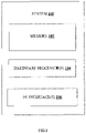
- FIG.1 illustrates an exemplary block diagram of a system 100 for estimating cardiac parameters when performing an activity using a personalized cardiovascular hemodynamic model, in accordance with some embodiments of the present disclosure.
- the system 100 includes one or more hardware processors 104, communication interface device(s) or input/output (I/O) interface(s) 106, and one or more data storage devices or memory 102 operatively coupled to the one or more hardware processors 104.
- the one or more hardware processors 104 can be implemented as one or more microprocessors, microcomputers, microcontrollers, digital signal processors, central processing units, state machines, graphics controllers, logic circuitries, and/or any devices that manipulate signals based on operational instructions.
- the processor(s) are configured to fetch and execute computer-readable instructions stored in the memory.
- the expressions 'processors' and 'hardware processors' may be used interchangeably.
- the system 100 can be implemented in a variety of computing systems, such as laptop computers, notebooks, hand-held devices, workstations, mainframe computers, servers, a network cloud and the like.
- I/O interface(s) 106 can include a variety of software and hardware interfaces, for example, a web interface, a graphical user interface, and the like and can facilitate multiple communications within a wide variety of networks N/W and protocol types, including wired networks, for example, LAN, cable, etc., and wireless networks, such as WLAN, cellular, or satellite.
- the I/O interface(s) can include one or more ports for connecting a number of devices to one another or to another server.
- the memory 102 may include any computer-readable medium known in the art including, for example, volatile memory, such as static random access memory (SRAM) and dynamic random access memory (DRAM), and/or non-volatile memory, such as read only memory (ROM), erasable programmable ROM, flash memories, hard disks, optical disks, and magnetic tapes.
- volatile memory such as static random access memory (SRAM) and dynamic random access memory (DRAM)
- non-volatile memory such as read only memory (ROM), erasable programmable ROM, flash memories, hard disks, optical disks, and magnetic tapes.
- ROM read only memory
- erasable programmable ROM erasable programmable ROM
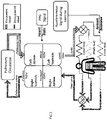
- FIG.2 illustrates an exemplary block diagram of a cardiovascular hemodynamic model triggered via activity related data, in accordance with some embodiments of the present disclosure.
- Heart is a muscular organ in which each half is composed of a pair of atria and a pair of ventricles serving like a pulsatile pump.
- a left heart chamber comprising a left ventricle ( lv ) and a left atrium ( la ), pumps oxygenated blood to all tissues of the body. This specific circulation is called systemic circulation.
- a right heart comprising a right ventricle (rv) and a right atrium ( ra ), drives deoxygenated blood to the lungs forming a pulmonic circulation.
- cardiac valves there are four cardiac valves namely, mitral ( mi ) and aortic ( ao ) valves in the left heart and tricuspid (tr) and pulmonic (pu) valves in the right heart respectively. These valves synchronously open and close based on a pressure difference within the cardiac chambers and ensures rhythmic unidirectional flow through the heart.
- dV/dt (t) Q1(t) ⁇ Q2(t).
- the valvular resistance across the mitral, aortic, tricuspid and pulmonic valves are R mi , R ao , R tr and R pu respectively.
- the vascular resistance and compliance pair, across the pulmonic and systemic vessels are R p , C pa and R s , C sa respectively.
- U i ; ⁇ i ⁇ mi , ao , tr, pu ⁇ are inputs for opening and closing of the heart valves as mentioned in equation (2) below.
- U mi ⁇ 1 , if , P la > P Lv ⁇ mi , otherwise ;
- U ao ⁇ 1 , if , P lv > P sa ⁇ ao , otherwise ;
- U tr ⁇ 1 , if , P ra > P rv ⁇ tr , otherwise ;
- U pu ⁇ 1 , if , P rv > P pa ⁇ pu , otherwise , where ⁇ mi , ⁇ ao , ⁇ tr , and ⁇ pu define regurgitation effect of the cardiovascular system .
- the cardiac chambers are activated sequentially, in a synchronized manner, by time-varying compliance functions.
- this activation starts from a sinoatrial node, which is located inside ra , then, it traverses to the la with a time delay of d la , causing them to contract for pumping the blood into the ventricles. After that, the activation traverses from the atrium to the ventricles via an atrioventricular node with a time delay of d , allowing the ventricles to fill with blood.
- the compliance functions C r ⁇ ( t ), C la ( t ), C lv ( t ) and C rv ( t ) to sequentially actuate ra, la , rv and lv are defined as given in equation (3), equation (4) and equation (5) respectively.
- the compliance function to actuate la is modeled using equation (3), where C min,la and C max,la are the minimum and maximum values of the la compliance, u(t) is the activation function of la and d la represent a delay in activation of la with respect to ra.
- C la t C min , la + 0.5 ⁇ C max , la ⁇ C min , la u t ⁇ d la
- the compliance functions to actuate rv and lv are represented by equation (5) below, where C i ; i ⁇ ⁇ lv , rv ⁇ is the systolic compliance across lv or rv and is estimated as a ratio of R-peak and T-peak of the ECG signal, u v ( t ) is the activation function, and d represents the delay in activation of lv or rv from ra .
- T 1 and T 2 are the systolic and diastolic activation time instances of the cardiac cycle respectively.
- V ⁇ j C j P ⁇ j ; ⁇ j ⁇ sa pa
- V total ⁇ i V i + ⁇ j V j
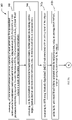
- FIG.3A through FIG.3B illustrate an exemplary flow diagram of a computer implemented method 300 for estimating cardiac parameters when performing an activity using a personalized cardiovascular hemodynamic model, in accordance with some embodiments of the present disclosure.
- the system 100 includes one or more data storage devices or memory 102 operatively coupled to the one or more hardware processors 104 and is configured to store instructions configured for execution of steps of the method 200 by the one or more hardware processors 104.
- the steps of the method 300 will now be explained in detail with reference to the components of the system 100 of FIG.1 .
- process steps, method steps, techniques or the like may be described in a sequential order, such processes, methods and techniques may be configured to work in alternate orders. In other words, any sequence or order of steps that may be described does not necessarily indicate a requirement that the steps be performed in that order.
- the steps of processes described herein may be performed in any order practical. Further, some steps may be performed simultaneously.
- the one or more hardware processors 104 are configured to estimate, at step 302, a plurality of input parameters for a personalized cardiovascular hemodynamic model associated with a subject being monitored.
- the plurality of input parameters comprises (i) Systemic Vascular Resistance (SVR), (ii) unstressed blood volume, (iii) total blood volume in a body of the subject, and (iv) heart rate of the subject.
- the amount of blood flowing through skeletal muscles is significantly increased and it is solely dependent on the intensity of the activity.
- systemic vessels are considered as resistive vessels. Therefore, to accommodate increasing blood flow across the systemic vessels, the vessel resistivity requires to be reduced that significantly reduces the resistance across the systemic vessels.
- the metabolic rate of the subject (which defines the rate of change energy expended per unit time) is considered as it is tightly coupled with vessel conductance.
- the metabolic rate is measured by a metabolic equivalent of task or Metabolic EquivalenT (MET) levels.
- the MET is defined as the ratio of an exercise metabolic rate to a resting metabolic rate.
- the MET value has been considered a constant.
- the MET level is consider to be less than 3
- moderate intensive activities walking at say 4.5 km/hr
- the MET level is considered between 3 and 6
- intensive exercises such as jogging at say 11 km/hr
- the MET level is greater than 6.
- the one or more hardware processors 104 are configured to estimate, at step 302a, the SVR using the MET levels associated with an activity level of the subject.
- the SVR is represented as given in equation (9) below.
- R s t ⁇ R s 0 if MET t ⁇ 2.5 R s 0 MET t ⁇ e ⁇ t ⁇ MET t ⁇ otherwise , wherein R s 0 represents the SVR at rest , MET t represents a metabolic equivalent of the activity performed at the t th time ,and ⁇ is a time constant .
- the systemic circulation part in a body is subdivided into three generic sections such as an upper body, a middle body and a lower body as shown in FIG.2 .
- the SVR across the section is modulated based on the MET level as described above, and the SVR in the other sections are assumed to be constant or modulated by an autoregulation method described hereinafter.
- Unstressed blood volume (or dead volume) is defined as the maximum volume of blood that can be placed inside a capacitive vessel without raising its effective pressure above 0 mmHg.
- V s 0.6 ⁇ V total
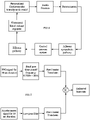
- FIG.4 illustrates an exemplary block diagram of the autoregulation method for estimating unstressed blood volume, in accordance with some embodiments of the present disclosure.
- the one or more hardware processors 104 are configured to update, at step 302b, the unstressed blood volume estimated at rest (from the total blood volume - refer equation 10) using the autoregulation method, when an activity is performed by the subject.
- the autoregulation method As seen in FIG.4 , four subsystems, viz., an afferent sympathetic pathway, central nervous system, efferent pathway and unstressed blood volume regulator cooperate to enable autoregulation.
- the autoregulation method comprises sensing aortic pressure by baroreceptors located at carotid sinus and aortic arch, at step 302b-1.
- the sensed aortic pressure is converted into a neural firing frequency f aff ( P ) via afferent sympathetic pathways, at step 302b-2.
- this functionality is defined as shown in equation (11) below.
- ⁇ aff P ⁇ min + ⁇ max ⁇ exp P K a 1 + exp P K a , where P defines the sensed aortic pressure , ⁇ min and ⁇ max are the upper and lower saturation frequencies , K a is a constant parameter having the same unit as pressure .
- ⁇ sa ⁇ sa ⁇ + f sa 0 ⁇ f sa ⁇ exp ⁇ K sa ⁇ ⁇ aff , where K sa , ⁇ sa and ⁇ sa 0 are constants .
- ⁇ psa ⁇ psa 0 + ⁇ psa ⁇ ⁇ exp ⁇ aff K psa 1 + exp ⁇ aff K psa , where K psa , ⁇ psa ⁇ and ⁇ psa 0 are constants .
- V s t V s 0 + ⁇ V s sa t + ⁇ V s psa t
- V s 0 defines the unstressed blood volume at rest
- ⁇ V s sa t and ⁇ V s psa t are the change in unstressed blood volume due to sympathetic and parasympathetic activities
- ⁇ V s sa t and ⁇ V s psa t are defined as given in equations (15) and (16) below.
- An additional blood demand representing the unstressed blood volume during the activity is then updated using the generated sympathetic and parasympathetic nervous activities, at step 302b-4.
- the total blood volume which is part of the plurality of input parameters is estimated using height and weight of the subject.
- the total blood volume across a healthy human body (who does not have any kind diseases) is closely related with Body-Surface-Area (BSA), and the BSA correlates with the height h and the weight w of the subject. Accordingly, the total blood volume is defined as shown in equation (17) below.
- the personalized cardiovascular hemodynamic model is executed considering the heart rate is continuously monitored when the subject performs an activity.
- the heart rate of the subject which is part of the plurality of input parameters, is estimated using (i) a Photoplethysmogram (PPG) signal and an accelerometer signal or (ii) an ECG signal, from a wearable device, such as a digital watch, worn by the subject when performing the activity.
- PPG Photoplethysmogram
- ECG ECG signal
- the heart rate for a specific time-window is estimated by employing noise cancellation technique using the accelerometer signal.
- the accelerometer signal is a 3-axis accelerometer signal, also captured continuously via the wearable device.
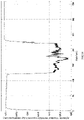
- FIG.5 illustrates heart rate estimation from a PPG signal and an accelerometer signal, in accordance with some embodiments of the present disclosure. Fast Fourier transform is performed on the PPG signal and the accelerometer signal in parallel for the same duration. Heart rate is estimated based on peaks available in the PPG signal after discarding the peaks (representing noise) obtained from the accelerometer signal.
- the step of estimating a plurality of input parameters is preceded by personalizing a cardiovascular hemodynamic model to obtain the personalized cardiovascular hemodynamic model.
- the personalizing is based on one or more of (i) cardiac parameters obtained from an echocardiogram such as valvular area, (ii) ECG signal obtained from a wearable device worn by the subject when performing the activity, and (iii) metadata of the subject including height and weight associated thereof.
- the cardiovascular hemodynamic model is configured with the compliance functions C ra ( t ), C la ( t ), C lv ( t ) and C r ⁇ ( t ) to sequentially actuate ra, la , rv and lv as explained above.
- Echocardiography information of the subject helps to derive constant parameters in equation (1) above.
- the mitral valve resistance R mi , the aortic valve resistance R ao , the tricuspid valve resistance R tr and the pulmonic valve resistance R pu may be accurately calculated from the echocardiography information, more specifically from information pertaining to valvular diameter of the subject, thereby personalizing the cardiovascular hemodynamic model.
- the parameters ⁇ mi , ⁇ ao , ⁇ tr , and ⁇ pu in equation (2) above, that define the regurgitation effect may be estimated from the echocardiography information.
- the parameters T and T a in equation (3) above, and T 1 , T 2 and d in equation (5) above may be estimated based on an ECG signal from the subject obtained using a wearable device. Metadata pertaining to the subject such as the height and the weight of the subject may be used to compute the total blood volume using equation (17) above.
- cardiac parameters are estimated via the personalized cardiovascular hemodynamic model, using the estimated plurality of input parameters estimated at step 302.
- the cardiac parameters include cardiac output, ejection fraction and mean arterial pressure.
- FIG.6 illustrates an input-output relation of the personalized cardiovascular hemodynamic model, when an activity is performed by the subject, in accordance with some embodiments of the present disclosure.
- While performing exercises on the treadmill two-channel PPG signals, three-axis acceleration signals, and one-channel ECG signals were simultaneously recorded from each subject.
- the PPG signals were recorded from the wrist by two pulse oximeters with green LEDs (wavelength: 515nm). Their distance (from center to center) was 2 cm.
- the acceleration signal was also recorded from the wrist by a three-axis accelerometer. Both the pulse oximeter and the accelerometer were embedded in a wristband, worn by the subjects.
- the ECG signal was recorded simultaneously from the chest using wet ECG sensors. All signals were sampled at 125 Hz and sent to a computer via Bluetooth.
- the cardiovascular hemodynamic model of the present disclosure was provided with the plurality of input parameters as described hereinafter.
- Total blood volume Subject specific metadata in the form of height and weight is not available in the Troika dataset. Hence, it was assumed that all the subjects are healthy, and the total blood volume was assumed to be 5 liters for each subject.
- FIG.7 illustrates estimation of the SVR of the lower body of a subject, based on MET levels, in accordance with some embodiments of the present disclosure.
- the value of R s (0) which defines the SVR at the resting state was chosen as 0.05 mmHgsec/mL and the time constant ⁇ was 0.5 sec.
- FIG.8 illustrates estimated heart rate from a raw ECG signal for a subject 11 (of the 11 subjects considered, the ECG signal from the 12 th subject being erroneous), in accordance with some embodiments of the present disclosure. Unstressed blood volume is approximated using the autoregulation method.
- FIG.9 illustrates heart rate in the form of a box plot for the subject 11, in accordance with some embodiments of the present disclosure, wherein the heart rate was estimated using the ECG signal. Similar box plots were derived for all the 11 subjects considered. Table 2 below provides a comparison of the estimated heart rate at different levels of physical activities for the 11 subjects.
- Table 2 Heart rate comparison Subject Number Levels of physical activities of Troika Dataset Level 1 (Rest for 30 sec.) Level 2 (Running @6-8 km/hr for 60 sec.) Level 3 (Running @12-15 km/hr for 60 sec.) Level 4 (Running @6-8 km/hr for 60 sec.) Level 5 (Running @12-15 km/hr for 60 sec.) Level 6 (Rest for 30 sec.) 1 72.84 ⁇ 10.14 100.16 ⁇ 12.71 139.95 ⁇ 11.21 153.72 ⁇ 1.86 157.68 ⁇ 5.75 160.98 ⁇ 4.27 2 78.64 ⁇ 13.7 99.62 ⁇ 11.1 128.65 ⁇ 9.26 133.26 ⁇ 8.26 134.63 ⁇ 8.82 143.84 ⁇ 4.68 3 94.82 ⁇ 12.04 108.22 ⁇ 10.39 143.27 ⁇ 9.35 142.98 ⁇ 8.12 145.4 ⁇ 10.31 158.60 ⁇ 1.8 4 82.46 ⁇ 10.52
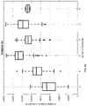
- FIG.10 illustrates cardiac output in the form of a box plots for the subject 11, in accordance with some embodiments of the present disclosure. Similar box plots were derived for all the 11 subjects considered. Table 3 below provides a comparison of the estimated cardiac output at different levels of physical activities for the 11 subjects.
- Table 3 Cardiac output comparison Subject Number Levels of physical activities of Troika Dataset Level 1 (Rest for 30 sec.) Level 2 (Running @6-8 km/hr for 60 sec.) Level 3 (Running @12-15 km/hr for 60 sec.) Level 4 (Running @6-8 km/hr for 60 sec.) Level 5 (Running @12-15 km/hr for 60 sec.) Level 6 (Rest for 30 sec.) 1 4.04 ⁇ 2.08 7.23 ⁇ 1.9 14.4 ⁇ 2.13 11.5 ⁇ 2.13 13.4 ⁇ 2 10.8 ⁇ 2.25 2 4.7 ⁇ 2.17 7.38 ⁇ 1.92 13.36 ⁇ 1.6 9.4 ⁇ 1.6 11.2 ⁇ 1.6 12.07 ⁇ 0.4 3 4.6 ⁇ 2.6 6.64 ⁇ 1.86 9.51 ⁇ 1.85 8.8 ⁇ 1.85 8.9 ⁇ 2.6 7.8 ⁇ 0.09 4 4.5 ⁇ 2.5 7.8 ⁇ 1.6 10.36 ⁇ 1.45 9.91 ⁇ 1.45 10.2 ⁇ 1.81 11.35 ⁇ 0.42 5 5.42 ⁇ 2.7 7.66 ⁇ 1.7 10.1 ⁇ 1.33 8.09 ⁇ 1.33 8.7 ⁇
- FIG.11 illustrates ejection fraction in the form of a box plot for the subject 11, in accordance with some embodiments of the present disclosure. Similar box plots were derived for all the 11 subjects considered. Table 4 below provides a comparison of the estimated ejection fraction at different levels of physical activities for the 11 subjects.
- Table 4 Ejection fraction comparison Subject Number Levels of physical activities of Troika Dataset Level 1 (Rest for 30 sec.) Level 2 (Running @6-8 km/hr for 60 sec.) Level 3 (Running @12-15 km/hr for 60 sec.) Level 4 (Running @6-8 km/hr for 60 sec.) Level 5 (Running @12-15 km/hr for 60 sec.) Level 6 (Rest for 30 sec.) 1 48.75+14.5 61.4+9.03 67.56+5.7 66.37 ⁇ 6.4 61.68+6.65 55.02+6.57 2 46.15 ⁇ 11.8 61.22+9.6 70 ⁇ 6.9 66.08 ⁇ 7.3 64.5 ⁇ 6.36 56.61 ⁇ 6.07 3 45.5 ⁇ 11.4 56.25 ⁇ 7.9 63.7 ⁇ 7.5 58.63 ⁇ 7.7 53.7 ⁇ 8.9 53.9 ⁇ 6.06 4 46.03+14.4 55.2+7.38 60.9+6.75 59.33+6.6 61.2+6.9 51.34+6.4 5 47.5 ⁇ 11.6 52
- FIG.12 illustrates mean arterial pressure in the form of a box plot for the subject 11, in accordance with some embodiments of the present disclosure. Similar box plots were derived for all the 11 subjects considered. Table 5 below provides a comparison of the estimated mean arterial pressure at different levels of physical activities for the 11 subjects. Table 5: Mean arterial pressure comparison.
- a wearable device Samsung Gear S2 TM classic Smart watch had been used for logging data from a tri-axis accelerometer sensor, and a PPG sensor.
- a custom application had been executed for logging the data and sharing with a host machine over Wi-Fi. All the signals were sampled at 100 Hz.
- the cardiovascular hemodynamic model of the present disclosure was provided with the plurality of input parameters as described hereinafter.
- Total blood volume In this dataset, the height and weight of all the subjects had been noted before the experiment. Depending on this, the total blood volume, in accordance with equation (17) was calculated and tabulated as shown below. Table 6: Total blood volume based on subject metadata Subject No Height (cm) Weight (Kg) Age Total Blood Volume (Liter) BMI (kg/m 2 ) 1 169 72 23 5.3 25.21 2 162.5 75 36 5.3 28.38 3 152.4 53 23 4.17 22.82 4 160.02 72 41 4.66 28.12 5 165.1 65 26 4.92 23.85 6 167.64 70 28 5.13 25.68
- % HRR Heart-rate reserve
- FIG.13 illustrates the estimated MET using a vertical accelerometer signal for a subject 2
- FIG. 14 illustrates the estimated SVR using the estimated MET for the subject 2, in accordance with some embodiment of the present disclosure.
- Heart rate estimation from PPG signal During the exercise condition, the signal quality of PPG is very poor. To overcome this challenge, a time-window of 10 sec was selected and the heart rate was estimated by comparing with the accelerometer signal as described previously.
- FIG.15 illustrates the estimated heart rate from PPG signal for the subject 2, in accordance with some embodiment of the present disclosure. During the simulation, the unstressed volume is approximated using the autoregulation method.
- FIG.16 illustrates estimated unstressed blood volume based on the autoregulation method for the subject 2, in accordance with some embodiment of the present disclosure.
- the cardiac parameters such as the cardiac output, the ejection fraction and the mean arterial pressure were estimated.
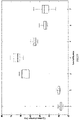
- FIG.17 illustrates estimated heart rate from PPG signal in the form of a box plot for the subject 2, in accordance with some embodiments of the present disclosure. Similar box plots were derived for all the 6 subjects considered. Table 7 below provides a comparison of the estimated heart rate at different levels of physical activities for the 6 subjects.
- Table 7 Heart rate comparison Sub No Levels of physical activities of TCS Dataset Level 1 (Rest) Level 2 (Walk) Level 3 (Walking up to (1/3) rd of the total stair) Level 4 (Walking up to (2/3) rd of the total stair) Level 5 (end of stair) Level 6 (Walk) Level 7 (Rest) 1 89.75 ⁇ 3.6 93.76 ⁇ 6.15 118.22 ⁇ 5.07 131.27 ⁇ 5 126.03 ⁇ 2.95 125.4 ⁇ 4.63 112.88 ⁇ 3 2 87.57 ⁇ 0.74 93.07 ⁇ 12.9 145.04 ⁇ 10.86 153.39 ⁇ 7.6 131.14 ⁇ 6.34 116.91 ⁇ 1.3 113.57 ⁇ 0.9 3 89.85 ⁇ 3.34 92.74 ⁇ 1.28 92.32 ⁇ 0.2 108.54 ⁇ 11.45 122.32 ⁇ 8.37 118.11 ⁇ 5.03 112.71 ⁇ 6.35 4 78.19+4.16 84.24+6.27 122.01 ⁇ 12.3 138.29 ⁇
- FIG.18 illustrates estimated cardiac output in the form of a box plot for the subject 2, in accordance with some embodiments of the present disclosure. Similar box plots were derived for all the 6 subjects considered. Table 8 below provides a comparison of the estimated cardiac output at different levels of physical activities for the 6 subjects.
- Table 8 Cardiac output comparison Subject Number Levels of physical activities of TCS Dataset Level 1 (Rest) Level 2 (Walk) Level 3 (Walking up to (1/3) rd of the total stair) Level 4 (Walking up to (2/3) rd of the total stair) Level 5 (end of stair) Level 6 (Walk) Level 7 (Rest) 1 5.6 ⁇ 1.2 6.9 ⁇ 1.8 9.6 ⁇ 0.55 12.14 ⁇ 0.83 10.88 ⁇ 2.6 8.23 ⁇ 0.26 7.25 ⁇ 0.2 2 5.04 ⁇ 1.25 5.16+0.5 9.17 ⁇ 0.95 12.03+0.9 8.89+0.3 7.05 ⁇ 0.14 6.42 ⁇ 0.05 3 5.13 ⁇ 1.51 5.87+2.5 5.85+0.7 8.43+1.05 8.65+1.5 5.32+0.3 7.01 ⁇ 0.4 4 5.37+0.9 6.4+0.5 9.82+1.01 13.36 ⁇ 1.6 12.27+0.86 8.9+0.23 9.01 ⁇ 0.95 5 5.14+1.4 6.44 ⁇ 0.5 9.46 ⁇ 0.88 11.62 ⁇ 1.11 10.78 ⁇ 0.33 8.8 ⁇ 0.13 7.6 ⁇ 0.16 6 6.1 ⁇
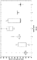
- FIG.19 illustrates estimated ejection fraction in the form of a box plot for the subject 2, in accordance with some embodiments of the present disclosure. Similar box plots were derived for all the 6 subjects considered. Table 9 below provides a comparison of the estimated ejection fraction at different levels of physical activities for the 6 subjects.
- Table 9 Ejection fraction comparison Subject Number Levels of physical activities of TCS Dataset Level 1 (Rest) Level 2 (Walk) Level 3 (Walking up to (1/3) rd of the total stair) Level 4 (Walking up to (2/3) rd of the total stair) Level 5 (end of stair) Level 6 (Walk) Level 7 (Rest) 1 49.3 ⁇ 7.2 49.33 ⁇ 0.65 66.9 ⁇ 3.1 72.77 ⁇ 3.7 69.02 ⁇ 1.98 63.9 ⁇ 0.45 59.56 ⁇ 2.74 2 48.5+7.26 50.02+0.56 68.17 ⁇ 2.14 71.9+2.14 62.67+0.93 57.2 ⁇ 1.2 57.03+1.94 3 57.95 ⁇ 9.5 60.16 ⁇ 0.16 72.74 ⁇ 0.33 80.7 ⁇ 7.07 76.93 ⁇ 9.72 63.1 ⁇ 3.95 60.4 ⁇ 2.95 4 52.8 ⁇ 7.4 52.6 ⁇ 0.66 70.6 ⁇ 1.1 79.95+2.71 70.8 ⁇ 1.17 60.5 ⁇ 1.66 60.9+2.83 5 49.9 ⁇ 9.25
- FIG.20 illustrates estimated mean arterial pressure in the form of a box plot for the subject 2, in accordance with some embodiments of the present disclosure. Similar box plots were derived for all the 6 subjects considered. Table 10 below provides a comparison of the estimated mean arterial pressure at different levels of physical activities for the 6 subjects.
- Table 10 Mean arterial pressure comparison Levels of physical activities of TCS Dataset Subject Number Level 1 (Rest) Level 2 (Walk) Level 3 (Walking up to (1/3) rd of the total stair) Level 4 (Walking up to (2/3) rd of the total stair) Level 5 (end of stair) Level 6 (Walk) Level 7 (Rest) 1 99.8 ⁇ 15.3 118.12+21.7 144.75+2.35 145.5 ⁇ 1 135+21.1 115.6 ⁇ 1 109.75+0.6 2 102.95 ⁇ 14.3 105.46+7.
- a care giver simulates different heart rate conditions along with post-operative valvular conditions and selects different exercise levels, linked with MET value.
- cardiac parameters like cardiac output, ejection fraction and mean arterial pressure are generated by the personalized cardiovascular hemodynamic model.
- the simulated cardiac output provides a baseline activity response based on which, the caregiver may plan daily ambulatory activities and therapy.
- the hardware device can be any kind of device which can be programmed including e.g. any kind of computer like a server or a personal computer, or the like, or any combination thereof.
- the device may also include means which could be e.g. hardware means like e.g. an application-specific integrated circuit (ASIC), a field-programmable gate array (FPGA), or a combination of hardware and software means, e.g.
- ASIC application-specific integrated circuit
- FPGA field-programmable gate array
- the means can include both hardware means, and software means.
- the method embodiments described herein could be implemented in hardware and software.
- the device may also include software means.
- the embodiments may be implemented on different hardware devices, e.g. using a plurality of CPUs.
- the embodiments herein can comprise hardware and software elements.
- the embodiments that are implemented in software include but are not limited to, firmware, resident software, microcode, etc.
- the functions performed by various components described herein may be implemented in other components or combinations of other components.
- a computer-usable or computer readable medium can be any apparatus that can comprise, store, communicate, propagate, or transport the program for use by or in connection with the instruction execution system, apparatus, or device.
- a computer-readable storage medium refers to any type of physical memory on which information or data readable by a processor may be stored.
- a computer-readable storage medium may store instructions for execution by one or more hardware processors, including instructions for causing the processor(s) to perform steps or stages consistent with the embodiments described herein.
- the term "computer-readable medium” should be understood to include tangible items and exclude carrier waves and transient signals, i.e., be non-transitory. Examples include random access memory (RAM), read-only memory (ROM), volatile memory, nonvolatile memory, hard drives, CD ROMs, DVDs, flash drives, disks, and any other known physical storage media.
Landscapes
- Health & Medical Sciences (AREA)
- Life Sciences & Earth Sciences (AREA)
- Engineering & Computer Science (AREA)
- Biomedical Technology (AREA)
- Medical Informatics (AREA)
- Public Health (AREA)
- General Health & Medical Sciences (AREA)
- Cardiology (AREA)
- Pathology (AREA)
- Molecular Biology (AREA)
- Heart & Thoracic Surgery (AREA)
- Veterinary Medicine (AREA)
- Animal Behavior & Ethology (AREA)
- Physics & Mathematics (AREA)
- Surgery (AREA)
- Biophysics (AREA)
- Physiology (AREA)
- Primary Health Care (AREA)
- Epidemiology (AREA)
- Neurology (AREA)
- Vascular Medicine (AREA)
- Data Mining & Analysis (AREA)
- Databases & Information Systems (AREA)
- Radiology & Medical Imaging (AREA)
- Nuclear Medicine, Radiotherapy & Molecular Imaging (AREA)
- Computer Vision & Pattern Recognition (AREA)
- Business, Economics & Management (AREA)
- Oral & Maxillofacial Surgery (AREA)
- Dentistry (AREA)
- General Business, Economics & Management (AREA)
- Neurosurgery (AREA)
- Artificial Intelligence (AREA)
- Psychiatry (AREA)
- Signal Processing (AREA)
- Pulmonology (AREA)
- Measuring Pulse, Heart Rate, Blood Pressure Or Blood Flow (AREA)
Abstract
Description
- The present application claims priority to
Indian application no. 202121010972, filed on 15th March, 2021 - The disclosure herein generally relates to the field of computer assisted modeling of hemodynamic patterns of physiologic blood flow, and, more particularly, to systems and methods for estimating cardiac parameters when performing an activity using a personalized cardiovascular hemodynamic model.
- Cardiovascular disease (CVD) is one of the main causes of deaths in the world today. With advancing age, the risk of CVD increases significantly. To reduce the effect of CVDs, it has been a well-established fact that the higher levels of regular physical exercises would reduce cardiovascular events and mortality. According to medical literature and guidelines, increasing levels of physical activity are the first-line approaches for preventing and treating vascular dysfunction and cardio-metabolic diseases. Moreover, during post-operative recovery of a patient who had suffered a cardiovascular surgery recently, performing prescribed exercises during the recovery phase is always recommended. However, unless the patient diligently and periodically consults a caregiver, it is challenging to gauge whether the prescribed exercises are yielding a desired outcome, or any change is required in the planned therapy.
- Embodiments of the present disclosure present technological improvements as solutions to one or more of the above-mentioned technical problems recognized by the inventors in conventional systems.
- In an aspect, there is provided a processor implemented method comprising the steps of: estimating, via one or more hardware processors, a plurality of input parameters for a personalized cardiovascular hemodynamic model associated with a subject being monitored, the plurality of input parameters comprising (i) Systemic Vascular Resistance (SVR), (ii) unstressed blood volume, (iii) total blood volume in a body of the subject, and (iv) heart rate of the subject, the step of estimating comprises: estimating the SVR using Metabolic EquivalenT (MET) levels associated with an activity level of the subject; and updating the unstressed blood volume estimated when the subject is at rest, using an autoregulation method, when an activity is performed by the subject, wherein the autoregulation method comprises: sensing aortic pressure by baroreceptors located at carotid sinus and aortic arch; converting the sensed aortic pressure into a neural firing frequency via afferent sympathetic pathways; generating sympathetic and parasympathetic nervous activities via a central nervous system and efferent pathway depending on the neural firing frequency; and updating an additional blood demand representing the unstressed blood volume during the activity using the generated sympathetic and parasympathetic nervous activities; and estimating, via the personalized cardiovascular hemodynamic model, cardiac parameters including cardiac output, ejection fraction and mean arterial pressure, using the estimated plurality of input parameters.
- In another aspect, there is provided a system comprising: one or more data storage devices operatively coupled to one or more hardware processors and configured to store instructions configured for execution via the one or more hardware processors to: estimate a plurality of input parameters for a personalized cardiovascular hemodynamic model associated with a subject being monitored, the plurality of input parameters comprising (i) Systemic Vascular Resistance (SVR), (ii) unstressed blood volume, (iii) total blood volume in a body of the subject, and (iv) heart rate of the subject, wherein estimating the plurality of input parameters comprises: estimating the SVR using Metabolic EquivalenT (MET) levels associated with an activity level of the subject; and updating the unstressed blood volume estimated when the subject is at rest, using an autoregulation method, when an activity is performed by the subject, wherein the autoregulation method comprises: sensing aortic pressure by baroreceptors located at carotid sinus and aortic arch; converting the sensed aortic pressure into a neural firing frequency via afferent sympathetic pathways; generating sympathetic and parasympathetic nervous activities via a central nervous system and efferent pathway depending on the neural firing frequency; and updating an additional blood demand representing the unstressed blood volume during the activity using the generated sympathetic and parasympathetic nervous activities; and estimate cardiac parameters including cardiac output, ejection fraction and mean arterial pressure, using the estimated plurality of input parameters.
- In yet another aspect, there is provided a computer program product comprising a non-transitory computer readable medium having a computer readable program embodied therein, wherein the computer readable program, when executed on a computing device, causes the computing device to: estimate a plurality of input parameters for a personalized cardiovascular hemodynamic model associated with a subject being monitored, the plurality of input parameters comprising (i) Systemic Vascular Resistance (SVR), (ii) unstressed blood volume, (iii) total blood volume in a body of the subject, and (iv) heart rate of the subject, wherein estimating the plurality of input parameters comprises: estimating the SVR using Metabolic EquivalenT (MET) levels associated with an activity level of the subject; and updating the unstressed blood volume estimated when the subject is at rest, using an autoregulation method, when an activity is performed by the subject, wherein the autoregulation method comprises: sensing aortic pressure by baroreceptors located at carotid sinus and aortic arch; converting the sensed aortic pressure into a neural firing frequency via afferent sympathetic pathways; generating sympathetic and parasympathetic nervous activities via a central nervous system and efferent pathway depending on the neural firing frequency; and updating an additional blood demand representing the unstressed blood volume during the activity using the generated sympathetic and parasympathetic nervous activities; and estimate cardiac parameters including cardiac output, ejection fraction and mean arterial pressure, using the estimated plurality of input parameters.
- In accordance with an embodiment of the present disclosure, the one or more hardware processors are configured to personalize a cardiovascular hemodynamic model to obtain the personalized cardiovascular hemodynamic model, prior to estimating the plurality of input parameters, based on one or more of (i) cardiac parameters obtained from an echocardiogram, (ii) ECG signal obtained from a wearable device worn by the subject when performing the activity and (iii) metadata of the subject including height and weight associated thereof.
- In accordance with an embodiment of the present disclosure, the one or more hardware processors are configured to perform sequential activation of a right atrium ra, a left atrium la, a right ventricle rv and a left ventricle lv of the personalized cardiovascular hemodynamic model using compliance functions Cra (t), Cla (t), Crv (t) and Clv (t) respectively, the compliance functions being defined as:
- (i) the compliance function to actuate ra, Cra (t) = Cmin,ra + 0.5 × (Cmax,ra ― Cmin,ra )u(t), wherein

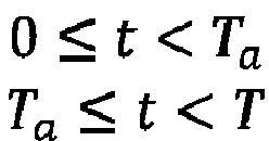
- (ii) the compliance function to actuate la, Cla (t) = Cmin,la + 0.5 × (Cmax,la ― Cmin,la )u(t ― dla ), wherein Cmin,la and Cmax,la are the minimum and maximum values of the la compliance and dla represents a time delay between activation of the ra and the la; and
- (iii) the compliance functions to actuate rv and lv are represented as


- In accordance with an embodiment of the present disclosure, the one or more hardware processors are configured to estimate the total blood volume using height and weight of the subject.
- In accordance with an embodiment of the present disclosure, the one or more hardware processors are configured to estimate the SVR based on

- In accordance with an embodiment of the present disclosure, the one or more hardware processors are configured to estimate the heart rate of the subject using (i) a Photoplethysmogram (PPG) signal and an accelerometer signal or (ii) an Electrocardiogram (ECG) signal, from a wearable device worn by the subject when performing the activity.
- It is to be understood that both the foregoing general description and the following detailed description are exemplary and explanatory only and are not restrictive of the invention, as claimed.
- The accompanying drawings, which are incorporated in and constitute a part of this disclosure, illustrate exemplary embodiments and, together with the description, serve to explain the disclosed principles:
-
FIG. 1 illustrates an exemplary block diagram of a system for estimating cardiac parameters when performing an activity using a personalized cardiovascular hemodynamic model, in accordance with some embodiments of the present disclosure. -
FIG.2 illustrates an exemplary block diagram of a cardiovascular hemodynamic model, in accordance with some embodiments of the present disclosure. -
FIG.3A through FIG.3B illustrates an exemplary flow diagram of a computer implemented method for estimating cardiac parameters when performing an activity using a personalized cardiovascular hemodynamic model, in accordance with some embodiments of the present disclosure. -
FIG.4 illustrates an exemplary block diagram of the autoregulation method for estimating unstressed blood volume, in accordance with some embodiments of the present disclosure. -
FIG.5 illustrates heart rate estimation from a Photoplethysmogram (PPG) signal and an accelerometer signal, in accordance with some embodiments of the present disclosure. -
FIG.6 illustrates an input-output relation of the personalized cardiovascular hemodynamic model, when an activity is performed by the subject, in accordance with some embodiments of the present disclosure. -
FIG.7 illustrates estimation of Systemic Vascular Resistance (SVR) of the lower body of a subject, based on Metabolic EquivalenT (MET) levels, in accordance with some embodiments of the present disclosure. -
FIG.8 illustrates estimated heart rate from a raw electrocardiogram (ECG) signal for asubject 11, in accordance with some embodiments of the present disclosure. -
FIG.9 illustrates heart rate in the form of a box plot for thesubject 11, in accordance with some embodiments of the present disclosure. -
FIG.10 illustrates cardiac output in the form of a box plot for thesubject 11, in accordance with some embodiments of the present disclosure. -
FIG.11 illustrates ejection fraction of a box plot for thesubject 11, in accordance with some embodiments of the present disclosure. -
FIG.12 illustrates mean arterial pressure in the form of a box plot for thesubject 11, in accordance with some embodiments of the present disclosure. -
FIG.13 illustrates estimated MET using a vertical accelerometer signal for asubject 2, in accordance with some embodiment of the present disclosure. -
FIG.14 illustrates estimated SVR using the estimated MET for thesubject 2, in accordance with some embodiment of the present disclosure. -
FIG.15 illustrates estimated heart rate from PPG signal for thesubject 2, in accordance with some embodiment of the present disclosure. -
FIG.16 illustrates estimated unstressed blood volume based on the autoregulation method for thesubject 2, in accordance with some embodiment of the present disclosure. -
FIG.17 illustrates estimated heart rate from PPG signal in the form of a box plot for thesubject 2, in accordance with some embodiments of the present disclosure. -
FIG.18 illustrates estimated cardiac output in the form of a box plot for thesubject 2, in accordance with some embodiments of the present disclosure. -
FIG.19 illustrates estimated ejection fraction in the form of a box plot for thesubject 2, in accordance with some embodiments of the present disclosure. -
FIG.20 illustrates estimated mean arterial pressure in the form of a box plot for thesubject 2, in accordance with some embodiments of the present disclosure. - Exemplary embodiments are described with reference to the accompanying drawings. In the figures, the left-most digit(s) of a reference number identifies the figure in which the reference number first appears. Wherever convenient, the same reference numbers are used throughout the drawings to refer to the same or like parts. While examples and features of disclosed principles are described herein, modifications, adaptations, and other implementations are possible without departing from the scope of the disclosed embodiments.
- Considering the risks posed by Cardiovascular disease (CVD), it is very important to periodically monitor a patient's cardiac parameters to assess effects of prescribed exercises on the cardiac parameters of the patient. Unless the patient diligently visits a caregiver, say during a post-operative period, assessing cardiovascular conditions for recovery is challenging. A lapse on the part of the patient may be detrimental to the patient's health. The present disclosure enables personalized cardiac rehabilitation guidance and care continuum using a personalized cardiovascular hemodynamic model that effectively simulates cardiac parameters when the patient performs an activity using a wearable device like a digital watch that can help capture Electrocardiogram (ECG) signal, Photoplethysmogram (PPG) signal and accelerometer signal. In the context of the present disclosure, the expression 'patient' may interchangeably be referred as a 'subject'.
- Referring now to the drawings, and more particularly to
FIG. 1 through FIG.20 , where similar reference characters denote corresponding features consistently throughout the figures, there are shown preferred embodiments and these embodiments are described in the context of the following exemplary system and/or method. -
FIG.1 illustrates an exemplary block diagram of asystem 100 for estimating cardiac parameters when performing an activity using a personalized cardiovascular hemodynamic model, in accordance with some embodiments of the present disclosure. In an embodiment, thesystem 100 includes one ormore hardware processors 104, communication interface device(s) or input/output (I/O) interface(s) 106, and one or more data storage devices ormemory 102 operatively coupled to the one ormore hardware processors 104. The one ormore hardware processors 104 can be implemented as one or more microprocessors, microcomputers, microcontrollers, digital signal processors, central processing units, state machines, graphics controllers, logic circuitries, and/or any devices that manipulate signals based on operational instructions. Among other capabilities, the processor(s) are configured to fetch and execute computer-readable instructions stored in the memory. In the context of the present disclosure, the expressions 'processors' and 'hardware processors' may be used interchangeably. In an embodiment, thesystem 100 can be implemented in a variety of computing systems, such as laptop computers, notebooks, hand-held devices, workstations, mainframe computers, servers, a network cloud and the like. - I/O interface(s) 106 can include a variety of software and hardware interfaces, for example, a web interface, a graphical user interface, and the like and can facilitate multiple communications within a wide variety of networks N/W and protocol types, including wired networks, for example, LAN, cable, etc., and wireless networks, such as WLAN, cellular, or satellite. In an embodiment, the I/O interface(s) can include one or more ports for connecting a number of devices to one another or to another server.
- The
memory 102 may include any computer-readable medium known in the art including, for example, volatile memory, such as static random access memory (SRAM) and dynamic random access memory (DRAM), and/or non-volatile memory, such as read only memory (ROM), erasable programmable ROM, flash memories, hard disks, optical disks, and magnetic tapes. In an embodiment, one or more modules (not shown) of thesystem 100 can be stored in thememory 102. -
FIG.2 illustrates an exemplary block diagram of a cardiovascular hemodynamic model triggered via activity related data, in accordance with some embodiments of the present disclosure. Heart is a muscular organ in which each half is composed of a pair of atria and a pair of ventricles serving like a pulsatile pump. A left heart chamber, comprising a left ventricle (lv) and a left atrium (la), pumps oxygenated blood to all tissues of the body. This specific circulation is called systemic circulation. On the other hand, a right heart, comprising a right ventricle (rv) and a right atrium (ra), drives deoxygenated blood to the lungs forming a pulmonic circulation. In addition, there are four cardiac valves namely, mitral (mi) and aortic (ao) valves in the left heart and tricuspid (tr) and pulmonic (pu) valves in the right heart respectively. These valves synchronously open and close based on a pressure difference within the cardiac chambers and ensures rhythmic unidirectional flow through the heart. - In accordance with the present disclosure, following assumptions have been considered to describe the hemodynamics of the cardiac system. Assumption 1: Each of the cardiac chambers is triggered by an autonomous compliance function (C(t)), due to the elasticity of the cardiac walls. So, the volume (V (t)), at time t, across any cardiac chamber is defined as V (t) = C(t) × P (t) + Vs; where P (t) is the pressure at the tth time and Vs is the unstressed volume of that particular chamber. Assumption 2: The cardiac chambers are considered compliance vessels. Hence, the rate of change of volume across a cardiac chamber at time t, is defined as a difference between an inflow Q1(t) and an outflow Q2(t), so, dV/dt (t) = Q1(t) ― Q2(t). Assumption 3: Each vessel is considered a resistive vessel, as the blood flow is impeded due to frictional forces, depending on viscosity of blood, diameter of the blood vessels, etc. Thus, a flow across a resistive vessel is Q(t) = ΔP(t)/R; where ΔP(t) is a pressure difference in successive compartments of the vessel, at time t and R is the vascular resistance of that vessel.
- Based on the three assumptions provided herein above, pressure dynamics of a cardiovascular hemodynamic model is analytically defined as:


- Ui ;∀i{mi, ao, tr, pu} are inputs for opening and closing of the heart valves as mentioned in equation (2) below.

- As per the
assumption 1, the cardiac chambers are activated sequentially, in a synchronized manner, by time-varying compliance functions. Typically, this activation starts from a sinoatrial node, which is located inside ra, then, it traverses to the la with a time delay of dla , causing them to contract for pumping the blood into the ventricles. After that, the activation traverses from the atrium to the ventricles via an atrioventricular node with a time delay of d, allowing the ventricles to fill with blood. In accordance with the present disclosure, the compliance functions Crα (t), Cla (t), Clv (t) and Crv (t) to sequentially actuate ra, la, rv and lv are defined as given in equation (3), equation (4) and equation (5) respectively.
- Similarly, in accordance with the present disclosure, the compliance function to actuate la is modeled using equation (3), where Cmin,la and Cmax,la are the minimum and maximum values of the la compliance, u(t) is the activation function of la and dla represent a delay in activation of la with respect to ra.

- Likewise, in accordance with the present disclosure, the compliance functions to actuate rv and lv are represented by equation (5) below, where Ci ; i ∈ {lv, rv} is the systolic compliance across lv or rv and is estimated as a ratio of R-peak and T-peak of the ECG signal, u v (t) is the activation function, and d represents the delay in activation of lv or rv from ra. T 1 and T 2 are the systolic and diastolic activation time instances of the cardiac cycle respectively.

- In accordance with the present disclosure, depending on pressure variations in the cardiac chambers, blood circulates as shown in
FIG.3 . Based on the circulation, the volume across the cardiac chambers also get developed as perassumption 2 mentioned above. The rate of change of volume across the cardiac chambers is measured as given in equation (6) below.
- Similarly, the rate of change of volume across the systemic and pulmonary arteries is evaluated using equation (7) below.

- The overall blood volume using equations (6) and (7) above is represented by equation (8) below.

-
FIG.3A through FIG.3B illustrate an exemplary flow diagram of a computer implementedmethod 300 for estimating cardiac parameters when performing an activity using a personalized cardiovascular hemodynamic model, in accordance with some embodiments of the present disclosure. In an embodiment, thesystem 100 includes one or more data storage devices ormemory 102 operatively coupled to the one ormore hardware processors 104 and is configured to store instructions configured for execution of steps of themethod 200 by the one ormore hardware processors 104. The steps of themethod 300 will now be explained in detail with reference to the components of thesystem 100 ofFIG.1 . Although process steps, method steps, techniques or the like may be described in a sequential order, such processes, methods and techniques may be configured to work in alternate orders. In other words, any sequence or order of steps that may be described does not necessarily indicate a requirement that the steps be performed in that order. The steps of processes described herein may be performed in any order practical. Further, some steps may be performed simultaneously. - Accordingly, in an embodiment of the present disclosure, the one or
more hardware processors 104, are configured to estimate, atstep 302, a plurality of input parameters for a personalized cardiovascular hemodynamic model associated with a subject being monitored. In an embodiment, the plurality of input parameters comprises (i) Systemic Vascular Resistance (SVR), (ii) unstressed blood volume, (iii) total blood volume in a body of the subject, and (iv) heart rate of the subject. - When the subject performs any activity like say physical exercise, the amount of blood flowing through skeletal muscles is significantly increased and it is solely dependent on the intensity of the activity. In accordance with the present disclosure, systemic vessels are considered as resistive vessels. Therefore, to accommodate increasing blood flow across the systemic vessels, the vessel resistivity requires to be reduced that significantly reduces the resistance across the systemic vessels. In accordance with the present disclosure, to estimate how much the vessel resistivity needs to be changed and how it is correlated with the level of exercise, the metabolic rate of the subject (which defines the rate of change energy expended per unit time) is considered as it is tightly coupled with vessel conductance.
- For a specific activity, the metabolic rate is measured by a metabolic equivalent of task or Metabolic EquivalenT (MET) levels. The MET is defined as the ratio of an exercise metabolic rate to a resting metabolic rate. Thus, for a given activity, the MET value has been considered a constant. For example, in light intensive activities, such as walking slowly, the MET level is consider to be less than 3, in moderate intensive activities (walking at say 4.5 km/hr), the MET level is considered between 3 and 6 and for intensive exercises, such as jogging at say 11 km/hr, the MET level is greater than 6.
- Thus, in accordance with the present disclosure, the one or
more hardware processors 104, are configured to estimate, atstep 302a, the SVR using the MET levels associated with an activity level of the subject. In an embodiment, the SVR is represented as given in equation (9) below.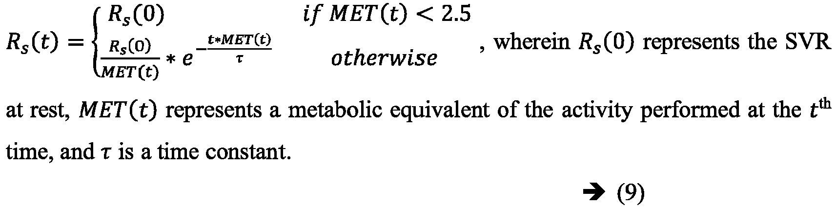
- In order to simulate the effect of the activity with the variation of systemic resistance, the systemic circulation part in a body is subdivided into three generic sections such as an upper body, a middle body and a lower body as shown in
FIG.2 . Depending on the activity performed by the subject, the SVR across the section is modulated based on the MET level as described above, and the SVR in the other sections are assumed to be constant or modulated by an autoregulation method described hereinafter. - Unstressed blood volume (or dead volume) is defined as the maximum volume of blood that can be placed inside a capacitive vessel without raising its effective pressure above 0 mmHg. As per literature, in normal cardiac condition (resting state), the unstressed blood volume is approximated as shown in equation (10) below.

- Therefore, in a resting state around 40% of the total blood volume (Vtotal ) is stressed volume (effectively participate in hemodynamics) and the rest is unstressed volume. When the subject performs an activity, or during an exercise condition, because of the increasing demand of blood, a portion of the unstressed blood is added into the stressed volume. Baroreflex regulates the effect of unstressed blood volume while enhancing the venous return of blood during a ventricular systolic phase.
FIG.4 illustrates an exemplary block diagram of the autoregulation method for estimating unstressed blood volume, in accordance with some embodiments of the present disclosure. In an embodiment of the present disclosure, the one ormore hardware processors 104, are configured to update, atstep 302b, the unstressed blood volume estimated at rest (from the total blood volume - refer equation 10) using the autoregulation method, when an activity is performed by the subject. As seen inFIG.4 , four subsystems, viz., an afferent sympathetic pathway, central nervous system, efferent pathway and unstressed blood volume regulator cooperate to enable autoregulation. - The effect of baroreflex starts from sensing of aortic pressure by baroreceptors located at carotid sinus and aortic arch. Accordingly, in an embodiment of the present disclosure, the autoregulation method comprises sensing aortic pressure by baroreceptors located at carotid sinus and aortic arch, at
step 302b-1. The sensed aortic pressure is converted into a neural firing frequency faff (P) via afferent sympathetic pathways, atstep 302b-2. Analytically, this functionality is defined as shown in equation (11) below.
- Sympathetic and parasympathetic nervous activities are then generated via the central nervous system and efferent pathway, depending on the neural firing frequency, at
step 302b-3. The sympathetic activity fsa is defined as shown in equation (12) below.
- The Parasympathetic activity fpsa is defined as shown in equation (13) below.

- Based on the generated sympathetic and parasympathetic nervous activities, the additional blood volume requirement during an exercise needs to be regulated. Autoregulation of the unstressed blood volume via the unstressed blood volume regulator is analytically represented as shown in equation (14) below.
- The governing equations of


- An additional blood demand representing the unstressed blood volume during the activity, is then updated using the generated sympathetic and parasympathetic nervous activities, at
step 302b-4. - In accordance with the present disclosure, the total blood volume which is part of the plurality of input parameters is estimated using height and weight of the subject. As per medical literature, the total blood volume across a healthy human body (who does not have any kind diseases) is closely related with Body-Surface-Area (BSA), and the BSA correlates with the height h and the weight w of the subject. Accordingly, the total blood volume is defined as shown in equation (17) below.

- The heart rate is an important parameter that dynamically changes during exercise. Hence, creating a model of heart rate based on a specific physical activity is very difficult to achieve. In accordance with the present disclosure, the personalized cardiovascular hemodynamic model is executed considering the heart rate is continuously monitored when the subject performs an activity. In accordance with the present disclosure, the heart rate of the subject, which is part of the plurality of input parameters, is estimated using (i) a Photoplethysmogram (PPG) signal and an accelerometer signal or (ii) an ECG signal, from a wearable device, such as a digital watch, worn by the subject when performing the activity. As per literature, noise effect of an ECG signal during activities is less than the PPG signal. Hence, if the ECG signal is available, the heart rate is easily measured continuously using detected peaks. If only the PPG signal is available, then the heart rate for each cycle may not be obtained. Hence, the heart rate for a specific time-window is estimated by employing noise cancellation technique using the accelerometer signal. In an embodiment, the accelerometer signal is a 3-axis accelerometer signal, also captured continuously via the wearable device.
FIG.5 illustrates heart rate estimation from a PPG signal and an accelerometer signal, in accordance with some embodiments of the present disclosure. Fast Fourier transform is performed on the PPG signal and the accelerometer signal in parallel for the same duration. Heart rate is estimated based on peaks available in the PPG signal after discarding the peaks (representing noise) obtained from the accelerometer signal. - In an embodiment, the step of estimating a plurality of input parameters is preceded by personalizing a cardiovascular hemodynamic model to obtain the personalized cardiovascular hemodynamic model. In an embodiment, the personalizing is based on one or more of (i) cardiac parameters obtained from an echocardiogram such as valvular area, (ii) ECG signal obtained from a wearable device worn by the subject when performing the activity, and (iii) metadata of the subject including height and weight associated thereof. In accordance with the present disclosure, the cardiovascular hemodynamic model is configured with the compliance functions Cra (t), Cla (t), Clv (t) and Crν (t) to sequentially actuate ra, la, rv and lv as explained above. Echocardiography information of the subject helps to derive constant parameters in equation (1) above. For instance, the mitral valve resistance Rmi , the aortic valve resistance Rao , the tricuspid valve resistance Rtr and the pulmonic valve resistance Rpu may be accurately calculated from the echocardiography information, more specifically from information pertaining to valvular diameter of the subject, thereby personalizing the cardiovascular hemodynamic model. Again, the parameters δmi , δao , δtr , and δpu in equation (2) above, that define the regurgitation effect may be estimated from the echocardiography information. Furthermore, the parameters T and Ta in equation (3) above, and T 1, T 2 and d in equation (5) above may be estimated based on an ECG signal from the subject obtained using a wearable device. Metadata pertaining to the subject such as the height and the weight of the subject may be used to compute the total blood volume using equation (17) above.
- In an embodiment of the present disclosure, at
step 304, cardiac parameters are estimated via the personalized cardiovascular hemodynamic model, using the estimated plurality of input parameters estimated atstep 302. In an embodiment, the cardiac parameters include cardiac output, ejection fraction and mean arterial pressure.FIG.6 illustrates an input-output relation of the personalized cardiovascular hemodynamic model, when an activity is performed by the subject, in accordance with some embodiments of the present disclosure. - Experiment 1: Publicly available Troika dataset was used. In these experiments, 12 subjects in the age group 18years to 35years were asked to run on a treadmill with changing speeds. The speed-variations are defined as follows. Rest for 30sec → 6-8km/hr for 1 min → 12-15km/hr for 1min → 6-8km/hr for 1 min → 12-15km/hr for 1 min → Rest for 30sec.
- While performing exercises on the treadmill, two-channel PPG signals, three-axis acceleration signals, and one-channel ECG signals were simultaneously recorded from each subject. For each subject, the PPG signals were recorded from the wrist by two pulse oximeters with green LEDs (wavelength: 515nm). Their distance (from center to center) was 2 cm. The acceleration signal was also recorded from the wrist by a three-axis accelerometer. Both the pulse oximeter and the accelerometer were embedded in a wristband, worn by the subjects. The ECG signal was recorded simultaneously from the chest using wet ECG sensors. All signals were sampled at 125 Hz and sent to a computer via Bluetooth.
- The cardiovascular hemodynamic model of the present disclosure was provided with the plurality of input parameters as described hereinafter. Total blood volume: Subject specific metadata in the form of height and weight is not available in the Troika dataset. Hence, it was assumed that all the subjects are healthy, and the total blood volume was assumed to be 5 liters for each subject.
- SVR during exercise: In the Troika dataset, the level of exercise has been clearly mentioned; hence, the respective MET levels (value) were predicted accurately from the MET table. The MET values for the respective activity duration are tabulated below in Table 1.
Table 1: MET table with respect to speed variation Activity duration (sec) Running Speed (km/hr) MET Value 0-30 Rest 2 31-90 6-8 8 91-150 12-15 15 151-210 6-8 8 211-270 12-15 15 271-300 Rest 2 - Depending on the MET values, the estimated SVR across the lower body was estimated using equation (9) above.
FIG.7 illustrates estimation of the SVR of the lower body of a subject, based on MET levels, in accordance with some embodiments of the present disclosure. In this simulation, the value of Rs (0) which defines the SVR at the resting state, was chosen as 0.05 mmHgsec/mL and the time constant τ was 0.5 sec. - Heart rate estimation from ECG signal: In the Troika dataset, as the ECG signal has been captured during the physical activities, by filtering and detecting the ECG peaks, the continuous heart rate was estimated.
FIG.8 illustrates estimated heart rate from a raw ECG signal for a subject 11 (of the 11 subjects considered, the ECG signal from the 12th subject being erroneous), in accordance with some embodiments of the present disclosure. Unstressed blood volume is approximated using the autoregulation method. - By inputting the plurality of input parameters mentioned above into the cardiovascular hemodynamic model of the present disclosure, the cardiac parameters such as the cardiac output, the ejection fraction and the mean arterial pressure were estimated.
FIG.9 illustrates heart rate in the form of a box plot for the subject 11, in accordance with some embodiments of the present disclosure, wherein the heart rate was estimated using the ECG signal. Similar box plots were derived for all the 11 subjects considered. Table 2 below provides a comparison of the estimated heart rate at different levels of physical activities for the 11 subjects.Table 2: Heart rate comparison Subject Number Levels of physical activities of Troika Dataset Level 1 (Rest for 30 sec.) Level 2 (Running @6-8 km/hr for 60 sec.) Level 3 (Running @12-15 km/hr for 60 sec.) Level 4 (Running @6-8 km/hr for 60 sec.) Level 5 (Running @12-15 km/hr for 60 sec.) Level 6 (Rest for 30 sec.) 1 72.84 ± 10.14 100.16 ± 12.71 139.95 ± 11.21 153.72 ± 1.86 157.68 ± 5.75 160.98 ± 4.27 2 78.64 ± 13.7 99.62 ± 11.1 128.65 ± 9.26 133.26 ± 8.26 134.63 ± 8.82 143.84 ± 4.68 3 94.82 ± 12.04 108.22 ± 10.39 143.27 ± 9.35 142.98 ± 8.12 145.4 ± 10.31 158.60 ± 1.8 4 82.46 ± 10.52 112.25 ± 10.45 142.16 ± 8.46 142.52 ± 5.58 154 ± 10.41 150.38 ± 5.55 5 106.11 ± 10.36 120.48 ± 9.05 149.60 ± 9.27 153.29 ± 8 154.86 ± 8.25 161.52 ± 3.32 6 70.49 ± 10.62 113.99 ± 13.93 143.64 ± 7.55 143.69 ± 6.97 144.61 ± 6.6 150.51 ± 3.05 7 93.02 ± 9.98 113.34 ± 7.52 142.4 ± 9.15 146.17 ± 3.23 151.4 ± 5.98 154.75 ± 2.34 8 77.59 ± 11.55 113.83 ± 10.59 139.21 ± 11.34 140.83 ± 7.42 135.54 ± 7.34 124.62 ± 1.87 9 78.60 ± 9.91 103.14 ± 9.54 134.9 ± 12.22 140.27 ± 6.65 140.01 ± 7.66 146.91 ± 4.77 10 124.62 ± 13.42 150.00 ± 9.99 166.47 ± 4.03 162.98 ± 4.02 172.93 ± 4.29 173.40 ± 2.79 11 97.94 ± 10.45 118.26 ± 10.74 153.4 ± 10.03 153.26 ± 5.98 155.89 ± 10.48 162.61 ± 5.54 -
FIG.10 illustrates cardiac output in the form of a box plots for the subject 11, in accordance with some embodiments of the present disclosure. Similar box plots were derived for all the 11 subjects considered. Table 3 below provides a comparison of the estimated cardiac output at different levels of physical activities for the 11 subjects.Table 3: Cardiac output comparison Subject Number Levels of physical activities of Troika Dataset Level 1 (Rest for 30 sec.) Level 2 (Running @6-8 km/hr for 60 sec.) Level 3 (Running @12-15 km/hr for 60 sec.) Level 4 (Running @6-8 km/hr for 60 sec.) Level 5 (Running @12-15 km/hr for 60 sec.) Level 6 (Rest for 30 sec.) 1 4.04 ±2.08 7.23± 1.9 14.4± 2.13 11.5± 2.13 13.4± 2 10.8± 2.25 2 4.7 ±2.17 7.38± 1.92 13.36± 1.6 9.4± 1.6 11.2± 1.6 12.07± 0.4 3 4.6±2.6 6.64± 1.86 9.51± 1.85 8.8± 1.85 8.9± 2.6 7.8± 0.09 4 4.5±2.5 7.8± 1.6 10.36± 1.45 9.91± 1.45 10.2± 1.81 11.35± 0.42 5 5.42±2.7 7.66± 1.7 10.1± 1.33 8.09± 1.33 8.7± 1.6 8.4± 0.17 6 4.53±1.7 7.52± 1.6 11.6± 1.19 10.13± 1.18 10.5± 1.36 10.1± 0.23 7 6.35±2.7 9.5± 1.94 12.9± 1.54 11.77± 1.54 12.7± 1.74 12.7± 0.19 8 4.6±2.01 7.9± 2.04 10.3± 1.18 8.69± 1.2 8.8± 1.13 8± 0.5 9 6.9±2.8 9.5± 1.95 13.87± 1.84 12.43± 1.8 13.2± 1.92 13.8± 0.45 10 6.5±2.2 10± 1.55 14± 1.19 11.68± 1.19 12.9± 1.6 12.9± 0.2 11 6.2±2.2 8.75± 1.6 12.53± 1.38 10.6± 1.38 11.75± 1.8 10.8± 0.37 -
FIG.11 illustrates ejection fraction in the form of a box plot for the subject 11, in accordance with some embodiments of the present disclosure. Similar box plots were derived for all the 11 subjects considered. Table 4 below provides a comparison of the estimated ejection fraction at different levels of physical activities for the 11 subjects.Table 4: Ejection fraction comparison Subject Number Levels of physical activities of Troika Dataset Level 1 (Rest for 30 sec.) Level 2 (Running @6-8 km/hr for 60 sec.) Level 3 (Running @12-15 km/hr for 60 sec.) Level 4 (Running @6-8 km/hr for 60 sec.) Level 5 (Running @12-15 km/hr for 60 sec.) Level 6 (Rest for 30 sec.) 1 48.75+14.5 61.4+9.03 67.56+5.7 66.37±6.4 61.68+6.65 55.02+6.57 2 46.15±11.8 61.22+9.6 70±6.9 66.08±7.3 64.5±6.36 56.61±6.07 3 45.5±11.4 56.25±7.9 63.7±7.5 58.63±7.7 53.7±8.9 53.9±6.06 4 46.03+14.4 55.2+7.38 60.9+6.75 59.33+6.6 61.2+6.9 51.34+6.4 5 47.5±11.6 52.06±7.7 59.8±5.7 54.6±5.8 55.2±6 54.55±6.05 6 48.5±12.07 54.9±8.6 65.05±5.98 65.2±5.06 60.1±6.46 52.9±6.03 7 46.1±13.16 58.75+7.9 67.9+7.3 66.5+6.5 62.7+6.24 57.25+5.9 8 55.15±13.5 56.3±9.35 62.9±6.5 62.23±5.7 57.2±6.06 53.9±6.2 9 48.8±15.2 58.9±8.7 68.74+6.4 63.01+6.6 64.8±7.15 57.5±7.01 10 41.45+9.23 53.65+5.9 65.04+5.7 59.15+4.5 59.2±6.1 53.5+5.93 11 43.26±10.4 56.75+7.6 64.9+6.7 58.9+5.97 60.7+6.25 53.34+6.57 -
FIG.12 illustrates mean arterial pressure in the form of a box plot for the subject 11, in accordance with some embodiments of the present disclosure. Similar box plots were derived for all the 11 subjects considered. Table 5 below provides a comparison of the estimated mean arterial pressure at different levels of physical activities for the 11 subjects.Table 5: Mean arterial pressure comparison. Subject Number Levels of physical activities of Troika Dataset Level 1 (Rest for 30 sec.) Level 2 (Running @6-8 km/hr for 60 sec.) Level 3 (Running @12-15 km/hr for 60 sec.) Level 4 (Running @6-8 km/hr for 60 sec.) Level 5 (Running @12-15 km/hr for 60 sec.) Level 6 (Rest for 30 sec.) 1 99.85+27.2 129.54±14.3 144.38+18.5 126.7±12.99 138.57±13.7 131.95±19.97 2 98.98+29.17 135.9±13.04 143.3±13.07 128.9+4.8 130.16+5.2 137.28±19.6 3 120.6+29.9 161.26±16.6 161.26+18.6 151.71±6.62 156.12+10.5 150.47+21.12 4 92.5+27.8 135.75±12.5 136.13±16.03 139.42+3.6 127.08+3.7 136.71±11.9 5 123.5±26.44 163.14±14.6 152.6±15.6 140.9±2.94 143.01±4.1 158.97±8.7 6 101.33+23.93 125.6+8.76 131.6+13.7 125.98+3.68 127.98+7.6 121.7+3.8 7 130.9+30.3 154.9+23.34 145.44+14.95 142.78±6.85 136.5+6.5 130.5+17.07 8 104.18±26.9 122.01±10.93 120.85±11.94 118.17±2.58 131.7±11.9 122.4+3.9 9 107.3+32.35 134.8±15.45 149.04±15.5 137.14+6.54 139.25+7.73 149.7±19.8 10 133.4±25.7 141.1±8.55 154.4±16.9 132.8±3.61 142.85±15.5 139.8±2.2 11 127.6+26.7 139.33±17.17 152.97±14.6 127.9±3.35 136.01±13.5 135.83+4.27 - Experiment 2: As the Troika dataset does not contain metadata such as the height, weight and age of the subjects, another experiment was conducted inhouse, where the subjects were asked to perform the following sequence of activities in an office environment.
- 1. Rest: The subject had to sit on a chair in relaxed position at a given cubicle of the work environment at the 4th floor, for 1 min duration.
- 2. Walk: After the resting period, the subject was required to walk to the staircase of the building.
- 3. Climbing stairs: As soon as the subject reached the stairs, he/she had to climb the stairs as fast as he could, leading to using a lot of energy and resulting in higher heart beats. This activity was continued till the subject reached 8th floor (i.e. climbs 4 floors).
- 4. Walk: Once the subject reached the 8th floor, he/she had to walk through the corridor for approaching the destination.
- 5. Rest: At the destination the subject had to sit on a chair in a relaxed position for 1 min duration.
- During the above sequence of activities, a wearable device, Samsung Gear S2™ classic Smart watch had been used for logging data from a tri-axis accelerometer sensor, and a PPG sensor. Inside the watch, a custom application had been executed for logging the data and sharing with a host machine over Wi-Fi. All the signals were sampled at 100 Hz. The cardiovascular hemodynamic model of the present disclosure was provided with the plurality of input parameters as described hereinafter.
- Total blood volume: In this dataset, the height and weight of all the subjects had been noted before the experiment. Depending on this, the total blood volume, in accordance with equation (17) was calculated and tabulated as shown below.
Table 6: Total blood volume based on subject metadata Subject No Height (cm) Weight (Kg) Age Total Blood Volume (Liter) BMI (kg/m2) 1 169 72 23 5.3 25.21 2 162.5 75 36 5.3 28.38 3 152.4 53 23 4.17 22.82 4 160.02 72 41 4.66 28.12 5 165.1 65 26 4.92 23.85 6 167.64 70 28 5.13 25.68 - SVR during exercise: In this experiment, it was observed that when a subject is climbing upward on the stairs, the speed of movement varies from subject to subject making it difficult to estimate a specific MET level for the entire experiment. Therefore, an evaluation of the SVR during the exercise became difficult to estimate. To approximate the SVR, a vertical accelerometer signal was employed to estimate the MET, and then from the MET, the SVR was estimated in accordance with equation (9) above. The corresponding procedure is described below.
- A collected vertical (z-axis) acceleration signal was initially passed through a low-pass filter in order to eliminate high frequency noise signals. Let us assume that the filtered vertical signal is zf . Heart-rate reserve (%HRR) was calculated as

- The maximum heart rate HR max was calculated as

- The MET level was estimated as

-
FIG.13 illustrates the estimated MET using a vertical accelerometer signal for a subject 2, andFIG. 14 illustrates the estimated SVR using the estimated MET for the subject 2, in accordance with some embodiment of the present disclosure. - Heart rate estimation from PPG signal: During the exercise condition, the signal quality of PPG is very poor. To overcome this challenge, a time-window of 10 sec was selected and the heart rate was estimated by comparing with the accelerometer signal as described previously.
FIG.15 illustrates the estimated heart rate from PPG signal for the subject 2, in accordance with some embodiment of the present disclosure. During the simulation, the unstressed volume is approximated using the autoregulation method.FIG.16 illustrates estimated unstressed blood volume based on the autoregulation method for the subject 2, in accordance with some embodiment of the present disclosure. - By inputting the plurality of input parameters mentioned above from
experiment 2 into the cardiovascular hemodynamic model of the present disclosure, the cardiac parameters such as the cardiac output, the ejection fraction and the mean arterial pressure were estimated. -
FIG.17 illustrates estimated heart rate from PPG signal in the form of a box plot for the subject 2, in accordance with some embodiments of the present disclosure. Similar box plots were derived for all the 6 subjects considered. Table 7 below provides a comparison of the estimated heart rate at different levels of physical activities for the 6 subjects.Table 7: Heart rate comparison Sub No Levels of physical activities of TCS Dataset Level 1 (Rest) Level 2 (Walk) Level 3 (Walking up to (1/3)rd of the total stair) Level 4 (Walking up to (2/3)rd of the total stair) Level 5 (end of stair) Level 6 (Walk) Level 7 (Rest) 1 89.75±3.6 93.76±6.15 118.22±5.07 131.27±5 126.03±2.95 125.4±4.63 112.88±3 2 87.57±0.74 93.07±12.9 145.04±10.86 153.39±7.6 131.14±6.34 116.91±1.3 113.57±0.9 3 89.85±3.34 92.74±1.28 92.32±0.2 108.54±11.45 122.32±8.37 118.11±5.03 112.71±6.35 4 78.19+4.16 84.24+6.27 122.01±12.3 138.29±11.6 130.97+6.6 113.40+2.7 108.18+11.34 5 84.91±0.82 94.68±11.77 132.98±9.5 126.95±10.4 120.85±3.32 114.15±2.03 105.74±2.16 6 93.44+1.35 94.57+2.68 105.35+1.8 133.31±18 139.66+14.03 112.83+1.24 113.96+1.29 -
FIG.18 illustrates estimated cardiac output in the form of a box plot for the subject 2, in accordance with some embodiments of the present disclosure. Similar box plots were derived for all the 6 subjects considered. Table 8 below provides a comparison of the estimated cardiac output at different levels of physical activities for the 6 subjects.Table 8: Cardiac output comparison Subject Number Levels of physical activities of TCS Dataset Level 1 (Rest) Level 2 (Walk) Level 3 (Walking up to (1/3)rd of the total stair) Level 4 (Walking up to (2/3)rd of the total stair) Level 5 (end of stair) Level 6 (Walk) Level 7 (Rest) 1 5.6±1.2 6.9±1.8 9.6±0.55 12.14±0.83 10.88±2.6 8.23±0.26 7.25±0.2 2 5.04±1.25 5.16+0.5 9.17±0.95 12.03+0.9 8.89+0.3 7.05±0.14 6.42±0.05 3 5.13±1.51 5.87+2.5 5.85+0.7 8.43+1.05 8.65+1.5 5.32+0.3 7.01±0.4 4 5.37+0.9 6.4+0.5 9.82+1.01 13.36±1.6 12.27+0.86 8.9+0.23 9.01±0.95 5 5.14+1.4 6.44±0.5 9.46±0.88 11.62±1.11 10.78±0.33 8.8±0.13 7.6±0.16 6 6.1±1.48 8.75+1.3 10.97±0.47 14.63+1.9 12.81+0.95 9.23+0.2 8.8±0.1 -
FIG.19 illustrates estimated ejection fraction in the form of a box plot for the subject 2, in accordance with some embodiments of the present disclosure. Similar box plots were derived for all the 6 subjects considered. Table 9 below provides a comparison of the estimated ejection fraction at different levels of physical activities for the 6 subjects.Table 9: Ejection fraction comparison Subject Number Levels of physical activities of TCS Dataset Level 1 (Rest) Level 2 (Walk) Level 3 (Walking up to (1/3)rd of the total stair) Level 4 (Walking up to (2/3)rd of the total stair) Level 5 (end of stair) Level 6 (Walk) Level 7 (Rest) 1 49.3±7.2 49.33±0.65 66.9±3.1 72.77±3.7 69.02±1.98 63.9±0.45 59.56±2.74 2 48.5+7.26 50.02+0.56 68.17±2.14 71.9+2.14 62.67+0.93 57.2±1.2 57.03+1.94 3 57.95±9.5 60.16±0.16 72.74±0.33 80.7±7.07 76.93±9.72 63.1±3.95 60.4±2.95 4 52.8±7.4 52.6±0.66 70.6±1.1 79.95+2.71 70.8±1.17 60.5±1.66 60.9+2.83 5 49.9±9.25 50.92±0.68 69.08±1.97 78.57±1.45 69.9±1.56 61.8±0.02 60.94±1.9 6 49.3±8.32 49.34±2.83 79.83±0.63 83.82±4.7 76.8±1.05 63.09±0.9 58.03±3.87 -
FIG.20 illustrates estimated mean arterial pressure in the form of a box plot for the subject 2, in accordance with some embodiments of the present disclosure. Similar box plots were derived for all the 6 subjects considered. Table 10 below provides a comparison of the estimated mean arterial pressure at different levels of physical activities for the 6 subjects.Table 10: Mean arterial pressure comparison Levels of physical activities of TCS Dataset Subject Number Level 1 (Rest) Level 2 (Walk) Level 3 (Walking up to (1/3)rd of the total stair) Level 4 (Walking up to (2/3)rd of the total stair) Level 5 (end of stair) Level 6 (Walk) Level 7 (Rest) 1 99.8±15.3 118.12+21.7 144.75+2.35 145.5±1 135+21.1 115.6±1 109.75+0.6 2 102.95±14.3 105.46+7. 6 113.86+5.25 129.6+5.75 117.02±10.6 114.8+0.6 109.54+0.73 3 86.13+19.7 111.4+26.5 111.9+12.04 126.98+3.12 120.04+1.7 98.72+2.15 111.95+7.36 4 93.23±15.02 105.8±1.3 140.1±2.2 157.6±6.7 140.9±6.9 125.4±4.07 122.4±12.35 5 89.25+18.2 98.6+1.05 141.4+2.85 156.5+11.75 121.21+0.5 101.8+0.03 107.44+12.28 6 106.09±19.2 115.2±1.01 144.95+0. 8 181.3+11.2 171.15+2.88 159.9+2.42 139.75+18.4 - As part of a post-operative monitoring process, during therapy planning, a care giver simulates different heart rate conditions along with post-operative valvular conditions and selects different exercise levels, linked with MET value. Depending upon the selected heart rate, MET, subject metadata and underlying cardiac pathology emulated in the personalized cardiovascular hemodynamic model for the subject being monitored, cardiac parameters like cardiac output, ejection fraction and mean arterial pressure are generated by the personalized cardiovascular hemodynamic model. The simulated cardiac output provides a baseline activity response based on which, the caregiver may plan daily ambulatory activities and therapy. The method and system of the present disclosure thus overcome the challenges of the art wherein firstly cardiovascular hemodynamic models used were not personalized for the subject being monitored. Secondly, the input parameters for the personalized hemodynamic model of the present disclosure is estimated using output of wearable devices, thereby facilitating personalized cardiac rehabilitation guidance and care continuum.
- The written description describes the subject matter herein to enable any person skilled in the art to make and use the embodiments. The scope of the subject matter embodiments is defined by the claims and may include other modifications that occur to those skilled in the art. Such other modifications are intended to be within the scope of the claims if they have similar elements that do not differ from the literal language of the claims or if they include equivalent elements with insubstantial differences from the literal language of the claims.
- It is to be understood that the scope of the protection is extended to such a program and in addition to a computer-readable means having a message therein; such computer-readable storage means contain program-code means for implementation of one or more steps of the method, when the program runs on a server or mobile device or any suitable programmable device. The hardware device can be any kind of device which can be programmed including e.g. any kind of computer like a server or a personal computer, or the like, or any combination thereof. The device may also include means which could be e.g. hardware means like e.g. an application-specific integrated circuit (ASIC), a field-programmable gate array (FPGA), or a combination of hardware and software means, e.g. an ASIC and an FPGA, or at least one microprocessor and at least one memory with software processing components located therein. Thus, the means can include both hardware means, and software means. The method embodiments described herein could be implemented in hardware and software. The device may also include software means. Alternatively, the embodiments may be implemented on different hardware devices, e.g. using a plurality of CPUs.
- The embodiments herein can comprise hardware and software elements. The embodiments that are implemented in software include but are not limited to, firmware, resident software, microcode, etc. The functions performed by various components described herein may be implemented in other components or combinations of other components. For the purposes of this description, a computer-usable or computer readable medium can be any apparatus that can comprise, store, communicate, propagate, or transport the program for use by or in connection with the instruction execution system, apparatus, or device.
- The illustrated steps are set out to explain the exemplary embodiments shown, and it should be anticipated that ongoing technological development will change the manner in which particular functions are performed. These examples are presented herein for purposes of illustration, and not limitation. Further, the boundaries of the functional building blocks have been arbitrarily defined herein for the convenience of the description. Alternative boundaries can be defined so long as the specified functions and relationships thereof are appropriately performed. Alternatives (including equivalents, extensions, variations, deviations, etc., of those described herein) will be apparent to persons skilled in the relevant art(s) based on the teachings contained herein. Such alternatives fall within the scope of the disclosed embodiments. Also, the words "comprising," "having," "containing," and "including," and other similar forms are intended to be equivalent in meaning and be open ended in that an item or items following any one of these words is not meant to be an exhaustive listing of such item or items, or meant to be limited to only the listed item or items. It must also be noted that as used herein and in the appended claims, the singular forms "a," "an," and "the" include plural references unless the context clearly dictates otherwise.
- Furthermore, one or more computer-readable storage media may be utilized in implementing embodiments consistent with the present disclosure. A computer-readable storage medium refers to any type of physical memory on which information or data readable by a processor may be stored. Thus, a computer-readable storage medium may store instructions for execution by one or more hardware processors, including instructions for causing the processor(s) to perform steps or stages consistent with the embodiments described herein. The term "computer-readable medium" should be understood to include tangible items and exclude carrier waves and transient signals, i.e., be non-transitory. Examples include random access memory (RAM), read-only memory (ROM), volatile memory, nonvolatile memory, hard drives, CD ROMs, DVDs, flash drives, disks, and any other known physical storage media.
- It is intended that the disclosure and examples be considered as exemplary only, with a true scope of disclosed embodiments being indicated by the following claims.
Claims (15)
- A processor implemented method (300) comprising the steps of:estimating, via one or more hardware processors, a plurality of input parameters for a personalized cardiovascular hemodynamic model associated with a subject being monitored (302), the plurality of input parameters comprising (i) Systemic Vascular Resistance (SVR), (ii) unstressed blood volume, (iii) total blood volume in a body of the subject, and (iv) heart rate of the subject, the step of estimating comprises:estimating the SVR using Metabolic EquivalenT (MET) levels associated with an activity level of the subject (302a); andupdating the unstressed blood volume estimated when the subject is at rest, using an autoregulation method, when an activity is performed by the subject (302b), wherein the autoregulation method comprises:sensing aortic pressure by baroreceptors located at carotid sinus and aortic arch (302b-1);converting the sensed aortic pressure into a neural firing frequency via afferent sympathetic pathways (302b-2);generating sympathetic and parasympathetic nervous activities via a central nervous system and efferent pathway depending on the neural firing frequency (302b-3); andupdating an additional blood demand representing the unstressed blood volume during the activity using the generated sympathetic and parasympathetic nervous activities (302b-4); andestimating, via the personalized cardiovascular hemodynamic model, cardiac parameters including cardiac output, ejection fraction and mean arterial pressure, using the estimated plurality of input parameters (304).
- The processor implemented method of claim 1, wherein the step of estimating a plurality of input parameters is preceded by personalizing a cardiovascular hemodynamic model to obtain the personalized cardiovascular hemodynamic model, the personalizing being based on one or more of (i) cardiac parameters obtained from an echocardiogram, (ii) ECG signal obtained from a wearable device worn by the subject when performing the activity and (iii) metadata of the subject including height and weight associated thereof.
- The processor implemented method of claim 2, wherein the step of estimating a plurality of input parameters comprises sequential activation of a right atrium ra, a left atrium la, a right ventricle rv and a left ventricle lv of the personalized cardiovascular hemodynamic model using compliance functions Crα (t), Cla (t), Crv (t) and Clv (t) respectively, the compliance functions being defined as:(i) the compliance function to actuate ra, Crα (t) = Cmin,ra + 0.5 × (Cmax,ra ― Cmin,ra )u(t), wherein
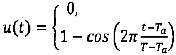
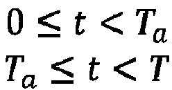 (ii) the compliance function to actuate la, Cla (t) = Cmin,la + 0.5 × (Cmax,la ― Cmin,la )u(t ― dla ), wherein Cmin,la and Cmax,la are the minimum and maximum values of the la compliance and dla represents a time delay between activation of the ra and the la; and(iii) the compliance functions to actuate rv and lv are represented as
- The processor implemented method of claim 1, wherein the total blood volume is estimated using height and weight of the subject.
- The processor implemented method of claim 1, wherein the step of estimating the SVR is represented as

- The processor implemented method of claim 1, wherein the heart rate of the subject is estimated using (i) a Photoplethysmogram (PPG) signal and an accelerometer signal or (ii) an Electrocardiogram (ECG) signal, from a wearable device worn by the subject when performing the activity.
- A system (100) comprising:
one or more data storage devices (102) operatively coupled to one or more hardware processors (104) and configured to store instructions for execution via the one or more hardware processors to:estimate a plurality of input parameters for a personalized cardiovascular hemodynamic model associated with a subject being monitored, the plurality of input parameters comprising (i) Systemic Vascular Resistance (SVR), (ii) unstressed blood volume, (iii) total blood volume in a body of the subject, and (iv) heart rate of the subject, wherein estimating the plurality of input parameters comprises:estimating the SVR using Metabolic EquivalenT (MET) levels associated with an activity level of the subject; andupdating the unstressed blood volume estimated when the subject is at rest, using an autoregulation method, when an activity is performed by the subject, wherein the autoregulation method comprises:sensing aortic pressure by baroreceptors located at carotid sinus and aortic arch;converting the sensed aortic pressure into a neural firing frequency via afferent sympathetic pathways;generating sympathetic and parasympathetic nervous activities via a central nervous system and efferent pathway depending on the neural firing frequency; andupdating an additional blood demand representing the unstressed blood volume during the activity using the generatedsympathetic and parasympathetic nervous activities; andestimate cardiac parameters including cardiac output, ejection fraction and mean arterial pressure, using the estimated plurality of input parameters. - The system of claim 7, wherein the one or more processors are configured to personalize a cardiovascular hemodynamic model to obtain the personalized cardiovascular hemodynamic model, prior to estimating the plurality of input parameters, based on one or more of (i) cardiac parameters obtained from an echocardiogram, (ii) ECG signal obtained from a wearable device worn by the subject when performing the activity and (iii) metadata of the subject including height and weight associated thereof.
- The system of claim 8, wherein the one or more processors are configured to perform sequential activation of a right atrium ra, a left atrium la, a right ventricle rv and a left ventricle lv of the personalized cardiovascular hemodynamic model using compliance functions Crα (t), Cla (t), Crv (t) and Clv (t) respectively, the compliance functions being defined as:(i) the compliance function to actuate ra, Cra (t) = Cmin,ra + 0.5 × (Cmax,ra ― Cmin,ra )u(t), wherein

 (ii) the compliance function to actuate la, Cla (t) = Cmin,la + 0.5 × (Cmax,la ― Cmin,la )u(t ― dla ), wherein Cmin,la and Cmax,la are the minimum and maximum values of the la compliance and dla represents a time delay between activation of the ra and the la; and(iii) the compliance functions to actuate rv and lv are represented as
- The system of claim 7, wherein the one or more processors are configured to estimate the total blood volume using height and weight of the subject.
- The system of claim 7, wherein the one or more processors are configured to estimate the SVR based on

- The system of claim 7, wherein the one or more processors are configured to estimate the heart rate of the subject using (i) a Photoplethysmogram (PPG) signal and an accelerometer signal or (ii) an Electrocardiogram (ECG) signal, from a wearable device worn by the subject when performing the activity.
- A computer program product comprising a non-transitory computer readable medium having a computer readable program embodied therein, wherein the computer readable program, when executed on a computing device, causes the computing device to:estimate a plurality of input parameters for a personalized cardiovascular hemodynamic model associated with a subject being monitored, the plurality of input parameters comprising (i) Systemic Vascular Resistance (SVR), (ii) unstressed blood volume, (iii) total blood volume in a body of the subject using height and weight of the subject, and (iv) heart rate of the subject using (i) a Photoplethysmogram (PPG) signal and an accelerometer signal or (ii) an Electrocardiogram (ECG) signal, from a wearable device worn by the subject when performing the activity, wherein estimating the plurality of input parameters comprises:estimating the SVR using Metabolic EquivalenT (MET) levels associated with an activity level of the subject; andupdating the unstressed blood volume estimated when the subject is at rest, using an autoregulation method, when an activity is performed by the subject, wherein the autoregulation method comprises:sensing aortic pressure by baroreceptors located at carotid sinus and aortic arch;converting the sensed aortic pressure into a neural firing frequency via afferent sympathetic pathways;generating sympathetic and parasympathetic nervous activities via a central nervous system and efferent pathway depending on the neural firing frequency; andupdating an additional blood demand representing the unstressed blood volume during the activity using the generatedsympathetic and parasympathetic nervous activities; andestimate cardiac parameters including cardiac output, ejection fraction and mean arterial pressure, using the estimated plurality of input parameters.
- The computer program product of claim 13, wherein the computer readable program further causes the computing device to personalize a cardiovascular hemodynamic model to obtain the personalized cardiovascular hemodynamic model, prior to estimating a plurality of input parameters, the personalizing being based on one or more of (i) cardiac parameters obtained from an echocardiogram, (ii) ECG signal obtained from a wearable device worn by the subject when performing the activity and (iii) metadata of the subject including height and weight associated thereof.
- The computer program product of claim 14, wherein the computer readable program further causes the computing device to (a) estimate a plurality of input parameters by sequential activation of a right atrium ra, a left atrium la, a right ventricle rv and a left ventricle lv of the personalized cardiovascular hemodynamic model using compliance functions Crα (t), Cla (t), Crv (t) and Clv (t) respectively, the compliance functions being defined as:(i) the compliance function to actuate ra, Crα (t) = Cmin,ra + 0.5 × (Cmax,ra ― Cmin,ra )u(t), wherein

 (ii) the compliance function to actuate la, Cla (t) = Cmin,la + 0.5 × (Cmax,la ― Cmin,la )u(t ― dla ), wherein Cmin,la and Cmax,la are the minimum and maximum values of the la compliance and dla represents a time delay between activation of the ra and the la; and(iii) the compliance functions to actuate rv and lv are represented as
(b) estimate the SVR as

Applications Claiming Priority (1)
| Application Number | Priority Date | Filing Date | Title |
|---|---|---|---|
| IN202121010972 | 2021-03-15 |
Publications (3)
| Publication Number | Publication Date |
|---|---|
| EP4060675A1 true EP4060675A1 (en) | 2022-09-21 |
| EP4060675C0 EP4060675C0 (en) | 2023-12-13 |
| EP4060675B1 EP4060675B1 (en) | 2023-12-13 |
Family
ID=78086228
Family Applications (1)
| Application Number | Title | Priority Date | Filing Date |
|---|---|---|---|
| EP21199728.3A Active EP4060675B1 (en) | 2021-03-15 | 2021-09-29 | Estimating cardiac parameters when performing an activity using a personalized cardiovascular hemodynamic model |
Country Status (2)
| Country | Link |
|---|---|
| US (1) | US20220287572A1 (en) |
| EP (1) | EP4060675B1 (en) |
-
2021
- 2021-09-29 EP EP21199728.3A patent/EP4060675B1/en active Active
- 2021-09-30 US US17/490,425 patent/US20220287572A1/en active Pending
Non-Patent Citations (2)
| Title |
|---|
| ROY DIBYENDU ET AL: "Hemodynamic Evaluation of a Novel Neuromodulation based Adaptive controller for Mitral Stenosis", 2020 EUROPEAN CONTROL CONFERENCE (ECC), EUCA, 12 May 2020 (2020-05-12), pages 264 - 270, XP033795789 * |
| ROY DIBYENDU ET AL: "Multimodal cardiovascular model for hemodynamic analysis: Simulation study on mitral valve disorders", PLOS ONE, vol. 16, no. 3, 4 March 2021 (2021-03-04), pages 1 - 28, XP055905181, Retrieved from the Internet <URL:https://doi.org/10.1371/journal.pone.0247921> [retrieved on 20220324], DOI: 10.1371/journal.pone.0247921 * |
Also Published As
| Publication number | Publication date |
|---|---|
| EP4060675C0 (en) | 2023-12-13 |
| US20220287572A1 (en) | 2022-09-15 |
| EP4060675B1 (en) | 2023-12-13 |
Similar Documents
| Publication | Publication Date | Title |
|---|---|---|
| US20230082362A1 (en) | Processes and methods to predict blood pressure | |
| US11311230B2 (en) | Medical premonitory event estimation | |
| Lu et al. | A human cardiopulmonary system model applied to the analysis of the Valsalva maneuver | |
| US11826559B2 (en) | Cardiac output control apparatus and method of use thereof | |
| US10750996B2 (en) | Multi-sensor body fluid volume index | |
| US20170282011A1 (en) | Method and apparatus for assessing cardiopulmonary fitness | |
| CA3181091A1 (en) | Systems, methods, and devices for non-invasive and continuous hemodynamic measurement | |
| CN105848581A (en) | Method and device for managing fluid volume in the body | |
| WO2018102431A1 (en) | Stroke detection using blood pressure surge | |
| US20210068683A1 (en) | Cardiovascular state monitoring - drug delivery apparatus and method of use thereof | |
| Pstras et al. | Mathematical modelling of cardiovascular response to the Valsalva manoeuvre | |
| EP4060675B1 (en) | Estimating cardiac parameters when performing an activity using a personalized cardiovascular hemodynamic model | |
| Roy et al. | Wearable sensor driven cardiac model to derive hemodynamic insights during exercise | |
| US20210146115A1 (en) | Cardiac output controlled electroactive polymer sleeve heart assist apparatus and method of use thereof | |
| US20260069168A1 (en) | Wearable computing device identification and use for role-based feedback | |
| US20250366719A1 (en) | Systems and methods for determining systemic vascular resistance using bioimpedance | |
| EP4736757A2 (en) | Lap signal processing to automatically calculate a/v ratio | |
| US20240342487A1 (en) | Systems and methods for cardiac pacing using beta blockers | |
| CN113164071A (en) | Health map for navigating health space | |
| Tu | High-Precision and Personalized Wearable Sensing Systems for Healthcare Applications | |
| Kazemi | Non-invasive cuff-less blood pressure estimation using LSTM-based recurrent neural networks | |
| Randall | Mathematical Analysis of Autonomic Control of Blood Pressure and Heart Rate | |
| Zaid | Modeling Cardiovascular, Ocular, and Urinary Systems for Advancing Basic Science, Disease Diagnosis, and Treatment | |
| EP4554461A1 (en) | Systems and methods for determining systemic vascular resistance using photoplethysmography | |
| CN121171473A (en) | Hypertension VR intervention method based on dynamic biofeedback and individuation modeling |
Legal Events
| Date | Code | Title | Description |
|---|---|---|---|
| PUAI | Public reference made under article 153(3) epc to a published international application that has entered the european phase |
Free format text: ORIGINAL CODE: 0009012 |
|
| STAA | Information on the status of an ep patent application or granted ep patent |
Free format text: STATUS: REQUEST FOR EXAMINATION WAS MADE |
|
| 17P | Request for examination filed |
Effective date: 20220727 |
|
| AK | Designated contracting states |
Kind code of ref document: A1 Designated state(s): AL AT BE BG CH CY CZ DE DK EE ES FI FR GB GR HR HU IE IS IT LI LT LU LV MC MK MT NL NO PL PT RO RS SE SI SK SM TR |
|
| REG | Reference to a national code |
Ref legal event code: R079 Ipc: A61B0005021000 Ref country code: DE Ref legal event code: R079 Ref document number: 602021007648 Country of ref document: DE Free format text: PREVIOUS MAIN CLASS: G16H0040630000 Ipc: A61B0005021000 |
|
| GRAP | Despatch of communication of intention to grant a patent |
Free format text: ORIGINAL CODE: EPIDOSNIGR1 |
|
| STAA | Information on the status of an ep patent application or granted ep patent |
Free format text: STATUS: GRANT OF PATENT IS INTENDED |
|
| RIC1 | Information provided on ipc code assigned before grant |
Ipc: A61B 5/00 20060101ALI20230531BHEP Ipc: G16H 50/20 20180101ALI20230531BHEP Ipc: G16H 40/63 20180101ALI20230531BHEP Ipc: G16H 50/50 20180101ALI20230531BHEP Ipc: A61B 5/0245 20060101ALI20230531BHEP Ipc: A61B 5/11 20060101ALI20230531BHEP Ipc: A61B 5/024 20060101ALI20230531BHEP Ipc: A61B 5/021 20060101AFI20230531BHEP |
|
| INTG | Intention to grant announced |
Effective date: 20230623 |
|
| GRAS | Grant fee paid |
Free format text: ORIGINAL CODE: EPIDOSNIGR3 |
|
| GRAA | (expected) grant |
Free format text: ORIGINAL CODE: 0009210 |
|
| STAA | Information on the status of an ep patent application or granted ep patent |
Free format text: STATUS: THE PATENT HAS BEEN GRANTED |
|
| AK | Designated contracting states |
Kind code of ref document: B1 Designated state(s): AL AT BE BG CH CY CZ DE DK EE ES FI FR GB GR HR HU IE IS IT LI LT LU LV MC MK MT NL NO PL PT RO RS SE SI SK SM TR |
|
| REG | Reference to a national code |
Ref country code: GB Ref legal event code: FG4D |
|
| REG | Reference to a national code |
Ref country code: CH Ref legal event code: EP |
|
| REG | Reference to a national code |
Ref country code: DE Ref legal event code: R096 Ref document number: 602021007648 Country of ref document: DE |
|
| REG | Reference to a national code |
Ref country code: IE Ref legal event code: FG4D |
|
| U01 | Request for unitary effect filed |
Effective date: 20231213 |
|
| U07 | Unitary effect registered |
Designated state(s): AT BE BG DE DK EE FI FR IT LT LU LV MT NL PT SE SI Effective date: 20231219 |
|
| PG25 | Lapsed in a contracting state [announced via postgrant information from national office to epo] |
Ref country code: GR Free format text: LAPSE BECAUSE OF FAILURE TO SUBMIT A TRANSLATION OF THE DESCRIPTION OR TO PAY THE FEE WITHIN THE PRESCRIBED TIME-LIMIT Effective date: 20240314 |
|
| PG25 | Lapsed in a contracting state [announced via postgrant information from national office to epo] |
Ref country code: ES Free format text: LAPSE BECAUSE OF FAILURE TO SUBMIT A TRANSLATION OF THE DESCRIPTION OR TO PAY THE FEE WITHIN THE PRESCRIBED TIME-LIMIT Effective date: 20231213 |
|
| PG25 | Lapsed in a contracting state [announced via postgrant information from national office to epo] |
Ref country code: GR Free format text: LAPSE BECAUSE OF FAILURE TO SUBMIT A TRANSLATION OF THE DESCRIPTION OR TO PAY THE FEE WITHIN THE PRESCRIBED TIME-LIMIT Effective date: 20240314 Ref country code: ES Free format text: LAPSE BECAUSE OF FAILURE TO SUBMIT A TRANSLATION OF THE DESCRIPTION OR TO PAY THE FEE WITHIN THE PRESCRIBED TIME-LIMIT Effective date: 20231213 |
|
| PG25 | Lapsed in a contracting state [announced via postgrant information from national office to epo] |
Ref country code: RS Free format text: LAPSE BECAUSE OF FAILURE TO SUBMIT A TRANSLATION OF THE DESCRIPTION OR TO PAY THE FEE WITHIN THE PRESCRIBED TIME-LIMIT Effective date: 20231213 Ref country code: NO Free format text: LAPSE BECAUSE OF FAILURE TO SUBMIT A TRANSLATION OF THE DESCRIPTION OR TO PAY THE FEE WITHIN THE PRESCRIBED TIME-LIMIT Effective date: 20240313 Ref country code: HR Free format text: LAPSE BECAUSE OF FAILURE TO SUBMIT A TRANSLATION OF THE DESCRIPTION OR TO PAY THE FEE WITHIN THE PRESCRIBED TIME-LIMIT Effective date: 20231213 |
|
| PG25 | Lapsed in a contracting state [announced via postgrant information from national office to epo] |
Ref country code: IS Free format text: LAPSE BECAUSE OF FAILURE TO SUBMIT A TRANSLATION OF THE DESCRIPTION OR TO PAY THE FEE WITHIN THE PRESCRIBED TIME-LIMIT Effective date: 20240413 |
|
| PG25 | Lapsed in a contracting state [announced via postgrant information from national office to epo] |
Ref country code: CZ Free format text: LAPSE BECAUSE OF FAILURE TO SUBMIT A TRANSLATION OF THE DESCRIPTION OR TO PAY THE FEE WITHIN THE PRESCRIBED TIME-LIMIT Effective date: 20231213 |
|
| PG25 | Lapsed in a contracting state [announced via postgrant information from national office to epo] |
Ref country code: SK Free format text: LAPSE BECAUSE OF FAILURE TO SUBMIT A TRANSLATION OF THE DESCRIPTION OR TO PAY THE FEE WITHIN THE PRESCRIBED TIME-LIMIT Effective date: 20231213 |
|
| PG25 | Lapsed in a contracting state [announced via postgrant information from national office to epo] |
Ref country code: SM Free format text: LAPSE BECAUSE OF FAILURE TO SUBMIT A TRANSLATION OF THE DESCRIPTION OR TO PAY THE FEE WITHIN THE PRESCRIBED TIME-LIMIT Effective date: 20231213 Ref country code: SK Free format text: LAPSE BECAUSE OF FAILURE TO SUBMIT A TRANSLATION OF THE DESCRIPTION OR TO PAY THE FEE WITHIN THE PRESCRIBED TIME-LIMIT Effective date: 20231213 Ref country code: RO Free format text: LAPSE BECAUSE OF FAILURE TO SUBMIT A TRANSLATION OF THE DESCRIPTION OR TO PAY THE FEE WITHIN THE PRESCRIBED TIME-LIMIT Effective date: 20231213 Ref country code: IS Free format text: LAPSE BECAUSE OF FAILURE TO SUBMIT A TRANSLATION OF THE DESCRIPTION OR TO PAY THE FEE WITHIN THE PRESCRIBED TIME-LIMIT Effective date: 20240413 Ref country code: CZ Free format text: LAPSE BECAUSE OF FAILURE TO SUBMIT A TRANSLATION OF THE DESCRIPTION OR TO PAY THE FEE WITHIN THE PRESCRIBED TIME-LIMIT Effective date: 20231213 |
|
| PG25 | Lapsed in a contracting state [announced via postgrant information from national office to epo] |
Ref country code: PL Free format text: LAPSE BECAUSE OF FAILURE TO SUBMIT A TRANSLATION OF THE DESCRIPTION OR TO PAY THE FEE WITHIN THE PRESCRIBED TIME-LIMIT Effective date: 20231213 |
|
| PG25 | Lapsed in a contracting state [announced via postgrant information from national office to epo] |
Ref country code: PL Free format text: LAPSE BECAUSE OF FAILURE TO SUBMIT A TRANSLATION OF THE DESCRIPTION OR TO PAY THE FEE WITHIN THE PRESCRIBED TIME-LIMIT Effective date: 20231213 |
|
| REG | Reference to a national code |
Ref country code: DE Ref legal event code: R097 Ref document number: 602021007648 Country of ref document: DE |
|
| PLBE | No opposition filed within time limit |
Free format text: ORIGINAL CODE: 0009261 |
|
| STAA | Information on the status of an ep patent application or granted ep patent |
Free format text: STATUS: NO OPPOSITION FILED WITHIN TIME LIMIT |
|
| U20 | Renewal fee for the european patent with unitary effect paid |
Year of fee payment: 4 Effective date: 20240926 |
|
| 26N | No opposition filed |
Effective date: 20240916 |
|
| U1H | Name or address of the proprietor changed after the registration of the unitary effect |
Owner name: TATA CONSULTANCY SERVICES LIMITED; IN |
|
| PG25 | Lapsed in a contracting state [announced via postgrant information from national office to epo] |
Ref country code: MC Free format text: LAPSE BECAUSE OF FAILURE TO SUBMIT A TRANSLATION OF THE DESCRIPTION OR TO PAY THE FEE WITHIN THE PRESCRIBED TIME-LIMIT Effective date: 20231213 |
|
| PG25 | Lapsed in a contracting state [announced via postgrant information from national office to epo] |
Ref country code: IE Free format text: LAPSE BECAUSE OF NON-PAYMENT OF DUE FEES Effective date: 20240929 |
|
| REG | Reference to a national code |
Ref country code: CH Ref legal event code: U11 Free format text: ST27 STATUS EVENT CODE: U-0-0-U10-U11 (AS PROVIDED BY THE NATIONAL OFFICE) Effective date: 20251001 |
|
| PGFP | Annual fee paid to national office [announced via postgrant information from national office to epo] |
Ref country code: GB Payment date: 20250923 Year of fee payment: 5 |
|
| U20 | Renewal fee for the european patent with unitary effect paid |
Year of fee payment: 5 Effective date: 20250925 |
|
| PGFP | Annual fee paid to national office [announced via postgrant information from national office to epo] |
Ref country code: CH Payment date: 20251001 Year of fee payment: 5 |
|
| PG25 | Lapsed in a contracting state [announced via postgrant information from national office to epo] |
Ref country code: CY Free format text: LAPSE BECAUSE OF FAILURE TO SUBMIT A TRANSLATION OF THE DESCRIPTION OR TO PAY THE FEE WITHIN THE PRESCRIBED TIME-LIMIT; INVALID AB INITIO Effective date: 20210929 |
|
| PG25 | Lapsed in a contracting state [announced via postgrant information from national office to epo] |
Ref country code: HU Free format text: LAPSE BECAUSE OF FAILURE TO SUBMIT A TRANSLATION OF THE DESCRIPTION OR TO PAY THE FEE WITHIN THE PRESCRIBED TIME-LIMIT; INVALID AB INITIO Effective date: 20210929 |